Pediatric viral respiratory infections in Saudi Arabia
11
KUWAIT MEDICAL JOURNAL 118 June 2020 Review Article Kuwait Medical Journal 2020; 52 (2): 118 - 128 Pediatric viral respiratory infections in Saudi Arabia: Narrative and descriptive revisits for the etiology, epidemiology and clinical phenotypes with diagnostic challenges highlights ABSTRACT KEY WORDS: children, Middle East, respiratory infections, Saudi Arabia, viruses Ayed A Shati 1 , Abdelwahid S Ali 2 , Ahmed M Al-Hakami 2 , Ali A Asseri 1 , Saleh M Al-Qahatani 1 1 Department of Child Health, College of Medicine, King Khalid University, Abha, Saudi Arabia 2 Department of Microbiology and Clinical Parasitology, College of Medicine, King Khalid University, Abha, Saudi Arabia Address correspondence to: Dr. Ayed Abdullah Shati, Department of Child Health, College of Medicine, King Khalid University, Abha, Saudi Arabia. Tel: +966555752063; Mob: +966172417750; E-mail: [email protected] Objective: To review, describe and narrate the etiology, epidemiology and clinical phenotypes of pediatric viral respiratory infections (PVRIs) in Saudi Arabia (KSA) Design: A comprehensive electronic search of the literature for PVRIs in KSA Seing: An electronic search in PubMed, SCOPUS, Google Scholar and MEDLINE Subjects: Articles published up till 2019 and those that dealt with the detection of viruses from clinical specimens Interventions: Retrieved articles were subtly studied. Data obtained included the virus reported, year of publication, diagnostics, region or city, research purpose, the season of infection and hospital of admission (if any). Main outcome measure(s): The etiology, epidemiological features and clinical phenotypes associated with the PVRIs in each study were recorded and analyzed. Result(s): The first report for PVRIs in KSA was in 1988. Up to 2019, 35 studies were published in the topic. The highest number of reports was for respiratory syncytial viruses (n=17, 48.6%), while bocaviruses are the least reported viruses (n=3, 8.6%). Clinical presentations reported suggested both upper and lower respiratory tract infections. PVRIs were reported from Riyadh, Al- Qassim, Jazan, Jeddah, Dammam, Najran, Taif and Abha. Immunofluorescence assays, enzyme-linked immunosorbent assay, polymerase chain reaction and virus isolation were employed. Conclusion(s): In this review communication, we described the etiology, epidemiology and clinical phenotypes of PVRIs in KSA reported from 1988 through 2019. We also showed some challenges associated with the diagnostic protocols employed. Directions for future research in this topic, particularly towards diagnostics and preventive medicine, were also recommended. INTRODUCTION It is well-established that respiratory tract infections are major causes of morbidities and mortalities among children worldwide [1] . A recent World Health Organization (WHO) report revealed that 18% of deaths among children under five years are aributable to pneumonia [2,3] . Among the different causes of respiratory infections, viruses were known to constitute the major etiological factor responsible for acute respiratory infections (ARIs) in pediatrics globally [4,5] . In poor and developing countries, these infections resulted in far higher mortality rates in children as compared to those in resource- rich countries. Inadequate data about the public health impacts and economic losses of these infections in developed countries are available [6-8] . These data are always critically essential for the design and development of efficient protective measures and treatment protocols. The viruses which are known to be responsible for ARIs in infants and children included influenza viruses, human respiratory syncytial virus (HRSV), human metapneumovirus (HMPV), human
-
Upload
khangminh22 -
Category
Documents
-
view
2 -
download
0
Transcript of Pediatric viral respiratory infections in Saudi Arabia
KUWAIT MEDICAL JOURNAL 118June 2020
Review Article
Kuwait Medical Journal 2020; 52 (2): 118 - 128
Pediatric viral respiratory infections in Saudi Arabia: Narrative and descriptive revisits for the
etiology, epidemiology and clinical phenotypes with diagnostic challenges highlights
ABSTRACT
KEY WORDS: children, Middle East, respiratory infections, Saudi Arabia, viruses
Ayed A Shati1, Abdelwahid S Ali2, Ahmed M Al-Hakami2, Ali A Asseri1, Saleh M Al-Qahatani1 1Department of Child Health, College of Medicine, King Khalid University, Abha, Saudi Arabia
2Department of Microbiology and Clinical Parasitology, College of Medicine, King Khalid University, Abha, Saudi Arabia
Address correspondence to:Dr. Ayed Abdullah Shati, Department of Child Health, College of Medicine, King Khalid University, Abha, Saudi Arabia. Tel: +966555752063; Mob: +966172417750; E-mail: [email protected]
Objective: To review, describe and narrate the etiology, epidemiology and clinical phenotypes of pediatric viral respiratory infections (PVRIs) in Saudi Arabia (KSA)Design: A comprehensive electronic search of the literature for PVRIs in KSASetting: An electronic search in PubMed, SCOPUS, Google Scholar and MEDLINESubjects: Articles published up till 2019 and those that dealt with the detection of viruses from clinical specimensInterventions: Retrieved articles were subtly studied. Data obtained included the virus reported, year of publication, diagnostics, region or city, research purpose, the season of infection and hospital of admission (if any).Main outcome measure(s): The etiology, epidemiological features and clinical phenotypes associated with the PVRIs in each study were recorded and analyzed.Result(s): The first report for PVRIs in KSA was in 1988.
Up to 2019, 35 studies were published in the topic. The highest number of reports was for respiratory syncytial viruses (n=17, 48.6%), while bocaviruses are the least reported viruses (n=3, 8.6%). Clinical presentations reported suggested both upper and lower respiratory tract infections. PVRIs were reported from Riyadh, Al-Qassim, Jazan, Jeddah, Dammam, Najran, Taif and Abha. Immunofluorescence assays, enzyme-linked immunosorbent assay, polymerase chain reaction and virus isolation were employed.Conclusion(s): In this review communication, we described the etiology, epidemiology and clinical phenotypes of PVRIs in KSA reported from 1988 through 2019. We also showed some challenges associated with the diagnostic protocols employed. Directions for future research in this topic, particularly towards diagnostics and preventive medicine, were also recommended.
INTRODUCTIONIt is well-established that respiratory tract
infections are major causes of morbidities and mortalities among children worldwide[1]. A recent World Health Organization (WHO) report revealed that 18% of deaths among children under five years are attributable to pneumonia[2,3]. Among the different causes of respiratory infections, viruses were known to constitute the major etiological factor responsible for acute respiratory infections (ARIs) in pediatrics globally[4,5]. In poor and developing countries, these
infections resulted in far higher mortality rates in children as compared to those in resource- rich countries. Inadequate data about the public health impacts and economic losses of these infections in developed countries are available[6-8]. These data are always critically essential for the design and development of efficient protective measures and treatment protocols. The viruses which are known to be responsible for ARIs in infants and children included influenza viruses, human respiratory syncytial virus (HRSV), human metapneumovirus (HMPV), human
June 2020119 Pediatric viral respiratory infections in Saudi Arabia: Narrative and descriptive revisits for the etiology...
coronaviruses (HCoVs), human parainfluenza viruses (HPIV), human adenoviruses (HAdVs), and human bocaviruses (HBoVs).
Although the burden and public health implications of influenza infections are yet to be known in many parts of the world, particularly in resource-poor countries, it has been shown that influenza is characterized by high incidence rates and hospitalizations in the tropics and subtropics[8]. The global incidences and mortalities due to influenza in children less than five years was also estimated and noted to be significantly high in terms of number of deaths, particularly in the developing countries[8-10]. In many epidemiological studies, HMPV were recognized as potential etiologic agents for acute lower respiratory tract infections (ALRTIs), namely bronchopneumonia and bronchiolitis, in children[11-16]. Although the life-threatening effects of HMPV infections in children were known in some cases, the fatalities were usually seen associated with other underlying chronic conditions such as chronic lung disease of prematurity, asthma[17], cancer[18] and immunosuppression[19,20]. Following intensive literature revisits and personal communications, there were no obvious distinctions in the epidemiological and clinical patterns between HRSV and HMPV infections in pediatric patients. Both viruses cause severe and ALRTIs in infants and children, and both represent primary reasons for emergency department visits and hospitalization[18,20]. However, HMPV infections were observed to occur in relatively older children, as compared to HRSV[17]. HCoVs were also noted, causing numerous seasonal clinical respiratory infections in children since ancient times[21]. The epidemiological patterns of these HCoVs-associated diseases were observed undergoing regular changes every time and the respiratory infections attributable to HCoVs were observed not only confined to upper respiratory tract infections (URTIs), but they are also seen to cause pneumonia in infants and children[22]. Several reports describing the role of HCoVs in the lower respiratory tract infections (LRTIs) with their clinical presentations and complications in the pediatrics were also published[23-25]. HCoV-severe acute respiratory syndrome (HCoV-SARS) has emerged as the first HCoV identified responsible for severe, acute and fatal respiratory infection in children in many parts of the world[26,27]. Recently, HCoV- Middle East Respiratory Syndrome (HCoV-MERS) has also emerged from KSA and proven as a potential etiologic agent for severe URTIs and LRTIs in children and young adults[28]. Although HAdVs were primarily recognized to cause mild URT and LRT infections in young children, severe infections of HAdVs during epidemics were also noted in immunocompromised patients[29]. Several serotypes of HAdVs were isolated
from children suffering from ALRTIs. It has been suggested that the epidemiological parameters of the infections including incidences, prevalences, morbidities and mortalities due to HAdVs are highly virus type dependent[30]. HPIVs were also recognized to be associated with URT and LRT illnesses in children with their pathology, symptomatology and epidemiology dependent on the virus serotype, age and season[31]. Among the four genotypes of HBoVs known so far, HBoV-1 is the predominant genotype known to infect the respiratory tract of children[32,33]. Throughout the years, human rhinoviruses (HRVs), were known as a significant cause of the “common cold” in children and adults worldwide, with considerable economic burdens incurred due to medical visits, schooling and work absenteeism[34]. Other viruses causing respiratory infections in children and reported at various points of time include Nipah and Hindra viruses[35].
Taken all together, viral respiratory infections are considered as major health concerns in pediatric practice in many parts of the world, particularly in developing and resource-poor countries. As diagnostic testing is improved, more respiratory viruses are expected to be explored. This review intended to focus on the clinical phenotypes and epidemiological pictures of pediatric viral respiratory infections (PVRIs) in Saudi Arabia (KSA) concerning the previously published reports in that respect. We would also like to cast some light on the diagnostic and medical intervention challenges.
METHODSSearch methodology
In this study, we performed a comprehensive electronic search of the literature for PVRIs in KSA in PubMed, SCOPUS, Google Scholar and MEDLINE databases. The keywords used in the search include Saudi Arabia, respiratory, children, infants and viruses (the word “virus” was used at times and the virus name was specified at other times). The search considered articles from ancient times up to 2019. The retrieved articles were carefully read to help in further search for additional articles. The only considered and documented studies are those that dealt with the detection and/or isolation of viruses from clinical specimens of the patients and published in peer-reviewed journals.
Data retrieval and analysisThe clinical and epidemiological features associated
with PVRIs in each study were recorded and analyzed. The elements recorded included the author(s) who conducted the research, year of publication, the virus reported to cause the infection, diagnostic technique employed, region or city of the study, the research purpose, the season of the infection and the hospital
KUWAIT MEDICAL JOURNAL 120June 2020
of patient admission (if any). A narrative description of the situations was then generated in this manuscript using this data.
LITERATURE REVIEWEpidemiology and clinical phenotypes of PVRIs
The previous reports for the detection of different respiratory viruses among pediatric patients in KSA from 1988 - 2019 are demonstrated in Table 1. The total number of reports of respiratory viruses in children in different cities of KSA is determined (Fig. 1). The geographical distribution of these infections in the different cities and regions of the country is also retrieved from the data obtained (Fig. 2). Detailed descriptions for the etiology, epidemiology and clinical features of the PVRIs in infants and children in KSA, as retrieved from the previously published research, are made in the following presentations:
InfluenzaInfluenza is a highly contagious disease caused
by influenza viruses type A, B and C (IAV, IBV and ICV). These viruses are characterized by segmented RNA genomes and antigenic diversities. They were classified with the family Orthomyxoviridae. IAV causes pandemics and epidemics worldwide; those belonging to group B cause epidemics and outbreaks; while those belonging to group C cause sporadic and mild respiratory infections[36]. In KSA, the first report for influenza was made in 1988, when a cross-sectional epidemiological study was carried out to determine the viral etiology of tonsillitis among children in Riyadh[37].
In another study, a significant percentage of infants and children attending the emergency or pediatric clinics of King Faisal Specialist Hospital (KFSH) in Riyadh also reported positive to influenza viruses in 1998[38]. IAV and IBV were also detected in 2005 in the nasopharyngeal secretions of young children admitted with acute LRTIs to Buraidah Maternity and Pediatric Hospital (BMPH) in Al-Qasim (central province of KSA)[39].
Additionally, during the swine flu global pandemic in 2009, AlMazroa and co-workers published a comprehensive report showing a significant number of cases affected with swine flu virus (A-H1N1) among children aged 1 - 10 years in different regions and localities of KSA. These children included both Saudi nationals and imported cases, and concluded that influenza A-H1N1 constitute a considerable threat to KSA people at the time of the study[40]. In another epidemiological study, six cases of IAV and one case of IBV were also diagnosed using multiplex RT-PCR among children at Najran Maternity and Children Hospital (NMCH) in Najran (southern region of KSA), without adequate description to the clinical and epidemiological patterns of these infections being reported[41]. In a comprehensive surveillance study in Jazan (southwestern region of KSA), IAV and IBV were also detected among pediatric patients without the determination of virus subtypes[42]. In a more recent study, the influenza virus A-H1N1 was isolated from children suspected with HCoV-MERS and presented to John Hopkins Aramco Healthcare facilities in Dammam (the eastern region of KSA)[43]. In this study,
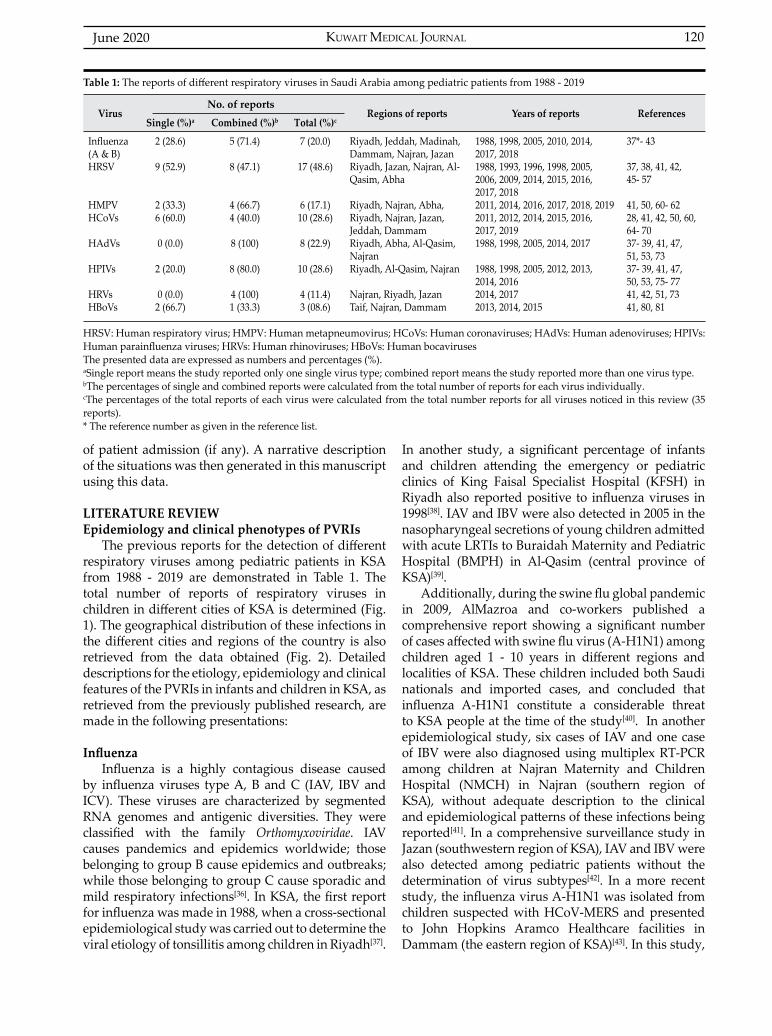
VirusNo. of reports
Regions of reports Years of reports ReferencesSingle (%)a Combined (%)b Total (%)c
Influenza (A & B)HRSV
HMPVHCoVs
HAdVs
HPIVs
HRVsHBoVs
2 (28.6)
9 (52.9)
2 (33.3)6 (60.0)
0 (0.0)
2 (20.0)
0 (0.0)2 (66.7)
5 (71.4)
8 (47.1)
4 (66.7)4 (40.0)
8 (100)
8 (80.0)
4 (100)1 (33.3)
7 (20.0)
17 (48.6)
6 (17.1)10 (28.6)
8 (22.9)
10 (28.6)
4 (11.4)3 (08.6)
Riyadh, Jeddah, Madinah, Dammam, Najran, JazanRiyadh, Jazan, Najran, Al-Qasim, Abha
Riyadh, Najran, Abha, Riyadh, Najran, Jazan, Jeddah, DammamRiyadh, Abha, Al-Qasim, NajranRiyadh, Al-Qasim, Najran
Najran, Riyadh, JazanTaif, Najran, Dammam
1988, 1998, 2005, 2010, 2014, 2017, 20181988, 1993, 1996, 1998, 2005, 2006, 2009, 2014, 2015, 2016, 2017, 2018 2011, 2014, 2016, 2017, 2018, 20192011, 2012, 2014, 2015, 2016, 2017, 2019 1988, 1998, 2005, 2014, 2017
1988, 1998, 2005, 2012, 2013, 2014, 20162014, 20172013, 2014, 2015
37*- 43
37, 38, 41, 42, 45- 57
41, 50, 60- 6228, 41, 42, 50, 60, 64- 7037- 39, 41, 47, 51, 53, 7337- 39, 41, 47, 50, 53, 75- 7741, 42, 51, 7341, 80, 81
Table 1: The reports of different respiratory viruses in Saudi Arabia among pediatric patients from 1988 - 2019
HRSV: Human respiratory virus; HMPV: Human metapneumovirus; HCoVs: Human coronaviruses; HAdVs: Human adenoviruses; HPIVs: Human parainfluenza viruses; HRVs: Human rhinoviruses; HBoVs: Human bocavirusesThe presented data are expressed as numbers and percentages (%).aSingle report means the study reported only one single virus type; combined report means the study reported more than one virus type.bThe percentages of single and combined reports were calculated from the total number of reports for each virus individually.cThe percentages of the total reports of each virus were calculated from the total number reports for all viruses noticed in this review (35 reports). * The reference number as given in the reference list.

June 2020121
Fig 1: Report rates of pediatric respiratory viruses in different cities of KSA Riyadh (central province); Dammam (eastern province); Al-Qasim (northern province); Abha (Asir region, southern province); Jeddah (western province); Najran (Najran region, southern province); Jazan (southwestern region); Taif (western province)Rates were calculated as percentages from number of reports in each region out of the total number of reports in the kingdom (35 reports)
Fig 2: Geographic distribution of viral respiratory infections in children and infants in Saudi Arabia through 1988-2019HRSV: human respiratory virus; HMPV: human metapneumovirus; HCoVs: human coronaviruses; HAdVs: human adenoviruses; HPIVs: human parainfluenza viruses; HRVs: human rhinoviruses; HBoVs: human bocaviruses
Pediatric viral respiratory infections in Saudi Arabia: Narrative and descriptive revisits for the etiology...
KUWAIT MEDICAL JOURNAL 122June 2020
the authors declared higher incidence among patients came from Ras Tanura city (far eastern region of KSA; Fig. 2).
In total, a considerable number of reports about influenza infections among pediatric patients in KSA was noted in the literature. The majority of the available data were those published during the global “avian flu” pandemic in 2005 - 2006 and the global “swine flu” pandemic in 2009 - 2010. Despite all these reports, we believe that adequate data pertaining to the virology, clinical phenotypes and epidemiological aspects of influenza viruses in KSA, among adult populations in general and children in specific, are still lacking.
Human respiratory syncytial virus (HRSV)HRSV was confirmed in many studies and surveys
as the leading cause of respiratory tract infections in infants and young children throughout the world. HRSV is a non-segmented, negative-sense, single-stranded RNA enveloped virus belonging to the family Pneumoviridae and the genus pneumovirus[44]. As compared to influenza, more studies were conducted to detect HRSV from children in different cities and regions of KSA, namely Riyadh[37,38,45-51], Najran[41], Jazan[42], Al-Qasim[52] and Abha[53]. In all of these studies, no detailed clinical presentations or specific epidemiological patterns for HRSV infections were described; however, bronchopneumonia (BP) and bronchiolitis were reported as the leading cause of morbidities and hospitalization. It was also revealed that the infections of HRSV peak in winters and autumn seasons. Deaths attributable to these infections among infants were also recorded, though dearth. The genetic analysis of HRSV group B (HRSV-B) strains circulating in KSA was also studied in 2014 by Almajhdi and co-researchers[54]. This study helped to cast some light on the circulation pattern and molecular characteristics of HRSV- B strain in the KSA.
Medical intervention of HRSV infectivity among children in KSA remains the most crucial concern to health practitioners and the public. Special attention was particularly directed towards hospitalized RSV- infected premature infants and children suffering from chronic lung disease or congenital heart disease. Palivizumab (Synagis, Medimmune) is a humanized monoclonal antibody, which is a combination of human (95%) and murine (5%) antibody sequences. In KSA, Palivizumb was used for the prevention of severe RSV infections in high-risk groups of children[55,56]. It was well-documented that Palivizumb is an effective agent for the prevention of RSV mortalities and fatalities. However, the notable possible drawbacks associated with this medication include the expense and extensiveness of use without rationality.
Conclusively, the different studies concerning the treatment of children against HRSV using different types of medicines and regimens in KSA were recently reviewed by Alharbi and co-researchers in 2018[57].
Human metapneumovirus (HMPV)HMPV was first recognized and described in
Netherlands in 2001 by van den Hoogen and others[58]. It is a negative sense, single-stranded RNA enveloped virus and classified as a member of the Paramyxoviridae family, pneumovirinae subfamily and pneumovirus genus[59]. In KSA, HMPV was first detected in 2011 among Saudi children hospitalized with ARI in Riyadh[60]. Afterward, the virus was also detected among children who presented with ARTIs to NMCH in Najran, with brief explanations of the clinical and epidemiological correlates of the infections[41]. The authors identified the virus in infants in both single viral infections and viral co-infections. HMPV was also isolated during an investigation in a cohort of children suffering severe LRTIs and hospitalized at a tertiary referral center in Riyadh in 2016[50]. The epidemiology and some genetic properties of that isolated virus were also studied[61]. That is considered the first report addressing the HMPV genotypes circulation in the KSA. HMPV was also detected in a retrospective study performed to determine the viral etiological agents causing RTIs among pediatric patients admitted to the Department of Pediatrics, King Abdul-Aziz Medical City (KAMC) in Riyadh[51].
In a recent publication, we reported the incidence of HMPV in the Aseer region (southern provinces of KSA) among children who attended the pediatric emergency rooms and were hospitalized in Aseer Central Hospital with acute bronchiolitis and BP[62]. In that study, we reported for the first time the large population of virus-infected children in the area, particularly in Abha city and the vicinity. In the previously published articles, reverse transcriptase PCR (RT-PCR) and direct fluorescent antibody (DFA) test were employed for the detection of the virus nucleic acid and antigens respectively, in the nasopharyngeal secretions from infected children. Apart from the previously mentioned reports of HMPV infections, the virus was not detected in any other region or city of the Kingdom. The role of HMPV in respiratory infections in pediatric medicine in the western, eastern and northern regions of KSA was not determined yet. The risk factors associated with the severity of HMPV in children in the KSA were not precisely known up to date. In conclusion, meager studies regarding the virus distribution, prevalence, clinical presentations and medical interventions of HMPV in KSA were made so far, and much research work is worthy in these aspects.
June 2020123
Human coronaviruses (HCoVs)Human coronaviruses (HCoVs) are a well-known
cause for URT and LRT infections in all age groups. They are positive-sense, single-stranded RNA viruses classified in the family Coronaviridae[63]. Before HCoV-SARS and HCoV-MERS, which cause severe and acute respiratory infections, four other HCoVs were recognized to be responsible for mild respiratory infections in adults and children namely HCoV-229E, HCoV-OC43, HCoV-NL63 and HCoV-HKU1. The first report for HCoVs infections among children in KSA was made by Al-Hajjar and co-workers in 2011[60]. In this study, HCoV-NL63 was isolated from children less than 16 years old hospitalized with ART illness during autumn and winter at KFSH in Riyadh. HCoV-NL63 and HCoV-OC43 were also detected among children presented with acute LRT infections and showing symptoms of bronchiolitis and pneumonia in Najran region of KSA in 2014[41]. In another cross-sectional study carried out to determine the prevalence of viruses causing ARTIs among hospitalized children in Riyadh area, a considerable number of them were diagnosed with HCoVs[50]. The HCoVs serotypes detected in this study were not specified and were most prevalent in infants less than six months old. Non-MERS HCoVs were also detected in nasopharyngeal swabs collected from asymptomatic outpatients less than 15 years old in Jazan province[42]. Despite the wide spread of HCoV-SARS during the global disease pandemic in 2003, no single report about the virus infectivity in children in KSA was made. As the HCoV-MERS originated from Jeddah (western region of KSA) in 2012[28], plenty of reports, thereafter, describing the virological, epidemiological and clinical characteristics of the virus were made in KSA at large. Primarily, a single pediatric case (out of 47 sampled patients), laboratory confirmed positive to HCoV-MERS was identified by Assiri and co-workers throughout KSA as a first report, in 2013[64]. Consequently, HCoV-MERS was also reported among eleven pediatric patients in Riyadh in 2014[65]. In this report, the clinical presentations and outcomes of the patients were adequately described; nine cases were asymptomatic, whereas two cases were admitted to the ICUs suffering severe respiratory infections, with one of whom died. A case report describing a complicated HCoV-MERS infection in a 9-month-old child admitted to the pediatric ICU of Prince Sultan Military Medical City in Riyadh, with infantile nephrotic syndrome that resulted in death, was also published by Thabet and his team in 2015[66]. In a comprehensive search in the literature made by Al-Tawfiq and co-researchers in 2016; a conclusion was drawn to indicate that low number of MERS pediatric cases were evident in KSA and the reasons for this low prevalence was unknown[67]. In this report, the
symptoms, mean age of infection and source of infection among pediatric patients were described to an optimal level. The various scientific elements of HCoV-MERS infectivity for children were also recently reviewed by Al-Sehaibany in 2017, when he concluded that MERS is a disease of adults, where few cases among children were known with different clinical manifestations and lower mortality rates as compared to infection in adults[68]. Conclusively, despite the intensive studies for detection of HCoV-MERS among adults in KSA and surrounding countries, a limited number of cases were reported for this virus in children. However, in recent publications, it had been reported that the proportion of asymptomatic cases among pediatric confirmed HCoV-MERS cases was significantly high[69,70].
Human adenoviruses (HAdVs)Human adenoviruses (HAdVs) were known to have
a significant contribution in PVRIs. HAdVs infections were observed as mild and indistinguishable from other viral respiratory infections in children on their pathology and clinical grounds[71]. However, acute and severe URT and LRT infections attributable to HAdVs were also noted among pediatric patients[71,72]. Since 1988 up to date, only nine studies discussing the incidences and other clinical and epidemiological parameters of adenoviral infections in children in KSA were made. In a serological and virological investigation by Hossain et al in 1988, the first report for HAdVs infections in children in KSA was made. They showed that HAdVs were the leading cause of tonsillitis among Saudi children during the study period[37]. About ten years later, adenovirus infections were also reported in children who attended the emergency of pediatric wards at KFSH and Research Center in Riyadh[38]. In the same year, HAdVs infections were also detected among Saudi children under five years who presented to King Khalid University Hospital (KKUH) in Riyadh with complaints of ARTIs[47]. In another prospective study, adenoviruses were also detected in the nasopharyngeal aspirates of children hospitalized due to severe bronchiolitis in Abha city[53]. In that study, the clinical features and risk factors of adenovirus-associated bronchiolitis were described to an adequate level. They specified the risk factors including prematurity, chronic lung diseases, atopic dermatitis, pure formula feeding, passive smoking and age. Also, Meqdam and coauthors reported the role of HAdVs in the ALRTIs among infants and young children admitted to BMPH in Al-Qassim[39]. Adenoviruses were also identified as a significant cause of respiratory infections in children less than five years in Najran city, and mainly observed to be associated with bronchiolitis and acute wheezing episodes[41]. In two recent epidemiological studies, the role of HAdVs
Pediatric viral respiratory infections in Saudi Arabia: Narrative and descriptive revisits for the etiology...
KUWAIT MEDICAL JOURNAL 124June 2020
in ARTIs among pediatric patients presenting to the King Fahad City[73] and KAMC[51] in Riyadh was also confirmed.
Human parainfluenza viruses (HPIVs)HPIVs are genetically and antigenically grouped
into four types namely HPIV-1, HPIV-2, HPIV-3 and HPIV-4. They are enveloped viruses with single-stranded, negative-sense RNA genomes and belong to the Paramyxoviridae family, paramyxovirinae subfamily and paramyxovirus genus[74]. HPIV-1, 2 and 3 were first reported in KSA among Saudi children below 15 years of age, who were suffering from acute tonsillitis in Riyadh[37]. HPIV-3 was particularly diagnosed in children presented to KKUH[47], and KFSH and Research Center[38] in Riyadh. HPIVs were also confirmed in another study among children suffering acute LRTIs in Al-Qassim, without specification for the virus type and clinical description of the infection[39]. In Abha city, HPIV-1, HPIV-2 and HPIV-3 were also isolated from children less than two years of age, who were diagnosed with bronchiolitis[53]. The first molecular epidemiological study in KSA was carried out to screen HPVI-3 in nasopharyngeal secretions collected from children hospitalized in Riyadh with acute respiratory diseases using nested RT-PCR[75]. In another similar study, HPIV-2 was also isolated from children in Riyadh[76]. Al-Ayed and co-workers also identified HPIV-1 and HPIV-3 in children less than five years old with ARTIs in Najran[41]. Although HPIVs were widely seen as infectious to URT, in a retrospective study performed at the Departments of Pediatrics, Pathology and Microbiology of KKUH in Riyadh, a small portion of children less than one-year-old were observed as suffering bronchiolitis and pneumonia due to HPIV-1, 2 and 3 infections[77]. In another epidemiological investigation among hospitalized children at a tertiary referral center in Riyadh, HPIVs were also detected in the nasopharyngeal aspirates using monoplex RT-PCR[50]. In that study, HPIVs were seen mostly prevalent in infants less than six months during winter.
In conclusion, HPIV-1 and HPIV-3 were the most commonly observed viruses causing RTIs among children in KSA. There were many factors reported to predispose the children to these infections, such as overcrowding, non-breast feeding and environmental smoke. Symptoms associated with HPIVs have also been correlated with the age of the child and virus type.
Human rhinoviruses (HRVs)HRVs were first identified in the 1950s and known
as the most predominant cause of URTIs. HRVs are non-enveloped viruses, with positive-sense, single-
stranded RNA genomes, and classified in the family Picornaviridae and the genus enterovirus[78]. Although HRVs were known since early days to cause respiratory infections among pediatric patients in many parts of the world, they have only recently been identified in KSA. The first report for HRVs was made in 2014 when recognized as one of the viral etiologies of respiratory infections in children in Najran in 2014[41]. Later, three reports were made for HRVs in 2017; two in Riyadh[51,73] and one in Jazan[42]. In all these studies, HRVs had seen to constitute the primary cause of RTIs among the tested children. Their epidemiological pattern and infectivity was noted to peak in winter, with serious clinical consequences in infants and young children. Dual infections with other viruses such as HRSV, HCoVs, HAdVs, HMPV and influenza were more likely to occur, as retrieved from these studies. In conclusion, a limited number of studies discussing the clinical and epidemiological elements of HPIVs were published in KSA.
Human bocaviruses (HBoVs)HBoV is a parvovirus first identified in 2005
and known as a causative agent of respiratory tract infections[32]. Four HBoV subtypes were recognized so far, namely HBoV-1, HBoV-2, HBoV-3 and HBoV-4. HBoV-1 was isolated from infants with respiratory manifestations including cough, dyspnea, wheezing and suffering from rhinitis, pharyngitis, pneumonia and otitis media. HBoVs are classified with the family Parvoviridae, subfamily Parvovirinae, genus Bocavirus[79]. Following a systematic search in the literature, only three reports for HBoV infections among pediatric patients in KSA were noticed. The first report for HBoV in KSA was made by Abdel-Moneim and co-workers in 2013 in Taif city (western region of KSA) when they detected the virus in 22% of the tested children with respiratory distress[80]. In that study, the authors confirmed that the circulating HBoV genotype among the tested patient was HBoV-1. They also showed that co-infection with other viruses was seen in the majority of samples. The second report was from Najran in 2014[41], while the third report for HBoV in KSA was made by Bubshait and his group in 2015 among children admitted to King Fahad University Hospital in Dammam. They reported five positive cases for HBoV, with their clinical presentations varying from mild to severe disease[81].
Diagnostic challenges The PVRIs in KSA have been reported for
thirty years. Several methods and techniques were employed for the diagnosis of these infections. Viral antigen detection using DFA, ELISA and virus propagation in cell cultures were the commonly
June 2020125
performed methodologies[37,38,53,77]. However, epidemiological studies for investigations of viral etiologies of respiratory tract infections later relied mainly on the PCR and RT-PCR for detection of DNA and RNA viruses, respectively[41,42,50,81]. These molecular methods were seen to significantly increase the viral detection sensitivities and specificities. A great debate for the reliability of these tests was raised, since positive reactions for asymptomatic patients were observed. The debate was also raised due to positive reactions observed for bronchiolitis cases associated with viruses which were well known to cause URTIs[51].
One of the great difficulties and challenges associated with the diagnostic concerns in the different regions of KSA was convincing the children, their parents, or representatives with sampling (pharyngeal swabs and aspirates, blood), particularly for research purposes. Another significant concern for health specialists and practitioners is the distribution of diagnostic logistics among the different regions and provinces of the kingdom. More focus and attention, as per the diagnostic facilities, are directed towards the central province of the kingdom (Riyadh area) with limited resources available at the peripheral regions. That had been obvious and affected the screening program during the HCoV-MERS epidemic in 2012 & 2013, when samples for virus detection had to be sent to central laboratories in Riyadh. Challenges associated with the availability of diagnosticians and laboratory technical staff were also raised on many occasions, since the qualified personnel are not available in all regions.
CONCLUSIONS· A total number of 35 studies were retrieved
from the literature that reported the detection of respiratory viruses in infants and children in KSA from 1988 until 2019.
· HRSVs are the most commonly reported virus involved in respiratory infections among pediatric patients in KSA, while HBoVs are the least reported viruses.
· The highest number (68.6%) of reports for PVRIs was observed from Riyadh region (including Riyadh city). This is presumably attributable to the fact that Riyadh, as the capital city of KSA, encompasses a high number of hospitals and medical cities as compared to other regions. Additionally, better diagnostic and research facilities are available in the research centers and university laboratories in Riyadh.
Directions for future researchAfter careful analysis and subtle investigations
in the previously published work in the topic, the following elements should have special emphasis in future research attempts: · Detailed description of the epidemiological
patterns of these viruses among different populations of children should be documented.
· Nationwide surveillance studies to reveal the exact prevalence of these viruses among newborns, infants and children in the country are recommended.
· The exact morbidities and long-term consequences of the early life infection with these viruses, especially among the high-risk groups (e.g., children with chronic lung disease and high risk of pulmonary asthma) should be determined.
ACKNOWLEDGMENTConflict of interest: The authors declare no conflict of interest.
Funding: The research was funded by the deanship of scientific research, King Khalid University, Abha, Saudi Arabia with project No. 190.
Authors ContributionAAS: literature search, data extraction and
recording, manuscript writing and revision, tables and figures preparation. ASA: literature search, data extraction and recording, manuscript writing and revision. AMH: manuscript writing and revision. AAA: manuscript revision, editorial and language. SMA: manuscript revision, editorial and language.
REFERENCES
1. Tregoning JS, Schwarze J. Respiratory viral infections in infants: causes, clinical symptoms, virology, and immunology. Clin Microbiol Rev 2010; 23(1):74-98.
2. Rudan I, Boschi-Pinto C, Biloglav Z, Mulholland K, Campbell H. Epidemiology and etiology of childhood pneumonia. Bull World Health Organ 2008; 86(5):408-416.
3. Black RE, Cousens S, Johnson HL, Lawn JE, Rudan I, Bassani DG, et al. Global, regional, and national causes of child mortality in 2008: a systematic analysis. Lancet 2010; 375(9730):1969-1987.
4. Tsolia MN, Psarras S, Bossios A, Audi H, Paldanius M, Gourgiotis D, et al. Etiology of community-acquired pneumonia in hospitalized school-age children: evidence for high prevalence of viral infections. Clin Infect Dis 2004; 39(5):681-686.
5. Arnold JC, Singh KK, Spector SA, Sawyer MH. Undiagnosed respiratory viruses in children. Pediatrics 2008; 121(3):e631-e637.
6. Williams BG, Gouws E, Boschi-Pinto C, Bryce J, Dye C. Estimates of world-wide distribution of child deaths from acute respiratory infections. Lancet Infect Dis 2002; 2(1):25-32.
Pediatric viral respiratory infections in Saudi Arabia: Narrative and descriptive revisits for the etiology...

KUWAIT MEDICAL JOURNAL 126June 2020
7. Cashat-Cruz M, Morales-Aguirre JJ, Mendoza-Azpiri M. Respiratory tract infections in children in developing countries. Semin Pediatr Infect Dis 2005; 16(2):84-92.
8. Nair H, Brooks WA, Katz M, Roca A, Berkley JA, Madhi SA, et al. Global burden of respiratory infections due to seasonal influenza in young children: a systematic review and meta-analysis. Lancet 2011; 378(9807):1917-1930.
9. Viboud C, Alonso WJ, Simonsen L. Influenza in tropical regions. PLoS Med 2006; 3(4):e89.
10. Brooks WA, Steinhoff M. Epidemiology of Influenza in Tropical and Subtropical Low-Income Regions. In: Rappuoli R, Giudice GD, editors. Influenza Vaccines for the Future. 2nd Ed. Basel, Switzerland: Springer; 2011. p 55-75.
11. Boivin G, Abed Y, Pelletier G, Ruel L, Moisan D, Cote S, et al. Virological features and clinical manifestations associated with human metapneumovirus: a new paramyxovirus responsible for acute respiratory-tract infections in all age groups. J Infect Dis 2002; 186(9):1330-1334.
12. Bastien N, Ward D, Van Caeseele P, Brandt K, Lee SHS, McNabb G, et al. Human metapneumovirus infection in the Canadian population. J Clin Microbiol 2003; 41(10):4642-4646.
13. Williams JV, Harris PA, Tollefson SJ, Halburnt-Rush LL, Pingsterhaus JM, Edwards KM, et al. Human metapneumovirus and lower respiratory tract disease in otherwise healthy infants and children. N Engl J Med 2004; 350(5):443-450.
14. Bosis S, Esposito S, Niesters HG, et al. Impact of human metapneumovirus in childhood: comparison with respiratory syncytial virus and influenza viruses. J Med Virol 2005; 75:101-104.
15. Lu G, Li J, Xie Z, Liu C, Guo L, Vernet G, et al. Human metapneumovirus associated with community-acquired pneumonia in children in Beijing, China. J Med Virol 2012; 85(1):138-143.
16. Panda S, Mohakud NK, Pena L, Kumar S. Human metapneumovirus: review of an important respiratory pathogen. Int J Infect Dis 2014; 25:45-52.
17. Williams JV. Human Metapneumovirus: An important cause of respiratory disease in children and adults. Curr Infect Dis Rep 2005; 7(3):204-210.
18. Williams JV, Martino R, Rabella N, Otegui M, Parody R, Heck JM, et al. A prospective study comparing human metapneumovirus with other respiratory viruses in adults with hematologic malignancies and respiratory tract infections. J Infect Dis 2005; 192(6):1061-1065.
19. Englund JA, Boeckh M, Kuypers J, Nichols WG, Hackman RC, Morrow RA, et al. Brief communication: fatal human metapneumovirus infection in stem-cell transplant recipients. Ann Intern Med 2006; 144(5):344-349.
20. Martin ET, Kuypers J, Heugel J, Englund JA. Clinical disease and viral load in children infected with respiratory syncytial virus or human metapneumovirus. Diagn Microbiol Infect Dis 2008; 62(4):382-388.
21. McIntosh K, Chao RK, Krause HE, Wasil R, Mocega HE, Mufson MA. Coronavirus infection in acute lower respiratory tract disease of infants. J Infect Dis 1974; 130:502-507.
22. Sizun J, Soupre D, Legrand MC, Giroux JD, Rubio S, Chastel C, et al. Pathogen role of coronavirus in pediatric intensive care: retrospective analysis of 19 positive samples with indirect immunofluorescence. Arch Pediatr 1994; 1(5):477-480.
23. Sizun J, Soupre D, Legrand MC, Giroux JD, Rubio S, Cauvin JM, et al. Neonatal nosocomial respiratory infection with coronavirus: a prospective study in a neonatal intensive care unit. Acta Paediatr 1995; 84(6):617-620.
24. Gagneur A, Sizun J, Vallet S, Legr MC, Picard B, Talbot PJ, et al. Coronavirus-related nosocomial viral respiratory infections in a neonatal and pediatric intensive care unit: a prospective study. J Hosp Infect 2002; 51(1):59-64.
25. Prill MM, Iwane MK, Edwards KM, Williams JV, Weinberg GA, Staat MA, et al. Human coronavirus in young children hospitalized for acute respiratory illness and asymptomatic controls. Pediatr Infect Dis J 2012; 31(3):235-240.
26. Drosten C, Gunther S, Preiser W, van der Werf S, Brodt HR, Becker S, et al. Identification of a novel coronavirus in patients with severe acute respiratory syndrome. N Engl J Med 2003; 348(20):1967-1976.
27. Van der Hoek L, Pyrc K, Jebbink MF, Vermeulen-Oost W, Berkhout RJ, Wolthers KC, et al. Identification of a new human coronavirus. Nat Med 2004; 10(4):368-373.
28. Zaki, AM, van Boheemen S, Bestebroer TM, Osterhaus AD, Fouchier RA, et al. Isolation of a novel coronavirus from a man with pneumonia in Saudi Arabia. N Engl J Med 2012; 367(19):1814-1820.
29. Lynch JP 3rd, Kajon AE. Adenovirus: Epidemiology, global spread of novel serotypes, and advances in treatment and prevention. Semin Respir Crit Care Med 2016; 37(4):586-602.
30. Tabain I, Ljubin-Sternak S, Cepin-Bogović J, Markovinovic L, Knezovic I, Mlinaric-Galinovic G. Adenovirus respiratory infections in hospitalized children: clinical findings in relation to species and serotypes. Pediatr Infect Dis J 2012; 31(7):680-684.
31. Denny FW Jr. The clinical impact of human respiratory virus infections. Am J Respir Crit Care Med 1995; 152(4 Pt 2):S4-12.
32. Allander T, Tammi MT, Eriksson M, Bjerkner A, Tiveljung-Lindell A, Andersson B. Cloning of a human parvovirus by molecular screening of respiratory tract samples. Proc Natl Acad Sci USA 2005; 102(36):12891-12896.
33. Guido M, Tumolo MR, Verri T, Romano A, Serio F, De Giorgi M. Human bocavirus: Current knowledge and future challenges. World J Gastroenterol 2016; 22(39):8684-8697.
34. Park SW, Kwon MJ, Yoo JY, Choi HJ, Ahn YJ. Antiviral activity and possible mode of action of ellagic acid identified in Lagerstroemia speciosa leaves toward human rhinoviruses. BMC Complement Altern Med 2014; 14:171.
35. Williams JV. The clinical presentation and outcomes of children infected with newly identified respiratory tract viruses. Infect Dis Clin North Am 2005; 19(3):569-584.
June 2020127
36. Poon LL, Song T, Rosenfeld R, Lin X, Rogers MB, Zhou B, et al. Quantifying influenza virus diversity and transmission in humans. Nat Genet 2016; 48(2):195-200.
37. Hossain A, Bakir TF, Zakzouk SM, et al. Tonsillitis and respiratory infections of viral etiology in a developing country. Ann Trop Paediatr 1988; 8(2):108-111.
38. Al-Hajjar S, Akhter J, Al Jumaah S, Hussain Qadri SM. Respiratory viruses in children attending a major referral centre in Saudi Arabia. Ann Trop Paediatr 1998; 18(2):87-92.
39. Meqdam MM, Subaih SH, Thwiny IR. Rapid detection and clinical features of influenza and parainfluenza in infants and young children hospitalized with acute lower respiratory illnesses. J Trop Pediatr 2005; 51(3):160-165.
40. AlMazroa MA, Memish ZA, AlWadey AM. Pandemic influenza A (H1N1) in Saudi Arabia: description of the first one hundred cases. Ann Saudi Med 2010; 30(1):11-14.
41. Asaad AM, Al-Ayed MSZ, Qureshi MA, Ameen MS. Viral etiology of respiratory infections in children in southwestern Saudi Arabia using multiplex reverse-transcriptase polymerase chain reaction. Saudi Med J 2014; 35(11):1348-1353.
42. Abdulhaq AA, Basode VK, Hashem AM, Alshrari AS, Badroon NA, Hassan AM, et al. Patterns of Human Respiratory Viruses and Lack of MERS-Coronavirus in Patients with Acute Upper Respiratory Tract Infections in Southwestern Province of Saudi Arabia. Adv Virol 2017; 2017:4247853.
43. Rabaan AA, Alshaikh SA, Bazzi AM. Influenza A (H1N1) pdm09 epidemiology in the Eastern Province of Saudi Arabia. J Infect Public Health 2018; 11(5):636-639.
44. Chanock R, Roizman B, Myers R. Recovery from infants with respiratory illness of a virus related to chimpanzee coryza agent (CCA). I. Isolation, properties and characterization. Am J Hyg 1957; 66(3):281-290.
45. Jamjoom GA, Al-Semrani AM, Arab Board, Al-Frayh AR, Artz F, Al-Mobaireek KF. Respiratory syncytial virus infection in young children hospitalized with respiratory illness in Riyadh. J Trop Pediatr 1993; 39(6):346-349.
46. Chowdhury D, Al-Howasi M, Ramia S, Al-Frayh ARS, Khalil M, Chowdhury S. Respiratory syncytial virus in Saudi patients admitted to hospital with bronchiolitis: Use of direct fluorescent antibody tests as a rapid diagnostic tool. Ann Saudi Med 1996; 16(1):90-92.
47. Bakir TM, Halawani M, Ramia S. Viral aetiology and epidemiology of acute respiratory infections in hospitalized Saudi children. J Trop Pediatr 1998; 44(2):100-103.
48. Al-Majhdi FN, Al-Jarallah A, Elaeed M, Latif A, Gissmann L, Amer HM. Prevalence of respiratory syncytial virus infection in Riyadh during the winter season 2007-2008 and different risk factors impact. Inter J Virol 2009; 5(4):154-163.
49. Ahmed A, Haider SH, Parveen S, Arshad M, Alsenaidy HA, Baaboud AO, et al. Co-Circulation
of 72bp Duplication Group A and 60bp Duplication Group B Respiratory Syncytial Virus (RSV) Strains in Riyadh, Saudi Arabia during 2014. PLoS One 2016; 11(11):e0166145.
50. Amer HM, Alshaman MS, Farrag MA, Hamad ME, Alsaadi MM, Almajhdi FN. Epidemiology of 11 respiratory RNA viruses in a cohort of hospitalized children in Riyadh, Saudi Arabia. J Med Virol 2016; 88(6):1086-1091.
51. Eifan SA, Hanif A, AlJohani SM, Atif M, et al. Respiratory tract viral infections and coinfections identified by Anyplex™ II RV16 detection kit in pediatric patients at a Riyadh Tertiary Care Hospital. Biomed Res Int 2017; 2017:1928795.
52. Meqdam MM, Subaih SH. Rapid detection and clinical features of infants and young children with acute lower respiratory tract infection due to respiratory syncytial virus. FEMS Immunol Med Microbiol 2006; 47(1):129-133.
53. Al-Shehri MA, Sadeq A, Quli K. Bronchiolitis in Abha, Southwest Saudi Arabia: viral etiology and predictors for hospital admission. West Afr J Med 2005; 24(4):299-304.
54. Almajhdi FN, Farrag MA, Amer HM. Group B strains of human respiratory syncytial virus in Saudi Arabia: molecular and phylogenetic analysis. Virus Genes 2014; 48(2):252-259.
55. Al-Alaiyan S, Pollack P, Notario GF. Safety and pharmacokinetics of extended use of palivizumab in Saudi Arabian infants and children. Drugs Context 2015; 4. pii: 212270.
56. Fahad A, Al-Hajjar S, Latifa BM, et al. Guidelines for palivizumab prophylaxis in infants and young children at increased risk for respiratory syncytial virus infection in Saudi Arabia. Inter J Pediatr Adolescent Med 2016; 3(1):38-42.
57. Alharbi AS, Alqwaiee M, Al-Hindi MY, Mosalli R, Al-Shamrani A, Alharbi S, et al. Bronchiolitis in children: The Saudi initiative of bronchiolitis diagnosis, management and prevention (SIBRO). Ann Thorac Med 2018; 13(3):127-143.
58. Van den Hoogen BG, de Jong JC, Groen J, Kuiken T, de Groot R, Fouchier RA, et al. A newly discovered human pneumovirus isolated from young children with respiratory tract disease. Nat Med 2001; 7(6):719- 724.
59. Domachowske JB. Human metapneumovirus: A newly described respiratory pathogen of humans. Clin Micro Newsletter 2003; 25(3):17-20.
60. Al Hajjar S, Al Thawadi S, Al Seraihi A, Al Muhsen S, Imambaccus H. Human matapneumovirus and human coronavirus infection and pathogenicity in Saudi Arabia children hospitalized with acute respiratory illness. Ann Saudi Med 2011; 31(5):523-527.
61. Amer HM. Molecular epidemiology of human metapneumovirus in Riyadh Province, Saudi Arabia. J Mol Microbiol Biotechnol 2016; 26(6):414-421.
62. Ali MA, Abdelwahid SA, Ahmed MA et al. Human Metapneumovirus in Pediatric Acute Respiratory Tract Infections in Aseer Region of Saudi Arabia: A Role in Clinical Outcomes and Complications. Saudi J Med Med Sc 2019; 6(5):80-85.
Pediatric viral respiratory infections in Saudi Arabia: Narrative and descriptive revisits for the etiology...

KUWAIT MEDICAL JOURNAL 128June 2020
63. Lai MMC, Perlman S, Anderson LJ. Coronaviridae, In: Fields BN, Knipe DM, Howley PM editors. Field Virology. 5th Ed. Philadelphia: Lippincott Williams & Wilkins Press; 2007. P 1305-1335.
64. Assiri A, Al-Tawfiq JA, Al-Rabeeah AA, Al-Rabiah FA, Al-Hajjar S, Al-Barrak A, et al. Epidemiological, demographic, and clinical characteristics of 47 cases of Middle East respiratory syndrome coronavirus disease from Saudi Arabia: a descriptive study. Lancet Infect Dis 2013; 13(9):752-761.
65. Memish ZA, Al-Tawfiq JA, Assiri A, et al. Middle East respiratory syndrome coronavirus disease in children. Pediatr Infect Dis J 2014; 33:904-906.
66. Thabet F, Chehab M, Bafaqih H, Al Mohaimeed S. Middle East respiratory syndrome coronavirus in children. Saudi Med J 2015; 36(4):484-486.
67. Al-Tawfiq JA, Kattan RF, Memish ZA. Middle East respiratory syndrome coronavirus disease is rare in children: An update from Saudi Arabia. World J Clin Pediatr 2016; 5(4):391-396.
68. Al-Sehaibany FS. Middle East respiratory syndrome in children. Dental considerations. Saudi Med J 2017; 38(4):339-343.
69. Al-Tawfiq JA, Gautret P. Asymptomatic Middle East Respiratory Syndrome Coronavirus (MERS-CoV) infection: Extent and implications for infection control: A systematic review. Travel Med Infect Dis 2019; 27:27-32.
70. Alfaraj SH, Al-Tawfiq JA, Altuwaijri TA, Memish ZA. Middle East respiratory syndrome coronavirus in pediatrics: a report of seven cases from Saudi Arabia. Front Med 2019; 13(1):126-130.
71. Edmond K, Scott S, Korczak V, Ward C, Sanderson C, Theodoratou E, et al. Long term sequelae from childhood pneumonia; systematic review and meta-analysis. PLoS ONE 2012; 7(2):e31239.
72. Jain S, Williams DJ, Arnold SR, Ampofo K, Bramley AM, Reed C, et al. Community acquired pneumonia requir ing hospi ta l izat ion among
U.S. children. N Engl J Med 2015; 372(9):835-845.73. Fagbo SF, Garbati MA, Hasan R, Al Shahrani
D, Al-Shehri M, AlFawaz T, et al. Acute viral respiratory infections among children in MERS-endemic Riyadh, Saudi Arabia, 2012-2013. J Med Virol 2017; 89(2):195-201.
74. Gisela Enders. Paramyxoviruses: General Concepts, Chapter 59. In: Samuel Baron editors. Medical Microbiology. 4th Ed. University of Texas Medical Branch at Galveston, 1996.
75. Almajhdi FN, Alshaman MS, Amer HM. Molecular characterization and phylogenetic analysis of human parainfluenza virus type 3 isolated from Saudi Arabia. J Med Virol 2012; 84(8):1304-1311.
76. Almajhdi FN, Alshaman MS, Amer HM. Human parainfluenza virus type 2 hemagglutinin-neuramindase gene: sequence and phylogenetic analysis of the Saudi strain Riyadh 105/2009. Virol J 2012; 9:316.
77. Bukhari EE, Elhazmi MM. Viral agents causing acute lower respiratory tract infections in hospitalized children at a tertiary care center in Saudi Arabia. Saudi Med J 2013; 34(11):1151-1155.
78. Hamparian VV, Colonno RJ, Cooney MK, Dick EC, Gwaltney JM Jr, Hughes JH, et al. A collaborative report: rhinoviruses--extension of the numbering system from 89 to 100. Virology 1987; 159(1):191-192.
79. Jartti T, Hedman K, Jartti L, Ruuskanen O, Allander T, Soderlund-Venermo M. Human bocavirus-the first 5 years. Rev Med Virol 2012; 22(1):46-64.
80. Abdel-Moneim AS, Kamel MM, Al-Ghamdi AS, Al-Malky MIR. Detection of bocavirus in children suffering from acute respiratory tract infections in Saudi Arabia. PLoS One 2013; 8(1):e55500.
81. Bubshait DK, Albuali WH, Yousef AA, Obeid OE, Alkharsah KR, Hassan MI, et al. Clinical description of human bocavirus viremia in children with LRTI, Eastern Province, Saudi Arabia. Ann Thorac Med 2015; 10(2):146-149.