PDF (2.47 MB) - Egyptian Dental Journal
14
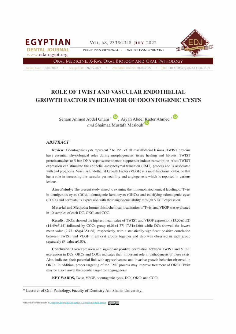
Submit Date : 19-04-2022 • Accept Date : 26-05-2022 • Available online: 30-06-2022 • DOI : 10.21608/edj.2022.133782.2074 Print ISSN 0070-9484 • Online ISSN 2090-2360 Oral Medicine, X-Ray, Oral Biology and Oral Pathology EGYPTIAN DENTAL JOURNAL Vol. 68, 2335: 2348, July, 2022 www. eda-egypt.org Article is licensed under a Creative Commons Attribution 4.0 International License * Lecturer of Oral Pathology, Faculty of Dentistry Ain Shams University. ROLE OF TWIST AND VASCULAR ENDOTHELIAL GROWTH FACTOR IN BEHAVIOR OF ODONTOGENIC CYSTS Seham Ahmed Abdel Ghani * , Aiyah Abdel Kader Ahmed * and Shaimaa Mustafa Masloub * ABSTRACT Review: Odontogenic cysts represent 7 to 15% of all maxillofacial lesions. TWIST proteins have essential physiological roles during morphogenesis, tissue healing and fibrosis. TWIST protein attaches to E-box DNA response members to suppress or induce transcription. Also, TWIST expression can stimulate the epithelial-mesenchymal transition (EMT) process and is associated with bad prognosis. Vascular Endothelial Growth Factor (VEGF) is a multifunctional cytokine that has a role in increasing the vascular permeability and angiogenesis which is reported in various lesions. Aim of study: The present study aimed to examine the immunohistochemical labeling of Twist in dentigerous cysts (DCs), odontogenic keratocysts (OKCs) and calcifying odontogenic cysts (COCs) and correlate its expression with their angiogenic ability through VEGF expression. Material and Methods: Immunohistochemical localization of Twist and VEGF was evaluated in 10 samples of each DC, OKC, and COC. Results: OKCs showed the highest mean value of TWIST and VEGF expression (13.53±5.52) (14.49±5.14) followed by COCs group (6.01±1.77) (7.51±1.66) while DCs showed the lowest mean value (2.77±.68)(4.35±.68), respectively, with a statistically significant positive correlation between TWIST and VEGF in all cyst groups together and also was observed in each group separately (P-value ≤0.05). Conclusion: Overexpression and significant positive correlation between TWIST and VEGF expression in DCs, OKCs and COCs indicates their important role in pathogenesis of these cysts. Also, indicates their potential link with aggressiveness and invasive growth behavior observed in OKCs. In addition, proper targeting of the EMT process may improve treatment of OKCs. Twist may be also a novel therapeutic target for angiogenesis KEY WARDS, Twist, VEGF, odontogenic cysts, DCs, OKCs and COCs
-
Upload
khangminh22 -
Category
Documents
-
view
1 -
download
0
Transcript of PDF (2.47 MB) - Egyptian Dental Journal
Submit Date : 19-04-2022 • Accept Date : 26-05-2022 • Available online: 30-06-2022 • DOI : 10.21608/edj.2022.133782.2074
Print ISSN 0070-9484 • Online ISSN 2090-2360
Oral Medicine, X-Ray, Oral Biology and Oral Pathology
EGYPTIANDENTAL JOURNAL
Vol. 68, 2335:2348, July, 2022
www.eda-egypt.org
Article is licensed under a Creative Commons Attribution 4.0 International License
* Lecturer of Oral Pathology, Faculty of Dentistry Ain Shams University.
ROLE OF TWIST AND VASCULAR ENDOTHELIAL GROWTH FACTOR IN BEHAVIOR OF ODONTOGENIC CYSTS
Seham Ahmed Abdel Ghani * , Aiyah Abdel Kader Ahmed *
and Shaimaa Mustafa Masloub*
ABSTRACT
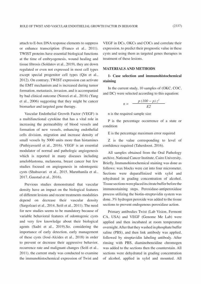
Review: Odontogenic cysts represent 7 to 15% of all maxillofacial lesions. TWIST proteins have essential physiological roles during morphogenesis, tissue healing and fibrosis. TWIST protein attaches to E-box DNA response members to suppress or induce transcription. Also, TWIST expression can stimulate the epithelial-mesenchymal transition (EMT) process and is associated with bad prognosis. Vascular Endothelial Growth Factor (VEGF) is a multifunctional cytokine that has a role in increasing the vascular permeability and angiogenesis which is reported in various lesions.
Aim of study: The present study aimed to examine the immunohistochemical labeling of Twist in dentigerous cysts (DCs), odontogenic keratocysts (OKCs) and calcifying odontogenic cysts (COCs) and correlate its expression with their angiogenic ability through VEGF expression.
Material and Methods: Immunohistochemical localization of Twist and VEGF was evaluated in 10 samples of each DC, OKC, and COC.
Results: OKCs showed the highest mean value of TWIST and VEGF expression (13.53±5.52) (14.49±5.14) followed by COCs group (6.01±1.77) (7.51±1.66) while DCs showed the lowest mean value (2.77±.68)(4.35±.68), respectively, with a statistically significant positive correlation between TWIST and VEGF in all cyst groups together and also was observed in each group separately (P-value ≤0.05).
Conclusion: Overexpression and significant positive correlation between TWIST and VEGF expression in DCs, OKCs and COCs indicates their important role in pathogenesis of these cysts. Also, indicates their potential link with aggressiveness and invasive growth behavior observed in OKCs. In addition, proper targeting of the EMT process may improve treatment of OKCs. Twist may be also a novel therapeutic target for angiogenesis
KEY WARDS, Twist, VEGF, odontogenic cysts, DCs, OKCs and COCs

(2336) Seham Ahmed Abdel Ghani, et al.E.D.J. Vol. 68, No. 3
INTRODUCTION
Odontogenic cysts represent 7 to 15% of all lesions affecting oral and maxillofacial region (Kambalimath et al., 2014). The majority of odontogenic cyst has no recurrence, except odontogenic keratocysts (OKCs) which recur with a rate of 58.3%. Clinically, cysts originating from odontogenic tissues have no symptoms and they are presented accidently during radiological examinations to find out why the tooth fails to erupt (Akyol and Salman, 2012). Essential constituent of odontogenic cysts is the epithelial lining and most of these cysts originate from rests of epithelium (Demirkol et al., 2014).
Dentigerous cyst (DC) is the most frequent developmental cyst arising from odontogenic tissues with favourable behavior and rarely recurs. It represents about 20% of oral cysts (Mohajerani et al., 2015). They are associated with impacted maxillary canines and third molars. These cysts originate from epithelial remnants of enamel organ after formation of enamel and reamin bounded to at the amelocemental junction, enclosing the crown of the tooth (Robinson, 2017). Microscopically DC consists of uniform non-keratinized stratified squamous lining overlie a connective tissue stroma, few mucous cells could also be seen in its lining and considered as a diagnostic feature to help in their diagnosis. (Robinson, 2017).
OKCs have characteristic clinical features, with unique histopathological features, aggressive behavior and high recurrence rate so they are categorized as odontogenic tumors and renamed as keratocystic odontogenic tumors (Johnson et al., 2014). They are asymptomatic unless infected or enlarge in size causing pain and root resorption, the posterior mandibular region is the most commonly affected region for OKCs (Myoung et al., 2001). OKCs which originate from the basal epithelial cells, has specific features of epithelial proliferation, presence of parakeratin on the surface and high
index of mitotic division and are accompanied by alterations of some genes (mutation in p53, tumor suppressor gene) (Alaeddini et al., 2009).
Calcifying odontogenic cysts (COCs) are rare lesions with different clinical behavior representing 1 to 7% of cyst and neoplasms arising from odontogenic remnants. Microscopically, they appear as cystic proliferations lined with odontogenic epithelium and surrounded by connective tissue wall. Calcified and keratinized ghost cells are characteristic features of COC (Shiva and Nosrati, 2015).
Epithelial Mesenchymal Transition (EMT) is a biological cell process, where there is a decrease in the epithelial cell attachment and morphology and gain mesenchymal features (Lamouille et al., 2014). EMT is presented normally during development, homeostasis, tissue healing, and fibrosis and is controlled through a transcriptional cascade and epigenetic events (Derynck and Weinberg, 2019). Recently, EMT is recognized as a reversible and dynamic process in which cells having special epithelial and mesenchymal features (Cook and Vanderhyden, 2019) and become in a state of hybrid epithelial and mesenchymal feaures, this results in a higher tumor-initiating and metastatic ability of these cells and increase apoptosis resistant (Pastushenko et al., 2018) so these cells are capable of penetrating the extracellular matrix, go inside the blood vessels and grow in a different tissues. These steps need a continuous vascular supply to the cells (Sciacovelli and Frezza, 2017).
Odontogenesis is regulated by epithelial and mesenchymal elements interaction of developing tissues responsible for teeth formation; these interactions are responsible for pathogenesis of these odontogenic lesions. The mesenchymal tissue has an important function in epithelial proliferation and corresponding changes in this stroma such as angiogenesis (Puthiyaveetil et al., 2016).
TWIST proteins have the same structure and
ROLE OF TWIST AND VASCULAR ENDOTHELIAL GROWTH FACTOR IN BEHAVIOR (2337)
attach to E-box DNA response elements to suppress or enhance transcription (Franco et al., 2011). TWIST proteins have essential biological functions at the time of embryogenesis, wound healing and tissue fibrosis (Soldatov et al., 2019), they are down regulated or even not expressed in most cell types except special progenitor cell types (Qin et al., 2012). On contrary, TWIST expression can activate the EMT mechanism and is increased during tumor formation, metastasis, invasion, and is accompanied by bad clinical outcome (Norozi et al., 2016) (Yang et al., 2006) suggesting that they might be cancer biomarker and targeted gene therapy.
Vascular Endothelial Growth Factor (VEGF) is a multifunctional cytokine that has a vital role in increasing the permeability of blood vessels and formation of new vessels, enhancing endothelial cells division, migration and increase density of small vessels by 5000 units more than histamines (Puthiyaveetil et al., 2016). VEGF is an essential modulator of normal and pathologic angiogenesis which is reported in many diseases including ameloblastoma, melanoma, breast cancer but few studies focused on angiogenesis in odontogenic cysts (Shahsavari et al., 2015, Maruthanila et al., 2017, Gaustad et al., 2016).
Previous studies demonstrated that vascular density have an impact on the biological features of different lesions and recent treatments modalities depend on decrease their vascular density (Sargolzaei et al., 2014, Seifi et al., 2011). The need for new studies seems to be mandatory because of variable behavioral features of odontogenic cysts and very few knowledge about their biological agents (Sadri et al., 2019).So, considering the importance of early detection, early management of these cysts (José-Alcides et al., 2018) in order to prevent or decrease their aggressive behavior, recurrence rate and malignant changes (Seifi et al., 2011), the current study was conducted to examine the immunohistochemical expression of Twist and
VEGF in DCs, OKCs and COCs and correlate their expression, to predict their prognostic value in these cysts and using them as targeted genes therapies in treatment of these lesions.
MATERIALS AND METHODS
1- Case selection and immunohistochemical staining
In the current study, 10 samples of (OKC, COC, and DC) were selected according to this equation:
n = p (100 − p) z2
E2
n is the required sample size
P is the percentage occurrence of a state or condition
E is the percentage maximum error required
Z is the value corresponding to level of confidence required (Taherdoost, 2016).
All samples obtained from the Oral Pathology archive, National Cancer Institute, Cairo University. Briefly, Immunohistochemical staining was done as follows: wax blocks were cut into four micrometer. Sections were deparaffinized with xylol and rehydrated in grading concentration of alcohol. Tissue sections were placed in citrate buffer before the immunostaining steps. Peroxidase-antiperoxidase process utilizing the biotin-streptavidin system was done, 3% hydrogen peroxide was added to the tissue sections to prevent endogenous peroxidase action.
Primary antibodies Twist (Lab Vision, Fermont CA, USA) and VEGF (Genome Me Lab) were applied and then incubated at room temperature overnight. After that they washed in phosphate buffer saline (PBS), and then link antibody was applied, followed by streptavidin labeling antibody. After rinsing with PBS, diaminobenzidine chromogen was added to the sections then the counterstain. All sections were dehydrated in grading concentration of alcohol, applied in xylol and mounted. All
(2338) Seham Ahmed Abdel Ghani, et al.E.D.J. Vol. 68, No. 3
the steps of immunohistochemical quantitative estimation were carried out on photomicrographs captured at a magnification of X40. The images are taken with a camera linked to the microscope and then the images taken are analyzed with the image software (Image J, 1.41a, NIH, USA).
2. Statistical analysis
Data was analyzed using Statistical Package for Social Science software computer program version 26 (SPSS, Inc., Chicago, IL, USA). Shapiro-wilk test was used to detect normal distribution of data. Quantitative data was parametric and prepared in mean and standard deviation. One way ANOVA (Variance analysis) followed by post-hoc tukey was used to compare more than two different groups of parametric data. Pearson’s correlation was used to correlate between Twist & VEGF. P value (less than 0.05) was considered sta tistically significant.
RESULTS
1. Immunohistochemical Results
A. TWIST
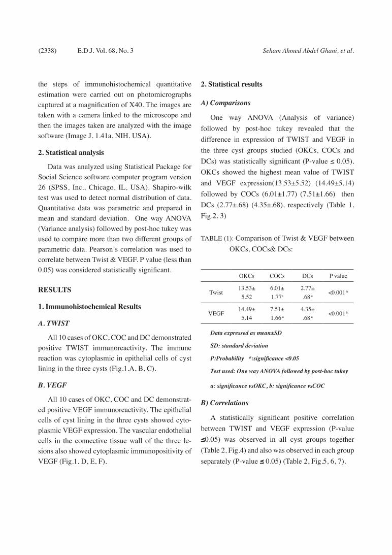
All 10 cases of OKC, COC and DC demonstrated positive TWIST immunoreactivity. The immune reaction was cytoplasmic in epithelial cells of cyst lining in the three cysts (Fig.1.A, B, C).
B. VEGF
All 10 cases of OKC, COC and DC demonstrat-ed positive VEGF immunoreactivity. The epithelial cells of cyst lining in the three cysts showed cyto-plasmic VEGF expression. The vascular endothelial cells in the connective tissue wall of the three le-sions also showed cytoplasmic immunopositivity of VEGF (Fig.1. D, E, F).
2. Statistical results
A) Comparisons
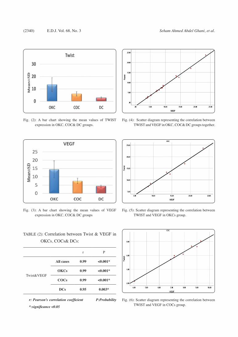
One way ANOVA (Analysis of variance) followed by post-hoc tukey revealed that the difference in expression of TWIST and VEGF in the three cyst groups studied (OKCs, COCs and DCs) was statistically significant (P-value ≤ 0.05). OKCs showed the highest mean value of TWIST and VEGF expression(13.53±5.52) (14.49±5.14) followed by COCs (6.01±1.77) (7.51±1.66) then DCs (2.77±.68) (4.35±.68), respectively (Table 1, Fig.2, 3)
TABLE (1): Comparison of Twist & VEGF between OKCs, COCs& DCs:
OKCs COCs DCs P value
Twist13.53±
5.526.01±1.77a
2.77±.68 a
<0.001*
VEGF14.49±
5.147.51±1.66 a
4.35±.68 a
<0.001*
Data expressed as mean±SD
SD: standard deviation
P:Probability *:significance <0.05
Test used: One way ANOVA followed by post-hoc tukey
a: significance vsOKC, b: significance vsCOC
B) Correlations
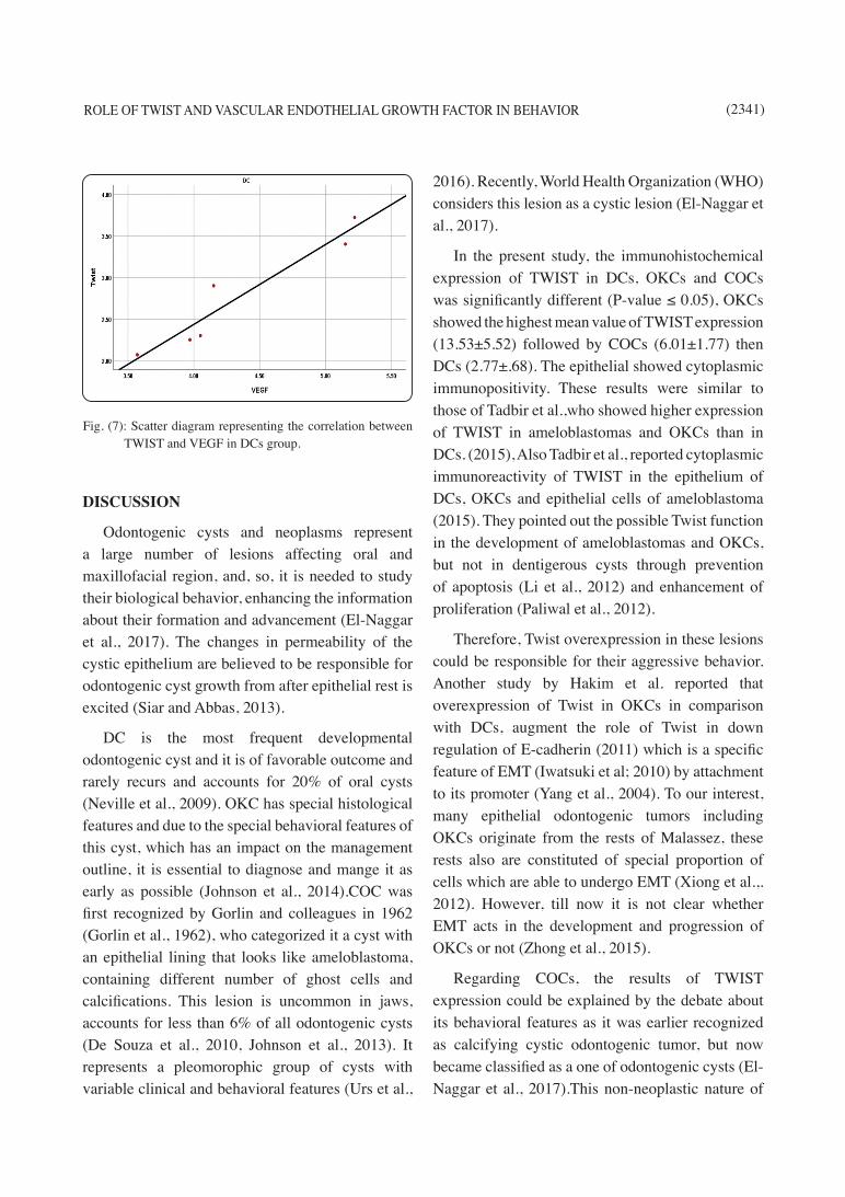
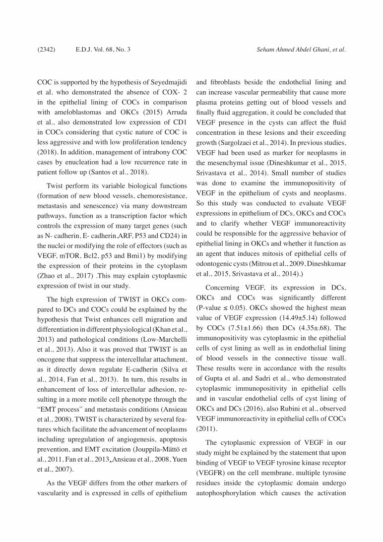
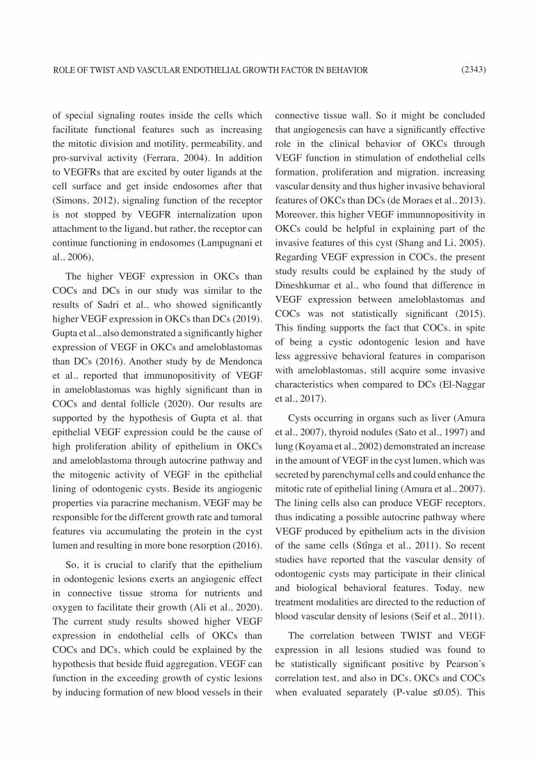
A statistically significant positive correlation between TWIST and VEGF expression (P-value ≤0.05) was observed in all cyst groups together (Table 2, Fig.4) and also was observed in each group separately (P-value ≤ 0.05) (Table 2, Fig.5, 6, 7).

ROLE OF TWIST AND VASCULAR ENDOTHELIAL GROWTH FACTOR IN BEHAVIOR (2339)
Fig. (1): Photomicrographs of immunohistochemical results of TWIST (A, B, C) showing the cytoplasmic expression of TWIST (blue arrows) in epithelial cells of cyst lining of OKC (A), COC (B) and DC (C), and VEGF (D, E, F) showing the cytoplasmic expression of VEGF in epithelial cells of cyst lining (yellow arrows) and endothelial cells of blood vessels (green arrows) of OKC (D), COC (E) and DC (F) (Orig. Mag. X40).
(2340) Seham Ahmed Abdel Ghani, et al.E.D.J. Vol. 68, No. 3
TABLE (2): Correlation between Twist & VEGF in OKCs, COCs& DCs:
r P
Twist&VEGF
All cases 0.99 <0.001*
OKCs 0.99 <0.001*
COCs 0.99 <0.001*
DCs 0.95 0.003*
r: Pearson’s correlation coefficient P:Probability
*:significance <0.05
Fig. (2): A bar chart showing the mean values of TWIST expression in OKC, COC& DC groups.
Fig. (4): Scatter diagram representing the correlation between TWIST and VEGF in OKC, COC& DC groups together.
Fig. (3): A bar chart showing the mean values of VEGF expression in OKC, COC& DC groups
Fig. (5): Scatter diagram representing the correlation between TWIST and VEGF in OKCs group.
Fig. (6): Scatter diagram representing the correlation between TWIST and VEGF in COCs group.
ROLE OF TWIST AND VASCULAR ENDOTHELIAL GROWTH FACTOR IN BEHAVIOR (2341)
DISCUSSION
Odontogenic cysts and neoplasms represent a large number of lesions affecting oral and maxillofacial region, and, so, it is needed to study their biological behavior, enhancing the information about their formation and advancement (El-Naggar et al., 2017). The changes in permeability of the cystic epithelium are believed to be responsible for odontogenic cyst growth from after epithelial rest is excited (Siar and Abbas, 2013).
DC is the most frequent developmental odontogenic cyst and it is of favorable outcome and rarely recurs and accounts for 20% of oral cysts (Neville et al., 2009). OKC has special histological features and due to the special behavioral features of this cyst, which has an impact on the management outline, it is essential to diagnose and mange it as early as possible (Johnson et al., 2014).COC was first recognized by Gorlin and colleagues in 1962 (Gorlin et al., 1962), who categorized it a cyst with an epithelial lining that looks like ameloblastoma, containing different number of ghost cells and calcifications. This lesion is uncommon in jaws, accounts for less than 6% of all odontogenic cysts (De Souza et al., 2010, Johnson et al., 2013). It represents a pleomorophic group of cysts with variable clinical and behavioral features (Urs et al.,
2016). Recently, World Health Organization (WHO) considers this lesion as a cystic lesion (El-Naggar et al., 2017).
In the present study, the immunohistochemical expression of TWIST in DCs, OKCs and COCs was significantly different (P-value ≤ 0.05), OKCs showed the highest mean value of TWIST expression (13.53±5.52) followed by COCs (6.01±1.77) then DCs (2.77±.68). The epithelial showed cytoplasmic immunopositivity. These results were similar to those of Tadbir et al.,who showed higher expression of TWIST in ameloblastomas and OKCs than in DCs. (2015), Also Tadbir et al., reported cytoplasmic immunoreactivity of TWIST in the epithelium of DCs, OKCs and epithelial cells of ameloblastoma (2015). They pointed out the possible Twist function in the development of ameloblastomas and OKCs, but not in dentigerous cysts through prevention of apoptosis (Li et al., 2012) and enhancement of proliferation (Paliwal et al., 2012).
Therefore, Twist overexpression in these lesions could be responsible for their aggressive behavior. Another study by Hakim et al. reported that overexpression of Twist in OKCs in comparison with DCs, augment the role of Twist in down regulation of E-cadherin (2011) which is a specific feature of EMT (Iwatsuki et al; 2010) by attachment to its promoter (Yang et al., 2004). To our interest, many epithelial odontogenic tumors including OKCs originate from the rests of Malassez, these rests also are constituted of special proportion of cells which are able to undergo EMT (Xiong et al.,. 2012). However, till now it is not clear whether EMT acts in the development and progression of OKCs or not (Zhong et al., 2015).
Regarding COCs, the results of TWIST expression could be explained by the debate about its behavioral features as it was earlier recognized as calcifying cystic odontogenic tumor, but now became classified as a one of odontogenic cysts (El-Naggar et al., 2017).This non-neoplastic nature of
Fig. (7): Scatter diagram representing the correlation between TWIST and VEGF in DCs group.
(2342) Seham Ahmed Abdel Ghani, et al.E.D.J. Vol. 68, No. 3
COC is supported by the hypothesis of Seyedmajidi et al. who demonstrated the absence of COX- 2 in the epithelial lining of COCs in comparison with ameloblastomas and OKCs (2015) Arruda et al., also demonstrated low expression of CD1 in COCs considering that cystic nature of COC is less aggressive and with low proliferation tendency (2018). In addition, management of intrabony COC cases by enucleation had a low recurrence rate in patient follow up (Santos et al., 2018).
Twist perform its variable biological functions (formation of new blood vessels, chemoresistance, metastasis and senescence) via many downstream pathways, function as a transcription factor which controls the expression of many target genes (such as N- cadherin, E- cadherin,ARF, P53 and CD24) in the nuclei or modifying the role of effectors (such as VEGF, mTOR, Bcl2, p53 and Bmi1) by modifying the expression of their proteins in the cytoplasm (Zhao et al., 2017) .This may explain cytoplasmic expression of twist in our study.
The high expression of TWIST in OKCs com-pared to DCs and COCs could be explained by the hypothesis that Twist enhances cell migration and differentiation in different physiological (Khan et al., 2013) and pathological conditions (Low-Marchelli et al., 2013). Also it was proved that TWIST is an oncogene that suppress the intercellular attachment, as it directly down regulate E-cadherin (Silva et al., 2014, Fan et al., 2013). In turn, this results in enhancement of loss of intercellular adhesion, re-sulting in a more motile cell phenotype through the “EMT process” and metastasis conditions (Ansieau et al., 2008). TWIST is characterized by several fea-tures which facilitate the advancement of neoplasms including upregulation of angiogenesis, apoptosis prevention, and EMT excitation (Jouppila-Mättö et al., 2011, Fan et al., 2013, Ansieau et al., 2008, Yuen et al., 2007).
As the VEGF differs from the other markers of vascularity and is expressed in cells of epithelium
and fibroblasts beside the endothelial lining and can increase vascular permeability that cause more plasma proteins getting out of blood vessels and finally fluid aggregation, it could be concluded that VEGF presence in the cysts can affect the fluid concentration in these lesions and their exceeding growth (Sargolzaei et al., 2014). In previous studies, VEGF had been used as marker for neoplasms in the mesenchymal issue (Dineshkumar et al., 2015, Srivastava et al., 2014). Small number of studies was done to examine the immunopositivity of VEGF in the epithelium of cysts and neoplasms. So this study was conducted to evaluate VEGF expressions in epithelium of DCs, OKCs and COCs and to clarify whether VEGF immunoreactivity could be responsible for the aggressive behavior of epithelial lining in OKCs and whether it function as an agent that induces mitosis of epithelial cells of odontogenic cysts (Mitrou et al., 2009, Dineshkumar et al., 2015, Srivastava et al., 2014).)
Concerning VEGF, its expression in DCs, OKCs and COCs was significantly different (P-value ≤ 0.05). OKCs showed the highest mean value of VEGF expression (14.49±5.14) followed by COCs (7.51±1.66) then DCs (4.35±.68). The immunopositivity was cytoplasmic in the epithelial cells of cyst lining as well as in endothelial lining of blood vessels in the connective tissue wall. These results were in accordance with the results of Gupta et al. and Sadri et al., who demonstrated cytoplasmic immunopositivity in epithelial cells and in vascular endothelial cells of cyst lining of OKCs and DCs (2016), also Rubini et al., observed VEGF immunoreactivity in epithelial cells of COCs (2011).
The cytoplasmic expression of VEGF in our study might be explained by the statement that upon binding of VEGF to VEGF tyrosine kinase receptor (VEGFR) on the cell membrane, multiple tyrosine residues inside the cytoplasmic domain undergo autophosphorylation which causes the activation
ROLE OF TWIST AND VASCULAR ENDOTHELIAL GROWTH FACTOR IN BEHAVIOR (2343)
of special signaling routes inside the cells which facilitate functional features such as increasing the mitotic division and motility, permeability, and pro-survival activity (Ferrara, 2004). In addition to VEGFRs that are excited by outer ligands at the cell surface and get inside endosomes after that (Simons, 2012), signaling function of the receptor is not stopped by VEGFR internalization upon attachment to the ligand, but rather, the receptor can continue functioning in endosomes (Lampugnani et al., 2006),
The higher VEGF expression in OKCs than COCs and DCs in our study was similar to the results of Sadri et al., who showed significantly higher VEGF expression in OKCs than DCs (2019). Gupta et al., also demonstrated a significantly higher expression of VEGF in OKCs and ameloblastomas than DCs (2016). Another study by de Mendonca et al., reported that immunopositivity of VEGF in ameloblastomas was highly significant than in COCs and dental follicle (2020). Our results are supported by the hypothesis of Gupta et al. that epithelial VEGF expression could be the cause of high proliferation ability of epithelium in OKCs and ameloblastoma through autocrine pathway and the mitogenic activity of VEGF in the epithelial lining of odontogenic cysts. Beside its angiogenic properties via paracrine mechanism, VEGF may be responsible for the different growth rate and tumoral features via accumulating the protein in the cyst lumen and resulting in more bone resorption (2016).
So, it is crucial to clarify that the epithelium in odontogenic lesions exerts an angiogenic effect in connective tissue stroma for nutrients and oxygen to facilitate their growth (Ali et al., 2020). The current study results showed higher VEGF expression in endothelial cells of OKCs than COCs and DCs, which could be explained by the hypothesis that beside fluid aggregation, VEGF can function in the exceeding growth of cystic lesions by inducing formation of new blood vessels in their
connective tissue wall. So it might be concluded that angiogenesis can have a significantly effective role in the clinical behavior of OKCs through VEGF function in stimulation of endothelial cells formation, proliferation and migration, increasing vascular density and thus higher invasive behavioral features of OKCs than DCs (de Moraes et al., 2013). Moreover, this higher VEGF immunnopositivity in OKCs could be helpful in explaining part of the invasive features of this cyst (Shang and Li, 2005). Regarding VEGF expression in COCs, the present study results could be explained by the study of Dineshkumar et al., who found that difference in VEGF expression between ameloblastomas and COCs was not statistically significant (2015). This finding supports the fact that COCs, in spite of being a cystic odontogenic lesion and have less aggressive behavioral features in comparison with ameloblastomas, still acquire some invasive characteristics when compared to DCs (El-Naggar et al., 2017).
Cysts occurring in organs such as liver (Amura et al., 2007), thyroid nodules (Sato et al., 1997) and lung (Koyama et al., 2002) demonstrated an increase in the amount of VEGF in the cyst lumen, which was secreted by parenchymal cells and could enhance the mitotic rate of epithelial lining (Amura et al., 2007). The lining cells also can produce VEGF receptors, thus indicating a possible autocrine pathway where VEGF produced by epithelium acts in the division of the same cells (Stînga et al., 2011). So recent studies have reported that the vascular density of odontogenic cysts may participate in their clinical and biological behavioral features. Today, new treatment modalities are directed to the reduction of blood vascular density of lesions (Seif et al., 2011).
The correlation between TWIST and VEGF expression in all lesions studied was found to be statistically significant positive by Pearson’s correlation test, and also in DCs, OKCs and COCs when evaluated separately (P-value ≤0.05). This

(2344) Seham Ahmed Abdel Ghani, et al.E.D.J. Vol. 68, No. 3
positive correlation between TWIST and VEGF could be explained by the results of Niu et al. study who reported that increase of Twist messenger RNA protein were positively accompanied by the up regula tion of VEGF expression in hepatocellular carcinoma (HCC) patients with bad clinical outcome, indicating that Twist could play a pivotal role in the angiogenesis and metastasis of HCC (2007). It was also reported that Twist, which is a highly conserved basic helix–loop–helix transcription factor, induce the process of changing epithelial cells to mesenchymal ones and was able to promote angiogenesis (Mironchik et al., 2005).
So, high Twist immunopositivity was found with bigger tumor size, more lymph node metastasis, and worse clinical stage (Zhang et al., 2015). These findings strongly supported that Twist/VEGF angio-genic axis has a pivotal function in angiogenesis of tumors (Zhang et al., 2019). Overall, several studies indicated that Twist over expression may augment tumor invasion and metastasis via upregulating VEGF expression, causing the enhancement of new blood vessels formation, which is crucial for chang-ing into an invasive phenotype (Lei et al., 2015).The present study is the first one conducted to evaluate the immunohistochemical expression of Twist and VEGF in DCs, OKCs and COCs and correlate their expression, to predict their prognostic value in these cysts and using them as targeted genes therapies in treatment of these lesions.
CONCLUSION
It could be concluded from the present study that TWIST and VEGF are overexpressed and have significant positive correlation between their expression in DCs, OKCs and COCs and these findings indicates their pivotal role in the pathogenesis of these cysts. Also, this supports their possible link with aggressiveness and invasive biological behavior observed in OKCs. In addition, suitable targeting of the EMT process could improve
management of OKCs. Twist may be also a novel therapeutic target for angiogenesis.
Recommendations
We still need to be sure about the precise mechanism behind correlation between EMT and pathological processes in OKCs by further investigations. Also, studies using advanced techniques and more cases should be done to investigate the function of VEGF in the development of OKCs and other odontogenic cysts, also for the use of anti VEGF treatment modalities for the management of these lesions.
REFERENCES
· Akyol U. and Salman I.: A case of an extensive dentig-erous cyst in the maxillary sinus leading to epiphora and nasal obstruction. J Emerg Med. 2012; 43: 1004-1007.
· Alaeddini M., Salah S., Dehghan F., Eshghyar N. and Ete-mad Moghadam S.: Comparison of angiogenesis in kera-tocystic odontogenic tumours, dentigerous cysts and AMs. Oral Dis. 2009; 15: 422-427.
· Ali K., Khan S., Sultana N., Alghamdi O., Mohammad S., Mokeem S., Ali S., Abduljabbar T. and Vohra F.: Assess-ment of Tumor Angiogenesis by Expression of CD 105 in Ameloblastoma, Odontogenic Keratocyst and Central Gi-ant Cell Lesion. Asian Pac J Cancer Prev.2020; 21:3373-3379.
· Amura C., Brodsky K., Groff R., Gattone V., Voelkel N. and Doctor R.: VEGF receptor inhibition blocks liver cyst growth in pkd2(WS25/-) mice. Am J Physiol Cell Physiol. 2007; 293: 19-28.
· Ansieau S., Bastid J., Doreau A., Morel A., Bouchet B., Thomas C, Fauvet F., Puisieux I., Doglioni C., Piccinin S., Maestro R., Voeltzel T., Selmi A., Wittmann S., Fro-mentel C. and Puisieux A.: Induction of EMT by twist proteins as a collateral effect of tumor-promoting inactiva-tion of premature senescence. 2008; 14: 79-89.
· Arruda J., Silva L., Silva L., Monteiro J., Álvares P., Sil-veira M. and Sobral A.: Calcifying odontogenic cyst: A 26-year retrospective clinicopathological analysis and im-munohistochemical study. J Clin Exp Dent. 2018; 10: 542-547.
ROLE OF TWIST AND VASCULAR ENDOTHELIAL GROWTH FACTOR IN BEHAVIOR (2345)
· Cook D. and Vanderhyden B.: Ovarian cancer and the evo-lution of subtype classifications using transcriptional pro-filing. Biol Reprod. 2019; 101:645–658.
· de Mendonça R., Balbinot K., Martins B., Kataoka M., s Mesquita R., Pinheiro J., and Júnior S.: Hypoxia and proangiogenic proteins in human ameloblastoma. Sci Rep.2020; 10: 175-167.
· De Moraes M., De Matos F., De Souza L., Freitas R. and Costa A.: Immunoexpression of RANK, RANKL, OPG, VEGF, and vWF in radicular and dentigerous cysts. J Oral Pathol Med. 2013; 42: 468-473.
· De Souza L., Gordon M., Nonaka C., De ˜ Medeiros M., Torres T. and Emiliano G.: “Odontogenic cysts: Demo-graphic profle in a Brazilian population over a 38-year period,” Medicina Oral Patología Oral y Cirugía Bucal. 2010; 15: 583–590.
· Demirkol M., Ege B., Yanik S., Aras M. and Ay S.: Clini-copathological study of jaw cysts in southeast region of Turkey. Eur J Dent. 2014; 8:107–111.
· Derynck R. and Weinberg R.: EMT and cancer: more than meets the eye. Dev Cell. 2019; 49: 313–316.
· Dineshkumar T., Priyadharsini N., Gnanaselvi U., Sathish-kumar S., Srikanth R. and Nagarathinam A.: Evaluation and comparison of vascular endothelial growth factor ex-pression between ameloblastoma and keratocystic odonto-genic tumor. J Int Oral Health. 2015; 7: 48-52.
· El-Naggar A., Chan J., Grandis J., Takata T. and Slootweg P.: WHO Classification of Head and Neck Tumors (IARC Press, Lyon, 2017).
· Fan C., Wang T., Cheng Y., Jiang S., Cheng C., Lee A. and Kao T.: Expression of E-cadherin, twist, and p53 and their prognostic value in patients with oral squamous cell car-cinoma. J Cancer Res ClinOncol. 2013; 139: 1735-1744.
· Ferrara N.: Vascular endothelial growth factor: Basic sci-ence and clinical progress. Endocr. Rev. 2004; 25: 581–611.
· Franco H., Casasnovas J., Rodríguez-Medina J. and Ca-dilla C.: Redundant or separate entities? - Roles of Twist1 and Twist2 as molecular switches during gene transcrip-tion. Nucleic Acids Res. 2011; 39: 1177–1186.
· Gaustad J., Simonsen T., Andersen L. and Rofstad E.: Properdistatin inhibits angiogenesis and improves vas-
cular function in human melanoma xenografts with low thrombospondin-1 expression. Oncotarget. 2016; 7:76806-76815.
· Gorlin R., Pindborg J., Clausen F. and Vickers R.: “Te cal-cifying odontogenic cyst-a possible analogue of the cuta-neous calcifying epithelioma of Malherbe. An analysis of ffeen cases,” Oral Surgery, Oral Medicine, Oral Pathology, Oral Radiology, and Endodontology. 1962; 15: 1235–1243.
· Gupta B., Chandra S., Singh A., Sah K., Raj V. and Gupta V.: The role of vascular endothelial growth factor in pro-liferation of odontogenic cysts and tumors: An immuno-histochemical study. Dental Research Journal. 2016; 13: 256-263.
· Hakim S., Kosmehl H., Sieg P., Trenkle T., Jacobsen H., Benedek G., Ribbat J. and Driemel O.: Altered expression of cell-cell adhesion molecules β-catenin/E-cadherin and related Wnt-signaling pathway in sporadic and syndromal keratocystic odontogenic tumors. Clin Oral Investig. 2011; 15: 321–328.
· Iwatsuki M., Mimori K., Yokobori T., Ishi H., Beppu T., Nakamori S., Baba H. and Mori M.: Epithelial-mesen-chymal transition in cancer development and its clinical significance. Cancer. 2010; 101: 293–299.
· Johnson N., Gannon O., Savage N. and Batstone M.: Fre-quency of odontogenic cysts and tumors: a systematic re-view. J Investig Clin Dent. 2014; 5: 9-14.
· Johnson N., Savage N., Kazoullis S. and Batstone M.: “A prospective epidemiological study for odontogenic and non-odontogenic lesions of the maxilla and mandible in Queensland,” Oral Surgery, Oral Medicine, Oral Pathol-ogy, Oral Radiology, and Endodontology. 2013; 115: 515–522.
· José-Alcides A., Leni-Verônica S., Leorik S., João-Luiz M., Pamella Á., Marcia S. and Ana-Paula S.: Calcifying odontogenic cyst: A 26-year retrospective clinicopatholog-ical analysis and immunohistochemical study.J Clin Exp Dent. 2018;10: 542-547.
· Jouppila-Mättö A., Närkiö-Mäkelä M., Soini Y., Pukkila M., Sironen R., Tuhkanen H., Mannermaa A. and Kosma V.: Twist and snai1 expression in pharyngeal squamous cell carcinoma stroma is related to cancer progression. BMC Cancer.2011; 11:350-358.
· Kambalimath D., Kambalimath H., Agrawal S., Singh M.,
(2346) Seham Ahmed Abdel Ghani, et al.E.D.J. Vol. 68, No. 3
Jain N., Anurag B. and Michael P.: Prevalence and dis-tribution of odontogenic cyst in Indian population: a 10 year retrospective study. J Maxillofac Oral Surg. 2014; 13:10–15.
· Khan M., Chen H., Zhang D. and Fu J.: Twist: A molecu-lar target in cancer therapeutics. Tumour Biol. 2013; 34: 2497-2506.
· Koyama S., Sato E., Tsukadaira A., Haniuda M., Numa-nami H., Kurai M., Nagai S. and Izumi T.: Vascular en-dothelial growth factor mRNA and protein expression in airway epithelial cell lines in vitro. Eur Respir J. 2002; 20:1449-1456.
· Lamouille S., Xu J. and Derynck R.: Molecular mecha-nisms of epithelialmesenchymal transition. Nat Rev Mol Cell Biol. 2014; 15: 178–196.
· Lampugnani M., Orsenigo F., Gagliani, M., Tacchetti C. and Dejana E.: Vascular endothelial cadherin controls VEGFR-2 internalization and signaling from intracellular compartments. J Cell Biol. 2006;174: 593–604.
· Lei P., Ding D., Xie J., Wang L., Liao Q. and Hu Y.: Ex-pression profile of Twist, vascular endothelial growth fac-tor and CD34 in patients with different phases of osteosar-coma. Oncology letters. 2015; 10:417-421.
· Li B., Han Q., Zhu Y., Yu Y.,Wang J. and Jiang X.: Down-regulation of miR-214 contributes to intrahepatic cholan-giocarcinoma metastasis by targeting Twist. FEBS J. 2012; 279: 2393–2398.
· Low-Marchelli J., Ardi V., Vizcarra E., van Rooijen N., Quigley J. and Yang J.: Twist1 induces CCL2 and recruits macro phages to promote angiogenesis. Cancer Res. 2013; 73: 662-671.
· Maruthanila V., Elancheran R., Kunnumakkara A., Kabi-lan S. and Kotoky J.: Recent development of targeted ap-proaches for the treatment of breast cancer. Breast Cancer. 2017; 24:191–219.
· Mironchik Y., Winnard PT Jr., Vesuna F., Kato Y., Wildes F., Pathak A., Kominsky S., Artemov D., Bhujwalla Z., Van Diest P., Burger H., Glackin C. and Raman V.: Twist overexpression induces in vivo angiogenesis and corre-lates with chromosomal instability in breast cancer. Can Res. 2005; 65: 10801–10809.
· Mitrou G., Tosios K., Kyroudi A. and Sklavounou A.: Odontogenic keratocyst expresses vascular endothelial growth factor: An immunohistochemical study. J Oral
Pathol Med. 2009; 38: 470-475.
· Mohajerani H., Esmaeelinejad M., Sabour S., Agh-dashi F. and Dehghani N.: Diagnostic factors of odonto-genic cysts in Iranian population: A Retrospective study over the Past two decades. Iran Red Crescent Med. J. 2015;17(6):e21793.
· Myoung H., Hong S., Hong S., Lee J., Lim C., Choung P., Lee J., Choi J., Seo B. and Kim MJ.: Odontogenic kerato-cyst: review of 256 cases for recurrence and clinicopatho-logic parameters. Oral Surg Oral Med Oral Pathol Oral Radiol Endod. 2001; 91: 328–333.
· Neville B., Damm D. and Allen C.: Oral & Maxillofacial Pathology. 3rd ed. Philadelphia: WB Saunders co. 2009; 678-701.
· Niu R., Zhang L., Xi G., Wei X., Yang Y., Shi Y. and Hao X.: Up-regulation of Twist induces angiogenesis and cor-relates with metastasis in hepatocellular carcinoma. J Exp Clin Cancer Res. 2007: 26; 385-394.
· Norozi F., Ahmadzadeh A., Shahjahani M., Shahrabi S. and Saki N.: Twist as a new prognostic marker in hematologi-cal malignancies. Clin Transl Oncol. 2016;18: 113–124.
· Paliwal P., Arora D. and Mishra A.: Epithelial mesenchy-mal transition in urothelial carcinoma: twist in the tale. In-dian J Pathol Microbiol. 2012; 55: 443–449.
· Pastushenko I., Brisebarre A., Sifrim A., Fioramonti M., Revenco T., Boumahdi S., Keymeulen A., Brown D., Mo-ers V., Lemaire S., Clercq S., Minguijón E., Balsat C., Sokolow Y., Dubois C., Cock F., Scozzaro S., Sopena F., Lanas A., D’Haene N., Salmon I., Marine J., Voet T., Soti-ropoulou B. and Blanpain C.: Identification of the tumour transition states occurring during EMT. Nature. 2018; 556: 463–468.
· Patidar K., Parwani R., Wanjari S. and Patidar A.: Mast cells in human odontogenic cysts. Biotech Histochem. 2012; 87: 397- 402.
· Puthiyaveetil J., Kota K., Chakkarayan R., Chakkarayan J. and Thodiyil A.: Epithelial - mesenchymal interactions in tooth development and the significant role of growth fac-tors and genes with emphasis on mesenchyme - a review. J Clin Diagn Res 2016; 10: 5-9.
· Qin Q., Xu Y., He T., Qin C. and Xu J.: Normal and dis-ease-related biological functions of Twist1 and underlying molecular mechanisms. Cell Res. 2012;22: 90–106.
ROLE OF TWIST AND VASCULAR ENDOTHELIAL GROWTH FACTOR IN BEHAVIOR (2347)
· Robinson A .: Diagnosing the most common odontogenic cystic and osseous lesions of the jaws for the practicing pathologist Modern Pathology. 2017; 30: 96–103.
· Rubini C., Artese L., Zizzi A., Fioroni M., Ascani G., Go-teri G., Stramazzotti D., Piccirilli M., Iezzi G. and Piattelli A.: Immunohistochemical expression of vascular endothe-lial growth factor (VEGF) in different types of odontogen-ic cysts. Clin Oral Invest. 2011: 15: 757–761.
· Sadri D., Farhadi S., Shahabi Z. and Sarshar S.: Expression of Vascular Endothelial Growth Factor in Odontogenic Cysts: Is There Any Impression on Clinical Outcome? The Open Dentistry Journal. 2016; 10: 752-759.
· Sadri D., Farhadi1 S. and Nourmohamadi P.: Angiogenesis in odontogenic keratocyst and dentigerous cyst: Evalua-tion of JunB and VEGF expression. Dental Research Jour-nal. 2019; 16: 327–332.
· Santos H., de Morais E., Moreira D., Neto L., Gomes P. and Freitas R.: Calcifying Odontogenic Cyst with Exten-sive Areas of Dentinoid: Uncommon Case Report and Up-date of Main Findings. 2018; ID 8323215.1-4.
· Sargolzaei S., Farhadi S., Kazemi B., Bandehpour M. and Kharazifard M.: The correlation between p16 expression and INK4a locus mutation with grades and stages in oral squamous cell carcinoma. Indian J Pathol Microbiol. 2014; 57: 24-30.
· Sato K., Miyakawa M., Onoda N., Demura H., Yamashita T., Miura M., Kasajima T., Yamazaki K. and Obara T.: In-creased concentration of vascular endothelial growth fac-tor/vascular permeability factor in cyst fluid of enlarging and recurrent thyroid nodules. J Clin Endocrinol Metab. 1997; 82: 1968-1973.
· Sciacovelli M. and Frezza C.: Metabolic reprogramming and epithelial to mesenchymal transition in cancer. FEBS J.2017; 284: 3132–3144.
· Seifi S., Shafaie S. and Ghadiri S.: Microvessel density in follicular cysts, keratocystic odontogenic tumours and am-eloblastomas. Asian Pac J Cancer Prev. 2011; 12: 351-356.
· Seyedmajidi M., Shafaee S., Siadati S., Moghaddam E., Ghasemi N., Bijani A. and Najafi M.: Immunohistochemi-cal analysis of COX-2 expres sion in dentigerous cyst, ker-atocystic odontogenic tumor and amelo blastoma: A com-parative study. Dent Res J (Isfahan). 2015; 12: 278-284.
· Shahsavari F., Farhadi S., Sadri D. and Sedehi M.: Evalua-
tion of microvascularity by CD34 expression in esophagus and oral squamous cell carcinoma. J Contemp Dent Pract. 2015; 16: 458-462.
· Shang Z. and Li J.: Expression of endothelial nitric ox-ide synthase and vascular endothelial growth factor in oral squamous cell carcinoma: its correlation with angiogen-esis and disease progression. J Oral Pathol Med. 2005; 34: 134–139.
· Shiva A. and Nosrati K.: Calcifying odontogenic cyst as-sociated with complex odontoma: a case report. J Babol Univ Med Sci. 2015; 17:57-61.
· Siar C. and Abbas S.: Claudin expression and tight junc-tion protein localization in the lining epithelium of the keratocystic odontogenic tumors, dentigerous cysts, and radicular cysts. Oral Surg Oral Med Oral Pathol Oral Ra-diol. 2013; 115: 652–659.
· Silva B., Silva F., Pontes H. and JúniorDdos S.: E-cadherin down regulation and twist over expression since early stages of oral carcinogenesis. J Oral Pathol Med. 2014; 43:125-131.
· Simons M.: An inside view: VEGF receptor trafficking and signaling. Physiology. 2012; 27:213–222.
· Soldatov R., Kaucka M., Kastriti M., Petersen J., Ch-ontorotzea T., Englmaier L., Akkuratova N., Yang Y., Häring M., Dyachuk V., Bock C., Farlik M., Piacentino M., Boismoreau F., Hilscher M., Yokota C., Qian X., Nilsson M., Bronner M., Croci L., Hsiao W., Guertin D., Brunet J., Consalez G., Ernfors P., Fried K., Kharch-enko P. and Adameyko I.: Spatiotemporal structure of cell fate decisions in murine neural crest.Science. 2019; 364(6444):eaas9536.
· Srivastava V., Gara R., Rastogi N., Mishra D., Ahmed M., Gupta S., Goel M. and Bhatt M.: Serum vascular endo-thelial growth factor-A (VEGF-A) as a biomarker in squa-mous cell carcinoma of head and neck patients undergoing chemoradiotherapy. Asian Pac J Cancer Prev. 2014; 15: 3261-3265.
· Stînga A., Margaritescu O., Stînga A., Pirici D., Ciurea R., Bunget A. and Cruce M.: VEGFR1 and VEGFR2 im-munohistochemical expression in oral squamous cell car-cinoma: A morphometric study. Rom J Morphol Embryol. 2011; 52: 1269-1275.
· Tadbir A., Pardis S. and Ranjbaran P.: Twist expression in
(2348) Seham Ahmed Abdel Ghani, et al.E.D.J. Vol. 68, No. 3
dentigerous cyst, odontogenic keratocyst and ameloblas-toma. Oral Maxillofac Surg. 2015; 19: 103–107.
· Taherdoost H.: Sampling Methods in Research Methodology; How to Choose a Sampling Technique for Research. International Journal of Advance Research in Management. 2016; 5(2): 18-27.
· Urs A., Augustine J., Singh H., Kureel K., Mohanty S. and Gupta S.: “Calcifying ghost cell odontogenic tumor (CG-COT) with predominance of clear cells: A case report with important diagnostic considerations,” Oral Surgery, Oral Medicine, Oral Pathology, Oral Radiology, and Endodon-tology. 2016; 121: 32–37.
· Xiong J., Mrozik K., Gronthos S., and Bartold P.: “Epi-thelial cell rests of malassez contain unique stem cell populations capable of undergoing epithelial-mesenchy-mal transition,” Stem Cells and Development. 2012; 43: 2012–2025.
· Yang J., Mani S. and Weinberg R.: Exploring a new twist on tumor metastasis.Cancer Res. 2006; 66:4549–4552.
· Yang J., Mani S., Donaher J., Ramaswamy S., Itzykson R., Come C. Savagner P., Gitelman I., Richardson A. and Weinberg R.: Twist, a master regulator of morphogenesis, plays an essential role in tumor metastasis. Cell. 2004; 117: 927–939.
· Yuen H., Chua C., Chan Y., Wong Y., Wang X. and Chan
K.: Significance of TWIST and E-cadherin expression in
the metastatic progression of prostatic cancer. Histopathol-
ogy 2007; 50: 648-658.
· Zhang Y., Chen W., Zhang F., Wei X., Zeng D., Liang Y.,
Wu J., Zhang L., Guo C., Zeng H., Hao S., Li R.,
Huang W. and Zhang G.: Over-expression of both VEGF-C
and Twist predicts poor prognosis in human breast cancer.
Clin Transl Oncol. 2019; 21: 1250-1259.
· Zhang Y., Wei X., Liang Y., Chen W., Zhang F., Bai
J., Qiu S., Du C., Huang W. and Zhang G.: Over-ex-
pressed Twist associates with markers of epithelial mes-
enchymal transition and predicts poor prognosis in breast
cancers via ERK and AKT activation. PLoS One. 2015;
10: 1-19.
· Zhao Z., Rahman M., Chen Z. and Shin D.: Multiple bio-
logical functions of Twist1 in various cancers. Onco target.
2017; 8: 20380-20393.
· Zhong W., Chen G., Zhang W., Ren J., Wu Z., Zhao Y.,
Liu B. and Zhao Y..: Epithelial-Mesenchymal Transition
in Keratocystic Odontogenic Tumor: Possible Role in Lo-
cally Aggressive Behavior. 2015; 10: e0135851.


![Download [ 72.13 MB ] - e-Gyanagar](https://static.fdokumen.com/doc/165x107/63223e97aa6c954bc7079c51/download-7213-mb-e-gyanagar.jpg)


![Download [ 3.87 MB ] - e-Gyanagar](https://static.fdokumen.com/doc/165x107/63174a78c72bc2f2dd056813/download-387-mb-e-gyanagar.jpg)
![Download [ 5,2 MB ] - e-Gyanagar](https://static.fdokumen.com/doc/165x107/6316c6e79076d1dcf80b8d47/download-52-mb-e-gyanagar.jpg)


![Download [ 12.18 MB ] - e-Gyanagar](https://static.fdokumen.com/doc/165x107/632776a96d480576770d498f/download-1218-mb-e-gyanagar.jpg)