Patterned Activity via Spinal Dorsal Quadrant Inputs Is Necessary for the Formation of Organized...
10
Development/Plasticity/Repair Patterned Activity via Spinal Dorsal Quadrant Inputs Is Necessary for the Formation of Organized Somatosensory Maps Neeraj Jain, 1 Pamela S. Diener, 2,3 Jacques-Olivier Coq, 1 and Jon H. Kaas 1 1 Department of Psychology, Vanderbilt University, Nashville, Tennessee 37240, 2 Department of Pharmacology and Experimental Therapeutics, University of Maryland, School of Medicine, Baltimore, Maryland 21201, and 3 Department of Physical Therapy, Marymount University, School of Health Professions, Arlington, Virginia 22207 The normal development of the somatosensory system requires intact sensory inputs from the periphery during a critical window of time early in development. Here we determined how the removal of only part of the ascending spinal inputs early in development affects the anatomical and neurophysiological development of the somatosensory system. We performed spinal overhemisections in rat pups at C3/C4 levels on the third day after birth. This procedure hemisects the spinal cord on one side and transects the dorsal funiculus on the other side. When the rats were 6 – 8 months old, the responsiveness and somatotopy of the primary somatosensory cortex (S1) contralat- eral to the hemisection were determined using standard multiunit mapping techniques. Sections of the flattened cortex were processed for cytochrome oxidase activity, Nissl substance, or myelin. We found that histologically apparent modules that are normally present in the regions of the forepaw and the hindpaw representations were absent, whereas the lateral barrel field representing the face was completely normal. The neurons in the forepaw regions of S1 either did not respond to the stimulation of the skin of any region of the body or responded to the stimulation of the upper arm afferents that enter the spinal cord rostral to the site of the lesion. The results show that a lack of normal sensory inputs via ascending pathways in the dorsal spinal cord during early development results in massive anatomical and neurophysiological abnormalities in the cortex. Intact crossed spinothalamic pathways are unable to support the normal develop- ment of the forepaw barrels. Key words: somatosensory cortex; development; cuneate nucleus; rat; spinal cord injury; plasticity Introduction Injuries that result in disruption of normal sensory inputs during the critical period of development lead to severe and irreversible changes in the topographic maps, connection patterns, and func- tioning of the mature brain. Somatotopic maps in the primary somatosensory cortex of rats develop abnormally after complete loss of sensory inputs by neonatal or prenatal amputation of a limb, removal of the facial whiskers, or nerve transections (Wool- sey and Wann, 1976; Jensen and Killackey, 1987; Killackey and Dawson, 1989; Waters et al., 1990; Rhoades et al., 1997). How- ever, somatosensory inputs segregate and ascend in multiple tracts in the spinal cord, including uncrossed fibers in the dorsal columns and dorsolateral spinal cord and the crossed spinotha- lamic pathways in the ventral and ventrolateral spinal cord. The relative contribution of sensory inputs via these different spinal pathways in the development of the normal somatosensory sys- tem is not clear. To determine how different input pathways contribute to the formation of the somatotopic patterns in the brain, we determined the effects of spinal overhemisections at C3/C4 3 d after birth on the physiological and anatomical orga- nization of the rat primary somatosensory cortex (S1). This pro- cedure, generally used to study the effects of spinal cord injury on the development of motor behavior and regenerative growth within the spinal cord, cuts all the ascending and descending fiber pathways on the hemisection side and the ascending uncrossed dorsal column somatosensory afferents and the descending dor- sal corticospinal motor axons on the oversection side. The pro- cedure, thus, deprives somatosensory cortex ipsilateral to the hemisection of somatosensory inputs from most of the contralat- eral body via both dorsal columns and spinothalamic pathways. Only inputs from the face and a few from the upper arm entering the spinal cord rostral to the lesion remain intact. In contrast, the cortex ipsilateral to the oversection maintains access to the crossed spinothalamic pathway but loses inputs via uncrossed pathways in the dorsal spinal cord. We used this preparation to address the following questions about the development of the somatosensory pathway in rats. (1) Will cortex deprived of activating inputs from the dorsal spinal cord soon after birth develop responsiveness to the intact spino- thalamic inputs? This is not the case in adult rats in which S1 cortex appears to be completely dependent on the dorsal column Received Feb. 27, 2003; revised Sept. 4, 2003; accepted Sept. 10, 2003. This work was supported by National Institutes of Health Grant NS 16446 (J.H.K.) and Christopher Reeve Paralysis Foundation Grants JB1-9803 (N.J.) and DA1-9802 (P.S.D.). We thank Drs. Christine Collins and V. Rema for helpful comments on this manuscript. Correspondence should be addressed to Neeraj Jain at his present address: National Brain Research Centre, Nainwal Mode, Manesar (Haryana), 122 050, India. E-mail: [email protected]. Copyright © 2003 Society for Neuroscience 0270-6474/03/2310321-10$15.00/0 The Journal of Neuroscience, November 12, 2003 • 23(32):10321–10330 • 10321
-
Upload
independent -
Category
Documents
-
view
0 -
download
0
Transcript of Patterned Activity via Spinal Dorsal Quadrant Inputs Is Necessary for the Formation of Organized...
Development/Plasticity/Repair
Patterned Activity via Spinal Dorsal Quadrant Inputs IsNecessary for the Formation of OrganizedSomatosensory Maps
Neeraj Jain,1 Pamela S. Diener,2,3 Jacques-Olivier Coq,1 and Jon H. Kaas1
1Department of Psychology, Vanderbilt University, Nashville, Tennessee 37240, 2Department of Pharmacology and Experimental Therapeutics, Universityof Maryland, School of Medicine, Baltimore, Maryland 21201, and 3Department of Physical Therapy, Marymount University, School of Health Professions,Arlington, Virginia 22207
The normal development of the somatosensory system requires intact sensory inputs from the periphery during a critical window of timeearly in development. Here we determined how the removal of only part of the ascending spinal inputs early in development affects theanatomical and neurophysiological development of the somatosensory system. We performed spinal overhemisections in rat pups atC3/C4 levels on the third day after birth. This procedure hemisects the spinal cord on one side and transects the dorsal funiculus on theother side. When the rats were 6 – 8 months old, the responsiveness and somatotopy of the primary somatosensory cortex (S1) contralat-eral to the hemisection were determined using standard multiunit mapping techniques. Sections of the flattened cortex were processedfor cytochrome oxidase activity, Nissl substance, or myelin. We found that histologically apparent modules that are normally present inthe regions of the forepaw and the hindpaw representations were absent, whereas the lateral barrel field representing the face wascompletely normal. The neurons in the forepaw regions of S1 either did not respond to the stimulation of the skin of any region of the bodyor responded to the stimulation of the upper arm afferents that enter the spinal cord rostral to the site of the lesion. The results show thata lack of normal sensory inputs via ascending pathways in the dorsal spinal cord during early development results in massive anatomicaland neurophysiological abnormalities in the cortex. Intact crossed spinothalamic pathways are unable to support the normal develop-ment of the forepaw barrels.
Key words: somatosensory cortex; development; cuneate nucleus; rat; spinal cord injury; plasticity
IntroductionInjuries that result in disruption of normal sensory inputs duringthe critical period of development lead to severe and irreversiblechanges in the topographic maps, connection patterns, and func-tioning of the mature brain. Somatotopic maps in the primarysomatosensory cortex of rats develop abnormally after completeloss of sensory inputs by neonatal or prenatal amputation of alimb, removal of the facial whiskers, or nerve transections (Wool-sey and Wann, 1976; Jensen and Killackey, 1987; Killackey andDawson, 1989; Waters et al., 1990; Rhoades et al., 1997). How-ever, somatosensory inputs segregate and ascend in multipletracts in the spinal cord, including uncrossed fibers in the dorsalcolumns and dorsolateral spinal cord and the crossed spinotha-lamic pathways in the ventral and ventrolateral spinal cord. Therelative contribution of sensory inputs via these different spinalpathways in the development of the normal somatosensory sys-tem is not clear. To determine how different input pathways
contribute to the formation of the somatotopic patterns in thebrain, we determined the effects of spinal overhemisections atC3/C4 3 d after birth on the physiological and anatomical orga-nization of the rat primary somatosensory cortex (S1). This pro-cedure, generally used to study the effects of spinal cord injury onthe development of motor behavior and regenerative growthwithin the spinal cord, cuts all the ascending and descending fiberpathways on the hemisection side and the ascending uncrosseddorsal column somatosensory afferents and the descending dor-sal corticospinal motor axons on the oversection side. The pro-cedure, thus, deprives somatosensory cortex ipsilateral to thehemisection of somatosensory inputs from most of the contralat-eral body via both dorsal columns and spinothalamic pathways.Only inputs from the face and a few from the upper arm enteringthe spinal cord rostral to the lesion remain intact. In contrast, thecortex ipsilateral to the oversection maintains access to thecrossed spinothalamic pathway but loses inputs via uncrossedpathways in the dorsal spinal cord.
We used this preparation to address the following questionsabout the development of the somatosensory pathway in rats. (1)Will cortex deprived of activating inputs from the dorsal spinalcord soon after birth develop responsiveness to the intact spino-thalamic inputs? This is not the case in adult rats in which S1cortex appears to be completely dependent on the dorsal column
Received Feb. 27, 2003; revised Sept. 4, 2003; accepted Sept. 10, 2003.This work was supported by National Institutes of Health Grant NS 16446 (J.H.K.) and Christopher Reeve Paralysis
Foundation Grants JB1-9803 (N.J.) and DA1-9802 (P.S.D.). We thank Drs. Christine Collins and V. Rema for helpfulcomments on this manuscript.
Correspondence should be addressed to Neeraj Jain at his present address: National Brain Research Centre,Nainwal Mode, Manesar (Haryana), 122 050, India. E-mail: [email protected] © 2003 Society for Neuroscience 0270-6474/03/2310321-10$15.00/0
The Journal of Neuroscience, November 12, 2003 • 23(32):10321–10330 • 10321
pathway for activation (Jain et al., 1995) but might be occurringin developing monkeys after dorsal column lesions (Jain et al.,2001b). (2) Would the barrel-like modular pattern of thalamo-cortical terminals that reflects specific body parts (e.g., digits andpads of forepaw) develop in portions of S1 deprived of dorsalcolumn but not spinothalamic sources of activation? Such mod-ules do not develop when both sources of inputs are removed bylimb amputation early in development (Dawson and Killackey,1987; Killackey and Dawson, 1989; Waters et al., 1990; Pearson etal., 1999). (3) Will surviving afferents from the face and upperarm activate larger than normal territories in S1, including por-tions of S1 deprived of normal somatosensory inputs? Such anexpansion of activated territory does not occur to any notableextent after dorsal column lesions in adult rats (Jain et al., 1995)(but see Wall and Egger, 1971), but it does in adult monkeys afterdorsal column lesions or limb loss (Florence and Kaas, 1995; Jainet al., 1997). To address these questions, we used multiunit map-ping techniques to study the responsiveness of neurons and his-tological methods to study the modular organization of S1.
Parts of this work have been published previously in abstractform (Jain et al., 2000b).
Materials and MethodsAll animal procedures followed National Institutes of Health guidelinesand were approved by Vanderbilt University and University of MarylandSchool of Medicine Animal Care and Use Committees.
Cervical spinal cord injury. Timed-pregnant multiparous SpragueDawley rats (Zivic Miller Laboratories, Zelienople, PA) were observedseveral times per day for the birth of pups. The day of birth was assignedas postnatal day 1 (P1). Newborn pups (n � 23) were anesthetized byhypothermia on P3. Viewing through a surgical microscope, each rat’sskin along the back of the neck was incised with a number 11 surgicalblade, and muscles were separated and retracted in layers to reveal thespinal column. A laminectomy was performed at the C3 spinal level. Thedural sheath overlying the C3 segment of spinal cord was slit with anumber 11 surgical blade, and the dorsal columns and dorsal rootletswere visualized. Using iridectomy scissors, an overhemisection lesionwas made. This lesion damages the dorsal funiculus, bilaterally, as well asthe lateral and ventral funiculi and gray matter on the right side of the C3spinal segment. Next, Sil-tec, a synthetic material to substitute for theabsence of dural membrane (Technical Products, Decatur, GA) was in-serted over the lesion cavity and topped by a saline-soaked gel foampledget. The muscles and overlying skin were then sutured in layers with6.0 silk. The rats recovered in a warm environment and were given asubcutaneous prophylactic dose of Bicillin (Wyeth Laboratories, Phila-delphia, PA) before being returned to their respective mothers. Thespinal-injured rat pups were then randomly selected for one of the twoexperimental groups. One group of postsurgical rat pups was exposeddaily to a variety of enriched sensorimotor environments, whereas theother group of injured rats developed within the confines of standardlaboratory housing. The development of forelimb motor activity wasassessed in all rats during their first 2–2.5 months of life (Diener, 2002).When the rats were 6 – 8 months old, we randomly selected a total of sixrats, of either sex, from the two groups for present studies. Four of the rats(99-27, 99-29, 99-33, and 99-41) were from the enriched group, and two(99-35 and 99-43) from the non-enriched group. However, no differ-ences were found between the two groups in this study, and the resultsfrom both groups are presented together. We also used six hemispheresfrom three normal rats for comparison.
Multiunit mapping. Rats were anesthetized with urethane (250 mg/kg,i.p.) supplemented with ketamine (90 mg/kg) as needed. A craniotomywas made to expose the S1 region contralateral to the hemisection (lefthemisphere), the dura was opened, and the brain was covered with sili-cone to prevent desiccation. Cortex was mapped using standard multi-unit mapping techniques using parylene-coated tungsten microelec-trodes (1 M� at 1 kHz; MicroProbe, Potomac, MD). The electrodepenetration sites were marked on a magnified photograph of the cortical
surface using surface vasculature as a guide. Receptors in the skin werestimulated using hand-held stimulators such as brushes and woodenprobes. Responses to the stimulation of deep receptors and movementsof the muscles and joints were also determined. The person mapping thereceptive fields was blind to the location of the electrode in the brain. Thereceptive fields were drawn on the drawings of body outlines. Toward theend of the mapping session, microlesions were made at selected sites bypassing cathodal current (10 �A for 10 sec) to aid in aligning the electro-physiological map with the anatomical map of the barrel field (for details,see Jain et al., 1995).
Cholera toxin subunit B linked to horseradish peroxidase injections. Toassess the extent and source of the remaining inputs, if any, attributableto an incomplete lesion, we injected transganglionic neuronal tracercholera toxin subunit B linked to horseradish peroxidase (B-HRP) (ListBiologic, Campbell, CA) at multiple sites (Jain et al., 1995, 1997) in theskin of the arms of three rats (99-35, 99-41, and 99-43). The injectionswere made at 8 –13 sites all over the arm (0.1% B-HRP, �4 �l/site). Seriesof sections from the spinal cord and lower medulla were processed forTMB reaction to visualize HRP (Gibson et al., 1984). Presence of theTMB reaction product in the cuneate nucleus was used to determine theextent and source of the intact dorsal column inputs, if any (Jain et al.,1995, 1997).
Perfusion and histological processing of the brain. At the end of themapping sessions, the rats were perfused transcardially with bufferedsaline, followed by buffered 2% paraformaldehyde and then by buffered2% paraformaldehyde containing 10% sucrose. The brain and spinalcord were removed. Cortex was separated from the brainstem and flat-tened between glass slides. The tissue was cryoprotected in 30% sucroseand sectioned frozen on a sliding microtome. The cortex was sectioned at60 or 70 �m parallel to the pial surface. Spinal cord was sectioned at 40�m in the horizontal plane, and lower brainstem was sectioned at 32 or34 �m in a coronal plane.
Series of sections from the cortex were stained for Nissl substance,cytochrome oxidase (CO) activity (Wong-Riley, 1979), or myelin (Jain etal., 1998, 2001a). Series of alternate sections from the medulla werestained for CO activity and for TMB reaction (Gibson et al., 1984). All ofthe sections from the spinal cord were stained for TMB reaction.
Reconstruction of the lesion site. Drawings of the sections of the spinalcord were made using a dark-field microscope equipped with cameralucida. The region of the spinal cord with the lesion was reconstructed ina coronal plane using the midline as the reference point.
ResultsRecordings were obtained from S1 in the left hemisphere that wasdeprived of the dorsal columns but not the spinothalamic inputs.The results show that this loss produced by spinal overhemisec-tions early in development results in complete lack of responsesto the deafferented parts of the skin, a limited expansion of thezone of cortex responsive to intact arm inputs, and a failure ofdevelopment of the normal modular histological appearance ofthe S1.
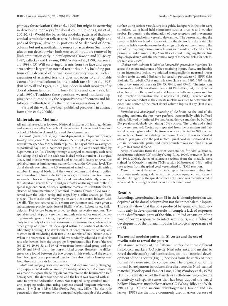
The normal modular pattern in S1 cortex and the use ofmyelin stain to reveal the patternWe stained sections of the flattened cortex for three differenthistological markers (CO activity, Nissl substance, and myelin) toreveal the effects of spinal hemisections on the anatomical devel-opment of the S1 cortex (Fig. 1). Sections from the cortex of threenormal rats were used for comparison. The organization of thenormal barrel pattern in rodents, first discovered in Nissl-stainedmaterial (Woolsey and Van der Loos, 1970; Woolsey et al., 1975)(Fig. 1B), reveals each of the barrels as a cell-dense ring enclosinga relatively cell-sparse center that has been dubbed the barrelhollow. However, metabolic markers CO (Wong-Riley and Welt,1980) (Fig. 1C) and succinic dehydrogenase (Dawson and Kil-lackey, 1987) are the more commonly used markers because of
10322 • J. Neurosci., November 12, 2003 • 23(32):10321–10330 Jain et al. • S1 Reorganization after Spinal Hemisections
the high contrast that they provide. The normal organization ofthe rat S1 has been described previously (Dawson and Killackey,1987). Briefly, cytochrome oxidase stains the area S1 darkly com-pared with the surrounding cortex (Fig. 1). Within S1, discreteCO-dark cell clusters (modules or barrels) are seen that are sur-rounded by CO-light regions called septa. The barrels are orga-nized in groups that correspond to the representations of differ-ent body parts. The lateralmost group, known as posteromedialbarrel subfield (PMBSF), represents whiskers on the face. Each ofthe large caudal barrels in PMBSF corresponds to the represen-tations of a single mystacial whisker (Woolsey et al., 1975). Directthalamocortical inputs from each whisker are highly restricted to
a specific barrel in S1. The more rostral barrels in this subfieldrepresent sinus hairs on the upper lip and the buccal pad (Welker,1976). Medial to PMBSF in the rostral S1, a small arch of barrelscorresponds to the representation of the hairs of the lower lip. Amore medial group of modules represents the digits and the padsof the forepaw (Dawson and Killackey, 1987; Waters et al., 1995),and another medialmost group corresponds to the representa-tion of the parts of the hindpaw (Dawson and Killackey, 1987).Caudal to the forepaw and the hindpaw representations, a zone ofmore uniform staining represents the trunk. The trunk represen-tation is connected to the forepaw and hindpaw subfields bynarrow rostrocaudal CO-dark strips corresponding to the repre-
Figure 1. The organization of S1 of rats as revealed in Nissl-, CO-, and myelin-stained sections of the flattened cortex. A, An outline diagram showing the locations of the mystacial vibrissae, lips,forepaw, hindpaw, and trunk representations in S1. Primary visual and auditory cortices are marked for reference. B, Photomontage of Nissl-stained sections from the flattened cortex of a ratshowing barrels and modules in the S1. The barrels appear with a cell-sparse “hollow” surrounded by cell-dense “walls.” C, In CO-stained sections, the barrels are visible as CO-dark modulessurrounded by CO-light septa. The distinct modular pattern is visible in the vibrissae, lips, forepaw, and hindpaw regions. In the trunk region, the staining is uniformly dark. The CO-dark patcheslateral to S1 correspond to second somatosensory (S2) and parietal ventral (PV) areas. D, We also stained sections of the flattened cortex for myelin. In myelin-stained sections through the middlelayers of the cortex, the barrels appear as myelin-light patches, whereas the septa are darkly stained. The regions of the trunk representation are uniformly stained as for CO and Nissl. In the deeperlayers, the staining pattern is reversed with myelin-dark barrels and myelin-light septa (see Fig. 6).
Jain et al. • S1 Reorganization after Spinal Hemisections J. Neurosci., November 12, 2003 • 23(32):10321–10330 • 10323
sentations of the arm and leg. Thus, a com-plete pattern of histological isomorphs ofbody parts can be clearly visualized in theS1 cortex (Fig. 1).
In addition to these often used CO andNissl stains, we also stained sections of theflattened cortex for myelin to reveal thebarrels (Fig. 1D). The barrels and clusterswere clearly seen in myelin-stained sec-tions from the flattened cortex. In the mid-dle layers of the cortex, these modulesstained lightly for myelin, whereas the in-terbarrel regions or septa were darklystained. In the infragranular layers, how-ever, the pattern of staining for myelin wasreversed. The modules stained darker, andthe septa were light in appearance (see Fig.6). The barrels were clearly visible in allregions of PMBSF, lower jaw, forepaw,and hindpaw, whereas the parts of the S1cortex representing the trunk, arm, and legwere more evenly stained, as seen in theCO- or Nissl-stained preparations.
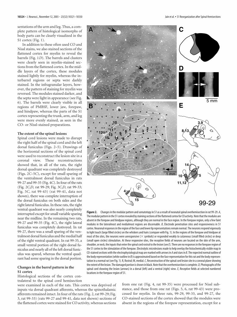
The extent of the spinal lesionsSpinal cord lesions were made to disruptthe right half of the spinal cord and the leftdorsal funiculus (Figs. 2–5). Drawings ofthe horizontal sections of the spinal cordwere used to reconstruct the lesion site in acoronal view. These reconstructionsshowed that, in all of the rats, the rightdorsal quadrant was completely destroyed(Figs. 2C–5C), except for small sparing ofthe ventralmost dorsal funiculus in rats99-27 and 99-35 (Fig. 4C). In four of the rats(Fig. 2C,D, rat 99-29; Fig. 3C,D, rat 99-33;Fig. 5C, rat 99-43) (rat 99-41, data notshown), there was complete interruption ofthe dorsal funiculus on both sides and theright lateral funiculus. In these rats, the rightventral quadrant was also nearly completelyinterrupted except for small variable sparingnear the midline. In the remaining two rats,99-27 and 99-35 (Fig. 4C), the right lateralfuniculus was completely destroyed. In rat99-27, there was a small sparing of the ven-tralmost dorsal funiculus and the medial halfof the right ventral quadrant. In rat 99-35, asmall ventral portion of the right dorsal fu-niculus and nearly all of the left dorsal funic-ulus was spared, whereas the ventral quad-rant had some sparing in the dorsal portion.
Changes in the barrel pattern in theS1 cortexHistological sections of the cortex con-tralateral to the spinal cord hemisectionwere examined in each of the rats. This cortex was deprived ofinputs via dorsal quadrant afferents, whereas the spinothalamicafferents remained intact. In four of the rats (Fig. 2, rat 99-29; Fig.3, rat 99-33) (rats 99-27 and 99-41, data not shown) sections ofthe flattened cortex were stained for CO activity, whereas sections
from one rat (Fig. 4, rat 99-35) were processed for Nissl sub-stance, and those from one rat (Figs. 5, 6, rat 99-43) were pro-cessed for myelin. In three rats, 99-29, 99-33, and 99-27, theCO-stained sections of the cortex showed that the modules wereabsent in the regions of the forepaw representation, except for a
Figure 2. Changes in the modular pattern and somatotopy in S1 as a result of neonatal spinal overhemisection in rat 99-29. A,The modular pattern in the S1 cortex revealed by staining sections of the flattened cortex for CO activity. Note that the modules areabsent in the forepaw and hindpaw regions, although they are normal in the face region. In the forepaw region, only a few faintmodules in the lateralmost and medialmost regions are discernable. B, Electrode penetration sites and responsiveness in S1cortex. Neuronal responses in the region of the face and lower lip representations remain normal. The neurons respond vigorouslyto light touch (large filled circles) on the whiskers and hairs (compare with Fig. 1). In the regions of the forepaw and hindpaw atmost of the sites, the neurons were unresponsive (� symbols) or responded weakly to cutaneous (small filled circles) or deep(small open circles) stimulation. At these responsive sites, the receptive fields of neurons are located on the skin of the arm,shoulder, or neck, the inputs that enter the spinal cord rostral to the lesion (see E). There are no responses in the forepaw region ofthe S1 cortex to the stimulation of the forepaw. Electrolytic microlesions made to help overlay the histochemically visible map inCO-stained sections with the electrophysiological map are marked with arrows in A and stars in B. The expected normal outline ofthe body representation (white outline in B) is approximated based on the face representation for this rat and the body represen-tation in a normal rat (see Fig. 1). R, Rostral; M, medial. C, Reconstruction of the spinal cord lesion site in a coronal plane showingthe extent of the lesion. The damaged portion is shown in black. Note that the overhemisection is complete. D, Photographs of thespinal cord showing the lesion (arrows) in a dorsal (left) and a ventral (right) view. E, Receptive fields at selected numberedlocations in the forepaw region of S1.
10324 • J. Neurosci., November 12, 2003 • 23(32):10321–10330 Jain et al. • S1 Reorganization after Spinal Hemisections
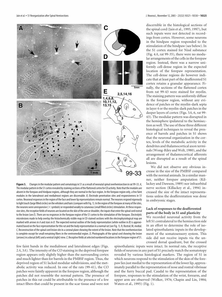
few faint bands in the medialmost and lateralmost edges (Figs.2A, 3A). The intensity of the CO staining in the deprived forepawregion appears only slightly higher than the surrounding cortexand much lighter than for barrels in the PMBSF region. Thus, thedeprived region of S1 lacked modular subdivisions, and the met-abolic activity was reduced. In one of the rats (99-41), the COpatches were faintly apparent in the forepaw region, although thepatches did not resemble the normal pattern. The presence ofpatches in this rat could be attributable to the presence of a fewintact fibers that could be present in the scar tissue and were not
discernible in the histological sections ofthe spinal cord (Jain et al., 1995, 1997), butsuch inputs were not detected in record-ings from cortex. However, some neuronsin the hindpaw region responded to thestimulation of the hindpaw (see below). Inthe S1 cortex stained for Nissl substance(Fig. 4A, rat 99-35), there were no modu-lar arrangements of the cells in the forepawregion. Instead, there was a narrow uni-formly cell-dense region in the expectedlocation of the forepaw representation.The cell-dense regions do however indi-cate that at least part of the deafferented S1cortex retains a granular appearance. Fi-nally, the sections of the flattened cortexfrom rat 99-43 were stained for myelin.The staining pattern was uniformly diffusein the forepaw region, without any evi-dence of patches or the myelin-dark septain layer 4 or the myelin-dark patches in thedeeper layers of cortex (Figs. 5A, 6, rat 99-43). The modular pattern was disrupted inthe hemisphere ipsilateral to the hemisec-tion as well. The use of these three differenthistological techniques to reveal the pres-ence of barrels and patches in S1 showsthat the neuronal organization in the cor-tex, levels of the metabolic activity in thedendrites and thalamocortical axon termi-nals (Wong-Riley and Welt, 1980), and thearrangement of thalamocortical afferentsall are disrupted as a result of the spinallesions.
We did not observe any obvious in-crease in the size of the PMBSF comparedwith the normal animals. In a similar man-ner, neither forepaw amputation (Kil-lackey and Dawson, 1989) nor infraorbitalnerve section (Killackey et al., 1994) in-creased the size of the intact representa-tions unless the deafferentation was donein embryonic stages.
Lack of responses to the deafferentedparts of the body in S1 and plasticityWe recorded neuronal activity from theleft S1 contralateral to the hemisection sidein an effort to determine the role of iso-lated spinothalamic inputs in the develop-ment of the somatosensory system. Thisside did not receive inputs via the un-crossed dorsal quadrant, but the crossed
spinothalamic inputs were intact. In normal rats, the receptivefields of neurons in any part of S1 precisely match the somatotopyrevealed by various histological markers. The region of S1 inwhich neurons respond to the stimulation of the skin of the fore-paw lies just medial to the representation of the lower lip, approx-imately parallel to the representation of nose and rostral vibrissaeand the furry buccal pad. Caudal to the representation of theforepaw, responses to the stimulation of the wrist, forearm, andupper arm are observed (Welker, 1976; Chapin and Lin, 1984;Waters et al., 1995) (Fig. 1).
Figure 3. Changes in the modular pattern and somatotopy in S1 as a result of neonatal spinal overhemisection in rat 99-33. A,The modular pattern in the S1 cortex revealed by staining sections of the flattened cortex for CO activity. Note that the modules areabsent in the forepaw and hindpaw regions, although they are normal in the face region. In the forepaw region only, a few faintmodules in the lateralmost and medialmost regions are discernable. B, Electrode penetration sites and responsiveness in S1cortex. Neuronal responses in the region of the face and lower lip representations remain normal. The neurons respond vigorouslyto light touch (large filled circles) on the whiskers and hairs (compare with Fig. 1). In the region of the forepaw at many of the sites,the neurons were unresponsive (� symbols) or responded weakly to cutaneous (small filled circles) stimulation. At these respon-sive sites, the receptive fields of neurons are located on the skin of the arm or shoulder, the inputs that enter the spinal cord rostralto the lesion (see E). There are no responses in the forepaw region of the S1 cortex to the stimulation of the forepaw. Electrolyticmicrolesions made to help overlay the histochemically visible map in CO-stained sections with the electrophysiological map aremarked with arrows in A and stars in B. The expected normal outline of the body representation (white outline in B) is approxi-mated based on the face representation for this rat and the body representation in a normal rat (see Fig. 1). R, Rostral; M, medial.C, Reconstruction of the spinal cord lesion site in a coronal plane showing the extent of the lesion. Note that the overhemisectionis complete except for small remaining fibers in the ventromedial region. D, Photographs of the spinal cord showing the lesion(arrows) in a dorsal (left) and a ventral (right) view. E, Receptive fields at selected numbered locations in the forepaw region of S1.
Jain et al. • S1 Reorganization after Spinal Hemisections J. Neurosci., November 12, 2003 • 23(32):10321–10330 • 10325
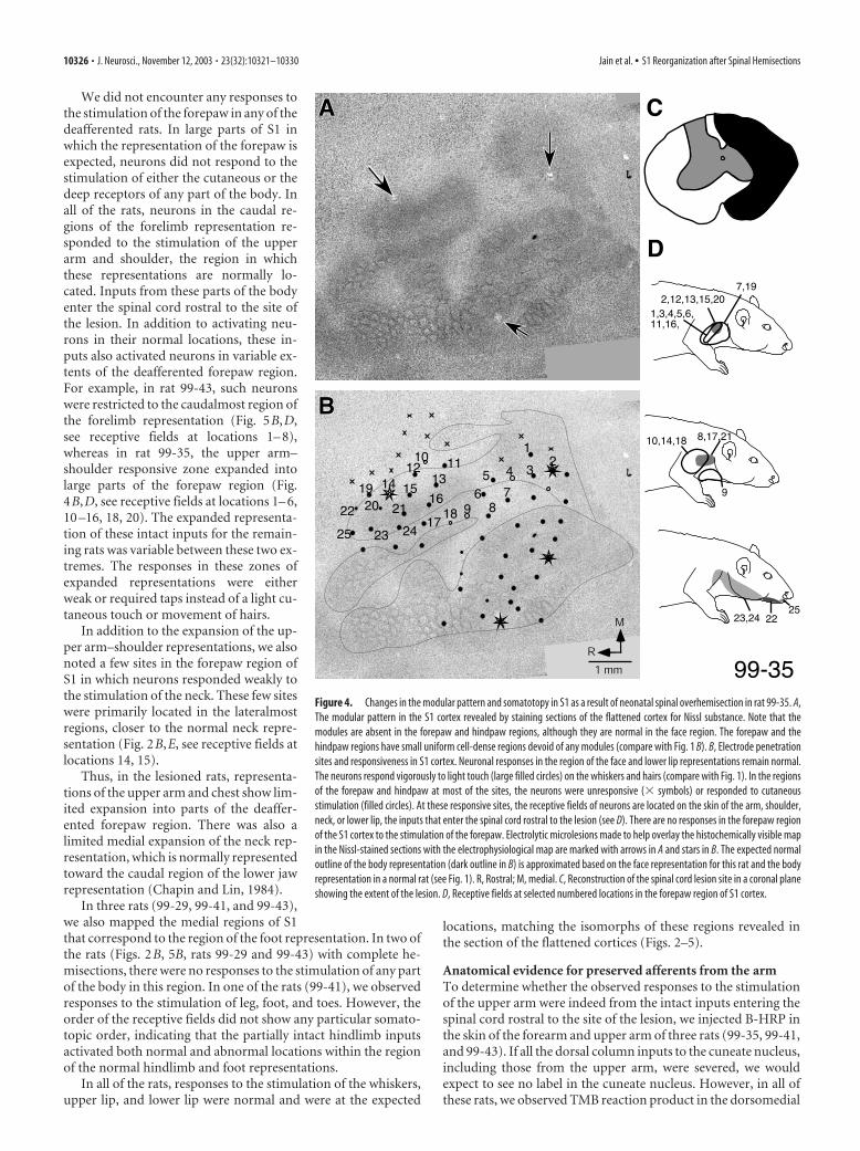
We did not encounter any responses tothe stimulation of the forepaw in any of thedeafferented rats. In large parts of S1 inwhich the representation of the forepaw isexpected, neurons did not respond to thestimulation of either the cutaneous or thedeep receptors of any part of the body. Inall of the rats, neurons in the caudal re-gions of the forelimb representation re-sponded to the stimulation of the upperarm and shoulder, the region in whichthese representations are normally lo-cated. Inputs from these parts of the bodyenter the spinal cord rostral to the site ofthe lesion. In addition to activating neu-rons in their normal locations, these in-puts also activated neurons in variable ex-tents of the deafferented forepaw region.For example, in rat 99-43, such neuronswere restricted to the caudalmost region ofthe forelimb representation (Fig. 5B,D,see receptive fields at locations 1– 8),whereas in rat 99-35, the upper arm–shoulder responsive zone expanded intolarge parts of the forepaw region (Fig.4B,D, see receptive fields at locations 1– 6,10 –16, 18, 20). The expanded representa-tion of these intact inputs for the remain-ing rats was variable between these two ex-tremes. The responses in these zones ofexpanded representations were eitherweak or required taps instead of a light cu-taneous touch or movement of hairs.
In addition to the expansion of the up-per arm–shoulder representations, we alsonoted a few sites in the forepaw region ofS1 in which neurons responded weakly tothe stimulation of the neck. These few siteswere primarily located in the lateralmostregions, closer to the normal neck repre-sentation (Fig. 2B,E, see receptive fields atlocations 14, 15).
Thus, in the lesioned rats, representa-tions of the upper arm and chest show lim-ited expansion into parts of the deaffer-ented forepaw region. There was also alimited medial expansion of the neck rep-resentation, which is normally representedtoward the caudal region of the lower jawrepresentation (Chapin and Lin, 1984).
In three rats (99-29, 99-41, and 99-43),we also mapped the medial regions of S1that correspond to the region of the foot representation. In two ofthe rats (Figs. 2B, 5B, rats 99-29 and 99-43) with complete he-misections, there were no responses to the stimulation of any partof the body in this region. In one of the rats (99-41), we observedresponses to the stimulation of leg, foot, and toes. However, theorder of the receptive fields did not show any particular somato-topic order, indicating that the partially intact hindlimb inputsactivated both normal and abnormal locations within the regionof the normal hindlimb and foot representations.
In all of the rats, responses to the stimulation of the whiskers,upper lip, and lower lip were normal and were at the expected
locations, matching the isomorphs of these regions revealed inthe section of the flattened cortices (Figs. 2–5).
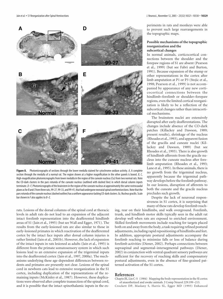
Anatomical evidence for preserved afferents from the armTo determine whether the observed responses to the stimulationof the upper arm were indeed from the intact inputs entering thespinal cord rostral to the site of the lesion, we injected B-HRP inthe skin of the forearm and upper arm of three rats (99-35, 99-41,and 99-43). If all the dorsal column inputs to the cuneate nucleus,including those from the upper arm, were severed, we wouldexpect to see no label in the cuneate nucleus. However, in all ofthese rats, we observed TMB reaction product in the dorsomedial
Figure 4. Changes in the modular pattern and somatotopy in S1 as a result of neonatal spinal overhemisection in rat 99-35. A,The modular pattern in the S1 cortex revealed by staining sections of the flattened cortex for Nissl substance. Note that themodules are absent in the forepaw and hindpaw regions, although they are normal in the face region. The forepaw and thehindpaw regions have small uniform cell-dense regions devoid of any modules (compare with Fig. 1 B). B, Electrode penetrationsites and responsiveness in S1 cortex. Neuronal responses in the region of the face and lower lip representations remain normal.The neurons respond vigorously to light touch (large filled circles) on the whiskers and hairs (compare with Fig. 1). In the regionsof the forepaw and hindpaw at most of the sites, the neurons were unresponsive (� symbols) or responded to cutaneousstimulation (filled circles). At these responsive sites, the receptive fields of neurons are located on the skin of the arm, shoulder,neck, or lower lip, the inputs that enter the spinal cord rostral to the lesion (see D). There are no responses in the forepaw regionof the S1 cortex to the stimulation of the forepaw. Electrolytic microlesions made to help overlay the histochemically visible mapin the Nissl-stained sections with the electrophysiological map are marked with arrows in A and stars in B. The expected normaloutline of the body representation (dark outline in B) is approximated based on the face representation for this rat and the bodyrepresentation in a normal rat (see Fig. 1). R, Rostral; M, medial. C, Reconstruction of the spinal cord lesion site in a coronal planeshowing the extent of the lesion. D, Receptive fields at selected numbered locations in the forepaw region of S1 cortex.
10326 • J. Neurosci., November 12, 2003 • 23(32):10321–10330 Jain et al. • S1 Reorganization after Spinal Hemisections
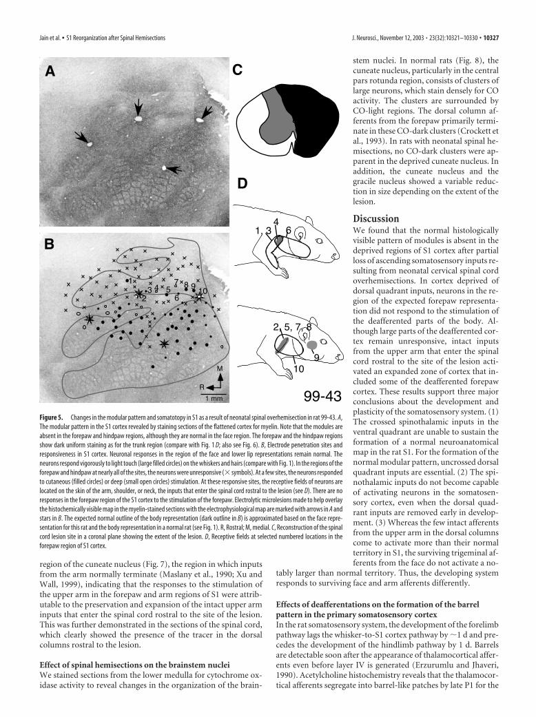
region of the cuneate nucleus (Fig. 7), the region in which inputsfrom the arm normally terminate (Maslany et al., 1990; Xu andWall, 1999), indicating that the responses to the stimulation ofthe upper arm in the forepaw and arm regions of S1 were attrib-utable to the preservation and expansion of the intact upper arminputs that enter the spinal cord rostral to the site of the lesion.This was further demonstrated in the sections of the spinal cord,which clearly showed the presence of the tracer in the dorsalcolumns rostral to the lesion.
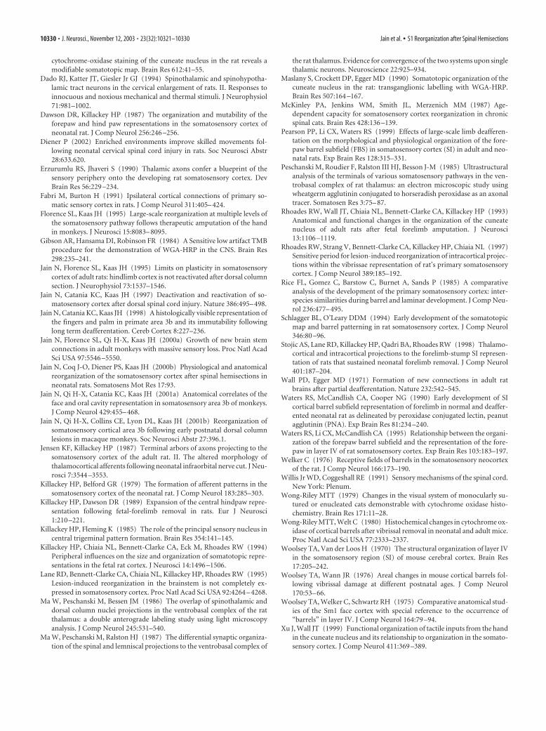
Effect of spinal hemisections on the brainstem nucleiWe stained sections from the lower medulla for cytochrome ox-idase activity to reveal changes in the organization of the brain-
stem nuclei. In normal rats (Fig. 8), thecuneate nucleus, particularly in the centralpars rotunda region, consists of clusters oflarge neurons, which stain densely for COactivity. The clusters are surrounded byCO-light regions. The dorsal column af-ferents from the forepaw primarily termi-nate in these CO-dark clusters (Crockett etal., 1993). In rats with neonatal spinal he-misections, no CO-dark clusters were ap-parent in the deprived cuneate nucleus. Inaddition, the cuneate nucleus and thegracile nucleus showed a variable reduc-tion in size depending on the extent of thelesion.
DiscussionWe found that the normal histologicallyvisible pattern of modules is absent in thedeprived regions of S1 cortex after partialloss of ascending somatosensory inputs re-sulting from neonatal cervical spinal cordoverhemisections. In cortex deprived ofdorsal quadrant inputs, neurons in the re-gion of the expected forepaw representa-tion did not respond to the stimulation ofthe deafferented parts of the body. Al-though large parts of the deafferented cor-tex remain unresponsive, intact inputsfrom the upper arm that enter the spinalcord rostral to the site of the lesion acti-vated an expanded zone of cortex that in-cluded some of the deafferented forepawcortex. These results support three majorconclusions about the development andplasticity of the somatosensory system. (1)The crossed spinothalamic inputs in theventral quadrant are unable to sustain theformation of a normal neuroanatomicalmap in the rat S1. For the formation of thenormal modular pattern, uncrossed dorsalquadrant inputs are essential. (2) The spi-nothalamic inputs do not become capableof activating neurons in the somatosen-sory cortex, even when the dorsal quad-rant inputs are removed early in develop-ment. (3) Whereas the few intact afferentsfrom the upper arm in the dorsal columnscome to activate more than their normalterritory in S1, the surviving trigeminal af-ferents from the face do not activate a no-
tably larger than normal territory. Thus, the developing systemresponds to surviving face and arm afferents differently.
Effects of deafferentations on the formation of the barrelpattern in the primary somatosensory cortexIn the rat somatosensory system, the development of the forelimbpathway lags the whisker-to-S1 cortex pathway by �1 d and pre-cedes the development of the hindlimb pathway by 1 d. Barrelsare detectable soon after the appearance of thalamocortical affer-ents even before layer IV is generated (Erzurumlu and Jhaveri,1990). Acetylcholine histochemistry reveals that the thalamocor-tical afferents segregate into barrel-like patches by late P1 for the
Figure 5. Changes in the modular pattern and somatotopy in S1 as a result of neonatal spinal overhemisection in rat 99-43. A,The modular pattern in the S1 cortex revealed by staining sections of the flattened cortex for myelin. Note that the modules areabsent in the forepaw and hindpaw regions, although they are normal in the face region. The forepaw and the hindpaw regionsshow dark uniform staining as for the trunk region (compare with Fig. 1 D; also see Fig. 6). B, Electrode penetration sites andresponsiveness in S1 cortex. Neuronal responses in the region of the face and lower lip representations remain normal. Theneurons respond vigorously to light touch (large filled circles) on the whiskers and hairs (compare with Fig. 1). In the regions of theforepaw and hindpaw at nearly all of the sites, the neurons were unresponsive (� symbols). At a few sites, the neurons respondedto cutaneous (filled circles) or deep (small open circles) stimulation. At these responsive sites, the receptive fields of neurons arelocated on the skin of the arm, shoulder, or neck, the inputs that enter the spinal cord rostral to the lesion (see D). There are noresponses in the forepaw region of the S1 cortex to the stimulation of the forepaw. Electrolytic microlesions made to help overlaythe histochemically visible map in the myelin-stained sections with the electrophysiological map are marked with arrows in A andstars in B. The expected normal outline of the body representation (dark outline in B) is approximated based on the face repre-sentation for this rat and the body representation in a normal rat (see Fig. 1). R, Rostral; M, medial. C, Reconstruction of the spinalcord lesion site in a coronal plane showing the extent of the lesion. D, Receptive fields at selected numbered locations in theforepaw region of S1 cortex.
Jain et al. • S1 Reorganization after Spinal Hemisections J. Neurosci., November 12, 2003 • 23(32):10321–10330 • 10327
whisker barrels and by P3 for the forepaw barrels (Schlagger andO’Leary, 1994; the day of birth is P1, and, in reviewing the previ-ous literature, we have adjusted accordingly). In Nissl and suc-cinic dehydrogenase preparations, the barrels become visible af-ter an additional 2 d (Killackey and Belford, 1979; Rice et al.,1985). The critical period for the development of the forepawmodules as well as the whisker barrels ends at approximately P6,well after we did our lesions.
Previously, it has been shown that complete deafferentationby removal of the forelimb on P3 or P4 results in a completedisruption of the modules in the forepaw region of the S1 cortex(Waters et al., 1990; Pearson et al., 1999). Our results show thattransecting uncrossed pathways in the dorsal and dorsolateralspinal cord, although leaving the crossed spinothalamic pathwaysintact, is sufficient to disrupt the formation of the normal patternof modules in the forepaw and hindpaw regions of the S1 cortex.Spinothalamic afferents, which remained intact in our prepara-tion, terminate throughout the ventroposterior nucleus of thethalamus intermingled with terminations of lemniscal inputs(Ma et al., 1986). In rats, the synapses of both of these ascendingsystems appear to be similar and indistinguishable (Peschanski etal., 1985), and �10% of the spinothalamic terminations are onthe neurons that also receive lemniscal inputs (Ma et al., 1987).However, it is possible that cutaneous inputs via the upper quad-rant of the spinal cord provide the most patterned activity to thedeveloping system, whereas the crossed spinothalamic inputswith mostly large overlapping receptive fields (Willis and Cogge-shall, 1991; Dado et al., 1994) are not sufficient to direct themaintenance of segregated thalamocortical afferents and the de-velopment of the modules. Alternatively, the level of subthresh-old activation via spinothalamic inputs, if any, may not be suffi-cient to overcome the loss of patterned activity attributable to the
spinal hemisections. We see no evidence in anesthetized rats ofneural spikes in S1 that are generated by spinothalamic pathways.In support of the present results, lesions of the principal trigem-inal nucleus, the major target of the trigeminal analog of thedorsal column afferents, are sufficient to disrupt the barreloidpattern in the ventroposterior nucleus of the thalamus (Killackeyand Fleming, 1985). An intact spinal trigeminal nucleus withnormal barrel patterns (barrelettes) in the subnucleus interpo-laris and caudalis of the spinal trigeminal nucleus cannot sustainthe formation of a normal thalamic barreloid pattern (Killackeyand Fleming, 1985).
Somatotopic reorganizationAlthough a large number of studies have examined the effects ofembryonic or neonatal peripheral injuries on the barrel pattern asrevealed by various histochemical stains, only a few examined thechanges in topography to determine correspondence between thealtered barrel pattern and the receptive field properties of neu-rons. In general, it appears that deafferentations done early indevelopment do not lead to a large-scale expansion of the intact,topographically adjacent body regions into the deafferented cor-tex. Rhoades and colleagues (Lane et al., 1995) report that, afterforelimb amputation on P1 (the day of birth), neurons at most ofthe recording sites in the forepaw region were responsive to thestimulation of the stump, and only at a few sites were there re-sponses to the stimulation of the hindlimb or inputs from thechin, whiskers, or neck. In a similar study, Pearson et al. (1999)found that amputation of the forelimb on P3 leads to multipleislands of stump-responsive sites in the forelimb area. The expan-sion of the trigeminal inputs into the forelimb area was extremelylimited and confined to the region adjacent to the face represen-tation. Even the forelimb amputations done earlier, on embry-onic day 16 or 17, failed to produce responses to the stimulationof the intact hindlimb in the deafferented forelimb region of S1(Killackey and Dawson, 1989).
The lack of activation by the spinothalamic inputs and the lackof any extensive large-scale reorganization after early deafferen-tations reported here is similar to that found after lesions in adult
Figure 7. Plot of the transganglionic neuronal tracer B-HRP in a section from the lowermedulla of rat 99-43. The tracer was injected in the skin of the upper arm (between the shoulderand the elbow) at 13 different locations. Presence of the tracer in the brainstem shows thatdorsal column inputs from this part of the arm were not interrupted by the spinal lesion becausethey entered the spinal cord rostral to the lesion. DMV, Dorsal motor nucleus of the vagus nerve;Hy, hypoglossal nucleus; IO, inferior olivary nucleus; Py Tr, pyramidal tract.
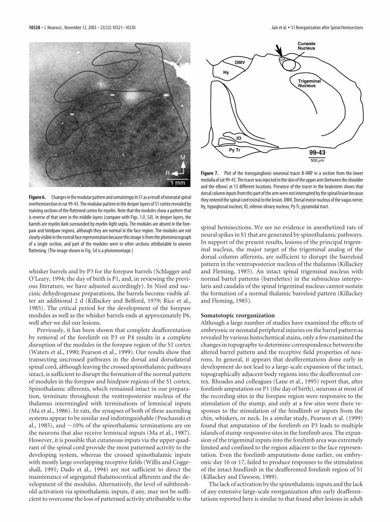
Figure 6. Changes in the modular pattern and somatotopy in S1 as a result of neonatal spinaloverhemisection in rat 99-43. The modular pattern in the deeper layers of S1 cortex revealed bystaining sections of the flattened cortex for myelin. Note that the modules show a pattern thatis reverse of that seen in the middle layers (compare with Figs. 1 D, 5A). In deeper layers, thebarrels are myelin dark surrounded by myelin-light septa. The modules are absent in the fore-paw and hindpaw regions, although they are normal in the face region. The modules are notclearly visible in the rostral face representation because this image is from the photomicrographof a single section, and part of the modules were in other sections attributable to unevenflattening. (The image shown in Fig. 5A is a photomontage.)
10328 • J. Neurosci., November 12, 2003 • 23(32):10321–10330 Jain et al. • S1 Reorganization after Spinal Hemisections
rats. Lesions of the dorsal columns of the spinal cord at thoraciclevels in adult rats do not lead to an expansion of the adjacentintact forelimb representation into the deafferented hindlimbareas of S1 (Jain et al., 1995) (but see Wall and Egger, 1971). Theresults from the early-lesioned rats are also similar to those inearly-lesioned primates in which reactivation of the deafferentedcortex by the intact face inputs after dorsal column injuries israther limited (Jain et al., 2001b). However, the lack of expansionof the intact inputs in rats lesioned as adults (Jain et al., 1995) isdifferent from the primate somatosensory system in which suchlesions lead to an extensive expansion of the trigeminal inputsinto the deafferented cortex (Jain et al., 1997, 2000a). The mech-anisms underlying these age-dependent differences between ro-dents and primates are presently not clear. Lesions of the spinalcord in newborn cats lead to extensive reorganization in the S1cortex, including duplication of the representations of the re-maining inputs (McKinley et al., 1987). However, these duplica-tions were observed after complete transection of the spinal cord,and it is possible that the intact spinothalamic inputs in the ex-
periments in rats and monkeys were ableto prevent such large rearrangements inthe topographic maps.
Possible mechanisms of the topographicreorganization and thesubcortical changesIn normal animals, corticocortical con-nections between the shoulder and theforepaw regions of S1 are absent (Pearsonet al., 1999) (but see Fabri and Burton,1991). Because expansion of the stump orother representations in the cortex afterlimb amputation at P1 or P3 (Stojic et al.,1998; Pearson et al., 1999) is not accom-panied by appearance of any new corti-cocortical connections between thehindlimb–forelimb or shoulder–forepawregions, even the limited cortical reorgani-zation is likely to be a reflection of thesubcortical changes rather than intracorti-cal mechanisms.
The brainstem nuclei are extensivelydisrupted after early deafferentations. Thechanges include absence of the CO-darkpatches (Killackey and Dawson, 1989;present results), shrinkage of the nucleus(Rhoades et al., 1993), and apparent fusionof the gracilis and cuneate nuclei (Kil-lackey and Dawson, 1989) (but seeRhoades et al., 1993). There is also growthof hindlimb afferents from the gracile nu-cleus into the cuneate nucleus after fore-limb amputation (Rhoades et al., 1993;Lane et al., 1995). In these animals, there isno growth from the trigeminal nucleus,apparently because the trigeminal path-way develops before the forelimb pathway.In our lesions, disruption of afferents toboth the cuneate and the gracile nucleusprecludes such growth.
Despite the lack of neuronal respon-siveness in S1 cortex, it is surprising thatmany of these rats develop forelimb reach-
ing, rear on their hindlimbs, and walk overground. Forelimb,trunk, and hindlimb motor skills typically seen in the adult ratdevelop well when rats are exposed to enriched environment.Skilled forelimb movements include reaching for targets placedboth on and away from the body, a task requiring refined posturaladjustments, including rapid repositioning of hindlimbs and feet.In addition, appropriate postural adjustments accompany theforelimb reaching to minimize falls or loss of balance duringforelimb activities (Diener, 2002). Perhaps connections betweensupraspinal and segmental–intersegmental pathways (Diener,2002) in conjunction with ventral quadrant sensory pathways aresufficient for the recovery of reaching skills and compensatorypostural adjustments, even in the absence of fine-grained pat-terned activation of the S1 cortex.
ReferencesChapin JK, Lin C-S (1984) Mapping the body representation in the SI cortex
of anaesthetized and awake animals. J Comp Neurol 229:199 –213.Crockett DP, Maslany S, Harris SL, Egger MD (1993) Enhanced
Figure 8. Photomicrographs of sections through the lower medulla stained for cytochrome oxidase activity. A, A completesection through the medulla of a normal rat. The region shown at a higher magnification in the other panels is boxed. B, C,High-magnification photomicrographs from lower medulla in the region of the cuneate nucleus (Cu) from two normal rats. Notethe CO-dark clusters in the pars rotunda of the cuneate nucleus (outlined with dashed line) in which dorsal column inputsterminate. D–F, Photomicrographs of the brainstem in the region of the cuneate nucleus at approximately the same rostrocaudalplane as for B and C from three rats, 99-27, 99-33, and 99-41, that had undergone neonatal spinal overhemisections. Note that thepars rotunda of the cuneate nucleus (dashed outline) has a uniform appearance lacking CO-dark clusters. Gr, Nucleus gracilis. Scalebar shown in F also applies to B–E.
Jain et al. • S1 Reorganization after Spinal Hemisections J. Neurosci., November 12, 2003 • 23(32):10321–10330 • 10329
cytochrome-oxidase staining of the cuneate nucleus in the rat reveals amodifiable somatotopic map. Brain Res 612:41–55.
Dado RJ, Katter JT, Giesler Jr GJ (1994) Spinothalamic and spinohypotha-lamic tract neurons in the cervical enlargement of rats. II. Responses toinnocuous and noxious mechanical and thermal stimuli. J Neurophysiol71:981–1002.
Dawson DR, Killackey HP (1987) The organization and mutability of theforepaw and hind paw representations in the somatosensory cortex ofneonatal rat. J Comp Neurol 256:246 –256.
Diener P (2002) Enriched environments improve skilled movements fol-lowing neonatal cervical spinal cord injury in rats. Soc Neurosci Abstr28:633.620.
Erzurumlu RS, Jhaveri S (1990) Thalamic axons confer a blueprint of thesensory periphery onto the developing rat somatosensory cortex. DevBrain Res 56:229 –234.
Fabri M, Burton H (1991) Ipsilateral cortical connections of primary so-matic sensory cortex in rats. J Comp Neurol 311:405– 424.
Florence SL, Kaas JH (1995) Large-scale reorganization at multiple levels ofthe somatosensory pathway follows therapeutic amputation of the handin monkeys. J Neurosci 15:8083– 8095.
Gibson AR, Hansama DI, Robinson FR (1984) A Sensitive low artifact TMBprocedure for the demonstration of WGA-HRP in the CNS. Brain Res298:235–241.
Jain N, Florence SL, Kaas JH (1995) Limits on plasticity in somatosensorycortex of adult rats: hindlimb cortex is not reactivated after dorsal columnsection. J Neurophysiol 73:1537–1546.
Jain N, Catania KC, Kaas JH (1997) Deactivation and reactivation of so-matosensory cortex after dorsal spinal cord injury. Nature 386:495– 498.
Jain N, Catania KC, Kaas JH (1998) A histologically visible representation ofthe fingers and palm in primate area 3b and its immutability followinglong term deafferentation. Cereb Cortex 8:227–236.
Jain N, Florence SL, Qi H-X, Kaas JH (2000a) Growth of new brain stemconnections in adult monkeys with massive sensory loss. Proc Natl AcadSci USA 97:5546 –5550.
Jain N, Coq J-O, Diener PS, Kaas JH (2000b) Physiological and anatomicalreorganization of the somatosensory cortex after spinal hemisections inneonatal rats. Somatosens Mot Res 17:93.
Jain N, Qi H-X, Catania KC, Kaas JH (2001a) Anatomical correlates of theface and oral cavity representation in somatosensory area 3b of monkeys.J Comp Neurol 429:455– 468.
Jain N, Qi H-X, Collins CE, Lyon DL, Kaas JH (2001b) Reorganization ofsomatosensory cortical area 3b following early postnatal dorsal columnlesions in macaque monkeys. Soc Neurosci Abstr 27:396.1.
Jensen KF, Killackey HP (1987) Terminal arbors of axons projecting to thesomatosensory cortex of the adult rat. II. The altered morphology ofthalamocortical afferents following neonatal infraorbital nerve cut. J Neu-rosci 7:3544 –3553.
Killackey HP, Belford GR (1979) The formation of afferent patterns in thesomatosensory cortex of the neonatal rat. J Comp Neurol 183:285–303.
Killackey HP, Dawson DR (1989) Expansion of the central hindpaw repre-sentation following fetal-forelimb removal in rats. Eur J Neurosci1:210 –221.
Killackey HP, Fleming K (1985) The role of the principal sensory nucleus incentral trigeminal pattern formation. Brain Res 354:141–145.
Killackey HP, Chiaia NL, Bennett-Clarke CA, Eck M, Rhoades RW (1994)Peripheral influences on the size and organization of somatotopic repre-sentations in the fetal rat cortex. J Neurosci 14:1496 –1506.
Lane RD, Bennett-Clarke CA, Chiaia NL, Killackey HP, Rhoades RW (1995)Lesion-induced reorganization in the brainstem is not completely ex-pressed in somatosensory cortex. Proc Natl Acad Sci USA 92:4264 – 4268.
Ma W, Peschanski M, Bessen JM (1986) The overlap of spinothalamic anddorsal column nuclei projections in the ventrobasal complex of the ratthalamus: a double anterograde labeling study using light microscopyanalysis. J Comp Neurol 245:531–540.
Ma W, Peschanski M, Ralston HJ (1987) The differential synaptic organiza-tion of the spinal and lemniscal projections to the ventrobasal complex of
the rat thalamus. Evidence for convergence of the two systems upon singlethalamic neurons. Neuroscience 22:925–934.
Maslany S, Crockett DP, Egger MD (1990) Somatotopic organization of thecuneate nucleus in the rat: transganglionic labelling with WGA-HRP.Brain Res 507:164 –167.
McKinley PA, Jenkins WM, Smith JL, Merzenich MM (1987) Age-dependent capacity for somatosensory cortex reorganization in chronicspinal cats. Brain Res 428:136 –139.
Pearson PP, Li CX, Waters RS (1999) Effects of large-scale limb deafferen-tation on the morphological and physiological organization of the fore-paw barrel subfield (FBS) in somatosensory cortex (SI) in adult and neo-natal rats. Exp Brain Res 128:315–331.
Peschanski M, Roudier F, Ralston III HJ, Besson J-M (1985) Ultrastructuralanalysis of the terminals of various somatosensory pathways in the ven-trobasal complex of rat thalamus: an electron microscopic study usingwheatgerm agglutinin conjugated to horseradish peroxidase as an axonaltracer. Somatosen Res 3:75– 87.
Rhoades RW, Wall JT, Chiaia NL, Bennett-Clarke CA, Killackey HP (1993)Anatomical and functional changes in the organization of the cuneatenucleus of adult rats after fetal forelimb amputation. J Neurosci13:1106 –1119.
Rhoades RW, Strang V, Bennett-Clarke CA, Killackey HP, Chiaia NL (1997)Sensitive period for lesion-induced reorganization of intracortical projec-tions within the vibrissae representation of rat’s primary somatosensorycortex. J Comp Neurol 389:185–192.
Rice FL, Gomez C, Barstow C, Burnet A, Sands P (1985) A comparativeanalysis of the development of the primary somatosensory cortex: inter-species similarities during barrel and laminar development. J Comp Neu-rol 236:477– 495.
Schlagger BL, O’Leary DDM (1994) Early development of the somatotopicmap and barrel patterning in rat somatosensory cortex. J Comp Neurol346:80 –96.
Stojic AS, Lane RD, Killackey HP, Qadri BA, Rhoades RW (1998) Thalamo-cortical and intracortical projections to the forelimb-stump SI represen-tation of rats that sustained neonatal forelimb removal. J Comp Neurol401:187–204.
Wall PD, Egger MD (1971) Formation of new connections in adult ratbrains after partial deafferentation. Nature 232:542–545.
Waters RS, McCandlish CA, Cooper NG (1990) Early development of SIcortical barrel subfield representation of forelimb in normal and deaffer-ented neonatal rat as delineated by peroxidase conjugated lectin, peanutagglutinin (PNA). Exp Brain Res 81:234 –240.
Waters RS, Li CX, McCandlish CA (1995) Relationship between the organi-zation of the forepaw barrel subfield and the representation of the fore-paw in layer IV of rat somatosensory cortex. Exp Brain Res 103:183–197.
Welker C (1976) Receptive fields of barrels in the somatosensory neocortexof the rat. J Comp Neurol 166:173–190.
Willis Jr WD, Coggeshall RE (1991) Sensory mechanisms of the spinal cord.New York: Plenum.
Wong-Riley MTT (1979) Changes in the visual system of monocularly su-tured or enucleated cats demonstrable with cytochrome oxidase histo-chemistry. Brain Res 171:11–28.
Wong-Riley MTT, Welt C (1980) Histochemical changes in cytochrome ox-idase of cortical barrels after vibrissal removal in neonatal and adult mice.Proc Natl Acad Sci USA 77:2333–2337.
Woolsey TA, Van der Loos H (1970) The structural organization of layer IVin the somatosensory region (SI) of mouse cerebral cortex. Brain Res17:205–242.
Woolsey TA, Wann JR (1976) Areal changes in mouse cortical barrels fol-lowing vibrissal damage at different postnatal ages. J Comp Neurol170:53– 66.
Woolsey TA, Welker C, Schwartz RH (1975) Comparative anatomical stud-ies of the Sm1 face cortex with special reference to the occurrence of“barrels” in layer IV. J Comp Neurol 164:79 –94.
Xu J, Wall JT (1999) Functional organization of tactile inputs from the handin the cuneate nucleus and its relationship to organization in the somato-sensory cortex. J Comp Neurol 411:369 –389.
10330 • J. Neurosci., November 12, 2003 • 23(32):10321–10330 Jain et al. • S1 Reorganization after Spinal Hemisections