BDNF regulates the expression and traffic of NMDA receptors in cultured hippocampal neurons
Upload
independentCategory
view
0download
0
PAR
BAAa
udb
lM
AnmtsheRvrvclmsdoocodabagdwL
*5EAcedhdteNoRRo
Neuroscience 167 (2010) 1057–1069
0d
ATHWAYS INVOLVED IN THE GENERATION OF REACTIVE OXYGENND NITROGEN SPECIES DURING GLUCOSE DEPRIVATION AND ITS
OLE ON THE DEATH OF CULTURED HIPPOCAMPAL NEURONSKx
GbbkpvhKcemptds(scrdNhhle2lsovRtarRR(gavasab
. PÁRAMO,a K. HERNÁNDEZ-FONSECA,a
. M. ESTRADA-SÁNCHEZ,a N. JIMÉNEZ,b
. HERNÁNDEZ-CRUZb AND L. MASSIEUa*
División de Neurociencias, Departamento de Neuropatología Molec-lar, Instituto de Fisiología Celular, Universidad Nacional Autónomae México, México D.F., México, CP 04510, AP 70-253
Departamento de Neurociencia Cognitiva, Instituto de Fisiología Ce-ular, Universidad Nacional Autónoma de México, México D.F.,
éxico, CP 04510, AP 70-253
bstract–Oxidative stress has been suggested as a mecha-ism contributing to neuronal death induced by hypoglyce-ia, and an early production of reactive species (RS) during
he hypoglycemic episode has been observed. However, theources of reactive oxygen (ROS) and nitrogen (RNS) speciesave not been fully identified. In the present study we havexamined the contribution of various enzymatic pathways toS production and neuronal death induced by glucose depri-ation (GD) in hippocampal cultures. We have observed aapid increase in RS during GD, which depends on the acti-ation of NMDA and non-NMDA receptors and on the influx ofalcium from the extracellular space. Accordingly, intracellu-
ar calcium concentration [Ca2�]i progressively increasesore than 30-fold during the GD period. It was observed that
uperoxide production through the activation of the calcium-ependent enzymes, phospholipase A2 (cPLA2) and xanthinexidase (XaO), contributes to neuronal damage, while nitricxide synthase (NOS) is apparently not involved. Inhibition ofPLA2 decreased RS at early times of GD whereas inhibitionf XaO diminished RS at more delayed times. The antioxi-ants trolox and ebselen also showed a protective effectgainst neuronal death and diminished RS generation. Inhi-ition of NADPH oxidase also contributed to the early gener-tion of superoxide. Taking together, the present results sug-est that the early activation of calcium-dependent ROS pro-ucing pathways is involved in neuronal death associatedith glucose deprivation. © 2010 IBRO. Published by Elseviertd. All rights reserved.
Corresponding author. Tel: �52-55/56-22-57-61; fax: �52-55/56-22-6-07.-mail address: [email protected] (L. Massieu).bbreviations: c-DCF, carboxy-2=,7=-dichlorofluorescein diacetate;-H2DCFDA, 5-(and-6-)carboxy-2=,7=-dichlorodihydrofluorescein diac-tate; cPLA2, calcium-dependent cytosolic phospholipase A2; DHE,ihydroethidium; DIV, days in vitro; EPTU, 2-ethyl-2-thiopseudoureaydrobromide; Et, ethidium; GD, glucose deprivation; LDH, lactateehydrogenase; LOX, lipoxygenase; MK-801, dizolcipine(�)-5-me-hyl-10,11-dihydroxy-5H-dibenzo(a,d)-cyclohepten-5,10-imine mal-ate; MTT, 3-(4,5-dimethylthiazol-2-yl)-2,5-diphenyltetrazoliumbromide;BQX, 2,3-dihydroxy-6-nitro-7-sulfamoyl-benzo[f]quinoxaline-2,3-di-ne; NDGA, nordihydroguaiaretic acid; NOS, nitric oxide synthase;FU, relative fluorescence units; RNS, reactive nitrogen species;
iOS, reactive oxygen species; RS, reactive species; XaO, xanthinexidase; 7-NI, 7-nitroindazole.
306-4522/10 $ - see front matter © 2010 IBRO. Published by Elsevier Ltd. All rightoi:10.1016/j.neuroscience.2010.02.074
1057
ey words: hypoglycemia, oxidative damage, excitotoxicity,anthine oxidase, phospholipase A2, NADPH oxidase.
lucose is the main energy source in the brain, wheneverlood glucose concentration decreases below 20 mg/dl,rain activity ceases and the hypoglycemic coma, alsonown as the isoelectric period, takes place. A 30–60 mineriod of isoelectricity induces selective brain damage inulnerable brain regions, including the cerebral cortex, theippocampus and the striatum (Auer et al., 1984a,b;alimo et al., 1985). The mechanisms underlying hypogly-emic neuronal damage are not completely understood butarly studies suggested the participation of an excitotoxicechanism triggered by the release of glutamate, andarticularly aspartate, soon after the onset of the isoelec-ric period (Sandberg et al., 1986; Wieloch, 1984). In ad-ition, recent investigations have suggested that oxidativetress is associated with hypoglycemic neuronal damageSuh et al., 2007, 2008; Haces et al., 2008, 2010). Studieshow that glucose reintroduction after the isoelectric periodorrelates with the presence of superoxide and nitroty-osine immunoreactivity, and suggest that glucose reintro-uction stimulates oxidative stress through the activity ofADPH oxidase (Suh et al., 2007, 2008). On the otherand, oxidative stress might be an early response to theypoglycemic condition because a selective increase in
ipoperoxidation is observed before the onset of the iso-lectric period, in vulnerable brain regions (Haces et al.,010). In agreement, other studies have shown increased
ipoperoxidation in brains from hypoglycemic mice non-ubjected to a coma period (Patocková et al., 2003). Thesebservations suggest that the hypoglycemic condition fa-ors the production of reactive oxygen and nitrogen (ROS/NS) species, which might participate in the induction of
he subsequent neuronal death. However, little is knownbout the pathways involved in the early generation ofeactive species (RS) during the hypoglycemic condition.ecent studies have reported increased mitochondrialOS production during hypoglycemia in newborn piglets
McGowan et al., 2006), as well as in cultured cerebellarranule neurons during glucose deprivation (GD) (Isaev etl., 2008). GD of cultured neurons can be used as an initro model to study the mechanisms of neuronal damagessociated with the hypoglycemic condition. In previoustudies we have shown that hippocampal cultured neuronsre highly vulnerable to energy failure, and die after inhi-ition of the glycolytic pathway with iodoacetate, a potent
nhibitor of glyceraldehyde-3-phosphate dehydrogenase.s reserved.
Ndt(adtnrs((wvgnueN
C
PWdssL
UcpUagLtoiPCi
dvmtt
R
Rtma(DruZiicdp
iedflmflrpeaeacZ
tez1((tJSaSd
sAeL�s2G
mmNbMAct
I
CbOArCTRMowoF4Ttwicc
B. Páramo et al. / Neuroscience 167 (2010) 1057–10691058
euronal death induced in this condition involves energyepletion, activation of glutamate receptors, elevation of
he intracellular calcium concentration and RS productionHernández-Fonseca et al., 2008). In the present study weimed to identify through a pharmacological approach, theifferent pathways involved in the production of RS, and
heir contribution to cell death in hippocampal culturedeurons deprived from glucose. Results indicate that GDapidly induces RS generation, involving the activity ofeveral enzymatic pathways including xanthine oxidaseXaO), the calcium-dependent cytosolic phospholipase A2
cPLA2), lipoxygenase (LOX) and NADPH oxidase. Mean-hile, nitric oxide synthase (NOS) is apparently not in-olved. RS generation is dependent on the activation oflutamate receptors and on calcium influx from the exter-al milieu. Results suggest that oxidative stress contrib-tes to neuronal damage, since the antioxidants trolox andbselen as well as the inhibitors of XaO, cPLA2 andADPH oxidase partially prevented neuronal death.
EXPERIMENTAL PROCEDURES
ell culture
rimary cultures of hippocampal neurons were prepared fromistar rat embryos of 17–18 days of gestation as previously
escribed (Hernández-Fonseca and Massieu, 2005). Cells wereuspended in Neurobasal culture medium (Brewer et al., 1993)upplemented with B27 (Gibco, Rockville, MD, USA), 0.5 mM-glutamine and 20 �g/ml gentamicin (Sigma RBI, St Louis, MO,SA), and plated at a density of 260–290�103/cm2 (1.5�106
ells/ml/well) in Costar 24-well plates (Cambridge, MA, USA),recoated with poly-L-lysine (5 �g/ml, Sigma RBI, St Louis, MO,SA). Cells were cultured for 10–13 days in vitro (DIV) at 37 °C inhumidified 5% CO2/95% air atmosphere. Four days after plating,lucose (5 mM) and cytosine arabinoside (10 �M, Sigma RBI, Stouis, MO, USA) were added. After 7 DIV medium was changedo a mixture of 50% fresh Neurobasal medium and 50% of theriginal plating medium. Animals were handled and cared accord-
ng to the NIH guide for care and use of laboratory animals (NIHublications No. 80-23, revised in 1996) and with the local Animalare Committee approval. All efforts were made in order to min-
mize animal suffering.After 10–13 DIV neurons were exposed to glucose-free me-
ium (Dulbecco’s Modified Eagle Medium, DMEM, Gibco, Rock-ille, MD, USA) for different periods of time. Then, aliquots of theedium were collected for quantification of glutamate and aspar-
ate levels, or cultures were processed for ATP or RS determina-ions.
OS and RNS determination
OS and RNS production was determined at different times afterhe exposure to glucose-free medium (0.25, 0.5, 1, 2 and 4 h) byeans of the fluorescent markers dihydroethidium (DHE, 3.2 �M),nd 5-(and-6-)carboxy-2=,7=-dichlorodihydrofluorescein diacetatec-H2DCFDA, 20 �M) (Molecular Probes, Eugene, OR, USA).HE has been used as a marker of superoxide because of its
elative specificity for this ROS, producing the fluorescent prod-cts ethidium (Et) and 2-hydroxyethidium (Bindokas et al., 1996;hao et al., 2003). DHE enters the cell and after its oxidation to Et
n the cytosol it is retained within the cell nucleus because of itsnteraction with DNA, staining the nucleus with bright-red fluores-ence (Bindokas et al., 1996). c-H2DCFDA is deacetylated, oxi-ized by ROS and RNS and converted to the fluorescent com-
ound 5-(and-6-)carboxy-2=,7=-dichlorofluorescein (c-DCF), stain- gng the cell cytoplasm with bright-green fluorescence (Hockenberyt al., 1993). Cells were plated on 24-well plates and after theifferent GD times, they were loaded during 20 min with theuorescent markers; washed with D-PBS medium containing (inM): 2.68 KCl, 1.46 KH2PO4, 136.8 NaCl, 8.1 NaH2PO4, and the
uorescence signal was monitored and reported as relative fluo-escence units (RFU) using a Synergy HT multi-detection micro-late reader (Biotek Intruments, Inc. Vermont NE, USA) (488 nmxcitation and 530 nm emission for c-DCF, and 530 nm excitationnd 590 nm emission for Et). Cells were observed under anpifluorescence microscope Carl Zeiss Axiovert 200M (Germany)nd representative images from each experimental condition wereaptured through the AxioVision AC 4.4 image analyzer (Carleiss imaging Systems).
Glutamate receptor antagonists, antioxidants and inhibitors ofhe different RS producing enzymes, were incubated during thexposure to glucose-free medium. Different concentrations of en-yme inhibitors were used in order to find an effective dose (Fig.supplemental material). The inhibitors used were: allopurinol
250 �M, Sigma RBI, St Louis, MO, USA) for XaO; 7-nitroindazole7-NI 10 �M, Sigma RBI, St Louis, MO, USA) and 2-ethyl-2-hiopseudourea hydrobromide (EPTU 500 �M, Calbiochem, Laolla, CA, USA) for the three isoforms of NOS; apocynin (500 �M,igma-Aldrich, St Louis, MO, USA) for NADPH oxidase, andrachidonyltrifluoromethyl ketone (AACOCF3 1 �M, USBiological,wampscott, MA, USA) for cPLA2. Inhibitors were preincubateduring 30 min before the exposure to glucose-free medium.
The glutathione peroxidase mimetic 2-phenyl-1,2-benziso-elenazol-3(2H)-one, ebselen (10 �M, Cayman Chemical, Annrbor, MI, USA), the vitamin E analog, 6-hydroxy-2,5,7,8-tetram-thylchroman-2-carboxylic acid, trolox (250 �M, Sigma-Aldrich, Stouis, MO, USA) and nordihydroguaiaretic acid (NDGA 15 and 50M, Sigma RBI, St Louis, MO, USA), an inhibitor of LOX and acavenger of several ROS and RNS (Floriano-Sánchez et al.,006) were used as antioxidants and were incubated during theD period.
The effects of the NMDA receptor blocker, dizolcipine(�)-5-ethyl-10,11-dihydroxy-5H-dibenzo(a,d)-cyclohepten-5,10-iminealeate (MK-801, 10 �M, Tocris, Ellisville, MO, USA), the non-MDA receptor antagonist, 2,3-dihydroxy-6-nitro-7-sulfamoyl-enzo[f]quinoxaline-2,3-dione (NBQX, 50 �M, Tocris, Ellisville,O, USA), the intracellular calcium chelator BAPTA-AM (100 nM,lomone Labs, Jerusalem, Israel) and the extracellular calciumhelator EGTA (1 mM, Sigma RBI, St Louis, MO, USA) were alsoested. These compounds were added during the GD period.
ntracellular calcium measurements
ultured hippocampal neurons were loaded with Fluo-4 by incu-ation for 1 h with 1 �M Fluo-4-AM (Molecular Probes, Eugene,R, USA) at room temperature with no dispersing agents added.coverslip containing hippocampal neurons was placed in a
ecording chamber (Mod. RC-25; Warner Instruments, Hamden,T, USA) on the stage of an inverted microscope (Nikon DiaphotMD), where they were continuously perfused (0.5 ml/min) withinger medium containing (in mM): 145 NaCl, 3 KCl, two CaCl2, 2gCl2, 1 NaHCO3, 0.5 NaH2PO4, 5 HEPES-Na, and either 25 mMf glucose or no glucose was added. The pH was adjusted to 7.4ith NaOH. Epifluorescence was collected with a water immersionbjective (30x, 0.9 NA, LOMO, St. Petersburg, Russia) and aluo-4 filter set (Excitation: D480/30; DCLP 505; Emission: 535/0; Chroma Technology Corp.) mounted on the Nikon DiaphotMD. A high-power light emitting diode (Luxeon V Star Lamber-
ian Royal Blue LED; Lumileds Lighting LLC, San Jose, CA, USA)as attached to an aluminum holder placed in an epifluorescence
llumination port of the microscope. The LED was controlled by austom-built stroboscopic power supply that provided 3 A of DCurrent pulses of 6 ms duration, which were synchronously trig-
ered by the exposure trigger signal of the CCD camera (Cool-
sp(mr
fhi(cetaKawnccS
A
AtadlUsiAbp
Dc
AHamipp(scUnadtaawo
C
CosatdS1
oWt
L
ClcracwtSdwLUcamw�Aacr
S
Iame
R
TmtiiWfltsvowefbbtprcst
B. Páramo et al. / Neuroscience 167 (2010) 1057–1069 1059
nap ES, Roper Scientific). Images were collected (6 ms exposureer image, 2.5 s interval) during 2 h with Image-Pro ExpressMedia Cybernetics, Silver Spring, MD, USA) and the resultingovies were analyzed with Image J software (version 1.42; http://
sb.info.nih.gov/nih-image).Continuous recordings of Fluo-4 fluorescence were obtained
or 2 h from a given field of view comprising approximately 40–60ippocampal neurons. Raw fluorescence (F) was quantified from
ndividual neurons in a given field, and the minimum fluorescenceFo) of each recording was also determined. Fluorescencehanges (�F) are reported as F–Fo. No attempts were made toxtrapolate from these data intracellular Ca2� concentrations. Athe end of the 2 h recording, a new acquisition started for 8 minnd after 2 min of recording 1 ml of a solution containing 100 mMCL, 2 mM CaCl2 and 10 mM HEPES (pH: 7.4 with KOH) wasdded to the chamber, while bath perfusion with Ringer mediumas maintained continuously. High K� exposure depolarized alleurons in the field and produced a transient fluorescence in-rease attributed to calcium influx through voltage-gated calciumhannels. Numerical results were plotted with Origin 3.8 (Microsoftoftware, Inc., Northampton, MA, USA)
TP determination
TP levels were determined at different times after the exposureo glucose-free medium (0.25, 0.5, 1, 2 and 4 h), and at the end of
3 h recovery period following 1 h of GD. ATP levels wereetermined by means of a luminometer through the luceferin-
uciferase quimioluminescent kit (Molecular Probes, Eugene, OR,SA), as previously described (Hernández-Fonseca and Mas-ieu, 2005) and ATP concentrations were calculated from read-ngs obtained from an ATP standard curve (from 6.5 to 250 pmol).liquots of cell homogenates were kept for protein determinationy the Bradford’s method (Bradford, 1976) and data are ex-ressed as pmol/�g of protein.
etermination of amino acid extracellularoncentration
mino acid concentration in the culture medium was measured byPLC in control cultures not exposed to glucose-free medium,nd in cultures exposed to GD for 1 and 4 h. Aliquots from culturededium were deproteinized with perchloric acid (7%) and neutral-
zed with KOH. Amino acid content was determined by HPLC asreviously reported (Estrada-Sánchez et al., 2007). Briefly, sam-les were derivatized with the same volume of o-phthalaldehydeSigma RBI, St Louis, MO, USA) and injected into an HPLCystem (Waters 600, Milford, MA, USA) equipped with an ODSolumn (25 cm�4 mm internal diameter, Supelco, Inc., Bellefonte,SA). The mobile phase consisted of 18% methanol: 22% aceto-itrile: 14% isopropanol: 46% phosphate buffer (60 mM, pH�6.65)nd a flux rate of 1 ml/min was used in a linear gradient of 33 minuration from 10 to 90% solvent mixture. Amino acid concentra-
ion was calculated by comparison with a standard mixture ofmino acids equally processed. Results are expressed as aminocid concentration in �M. When the effect of MK-801 and NBQXas tested, the antagonists were present during the GD period (1r 4 h).
ell survival
ultures were exposed to glucose-free medium for different peri-ds of time (1, 2, 4 and 8 h). Immediately after, medium wasubstituted for the medium previously withdrawn from each well,nd cells were left to recover for 23, 22, 20 or 16 h, according to
he GD period. Cell survival was monitored by the MTT (3-(4,5-imethylthiazol-2-yl)-2,5-diphenyltetrazoliumbromide, Sigma RBI,t Louis, MO, USA) assay (Mosmann, 1983; Berridge and Tan,
993) as previously described (Hernández-Fonseca et al., 2008) ar by the lactate dehydrogenase release (LDH) assay (see below).hen MK-801 and NBQX were tested, they were added during
he GD and the recovery periods.
DH release
ell survival was also monitored by the assessment of LDH re-ease 17–24 h after 1 h of GD. LDH activity was assessed in theulture medium by measuring the decrease in optical densityesulting from the oxidation of NADH at 340 nm using pyruvate assubstrate (Bergmeyer and Bernt, 1968). Briefly, after treatment,
ulture medium was incubated in phosphate buffer (1 mM, pH 7.5)ith NADH (9.4 mM, Sigma RBI, St Louis, MO, USA). The reac-
ion was started with the addition of pyruvate (20 mM, Sigma RBI,t Louis, MO, USA) to the mixture, and the change in opticalensity was monitored during 180 s in a sprectrophotometer. Cellsere homogenized and protein content was measured by theowry’s method using the BioRad kit (BioRad Laboratories CA,SA). Data are expressed as �mol of lactate/mg/hour. In thisase, MK-801 (10 �M) and NBQX (50 �M) were added during andfter the GD periods. The antioxidants ebselen (10 �M), trolox (1M) and NDGA (15 and 50 �M) were incubated only during GD,hile the enzyme inhibitors, allopurinol (250 �M), oxipurinol (50M), 7-NI (10 �M), EPTU (500 �M), apocynin (1 mM) andACOCF3 (1 �M) were either added only during GD or during andfter GD. The concentrations used of enzyme inhibitors werehosen according to dose-response curves showing a significanteduction in ROS production (Fig. 1 supplemental material).
tatistics
n all cases data were analyzed by one-way ANOVA followed byFisher’s multiple comparison test and are expressed as
ean�SEM. Significant differences between data were consid-red when P�0.05.
RESULTS
S production during GD
he production of ROS and RNS was determined byeans of the fluorescent markers Et and c-DCF at different
imes after the exposure to glucose-free medium. Resultsn Fig. 1B show that GD induces a rapid and significantncrease in RS from 15 min, reaching a maximum after 1 h.
hen cells were exposed to GD during 4 h a lower Etuorescent signal was observed, suggesting that at thisime, the production of ROS diminishes or that neuronstart to die. Nonetheless, Et fluorescence remained ele-ated relative to the control condition (Fig. 1B). In the casef c-DCF, a significant increase in the fluorescent signalas detected at later times (1–4 h of GD). A representativexperiment showing Et- and c-DCF-positive cells after dif-erent exposure times to GD is depicted in Fig. 1A. As cane observed 30 min after GD many cells are positive tooth fluorescent markers. At 1 h many cells are still posi-ive to the fluorescent markers, look swollen (arrows in topanel) and nuclei appear shrunken and condensed (ar-ows in middle panel). At 4 h cultures look damaged, manyell somata are no longer visible and nuclei appearhrunken. From these experiments it can be concludedhat 4 h after the continuous exposure to GD many cells
re damaged (Fig. 1A).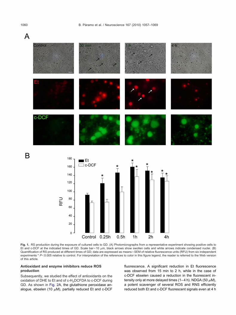
Ap
SoGa
flwcta
FEQeo
B. Páramo et al. / Neuroscience 167 (2010) 1057–10691060
ntioxidant and enzyme inhibitors reduce ROSroduction
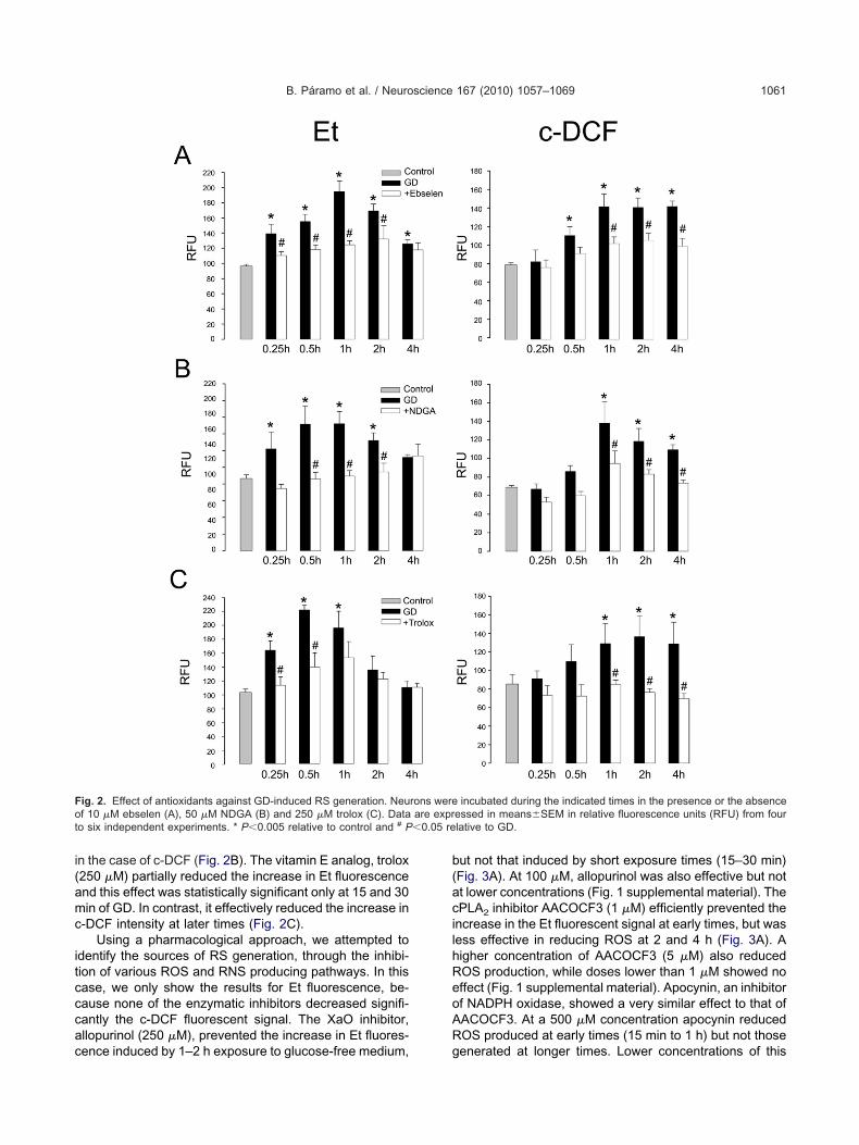
ubsequently, we studied the effect of antioxidants on thexidation of DHE to Et and of c-H2DCFDA to c-DCF duringD. As shown in Fig. 2A, the glutathione peroxidase an-
ig. 1. RS production during the exposure of cultured cells to GD. (A)t and c-DCF at the indicated times of GD. Scale bar�10 �m, blackuantification of RS produced at different times of GD; data are expresxperiments * P�0.005 relative to control. For interpretation of the refef this article.
logue, ebselen (10 �M), partially reduced Et and c-DCF r
uorescence. A significant reduction in Et fluorescenceas observed from 15 min to 2 h, while in the case of-DCF ebselen caused a reduction in the fluorescent in-ensity only at more delayed times (1–4 h). NDGA (50 �M),
potent scavenger of several ROS and RNS efficiently
icrographs from a representative experiment showing positive cells toshow swollen cells and white arrows indicate condensed nuclei. (B)eans�SEM of relative fluorescence units (RFU) from six independento color in this figure legend, the reader is referred to the Web version
Photomarrows
sed as mrences t
educed both Et and c-DCF fluorescent signals even at 4 h
i(amc
itcccac
b(acilhReoAR
Fot �0.05 re
B. Páramo et al. / Neuroscience 167 (2010) 1057–1069 1061
n the case of c-DCF (Fig. 2B). The vitamin E analog, trolox250 �M) partially reduced the increase in Et fluorescencend this effect was statistically significant only at 15 and 30in of GD. In contrast, it effectively reduced the increase in
-DCF intensity at later times (Fig. 2C).Using a pharmacological approach, we attempted to
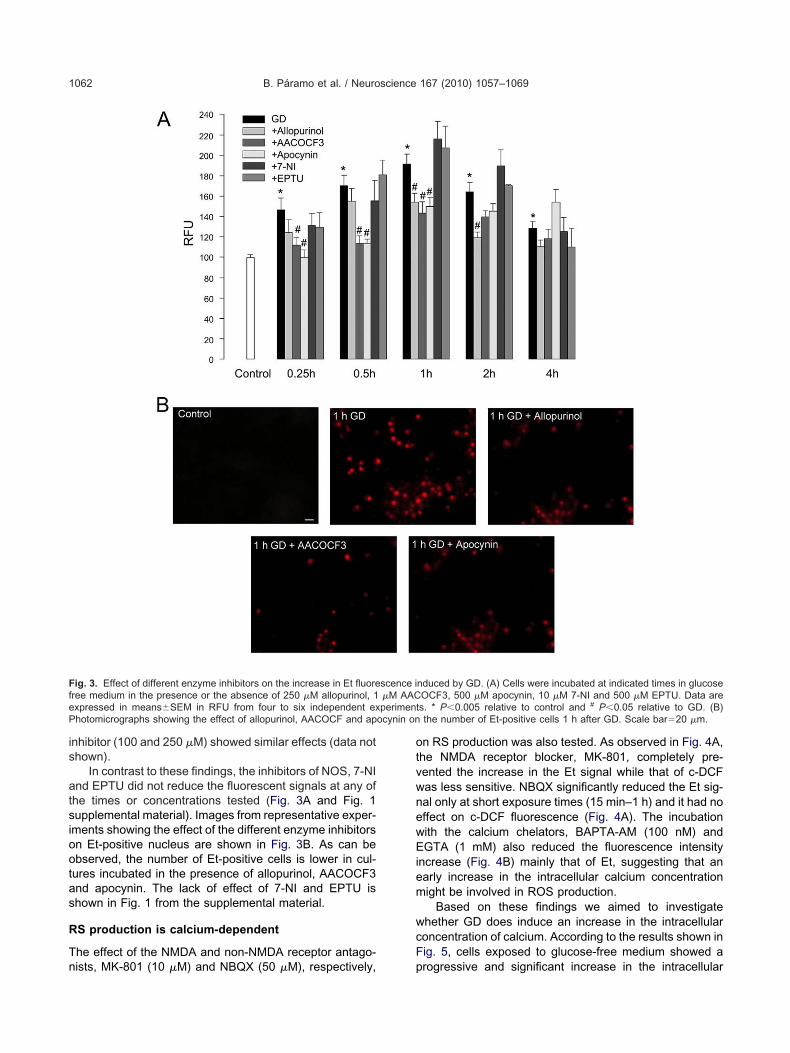
dentify the sources of RS generation, through the inhibi-ion of various ROS and RNS producing pathways. In thisase, we only show the results for Et fluorescence, be-ause none of the enzymatic inhibitors decreased signifi-antly the c-DCF fluorescent signal. The XaO inhibitor,llopurinol (250 �M), prevented the increase in Et fluores-
ig. 2. Effect of antioxidants against GD-induced RS generation. Neuf 10 �M ebselen (A), 50 �M NDGA (B) and 250 �M trolox (C). Data
o six independent experiments. * P�0.005 relative to control and # P
ence induced by 1–2 h exposure to glucose-free medium, g
ut not that induced by short exposure times (15–30 min)Fig. 3A). At 100 �M, allopurinol was also effective but nott lower concentrations (Fig. 1 supplemental material). ThePLA2 inhibitor AACOCF3 (1 �M) efficiently prevented thencrease in the Et fluorescent signal at early times, but wasess effective in reducing ROS at 2 and 4 h (Fig. 3A). Aigher concentration of AACOCF3 (5 �M) also reducedOS production, while doses lower than 1 �M showed noffect (Fig. 1 supplemental material). Apocynin, an inhibitorf NADPH oxidase, showed a very similar effect to that ofACOCF3. At a 500 �M concentration apocynin reducedOS produced at early times (15 min to 1 h) but not those
incubated during the indicated times in the presence or the absenceessed in means�SEM in relative fluorescence units (RFU) from fourlative to GD.
rons wereare expr
enerated at longer times. Lower concentrations of this
is
atsiootas
R
Tn
otvwnewEiem
wcF
FfeP ocynin on
B. Páramo et al. / Neuroscience 167 (2010) 1057–10691062
nhibitor (100 and 250 �M) showed similar effects (data nothown).
In contrast to these findings, the inhibitors of NOS, 7-NInd EPTU did not reduce the fluorescent signals at any ofhe times or concentrations tested (Fig. 3A and Fig. 1upplemental material). Images from representative exper-ments showing the effect of the different enzyme inhibitorsn Et-positive nucleus are shown in Fig. 3B. As can bebserved, the number of Et-positive cells is lower in cul-ures incubated in the presence of allopurinol, AACOCF3nd apocynin. The lack of effect of 7-NI and EPTU ishown in Fig. 1 from the supplemental material.
S production is calcium-dependent
he effect of the NMDA and non-NMDA receptor antago-
ig. 3. Effect of different enzyme inhibitors on the increase in Et fluoreree medium in the presence or the absence of 250 �M allopurinol, 1xpressed in means�SEM in RFU from four to six independent exhotomicrographs showing the effect of allopurinol, AACOCF and ap
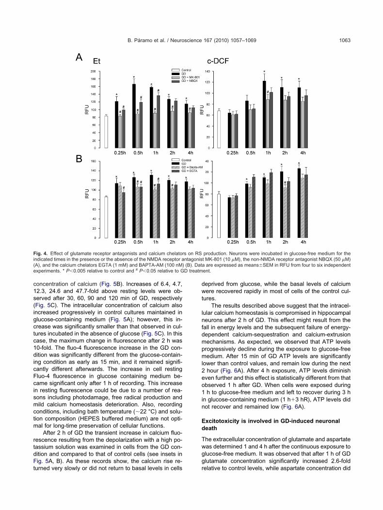
ists, MK-801 (10 �M) and NBQX (50 �M), respectively, p
n RS production was also tested. As observed in Fig. 4A,he NMDA receptor blocker, MK-801, completely pre-ented the increase in the Et signal while that of c-DCFas less sensitive. NBQX significantly reduced the Et sig-al only at short exposure times (15 min–1 h) and it had noffect on c-DCF fluorescence (Fig. 4A). The incubationith the calcium chelators, BAPTA-AM (100 nM) andGTA (1 mM) also reduced the fluorescence intensity
ncrease (Fig. 4B) mainly that of Et, suggesting that anarly increase in the intracellular calcium concentrationight be involved in ROS production.
Based on these findings we aimed to investigatehether GD does induce an increase in the intracellularoncentration of calcium. According to the results shown inig. 5, cells exposed to glucose-free medium showed a
nduced by GD. (A) Cells were incubated at indicated times in glucoseOCF3, 500 �M apocynin, 10 �M 7-NI and 500 �M EPTU. Data ares. * P�0.005 relative to control and # P�0.05 relative to GD. (B)the number of Et-positive cells 1 h after GD. Scale bar�20 �m.
scence i�M AACperiment
rogressive and significant increase in the intracellular
c1s(igctc1dicFcismctm
rtdFt
dwt
lnfdmpml2eo1in
Ed
Twgg
Fi( ) (B). Date D treatm
B. Páramo et al. / Neuroscience 167 (2010) 1057–1069 1063
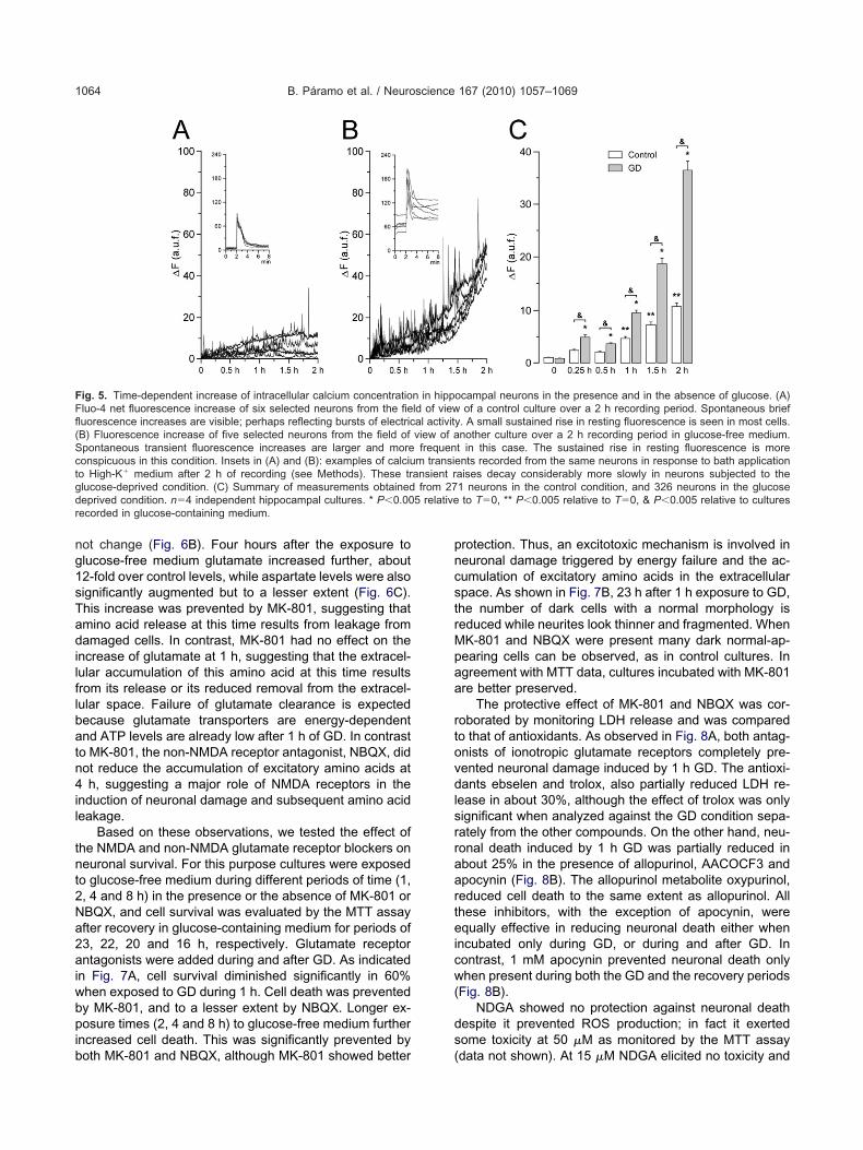
oncentration of calcium (Fig. 5B). Increases of 6.4, 4.7,2.3, 24.6 and 47.7-fold above resting levels were ob-erved after 30, 60, 90 and 120 min of GD, respectivelyFig. 5C). The intracellular concentration of calcium alsoncreased progressively in control cultures maintained inlucose-containing medium (Fig. 5A); however, this in-rease was significantly smaller than that observed in cul-ures incubated in the absence of glucose (Fig. 5C). In thisase, the maximum change in fluorescence after 2 h was0-fold. The fluo-4 fluorescence increase in the GD con-ition was significantly different from the glucose-contain-
ng condition as early as 15 min, and it remained signifi-antly different afterwards. The increase in cell restingluo-4 fluorescence in glucose containing medium be-ame significant only after 1 h of recording. This increasen resting fluorescence could be due to a number of rea-ons including photodamage, free radical production andild calcium homeostasis deterioration. Also, recording
onditions, including bath temperature (�22 °C) and solu-ion composition (HEPES buffered medium) are not opti-al for long-time preservation of cellular functions.
After 2 h of GD the transient increase in calcium fluo-escence resulting from the depolarization with a high po-assium solution was examined in cells from the GD con-ition and compared to that of control cells (see insets inig. 5A, B). As these records show, the calcium rise re-
ig. 4. Effect of glutamate receptor antagonists and calcium chelatorndicated times in the presence or the absence of the NMDA receptor aA), and the calcium chelators EGTA (1 mM) and BAPTA-AM (100 nMxperiments. * P�0.005 relative to control and # P�0.05 relative to G
urned very slowly or did not return to basal levels in cells r
eprived from glucose, while the basal levels of calciumere recovered rapidly in most of cells of the control cul-
ures.The results described above suggest that the intracel-
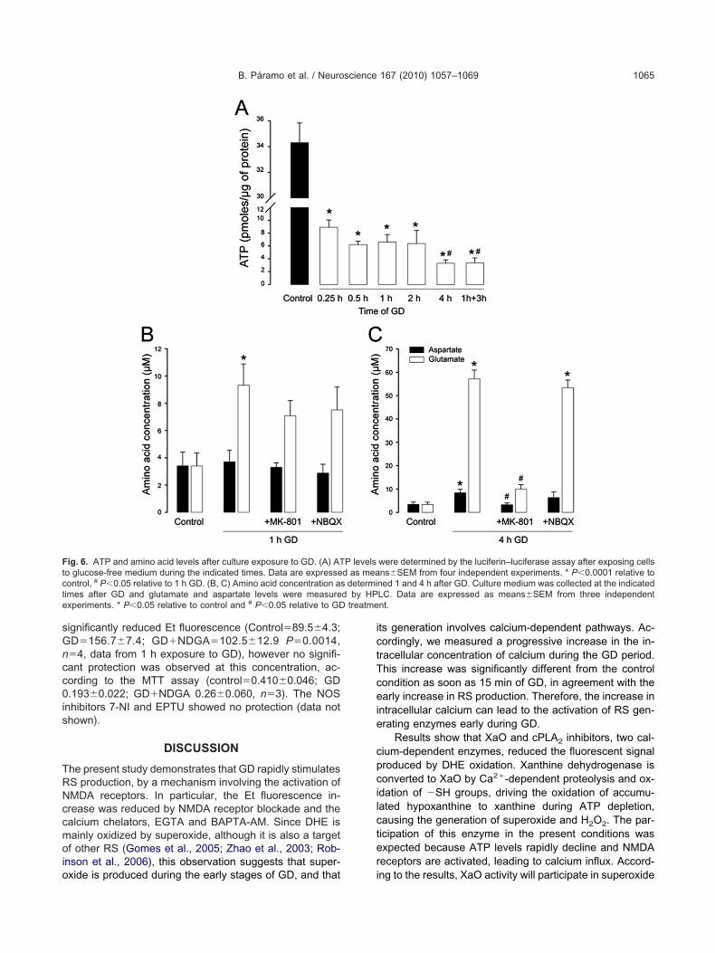
ular calcium homeostasis is compromised in hippocampaleurons after 2 h of GD. This effect might result from theall in energy levels and the subsequent failure of energy-ependent calcium-sequestration and calcium-extrusionechanisms. As expected, we observed that ATP levelsrogressively decline during the exposure to glucose-freeedium. After 15 min of GD ATP levels are significantly
ower than control values, and remain low during the nexthour (Fig. 6A). After 4 h exposure, ATP levels diminish
ven further and this effect is statistically different from thatbserved 1 h after GD. When cells were exposed duringh to glucose-free medium and left to recover during 3 h
n glucose-containing medium (1 h�3 hR), ATP levels didot recover and remained low (Fig. 6A).
xcitotoxicity is involved in GD-induced neuronaleath
he extracellular concentration of glutamate and aspartateas determined 1 and 4 h after the continuous exposure tolucose-free medium. It was observed that after 1 h of GDlutamate concentration significantly increased 2.6-fold
production. Neurons were incubated in glucose-free medium for thet MK-801 (10 �M), the non-NMDA receptor antagonist NBQX (50 �M)a are expressed as means�SEM in RFU from four to six independentent.
s on RSntagonis
elative to control levels, while aspartate concentration did
ng1sTadilflbatn4il
tnt2Na2aiwbpib
pncstrMpaa
rtovdlsrraarteicw(
ds
FFfl(Sctgdr
B. Páramo et al. / Neuroscience 167 (2010) 1057–10691064
ot change (Fig. 6B). Four hours after the exposure tolucose-free medium glutamate increased further, about2-fold over control levels, while aspartate levels were alsoignificantly augmented but to a lesser extent (Fig. 6C).his increase was prevented by MK-801, suggesting thatmino acid release at this time results from leakage fromamaged cells. In contrast, MK-801 had no effect on the
ncrease of glutamate at 1 h, suggesting that the extracel-ular accumulation of this amino acid at this time resultsrom its release or its reduced removal from the extracel-ular space. Failure of glutamate clearance is expectedecause glutamate transporters are energy-dependentnd ATP levels are already low after 1 h of GD. In contrasto MK-801, the non-NMDA receptor antagonist, NBQX, didot reduce the accumulation of excitatory amino acids ath, suggesting a major role of NMDA receptors in the
nduction of neuronal damage and subsequent amino acideakage.
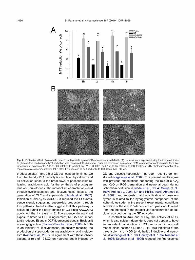
Based on these observations, we tested the effect ofhe NMDA and non-NMDA glutamate receptor blockers oneuronal survival. For this purpose cultures were exposedo glucose-free medium during different periods of time (1,, 4 and 8 h) in the presence or the absence of MK-801 orBQX, and cell survival was evaluated by the MTT assayfter recovery in glucose-containing medium for periods of3, 22, 20 and 16 h, respectively. Glutamate receptorntagonists were added during and after GD. As indicated
n Fig. 7A, cell survival diminished significantly in 60%hen exposed to GD during 1 h. Cell death was preventedy MK-801, and to a lesser extent by NBQX. Longer ex-osure times (2, 4 and 8 h) to glucose-free medium further
ncreased cell death. This was significantly prevented by
ig. 5. Time-dependent increase of intracellular calcium concentratioluo-4 net fluorescence increase of six selected neurons from the fieuorescence increases are visible; perhaps reflecting bursts of electricB) Fluorescence increase of five selected neurons from the field ofpontaneous transient fluorescence increases are larger and moreonspicuous in this condition. Insets in (A) and (B): examples of calciuo High-K� medium after 2 h of recording (see Methods). These trlucose-deprived condition. (C) Summary of measurements obtainedeprived condition. n�4 independent hippocampal cultures. * P�0.00ecorded in glucose-containing medium.
oth MK-801 and NBQX, although MK-801 showed better (
rotection. Thus, an excitotoxic mechanism is involved ineuronal damage triggered by energy failure and the ac-umulation of excitatory amino acids in the extracellularpace. As shown in Fig. 7B, 23 h after 1 h exposure to GD,he number of dark cells with a normal morphology iseduced while neurites look thinner and fragmented. WhenK-801 and NBQX were present many dark normal-ap-earing cells can be observed, as in control cultures. Ingreement with MTT data, cultures incubated with MK-801re better preserved.
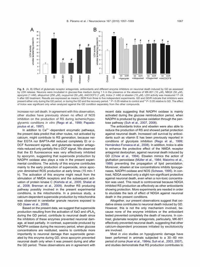
The protective effect of MK-801 and NBQX was cor-oborated by monitoring LDH release and was comparedo that of antioxidants. As observed in Fig. 8A, both antag-nists of ionotropic glutamate receptors completely pre-ented neuronal damage induced by 1 h GD. The antioxi-ants ebselen and trolox, also partially reduced LDH re-
ease in about 30%, although the effect of trolox was onlyignificant when analyzed against the GD condition sepa-ately from the other compounds. On the other hand, neu-onal death induced by 1 h GD was partially reduced inbout 25% in the presence of allopurinol, AACOCF3 andpocynin (Fig. 8B). The allopurinol metabolite oxypurinol,educed cell death to the same extent as allopurinol. Allhese inhibitors, with the exception of apocynin, werequally effective in reducing neuronal death either when
ncubated only during GD, or during and after GD. Inontrast, 1 mM apocynin prevented neuronal death onlyhen present during both the GD and the recovery periods
Fig. 8B).NDGA showed no protection against neuronal death
espite it prevented ROS production; in fact it exertedome toxicity at 50 �M as monitored by the MTT assay
ocampal neurons in the presence and in the absence of glucose. (A)of a control culture over a 2 h recording period. Spontaneous brief
. A small sustained rise in resting fluorescence is seen in most cells.nother culture over a 2 h recording period in glucose-free medium.t in this case. The sustained rise in resting fluorescence is morents recorded from the same neurons in response to bath application
aises decay considerably more slowly in neurons subjected to the1 neurons in the control condition, and 326 neurons in the glucoseto T�0, ** P�0.005 relative to T�0, & P�0.005 relative to cultures
n in hippld of viewal activityview of afrequen
m transieansient r
from 275 relative
data not shown). At 15 �M NDGA elicited no toxicity and
sGncc0is
TRNccmoio
ictTceie
cpcilcter
Ftct d by HPe treatme
B. Páramo et al. / Neuroscience 167 (2010) 1057–1069 1065
ignificantly reduced Et fluorescence (Control�89.5�4.3;D�156.7�7.4; GD�NDGA�102.5�12.9 P�0.0014,�4, data from 1 h exposure to GD), however no signifi-ant protection was observed at this concentration, ac-ording to the MTT assay (control�0.410�0.046; GD.193�0.022; GD�NDGA 0.26�0.060, n�3). The NOS
nhibitors 7-NI and EPTU showed no protection (data nothown).
DISCUSSION
he present study demonstrates that GD rapidly stimulatesS production, by a mechanism involving the activation ofMDA receptors. In particular, the Et fluorescence in-rease was reduced by NMDA receptor blockade and thealcium chelators, EGTA and BAPTA-AM. Since DHE isainly oxidized by superoxide, although it is also a targetf other RS (Gomes et al., 2005; Zhao et al., 2003; Rob-
nson et al., 2006), this observation suggests that super-
ig. 6. ATP and amino acid levels after culture exposure to GD. (A) ATo glucose-free medium during the indicated times. Data are expresseontrol, # P�0.05 relative to 1 h GD. (B, C) Amino acid concentration asimes after GD and glutamate and aspartate levels were measurexperiments. * P�0.05 relative to control and # P�0.05 relative to GD
xide is produced during the early stages of GD, and that i
ts generation involves calcium-dependent pathways. Ac-ordingly, we measured a progressive increase in the in-racellular concentration of calcium during the GD period.his increase was significantly different from the controlondition as soon as 15 min of GD, in agreement with thearly increase in RS production. Therefore, the increase in
ntracellular calcium can lead to the activation of RS gen-rating enzymes early during GD.
Results show that XaO and cPLA2 inhibitors, two cal-ium-dependent enzymes, reduced the fluorescent signalroduced by DHE oxidation. Xanthine dehydrogenase isonverted to XaO by Ca2�-dependent proteolysis and ox-
dation of �SH groups, driving the oxidation of accumu-ated hypoxanthine to xanthine during ATP depletion,ausing the generation of superoxide and H2O2. The par-icipation of this enzyme in the present conditions wasxpected because ATP levels rapidly decline and NMDAeceptors are activated, leading to calcium influx. Accord-
were determined by the luciferin–luciferase assay after exposing cellsns�SEM from four independent experiments. * P�0.0001 relative to
ned 1 and 4 h after GD. Culture medium was collected at the indicatedLC. Data are expressed as means�SEM from three independentnt.
P levelsd as meadetermi
ng to the results, XaO activity will participate in superoxide
ptildtgIctaaetsiplv
Gswai1aziafc
wamtn
Fti 0.0001 ar ells to G
B. Páramo et al. / Neuroscience 167 (2010) 1057–10691066
roduction after 1 and 2 h of GD but not at earlier times. Onhe other hand, cPLA2 activity is stimulated by calcium andts activation leads to the breakdown of phospholipids re-easing arachidonic acid for the synthesis of prostaglan-ins and leukotrienes. The metabolism of arachidonic acidhrough cycloxygenases and lipoxygenases leads to theeneration of OH● and superoxide (Nanda et al., 2007).
nhibition of cPLA2 by AACOCF3 reduced the Et fluores-ence signal, suggesting superoxide production throughhis pathway. Results also suggest that this pathway isctivated during the early phases of GD since AACOCF3bolished the increase in Et fluorescence during shortxposure times to GD. In agreement, NDGA also impor-antly reduced Et and c-DCF fluorescent signals. Besides itcavenging action (Floriano-Sánchez et al., 2006), NDGA
s an inhibitor of lipoxygenases, potentially reducing theroduction of superoxide during arachidonic acid metabo-
ism (Nanda et al., 2007). In agreement with these obser-
ig. 7. Protective effect of glutamate receptor antagonists against GDo glucose-free medium and MTT reduction was measured 16–24 h landependent experiments. * P�0.001 relative to control and ## P�epresentative experiment taken 24 h after 1 h exposure of cultured c
ations, a role of 12-LOX on neuronal death induced by a
D and glucose reperfusion has been recently demon-trated (Nagasawa et al., 2007). The present results agreeith previous observations supporting the role of cPLA2
nd XaO on ROS generation and neuronal death duringschemia/reperfusion (Owada et al., 1994; Saluja et al.,997; Arai et al., 2001; Lin and Phillis, 1991; Abramov etl., 2007), and suggests that the activation of these en-ymes is related to the hypoglycemic component of the
schemic episode. In the present experimental conditionsctivation of these Ca2�-dependent enzymes would result
rom the increase in the intracellular concentration of cal-ium recorded during the GD episode.
In contrast to XaO and cPLA2, the activity of NOS,hich is also calcium-dependent, does not appear to haven important contribution to RS production in our cellodel, since neither 7-NI nor EPTU, two inhibitors of the
hree isoforms of NOS (endothelial, inducible and neuro-al) (Babbedge et al., 1993; Garvey et al., 1994; Nakane et
neuronal death. (A) Neurons were exposed during the indicated timesare expressed as means�SEM in percent of control values from fivend # P�0.05 relative to GD treatment. (B) Photomicrographs of aD. Scale bar�50 �m.
-inducedter. Data
l., 1995; Southan et al., 1995) reduced the fluorescence
ioigp
tctDntbNmmyhsvapccwG
pdtaNciant
raNt
radcHtaGg1Mntatistd
dHctteca
bp
Fbahpo separate
B. Páramo et al. / Neuroscience 167 (2010) 1057–1069 1067
ncrease nor cell death. In agreement with this observation,ther studies have previously shown no effect of NOS
nhibition on the production of RS during ischemic/hypo-lycemic conditions in vitro (Rego et al., 1999; Papado-oulus et al., 1997).
In addition to Ca2�-dependent enzymatic pathways,he present data predict that other routes, not activated byalcium, might contribute to RS generation, because nei-her EGTA nor BAPTA-AM reduced completely Et or c-CF fluorescent signals, and glutamate receptor antago-ists reduced only partially the c-DCF signal. We observedhat the Et fluorescence was very effectively inhibitedy apocynin, suggesting that superoxide production byADPH oxidase also plays a role in the present experi-ental conditions. The activity of this enzyme contributesainly to the early production of superoxide, since apoc-
nin diminished ROS production at early times (15 min–1). The activation of this enzyme might result from thetimulation of NMDA receptors and the subsequent acti-ation of protein kinase C (Kishida et al., 2005; Shelat etl., 2008; Brennan et al., 2009). Another RS producingathway possibly involved in the present experimentalonditions, is the mitochondria. In a previous study, aalcium-dependent superoxide production by mitochondriaas observed in cerebellar granule neurons exposed toD (Isaev et al., 2008).
Based on the present data, we suggest that superoxideroduction resulting from the activation of XaO and cPLA2
uring the GD period, contribute to neuronal death sincehe inhibitors of these enzymes prevented neuronal dam-ge, at least partially. In contrast, superoxide produced byADPH oxidase during the recovery period, when glucoseoncentrations are restituted, seems to contribute moremportantly to neuronal damage than superoxide gener-ted by this enzyme during GD, since apocynin preventedeuronal death only when it was present during and after
ig. 8. (A, B) Effect of glutamate receptor antagonists, antioxidants ay LDH release. Neurons were incubated in glucose-free medium dupocynin (1 mM), allopurinol (250 �M), oxypurinol (50 �M), AACOCF3after GD treatment. Results are expressed as means�SEM from thrresent either only during the GD period, or during the GD and the recof trolox was significant only when analyzed against the GD condition
he GD period. These observations are in agreement with a
ecent data suggesting that NADPH oxidase is mainlyctivated during the glucose reintroduction period, whenADPH is produced by glucose oxidation through the pen-
ose pathway (Suh et al., 2007, 2008).The antioxidants trolox and ebselen were also able to
educe the production of RS and showed partial protectiongainst neuronal death. Increased cell survival by antioxi-ants such as vitamin E has been previously reported inonditions of glycolysis inhibition (Rego et al., 1999;ernández-Fonseca et al., 2008). In addition, trolox is able
o enhance the protective effect of the NMDA receptorntagonist dextorphan, against neuronal death induced byD (Chow et al., 1994). Ebselen mimics the action ofluthation peroxidase (Müller et al., 1984; Maiorino et al.,988) preventing the propagation of lipid peroxidation.oreover, ebselen at low concentrations inhibits lipoxyge-ases, NADPH oxidase and NOS (Schewe, 1995). In con-rast, NDGA exerted only a slight non-significant protectivegainst neuronal death, even when a non-toxic concentra-ion was used. This result is controversial because NDGAnhibited RS production as effectively as other antioxidantshowing protection. More experiments are needed in ordero elucidate the lack of effect of NDGA against neuronaleath in the present conditions.
Altogether, our present observations suggest that oxi-ative stress contributes to neuronal death induced by GD.owever, this is not the only mechanism involved, be-ause none of the enzyme inhibitors or the antioxidantested prevented completely the death of neurons. In con-rast, glutamate receptor antagonists, particularly, MK-801ffectively prevented neuronal death, suggesting that otheralcium-dependent processes initiated by excitotoxicityre involved.
Most in vivo studies on hypoglycemic damage haveeen performed in animals subjected to a 30–60 mineriod of coma (Auer et al., 1984a; Suh et al., 2003, 2007),
nt enzyme inhibitors on neuronal death induced by GD as assessedn the presence or the absence of MK-801 (10 �M), NBQX (50 �M),trolox (1 mM) or ebselen (10 �M). LDH activity was measured 17–24independent experiments. GD and GD/R indicate that inhibitors wered. * P�0.05 relative to control and # P�0.05 relative to GD. The effectly from the other compounds.
nd differering 1 h i
(1 �M),ee to fivevery perio
nd studies demonstrate that RS production contributes to
tOtensEcpmawhdadpb
At4s
A
A
A
A
B
B
B
B
B
B
B
C
E
E
F
G
G
H
H
H
H
H
I
K
K
L
M
M
M
M
B. Páramo et al. / Neuroscience 167 (2010) 1057–10691068
he subsequent death of neurons (Suh et al., 2007, 2008).n the other hand, recent investigations have shown that
he hypoglycemic condition, even in the absence of iso-lectricity, might induce discrete neuronal damage in vul-erable regions, such as the cerebral cortex, when it isustained for prolonged periods of time (Tkacs et al., 2005;nnis et al., 2008; Haces et al., 2010). Furthermore, in-reased lipoperoxidation has been observed in the hip-ocampus, cerebral cortex and the striatum of hypoglyce-ic animals not subjected to a coma period (Patocková etl., 2003; Haces et al., 2010). These observations togetherith the present results support the hypothesis that theypoglycemic condition is sufficient to stimulate RS pro-uction, and that XaO, cPLA2, LOX and NADPH oxidasere involved in ROS production early after glucose with-rawal. Whether these enzymes contribute to the earlyroduction of RS during in vivo hypoglycemia, remains toe studied.
cknowledgments—The authors thank Ms. Teresa Montiel for herechnical assistance. This work was supported by CONACyT8645-Q grant to L.M. and 221021 and 194940 CONACyT fellow-hips to B.P. and A.M.E-S.
REFERENCES
bramov AY, Scorziello A, Duchen MR (2007) Three distinct mecha-nisms generate oxygen free radicals in neurons and contribute tocell death during anoxia and reoxygenation. J Neurosci 275:1129–1138.
rai K, Ikegaya Y, Nakatani Y, Kudo I, Nishiyama N, Matsuki N (2001)Phospholipase A2 mediates ischemic injury in the hippocampus: aregional difference of neuronal vulnerability. Eur J Neurosci 13:2319–2323.
uer RN, Olsson Y, Siesjö BK (1984a) Hypoglycemic brain injury in therat. Correlation of density of brain damage with the EEG isoelectrictime: a quantitative study. Diabetes 33:1090–1098.
uer RN, Wieloch T, Olsson Y, Siesjö BK (1984b) The distribution ofhypoglycemic brain damage. Acta Neuropathol 64:177–191.
abbedge RC, Bland-Ward PA, Hart SL, Moore PK (1993) Inhibition ofrat cerebellar nitric oxide synthase by 7-nitro indazole and relatedsubstituted indazoles. Br J Pharmacol 110:225–228.
ergmeyer HU, Bernt EH (1968) Methods of enzymatic analysis, pp736–741. New York: Academic Press.
erridge MV, Tan AS (1993) Characterization of the cellular reductionof 3-(4,5-dimethylthiazol-2-yl)-2,5-diphenyltetrazolium bromide(MTT): subcellular localization, substrate dependence, and in-volvement of mitochondrial electron transport in MTT reduction.Arch Biochem Biophys 303:474–482.
indokas V, Jordan J, Lee C, Miller R (1996) Superoxide production inrat hippocampal neurons: selective imaging with hydroethidine.J Neurosci 16:1324–1336.
radford MM (1976) A rapid and sensitive method for the quantitationof microgram quantities of protein utilizing the principle of protein-dye binding. Anal Biochem 72:248–254.
rennan AM, Suh SW, Won SJ, Narasimhan P, Kauppinen TM, Lee H,Edling Y, Chan PH, Swanson RA (2009) NADPH oxidase is theprimary source of superoxide induced by NMDA receptor activa-tion. Nat Neurosci 12:857–863.
rewer GJ, Torricelli JR, Evege EK, Price PJ (1993) Optimized sur-vival of hippocampal neurons in B27-suplemented neurobasal, anew serum free medium combination. J Neurosci Res 35:567–576.
how HS, Lynch JJ 3rd, Rose K, Choi DW (1994) Trolox attenuatescortical neuronal injury induced by iron, ultraviolet light, glucose
deprivation, or AMPA. Brain Res 639:102–108.nnis K, Tran PV, Seaquist ER, Rao R (2008) Postnatal age influ-ences hypoglycemia-induced neuronal injury in the rat brain. BrainRes 1224:119–126.
strada-Sánchez AM, Camacho A, Montiel T, Massieu L (2007) Cer-ebellar granule neurons are more vulnerable to transient transport-mediated glutamate release than to glutamate uptake blockade.Correlation with excitatory amino acids levels. Neurochem Res32:423–432.
loriano-Sánchez E, Villanueva C, Medina-Campos ON, Rocha D,Sánchez-Gonzalez DJ, Cardenas-Rodriguez N, Pedraza-ChaverriJ (2006) Nordihydroguaiaretic acid is a potent in vitro scavenger ofperoxynitrite, singlet oxygen, hydroxyl radical, superoxide anionand hypochlorous acid and prevents in vivo ozone-induced ty-rosine nitration in lungs. Free Radic Res 40:523–533.
arvey EP, Oplinger JA, Tanoury GJ, Sherman PA, Fowler M, Mar-shall S, Harmon MF, Paith JE, Furfine ES (1994) Potent andselective inhibition of human nitric oxide synthases. Inhibition bynon-amino acid isothioureas. J Biol Chem 269:26669–26676.
omes A, Fernandes E, Lima JL (2005) Fluorescence probes used fordetection of reactive oxygen species. J Biochem Biophys Methods65:45–80.
aces ML, Hernández-Fonseca K, Medina-Campos ON, Montiel T,Pedraza-Chaverri J, Massieu L (2008) Antioxidant capacity con-tributes to protection of ketone bodies against oxidative damageinduced during hypoglycemic conditions. Exp Neurol 211:85–96.
aces ML, Montiel T, Massieu L (2010) Selective vulnerability of brainregions to oxidative stress in a non-coma model of insulin-inducedhypoglycemia. Neuroscience 165:28–38.
ernández-Fonseca K, Cárdenas-Rodríguez N, Pedraza-Chaverri J,Massieu L (2008) Calcium-dependent production of reactive oxy-gen species is involved in neuronal damage induced during glyco-lysis inhibition in cultured hippocampal neurons. J Neurosci Res86:1768–1780.
ernández-Fonseca K, Massieu L (2005) Disruption of endoplasmicreticulum calcium stores is involved in neuronal death induced byglycolysis inhibition in cultured hippocampal neurons. J NeurosciRes 82:196–205.
ockenbery DM, Oltvai ZN, Yin XM, Milliman CL, Korsmeyer SJ(1993) Bcl-2 functions in an antioxidant pathway to prevent apo-ptosis. Cell 75:241–251.
saev NK, Stelmashook EV, Dirnagl U, Plotnikov EY, Kuvshinova EA,Zorov DB (2008) Mitochondrial free radical production induced byglucose deprivation in cerebellar granule neurons. BiochemistryMosc 73:149–155.
alimo H, Auer RN, Siesjö BK (1985) The temporal evolution ofhypoglycemic brain damage. III. Light and electron microscopicfindings in the rat caudoputamen. Acta Neuropathol 67:37–50.
ishida KT, Pao M, Holland SM, Klann E (2005) NADPH oxidase isrequired for NMDA receptor-dependent activation of ERK in hip-pocampal area CA1. J Neurochem 94:299–306.
in Y, Phillis JW (1991) Oxypurinol reduces focal ischemic brain injuryin the rat. Neurosci Lett 126:187–190.
aiorino M, Roveri A, Ursini F (1988) Antioxidant effect of ebselen (PZ51): peroxidase mimetic activity on phospholipid and cholesterolhydroperoxides vs free radical scavenger activity. Arch BiochemBiophys 295:404–409.
cGowan JE, Chen L, Gao D, Trush M, Wei C (2006) Increasedmitochondrial reactive oxygen species production in newborn brainduring hypoglycemia. Neurosci Lett 399:111–114.
osmann T (1983) Rapid colorimetric assay for cellular growth andsurvival: application to proliferation and cytotoxicity assays. J Im-munol Methods 65:55–63.
üller A, Cadenas E, Graf P, Sies H (1984) A novel biologically activeseleno-organic compound. I. Glutathione peroxidase-like activityin-vitro and antioxidant capacity of PZ 51 (ebselen). Biochem
Pharmacol 33:3235–3239.
N
N
N
O
P
P
R
R
S
S
S
S
S
S
S
S
T
W
Z
S
S
B. Páramo et al. / Neuroscience 167 (2010) 1057–1069 1069
agasawa K, Kakuda T, Higashi Y, Fujimoto S (2007) Possible in-volvement of 12-lipoxygenase activation in glucose-deprivation/reload-treated neurons. Neurosci Lett 429:120–125.
akane M, Klinghofer V, Kuk JE, Donnelly JL, Budzik GP, Pollock JS,Basha F, Carter GW (1995) Novel potent and selective inhibitors ofinducible nitric oxide synthase. Mol Pharmacol 47:831–834.
anda BL, Nataraju A, Rajesh R, Rangappa KS, Shekar MA, Vish-wanath BS (2007) PLA2 mediated arachidonate free radicals:PLA2 inhibition and neutralization of free radicals by anti-oxi-dants-a new role as anti-inflammatory molecule. Curr Top MedChem 7:765–777.
wada Y, Tominaga T, Yoshimoto T, Kondo H (1994) Molecularcloning of rat cDNA for cytosolic phospholipase A2 and the in-creased gene expression in the dentate gyrus following transientforebrain ischemia. Mol Brain Res 25:364–368.
apadopoulus MC, Koumenis IL, Dugan LL, Giffard RG (1997) Vul-nerability of glucose deprivation injury correlates with glutathionelevels in astrocytes. Brain Res 748:151–156.
atocková J, Marhol P, Tumova E, Krsiak M, Rokyta R, Stipek S,Crkovska J, Andel M (2003) Oxidative stress in the brain tissue oflaboratory mice with acute post insulin hypoglycemia. Physiol Res52:131–135.
ego AC, Santos MS, Oliveira CR (1999) Influence of the antioxidantsvitamin E and idebenone on retinal cell injury mediated by chemicalischemia, hypoglycemia, or oxidative stress. Free Radic Biol Med26:1405–1407.
obinson KM, Janes MS, Pehar M, Monette JS, Ross MF, Hagen TM,Murphy MP, Beckman JS (2006) Selective fluorescent imaging ofsuperoxide in vivo using ethidium-based probes. Proc Natl AcadSci U S A 103:15038–15043.
aluja I, Song D, Oregan MH, Phillis JW (1997) Role of phospholipaseA2 in the release of free fatty acids during ischemia-reperfusion inthe rat cerebral cortex. Neurosci Lett 233:97–100.
andberg M, Butcher ST, Hagberg H (1986) Extracellular overflow ofneuroactive amino acids during severe insulin-induced hypoglyce-mia: in vivo dialysis of the rat hippocampus. J Neurochem 47:
178–184. tchewe T (1995) Molecular actions of ebselen-an anti-inflamatoryantioxidant. Gen Pharmacol 26:1153–1169.
helat PB, Chalimoniuk M, Wang JH, Strosznajder JB, Lee JC, SunAY, Simonyi A, Sun GY (2008) Amyloid beta peptide and NMDAinduce ROS from NADPH oxidase and AA release from cytosolicphospholipase A2 in cortical neurons. J Neurochem 106:45–55.
outhan GJ, Szabó C, Thiemermann C (1995) Isothioureas: potentinhibitors of nitric oxide synthases with variable isoform selectivity.Br J Pharmacol 114:510–516.
uh SW, Aoyama K, Chen Y, Garnier P, Matsumori Y, Gum E, Liu J,Swanson RA (2003) Hypoglycemic neuronal death and cognitiveimpairment are prevented by poly(ADP-ribose) polymerase inhib-itors administered after hypoglycemia. J Neurosci 23:10681–10690.
uh SW, Gum E, Hamby AM, Chan PH, Swanson RA (2007) Hypo-glycemic neuronal death is triggered by glucose reperfusion andactivation of neuronal NADPH oxidase. J Clin Invest 117:910–918.
uh SW, Hamby AM, Gum ET, Shin BS, Won SJ, Sheline CT, ChanPH, Swanson RA (2008) Sequential release of nitric oxide, zinc,and superoxide in hypoglycemic neuronal death. J Cereb BloodFlow Metab 28:1697–1706.
kacs NC, Pan Y, Raghupathi R, Dunn-Meynell AA, Levin BE (2005)Cortical Fluoro-Jade staining and blunted adrenomedullary re-sponse to hypoglycemia after noncoma hypoglycemia in rats.J Cereb Blood Flow Metab 25:1645–1655.
ieloch T (1984) Hypoglycemia-induced neuronal damage preventedby an N-methyl-D-aspartate antagonist. Science 230:681–683.
hao H, Kalivendi S, Zhang H, Joseph J, Nithipatikom K, Vasquez-Vivar J, Kalyanaraman B (2003) Superoxide reacts with hydroethi-dine but forms a fluorescent product that is distinctly different fromethidium: potential implications in intracellular fluorescence detec-tion of superoxide. Free Radic Biol Med 34:1359–1368.
APPENDIX
upplementary data
upplementary data associated with this article can be found, in
he online version, at doi:10.1016/j.neuroscience.2010.02.074.(Accepted 28 February 2010)(Available online 10 March 2010)
Copyright © 2022 FDOKUMEN


![Role of desensitization of AMPA receptors on the neuronal viability and on the [Ca2+]i changes in cultured rat hippocampal neurons](https://static.fdokumen.com/doc/165x107/63230bd664690856e1096f90/role-of-desensitization-of-ampa-receptors-on-the-neuronal-viability-and-on-the-ca2i.jpg)