PATHOLOGUE - The Hong Kong College of Pathologists
24
Message from the President Announcement from the Training and Examinations Committee College Examinations 2021 Fellows’ Laurels Professor LO Yuk Ming Dennis Professor Joseph Sriyal Malik PEIRIS and Professor YUEN Kwok Yung Annual General Meeting 2020 16th Trainee Presentation Session From the Young Fellows’ Chapter: Medical Education Conference 2021 Topical Update: Diagnosis of COVID-19 COVID-19 vaccination promotional material INDEX INDEX The Hong Kong College of Pathologists, Incorporated in Hong Kong with Limited Liability PATHOLOGUE PATHOLOGUE Message from the President Message from the President VOLUME 30, ISSUE 1 OCTOBER 2021 In this issue of the College Newsletter, I am delighted to announce that Professor Dennis LO, Professor Joseph Sriyal Malik PEIRIS and Professor Kwok Yung YUEN (in the order of their surnames) have received the prestigious Royal Medal of The Royal Society as well as the Future Science Prize 2021 in life sciences for their contributions in the discoveries of cell-free DNA for non- invasive prenatal diagnosis and zoonotic transfer of coronavirus to human causing Severe Acute Respiratory Syndrome (SARS) in 2003, respectively. My heartfelt congratulations! Under the new normal of COVID-19, all examinations in 2021 were still conducted using video conferencing technology. I would also like to thank all external and local examiners for their hard work and to congratulate all successful candidates in the College Membership Examination and Fellowship Assessment. They have added new workforce to the diagnostic services in Hong Kong. Another issue of postgraduate education under the new normal is how to receive training without a face-to-face arrangement. The Academy recently held a Medical Education Conference to discuss the way forward on e-learning, competency-based training and continuous workplace-based assessment. Both educators and students need to acquire new skills and improve their resilience to training progress. Dr Derek HUNG and Professor KY YUEN wrote a Topical Update in this issue of newsletter titled “Diagnosis of COVID-19”. It is a timely topic to review all the diagnostic modalities to combat COVID-19. Last but not least, our College, jointly with Academy, has prepared promotional materials for COVID-19 vaccination including posters and videos. They are uploaded to the COVID corner of our College website and Facebook. Please feel free to share them with your friends and family. Finally, allow me to wish you all ‘good health’ going forward ! Dr CHAN Ho Ming President 1 2 3 6 8 9 11 THE EDITORIAL BOARD THE EDITORIAL BOARD Dr. CHEONG Renee Constance Yue-Kew (Chief Editor) Dr. CHAU Ka Yee Dr. CHEUNG Tin Yan, Elaine Dr. CHUNG Ivy Ah-yu Dr. IP Ho Wan Dr. LO Hui Yin Dr. LOK Johann Please send comments to: Dr. CHEONG Renee Constance Yue-kew, Address: Department of Pathology, 2/F, North District Hospital, 9 Po Kin Road, Sheung Shui, New Territories, Hong Kong Email: [email protected] Phone: 2683 8154 Fax: 2683 8176 12 22
-
Upload
khangminh22 -
Category
Documents
-
view
3 -
download
0
Transcript of PATHOLOGUE - The Hong Kong College of Pathologists
Message from the President
Announcement from the Training and Examinations Committee
College Examinations 2021
Fellows’ Laurels Professor LO Yuk Ming Dennis Professor Joseph Sriyal Malik PEIRIS and Professor YUEN Kwok Yung Annual General Meeting 2020
16th Trainee Presentation Session
From the Young Fellows’ Chapter: Medical Education Conference 2021
Topical Update: Diagnosis of COVID-19
COVID-19 vaccination promotional material
I N D E XI N D E X
The Hong Kong College of Pathologists, Incorporated in Hong Kong with Limited Liability
P A T H O L O G U EP A T H O L O G U EMessage from the PresidentMessage from the President
VOLUME 30, ISSUE 1 OCTOBER 2021
In this issue of the College Newsletter, I am delighted to announce that Professor Dennis LO, Professor Joseph Sriyal Malik PEIRIS and Professor Kwok Yung YUEN (in the order of their surnames) have received the prestigious Royal Medal of The Royal Society as well as the Future Science Prize 2021 in life sciences for their contributions in the discoveries of cell-free DNA for non-invasive prenatal diagnosis and zoonotic transfer of coronavirus to human causing Severe Acute Respiratory Syndrome (SARS) in 2003, respectively. My heartfelt congratulations!
Under the new normal of COVID-19, all examinations in 2021 were still conducted using video conferencing technology. I would also like to thank all external and local examiners for their hard work and to congratulate all successful candidates in the College Membership Examination and Fellowship Assessment. They have added new workforce to the diagnostic services in Hong Kong.
Another issue of postgraduate education under the new normal is how to receive training without a face-to-face arrangement. The Academy recently held a Medical Education Conference to discuss the way forward on e-learning, competency-based training and continuous workplace-based assessment. Both educators and students need to acquire new skills and improve their resilience to training progress.
Dr Derek HUNG and Professor KY YUEN wrote a Topical Update in this issue of newsletter titled “Diagnosis of COVID-19”. It is a timely topic to review all the diagnostic modalities to combat COVID-19.
Last but not least, our College, jointly with Academy, has prepared promotional materials for COVID-19 vaccination including posters and videos. They are uploaded to the COVID corner of our College website and Facebook. Please feel free to share them with your friends and family.
Finally, allow me to wish you all ‘good health’ going forward !
Dr CHAN Ho Ming
President
1
2
3
6
8
9
11
THE EDITORIAL BOARDTHE EDITORIAL BOARD
Dr. CHEONG Renee Constance Yue-Kew (Chief Editor)Dr. CHAU Ka YeeDr. CHEUNG Tin Yan, ElaineDr. CHUNG Ivy Ah-yuDr. IP Ho WanDr. LO Hui YinDr. LOK JohannPlease send comments to:Dr. CHEONG Renee Constance Yue-kew, Address: Department of Pathology, 2/F, North District Hospital,9 Po Kin Road, Sheung Shui, New Territories, Hong Kong Email: [email protected] Phone: 2683 8154 Fax: 2683 8176
12
22
8 PATHOLOGUEPATHOLOGUE
We are pleased to announce that the following candidates have passed the Fellowship Assessment or Membership Examination – Congratulations!!
Announcement from Announcement from
the Training and Examinations Committeethe Training and Examinations Committee
CHAN Angela Zaneta ( Fellowship Assessment – Anatomical Pathology ) CHOW Che Ying Maria Bernadette ( Fellowship Assessment – Anatomical Pathology ) FUNG Ching Ki ( Fellowship Assessment – Anatomical Pathology ) GAO Yang ( Fellowship Assessment – Anatomical Pathology ) HAU Man Nga ( Fellowship Assessment – Anatomical Pathology ) HO Man Kit ( Fellowship Assessment – Anatomical Pathology ) KWOK Lok Ming Angie ( Fellowship Assessment – Anatomical Pathology ) NG Ka Man Joanna ( Fellowship Assessment – Anatomical Pathology ) YEUNG Chun Fai ( Fellowship Assessment – Anatomical Pathology ) CHENG Hua Tse, Timothy ( Fellowship Assessment – Chemical Pathology ) HUNG Ling Yin ( Fellowship Assessment – Chemical Pathology ) LAU Kwai Cheung ( Fellowship Assessment – Chemical Pathology ) LEUNG Mei Tik ( Fellowship Assessment – Chemical Pathology ) CHIU Hei Yeung Kelvin ( Fellowship Assessment – Clinical Microbiology & Infection ) SIN Ching Tai Eugene ( Fellowship Assessment – Clinical Microbiology & Infection) TSANG Lok Man ( Fellowship Assessment – Clinical Microbiology & Infection) CHEUNG Hiu Ni ( Fellowship Assessment – Forensic Pathology) LI Ting Hon, Stanford ( Fellowship Assessment – Haematology) FONG Nga Yee ( Membership Examination – Anatomical Pathology ) LAU Cheuk Hei ( Membership Examination – Anatomical Pathology ) LI Po Yin ( Membership Examination – Anatomical Pathology ) LING Cheuk Nam ( Membership Examination – Anatomical Pathology ) LUNG Chee Heng, Cheryl ( Membership Examination – Anatomical Pathology ) TSANG Cheuk Ho ( Membership Examination – Anatomical Pathology ) LAM Tony (Membership Examination – Forensic Pathology) LAM Wing Kit (Membership Examination – Haematology)
2 PATHOLOGUEPATHOLOGUE
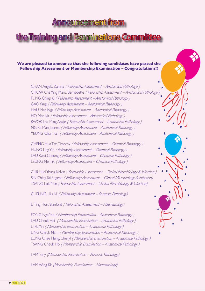
Panel of Examiners for Anatomical Pathology Fellowship Examination 2021
College Examinations 2021College Examinations 2021
t From left to right:Dr LAM Wing Sun, Dr CHOI Cheung Lung, Paul, Prof TO Ka Fai (Chief Exam-iner), Dr KAN Nim Chi, Amanda, Prof Richard WILLIAMS (on the screen, Ex-ternal Examiner), Dr CHAN Ngot Htain, Alice, Prof KHOO Ui Soon, Dr MAK Siu Ming (Deputy Chief Examiner).
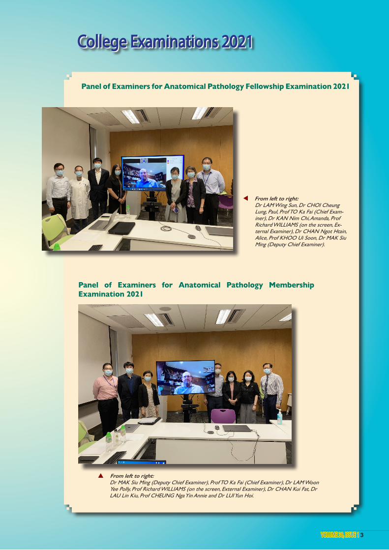
p From left to right:Dr MAK Siu Ming (Deputy Chief Examiner), Prof TO Ka Fai (Chief Examiner), Dr LAM Woon Yee Polly, Prof Richard WILLIAMS (on the screen, External Examiner), Dr CHAN Kui Fat, Dr LAU Lin Kiu, Prof CHEUNG Nga Yin Annie and Dr LUI Yun Hoi.
Panel of Examiners for Anatomical Pathology Membership Examination 2021
VOLUME 30, ISSUE 1VOLUME 30, ISSUE 1 3
College Examinations 2021College Examinations 2021
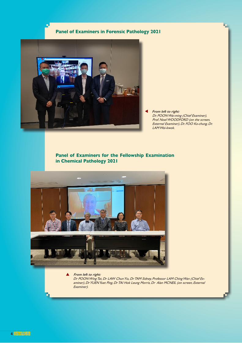
Panel of Examiners in Forensic Pathology 2021
t From left to right:Dr. POON Wai-ming (Chief Examiner), Prof. Noel WOODFORD (on the screen, External Examiner), Dr. FOO Ka-chung, Dr. LAM Wai-kwok.
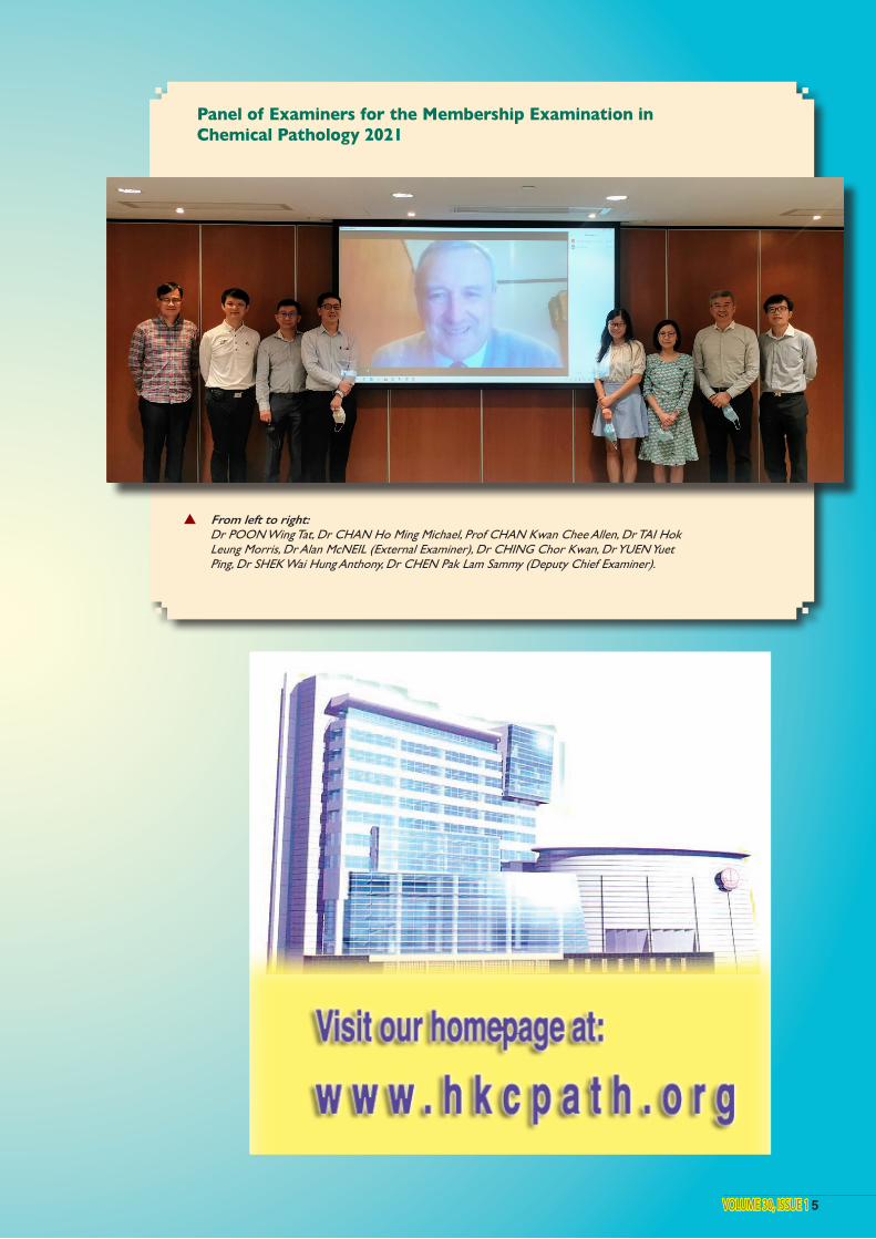
p From left to right:Dr POON Wing Tat, Dr LAW Chun Yiu, Dr TAM Sidney, Professor LAM Ching Wan (Chief Ex-aminer), Dr YUEN Yuet Ping, Dr TAI Hok Leung Morris, Dr Alan MCNEIL (on screen, External Examiner).
Panel of Examiners for the Fellowship Examination in Chemical Pathology 2021
4 PATHOLOGUEPATHOLOGUE
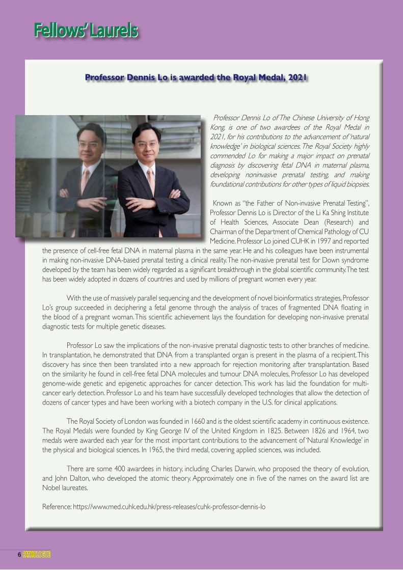
Panel of Examiners for the Membership Examination in Chemical Pathology 2021
p From left to right:Dr POON Wing Tat, Dr CHAN Ho Ming Michael, Prof CHAN Kwan Chee Allen, Dr TAI Hok Leung Morris, Dr Alan McNEIL (External Examiner), Dr CHING Chor Kwan, Dr YUEN Yuet Ping, Dr SHEK Wai Hung Anthony, Dr CHEN Pak Lam Sammy (Deputy Chief Examiner).
VOLUME 30, ISSUE 1VOLUME 30, ISSUE 1 5
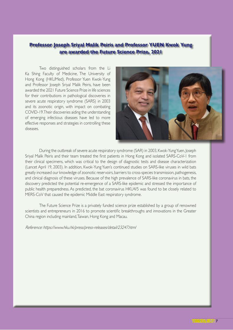
Professor Dennis Lo of The Chinese University of Hong Kong, is one of two awardees of the Royal Medal in 2021, for his contributions to the advancement of ‘natural knowledge’ in biological sciences. The Royal Society highly commended Lo for making a major impact on prenatal diagnosis by discovering fetal DNA in maternal plasma, developing noninvasive prenatal testing, and making foundational contributions for other types of liquid biopsies.
Known as “the Father of Non-invasive Prenatal Testing”, Professor Dennis Lo is Director of the Li Ka Shing Institute of Health Sciences, Associate Dean (Research) and Chairman of the Department of Chemical Pathology of CU Medicine. Professor Lo joined CUHK in 1997 and reported
the presence of cell-free fetal DNA in maternal plasma in the same year. He and his colleagues have been instrumental in making non-invasive DNA-based prenatal testing a clinical reality. The non-invasive prenatal test for Down syndrome developed by the team has been widely regarded as a significant breakthrough in the global scientific community. The test has been widely adopted in dozens of countries and used by millions of pregnant women every year. With the use of massively parallel sequencing and the development of novel bioinformatics strategies, Professor Lo’s group succeeded in deciphering a fetal genome through the analysis of traces of fragmented DNA floating in the blood of a pregnant woman. This scientific achievement lays the foundation for developing non-invasive prenatal diagnostic tests for multiple genetic diseases. Professor Lo saw the implications of the non-invasive prenatal diagnostic tests to other branches of medicine. In transplantation, he demonstrated that DNA from a transplanted organ is present in the plasma of a recipient. This discovery has since then been translated into a new approach for rejection monitoring after transplantation. Based on the similarity he found in cell-free fetal DNA molecules and tumour DNA molecules, Professor Lo has developed genome-wide genetic and epigenetic approaches for cancer detection. This work has laid the foundation for multi-cancer early detection. Professor Lo and his team have successfully developed technologies that allow the detection of dozens of cancer types and have been working with a biotech company in the U.S. for clinical applications.
The Royal Society of London was founded in 1660 and is the oldest scientific academy in continuous existence. The Royal Medals were founded by King George IV of the United Kingdom in 1825. Between 1826 and 1964, two medals were awarded each year for the most important contributions to the advancement of ‘Natural Knowledge’ in the physical and biological sciences. In 1965, the third medal, covering applied sciences, was included.
There are some 400 awardees in history, including Charles Darwin, who proposed the theory of evolution, and John Dalton, who developed the atomic theory. Approximately one in five of the names on the award list are Nobel laureates.
Reference: https://www.med.cuhk.edu.hk/press-releases/cuhk-professor-dennis-lo
Professor Dennis Lo is awarded the Royal Medal, 2021
Fellows’ LaurelsFellows’ LaurelsFellows’ LaurelsFellows’ Laurels
6 PATHOLOGUEPATHOLOGUE
Two distinguished scholars from the Li Ka Shing Faculty of Medicine, The University of Hong Kong (HKUMed), Professor Yuen Kwok-Yung and Professor Joseph Sriyal Malik Peiris, have been awarded the 2021 Future Science Prize in life sciences for their contributions in pathological discoveries in severe acute respiratory syndrome (SARS) in 2003 and its zoonotic origin, with impact on combating COVID-19. Their discoveries aiding the understanding of emerging infectious diseases have led to more effective responses and strategies in controlling these diseases.
During the outbreak of severe acute respiratory syndrome (SAR) in 2003, Kwok-Yung Yuen, Joseph Sriyal Malik Peiris and their team treated the first patients in Hong Kong and isolated SARS-CoV-1 from their clinical specimens, which was critical to the design of diagnostic tests and disease characterization (Lancet April 19, 2003). In addition, Kwok-Yung Yuen’s continued studies on SARS-like viruses in wild bats greatly increased our knowledge of zoonotic reservoirs, barriers to cross-species transmission, pathogenesis, and clinical diagnosis of these viruses. Because of the high prevalence of SARS-like coronavirus in bats, the discovery predicted the potential re-emergence of a SARS-like epidemic and stressed the importance of public health preparedness. As predicted, the bat coronavirus HKU4/5 was found to be closely related to MERS-CoV that caused the epidemic Middle East respiratory syndrome.
The Future Science Prize is a privately funded science prize established by a group of renowned scientists and entrepreneurs in 2016 to promote scientific breakthroughs and innovations in the Greater China region including mainland, Taiwan, Hong Kong and Macau.
Reference: https://www.hku.hk/press/press-releases/detail/23247.html
Professor Joseph Sriyal Malik Peiris and Professor YUEN Kwok Yungare awarded the Future Science Prize, 2021
VOLUME 30, ISSUE 1VOLUME 30, ISSUE 1 7
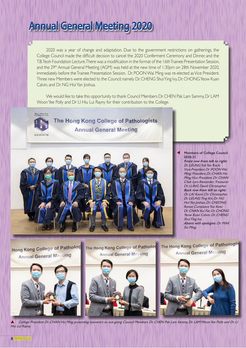
2020 was a year of change and adaptation. Due to the government restrictions on gatherings, the College Council made the difficult decision to cancel the 2020 Conferment Ceremony and Dinner, and the T.B. Teoh Foundation Lecture. There was a modification in the format of the 16th Trainee Presentation Session, and the 29th Annual General Meeting (AGM) was held at the new time of 1:30pm on 28th November 2020, immediately before the Trainee Presentation Session. Dr. POON Wai Ming was re-elected as Vice President. Three new Members were elected to the Council; namely Dr. CHENG Shui Ying Ivy, Dr. CHONG Yeow Kuan Calvin, and Dr. NG Hoi Yan Joshua.
We would like to take this opportunity to thank Council Members Dr. CHEN Pak Lam Sammy, Dr LAM Woon Yee Polly and Dr LI Hiu Lui Rayny for their contribution to the College.
8 PATHOLOGUEPATHOLOGUE
Annual General Meeting 2020Annual General Meeting 2020
p College President Dr. CHAN Ho Ming presenting souvenirs to out-going Council Members Dr. CHEN Pak Lam Sammy, Dr. LAM Woon Yee Polly and Dr. LI Hiu Lui Rayny.
t Members of College Council 2020-21
Front row from left to right: Dr. LEUNG Yuk Yan Rock;
Vice-President Dr POON Wai Ming; President Dr. CHAN Ho Ming; Vice President Dr. CHAN Chak Lam Alexander; Treasurer Dr. LUNG David Christopher.
Back row from left to right: Dr. LAI Koon Chi Christopher, Dr. LEUNG Ying Kit; Dr. NG Hoi Yan Joshua; Dr. CHEONG Renee Constance Yue Kew;
Dr. CHAN Kui Fat; Dr. CHONG Yeow Kuan Calvin; Dr. CHENG
Shui Ying Ivy. Absent with apologies: Dr. MAK
Siu Ming.
Annual General Meeting 2020Annual General Meeting 2020
The 16th Trainee Presentation Session (TPS) took on a new look on 26th November 2020. A combined physical and virtual format, the latter via Zoom Webinar, created a new experience for our fellows and members. Whilst eight oral and 16 poster presentations were performed on stage, the virtual e-posters were posted and available for download on the College’s website (http://www.hkcpath.org/article/trainee-presentation-session-2020). I would like to take this opportunity to thank our judges Dr. LEUNG Fung Shan Kate (Haematology, Princess Margaret Hospital), Dr. WONG Sai Yin Samson (Microbiology, Tuen Mun Hospital), Dr. CHONG Yeow Kuan Calvin (Chemical Pathology, Princess Margaret Hospital) and Dr. FUNG Ngai Sheung Mandy (Anatomical Pathology, United Christian Hospital) for spending their precious time with us and for their invaluable comments. We congratulate all candidates and their supervisors for their excellent work. Dr. LEE Lok Hang, Alfred (Microbiology, PWH) won the best trainee presentation award on the topic “Diagnostic stewardship program for urine culture – the impact on antimicrobial prescription in a multi-centre cohort”. His abstract was as follows:
Background Restricting urine culture to patients with genuine urinary tract infection (UTI) reduces excessive
antimicrobial prescription for asymptomatic bacteriuria.
Aim To evaluate the impact of urine culture diagnostic stewardship on antimicrobial consumption
Methods This was a quasi-study based in Prince of Wales Hospital, North District Hospital and general out-
patient clinics affiliated with New Territories East Cluster. In pre-intervention phase (25/11/2018-2/2/2019), microscopy and culture results of all urine specimens were reported. In post-intervention phase (25/11/2019-2/2/2020), urine culture was processed and reported only in the following scenarios (screening criteria):
1. Presence of leucocytes or bacteria on microscopy 2. Patients from Obstetrics, Urology, Paediatrics, Oncology and Renal transplant wards 3. Urine specimens labelled as “Pregnancy”, “Urological procedure”, “Renal transplant” and “Neutropenic” 4. Ureteric, nephrostomy or suprapubic urine
For a urine specimen not fulfilling the screening criteria, only microscopy result and rejection comment were reported, and culture result was not reported.
Findings12282 urine specimens were included in the intervention phase. 4757 (38.7%) specimens did not
fulfill the screening criteria and had only microscopy result reported. 163 (3.4%) non-reported urine cultures yielded significant bacterial growth, the majority being Escherichia coli (58 cases, 35.6%). Diagnostic stewardship was independently associated with lower antimicrobial consumption (adjusted odds ratio = 0.76, 95% CI 0.70-0.83, p <0.001) in multivariable logistic regression across all healthcare settings. Diagnostic stewardship had no effect on patient mortality (adjusted hazard ratio = 0.95, 95% CI 0.89-1.01, p = 0.08). No patient with non-reported urine culture developed bacteraemia from untreated UTI.
ConclusionDiagnostic stewardship of urine culture safely reduced excessive antimicrobial prescription for
asymptomatic bacteriuria.
1616thth Trainee Presentation Session Trainee Presentation Session
VOLUME 30, ISSUE 1VOLUME 30, ISSUE 1 9
1616thth Trainee Presentation Session Trainee Presentation Session

Post-event Survey
In addition to those who physically attended the TPS session, 29 participants attended via the Zoom Webinar, with average log-in duration of 124 minutes. Fifteen online participants responded to our post-event survey that gave us invaluable information in the planning of future events.
t Judges at the 16th Trainee Presentation SessionLeft to Right: Dr WONG Sai Yin Samson, Dr LEUNG Fung Shan Kate, Dr FUNG Ngai Sheung Mandy, Dr LAI Koon Chi Christopher (Chairman of the Education Committee), Dr YAU Tsz Wai Derek (Vice-Chairman of the Education Committee), Dr POON Wai Ming (Vice President of The Hong Kong College of Pathologists), Dr CHAN Chak Lam Alexander (Chairman of the Training and Examinations Committee), Dr CHONG Yeow Kuan Calvin.
Q1. What do you think overall of the virtual TPS experience?
(Good to Bad: 5 to 1)
455545555535453
Q2. How do you think the virtual format of TPS when compared to physical TPS?
SimilarPhysical format betterSimilarVirtual format betterSimilarSimilarSimilarSimilarSimilarVirtual format betterVirtual format betterVirtual format betterSimilarVirtual format betterPhysical format better
Q3. Do you prefer in the future TPS held in physical or virtual formats?
(physical only, virtual only, Hybrid)
Hybrid formatHybrid formatHybrid formatHybrid formatHybrid formatHybrid formatHybrid formatHybrid formatHybrid formatHybrid formatVirtual onlyHybrid formatHybrid formatHybrid formatHybrid format
10 PATHOLOGUEPATHOLOGUE
zp Dr LEE Lok Hang Alfred received prize for best presentation of his project on Diagnostic Stew-ardship in urine culture.
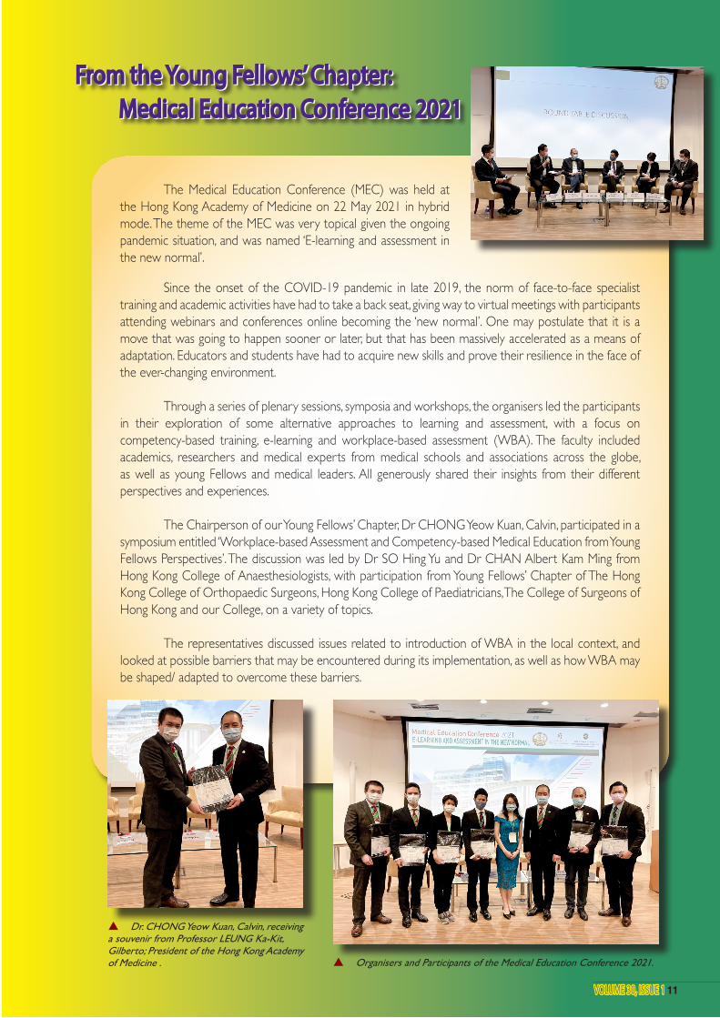
The Medical Education Conference (MEC) was held at the Hong Kong Academy of Medicine on 22 May 2021 in hybrid mode. The theme of the MEC was very topical given the ongoing pandemic situation, and was named ‘E-learning and assessment in the new normal’.
From the Young Fellows’ Chapter: From the Young Fellows’ Chapter: Medical Education Conference 2021 Medical Education Conference 2021
Since the onset of the COVID-19 pandemic in late 2019, the norm of face-to-face specialist training and academic activities have had to take a back seat, giving way to virtual meetings with participants attending webinars and conferences online becoming the ‘new normal’. One may postulate that it is a move that was going to happen sooner or later, but that has been massively accelerated as a means of adaptation. Educators and students have had to acquire new skills and prove their resilience in the face of the ever-changing environment.
Through a series of plenary sessions, symposia and workshops, the organisers led the participants in their exploration of some alternative approaches to learning and assessment, with a focus on competency-based training, e-learning and workplace-based assessment (WBA). The faculty included academics, researchers and medical experts from medical schools and associations across the globe, as well as young Fellows and medical leaders. All generously shared their insights from their different perspectives and experiences.
The Chairperson of our Young Fellows’ Chapter, Dr CHONG Yeow Kuan, Calvin, participated in a symposium entitled ‘Workplace-based Assessment and Competency-based Medical Education from Young Fellows Perspectives’. The discussion was led by Dr SO Hing Yu and Dr CHAN Albert Kam Ming from Hong Kong College of Anaesthesiologists, with participation from Young Fellows’ Chapter of The Hong Kong College of Orthopaedic Surgeons, Hong Kong College of Paediatricians, The College of Surgeons of Hong Kong and our College, on a variety of topics.
The representatives discussed issues related to introduction of WBA in the local context, and looked at possible barriers that may be encountered during its implementation, as well as how WBA may be shaped/ adapted to overcome these barriers.
p Organisers and Participants of the Medical Education Conference 2021.
From the Young Fellows’ Chapter: From the Young Fellows’ Chapter: Medical Education Conference 2021 Medical Education Conference 2021
p Dr. CHONG Yeow Kuan, Calvin, receiving a souvenir from Professor LEUNG Ka-Kit, Gilberto; President of the Hong Kong Academy of Medicine .
VOLUME 30, ISSUE 1VOLUME 30, ISSUE 1 11
Diagnosis of COVID-19 Diagnosis of COVID-19
THE HONG KONG COLLEGE OF PATHOLOGISTS:THE HONG KONG COLLEGE OF PATHOLOGISTS:
Editorial note: Coronavirus disease 2019 (COVID-19) is undoubtedly the most topical subject not only in the medical field, but also for humanity globally. In this issue of the Topical Update, Dr. Derek Hung and Prof. Kwok Yung Yuen present an overview on the diagnosis of COVID-19, which underpins effective disease control. We welcome any feedback or suggestion. Please direct them to Dr. Janice Lo (e-mail: [email protected]), Education Committee, The Hong Kong College of Pathologists. Opinions expressed are those of the authors or named individuals, and are not necessarily those of the Hong Kong College of Pathologists.
Dr. Derek HUNG and Prof. Kwok Yung YUENResident, Department of Microbiology, Queen Mary Hospital, Hospital Authority andProfessor, Department of Microbiology, Faculty of Medicine, The University of Hong Kong
Specimen collection Obtaining the best specimen optimizes the possibility of getting the correct diagnosis based on clinical suspicion. Being a predominantly respiratory pathogen, obtaining respiratory specimens for viral detection remains the primary modality for making a diagnosis of acute infection by SARS-CoV-2. The viral load is highest at or soon after symptom onset4, with the viral load in the upper respiratory tract peaking earlier than the lower respiratory tract5. The viral load decreases in the respiratory tract at a rate of 1 log10 per week6. The World Health Organization (WHO) suggests that testing upper respiratory tract specimens is adequate for early stage infection, especially asymptomatic or mild cases7. The Centers for Disease Control and Prevention (CDC) recognizes nasopharyngeal swab, nasopharyngeal wash, nasal wash obtained by health care professionals; nasal mid-turbinate swab, nasal swab obtained by either health care professionals or supervised self-collection on site; and posterior oropharyngeal saliva (POS) by supervised self-collection as valid specimens. Patients with
Overview Coronavirus disease 2019 (COVID-19) caused by severe acute respiratory syndrome coronavirus 2 (SARS-CoV-2) since December 2019 has infected 54 million population in all six major continents, resulting in over 1.3 million deaths by mid-November 20201. One of the most important aspects in curbinag the spread of the virus is rapid yet accurate diagnosis of infection followed by timely isolation and contact tracing. Molecular testing is now the mainstay of diagnosis, supplemented by viral antigen testing2. Antibody detection aids in assessment of immunity and disease prevalence in the population3. As the disease progresses, there are worldwide efforts in developing a multitude of diagnostic platforms, both in-house and commercial. Studies also endeavour to assess optimal types and timing of specimen collection to enhance diagnostic yield. In this review, we would look at some of the knowledge and practices in making a diagnosis of COVID-19.
12 PATHOLOGUEPATHOLOGUE
Volume 16, Issue 1 January 2021Volume 16, Issue 1 January 2021
The Hong Kong College of Pathologists, Incorporated in Hong Kong with Limited Liability
TOPICAL TOPICAL U PDATEU PDATETOPICAL TOPICAL U PDATEU PDATE
lower respiratory tract symptoms such as productive cough, shortness of breath, or suspicious radiological findings should send sputum to enhance sensitivity. Induced sputum is not recommended due to increased risk of aerosol transmission8,9. Among different respiratory specimens, broncho-alveolar lavage (BAL) showed the highest positive rate10.
For the upper respiratory tract specimen, comparing combined nasal swab/throat swab with nasopharyngeal swab, Vlek et al showed high concordance between these two methods (kappa coefficient 0.95) despite the cycle threshold value (Ct value) obtained from nasopharyngeal swab being lower11. Another study suggested nasal swab alone also has good concordance with nasopharyngeal sampling12. In contrast, oropharyngeal swab alone has inferior performance. Wang et al showed the sensitivity of oropharyngeal swab was 21.1%13 and meta-analysis by Bwire et al suggested the positive rate is as low as 7.6% in suspected cases, comparing with 69.6% and 71.3% for nasopharyngeal swab and lower respiratory tract specimen respectively14. POS is increasingly studied as an alternative respiratory tract specimen for diagnosis. Theoretically well produced POS can concentrate secretions dripping down from nasopharynx and lower respiratory secretion moved up by ciliary activity of respiratory epithelium. It can be saved by patients themselves with instructions, thus reducing discomfort in specimen collection and minimizing aerosol exposure for health care professionals. The cost of collecting POS could be 2.59-fold lower than nasopharyngeal specimen, which could be significant in resource limited setting15. The concordance between POS and nasopharyngeal swab is high16, especially in the first 7 days of infection, up to 96.6% positive percent agreement17. The sensitivity is comparable with nasopharyngeal swab in properly collected specimen18. The sensitivity does not vary much between early morning and at least 2 hours after meal, which provides a convenient option for specimen collection19. CDC and Hospital Authority of Hong Kong have adopted POS as an alternative option for upper respiratory specimen collection8,20.
Viral shedding is also found in other specimens with stool being more studied. Meta-analysis showed viral shedding was found in faecal material in 40.5% of patients21. The viral shedding in stool is more prevalent in those with gastrointestinal symptoms22 and may last longer than the shedding in respiratory tract23. Viral RNA detected in blood and urine is relatively uncommon, respectively only 1% and 0% in one study with more than 200 patients10. Even
without ocular symptoms, the conjunctival secretion may contain a small amount of SARS-CoV-2 RNA in around 8% of patients, warranting appropriate infection control measure in ophthalmological assessment24.
Molecular testing Detection of nucleic acid remains the backbone of diagnosing COVID-19 for treatment and public health purposes. Reverse-transcriptase polymerase chain reaction (RT-PCR) is the most widely used technique. After transcribing the viral RNA into complementary DNA (cDNA) with reverse transcriptase, the cDNA would be amplified and detected by real-time PCR. Potential molecular targets for SARS-CoV-2 include genes encoding structural proteins, e.g. spike (S), envelop (E), helicase (hel), nucleocapsid (N-N1 and N2), transmembrane (M); and non-structural regions, e.g. RNA-dependent RNA polymerase region (RdRp), haemagglutinin-esterase (HE), and open reading frame 1a (ORF1a) and ORF1b25. Most scientific institutes and commercial platforms would design primers to target more than one gene, or to target multiple loci of the same gene to enhance diagnostic sensitivity and specificity. Though N gene RNA is shown by nanopore direct RNA sequencing study to be the most abundantly expressed transcript in SARS-CoV-2 infected cells26, there is no consensus on which gene confers the best diagnostic performance. Presently, one conserved and one specific target region are recommended to mitigate effect of random mutation or genetic drift while maintaining specificity25. Various regimens for testing are proposed in the literature. Corman et al recommended the Charité protocol, which was to use E gene for screening and RdRp gene for confirmation27. CDC used N1 and N2 genes as their diagnostic panel28. Chu et al used N gene as screening test and ORF1b as confirmatory assay because the screening N gene assay is 10 times more sensitive than ORF1b29. As an alternative confirmatory assay, Chan et al developed a real-time RT-PCR assay locally, targeting RdRp/Hel. This COVID-19-RdRp/Hel assay demonstrated significantly higher sensitivity and specificity for the detection of SARS-CoV-2 RNA than the RdRp-P2 assay in clinical evaluation30.
Multiple commercial platforms were developed for molecular SARS-CoV-2 diagnosis for their high throughput, rapid turnaround time and ease of use with automation. Examples are Roche Cobas 6800/8800 system (targets ORF1a and E genes) and Abbott Alinity m SARS-CoV-2 assay (targets RdRp and N genes), where sample preparation,
VOLUME 30, ISSUE 1VOLUME 30, ISSUE 1 13
14 PATHOLOGUEPATHOLOGUE
genetic material extraction, target amplification and result reporting are automated inside the system. Molecular point-of-care testing (POCT) refers to diagnostic platform that is portable (often desktop-size), requires minimal sample preparation steps and can provide reliable molecular results within 2 hours31. POCT like Cepheid GeneXpert (Xpert Xpress SARS-CoV-2 assay, targets E and N2 genes) enables rapid testing near the site of collection in areas with little laboratory support. Fewer steps in manipulation reduce risk of cross contamination and laboratory error in processing. Many evaluation studies have been published to compare the performance of these commercial platforms against in-house diagnostic tests and for head-to-head comparison between platforms. For example, Cobas system is shown to have high diagnostic agreement with in-house molecular assays32,33, as well as with other commercial platforms such as Hologic Panther Fusion system34 and Cepheid GeneXpert35. Cepheid GeneXpert reaches an agreement of 100 % compared to three in-house RT-PCRs in a multicentre evaluation in the Netherlands36. Among commercial platforms there might be minor discordance between assays at very high Ct values and the viral load of clinical samples used in evaluative studies should be noted in particular37,38.
Another molecular technique is reverse-transcriptase loop-mediated isothermal amplification (RT-LAMP) test. Using multiple primers for the genetic target, RT-LAMP amplified nucleic acid by strand displacement in an isothermal condition of around 60- 65oC39. It allows synthesis of large amount of genetic material up to 106 to 109 copies of target DNA within 30-60 minutes2. Without the need of thermal cycler as in RT-PCR, RT-LAMP facilitates development of rapid molecular POCT and has an expanding market in commercial diagnostic platform. On the down side, since multiple primers over a relatively small genetic region are needed for amplification, there are constraints in properly designing the primers40. Abbott ID NOW is a commercial POCT platform using RT-LAMP, allowing real time detection of SARS-CoV-2 within 15 minutes targeting RdRp gene. Evaluation of ID NOW against other RT-PCR based platforms appears suboptimal in terms of diagnostic sensitivity. Compared to Cobas, ID NOW achieved only 73.9% positive agreement while GeneXpert achieved 98.9% positive agreement. In samples with Ct values greater than 30, positive agreement was 34.3% for ID Now and 97.1% for GeneXpert41. A lower sensitivity of ID NOW over GeneXpert was also reported in another evaluation by Basu et al42. In contrary, good diagnostic utility has been demonstrated in many other centres including Hong Kong
that have designed their own RT-LAMP for COVID-19. Chow et al reported sensitivity of 95% at 60 minutes using RT-LAMP targeting a region across ORF3a/E gene as compared to RT-PCR43. Lu et al achieved concordance rate of 93% against RT-PCR using in-house E gene RT-LAMP assay44.
In order to improve the diagnostic sensitivity of molecular assays, clustered regularly interspaced short palindromic repeat (CRISPR)-based technology has been employed by coupling with Cas enzyme. The enzyme would be directed to the target DNA/RNA by a guide RNA complementary to the target sequence. Once bound, the collateral nuclease activity of the Cas enzyme would cleave surrounding reporter fluorophore and lead to signal amplification45.
DETECTR technology uses Cas12a enzyme to bind target DNA; while SHERLOCK technology uses Cas13a enzymes to bind target RNA46. This technology can be incorporated in molecular techniques especially RT-LAMP to enhance the sensitivity and to lower the detection limit47.
Next generation sequencing (NGS) enables sequencing of the entire genome in a relatively short period of time. Sharing of genetic data facilitates tracking of disease spread, understanding of disease transmission route, monitoring viral genome evolution and detecting emergence of mutation that may escape detection or enhance virulence. The cost and infrastructure required of NGS and the need of bioinformatics expertise limit its use to larger hospital and research centres.
Antigen detection Like other respiratory viruses such as influenza and respiratory syncytial virus (RSV), direct antigen detection from respiratory specimen especially nasopharyngeal sample is another way of making a diagnosis of COVID-19. N protein was found previously to be the predominant structural protein released in large amount in nasopharyngeal aspirate during infection of SARS-CoV48, and the same phenomenon is also shown in SARS-CoV-2 where the abundantly expressed N protein is widely used as an antigen detection target in COVID-1949. Detection is achieved by capturing viral antigen in clinical specimens by monoclonal antibodies or monospecific polyclonal antibody fixed on a membrane, usually indicated by colour change of the strip in colorimetric lateral flow immunoassay. The assay can be delivered as POCT in an office setting since no complex laboratory support is required
and the result can be available within a short period of time, usually <30 minutes. The major setback is the suboptimal sensitivity as compared to molecular diagnosis especially in samples with high Ct values. Evaluation by Lambert-Niclot et al using COVID-19 Ag Respi-Strip CORIS, a nitrocellulose membrane technology with colloidal gold nanoparticles sensitized with monoclonal antibodies directed against SARS-CoV-2 nucleoprotein (NP) antigens, showed sensitivity of only 50% when compared against multiple RT-PCR platforms50. For samples with Ct value <25, the sensitivity is higher at 82.2%. In a local evaluation using Biocredit COVID-19 Ag test, the antigen test is 105 fold less sensitive than RT-PCR and it yielded a positive result in 45.7% RT-PCR positive combined nasopharyngeal swab/throat swab specimens only51. There are attempts to improve sensitivity of rapid antigen assay. Porte et al evaluated an immune-chromatographic antigen assay using fluorescence signal showing sensitivity of 93% but the Ct value of the sample included in this study is relatively low with mean of 2052. Other approaches by concentrating the antigen in specimens before testing with monoclonal antibodies targeting multiple different epitopes of the antigen were also reported53. Based on a meta-analysis by Dinnes et al, the average sensitivity is around 56.2% for antigen assay with a high average specificity of 99.5%54. Further refinement in antigen detection employs the detection of the change in bioelectric property by antigen binding to the antibody coated membrane. In Seo et al, anti-S antibody binds to SARS-CoV-2 particles to fabricate graphene-based field-effect-transistors (FET) biosensors and can respond down to 16 pfu/mL of virus55. One challenge to this advance is the high background noise which can reduce sensitivity of detection. Overall, rapid antigen detection serves only an adjunctive role to molecular assay in making a diagnosis especially in outbreak situation where prevalence is high and molecular assay is not available. WHO has issued interim guidance of use of rapid antigen immunoassays56.
Antibody detection While antibody testing may not be useful in acute setting for COVID-19, it helps establish retrospective diagnosis, predict immunity and understand seroprevalence in a defined community57. Commonly employed techniques are lateral flow immunoassay, chemiluminescent immunoassay, immunofluorescent assay, and enzyme-linked immunosorbent assay (ELISA)58. Median seroconversion times following symptom onset are 11 days for total antibodies, 12 days and 14 days for IgM and IgG respectively59. Detection rate for IgM ranges from 11-71% in the first 7 days of infection, 36-
87% between 8-14 days, and 56-97% after 14 days. For IgG, it ranges from 4-57% in first 7 days, 54-88% between 8-14 days, and 91-100% after 14 days60. For SARS-CoV-2, there does not seem to have significant time difference between IgM and IgG response61. IgM peaked at around 3 weeks after symptom onset and fell to baseline level at around day 3662. The duration of IgG seropositivity remains unknown and longer longitudinal studies are required. Study from Iceland involving over 1200 confirmed patients showed no evidence of antiviral antibody decline by 4 months after diagnosis63; and most other studies showed persistently detectable antibodies by 2-3 months after infection60. On the other hand, there are some evidences that the IgG level may decline faster in mild64 and asymptomatic61 COVID-19 cases.
S protein is an important antigen for neutralizing antibody production. The S1 domain is responsible for receptor binding while the S2 domain is responsible for fusion. The receptor binding domain (RBD) is located at S1. NP, which is a structural component of the helical nucleocapsid, also appears to be an important antigen for the development of serological assays to detect COVID-19. Earlier in the pandemic, using sera collected more than 14 days after symptom onset from 16 patients, To et al showed rates of seropositivity were 94% for anti-NP IgG, 88% for anti-NP IgM, 100% for anti-RBD IgG, and 94% for anti-RBD IgM65. Another study compares sensitivity and specificity in testing anti-S and anti-NP IgG for evidence of immunity across multiple platforms, which shows they are comparable by day 37 after infection though seroconversion of anti-NP IgG may precede anti-S IgG by around 2 days (day 9-10 v day 11-12)66. Caruana et al observed that the decline of anti-NP antibody may be faster than anti-S and thus could be less sensitive longer after infection67. Also titre of anti-S antibody may better reflect protection against reinfection67. Multiple commercial platforms were developed for high-throughput antibody testing in clinical laboratory. Automatic platforms such as Abbott SARS-CoV-2 IgG, which is a chemiluminescent micro-particle immunoassay, are also used in public hospital of Hong Kong for a shorter turnaround time.
Neutralization antibody test is important in assessing in vitro the functional capacity of the humoral response of COVID-19 patients to prevent reinfection by the virus. Traditional neutralization assay such as microneutralization and plaque reduction assay require manipulation of live virus and necessitate biosafety level 3 laboratories. As a result, pseudovirus neutralization assay has been developed. Vesicular stomatitis virus (VSV) expressing S protein of SARS-CoV-2,
VOLUME 30, ISSUE 1VOLUME 30, ISSUE 1 15
16 PATHOLOGUEPATHOLOGUE
containing the RBD, is used so that the assay can be performed in biosafety level 2 facilities68. SARS-CoV-2 neutralizing antibody starts to rise at around 7-10 days after symptom onset and the median peak time is 33 days after symptom onset. The neutralization titres then decline in 93% of the patients and by a median level of 35% over 3 months69. Patients with more severe disease requiring ICU admission have accelerated and augmented neutralizing antibody response compared with non-ICU cases70. In non-severe cases who have low peak neutralizing antibody titre, neutralizing antibody level might return to baseline within 2 months71. Another clinical use of neutralization assay would be to confirm potentially false positive SARS-CoV-2 serology result. Three children with Kawasaki disease without symptoms or epidemiological linkage to COVID-19 were tested positive to anti-RBD and anti-NP antibodies by a microparticle-based immunoassay but were confirmed negative by microneutralization test72.
Studies have shown there are serological cross-reactivity between SARS-CoV-2 and SARS-CoV. Testing sera taken from COVID-19 patients by ELISA, cross-reactivity is seen against S protein and RBD of SARS-CoV, though the intensity of cross-reaction against RBD is weaker than S protein73. For the full length S protein, the amino acid sequence homology between SARS-CoV-2 and SARS-CoV is around 75%. The homology between them for RBD which is located in S1 domain is around 74%. For the receptor binding motif (RBM) of the RBD where the virus directly binds to angiotensin-converting enzyme 2 (ACE2), the homology is only 50%74. The degree of amino acid homology explains the difference in the level of cross-reaction between them on ELISA. Chia et al showed even more significant cross-reactivity between SARS-CoV-2 and SARS-CoV antibody against NP by Luminex assay than antibody against S1 or RBD as the homology between the NP of these 2 viruses is around 90%75. Despite some cross-reaction between antibodies against RBD on ELISA, there does not seem to have significant cross neutralization effect73. Only 1 out of 15 COVID-19 sera showed cross neutralization with SARS-CoV at very low titre. Overall the effect of cross-protection in vaccination and whether antibody-dependent enhancement effect would be seen between these 2 closely related viruses remains unknown.
Cross-reactivity against other human coronaviruses in SARS-CoV-2 infection has been investigated in a few trials. In a study by Wölfel et al76, using immunofluorescence assay
against recombinant S protein, cross-reactivity of SARS-CoV-2 sera is found against human coronaviruses OC43, NL63, HKU1 and 229E on comparing the titres between admission and convalescence samples, especially HKU1 and OC43 which are both betacoronavirus. In Shrock et al77, deep serological profiling of sera from SARS-CoV-2 patients and pre-COVID sera are performed. Antibodies against S and NP are the most specific assay to differentiate SARS-CoV-2 and pre-COVID sera. Those with dramatic increase in anti-S antibody after COVID-19 infection also have increase in the intensity of cross-reactivity against other human coronaviruses, especially over more homologous regions of the S protein e.g. at residue 811-830 and 1144-1163. It could be novel antibodies of SARS-CoV-2 that cross-react or boost the anamnestic response against SARS-CoV-2 infection due to existing memory towards other human coronaviruses from past exposure. Moreover, pre-COVID sera also show some cross-reaction towards the homologous region of SARS-CoV-2 S protein and ORF1 in the same study.
Viral culture Demonstration of live SARS-CoV-2 in cell culture requires biosafety level 3 facilities and are not routinely performed in most of the clinical laboratories. However, live virus isolation is still important for some diagnostic and research purposes so as to determine whether the amount of virus present is infectious to others, to evaluate therapeutic efficacy of potential antiviral compound, to develop viral neutralization assay for testing convalescent sera, to provide positive control for molecular assay development, and to develop vaccine strains78. The host cell receptor for SARS-CoV-2 is ACE2. Non-human cell lines such as Vero E6 and Vero CCL-61 which have abundant ACE2 expression are commonly used for isolation79. Cytopathic effect is seen by 3 days after inoculation80. SARS-CoV-2 also grows in human continuous cell lines such as Calu3 (pulmonary cell line), Caco2 (intestinal cell line), Huh7 (hepatic cell line), and 293T (renal cell line)81. It grows modestly on U251 (neuronal cell line) which is not seen in SARS-CoV81. Confirmation of SARS-CoV-2 replication in the cell line can be done by molecular testing or immunostaining techniques. Cell lines can be engineered to express a transmembrane serine protease TMPRSS2 for priming of S protein and to facilitate the entry of SARS-CoV-2 into host cell82. Organoid systems such as bat and human intestinal organoids are susceptible to SARS-CoV-2 and are developed to better study tissue tropism, the dynamics of infection and testing of therapeutic targets83.
Radiological diagnosis and artificial intelligence There are no pathognomonic radiological features on chest imaging for COVID-19 and the disease should not be ruled in or ruled out based on imaging alone. However, presence of suggestive imaging features can prompt further investigations in suspicious cases, such as lower respiratory tract viral testing for confirmation. Reports in literature have suggested that in some patients, radiological findings may precede the detection of SARS-CoV-2 in clinical specimen84,85. Chest X-ray (CXR) is a less sensitive modality than computed tomography of the thorax (CT thorax) with a reported CXR sensitivity of 69%85. As in other viral pneumonia, COVID-19 typically presents with multifocal air-space disease, especially with a bilateral lower lung distribution86. More specific to COVID-19, it tends to have peripheral lung involvement, seen in 58% of CXR in one study87. CT thorax has a higher sensitivity than CXR, quoted at around 60-98%88. CT thorax often demonstrates the typical findings of peripheral bilateral ground glass opacities (GGO) with or without consolidation or ‘crazy-paving pattern’. Sometimes the GGO would arrange in a rounded pattern. Isolated lobar or segmental consolidation without GGO, centrilobular shadows, cavitory changes, lymphadenopathy and pleural effusions are rare86. As the disease advances, the opacities might coalesce, affecting central and bilateral upper lobes and may manifest as ‘white lung’ with diffuse infiltrate89. The abnormalities usually peak by 2 weeks after symptom onset, replaced by scar tissue with recovery90. In the COVID-19 pandemic, artificial intelligence (AI) programme is increasingly studied for screening abnormal radiological result which would be particularly useful for mass screening strategy in outbreak situation. The performance of AI is dependent on the radiological imaging algorithm being fed into the system for deep learning process. So far the result of this research has been promising with reported area under receiver operating characteristic curves greater than 0.991,92. However, there are still lots of technical and ethical issue to resolve which include dataset bias, data privacy, and the distribution of ultimate accountability of result93.
Detection of host inflammatory reaction In COVID-19, there are studies to diagnose and predict severe diseases by the host inflammatory response. Apart from direct viral damage, uncontrolled cytokine storm triggered by the virus leads to tissue damage and multiorgan failure94. Mean interleukin-6 (IL-6) concentration in serum was found to be 2.9 fold higher in patients with complicated COVID-19 disease than non-complicated disease95. It became one of the markers clinicians could use to predict progression into severe disease. Roche Elecsys IL-6 immunoassay received FDA Emergency Use Authorization to help identify patients at high risk of requiring intubation with mechanical ventilation96. Molecules targeting IL-6 such as tocilizumab are also studied as therapeutic to prevent disease progress by blocking the inflammatory pathway97. It does not show efficacy in preventing intubation or death in moderately ill hospitalized patients in the BACC Bay trial98. Elevated CRP is associated with worse outcome99, as well as elevated IL-10 which may be related to compensatory anti-inflammatory response and secondary infections100. Haematologically, severe disease is associated with higher absolute neutrophil count101, D-dimer102 and LDH103 but lower absolute lymphocyte101 and platelet count104.
Conclusion Global COVID-19 pandemic stimulates global effort in development of rapid yet accurate diagnostic techniques. Diagnosis is often limited by the low level of viral particles in the specimen and the subtle clinical features in early infection. Though traditional methods like RT-PCR are still the mainstay, we see expanding endeavours to strive for higher speed and lower limit of detection at an earlier time. Molecular techniques such as RT-LAMP, CRISPR/Cas, biosensor technology in antigen detection, AI operating system for image interpretation are pushing the diagnostic ability to the limit. Despite these scientific advances, there are still a lot of gaps to fill especially in understanding the nature and duration of humoral immunity response and its protection against re-infection. All these require continuous global cooperation and information exchange to make them possible.
References1 Weekly epidemiological update – 17 November 2020, World Health
Organization. https://www.who.int/publications/m/item/weekly-epidemiological-update---17-november-2020. Accessed on 20th November 2020
2 Ji T, Liu Z, Wang G, Guo X, Akbar Khan S, Lai C, et al. Detection of COVID-19: A review of the current literature and future perspectives. Biosens Bioelectron. 2020 Oct 15;166:112455. doi:
10.1016/j.bios.2020.112455. Epub 2020 Jul 21.3 Ejazi SA, Ghosh S, Ali N. Antibody detection assays for COVID-19
diagnosis: an early overview. Immunol Cell Biol. 2020 Aug 30. doi: 10.1111/imcb.12397.
4 Walsh KA, Jordan K, Clyne B, Rohde D, Drummond L, Byrne P, et al. SARS-CoV-2 detection, viral load and infectivity over the course of an infection. J Infect. 2020 Sep;81(3):357-371. doi: 10.1016/j.jinf.2020.06.067. Epub 2020 Jun 29.
VOLUME 30, ISSUE 1VOLUME 30, ISSUE 1 17
18 PATHOLOGUEPATHOLOGUE
5 Weiss A, Jellingsø M, Sommer MOA. Spatial and temporal dynamics of SARS-CoV-2 in COVID-19 patients: A systematic review and meta-analysis. EBioMedicine. 2020 Aug;58:102916. doi: 10.1016/j.ebiom.2020.102916. Epub 2020 Jul 22.
6 To KK, Tsang OT, Leung WS, Tam AR, Wu TC, Lung DC, et al. Temporal profiles of viral load in posterior oropharyngeal saliva samples and serum antibody responses during infection by SARS-CoV-2: an observational cohort study. Lancet Infect Dis. 2020 May;20(5):565-574. doi: 10.1016/S1473-3099(20)30196-1. Epub 2020 Mar 23.
7 Interim guidance on diagnostic testing for SARS-CoV-2, World Health Organization. Published on 11th September 2020. Accessed on 4th November 2020
8 https://www.cdc.gov/coronavirus/2019-ncov/lab/guidelines-clinical-specimens.html. Interim Guidelines for Collecting, Handling, and Testing Clinical Specimens for COVID-19, Centers for Disease Control and Prevention. Accessed 20th November, 2020
9 https://www.who.int/publications/i/item/clinical-management-of-covid-19. Clinical management of COVID-19 – Interim guidance. Accessed on 20th November, 2020
10 Wang W, Xu Y, Gao R, Lu R, Han K, Wu G, Tan W. Detection of SARS-CoV-2 in different types of clinical specimens. JAMA. 2020 May 12;323(18):1843-1844. doi: 10.1001/jama.2020.3786.
11 Vlek ALM, Wesselius TS, Achterberg R, Thijsen SFT. Combined throat/nasal swab sampling for SARS-CoV-2 is equivalent to nasopharyngeal sampling. Eur J Clin Microbiol Infect Dis. 2020 Jul 14:1–3. doi: 10.1007/s10096-020-03972-y. Epub ahead of print.
12 Péré H, Podglajen I, Wack M, Flamarion E, Mirault T, Goudot G, et al. Nasal swab sampling for SARS-CoV-2: a convenient alternative in times of nasopharyngeal swab shortage. J Clin Microbiol. 2020 May 26;58(6):e00721-20. doi: 10.1128/JCM.00721-20.
13 Wang H, Liu Q, Hu J, Zhou M, Yu MQ, Li KY, et al. Nasopharyngeal swabs are more sensitive than oropharyngeal swabs for COVID-19 diagnosis and monitoring the SARS-CoV-2 load. Front Med (Lausanne). 2020 Jun 18;7:334. doi: 10.3389/fmed.2020.00334.
14 Bwire GM, Majigo MV, Njiro BJ, Mawazo A. Detection profile of SARS-CoV-2 using RT-PCR in different types of clinical specimens: A systematic review and meta-analysis. J Med Virol. 2020 Jul 24:10.1002/jmv.26349. doi: 10.1002/jmv.26349. Epub ahead of print.
15 To KKW, Yip CCY, Lai CYW, Wong CKH, Ho DTY, Pang PKP, et al. Saliva as a diagnostic specimen for testing respiratory virus by a point-of-care molecular assay: a diagnostic validity study. Clin Microbiol Infect. 2019 Mar;25(3):372-378. doi: 10.1016/j.cmi.2018.06.009. Epub 2018 Jun 12.
16 Leung EC, Chow VC, Lee MK, Lai RW. Deep throat saliva as an alternative diagnostic specimen type for the detection of SARS-CoV-2. J Med Virol. 2020 Jul 4:10.1002/jmv.26258. doi: 10.1002/jmv.26258. Epub ahead of print.
17 Wong SCY, Tse H, Siu HK, Kwong TS, Chu MY, Yau FYS, et al. Posterior oropharyngeal saliva for the detection of SARS-CoV-2. Clin Infect Dis. 2020 Jun 21:ciaa797. doi: 10.1093/cid/ciaa797. Epub ahead of print.
18 Wyllie AL, Fournier J, Casanovas-Massana A, Campbell M, Tokuyama M, Vijayakumar P, et al. Saliva or nasopharyngeal swab specimens for detection of SARS-CoV-2. N Engl J Med. 2020 Sep 24;383(13):1283-1286. doi: 10.1056/NEJMc2016359. Epub 2020 Aug 28.
19 Hung DL, Li X, Chiu KH, Yip CC, To KK, Chan JF, et al. Early-morning vs spot posterior oropharyngeal saliva for diagnosis of SARS-CoV-2
infection: Implication of timing of specimen collection for community-wide screening. Open Forum Infect Dis. 2020 Jun 7;7(6):ofaa210. doi: 10.1093/ofid/ofaa210.
20 Interim recommendation on clinical management of adult cases with Coronavirus Disease 2019 (COVID-19) [version 1.2], Hospital Authority, Hong Kong SAR. Accessed on 4th November 2020
21 Parasa S, Desai M, Thoguluva Chandrasekar V, Patel HK, Kennedy KF, Roesch T, et al. Prevalence of gastrointestinal symptoms and fecal viral shedding in patients with Coronavirus Disease 2019: A systematic review and meta-analysis. JAMA Netw Open. 2020 Jun 1;3(6):e2011335. doi: 10.1001/jamanetworkopen.2020.11335.
22 Cheung KS, Hung IFN, Chan PPY, Lung KC, Tso E, Liu R, et al. Gastrointestinal manifestations of SARS-CoV-2 infection and virus load in fecal samples from a Hong Kong cohort: Systematic review and meta-analysis. Gastroenterology. 2020 Jul;159(1):81-95. doi: 10.1053/j.gastro.2020.03.065. Epub 2020 Apr 3.
23 Chen Y, Chen L, Deng Q, Zhang G, Wu K, Ni L, et al. The presence of SARS-CoV-2 RNA in the feces of COVID-19 patients. J Med Virol. 2020 Jul;92(7):833-840. doi: 10.1002/jmv.25825. Epub 2020 Apr 25.
24 Li X, Chan JF, Li KK, Tso EY, Yip CC, Sridhar S, et al. Detection of SARS-CoV-2 in conjunctival secretions from patients without ocular symptoms. Infection. 2020 Sep 17:1–9. doi: 10.1007/s15010-020-01524-2. Epub ahead of print.
25 Tang YW, Schmitz JE, Persing DH, Stratton CW. Laboratory diagnosis of COVID-19: Current issues and challenges. J Clin Microbiol. 2020 May 26;58(6):e00512-20.
26 Kim D, Lee JY, Yang JS, Kim JW, Kim VN, Chang H. The architecture of SARS-CoV-2 transcriptome. Cell. 2020 May 14;181(4):914-921.e10. doi: 10.1016/j.cell.2020.04.011. Epub 2020 Apr 23.
27 Corman VM, Landt O, Kaiser M, et al. Detection of 2019 novel coronavirus (2019-nCoV) by real-time RT-PCR. Euro Surveill. 2020;25(3):2000045. doi:10.2807/1560-7917.ES.2020.25.3.2000045
28 https://www.cdc.gov/coronavirus/2019-ncov/lab/virus-requests.html. Accessed on 16th November 2020.
29 Chu DKW, Pan Y, Cheng SMS, et al. Molecular diagnosis of a novel coronavirus (2019-nCoV) causing an outbreak of pneumonia. Clin Chem. 2020;66(4):549-555. doi:10.1093/clinchem/hvaa029
30 Chan JF, Yip CC, To KK, Tang TH, Wong SC, Leung KH, et al. Improved molecular diagnosis of COVID-19 by the novel, highly sensitive and specific COVID-19-RdRp/Hel real-time reverse transcription-PCR assay validated in vitro and with clinical specimens. J Clin Microbiol. 2020 Apr 23;58(5):e00310-20. doi: 10.1128/JCM.00310-20.
31 Dinnes J, Deeks JJ, Adriano A, Berhane S, Davenport C, Dittrich S, et al. Cochrane COVID-19 Diagnostic Test Accuracy Group. Rapid, point-of-care antigen and molecular-based tests for diagnosis of SARS-CoV-2 infection. Cochrane Database Syst Rev. 2020 Aug 26;8:CD013705. doi: 10.1002/14651858.CD013705.
32 Opota O, Brouillet R, Greub G, Jaton K. Comparison of SARS-CoV-2 RT-PCR on a high-throughput molecular diagnostic platform and the cobas SARS-CoV-2 test for the diagnostic of COVID-19 on various clinical samples. Pathog Dis. 2020 Nov 11;78(8):ftaa061. doi: 10.1093/femspd/ftaa061.
33 Dust K, Hedley A, Nichol K, Stein D, Adam H, Karlowsky JA, et al. Comparison of commercial assays and laboratory developed tests for detection of SARS-CoV-2. J Virol Methods. 2020 Nov;285:113970. doi: 10.1016/j.jviromet.2020.113970. Epub 2020 Sep 10.
34 Craney AR, Velu PD, Satlin MJ, Fauntleroy KA, Callan K, Robertson A,
et al. Comparison of two high-throughput reverse transcription-PCR systems for the detection of Severe Acute Respiratory Syndrome Coronavirus 2. J Clin Microbiol. 2020 Jul 23;58(8):e00890-20. doi: 10.1128/JCM.00890-20.
35 Moran A, Beavis KG, Matushek SM, Ciaglia C, Francois N, Tesic V, Love N. Detection of SARS-CoV-2 by use of the Cepheid Xpert Xpress SARS-CoV-2 and Roche cobas SARS-CoV-2 assays. J Clin Microbiol. 2020 Jul 23;58(8):e00772-20. doi: 10.1128/JCM.00772-20.
36 Wolters F, van de Bovenkamp J, van den Bosch B, van den Brink S, Broeders M, Chung NH, et al. Multi-center evaluation of Cepheid Xpert® Xpress SARS-CoV-2 point-of-care test during the SARS-CoV-2 pandemic. J Clin Virol. 2020 Jul;128:104426. doi: 10.1016/j.jcv.2020.104426. Epub 2020 May 11.
37 Lieberman JA, Pepper G, Naccache SN, Huang ML, Jerome KR, Greninger AL. Comparison of commercially available and laboratory-developed assays for in vitro detection of SARS-CoV-2 in clinical laboratories. J Clin Microbiol. 2020 Jul 23;58(8):e00821-20. doi: 10.1128/JCM.00821-20.
38 Broder K, Babiker A, Myers C, White T, Jones H, Cardella J, et al. Test Agreement between Roche Cobas 6800 and Cepheid GeneXpert Xpress SARS-CoV-2 assays at high cycle threshold ranges. J Clin Microbiol. 2020 Jul 23;58(8):e01187-20. doi: 10.1128/JCM.01187-20.
39 Younes N, Al-Sadeq DW, Al-Jighefee H, Younes S, Al-Jamal O, Daas HI, et al. Challenges in laboratory diagnosis of the novel coronavirus SARS-CoV-2. Viruses. 2020 May 26;12(6):582. doi: 10.3390/v12060582.
40 Zaghloul H, El-Shahat M. Recombinase polymerase amplification as a promising tool in hepatitis C virus diagnosis. World J Hepatol. 2014 Dec 27;6(12):916-22.
41 Smithgall MC, Scherberkova I, Whittier S, Green DA. Comparison of Cepheid Xpert Xpress and Abbott ID Now to Roche cobas for the rapid detection of SARS-CoV-2. J Clin Virol. 2020 Jul;128:104428. doi: 10.1016/j.jcv.2020.104428. Epub 2020 May 13.
42 Basu A, Zinger T, Inglima K, Woo KM, Atie O, Yurasits L, See B, Aguero-Rosenfeld ME. Performance of Abbott ID Now COVID-19 rapid nucleic acid amplification test using nasopharyngeal swabs transported in viral transport media and dry nasal swabs in a New York City academic institution. J Clin Microbiol. 2020 Jul 23;58(8):e01136-20. doi: 10.1128/JCM.01136-20.
43 Chow FW, Chan TT, Tam AR, Zhao S, Yao W, Fung J, et al. A rapid, simple, inexpensive, and mobile colorimetric assay COVID-19-LAMP for mass on-site screening of COVID-19. Int J Mol Sci. 2020 Jul 29;21(15):5380. doi: 10.3390/ijms21155380
44 Lu R, Wu X, Wan Z, Li Y, Jin X, Zhang C. A novel reverse transcription loop-mediated isothermal amplification method for rapid detection of SARS-CoV-2. Int J Mol Sci. 2020 Apr 18;21(8):2826. doi: 10.3390/ijms21082826.
45 Fernandez-Garcia L, Pacios O, González-Bardanca M, Blasco L, Bleriot I, Ambroa A, et al. Viral related tools against SARS-CoV-2. Viruses. 2020 Oct 16;12(10):1172. doi: 10.3390/v12101172.
46 Jolany Vangah S, Katalani C, Booneh HA, Hajizade A, Sijercic A, Ahmadian G. CRISPR-based diagnosis of infectious and noninfectious diseases. Biol Proced Online. 2020 Sep 14;22:22. doi: 10.1186/s12575-020-00135-3.
47 D’Cruz RJ, Currier AW, Sampson VB. Laboratory testing methods for novel Severe Acute Respiratory Syndrome-Coronavirus-2
(SARS-CoV-2). Front Cell Dev Biol. 2020 Jun 4;8:468. doi: 10.3389/fcell.2020.00468.
48 Das D, Kammila S, Suresh MR. Development, characterization, and application of monoclonal antibodies against severe acute respiratory syndrome coronavirus nucleocapsid protein. Clin Vaccine Immunol. 2010 Dec;17(12):2033-6. doi: 10.1128/CVI.00293-10. Epub 2010 Oct 6.
49 Zeng W, Liu G, Ma H, Zhao D, Yang Y, Liu M, et al. Biochemical characterization of SARS-CoV-2 nucleocapsid protein. Biochem Biophys Res Commun. 2020 Jun 30;527(3):618-623. doi: 10.1016/j.bbrc.2020.04.136. Epub 2020 Apr 30.
50 Lambert-Niclot S, Cuffel A, Le Pape S, Vauloup-Fellous C, Morand-Joubert L, Roque-Afonso AM, et al. Evaluation of a rapid diagnostic assay for detection of SARS-CoV-2 antigen in nasopharyngeal swabs. J Clin Microbiol. 2020 Jul 23;58(8):e00977-20. doi: 10.1128/JCM.00977-20.
51 Mak GC, Cheng PK, Lau SS, Wong KK, Lau CS, Lam ET, et al. Evaluation of rapid antigen test for detection of SARS-CoV-2 virus. J Clin Virol. 2020 Aug;129:104500. doi: 10.1016/j.jcv.2020.104500. Epub 2020 Jun 8.
52 Porte L, Legarraga P, Vollrath V, Aguilera X, Munita JM, Araos R, et al. Evaluation of a novel antigen-based rapid detection test for the diagnosis of SARS-CoV-2 in respiratory samples. Int J Infect Dis. 2020 Oct;99:328-333. doi: 10.1016/j.ijid.2020.05.098. Epub 2020 Jun 1.
53 Abduljalil JM. Laboratory diagnosis of SARS-CoV-2: available approaches and limitations. New Microbes New Infect. 2020 Jun 14;36:100713. doi: 10.1016/j.nmni.2020.100713.
54 Dinnes J, Deeks JJ, Adriano A, Berhane S, Davenport C, Dittrich S, et al. Cochrane COVID-19 Diagnostic Test Accuracy Group. Rapid, point-of-care antigen and molecular-based tests for diagnosis of SARS-CoV-2 infection. Cochrane Database Syst Rev. 2020 Aug 26;8:CD013705. doi: 10.1002/14651858.CD013705.
55 Seo G, Lee G, Kim MJ, Baek SH, Choi M, Ku KB, et al. Rapid detection of COVID-19 causative virus (SARS-CoV-2) in human nasopharyngeal swab specimens using field-effect transistor-based biosensor. ACS Nano. 2020 Apr 28;14(4):5135-5142. doi: 10.1021/acsnano.0c02823. Epub 2020 Apr 20. Erratum in: ACS Nano. 2020 Sep 22;14(9):12257-12258.
56 Antigen-detection in the diagnosis of SARS-CoV-2 infection using rapid immunoassays – Interim Guidance, World Health Organization. Issued on 11th September 2020. Accessed on 7th November 2020.
57 Anand S, Montez-Rath M, Han J, Bozeman J, Kerschmann R, Beyer P, et al. Prevalence of SARS-CoV-2 antibodies in a large nationwide sample of patients on dialysis in the USA: a cross-sectional study. Lancet. 2020 Sep 25;396(10259):1335–44. doi: 10.1016/S0140-6736(20)32009-2.
58 Jarrom D, Elston L, Washington J, Prettyjohns M, Cann K, Myles S, Groves P. Effectiveness of tests to detect the presence of SARS-CoV-2 virus, and antibodies to SARS-CoV-2, to inform COVID-19 diagnosis: a rapid systematic review. BMJ Evid Based Med. 2020 Oct 1:bmjebm-2020-111511. doi: 10.1136/bmjebm-2020-111511. Epub ahead of print.
59 Zhao J, Yuan Q, Wang H, Liu W, Liao X, Su Y, et al. Antibody responses to SARS-CoV-2 in patients of novel coronavirus disease 2019. Clin Infect Dis. 2020 Mar 28:ciaa344. doi: 10.1093/cid/ciaa344. Epub ahead of print.
VOLUME 30, ISSUE 1VOLUME 30, ISSUE 1 19
20 PATHOLOGUEPATHOLOGUE
60 O Murchu E, Byrne P, Walsh KA, Carty PG, Connolly M, De Gascun C, et al. Immune response following infection with SARS-CoV-2 and other coronaviruses: A rapid review. Rev Med Virol. 2020 Sep 23:e2162. doi: 10.1002/rmv.2162. Epub ahead of print.
61 Long QX, Tang XJ, Shi QL, Li Q, Deng HJ, Yuan J, et al. Clinical and immunological assessment of asymptomatic SARS-CoV-2 infections. Nat Med. 2020 Aug;26(8):1200-1204. doi: 10.1038/s41591-020-0965-6. Epub 2020 Jun 18.
62 Shu H, Wang S, Ruan S, Wang Y, Zhang J, Yuan Y, et al. Dynamic changes of antibodies to SARS-CoV-2 in COVID-19 patients at early stage of outbreak. Virol Sin. 2020 Jul 27:1–8. doi: 10.1007/s12250-020-00268-5. Epub ahead of print.
63 Gudbjartsson DF, Norddahl GL, Melsted P, Gunnarsdottir K, Holm H, Eythorsson E, et al. Humoral immune response to SARS-CoV-2 in Iceland. N Engl J Med. 2020 Oct 29;383(18):1724-1734. doi: 10.1056/NEJMoa2026116. Epub 2020 Sep 1.
64 Ibarrondo FJ, Fulcher JA, Goodman-Meza D, Elliott J, Hofmann C, Hausner MA, et al. Rapid decay of anti-SARS-CoV-2 antibodies in persons with mild Covid-19. N Engl J Med. 2020 Sep 10;383(11):1085-1087. doi: 10.1056/NEJMc2025179. Epub 2020 Jul 21. Erratum in: N Engl J Med. 2020 Jul 23
65 To KK, Tsang OT, Leung WS, Tam AR, Wu TC, Lung DC, et al. Temporal profiles of viral load in posterior oropharyngeal saliva samples and serum antibody responses during infection by SARS-CoV-2: an observational cohort study. Lancet Infect Dis. 2020 May;20(5):565-574. doi: 10.1016/S1473-3099(20)30196-1. Epub 2020 Mar 23.
66 Van Elslande J, Decru B, Jonckheere S, Van Wijngaerden E, Houben E, Vandecandelaere P, et al. Antibody response against SARS-CoV-2 spike protein and nucleoprotein evaluated by four automated immunoassays and three ELISAs. Clin Microbiol Infect. 2020 Nov;26(11):1557.e1-1557.e7. doi: 10.1016/j.cmi.2020.07.038.
67 Caruana G, Croxatto A, Coste AT, Opota O, Lamoth F, Jaton K, Greub G. Diagnostic strategies for SARS-CoV-2 infection and interpretation of microbiological results. Clin Microbiol Infect. 2020 Jun 25;26(9):1178–82. doi: 10.1016/j.cmi.2020.06.019. Epub ahead of print.
68 Nie J, Li Q, Wu J, Zhao C, Hao H, Liu H, et al. Establishment and validation of a pseudovirus neutralization assay for SARS-CoV-2. Emerg Microbes Infect. 2020 Dec;9(1):680-686.
69 Kai Wang, Quan-Xin Long, Hai-Jun Deng, Jie Hu, Qing-Zhu Gao, Gui-Ji Zhang, et al. Longitudinal dynamics of the neutralizing antibody response to Severe Acute Respiratory Syndrome Coronavirus 2 (SARS-CoV-2) infection. Clin Infect Dis. ciaa1143, https://doi.org/10.1093/cid/ciaa1143
70 Liu L, To KK, Chan KH, Wong YC, Zhou R, Kwan KY, et al. High neutralizing antibody titer in intensive care unit patients with COVID-19. Emerg Microbes Infect. 2020 Dec;9(1):1664-1670.
71 Seow J, Graham C, Merrick B, Acors S, Pickering S, Steel KJA, et al. Longitudinal observation and decline of neutralizing antibody responses in the three months following SARS-CoV-2 infection in humans. Nat Microbiol. 2020 Oct 26.
72 To KK, Chua GT, Kwok KL, Wong JS, Au DCY, Lam YY, et al. False-positive SARS-CoV-2 serology in 3 children with Kawasaki disease. Diagn Microbiol Infect Dis. 2020 Nov;98(3):115141. doi: 10.1016/j.diagmicrobio.2020.115141. Epub 2020 Jul 17.
73 Lv H, Wu NC, Tsang OT, Yuan M, Perera RAPM, Leung WS, et al. Cross-reactive antibody response between SARS-CoV-2 and SARS-CoV infections. Cell Rep. 2020 Jun 2;31(9):107725. doi: 10.1016/j.celrep.2020.107725. Epub 2020 May 18.
74 Jaimes JA, André NM, Chappie JS, Millet JK, Whittaker GR. Phylogenetic analysis and structural modeling of SARS-CoV-2 spike protein reveals an evolutionary distinct and proteolytically sensitive activation loop. J Mol Biol. 2020 May 1;432(10):3309-3325. doi: 10.1016/j.jmb.2020.04.009. Epub 2020 Apr 19
75 Chia WN, Tan CW, Foo R, Kang AEZ, Peng Y, Sivalingam V, et al. Serological differentiation between COVID-19 and SARS infections. Emerg Microbes Infect. 2020 Dec;9(1):1497-1505.
76 Wölfel R, Corman VM, Guggemos W, Seilmaier M, Zange S, Müller MA, et al. Virological assessment of hospitalized patients with COVID-2019. Nature. 2020 May;581(7809):465-469. doi: 10.1038/s41586-020-2196-x. Epub 2020 Apr 1.
77 Shrock E, Fujimura E, Kula T, Timms RT, Lee IH, Leng Y, et al. Viral epitope profiling of COVID-19 patients reveals cross-reactivity and correlates of severity. Science. 2020 Sep 29:eabd4250. doi: 10.1126/science.abd4250. Epub ahead of print.
78 Stelzer-Braid S, Walker GJ, Aggarwal A, Isaacs SR, Yeang M, Naing Z, et al. Virus isolation of severe acute respiratory syndrome coronavirus 2 (SARS-CoV-2) for diagnostic and research purposes. Pathology. 2020 Oct 8:S0031-3025(20)30939-9. doi: 10.1016/j.pathol.2020.09.012.
79 Caly L, Druce J, Roberts J, Bond K, Tran T, Kostecki R, et al. Isolation and rapid sharing of the 2019 novel coronavirus (SARS-CoV-2) from the first patient diagnosed with COVID-19 in Australia. Med J Aust. 2020 Jun;212(10):459-462.
80 Harcourt J, Tamin A, Lu X, Kamili S, Sakthivel SK, Murray J, et al. Severe Acute Respiratory Syndrome Coronavirus 2 from patient with coronavirus disease, United States. Emerg Infect Dis. 2020 Jun;26(6):1266-1273.
81 Chu H, Chan JF, Yuen TT, Shuai H, Yuan S, Wang Y, et al. Comparative tropism, replication kinetics, and cell damage profiling of SARS-CoV-2 and SARS-CoV with implications for clinical manifestations, transmissibility, and laboratory studies of COVID-19: an observational study. Lancet Microbe. 2020 May;1(1):e14-e23.
82 Hoffmann M, Kleine-Weber H, Schroeder S, Krüger N, Herrler T, Erichsen S, et al. SARS-CoV-2 cell entry depends on ACE2 and TMPRSS2 and is blocked by a clinically proven protease inhibitor. Cell. 2020 Apr 16;181(2):271-280.e8.
83 Zhou J, Li C, Liu X, Chiu MC, Zhao X, Wang D, et al. Infection of bat and human intestinal organoids by SARS-CoV-2. Nat Med. 2020 Jul;26(7):1077-1083.
84 Ai T, Yang Z, Hou H, Zhan C, Chen C, Lv W, et al. Correlation of chest CT and RT-PCR testing for Coronavirus Disease 2019 (COVID-19) in China: A report of 1014 cases. Radiology. 2020 Aug;296(2):E32-E40. doi: 10.1148/radiol.2020200642. Epub 2020 Feb 26.
85 Wong HYF, Lam HYS, Fong AH, Leung ST, Chin TW, Lo CSY, et al. Frequency and distribution of chest radiographic findings in patients positive for COVID-19. Radiology. 2020 Aug;296(2):E72-E78. doi: 10.1148/radiol.2020201160. Epub 2020 Mar 27.
86 Jacobi A, Chung M, Bernheim A, Eber C. Portable chest X-ray in coronavirus disease-19 (COVID-19): A pictorial review. Clin Imaging. 2020 Aug;64:35-42. doi: 10.1016/j.clinimag.2020.04.001. Epub 2020 Apr 8.
87 Cozzi D, Albanesi M, Cavigli E, Moroni C, Bindi A, Luvarà S, et al. Chest X-ray in new Coronavirus Disease 2019 (COVID-19) infection: findings and correlation with clinical outcome. Radiol Med. 2020 Aug;125(8):730-737. doi: 10.1007/s11547-020-01232-9. Epub 2020 Jun 9.
88 Simpson S, Kay FU, Abbara S, Bhalla S, Chung JH, Chung M, et al. Radiological Society of North America Expert consensus statement on reporting chest CT findings related to COVID-19. Endorsed by the Society of Thoracic Radiology, the American College of Radiology, and RSNA - Secondary Publication. J Thorac Imaging. 2020 Jul;35(4):219-227. doi: 10.1097/RTI.0000000000000524.
89 Li Y, Xia L. Coronavirus Disease 2019 (COVID-19): Role of chest CT in diagnosis and management. AJR Am J Roentgenol. 2020 Jun;214(6):1280-1286. doi: 10.2214/AJR.20.22954. Epub 2020 Mar 4.
90 Chan JCX, Kwok KY, Ma JKF, Wong YC. Radiology and COVID-19. Hong Kong Med J. 2020 Aug;26(4):286-288.
91 Mei X, Lee HC, Diao KY, Huang M, Lin B, Liu C, et al. Artificial intelligence-enabled rapid diagnosis of patients with COVID-19. Nat Med. 2020 Aug;26(8):1224-1228. doi: 10.1038/s41591-020-0931-3. Epub 2020 May 19.
92 Li L, Qin L, Xu Z, Yin Y, Wang X, Kong B, et al. Using Artificial intelligence to detect COVID-19 and community-acquired pneumonia based on pulmonary CT: Evaluation of the diagnostic accuracy. Radiology. 2020 Aug;296(2):E65-E71. doi: 10.1148/radiol.2020200905. Epub 2020 Mar 19.
93 Geis JR, Brady AP, Wu CC, Spencer J, Ranschaert E, Jaremko JL, et al. Ethics of artificial intelligence in radiology: Summary of the Joint European and North American Multisociety Statement. Can Assoc Radiol J. 2019 Nov;70(4):329-334. doi: 10.1016/j.carj.2019.08.010. Epub 2019 Oct 1.
94 Coperchini F, Chiovato L, Croce L, Magri F, Rotondi M. The cytokine storm in COVID-19: An overview of the involvement of the chemokine/chemokine-receptor system. Cytokine Growth Factor Rev. 2020 Jun;53:25-32. doi: 10.1016/j.cytogfr.2020.05.003. Epub 2020 May 11.
95 Coomes EA, Haghbayan H. Interleukin-6 in Covid-19: A systematic review and meta-analysis. Rev Med Virol. 2020 Aug 26:e2141. doi: 10.1002/rmv.2141. Epub ahead of print.
96 https://www.roche.com/dam/jcr :a12d0dbe-0e3d-4591-8e25-386029fca684/en/roche-mediarelease-il-6-fda-eua-04062020.pdf. Accessed on 8th November 2020
97 Zhao M, Lu J, Tang Y, Dai Y, Zhou J, Wu Y. Tocilizumab for treating COVID-19: a systemic review and meta-analysis of retrospective studies. Eur J Clin Pharmacol. 2020 Oct 13:1–9. doi: 10.1007/s00228-020-03017-5. Epub ahead of print.
98 Stone JH, Frigault MJ, Serling-Boyd NJ, Fernandes AD, Harvey L, Foulkes AS, et al; BACC Bay Tocilizumab Trial Investigators. Efficacy of Tocilizumab in patients hospitalized with Covid-19. N Engl J Med. 2020 Oct 21. doi: 10.1056/NEJMoa2028836. Epub ahead of print
99 Liu Y, Yang Y, Zhang C, Huang F, Wang F, Yuan J, et al. Clinical and biochemical indexes from 2019-nCoV infected patients linked to viral loads and lung injury. Sci China Life Sci. 2020 Mar;63(3):364-374. doi: 10.1007/s11427-020-1643-8. Epub 2020 Feb 9.
100 Henry BM, de Oliveira MHS, Benoit S, Plebani M, Lippi G. Hematologic, biochemical and immune biomarker abnormalities associated with severe illness and mortality in coronavirus disease 2019 (COVID-19): a meta-analysis. Clin Chem Lab Med. 2020 Jun 25;58(7):1021-1028. doi: 10.1515/cclm-2020-0369.
101 Zheng Y, Zhang Y, Chi H, Chen S, Peng M, Luo L, et al. The hemocyte counts as a potential biomarker for predicting disease progression in COVID-19: a retrospective study. Clin Chem Lab Med. 2020 Jun 25;58(7):1106-1115. doi: 10.1515/cclm-2020-0377.
102 Yao Y, Cao J, Wang Q, Shi Q, Liu K, Luo Z, et al. D-dimer as a biomarker for disease severity and mortality in COVID-19 patients: a case control study. J Intensive Care. 2020 Jul 10;8:49. doi: 10.1186/s40560-020-00466-z.
103 Henry BM, Aggarwal G, Wong J, et al. Lactate dehydrogenase levels predict coronavirus disease 2019 (COVID-19) severity and mortality: A pooled analysis. Am J Emerg Med. 2020;38(9):1722-1726.
104 Lippi G, Plebani M, Henry BM. Thrombocytopenia is associated with severe coronavirus
VOLUME 30, ISSUE 1VOLUME 30, ISSUE 1 21
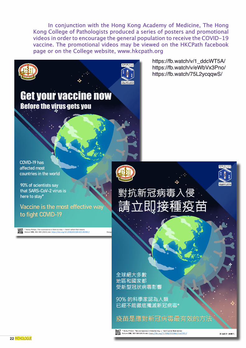
* Nicky Philips. The coronavirus is here to stay — here’s what that means Nature 590, 382-384 (2021) doi: https://doi.org/10.1038/d41586-021-00396-2 Design and Illust: Tong Lok Yee Sylvia
Get your vaccine nowBefore the virus gets you
90% of scientists say that SARS-CoV-2 virus is here to stay*
COVID-19 has affected most countries in the world
Vaccine is the most effective way to fight COVID-19
In conjunction with the Hong Kong Academy of Medicine, The Hong In conjunction with the Hong Kong Academy of Medicine, The Hong Kong College of Pathologists produced a series of posters and promotional Kong College of Pathologists produced a series of posters and promotional videos in order to encourage the general population to receive the COVID-19 videos in order to encourage the general population to receive the COVID-19 vaccine. The promotional videos may be viewed on the HKCPath facebook vaccine. The promotional videos may be viewed on the HKCPath facebook page or on the College website, www.hkcpath.org page or on the College website, www.hkcpath.org
https://fb.watch/v/1_ddcWT5A/ https://fb.watch/v/1_ddcWT5A/ https://fb.watch/v/eWbVx3Pno/ https://fb.watch/v/eWbVx3Pno/ https://fb.watch/75L2ycqqwS/ https://fb.watch/75L2ycqqwS/
22 PATHOLOGUEPATHOLOGUE
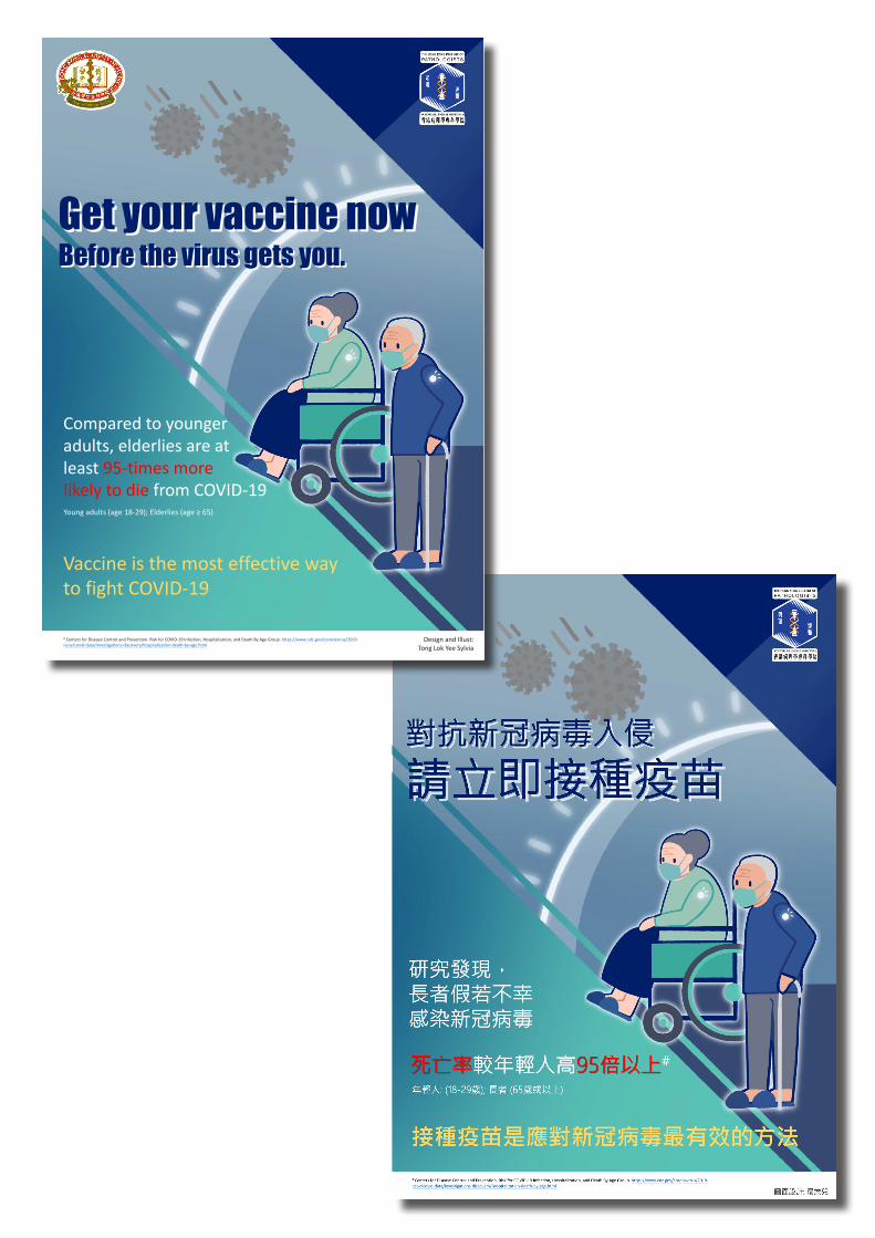
Design and Illust: Tong Lok Yee Sylvia
Get your vaccine nowBefore the virus gets you.
Vaccine is the most effective wayto fight COVID-19
Compared to younger adults, elderlies are at least 95-times more
Young adults (age 18-29); Elderlies (age ≥ 65)
# Centers for Disease Control and Prevention. Risk for COVID-19 Infection, Hospitalization, and Death By Age Group. https://www.cdc.gov/coronavirus/2019-ncov/covid-data/investigations-discovery/hospitalization-death-by-age.html
likely to die from COVID-19
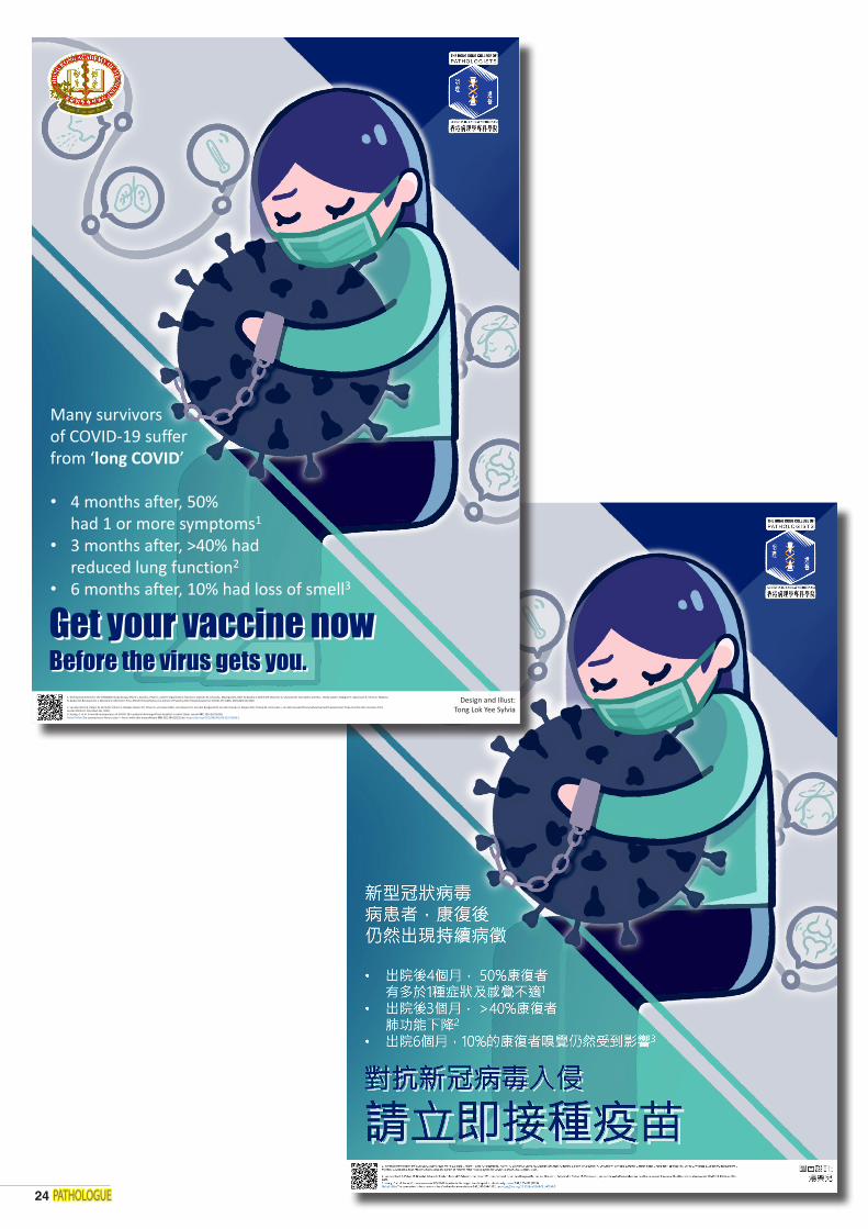
1. Writing Committee for the COMEBAC Study Group, Morin L, Savale L, Pham T, Colle R, Figueiredo S, Harrois A, Gasnier M, Lecoq AL, Meyrignac O, Noel N, Baudry E, Bellin MF, Beurnier A, Choucha W, Corruble E, Dortet L, Hardy-Leger I, Radiguer F, Sportouch S, Verny C, WyploszB, Zaidan M, Becquemont L, Montani D, Monnet X. Four-Month Clinical Status of a Cohort of Patients After Hospitalization for COVID-19. JAMA. 2021;325(15):1525.
2. van den Borst B, Peters JB, Brink M, Schoon Y, Bleeker-Rovers CP, Schers H, van Hees HWH, van Helvoort H, van den Boogaard M, van der Hoeven H, Reijers MH, Prokop M, Vercoulen J, van den Heuvel MComprehensive health assessment three months after recovery from acute COVID-19 Clin Infect Dis. 2020;3. Huang, C. et al. 6-month consequences of COVID-19 in patients discharged from hospital: a cohort study. Lancet 397, 220–232 (2021).Nicky Phillips The coronavirus is here to stay — here’s what that meansNature 590, 382-384 (2021) doi: https://doi.org/10.1038/d41586-021-00396-2
Get your vaccine nowBefore the virus gets you.
Many survivors of COVID-19 suffer from ‘long COVID’
• 4 months after, 50% had 1 or more symptoms1
• 3 months after, >40% had reduced lung function2
• 6 months after, 10% had loss of smell3
Design and Illust: Tong Lok Yee Sylvia
24 PATHOLOGUEPATHOLOGUE