Seasoned adaptive antibody immunity for highly pathogenic pandemic influenza in humans
Upload
khangminh22Category
view
1download
0
Article
Passive Immunity and Antibody Response Induced byToxoplasma gondii VLP Immunization
Hae-Ji Kang 1,†, Min-Ju Kim 1,†, Ki-Back Chu 1, Su-Hwa Lee 2, Eun-Kyung Moon 2 and Fu-Shi Quan 2,3,*
�����������������
Citation: Kang, H.-J.; Kim, M.-J.;
Chu, K.-B.; Lee, S.-H.; Moon, E.-K.;
Quan, F.-S. Passive Immunity and
Antibody Response Induced by
Toxoplasma gondii VLP Immunization.
Vaccines 2021, 9, 425. https://
doi.org/10.3390/vaccines9050425
Academic Editor: Tatsuya Yamazaki
Received: 26 February 2021
Accepted: 20 April 2021
Published: 23 April 2021
Publisher’s Note: MDPI stays neutral
with regard to jurisdictional claims in
published maps and institutional affil-
iations.
Copyright: © 2021 by the authors.
Licensee MDPI, Basel, Switzerland.
This article is an open access article
distributed under the terms and
conditions of the Creative Commons
Attribution (CC BY) license (https://
creativecommons.org/licenses/by/
4.0/).
1 Department of Biomedical Science, Graduate School, Kyung Hee University, Seoul 02447, Korea;[email protected] (H.-J.K.); [email protected] (M.-J.K.); [email protected] (K.-B.C.)
2 Department of Medical Zoology, Kyung Hee University School of Medicine, Seoul 02447, Korea;[email protected] (S.-H.L.); [email protected] (E.-K.M.)
3 Department of Medical Research Center for Bioreaction to Reactive Oxygen Species and Biomedical ScienceInstitute, School of Medicine, Graduate School, Kyung Hee University, Seoul 02447, Korea
* Correspondence: [email protected]; Tel.: +82-2-961-2302† These authors contributed equally to this work.
Abstract: Passive immunity can provide immediate protection against infectious pathogens. To date,only a few studies have investigated the effect of passive immunization against Toxoplasma gondii,and the use of immune sera acquired from VLP-vaccinated mice for passive immunity assessmentremains unreported. In this study, immune sera were produced by a single immunization with virus-like particles (VLPs) expressing the inner membrane complex (IMC), rhoptry protein 18 (ROP18),and microneme protein 8 (MIC8) of Toxoplasma gondii, with or without a CpG-ODN adjuvant. Thepassive immunization of immune sera conferred protection in mice, as indicated by their potentparasite-specific antibody response, lessened brain cyst counts, lower bodyweight loss, and enhancedsurvival. In order to confirm that the immune sera of the VLP-immunized mice were truly protective,the antibody responses and other immunological parameters were measured in the VLP-immunizedmice. We found that VLP immunization induced higher levels of parasite-specific IgG, IgG subclass,and IgM antibody responses in the sera and intestines than in the controls. Enhanced Th1 and Th2-associated cytokines in the spleen, diminished brain cyst counts, and lessened body weight loss werefound following T. gondii ME49 challenge infection. These results suggest that passive immunizationwith the immune sera acquired from VLP-vaccinated mice can confer adequate protection againstT. gondii infection.
Keywords: Toxoplasma gondii; virus-like particle; CpG-ODN; antibody response; cytokine
1. Introduction
Toxoplasma gondii is a notorious intracellular protozoan parasite which infects a widerange of warm-blooded vertebrates such as cats, sheep, mice, and humans. As the causativeagent of toxoplasmosis, T. gondii infections in healthy individuals are often asymptomatic,whereas pregnant women and acquired immune deficiency syndrome (AIDS) patients canexperience severe life-threatening symptoms such as miscarriage, retinitis, and sight lossupon infection [1,2]. The major routes of human T. gondii infection are the ingestion ofuncooked meat containing T. gondii cysts or oocyst-contaminated water [3]. It is estimatedthat one-third of the world’s population has been infected with T. gondii [4,5]. As ofthis moment, vaccination remains the most effective method for controlling infectiousdiseases [6]. Toxovax, albeit limited to veterinary usage, is the only commercially availableT. gondii vaccine. For this reason, it is imperative that an additional vaccine is developedfor clinical purposes [7].
To date, multiple studies have demonstrated the efficacy of a single immunizationfor various diseases. Immunizing mice with attenuated Brucella melitensis encapsulated inalginate microspheres conferred protection against challenge infection with the virulent
Vaccines 2021, 9, 425. https://doi.org/10.3390/vaccines9050425 https://www.mdpi.com/journal/vaccines

Vaccines 2021, 9, 425 2 of 14
wild type [8]. A chimeric vaccine engineered using the structural components of theyellow fever virus and the Zika virus fully protected mice against the yellow fever viruschallenge following a single immunization [9]. Adjuvants are commonly incorporated intothe vaccination formulae in order to bolster the protective efficacy. Evidently, the protectiveefficacy of a single dose of adenoviral-vectored malaria vaccine was improved throughadjuvant use [10]. Synthetic oligodeoxynucleotides containing unmethylated CpG motifs(CpG ODN) are an effective adjuvant of interest with numerous studies documentingtheir capabilities for enhancing vaccine efficacies through the activation of both innateand acquired immunities in humans and other animals [11–15]. In this regard, a singleimmunization with the CpG-adjuvanted human papillomavirus vaccine demonstrated aprophylactic effect in mice by reducing tumor volume and emergence [16].
Studies reporting the protective efficacies of a single immunization with the T. gondiivaccine are extremely limited. A profound reduction of the T. gondii Fukaya strain wasobserved in mice immunized with a DNA vaccine expressing T. gondii heat shock protein70 [17]. A single immunization with microparticles encompassing the chimeric surfaceantigen protein (rSAG1/2) protected mice against a lethal challenge with the virulentT. gondii RH strain [18]. To date, VLP vaccine efficacies against T. gondii infections weremostly evaluated following a prime-boost immunization regimen [19–21]. Recently, in ourprevious study, the protective efficacies of CpG-ODN adjuvanted T. gondii VLPs from micereceiving a differing number of immunizations were assessed [22,23]. Although limitedinformation has been presented for a single immunization, the results of our previousstudy revealed that VLP-immunization induces potent levels of parasite-specific antibodyresponses, which signified its potential application in passive immunization studies.
Passive immunization refers to the process of providing antibodies to protect againstinfection. Their role in protection is well-established, especially against respiratory virusesand enteric pathogens [24,25]. To date, only a few studies have investigated the effect ofpassive immunity against toxoplasmosis, with conflicting findings. While several groupsreported the protective role of passive immunization against toxoplasmosis [26,27], othershave demonstrated that passive antibody immunization merely suppresses the humoralimmune response, and does not prolong the survival of challenge-infected animals [28–30].The underlying cause of this discrepancy can be attributed to the method of active immu-nization and the route selected for passive immunization. The active immunization togenerate antibodies for the aforementioned passive immunity studies was performed usingeither killed parasites or experimental infection with T. gondii, and the passive immuniza-tions were predominantly performed through the intraperitoneal route [29,30]. Antigenimmunogenicity can be affected during the inactivation process, which results in weakimmune response induction. As such, the antibody titers induced by inactivated vaccinesare weak and tend to fade over time, thus requiring multiple booster doses [31]. The majordrawback of the intraperitoneal antibody administration method is the fact that inoculatedsubstances must undergo hepatic metabolism prior to entering the circulatory system,which incurs inoculum loss and possibly leads to erroneous results [32]. Intravenousinjection is considered to be the most efficient method of substance delivery because theneed for solute absorption is virtually non-existent [32]. Therefore, addressing these issuesthrough the intravenous administration of highly immunogenic VLP-induced antibodiesfor passive immunization could elucidate their protective role in toxoplasmosis. In ourcurrent study, we evaluated the adjuvanted VLP vaccine-induced antibody responses (IgG,IgG1, IgG2a, IgG2b, IgM) and attempted to accurately assess the protection conferredby passively immunizing naïve mice with the sera of VLP-immunized mice through theintravenous route.
2. Materials and Methods2.1. Ethics Statement
All of the animal experiments in this study were carried out under the guidelines setout by Kyung Hee University IACUC. The experimental protocols were approved by the

Vaccines 2021, 9, 425 3 of 14
Animal Ethics Committee of the Kyung Hee University (permit number: KHUASP(SE)-18-050). The immunization and blood collection were performed under mild anesthesia,which was induced and maintained with ketamine hydrochloride and xylazine. All effortswere made to minimize the number of animals used in the experiment as well as theirsuffering.
2.2. Animals, Parasites, Cells and Antibodies
The Toxoplasma gondii RH and ME49 strains were maintained in seven-week-old femaleBALB/c mice purchased from NARA Biotech (Seoul, Korea), as previously described [22].The Spodoptera frugiperda Sf9 insect cells (ATCC, CRL-1711) were cultured in serum-freeSF900-II medium (Invitrogen, Carlsbad, CA, USA), and were subsequently used to gen-erate recombinant baculovirus (rBV) and virus-like particles (VLPs). Polyclonal T. gondiiantibodies isolated from the blood of T. gondii ME49-infected mice were used to determinethe T. gondii-specific antibody responses. Horseradish peroxidase (HRP)-conjugated goatanti-mouse IgG, IgG1, IgG2a, IgG2b, and IgM were purchased from Southern Biotech(Birmingham, AL, USA).
2.3. Passive Immunization with the Sera of VLP-Immunized Mice
In order to assess the protection conferred by passive immunization with the seraof VLP-immunized mice, seven-week-old female BALB/c mice (n = 6 per group) wereorally infected with a lethal dose of T. gondii ME49 (450 cysts). After 16 h, 100 µL of thepooled sera from TG 146 VLPs or TG 146 VLPs + CpG-immunized mice were injectedinto the tail veins of ME49-infected mice. The control group mice received 100 µL of PBS(unimmunized).
2.4. Parasite-Specific IgG Antibody Responses in Mice Passively Immunized with VLPs
T. gondii RH antigens were prepared by sonicating tachyzoites in PBS, as describedpreviously [22]. The T. gondii RH antigen-specific IgG antibody responses were determinedby ELISA, as described [21]. Briefly, T. gondii RH antigens were coated at a concentrationof 4 µg/mL in 96-well plates. The sera of passively-immunized mice were collected30 days post-immunization, and 100 µL of the pooled sera (1:100 in PBS) were added tothe respective wells, and then the plates were subsequently incubated at 37 ◦C for 1 h.Afterward, 100 µL of HRP-conjugated goat anti-mouse IgG antibodies (1:2000 in PBST)were added and incubated at 37 ◦C for 1 h. The optical density (OD) at 450 nm was readusing an ELISA reader (EZ Read 400, Biochrom, Cambridge, UK).
2.5. Protection in Mice Passively Immunized with Sera from VLPs Immunization
In order to determine the protection offered by passive immunity, naïve mice wereinfected with a lethal dose of T. gondii ME49, and received the immune sera from VLPimmunization as described above. The mice were monitored daily for 30 days in order toevaluate their changes in bodyweight and survival. At 30 days post-infection (dpi), themouse brain tissues were isolated for cyst counting, as previously described [22]. Mice thatlost 20% of initial bodyweight were humanely euthanized, as described [21,23].
2.6. Generation of Recombinant Baculovirus and Virus-Like Particles
Multi-antigenic TG146 VLPs were generated as previous described [20], and thesewere inoculated into mice in order to raise the antibodies required for passive immunization.Briefly, recombinant baculoviruses (rBVs) expressing the T. gondii inner membrane complex(TG1), T. gondii rhoptry protein 18 (TG4), T. gondii microneme protein 8 (TG6) and influenzaM1 were produced. Sf9 cells were simultaneously infected with these rBVs for TG146 VLPproduction, and were incubated at 27 ◦C for 3 days. The TG146 VLPs were purified andsubsequently quantified by BCA protein assay.

Vaccines 2021, 9, 425 4 of 14
2.7. VLPs Immunization and Challenge
In order to prepare the immune sera for passive immunization, seven-week-old femaleBALB/c mice were intranasally immunized singularly with either unadjuvanted or CpGODN-adjuvanted TG146 VLP vaccines. The immunized mice were challenge-infectedorally with a lethal dose of 450 ME49 cysts 30 days after immunization. Mice that lost20% of their initial bodyweight were considered dead and were humanely euthanized asdescribed [21,23].
2.8. Mouse Sample Collection
Sera collected at weeks 1 and 4 after immunization, and week 1 post-challenge in-fection were used to determine the T. gondii specific-antibody response. At 30 dpi, threemice from each group were sacrificed, and their brain, spleen, and duodenum were col-lected. Isolated samples were individually processed, as previously described [33–36]. Theremaining mice from each group were monitored until 30 dpi in order to measure theirbody weight changes and survival rates.
2.9. Antibody Responses in the Sera and Intestines
T. gondii RH antigen-specific IgG, IgG1, IgG2a, IgG2b, and IgM antibody responseswere determined by ELISA, as previously described [21]. As primary antibodies, 100 µLof the diluted sera (1:100 in PBS), or undiluted brain and duodenum samples were addedto respective wells, and then the plates were subsequently incubated at 37 ◦C for 1 h.Afterward, 100 µL of HRP-conjugated goat anti-mouse secondary antibodies (IgG, IgG1,IgG2a, IgG2b, and IgM diluted 1:2000 in PBST) were added and incubated at 37 ◦C for1 h. The optical density (OD) at 450 nm was read using an ELISA reader (EZ Read 400,Biochrom, Cambridge, UK).
2.10. Analysis of Cytokines in the Brain and Spleen
Supernatants isolated from the brain tissue homogenates and spleen tissue wereused to determine the concentrations of cytokine tumor necrosis factor-alpha (TNF-α),interferon-gamma (IFN-γ), and IL-4 and interleukin-10 (IL-10). BD OptEIA TNF-α, IFN-γand IL-10 ELISA kits (BD Biosciences, San Jose, CA, USA) were used, and the cytokineconcentrations were determined following the manufacturer’s instructions.
2.11. Collection of T. gondii ME49 Cysts in the Brain
The cysts were purified prior to counting using the method previously described [20].After removing the supernatant from the brain tissue homogenates, pelleted tissues wereused to count the T. gondii ME49 cysts. The sedimented tissues were resuspended in 40%Percoll solution and centrifuged at 12,100 rpm for 20 min. The layer containing the T. gondiiME49 cysts was carefully collected and centrifuged at 6000 rpm for 20 min with repeatedwashing in PBS. The pellets were resuspended in PBS, and 5 µL of the collected cysts wereplaced on a slide glass for counting under the microscope (Leica DMi8, Leica, Wetzlar,Germany). A total of 5 different fields were viewed for each sample, and the cyst countswere normalized to the total volume.
2.12. Statistical Analysis
The statistical analysis was performed using GraphPad Prism version 5 (San Diego,CA, USA). The data were analyzed using one-way ANOVA with Tukey’s post hoc test,or 2-way ANOVA with Bonferroni’s test. The statistical significance was denoted usingasterisks (* p < 0.05, ** p < 0.01, *** p < 0.001).

Vaccines 2021, 9, 425 5 of 14
3. Results3.1. Experimental Schedule for Passive Immunization with Immune Sera from SingleVLPs Immunization
As seen in Figure 1A, naïve mice were infected with T. gondii (ME49), and immunesera were intravenously administered after 16 h. In order to determine protection, thebodyweight and survival rate of the mice were monitored, and cyst counts were deter-mined at 30 dpi. As seen in Figure 1B, the immune sera for passive immunization wereobtained from a single VLP immunization. The parasite-specific antibody responses in seraand intestines, Th1- or Th2-like cytokine responses, and protection were determined asscheduled.
Vaccines 2021, 9, x FOR PEER REVIEW 5 of 14
2-way ANOVA with Bonferroni’s test. The statistical significance was denoted using as-terisks (*p < 0.05, **p < 0.01, ***p < 0.001).
3. Results 3.1. Experimental Schedule for Passive Immunization with Immune Sera from Single VLPs Immunization
As seen in Figure 1A, naïve mice were infected with T. gondii (ME49), and immune sera were intravenously administered after 16 h. In order to determine protection, the bodyweight and survival rate of the mice were monitored, and cyst counts were deter-mined at 30 dpi. As seen in Figure 1B, the immune sera for passive immunization were obtained from a single VLP immunization. The parasite-specific antibody responses in sera and intestines, Th1- or Th2-like cytokine responses, and protection were determined as scheduled.
Figure 1. Experimental schedule for passive immunization. (A) Naïve mice were initially infected with T. gondii (ME49), and the immune sera of VLP-immunized mice were subsequently adminis-tered. Protection was determined at 30 dpi. (B) Immune sera were obtained after a single VLP im-munization from mice, and their antibody responses, Th1-, Th2-like cytokine responses, and pro-tection were determined.
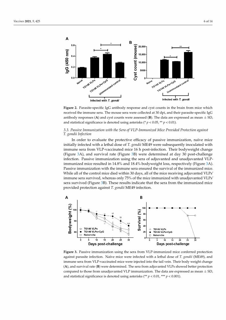
3.2. Passive Immunization with Sera from Mice Immunized with VLPs Showed Higher Levels of Parasite-specific IgG Antibody Response and a Significant Reduction of Cyst Counts in the Brain
The parasite-specific IgG antibody responses and cyst counts in the brain were deter-mined at 30 dpi. As indicated in Figure 2A, the parasite-specific IgG antibody responses were found to be higher in mice passively immunized with the immune sera compared to the unimmunized controls. Of the two immune sera used for the passive immunization, the mice receiving the immune sera of CpG-ODN adjuvanted VLPs elicited greater IgG antibody responses. The brain cyst counts were inversely proportional to the parasite-specific IgG responses. As shown in Figure 2B, the lowest cyst counts were observed from mice passively immunized with the sera of adjuvanted VLPs. These results indicate that the sera from immunized mice are capable of reducing the parasite loads in the brain.
Figure 1. Experimental schedule for passive immunization. (A) Naïve mice were initially infectedwith T. gondii (ME49), and the immune sera of VLP-immunized mice were subsequently administered.Protection was determined at 30 dpi. (B) Immune sera were obtained after a single VLP immunizationfrom mice, and their antibody responses, Th1-, Th2-like cytokine responses, and protection weredetermined.
3.2. Passive Immunization with Sera from Mice Immunized with VLPs Showed Higher Levels ofParasite-Specific IgG Antibody Response and a Significant Reduction of Cyst Counts in the Brain
The parasite-specific IgG antibody responses and cyst counts in the brain were deter-mined at 30 dpi. As indicated in Figure 2A, the parasite-specific IgG antibody responseswere found to be higher in mice passively immunized with the immune sera compared tothe unimmunized controls. Of the two immune sera used for the passive immunization,the mice receiving the immune sera of CpG-ODN adjuvanted VLPs elicited greater IgG an-tibody responses. The brain cyst counts were inversely proportional to the parasite-specificIgG responses. As shown in Figure 2B, the lowest cyst counts were observed from micepassively immunized with the sera of adjuvanted VLPs. These results indicate that the serafrom immunized mice are capable of reducing the parasite loads in the brain.
Vaccines 2021, 9, 425 6 of 14
1
Figure 2. Parasite-specific IgG antibody response and cyst counts in the brain from mice whichreceived the immune sera. The mouse sera were collected at 30 dpi, and their parasite-specific IgGantibody responses (A) and cyst counts were assessed (B). The data are expressed as mean ± SD,and statistical significance is denoted using asterisks (* p < 0.05, ** p < 0.01).
3.3. Passive Immunization with the Sera of VLP-Immunized Mice Provided Protection againstT. gondii Infection
In order to evaluate the protective efficacy of passive immunization, naïve miceinitially infected with a lethal dose of T. gondii ME49 were subsequently inoculated withimmune sera from VLP-vaccinated mice 16 h post-infection. Their bodyweight change(Figure 3A), and survival rate (Figure 3B) were determined at day 30 post-challengeinfection. Passive immunization using the sera of adjuvanted and unadjuvanted VLP-immunized mice resulted in 14.8% and 18.4% bodyweight loss, respectively (Figure 3A).Passive immunization with the immune sera ensured the survival of the immunized mice.While all of the control mice died within 30 days, all of the mice receiving adjuvanted VLPs’immune sera survived, whereas only 75% of the mice immunized with unadjuvanted VLPs’sera survived (Figure 3B). These results indicate that the sera from the immunized miceprovided protection against T. gondii ME49 infection.
Vaccines 2021, 9, x FOR PEER REVIEW 6 of 14
Figure 2. Parasite-specific IgG antibody response and cyst counts in the brain from mice which received the immune sera. The mouse sera were collected at 30 dpi, and their parasite-specific IgG antibody responses (A) and cyst counts were assessed (B). The data are expressed as mean ± SD, and statistical significance is denoted using asterisks (* p < 0.05, ** p < 0.01).
3.3. Passive Immunization with the Sera of VLP-immunized Mice Provided Protection Against T. gondii Infection
In order to evaluate the protective efficacy of passive immunization, naïve mice ini-tially infected with a lethal dose of T. gondii ME49 were subsequently inoculated with immune sera from VLP-vaccinated mice 16 h post-infection. Their bodyweight change (Figure 3A), and survival rate (Figure 3B) were determined at day 30 post-challenge in-fection. Passive immunization using the sera of adjuvanted and unadjuvanted VLP-im-munized mice resulted in 14.8% and 18.4% bodyweight loss, respectively (Figure 3A). Pas-sive immunization with the immune sera ensured the survival of the immunized mice. While all of the control mice died within 30 days, all of the mice receiving adjuvanted VLPs’ immune sera survived, whereas only 75% of the mice immunized with unadju-vanted VLPs’ sera survived (Figure 3B). These results indicate that the sera from the im-munized mice provided protection against T. gondii ME49 infection.
Figure 3. Passive immunization using the sera from VLP-immunized mice conferred protection against parasite infection. Naïve mice were infected with a lethal dose of T. gondii (ME49), and immune sera from VLP-vaccinated mice were injected into the tail vein. Their body weight change (A), and survival rate (B) were determined. The sera from adjuvanted VLPs showed better protec-tion compared to those from unadjuvanted VLP immunization. The data are expressed as mean ± SD, and statistical significance is denoted using asterisks (** p < 0.01, *** p < 0.001).
Figure 3. Passive immunization using the sera from VLP-immunized mice conferred protectionagainst parasite infection. Naïve mice were infected with a lethal dose of T. gondii (ME49), andimmune sera from VLP-vaccinated mice were injected into the tail vein. Their body weight change(A), and survival rate (B) were determined. The sera from adjuvanted VLPs showed better protectioncompared to those from unadjuvanted VLP immunization. The data are expressed as mean ± SD,and statistical significance is denoted using asterisks (** p < 0.01, *** p < 0.001).
Vaccines 2021, 9, 425 7 of 14
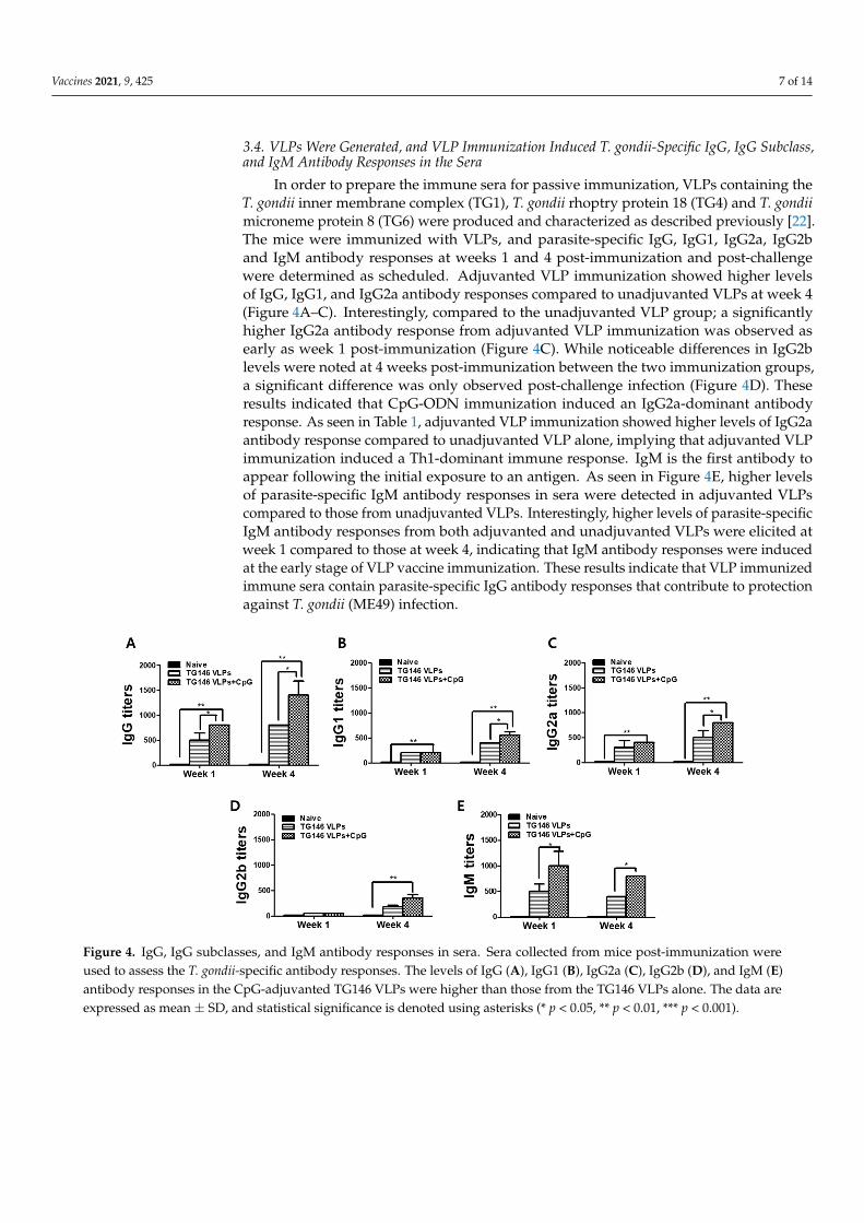
3.4. VLPs Were Generated, and VLP Immunization Induced T. gondii-Specific IgG, IgG Subclass,and IgM Antibody Responses in the Sera
In order to prepare the immune sera for passive immunization, VLPs containing theT. gondii inner membrane complex (TG1), T. gondii rhoptry protein 18 (TG4) and T. gondiimicroneme protein 8 (TG6) were produced and characterized as described previously [22].The mice were immunized with VLPs, and parasite-specific IgG, IgG1, IgG2a, IgG2band IgM antibody responses at weeks 1 and 4 post-immunization and post-challengewere determined as scheduled. Adjuvanted VLP immunization showed higher levelsof IgG, IgG1, and IgG2a antibody responses compared to unadjuvanted VLPs at week 4(Figure 4A–C). Interestingly, compared to the unadjuvanted VLP group; a significantlyhigher IgG2a antibody response from adjuvanted VLP immunization was observed asearly as week 1 post-immunization (Figure 4C). While noticeable differences in IgG2blevels were noted at 4 weeks post-immunization between the two immunization groups,a significant difference was only observed post-challenge infection (Figure 4D). Theseresults indicated that CpG-ODN immunization induced an IgG2a-dominant antibodyresponse. As seen in Table 1, adjuvanted VLP immunization showed higher levels of IgG2aantibody response compared to unadjuvanted VLP alone, implying that adjuvanted VLPimmunization induced a Th1-dominant immune response. IgM is the first antibody toappear following the initial exposure to an antigen. As seen in Figure 4E, higher levelsof parasite-specific IgM antibody responses in sera were detected in adjuvanted VLPscompared to those from unadjuvanted VLPs. Interestingly, higher levels of parasite-specificIgM antibody responses from both adjuvanted and unadjuvanted VLPs were elicited atweek 1 compared to those at week 4, indicating that IgM antibody responses were inducedat the early stage of VLP vaccine immunization. These results indicate that VLP immunizedimmune sera contain parasite-specific IgG antibody responses that contribute to protectionagainst T. gondii (ME49) infection.
Vaccines 2021, 9, x FOR PEER REVIEW 7 of 14
3.4. VLPs were Generated, and VLP Immunization Induced T. gondii-specific IgG, IgG Subclass, and IgM Antibody Responses in the Sera
In order to prepare the immune sera for passive immunization, VLPs containing the T. gondii inner membrane complex (TG1), T. gondii rhoptry protein 18 (TG4) and T. gondii microneme protein 8 (TG6) were produced and characterized as described previously [22]. The mice were immunized with VLPs, and parasite-specific IgG, IgG1, IgG2a, IgG2b and IgM antibody responses at weeks 1 and 4 post-immunization and post-challenge were determined as scheduled. Adjuvanted VLP immunization showed higher levels of IgG, IgG1, and IgG2a antibody responses compared to unadjuvanted VLPs at week 4 (Figure 4A–C). Interestingly, compared to the unadjuvanted VLP group; a significantly higher IgG2a antibody response from adjuvanted VLP immunization was observed as early as week 1 post-immunization (Figure 4C). While noticeable differences in IgG2b levels were noted at 4 weeks post-immunization between the two immunization groups, a significant difference was only observed post-challenge infection (Figure 4D). These results indicated that CpG-ODN immunization induced an IgG2a-dominant antibody response. As seen in Table 1, adjuvanted VLP immunization showed higher levels of IgG2a antibody response compared to unadjuvanted VLP alone, implying that adjuvanted VLP immunization in-duced a Th1-dominant immune response. IgM is the first antibody to appear following the initial exposure to an antigen. As seen in Figure 4E, higher levels of parasite-specific IgM antibody responses in sera were detected in adjuvanted VLPs compared to those from unadjuvanted VLPs. Interestingly, higher levels of parasite-specific IgM antibody responses from both adjuvanted and unadjuvanted VLPs were elicited at week 1 com-pared to those at week 4, indicating that IgM antibody responses were induced at the early stage of VLP vaccine immunization. These results indicate that VLP immunized immune sera contain parasite-specific IgG antibody responses that contribute to protection against T. gondii (ME49) infection.
Figure 4. IgG, IgG subclasses, and IgM antibody responses in sera. Sera collected from mice post-immunization were used to assess the T. gondii-specific antibody responses. The levels of IgG (A), IgG1 (B), IgG2a (C), IgG2b (D), and IgM (E) antibody responses in the CpG-adjuvanted TG146 VLPs were higher than those from the TG146 VLPs alone. The data are expressed as mean ± SD, and statistical significance is denoted using asterisks (* p < 0.05, ** p < 0.01, *** p < 0.001).
Figure 4. IgG, IgG subclasses, and IgM antibody responses in sera. Sera collected from mice post-immunization wereused to assess the T. gondii-specific antibody responses. The levels of IgG (A), IgG1 (B), IgG2a (C), IgG2b (D), and IgM (E)antibody responses in the CpG-adjuvanted TG146 VLPs were higher than those from the TG146 VLPs alone. The data areexpressed as mean ± SD, and statistical significance is denoted using asterisks (* p < 0.05, ** p < 0.01, *** p < 0.001).
Vaccines 2021, 9, 425 8 of 14
Table 1. Th1:Th2 index for VLPs alone, and for adjuvanted VLPs.
Groups(n = 6)
Serum Intestine
TG146 TG146 + CpG TG146 TG146 + CpG
IgG1 (Th2) 0.13 ± 0.002 0.16 ± 0.004 0.39 ± 0.008 0.45 ± 0.005
IgG2a (Th1) 0.18 ± 0.001 0.29 ± 0.003 0.55 ± 0.007 0.66 ± 0.006
IgG2b (Th1) 0.11 ± 0.006 0.14 ± 0.004 0.33 ± 0.002 0.45 ± 0.003
Th1:Th2 Index 1.11 ± 0.004 1.32 * ± 0.003 1.12 ± 0.003 1.23 * ± 0.004
The sera and intestines were collected at day 30 post-challenge infection, and the IgG1 and IgG2a antibodyresponses in both the sera and intestines were determined. The values are optical density readings at 100-foldserum dilution. Index <1 = Th2 polarization. Index >1 = Th1 polarization. Th1 (IgG2a and IgG2b) or Th2(IgG1) polarization were determined by Th1:Th2 index calculation ([IgG2a + IgG2b]/2)/(IgG1), as previouslydescribed [11] (* p < 0.05).
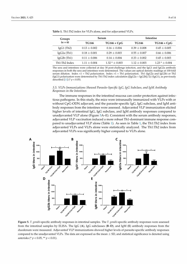
3.5. VLPs Immunizations Showed Parasite-Specific IgG, IgG Subclass, and IgM AntibodyResponses in the Intestines
The immune responses in the intestinal mucosa can confer protection against infec-tious pathogens. In this study, the mice were intranasally immunized with VLPs with orwithout CpG-ODN adjuvant, and the parasite-specific IgG, IgG subclass, and IgM anti-body responses from the intestines were assessed. Adjuvanted VLP immunization elicitedhigher levels of intestinal IgG, IgG subclass, and IgM antibody responses compared tounadjuvanted VLP alone (Figure 5A–E). Consistent with the serum antibody responses,adjuvanted VLP vaccination induced a more robust Th1-dominant immune response com-pared to unadjuvanted VLP alone (Table 1). As seen in Table 1, the Th1:Th2 index fromadjuvanted VLPs and VLPs alone were statistically analyzed. The Th1:Th2 index fromadjuvanted VLPs was significantly higher compared to VLPs alone.
Vaccines 2021, 9, x FOR PEER REVIEW 8 of 14
Table 1. Th1:Th2 index for VLPs alone, and for adjuvanted VLPs.
Groups (n = 6)
Serum Intestine TG146 TG146 + CpG TG146 TG146 + CpG
IgG1 (Th2) 0.13 0.002 0.16 0.004 0.39 0.008 0.45 0.005
IgG2a (Th1) 0.18 0.001 0.29 0.003 0.55 0.007 0.66 0.006
IgG2b (Th1) 0.11 0.006 0.14 0.004 0.33 0.002 0.45 0.003
Th1:Th2 Index 1.11 0.004 1.32* 0.003 1.12 0.003 1.23* 0.004
The sera and intestines were collected at day 30 post-challenge infection, and the IgG1 and IgG2a antibody responses in both the sera and intestines were determined. The values are optical density readings at 100-fold serum dilution. Index <1 = Th2 polarization. Index >1 = Th1 polarization. Th1 (IgG2a and IgG2b) or Th2 (IgG1) polarization were determined by Th1:Th2 index calculation ([IgG2a + IgG2b]/2)/(IgG1), as previously described [11] (*p < 0.05).
3.5. VLPs Immunizations Showed Parasite-specific IgG, IgG Subclass, and IgM Antibody Responses in the Intestines
The immune responses in the intestinal mucosa can confer protection against infec-tious pathogens. In this study, the mice were intranasally immunized with VLPs with or without CpG-ODN adjuvant, and the parasite-specific IgG, IgG subclass, and IgM anti-body responses from the intestines were assessed. Adjuvanted VLP immunization elicited higher levels of intestinal IgG, IgG subclass, and IgM antibody responses compared to unadjuvanted VLP alone (Figure 5A–E). Consistent with the serum antibody responses, adjuvanted VLP vaccination induced a more robust Th1-dominant immune response compared to unadjuvanted VLP alone (Table 1). As seen in Table 1, the Th1:Th2 index from adjuvanted VLPs and VLPs alone were statistically analyzed. The Th1:Th2 index from adjuvanted VLPs was significantly higher compared to VLPs alone.
Figure 5. T. gondii-specific antibody responses in intestinal samples. The T. gondii-specific antibody responses were assessed from the intestinal samples by ELISA. The IgG (A), IgG subclasses (B–D), and IgM (E) antibody responses from the duodenum were measured. Adjuvanted VLP immuniza-tions showed higher levels of parasite-specific antibody responses compared to the unadjuvanted VLPs. The data are expressed as the mean ± SD, and statistical significance is denoted using aster-isks (* p < 0.05, ** p < 0.01).
Figure 5. T. gondii-specific antibody responses in intestinal samples. The T. gondii-specific antibody responses were assessedfrom the intestinal samples by ELISA. The IgG (A), IgG subclasses (B–D), and IgM (E) antibody responses from theduodenum were measured. Adjuvanted VLP immunizations showed higher levels of parasite-specific antibody responsescompared to the unadjuvanted VLPs. The data are expressed as the mean ± SD, and statistical significance is denoted usingasterisks (* p < 0.05, ** p < 0.01).
Vaccines 2021, 9, 425 9 of 14
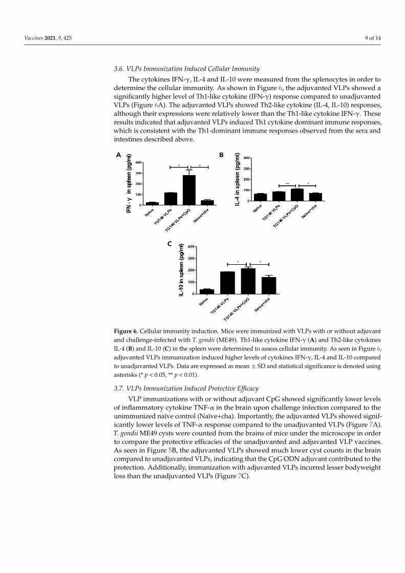
3.6. VLPs Immunization Induced Cellular Immunity
The cytokines IFN-γ, IL-4 and IL-10 were measured from the splenocytes in order todetermine the cellular immunity. As shown in Figure 6, the adjuvanted VLPs showed asignificantly higher level of Th1-like cytokine (IFN-γ) response compared to unadjuvantedVLPs (Figure 6A). The adjuvanted VLPs showed Th2-like cytokine (IL-4, IL-10) responses,although their expressions were relatively lower than the Th1-like cytokine IFN-γ. Theseresults indicated that adjuvanted VLPs induced Th1 cytokine dominant immune responses,which is consistent with the Th1-dominant immune responses observed from the sera andintestines described above.
Vaccines 2021, 9, x FOR PEER REVIEW 9 of 14
3.6. VLPs Immunization Induced Cellular Immunity The cytokines IFN-γ, IL-4 and IL-10 were measured from the splenocytes in order to
determine the cellular immunity. As shown in Figure 6, the adjuvanted VLPs showed a significantly higher level of Th1-like cytokine (IFN-γ) response compared to unadju-vanted VLPs (Figure 6A). The adjuvanted VLPs showed Th2-like cytokine (IL-4, IL-10) responses, although their expressions were relatively lower than the Th1-like cytokine IFN-γ. These results indicated that adjuvanted VLPs induced Th1 cytokine dominant im-mune responses, which is consistent with the Th1-dominant immune responses observed from the sera and intestines described above.
Figure 6. Cellular immunity induction. Mice were immunized with VLPs with or without adju-vant and challenge-infected with T. gondii (ME49). Th1-like cytokine IFN-γ (A) and Th2-like cyto-kines IL-4 (B) and IL-10 (C) in the spleen were determined to assess cellular immunity. As seen in Figure 6, adjuvanted VLPs immunization induced higher levels of cytokines IFN-γ, IL-4 and IL-10 compared to unadjuvanted VLPs. Data are expressed as mean ± SD and statistical significance is denoted using asterisks (* p < 0.05, ** p < 0.01).
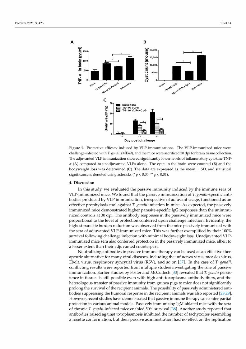
3.7. VLPs Immunization Induced Protective Efficacy VLP immunizations with or without adjuvant CpG showed significantly lower levels
of inflammatory cytokine TNF-α in the brain upon challenge infection compared to the unimmunized naïve control (Naïve+cha). Importantly, the adjuvanted VLPs showed sig-nificantly lower levels of TNF-α response compared to the unadjuvanted VLPs (Figure 7A). T. gondii ME49 cysts were counted from the brains of mice under the microscope in order to compare the protective efficacies of the unadjuvanted and adjuvanted VLP vac-cines. As seen in Figure 5B, the adjuvanted VLPs showed much lower cyst counts in the brain compared to unadjuvanted VLPs, indicating that the CpG ODN adjuvant contrib-uted to the protection. Additionally, immunization with adjuvanted VLPs incurred lesser bodyweight loss than the unadjuvanted VLPs (Figure 7C).
Figure 6. Cellular immunity induction. Mice were immunized with VLPs with or without adjuvantand challenge-infected with T. gondii (ME49). Th1-like cytokine IFN-γ (A) and Th2-like cytokinesIL-4 (B) and IL-10 (C) in the spleen were determined to assess cellular immunity. As seen in Figure 6,adjuvanted VLPs immunization induced higher levels of cytokines IFN-γ, IL-4 and IL-10 comparedto unadjuvanted VLPs. Data are expressed as mean ± SD and statistical significance is denoted usingasterisks (* p < 0.05, ** p < 0.01).
3.7. VLPs Immunization Induced Protective Efficacy
VLP immunizations with or without adjuvant CpG showed significantly lower levelsof inflammatory cytokine TNF-α in the brain upon challenge infection compared to theunimmunized naïve control (Naïve+cha). Importantly, the adjuvanted VLPs showed signif-icantly lower levels of TNF-α response compared to the unadjuvanted VLPs (Figure 7A).T. gondii ME49 cysts were counted from the brains of mice under the microscope in orderto compare the protective efficacies of the unadjuvanted and adjuvanted VLP vaccines.As seen in Figure 5B, the adjuvanted VLPs showed much lower cyst counts in the braincompared to unadjuvanted VLPs, indicating that the CpG ODN adjuvant contributed to theprotection. Additionally, immunization with adjuvanted VLPs incurred lesser bodyweightloss than the unadjuvanted VLPs (Figure 7C).
Vaccines 2021, 9, 425 10 of 14
1
Figure 7. Protective efficacy induced by VLP immunizations. The VLP-immunized mice werechallenge-infected with T. gondii (ME49), and the mice were sacrificed 30 dpi for brain tissue collection.The adjuvanted VLP immunization showed significantly lower levels of inflammatory cytokine TNF-α (A) compared to unadjuvanted VLPs alone. The cysts in the brain were counted (B) and thebodyweight loss was determined (C). The data are expressed as the mean ± SD, and statisticalsignificance is denoted using asterisks (* p < 0.05, ** p < 0.01).
4. Discussion
In this study, we evaluated the passive immunity induced by the immune sera ofVLP-immunized mice. We found that the passive immunization of T. gondii-specific anti-bodies produced by VLP immunization, irrespective of adjuvant usage, functioned as aneffective prophylaxis tool against T. gondii infection in mice. As expected, the passivelyimmunized mice demonstrated higher parasite-specific IgG responses than the unimmu-nized controls at 30 dpi. The antibody responses in the passively immunized mice wereproportional to the level of protection conferred upon challenge infection. Evidently, thehighest parasite burden reduction was observed from the mice passively immunized withthe sera of adjuvanted VLP-immunized mice. This was further exemplified by their 100%survival following challenge infection with minimal bodyweight loss. Unadjuvanted VLP-immunized mice sera also conferred protection in the passively immunized mice, albeit toa lesser extent than their adjuvanted counterpart.
Neutralizing antibodies in passive immune therapy can be used as an effective ther-apeutic alternative for many viral diseases, including the influenza virus, measles virus,Ebola virus, respiratory syncytial virus (RSV), and so on [37]. In the case of T. gondii,conflicting results were reported from multiple studies investigating the role of passiveimmunization. Earlier studies by Foster and McCulloch [30] revealed that T. gondii persis-tence in tissues is still possible even with high anti-toxoplasma antibody titers, and theheterologous transfer of passive immunity from guinea pigs to mice does not significantlyprolong the survival of the recipient animals. The possibility of passively administered anti-bodies suppressing the humoral response in the recipient animals was also reported [28,29].However, recent studies have demonstrated that passive immune therapy can confer partialprotection in various animal models. Passively immunizing IgM-ablated mice with the seraof chronic T. gondii-infected mice enabled 50% survival [38]. Another study reported thatantibodies raised against toxoplasmosis inhibited the number of tachyzoites resemblinga rosette conformation, but their passive administration had no effect on the replication
Vaccines 2021, 9, 425 11 of 14
rate [39]. Administering the sera of T. gondii RH-infected guinea pigs into recipients con-ferred partial protection [26]. The intraperitoneal administration of anti-toxoplasma serumin rabbits conferred protection against lethal challenge infection with the T. gondii Alt strain,as indicated by a 55% brain cyst reduction and a 89% survival rate [27]. Consistent withthese recent findings reported above, our study confirmed that passive immunization usinganti-T. gondii antibodies can confer protection against T. gondii infection. This is furthersupported by our previous study using VLPs expressing ROP18 and MIC8 antigens. BothROP18 and MIC8 VLP-induced antibodies were capable of neutralizing the T. gondii GT1strain to a small extent, but parasite-neutralizing activity exceeding 50% was observedwhen the immune sera of ROP18+MIC8 VLP-immunized mice reacted with the T. gondiiGT1 [40].
T. gondii can be subdivided into three major genotypes, which are classified as types I,II, and III. Type I strains are generally virulent and lethal, whereas type II strains are lessvirulent than type I. Type III strains, such as C56, were reported to be rarely associated withthe disease [41,42]. RH and GT1, both belonging to the type I clonal lineage, were reportedto share a high degree of sequence similarity [43]. Based on this rationale, immunitydeveloped against GT1 strain could also be applied to RH strain as well. The potential forheterologous protection can also be exemplified using our current and previous studies.Previously, we demonstrated that VLPs based on ROP18 and MIC8 conferred protectionagainst the GT1 strain and exhibited strong parasite neutralizing activity [40]. Sera acquiredfrom mice immunized with the TG146 VLP vaccine, which is identical to the VLP vaccineused in the current study, were capable of neutralizing as much as 60% of the T. gondiiGT1 strain [20]. Here, we showed that passively immunizing the sera raised against thisTG146 VLP conferred protection against the type II ME49 strain. Combined, our resultsimply that IMC, ROP18, and MIC8-expressing VLP immunization induces the productionof broadly neutralizing antibodies that can effectively restrict the growth of type I and IIT. gondii strains.
Next, we assessed the protective immunity in mice immunized with VLPs encodingthe IMC, ROP18, and MIC8 of T. gondii. Consistent with the protection observed from pas-sively immunized mice, significantly higher levels of parasite-specific IgG, IgG subclasses,and IgM antibodies were observed from the sera and intestinal mucosa of adjuvantedVLP-immunized mice than the unadjuvanted VLP group. CpG ODN stimulates Toll-likereceptor 9 (TLR9) in lymphocytes, and induces the production of cytokines such as IFN-γ,IL-6, IL-10, and TNF-α [44]. CpG ODN-adjuvanted vaccines are under development forthe prevention of cancer, allergies, and various infectious diseases [45]. To date, only alimited number of studies have investigated the protective efficacies of T. gondii vaccinesadjuvanted with CpG. Recently, it was reported that immunization with T. gondii lysateantigen and CpG induces T. gondii-specific antibody responses and IFN-γ production inthe spleen and MLN of mice [46]. Consistent with these reports, our study indicatedthat CpG-adjuvanted VLP immunization induced higher levels of parasite-specific IgG2aantibody responses in the sera and production of cytokines IFN-γ and IL-10 in comparisonto the unadjuvanted VLPs. On a similar note, the enhanced IgG1 responses observedfrom adjuvanted VLPs contributed to a marginal increase in the Th2 cytokine IL-4 expres-sion. Elevated IgG2a and IgG2b serum and intestinal antibody responses, paired withthe heightened IgA response reported in our previous study via adjuvanted VLPs [22],may have contributed to better protection against T. gondii than the unadjuvanted VLPs.Interestingly, the IFN-γ production in adjuvanted VLP-immunized mice far exceeded theconcentrations of IL-10, while such a phenomenon was not observed from unadjuvantedVLP-immunized mice. The striking difference in IFN-γ production between the two immu-nization groups can be attributed to the presence of CpG ODN adjuvants, thereby implyingtheir contribution and necessity for mounting a potent Th1-dominant immune response.
IgM is the first antibody secreted by the adaptive immune system in response toa foreign antigen. However, it is widely neglected when evaluating vaccine-inducedimmunity, as IgG and IgA antibodies are the main targets for immune response assessment.
Vaccines 2021, 9, 425 12 of 14
IgM molecules, characterized by their ten antigen-binding sites that contribute to theirhigh antigen avidity, can neutralize pathogens and protect mucosal surfaces [47]. In thisstudy, VLP immunizations induced parasite-specific IgM in sera and intestines, which maycontribute to the protection against T. gondii challenge infection. Furthermore, serum IgMantibodies have been shown to contribute to high levels of opsonophagocytic activities in asingle dose immunization study with pneumococcal conjugate vaccine [48]. The role ofopsonophagocytic activity contributing to the protection against T. gondii infection needsfurther study.
In conclusion, our results demonstrated that a single immunization with the TG146VLPs induced the production of neutralizing antibodies, conferring protection against T.gondii in mice. Passively immunizing mice with the sera of VLP-immunized mice revealedthat the sera were protective irrespective of the adjuvant inclusion. Formulating CpGadjuvants with TG146 VLPs enhances the parasite-specific IgG, IgG subclass, and IgMantibody responses, thereby indicating their potential as a passive immune therapeuticoption.
Author Contributions: Conceptualization, F.-S.Q.; formal analysis, H.-J.K., M.-J.K. and S.-H.L.;writing–original draft preparation, H.-J.K. and M.-J.K.; writing–review and editing, H.-J.K., M.-J.K.,K.-B.C., E.-K.M. and F.-S.Q.; supervision, F.-S.Q.; funding acquisition, F.-S.Q. All authors have readand agreed to the published version of the manuscript.
Funding: This research was funded by the National Research Foundation of Korea (NRF), grant num-ber 2018R1A6A1A03025124, and the Ministry of Health & Welfare, Republic of Korea [HV20C0085,HV20C0142].
Institutional Review Board Statement: The study was conducted according to the guidelines ofKyung Hee University IACUC, and approved by the the Animal Ethics Committee of the Kyung HeeUniversity (permit number: KHUASP(SE)-18-050).
Informed Consent Statement: Not applicable.
Data Availability Statement: Not applicable.
Conflicts of Interest: The authors declare no conflict of interest.
References1. Luft, B.J.; Remington, J.S. Toxoplasmic encephalitis in AIDS. Clin. Infect. Dis 1992, 15, 211–222. [CrossRef] [PubMed]2. Lopes, F.M.; Gonçalves, D.D.; Mitsuka-Breganó, R.; Freire, R.L.; Navarro, I.T. Toxoplasma gondii infection in pregnancy. Braz. J.
Infect. Dis. 2007, 11, 496–506. [CrossRef] [PubMed]3. Pittman, K.J.; Knoll, L.J. Long-Term Relationships: The Complicated Interplay between the Host and the Developmental Stages of
Toxoplasma gondii during Acute and Chronic Infections. Microbiol. Mol. Biol. Rev. 2015, 79, 387–401. [CrossRef]4. Saadatnia, G.; Golkar, M. A review on human toxoplasmosis. Scand. J. Infect. Dis. 2012, 44, 805–814. [CrossRef] [PubMed]5. Pappas, G.; Roussos, N.; Falagas, M.E. Toxoplasmosis snapshots: Global status of Toxoplasma gondii seroprevalence and implica-
tions for pregnancy and congenital toxoplasmosis. Int. J. Parasitol. 2009, 39, 1385–1394. [CrossRef] [PubMed]6. Kushnir, N.; Streatfield, S.J.; Yusibov, V. Virus-like particles as a highly efficient vaccine platform: Diversity of targets and
production systems and advances in clinical development. Vaccine 2012, 31, 58–83. [CrossRef] [PubMed]7. Dubey, J.P. Toxoplasmosis in sheep—The last 20 years. Vet. Parasitol. 2009, 163, 1–14. [CrossRef]8. Arenas-Gamboa, A.M.; Ficht, T.A.; Kahl-McDonagh, M.M.; Rice-Ficht, A.C. Immunization with a single dose of a microencapsu-
lated Brucella melitensis mutant enhances protection against wild-type challenge. Infect. Immun. 2008, 76, 2448–2455. [CrossRef][PubMed]
9. Kum, D.B.; Boudewijns, R.; Ma, J.; Mishra, N.; Schols, D.; Neyts, J.; Dallmeier, K. A chimeric yellow fever-Zika virus vaccinecandidate fully protects against yellow fever virus infection in mice. Emerg. Microbes Infect. 2020, 9, 520–533. [CrossRef] [PubMed]
10. Milicic, A.; Rollier, C.S.; Tang, C.K.; Longley, R.; Hill, A.V.S.; Reyes-Sandoval, A. Adjuvanting a viral vectored vaccine againstpre-erythrocytic malaria. Sci. Rep. 2017, 7, 7284. [CrossRef]
11. Quan, F.S.; Ko, E.J.; Kwon, Y.M.; Joo, K.H.; Compans, R.W.; Kang, S.M. Mucosal adjuvants for influenza virus-like particle vaccine.Viral Immunol. 2013, 26, 385–395. [CrossRef]
12. Bode, C.; Zhao, G.; Steinhagen, F.; Kinjo, T.; Klinman, D.M. CpG DNA as a vaccine adjuvant. Expert Rev. Vaccines 2011, 10,499–511. [CrossRef]
Vaccines 2021, 9, 425 13 of 14
13. Lipford, G.B.; Bauer, M.; Blank, C.; Reiter, R.; Wagner, H.; Heeg, K. CpG-containing synthetic oligonucleotides promote B andcytotoxic T cell responses to protein antigen: A new class of vaccine adjuvants. Eur. J. Immunol. 1997, 27, 2340–2344. [CrossRef][PubMed]
14. Klinman, D.M.; Currie, D.; Gursel, I.; Verthelyi, D. Use of CpG oligodeoxynucleotides as immune adjuvants. Immunol. Rev. 2004,199, 201–216. [CrossRef]
15. Kringel, H.; Dubey, J.P.; Beshah, E.; Hecker, R.; Urban, J.F., Jr. CpG-oligodeoxynucleotides enhance porcine immunity to Toxoplasmagondii. Vet. Parasitol. 2004, 123, 55–66. [CrossRef]
16. Yang, Y.; Che, Y.; Zhao, Y.; Wang, X. Prevention and treatment of cervical cancer by a single administration of human papil-lomavirus peptide vaccine with CpG oligodeoxynucleotides as an adjuvant in vivo. Int. Immunopharmacol. 2019, 69, 279–288.[CrossRef] [PubMed]
17. Mohamed, R.M.; Aosai, F.; Chen, M.; Mun, H.S.; Norose, K.; Belal, U.S.; Piao, L.X.; Yano, A. Induction of protective immunity byDNA vaccination with Toxoplasma gondii HSP70, HSP30 and SAG1 genes. Vaccine 2003, 21, 2852–2861. [CrossRef]
18. Chuang, S.C.; Chung, Y.C.; Yang, C.D. Protective immunity against toxoplasmosis in mice induced by single-dose immunizationwith rSAG1/2 protein released from poly(lactide-co-glycolide) microparticles. Parasite 2017, 24, 5. [CrossRef] [PubMed]
19. Lee, S.H.; Chu, K.B.; Kang, H.J.; Quan, F.S. Virus-like particles containing multiple antigenic proteins of Toxoplasma gondii inducememory T cell and B cell responses. PLoS ONE 2019, 14, e0220865. [CrossRef] [PubMed]
20. Lee, S.H.; Kang, H.J.; Lee, D.H.; Quan, F.S. Protective Immunity Induced by Incorporating Multiple Antigenic Proteins ofToxoplasma gondii Into Influenza Virus-Like Particles. Front. Immunol. 2018, 9, 3073. [CrossRef]
21. Lee, S.H.; Kim, A.R.; Lee, D.H.; Rubino, I.; Choi, H.J.; Quan, F.S. Protection induced by virus-like particles containing Toxoplasmagondii microneme protein 8 against highly virulent RH strain of Toxoplasma gondii infection. PLoS ONE 2017, 12, e0175644.[CrossRef] [PubMed]
22. Kang, H.J.; Chu, K.B.; Kim, M.J.; Park, H.; Jin, H.; Lee, S.H.; Moon, E.K.; Quan, F.S. Evaluation of CpG-ODN-AdjuvantedToxoplasma gondii Virus-Like Particle Vaccine upon One, Two, and Three Immunizations. Pharmaceutics 2020, 12, 989. [CrossRef][PubMed]
23. Kang, H.J.; Chu, K.B.; Kim, M.J.; Lee, S.H.; Park, H.; Jin, H.; Moon, E.K.; Quan, F.S. Protective immunity induced by CpGODN-adjuvanted virus-like particles containing Toxoplasma gondii proteins. Parasite Immunol. 2021, 43, e12799. [CrossRef]
24. Villafana, T.; Falloon, J.; Griffin, M.P.; Zhu, Q.; Esser, M.T. Passive and active immunization against respiratory syncytial virus forthe young and old. Expert Rev. Vaccines 2017, 16, 1–13. [CrossRef] [PubMed]
25. Mine, Y.; Kovacs-Nolan, J. Chicken egg yolk antibodies as therapeutics in enteric infectious disease: A review. J. Med. Food 2002,5, 159–169. [CrossRef] [PubMed]
26. Pavia, C.S. Protection against experimental toxoplasmosis by adoptive immunotherapy. J. Immunol. 1986, 137, 2985–2990.[PubMed]
27. Masihi, K.N.; Werner, H. The effect of passively transferred heterologous serum on Toxoplasma gondii in NMRI mice. Influence ofthe treatment on course of infection and cyst formation. Zentralbl. Bakteriol. Orig. A 1978, 240, 135–142.
28. Araujo, F.G.; Remington, J.S. Induction of tolerance to an intracellular protozoan (Toxoplasma gondii) by passively administeredantibody. J. Immunol. 1974, 113, 1424–1428.
29. Masihi, K.N.; Werner, H. Suppression and enhancement of humoral immune response to Toxoplasma gondii by passive antibody.Experientia 1977, 33, 1586–1587. [CrossRef]
30. Foster, B.G.; McCulloch, W.F. Studies of active and passive immunity in animals inoculated with Toxoplasma gondii. Can. J.Microbiol. 1968, 14, 103–110. [CrossRef] [PubMed]
31. Kyriakidis, N.C.; López-Cortés, A.; González, E.V.; Grimaldos, A.B.; Prado, E.O. SARS-CoV-2 vaccines strategies: A comprehen-sive review of phase 3 candidates. NPJ Vaccines 2021, 6, 28. [CrossRef] [PubMed]
32. Turner, P.V.; Brabb, T.; Pekow, C.; Vasbinder, M.A. Administration of substances to laboratory animals: Routes of administrationand factors to consider. J. Am. Assoc. Lab. Anim. Sci. 2011, 50, 600–613.
33. Quan, F.S.; Huang, C.; Compans, R.W.; Kang, S.M. Virus-like particle vaccine induces protective immunity against homologousand heterologous strains of influenza virus. J. Virol. 2007, 81, 3514–3524. [CrossRef] [PubMed]
34. Quan, F.S.; Compans, R.W.; Kang, S.M. Oral vaccination with inactivated influenza vaccine induces cross-protective immunity.Vaccine 2012, 30, 180–188. [CrossRef] [PubMed]
35. Kang, H.J.; Lee, S.H.; Kim, M.J.; Chu, K.B.; Lee, D.H.; Chopra, M.; Choi, H.J.; Park, H.; Jin, H.; Quan, F.S. Influenza Virus-LikeParticles Presenting both Toxoplasma gondii ROP4 and ROP13 Enhance Protection against T. gondii Infection. Pharmaceutics 2019,11, 342. [CrossRef] [PubMed]
36. Kim, M.C.; Kim, K.H.; Lee, J.W.; Lee, Y.N.; Choi, H.J.; Jung, Y.J.; Kim, Y.J.; Compans, R.W.; Prausnitz, M.R.; Kang, S.M. Co-Deliveryof M2e Virus-Like Particles with Influenza Split Vaccine to the Skin Using Microneedles Enhances the Efficacy of Cross Protection.Pharmaceutics 2019, 11, 188. [CrossRef] [PubMed]
37. Ye, J.; Shao, H.; Perez, D.R. Passive immune neutralization strategies for prevention and control of influenza A infections.Immunotherapy 2012, 4, 175–186. [CrossRef]
38. Frenkel, J.K.; Taylor, D.W. Toxoplasmosis in immunoglobulin M-suppressed mice. Infect. Immun. 1982, 38, 360–367. [CrossRef][PubMed]
Vaccines 2021, 9, 425 14 of 14
39. Masihi, K.N.; Werner, H. Kinetics of antibody mediated suppression of humoral immune response to Toxoplasma gondii at acellular level. Zentralbl. Bakteriol. Orig. A 1977, 237, 405–410. [PubMed]
40. Lee, S.H.; Kang, H.J.; Lee, D.H.; Kang, S.M.; Quan, F.S. Virus-like particle vaccines expressing Toxoplasma gondii rhoptry protein 18and microneme protein 8 provide enhanced protection. Vaccine 2018, 36, 5692–5700. [CrossRef] [PubMed]
41. Howe, D.K.; Sibley, L.D. Toxoplasma gondii comprises three clonal lineages: Correlation of parasite genotype with human disease.J. Infect. Dis. 1995, 172, 1561–1566. [CrossRef] [PubMed]
42. Lee, D.H.; Kim, A.R.; Lee, S.H.; Quan, F.S. Cross-protection induced by Toxoplasma gondii virus-like particle vaccine uponintraperitoneal route challenge. Acta Trop. 2016, 164, 77–83. [CrossRef] [PubMed]
43. Lau, Y.L.; Lee, W.C.; Gudimella, R.; Zhang, G.; Ching, X.T.; Razali, R.; Aziz, F.; Anwar, A.; Fong, M.Y. Deciphering the DraftGenome of Toxoplasma gondii RH Strain. PLoS ONE 2016, 11, e0157901. [CrossRef] [PubMed]
44. Santamaria, M.H.; Perez Caballero, E.; Corral, R.S. Unmethylated CpG motifs in Toxoplasma gondii DNA induce TLR9- andIFN-β-dependent expression of α-defensin-5 in intestinal epithelial cells. Parasitology 2016, 143, 60–68. [CrossRef] [PubMed]
45. Hanagata, N. CpG oligodeoxynucleotide nanomedicines for the prophylaxis or treatment of cancers, infectious diseases, andallergies. Int J. Nanomed. 2017, 12, 515–531. [CrossRef] [PubMed]
46. MA, E.L.-M.; Al-Harthi, S.A.; Mohamed, R.T.; MA, E.L.B.; Saudy, N.S. Vaccination with Toxoplasma lysate antigen and CpGoligodeoxynucleotides: Comparison of immune responses in intranasal versus intramuscular administrations. Parasitol. Res.2014, 113, 2277–2284. [CrossRef]
47. Justiz Vaillant, A.A.; Jamal, Z.; Ramphul, K. Immunoglobulin; StatPearls Publishing LLC.: Treasure Island, FL, USA, 2021.48. Simell, B.; Nurkka, A.; Ekström, N.; Givon-Lavi, N.; Käyhty, H.; Dagan, R. Serum IgM antibodies contribute to high levels of
opsonophagocytic activities in toddlers immunized with a single dose of the 9-valent pneumococcal conjugate vaccine. Clin.Vaccine Immunol. 2012, 19, 1618–1623. [CrossRef] [PubMed]
Copyright © 2022 FDOKUMEN