PDZBase: a protein-protein interaction database for PDZ-domains
Upload
independentCategory
view
0download
0
BioMed CentralBMC Bioinformatics
ss
Open AcceDatabasePARPs database: A LIMS systems for protein-protein interaction data mining or laboratory information management systemArnaud Droit1,2, Joanna M Hunter3, Michèle Rouleau1, Chantal Ethier1, Aude Picard-Cloutier1, David Bourgais1 and Guy G Poirier*1,2Address: 1Health and Environment Unit, Laval University Medical research Center, CHUQ, Québec, Canada, 2Eastern Quebec Proteomic Center, CHUQ, Quebec, Canada and 3Caprion Pharmaceuticals, Montréal, Canada
Email: Arnaud Droit - [email protected]; Joanna M Hunter - [email protected]; Michèle Rouleau - [email protected]; Chantal Ethier - [email protected]; Aude Picard-Cloutier - [email protected]; David Bourgais - [email protected]; Guy G Poirier* - [email protected]
* Corresponding author
AbstractBackground: In the "post-genome" era, mass spectrometry (MS) has become an importantmethod for the analysis of proteins and the rapid advancement of this technique, in combinationwith other proteomics methods, results in an increasing amount of proteome data. This data mustbe archived and analysed using specialized bioinformatics tools.
Description: We herein describe "PARPs database," a data analysis and management pipeline forliquid chromatography tandem mass spectrometry (LC-MS/MS) proteomics. PARPs database is aweb-based tool whose features include experiment annotation, protein database searching, proteinsequence management, as well as data-mining of the peptides and proteins identified.
Conclusion: Using this pipeline, we have successfully identified several interactions of biologicalsignificance between PARP-1 and other proteins, namely RFC-1, 2, 3, 4 and 5.
BackgroundProteomics aims to identify, characterize and quantify allof the proteins expressed by a given living organism, tissueor cell line[1]. Typically, this approach subjects proteinmixtures to proteolytic digestion prior to liquid chroma-tographic separation and MS/MS analysis of the resultingpeptides [2]. Several database search engines, notablyMascot[3], Sequest [4], and X!Tandem [5] assign probablepeptide sequences to MS/MS spectra and infer the identityof the proteins present in the sample analyzed. High-throughput proteomic experiments generate large datasets of protein identifications, which can only be properlyvalidated and reported through adequate data processing[1,6,7]. Subsequent integration, sorting and comparison
of these datasets pose significant challenges, especiallywhen simultaneously analysing multiple experiments.
One of the most effective approaches to elucidate the bio-logical function of a protein is the identification of itsinteraction partners. We are only now beginning to appre-ciate the nature and complexity of networks of interactingproteins. The unravelling of any such network using tradi-tional biochemical approaches remains a significant chal-lenge. Recently, however the application of high-throughput technologies, such as large-scale yeast two-hybrid analysis and mass spectrometry coupled toimmuno- or affinity-based capture has made possible therapid generation of huge protein interaction datasets [8-
Published: 19 December 2007
BMC Bioinformatics 2007, 8:483 doi:10.1186/1471-2105-8-483
Received: 14 August 2007Accepted: 19 December 2007
This article is available from: http://www.biomedcentral.com/1471-2105/8/483
© 2007 Droit et al; licensee BioMed Central Ltd. This is an Open Access article distributed under the terms of the Creative Commons Attribution License (http://creativecommons.org/licenses/by/2.0), which permits unrestricted use, distribution, and reproduction in any medium, provided the original work is properly cited.
Page 1 of 16(page number not for citation purposes)

BMC Bioinformatics 2007, 8:483 http://www.biomedcentral.com/1471-2105/8/483
10]. As consequence, researchers often face the dilemmaof how to effectively utilize all available data. Investiga-tors relying solely on a traditional approach to draw con-clusions or set research priorities are likely to findthemselves outpaced by peers who combine in silico biol-ogy and empirical methods. Thus for protein interactionstudies, there is clearly a need to develop a systematic andstepwise in silico approach to predict potential interactors.This approach will most likely improve our understand-ing of how complex biological systems work.
The need to develop a Laboratory Information Manage-ment System (LIMS) for researchers in the field of pro-teomics that would allow to track, archive and aid in agreater understanding of how biological systems work hasbeen recognized. In 2002, Cho and co-workers developedan original LIMS for proteome research (YPRC-PDB)[11],constructed using a commercial database (RDB). Theyintended to establish YPRC-RDB as a proteome data ware-house. In 2003, Goh and co-workers [12] developedSPINE2, a LIMS for structural proteomics, constructedwith MySQL and Perl, and also designed to work as a pipe-line to public data resources. In 2004, Garwood and co-workers [13] developed PEDRo, The Proteomic Experi-mental Data Repository, constructed with a native XMLdatabase, Xindice with an ambitious Apache SoftwareFoundation basis. The XML-based document format hasbeen chosen for communication that the other formats.The native XML database has great potential, but manyhave critical limitations for proteomic research. On theother hand, commercially available LIMSes (AmershamBiosciences and Bio-Rad Laboratories Inc, etc.) have alsobeen developed and released, but they are not exactly suit-able for laboratories like ours because the generic solu-tions are first and foremost prohibitively expensive. Thesesystems are usually re-packagings of applications devel-oped for the pharmaceutical industry for drug discoveryand development.
The focus of our laboratory is the study of the action ofpoly(ADP-ribose) polymerases (PARPs) and their role inthe cell. Poly(ADP-ribosylation) is a post-translationalprotein modification consisting of long chains ofpoly(ADP-ribose) (pADPr) synthesized by PARPs at theexpenses of NAD+. Poly(ADP-ribose) chains are short-lived owing to the activity of the poly(ADP-ribose) glyco-hydrolase enzyme, which catabolizes the pADPr withinminutes of its synthesis[14]. The PARP family may com-prise as many as 17 members which share a common cat-alytic domain responsible for the synthesis of poly(ADP-ribose) [15-17]. The best characterized and abundantmember of this family is PARP-1, a 113-kDa nuclear pro-tein comprising a DNA-binding domain made of two zincfingers that allow PARP-1 to be rapidly activated inresponse to DNA damage. Poly(ADP-ribose) crucially
contributes to chromatin remodelling, DNA damagerepair, regulation of transcription, and cell division [18-20]; and PARP-1 is an important actor in many key cellu-lar processes, including BER, transcription, and apoptosis.
We herein describe the architecture and major features ofa web-based utility called "PARPs database" (PARPs-DB),which is designed to rationally organize the protein andpeptide data generated by the LC-MS/MS analysis of tryp-tic digest of proteins that co-immunoprecipitate withPARPs proteins into reports meaningful to biologicalresearchers. PARPs-DB offers a LIMS work environment toannotate and study protein-protein interactions and itseasy-to-use relational data management system can rap-idly supply information pertaining to the biological char-acteristics of a majority of proteins in a proteomic dataset.The major features of our LIMS are flexibility, compact-ness, and connectivity to public databases.
Also presented is a list of previously unidentified PARP-1interactors that were found via affinity co-immunoprecip-itation, as well as the analysis of these new PARP-1 inter-actions generated through PARPs-DB.
Given the advantages provided by an in silico approachthat can predict or prioritize potential interactors, it seemsreasonable to propose that PARPs-DB will become anessential tool for initially evaluating novel hypotheses andwill offer improved rationale for the prioritization ofpotential interactors. In effect, by facilitating the process-ing of protein-protein interactions and the selection of themost promising interactors (to be submitted first toempirical measurements) PARPs-DB should lower thecost and shorten the time necessary to discover the mostbiologically significant interactions between PARPs andother proteins.
We don't have infrastructure to access on-line but thePARPs database source codes and user documentation areavailable for the scientific community [21]. Tools fromthe Sashimi project that were used in PARPs-DB are alsoavailable [22]. Licensed programs in Sourceforge such asMascot, Sequest or Oracle were not included in the PARPsrepository. Dialects for MySQL and PostgreSQL serverswere developed in alpha version and are available uponrequest. Further information on these scripts can beobtained from the corresponding author (see Additionalfile 1).
Construction and ContentDesign of the PARPs database softwarePARPs Database consists of a core system of services thatprovide underlying system functionality. Modules, whichprovide most data handling and analytical support (suchas LC-MS/MS data mining), plug into the core. This design
Page 2 of 16(page number not for citation purposes)
BMC Bioinformatics 2007, 8:483 http://www.biomedcentral.com/1471-2105/8/483
means the platform is easily expandable: the architectureallows new analytical modules to be added and integratedwithout having to modify the core system. PARPs data-base was designed to be platform-independent and easyto maintain, and is implemented in Solaris Sun Operatingsystem 10 (Sun Microsystems, Santa Clara, CA, USA). Itrequires access to an Oracle 10 g relational database (Ora-cle, Redwood Shores, CA, USA), with which it communi-cates through an abstraction layer that isolates the coresystem from subtle differences between Oracle databasebuilds. The user interface supports the use of the Apacheserver[23] for external access via the Hyper Text TransferProtocol (HTTP). It consists of a set of programs, writtenin the Practical Extraction and Report Language (Perl) andin PL/SQL, which generates the user interface in HypertextTransfer Markup Language (HTML), using Cascading StyleSheets (CSS), eXtensible Markup Language (XML) Stylesheet Language Transformation (XSLT), and the ScalableVector Graphics language (SVG). A web browser is used asthe user interface of the LIMS, because it is universallyavailable on most client systems. Internet Explorer 6.0 or
later, Netscape 7.1 or later, or Firefox version 1.03 or latershould be installed in the client PC.
Dialects for PostgreSQL and MySQL servers were imple-mented, and support for Microsoft SQL is under develop-ment.
Database designFigure 1 outlines the database schema for the data pertain-ing to experimental protocols, data analysis and results(the full-scale schema is available on-line as Supplemen-tal Figure 1). The database is defined in the Unified Mod-eling Language [24], which is a standard notationdesigned to improve the process of developing large soft-ware systems [25]. In this context, it allows us to describeexperimental methods, results, and subsequent analysesin an implementation-independent manner. UML sche-mas (Figure 1) are referred to as class diagrams. They con-sist of boxes (classes), representing important entity types,connected by various types of lines and arrows signifyingthe relationship between them.
A simplified schematic representation of the structure of PARPs databaseFigure 1A simplified schematic representation of the structure of PARPs database. Different classes (rectangles) with their associations (lines) are shown. A class is described by its attributes, e.g. a sample can be specified by its name, date. Colours denote sample generation (red), sample processing (purple), mass spectrometry (orange), MS results analysis (green), and in sil-ico MS results analysis (blue). The full-scale schema is available on-line as supplemental Figure.
Page 3 of 16(page number not for citation purposes)
BMC Bioinformatics 2007, 8:483 http://www.biomedcentral.com/1471-2105/8/483
The sample origin (shown in red) holds basic informa-tion, such as the specific biological material used, whichsubcellular fraction was studied, and the experimentalconditions to which the organism was subjected. Sampleorigin has also two offsprings: 'organism' holds the nameof the species/strain used and a list of the relevant gene/mutations carried; and 'tagging process' describes thelabelling of the parts of a combined sample for differentialexpression studies, such as isotope-coded affinity tag(ICAT) mass spectrometry[26].
The sample (shown in purple) simply holds an identifi-cation code (laboratory-specific), the production date,and the name of the responsible person.
The next classes represent sample's processing step beforemoving on to run a mass spectrometry experiment. Forexample, running a two-dimensional gel with sample,then putting a spot from that gel through two-dimen-sional liquid chromatography. The class HPLC describesthe equipment's origin, its dimensions, the stationaryphase, the pore size in the beads, the total injection vol-ume, and the flow rate. The class gel capture the descrip-tion of the gel, the image analysis software used, andwhatever images of the gel are available, referred to byURIs (Universal Resource Identifiers). There are also sev-eral parameters describing the gel itself (for example, per-cent acrylamide in the mix, the solubilisation buffer andstain used, a measure of the total protein on the gel, thein-gel digest).
Mass spectrometry (shown in orange). Details about themakeup of the mass spectrometry machine is stored inseven classes. Source is an abstract class that will, in prac-tice, be either MALDI or Electrospray, each of which hasits own set of fields (voltages of various kinds; tip, solvent,and interface details for electrospray; laser wavelength andmatrix type for MALDI runs). Instrument represents themass analysing and fragmentation section of the massspectrometer (for example, Quadrupole, Ion Trap, or Col-lision Cell, each with its own parameters).
MS results analysis (shown in green). To perform a pro-tein identification, a particular Peak list would be submit-ted to an identification tool, such as Mascot, Sequest andX!Tandem. The classes 'DBSearch Parameters' captureinformation about who processed data, when they did it,what program they used, what database was used, whaterrors were taken into account when searching, whatpotential modifications were allowed on proteins fromthe sample that generated the peak list.
The protein tables (shown in blue) store identifiers(accession numbers) that point to external web-basedinformation sources. Short text annotations such as Gene
Ontology [27] descriptions, descriptions of functional orstructural regions within the protein sequence, and infor-mation about associated diseases and biological pathwaysare also stored when available. While the identifiers serveas links to external databases and web pages, the annota-tions stored within PARPs-DB are human readable andeasily searchable. PARPs-DB also supports input of localprotein sequences and annotations, as well as pointers tolocal databases. A sequence or annotation marked as"defunct" will not automatically be deleted from the data-base, which means old FASTA files can be reanalysed withnew annotations even if their records have been deleted orreplaced by subsequent information in the primarysource.
The database was designed to contain a minimal amountof information but still sufficient data to allow effectiveStructured Query Language (SQL) queries. These queriesenable ready access to any information stored in the data-base as well as in the XML files generated by the data anal-ysis server. With the tables and XML files serving as theprimary data storage objects, the relational dataset is rela-tively easy to build, maintain and query.
LC-MS/MS data analytical moduleA key design element of PARPs database is the ability togenerate analytic modules that plug into and use the coreof PARPs system. Three pivotal LC-MS/MS tools inte-grated in PARPs-DB are the peptide-spectrum matchingprograms: Sequest, X!Tandem and Mascot. We have alsoincluded PeptideProphet to validate peptides assigned toMS/MS spectra [28] and ProteinProphet to infer the pro-teins[29] present in the sample from the list of observedpeptides. These open-source tools are components of theTrans Proteomic Pipeline (TPP) from Seattle Institute forSystems Biology (ISB) [30]. To access MS/MS data, RAWfiles were converted to the open file format (mzXML ormzData) using Readw.exe from Sashimi for LTQ massspectrometer for example or our own conversion soft-ware. Sashimi [22]is a project initiated at the ISB that aimsat providing the scientific community with free and open-source software tools for the downstream analysis of massspectrometric data. Sashimi is focused on the bioinfor-matics standards necessary to the set up of a generic pro-teomic pipeline using common output formats at eachprocessing step. We have also integrated three executable:Sequest2XML, Mascot2XML and Tandem2XML, also fromSashimi, to convert search engine outputs (DAT, OUT andXML files) to pepXML[31].
Links to public databasesThe underlying protein knowledge base used by PARPs-DB was extracted from multiple online resources, basedon cross-references. Five human gene and protein datasources were integrated within PARPs-DB : protein data-
Page 4 of 16(page number not for citation purposes)

BMC Bioinformatics 2007, 8:483 http://www.biomedcentral.com/1471-2105/8/483
bases maintained by IPI [32] and UniProt [33], and threeNCBI databases: Entrez Gene [34], RefSeq [35], and Gen-Pept. Three protein-protein interactions databases werealso included in PARPs-DB's knowledge base : the Biomo-lecular Interaction Network Database (BIND)[36], theDatabase of Interacting Proteins (DIP)[37] and HumanProtein Reference Database (HPRD) [38]. For each identi-fied protein stored in PARPs-DB, the data analysis servergathers the protein's function, sequence and post-transla-tional modifications from the above sources and presentsthe extracted data along with the identified protein. Dif-ferent strategies have been used to update our databases.For protein databases such as Uniprot and IPI, we use Perlscripts to download Fasta files from the Uniprot and EBIserver. A report of the new release updates is produced.For protein-protein interaction databases such as HPRDand BIND, monthly updates are also performed throughPerl scripts.
Protein-Protein interaction viewerFinally, in order to visualize protein-protein interactionnetworks, we have developed a protein-protein interac-tion viewer, in Java language (Java JDK 1.4.2_05 and Net-beans 3.6). This viewer uses three libraries: Xerces JavaParser 2.6.2, Piccolo Java 1.1, and JDOM 1.0 this lastlibrary being used to manipulate and parse the XML files.The central organization of the protein-protein interac-tion viewer is a network graph with molecular species rep-resented as nodes and intermolecular interactionsrepresented as links, that is, edges between nodes. Thisapplication provides basic functionality for integratingdata on the graph, a visual representation of the graph andintegrated data. Data are integrated with the graph modelusing attributes. Graphical browsers allow the user toexamine all attributes on the currently selected nodes andedges.
One of the most fundamental tools for interpretingmolecular interaction data is visualization of nodes andedge as two dimensional network. It utilizes a relaxationlayout algorithm which attempts to prevent overlappingof nodes. This viewer is small, stable, multi-platform andsimple to use. It can function as a stand-alone applet or beintegrated into a web application.
PARP-1 co-immunoprecipitationCell cultureHuman cervical carcinoma HeLa cells obtained fromATCC (Manassas, VA, USA) were cultured in Dulbecco'smodified Eagle's medium (DMEM) supplemented with10% fetal bovine serum, 2 mM L-glutamine, 100 U/mlpenicillin and 100 μg/ml streptomycin in an humidifiedatmosphere of 5% CO2 at 37°C. All the above-mentionedreagents were purchased from Invitrogen (Burlington,ON, Canada).
Immunoprecipitation of endogeneous PARP-1Cells grown in 150 mm culture dishes were washed withice-cold phosphate-buffered saline (PBS). 400 μl/dish ofice-cold lysis buffer (175 mM KPO4, pH 8.0, 150 mMNaCl, 1% NP-40, 1 mM DTT, 0.5 mM PMSF and Com-plete™ protease-inhibitor cocktail (according to Rochediagnostics instructions)) was added to the cells. Cellswere harvested using a cell scraper. Lysed cells comingfrom three dishes were pooled then gently mixed by inver-sion for 1 hour at 4°C and centrifuged 10 minutes at 6000g at 4°C to remove insoluble cellular debris. The cellularextract was mixed with 180 μl of magnetic beads coupledto protein G (Dynal, Invitrogen) and 8 μl of monoclonalantibody to PARP-1(F1-23)or 8 μl of normal mouse IgGas control and incubated during 2 hours at 4°C with rota-tion. The beads had been previously blocked during 1hour with 1% BSA and washed with lysis buffer. At theend of the incubation period, the beads were washed 3times with lysis buffer. 180 μl of 2× Laemmli SDS samplebuffer containing 5% (v/v) β-mercaptoethanol was addedto the beads and they were placed in a boiling bath for 5minutes to elute the immunoprecipitated proteins.
Protein separation and digestionImmunoprecipitated proteins were separated by SDS 8%PAGE. The gel was fixed for 30 min with 10% (v/v) meth-anol and 7% (v/v) acetic acid solution, then stained withSYPRO Ruby fluorescent protein stain (Bio-Rad, Hercules,CA, USA) according to the manufacturer's instruction. Theentire protein profile of the immunoprecipitated proteinswas sliced from the gel into 50 bands using a gel excisionLanepicker™ (The Gel Company) and placed into a 96-well plate. In-gel protein digests were performed on aMassPrep™ liquid handling station (Waters) usingsequencing-grade modified trypsin (Promega) accordingto the manufacturer's instructions. Peptide extracts wereevaporated to dryness using a SpeedVac™ and resus-pended in 10 μl of 0.1% formic acid in water.
LC-MS/MSFinal extracts were analysed by LC-MS/MS using an LCQ-DECA XP mass spectrometer equipped with a nanosprayESI (electrospray ionization) source and a Surveyorautosampler and HPLC system (Thermo Electron). A 5 μlvolume of extract was first focused on a Peptide CapTrap™(Michrom Bioresources) and then loaded on a BiobasicC18 PicoFRIT™capillary column (PFC7515-BI-10; NewObjective). Elution of peptides was performed using a lin-ear acetonitrile gradient (0–60%) over 20 min at a flowrate of approximately 200 nl/min (buffer A: 0.1% formicacid in water; buffer B: 0.1% formic acid in acetonitrile).MS, including collision-induced dissociation, was per-formed in an automated fashion using the dynamic exclu-sion option.
Page 5 of 16(page number not for citation purposes)

BMC Bioinformatics 2007, 8:483 http://www.biomedcentral.com/1471-2105/8/483
Protein identificationPeptides were assigned MS/MS spectra by searching usingSequest (version 2.0 SR2), Mascot (version 2.1) andX!Tandem (2006.04.01.2) and the assignments were alsovalidated with Scaffold software (Proteome Software Inc.;version Scaffold-01_03_02). MS/MS spectra weresearched against the IPI human protein database (version3.01)[32] to which the sequences of protein constructs,proteins of interest, and common contaminants wereadded. Searches were performed specifying complete(fixed) carbamidomethylation modification of cysteine(+57 Da) and oxidation of methionine (+16 Da) residues.The digestion enzyme parameter was set to trypsin. Theproteins identified in this paper were obtained with aScaffold probability cut-off of 80%.
Western blotsTotal protein extracts and proteins eluted from the immu-noprecipitations were separated by 8% SDS-PAGE andthen transferred onto a 0.45 μm pore-size PVDF mem-brane (Millipore, Bedford, MA, USA). After incubating 1hour with the blocking solution (PBS with 0.1% (v/v)Tween-20 (PBS-T) containing 5% non-fat milk), themembrane was probed with primary antibodies to PARP1(C2-10, mouse monoclonal 1:5000) or RFC1 (Replicationfactor C, 140 kDa subunit, rabbit polyclonal antibody1:2500) (Bethyl Laboratories, Montgomery, TX, USA)overnight at room temperature with shaking. After wash-ing with PBS-T, species-specific horseradish peroxidase-conjugated secondary antibody was added for 1 hour atroom temperature. The signals were finally detected withthe Western Lightning™ Chemiluminescence reagent pluskit (Perkin Elmer, Boston, MA, USA).
Utility and DiscussionProtein interaction research workflowThe workflow of the PARPs protein interaction research isillustrated in Figure 2. In our LIMS, the data processing isdivided into sections corresponding to the four mainsteps: sample preparation, MS Data acquisition, proteinidentification, and PARPs-DB.
The sample preparation (Figure 2A) section allows laborato-ries to track and organize biological experiments and viewthe workflow of those experiments.
For the purpose of MS data acquisition (Figure 2B), differ-ent types of mass spectrometers, using different methodsfor ionization and mass determination, may be used. Asthe instruments from diverse suppliers use different for-mats to store instrument parameters and spectral data,PARPs-DB uses parsers to convert the data from the differ-ent mass spectrometers (LTQ and QSTAR) into mzXML/mzData. PARPs-DB is very flexible. Additional mass spec-trometers and converters to XML files can be easily
included. mzXML and mzData are designed to encompassall of the information required by the peptide-spectrummatching software such as Sequest and Mascot. Moreoverthese data representations, developed respectively by Seat-tle Institute for Systems Biology (ISB) and the EuropeanBioinformatics Institute (EBI), provide an OS and archi-tecture-independent standardized file format and removethe burden of having to support multiple native formats.The Sashimi project currently provides converters fromnative binary files to mzXML (example ReAdW, convertthe RAW files generated by Xcalibur). Unfortunately, thereis no program available to convert proprietary binary for-mat to mzData. Therefore, the easiest way to publish apeak list data in mzData today is to convert mzXML intomzData using any XML parser.
By converting all native binary data to mzXML/mzDataand using these standards at the start of our analysis pipe-line, the downstream software tools, specifically the data-base search module and raw spectral viewer, can be usedin a uniform manner regardless of the instrument used toacquire the MS/MS spectra.
Schematic overview of the approach for PARPs protein inter-action researchFigure 2Schematic overview of the approach for PARPs pro-tein interaction research. The steps are (A) PARPs co-immunoprecipitation and with interactors; (B) generation of mass spectral data; (C) peptide sequence assignments using different search engines and protein identifications using dif-ferent methods of inference; (D) the annotations and results are loaded automatically into PARPs database for viewing, annotation and analyses.
Page 6 of 16(page number not for citation purposes)

BMC Bioinformatics 2007, 8:483 http://www.biomedcentral.com/1471-2105/8/483
In order to identify proteins from the tandem mass (MS/MS) spectra, the protein identification section (Figure 2C) isused to submit the spectra to three search engines, namelySequest, Mascot and X!Tandem. PARPs-DB MS/MS ana-lytical module stores, shares, analyses, mines and pub-lishes tandem MS data. This module supports pepXML,which stores the results of peptide sequence assignmentsand subsequent peptide-level analyses in a XML files. Aftersearch results have been written or converted to pepXML,they can uniformly be subjected to peptide-level applica-tions and viewed without regard to the algorithm used toassign peptides to MS/MS spectra. Users can examine indi-vidual LC-MS/MS runs and groups of runs using complexcustomizable analytical filters for peptides and proteinson the various search engine specific scores (XCorr forSequest, log(e) for X!Tandem). These filters can be savedfor later use. Finally, protein identifications are stored inprotXML. The multiple possibility discordant sequenceidentification presented in each run is encompassed byprotein ProteinProphet which all peptide evidence iscombined. This data in XML format (developed by Sash-imi) stores protein identifications inferred from input listsof peptides and their subsequent protein-level analyses.After protein identifications are converted to protXML,protein-level analyses such as protein quantification canproceed and results viewed without regard to the methodused to infer protein identification. With the help of thisstandard, we have used a set of open source tools, Pepti-deProphet and ProteinProphet, which provide a standard-ized method of validating MS/MS data. For example,accurate probabilities provided by PeptideProphet andProteinProphet serve as guides for the interpretation ofpeptide and protein identifications, respectively, and ena-ble the prediction of false positive error rates that can beused as objective criteria for the comparison of data setsgenerated by different researchers. This module interactswith the protein annotation (described in the next sec-tion) module to display information rich annotations forputative protein identifications.
This last section represents the PARPs database (Figure2D). Following the execution of the data processing meth-ods described above, the results are loaded automaticallyinto PARPs-DB for viewing. The database system is inter-connected with the protein annotation module (Figure2D). This module manages protein sequence annotationsto help investigators cope with any newly updated orrevised information about proteins and their properties.Sequence annotations are automatically updated. How-ever, updates to the system are stored incrementally sothat any previous version of a database annotation can beretrieved at any time. Protein annotations interact closelywith the protein identification section to allow users toview up-to-date descriptions of protein sequence thathave been identified.
A sequence or annotation marked as "defunct" will notautomatically be deleted from the database, which meansold FASTA files can be reanalyzed with new annotationseven if their records have been deleted or replaced by sub-sequent information in the primary source. Specific data-bases such as UniProt, IPI, RefSeq, BIND, HPRD, GeneOntology are downloaded in the PARPs-DB.
Accessing and navigating experiments in PARPs databaseTo facilitate data analysis, a graphical user interface (GUI)was developed. The GUI guides the user through all stepsof the experiment to enter information such as immuno-precipitation methods, gel images, mass spectra, searchengine results, etc. (Figure 3), which ensure a completedocumentation of the experimental setting. After all nec-essary data have been stored in the system, the user canselect data sets for visualization.
To access the LIMS server using a web-based client, theuser must first login with an authorized username and agiven password. PARPs-DB users are authenticated againsta Lightweight Directory Access Protocol (LDAP) pro-vider such as institution's name server. Experimental dataand other materials are stored in projects and their sub-folders, much like a file system. Each project has one ormore groups of users associated with it, and each groupcan have a distinct set of permissions (e.g., read only, readand write) to each of the project's folder. When users login, the authorization system determines what data theyhave permission to view, edit, and/or delete and providesaccess accordingly.
Inside the PARPs-DB, there are three main sections, "sam-ple origin," "mass spectrometry" and "sample results" corre-sponding to different steps of a proteomic experiment.These sections allow users to store experimental parame-ters, results and annotations. Each section has a distinctset of permissions. For example, a molecular biologistcannot access the mass spectrometry section, and con-versely, mass spectrometer users cannot access the molec-ular biology section. In each section, we have developedtools to help the user reduce the time needed to analysedata.
First, the sample origin section enables the user to enterexperimental parameters by selecting a number ofoptions. Experimental information includes cell type andcellular conditions, method of gene transfer (when appli-cable) and gene sequence, and details of the immunopre-cipitation method such as lysis buffer composition,antibodies, cell lysis. The user can print experimentaldetails entered in the database (Figure 3A). For example,an image of a stained gel showing proteins immunopre-cipitated in the described experiment may be loaded intothe database.
Page 7 of 16(page number not for citation purposes)

BMC Bioinformatics 2007, 8:483 http://www.biomedcentral.com/1471-2105/8/483
Page 8 of 16(page number not for citation purposes)
PARPs database user interfacesFigure 3PARPs database user interfaces. (A) In the Sample Origin section the user can enter experimental parameters and visual-ize the experimental protocol; (B) In the Mass Spectrometry section the user defines the parameters of the mass spectrome-ter; IPxxx is the identification of immunoprecipitation experience. (C) The Protein Identification section summarizes protein identifications, including protein accession number, entrez gene accession, number of peptides identified and a protein sum-mary function; (D) The Protein Card Layout contains links to a variety of external public sources; (E) The Protein-Protein Interaction Viewer allows the user to display protein-protein interactions from internal and external (publicly available) data sources. The full-scale schema is available on-line as supplemental Figure.

BMC Bioinformatics 2007, 8:483 http://www.biomedcentral.com/1471-2105/8/483
The second section of PARPs-DB is the mass spectrometrysection. In this section, the user may define the parametersof a mass spectrometry experiment including: the platenumber, the spot position, method files (Figure 3B) andparameters for search engines. A tabular file is generatedto upload the list of samples into the mass spectrometersoftware. At the end of MS/MS analysis, the raw data istransformed in mzXML and mzData automatically in thebackground. Unfortunately, all converters run on win-dows environment because some Windows specific librar-ies are necessary. After each run, the binary data files aretransformed locally in XML files on windows computer.After the conversion, each XML files are transferred withSecure File Transfert Protocol (SFTP) to UNIX server.
The user accesses database searching through another sec-tion of the user interface in order to set specific searchengine parameters such as the database to be searched andamino acid modifications. The data pipeline will submitthe mzXML or mzData to the search algorithms and man-age the specification of search parameters and FASTA files.Once analysed, the system offers graphical and tabularviews of the experimental steps and their input and out-put. Users can monitor the progress of their searches viathe web interface.
The last section of PARPs-DB is the sample results section.Access to LC-MS/MS results is available in this section,which shows protein and peptide identifications in the listview of the PARPs database. The data may be sortedaccording to certain experimental protocols (e.g. diges-tion) or according to the identification probabilities (Fig-ure 3C). Display columns include the UniProt[33] orIPI[32] or RefSeq [35] annotation, the number ofuniquely identified peptides per protein, and the totalnumber of identified peptides per protein. MS/MS searchresults can be evaluated using this module, which allowsproteins and peptides to be sorted and filtered by variouscriteria. Each identified protein is linked to the proteinannotation module (see below), through which it is auto-matically linked following parsing of the FASTA file,allowing access to a variety of up-to-date external sources(Figure 3D). The accession numbers of proteins identifiedfrom the Sequest, X!Tandem, or Mascot searches arematched with those from IPI, and specific informationregarding the protein of interest is automatically retrievedand displayed within the database window. Additionalinformation from the software-assisted identification ofthe protein is displayed in a portal view, including identi-fied peptides. The purpose of this feature of the PARPsdatabase is to automatically connect protein identifica-tions to their function and other relevant biological infor-mation extracted from external databases. A statisticsmodule within PARPs-DB provides basic informationabout each experiment in the form of charts (e.g. gene
ontology annotations, the number of peptides per pro-tein, proteins identified with a certain ProteinProphetprobability, etc.). In addition, the database allows for thecomparison of data from different experiments at proteinand peptide levels. Users are able to query the database,add notes to specific identifications, and select and exportlists of interesting proteins including annotations.
Different tools are accessible throughout PARPs databasenavigation. These include BLAST, CLUSTALW and ourprotein-protein interaction viewer, a graphical tool that islinked to PARPs-DB. The viewer displays protein-proteininteractions from the PARPs-DB (Figure 3E). The proteininteraction network is shown as nodes (proteins) andedges (interactions). The interaction network can also bedisplayed with the annotations for the proteins in thenodes. Each node is linked to the protein annotationmodule. We displayed the confidence of each externalprotein-protein interaction using the thickness of the edge(default value 2). Redundant interactions independentreports in each external data source were assigned confi-dence values of 3. In addition, the colour (red: Bind data-base, blue: HPRD database, or green: PARPs database) ofthe edges can be selected to indicate the respective datasources.
The user can scan all the deposited internal (our protein-protein interaction assays) and external protein-proteininteractions (from publicly available data sources:INTACT, BIND, HPRD, String) in the database. Informa-tion about protein-protein interactions beyond the targetprotein is shown in the interaction network to visuallycharacterize the protein network. Proteins of interest canbe searched by either accession number or keywords.When users input the accession number of a protein, theprotein interaction network is shown as nodes (proteins)and edges (interactions). The interaction network can alsobe displayed with the annotations for the proteins in thenodes. Each node is linked to the protein annotation.Data and results can be exported to other formats includ-ing PSI-MI, Excel and DTA (Sequest files) for additionalanalysis using other tools. This method was created toexchange data easily between different laboratories.
Using proteomics standards in PARPs databaseA major obstacle to uniform proteomic analysis has beenthe great heterogeneity of data formats at three distinctlevels: different mass spectrometers output their raw spec-tral data in different proprietary formats, methods thatassign peptides to MS/MS spectra output their results in avariety of formats, and different methods to infer proteinidentifications from lists of peptides output their results indifferent formats. The proteomics community has recog-nized this problem and is tackling it through the forma-tion of working groups (Proteomics Standard Initiative;
Page 9 of 16(page number not for citation purposes)

BMC Bioinformatics 2007, 8:483 http://www.biomedcentral.com/1471-2105/8/483
Institute for Systems biology) concerned with the devel-opment of standards for the capture and sharing of pro-teomics data. The PARPs database was developed inagreement with the HUPO-PSI (Human Proteome Organ-ization-Proteomics Standard Initiative), which includesPSI-MI (Molecular Interactions), MS (Mass spectrometry)and GPS (General Proteomics Standards). The GPS devel-opment of standard ways to represent proteomics dataand an agreed minimum required level of detail are bothurgently required to facilitate the analysis, disseminationand exchange of proteomics data. Minimal Informationabout Proteomic Experiment (MIAPE) is a proposedstandard format for proteomics covering 2-DE and MS.The PARPs database contains classes derived fromPEDRo[13]. As mentioned earlier, the latest proteomic
standards such as mzXML, mzData and PSI-MI have beenincorporated into our pipeline. We expect the new formatsuch as mzXML to facilitate the exchange and publicationof MS-based proteomics data and that our PARPs databasewill provide a consistent platform for the development ofnew analytical tools.
We have developed PARPs-DB as an in-house, flexibleLaboratory Information Management System (LIMS)which integrates all aspects of the study of protein-proteininteractions by mass spectrometry-based proteomics,from sample processing information to protein interac-tions visualization. PARPs-DB allows easy network accessto public databases, manages the output of differentsearch engines (MASCOT, X!Tandem and Sequest) and
The biochemical and physical protein interaction network for PARP-1Figure 4The biochemical and physical protein interaction network for PARP-1. This figure is a summary of the results of the protein-protein interaction databases and literature searches for PARP-1 substrates and cooperators. This figure is a hand-drawn representation from the interaction viewer.
Page 10 of 16(page number not for citation purposes)
BMC Bioinformatics 2007, 8:483 http://www.biomedcentral.com/1471-2105/8/483
supports HUPO's Proteomic Standard Initiative formats.Although several LIMS have been made freely availableduring last few years, none of these existed when work onthe PARPs database was initiated in 2002. Advantages ofPARPs-DB over these free LIMS includes: integration of alldata related to the mass spectrometry-based study of pro-tein-protein interactions; multiple search engine support;and user-friendliness.
It is indicative of the poor availability of appropriate com-mercial systems that development of in-house LIMS suchas PARPs-DB started at around the same time in differentlaboratories across the world, in order to fill the urgentneed to automate proteomic data storage and analysis.Commercial LIMS now available remain however prohib-itively expensive for small MS facilities and more rigidthan an in-house system. These systems often include rig-orously defined user roles and access privileges, as well asextensive auditing of data file changes. Although essentialto pharmaceutical companies filing drug applicationswhich must comply to regulatory standards (e.g. 21 CFRpart 11), these features can hamper academic researchbecause files can not easily be modified, maintained, andupdated. Additionally, integration of the output of a newmass spectrometer can be more difficult and costly thanwith an in-house, flexible system. Finally, available LIMSfrom mass spectrometer vendors present many limita-tions: there is notably no integration of the data generatedby other vendors' instruments, nor of the data not directlyto the mass spectrometer; for instance, they do not handlepre-MS sample processing information and protein inter-action data.
Constructing protein-protein interaction network for PARP-1To illustrate the use of PARPs-DB for discovery of protein-protein interactions, we describe here an experiment forPARP-1 co-immunoprecipitation with interacting pro-teins. This co-immunoprecipitation is part of experi-ments aiming at identifying PARP-1 interactors whichwill be published elsewhere (Ethier et al., manuscript inpreparation). Construction of this interaction networkinvolved three bioinformatics steps and the predictedinteractions were then verified using standard biochemi-cal techniques.
Step1. Identification of PARP-1 interacting proteins from published experimental studiesThe first step in the generation of PARP-1 interaction modelis an extensive search of the literature in order to collectpublished experimental data on PARP-1 interactions. Thekeyword used in the PARPs-DB search for literature wasPARP-1. However, a functional network is not only limitedto physical protein-protein interactions but also includesgenetic and biochemical interactions (Figure 4).
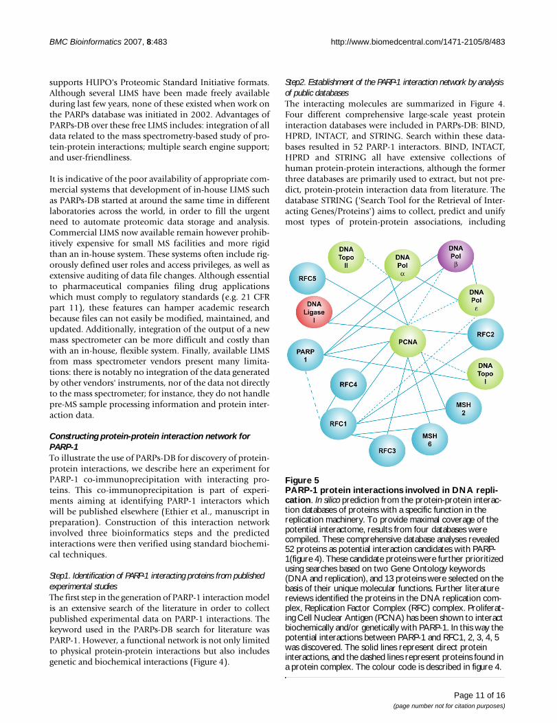
Step2. Establishment of the PARP-1 interaction network by analysis of public databasesThe interacting molecules are summarized in Figure 4.Four different comprehensive large-scale yeast proteininteraction databases were included in PARPs-DB: BIND,HPRD, INTACT, and STRING. Search within these data-bases resulted in 52 PARP-1 interactors. BIND, INTACT,HPRD and STRING all have extensive collections ofhuman protein-protein interactions, although the formerthree databases are primarily used to extract, but not pre-dict, protein-protein interaction data from literature. Thedatabase STRING ('Search Tool for the Retrieval of Inter-acting Genes/Proteins') aims to collect, predict and unifymost types of protein-protein associations, including
PARP-1 protein interactions involved in DNA replicationFigure 5PARP-1 protein interactions involved in DNA repli-cation. In silico prediction from the protein-protein interac-tion databases of proteins with a specific function in the replication machinery. To provide maximal coverage of the potential interactome, results from four databases were compiled. These comprehensive database analyses revealed 52 proteins as potential interaction candidates with PARP-1(figure 4). These candidate proteins were further prioritized using searches based on two Gene Ontology keywords (DNA and replication), and 13 proteins were selected on the basis of their unique molecular functions. Further literature reviews identified the proteins in the DNA replication com-plex, Replication Factor Complex (RFC) complex. Proliferat-ing Cell Nuclear Antigen (PCNA) has been shown to interact biochemically and/or genetically with PARP-1. In this way the potential interactions between PARP-1 and RFC1, 2, 3, 4, 5 was discovered. The solid lines represent direct protein interactions, and the dashed lines represent proteins found in a protein complex. The colour code is described in figure 4.
Page 11 of 16(page number not for citation purposes)

BMC Bioinformatics 2007, 8:483 http://www.biomedcentral.com/1471-2105/8/483
direct and indirect associations. In order to cover organ-isms not yet addressed experimentally, STRING runs a setof prediction algorithms, and transfers known interac-tions from model organisms to other species based onpredicted orthology of the respective proteins.
One important point in the analysis of data from publicprotein-protein interaction databases is the quality of theresults. Indeed, Deng et al compared the data from all thelarge-scale yeast interactions screens present in the publicprotein interaction databases. They developed a maxi-mum likelihood estimation method to access the reliabil-ity of the interaction data, and found that the Uetzdatawere more reliable than the Ito[8] data, and that the Gavindata were more reliable than the Ho data. Therefore, acautious use of public databases is indicated. In addition,they suggested that the MS-based analysis of protein com-plexes performed better in function predictions than thetwo-hybrid data, thus validating the theory that each com-ponent of a complex can be assigned a function based onthat of the whole complex. It is clear that yeast two-hybridand MS-based techniques have both independently madesignificant impacts on our understanding of the interac-tome. However, each technique has specific drawbacksthat limit the information provided if used alone.
Step3. Selection of Protein-Protein Interaction by Gene Ontology AccessionThe next step was to group protein-protein interactions bymolecular function via the Gene Ontology [27] controlledvocabulary included in PARPs-DB. Because intracellularevents may be compartmentalized to unique intracellularlocation, to provide additional specificity for target selectionwe also included a spatial component to further refine theconstruction of the model. Therefore, we further prioritizedour target selection by using two keywords (DNA replicationand nucleus). We chose these two keywords to illustrate ourapproach but PARP-1 is involved in many other cellularprocesses. After filtering according to function, six out of the52 initial proteins interactors remained in the networks:PCNA, topoisomerase I and II, DNA ligase I, DNA Pol α andβ. An extensive literature search about these proteins in thecontext of replication and data mining helped to construct ahuman PARP-1-protein interaction map in PARPs-DB. Wefound 13 proteins and one complex that interact with PARP-1 in the complex machinery replication. Of the 13 interact-ing proteins, PCNA, topoisomerase I, DNA ligase I, DNA Polα and β have been previously reported to interact with PARP-1. Topo2, MSH2, RFC1, RFC2, RFC3, RFC4, RFC5 have beenreported to be related to DNA replication. This analysis raisesthe possibility that PARP-1 may regulate these complexes asa whole rather than regulate one or more the individualcomponents. Of all the potential candidates, we proposethat PARP-1 could interact with other proteins in RFC (RFC1to RFC5) in the context of machinery replication (Figure 5).
Demonstration of biochemical interactions between PARP-1 and RFC1Verifying the interactions from molecules identified in sil-ico is vital to provide a confident interaction network use-ful for further study. With the exception of PCNA, whichhas been characterized, the prioritized candidates werenext tested experimentally to confirm the predicted pro-tein-protein interactions. This was carried out using co-immunoprecipitation of PARP-1 followed by mass spec-trometry to identify the interacting proteins.
Scaffold (version Scaffold-01_03_02) was used to groupand validate MS/MS based peptide and protein identifica-tions from Sequest, Mascot and X!Tandem. This softwareis based on the PeptideProphet algorithm which providesan empirical statistical model which estimates the accu-racy of peptide identifications made by database searchengines. For each tandem mass spectrum, PeptideProphetdetermines the probability that the spectrum is correctlyassigned to a peptide. Scaffold system was used to groupthe assigned peptides according to corresponding proteinand to compute probability of a correct protein assign-ment for each protein. Peptide identifications with Scaf-fold software were accepted if they could be established atgreater than 80.0% probability as specified by the Peptide-Prophet algorithm. For the co-immunoprecipitation elu-ate, protein identifications were accepted if they could beestablished at greater than 95% probability and containedat least 2 identified peptides. Table 1 lists all the RFC pro-teins identified with a minimum probability of 95%. Itshould be noted that the maximal protein probability islimited to 95% in Scaffold. This was set to take intoaccount the light risk that a peptide spectrum match
PARP-1 and the interactor RFC1 were immunoprecipitated with mouse monoclonal F1-23 antibody. RFC1 was detected by western blot with a rabbit polyclonal RFC1 antibodyFigure 6PARP-1 and the interactor RFC1 were immunopre-cipitated with mouse monoclonal F1-23 antibody. RFC1 was detected by western blot with a rabbit pol-yclonal RFC1 antibody. PARP-1 was detected by western blot with the mouse monoclonal C2-10 antibody. Ctr : Immunoprecipitation was done with normal mouse IgG instead of F1-23 antibody.
PARP-1 113 kDa
Ctr CtrF1-23 F1-23
Total lysate IPWB MW
RFC1 140 kDa
Page 12 of 16(page number not for citation purposes)

BMC Bioinformatics 2007, 8:483 http://www.biomedcentral.com/1471-2105/8/483
(PSM) is incorrect even if the theoretical and experimentalspectra are very similar (e.g: correct peptide absent fromthe search database).
The co-immunoprecipitation assay with RFC1 antibodyperformed in HeLa cells suggests that Replication Factor Csubunit (RFC1, 2, 3, 4, 5) (Table 1 and Table 2) formed acomplex with endogenous PARP-1. The protein sequencecoverage of PARP-1 in this study is 60%.
RFC-2, 3, 4 and 5 were each identified with a minimumprobability of 95% and by more than 4 peptides with aminimum probability of 95%. The confidence of theidentification of RFC-2, 3, 4 and 5 is thus very high and,moreover, we have identified this RFC complex in several
co-immunoprecipitates. The case of RFC-1 is different as itwas identified by only two peptides of probabilities of95% and identified in only one co-immunoprecipitate.For this reason, this potential interaction was confirmedby western blot analysis of complexes immunopurifiedwith mouse monoclonal F1-23 antibody (Figure 6). RFC-1 was detected by western blot analysis with rabbit poly-clonal RFC1 antibody. As expected, RFC-1 was pulled-down by PARP-1. Although substantially more work isrequired to determine whether RFC complex interactsdirectly with PARP-1 or via other proteins such as PCNA,DNA polymerase or through interaction with poly(ADP-ribose), the findings reported here suggest that the PARPs-DB may be useful for finding interacting proteins.
Table 1: List of peptides of RFC-1-5. List of peptides of RFC1-5 subunits identified in an immunoprecipitation of PARP-1 from a HeLa cell lysate by LC MS/MS to the experiment number assigned by the PARPs-DB. This table summarizes the identification from 5 independent immunoprecipitation experiments.
IPI SwissProt Protein identification Gene name Peptide probability
IPI00375358 P35251 Splice Isoform 1 of Activator 1 140 kDa subunit RFC1AALLSGPPGVGK 95,00%AIVAESLNNTSIK 95,00%
IPI00017412 P35250 Splice Isoform 1 of Activator 1 40 kDa subunit RFC2DAMLELNASNDR 82,70%EGNVPNIIIAGPPGTGK 95,00%IIEPIQSR 94,10%LNEIVGNEDTVSR 95,00%LTDAQILTR 95,00%
IPI00031521 P40938 Activator 1 38 kDa subunit RFC3ETANAIVSQQTPQR 95,00%KFMEDGLEGMMF 95,00%LILCCNSTSK 95,00%TVAQSQQLETNSQR 95,00%VVLLTEVDKLTK 95,00%
IPI00017381 P35249 Activator 1 37 kDa subunit RFC4AITFLQSATR 95,00%ELFGPELFR 95,00%GTSISTKPPLTK 95,00%IIEPLTSR 91,40%ISDEGIAYLVK 95,00%LRVLELNASDER 95,00%NFAQLTVSGSR 95,00%VITDIAGVIPAEK 95,00%VKNFAQLTVSGSR 95,00%VLELNASDER 95,00%
IPI00031514 P40937 Activator 1 36 kDa subunit RFC5ALNILQSTNMAFGK 95,00%FCLICNYLSK 95,00%FGPLTPELMVPR 83,00%GPILSFASTR 95,00%MADIEYR 92,40%TSTILACAK 95,00%
Page 13 of 16(page number not for citation purposes)

BMC Bioinformatics 2007, 8:483 http://www.biomedcentral.com/1471-2105/8/483
ConclusionThe work presented here has demonstrated how bioinfor-matics can supplement conventional biological investiga-tion. The PARPs-DB enables storage, annotation andrepresentation of data generated by molecular biology.Moreover this system has identified a previouslyunknown protein interaction of PARP-1. The PARPs data-base allows the effective description of proteomics exper-iments and analysis of protein-protein interactions.
Because the PARPs database was developed to facilitatedata sharing and exchange, it includes the latest standardformat to allow sharing of experimental design and resultswith the scientific community. We have incorporatedtools allowing the extraction of protein-protein interac-tions from the HPRD, DIP and BIND public databases, lit-erature and other sources of information. Reports forpeptide and protein analyses are output. These providecomparison reports from multiple or concatenated exper-iments, thereby significantly increasing the confidence inpeptide and protein identifications.
The biochemical data between PARP-1 and RFC complexconfirmed the interaction reported earlier. However, sub-stantially more work is required to delineate the specifi-city and the structural interaction with respect to theregulation of their cellular function between PARP-1 andRFC complex. It is anticipated that the building of such anintegrated platform, which can be constantly up-graded,could provide a predictive understanding of a novel gene's
function in its biological context. A key design element ofPARPs database is the ability to add tools or module thatplug into and use the core systems. The PARPs-DB will beexpanded as needed in order to make the analyses moreefficient.
Availability and requirementsProject name: PARPs-DB
Project home page: http://sourceforge.net/projects/parpdb/
Operating system(s) : Unix, Linux, Oracle and MySQL;
Programming Language: Perl, JAVA, SQL;
Licence: GNU GPL;
PARPs-DB is distributed under the GNU GPL licence andavailable from the website http://sourceforge.net/projects/parpdb/
AbbreviationsSQL: Standard Query Language; NCBI: National Centerfor Biotechnology Information; MS: Mass spectrometry;PARP: Poly(ADP-Ribose) Polymerase; ISB: Institute forSystems Biology; EBI: European Bioinformatics Institute;LDAP: Lightweight Directory Access Protocol; IPI: Interna-tional Protein Index; HPRD: Human Protein ReferenceDatabase; BIND: Biology Interaction Database.
Table 2: List of proteins found. List of proteins found with Sequest and X!Tandem from the PARP-1 immunoprecipitation assay and validated with Scaffold software. Biological sample name refers to the experiment number assigned by the PARPs-DB. This table summarizes the identification from 5 independent immunoprecipitation experiments.
Biological sample name
International Protein Index
SwissProt/Uniprot accession no.
Protein identification Protein identification probability
Gene name
IP111 IPI00375358 P35251 Splice Isoform 1 of Activator 1 140 kDa subunit 99.8% RFC1IP111 IPI00017412 P35250 Splice Isoform 1 of Activator 1 40 kDa subunit 92.9% RFC2IP577 IPI00017412 P35250 Splice Isoform 1 of Activator 1 40 kDa subunit 100.0% RFC2IP597 IPI00017412 P35250 Splice Isoform 1 of Activator 1 40 kDa subunit 100.0% RFC3IP230 IPI00031521 P40938 Similar to human replication factor C (activator 1) 3, 38 kDa 87.7% RFC3IP372 IPI00031521 P40938 Similar to human replication factor C (activator 1) 3, 38 kDa 99.8% RFC3IP577 IPI00031521 P40938 Similar to human replication factor C (activator 1) 3, 38 kDa 100.0% RFC3IP597 IPI00031521 P40938 Similar to human replication factor C (activator 1) 3, 38 kDa 100.0% RFC3IP111 IPI00017381 P35249 Activator 1 37 kDa subunit 92.9% RFC4IP577 IPI00017381 P35249 Activator 1 37 kDa subunit 100.0% RFC4IP597 IPI00017381 P35249 Activator 1 37 kDa subunit 100.0% RFC4IP577 IPI00031514 P40937 Activator 1 36 kDa subunit 100.0% RFC5IP597 IPI00031514 P40937 Activator 1 36 kDa subunit 100.0% RFC5IP111 IPI00449049 P09874 Poly [ADP-ribose] polymerase 1 100.0% PAPR-1IP230 IPI00449049 P09874 Poly [ADP-ribose] polymerase 1 100.0% PARP-1IP372 IPI00449049 P09874 Poly [ADP-ribose] polymerase 1 100.0% PARP-1IP577 IPI00449049 P09874 Poly [ADP-ribose] polymerase 1 100.0% PARP-1IP597 IPI00449049 P09874 Poly [ADP-ribose] polymerase 1 100.0% PAPR-1
Page 14 of 16(page number not for citation purposes)

BMC Bioinformatics 2007, 8:483 http://www.biomedcentral.com/1471-2105/8/483
Authors' contributionsAD implemented, designed the database for the massspectrometry. MR, CE and APC performed molecular biol-ogy approach and helped revise the manuscript. GGP par-ticipated and supervised the project. All authors read andapproved the final manuscript.
Additional material
AcknowledgementsThis study was supported by the Canadian Institutes of Health Research and the program in functional Genomics from CIHR. We thank Pierre Gagné and Eric Winstall for critical revision of the manuscript and Ken Sin Lo for data processing.
References1. Hunt DF: Personal commentary on proteomics. J Proteome Res
2002, 1(1):15-19.2. Link AJ, Eng J, Schieltz DM, Carmack E, Mize GJ, Morris DR, Garvik
BM, Yates JR 3rd: Direct analysis of protein complexes usingmass spectrometry. Nat Biotechnol 1999, 17(7):676-682.
3. Perkins DN, Pappin DJ, Creasy DM, Cottrell JS: Probability-basedprotein identification by searching sequence databases usingmass spectrometry data. Electrophoresis 1999,20(18):3551-3567.
4. Yates JR 3rd, Eng JK, McCormack AL, Schieltz D: Method to corre-late tandem mass spectra of modified peptides to amino acidsequences in the protein database. Anal Chem 1995,67(8):1426-1436.
5. Craig R, Beavis RC: TANDEM: matching proteins with tandemmass spectra. Bioinformatics 2004.
6. Fenyo D: Identifying the proteome: software tools. Curr OpinBiotechnol 2000, 11(4):391-395.
7. Gomez SM, Noble WS, Rzhetsky A: Learning to predict protein-protein interactions from protein sequences. Bioinformatics2003, 19(15):1875-1881.
8. Ito T, Chiba T, Ozawa R, Yoshida M, Hattori M, Sakaki Y: A compre-hensive two-hybrid analysis to explore the yeast proteininteractome. Proc Natl Acad Sci U S A 2001, 98(8):4569-4574.
9. Newman JR, Wolf E, Kim PS: A computationally directed screenidentifying interacting coiled coils from Saccharomyces cer-evisiae. Proc Natl Acad Sci U S A 2000, 97(24):13203-13208.
10. Uetz P, Giot L, Cagney G, Mansfield TA, Judson RS, Knight JR, Lock-shon D, Narayan V, Srinivasan M, Pochart P, et al.: A comprehen-sive analysis of protein-protein interactions inSaccharomyces cerevisiae. Nature 2000, 403(6770):623-627.
11. Cho SY, Park KS, Shim JE, Kwon MS, Joo KH, Lee WS, Chang J, KimH, Chung HC, Kim HO, et al.: An integrated proteome databasefor two-dimensional electrophoresis data analysis and labo-ratory information management system. Proteomics 2002,2(9):1104-1113.
12. Goh CS, Lan N, Echols N, Douglas SM, Milburn D, Bertone P, Xiao R,Ma LC, Zheng D, Wunderlich Z, et al.: SPINE 2: a system for col-laborative structural proteomics within a federated data-base framework. Nucleic Acids Res 2003, 31(11):2833-2838.
13. Taylor CF, Paton NW, Garwood KL, Kirby PD, Stead DA, Yin Z,Deutsch EW, Selway L, Walker J, Riba-Garcia I, et al.: A systematicapproach to modeling, capturing, and disseminating pro-teomics experimental data. Nat Biotechnol 2003, 21(3):247-254.
14. D'Amours D, Desnoyers S, D'Silva I, Poirier GG: Poly(ADP-ribo-syl)ation reactions in the regulation of nuclear functions. Bio-chem J 1999, 342(Pt 2):249-268.
15. Ame JC, Spenlehauer C, de Murcia G: The PARP superfamily.Bioessays 2004, 26(8):882-893.
16. Otto H, Reche PA, Bazan F, Dittmar K, Haag F, Koch-Nolte F: In sil-ico characterization of the family of PARP-like poly(ADP-ribosyl)transferases (pARTs). BMC Genomics 2005, 6:139.
17. Aubert B, Barate R, Boutigny D, Gaillard JM, Hicheur A, Karyotakis Y,Lees JP, Robbe P, Tisserand V, Zghiche A, et al.: Measurement ofthe branching fraction, and bounds on the CP-violatingasymmetries, of neutral B decays to D*+/- D-/+. Phys Rev Lett2003, 90(22):221801.
18. Tulin A, Chinenov Y, Spradling A: Regulation of chromatin struc-ture and gene activity by poly(ADP-ribose) polymerases.Curr Top Dev Biol 2003, 56:55-83.
19. Dynek JN, Smith S: Resolution of sister telomere association isrequired for progression through mitosis. Science 2004,304(5667):97-100.
20. Rouleau M, Aubin RA, Poirier GG: Poly(ADP-ribosyl)ated chro-matin domains: access granted. J Cell Sci 2004, 117(Pt6):815-825.
21. Sourceforge PARPs-DB [http://sourceforge.net/projects/parpdb/]
22. Sashimi [http://sashimi.sourceforge.net]23. Apache [http://apache.org]24. Unified Modified Language [http://www.UML.org]25. Rumbaugh J, Jacobson I, Booh G: The Unified Modeling Language
Reference Manual. 1999.26. Gygi S, Rist B, Gerber S, Turecek F, Gelb M, Aebersold R: Quanti-
tative analysis of complex protein mixtures using isotope-coded affinity tags. 1999, 17:994-999.
27. Ashburner M, Ball CA, Blake JA, Botstein D, Butler H, Cherry JM,Davis AP, Dolinski K, Dwight SS, Eppig JT, et al.: Gene ontology:tool for the unification of biology. The Gene Ontology Con-sortium. Nat Genet 2000, 25(1):25-29.
28. Keller A, Nesvizhskii AI, Kolker E, Aebersold R: Empirical statisti-cal model to estimate the accuracy of peptide identificationsmade by MS/MS and database search. Anal Chem 2002,74(20):5383-5392.
29. Nesvizhskii A, Keller A, Kolker E, Aebersold R: A statistical modelfor identifying proteins by tandem mass spectrometry. 2003,75:4646-4658.
30. Proteome center [http://proteomecenter.org]31. Keller A, Eng J, Zhang N, Li XJ, Aebersold R: A uniform proteom-
ics MS/MS analysis platform utilizing open XML file formats.Mol Syst Biol 2005, 1:0017.
32. Kersey PJ, Duarte J, Williams A, Karavidopoulou Y, Birney E,Apweiler R: The International Protein Index: an integrateddatabase for proteomics experiments. Proteomics 2004,4(7):1985-1988.
33. Bairoch A, Apweiler R, Wu CH, Barker WC, Boeckmann B, Ferro S,Gasteiger E, Huang H, Lopez R, Magrane M, et al.: The UniversalProtein Resource (UniProt). Nucleic Acids Res 2005:D154-159.
34. Maglott D, Ostell J, Pruitt KD, Tatusova T: Entrez Gene: gene-centered information at NCBI. Nucleic Acids Res 2005:D54-58.
35. Pruitt KD, Tatusova T, Maglott DR: NCBI Reference Sequence(RefSeq): a curated non-redundant sequence database ofgenomes, transcripts and proteins. Nucleic Acids Res2005:D501-504.
36. Bader GD, Betel D, Hogue CW: BIND: the Biomolecular Inter-action Network Database. Nucleic Acids Res 2003, 31(1):248-250.
37. Xenarios I, Salwinski L, Duan XJ, Higney P, Kim SM, Eisenberg D: DIP,the Database of Interacting Proteins: a research tool forstudying cellular networks of protein interactions. NucleicAcids Res 2002, 30(1):303-305.
38. Peri S, Navarro JD, Kristiansen TZ, Amanchy R, Surendranath V,Muthusamy B, Gandhi TK, Chandrika KN, Deshpande N, Suresh S, etal.: Human protein reference database as a discoveryresource for proteomics. Nucleic Acids Res 2004:D497-501.
39. Xerces [http://xerces.apache.org/]40. Piccolo Java 1.1 [http://www.cs.umd.edu/hcil/piccolo/]41. JDOM [http://www.jdom.org/]42. Tollis I, Battista G, Eades P, Tamassia R: Graph drawing-Algo-
rithms for the visualization of graphs. Prentice Hall, Upper Sad-dle River, NJ; 1999.
Additional file 1How to install PARPs-DB. This document gives some help to install Ora-cle and PARPs-DBClick here for file[http://www.biomedcentral.com/content/supplementary/1471-2105-8-483-S1.doc]
Page 15 of 16(page number not for citation purposes)

BMC Bioinformatics 2007, 8:483 http://www.biomedcentral.com/1471-2105/8/483
Publish with BioMed Central and every scientist can read your work free of charge
"BioMed Central will be the most significant development for disseminating the results of biomedical research in our lifetime."
Sir Paul Nurse, Cancer Research UK
Your research papers will be:
available free of charge to the entire biomedical community
peer reviewed and published immediately upon acceptance
cited in PubMed and archived on PubMed Central
yours — you keep the copyright
Submit your manuscript here:http://www.biomedcentral.com/info/publishing_adv.asp
BioMedcentral
43. Lamarre D, Talbot B, de Murcia G, Laplante C, Leduc Y, Mazen A,Poirier GG: Structural and functional analysis of poly(ADPribose) polymerase: an immunological study. Biochim BiophysActa 1988, 950(2):147-160.
44. Droit A, Fillon J, Morisssette J, Poirier G: Bioinformatic Standardsfor Proteomics-Oriented Mass Spectrometry. Current Pro-teomics 2006, 3(2):119-128.
45. Stromback L, Lambrix P: Representations of molecular path-ways: an evaluation of SBML, PSI MI and BioPAX. Bioinformat-ics 2005, 21(24):4401-4407.
46. Keller A, Eng J, Zhang N, Li X-J, Aebersold R: A uniform proteom-ics MS/MS analysis platform utilizing open XML file formats.Molecular Systems Biology 2005.
47. Orchard S, Hermjakob H, Julian RK Jr, Runte K, Sherman D, WojcikJ, Zhu W, Apweiler R: Common interchange standards for pro-teomics data: Public availability of tools and schema. Proteom-ics 2004, 4(2):490-491.
48. Deng M, Sun F, Chen T: Assessment of the reliability of protein-protein interactions and protein function prediction. PacSymp Biocomput 2003:140-151.
49. Uetz P, Hughes RE: Systematic and large-scale two-hybridscreens. Curr Opin Microbiol 2000, 3(3):303-308.
50. Gavin AC, Bosche M, Krause R, Grandi P, Marzioch M, Bauer A,Schultz J, Rick JM, Michon AM, Cruciat CM, et al.: Functional organ-ization of the yeast proteome by systematic analysis of pro-tein complexes. Nature 2002, 415(6868):141-147.
51. Ho Y, Gruhler A, Heilbut A, Bader GD, Moore L, Adams SL, Millar A,Taylor P, Bennett K, Boutilier K, et al.: Systematic identificationof protein complexes in Saccharomyces cerevisiae by massspectrometry. Nature 2002, 415(6868):180-183.
52. Droit A, Poirier GG, Hunter JM: Experimental and bioinformaticapproaches for interrogating protein-protein interactions todetermine protein function. J Mol Endocrinol 2005,34(2):263-280.
53. Frouin I, Maga G, Denegri M, Riva F, Savio M, Spadari S, Prosperi E,Scovassi AI: Human proliferating cell nuclear antigen,poly(ADP-ribose) polymerase-1, and p21waf1/cip1. Adynamic exchange of partners. J Biol Chem 2003,278(41):39265-39268.
Publish with BioMed Central and every scientist can read your work free of charge
"BioMed Central will be the most significant development for disseminating the results of biomedical research in our lifetime."
Sir Paul Nurse, Cancer Research UK
Your research papers will be:
available free of charge to the entire biomedical community
peer reviewed and published immediately upon acceptance
cited in PubMed and archived on PubMed Central
yours — you keep the copyright
Submit your manuscript here:http://www.biomedcentral.com/info/publishing_adv.asp
BioMedcentral
Page 16 of 16(page number not for citation purposes)
Copyright © 2022 FDOKUMEN