The Protein-DNA Interface database
12
Norambuena and Melo BMC Bioinformatics 2010, 11:262 http://www.biomedcentral.com/1471-2105/11/262 Open Access DATABASE BioMed Central © 2010 Norambuena and Melo; licensee BioMed Central Ltd. This is an Open Access article distributed under the terms of the Creative Commons Attribution License (http://creativecommons.org/licenses/by/2.0), which permits unrestricted use, distribution, and repro- duction in any medium, provided the original work is properly cited. Database The Protein-DNA Interface database Tomás Norambuena and Francisco Melo* Abstract The Protein-DNA Interface database (PDIdb) is a repository containing relevant structural information of Protein-DNA complexes solved by X-ray crystallography and available at the Protein Data Bank. The database includes a simple functional classification of the protein-DNA complexes that consists of three hierarchical levels: Class, Type and Subtype. This classification has been defined and manually curated by humans based on the information gathered from several sources that include PDB, PubMed, CATH, SCOP and COPS. The current version of the database contains only structures with resolution of 2.5 Å or higher, accounting for a total of 922 entries. The major aim of this database is to contribute to the understanding of the main rules that underlie the molecular recognition process between DNA and proteins. To this end, the database is focused on each specific atomic interface rather than on the separated binding partners. Therefore, each entry in this database consists of a single and independent protein-DNA interface. We hope that PDIdb will be useful to many researchers working in fields such as the prediction of transcription factor binding sites in DNA, the study of specificity determinants that mediate enzyme recognition events, engineering and design of new DNA binding proteins with distinct binding specificity and affinity, among others. Finally, due to its friendly and easy-to-use web interface, we hope that PDIdb will also serve educational and teaching purposes. Background The ability of some proteins to bind selectively to DNA constitutes the basis of key cell processes such as RNA transcription, DNA packing, DNA replication, DNA recombination and DNA repair. The understanding of the molecular recognition process that mediates the spe- cific protein-DNA binding selectivity is one of the most interesting challenges in structural biology. To date, there are several hundreds of protein-DNA complexes that have been solved by X-ray crystallography. These experi- mental structures, deposited at the Protein Data Bank (PDB) [1] and publicly available to the scientific commu- nity, constitute a rich source of information to study the different binding modes and the determinants of protein- DNA binding specificity. To facilitate the investigation about the mechanisms involved in the protein-DNA recognition process, several databases of protein-DNA complexes and associated software have been developed (Additional file 1). Among these resources we find AANT [2], which has statistical information on aminoacid-nucleotide interactions; Pro- NuC [3], a database that provides a list of atomic contact pairs between proteins and DNA; ProNIT [4], which gathers experimental binding data of protein-nucleic acid complexes that have been described in the literature; NPIDB [5], a database that contains a description of hydrogen bonds and hydrophobic interactions between proteins and nucleic acids; BIPA [6], a database contain- ing several physicochemical features of protein-nucleic acid interfaces and multiple structural alignments of nucleic-acid binding protein families; and 3D-Footprint [7], which provides estimates of binding specificity for all protein-DNA complexes available at the PDB, among other features. In addition to these resources, that catalogue important information on protein-DNA interactions, the interest has also been put on classification of the complexes. In this respect, we now have a more or less complete vision about the distinct protein architectures and how they bind to DNA. A detailed fold catalogue that describes the complexes in terms of their function and structure is available [8]. This protein view of the protein-DNA inter- action has been the overall trend in classifying protein- DNA complexes. On the other hand, Sarai and colleagues have set up a new approach for the classification of pro- tein-DNA complexes, which is based on some DNA fea- tures instead of using only protein features [9]. The * Correspondence: [email protected] 1 Departamento de Genética Molecular y Microbiología, Facultad de Ciencias Biológicas, Pontificia Universidad Católica de Chile, Alameda 340, Santiago, Chile Full list of author information is available at the end of the article
Transcript of The Protein-DNA Interface database

Norambuena and Melo BMC Bioinformatics 2010, 11:262http://www.biomedcentral.com/1471-2105/11/262
Open AccessDATABASE
DatabaseThe Protein-DNA Interface databaseTomás Norambuena and Francisco Melo*
AbstractThe Protein-DNA Interface database (PDIdb) is a repository containing relevant structural information of Protein-DNA complexes solved by X-ray crystallography and available at the Protein Data Bank. The database includes a simple functional classification of the protein-DNA complexes that consists of three hierarchical levels: Class, Type and Subtype. This classification has been defined and manually curated by humans based on the information gathered from several sources that include PDB, PubMed, CATH, SCOP and COPS. The current version of the database contains only structures with resolution of 2.5 Å or higher, accounting for a total of 922 entries. The major aim of this database is to contribute to the understanding of the main rules that underlie the molecular recognition process between DNA and proteins. To this end, the database is focused on each specific atomic interface rather than on the separated binding partners. Therefore, each entry in this database consists of a single and independent protein-DNA interface.We hope that PDIdb will be useful to many researchers working in fields such as the prediction of transcription factor binding sites in DNA, the study of specificity determinants that mediate enzyme recognition events, engineering and design of new DNA binding proteins with distinct binding specificity and affinity, among others. Finally, due to its friendly and easy-to-use web interface, we hope that PDIdb will also serve educational and teaching purposes.
BackgroundThe ability of some proteins to bind selectively to DNAconstitutes the basis of key cell processes such as RNAtranscription, DNA packing, DNA replication, DNArecombination and DNA repair. The understanding ofthe molecular recognition process that mediates the spe-cific protein-DNA binding selectivity is one of the mostinteresting challenges in structural biology. To date, thereare several hundreds of protein-DNA complexes thathave been solved by X-ray crystallography. These experi-mental structures, deposited at the Protein Data Bank(PDB) [1] and publicly available to the scientific commu-nity, constitute a rich source of information to study thedifferent binding modes and the determinants of protein-DNA binding specificity.
To facilitate the investigation about the mechanismsinvolved in the protein-DNA recognition process, severaldatabases of protein-DNA complexes and associatedsoftware have been developed (Additional file 1). Amongthese resources we find AANT [2], which has statisticalinformation on aminoacid-nucleotide interactions; Pro-
NuC [3], a database that provides a list of atomic contactpairs between proteins and DNA; ProNIT [4], whichgathers experimental binding data of protein-nucleic acidcomplexes that have been described in the literature;NPIDB [5], a database that contains a description ofhydrogen bonds and hydrophobic interactions betweenproteins and nucleic acids; BIPA [6], a database contain-ing several physicochemical features of protein-nucleicacid interfaces and multiple structural alignments ofnucleic-acid binding protein families; and 3D-Footprint[7], which provides estimates of binding specificity for allprotein-DNA complexes available at the PDB, amongother features.
In addition to these resources, that catalogue importantinformation on protein-DNA interactions, the interesthas also been put on classification of the complexes. Inthis respect, we now have a more or less complete visionabout the distinct protein architectures and how theybind to DNA. A detailed fold catalogue that describes thecomplexes in terms of their function and structure isavailable [8]. This protein view of the protein-DNA inter-action has been the overall trend in classifying protein-DNA complexes. On the other hand, Sarai and colleagueshave set up a new approach for the classification of pro-tein-DNA complexes, which is based on some DNA fea-tures instead of using only protein features [9]. The
* Correspondence: [email protected] Departamento de Genética Molecular y Microbiología, Facultad de Ciencias Biológicas, Pontificia Universidad Católica de Chile, Alameda 340, Santiago, ChileFull list of author information is available at the end of the article
BioMed Central© 2010 Norambuena and Melo; licensee BioMed Central Ltd. This is an Open Access article distributed under the terms of the CreativeCommons Attribution License (http://creativecommons.org/licenses/by/2.0), which permits unrestricted use, distribution, and repro-duction in any medium, provided the original work is properly cited.

Norambuena and Melo BMC Bioinformatics 2010, 11:262http://www.biomedcentral.com/1471-2105/11/262
Page 2 of 12
existence of these somehow separated or independentviews of the protein-DNA complexes have prompted usto develop a more complete annotation of the solved pro-tein-DNA complexes by taking into account the interfaceas a central feature.
The major aim of the new database described here, theProtein DNA Interaction Database (PDIdb), is to contrib-ute to the understanding of the main rules that underliethe molecular recognition process between DNA andproteins. To this end, we have focused on each specificatomic interface rather than on the separated bindingpartners (e.g. protein or DNA molecules alone). There-fore, each entry in this database consists of a single andindependent protein-DNA interface, which has beenmanually inspected and curated by humans, to avoid orminimize any downstream accumulation of errors in sub-sequent analysis.
We hope this resource will not be only valuable toresearchers and software developers working in differentareas such as the structure-based prediction of transcrip-tion factor binding sites, the engineering of DNA bindingproteins and the computer-based prediction of protein-DNA complex three-dimensional structures, but mayalso serve for educational purposes.
Construction and ContentPDB features and interface definitionA dataset of protein-DNA complex structures solved byX-ray crystallography was extracted from the PDB in Jan-uary 12, 2009. The protein-DNA complexes were furtherselected only if they were solved at a resolution of 2.5 Å orhigher. Finally, only those complexes that contained dou-ble strand DNA were retained. In addition to this semi-automated filtered search, a manual and visual inspectionof all complexes was carried out to determine if theirasymmetric and biological units had differences. If thiswas the case, the corresponding biological units wereobtained from a special repository available at the PDBweb site in order to have fully-restored structures (Figure1, panel A).
According to the visual inspection of each complex, thefollowing PDB features were extracted: number of com-plexes per PDB, which is the number of independentcomplexes that appear in a PDB entry (sometimes thisnumber matches the number of biological units in thecrystal) (Figure 1, panel B); and number of interfaces percomplex, which is the number of independent protein-DNA interfaces per complex (Figure 1, panel C).
An interface is defined when one or more protein sub-units interacting with DNA can be isolated. For example,the structure with PDB code 1am9 has two complexes,each with one independent interface consisting of a pro-tein dimer interacting with DNA (Figure 1, panel B),while the structure with PDB code 1h89 has one complex
with two interfaces (one with a protein monomer inter-acting with DNA and the other with a protein dimerinteracting with DNA) (Figure 1, panel C).
Defined as mentioned above, each interface representsa specific entry of the database and, as such, it is associ-ated with a unique identifier (ID). This ID was con-structed by taking into account both the number ofcomplexes per PDB and the number of interfaces percomplex. For instance, the structure 1am9 will give rise totwo entries with unique identifiers: 1am9_1_1 and1am9_2_1. The first ID stands for the structure 1am9,complex 1, interface 1; while the second ID stands for thestructure 1am9, complex 2, interface 1 (Figure 1, panel B,top). Similarly, the PDB entry 1h89 will be also convertedinto two independent entries: 1h89_1_1 and 1h89_1_2. Inthis case, the second ID stands for the structure 1h89,complex 1, interface 2 (Figure 1, panel B, bottom). Theenumeration order of the complexes and interfaces is
Figure 1 PDB complexes and interfaces definition. (A) Example of a structure whose asymmetric unit contained half of the known bio-logical unit (left). For each entry of this type, the complete biological unit was obtained from the specialized ftp site of PDB at ftp://ftp.wwp-db.org. (B) Each entry of the database consists of a single and indepen-dent protein-DNA interface, which is isolated from the whole PDB complex. Here, two examples that illustrate this feature are shown. (Top) 1am9, which PDB file contains two separable complexes, each having a single protein-DNA interface; (Bottom) 1h89, which has one complex, but with two independent protein-DNA interfaces. Each in-terface has assigned a unique ID in the database.
Norambuena and Melo BMC Bioinformatics 2010, 11:262http://www.biomedcentral.com/1471-2105/11/262
Page 3 of 12
assigned based on the increasing alphabetic chain IDs asrecorded in the PDB file.
Other PDB features extracted from the structureinspection were also recorded, which included the resolu-tion, source species information, PubMed ID, the numberof biological units, if the asymmetric unit is the same asthe biological unit, and if the structure contains oxygenatoms belonging to water molecules.
Protein featuresA simple function/structure-based classification for eachentry was defined from the point of view of the proteinpart of the interface. Following the logic of a previouswork [8] and using as a source of information all thatavailable at PubMed, PDB [1], CATH [10], SCOP [11] andCOPS [12] databases, we defined three classification cat-egories: Class, Type and Subtype (Table 1). The categoryClass is function-based and contains three subcategories:Enzyme, if the main function of the protein is to modifyDNA; Transcription Factor, if the main function of theprotein is to regulate transcription and gene expression;and Structural/DNA Binding Protein, if the main functionof the protein is to support DNA structure, DNA bendingor to aggregate other proteins. The category Type is func-tion/structure-based and has 15 subcategories for theEnzyme Class (Dioxygenase, Endonuclease, Excisionase,Glucosyltransferase, Glycosylase, Helicase, Ligase, Meth-yltransferase, Nuclease, Photolyase, Polymerase, Recom-binase, Topoisomerase, Translocase and Transposase), 7subcategories for the Transcription Factor Class (AlphaHelix, Alpha/Beta, Beta Sheet, Helix Turn Helix, Ribbon/Helix/Helix, Zinc Coordinating and Zipper Type), and 8subcategories for the Structural/DNA Binding ProteinClass (Centromeric Protein, DNA Packaging, Mainte-nance/Protection, DNA Bending, Repair Protein, Repli-cation, Telomeric Protein and Zalpha). The categorySubtype involves a more specific classification that takesinto account domains, specific reaction of an enzyme,specific DNA binding sites, etc. The detailed descriptionof all three categories can be obtained by quering thedatabase.
In addition to this classification, the following proteinfeatures were also recorded for each entry in the data-base: the number of protein monomers (or chains) inter-acting with DNA and being part of the interface; the typeof multimerization, that accounts for whether the pro-teins are homomultimeric, heteromultimeric, or if bothtypes can be found simultaneously at the interface; andthe type of protein-protein interactions in the interface,which represents the way multimeric proteins interactwith each other when contacting the DNA. In this regard,we have defined three interaction modes: Mode 1, wherethe direction of the protein interaction and the doublehelix axis are orthogonal (Figure 2, panel A); Mode 2,
where the direction of the interaction is parallel to thedouble helix axis (Figure 2, panel B); and Mode 3, whereboth previous modes of interaction are observed at thesame time (Figure 2, panel C).
DNA featuresIn addition to the PDB and protein features describedabove, several DNA features were also recorded for eachentry (Figure 3). These features include: double/singlestrand (Figure 3, panel A), where in the current version ofthe database the only possible types are double strand orsingle strand in the asymmetric unit (because the data-base contains only double strand DNA); sticky ends (Fig-ure 3, panel B), that represent the unpaired bases at theend of the double-stranded DNA; flipped base (Figure 3,panel C), which represents whether the DNA has flippedbases; nicked DNA (Figure 3, panel D), that accounts forwhether the DNA molecule has a broken phosphodiesterbond in one or both strands; gapped DNA (Figure 3,panel E), which denotes if the DNA lacks one or morebases in the middle of one strand; modified DNA (Figure3, panel F), that indicates if the DNA molecule containschemically-modified or non-standard bases; open DNA(Figure 3, panel G), that occurs when a DNA moleculehas unpaired canonical Watson-Crick bases toward theends of the molecule; and Z-DNA, which representswhether the DNA molecule is in left-handed conforma-tion or not.
It is worth noting that these features are applicable tothe DNA structure present in the PDB file, so that twodifferent interfaces coming from the same complex willnormally share the same DNA features, unless the com-plex has more than one DNA molecule, which for exam-ple is the particular case of the structure with PDB code1iaw.
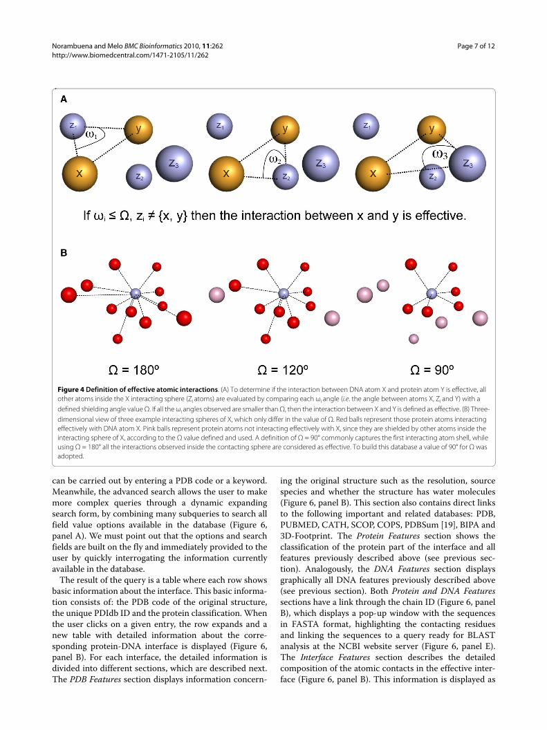
Interface features and effective interactionsThese features take into account detailed atomic charac-teristics involving the interaction between protein andDNA. All these features rely on the results obtained afterapplying a recently described methodology [13] thatallows the extraction of the effective atomic interactionsbetween two molecules forming a complex. Briefly, agiven atom in a DNA or protein structure can have manyneighbour atoms in three-dimensional space, which aretypically defined by setting up a fixed maximum distancethreshold. In the absence of additional definitions, allthese atoms found in such neighbourhood (ie. within thesphere defined by its centre and its radius) are consideredto be interacting with it. However, by using this simpleapproach, many indirect interactions that in fact areshielded by other atoms and thus could not be relevantfrom a physical point of view, will still be included in theanalysis. In order to avoid this problem, additional

Norambuena and Melo BMC Bioinformatics 2010, 11:262http://www.biomedcentral.com/1471-2105/11/262
Page 4 of 12
Table 1: Description of protein features classes and types
Class Type Description
Enzyme Dioxygenase Enzyme that repairs DNA base lesions by using a direct oxidative dealkylation mechanism [25].
Endonuclease Restriction enzyme that cleaves DNA at specific sites [26].
Excisionase Enzyme that controls integrase-mediated DNA rearrangement [27].
Glucosyltransferase Enzyme that binds DNA in abasic site and flips it. Glucosylation is on a 5-hydroximethylcytosine in duplex DNA using UDP-glucose [28].
Glycosylase Enzyme involved in base excision repair, a mechanism by which, damaged nucleotides in DNA are removed and replaced. It catalyses the first step in the process [29].
Helicase Enzyme that unwinds double helices using ATP hydrolysis [30].
Ligase Enzyme that recognizes nicks and states for strand closure [31].
Methyltransferase Enzyme responsible for the generation of the genome methylation patterns leading to gene silencing [32].
Nuclease Enzyme that cleaves DNA, but that are not classified as Endonuclease.
Photolyase Enzyme that uses light to repair DNA having UV-induced lesions [33].
Polymerase Enzyme that takes nucleotides from solvent, and catalyses the synthesis of a polynucleotide sequence against a nucleotide template strand using base-pairing interactions [34].
Recombinase Enzyme that catalyses the reciprocal exchange of DNA strands in the direct site-specific DNA recombination process [35].
Topoisomerase Enzyme that promotes the relaxation of DNA superhelical lesions by introducing a transient single stranded break in duplex DNA [36].
Translocase Enzyme that segregates dimeric circular chromosomes, formed by recombination of monomer sisters [37].
Transposase Enzyme that mediates transposition, a process whereby defined DNA segments move freely about the genome [38].
Structural/DNA Binding Centromeric Protein Protein that is part of a chromosome centromere.
DNA Packaging Protein that is part of the chromosome and packages the DNA.
Maintenance/Protection Protein involved in the protection and maintenance of the genome.
DNA Bending Protein that bends DNA with a highly component of indirect readout.

Norambuena and Melo BMC Bioinformatics 2010, 11:262http://www.biomedcentral.com/1471-2105/11/262
Page 5 of 12
Repair Protein Protein that recognizes damaged DNA and recruits other proteins or enzymes.
Replication Protein involved in the DNA replication process.
Telomeric Protein Protein that binds telomere parts of a chromosome contributing to its stability.
Zalpha Protein that binds left-handed DNA.
Transcription Factor Alpha Helix Protein that interacts with DNA mainly through α-helices.
Alpha/Beta Protein interacting with DNA through α-helices and β-strands.
Beta Sheet Protein that interacts with DNA mainly through β-sheets.
Helix Turn Helix Protein that contains the HtH motif according to the information available in PDB. It includes those proteins containing the "winged helix" domain.
Ribbon/Helix/Helix Protein that contains the RHH fold according to the information available in PDB.
Zinc Coordinating Protein that coordinates the metal in order to bind DNA.
Zipper Type Protein that contains the zipper motif, including the helix-loop-helix one.
Table 1: Description of protein features classes and types (Continued)
restraints are introduced so as to select only the direct have defined the following five major classes of interac-
interactions between two atoms. Direct or effective inter-actions are defined as those atom-atom interactions thatare not shielded or masked by any other atom in three-dimensional space. A simple geometric algorithm wasdeveloped to assess the shielding effect that any atom hason the interaction of two other atoms [13].Based on this methodology, it is possible to classifyeach interaction observed at a given protein-DNA inter-face as being either effective or not (Figure 4). Here wehave used this methodology to select for each interfacethose protein-DNA atom pairs that interact effectively ata maximum distance of 7 Å. The atoms selected this waymake up a cloud of points in three-dimensional space thatwe define as the effective interface, where every proteinatom has an effective interacting DNA counterpart.
As each atomic pairwise interaction in the effectiveinterface is identified in detail, we have classified themaccording to their position in the DNA grooves and totheir physical-chemical nature. The specific groove loca-tion of DNA atoms was assigned according to the classi-cal definition in B-DNA [14,15]. Thus, there are DNAatoms in the major groove, minor groove, backbone(phosphate and sugar), and atoms assigned to any loca-tion (i.e. ambiguous location) (Figure 5). Regarding thephysical-chemical nature of the interacting atom pair we
tions (Table 2): CHb, interactions that resemble canonicalH-bond (i.e. where the heavy atoms are either nitrogen oroxygen) [16]; SHb, interactions that resemble H-bondswith Sulphur [17]; CHO, contacts that resemble H-bondsof type CH··· O [18]; Ion, are interactions betweencharged atoms (i.e. any interaction where the proteinatom is either NZ from lysine, NH1, NH2 from arginine,OD1, OD2 from aspartic acid, OE1, OE2 from glutamicacid, and the DNA atom is any oxygen from the phos-phate groups); and Hph, contacts consisting of hydropho-bic interactions. These five interaction classes in turnhave subcategories that take into account the atom iden-tity, the position of the atoms in both residues (i.e. edge,sidechain or backbone) and whether the atoms are donoror acceptor in the case they constitute an H-bond (Table2). The total number of interaction types defined by usingthis procedure is 19, with an additional subcategory thatis used to classify those interactions that do not belong toany of the types defined (i.e. not assigned).
Database redundancy, clustering and representative membersTo remove obvious redundancy from this database, wehave performed two independent clustering/groupingschemes: 1) a clustering based on protein sequence iden-

Norambuena and Melo BMC Bioinformatics 2010, 11:262http://www.biomedcentral.com/1471-2105/11/262
Page 6 of 12
tity and fraction of aligned regions for those proteins thatform part of the interface with DNA; and 2) a clusteringbased on the effective interactions observed for eachinterface between proteins and DNA.
The sequence-based clustering was obtained by align-ing all the protein sequence chains that interact withDNA in a pairwise fashion. Sequences were clustered ingroups according to a length coverage threshold of 90%and percentage sequence identity of 70%, using blastclustsoftware. Therefore, two protein-DNA interfaces wereclustered together if any two protein chains found at thetwo interfaces shared more than 70% sequence identityfor at least 90% of the length of both sequences. A total of246 non-redundant interfaces, out of the initial 922entries, were obtained with this procedure.
The interface-based clustering was obtained by calcu-lating a dissimilarity measure between all different inter-face pairs. This measure is described in detail in thedatabase web site ("About" section). This dissimilaritymeasure ranges between 0, for identical interfaces, and 1,for two interfaces that have no interactions in common.Using this measure, a difference table was built for all
possible interface pairs and hierarchical clustering car-ried out with the group average algorithm. We used athreshold of 0.25 to define the non-redundant groups.This means that two interfaces were clustered together ifthey had more than 75% of their effective interactions incommon. A total of 671 non-redundant interfaces, out ofthe initial 922 entries, were obtained with this procedure.
The detailed list of representative interfaces, as well asthe members belonging to each group, are availableonline at the database web site for both clustering/group-ing schemes. Additionally, complex queries for each clus-tered set can be launched through the advanced searchoption of the web interface. Finally, a set of PDB files ofthe representative members for each clustered set, thedetailed interface data, as well as other related sets can bedownloaded directly from the database web site ("Down-load" section).
Utility and DiscussionWeb User InterfacePDIdb was built using the PHP framework Symfony, theAJAX technology and the MySQL database managementsystem. The database can be accessed through Internet athttp://melolab.org/pdidb and several options are avail-able (described below).
The core of the database is its search engine. There istwo search modes: basic and advanced. A basic search
Figure 2 Protein-protein interaction modes with DNA. Three modes of protein-protein interaction with DNA are defined, according to the direction and the axis of the DNA helix. (A) Mode 1, the direction of the protein interaction and the double helix axis are orthogonal. (B) Mode 2, the direction of the interaction is parallel to the double helix axis. (C) Mode 3, both previous modes are observed at the same time. In the Mode 3 example, the histone core shown is the only instance of this case in the current version of the database. The histone core pres-ents a set of proteins interacting with each other, thus making up a continuous interface with DNA. Additionally, a Mode 0 to assign those interfaces with only one protein has also been defined (not shown).
Figure 3 DNA features. Several DNA features has been defined. (A) Double strand or single strand in the asymmetric unit. This is useful to identify those interfaces coming from the reconstruction of the biolog-ical unit. (B) Sticky ends were defined based on the specific strands and the number of free bases at their ends. (C) Presence of flipped bases. (C) Existance of nicked DNA. (E) Existance of gapped DNA. (F) Presence of modified or non-standard DNA bases. (G) Presence of opened or un-paired bases at the DNA ends. Although not depicted here, left-hand-ed DNA conformation was also recorded (Z-DNA).
Norambuena and Melo BMC Bioinformatics 2010, 11:262http://www.biomedcentral.com/1471-2105/11/262
Page 7 of 12
can be carried out by entering a PDB code or a keyword.Meanwhile, the advanced search allows the user to makemore complex queries through a dynamic expandingsearch form, by combining many subqueries to search allfield value options available in the database (Figure 6,panel A). We must point out that the options and searchfields are built on the fly and immediately provided to theuser by quickly interrogating the information currentlyavailable in the database.
The result of the query is a table where each row showsbasic information about the interface. This basic informa-tion consists of: the PDB code of the original structure,the unique PDIdb ID and the protein classification. Whenthe user clicks on a given entry, the row expands and anew table with detailed information about the corre-sponding protein-DNA interface is displayed (Figure 6,panel B). For each interface, the detailed information isdivided into different sections, which are described next.The PDB Features section displays information concern-
ing the original structure such as the resolution, sourcespecies and whether the structure has water molecules(Figure 6, panel B). This section also contains direct linksto the following important and related databases: PDB,PUBMED, CATH, SCOP, COPS, PDBSum [19], BIPA and3D-Footprint. The Protein Features section shows theclassification of the protein part of the interface and allfeatures previously described above (see previous sec-tion). Analogously, the DNA Features section displaysgraphically all DNA features previously described above(see previous section). Both Protein and DNA Featuressections have a link through the chain ID (Figure 6, panelB), which displays a pop-up window with the sequencesin FASTA format, highlighting the contacting residuesand linking the sequences to a query ready for BLASTanalysis at the NCBI website server (Figure 6, panel E).The Interface Features section describes the detailedcomposition of the atomic contacts in the effective inter-face (Figure 6, panel B). This information is displayed as
Figure 4 Definition of effective atomic interactions. (A) To determine if the interaction between DNA atom X and protein atom Y is effective, all other atoms inside the X interacting sphere (Zi atoms) are evaluated by comparing each ωi angle (i.e. the angle between atoms X, Zi and Y) with a defined shielding angle value Ω. If all the ωi angles observed are smaller than Ω, then the interaction between X and Y is defined as effective. (B) Three-dimensional view of three example interacting spheres of X, which only differ in the value of Ω. Red balls represent those protein atoms interacting effectively with DNA atom X. Pink balls represent protein atoms not interacting effectively with X, since they are shielded by other atoms inside the interacting sphere of X, according to the Ω value defined and used. A definition of Ω = 90° commonly captures the first interacting atom shell, while using Ω = 180° all the interactions observed inside the contacting sphere are considered as effective. To build this database a value of 90° for Ω was adopted.

Norambuena and Melo BMC Bioinformatics 2010, 11:262http://www.biomedcentral.com/1471-2105/11/262
Page 8 of 12
Table 2: Definition of interaction classes and types
Class Type Detail
CHb 1 DBE-PSC: NA - ND
2 DBE-PSC: NA - OD
3 DBE-PSC: OA - ND
4 DBE-PSC: OA - OD
5 DBE-PSC: ND - OA
6 DBE-PBB: NA - ND
7 DBE-PBB: ND - OA
8 DBE-PBB: OA - ND
9 DBB-PSC: OA - ND
10 DBB-PSC: OA - OD
11 DBB-PBB: OA - ND
SHb 12 DBB-PSC: OA - SD
13 DBE-PSC: NA - SD
14 DBE-PSC: OA - SD
15 DBE-PSC: ND - SA
CHO 16 DBE-PSC: CD - OA
17 DBE-PSB: CD - OA
Ion 18 Ionic bond: (+)··· (-)
Hph 19 C - C
20 Not assigned
The nomenclature of the abbreviations used in this Table is the following: DBE, DNA Base edge; DBB, DNA Backbone; PSC, Protein Sidechain; PBB, Protein Backbone; XA, Acceptor; XD, Donor; O, Oxygen; N, Nitrogen; S, Sulphur; C, Carbon. See text for more information.
bar plots and includes the groove contacts as well as theinteraction classes and types (see previous section above).When a plot is clicked, a pop-up window opens showing
the detailed view of the graphs and the respective legends(Figure 6, panel C). The Interface Features section alsoincludes the display of a NUCPLOT graph [20], whichmaps onto a plane direct or water-mediated hydrogenbonds between aminoacids and nucleotides as deter-mined by HBPLUS [21] (Figure 6, panel F), and a link to apop-up displaying all interfaces where protein sequencesbelong to the same sequence group or cluster (see above).Independent files containing the parsed PDB structure,the 3D coordinates of all atoms being part of the effectiveinterface in PDB format and the detailed information ofthe interaction types found at the effective interface inplain text format, can be downloaded in the Downloadsection.
Next to the sections described above, a Jmol [22] appletis available to explore the structure and the atomic inter-actions constituting the effective interface in three-dimensional space. Several molscripts were created inorder to survey the groove contacts as well as the interac-tion classes and types through this applet. We have alsoincluded coordinate files containing the middle points foreach pair of contacting atoms for further comparisonpurposes. For a detailed view of the structure, a pop-upwindow of the applet can also be launched by the user.This pop-up window includes more options such asdrawing lines and showing distances between the con-tacting atoms, zooming, stereo and anaglyph views (Fig-ure 6, panel D). Console driven, user custom Jmol scriptscan also be directly launched from here.
Database StatisticsIn the current version of the database there are a total of922 entries, which will be doubtless increased with futureupdates. By considering the classification of the proteinpart of the interfaces introduced in this report, out of thetotal entries, 528 (57%) are categorized as Enzyme, whosemain types are Polymerase and Endonuclease (37% and29%, respectively); 295 (32%) belong to the class Tran-scription Factor, which contains the main types Helix-turn-Helix (42%) and Zinc Coordinating (20%); and 99(11%) are classified as Structural/DNA Binding Protein,whose most abundant category is the type DNA Packag-ing (34%), which in turn includes the subtypes Histoneand Histone-like proteins. A graphical representation ofthese figures can be also found at the web site under themenu Statistics.
The multimerization state in the interfaces shows that608 (66%) proteins are monomeric, 294 (32%) aredimeric, and the rest with 3 or more monomeric units.Out of the interfaces with multimeric proteins, 261 (83%)are homomultimeric and 41 (13%) are heteromultimeric.
As to the DNA features, 427 (46%) interfaces out of thetotal have DNA molecules with no sticky ends, 266 (29%)have sticky ends in both strands, 200 (22%) have stickyends in one strand at one end, and the remainder (3%),

Norambuena and Melo BMC Bioinformatics 2010, 11:262http://www.biomedcentral.com/1471-2105/11/262
Page 9 of 12
have sticky ends at both ends in one strand; 150 (16%) outof the total number of interfaces have flipped bases; 39(4%) structures have nicked DNA, 22 of which at bothstrands; 49 (5%) interfaces have gapped DNA, while 62(7%) have open DNA; 278 (30%) have at least a modifiedor non-standard base; and finally, only 14 (2%) interfaceshave left-handed DNA.
When effective contacts are regarded, on average allinterfaces have 296.3 total contacts, 18.11% of whichoccurs with the major groove, 7.92% with the minorgroove, and 72.92% with the backbone (phosphate andsugar). Concerning interaction classes, on average 15.83%out of all interactions can be classified as canonicalhydrogen bonds, 1.24% are hydrogen bonds of the classCH··· O, 5.56% are classified as ionic bonds, and 16.49%are considered as hydrophobic interactions. Out of allinteractions, 60.79% on average cannot be assigned toone of the 19 defined classes. The detailed description ofthese statistics can be found in Additional file 2.
Download Data and SoftwareAt the web site, the complete data that constitute thedatabase in raw format is also available for download. Thedata includes the corresponding parsed PDB files, theeffective atomic interfaces (in PDB format) and the rele-vant protein and DNA features, along with other usefuldetailed data calculated from the effective interfaces.
The database is also accompanied with computer soft-ware. This specialized software was developed to extract
the effective atomic interfaces of protein-DNA complexesand to classify their interaction types. As previously men-tioned, the analysis of effective atomic interactions is use-ful to better characterize and describe the common andunique features present at different complex interfaces,which in turn could help to elucidate the key specificitydeterminants involved in a particular protein-DNA rec-ognition process.
The software is composed of main and supportingapplications written in C++ for command-line execution.These applications are highly customizable, use PDB filesas input and include different options. There are twomain applications: one is used to analyse any kind ofatom-atom contact, where the user can define atomtypes, centroids (i.e. average 3D coordinates of severalatom positions) and distance ranges; in the other applica-tion, the user can also define the type of interaction thatany pair of atoms may have (e.g. hydrogen bonds, hydro-phobic interactions, etc.). These analyses could be doneincluding water molecules at the time of detecting theeffective atomic interactions at the complex interface.According to the options selected, the output of the soft-ware can be the contact matrices, files with detailedinformation about interactions and/or molscripts to visu-alize the interactions. The components and modules ofthis software are fully documented in the software distri-bution.
Future DirectionsSeveral improvements are going to be introduced in thenear future to PDIdb. We intend to include more interac-tion types such as cation-π contacts [23], interactionswith cation/anion ligands, a more detailed description ofwater-mediated interactions and the inclusion of non-standard or modified bases in the interfaces. We also planto include more DNA features such as the groove lengthsand the electrostatic potential at the interface [24]. At themoment, we are doing a deeper analysis of the data keptin the database, including a structural clustering of theeffective interfaces, which we hope results in a new classi-fication of protein-DNA complexes. Finally, we are alsodeveloping a new interface graphical representation thatwill include the effective interaction classes.
ConclusionsThe existing resources and databases advocated to thestudy of protein-DNA interactions, as well as the increas-ing amount of protein-DNA complexes available at thePDB, are a result of the great interest for better under-standing the molecular recognition process betweenDNA and proteins. The PDIdb represents a novel reposi-tory that disaggregates three-dimensional protein-DNAstructure complexes toward the level of interface, whichconstitutes a simultaneous view of both the protein and
Figure 5 Classification of DNA atoms. All the interactions occurring in an effective interface were classified according to the chemical/structural/groove position of the DNA atoms. Pink-highlighted atoms were classified as being part of the major groove, green-highlighted atoms belong to the minor groove, blue-highlighted atoms belong to the backbone and sugar, and yellow-highlighted atoms were classified as not assigned since they are in an ambiguous location.

Norambuena and Melo BMC Bioinformatics 2010, 11:262http://www.biomedcentral.com/1471-2105/11/262
Page 10 of 12
Figure 6 Web user interface of PDIdb. (A) The database search engine. There are two search modes: basic and advanced. A basic search can be carried out by entering a PDB code or a keyword. The advanced search allows the user to make more complex queries through a dynamic expanding search form, by combining many subqueries that search all fields available in the database. (B) The result of the query is a table where each row shows basic information about the interface. When the user clicks on it, the row expands and a new table with detailed information, 2D and 3D molecular graphics is displayed. (C) For each interface, this information includes the protein and DNA features, as well as the detailed composition of the atomic contacts. (D) The user can inspect graphical information at both the sequence and structure level. A Jmol applet is available to explore the structure and the atomic interactions conforming the interface in three-dimensional space. (E) Sequences highlighting the contacting residues are available in FASTA format for further analysis (e.g. BLAST). (F) The protein-DNA complex can be also explored by means of NUCPLOT graphs, which map onto a plane direct or water-mediated hydrogen bonds between aminoacids and nucleotides.

Norambuena and Melo BMC Bioinformatics 2010, 11:262http://www.biomedcentral.com/1471-2105/11/262
Page 11 of 12
DNA parts of the interacting complex. The most impor-tant added value of this database is that most of the infor-mation recorded has been manually curated. This meansthat each original PDB file has been visually inspected,dissected in interfaces, analysed in its constituent partsand finally classified by using simple but well-defined cri-teria. The automated process involved in the building ofthis database has only to do with the obtention of theeffective interface and the classification of the interac-tions. When we relate and integrate both kind of informa-tion, a great diversity of binding modes are seen at aglance, which certainly needs a deeper future analysis byproperly mining the information available here.
Consequently, this database will be useful to those peo-ple working in the fields of prediction of transcriptionfactor binding sites in DNA, study of specificity determi-nants that mediates different enzyme recognition events,engineering and design of new transcription factors withdistinct binding specificity and affinity and many otherapplications. It is important to mention that due to itsfriendly and easy-to-use web user interface, this databasemight also serve educational and teaching purposes.
Availability and RequirementsThe database and software are freely accesible to aca-demic and non-academic users from our web site locatedat: http://melolab.org/pdidb.
The complete set of experimental data used in thiswork and the results obtained are available at our website: http://protein.bio.puc.cl/sup-mat.html
Additional material
Authors' contributionsFM conceived the project. TN wrote all required computer software to buildthe database and processed each entry in a semiautomated fashion. TN alsodesigned and implemented the web interface and manually inspected/curated the complete database content. Both authors wrote this manuscript.
AcknowledgementsTN acknowledges a PhD program fellowship from CONICYT Chile. This work was funded by a grant 1080158 from FONDECYT Chile.
Author DetailsDepartamento de Genética Molecular y Microbiología, Facultad de Ciencias Biológicas, Pontificia Universidad Católica de Chile, Alameda 340, Santiago, Chile
References1. Berman HM, Westbrook J, Feng Z, Gilliland G, Bhat TN, Weissig H,
Shindyalov IN, Bourne PE: The Protein Data Bank. Nucleic Acids Res 2000, 28:235-242.
2. Hoffman MM, Khrapov MA, Cox JC, Yao J, Tong L, Ellington AD: AANT: the Amino Acid-Nucleotide Interaction Database. Nucleic Acids Res 2004, 32:D174-D181.
3. An J, Nakama T, Kubota Y, Sarai A: 3DinSight: an integrated relational database and search tool for the structure, function and properties of biomolecules. Bioinformatics 1998, 14:188-195.
4. Prabakaran P, An J, Gromiha MM, Selvaraj S, Uedaira H, Kono H, Sarai A: Thermodynamic database for protein-nucleic acid interactions (ProNIT). Bioinformatics 2001, 17:1027-1034.
5. Spirin S, Titov M, Karyagina A, Alexeevski A: NPIDB: a database of nucleic acids-protein interactions. Bioinformatics 2007, 23:3247-3248.
6. Lee S, Blundell TL: BIPA: a database for protein-nucleic acid interaction in 3D structures. Bioinformatics 2009, 25:1559-1560.
7. Contreras-Moreira B: 3D-footprint: a database for the structural analysis of protein-DNA complexes. Nucleic Acids Res 2010, 38:D91-D97.
8. Luscombe NM, Austin SE, Berman HM, Thornton JM: An overview of the structures of protein-DNA complexes. Genome Biol 2000, 1:REVIEWS001.
9. Prabakaran P, Siebers JG, Ahmad S, Gromiha MM, Singarayan MG, Sarai A: Classification of protein-DNA complexes based on structural descriptors. Structure 2006, 14:1355-1367.
10. Orengo CA, Michie AD, Jones S, Jones DT, Swindells MB, Thornton JM: CATH--a hierarchic classification of protein domain structures. Structure 1997, 5:1093-1108.
11. Murzin AG, Brenner SE, Hubbard T, Chothia C: SCOP: a structural classification of proteins database for the investigation of sequences and structures. J Mol Biol 1995, 247:536-540.
12. Suhrer SJ, Wiederstein M, Gruber M, Sippl MJ: COPS--a novel workbench for explorations in fold space. Nucleic Acids Res 2009, 37:W539-W544.
13. Ferrada E, Melo F: Effective knowledge-based potentials. Protein Sci 2009, 18:1469-1485.
14. Seeman NC, Rosenberg JM, Rich A: Sequence-specific recognition of double helical nucleic acids by proteins. Proc Natl Acad Sci USA 1976, 73:804-808.
15. Pabo CO, Sauer RT: Protein-DNA recognition. Annu Rev Biochem 1984, 53:293-321.
16. Mandel-Gutfreund Y, Schueler O, Margalit H: Comprehensive analysis of hydrogen bonds in regulatory protein DNA-complexes: in search of common principles. J Mol Biol 1995, 253:370-382.
17. Zhou P, Tian F, Lv F, Shang Z: Geometric characteristics of hydrogen bonds involving sulfur atoms in proteins. Proteins 2009, 76:151-163.
18. Mandel-Gutfreund Y, Margalit H, Jernigan RL, Zhurkin VB: A role for CH...O interactions in protein-DNA recognition. J Mol Biol 1998, 277:1129-1140.
19. Laskowski RA, Hutchinson EG, Michie AD, Wallace AC, Jones ML, Thornton JM: PDBsum: a Web-based database of summaries and analyses of all PDB structures. Trends Biochem Sci 1997, 22:488-490.
20. Luscombe NM, Laskowski RA, Thornton JM: NUCPLOT: a program to generate schematic diagrams of protein-nucleic acid interactions. Nucleic Acids Res 1997, 25:4940-4945.
21. McDonald IK, Thornton JM: Satisfying hydrogen bonding potential in proteins. J Mol Biol 1994, 238:777-793.
22. Jmol: an open-source Java viewer for chemical structures in 3D [http://www.jmol.org/]
23. Wintjens R, Liévin J, Rooman M, Buisine E: Contribution of cation-pi interactions to the stability of protein-DNA complexes. J Mol Biol 2000, 302:395-410.
24. Rohs R, West SM, Sosinsky A, Liu P, Mann RS, Honig B: The role of DNA shape in protein-DNA recognition. Nature 2009, 461:1248-1253.
25. Yang CG, Yi C, Duguid EM, Sullivan CT, Jian X, Rice PA, He C: Crystal structures of DNA/RNA repair enzymes AlkB and ABH2 bound to dsDNA. Nature 2008, 452:961-965.
26. Williams RJ: Restriction endonucleases: classification, properties, and applications. Mol Biotechnol 2003, 23:225-243.
27. Sam MD, Cascio D, Johnson RC, Clubb RT: Crystal structure of the excisionase-DNA complex from bacteriophage lambda. J Mol Biol 2004, 338:229-240.
Additional file 1 Protein-DNA interactions resources and databases. A list of currently available resources with data about protein-DNA complexes and their corresponding references.Additional file 2 Detailed statistics of effective atomic interfaces. Fig-ures and percentages of effective interactions obtained from protein-DNA complexes, categorized according to groove contacts and interactions classes.
Received: 5 March 2010 Accepted: 18 May 2010 Published: 18 May 2010This article is available from: http://www.biomedcentral.com/1471-2105/11/262© 2010 Norambuena and Melo; licensee BioMed Central Ltd. This is an Open Access article distributed under the terms of the Creative Commons Attribution License (http://creativecommons.org/licenses/by/2.0), which permits unrestricted use, distribution, and reproduction in any medium, provided the original work is properly cited.BMC Bioinformatics 2010, 11:262
http://www.ncbi.nlm.nih.gov/entrez/query.fcgi?cmd=Retrieve&db=PubMed&dopt=Abstract&list_uids=9545451
http://www.ncbi.nlm.nih.gov/entrez/query.fcgi?cmd=Retrieve&db=PubMed&dopt=Abstract&list_uids=9309224
http://www.ncbi.nlm.nih.gov/entrez/query.fcgi?cmd=Retrieve&db=PubMed&dopt=Abstract&list_uids=7723011
http://www.ncbi.nlm.nih.gov/entrez/query.fcgi?cmd=Retrieve&db=PubMed&dopt=Abstract&list_uids=1062791
http://www.ncbi.nlm.nih.gov/entrez/query.fcgi?cmd=Retrieve&db=PubMed&dopt=Abstract&list_uids=6236744
http://www.ncbi.nlm.nih.gov/entrez/query.fcgi?cmd=Retrieve&db=PubMed&dopt=Abstract&list_uids=7563096
http://www.ncbi.nlm.nih.gov/entrez/query.fcgi?cmd=Retrieve&db=PubMed&dopt=Abstract&list_uids=9571027
http://www.ncbi.nlm.nih.gov/entrez/query.fcgi?cmd=Retrieve&db=PubMed&dopt=Abstract&list_uids=9433130
http://www.ncbi.nlm.nih.gov/entrez/query.fcgi?cmd=Retrieve&db=PubMed&dopt=Abstract&list_uids=9396800

Norambuena and Melo BMC Bioinformatics 2010, 11:262http://www.biomedcentral.com/1471-2105/11/262
Page 12 of 12
28. Larivière L, Sommer N, Moréra S: Structural evidence of a passive base-flipping mechanism for AGT, an unusual GT-B glycosyltransferase. J Mol Biol 2005, 352:139-150.
29. Fromme JC, Banerjee A, Verdine GL: DNA glycosylase recognition and catalysis. Curr Opin Struct Biol 2004, 14:43-49.
30. Lee JY, Yang W: UvrD helicase unwinds DNA one base pair at a time by a two-part power stroke. Cell 2006, 127:1349-1360.
31. Nandakumar J, Nair PA, Shuman S: Last stop on the road to repair: structure of E. coli DNA ligase bound to nicked DNA-adenylate. Mol Cell 2007, 26:257-271.
32. Brenner C, Fuks F: DNA methyltransferases: facts, clues, mysteries. Curr Top Microbiol Immunol 2006, 301:45-66.
33. Mees A, Klar T, Gnau P, Hennecke U, Eker AP, Carell T, Essen LO: Crystal structure of a photolyase bound to a CPD-like DNA lesion after in situ repair. Science 2004, 306:1789-1793.
34. Brautigam CA, Steitz TA: Structural and functional insights provided by crystal structures of DNA polymerases and their substrate complexes. Curr Opin Struct Biol 1998, 8:54-63.
35. Guo F, Gopaul DN, van Duyne GD: Structure of Cre recombinase complexed with DNA in a site-specific recombination synapse. Nature 1997, 389:40-46.
36. Redinbo MR, Stewart L, Kuhn P, Champoux JJ, Hol WG: Crystal structures of human topoisomerase I in covalent and noncovalent complexes with DNA. Science 1998, 279:1504-1513.
37. Löwe J, Ellonen A, Allen MD, Atkinson C, Sherratt DJ, Grainge I: Molecular mechanism of sequence-directed DNA loading and translocation by FtsK. Mol Cell 2008, 31:498-509.
38. Davies DR, Goryshin IY, Reznikoff WS, Rayment I: Three-dimensional structure of the Tn5 synaptic complex transposition intermediate. Science 2000, 289:77-85.
doi: 10.1186/1471-2105-11-262Cite this article as: Norambuena and Melo, The Protein-DNA Interface data-base BMC Bioinformatics 2010, 11:262
http://www.ncbi.nlm.nih.gov/entrez/query.fcgi?cmd=Retrieve&db=PubMed&dopt=Abstract&list_uids=9519297
http://www.ncbi.nlm.nih.gov/entrez/query.fcgi?cmd=Retrieve&db=PubMed&dopt=Abstract&list_uids=9288963