Pain modulation induced by respiration: Phase and frequency effects
11
PAIN MODULATION INDUCED BY RESPIRATION: PHASE AND FREQUENCY EFFECTS MARIANNE ARSENAULT, a,b ALEXANDRA LADOUCEUR, a,b,d ALEXANDRE LEHMANN, a,b PIERRE RAINVILLE a,b,c AND MATHIEU PICHE ´ a,b,d * a Centre de Recherche en Neuropsychologie et Cognition (CERNEC), Universite ´ de Montre ´al, Montre ´al, QC, Canada H3C 3J7 b Centre de Recherche de l’Institut Universitaire de Ge ´riatrie de Montre ´al (CRIUGM), Universite ´ de Montre ´al, Montre ´al, QC, Canada H3W 1W5 c De ´ partment de Stomatologie, Universite ´ de Montre ´al, Montre ´al, QC, Canada H3T 1J4 d De ´ partment de Chiropratique, Universite ´ du Que ´bec a ` Trois-Rivie `res, Trois-Rivie `res, QC, Canada G9A 5H7 Abstract—The voluntary control of respiration is used as a common means to regulate pain and emotions and is funda- mental to various relaxation and meditation techniques. The aim of the present study was to examine how breathing fre- quency and phase affect pain perception, spinal nociceptive activity (RIII-reflex) and brain activity (scalp somatosensory- evoked potentials – SEP’s). In 20 healthy volunteers, painful electric shocks individually adjusted to 120% of the RIII- reflex threshold were delivered to the sural nerve near the end of inspiration or expiration phases, during three cued- breathing conditions: (1) slow breathing (0.1 Hz) with slow (4 s) inspiration (0.1 Hz-SlowIns), (2) slow breathing (0.1 Hz) with fast (2 s) inspiration (0.1 Hz-FastIns), and (3) normal breathing (0.2 Hz) with fast (2 s) inspiration (0.2 Hz). Pain ratings were not affected by breathing patterns (p = 0.3), but were significantly lower during inspiration compared with expiration (p = 0.02). This phase effect was also observed on the N100 component of SEP’s, but only in the 0.1-Hz-FastIns condition (p = 0.03). In contrast, RIII- reflex amplitude was greater during inspiration compared with expiration (p = 0.02). It was also decreased in the 0.1- Hz-SlowIns compared with the 0.2-Hz condition (p = 0.01). Slow breathing also increased the amplitude of respiratory sinus arrhythmia (RSA), although these changes were not significantly associated with changes in pain responses. In conclusion, this study shows that pain and pain-related brain activity may be reduced during inspiration but these changes are dissociated from spinal nociceptive transmis- sion. The small amplitude of these effects suggests that fac- tors other than respiration contribute to the analgesic effects of relaxation and meditation techniques. Ó 2013 IBRO. Published by Elsevier Ltd. All rights reserved. Key words: pain, breathing, analgesia, RIII-reflex, autonomic, somatosensory-evoked-potentials. INTRODUCTION Breathing techniques are key components of yoga, meditation and relaxation practices that are well known to reduce anxiety and improve well-being (Grossman et al., 2004; Brown and Gerbarg, 2005a; Iglesias et al., 2012). For instance, slow breathing with focused attention on respiration is used in many interventions aiming at inducing a relaxation state (Miller and Perry, 1990), including meditation (Lee et al., 2007). Deep- and slow-breathing techniques are also used in the treatment of many conditions such as stress, anxiety, panic disorder and depression (Kim and Kim, 2005; Brown and Gerbarg, 2005b; Meuret et al., 2010). Many of these techniques have also been used with some success to improve pain management (Bertisch et al., 2009) and in a variety of clinical situations associated with acute pain such as labor (Adams and Bianchi, 2008) or injections in children (Peretz and Gluck, 1999). Interestingly, it has been suggested that part of the analgesic effects of meditation may be related to changes in respiration (Grant and Rainville, 2009). In that study, experienced Zen meditators had lower pain sensitivity and reported analgesic effects during mindfulness states. However, although these effects likely involve self-regulatory skills related to the concepts of mindfulness, correlation analyses also revealed that they may reflect basic physiological mechanisms related to changes in respiration. Nevertheless, the exact psychological and physiological mechanisms by which the voluntary control of respiration can modulate pain perception are still unclear. Respiration phase and frequency are known to affect cardiac function. Under normal conditions, the heart rate fluctuates cyclically due to the action of the central respiratory generator, which generates rhythmic activity in the phrenic nerve (innervating the diaphragm) and modulates vagal motor neurons in parallel (Berntson et al., 1993). During inspiration, the central respiratory generator inhibits vagal motor neurons, which increase the heart rate. As lungs are inflating, pulmonary stretch receptors are activated, which activates the nucleus of 0306-4522/13 $36.00 Ó 2013 IBRO. Published by Elsevier Ltd. All rights reserved. http://dx.doi.org/10.1016/j.neuroscience.2013.07.048 * Correspondence to: Mathieu Piche´, De´partement de chiropratique, Universite´ du Que´bec a` Trois-Rivie`res, Trois-Rivie` res, Que´bec, Canada G9A 5H7 Te´l.: 01+819-376-5011x3998 Fax: 01+819- 376-5204. E-mail address: [email protected] (M. Piche´). Abbreviations: EMG, electromyography; EOG, electroculographic activity; HF, high frequency; LF, low frequency; NTS, nucleus of the solitary tract; PCS, pain-catastrophizing scale; RSA, respiratory sinus arrhythmia; RIII-reflex, nociceptive flexion reflex; SEPs, somatosensory-evoked-potentials; STAI, State-Trait Anxiety Inventory. Neuroscience 252 (2013) 501–511 501
-
Upload
independent -
Category
Documents
-
view
2 -
download
0
Transcript of Pain modulation induced by respiration: Phase and frequency effects

Neuroscience 252 (2013) 501–511
PAIN MODULATION INDUCED BY RESPIRATION: PHASE ANDFREQUENCY EFFECTS
MARIANNE ARSENAULT, a,b
ALEXANDRA LADOUCEUR, a,b,d ALEXANDRELEHMANN, a,b PIERRE RAINVILLE a,b,c AND MATHIEUPICHE a,b,d*
aCentre de Recherche en Neuropsychologie et Cognition
(CERNEC), Universite de Montreal, Montreal, QC, Canada H3C 3J7
bCentre de Recherche de l’Institut Universitaire de Geriatrie de
Montreal (CRIUGM), Universite de Montreal, Montreal, QC, Canada
H3W 1W5
cDepartment de Stomatologie, Universite de Montreal, Montreal,
QC, Canada H3T 1J4
dDepartment de Chiropratique, Universite du Quebec a
Trois-Rivieres, Trois-Rivieres, QC, Canada G9A 5H7
Abstract—The voluntary control of respiration is used as a
common means to regulate pain and emotions and is funda-
mental to various relaxation and meditation techniques. The
aim of the present study was to examine how breathing fre-
quency and phase affect pain perception, spinal nociceptive
activity (RIII-reflex) and brain activity (scalp somatosensory-
evoked potentials – SEP’s). In 20 healthy volunteers, painful
electric shocks individually adjusted to 120% of the RIII-
reflex threshold were delivered to the sural nerve near the
end of inspiration or expiration phases, during three cued-
breathing conditions: (1) slow breathing (0.1 Hz) with slow
(4 s) inspiration (0.1 Hz-SlowIns), (2) slow breathing
(0.1 Hz) with fast (2 s) inspiration (0.1 Hz-FastIns), and (3)
normal breathing (0.2 Hz) with fast (2 s) inspiration (0.2 Hz).
Pain ratings were not affected by breathing patterns
(p= 0.3), but were significantly lower during inspiration
compared with expiration (p= 0.02). This phase effect was
also observed on the N100 component of SEP’s, but only
in the 0.1-Hz-FastIns condition (p= 0.03). In contrast, RIII-
reflex amplitude was greater during inspiration compared
with expiration (p= 0.02). It was also decreased in the 0.1-
Hz-SlowIns compared with the 0.2-Hz condition (p= 0.01).
Slow breathing also increased the amplitude of respiratory
sinus arrhythmia (RSA), although these changes were not
significantly associated with changes in pain responses.
In conclusion, this study shows that pain and pain-related
brain activity may be reduced during inspiration but these
changes are dissociated from spinal nociceptive transmis-
sion. The small amplitude of these effects suggests that fac-
0306-4522/13 $36.00 � 2013 IBRO. Published by Elsevier Ltd. All rights reservehttp://dx.doi.org/10.1016/j.neuroscience.2013.07.048
*Correspondence to: Mathieu Piche, Departement de chiropratique,Universite du Quebec a Trois-Rivieres, Trois-Rivieres, Quebec,Canada G9A 5H7 Tel.: 01+819-376-5011x3998 Fax: 01+819-376-5204.
E-mail address: [email protected] (M. Piche).Abbreviations: EMG, electromyography; EOG, electroculographicactivity; HF, high frequency; LF, low frequency; NTS, nucleus of thesolitary tract; PCS, pain-catastrophizing scale; RSA, respiratory sinusarrhythmia; RIII-reflex, nociceptive flexion reflex; SEPs,somatosensory-evoked-potentials; STAI, State-Trait Anxiety Inventory.
501
tors other than respiration contribute to the analgesic
effects of relaxation and meditation techniques.
� 2013 IBRO. Published by Elsevier Ltd. All rights reserved.
Key words: pain, breathing, analgesia, RIII-reflex, autonomic,
somatosensory-evoked-potentials.
INTRODUCTION
Breathing techniques are key components of yoga,
meditation and relaxation practices that are well known
to reduce anxiety and improve well-being (Grossman
et al., 2004; Brown and Gerbarg, 2005a; Iglesias et al.,
2012). For instance, slow breathing with focused
attention on respiration is used in many interventions
aiming at inducing a relaxation state (Miller and Perry,
1990), including meditation (Lee et al., 2007). Deep-
and slow-breathing techniques are also used in the
treatment of many conditions such as stress, anxiety,
panic disorder and depression (Kim and Kim, 2005;
Brown and Gerbarg, 2005b; Meuret et al., 2010). Many
of these techniques have also been used with some
success to improve pain management (Bertisch et al.,
2009) and in a variety of clinical situations associated
with acute pain such as labor (Adams and Bianchi,
2008) or injections in children (Peretz and Gluck, 1999).
Interestingly, it has been suggested that part of the
analgesic effects of meditation may be related to
changes in respiration (Grant and Rainville, 2009). In
that study, experienced Zen meditators had lower pain
sensitivity and reported analgesic effects during
mindfulness states. However, although these effects
likely involve self-regulatory skills related to the
concepts of mindfulness, correlation analyses also
revealed that they may reflect basic physiological
mechanisms related to changes in respiration.
Nevertheless, the exact psychological and physiological
mechanisms by which the voluntary control of
respiration can modulate pain perception are still unclear.
Respiration phase and frequency are known to affect
cardiac function. Under normal conditions, the heart rate
fluctuates cyclically due to the action of the central
respiratory generator, which generates rhythmic activity
in the phrenic nerve (innervating the diaphragm) and
modulates vagal motor neurons in parallel (Berntson
et al., 1993). During inspiration, the central respiratory
generator inhibits vagal motor neurons, which increase
the heart rate. As lungs are inflating, pulmonary stretch
receptors are activated, which activates the nucleus of
d.

502 M. Arsenault et al. / Neuroscience 252 (2013) 501–511
the solitary tract (NTS) and further decreases the vagal
outflow to the heart. This further increases the heart
rate. During expiration, the respiration generator
activates vagal motor neurons, which also lose their
inhibitory drive from pulmonary receptors, leading to a
decrease in the heart rate.
During deep inspiration, there is a sharp increase in
intra-thoracic pressure, blood pressure, and venous
return (Triedman and Saul, 1994). These changes
activate arterial and cardiopulmonary baroreceptors,
sensitive to increased blood pressure and volume. This
activates the NTS through glossopharyngeal and vagal
afferents (Chapleau, 2012), which allow interactions
between cardiovascular and pain regulatory systems
during respiration (Bruehl and Chung, 2004). Indeed,
activation of the NTS by baroreceptors decreases the
vagal output to the heart (Berntson et al., 1993) and
activates pain inhibitory mechanisms (Randich and
Maixner, 1984; Bruehl and Chung, 2004).
In a study by Maixner and Randich (1984), cardio-
pulmonary vagal afferents were stimulated by volume
expansion induced by an i.v. infusion of a Ficoll solution
in rats. These changes produced an inhibition of the tail-
flick responses to radiant heat, which was attenuated by
surgical denervation of the right cervical vagus nerve.
The involvement of the cardio-pulmonary vagal afferents
in this effect is further supported by the observation that
electrical stimulation of cervical or cardiac vagal
afferents inhibits the nociceptive digastric reflex induced
by tooth pulp stimulation in cats (Maixner et al., 1991;
Bossut et al., 1992). Consistent with these results,
human research suggests that the analgesic effects of
slow deep breathing may depend on the activation of
cardio-pulmonary vagal afferents, which produce a
cardio-motor response that can be indexed by the
amplitude of respiratory sinus arrhythmia (RSA)
(Chalaye et al., 2009). Accordingly, the stimulation of
cardio-pulmonary baroreceptors by passive leg elevation
can decrease experimental pain induced by mechanical
finger pressure (D’Antono et al., 2000). Considering that
these effects may be partly mediated by descending
inhibition of spinal nociceptive processing, it is expected
that pain and pain-related activity is associated with
inhibition of spinal nociception during inspiration.
In the present study, changes in autonomic and
nociceptive processes were assessed during the
voluntary control of the respiration pattern in healthy
individuals. The aim of the study was to determine
whether slow breathing decreases pain perception and
the associated brain activity through descending
inhibition of spinal nociceptive processes. We further
examined whether changes in pain and spinal
responses were associated with respiration-induced
changes in RSA, since increased RSA may be
associated with the activation of descending inhibitory
pathways. In order to determine the effects of specific
breathing parameters, respiration frequency and
inspiration duration were manipulated while painful
stimuli were administered during the controlled
inspiration or expiration. Based on the vagal model of
pain modulation, we hypothesized that both respiratory
phase and frequency would affect pain perception and
pain-related brain activity. Stronger analgesic effects
were expected at lower frequency, due to the greater
activation of vagal afferents, which was indexed by the
amplitude of RSA. Stronger analgesic effects were also
expected during the inspiration phase (particularly
during fast inspiration), as a result of the greater and
faster increase in intra-thoracic and arterial pressure
(Triedman and Saul, 1994).
EXPERIMENTAL PROCEDURES
Ethics approval
All experimental procedures conformed to the standards
set by the latest revision of the Declaration of Helsinki
and were approved by the Research Ethics Board of
‘‘Universite du Quebec a Trois-Rivieres’’. All participants
gave written-informed consent, acknowledging their right
to withdraw from the experiment without prejudice and
received a compensation of $20 for their travel
expenses, time and commitment. The study consisted in
one session of 90 min, including the determination of
the nociceptive flexion reflex (RIII-reflex) threshold and
the assessment of pain, scalp somatosensory-evoked-
potentials (SEPs) and RIII-reflex amplitude during the
voluntary control of respiration.
Study participants
Twenty-one young healthy volunteers were recruited in
this study. One participant could not be included due to
technical failure of the stimulus presentation system.
Therefore, 20 subjects were included for all analyses
(11 men and 9 women; range 21–42 y.o.; mean ± SD:
26.9 ± 6.1 y.o.). They were recruited by advertisement
on the campus of ‘‘Universite du Quebec a Trois-
Rivieres’’. Participants were included if they were
between 18 and 50 years old and were excluded if they
had taken any medication before the experiment, if they
had a history of chronic pain, acute or chronic illness, or
a psychiatric or neurological disorder. No participant
reported any history of cardio-vascular problems
including hypertension.
Experimental design
This study relied on a repeated-measures design to
examine the effects of respiration phase and frequency
(Fig. 1). A total of 120 shocks were delivered with a
constant inter-stimulus interval of 10 s and were
distributed equally in six conditions (20 shocks per
condition), corresponding to two phases (PHASE):
inspiration and expiration, and three breathing patterns
(PATTERN): (1) paced slow breathing at 0.1 Hz
(6 breath/min) with slow (4 s) inspiration (0.1 Hz-SlowIns),
(2) paced slow breathing at 0.1 Hz (6 breath/min) with
fast (2 s) inspiration (0.1 Hz-FastIns), and (3) paced
breathing at a frequency of 0.2 Hz (12 breath/min)
(0.2 Hz). For slow breathing, the 0.1-Hz respiration
cycle was chosen on the basis that it produces greater
RSA compared to lower or higher frequencies, to

Fig. 1. Experimental paradigm. Electrical stimuli (vertical black lines) were delivered during inspiration or expiration, and distributed in three
conditions: (A) 4-s-inspiration slow breathing at a frequency of 6 breaths/min (0.1-Hz SlowIns) (B) 2 s-inspiration slow breathing at a frequency of
6 breaths/min (0.1-Hz FastIns) and (C) regular breathing at a frequency of 12 breaths/min (0.2 Hz). High-pitch and low-pitch auditory cues indicated
the beginning of inspiration and expiration (upward and downward black arrows). Stimuli were delivered 500 ms before the expiration cue or 1 or 2 s
before the inspiration cue.
M. Arsenault et al. / Neuroscience 252 (2013) 501–511 503
maximize the hypothesized effects of respiration
(Bernardi et al., 2001). The two conditions at 0.1 Hz
varying in inspiration duration (2 and 4 s) were selected
in order to allow a comparison with a third condition at
0.2 Hz with a short inspiration (2 s). Within each
condition, stimuli were delivered with a constant inter-
stimulus interval of 10 s.
Painful electrical stimulation
Transcutaneous electrical stimulation (trains of 10 � 1-ms
pulses at 333 Hz) was delivered with an isolated DS7A
constant current stimulator (Digitimer Ltd., Welwyn
Garden City, Hertfordshire, UK) triggered by a Grass
S88 train generator (Grass Medical Instruments, Quincy,
MA, USA) and controlled by a computer with a stimulus
presentation program (E-Prime2, Psychology Software
Tools, Sharpsburg, PA, USA). Degreased skin over the
retromalleolar path of the right sural nerve was
stimulated by a pair of custom-made surface electrodes
(1 cm2; 2-cm inter-electrode distance). The RIII-reflex
threshold was determined using the staircase method
(Willer, 1977) including at least four series of stimuli of
increasing and decreasing intensity. Each series always
began with an intensity of 1 mA and was followed by
increments of 1 mA, reaching levels clearly above the
threshold but below individual tolerance levels. Stimulus
intensity was then decreased by steps of 1 mA. Reflex
threshold was defined as the intensity producing a clear
response in at least 50% of trials (Piche et al., 2011).
The intensity of stimulation was then adjusted
individually at 120% of the RIII reflex threshold and
remained constant for the remainder of the experiment.
Pain and anxiety ratings
Pain intensity and anxiety were rated verbally every five
stimuli using a numerical scale (NRS) ranging from zero
‘‘no pain or no anxiety’’ to 100 ‘‘worst pain imaginable or
worst anxiety imaginable’’ where one was defined as the

504 M. Arsenault et al. / Neuroscience 252 (2013) 501–511
pain/anxiety threshold. Anxiety was also evaluated with
the State-Trait Anxiety Inventory (STAI-Y) questionnaire
(Spielberger et al., 1983) before the experiment. The
French–Canadian translation of the STAI-Y (Gauthier
and Bouchard, 1993) was used in this study. The pain
catastrophizing scale (PCS) was used as a measure of
pain-related catastrophic thinking (Sullivan et al., 1995).
RIII-reflex measure and analyses
Participants lay comfortably in a supine position with knee
flexion of approximately 120�. Electromyography (EMG)
of the short head of the biceps femoris was recorded
with a pair of surface electrodes (EL-508, Biopac
Systems, Inc., Goleta, CA, USA). It was amplified 2000
times, band pass filtered (10–500 Hz), sampled at
1000 Hz (MP150, Biopac Systems, Inc., Goleta, CA,
USA) and stored on a personal computer for off-line
analyses. The raw EMG recordings were transformed
using the root-mean-square (10 ms window) and the
resulting signal was used to quantify the amplitude of
RIII-reflex to each shock by extracting the integral value
of a 90-ms window beginning 90 ms after shock-onset.
This amplitude was standardized across all trials
performed within each series and each individual using
t-scores (mean ± SD= 50± 10). For group analyses,
the mean t-score of the 20 responses in each condition
was calculated for each subject and the group average
was computed across all subjects to compare the six
experimental conditions.
SEPs
Event-related potentials were recorded at C3, C4, Cz, Fz
and Pz using a monopolar montage with a right ear-
reference and Fpz as a ground (Electro-cap
International Inc., Eaton, OH, USA). Electroculographic
activity (EOG) was recorded using two pairs of
electrodes placed at the outer canthi of both eyes
(horizontal EOG – HEOG) and above and below the
right eye (vertical EOG – VEOG) with a ground placed
on the forehead. EEG and EOG signals were sampled
at 250 Hz and filtered online with a 0.1–35 Hz bandpass
(EEG100C, EOG100C, MP150, Biopac Systems, Inc.,
Goleta, CA, USA).
All analyses were performed using EEGLAB version
10.0 and ERPLAB version 2.0 (Delorme and Makeig,
2004). Non-stereotyped artifacts were rejected before
applying an independent component analysis across all
electrodes to remove components related to eye
movements. Event-related potentials were time-locked
to sural nerve stimulation, baseline-corrected between 0
and 100 ms prior to sural nerve stimulation and
averaged for each condition. The signal was epoched to
analyze both early (P45, N100) and late components
(N150, P260). The amplitude of these components was
quantified using the mean amplitude between two fixed
latencies (P45: 45–55 ms post-stimulus; N100: 90–
120 ms; N150: 135–150 ms; P260: 280–350 ms).
Cardiac activity and breathing monitoring
To assess cardiac activity, the finger pulse was sampled
at 1000 Hz and was recorded continuously using a
plethysmograph pulse oxymeter (TSD124A, OXY100C,
Biopac Systems, Inc., Goleta, CA, USA). Finger pulse
data was converted to an inter-pulse interval tachogram
(latency between successive systolic peaks) and the
raw signal and tachogram were visually inspected offline
to detect and correct artifacts. Frequency analysis was
done in Acknowledge 4.2 software using an exact Fast
Fourier Transform (FFT) applied to the continuous
tachogram on the first 8.73812 min (219 samples) of
each breathing pattern condition. Low-frequency (LF)
and high-frequency (HF) activity were determined as the
integral of the power spectrum from 0.05 to 0.15 Hz and
0.15 to 0.4 Hz, respectively (Task force of the European
Society of Cardiology, 1996). Under normal breathing
conditions, LF power is unrelated to respiration but
reflects both sympathetic and vagal activity, while HF
power is tightly coupled to respiration and reflects vagal
activity that mediates RSA (Berntson et al., 1997). In
slow breathing conditions however, respiration-induced
oscillations shift into the LF band, causing an increase
in LF power (Lehrer et al., 2000; Vaschillo et al., 2006).
The respiratory frequency and depth were monitored
online with a strain-gauge belt transducer placed over
the lower ribs to insure that subjects followed the
respiratory cues (TSD2012, RESP100C, Biopac
Systems, Inc., Goleta, CA, USA). RSA was assessed
by the subtraction of the minimum from the maximum
cardiac beat-to-beat interval recorded during each
respiration cycle.
Experimental paradigm
The experimental paradigm is illustrated in Fig. 1. After
RIII-threshold assessment (see above), subjects were
instructed to synchronize their respiration to an auditory
cue adjusted to a comfortable intensity level and
controlled by the stimulus presentation software used to
trigger the electrical stimulation. Short high-pitch and
low-pitch tones of 40-ms duration indicated the
beginning of inspiration and expiration, respectively. No
stimulation was delivered during the first minute of each
condition, allowing participants to synchronize their
respiration with the cues. Three breathing patterns were
tested as described above in the Experimental designsection: (A) 0.1 Hz-SlowIns (B) 0.1 Hz-FastIns (C)
0.2 Hz. The electrical stimuli were administered in
blocks of five trials (ISI = 10 s) close to the end of the
target inspiration or expiration phases as shown in
Fig. 1. The order of the three breathing patterns was
counterbalanced between-subjects. For each breathing
pattern, half of the stimuli were given in blocks of five
trials (ISI = 10 s) alternating between the inspiration
and the expiration phase. After each block, participants
were asked to rate pain intensity and anxiety
experienced in response to the five preceding
stimulations. Each breathing pattern condition lasted
approximately 10 min including the 40 stimuli (20 per
phase) and the eight rating periods.

M. Arsenault et al. / Neuroscience 252 (2013) 501–511 505
Statistical analyses
All results are expressed as mean ± SEM. The data were
analyzed by SPSS with significance thresholds set to
p 6 0.05. RIII-reflex, pain intensity, anxiety, and SEPs
were compared by repeated-measures ANOVAs with
PATTERN and PHASE as within-subject factors (3 � 2).
Planned contrasts were then used to test a priori
hypotheses and decompose significant effects. Cardiac
autonomic activity (LF, HF and RSA) was assessed
using repeated-measures one-way ANOVAs with
PATTERN as repeated factor followed by planned
contrasts to decompose significant effects. The relation
between RSA amplitude and the significant changes in
RIII-reflex amplitude, SEP’s amplitude and pain
perception were examined using Pearson’s correlation.
Effect sizes are reported based on partial eta-squared
(g2).
RESULTS
Cardiac activity
As expected, the mean amplitude of RSA extracted from
each respiration cycle showed a significant modulation of
the respiratory pattern (main effect: p< 0.001; see
Table 1). Planned contrasts revealed that the RSA
amplitude was greater during the two slow breathing
conditions (0.1-Hz-SlowIns and 0.1-Hz-FastIns)
compared with the 0.2-Hz condition (both p< 0.001). In
addition, LF power was significantly affected by
breathing patterns (p< 0.001; see Table 1). More
specifically, LF power was higher during the two slow
breathing conditions (0.1-Hz-SlowIns and 0.1-Hz-
FastIns) compared with the 0.2-Hz condition (both
p< 0.001), consistent with the target respiratory
frequency of 0.1 Hz in these conditions. These results
indicate a shift of vagally-mediated RSA in the range of
the low-frequency band of heart rate variability. In
contrast, HF power was not significantly affected by
breathing patterns (main effect: p= 0.27; see Table 1).
Pain modulation
Pain ratings were in the range of low to moderate pain for
the six conditions tested (pattern and phase
manipulations, see Table 1 and Fig. 2A). Pain ratings
were not affected by the breathing patterns (main effect
of PATTERN: p= 0.3) but were significantly lower
during inspiration compared with expiration (main effect
of PHASE: p= 0.02). No interaction effect was
observed (p= 0.9).
Anxiety modulation
Subjects scored in the lower range of the anxiety and pain
catastrophizing scales (STAI-S: mean = 27.0 SD= 7.5,
STAI-T: mean = 31.1 SD= 6.5, PCS: mean = 9.4
SD= 9.7). Anxiety evoked by electrical stimulation was
also generally rated in the lower range of the numerical
scale during the experiment (see Table 1 and Fig. 2B).
Shock-anxiety ratings were not affected by breathing
pattern (main effect: p= 0.4) or phase (main effect:
p= 0.6). In addition, shock-anxiety ratings were not
significantly different when comparing breathing patterns
across phases (interaction: p= 0.4).
RIII-reflex modulation
Spinal nociceptive responses were compared between
conditions (frequency and phase manipulation) using a
repeated-measures ANOVA followed by planned
contrasts to decompose significant effects and test a
priori hypotheses (see Table 1 and Fig. 2C). RIII-reflex
amplitude was significantly modulated by the breathing
pattern (main effect: p= 0.01). The RIII-reflex amplitude
was significantly lower in the 0.1-Hz-SlowIns condition
compared with the 0.2-Hz condition (p= 0.02).
However, RIII-reflex amplitude was not significantly
different between the 0.1-Hz-FastIns and the 0.2-Hz
conditions (p= 0.6). There was also a main effect of
phase (main effect: p= 0.02), with the RIII-reflex
amplitude being greater during inspiration compared
with expiration. Notably, this effect is in the opposite
direction compared to the effect of phase on pain
ratings. The effects of pattern and phase did not interact
significantly (p= 0.9).
SEPs
Mean amplitude of four components of the sural SEPs
(P45, N100, N150 and P260) were compared between
conditions (breathing pattern and phase manipulation)
using repeated-measures ANOVAs, followed by planned
contrasts to decompose significant effects and test a
priori hypotheses (see Table 1 and Fig. 3). Only the
N100 was modulated significantly by the manipulation of
respiration. This was evidenced by a significant
interaction between breathing pattern and phase for this
component (interaction: p= 0.048; see Fig. 2D; main
effects of pattern or phase were not significant).
Decreased N100 amplitude was observed during
inspiration compared with expiration but only for the 0.1-
Hz-FastIns condition (p= 0.03). Analyses of the P45,
N150 and P260 amplitudes did not show significant
main effects of, or interaction between, pattern and
phase; all p’s > 0.05).
Relation between cardiac activity and changes in RIII-reflex amplitude, brain activity and pain perception
Because increased RSA may be associated with the
activation of descending inhibitory pathways, the RSA
amplitude during slow breathing may be associated with
the significant decrease of RIII-reflex amplitude caused
by the breathing pattern. However, Pearson’s correlation
performed across subjects revealed no significant
association between changes in RSA amplitude during
the 0.1-Hz-SlowIns condition relative to the 0.2-Hz
condition (0.1-Hz-SlowIns minus 0.2 Hz) and the
corresponding changes in RIII-reflex amplitude
(R2 = 0.02, p= 0.5). Similar results were obtained for
the correlation between LF power and RIII-reflex
(R2 = 0.01, p= 0.7).
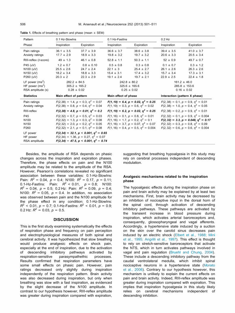
Table 1. Effects of breathing pattern and phase (mean ± SEM)
Pattern 0.1 Hz-SlowIns 0.1 Hz-FastIns 0.2 Hz
Phase Inspiration Expiration Inspiration Expiration Inspiration Expiration
Pain ratings 36.1 ± 3.5 37.7 ± 3.9 36.6 ± 3.7 38.6 ± 3.8 39.4 ± 3.5 41.0 ± 3.7
Anxiety ratings 17.7 ± 2.9 18.9 ± 3.3 19.8 ± 3.2 19.7 ± 3.2 20.6 ± 3.3 20.5 ± 3.4
RIII-reflex (t-score) 49 ± 1.3 46.1 ± 0.8 52.8 ± 1.1 50.3 ± 1.1 52 ± 0.9 49.7 ± 0.7
P45 (uV) 1.2 ± 0.7 0.8 ± 0.10 0.5 ± 0.8 0.3 ± 0.8 0.1 ± 0.7 0.3 ± 1.2
N100 (uV) �25.5 ± 2.6 �24.7 ± 2.4 �22.1 ± 3 �25.4 ± 2.7 �26.1 ± 2.6 �26.3 ± 2.6
N150 (uV) �18.2 ± 3.4 �18.8 ± 3.3 �15.4 ± 3.1 �17.4 ± 3.2 �15.7 ± 3.4 �17.3 ± 3.1
P260 (uV) 20.3 ± 2 22.3 ± 2.9 19.1 ± 2.4 19.7 ± 2.1 22.9 ± 2.5 22.4 ± 1.8
LF power (ms2) 282.2 ± 84.5 242.8 ± 80.2 161.2 ± 46.0
HF power (ms2) 305.2 ± 165.2 325.6 ± 165.6 285.8 ± 153.6
RSA amplitude (s) 0.28 ± 0.02 0.25 ± 0.02 0.16 ± 0.02
Statistics Main effect of pattern Main effect of phase Interaction (pattern X phase)
Pain ratings F(2,38) = 1.4, p= 0.3, g2 = 0.07 F(1,19) = 6.4, p = 0.02, g2 = 0.25 F(2,38) = 0.1, p= 0.9, g2 = 0.01
Anxiety ratings F(2,38) = 0.8, p= 0.4, g2 = 0.04 F(1,19) = 0.3, p= 0.6, g2 = 0.02 F(2,38) = 1.0, p= 0.4, g2 = 0.05
RIII-reflex F(2,38) = 4.8, p = 0.01, g2 = 0.2 F(1,19) = 6.2, p = 0.02, g2 = 0.25 F(2,38) = 0.2, p= 0.9, g2 = 0.01
P45 F(2,32) = 0.7, p= 0.5, g2 = 0.03 F(1,16) = 0.1, p= 0.8, g2 = 0.01 F(2,32) = 0.1, p= 0.9, g2 = 0.004
N100 F(2,32) = 1.3, p= 0.3, g2 = 0.08 F(1,16) = 1.7, p= 0.2, g2 = 0.1 F(2,32)= 3.3, p= 0.048, g2 = 0.17
N150 F(2,32) = 2.0, p= 0.2, g2 = 0.05 F(1,16) = 3.7, p= 0.07, g2 = 0.07 F(2,32) = 0.3, p= 0.8, g2 = 0.09
P260 F(2,32) = 2.1, p= 0.1, g2 = 0.08 F(1,16) = 0.4, p= 0.5, g2 = 0.004 F(2,32) = 0.6, p= 0.6, g2 = 0.004
LF power F(2,34) = 32.1, p < 0.001, g2 = 0.65
HF power F(2,34) = 1.36, p= 0.27, g2 = 0.07
RSA amplitude F(2,34)= 47.5, p < 0.001, g2 = 0.74
506 M. Arsenault et al. / Neuroscience 252 (2013) 501–511
Besides, the amplitude of RSA depends on phasic
changes across the inspiration and expiration phases.
Therefore, the phase effects on pain and the N100
amplitude may be related to the amplitude of the RSA.
However, Pearson’s correlations revealed no significant
association between these variables: 0.1-Hz-SlowIns:
Pain: R2 = 0.04, p= 0.4; N100: R2 = 0.17, p= 0.11;
0.1-Hz-FastIns: Pain: R2 < 0.01, p= 0.8; N100:
R2 = 0.04, p= 0.5; 0.2 Hz: Pain: R2 = 0.09, p= 0.4;
N100: R2 = 0.05, p= 0.4. In addition, no association
was observed between pain and the N100 amplitude for
the phase effect in any condition: 0.1-Hz-SlowIns:
R2 < 0.01, p= 0.7; 0.1-Hz-FastIns: R2 < 0.01, p= 0.9;
0.2 Hz: R2 = 0.03, p= 0.5.
DISCUSSION
This is the first study examining systematically the effects
of respiration phase and frequency on pain perception
and electrophysiological measures of both spinal and
cerebral activity. It was hypothesized that slow breathing
would produce analgesic effects on shock pain,
especially at the end of inspiration, due to the activation
of descending inhibitory pathways activated by
respiration-sensitive parasympathethic processes.
Results confirmed that respiration parameters have
some small effects on phasic pain. However, pain
ratings decreased only slightly during inspiration
independently of the respiration pattern. Brain activity
was also decreased during inspiration, but only when
breathing was slow with a fast inspiration, as evidenced
by the slight decrease of the N100 amplitude. In
contrast to our hypothesis however, RIII-reflex amplitude
was greater during inspiration compared with expiration,
suggesting that breathing hypoalgesia in this study may
rely on cerebral processes independent of descending
modulation.
Analgesic mechanisms related to the inspirationphase
The hypoalgesic effects during the inspiration phase on
pain and brain activity may be explained by at least two
mechanisms. First, brain activity may be decreased by
an inhibition of nociceptive input in the dorsal horn of
the spinal cord, through activation of descending
inhibitory pathways. These pathways are activated by
the transient increase in blood pressure during
inspiration, which activates arterial baroreceptors and,
consequently, glossopharyngeal and vagal afferents.
Accordingly, a hypertensive state induced by a suction
on the skin over the carotid sinus decreases pain
induced by an electric shock (Elbert et al., 1988; Mini
et al., 1995; Angrilli et al., 1997). This effect is thought
to rely on stretch-sensitive baroreceptors that activate
the NTS, which in turn activates pathways involved in
vagal and pain regulation (Bruehl and Chung, 2004).
These include a descending inhibitory pathway from the
caudal ventrolateral medulla, which inhibit spinal
nociceptive neurons in a hypertensive state (Morato
et al., 2006). Contrary to our hypothesis however, this
mechanism is unlikely to explain the current effects on
pain and brain activity. Indeed, RIII-reflex amplitude was
greater during inspiration compared with expiration. This
implies that inspiration hypoalgesia in this study likely
relies on cerebral mechanisms independent of
descending inhibition.

(A)
(B)
(C)
(D)
Fig. 2. Modulation of pain and anxiety ratings, RIII-reflex amplitude and N100 scalp-evoked-potentials. Histograms represent the mean value and
the SEM of (A) pain ratings, (B) shock-anxiety ratings, (C) RIII amplitude presented in t-score and (D) N100 scalp-evoked-potentials across the six
conditions. Significant differences were found in pain ratings, RIII and N100 as reported in A, C and D (⁄p< 0.05, ⁄⁄p< 0.01).
M. Arsenault et al. / Neuroscience 252 (2013) 501–511 507
Another possibility to consider in explaining the effect
of inspiration on pain is the distraction effect of cued
respiration. Accordingly, selective attention may have
been directed away from the shock by the respiration
cue, at a different degree for each condition. In a
previous study investigating human intracranial cortical
responses evoked by painful electrical stimulation of the
sural nerve, it has been shown that the central
negativity from 70 to 110 ms post stimulation is
associated with the activity in the supplementary
somatosensory area (Dowman et al., 2007). This area is
located posterior to the primary somatosensory cortex, it
receives input from the medial thalamic nuclei
(Schmahmann and Pandya, 1990) and its activation
would be related to attention toward pain (Dowman
et al., 2007). This raises the possibility that the
reduction of the N100 amplitude recorded at Cz from 90
to 120 ms in the current study may reflect a shift of
attention from the painful shock toward the voluntary
control of respiration. However, respiration was cued in
the same way across conditions so it seems unlikely
that attention explains the phase effect on pain and the
N100 amplitude. Nevertheless, this possibility cannot be
completely excluded since fast inspiration, the condition
during which the phase effect was observed on the
N100 amplitude, may have required more attention
resources. Another result supporting the involvement of
attention processes is the greater RIII-reflex amplitude
during the inspiration phase compared with the
expiration phase. Indeed, the RIII-reflex amplitude is
sometimes increased or dissociated from pain
perception by distraction (Terkelsen et al., 2004; Roy
et al., 2011). Therefore, although the specific
mechanisms underlying the phase effects in the current
study are not clear, the small hypoalgesic effect found
during inspiration likely involves cerebral processes
independent from descending inhibition of spinal
nociceptive transmission. The recording of scalp-
evoked-potentials in the present study only captured
phasic activity associated with the painful shock. Other
cerebral processes not measured here may have
contributed to the observed hypoalgesic effects,
including activation of the left insula by slow breathing
itself (Rosenkranz et al., 2005) and activation of the left
anterior cingulate cortex that is associated with
increased heart rate variability (Matthews et al., 2004),
as suggested in a previous study (Zautra et al., 2010).
Effect of breathing frequency on pain perception
Manipulation of the breathing pattern induced robust
changes in cardiac activity. As predicted, the two slow
breathing conditions increased the RSA amplitude, likely
reflecting reflex modulation of vagal efferent activity.
Slow deep breathing is known to produce a sharp
increase in intra-thoracic pressure, venous return and

Fig. 3. Grand mean average of scalp-evoked-potentials at Cz. The amplitude of the N100 component of the sural nerve potentials was decreased
during the inspiration in the 0.1-Hz-SlowIns condition. However, the P45, N150 and P260 were unaffected by respiration phase and frequency. The
N100 component is indicated on the graph of the 0.1-Hz-SlowIns condition.
508 M. Arsenault et al. / Neuroscience 252 (2013) 501–511
systolic blood pressure (Triedman and Saul, 1994). The
associated increase in respiratory volume also
increases RSA amplitude (Grossman et al., 1990).
Indeed, the greater activation of vagal afferents by the
activation of baroreceptors and pulmonary stretch
receptors during inspiration decreases vagal efferent
activity, resulting in a greater amplitude of the RSA
(Berntson et al., 1993). However, slow breathing and
the associated changes in RSA amplitude did not
explain pain modulation in the current study, contrary to
our hypothesis.
Our result contrasts with those of previous studies
showing that heat pain threshold and tolerance
increased with slow deep breathing (Chalaye et al.,
2009), and that pain induced by sural nerve stimulation
slightly decreased with slow breathing (Martin et al.,
2012). One aspect to consider in relation to the study by
Chalaye et al. is that brief electrical pain involves
primarily a strong alerting component that may be less
prone to modulation by respiration patterns compared
with a natural longer lasting stimulus such as heat pain.
As for the discrepancy observed with the study by
Martin et al., electrical stimulation evoked only very
slight pain and no pain at all in some trials and possibly
in some subjects in that previous report (i.e. the mean
pain report is at pain threshold in that study) so it is not
clear that these previous results can be generalized to
moderate pain, as reported in the current study.
Changes in vagal activity induced by the breathing
pattern were not associated with the magnitude of pain
modulation in the current study (consistent with Martin
et al., 2012). This contrasts with previous studies on
meditation and relaxation in which slow breathing was
suggested as a potential analgesic mechanism (Grant
and Rainville, 2009). Indeed, the current results indicate
that voluntary regulation of respiration may not be
M. Arsenault et al. / Neuroscience 252 (2013) 501–511 509
sufficient to explain analgesia in these interventions.
Therefore, the effects of meditation may be more
specifically related to processes associated with
relaxation and mindful states. The current results also
imply that the significant analgesic effects reported in
previous studies using biofeedback (Hassett et al.,
2007), Qigong (Lee et al., 2007), yoga (Posadzki et al.,
2011), relaxation (Miller and Perry, 1990) and meditation
(Grant and Rainville, 2009) are unlikely to be explained
solely by changes in respiration. However, in these
studies, pain was not produced by electrical stimulation
and it may, at least in part, explain the discrepancy with
our results, as mentioned above. In a recent study on
the effect of different kinds of deep and slow breathing
on thermal pain perception, deep slow breathing
decreased pain sensitivity (increased pain threshold) but
only when it was combined with relaxation instructions
(Busch et al., 2012). Decreased pain sensitivity was
also associated with a decrease in sympathetic activity
(galvanic skin response) but only during relaxing deep
slow breathing. Based on these studies and on the
current results, we suggest that the analgesic effects of
slow breathing are not sufficient to account for analgesia
induced by relaxation techniques centered on the
regulation and monitoring of respiration.
Study limitations and future directions
An important factor that should be considered for slow
breathing analgesia is the modulation of anxiety. It has
been suggested that slow breathing facilitates emotional
regulation and the maintenance of homeostasis under
challenging conditions (Zautra et al., 2010). In addition,
RSA reactivity has been linked with adaptive emotional
regulation (Gentzler et al., 2009). Therefore, anxiety
modulation may significantly contribute to breathing
analgesia. In the current study, baseline pain-related
anxiety was low, which may decrease the sensitivity to
detect anxiety changes. In addition, pain-related anxiety
was not affected by the respiration phase and
frequency. This may partly explain the modest
hypoalgesic effect observed in the current study. As
pain-related anxiety and negative emotions are
associated with hyperalgesia (Rainville, 2004; Rainville
et al., 2005; Rhudy et al., 2008) slow breathing may
have anti-hyperalgesic effects, rather than direct
analgesic effects, which are more likely to be observed
in anxiogenic clinical conditions or in individuals showing
higher levels of pain-related anxiety. Another limitation
of the current study is related to the pain stimulus.
Transcutaneous electrical stimulation in the current
experiment was motivated by the possibility of
measuring spinal and cerebral responses associated
with sural nerve activity. However, pain evoked by such
brief electrical stimuli may not be as sensitive to
respiration-analgesia or anti-hyperalgesia as pain
induced by longer lasting natural stimuli or clinical pain.
It should also be mentioned that changes in pain and
evoked potential amplitude were only partly consistent
and were not correlated with each other. Finally,
corrections for multiple comparisons were not applied in
this study, although it comprises several dependent
variables and a low number of subjects. Therefore, we
cannot completely rule out false positive results,
especially given that the effects were of very small
amplitude. Therefore, the current findings need to be
replicated in future studies.
CONCLUSION
In the current study, manipulation of respiration
parameters produced a very small effect on pain and
the associated brain activity. However, significant
changes were limited to the respiration phase while the
respiration frequency did not affect pain or brain activity.
In addition, pain inhibition was marginal in comparison
to studies on meditation, relaxation, yoga or qi-Jong, in
which the regulation of respiration is a key component.
This suggests that the analgesic effect of these
techniques is not only explained by autonomic changes
induced by respiration. Nevertheless, the current results
combined with the available literature on respiration-
related effects support further investigation of the effects
of deep inspiration on acute pain.
Acknowledgments—This study was funded by operating Grants
from the National Science and Engineering Research Council
of Canada (M.P.), the Canadian Institutes of Health Research
(CIHR; P.R. and M.P.), and the UQTR research chair in pain neu-
rophysiology (M.P.). Marianne Arsenault was supported by
‘‘Fonds de la recherche en sante du Quebec’’ (FRSQ) and CIHR.
REFERENCES
Adams ED, Bianchi AL (2008) A practical approach to labor support.
J Obstet Gynecol Neonatal Nurs 37:106–115.
Angrilli A, Mini A, Mucha RF, Rau H (1997) The influence of low blood
pressure and baroreceptor activity on pain responses. Physiol
Behav 62:391–397.
Bernardi L, Porta C, Gabutti A, Spicuzza L, Sleight P (2001)
Modulatory effects of respiration. Auton Neurosci 90:47–56.
Berntson GG, Bigger Jr JT, Eckberg DL, Grossman P, Kaufmann PG,
Malik M, Nagaraja HN, Porges SW, Saul JP, Stone PH, van der
Molen MW (1997) Heart rate variability: origins, methods, and
interpretive caveats. Psychophysiology 34:623–648.
Berntson GG, Cacioppo JT, Quigley KS (1993) Respiratory sinus
arrhythmia: autonomic origins, physiological mechanisms, and
psychophysiological implications. Psychophysiology 30:183–196.
Bertisch SM, Wee CC, Phillips RS, McCarthy EP (2009) Alternative
mind-body therapies used by adults with medical conditions. J
Psychosom Res 66:511–519.
Bossut DF, Whitsel EA, Maixner W (1992) A parametric analysis of
the effects of cardiopulmonary vagal electrostimulation on the
digastric reflex in cats. Brain Res 579:253–260.
Brown RP, Gerbarg PL (2005a) Sudarshan Kriya Yogic breathing in
the treatment of stress, anxiety, and depression. Part II – clinical
applications and guidelines. J Altern Complement Med
11:711–717.
Brown RP, Gerbarg PL (2005b) Sudarshan Kriya Yogic breathing in
the treatment of stress, anxiety, and depression: part I-
neurophysiologic model. J Altern Complement Med 11:189–201.
Bruehl S, Chung OY (2004) Interactions between the cardiovascular
and pain regulatory systems: an updated review of mechanisms
and possible alterations in chronic pain. Neurosci Biobehav Rev
28:395–414.
Busch V, Magerl W, Kern U, Haas J, Hajak G, Eichhammer P (2012)
The effect of deep and slow breathing on pain perception,

510 M. Arsenault et al. / Neuroscience 252 (2013) 501–511
autonomic activity, and mood processing – an experimental study.
Pain Med 13:215–228.
Chalaye P, Goffaux P, Lafrenaye S, Marchand S (2009) Respiratory
effects on experimental heat pain and cardiac activity. Pain Med
10:1334–1340.
Chapleau MW (2012) Baroreceptor reflexes. In: Robertson D,
Biaggioni I, Burnstock G, Low PA, Paton JFR, editors. Primer
on the autonomic nervous system. Elsevier.
D’Antono B, Ditto B, Sita A, Miller SB (2000) Cardiopulmonary
baroreflex stimulation and blood pressure-related hypoalgesia.
Biol Psychol 53:217–231.
Delorme A, Makeig S (2004) EEGLAB: an open source toolbox for
analysis of single-trial EEG dynamics including independent
component analysis. J Neurosci Methods 134:9–21.
Dowman R, Darcey T, Barkan H, Thadani V, Roberts D (2007)
Human intracranially-recorded cortical responses evoked by
painful electrical stimulation of the sural nerve. NeuroImage
34:743–763.
Elbert T, Rockstroh B, Lutzenberger W, Kessler M, Pietrowsky R
(1988) Baroreceptor stimulation alters pain sensation depending
on tonic blood pressure. Psychophysiology 25:25–29.
Gauthier J, Bouchard S (1993) Adaptation canadienne-francaise de
la forme revisee du ‘‘state-trait anxiety inventory’’ de Spielberger.
Can J Behav Sci 25:559–578.
Gentzler AL, Santucci AK, Kovacs M, Fox NA (2009) Respiratory
sinus arrhythmia reactivity predicts emotion regulation and
depressive symptoms in at-risk and control children. Biol
Psychol 82:156–163.
Grant JA, Rainville P (2009) Pain sensitivity and analgesic effects of
mindful states in Zen meditators: a cross-sectional study.
Psychosom Med 71:106–114.
Grossman P, Niemann L, Schmidt S, Walach H (2004) Mindfulness-
based stress reduction and health benefits. A meta-analysis. J
Psychosom Res 57:35–43.
Grossman P, Stemmler G, Meinhardt E (1990) Paced respiratory
sinus arrhythmia as an index of cardiac parasympathetic
tone during varying behavioral tasks. Psychophysiology
27:404–416.
Hassett AL, Radvanski DC, Vaschillo EG, Vaschillo B, Sigal LH,
Karavidas MK, Buyske S, Lehrer PM (2007) A pilot study of the
efficacy of heart rate variability (HRV) biofeedback in patients with
fibromyalgia. Appl Psychophysiol Biofeedback 32:1–10.
Iglesias SL, Azzara S, Argibay JC, Arnaiz ML, de Valle Carpineta M,
Granchetti H, Lagomarsino E (2012) Psychological and
physiological response of students to different types of stress
management programs. Am J Health Promot 26:e149–158.
Kim SD, Kim HS (2005) Effects of a relaxation breathing exercise on
anxiety, depression, and leukocyte in hemopoietic stem cell
transplantation patients. Cancer Nurs 28:79–83.
Lee MS, Pittler MH, Ernst E (2007) External qigong for pain
conditions: a systematic review of randomized clinical trials. J
Pain 8:827–831.
Lehrer PM, Vaschillo E, Vaschillo B (2000) Resonant frequency
biofeedback training to increase cardiac variability: rationale and
manual for training. Appl Psychophysiol Biofeedback 25:177–191.
Maixner W, Bossut DF, Whitsel EA (1991) Evaluation of vagal
afferent modulation of the digastric reflex in cats. Brain Res
560:55–62.
Maixner W, Randich A (1984) Role of the right vagal nerve trunk in
antinociception. Brain Res 298(2):374–377.
MartinSL,KerrKL,BartleyEJ,KuhnBL,PalitS,TerryEL,DelVenturaJL,
Rhudy JL (2012) Respiration-induced hypoalgesia: exploration of
potentialmechanisms. JPain 13:755–763.
Matthews SC, Paulus MP, Simmons AN, Nelesen RA, Dimsdale JE
(2004) Functional subdivisions within anterior cingulate cortex
and their relationship to autonomic nervous system function.
NeuroImage 22:1151–1156.
Meuret AE, Rosenfield D, Seidel A, Bhaskara L, Hofmann SG (2010)
Respiratory and cognitive mediators of treatment change in panic
disorder: evidence for intervention specificity. J Consult Clin
Psychol 78:691–704.
Miller KM, Perry PA (1990) Relaxation technique and postoperative
pain in patients undergoing cardiac surgery. Heart Lung
19:136–146.
Mini A, Rau H, Montoya P, Palomba D, Birbaumer N (1995)
Baroreceptor cortical effects, emotions and pain. Int J
Psychophysiol 19:67–77.
Morato M, Pinho D, Sousa T, Tavares I, Albino-Teixeira A (2006)
Inhibition of nociceptive responses of spinal cord neurones during
hypertension involves the spinal GABAergic system and a pain
modulatory center located at the caudal ventrolateral medulla. J
Neurosci Res 83:647–655.
Peretz B, Gluck GM (1999) Assessing an active distracting technique
for local anesthetic injection in pediatric dental patients:
repeated deep breathing and blowing out air. J Clin Pediatr
Dent 24:5–8.
Piche M, Bouin M, Arsenault M, Poitras P, Rainville P (2011)
Decreased pain inhibition in irritable bowel syndrome depends on
altered descending modulation and higher-order brain processes.
Neuroscience 195:166–175.
Posadzki P, Ernst E, Terry R, Lee MS (2011) Is yoga effective for
pain? A systematic review of randomized clinical trials.
Complement Ther Med 19:281–287.
Rainville P (2004) Pain and emotions. In: Price DD, Bushnell MC,
editors. Psychological methods of pain control: basic science and
clinical perspectives⁄⁄⁄⁄. Progress in Pain Research and
Management: In. p. 117–141.
Rainville P, Bao QV, Chretien P (2005) Pain-related emotions
modulate experimental pain perception and autonomic
responses. Pain 118:306–318.
Randich A, Maixner W (1984) Interactions between cardio-
vascular and pain regulatory systems. Neurosci Biobehav Rev
8:343–367.
Rhudy JL, Williams AE, McCabe KM, Russell JL, Maynard LJ (2008)
Emotional control of nociceptive reactions (ECON): do affective
valence and arousal play a role? Pain 136:250–261.
Rosenkranz MA, Busse WW, Johnstone T, Swenson CA, Crisafi GM,
Jackson MM, Bosch JA, Sheridan JF, Davidson RJ (2005) Neural
circuitry underlying the interaction between emotion and asthma
symptom exacerbation. Proc Natl Acad Sci U S A
102:13319–13324.
Roy M, Lebuis A, Peretz I, Rainville P (2011) The modulation
of pain by attention and emotion: a dissociation of
perceptual and spinal nociceptive processes. Eur J Pain
15(641):e641–610.
Schmahmann JD, Pandya DN (1990) Anatomical investigation of
projections from thalamus to posterior parietal cortex in the rhesus
monkey: a WGA-HRP and fluorescent tracer study. J Comp
Neurol 295:299–326.
Spielberger CD, Gorsuch PR, Lushene PR, Jacobs VAG (1983)
Manual for the state-trait anxiety inventory (Form Y). Palo Alto,
CA: Consulting Psychologists Press, Inc..
Sullivan MJL, Biship SR, Pivik J (1995) The pain catastrophizing
scale: development and validation. Psychol Assess 7:524–532.
Task Force of the European Society of Cardiology. Heart rate
variability: standards of measurement Piacu (1996) Heart rate
variability: standards of measurement, physiological interpretation
and clinical use. Circulation 93:1043–1065.
Terkelsen AJ, Andersen OK, Molgaard H, Hansen J, Jensen TS
(2004) Mental stress inhibits pain perception and heart rate
variability but not a nociceptive withdrawal reflex. Acta Physiol
Scand 180:405–414.
Triedman JK, Saul JP (1994) Blood pressure modulation by central
venous pressure and respiration. Buffering effects of the heart
rate reflexes. Circulation 89:169–179.
Vaschillo EG, Vaschillo B, Lehrer PM (2006) Characteristics of
resonance in heart rate variability stimulated by biofeedback. Appl
Psychophysiol Biofeedback 31:129–142.

M. Arsenault et al. / Neuroscience 252 (2013) 501–511 511
Willer JC (1977) Comparative study of perceived pain and
nociceptive flexion reflex in man. Pain 3:69–80.
Zautra AJ, Fasman R, Davis MC, Craig AD (2010) The effects of slow
breathing on affective responses to pain stimuli: an experimental
study. Pain 149:12–18.
(Accepted 22 July 2013)(Available online 29 July 2013)