Outcome in children with severe head injuries
6
Child's Nerv Syst (1985) 1:109-114 mGlqS © Springer-Verlag 1985 Outcome in children with severe head injuries Javier Esparza I *, Jaime M-Portillo 2, Maria Sarabia 2, Jos6 A. Yuste 2, Ricardo Roger 3 and Eduardo Lamas 1 Pediatric Neurosurgery, 2 Anesthesiology and 3 Neuroradiology, Hospital Infantil, 1° de Octubre, Madrid, Spain Abstract. We present a series of 56 children who suffered severe head injuries, with a Glasgow Coma Score (GCS) of less than 8. The cases were classified according to the type of morphologic lesion on computed tomography (CT) scan. Intracranial pressure (ICP) was monitored in all children in this series. A protocol that included artificial ventilation and other measures of treatment for intra- cranial hypertension was applied. Results were analyzed according to age, type of lesion, and ICP. The usefulness of the ICP recording and of obtaining a correct classi- fication of lesions using the CT scan is emphasized. Key words: Head trauma - Children - CT scan - ICP - Outcome. Among the many factors influencing the final outcome of severe cranial trauma [11], Bruce [2, 3] has analyzed the differences that might exist between adults and children, mentioning that there is a more favorable outcome in the latter. Later, other series have been published that deal with severe head trauma in children [8, 10, 16, 17], but some aspects of the problem remain debatable [8]. Because of this, we think that the experience pediatric neurotrauma units have had with head trauma may be helpful. The purpose of this paper is to analyze some of the aspects that most influence the outcome of severe head trauma in children [e.g., age, the type of lesion on the CT scan, and the intracranial pressure (ICP)] in the series from our children's hospital. Material and methods In the Children's Hospital 1° de Octubre, we selected 56 children who, after suffering cranial trauma, were admitted to the Intensive Care Unit (ICU) between 1980 and 1984. The criteria for including these children in a previously standardized protocol for severe head trauma was a Glasgow Coma Score (GCS) of less * To whom offprint requests should be addressed than 8 at least 6 h following trauma, or subsequent deterioration. We excluded all children with a higher GCS, even if they needed to be admitted to the ICU, or if they had lesions that had to be surgically treated. We also excluded those who had a low GCS transiently as a consequence of seizures, and those in whom extracranial trauma was clearly dominant. The 56 children selected represent 1.06% of all children admitted because of head trauma to our emergency service during the same period of time, and 24.75% of those who were admitted to the ICU because of head trauma. The mean age of our series was 7.6 years (range from 3 months to 14 years). Our children's hospital is an urban tertiary center that receives patients directly from the scene of trauma and also from other primary hospitals. Nevertheless, all cases in the series were admitted within 8 h of trauma. The trauma was caused by a traffic accident in 40 cases and falls from a height in 14 cases; there were also 2 battered children. All patients with severe head trauma who arrive in the hospital are immediately taken to the ICU where, after a quick evaluation of their general and neurologic status, resuscitative measures are stated, including intubation if necessary. This is followed by a radiological check and a CT scan to make the diagnosis. Based on this clinical and radiological information, we decide which cases should be included in the severe head trauma protocol. The protocol includes: intubation for artificial ventila- tion, with a volumetric respirator to maintain arterial pCO2 at 25-30 mm Hg and arterial pO2 over 75 mm Hg; dexamethasone (0.5-2 mg/kg per day) administered in all cases during a mean period of 5 days; monitoring of ICP in the operating room in all cases, by means of an epidural fiberoptic sensor (Ladd system) in 38 cases, and an intraventricular catheter in 18 cases. The average duration of continuous recording of ICP was 7 days. We consider 20ram Hg as the limit at which we start anti-hypertensive measures. These measures firstly include some bolus of mannitol (0.3-0.5 g/kg) controlling blood osmolarity. If this is not enough, we add a bolus of thiopental (3-5 mg/kg). If the previous mea- sures are still not sufficient, we begin continuous administration of thiopental up to a total daily dose of 40 mg/kg, without sup- pression of mannitol administration if blood osmolarity permits. In some cases we have also used hypothermia. Children with severe head trauma whose CT studies sug- gested the need for surgical treatment, were monitored for sub- sequent control of ICP at the same surgical sitting. After this, they continued in the ICU with the protocol explained above. Surgical treatment included evacuation of extra- or intracerebral hema- tomas and resection of contusions. In no case was decompressive craniectomy done, with the exception of instances of depressed skull fractures which required removal of bone fragments. In all cases, CT scan was performed on admission and on the 3rd day. Additional examinations were also performed as indi- cated by ICP changes. Subsequent CT scans were performed at
-
Upload
independent -
Category
Documents
-
view
0 -
download
0
Transcript of Outcome in children with severe head injuries
Child's Nerv Syst (1985) 1:109-114 mGlqS © Springer-Verlag 1985
Outcome in children with severe head injuries
J a v i e r E s p a r z a I *, J a i m e M - P o r t i l l o 2, M a r i a S a r a b i a 2, Jos6 A. Y u s t e 2, R i c a r d o R o g e r 3 a n d E d u a r d o L a m a s 1
Pediatric Neurosurgery, 2 Anesthesiology and 3 Neuroradiology, Hospital Infantil, 1 ° de Octubre, Madrid, Spain
Abstract. We present a series of 56 ch i ldren who suffered severe head injuries, with a Glasgow C o m a Score (GCS) of less than 8. The cases were classified according to the type of morpho log ic lesion on c o m p u t e d t o m o g r a p h y (CT)
scan. In t rac ran ia l pressure ( ICP) was m o n i t o r e d in all ch i ldren in this series. A protocol that i nc luded artificial ven t i la t ion a nd o ther measures of t r ea tmen t for in t ra- cranial hype r t ens ion was applied. Results were ana lyzed according to age, type of lesion, and ICP. The usefulness of the ICP recording and of ob t a in ing a correct classi- f ication of lesions us ing the C T scan is emphas ized .
Key words: Head t r a u m a - Ch i ld ren - C T scan - ICP - Outcome.
A m o n g the m a n y factors in f luenc ing the f inal ou tcome of severe crania l t r a u m a [11], Bruce [2, 3] has ana lyzed the differences that migh t exist be tween adul ts and chi ldren,
m e n t i o n i n g that there is a more favorable ou tcome in the latter. Later, o ther series have been pub l i shed that deal with severe head t r a u m a in ch i ldren [8, 10, 16, 17], bu t some aspects of the p r o b l e m r e m a i n deba tab le [8]. Because of this, we th ink that the experience pedia t r ic n e u r o t r a u m a uni ts have had with head t r a u m a m a y be helpful.
The purpose o f this pape r is to analyze some of the aspects that mos t in f luence the ou tcome o f severe head t r auma in ch i ldren [e.g., age, the type o f les ion on the C T scan, a nd the in t rac ran ia l pressure (ICP)] in the series from our chi ldren ' s hospital .
Material and methods
In the Children's Hospital 1 ° de Octubre, we selected 56 children who, after suffering cranial trauma, were admitted to the Intensive Care Unit (ICU) between 1980 and 1984. The criteria for including these children in a previously standardized protocol for severe head trauma was a Glasgow Coma Score (GCS) of less
* To whom offprint requests should be addressed
than 8 at least 6 h following trauma, or subsequent deterioration. We excluded all children with a higher GCS, even if they needed to be admitted to the ICU, or if they had lesions that had to be surgically treated. We also excluded those who had a low GCS transiently as a consequence of seizures, and those in whom extracranial trauma was clearly dominant.
The 56 children selected represent 1.06% of all children admitted because of head trauma to our emergency service during the same period of time, and 24.75% of those who were admitted to the ICU because of head trauma. The mean age of our series was 7.6 years (range from 3 months to 14 years).
Our children's hospital is an urban tertiary center that receives patients directly from the scene of trauma and also from other primary hospitals. Nevertheless, all cases in the series were admitted within 8 h of trauma. The trauma was caused by a traffic accident in 40 cases and falls from a height in 14 cases; there were also 2 battered children.
All patients with severe head trauma who arrive in the hospital are immediately taken to the ICU where, after a quick evaluation of their general and neurologic status, resuscitative measures are stated, including intubation if necessary. This is followed by a radiological check and a CT scan to make the diagnosis. Based on this clinical and radiological information, we decide which cases should be included in the severe head trauma protocol. The protocol includes: intubation for artificial ventila- tion, with a volumetric respirator to maintain arterial pCO2 at 25-30 mm Hg and arterial pO2 over 75 mm Hg; dexamethasone (0.5-2 mg/kg per day) administered in all cases during a mean period of 5 days; monitoring of ICP in the operating room in all cases, by means of an epidural fiberoptic sensor (Ladd system) in 38 cases, and an intraventricular catheter in 18 cases. The average duration of continuous recording of ICP was 7 days. We consider 20ram Hg as the limit at which we start anti-hypertensive measures. These measures firstly include some bolus of mannitol (0.3-0.5 g/kg) controlling blood osmolarity. If this is not enough, we add a bolus of thiopental (3-5 mg/kg). If the previous mea- sures are still not sufficient, we begin continuous administration of thiopental up to a total daily dose of 40 mg/kg, without sup- pression of mannitol administration if blood osmolarity permits. In some cases we have also used hypothermia.
Children with severe head trauma whose CT studies sug- gested the need for surgical treatment, were monitored for sub- sequent control of ICP at the same surgical sitting. After this, they continued in the ICU with the protocol explained above. Surgical treatment included evacuation of extra- or intracerebral hema- tomas and resection of contusions. In no case was decompressive craniectomy done, with the exception of instances of depressed skull fractures which required removal of bone fragments.
In all cases, CT scan was performed on admission and on the 3rd day. Additional examinations were also performed as indi- cated by ICP changes. Subsequent CT scans were performed at

110
variable intervals, depending upon each particular case. Anti- hypertensive treatment is always suppressed progressively, depending upon the type of lesion and the ICP recording. The latter was maintained for 24-48 h after total cessation of treat- ment. The final outcome was evaluated according to the Jennett and Bond scale, grouping as good outcome all cases of good recovery and moderate disability, and as poor outcome the cases of severe disability, persistent vegetative state, and death.
Pat tern 6
Dif fuse axona l injury - we also used the radiological charac- teristics established by Zimmerman [19] to classify lesions in this pattern, defined as: "eccentric hemorrhage in the corpus callosum, diffuse cerebral swelling, subarachnoid hemorrhage, and less frequently, hemorrhage around the third ventricular region and in the cerebral white matter."
Analyt ic criteria
We classified our children into two groups according to the GCS: group I, those with a GCS of 3, 4 (30 patients in the series); group II, those with a GCS of 5 -7 (the other 26 patients in the series). In order to analyze age as a prognostic factor better, we also classified children into two subgroups: group A, children up to 5years old (14 cases); group B, between 6 and 14 years (42 cases).
To classify the lesions seen on the CT scan morpho- logically, we employed a slightly modified classification of the one recently published by members of our neuro- surgery department [12]. This classification may be sum- marized in seven morphological patterns.
Pat tern 1
Pure ex tracerebral h e m a t o m a - extra or subdural hema- tomas without underlying or accompanying hemispheric lesions.
Pat tern 2
Ex t racerebra l h e m a t o m a p l u s under ly ing hemispher ic
swel l ing - cases presenting extra- or subdural hematomas with associated hemispheric lesion, ipsi- or contralateral, i.e., swelling, contusion or hemorrhagic lesion.
Pat tern 3
S ingle brain contus ion - parenchymatous contusion or hematomas in any location, as well as those depressed skull fractures with underlying contusion and small extra- cerebral hematomas with associated predominant paren- chymatous contusion.
Pat tern 7 N o r m a l C T scan.
Resul ts
C T s c a n (Table 1)
In accordance with the morphological type of lesion found on CT scan, the results may be summarized as follows.
Pat tern 1 - five cases with extradural hematomas and no subdural hematoma. All five patients had normal ICP and made a good recovery. None of the five showed a midline shift in the CT scan performed inmediately after surgery.
Pat tern 2 - seven cases (four extradural and three sub- dural hematomas). The only survivor was a child with an extradural hematoma and contralateral contusion, whose ICP ranged between 20 and 40 mm Hg, and the initial GCS was group II. The other six children died, with raised ICP. There was underlying hemispheric swelling on the CT scan.
Pat tern 3 - nine cases (four frontal, two temporal, and three deep contusions). Two patients in this group died, both with deep contusion. One had severe intracranial hypertension; the rest made a good recovery. Four cases were treated surgically within the first 48 h of trauma, with midline shift on the CT scan and the ICP recording.
Table 1. Distribution of the 56 cases, depending on the CT scan pattern. GCS I: scores 3 and 4. GCS II: scores 5, 6 and 7. Out- come: good - mild disability; poor - disability, persistent vegeta- tive state, or death
Pat tern 4
Mul t ip l e brain contus ion - multiple parenchymatous contusion or hematomas that can be in any location and in one or both hemispheres.
Pat tern 5
Di f fuse brain swel l ing - cases with radiological features defined by Zimmerman [18]: "absence or compression of the lateral and third ventricles and perimesencephalic cisterns", with no focal lesion.
CTscan Glasgow Intracranial pressure Outcome (pattern) Coma (ICP)
Score (GCS)
I II 0-20 20-40 40-60 >60 Good Poor
1 1 4 5 0 0 0 5 0 2 6 1 0 1 2 4 1 6 3 3 6 4 4 0 1 7 2 4 2 0 0 2 0 0 0 2 5 7 12 15 3 0 1 18 1 6 11 2 3 6 1 3 5 8 7 0 1 1 0 0 0 1 0

111
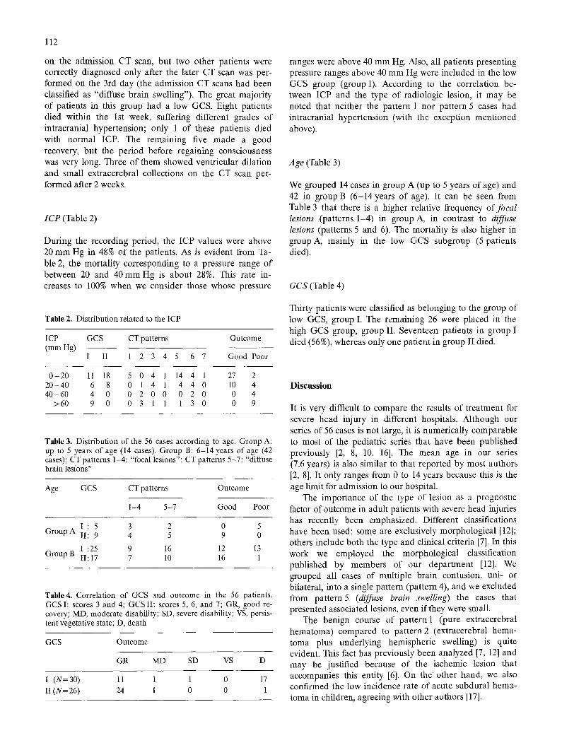
Fig. 1 a-f. Evolution of diffuse brain swelling on the CT scan. In the scan performed on admission (a, b), the collapse of the ven- tricular system is evident, as is the subarachnoid hemorrhage. In the scan performed 2 weeks later (e, d), and after 1 month (e, f), there are slight ventricular dilation and extracerebral fluid col- lections
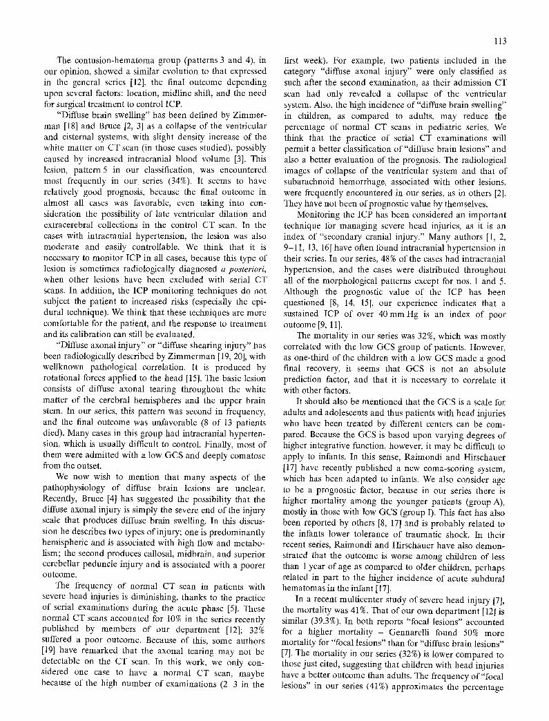
Fig. 2 a-d. CT scan performed on admission showing the collapse of both the perimesencephalic cisterns and the ventricular system. A hemorrhagic area is visible in the white matter of the right hemi- sphere (b, e) and there is some intracranial air
Pattern 4 - only two cases presented multiple bilateral contusion and swelling, and both died with severe intra- cranial hypertension.
Pattern 5 (Fig. 1) - 19 cases presented this radiological lesion (one-third of our series). Eighteen made a good recovery, including 6 of the 7 presenting with low GCS; 3 of the latter 6 had moderate intracranial hypertension, which was easily controlled with treatment. They were
deeply comatose from the outset, and their time period before recovering consciousness was longer. The later CT scan showed moderate ventricular dilation or extra- parenchymatous collections in 13 cases, but none required shunt treatment. Only 1 patient died in this group, who had severe intracranial hypertension.
Pattern 6 (Fig. 2) - 13 patients were in this group (almost one-quarter of the series). Eleven of them were diagnosed
112
on the admission CT scan, but two other patients were correctly diagnosed only after the later CT scan was per- formed on the 3rd day (the admission CT scans had been classified as "diffuse brain swelling"). The great majori ty of patients in this group had a low GCS. Eight patients died within the 1st week, suffering different grades of intracranial hypertension; only 1 of these patients died with normal ICP. The remaining five made a good recovery, but the period before regaining consciousness was very long. Three of them showed ventricular dilation and small extracerebral collections on the CT scan per- formed after 2 weeks.
ICP (Table 2)
During the recording period, the ICP values were above 20 m m Hg in 48% of the patients. As is evident from Ta- ble 2, the mortality corresponding to a pressure range of between 20 and 40 m m Hg is about 28%. This rate in- creases to 100% when we consider those whose pressure
ranges were above 40 m m Hg. Also, all patients presenting pressure ranges above 40 m m Hg were included in the tow GCS group (group I). According to the correlation be- tween ICP and the type of radiologic lesion, it may be noted that neither the pattern 1 nor pattern 5 cases had intracranial hypertension (with the exception ment ioned above).
Age (Table 3)
We grouped 14 cases in group A (up to 5 years of age) and 42 in group B (6-14 years of age). It can be seen from Table 3 that there is a higher relative frequency of focal lesions (patterns 1-4) in group A, in contrast to diffuse lesions (patterns 5 and 6). The mortali ty is also higher in group A, mainly in the low GCS subgroup (5 patients died).
GCS (Table 4)
Table 2. Distribution related to the ICP
ICP GCS CT patterns Outcome (mm Hg)
I II 1 2 3 4 5 6 7 Good Poor
Thirty patients were classified as belonging to the group of low GCS, group I. The remaining 26 were placed in the high GCS group, group II. Seventeen patients in group I died (56%), whereas only one patient in group II died.
0 - 2 0 11 18 5 0 4 1 14 4 1 27 2 20-40 6 8 0 1 4 1 4 4 0 10 4 40-60 4 0 0 2 0 0 0 2 0 0 4
>60 9 0 0 3 1 1 1 3 0 0 9
Table 3. Distribution of the 56 cases according to age. Group A: up to 5 years of age (14 cases). Group B: 6-14 years of age (42 cases); CT patterns 1-4: "focal lesions"; CT patterns 5-7: "diffuse brain lesions"
Age GCS CT patterns Outcome
1-4 5-7 Good Poor
Group A I : 5 3 2 0 5 II: 9 4 5 9 0
Group B I :25 9 16 12 13 II: 17 7 10 16 1
Table4. Correlation of GCS and outcome in the 56 patients. GCS I: scores 3 and 4; GCS II: scores 5, 6, and 7; GR, good re- covery; MD, moderate disability; SD, severe disability; VS, persis- tent vegetative state; D, death
GCS Outcome
GR MD SD VS D
I (N= 30) 11 1 1 0 17 II (N= 26) 24 1 0 0 1
Discuss ion
It is very difficult to compare the results of t reatment for severe head injury in different hospitals. Although our series of 56 cases is not large, it is numerically comparable to most o f the pediatric series that have been published previously [2, 8, 10, 161. The mean age in our series (7.6 years) is also similar to that reported by most authors [2, 8]. It only ranges from 0 to 14 years because this is the age limit for admission to our hospital.
The importance of the type of lesion as a prognostic factor of outcome in adult patients with severe head injuries has recently been emphasized. Different classifications have been used: some are exclusively morphological [12]; others include both the type and clinical criteria [7]. In this work we employed the morphological classification published by members of our depar tment [12]. We grouped all cases of multiple brain contusion, uni- or bilateral, into a single pattern (pattern 4), and we excluded from pa t te rn5 (diffuse brain swelling) the cases that presented associated lesions, even if they were small.
The benign course of pattern 1 (pure extracerebral hematoma) compared to pa t t e rn2 (extracerebral hema- toma plus underlying hemispheric swelling) is quite evident. This fact has previously been analyzed [7, 12] and may be justified because of the ischemic lesion that accompanies this entity [6]. On the other hand, we also confirmed the low incidence rate of acute subdural hema- toma in children, agreeing with other authors [17].
113
The contusion-hematoma group (patterns 3 and 4), in our opinion, showed a similar evolution to that expressed in the general series [12], the final outcome depending upon several factors: location, midline shift, and the need for surgical treatment to control ICP.
"Diffuse brain swelling" has been defined by Zimmer- man [18] and Bruce [2, 3] as a collapse of the ventricular and cisternal systems, with slight density increase of the white matter on CT scan (in those cases studied), possibly caused by increased intracranial blood volume [3]. This lesion, pattern 5 in our classification, was encountered most frequently in our series (34%). It seems to have relatively good prognosis, because the final outcome in almost all cases was favorable, even taking into con- sideration the possibility of late ventricular dilation and extracerebral collections in the control CT scan. In the cases with intracranial hypertension, the lesion was also moderate and easily controllable. We think that it is necessary to monitor ICP in all cases, because this type of lesion is sometimes radiologically diagnosed a posteriori, when other lesions have been excluded with serial CT scans. In addition, the ICP monitoring techniques do not subject the patient to increased risks (especially the epi- dural technique). We think that these techniques are more comfortable for the patient, and the response to treatment and its calibration can still be evaluated.
"Diffuse axonal injury" or "diffuse shearing injury" has been radiologically described by Zimmerman [19, 20], with wellknown pathological correlation. It is produced by rotational forces applied to the head [15]. The basic lesion consists of diffuse axonal tearing throughout the white matter of the cerebral hemispheres and the upper brain stem. In our series, this pattern was second in frequency, and the final outcome was unfavorable (8 of 13 patients died). Many cases in this group had intracranial hyperten- sion, which is usually difficult to control. Finally, most of them were admitted with a low GGS and deeply comatose from the outset.
We now wish to mention that many aspects of the pathophysiology of diffuse brain lesions are unclear. Recently, Bruce [4] has suggested the possibility that the diffuse axonal injury is simply the severe end of the injury scale that produces diffuse brain swelling. In this discus- sion he describes two types of injury; one is predominantly hemispheric and is associated with high flow and metabo- lism; the second produces callosal, midbrain, and superior cerebellar peduncle injury and is associated with a poorer o u t c o m e .
The frequency of normal CT scan in patients with severe head injuries is diminishing, thanks to the practice of serial examinations during the acute phase [5]. These normal CT scans accounted for 10% in the series recently published by members of our department [12]; 32% suffered a poor outcome. Because of this, some authors [19] have remarked that the axonal tearing may not be detectable on the CT scan. In this work, we only con- sidered one case to have a normal CT scan, maybe because of the high number of examinations (2-3 in the
first week). For example, two patients included in the category "diffuse axonal injury" were only classified as such after the second examination, as their admission CT scan had only revealed a collapse of the ventricular system. Also, the high incidence of "diffuse brain swelling" in children, as compared to adults, may reduce the percentage of normal CT scans in pediatric series. We think that the practice of serial CT examinations will permit a better classification of "diffuse brain lesions" and also a better evaluation of the prognosis. The radiological images of collapse of the ventricular system and that of subarachnoid hemorrhage, associated with other lesions, were frequently encountered in our series, as in others [2]. They have not been of prognostic value by themselves.
Monitoring the ICP has been considered an important technique for managing severe head injuries, as it is an index of "secondary cranial injury." Many authors [1, 2, 9-11, 13, 16] have often found intracranial hypertension in their series. In our series, 48% of the cases had intracranial hypertension, and the cases were distributed throughout all of the morphological patterns except for nos. 1 and 5. Although the prognostic value of the ICP has been questioned [8, 14, 15], our experience indicates that a sustained ICP of over 40 mm Hg is an index of poor outcome [9, 11].
The mortality in our series was 32%, which was mostly correlated with the low GCS group of patients. However, as one-third of the children with a low GCS made a good final recovery, it seems that GCS is not an absolute prediction factor, and that it is necessary to correlate it with other factors.
It should also be mentioned that the GCS is a scale for adults and adolescents and thus patients with head injuries who have been treated by different centers can be com- pared. Because the GCS is based upon varying degrees of higher integrative function, however, it may be difficult to apply to infants. In this sense, Raimondi and Hirschauer [17] have recently published a new coma-scoring system, which has been adapted to infants. We also consider age to be a prognostic factor, because in our series there is higher mortality among the younger patients (group A), mostly in those with low GCS (group I). This fact has also been reported by others [8, 17] and is probably related to the infants lower tolerance of traumatic shock. In their recent series, Raimondi and Hirschauer have also demon- strated that the outcome is worse among children of less than 1 year of age as compared to older children, perhaps related in part to the higher incidence of acute subdural hematomas in the infant [17].
In a recent multicenter study of severe head injury [7], the mortality was 41%. That of our own department [12] is similar (39.3%). In both reports "focal lesions" accounted for a higher mortality - Gennarelli found 50% more mortality for "focal lesions" than for "diffuse brain lesions" [7]. The mortality in our series (32%) is lower compared to those just cited, suggesting that children with head injuries have a better outcome than adults. The frequency of "focal lesions" in our series (41%) approximates the percentage
114
cited by the above-mentioned authors. It is higher than the percentages reported by some pediatric series published [2, 81. The incidence of "focal lesions" in our series was higher, as many of these patients were younger children, and the incidence may be related to the higher frequency of falls associated with a higher mortality.
We conclude that a morphological classification of the CT findings and ICP recordings is useful for both treat- ment and better prediction of the prognosis o f children with severe head injuries. Some authors [4] have reported a much better outcome in children with head injuries compared to adults. However, according to our own experience, as well as that of other authors [8], the mortality in children with head injuries is only slightly lower than that of adults. Therefore, we believe that this mortality (over 30%) shows that severe head trauma in children is still a worrying problem.
References
1. Becker DP, Miller JD, Ward JD, Greenberg RP, Young HF, Sakalas R (1977) The outcome from severe head injury with early diagnosis and intensive management. J Neurosurg 47:491-502
2. Bruce DA, Raphaely RC, Goldberg AI, Zimmerman RA, Bilaniuk LT, Schut U Kuhl DE (1979) Pathophysiology, treat- ment and outcome following severe head injury in children. Child's Brain 5:174-191
3. Bruce DA, Alavi A, Bilaniuk L, Dolinskas C, Obrist W, Uzzelt B (1981) Diffuse cerebral swelling following head injuries in children: the syndrome of malignant brain edema. J Neuro- surg 54:170 178
4. Bruce DA (1982) Clinical reflections of brain stem injury in children. Excerpta Medica, Amsterdam (Advances in neuro- traumatology)
5. Cooper PR, Maravilla K, Moody S (1979) Serial computerized tomographic scanning and the prognosis of severe head injury. Neurosurgery 5:566-569
6. Gennarelli TA, Thibault LE (1982) Biomechanics of acute subdural hematoma. J Trauma 22:680-686
7. Gennarelli TA, Spielman GM, Langfitt TW, Gildenberg TL, Harrington T, Jane JA, Marshall LF, Miller JD, Pitts LH (1982) Influence of the type of intracranial lesion on outcome from severe head injury: a multicenter study using a new classification system. J Neurosurg 56:26-32
8. Humphreys RP (1983) Outcome of severe head injury in children. Concepts in pediatric neurosurgery III. Karger, Basel
9. Johnston IH, Johnston JA, Jennett B (1970) Intracranial pressure changes following head injury. Lancet I:433-436
10. Klages G (1982) Monitoring of intracranial pressure in com- parison to other clinical data in judgement of the course of severe head-injured children. Monogr Paediatr 15: 88-91
11. Langfitt TW, Gennarelli TA (1982) Can the outcome from head injury be improved? J Neurosurg 56:19-25
12. Lobato RD, Cordob6s F, Rivas JA, De la Fuente M, Montero A, BSrcena A, P6rez C, Cabrera A, Lamas E (1983) Outcome from severe head injury related to the type of intra- cranial lesion. A computerized tomography study. J Neuro- surg 59:762-774
13. Miller J (1977) Significance of intracranial hypertension in severe head injury. J Neurosurg 47:503-516
14. Narayan RK, Greenberg RP, Miller JD (1981) Improved con- fidence of outcome prediction in severe head injury. A com- parative analysis of the clinical examination, multimodality evoked potentials, CT scanning, and intracranial pressure. J Neurosurg 54:751-762
15. Ommaya AK, Gennarelli TA (1976) A physiopathologic basis for noninvasive diagnosis and prognosis of head injury severity. Head injuries. Grune & Stratton, New York
16. Papo I, Caruselli G, Luongo A (1982) Intracranial pressure in children and adolescents with severe head injuries. Monogr Paediatr 15:75-78
17. Raimondi AJ, Hirschauer J (1984) Head injury in the infant and toddler. Coma scoring and outcome scale. Child's Brain 11:12-35
18. Zimmerman RA, Bilaniuk LT, Bruce D, Dolinskas C, Obrist W, Kuhl D (1978) Computed tomography of pediatric head trauma: acute general cerebral swelling. Radiology 126: 403-408
19. Zimmerman RA, Bilaniuk LI, Gennarelli T (1978) Computed tomography of shearing injuries of the cerebral white matter. Radiology 127:393-396
20. Zimmerman RA, Bilaniuk LT (1979) Computed tomography in diffuse traumatic cerebral injury. Neural trauma. Raven Press, New York