Outcome after laparoscopic antireflux surgery and hiatal ...
171
Outcome after laparoscopic antireflux surgery and hiatal hernia repair Citation for published version (APA): Castelijns, PSS. (2018). Outcome after laparoscopic antireflux surgery and hiatal hernia repair. Maastricht University. https://doi.org/10.26481/dis.20181219pc Document status and date: Published: 01/01/2018 DOI: 10.26481/dis.20181219pc Document Version: Publisher's PDF, also known as Version of record Please check the document version of this publication: • A submitted manuscript is the version of the article upon submission and before peer-review. There can be important differences between the submitted version and the official published version of record. People interested in the research are advised to contact the author for the final version of the publication, or visit the DOI to the publisher's website. • The final author version and the galley proof are versions of the publication after peer review. • The final published version features the final layout of the paper including the volume, issue and page numbers. Link to publication General rights Copyright and moral rights for the publications made accessible in the public portal are retained by the authors and/or other copyright owners and it is a condition of accessing publications that users recognise and abide by the legal requirements associated with these rights. • Users may download and print one copy of any publication from the public portal for the purpose of private study or research. • You may not further distribute the material or use it for any profit-making activity or commercial gain • You may freely distribute the URL identifying the publication in the public portal. If the publication is distributed under the terms of Article 25fa of the Dutch Copyright Act, indicated by the “Taverne” license above, please follow below link for the End User Agreement: www.umlib.nl/taverne-license Take down policy If you believe that this document breaches copyright please contact us at: [email protected] providing details and we will investigate your claim. Download date: 11 Jan. 2022
-
Upload
khangminh22 -
Category
Documents
-
view
1 -
download
0
Transcript of Outcome after laparoscopic antireflux surgery and hiatal ...
Outcome after laparoscopic antireflux surgery andhiatal hernia repairCitation for published version (APA):
Castelijns, PSS. (2018). Outcome after laparoscopic antireflux surgery and hiatal hernia repair. MaastrichtUniversity. https://doi.org/10.26481/dis.20181219pc
Document status and date:Published: 01/01/2018
DOI:10.26481/dis.20181219pc
Document Version:Publisher's PDF, also known as Version of record
Please check the document version of this publication:
• A submitted manuscript is the version of the article upon submission and before peer-review. There canbe important differences between the submitted version and the official published version of record.People interested in the research are advised to contact the author for the final version of the publication,or visit the DOI to the publisher's website.• The final author version and the galley proof are versions of the publication after peer review.• The final published version features the final layout of the paper including the volume, issue and pagenumbers.Link to publication
General rightsCopyright and moral rights for the publications made accessible in the public portal are retained by the authors and/or other copyrightowners and it is a condition of accessing publications that users recognise and abide by the legal requirements associated with theserights.
• Users may download and print one copy of any publication from the public portal for the purpose of private study or research.• You may not further distribute the material or use it for any profit-making activity or commercial gain• You may freely distribute the URL identifying the publication in the public portal.
If the publication is distributed under the terms of Article 25fa of the Dutch Copyright Act, indicated by the “Taverne” license above,please follow below link for the End User Agreement:
www.umlib.nl/taverne-license
Take down policyIf you believe that this document breaches copyright please contact us at:
providing details and we will investigate your claim.
Download date: 11 Jan. 2022

Outcome after laparoscopic antireflux
surgery and hiatal hernia repair
Printing of this thesis was financially supported by:
Rabobank – Chipsoft – Storz ‐ Catharina Ziekenhuis Eindhoven ‐ Maastricht Universitair Medisch
Centrum
All rights are reserved. No part of this book may be reproduced or transmitted in any form or by
any means, without the written permission from the author or, where appropriate, the publisher
of the article.
© Bas Castelijns, Maastricht 2018
Layout: Tiny Wouters
Cover design: Christine Muris
Production: Gildeprint, Enschede, The Netherlands
ISBN: 978‐94‐9301‐495‐4

Outcome after laparoscopic antireflux
surgery and hiatal hernia repair
PROEFSCHRIFT
ter verkrijging van de graad van doctor aan de Universiteit Maastricht,
op gezag van de Rector Magnificus, Prof. dr. Rianne M. Letschert,
volgens het besluit van het College van Decanen,
in het openbaar te verdedigen
op woensdag 19 december 2018 om 14.00 uur
door
Petrus Sebastianus Simon Castelijns
Geboren te Geldrop op 4 juni 1991
Promotor
Prof. dr. N.D. Bouvy
Co‐promotor
Dr. M.C.G. van de Poll
Beoordelingscommissie
Prof. dr. L.P.S. Stassen (voorzitter)
Dr. E.J. Hazebroek, Rijnstate Ziekenhuis, Arnhem
Prof. dr. A.A.M. Masclee
Prof. dr. H.J.T. Rutten, Catharina Ziekenhuis, Eindhoven
Dr. J.H.M.B. Stoot, Zuyderland Medisch Centrum, Heerlen‐Sittard‐Geleen
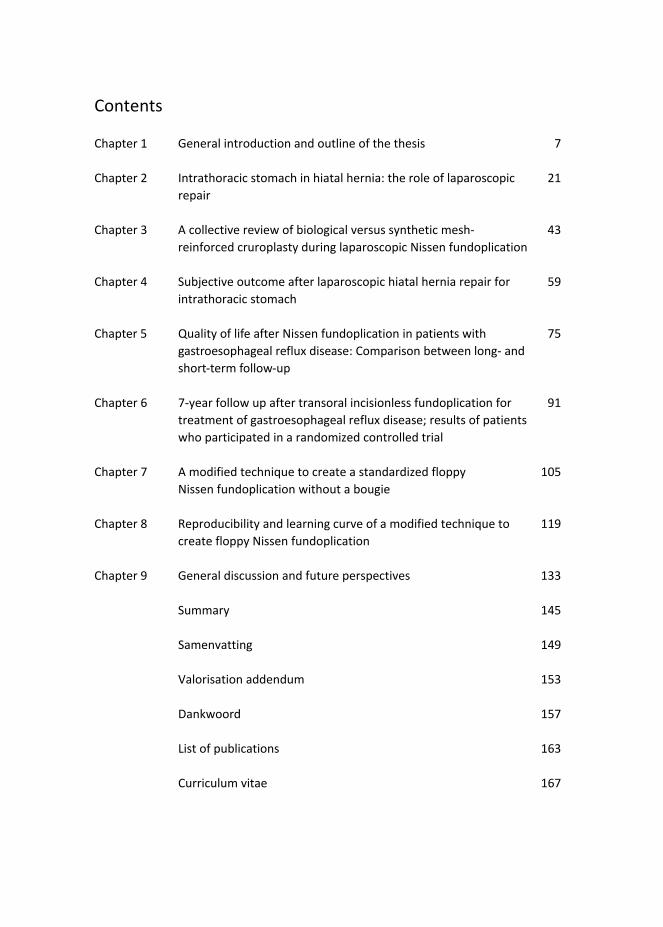
Contents
Chapter 1 General introduction and outline of the thesis 7
Chapter 2 Intrathoracic stomach in hiatal hernia: the role of laparoscopic 21
repair
Chapter 3 A collective review of biological versus synthetic mesh‐ 43
reinforced cruroplasty during laparoscopic Nissen fundoplication
Chapter 4 Subjective outcome after laparoscopic hiatal hernia repair for 59
intrathoracic stomach
Chapter 5 Quality of life after Nissen fundoplication in patients with 75
gastroesophageal reflux disease: Comparison between long‐ and
short‐term follow‐up
Chapter 6 7‐year follow up after transoral incisionless fundoplication for 91
treatment of gastroesophageal reflux disease; results of patients
who participated in a randomized controlled trial
Chapter 7 A modified technique to create a standardized floppy 105
Nissen fundoplication without a bougie
Chapter 8 Reproducibility and learning curve of a modified technique to 119
create floppy Nissen fundoplication
Chapter 9 General discussion and future perspectives 133
Summary 145
Samenvatting 149
Valorisation addendum 153
Dankwoord 157
List of publications 163
Curriculum vitae 167


Chapter 1 General introduction and outline of the thesis

Chapter 1
8

General introduction
9
1 History
The first reports of congenital and posttraumatic diaphragmatic hernias date back as far
as the 16th century. The first pioneers on hiatal hernia treatment were Ambrose Pare
(1579), Revierius Lazari (1689), Giovanni Batista Morgagni (1761) and Vincent
Alexander Bochdalek (1848).1 The first surgeon to describe the elective surgical
treatment of hiatal hernias was Angelo Soresi in the year of 1919.2 Major
advancements in the treatment of this condition have been achieved in the second half
of the 20th century. In 1950 Richard Sweet published a transthoracic technique to repair
the hiatal defect. He even used a piece of fascia lata from the left leg to strengthen the
sutures, which may be seen as the predecessor of the later use of pledges or mesh
reinforcement.3 It was in the same timeframe that Philip Allison and Norman Barrett
described the relation between the changed anatomy that comes with a hiatal hernia
and functional complaints. They were the first to suggest that this might even lead to
oesophagitis in severe cases.4 It was this association that triggered Rudolph Nissen to
explore the possibility to treat acid reflux surgically, which resulted in the first two
successful fundoplications in 1956.5 Over time several modifications and other
techniques have been proposed by Ronald Belsey (1961)6, J. Leigh Collis (1957)7, Alan
Thal (1965)8, Lucius Hill (1967)9, and Mario Rosetti (1970). A common side effect of this
type of surgery was postoperative dysphagia. In an attempt to reduce the incidence of
postoperative dysphagia, partial fundoplications have been described by Dor et al. in
196210 and Andre Toupet in 1963.11
The development of minimal invasive surgery has led to an increase in anti‐reflux
procedures. The first laparoscopic fundoplication was performed by Cushieri et al in
1992.12 The benefits of laparoscopic surgery compared to the open technique can be
found in a reduced complications rate, shorter length of hospital stay, lower morbidity
and minimal mortality.13 Nowadays laparoscopic treatment is the gold standard for
both gastroesophageal reflux disease (GERD) and hiatal hernia repair.
Gastroesophageal reflux disease
Gastroesophageal reflux disease (GERD) is a condition that develops when the reflux of
stomach contents causes troublesome symptoms and/or complications.14 Several
mechanisms play a role in the development of this condition. Most common cause is
disruption of the anti‐reflux barrier. Both an intrinsic and an extrinsic sphincter form
the anti‐reflux barrier.15 The intrinsic sphincter consists of a circular muscle, the lower
oesophageal sphincter (LES). The extrinsic sphincter is formed by the phreno‐
esophageal ligaments, which keep the LES in place, and the crural diaphragm.
Furthermore, these ligaments create the angle of His, which functions as a flap‐valve

Chapter 1
10
effect to prevent the gastric content to reflux into the oesophagus.16,17 Besides failure
of the antireflux barrier, an increase of gastric contents and/or increased abdominal
pressure plays an important role in the pathogenesis of GERD. Delayed gastric motility
and emptying, as well as motility disorders of the oesophagus may predispose patients
to develop GERD. Patients with GERD present with typical symptoms of heartburn and
regurgitation, which leads to mucosal damage of the oesophagus. In severe cases, this
may even result in oesophagitis, Barrett’s oesophagus or even oesophageal cancer.18
Atypical symptoms that come with GERD are nausea, dysphagia and chronic cough
(gastric asthma).14
Gastroesophageal reflux disease is the most common benign disorder of the upper
gastrointestinal tract, 10‐20% of the Western population reports some degree of
heartburn or regurgitation on a weekly basis.19 GERD has been proven to reduce quality
of life in patients compared with the healthy control population and therefore warrants
treatment.20 Many studies report on the short term quality of life after anti‐reflux
surgery, however long‐term results are scarce. Besides, it is unknown whether the long‐
term quality of life is different from the early results. This topic will be addressed in
chapter five of this thesis.
Hiatal hernia
Hiatal hernia is closely related to gastroesophageal reflux disease. This correlation is
due to the lack of the extrinsic sphincter. In every human being, there is an opening in
the diaphragm, allowing the oesophagus to pass through the thorax into the abdomen.
However, in some patients this opening, or hernia, is too large, leading to a shift of the
gastroesophageal junction (GEJ) and disruption of the angle of His. This may lead to
either GERD, dysphagia, belching or chest pain. This is called a symptomatic hiatal
hernia. A hiatal hernia can be either congenital or acquired. There are two types of
congenital diaphragmatic defects that have been described by Morgagni and
Bochdalek.1 However, most patients present with an acquired hiatal hernia, as a result
of weakening of the tissue and/or increased abdominal pressure. Hiatal hernia is more
frequent among elderly, supporting the hypothesis that weakening of the tissue plays a
role in the development of this disease.
In time, four types of hiatal hernia have been described. We will discuss the treatment
options for hiatal hernia in detail in chapter two of this thesis. Type I, a sliding hernia,
means that the gastroesophageal junction migrates into the thorax. This is the most
common type (95%) and is closely related to gastroesophageal reflux disease.12,21
Type II, para‐oesophageal hernia, is a defect of the diaphragm with herniation of the
gastric fundus through the hiatus into the thorax. Type III is a mixed type of hernia with
both aspects of a type I and type II hernia. sometimes more than 50% of the stomach is
General introduction
11
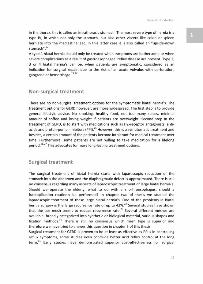
1 in the thorax, this is called an intrathorasic stomach. The most severe type of hernia is a
type IV, in which not only the stomach, but also other viscera like colon or spleen
herniate into the mediastinal sac. In this latter case it is also called an “upside‐down
stomach”.22
A type 1 hiatal hernia should only be treated when symptoms are bothersome or when
severe complications as a result of gastroesophageal reflux disease are present. Type 2,
3 or 4 hiatal hernia’s can be, when patients are symptomatic, considered as an
indication for surgical repair, due to the risk of an acute volvulus with perforation,
gangrene or hemorrhage.23,24
Non‐surgical treatment
There are no non‐surgical treatment options for the symptomatic hiatal hernia’s. The
treatment options for GERD however, are more widespread. The first step is to provide
general lifestyle advice. No smoking, healthy food, not too many spices, minimal
amount of coffee and losing weight if patients are overweight. Second step in the
treatment of GERD, is to start with medications such as H2‐receptor antagonists, anti‐
acids and proton‐pump inhibitors (PPI).25 However, this is a symptomatic treatment and
besides, a certain amount of the patients become intolerant for medical treatment over
time. Furthermore, some patients are not willing to take medication for a lifelong
period.26,27 This advocates for more long‐lasting treatment options.
Surgical treatment
The surgical treatment of hiatal hernia starts with laparoscopic reduction of the
stomach into the abdomen and the diaphragmatic defect is approximated. There is still
no consensus regarding many aspects of laparoscopic treatment of large hiatal hernia’s.
Should we operate the elderly, what to do with a short oesophagus, should a
fundoplication routinely be performed? In chapter two of thesis we studied the
laparoscopic treatment of these large hiatal hernia’s. One of the problems in hiatal
hernia surgery is the large recurrence rate of up to 42%.28 Several studies have shown
that the use mesh seems to reduce recurrence rate.29 Several different meshes are
available, broadly categorized into synthetic or biological material, various shapes and
fixation methods.30 There is still no consensus which mesh type is superior and
therefore we have tried to answer this question in chapter 3 of this thesis.
Surgical treatment for GERD is proven to be at least as effective as PPI’s in controlling
reflux symptoms, some studies even conclude better acid reflux control at the long
term.31 Early studies have demonstrated superior cost‐effectiveness for surgical
Chapter 1
12
treatment compared to high dose of PPI usage,32 although decreased costs of PPI
probably reduced this effect. On the other hand, an increasing number of side effects of
long term PPI use are becoming apparent such as an increased the risk of dementia,
pneumonia, rib fractures, magnesium deficiency, gastrointestinal infections and might
even predispose for upper gastrointestinal malignancies.33,34 However, when long term
PPI use is not desired or not effective, laparoscopic fundoplication is the gold standard
in the treatment of GERD. Unfortunately, laparoscopic anti‐reflux surgery comes with
the risk of complications and recurrences. Several long‐term studies report a high
degree of dysphagia, gas bloating, inability to belch, vagal nerve injury and even a re‐
operation rate of up to 15% is seen.35,36 Furthermore, some studies report recurrent
acid reflux in 10‐20% of the patients after surgery for a median follow‐up of
6.5 years.37‐39 Due to these side effects that come with anti‐reflux surgery, minimal
invasive techniques are being developed.
Endoscopic treatment
One of the less invasive endoscopic treatment options for patients with GERD is the
transoral incisionless fundoplication (TIF). There are currently 3 different devices
available for this treatment modality. The EsophyX® device (EndoGastric Solutions, Redmond, WA, United States), the Medigus Ultrasonic Surgical Endostapler system
(MUSE™, Medigus Ltd., Omer; Israel) and the GERDX™ (G‐SURG GmbH, Seeon‐
Seebruck, Germany).40,41 There are some small differences between the three devices.
For the EsophyX, an endoscope is inserted and by means of the application of several
polypropylene fastener a 210‐270 degrees fundoplication is created. The MUSE device
staples the fundus of the stomach tot the oesophagus below the diaphragm using
multiple sets of metal stiches placed under an ultrasound‐guided technique. The GERDX
system also places several sets of stitches to create a full thickness fundoplication. At
the short term, TIF seems promising and leads to good symptom control.40,42,43 There is
one prospective randomized controlled trial that is performed comparing TIF with PPI
that shows disappointing results at 1‐year of follow‐up, with an increasing failure rate
over time.44 However, even if TIF fails, it is still possible to perform additional anti‐reflux
surgery in a safe and effective manner.45 Due to disappointing results of these
endoscopic treatment, the laparoscopic fundoplication is still the golden standard for
the treatment of GERD. Long term effects of EsophyX should be evaluated and there
might be a group of patients that benefit from this minimal invasive procedure to treat
GERD.
General introduction
13
1 Other surgical techniques
We have outlined the invasive anti‐reflux surgery, the minimal invasive endoscopic
techniques and the medical treatment for hiatal hernia and GERD. There are 2 other
techniques available for the treatment of GERD, which are the EndoStim LES
Stimulation System (Endostim BV, The Hague, the Netherlands) and the the LINX®Reflux
Management System (Torax Medical, St. Paul, MN, USA). With the EndoStim, two
electrodes are laparoscopically placed at the level of the GE‐junction and connected to
a device that stimulates the LES and therefore enhances the intrinsic anti‐reflux barrier.
This leads to good anti‐reflux control and improved quality of life after 12 months, for a
small group of patients. However, the long‐term efficacy has yet to be established.46
The other technique is the LINX device, which involves the use of magnetic attraction
through adjacent magnetic beads, which augments the resistance of the oesophageal
sphincter to abnormal opening associated with reflux.47 Results regarding reflux control
are promising, however a high rate of postoperative dysphagia up to 68% has been
reported and several reports of erosion can be found in the literature.48‐50 Since the
serious complications that come with erosion of devices, the LINX device should be
used with care.
Quality of life
The outcome of surgical treatment can be measured by several methods. In an
increasing amount of clinical trials, quality of life has become a third dimension, besides
efficacy and safety. Especially for anti‐reflux surgery and hiatal hernia repair, since
there is very little evidence that this type of surgery prevents cancer or even reduces
mortality. The main goal of these treatment modalities is to improve the quality of life
in patients with a benign condition of the upper gastrointestinal tract. And as
mentioned before, it is proven that GERD and a symptomatic hiatal hernia have a
negative impact on the quality of life of patients.20 Furthermore, there is very little
association between anatomical recurrences and symptoms.51 Even the 24‐hour PH‐
metry has a low sensitivity in detecting recurrent GERD. Up to 16% of patients with a
positive 24‐h PH monitoring had negative results in the follow‐up PH‐metry. And even
25% of the patients with erosive oesophagitis present with a negative 24‐h PH metry.52
Since the aim of the treatment is to improve quality of life, several questionnaires have
been developed over time to assess this outcome measurement. For GERD related
complaints, the most commonly used questionnaire is the GERD‐HRQL, which is a 16‐
item questionnaire, addressing most bothersome symptoms and its severity.53 For
patients with the typical symptoms, anti‐reflux surgery leads to an improved quality of
life in 90% for the patients. However, when atypical symptoms are present, this is only

Chapter 1
14
70%.54,55 There are several aspects regarding the treatment of GERD and hiatal hernia,
that affect the postoperative quality of life and that are still unknown. We therefore
studied the long‐term quality of life after both anti‐reflux surgery and hiatal hernia
repair in this thesis.
Experience of the surgeon
Anti‐reflux surgery and hiatal hernia repair requires advanced laparoscopic skills. Many
studies have demonstrated that these operations come with a learning curve of at least
20 procedures. But, even after 100 operations, there is still an improvement in
complication rate, recurrence rate, operating time and symptomatic outcome.56 When
anti‐reflux surgery is performed less than 10 times a year in an institution, this is an
independent risk factor for a recurrence.57,58 Therefore, it is best if anti‐reflux surgery is
performed in high volume centers with dedicated anti‐reflux surgeons. Besides, a
proper pre‐operative work‐up is necessary to avoid complications. All patients should
undergo endoscopy combined with PH‐metry to retrieve the highest sensitivity for
detecting GERD. Furthermore, a manometry should be performed to rule out achalasia
and PH‐metry should be combined with impedance measurement. Patients with
aerophagia should be treated with care, due to the increased risk of postoperative gas
bloating.59
One of the causes for this learning curve is the creation of a proper fundoplication.
When the fundoplication is to narrow, this leads to increased postoperative
dysphagia.60 Even rotation in the fundoplication seems to increase dysphagia, which
emphasizes the importance of a symmetrical fundoplication.61 We have therefore
developed a modified technique which we describe in this thesis. We further studies
whether this technique indeed creates a more reproducible and symmetrical
fundoplication. Theoretically modification may reduce the long learning curve.
General introduction
15
1 Aim of this thesis
In this thesis, we aimed to study the long‐term symptomatic outcome after different
treatment options for both hiatal hernia and GERD. We also tried to modify the current
technique for Nissen fundoplication with the goal to reduce the learning curve and help
beginning surgeons in performing a perfect fundoplication.
Unsolved issues that we have studied in this thesis
Several technical aspects of the laparoscopic treatment of intrathoracic stomach
Which type of mesh is superior for the reinforcement of the crural repair
The long‐term quality of life after anti‐reflux surgery for GERD
The long‐term quality of life after intrathoracic repair
Long‐term quality of life after transoral incisionless fundoplication (TIF)
The large learning curve in creating a proper fundoplication
A method for creating a standardized fundoplication without rotation

Chapter 1
16
Outline
Chapter 2 is a narrative review that describes the development of laparoscopy in the
treatment of large hiatal hernias and intrathoracic stomach. Many aspects of the
treatment are evaluated and the most recent literature has been studied and compiled
into a review format. Recommendations for the treatment of an intrathoracic stomach
are given.
Chapter 3 is a collective review that evaluates the results of mesh reinforcement in
hiatal hernia repair. All high‐quality studies from the past 11 years were studied and
data is combined. Recurrence and symptomatic outcome after both biological mesh
repair and synthetic mesh repair are compared.
Chapter 4 studies the quality of life after intra‐thoracic stomach repair in a large single
center cohort, with a follow‐up of up to 9.6 years. All patients were sent a standardized
questionnaire to evaluate quality of life and re‐operation rate. Long‐term quality of life,
recurrence rate and complication rate are reported.
Chapter 5 analyses the quality of life after Nissen fundoplication for patients with
GERD. In this retrospective cohort study, we sent all patients the GERD‐HRQL
questionnaire and evaluated the results. Outcome for patients with a follow‐up longer
than 5 years and for patients with a follow‐up shorter than 5 years is compared.
Chapter 6 describes the 7‐years follow‐up after a randomized controlled trial that
compared the transoral incisionless fundoplication(TIF) with the treatment of PPI.
Quality of life is primary outcome measurement, but also re‐operation rate is
evaluated. A multi‐variate analysis is performed with the goal to find predictors for
successful treatment with TIF.
Chapter 7 Describes a modified technique to create a floppy Nissen fundoplication
without the use of a bougie. Feasibility and safety in clinical practice are studied. The
goal of this technique is to help beginning surgeons to perform a proper fundoplication.
Chapter 8 Studies the actual reproducibility of the modified technique for Nissen
fundoplication that has been described in chapter 7. Several fundoplications are
created in an ex‐vivo experiment by both a resident and an experienced anti‐reflux
surgeon. Symmetry, size and reproducibility are evaluated.
Chapter 9 provides a general discussion and proposes future perspectives.

General introduction
17
1 References
1. Hochberg LA. Thoracic surgery before the 20th century. New York: Vantage Press; 1960. 2. Soresi AL. Diaphragmatic Hernia: Its Unsuspected Frequency: Its Diagnosis: Technic for Radical Cure.
Ann Surg. 1919;69(3):254‐70.
3. Sweet RH. Esophageal hiatus hernia of the diaphragm; the anatomical characteristics, technic of repair, and results of treatment in 111 consecutive cases. Ann Surg. 1952;135(1):1‐13.
4. Allison PR. Reflux esophagitis, sliding hiatal hernia, and the anatomy of repair. Surg Gynecol Obstet.
1951;92(4):419‐31. 5. Stylopoulos N, Rattner DW. The history of hiatal hernia surgery: from Bowditch to laparoscopy. Ann
Surg. 2005;241(1):185‐93.
6. Hiebert CA. Surgical management of esophageal reflux and hiatal hernia. The Ann Thorac Surg. 1961;52(1):159‐60.
7. Collis JL. An operation for hiatus hernia with short oesophagus. Thorax. 1957; 12(3):181‐8.
8. Thal AP. A unified approach to surgical problems of the esophagogastric junction. Ann Surg. 1968;168(3):542‐50.
9. Hill LD. An effective operation for hiatal hernia: an eight year appraisal. Ann Surg. 1967;166(4):681‐92.
10. Dor J, Humbert P, Dor V, et al. The role of the modified Nissen procedure in the prevention of reflux following Heller's extramucosal cardiomyotomy. Mem Acad Chir. 1962;88:877–82.
11. Toupet A. Technique d'eosophago‐gastroplastie avec phreno‐gastropexie dans la cure radicales des
hernies hiatales et comme complement de l'operation de Heller dans les cardiospasmes. Mem Acad Chir. 1963;89:394–9.
12. Cuschieri A, Shimi S, Nathanson LK. Laparoscopic reduction, crural repair, and fundoplication of large
hiatal hernia. Am J Surg. 1992;163(4):425‐30. 13. Peters MJ, Mukhtar A, Yunus RM, Khan S, Pappalardo J, Memon B, et al. Meta‐analysis of randomized
clinical trials comparing open and laparoscopic anti‐reflux surgery. Am J Gastroenterol.
2009;104(6):1548‐61; quiz 7, 62. 14. Vakil N, van Zanten SV, Kahrilas P, Dent J, Jones R, Global Consensus G. The Montreal definition and
classification of gastroesophageal reflux disease: a global evidence‐based consensus. Am J
Gastroenterol. 2006;101(8):1900‐20; quiz 43. 15. Mittal RK, Balaban DH. The esophagogastric junction. N Engl J Med. 1997; 336(13):924‐32.
16. Liebermann‐Meffert D, Allgower M, Schmid P, Blum AL. Muscular equivalent of the lower esophageal
sphincter. Gastroenterology. 1979;76(1):31‐8. 17. Thor KB, Hill LD, Mercer DD, Kozarek RD. Reappraisal of the flap valve mechanism in the
gastroesophageal junction. A study of a new valvuloplasty procedure in cadavers. Acta Chir Scand.
1987;153(1):25‐8. 18. Dent J, Holloway RH, Toouli J, Dodds WJ. Mechanisms of lower oesophageal sphincter incompetence in
patients with symptomatic gastrooesophageal reflux. Gut. 1988;29(8):1020‐8.
19. Dent J, El‐Serag HB, Wallander MA, Johansson S. Epidemiology of gastro‐oesophageal reflux disease: a systematic review. Gut. 2005;54(5):710‐7.
20. Revicki DA, Wood M, Maton PN, Sorensen S. The impact of gastroesophageal reflux disease on health‐
related quality of life. Am J Med. 1998;104(3):252‐8. 21. Athanasakis H, Tzortzinis A, Tsiaoussis J, Vassilakis JS, Xynos E. Laparoscopic repair of paraesophageal
hernia. Endoscopy. 2001;33(7):590‐4.
22. Hill LD, Tobias JA. Paraesophageal hernia. Archives of surgery. 1968;96(5):735‐44. 23. Leeder PC, Smith G, Dehn TC. Laparoscopic management of large paraesophageal hiatal hernia. Surg
Endosc. 2003;17(9):1372‐5.
24. Polomsky M, Jones CE, Sepesi B, O'Connor M, Matousek A, Hu R, et al. Should elective repair of intrathoracic stomach be encouraged? J Gastrointest Surg. 2010; 14(2):203‐10.
25. Tytgat N, McColl K, Tack J, Holtmann G, Hunt RH, Malfertheiner P, et al. New algorithm for the
treatment of gastro‐oesophageal reflux disease. Aliment Pharmacol Ther. 2008;27(3):249‐56.

Chapter 1
18
26. El‐Serag HB. Time trends of gastroesophageal reflux disease: a systematic review. Clin Gastroenterol
Hepatol. 2007;5(1):17‐26. 27. Metz DC. Managing gastroesophageal reflux disease for the lifetime of the patient: evaluating the long‐
term options. Am J Med. 2004;117 Suppl 5A:49S‐55S.
28. Draaisma WA, Gooszen HG, Tournoij E, Broeders IA. Controversies in paraesophageal hernia repair: a review of literature. Surg Endosc. 2005;19(10): 1300‐8.
29. Furnee E, Hazebroek E. Mesh in laparoscopic large hiatal hernia repair: a systematic review of the
literature. Surg Endosc. 2013;27(11):3998‐4008. 30. Herbella FA, Patti MG, Del Grande JC. Hiatal mesh repair‐‐current status. Surg Laparosc Endosc
Percutan Tech. 2011;21(2):61‐6.
31. Zhang C, Hu ZW, Yan C, Wu Q, Wu JM, Du X, et al. Nissen fundoplication vs proton pump inhibitors for laryngopharyngeal reflux based on pH‐monitoring and symptom‐scale. World J Gastroenterol.
2017;23(19):3546‐55.
32. Funk LM, Zhang JY, Drosdeck JM, Melvin WS, Walker JP, Perry KA. Long‐term cost‐effectiveness of medical, endoscopic and surgical management of gastroesophageal reflux disease. Surgery.
2015;157(1):126‐36.
33. Corsonello A, Lattanzio F, Bustacchini S, Garasto S, Cozza A, Schepisi R, et al. Adverse events of proton pump inhibitors: potential mechanisms. Curr Drug Metab. 2017.
34. de la Coba Ortiz C, Arguelles Arias F, Martin de Argila de Prados C, Judez Gutierrez J, Linares Rodriguez
A, Ortega Alonso A, et al. Proton‐pump inhibitors adverse effects: a review of the evidence and position statement by the Sociedad Espanola de Patologia Digestiva. Rev Esp Enferm Dig. 2016;108(4):207‐24.
35. Castelijns PSS, Ponten JEH, Vd Poll MCG, Bouvy ND, Smulders JF. Quality of life after nissen
fundoplication in patients with gastroesophageal reflux disease: Comparison between long‐ and short‐term follow‐up. J Minim Access Surg. 2017.
36. van Rijn S, Rinsma NF, van Herwaarden‐Lindeboom MY, Ringers J, Gooszen HG, van Rijn PJ, et al. Effect
of Vagus Nerve Integrity on Short and Long‐Term Efficacy of Antireflux Surgery. Am J Gastroenterol. 2016;111(4):508‐15.
37. Carlson MA, Frantzides CT. Complications and results of primary minimally invasive antireflux
procedures: a review of 10,735 reported cases. J Am Coll Surg. 2001;193(4):428‐39. 38. Catarci M, Gentileschi P, Papi C, Carrara A, Marrese R, Gaspari AL, et al. Evidence‐based appraisal of
antireflux fundoplication. Ann Surg. 2004;239(3):325‐37.
39. Maret‐Ouda J, Wahlin K, El‐Serag HB, Lagergren J. Association Between Laparoscopic Antireflux Surgery and Recurrence of Gastroesophageal Reflux. JAMA. 2017;318(10):939‐46.
40. Testoni PA, Mazzoleni G, Testoni SG. Transoral incisionless fundoplication for gastro‐esophageal reflux
disease: Techniques and outcomes. World J Gastrointest Pharmacol Ther. 2016;7(2):179‐89. 41. von Renteln D, Schiefke I, Fuchs KH, Raczynski S, Philipper M, Breithaupt W, et al. Endoscopic full‐
thickness plication for the treatment of gastroesophageal reflux disease using multiple Plicator
implants: 12‐month multicenter study results. Surg Endosc. 2009;23(8):1866‐75. 42. Witteman BP, Strijkers R, de Vries E, Toemen L, Conchillo JM, Hameeteman W, et al. Transoral
incisionless fundoplication for treatment of gastroesophageal reflux disease in clinical practice. Surg
Endosc. 2012;26(11):3307‐15. 43. Hakansson B, Montgomery M, Cadiere GB, Rajan A, Bruley des Varannes S, Lerhun M, et al.
Randomised clinical trial: transoral incisionless fundoplication vs. sham intervention to control chronic
GERD. Aliment Pharmacol Ther. 2015;42(11‐12):1261‐70. 44. Witteman BP, Conchillo JM, Rinsma NF, Betzel B, Peeters A, Koek GH, et al. Randomized controlled trial
of transoral incisionless fundoplication vs. proton pump inhibitors for treatment of gastroesophageal
reflux disease. Am J Gastroenterol. 2015;110(4):531‐42. 45. Witteman BP, Kessing BF, Snijders G, Koek GH, Conchillo JM, Bouvy ND. Revisional laparoscopic
antireflux surgery after unsuccessful endoscopic fundoplication. Surg Endosc. 2013;27(6):2231‐6.
46. Rodriguez L, Rodriguez P, Gomez B, Ayala JC, Oksenberg D, Perez‐Castilla A, et al. Long‐term results of electrical stimulation of the lower esophageal sphincter for the treatment of gastroesophageal reflux
disease. Endoscopy. 2013;45(8):595‐604.

General introduction
19
1 47. Ganz RA, Gostout CJ, Grudem J, Swanson W, Berg T, DeMeester TR. Use of a magnetic sphincter for the
treatment of GERD: a feasibility study. Gastrointest Endosc. 2008;67(2):287‐94. 48. Ganz RA, Peters JH, Horgan S, Bemelman WA, Dunst CM, Edmundowicz SA, et al. Esophageal sphincter
device for gastroesophageal reflux disease. N Engl J Med. 2013;368(8):719‐27.
49. Salvador R, Costantini M, Capovilla G, Polese L, Merigliano S. Esophageal Penetration of the Magnetic Sphincter Augmentation Device: History Repeats Itself. J Laparoendosc Adv Surg Tech A.
2017;27(8):834‐8.
50. Bauer M, Meining A, Kranzfelder M, Jell A, Schirren R, Wilhelm D, et al. Endoluminal perforation of a magnetic antireflux device. Surg Endosc. 2015;29(12):3806‐10.
51. Mittal SK, Bikhchandani J, Gurney O, Yano F, Lee T. Outcomes after repair of the intrathoracic stomach:
objective follow‐up of up to 5 years. Surg endosc. 2011;25(2):556‐66. 52. Swidnicka‐Siergiejko A, Dabrowski A. Prolonged 2‐day esophageal pH‐metry with impedance
monitoring improves symptom‐reflux association analysis. Dig Dis Sci. 2013;58(9):2556‐63.
53. Velanovich V, Vallance SR, Gusz JR, Tapia FV, Harkabus MA. Quality of life scale for gastroesophageal reflux disease. J Am Coll Surg. 1996;183(3):217‐24.
54. Kaufman JA, Houghland JE, Quiroga E, Cahill M, Pellegrini CA, Oelschlager BK. Long‐term outcomes of
laparoscopic antireflux surgery for gastroesophageal reflux disease (GERD)‐related airway disorder. Surg Endosc. 2006;20(12):1824‐30.
55. Pessaux P, Arnaud JP, Delattre JF, Meyer C, Baulieux J, Mosnier H. Laparoscopic antireflux surgery: five‐
year results and beyond in 1340 patients. Arch Surg. 2005;140(10):946‐51. 56. Soot SJ, Eshraghi N, Farahmand M, Sheppard BC, Deveney CW. Transition from open to laparoscopic
fundoplication: the learning curve. Arch Surg. 1999;134(3):278‐81; discussion 82.
57. Antiporda M, Veenstra B, Jackson C, Kandel P, Daniel Smith C, Bowers SP. Laparoscopic repair of giant paraesophageal hernia: are there factors associated with anatomic recurrence? Surg Endosc.
2018;32(2):945‐54.
58. Whealon MD, Blondet JJ, Gahagan JV, Phelan MJ, Nguyen NT. Volume and outcomes relationship in laparoscopic diaphragmatic hernia repair. Surg Endosc. 2017.
59. Bello B, Zoccali M, Gullo R, Allaix ME, Herbella FA, Gasparaitis A, et al. Gastroesophageal reflux disease
and antireflux surgery‐what is the proper preoperative work‐up? J Gastrointest Surg. 2013;17(1):14‐20; discussion p
60. Patterson EJ, Herron DM, Hansen PD, Ramzi N, Standage BA, Swanstrom LL. Effect of an esophageal
bougie on the incidence of dysphagia following nissen fundoplication: a prospective, blinded, randomized clinical trial. Arch Surg. 2000;135(9):1055‐61; discussion 61‐2.
61. Leggett PL, Bissell CD, Churchman‐Winn R, Ahn C. A comparison of laparoscopic Nissen fundoplication
and Rossetti's modification in 239 patients. Surg Endosc. 2000;14(5):473‐7.

Chapter 1
20
Chapter 2 Intrathoracic stomach in hiatal hernia:
the role of laparoscopic repair
P.S.S. Castelijns
J.E.H. Ponten
N.D. Bouvy
J.F. Smulders
M.C.G. van de Poll
Minerva Chir. 2018;73(1):64‐76
Chapter 2
22
Abstract
Introduction
For decades, intrathoracic stomach has been an indication for surgical repair and over
time laparoscopy has become standard treatment. However, there are still many
aspects in the treatment of intrathoracic stomach that subject of debate. We
performed a literature review to discuss the role of laparoscopy in intrathoracic
stomach repair.
Evidence acquisition
We performed an extensive literature search in PubMed, Embase and Cochrane and
reviewed studies from the last 5 years. To provide a complete overview, references
from the found studies are also used. All data was compiled into a review format.
Evidence synthesis
Laparoscopic surgery is proven superior to open hiatal hernia repair in the treatment of
intrathoracic stomach. The role of hernia sac excision, short oesophagus, mesh
reinforcement, fundoplication, complications and future perspectives are discussed in
this review.
Conclusions
Laparoscopy plays a major role in the treatment of intrathoracic stomach and regarding
most aspects of the treatment. All available techniques have their advantages and
disadvantages, and the decision on how to repair the intrathoracic stomach, remains a
tailored based decision.
Intrathoracic stomach in hiatal hernia
23
2
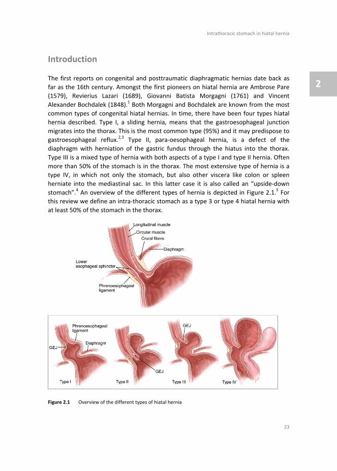
Introduction
The first reports on congenital and posttraumatic diaphragmatic hernias date back as
far as the 16th century. Amongst the first pioneers on hiatal hernia are Ambrose Pare
(1579), Revierius Lazari (1689), Giovanni Batista Morgagni (1761) and Vincent
Alexander Bochdalek (1848).1 Both Morgagni and Bochdalek are known from the most
common types of congenital hiatal hernias. In time, there have been four types hiatal
hernia described. Type I, a sliding hernia, means that the gastroesophageal junction
migrates into the thorax. This is the most common type (95%) and it may predispose to
gastroesophageal reflux.2,3 Type II, para‐oesophageal hernia, is a defect of the
diaphragm with herniation of the gastric fundus through the hiatus into the thorax.
Type III is a mixed type of hernia with both aspects of a type I and type II hernia. Often
more than 50% of the stomach is in the thorax. The most extensive type of hernia is a
type IV, in which not only the stomach, but also other viscera like colon or spleen
herniate into the mediastinal sac. In this latter case it is also called an “upside‐down
stomach”.4 An overview of the different types of hernia is depicted in Figure 2.1.5 For
this review we define an intra‐thoracic stomach as a type 3 or type 4 hiatal hernia with
at least 50% of the stomach in the thorax.
Figure 2.1 Overview of the different types of hiatal hernia
Chapter 2
24
Surgical treatment
The first surgeon to describe the elective surgical treatment of hiatal hernias was
Angelo Soresi in the year of 1919.6 It was 10 years later when Stuart Harrington and his
colleagues performed a trial in The Mayo Clinic where they observed asymptomatic
patients and performed transabdominal surgery on patients who had a symptomatic
diaphragmatic hernia.7 If they were unable to close the diaphragmatic defect, they
stitched the organs to the abdominal wall to keep them in place; this was called a
palliative treatment.
Major advancements in the treatment of this condition have been developed in the
second half of the 20th century. In the year of 1950 Richard Sweet published a
transthoracic method to repair a hiatal hernia.8 He even described the use of a piece of
the fascia lata from the left leg to strengthen the sutures, this may be seen as the
predecessor for the later use of pledges in hiatal hernia surgery.
Although the relation between a sliding hiatal hernia and acid reflux nowadays is very
obvious, this pathophysiological mechanism was only discovered for the first time in
the second half of the 20th century.9 Philip Allison and Norman Barrett played a major
role in the changing perception that an anatomic mechanical condition leads to
functional complaints and to oesophagitis in severe cases. They may therefore be
credited for the current era of the antireflux surgery. The notion that a hiatal hernia
was associated with symptomatic acid reflux was the basis for Rudolph Nissen to
explore the possibility to treat acid reflux surgically, leading to his report on the two
first successful fundoplications in 1956.10 Another pioneer of anti‐reflux surgery was
Ronald Belsey who started to develop a fundoplication technique in 1949. It was only
after three failed trials that he eventually published the Belsey Mark IV procedure in
1961.11
Several modifications to these techniques have been developed over time by J. Leigh
Collis (1957)12, Alan Thal (1965)13, Lucius Hill (1967)14 and Mario Rosetti (1970). A
common and unwanted side effect of this type of surgery was postoperative dysphagia.
Partial fundoplications were therefore described by Dor et al. in 196215 and Andre
Toupet in 1963.16 The development of minimal invasive techniques has led to an
increase in anti‐reflux procedures. The first laparoscopic Nissen fundoplication is
performed by Cuschieri et al. in 1992.2 In the beginning, 40‐50% of the procedures was
converted to an open repair, however over time this is reduced to a very minimum with
not even 0.5% conversion rate.17‐19 Several types of fundoplication and techniques for
hiatal hernia surgery are summarized in Table 2.1.
Several studies have shown better outcome after laparoscopic repair compared with
open repair. The mean length of postoperative hospital stay is reduced significantly by
one third after laparoscopic repair.20 Complication rate is reduced as well by a half for
the laparoscopic repair group compared with the open technique. The recurrence rate
seems similar around 10% for both techniques.21 Also, the number of patients

Intrathoracic stomach in hiatal hernia
25
2
experiencing complications during the initial hospital admission, was reduced from
20.6% to 8.5% in the patients that were treated by laparoscopic hiatal hernia repair.22
Therefore, laparoscopy has led to a significant shorter postoperative length of stay,
lower complication rate, recurrence rate, mortality and even a lower morbidity
compared with the open technique.23 Nowadays laparoscopy is considered to be the
gold standard for the surgical treatment of the intrathoracic stomach. However, several
questions remain subject of debate, including the role and timing of laparoscopy in the
emergency setting, the use of mesh reinforcement of the cruroplasty, the application of
an additional fundoplication to the crural approximation, the hernia sac excision and as
well the indication for elective repair in elderly patients with minimal complaints may
be questioned.
Table 2.1 Overview of techniques used in hiatal hernia surgery
Name Wrap
circumference
Description
Nissen 360 Total fundoplication, posterior, with complete dissection of the vasa brevia
Toupet 270 Partial fundoplication, posterior, fixation to the oesophagus
Nissen‐Rosetti 360 Total fundoplication, posterior, without dissection of the vasa brevia
Dor 90 Partial fundoplication, anterior, fixation to oesophagus
Watson 180 Partial fundoplication, anterior, fixation to right crus Collis procedure ‐ Oesophageal lengthening procedure
Hill 360 Suturing the anterior and posterior phrenoesophageal bundle to
the preaortic fascia Belsey‐mark IV 270 Partial fundoplication, posterior, thoracic approach
Anterior gastropexy ‐ Suturing the stomach to the ventral abdominal wall
Thal 180 Partial fundoplication, anterior, fixation to oesophagus
Special considerations
As mentioned above, laparoscopic repair of a para‐oesophageal hernia (PEH) or
intrathoracic stomach (ITS) seems to be safe and is proven effective. However, there
are some aspects that have to be kept in mind. This type of surgery requires advanced
laparoscopic skills and therefore comes with a learning curve. It is known for antireflux
surgery, which has a close relation with hiatal hernia repair, since these types of
surgery are often combined, that the learning curve is at least 20 procedures. However,
after 100 procedures there is still an improvement in operation time, conversion rate
and a reduction of intra‐operative complications.24 We also know that reoperation for
dysphagia can be reduced from 22% to as low as 4% if the initial procedure is
performed by an experienced antireflux surgeon.25 If this procedure is performed less
than 10 times a year in an institution, this is an independent risk factor for recurrence
Chapter 2
26
and even mortality rate is doubled in low volume centers.26,27 The recommendation is
therefore, to operate PEH and ITS in a high‐volume centre with dedicated and
experienced fore‐gut surgeons to minimize complications, mortality and recurrence
rate.
Evidence acquisition
For this study, we performed a literature search in PubMed, Embase and Cochrane with
the following terms: (((intrathoracic stomach) OR hiatal hernia)) AND (((laparoscopic)
OR laparoscopy) OR laparoscopic surgery) We only searched for high quality studies
defined as reviews, meta‐analysis, randomized controlled trials and retrospective
cohort studies with at least 100 patients. Minimum follow‐up should be 5 years and
only studies published in the last 5 years were seen as recent literature. For this study,
we describe the role of laparoscopy in intrathoracic stomach repair in adults, therefore
studies that describe patients younger than the age of 18 were excluded. To provide a
complete overview of the current literature regarding this subject, we also searched in
the references of the included studies for additional valuable studies. All gathered
information was examined and compiled into a review format. Since this a narrative
review without a specific research question, there is no meta‐analysis or statistical
analysis performed.
Elective vs. emergent repair
Most patients with a para‐oesophageal hernia or an intrathoracic stomach present with
complaints of dysphagia, however also anaemia, chronic cough or acid reflux is
reported. These symptoms warrant a concise diagnostic process including oesophageal
manometry and 24‐hours impedance measurements followed by elective repair if the
hernia is indeed considered to be symptomatic. We know from literature that good
symptomatic outcome is reported and patients experience little complications after
elective repair.28,29
Both PEH and ITS may become incarcerated which is a reason for emergent
decompression by nasogastric tube followed by surgery in the next few days. Typical
complaints for incarceration are pain, hematemesis and profound vomiting. If
complaints do not decrease with gastric decompression, surgery should be performed
the same day.30 Emergency surgery for acute incarceration is related with a higher
morbidity and mortality rates up to 3.7% have been reported, compared with 0.37% for
elective repair.31 This might be due to the population of particularly elderly patients
with more comorbidities that experience an acute incarcerated stomach. When
correction for age and other risk factors is performed in a multivariate analysis, the
higher mortality rate after emergency ITS repair diminishes; the risk of complications

Intrathoracic stomach in hiatal hernia
27
2
however remains increased after emergency ITS repair.32 Therefore elective repair
comes with less morbidity and should be performed if patients are symptomatic.
However, if emergency surgery is needed, laparoscopy has been proven superior to
open surgery for hiatal hernia repair. Mortality rate after emergency repair of an
incarcerated intrathoracic stomach is significantly reduced from 5.0% in the open repair
to 1.5% for the laparoscopic repair. However, what should be done when patients are
of an older age and present with minimal symptoms, should elective surgery be
performed or should we choose for watchful waiting.
The elderly patient
If a patient presents with minimal complaints, the questions is raised whether surgery is
actually indicated. To this end the complication risk of elective repair should be
outweighed to the chance of undergoing emergency surgery and the increased
complication risk associated with emergency surgery. Based on a Monte Carlo
probabilistic model, the annual risk to undergo emergency surgery is estimated to be
1.1% with a mortality rate of approximately 3.7‐5.5% compared with a mortality rate of
0.37‐1.4% for the elective repair.32,33 Thus, for a patient that is older than 65, the risks
should not outweigh the benefits of an elective repair and watchful waiting is advised.
This is supported by a nationwide registry study in the USA performed in 2008 that
found a six‐fold increased mortality rate of 15.6% in patients over the age of 80.34
However, it should be realized that most data from previous studies were gathered in
the pioneering period of laparoscopic repair and since surgeons have become more
experienced with minimal invasive surgery over the subsequent years, morbidity and
mortality after elective repair has diminished.28 Furthermore, the general population
gets older and are more vital at an older age.35 Even for patients aged over 70, good
results have been reported and they should therefore not be withdrawn from elective
hiatal hernia repair solitarily based on their age.36‐38 Elective repair of PEH or ITS
remains a tailored made decision and even if symptoms are mild or patients are of an
older age, elective surgery should be considered. If the decision is made to perform an
operative repair of the hiatal hernia, the operative approach should be chosen. It is
possible to approach the hiatal hernia from both the thorax as well as from the
abdomen. Is there any evidence proving the superiority of either one of these
techniques?

Chapter 2
28
Transabdominal vs. Transthoracic
Since a hiatal hernia originates from the diaphragm between the thorax and the
abdomen, approach can be from either side, i.e. transabdominal as well as
transthoracic. There are no randomized controlled trials published so far, that compare
transthoracic hiatal hernia repair with the transabdominal technique. In the early years,
74% of the procedures was performed transabdominal with open surgery, 17%
transthoracic and only 9% was performed via transabdominal laparoscopy.39 Over time
laparoscopy surpassed the open abdominal repair and has become gold standard for
hiatal hernia repair. From earlier data, we know that postoperative hospital stay is
longer for transthoracic surgery, patients require more often mechanical ventilation
(5.6% of the patients after transthoracic hernia repair) and have a higher risk of a
pulmonary embolism.39,40 However, these negative effects are seen in the open
thoracic approach. Nowadays, thoracoscopic surgery is more often performed and this
minimal invasive technique provides a great overview of the oesophagus and the
diaphragm. Complication rate is far lower after thoracoscopic techniques, when
compared with open thoracic approach. Therefore, when previous abdominal
laparoscopic surgery has failed or when the hiatal hernia is fixed in the chest, video‐
assisted thoracoscopic surgical(VATS) repair may play a promising role for these select
group of patients where recurrent abdominal approach seems impossible.41 A recent
study shows promising results for a combined procedure, where abdominal
laparoscopy is assisted by VATS in complicated patients with giant hiatal hernias.42 In
conclusion, the laparoscopic abdominal approach is golden standard, but in select
patients the thoracic approach should definitely be considered.
Technical issues
As mentioned above, the transabdominal laparoscopic technique is the preferred
method to repair a hiatal hernia. The instrumentation for this technique is relatively
simple and is available in most hospitals that perform laparoscopic surgery. First
consideration is the placement of the trocars. The procedure starts with placing the
patient in supine position, with the surgeon between the legs. (French position) Then a
pneumoperitoneum is created, which can be achieved by open or closed introduction
according the preference of the operating surgeon. The trocar that is used for the
camera, should be at a ratio of 1/3 to 2/3 between the umbilicus and the xyphoid notch
to obtain the best overview during the procedure. For this type of surgery, a 30‐degree
scope is recommended. Under direct vision the other trocars a placed, to minimize
damage to the internal organs. The two trocars that are used to perform the surgery
are placed approximately 6 cm lateral on each side of the first trocar. The liver retractor
to facilitate a clear view during the procedure is placed just underneath the Xyphoid
notch, or in the right upper quadrant. Finally, one additional trocar should be placed a

Intrathoracic stomach in hiatal hernia
29
2
few centimetres lateral of the most left trocar, this can be used by the assistant to aid
the surgeon during the procedure. An overview of the position of the trocars is
depicted in Figure 2.2. For dissection of the crus several instruments can be used,
varying from a diathermy hook to ultrasonic scissors. Some authors advocate that even
blunt dissection is possible and recommended to minimize damage to other organs.
Figure 2.2 Placement of the trocars
For reducing the hernia content and for mobilization of the stomach and other intra‐
abdominal structures, standard laparoscopic equipment can be used, according to the
preference of the operating surgeon. The suture repair of the crus should be performed
with non‐absorbable sutures. The application of a mesh is subject of debate and will be
discussed later in this review. There are several other technical aspects in the
laparoscopic repair of PEH and ITS requiring further attention and they will be discussed
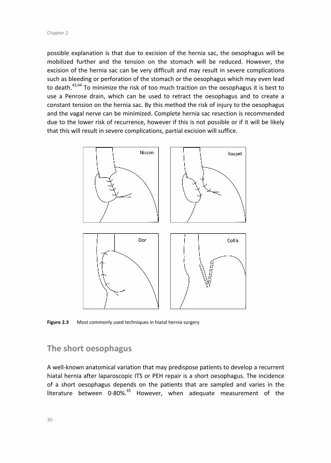
hereafter. in Figure 2.3 the most common used techniques in hiatal hernia surgery are
shown.
Hernia sac excision
A key step during laparoscopic hiatal hernia surgery is to reduce the contents of the
hernia back into the abdomen. Especially when the herniation was present for a long
time, this can be a challenging part of the procedure, due to the adhesions that have
developed over time. Several studies have proven that excision of the hernia sac leads
to a lower recurrence rate, especially in the early postoperative period.17,18 One
Chapter 2
30
possible explanation is that due to excision of the hernia sac, the oesophagus will be
mobilized further and the tension on the stomach will be reduced. However, the
excision of the hernia sac can be very difficult and may result in severe complications
such as bleeding or perforation of the stomach or the oesophagus which may even lead
to death.43,44 To minimize the risk of too much traction on the oesophagus it is best to
use a Penrose drain, which can be used to retract the oesophagus and to create a
constant tension on the hernia sac. By this method the risk of injury to the oesophagus
and the vagal nerve can be minimized. Complete hernia sac resection is recommended
due to the lower risk of recurrence, however if this is not possible or if it will be likely
that this will result in severe complications, partial excision will suffice.
Figure 2.3 Most commonly used techniques in hiatal hernia surgery
The short oesophagus
A well‐known anatomical variation that may predispose patients to develop a recurrent
hiatal hernia after laparoscopic ITS or PEH repair is a short oesophagus. The incidence
of a short oesophagus depends on the patients that are sampled and varies in the
literature between 0‐80%.45 However, when adequate measurement of the

Intrathoracic stomach in hiatal hernia
31
2
oesophagus, after mobilization, is performed, it seems that a true short oesophagus is
very rare.46 A short oesophagus is defined as a GE junction that is <2‐3 cm intra‐
abdominal and is associated with a recurrent hiatal hernia in 15‐25% of all
recurrences.45,47 The treatment for short oesophagus is still subject of debate. Some
advocate to accept the short oesophagus, where others advocate the need for
intervention. Methods to treat the short oesophagus vary from extensive intra‐thoracic
mobilization of the oesophagus to performing an oesophageal lengthening procedure,
from which the Collis gastroplasty is most commonly performed.48 Besides the
increased risk of perioperative complications due to the oesophageal lengthening
procedure, including vagal nerve injury, there are also long‐term complications. These
include oesophageal aperistalsis and acid producing mucosa above the fundoplication
leading to increased postoperative acid reflux.49 Despite the possible complications,
functional outcome between hiatal hernia repair with EL or without EL seems similar.48
Extensive mobilization of the oesophagus comes with perioperative complications as
well from which opening the pleura is most commonly seen. When routine mobilization
of the oesophagus is performed, complications rarely occur and when measured
adequately, a true short oesophagus is rare. Surgeons should therefore not routinely
perform a Collis gastroplasty, due to the possible increased risk of complications. When
a true short oesophagus is present, the Collis gastroplasty seems to be a safe and
effective surgical treatment for this entity and is recommended above extensive
oesophageal mobilization.
Mesh reinforcement
Once the hernia content is reduced, the question is raised on how to close the crural
defect. Different techniques are available, almost all starting with suture repair of the
crus. There is very little evidence regarding this aspect of the procedure. Most obvious,
non‐absorbable sutures should be used and if we compare the difference between
anterior or posterior suture repair, no significant results regarding symptom control or
recurrence rate are found.26
Another very large topic of debate is the use of prosthetic or biological material to
support the crural repair. Due to the rare but potential life‐threatening complications
such as oesophageal or aortic erosion that can come with mesh repair, surgeons are
conservative in using prosthetic material. However, several reviews have shown the
advantage of the reduced recurrence rate due to mesh reinforcement.50‐53 A significant
reduction in recurrence rate is seen from 22.5% to 12.1% when mesh is used. Even
after sub‐analysis for mesh type or for follow‐up longer than 2 years, this reduced
recurrence rate is still valid. Complication rate for both groups is approximately 15%.54
Chapter 2
32
Besides the general complications related to the procedure, the use of a mesh also
comes with specific mesh related complications. In general, these are very poorly
described in the literature. There are 124 studies, with a total of 5499 patients, that
describe mesh related complications. 50 complications are described by case reports
where 41 mesh related complications are mentioned in RCT’s and observational cohort
studies (OCS). This results in a mesh related complication rate of 1.9%. Most reported
complications include mesh erosion into either oesophagus, aorta or stomach (53.8%),
stenosis (29.7%), cardiac tamponade (8.8%), fibrosis (5.5%), aortic lesion (2.2%) and a
fistula (1.1%).54,55 Mesh related complications are most probably underreported, but
the total amount of implanted meshes is not mentioned in most studies. It is therefore
difficult to estimate the actual mesh related complication rate.
The most severe complications that come with mesh placement are related to the
fixation of the mesh. In the literature, there are four cases described of cardiac
tamponade that was caused by staplers that perforated the pericardium. All patients
died due to this complication.56‐58 Another explanation for mesh related complications
is the prosthetic material that is placed either around the oesophagus or in close
proximity to the oesophagus with the ‘sharp’ edges of the mesh against it. Since
meshes shrink after placement, even up to 70%, it is expected that when a mesh is
placed circumferential around the oesophagus, that this may lead to stenosis.59
In order to minimize complications related to the material and to still minimize the risk
of recurrence, several mesh types have been developed over time. Biological meshes
should result in less material related complications, however they seem to come with a
higher recurrence rate.60 A review published by the authors of this article, describes the
different types of mesh and their results.61 From this analysis it seems that synthetic
mesh is superior to biological mesh regarding recurrence rate. Synthetic meshes are
also less expensive which may be another argument to prefer synthetic over biological
meshes. But a very large limitation of this study is the large heterogeneity of the
different studies, which makes it impossible to perform a meta‐analysis and to draw
any strong conclusions.
Fundoplication
Most patients present with predominantly obstructive symptoms and complaints of
dysphagia, between 13% and 60% of the patients however also experience some
degree of reflux.62‐64 The rationale for acid reflux that comes with large hiatal hernia is
the disruption of the anatomy. The herniation of stomach into the thorax flattens the
angle of His and therefore leads to dysfunction of the lower oesophageal sphincter
(LES). Even when the stomach is reduced back into the abdomen, often the function of
the LES remains disrupted and results in acid reflux. For this reason, most surgeons

Intrathoracic stomach in hiatal hernia
33
2
perform an additional fundoplication following the hiatal hernia repair. The addition of
a fundoplication does not decrease the incidence of a recurrent hiatal hernia.65
Therefore the main reason to perform a fundoplication should be the prevention of
acid reflux or the treatment of pre‐existent reflux. It has been a large subject of debate
which type of fundoplication is superior and many meta‐analysis and systematic
reviews have been published regarding this subject. Most studies describe patient with
mainly gastroesophageal reflux disease (GERD) and not patients with large hiatal
hernia. Besides, it is not in the scope of this review to address this discussion in detail.
The overall conclusion of the literature is that the higher the degree of fundoplication,
the better the reflux control. However, this also comes with an increased risk of
postoperative dysphagia.66 Dysphagia after hiatal hernia repair with Nissen
fundoplication is approximately between 3% and 24%.67,68 it should kept in mind, that
in only 13‐47% of the patients with a type III or type IV hernia, the preoperative
endoscopy showed signs of oesophagitis. Severe oesophagitis as a result of long time
acid reflux is even more rare.64 Postoperative reflux is significantly more prominent in
patients that did not receive a fundoplication after ITS repair, however, this did not
influence quality of life.69 In the contrary, the possible negative side effects of a
fundoplication, in particular dysphagia, were not found to be significantly more
present.70 The routine use of a fundoplication after hiatal hernia repair for intrathoracic
stomach should be performed with care, since the clinical benefit seems to be limited.
The application of an anterior gastropexy is less invasive compared to a complete or
partial fundoplication and should as well restore the angle of His. This technique is a
safe alternative for a fundoplication and should be considered in the elderly patients or
in other patients where the surgical risks should be minimized.71 The routine
application of a gastropexy after cruroplasty in patients with a hiatal hernia shows good
control of reflux symptoms. A total of 70.3% of the patients were asymptomatic after
2 years of follow‐up and only 9.9% of the patients required antireflux medication on
daily basis.72
To conclude, it is justified to individualize the decision whether to perform a
fundoplication in the laparoscopic treatment of an intrathoracic stomach or large para‐
oesophageal hernia repair. If there is preoperative evidence of severe oesophagitis or
severe complaints of reflux, a fundoplication should be considered. In all other cases, a
partial fundoplication or gastropexy should be performed to minimize the risk of
postoperative dysphagia and to minimize operative time. Even more, since most
postoperative acid reflux symptoms are mild and can be controlled with medication.

Chapter 2
34
Complications after laparoscopic hiatal hernia repair
There are several complications that are likely to occur in ITS and PEH repair, as a result
of the vital structures that may be involved with the herniation and are in close
proximity to the area where the surgeon has to perform. Large bleedings from either
the aorta, spleen, liver or short‐gastric vessels have been reported.73 However most
bleedings are arterial, also bleedings from large veins such as the vena cave can occur.74
These are more feared, since the thin wall of the veins makes it harder to control the
bleeding. Most injuries are caused by coagulation devices, electrocautery or ultrasonic
devices.75
Furthermore, the thoracic organs can be damaged, in particular when a mesh is fixated
with tackers, this may cause severe cardiac complications with a high mortality rate.76
Damage of other abdominal organs is possible as well, where most injuries occur during
the reduction of the content of the hernia sac. Pleural rupture is very often seen in
hiatal hernia surgery. Usually, thoracic drainage is not necessary, when positive end
expiratory pressure at the end of surgery is kept high (>10 cmh2O), a pneumothorax will
rarely occur. If a larger pleural defect is present, a chest tube may be inevitable.77
Delayed gastric emptying (DGE) is a complication that is seen after both ITS and PEH
repair. This may be the result of organic causes (Diabetes, ulcers, auto‐immune disease,
neuromotor dysfunction) as well technical causes (Vagal nerve injury or obstruction).
The presence of DGE preoperatively is very hard to determine, since gastric emptying
studies are not reliable due to the intrathoracic stomach. The rate of DGE in the
literature is less than 3%, however with more re‐operations the risk of delayed gastric
emptying increases. The rationale for this effect may be the increasing risk of vagal
nerve damage.78 Symptoms that can come with vagal nerve injury are nausea (15%),
vomiting (4%), diarrhoea (20‐30%) and dumping syndrome (10%). In contrast with the
low incidence of DGE mentioned above, the rate of vagal nerve injury is supposed to be
as high as 10‐42%.79 The result of vagal nerve injury is increased presence of acid reflux
and a higher reoperation rate for severe acid reflux. Also, the quality of life and the
postoperative satisfaction is significantly lower when the vagal nerve is damaged.80 For
some reason, vagal nerve injury is often neglected by the surgeon and symptoms
related to unintentional vagal nerve injury are often not recognized. Vagal nerve injury
may lead to severe symptoms and a reduced quality of life, therefore, great caution is
needed when mobilizing the oesophagus and direct traction to the oesophagus should
be minimized.
Intrathoracic stomach in hiatal hernia
35
2
Predictors for outcome
A very important question is, whether there are patient related factors or technical
aspects that may predict outcome after ITS or PEH repair. There is still no consistent
standard definition for recurrence. The most objective method might be a radiological
finding where at least 2 cm of migration on the barium swallow examination is seen.81
This results in a recurrence rate up to 57%. However most patients remain
asymptomatic, thus the relevance of this method may be questioned.82,83 Median time
for radiologic recurrence is 40 months.84 It might be beneficial to only report
symptomatic recurrence, which is more relevant in the treatment of patients. Most
technical aspects that might influence postoperative outcome such as resection of the
hernia sac, mobilization of the oesophagus, mesh reinforcement and performing a
fundoplication are mentioned above.
Additional patient related factors such as size of the hernia, age, elevated body mass
index (BMI), pulmonary disease have been mentioned as a possible risk factor for
recurrence.85
A larger size of the hiatal hernia is associated with an increased risk of re‐herniation
after primary repair. However, for recurrent hiatal hernia repair, this association
disappears. This may be due to other, not yet discovered, patient related factors that
play an important role in hernia recurrence.86
An elevated BMI results in an increased intra‐abdominal pressure and might therefore
predispose for failure of the cruroplasty and result into an elevated recurrence rate. For
antireflux surgery, it has been proven that an increased BMI leads to a high failure rate
of the surgical treatment.87 For PEH and ITS repair however, this difference in clinical
outcome for patients with an increased BMI compared with patients with a normal BMI
cannot be confirmed.29,88 Nonetheless, surgeons should recommend patient to lose
weight, since an increased BMI leads to technical difficulties and overall health is
affected by the extra weight.
There are some patient related factors that do influence the postoperative outcome.
Both a higher American Society of Anaesthesiologists (ASA) score and an age >70 is
associated with a longer postoperative hospital stay and a higher complication rate.29,38
Age, gender, smoking status, diabetes or pulmonary disease have not been associated
with a difference in radiologic recurrence.37,83
Future perspectives
In this review, we have described the role of laparoscopy from the beginning in 1992
until now. As we have seen, there are several aspects in laparoscopic hiatal hernia
repair that are still subject of debate. With improving technology, new techniques such
Chapter 2
36
as 3D and robotic surgery are coming more and more available in the field of minimal
invasive surgery. In theory, these techniques may reduce operative time, complication
rate and may improve patient outcomes. A recent pilot study investigating the
advantage of 3D visualization during laparoscopic hiatal hernia repair compared with
2D visualization, proved that the operative time is significantly reduced by 22% when
3D vision is used.89 Furthermore, robotic surgery is becoming more available in many
institutions, starting a whole new era. However, the advantage of robot surgery in
hiatal hernia repair still has to be discovered. Initial reports seem promising, however a
high recurrence rate of up to 42% after 2 years is seen.90 This might be due to the lack
of experience with robot‐assisted surgery and learning curve is still present. Therefore,
results will most likely improve over time and further studies are needed to prove
whether robot‐assisted ITS and PEH repair is beneficial compared with traditional
laparoscopic repair.91
Conclusion
This is an extensive review of the current literature and describes the role of
laparoscopy in intrathoracic stomach repair. That are still many aspects of the
treatment that are subject of debate and no gold standard for the treatment of an
intrathoracic stomach or large para‐oesophageal hernia repair is achieved. It is widely
accepted that laparoscopy is superior to laparotomy and leads to better postoperative
outcome. Furthermore, the transabdominal approach is standard treatment. However,
the thoracic approach should be kept in mind when patients have a complex abdominal
history and abdominal approach seems not possible. Since acute incarceration comes
with increased morbidity and mortality, elective repair of intrathoracic stomach is
recommended, even for the elderly with minimal complaints. However, the decision to
perform a laparoscopic hiatal hernia repair should be tailored made, since not every
octogenarian is suitable for surgery.
When the hernia content is reduced, is recommended to excise the hernia sac
completely to minimize the risk of recurrence. True short oesophagus is rare when
adequate mobilization of the oesophagus is performed, but when still present, Collis
gastroplasty should be performed to minimize traction on the stomach and to reduce
recurrence rate. Mesh reinforcement of the cruroplasty reduces recurrence rate,
however there is a small risk of mesh related complications. There are several types of
mesh available, but a synthetic mesh seems superior tot biological meshes, based on
costs and recurrence rate. Fundoplication should not routinely be performed, only
when severe acid reflux is present. When a fundoplication is needed, a partial or
complete fundoplication should be considered based on the preference of the
operating surgeon. An anterior gastropexy however, seems to reduce recurrence rate,

Intrathoracic stomach in hiatal hernia
37
2
leads to less dysphagia and provides good acid reflux control and might therefore be
superior to a fundoplication. Laparoscopic hiatal hernia surgery is complex and requires
advanced laparoscopic skills to minimize the risk of complications. The pleural cavity is
almost always opened, but does not require chest tube drainage in most cases. Vagal
nerve injury is an underdiagnosed complication, but results in increased acid reflux,
increased reoperation rate and a worse quality of life. Surgeons should be careful with
mobilization of the oesophagus to minimize the risk of this complication. Increased
hernia size leads to a higher recurrence rate, where other patient’s factors as BMI,
gender, smoking, pulmonary disease have no proven effect on the postoperative
outcome. As novel techniques are still being developed and robotic surgery and 3D
vision is more widely available, this might result into another approach of hiatal hernia.
The first results of these novel techniques seem promising, but the advantage of robot‐
assisted surgery and 3D vision has yet to be proved.
Chapter 2
38
References
1. Hochberg LA. Thoracic surgery before the 20th century, Vantage Press, New York 1960. 2. Cuschieri A, Shimi S, Nathanson LK. Laparoscopic reduction, crural repair, and fundoplication of large
hiatal hernia. Am J Surg 1992;163:425‐30.
3. Athanasakis H, Tzortzinis A, Tsiaoussis J, Vassilakis JS, Xynos E. Laparoscopic repair of paraesophageal hernia. Endoscopy 2001;33:590‐4.
4. Hill LD, Tobias JA. Paraesophageal hernia. Arch Surg 1968;96:735‐44.
5. Sugerbaker DJ, Bueno R, Colson YL, Jaklitsch MT, Krasna MJ, Mentzer SJ, et al. Adult chest surgery, 2nd Edition, http://www.accessurgery.com/
6. Soresi AL. Diaphragmatic Hernia: Its Unsuspected Frequency: Its Diagnosis: Technic for Radical Cure.
Ann Surg 1919;69:254‐70. 7. Harrington SW. The Surgical Treatment of the More Common Type of Diaphragmatic Hernia : Report of
404 Cases. Ann Surg 1945;122:546‐68.
8. Sweet RH. Esophageal hiatus hernia of the diaphragm; the anatomical characteristics, technic of repair, and results of treatment in 111 consecutive cases. Ann Surg 1952;135:1‐13.
9. Allison PR. Reflux esophagitis, sliding hiatal hernia, and the anatomy of repair. Surg Gynecol Obstet
1951;92:419‐31. 10. Stylopoulos N, Rattner DW. The history of hiatal hernia surgery: from Bowditch to laparoscopy. Ann
Surg 2005;241:185‐93.
11. Hiebert CA. Surgical management of esophageal reflux and hiatal hernia. Ann Thorac Surg 1961;52: 159‐60.
12. Collis JL. An operation for hiatus hernia with short oesophagus. Thorax 1957;12:181‐8.
13. Thal AP. A unified approach to surgical problems of the esophagogastric junction. Ann Surg 1968;168:542‐50.
14. Hill LD. An effective operation for hiatal hernia: an eight year appraisal. Ann Surg 1967;166:681‐92.
15. Dor J, Humbert P, Dor V, et al. The role of the modified Nissen procedure in the prevention of reflux following Heller's extramucosal cardiomyotomy. Mem Acad Chir 188:877‐82.
16. Toupet A. Technique d'eosophago‐gastroplastie avec phreno‐gastropexie dans la cure radicales des
hernies hiatales et comme complement de l'operation de Heller dans les cardiospasmes. Mem Acad Chir 1963;89:394‐9.
17. Watson DI, Davies N, Devitt PG, Jamieson GG. Importance of dissection of the hernial sac in
laparoscopic surgery for large hiatal hernias. Arch Surg 1999134:1069‐73. 18. Edye M, Salky B, Posner A, Fierer A. Sac excision is essential to adequate laparoscopic repair of
paraesophageal hernia. Surg Endosc '1998;12:1259‐63.
19. Mitiek MO, Andrade RS. Giant hiatal hernia. Ann Thorac Surg 2010;89:S2168‐73. 20. Diez Tabernilla M, Ruiz‐Tovar J, Grajal Marino R, Calero Garcia P, Pina Hernandez JD, Chames Vaisman
A, et al. Paraesophageal hiatal hernia. Open vs. laparoscopic surgery. Rev Esp Enferm Dig 2009;101:706‐
11. 21. Karmali S, McFadden S, Mitchell P, Graham A, Debru E, Gelfand G, et al. Primary laparoscopic and open
repair of paraesophageal hernias: a comparison of short‐term outcomes. Dis Esophagus 2008;21:63‐.
22. Klinginsmith M, Jolley J, Lomelin D, Krause C, Heiden J, Oleynikov D. Paraesophageal hernia repair in the emergency setting: is laparoscopy with the addition of a fundoplication the new gold standard? Surg
Endosc 2016;30:1790‐5.
23. Peters MJ, Mukhtar A, Yunus RM, Khan S, Pappalardo J, Memon B, et al. Meta‐analysis of randomized clinical trials comparing open and laparoscopic anti‐reflux surgery. Am J Gastroenterol 2009;104:1548‐
61; quiz 1547, 1562.
24. Soot SJ, Eshraghi N, Farahmand M, Sheppard BC, Deveney CW. Transition from open to laparoscopic fundoplication: the learning curve. Arch Surg 1999;134:278‐81; discussion 282.
25. Hwang H, Turner LJ, Blair NP. Examining the learning curve of laparoscopic fundoplications at an urban
community hospital. Am J Surg 2005;189:522‐6; discussion 526.

Intrathoracic stomach in hiatal hernia
39
2
26. Antiporda M, Veenstra B, Jackson C, Kandel P, Daniel Smith C, Bowers SP. Laparoscopic repair of giant
paraesophageal hernia: are there factors associated with anatomic recurrence? Surg Endosc 2018; 32(2):945‐54.
27. Whealon MD, Blondet JJ, Gahagan JV, Phelan MJ, Nguyen NT. Volume and outcomes relationship in
laparoscopic diaphragmatic hernia repair. Surg Endosc 2017;31(10):4224‐30. 28. Castelijns PSS, Ponten JEH, Van de Poll MCG, Nienhuijs SW, Smulders JF. Subjective outcome after
laparoscopic hiatal hernia repair for intrathoracic stomach. Langenbecks Arch Surg 2017;402:521‐30.
29. Karim MA, Maloney J, Ali A (2016) Laparoscopic Repair of Intrathoracic Stomach: Clinical and Health‐related Quality of Life Outcomes. Surg Laparosc Endosc Percutan Tech 2016;26:484‐7.
30. Wirsching A, El Lakis MA, Mohiuddin K, Pozzi A, Hubka M, Low DE. Acute vs. elective paraesophageal
hernia repair: Endoscopic gastric decompression allows semi‐elective surgery in a majority of acute patients. J Gastrointest Surg 2018;22(2):194‐202.
31. Jassim H, Seligman JT, Frelich M, Goldblatt M, Kastenmeier A, Wallace J, et al. A population‐based
analysis of emergent versus elective paraesophageal hernia repair using the Nationwide Inpatient Sample. Surg Endosc 2014;28:3473‐8.
32. Kaplan JA, Schecter S, Lin MY, Rogers SJ, Carter JT. Morbidity and Mortality Associated With Elective or
Emergency Paraesophageal Hernia Repair. JAMA Surg 2015;150:1094‐6. 33. Stylopoulos N, Gazelle GS, Rattner DW. Paraesophageal hernias: operation or observation? Ann Surg
2002;236:492‐500; discussion 500‐491.
34. Poulose BK, Gosen C, Marks JM, Khaitan L, Rosen MJ, Onders RP, et al. Inpatient mortality analysis of paraesophageal hernia repair in octogenarians. J Gastrointest Surg 2008;12:1888‐92.
35. Jay AP, Watson DI. Changing work patterns for benign upper gastrointestinal and biliary disease: 1994‐
2007. ANZ J Surg 2010;80:519‐25. 36. Grotenhuis BA, Wijnhoven BP, Bessell JR, Watson DI. Laparoscopic antireflux surgery in the elderly. Surg
Endosc 2008;22:1807‐12
37. El Lakis MA, Kaplan SJ, Hubka M, Mohiuddin K, Low DE. The Importance of Age on Short‐Term Outcomes Associated With Repair of Giant Paraesophageal Hernias. Ann Thorac Surg 2017;103: 1700‐9.
38. Parker DM, Rambhajan AA, Horsley RD, Johanson K, Gabrielsen JD, Petrick AT. Laparoscopic
paraesophageal hernia repair is safe in elderly patients. Surg Endosc 2017;31:1186‐91. 39. Paul S, Nasar A, Port JL, Lee PC, Stiles BC, Nguyen AB, et al. Comparative analysis of diaphragmatic
hernia repair outcomes using the nationwide inpatient sample database. Arch Surg 2012;147:607‐12.
40. Kohn GP, Price RR, DeMeester SR, Zehetner J, Muensterer OJ, Awad Z, et al. Guidelines for the management of hiatal hernia. Surg Endosc 2013;27:4409‐28.
41. Migliore M, Arcerito M, Vagliasindi A, Puleo R, Basile F, Deodato G. The place of Belsey Mark IV
fundoplication in the era of laparoscopic surgery. Eur J Cardiothorac Surg 2003;24:625‐30. 42. Molena D, Mungo B, Stem M, Lidor AO. Novel combined VATS/laparoscopic approach for giant and
complicated paraesophageal hernia repair: description of technique and early results. Surg Endosc
2015;29:185‐91. 43. Wiechmann RJ, Ferguson MK, Naunheim KS, McKesey P, Hazelrigg SJ, Santucci TS, et al. Laparoscopic
management of giant paraesophageal herniation. Ann Thorac Surg 2001;71:1080‐6; discussion 1086‐7.
44. Pierre AF, Luketich JD, Fernando HC, Christie NA, Buenaventura PO, Litle VR, et al. Results of laparoscopic repair of giant paraesophageal hernias: 200 consecutive patients. Ann Thorac Surg
2002;74:1909‐15; discussion 1915‐6.
45. Lugaresi M, Mattioli S, Aramini B, D'Ovidio F, Di Simone MP, Perrone O. The frequency of true short oesophagus in type II‐IV hiatal hernia. Eur J Cardiothorac Surg 2013;43:e30‐6.
46. Migaczewski M, Zub‐Pokrowiecka A, Grzesiak‐Kuik A, Pedziwiatr M, Major P, Rubinkiewicz M, et al.
Incidence of true short esophagus among patients submitted to laparoscopic Nissen fundoplication. Wideochir Inne Tech Maloinwazyjne 2015;10:10‐4.
47. Horvath KD, Swanstrom LL, Jobe BA. The short esophagus: pathophysiology, incidence, presentation,
and treatment in the era of laparoscopic antireflux surgery. Ann Surg 2000;232:630‐40. 48. Puri V, Jacobsen K, Bell JM, Crabtree TD, Kreisel D, Krupnick AS, et al. Hiatal hernia repair with or
without esophageal lengthening: is there a difference? Innovations (Phila) 2013;8:341‐7.

Chapter 2
40
49. Lin E, Swafford V, Chadalavada R, Ramshaw BJ, Smith CD. Disparity between symptomatic and
physiologic outcomes following esophageal lengthening procedures for antireflux surgery. J Gastrointest Surg 2004;8:31‐9; discussion 38‐9.
50. Antoniou SA, Koch OO, Antoniou GA, Pointner R, Granderath FA. Mesh‐reinforced hiatal hernia repair: a
review on the effect on postoperative dysphagia and recurrence. Langenbecks Arch Surg 2012;397: 19‐27.
51. Puri V, Kakarlapudi GV, Awad ZT, Filipi CJ. Hiatal hernia recurrence: 2004. Hernia 2004;8:311‐7.
52. Targarona EM, Bendahan G, Balague C, Garriga J, Trias M. Mesh in the hiatus: a controversial issue. Arch Surg 2004;139:1286‐96; discussion 1296.
53. Furnee E, Hazebroek E. Mesh in laparoscopic large hiatal hernia repair: a systematic review of the
literature. Surg Endosc 2013;27:3998‐4008. 54. Muller‐Stich BP, Kenngott HG, Gondan M, Stock C, Linke GR, Fritz F, et al. Use of Mesh in Laparoscopic
Paraesophageal Hernia Repair: A Meta‐Analysis and Risk‐Benefit Analysis. PLoS One 2015;10:
e0139547. 55. Stadlhuber RJ, Sherif AE, Mittal SK, Fitzgibbons RJ, Jr., Michael Brunt L, Hunter JG, et al. Mesh
complications after prosthetic reinforcement of hiatal closure: a 28‐case series. Surg Endosc
2009;23:1219‐26. 56. Kemppainen E, Kiviluoto T. Fatal cardiac tamponade after emergency tension‐free repair of a large
paraesophageal hernia. Surg Endosc 2000;14:593.
57. Muller‐Stich BP, Linke G, Leemann B, Lange J, Zerz A. Cardiac tamponade as a life‐threatening complication in antireflux surgery. Am J Surg 2006;191:139‐41.
58. Frantzides CT, Welle SN. Cardiac tamponade as a life‐threatening complication in hernia repair. Surgery
2012;152:133‐5. 59. Brown CN, Finch JG. Which mesh for hernia repair? Ann R Coll Surg Engl 2010;92:272‐8.
60. Huddy JR, Markar SR, Ni MZ, Morino M, Targarona EM, Zaninotto G, et al. Laparoscopic repair of hiatus
hernia: Does mesh type influence outcome? A meta‐analysis and European survey study. Surg Endosc 2016;30:5209‐21.
61. Castelijns PSS, Ponten JEH, van de Poll MCG, Nienhuijs SW, Smulders JF. A collective review of biological
versus synthetic mesh‐reinforced cruroplasty during laparoscopic Nissen fundoplication. J Minim Access Surg 2018;14(2):87‐94.
62. Braghetto I, Korn O, Csendes A, Burdiles P, Valladares H, Brunet L. Postoperative results after
laparoscopic approach for treatment of large hiatal hernias: is mesh always needed? Is the addition of an antireflux procedure necessary? Int Surg 2010;95:80‐7.
63. Avidan B, Sonnenberg A, Schnell TG, Sontag SJ. Hiatal hernia and acid reflux frequency predict presence
and length of Barrett's esophagus. Dig Dis Sci 2002;47:256‐64. 64. Rakic S, Pesko P, Dunjic MS, Gerzic Z. Paraoesophageal hernia repair with and without concomitant
fundoplication. Br J Surg 1994;81:1162‐3.
65. Furnee EJ, Draaisma WA, Simmermacher RK, Stapper G, Broeders IA. Long‐term symptomatic outcome and radiologic assessment of laparoscopic hiatal hernia repair. Am J Surg 2010;199:695‐701.
66. Broeders JA, Sportel IG, Jamieson GG, Nijjar RS, Granchi N, Myers JC, et al. Impact of ineffective
oesophageal motility and wrap type on dysphagia after laparoscopic .fundoplication. Br J Surg 2011;98:1414‐21.
67. Granderath FA, Schweiger UM, Kamolz T, Asche KU, Pointner R. Laparoscopic Nissen fundoplication
with prosthetic hiatal closure reduces postoperative intrathoracic wrap herniation: preliminary results of a prospective randomized functional and clinical study. Arch Surg 2005;140:40‐8.
68. Hunter JG, Swanstrom L, Waring JP. Dysphagia after laparoscopic antireflux surgery. The impact of
operative technique. Ann Surg 1996;224:51‐7. 69. Svetanoff WJ, Pallati P, Nandipati K, Lee T, Mittal SK. Does the addition of fundoplication to repair the
intra‐thoracic stomach improve quality of life? Surg Endosc 2016;30:4590‐7.
70. Muller‐Stich BP, Achtstatter V, Diener MK, Gondan M, Warschkow R, Marra F, et al. Repair of Paraesophageal Hiatal Hernias‐Is a Fundoplication Needed? A Randomized Controlled Pilot Trial. J Am
Coll Surg 2015;221:602‐10.

Intrathoracic stomach in hiatal hernia
41
2
71. Higashi S, Nakajima K, Tanaka K, Miyazaki Y, Makino T, Takahashi T, et al. Laparoscopic anterior
gastropexy for type III/IV hiatal hernia in elderly patients. Surg Case Rep 2017;3:45. 72. Daigle CR, Funch‐Jensen P, Calatayud D, Rask P, Jacobsen B, Grantcharov TP. Laparoscopic repair of
paraesophageal hernia with anterior gastropexy: a multicenter study. Surg Endosc 2015;29:1856‐61.
73. Leggett PL, Bissell CD, Churchman‐Winn R. Aortic injury during laparoscopic fundoplication: an underreported complication. Surg Endosc 2002;16:362.
74. Baigrie RJ, Watson DI, Game PA, Jamieson GG. Vascular perils during laparoscopic dissection of the
oesophageal hiatus. Br J Surg 1997;84:556‐7. 75. Cano‐Valderrama O, Marinero A, Sanchez‐Pernaute A, Dominguez‐Serrano I, Perez‐Aguirre E, Torres AJ.
Aortic injury during laparoscopic esophageal hiatoplasty. Surg Endosc 2013;27:3000‐2.
76. Fernandez Mdel C, Diaz M, Lopez F, Marti‐Obiol R, Ortega J. Cardiac complications after laparoscopic large hiatal hernia repair. Is it related with staple fixation of the mesh? ‐Report of three cases. Ann Med
Surg (Lond) 2015;4:395‐8.
77. Yang X, Hua R, He K, Shen Q, Yao Q. Laparoscopic hernioplasty of hiatal hernia. Ann Transl Med 2016;4:343.
78. Hamrick MC, Davis SS, Chiruvella A, Coefield RL, Waring JP, Sweeney JF, et al. Incidence of delayed
gastric emptying associated with revisional laparoscopic paraesophageal hernia repair. J Gastrointest Surg 2013;17:213‐7
79. van Rijn S, Roebroek YG, Conchillo JM, Bouvy ND, Masclee AA. Effect of Vagus Nerve Injury on the
Outcome of Antireflux Surgery: An Extensive Literature Review. Dig Surg 2016;33:230‐9. 80. van Rijn S, Rinsma NF, van Herwaarden‐Lindeboom MY, Ringers J, Gooszen HG, van Rijn PJ, et al. Effect
of Vagus Nerve Integrity on Short and Long‐Term Efficacy of Antireflux Surgery. Am J Gastroenterol
2016;111:508‐15. 81. Lidor AO, Kawaji Q, Stem M, Fleming RM, Schweitzer MA, Steele KE, et al. Defining recurrence after
paraesophageal hernia repair: correlating symptoms and radiographic findings. Surgery 2013;154:
171‐8. 82. Oelschlager BK, Petersen RP, Brunt LM, Soper NJ, Sheppard BC, Mitsumori L, et al. Laparoscopic
paraesophageal hernia repair: defining long‐term clinical and anatomic outcomes. J Gastrointest Surg
2012;16:453‐9. 83. Lidor AO, Steele KE, Stem M, Fleming RM, Schweitzer MA, Marohn MR. Long‐term quality of life and
risk factors for recurrence after laparoscopic repair of paraesophageal hernia. JAMA Surg
2015;150:424‐31. 84. Furtado RV, Vivian SJ, van der Wall H, Falk GL. Medium‐term durability of giant hiatus hernia repair
without mesh. Ann R Coll Surg Engl 2016;98:450‐5.
85. Zaman JA, Lidor AO. The Optimal Approach to Symptomatic Paraesophageal Hernia Repair: Important Technical Considerations. Curr Gastroenterol Rep 2016;18:53.
86. Koch OO, Asche KU, Berger J, Weber E, Granderath FA, Pointner R. Influence of the size of the hiatus on
the rate of reherniation after laparoscopic fundoplication and refundopilication with mesh hiatoplasty. Surg Endosc 2011;25:1024‐30.
87. Morgenthal CB, Lin E, Shane MD, Hunter JG, Smith CD. Who will fail laparoscopic Nissen
fundoplication? Preoperative prediction of long‐term outcomes. Surg Endosc 2007;21:1978‐84. 88. Lidor AO, Chang DC, Feinberg RL, Steele KE, Schweitzer MA, Franco MM. Morbidity and mortality
associated with antireflux surgery with or without paraesophogeal hernia: a large ACS NSQIP analysis.
Surg Endosc 2011;25:3101‐8. 89. Leon P, Rivellini R, Giudici F, Sciuto A, Pirozzi F, Corcione F. 3D Vision Provides Shorter Operative Time
and More Accurate Intraoperative Surgical Performance in Laparoscopic Hiatal Hernia Repair Compared
With 2D Vision. Surg Innov 2017;24:155‐61. 90. Galvani CA, Loebl H, Osuchukwu O, Samame J, Apel ME, Ghaderi I. Robotic‐Assisted Paraesophageal
Hernia Repair: Initial Experience at a Single Institution. J Laparoendosc Adv Surg Tech A 2016;26:290‐5.
91. Sarkaria IS, Latif MJ, Bianco VJ, Bains MS, Rusch VW, Jones DR, et al. (2017) Early operative outcomes and learning curve of robotic assisted giant paraesophageal hernia repair. Int J Med Robot 2017;13(1).

Chapter 2
42
Chapter 3 A collective review of biological versus synthetic
mesh‐reinforced cruroplasty during laparoscopic
Nissen fundoplication
P.S.S. Castelijns
J.E.H. Ponten
M.C.G. van de Poll
S.W. Nienhuijs
J.F. Smulders
J Minim Access Surg. 2018;14(2):87‐94
Chapter 3
44
Abstract
Background Laparoscopic cruroplasty and fundoplication have become the gold standard in the treatment of hiatal hernia and gastroesophageal reflux disease (GERD). The use of a mesh‐reinforcement of the cruroplasty has been proven effective; although there is a lack of evidence considering which type of mesh is superior. The aim of this study was to compare recurrence rates after mesh reinforced cruroplasty using biological versus synthetic meshes. Methods We performed a systematic review of all clinical trials published between January 2004 and September 2015 describing the application of a mesh in the hiatal hernia repair during Nissen fundoplication for both GERD and hiatal hernia. The primary outcome was the recurrence rate, and secondary outcomes were complication rate, mortality and symptomatic outcome. Results We included 16 studies and extracted data regarding 1089 mesh operated patients of whom 385 received a biological mesh and 704 a synthetic mesh. The mean follow‐up was 53.4 months. The recurrence rate in the synthetic mesh group was 6.8% compared to 16.1% in the biological mesh group (P < 0.05). The complication rate was 5.1% and 4.6% (P = 0.694), respectively, and there were 12 mesh‐related complications. No mesh‐related mortality was reported. Conclusion Mesh reinforcement of hiatal hernia repair seems safe in the short‐term follow‐up. The available literature suggests no clear advantage of biological over synthetic meshes. Regarding cost‐efficiency and short‐term results, the use of synthetic non‐absorbable meshes might be advocated.
Biological versus synthetic mesh‐reinforced cruroplasty during laparoscopic Nissen fundoplication
45
3
Introduction
In 1992, the first report on laparoscopic hiatal hernia repair was published, and over
the subsequent years the laparoscopic approach has become standard treatment.1,2
Hiatal herniation is a common disorder of the digestive tract, which is defined by a
protrusion of the stomach into the thoracic cavity through a widening of the right crus
of the diaphragm.3 Anatomically, hiatal hernias can be divided into four categories.
Type I, a sliding hernia, means that the gastroesophageal junction migrates into the
thorax. This is the most common type (95%) and it may predispose to gastroesophageal
reflux.2,4 Type II, para‐oesophageal hernia, is a defect of the diaphragm with herniation
of the gastric fundus through the hiatus into the thorax. Type III is a mixed type of
hernia with both aspects of a type I and type II hernia. Often more than 50% of the
stomach is in the thorax. The most severe type of hernia is a type IV, in which not only
the stomach, but also other viscera like colon or spleen herniate into the mediastinal
sac. In this latter case it is also called an “upside‐down stomach”.5
The possible life‐threatening aspects of a hiatal hernia, such as obstruction, acute
dilatation, perforation, or bleeding of the stomach mucosa, make it important to
diagnose and repair the hiatal hernia.6 The use of mesh to repair a hiatal hernia had
already started in the era of the open surgery.7 However, there is growing interest in
the use of a mesh during laparoscopic surgery as well. According to the Society of
American Gastrointestinal and Endoscopic Surgeons (SAGES) guidelines, the
laparoscopic Nissen fundoplication and cruroplasty is the gold standard for both
gastroesophageal reflux disease (GERD)8 and hiatal hernia repair.9 Recent studies10‐12
have suggested that reinforcement of the cruroplasty with a mesh results in a reduction
in the recurrence rate. Several different meshes are available, broadly categorised into
synthetic or biological material, various shapes or the fixation method.13 Both biological
and synthetic meshes have been applied. The theoretical advantages of a biological
mesh are less mesh related complications such as erosion or migration of the mesh,
infection of the mesh and development of fibrotic strictures. However, considering the
considerably higher costs of biological meshes and a possible higher recurrence rate,
these theoretical advantages should be supported by clinical evidence before the
standard use of biological meshes for cruroplasty reinforcement can be advocated.
Only one randomised clinical trial has been performed addressing the direct question
whether biological meshes are indeed superior to synthetic meshes.14 However, this
study only reports short term follow‐up. We systematically reviewed the outcomes of
mesh reinforced cruroplasty, comparing biological and synthetic meshes. By including
the “mesh‐arms” of randomised controlled “mesh vs. non‐mesh‐trials,” we attempted
to reduce selection bias.
Chapter 3
46
We evaluated the recurrence rate in the different mesh groups as the primary
outcome. Complications, mortality and the symptomatic success rate are seen as
secondary outcomes.
Methods
Search strategy
PubMed database, including an e‐link to related articles, Embase, and the clinical trial
registry for the Cochrane Collaboration (CENTRAL) were searched for relevant studies
reporting a mesh‐based hiatal hernia repair and the outcome of recurrence of hiatal
hernias.
The first search was performed in PubMed with the following terms: ((((((“Hernia,
Hiatal”[Mesh]) OR hiatal hernia) OR hiatus)) AND (((“Surgical Mesh”[Mesh]) OR mesh)
OR mesh*)) AND ((“Laparoscopy”[Mesh]) OR laparoscopic)) AND
((((“Fundoplication”[Mesh]) OR Nissen) OR Toupet) OR fundoplication). When searching
in Embase, the following search was used to identify the related articles: (hiatal hernia
and laparoscopic and mesh and fundoplication).
In 2005 a large review describeed the results of laparoscopic versus open hiatal hernia
repair up to 2004.15 Therefore, studies from January 1, 2004 to September 30, 2015
were selected. Two reviewers independently reviewed the articles (BC/JP). In case of
discrepancy between the two reviewers, a third party (JFS) was consulted. Restrictions
were: at least n=15 and minimum follow‑up of 12 months, primary repairs only. There
were no limitations on language or study design. Selection was based on title and
abstract, and when in doubt, full text.
Selection criteria
All cohort studies, both retrospective and prospective, case‐control studies and RCT,
which reported recurrence rates of mesh‐based repair of hiatal hernia were selected,
and studies with a level of evidence ≥3B were included in the study. Paediatric studies,
reviews, meta‐analyses, case reports and case‐series were excluded. The recurrence
rate and mesh characteristics were obligatory to be included; the symptomatic
outcome, however, was not an inclusion criteria. Only non‐absorbable synthetic
meshes are included in this study. A recurrence is defined as an objectified recurrence,
if a hiatal hernia is seen on endoscopy or a Barium swallow examination. Figure 3.1
gives an overview of the search strategy.
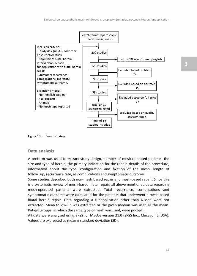
Biological versus synthetic mesh‐reinforced cruroplasty during laparoscopic Nissen fundoplication
47
3
Figure 3.1 Search strategy
Data analysis
A preform was used to extract study design, number of mesh operated patients, the
size and type of hernia, the primary indication for the repair, details of the procedure,
information about the type, configuration and fixation of the mesh, length of
follow‑up, recurrence rate, all complications and symptomatic outcome.
Some studies described both non‐mesh based repair and mesh‐based repair. Since this
is a systematic review of mesh‐based hiatal repair, all above mentioned data regarding
mesh‐operated patients were extracted. Total recurrence, complications and
symptomatic outcome were calculated for the patients that underwent a mesh‐based
hiatal hernia repair. Data regarding a fundoplication other than Nissen were not
extracted. Mean follow‐up was extracted or the given median was used as the mean.
Patient groups, in which the same type of mesh was used, were pooled.
All data were analysed using SPSS for MacOs version 21.0 (SPSS Inc., Chicago, IL, USA).
Values are expressed as mean ± standard deviation (SD).
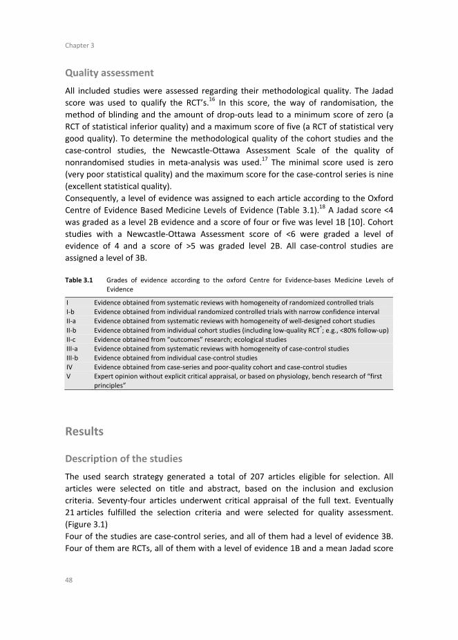
Chapter 3
48
Quality assessment
All included studies were assessed regarding their methodological quality. The Jadad
score was used to qualify the RCT’s.16 In this score, the way of randomisation, the
method of blinding and the amount of drop‐outs lead to a minimum score of zero (a
RCT of statistical inferior quality) and a maximum score of five (a RCT of statistical very
good quality). To determine the methodological quality of the cohort studies and the
case‐control studies, the Newcastle‐Ottawa Assessment Scale of the quality of
nonrandomised studies in meta‐analysis was used.17 The minimal score used is zero
(very poor statistical quality) and the maximum score for the case‐control series is nine
(excellent statistical quality).
Consequently, a level of evidence was assigned to each article according to the Oxford
Centre of Evidence Based Medicine Levels of Evidence (Table 3.1).18 A Jadad score <4
was graded as a level 2B evidence and a score of four or five was level 1B [10]. Cohort
studies with a Newcastle‐Ottawa Assessment score of <6 were graded a level of
evidence of 4 and a score of >5 was graded level 2B. All case‐control studies are
assigned a level of 3B.
Table 3.1 Grades of evidence according to the oxford Centre for Evidence‐bases Medicine Levels of
Evidence
I Evidence obtained from systematic reviews with homogeneity of randomized controlled trials
I‐b Evidence obtained from individual randomized controlled trials with narrow confidence interval
II‐a Evidence obtained from systematic reviews with homogeneity of well‐designed cohort studies
II‐b Evidence obtained from individual cohort studies (including low‐quality RCT*; e.g., <80% follow‐up)
II‐c Evidence obtained from “outcomes” research; ecological studies III‐a Evidence obtained from systematic reviews with homogeneity of case‐control studies
III‐b Evidence obtained from individual case‐control studies
IV Evidence obtained from case‐series and poor‐quality cohort and case‐control studies V Expert opinion without explicit critical appraisal, or based on physiology, bench research of “first
principles”
Results
Description of the studies
The used search strategy generated a total of 207 articles eligible for selection. All
articles were selected on title and abstract, based on the inclusion and exclusion
criteria. Seventy‐four articles underwent critical appraisal of the full text. Eventually
21 articles fulfilled the selection criteria and were selected for quality assessment.
(Figure 3.1)
Four of the studies are case‐control series, and all of them had a level of evidence 3B.
Four of them are RCTs, all of them with a level of evidence 1B and a mean Jadad score
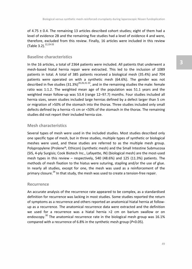
Biological versus synthetic mesh‐reinforced cruroplasty during laparoscopic Nissen fundoplication
49
3
of 4.75 ± 0.4. The remaining 13 articles described cohort studies; eight of them had a
level of evidence 2B and the remaining five studies had a level of evidence 4 and were,
therefore, excluded from this review. Finally, 16 articles were included in this review
(Table 3.2).12,19‐33
Baseline characteristics
In the 16 articles, a total of 2364 patients were included. All patients that underwent a
mesh‐based hiatal hernia repair were extracted. This led to the inclusion of 1089
patients in total. A total of 385 patients received a biological mesh (35.4%) and 704
patients were operated on with a synthetic mesh (64.6%). The gender was not
described in five studies (31.3%)20,26,31‐33, and in the remaining studies the male: female
ratio was 1:1.2. The weighted mean age of the population was 51.1 years and the
weighted mean follow‐up was 53.4 (range 12–97.7) months. Four studies included all
hernia sizes, seven studies included large hernias defined by a defect larger than 5 cm
or migration of >50% of the stomach into the thorax. Three studies included only small
defects defined by a hernia <5 cm or <50% of the stomach in the thorax. The remaining
studies did not report their included hernia size.
Mesh characteristics
Several types of mesh were used in the included studies. Most studies described only
one specific type of mesh, but in three studies, multiple types of synthetic or biological
meshes were used, and these studies are referred to as the multiple mesh group.
Polypropylene (Prolene®; Ethicon) (synthetic mesh) and the Small Intestine Submucosa
(SIS, 4‐ply Surgisis; Cook Biotech Inc., Lafayette, IN) (biological mesh) are the most‐used
mesh types in this review – respectively, 540 (48.6%) and 125 (11.3%) patients. The
methods of mesh fixation to the hiatus were suturing, stapling and/or the use of glue.
In nearly all studies, except for one, the mesh was used as a reinforcement of the
primary closure.26 In that study, the mesh was used to create a tension‐free repair.
Recurrence
An accurate analysis of the recurrence rate appeared to be complex, as a standardised
definition for recurrence was lacking in most studies. Some studies reported the return
of symptoms as a recurrence and others reported an anatomical hiatal hernia at follow‐
up as a recurrence. The anatomical recurrence data were extracted and the definition
we used for a recurrence was a hiatal hernia >2 cm on barium swallow or on
endoscopy.30 The anatomical recurrence rate in the biological mesh group was 16.1%
compared with a recurrence of 6.8% in the synthetic mesh group (P<0.05).

Chapter 3
50
Table 3.2
Overview of included
articles
Referen
ce
Level of
evidence
Mesh type
Synthetic /
biological
Indication for
surgery
Size of hernia
Mesh
operated
patients
Mean
follow up
(months)
Recurrence
Complications
Symptomatic
successful
outcome (%)
Brandalise et
al. 201219
2B
Polypropylene
Synthetic
Type 2‐4 Hernia
>5cm
70
49
9/60
1/70
NR
Gouvas et al.
201120
3B
Multiple
synthetic
Synthetic
Type 2‐4 Hernia
>8cm
20
12
3/20
2/20
NR
Granderath et
al. 200521
1B
Polypropylene
Synthetic
GER
D
>5cm
(58%),
<5(42%)
50
12
4/45
0/50
NR
Granderath et
al. 200622
1B
Polypropylene
Synthetic
GER
D
<5cm
(55%),
>5cm
(45%)
20
12
1/20
0/20
19/20 (95.0%)
Jacobs et al.
200723
3B
SIS
Biological
GER
D
NR
92
38,4
3/92
8/92
74/92 (80.4%)
Lee et al.
200724
2B
HADM
Biological
Type 2 and
recurren
t Hernia 4‐7cm
17
14,4
2/14
3/17
15/17 (88.2%)
Lubezky et al.
200725
2B
Multiple
synthetic
Synthetic
Type 2 Hernia
NR
59
28,4
6/45
10/56
40/54 (74.1%)
Morino et al.
200626
3B
Polypropylene
Synthetic
GER
D
>6cm
23
43
7/23
5/23
NR
Oelschlager
et al. 201127
1B
SIS
Biological
Type 2 Hernia
Mean 6cm
33
58
14/26
NR
NR
Parsak et al.
201128
1B
Polypropylene
Synthetic
GER
D
no (4%),
<3cm
(53.3%),
3‐5cm
(36.0%),
>6cm
(6.7%)
75
36,8
4/75
6/75
72/75 (96,0%)
Priego et al.
201229
2B
PTFE/ePTFE
Synthetic
GER
D
>5cm
50
62
3/50
5/50
47/50 (94.0%)
Schmidt et al.
201430
2B
HADM
Biological
All Hernia
1‐5cm
38
13,3
0/38
0/38
38/38 (100%
)
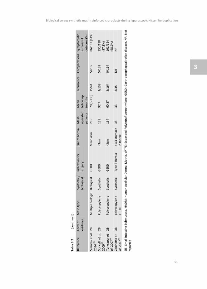
Biological versus synthetic mesh‐reinforced cruroplasty during laparoscopic Nissen fundoplication
51
3
Table 3.2
(continued
)
Referen
ce
Level of
evidence
Mesh type
Synthetic /
biological
Indication for
surgery
Size of hernia
Mesh
operated
patients
Mean
follow up
(months)
Recurrence
Complications
Symptomatic
successful
outcome (%)
Simorov et al.
2014 31
2B
Multiple biologic
Biological
GER
D
Mean 4cm
205
70(6‐135)
15/41
5/205
86/102 (84%
)
Soricelli et al.
200932
2B
Polypropylene
Synthetic
GER
D
>3cm
138
97,7
3/138
5/138
135/138
(97,8%)
Turkcapar et
al. 200733
2B
Polypropylene
Synthetic
GER
D
>3cm
164
60,37
3/164
0/164
161/164
(98,2%)
Zaninotto et
al. 200712
3B
polypropylene‐
ePTFE
Synthetic
Type 3 Hernia
>1/3 stomach
in thorax
35
33
3/35
NR
NR
SIS: Small Intestine Submucosa, HADM: Human
Acellular Dermal M
atrix, ePTFE: Expanded
Polytetrafluoroethylene, GER
D: Gastr‐oesophageal reflux disease, NR: Not
reported
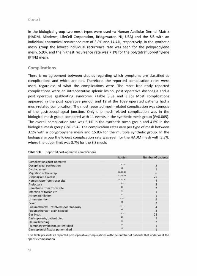
Chapter 3
52
In the biological group two mesh types were used –a Human Acellular Dermal Matrix
(HADM, Alloderm; LifeCell Corporation, Bridgewater, NJ, USA) and the SIS with an
individual anatomical recurrence rate of 3.8% and 14.4%, respectively. In the synthetic
mesh group the lowest individual recurrence rate was seen for the polypropylene
mesh, 5.9%, and the highest recurrence rate was 7.1% for the polytetrafluoroethylene
(PTFE) mesh.
Complications
There is no agreement between studies regarding which symptoms are classified as
complications and which are not. Therefore, the reported complication rates were
used, regardless of what the complications were. The most frequently reported
complications were an intraoperative splenic lesion, post‑operative dysphagia and a
post‑operative gasbloating syndrome. (Table 3.3a and 3.3b) Most complications
appeared in the post‑operative period, and 12 of the 1089 operated patients had a
mesh‐related complication. The most reported mesh‐related complication was stenosis
of the gastroesophageal junction. Only one mesh‐related complication was in the
biological mesh group compared with 11 events in the synthetic mesh group (P=0.065).
The overall complication rate was 5.1% in the synthetic mesh group and 4.6% in the
biological mesh group (P=0.694). The complication rates vary per type of mesh between
3.1% with a polypropylene mesh and 15.8% for the multiple synthetic group. In the
biological group the lowest complication rate was seen for the HADM mesh with 5.5%,
where the upper limit was 8.7% for the SIS mesh.
Table 3.3a Reported post‐operative complications
Studies Number of patients
Complications post‐operative Oesophageal perforation
25, 26 2
Cardiac arrest 12 1
Migration of the wrap 12, 23, 29
6 Dysphagia > 4 weeks
12, 23, 28 25
Hemorrhage from trocar site 12, 20, 30
4 Atelectasis
20, 41 3
Hematome from trocar site 20 2
Infection of trocar site 20 1
Atrium fibrillation 20 1
Urine retention 31, 41
9 Fever
41 2
Pneumothorax – resolved spontaneously 25, 41
4 Pneumothorax – drain needed
31 4
Gas bloat 28, 32
22 Gastroparesis, patient died
23 1
Pleural bleeding 25 2
Pulmonary embolism, patient died 29 1
Gastropleural fistula, patient died 19 1
This table presents all reported post‐operative complications with the number of patients that underwent the specific complication

Biological versus synthetic mesh‐reinforced cruroplasty during laparoscopic Nissen fundoplication
53
3
Table 3.3b Reported operative and mesh‐related complications
Studies Number of patients
Complications during surgery
Gastric lesion 12 1
Gastric perforation 24, 32
2
Pleural tear 32 1
Pleural lesion 32 3
Conversion to open 25, 29
5
Pneumothorax 25, 29, 30
9
Splenic lesion 12, 23, 28, 29, 32
11 Mesh‐related complications
Not further specified 32 1
Erosion of mesh 12, 20
2
Synthetic mesh
Stenosis
20, 25 8
Biological mesh Fibrosis 24 1
This table presents all reported operative and mesh‐related complications with the number of patients that underwent the specific complication
Symptomatic outcome
All studies used different criteria for a symptomatic successful outcome. Some authors
used the complete remission of symptoms as a successful outcome; others were
satisfied when patients suffered less severe post‑operative symptoms when compared
with their pre‑operative symptoms. The successful outcome reported in the individual
articles is analysed in this review, regardless of the criteria that were used. Six studies
did not report symptomatic outcomes.12,19‐21,26,27 An overall symptomatic success rate
of 85.5% was found in the biological mesh group, and a success rate of 94.6% in the
synthetic mesh group. The highest success rate overall was in the Polypropylene group
with 97.5% of the patients being relieved of their symptoms at a mean follow‐up of
48.6 months. The lowest symptomatic success rate was found in the SIS group; 80.4%
of these patients were relieved of their symptoms.
Mortality
Mortality after laparoscopic fundoplication and hiatal hernia repair is low. No mesh‐
related mortality was reported in the included studies (Table 3.4).
Table 3.4 Causes of death
Study Age Gender Type of mesh Cause of death Relation to mesh
19 74 NR Polypropylene Laceration of the fundoplication causing a
gastropleural fistula leading to respiratory
insufficiency
No
29 NR NR PTFE‐ePTFE Pulmonary embolism No
23 NR NR SIS Aspiration due to gastric paresis No
This table presents the reported causes of death with the specific details. ePTFE: Expanded polytetrafluoroethylene, NR: Not reported, SIS: Small Intestine Submucosa

Chapter 3
54
Discussion
The aim of this study was to evaluate the recurrence rate in mesh repaired hiatal
hernia. Recent studies have shown that a mesh‐based hiatal hernia repair is superior to
a non‐mesh based repair.10‐12 Until now, various recurrence rates have been reported
for the use of synthetic meshes, varying between 0–44%, with a median recurrence
rate of 7% in a large review by Draaisma et al..15,34,35 This spread may be caused by the
difference in follow‐up, since higher recurrence rates are found in studies with a longer
follow‐up. Another explanation for this wide spread is that different definitions for
recurrence are available. One can define recurrence as the return of symptoms or as a
radiological recurrence as well. If the latter one is used, recurrence rates up to 54%
have been reported in patients that received a biological mesh‐based repair.27 In 2006,
Oelschlager et al. reported that a radiographic recurrence rate of 24% in patients in
which the hiatal hernia was primarily closed compared with a recurrence rate of 9% in
patients who received a biological mesh for a follow‐up period of 6 months.36 However,
when they describe the same cohort in 2011 and report the 5‑year follow up,27 the
recurrence rates are 59% and 54%, respectively. It seems that the advantage in
recurrence rate of the biological mesh disappears when the follow‐up becomes longer.
Unfortunately, it is very difficult to draw strong conclusions based on published studies,
since there are only five studies describing a biological mesh and most studies only
report short‐term results. Nevertheless, we found a significantly higher recurrence rate
in the absorbable mesh group. This finding is supported by Watson et al.14; however,
there was no statistically significant difference in this study (P=0.161). We believe that
this result may be due to the small sample size, since the reported recurrence rates are
30.8% after biological absorbable mesh compared with 12.8 for the non‐absorbable
synthetic mesh. This study also reports a short‐term follow‐up of only 6 months.
The secondary outcomes of this review were the complication rate, mortality and the
symptomatic success rate. There is a large heterogeneity among the studies regarding
the different outcomes; nevertheless, the complication rates and symptomatic
outcomes are pooled. Due to the large heterogeneity, it was not possible to apply an
extensive statistical analysis on the data. If the complications are pooled, an overall
complication rate of 4.9% is found (Table 3.5). There is a large diversity in the reported
complications. This can probably be explained by the fact that some studies describe a
symptom as a complication, for example dysphagia, where others report this as a
common side effect of the procedure. Dysphagia is the main problem after antireflux
surgery with an incidence of 3%‐24% after Nissen fundoplication.21,37 Most
complications are not related to the use of a mesh during the procedure, but there
were 12 mesh‐related complications. The mesh‐related complication reported the most
is a stenosis at the gastroesophageal junction. This is probably due to shrinkage of the
mesh, which is more common in a synthetic mesh. The shrinkage rate can be up to 70%
for a polypropylene mesh.38 Since the sample sizes of the individual series are rather

Biological versus synthetic mesh‐reinforced cruroplasty during laparoscopic Nissen fundoplication
55
3
small, there is a considerable risk of underreporting of complications due to publication
bias.
Table 3.5 Pooled results of the studies
Mesh
type
No of
studies
No of
patient
s
No of
mesh
operated patients
Weighted
mean
follow up (months)
Recurrence
n / N (%)*
Complications
n / N (%)
Mortality
n / N (%)
Symptomatic
succesful n /
N (%)
Synthetic 11 12,19‐22,
25,26,28,29,
32,33
1652 704 53.4 46/675
(6.8)
34/666 (5.1) 2/704
(0.28)
474/501
(94.6)
Biologic 5 23,24,27,
30,31
712 385 53.4 34/211
(16.1)
16/352 (4.6) 1/385
(0.26)
213/249
(85.5)
Overall 16 2364 1089 53.4 80/886 (9.0)
50/1018 (4.9) 3/1018 (0.29)
687/750 (91.6)
Summary of the results of all included studies. Data marked with * are considered statistically significant
Theoretically, a biological mesh could show lower mesh‐related complication rates,
since this type of mesh is made of collagen, which will be absorbed by the body over
time. Furthermore, the biological material may still contain some growth factors, which
may induce the remodelling faster. In this review, there were no complications
reported in the biological mesh‐based hiatal hernia repairs. However, Stadlhuber et
al.39 described in 2009 a series of 28 case reports describing mesh‐related
complications. Seven of these cases described mesh‐related complications in biological
meshes, including stenosis, fibrosis and even one case of erosion of the mesh into the
oesophagus. There is only one mesh‐related complication found in the included studies,
which was described by Soricelli et al..32 This concerned a polypropylene mesh after a
mean follow‐up of 89 ± SD 29.8 months. The specific follow‐up in this case is not
reported and further details on this specific case are not reported either. Still, hiatal
hernia repair with the use of a mesh seems to be a safe procedure since low mortality
rates were reported.
The overall mortality rate was 0.29%, which is very low, and all deaths were due to non‐
mesh related causes (Table 3.4).
As demonstrated in Table 3.2, the symptomatic outcome is reported in 10 studies and
the overall success rate is 687/750 (91.6%). The most common method to measuring
the symptomatic outcome is by a 0–4 point Likert scale,20,22,27,30 Visick score,26,28,33 or by
a yes/no scoring system.23‐25 The definition for a successful symptomatic outcome used
in this review is an improvement of the previously mentioned scores. Since there is a
large heterogeneity in the symptomatic outcome, the only conclusion that can be made
is that there is an increasing need for a standardised method to report the symptomatic
outcome of hiatal hernia repair.

Chapter 3
56
Another aspect that needs to be considered is the cost of the different mesh types.
Unfortunately, the price of the meshes was not mentioned in any of the articles.
However, there is one literature review done in 2011 by Shankaran et al..40 They
evaluated the cost of different mesh types. The conclusion of this review is that the
price of a synthetic mesh is between $0.20 and $1.00 per cm2. The biological mesh is
available for a price between $8.00 and $26.00 per cm2. Therefore, one can conclude
that the biological mesh is on average far more expensive than a synthetic mesh.
Conclusion
This is the first review that gives an overview of performed studies between 2004 and
September 2015 describing hiatal hernia repair with the use of a mesh in Nissen
fundoplication. Despite the large heterogeneity, some differences were found in
recurrence, complications and symptomatic outcomes between a synthetic and a
biological mesh. The use of a synthetic mesh showed a significant lower recurrence
rate. Regarding the complication rate, there is no advantage of one mesh over the
other; however, it seems that with the use of a synthetic mesh more mesh‐related
complications occur. For both synthetic and biological meshes, the mortality rate is very
low. Mesh repair of a hiatal hernia seems safe in the short‐term; however, more long‐
term studies are needed to analyse the mesh‐related complications. Since the biological
mesh is more expensive and does not show better results, a synthetic mesh seems a
good choice to use for hiatal hernia repair.
In the synthetic mesh group, the polypropylene shows a very low recurrence rate and
the lowest complication rate. This type of mesh might be superior to the other
synthetic meshes. There is a lot of heterogeneity among the studies so far, and only few
studies report on a biological mesh. There is a need for more RCTs comparing synthetic
meshes with biological meshes with a long‐term follow up, and a standardised method
to report the success ratio of the procedure, both symptomatic and anatomic.
Biological versus synthetic mesh‐reinforced cruroplasty during laparoscopic Nissen fundoplication
57
3
References
1. Watson DI. Evolution and development of surgery for large paraesophageal hiatus hernia. World J Surg 2011;35:1436‐41.
2. Cuschieri A, Shimi S, Nathanson LK. Laparoscopic reduction, crural repair, and fundoplication of large
hiatal hernia. Am J Surg 1992;163:425‐30. 3. Andujar JJ, Papasavas PK, Birdas T, Robke J, Raftopoulos Y, Gagne DJ, et al. Laparoscopic repair of large
paraesophageal hernia is associated with a low incidence of recurrence and reoperation. Surg Endosc
2004;18:444‐7. 4. Athanasakis H, Tzortzinis A, Tsiaoussis J, Vassilakis JS, Xynos E. Laparoscopic repair of paraesophageal
hernia. Endoscopy 2001;33:590‐4.
5. Hill LD, Tobias JA. Paraesophageal hernia. Arch Surg 1968;96:735‐44. 6. Tsuboi K, Tsukada K, Nakabayashi T, Kato H, Miyazaki T, Masuda N, et al. Paraesophageal hiatus hernia,
which has progressed for 8 years: report of a case. Hepatogastroenterology 2002;49:992‐4.
7. Carlson MA, Condon RE, Ludwig KA, Schulte WJ. Management of intrathoracic stomach with polypropylene mesh prosthesis reinforced transabdominal hiatus hernia repair. J Am Coll Surg 1998;
187:227‐30
8. Stefanidis D, Hope WW, Kohn GP, Reardon PR, Richardson WS, Fanelli RD, et al. Guidelines for surgical treatment of gastroesophageal reflux disease. Surg Endosc 2010;24:2647‐69.
9. Peters JH. SAGES guidelines for the management of hiatal hernia. Surg Endosc 2013;27:4407‐8.
10. Furnee E, Hazebroek E. Mesh in laparoscopic large hiatal hernia repair: a systematic review of the literature. Surg Endosc 2013;27:3998‐4008.
11. Chilintseva N, Brigand C, Meyer C, Rohr S. Laparoscopic prosthetic hiatal reinforcement for large hiatal
hernia repair. J Visc Surg 2012;149:e215‐20. 12. Zaninotto G, Portale G, Costantini M, Fiamingo P, Rampado S, Guirroli E, et al. Objective follow‐up after
laparoscopic repair of large type III hiatal hernia. Assessment of safety and durability. World J Surg
2007;31:2177‐83. 13. Herbella FA, Patti MG, Del Grande JC. Hiatal mesh repair‐‐current status. Surg Laparosc Endosc
Percutan Tech 2011;21:61‐6.
14. Watson DI, Thompson SK, Devitt PG, Smith L, Woods SD, Aly A, et al. Laparoscopic repair of very large hiatus hernia with sutures versus absorbable mesh versus nonabsorbable mesh: a randomized
controlled trial. Ann Surg 2015;261:282‐9.
15. Draaisma WA, Gooszen HG, Tournoij E, Broeders IA. Controversies in paraesophageal hernia repair: a review of literature. Surg Endosc 2005;19:1300‐8.
16. Olivo SA, Macedo LG, Gadotti IC, Fuentes J, Stanton T, Magee DJ. Scales to assess the quality of
randomized controlled trials: a systematic review. Phys Ther 2008;88:156‐75. 17. Stang A. Critical evaluation of the Newcastle‐Ottawa scale for the assessment of the quality of
nonrandomized studies in meta‐analyses. Eur J Epidemiol 2010;25:603‐5.
18. CEBM. Oxford Centre for Evidence‐based Medicine ‐ Levels of Evidence. 2009. 19. Brandalise A, Aranha NC, Brandalise NA. The polypropylene mesh in the laparoscopic repair of large
hiatal hernias: technical aspects. Arq Bras Cir Dig 2012;25:224‐8.
20. Gouvas N, Tsiaoussis J, Athanasakis E, Zervakis N, Pechlivanides G, Xynos E. Simple suture or prosthesis hiatal closure in laparoscopic repair of paraesophageal hernia: a retrospective cohort study. Dis
Esophagus 2011;24:69‐78.
21. Granderath FA, Schweiger UM, Kamolz T, Asche KU, Pointner R. Laparoscopic Nissen fundoplication with prosthetic hiatal closure reduces postoperative intrathoracic wrap herniation: preliminary results
of a prospective randomized functional and clinical study. Arch Surg 2005;140:40‐8.
22. Granderath FA, Kamolz T, Schweiger UM, Pointner R. Impact of laparoscopic nissen fundoplication with prosthetic hiatal closure on esophageal body motility: Results of a prospective randomized trial. Arch
Surg 2006;141:625‐32.
23. Jacobs M, Gomez E, Plasencia G, Lopez‐Penalver C, Lujan H, Velarde D, et al. Use of surgisis mesh in laparoscopic repair of hiatal hernias. Surg Laparosc Endosc Percutan Tech 2007;17:365‐8.
Chapter 3
58
24. Lee E, Frisella MM, Matthews BD, Brunt LM. Evaluation of acellular human dermis reinforcement of the
crural closure in patients with difficult hiatal hernias. Surg Endosc 2007;21:641‐5. 25. Lubezky N, Sagie B, Keidar A, Szold A. Prosthetic mesh repair of large and recurrent diaphragmatic
hernias. Surg Endosc 2007;21:737‐41.
26. Morino M, Giaccone C, Pellegrino L, Rebecchi F. Laparoscopic management of giant hiatal hernia: factors influencing long‐term outcome. Surg Endosc 2006;20:1011‐6.
27. Oelschlager BK, Pellegrini CA, Hunter JG, Brunt ML, Soper NJ, Sheppard BC, et al. Biologic prosthesis to
prevent recurrence after laparoscopic paraesophageal hernia repair: long‐term follow‐up from a multicenter, prospective, randomized trial. J Am Coll Surg 2011;213:461‐8.
28. Parsak CK, Erel S, Seydaoglu G, Akcam T, Sakman G. Laparoscopic antireflux surgery with polyglactin
(vicryl) mesh. Surg Laparosc Endosc Percutan Tech 2011;21:443‐9. 29. Priego P, Ruiz‐Tovar J, Perez de Oteyza J. Long‐term results of giant hiatal hernia mesh repair and
antireflux laparoscopic surgery for gastroesophageal reflux disease. J Laparoendosc Adv Surg Tech A
2012;22:139‐41. 30. Schmidt E, Shaligram A, Reynoso JF, Kothari V, Oleynikov D. Hiatal hernia repair with biologic mesh
reinforcement reduces recurrence rate in small hiatal hernias. Dis Esophagus 2014;27:13‐7.
31. Simorov A, Ranade A, Jones R, Tadaki C, Shostrom V, Boilesen E, et al. Long‐term patient outcomes after laparoscopic anti‐reflux procedures. J Gastrointest Surg 2014;18:157‐62; discussion 162‐3.
32. Soricelli E, Basso N, Genco A, Cipriano M. Long‐term results of hiatal hernia mesh repair and antireflux
laparoscopic surgery. Surg Endosc 2009;23:2499‐504. 33. Turkcapar A, Kepenekci I, Mahmoud H, Tuzuner A. Laparoscopic fundoplication with prosthetic hiatal
closure. World J Surg 2007;31:2169‐76.
34. Frantzides CT, Madan AK, Carlson MA, Stavropoulos GP. A prospective, randomized trial of laparoscopic polytetrafluoroethylene (PTFE) patch repair vs simple cruroplasty for large hiatal hernia. Arch Surg
2002;137:649‐52.
35. Carlson MA, Richards CG, Frantzides CT. Laparoscopic prosthetic reinforcement of hiatal herniorrhaphy. Dig Surg 1999;16:407‐10.
36. Oelschlager BK, Pellegrini CA, Hunter J, Soper N, Brunt M, Sheppard B, et al. Biologic prosthesis reduces
recurrence after laparoscopic paraesophageal hernia repair: a multicenter, prospective, randomized trial. Ann Surg 2006;244:481‐90.
37. Hunter JG, Swanstrom L, Waring JP. Dysphagia after laparoscopic antireflux surgery. The impact of
operative technique. Ann Surg 1996;224:51‐7. 38. Brown CN, Finch JG. Which mesh for hernia repair? Ann R Coll Surg Engl 2010;92:272‐8.
39. Stadlhuber RJ, Sherif AE, Mittal SK, Fitzgibbons RJ, Jr., Michael Brunt L, Hunter JG, et al. Mesh
complications after prosthetic reinforcement of hiatal closure: a 28‐case series. Surg Endosc 2009;23:1219‐26.
40. Shankaran V, Weber DJ, Reed RL, 2nd, Luchette FA. A review of available prosthetics for ventral hernia
repair. Ann Surg 2011;253:16‐26. 41. Ringley CD, Bochkarev V, Ahmed SI, Vitamvas ML, Oleynikov D. Laparoscopic hiatal hernia repair with
human acellular dermal matrix patch: our initial experience. Am J Surg 2006;192:767‐72.
Chapter 4 Subjective outcome after laparoscopic hiatal hernia
repair for intrathoracic stomach
P.S.S. Castelijns
J.E.H. Ponten
M.C.G. van de Poll
S.W. Nienhuijs
J.F. Smulders
Langenbecks Arch Surg. 2017;402(3):521‐530
Chapter 4
60
Abstract
Purpose For decades, an intrathoracic stomach (ITS) has been a definite indication for surgery due to the perceived risk of an acute volvulus with perforation, gangrene, or haemorrhage. At the present time, elective laparoscopic repair is the first choice for treatment of ITS. There is a lack of evidence in the long‐term quality of life after a hiatal hernia repair for an intrathoracic stomach. Methods A retrospective analysis was performed on all patients undergoing a hiatal hernia repair for an intrathoracic stomach between January 2004 and January 2015. Additionally, to a hiatal closure, the patients received an antireflux procedure. Outcome measures included patient characteristics, operative details, complications, and postoperative morbidity and mortality. All patients were sent a quality of life questionnaire to assess long‐term quality of life and patient satisfaction. A higher quality of life score represents a better quality of life. Results Eighty‐six patients underwent laparoscopic repair for ITS, from which, one patient died during surgery. Eighty‐five patients were contacted and 81 completed the questionnaire, resulting in a response rate of 95.3%. At a median follow‐up of 2.7 years (range 0.1‐9.6) the mean quality of life score was 13.5 (standard deviation 2.8). The mean overall satisfaction was 8.4. There were four recurrences: three in the first 12 days after surgery and one in 2.4 years. Conclusions Very good results in patient satisfaction and symptom reduction were achieved after a median follow‐up of 2.7 years in this laparoscopic repair of the intrathoracic stomach single centre experience study. The symptomatic recurrence rate was very low.
Subjective outcome after laparoscopic hiatal hernia repair for intrathoracic stomach
61
4
Introduction
For decades, an intrathoracic stomach (ITS) has been a definite indication for surgery
due to the perceived risk of an acute volvulus with perforation, gangrene, or
hemorrhage.1,2 Due to the increasing incidence of gastro oesophageal reflux disease
(GERD), more people undergo diagnostic workups for reflux symptoms. One of the
causes for the increasing incidence of GERD is the exponentially growing problem of
obesity. In some of these patients the GERD symptoms are caused by the large hiatal
hernia rather than by obesity itself. An intrathoracic stomach may also be found as an
accidental finding. These developments resulted in an increased number of patients
with an intrathoracic stomach.3
The increasing availability of minimal invasive surgery has lowered the threshold to
perform surgery on these patients. However, whether these patients with minimal
symptoms actually benefit from such surgical treatment of intrathoracic stomach is
unclear.
The first laparoscopic para‐oesophageal hernia (PEH) repair was reported in 1992 by
Cuschieri et al..4 Over the succeeding years, antireflux surgery with repair of the hiatal
hernia has become the standard treatment for all types of hiatal hernias.5 The
symptomatic outcome of antireflux surgery for GERD is well documented in a large
series of cases and clinical trials.6 Nevertheless, only data regarding the recurrence
rates for an intrathoracic stomach repair are available. There is a lack of substantial
data describing the quality of life after elective surgery for intrathoracic stomach. The
largest current series describes a cohort of 73 patients with a reported objective follow‐
up of 5 years.7 However, only 33 patients completed the 3‐year follow‐up and only
12 patients completed the entire 5‐year follow‐up.
In this article, we reported on a large series of patients with intrathoracic stomachs
who underwent an elective minimal invasive surgery. We presented long‐term follow‐
up data with a special emphasis on symptomatic outcome and patient satisfaction.
These factors were assessed by a standardized questionnaire.
Materials and methods
We included patients who underwent surgery for a primary intrathoracic stomach,
defined as >50% of the stomach into the thorax on barium swallow investigation or on
CT‐scan.5 All the data was retrieved from the hospital information system at our
institution for patients who were operated on between January 1, 2004 and January 1,
2015. Parameters that were extracted included patient characteristics, preoperative
symptoms, preoperative medication usage, and diagnostic workups.

Chapter 4
62
Patients received a standardized questionnaire by mail. This questionnaire was a
modified version of one that was used by Mittal et al.7 (Appendix 4.1). Items on the
questionnaire included heartburn, regurgitation, dysphagia, retrosternal pain, gas
bloating, and the use of antacids. In addition to these questions, patients were
requested to score the result of the procedure on a visual analogue scale (VAS).
Patients who did not respond were contacted by phone up to three times to maximize
the response rate.
Surgical technique
All procedures were performed laparoscopically. After repositioning the stomach and
dissecting the hernia sac, the hiatus was closed using non‐absorbable woven sutures
(Ti‐cron™, Covidien, New Haven, CT, USA). In several cases the cruroplasty was
reinforced using a prosthetic mesh (Parietex™, Covidien, New Haven, CT, USA) at the
discretion of the operating surgeon. This was then followed by a 360‐degree floppy
Nissen fundoplication. We did not perform gastropexy or a gastrostomy. The most
distal suture fixates the wrap to the wall of the oesophagus to prevent telescoping.
Immediate postoperative period
Patients were put on a liquid diet for 2 weeks. Operation time, complications during
surgery, length of stay (LOS), in‐hospital postoperative complications and re‐admissions
were retrieved from the hospital information system.
Statistics
A retrospective database was managed in Access 2010. (Microsoft, USA) All data was analysed using SPSS for MacOs version 21.0(SPSS Inc., Chicago, IL, USA).
Results
Patient characteristics
A total number of 86 patients were operated on. One patient died during the
procedure. Out of the 85 patients that were contacted (20 were male), 81 (18 were
male) responded, concluding a 95.3% response rate. Data are reported on respondents
only.
The median age was 63 years (42‐80). The median BMI was 27 (20‐42). Twenty patients
(24.7%) received a mesh‐based repair. The two most dominant symptoms prior to
surgery were retrosternal pain and dysphagia. Baseline characteristics are summarized
in Table 4.1.
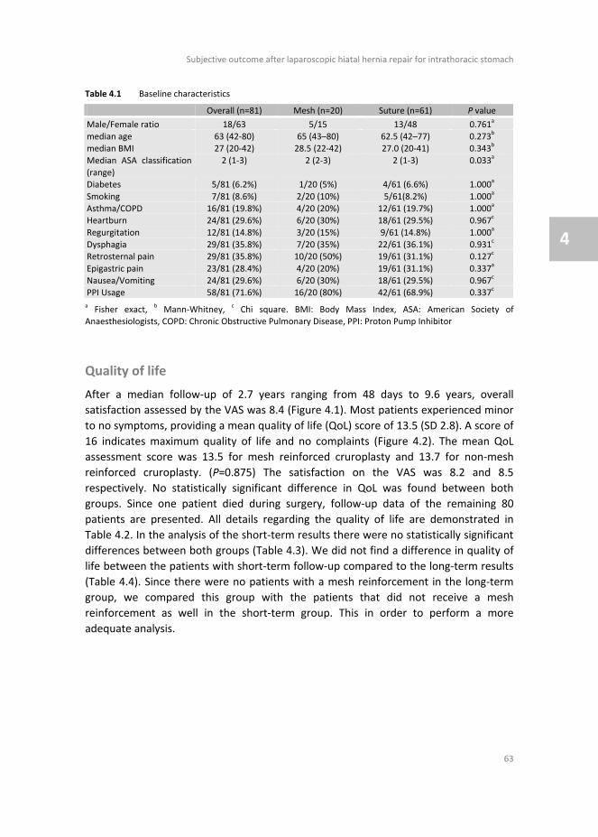
Subjective outcome after laparoscopic hiatal hernia repair for intrathoracic stomach
63
4
Table 4.1 Baseline characteristics
Overall (n=81) Mesh (n=20) Suture (n=61) P value
Male/Female ratio 18/63 5/15 13/48
0.761a
median age 63 (42‐80) 65 (43–80) 62.5 (42–77)
0.273b
median BMI 27 (20‐42) 28.5 (22‐42) 27.0 (20‐41)
0.343b
Median ASA classification
(range)
2 (1‐3) 2 (2‐3) 2 (1‐3)
0.033a
Diabetes 5/81 (6.2%) 1/20 (5%) 4/61 (6.6%)
1.000a
Smoking 7/81 (8.6%) 2/20 (10%) 5/61(8.2%) 1.000a
Asthma/COPD 16/81 (19.8%) 4/20 (20%) 12/61 (19.7%) 1.000a
Heartburn 24/81 (29.6%) 6/20 (30%) 18/61 (29.5%)
0.967c
Regurgitation 12/81 (14.8%) 3/20 (15%) 9/61 (14.8%)
1.000a
Dysphagia 29/81 (35.8%) 7/20 (35%) 22/61 (36.1%)
0.931c
Retrosternal pain 29/81 (35.8%) 10/20 (50%) 19/61 (31.1%)
0.127c
Epigastric pain 23/81 (28.4%) 4/20 (20%) 19/61 (31.1%)
0.337a
Nausea/Vomiting 24/81 (29.6%) 6/20 (30%) 18/61 (29.5%)
0.967c
PPI Usage 58/81 (71.6%) 16/20 (80%) 42/61 (68.9%)
0.337c
a Fisher exact,
b Mann‐Whitney,
c Chi square. BMI: Body Mass Index, ASA: American Society of
Anaesthesiologists, COPD: Chronic Obstructive Pulmonary Disease, PPI: Proton Pump Inhibitor
Quality of life
After a median follow‐up of 2.7 years ranging from 48 days to 9.6 years, overall
satisfaction assessed by the VAS was 8.4 (Figure 4.1). Most patients experienced minor
to no symptoms, providing a mean quality of life (QoL) score of 13.5 (SD 2.8). A score of
16 indicates maximum quality of life and no complaints (Figure 4.2). The mean QoL
assessment score was 13.5 for mesh reinforced cruroplasty and 13.7 for non‐mesh
reinforced cruroplasty. (P=0.875) The satisfaction on the VAS was 8.2 and 8.5
respectively. No statistically significant difference in QoL was found between both
groups. Since one patient died during surgery, follow‐up data of the remaining 80
patients are presented. All details regarding the quality of life are demonstrated in
Table 4.2. In the analysis of the short‐term results there were no statistically significant
differences between both groups (Table 4.3). We did not find a difference in quality of
life between the patients with short‐term follow‐up compared to the long‐term results
(Table 4.4). Since there were no patients with a mesh reinforcement in the long‐term
group, we compared this group with the patients that did not receive a mesh
reinforcement as well in the short‐term group. This in order to perform a more
adequate analysis.
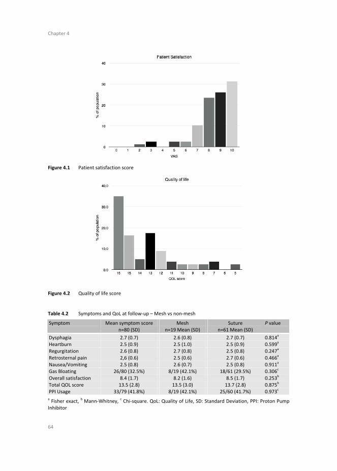
Chapter 4
64
Figure 4.1 Patient satisfaction score
Figure 4.2 Quality of life score
Table 4.2 Symptoms and QoL at follow‐up – Mesh vs non‐mesh
Symptom Mean symptom score n=80 (SD)
Mesh n=19 Mean (SD)
Suture n=61 Mean (SD)
P value
Dysphagia 2.7 (0.7) 2.6 (0.8) 2.7 (0.7) 0.814a
Heartburn 2.5 (0.9) 2.5 (1.0) 2.5 (0.9) 0.599a
Regurgitation 2.6 (0.8) 2.7 (0.8) 2.5 (0.8) 0.247a
Retrosternal pain 2,6 (0.6) 2.5 (0.6) 2.7 (0.6) 0.466a
Nausea/Vomiting 2.5 (0.8) 2.6 (0.7) 2.5 (0.8) 0.911a
Gas Bloating 26/80 (32.5%) 8/19 (42.1%) 18/61 (29.5%) 0.306c
Overall satisfaction 8.4 (1.7) 8.2 (1.6) 8.5 (1.7) 0.253b
Total QOL score 13.5 (2.8) 13.5 (3.0) 13.7 (2.8) 0.875b
PPI Usage 33/79 (41.8%) 8/19 (42.1%) 25/60 (41.7%) 0.973c
a Fisher exact,
b Mann‐Whitney,
c Chi‐square. QoL: Quality of Life, SD: Standard Deviation, PPI: Proton Pump
Inhibitor

Subjective outcome after laparoscopic hiatal hernia repair for intrathoracic stomach
65
4
Table 4.3 Symptoms and QoL at follow‐up <5 years
Symptom Mean symptom score
n=66 (SD)
Mesh
n=19 Mean (SD)
Suture
n=47 Mean (SD)
P value
Dysphagia 2.6 (0.7) 2.6 (0.8) 2.6 (0.7) 0.902a
Heartburn 2.5 (0.9) 2.5 (1.0) 2.5 (0.8) 0.504a
Regurgitation 2.6 (0.8) 2.7 (0.8) 2.6 (0.7) 0.550a
Retrosternal pain 2.6 (0.6) 2.5 (0.6) 2.7 (0.6) 0.386a
Nausea/Vomiting 2.5 (0.8) 2.6 (0.7) 2.5 (0.8) 1.000a
Gas Bloating 22/66 (33.3%) 8/19 (42.1%) 14/47 (29.8%) 0.336b
Overall satisfaction 8.4 (1.8) 8.2 (1.6) 8.5 (1.9) 0.539d
Total QOL score
(mean/range)
13.6 (2.9) 13.5 (3.0) 13.6 (2.9) 0.660c
PPI Usage 24/65 (36.9%) 8/19 (42.1%) 16/46 (34.8%) 0.578b
a Fisher exact,
b Chi square, c Mann‐Whitney U,
d Independent Sample T‐test. QoL: Quality of Life, SD:
Standard Deviation, PPI: Proton Pump Inhibitor
Table 4.4 Symptoms and QoL <5 years vs >5 years for suture repair only
Symptom >5 years
n=14 Mean (SD)
<5 years
n=47 Mean (SD)
P value
Suture/mesh 14:0 47:0
Follow‐up 7.0 (1.7) 2.6 (1.4) <0.05a
Dysphagia 2.8 (0.6) 2.6 (0.7) 1.000b
Heartburn 2.4 (1.0) 2.5 (0.8) 0.761b
Regurgitation 2.3 (0.8) 2.6 (0.7) 0.275b
Retrosternal pain 2.7 (0.5) 2.7 (0.6) 0.756b
Nausea/Vomiting 2.4 (0.6) 2.5 (0.8) 0.141b
Gas Bloating 4/14 (28.6%) 14/47 (29.8%) 1.000b
Overall satisfaction (median/range) 8.7(1.1) 8.5(1.9) 0.827c
Total QOL score (mean/range) 13.3 (2.4) 13.6 (2.9) 0.317c
PPI Usage 9/14 (64.3%) 16/46 (34.8%) 0.050d
a Independent Samples T‐test,
b Fisher’s exact,
c Mann‐Whitney U,
d Chi‐squared. QoL: Quality of Life, SD:
Standard Deviation, PPI: Proton Pump Inhibitor
Operative outcome
All procedures were performed in a non‐emergency setting by the senior author (JS) or
under his supervision. The median operative time was 89 min (range 53‐212 min).
Twenty patients received a mesh‐reinforced cruroplasty, whereas 61 patients received
a pure suture repair of the hiatal hernia. The decision to use a mesh was made during
the surgery at the discretion of the operating surgeon. The reason for using a mesh
reinforcement was weakness of the right crus that was noticed in six patients and a
very large defect (>8 cm) in 14 patients. Nine minor complications and two major
complications were reported, resulting in a complication rate of 12.3%. Minor
complications included a serosa injury and opening of the pleura, while major
complications included a laceration of the oesophagus and a perforation of the aorta.
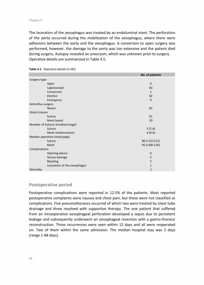
Chapter 4
66
The laceration of the oesophagus was treated by an endoluminal stent. The perforation
of the aorta occurred during the mobilization of the oesophagus, where there were
adhesions between the aorta and the oesophagus. A conversion to open surgery was
performed, however, the damage to the aorta was too extensive and the patient died
during surgery. Autopsy revealed an aneurysm, which was unknown prior to surgery.
Operative details are summarized in Table 4.5.
Table 4.5 Operative details (n=82)
No. of patients
Surgery type Open 0
Laparoscopic 82
Conversion 1 Elective 82
Emergency 0
Antireflux surgery Nissen 81
Hiatal closure
Suture 61 Mesh based 20
Number of Sutures (median/range)
Suture 3 (2‐6) Mesh reinforcement 4 (0‐6)
Median operative time(range)
Suture 86.5 (53‐212) Mesh 95.0 (68‐126)
Complications
Opening pleura 6 Serosa damage 2
Bleeding 2
Laceration of the oesophagus 1 Mortality 1
Postoperative period
Postoperative complications were reported in 12.5% of the patients. Most reported
postoperative complaints were nausea and chest pain, but these were not classified as
complications. Five pneumothoraces occurred of which two were treated by chest tube
drainage and three resolved with supportive therapy. The one patient that suffered
from an intraoperative oesophageal perforation developed a sepsis due to persistent
leakage and subsequently underwent an oesophageal resection with a gastro‐thoracic
reconstruction. Three recurrences were seen within 12 days and all were reoperated
on. Two of them within the same admission. The median hospital stay was 2 days
(range 1‐48 days).

Subjective outcome after laparoscopic hiatal hernia repair for intrathoracic stomach
67
4
Mortality
One patient died during the procedure due to perforation of the aorta. Emergency
laparotomy was performed, but the patient could not be saved.
Reoperation
Apart from the three early recurrences mentioned above, five other patients
underwent a reoperation during their follow‐up. The total percentage of reoperation
was therefore 9.9%. Two patients were operated on because of persistent dysphagia.
Two other patients were operated on for GERD due to failure of the fundoplication,
while the last one underwent a reoperation for a late recurrent hiatal hernia (868 days
after the initial procedure). No symptomatic recurrences were seen after the mesh‐
reinforced cruroplasty. All causes for reoperation along with their corresponding
follow‐ups are presented in Table 4.6. Median quality of life after reoperation was 12
compared to a median of 15 in patients that did not undergo reoperation (P=0.098).
Table 4.6 Reoperations
Causes of re‐operation Follow‐up (days)
Failure wrap, crus intact 252
Torsion wrap, crus intact 1164 Early recurrence, due to vomit 2
Wrap in thorax 5
Rupture crus suture 868 Persistent dysphagia, redo Toupet, remove one suture from crus 439
Persistent dysphagia, redo Toupet 238
Wrap in thorax, failure crus sutures 12
Discussion
In this paper, we report one of the largest single centre experiences on laparoscopic
repair of an intrathoracic stomach accompanied by a great response rate. We found
very good results in patient satisfaction and quality of life after a median follow‐up of
2.7 years.
Nissen fundoplication in patients with GERD is proven effective and leads to a great
symptomatic outcome.8 The quality of life in patients with GERD is measured by the
HRQL‐GERD questionnaire, which is a validated list of questions regarding the most
common symptoms. However, since patients with ITS reported different symptoms at
baseline, this questionnaire is not suitable in our population. There is still no validated
questionnaire available that represents the quality of life in patients with an
intrathoracic stomach. Therefore, we used a modified standardized questionnaire that

Chapter 4
68
was previously published by Mittal et al.7 (Appendix 4.1). In this questionnaire, the
most common symptoms in patients with an intrathoracic stomach were scored. The
quality of life we found in our large cohort of patients was in line with the great results
described previously.7
Due to the retrospective aspect of this study, we did not have the preoperative quality
of life assessment. For this reason, we were unable to compare our postoperative
results with the preoperative data that we retrieved from the patient information
system. This is one of the most limiting aspects of this study. Nonetheless, if we
compared our results with the data reported by Mittal et al., who used the same
questionnaire, we would see comparable results. They reported a mean satisfaction
score of 9.0 at a 3‐year follow‐up compared to a mean satisfaction of 8.4 for our cohort.
The individual symptom scores were also comparable at the 3‐year follow‐up.7
Three patients had an early recurrence in our study. This is probably due to persistent
postoperative vomiting which increases the intra‐abdominal pressure, leading to re‐
herniation of the wrap into the thorax. Iqbal et al has described a relation between
failure of the procedure and postoperative nausea and vomiting (PONV) prevention
after a Nissen fundoplication in 2006.9 They concluded that it is important to prescribe
antiemetics after the procedure. Antoniou et al. described that postoperative vomiting
as a result of the procedure only occurred on the first postoperative day.10 However,
we believe that antiemetics might need to be prescribed for a longer period of time,
since we have seen early recurrences due to vomiting up to 12 days.
We noticed only one late recurrence after a follow‐up of 2.4 years. The total recurrence
rate is therefore 4.9% and is comparable with the reported median recurrence rate of
7% (range 0‐42%) reported by Draaisma et al. in a large review describing 32 studies.11
Not all of our patients received a standard objective follow‐up by means of radiology
investigations. We therefore may have underestimated the anatomical recurrence rate,
especially since several studies demonstrated a very poor relationship between
symptoms and the presence of an anatomical recurrence. Most recurrences appeared
to be asymptomatic.7,12
Other causes for re‐operation were failure of the fundoplication and persistent
dysphagia. Dysphagia is a common side effect of the procedure and an incidence of
13% is reported in the literature.13 This is in contrast with our findings in the quality of
life questionnaires. Nineteen (23.8%) patients in our study reported some kind of
dysphagia after a median follow‐up of 3 years. This difference can be explained by the
fact that we operated on symptomatic patients with an ITS, whereas most studies
reported results of patients with GERD symptoms. Another explanation for the high
rate of persistent dysphagia might be due to a too tight wrap. Despite the high rate of
dysphagia, the reported quality of life in this study was excellent and patients rated the
overall procedure with a mean satisfaction of 8.4 on the VAS. This may be due to a
reduction in severity of dysphagia rather than a complete relief of symptoms. It should
be noted that this study described patients with an intrathoracic stomach and not with

Subjective outcome after laparoscopic hiatal hernia repair for intrathoracic stomach
69
4
GERD. Therefore, these results may not be applicable to the population that receives a
Nissen fundoplication for reflux disease, since this group of patients had other
symptoms at baseline.
We found two major operative complications, which were a perforation of the
oesophagus during manipulation and a bleeding of the aorta leading to the death of the
patient. To repair the perforation of the oesophagus, an endoluminal stent was placed
intraoperatively and the defect was closed with sutures. The leakage persisted due to
partial necrosis of the oesophageal wall. Therefore, a re‐operation was necessary and
eventually the patients had to undergo an oesophageal resection and gastric‐tube
reconstruction. An oesophageal perforation or laceration is a feared yet known
complication and has been reported previously in the literature. In all cases this
complication did not result in death of the patient.14,15
The second major complication was a perforation of the aorta. Aortic injuries during
laparoscopic fundoplication are very rare and have only been detected twice in the past
according to the literature.16,17
Another common complication of the Nissen fundoplication is a pneumothorax.18 The
reported incidence of this complication in the literature was 4% in all Nissen
fundoplications. However, the true percentage may be even higher since patients did
not receive a routine X‐thorax despite the fact that during surgery the pleura is often
opened. Five of our patients (6.25%) developed a symptomatic pneumothorax, which
received a thorax drainage in two cases. The reason for this increased incidence of
pneumothoraces is that we only operated on patients with an ITS, whereas most
literature describes only patients with GERD who are less prone to develop a
pneumothorax.
In 20 of our cases a mesh reinforced the cruroplasty. The choice for the use of a mesh
application was made by individual preference of the operating surgeon. Although the
small sample size and unequal groups prohibit proper statistical comparison between
patients that were treated with or without a mesh. Nevertheless, it is striking to see
that no recurrences occurred in patients that received a mesh‐reinforced cruroplasty. In
addition, the complication rate and symptomatic outcome were comparable. Mesh
related complications reported in the literature included erosion of the oesophagus and
migration or infection of the mesh.19 We did not see any of these complications in our
study. This can be explained by the fact that we reinforced the cruroplasty with a mesh
only in the past few years. This is due to our increasing experiences with intrathoracic
stomach repair and the presence of early recurrences in our cohort. As a result, we do
not have any long‐term follow‐up data describing the mesh‐based hiatal hernia repairs.
A large meta‐analysis published by Müller‐Stich et al. in 2015 described the effect of
mesh augmentation in hiatal hernia repairs in patients with a para‐oesophageal hernia.
They concluded that the mesh reinforcement does reduce the recurrence rate without
increasing the procedure related complications and mortality, at least for the mid‐term
follow‐up. The reported mesh related complication rate in this meta‐analysis was
Chapter 4
70
1.7%.20 We used a synthetic mesh, but biological meshes are also available. However,
the type of mesh that is more suitable for hiatal hernia repair has not yet been
researched and therefore further randomized studies are needed.
Conclusion
This large single centre experience on laparoscopic repair of an intrathoracic stomach
resulted in a high response rate. Although we do not have a preoperative comparison,
fairly good results in patient satisfaction and symptom reduction were achieved after a
median follow‐up of 2.7 years. The symptomatic recurrence rate was very low,
especially in the mesh‐based cruroplasty.

Subjective outcome after laparoscopic hiatal hernia repair for intrathoracic stomach
71
4
References
1. Leeder PC, Smith G, Dehn TC. Laparoscopic management of large paraesophageal hiatal hernia. Surg Endosc. 2003;17(9):1372‐5.
2. Polomsky M, Jones CE, Sepesi B, O'Connor M, Matousek A, Hu R, Raymond DP, Litle VR, Watson TJ,
Peters JH. Should elective repair of intrathoracic stomach be encouraged? Journal of gastrointestinal surgery : official journal of the Society for Surgery of the Alimentary Tract. 2010;14(2):203‐10.
3. Boeckxstaens G, El‐Serag HB, Smout AJ, Kahrilas PJ. Symptomatic reflux disease: the present, the past
and the future. Gut. 2014;63(7):1185‐93. 4. Cuschieri A, Shimi S, Nathanson LK. Laparoscopic reduction, crural repair, and fundoplication of large
hiatal hernia. Am J Surg. 1992;163(4):425‐30.
5. Peters JH. SAGES guidelines for the management of hiatal hernia. Surg Endosc. 2013;27(12):4407‐8. 6. Engstrom C, Cai W, Irvine T, Devitt PG, Thompson SK, Game PA, Bessell JR, Jamieson GG, Watson DI.
Twenty years of experience with laparoscopic antireflux surgery. Br J Surg. 2012;99(10):1415‐21.
7. Mittal SK, Bikhchandani J, Gurney O, Yano F, Lee T. Outcomes after repair of the intrathoracic stomach: objective follow‐up of up to 5 years. Surg Endosc. 2011;25(2):556‐66.
8. Kappaz GT, Sallum RA, Szachnowicz S, Rocha JR, Cecconello I. Improvement in quality‐of‐life after
laparoscopic Nissen fundoplication. Arquivos de gastroenterologia 2014;51(3):212‐6. 9. Iqbal A, Kakarlapudi GV, Awad ZT, Haynatzki G, Turaga KK, Karu A, Fritz K, Haider M, Mittal SK, Filipi CJ.
Assessment of diaphragmatic stressors as risk factors for symptomatic failure of laparoscopic nissen
fundoplication. J Gastrointest Surg. 2006;10(1):12‐21. 10. Antoniou SA, Delivorias P, Antoniou GA, Natsiopoulos I, Kalambakas A, Dalenback J, Makridis C.
Symptom‐focused results after laparoscopic fundoplication for refractory gastroesophageal reflux
disease‐‐a prospective study. Langenbecks Arch Surg. 2008;393(6):979‐84. 11. Draaisma WA, Gooszen HG, Tournoij E, Broeders IA. Controversies in paraesophageal hernia repair: a
review of literature. Surg Endosc. 2005;19(10):1300‐8.
12. Kang T, Urrego H, Gridley A, Richardson WS. Pledgeted repair of giant hiatal hernia provides excellent long‐term results. J Laparoendosc Adv Surg Tech A. 2014;24(10):684‐7.
13. Targarona EM, Grisales S, Uyanik O, Balague C, Pernas JC, Trias M. Long‐term outcome and quality of life
after laparoscopic treatment of large paraesophageal hernia. World J Surg. 2013;37(8):1878‐82. 14. Lubezky N, Sagie B, Keidar A, Szold A. Prosthetic mesh repair of large and recurrent diaphragmatic
hernias. Surg Endosc. 2007;21(5):737‐41.
15. Morino M, Giaccone C, Pellegrino L, Rebecchi F. Laparoscopic management of giant hiatal hernia: factors influencing long‐term outcome. Surg Endosc. 2006;20(7):1011‐6.
16. Leggett PL, Bissell CD, Churchman‐Winn R. Aortic injury during laparoscopic fundoplication: an
underreported complication. Surg Endosc. 2002;16(2):362. 17. Yano F, Omura N, Tsuboi K, Kashiwagi H, Yanaga K. Thoracic aortic injury during laparoscopic
fundoplication for reflux esophagitis. Int J Surg. 2008;6(6):490‐2.
18. Thomas H, Agrawal S. Systematic review of day‐case laparoscopic fundoplication. J Laparoendosc Adv Surg Tech A. 2011;21(9):781‐8.
19. Stadlhuber RJ, Sherif AE, Mittal SK, Fitzgibbons RJ, Jr., Michael Brunt L, Hunter JG, Demeester TR,
Swanstrom LL, Daniel Smith C, Filipi CJ. Mesh complications after prosthetic reinforcement of hiatal closure: a 28‐case series. Surg Endosc. 2009;23(6):1219‐26.
20. Muller‐Stich BP, Kenngott HG, Gondan M, Stock C, Linke GR, Fritz F, Nickel F, Diener MK, Gutt CN,
Wente M, Buchler MW, Fischer L. Use of Mesh in Laparoscopic Paraesophageal Hernia Repair: A Meta‐Analysis and Risk‐Benefit Analysis. PLoS One 2015;10(10):e0139547.

Chapter 4
72
Appendix 4.1
Quality of life questionnaire
Name: Date:
Date of birth: Gender: M / F
Are you on anti‐acid medication?
☐ yes, what type of medication?
☐ No, for how long? Months/days
How severe is your heartburn?
Times a day / week / month
☐ 0 ‐ none
☐ 1 ‐ Minimal – episodic, no treatment required
☐ 2 ‐ Moderate – controlled with medication
☐ 3 ‐ Severe – Interferes with daily activity or not controlled with medication
How severe is your dysphagia?
☐ 0 ‐ No dysphagia
☐ 1 ‐ Once a week or less
☐ 2 ‐ More than once a week, requiring dietary adjustment
☐ 3 ‐ Severe, preventing ingestion of solid food
How severe is your regurgitation of food?
Times a day / week / month
☐ 0 ‐ No regurgitation
☐ 1 ‐ Mild – after straining or a large meal
☐ 2 ‐ Moderate – position dependent
☐ 3 ‐ Severe – constant regurgitation with or without aspiration
How severe is your chest pain?
Times a day / week / month
☐ 0 ‐ None
☐ 1 ‐ Minimal – Episodic
☐ 2 ‐ Moderate – Reason to contact a doctor
☐ 3 ‐ Severe – interference with daily activity
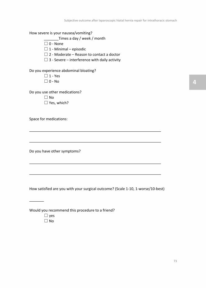
Subjective outcome after laparoscopic hiatal hernia repair for intrathoracic stomach
73
4
How severe is your nausea/vomiting?
Times a day / week / month
☐ 0 ‐ None
☐ 1 ‐ Minimal – episodic
☐ 2 ‐ Moderate – Reason to contact a doctor
☐ 3 ‐ Severe – interference with daily activity
Do you experience abdominal bloating?
☐ 1 ‐ Yes
☐ 0 ‐ No
Do you use other medications?
☐ No
☐ Yes, which?
Space for medications:
Do you have other symptoms?
How satisfied are you with your surgical outcome? (Scale 1‐10, 1‐worse/10‐best)
Would you recommend this procedure to a friend?
☐ yes
☐ No

Chapter 4
74
Chapter 5 Quality of life after Nissen fundoplication in patients
with GERD – Comparison between long‐and
short‐term follow‐up
P.S.S. Castelijns
J.E.H. Ponten
M.C.G. van de Poll
N.D. Bouvy
J.F. Smulders
J Minim Access Surg. 2018;14(3):213‐220

Chapter 5
76
Abstract
Introduction Nissen fundoplication is the golden standard for surgical treatment of gastroesophageal reflux disease (GERD). Numerous studies report excellent short‐term results. However, data regarding long‐term quality of life are lacking. The aim of this study is to investigate the long‐term quality of life after Nissen fundoplication in patients with GERD and to compare this with the short‐term results. Patients and methods We retrospectively analysed all patients who underwent laparoscopic Nissen fundoplication for GERD between January 2004 and January 2016. All patients received a validated GERD‐HRQL questionnaire by mail to assess postoperative quality of life. Maximum quality of life is represented by a score of 75. Secondary outcome measures were complications and recurrence rate. Results One hundred and seventy‐five of the 227 operated patients returned the questionnaire (77.1%). The median follow‐up was 3.7 (0.1‐10.3) years. Mean age was 51.6 (range 15‐85) and 72 patients were male. We report an excellent quality of life with a median total score of 70 (range 2‐75). Reoperation rate was 13.6% (23/169), in 12 patients the reoperation was due to recurrent reflux and in 11 patients due to persistent dysphagia. 91.3% of the re‐operations were performed within the first 5 years after surgery. Mortality rate was zero. Conclusion We report a large series of single centre, single surgeon laparoscopic Nissen fundoplication. Despite the reoperation rate of 13.6% we found excellent long‐term symptomatic outcome. There was no difference between short‐term and long‐term results.

Quality of life after Nissen fundoplication in patients with GERD
77
5
Introduction
Laparoscopic antireflux surgery (LARS) is considered the golden standard in the surgical
treatment of gastroesophageal reflux disease (GERD).1,2 Since Cuschieri et al.
performed the first laparoscopic Nissen fundoplication in 1992, this procedure gained
increasing popularity over time.3 Many studies report good results after Nissen
fundoplication with a successful symptomatic outcome in 80%–95% of the patients at a
long‐term follow‐up of up of 5 years.4‐7 Despite the good symptomatic results there are
still many patients that report recurrent reflux or new symptoms that were not present
before surgery. This may negatively influence the quality of life.
The GERD‐Health Related Quality of Life (GERD‐HRQL) is a validated instrument to
measure the quality of life after LARS for patients with GERD.8,9 The questionnaire
assesses most common symptoms and their impact on daily life. Many studies report
symptomatic outcome after Nissen fundoplication. However, very few studies report on
the long‐term quality of life. Therefore, the aim of this study is to report on the quality
of life after Nissen fundoplication at a long‐term follow‐up of up to 10.3 years. We
compared the long‐term results with the short‐term outcome. This is a single‑centre,
single surgeon study.
Patients and methods
For this study, we included patients who underwent antireflux surgery for GERD.
Initially patients were diagnosed by 24‐h PH‐metry of by endoscopy with proven
oesophagitis. Since the introduction of the SAGES guideline for GERD in 2010, patients
were diagnosed according this guideline2. All data were retrieved from the hospital
information system for patients who were operated at our institution between January
1st 2004 and January 1st 2016. Parameters that were extracted included patient
characteristics, pre‑operative symptoms, medication usage, diagnostic workup and
operative outcome.
All patients were sent the GERD‐HRQL questionnaire by mail. This is a validated and
standardized questionnaire that is proven effective in reporting quality of life for
patients with GERD.8,9 A maximum score of 75 represents excellent overall quality of
life and a score of 0 reports a very poor quality of life. To make the results easier to
compare, we classified this wide score into three groups. A score of 0‐25 is classified as
a poor outcome, 25‐50 as a medium outcome and a quality of life score of ≥50 was
classified as a good outcome. Specific items on heartburn and regurgitation both result
in a score with a maximum of 30, which represents no burden at all. A score of 0 is
associated with a high rate of complaints of the specific symptom. A heartburn or
regurgitation score >18 represents elimination of the particular symptom. The

Chapter 5
78
questionnaire is attached as Appendix 5.1. Non‐responders were contacted by phone
up to 3 times to maximize the response rate. Only follow‐up data regarding
respondents are presented.
Surgical technique
All procedures were performed laparoscopically. In all patients, the hiatus was
dissected completely and a posterior cruroplasty was performed using non‐absorbable
braided sutures (Ti‐cron™, Covidien, New Haven, CT, USA). The patency must be at least
10 mm besides the oesophagus. If a large hiatal hernia was present, the cruroplasty
was reinforced using a non‐absorbable prosthetic mesh (Parietex™, Covidien, New
Haven, CT, USA) at the discretion of an operating surgeon. After dissection of the short
gastric vessels, a 360° floppy Nissen fundoplication was created. We did not use a
boogie to calibrate the fundoplication. The most distal suture fixates the wrap to the
wall of the oesophagus to prevent telescoping. We did not perform any gastropexy. The
patients did receive neither a post‑operative drain, nor epidural analgesia.
Immediate postoperative period
Patients were prescribed a semi‐liquid diet for 2 weeks after the operation. Operation
time, complications during surgery, length of stay (LOS), in‐hospital post‑operative
complications and re‐admissions were retrieved from the hospital information system.
Postoperative follow‐up
Patients were examined by the surgeon in the outpatient clinic 8 weeks after discharge.
Proton pump inhibitor (PPI) was continued until the first appointment. If no
complications were found, the patients were referred to a general practitioner (GP) or
gastroenterologist for further follow‐up. The medical treatment was evaluated by the
GP or gastroenterologist. We did not perform routine PH‐studies or endoscopy. We
performed additional diagnostics for both pre‐operative workup in the case of a re‐
operation and for persistent or recurrent complaints after the initial procedure.
Statistical analysis
A retrospective database was managed in Access 2010. (Microsoft, USA) All data were
analysed using SPSS for MacOS version 21.0 (SPSS Inc., Chicago, IL, USA).
Ethical approval
No ethical approval was required for this study and permission was granted by the
institutional review board of our institution to perform this retrospective study.
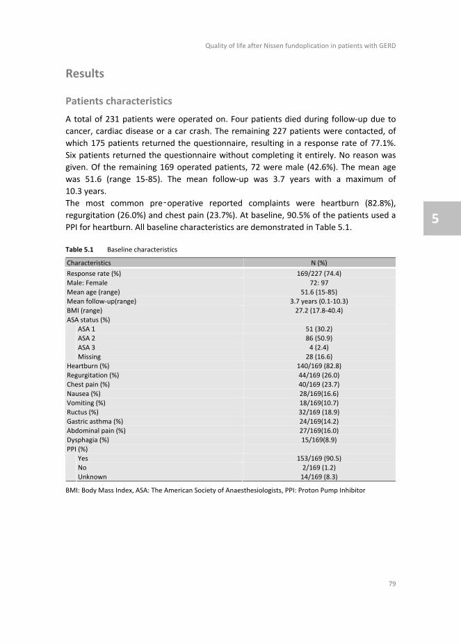
Quality of life after Nissen fundoplication in patients with GERD
79
5
Results
Patients characteristics
A total of 231 patients were operated on. Four patients died during follow‐up due to
cancer, cardiac disease or a car crash. The remaining 227 patients were contacted, of
which 175 patients returned the questionnaire, resulting in a response rate of 77.1%.
Six patients returned the questionnaire without completing it entirely. No reason was
given. Of the remaining 169 operated patients, 72 were male (42.6%). The mean age
was 51.6 (range 15‐85). The mean follow‐up was 3.7 years with a maximum of
10.3 years.
The most common pre‑operative reported complaints were heartburn (82.8%),
regurgitation (26.0%) and chest pain (23.7%). At baseline, 90.5% of the patients used a
PPI for heartburn. All baseline characteristics are demonstrated in Table 5.1.
Table 5.1 Baseline characteristics
Characteristics N (%)
Response rate (%) 169/227 (74.4)
Male: Female 72: 97
Mean age (range) 51.6 (15‐85) Mean follow‐up(range) 3.7 years (0.1‐10.3)
BMI (range) 27.2 (17.8‐40.4)
ASA status (%) ASA 1 51 (30.2)
ASA 2 86 (50.9)
ASA 3 4 (2.4)
Missing 28 (16.6)
Heartburn (%) 140/169 (82.8)
Regurgitation (%) 44/169 (26.0) Chest pain (%) 40/169 (23.7)
Nausea (%) 28/169(16.6)
Vomiting (%) 18/169(10.7) Ructus (%) 32/169 (18.9)
Gastric asthma (%) 24/169(14.2)
Abdominal pain (%) 27/169(16.0) Dysphagia (%) 15/169(8.9)
PPI (%)
Yes 153/169 (90.5) No 2/169 (1.2)
Unknown 14/169 (8.3)
BMI: Body Mass Index, ASA: The American Society of Anaesthesiologists, PPI: Proton Pump Inhibitor
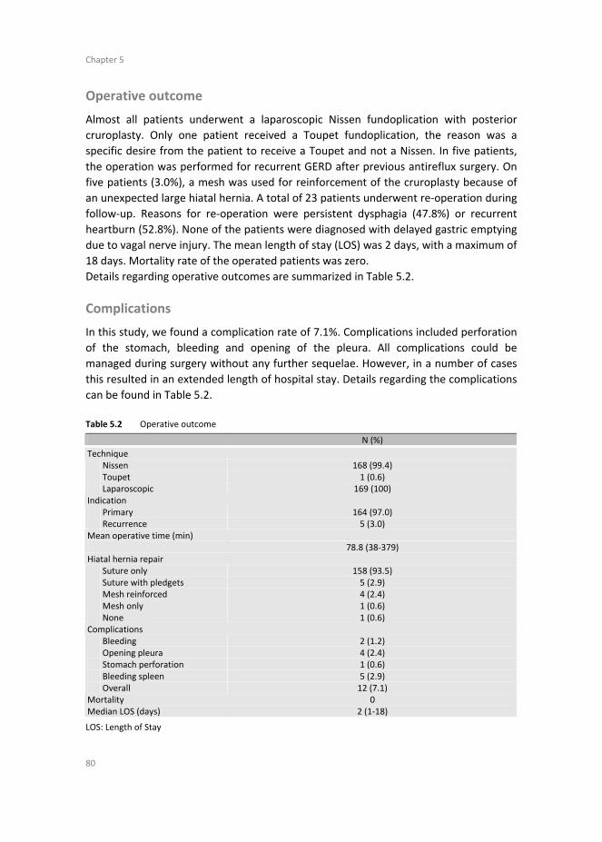
Chapter 5
80
Operative outcome
Almost all patients underwent a laparoscopic Nissen fundoplication with posterior
cruroplasty. Only one patient received a Toupet fundoplication, the reason was a
specific desire from the patient to receive a Toupet and not a Nissen. In five patients,
the operation was performed for recurrent GERD after previous antireflux surgery. On
five patients (3.0%), a mesh was used for reinforcement of the cruroplasty because of
an unexpected large hiatal hernia. A total of 23 patients underwent re‐operation during
follow‐up. Reasons for re‐operation were persistent dysphagia (47.8%) or recurrent
heartburn (52.8%). None of the patients were diagnosed with delayed gastric emptying
due to vagal nerve injury. The mean length of stay (LOS) was 2 days, with a maximum of
18 days. Mortality rate of the operated patients was zero.
Details regarding operative outcomes are summarized in Table 5.2.
Complications
In this study, we found a complication rate of 7.1%. Complications included perforation
of the stomach, bleeding and opening of the pleura. All complications could be
managed during surgery without any further sequelae. However, in a number of cases
this resulted in an extended length of hospital stay. Details regarding the complications
can be found in Table 5.2.
Table 5.2 Operative outcome
N (%)
Technique Nissen 168 (99.4) Toupet 1 (0.6)
Laparoscopic 169 (100) Indication
Primary 164 (97.0) Recurrence 5 (3.0) Mean operative time (min) 78.8 (38‐379) Hiatal hernia repair
Suture only 158 (93.5) Suture with pledgets 5 (2.9) Mesh reinforced 4 (2.4) Mesh only 1 (0.6)
None 1 (0.6) Complications
Bleeding 2 (1.2) Opening pleura 4 (2.4) Stomach perforation 1 (0.6) Bleeding spleen 5 (2.9)
Overall 12 (7.1) Mortality 0 Median LOS (days) 2 (1‐18)
LOS: Length of Stay
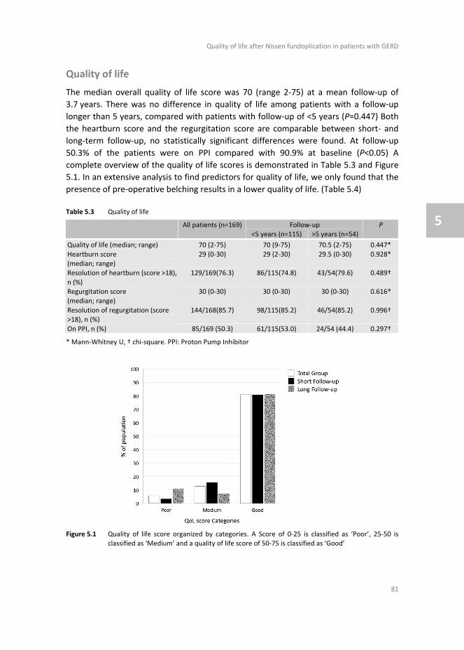
Quality of life after Nissen fundoplication in patients with GERD
81
5
Quality of life
The median overall quality of life score was 70 (range 2‐75) at a mean follow‐up of
3.7 years. There was no difference in quality of life among patients with a follow‐up
longer than 5 years, compared with patients with follow‐up of <5 years (P=0.447) Both
the heartburn score and the regurgitation score are comparable between short‐ and
long‐term follow‐up, no statistically significant differences were found. At follow‐up
50.3% of the patients were on PPI compared with 90.9% at baseline (P<0.05) A
complete overview of the quality of life scores is demonstrated in Table 5.3 and Figure
5.1. In an extensive analysis to find predictors for quality of life, we only found that the
presence of pre‐operative belching results in a lower quality of life. (Table 5.4)
Table 5.3 Quality of life
Follow‐up All patients (n=169) <5 years (n=115) >5 years (n=54)
P
Quality of life (median; range) 70 (2‐75) 70 (9‐75) 70.5 (2‐75) 0.447*
Heartburn score (median; range)
29 (0‐30) 29 (2‐30) 29.5 (0‐30) 0.928*
Resolution of heartburn (score >18),
n (%)
129/169(76.3) 86/115(74.8) 43/54(79.6) 0.489†
Regurgitation score
(median; range)
30 (0‐30) 30 (0‐30) 30 (0‐30) 0.616*
Resolution of regurgitation (score >18), n (%)
144/168(85.7) 98/115(85.2) 46/54(85.2) 0.996†
On PPI, n (%) 85/169 (50.3) 61/115(53.0) 24/54 (44.4) 0.297†
* Mann‐Whitney U, † chi‐square. PPI: Proton Pump Inhibitor
Figure 5.1 Quality of life score organized by categories. A Score of 0‐25 is classified as ‘Poor’, 25‐50 is classified as ‘Medium’ and a quality of life score of 50‐75 is classified as ‘Good’

Chapter 5
82
Table 5.4 Pre‐operative predictors for quality of life
Symptom QoL if symptom present
(median, range)
QoL if symptom not
present (median, range)
P
Heartburn 70 (2‐75) 71 (30‐75) 0.660* Regurgitation 70 (2‐75) 70 (7‐75) 0.782*
Dysphagia 74 (9‐75) 70 (2‐75) 0.574*
Chest pain 65.5 (7‐75) 71 (2‐75) 0.115* Nausea 63.5 (2‐75) 71 (3‐75) 0.109*
Vomiting 72 (3‐75) 70 (2‐75) 0.842*
Ructus† 63 (2‐75) 71 (3‐75) 0.026* Abdominal pain 67 (22‐75) 70 (2‐75) 0.997*
BMI
≥25 71 (2‐75) 64 (7‐75) 0.241* ≥30 72 (2‐75) 67 (7‐75) 0.263*
* Mann‐Whitney U. QoL: Quality of Life, BMI: Body Mass Index. Data marked with † are considered statistically significant
Long‐term versus short‑ term quality of life
Short‐term follow‐up was defined as <5 years. A total of 115 patients had a follow‐up of
<5 years, with a median of 1.9 years (range 0.1‐4.6). The remaining 54 patients
reported a median follow‐up of 7.1 years (range 5.1‐10.3). The median quality of life
was 70.0 for the short‐term group, compared with 70.5 for the long‐term group
(P=0.447). If we compare post‑operative heartburn, regurgitation or usage of PPI, we
did not find any statistically significant differences. Results are demonstrated in
Table 5.3.
Re‐operation
A total of 23 patients underwent a re‐operation during follow‐up (13.6%). Eleven
patients were re‐operated due to persistent dysphagia (47.8%) and 12 due to recurrent
heartburn (52.2%) (Figure 5.2) The median time to re‐operation was 380 days (75‐3488
days), 21 (91.3%) re‐operations occurred within the first 5 years after the initial
operation.
If dysphagia was the reason for re‐operation, the initial Nissen fundoplication was
converted to a Toupet. When recurrent heartburn was present, we re‐constructed the
Nissen fundoplication. In most cases the wrap seemed to lose, or the sutures were
ruptured.
Mean quality of life was 62.7 for patients who did not require re‐operation compared
to 53.1 for patients who did undergo re‐operation (P=0.196). This latter quality of life is
measured after the re‐operation was performed.
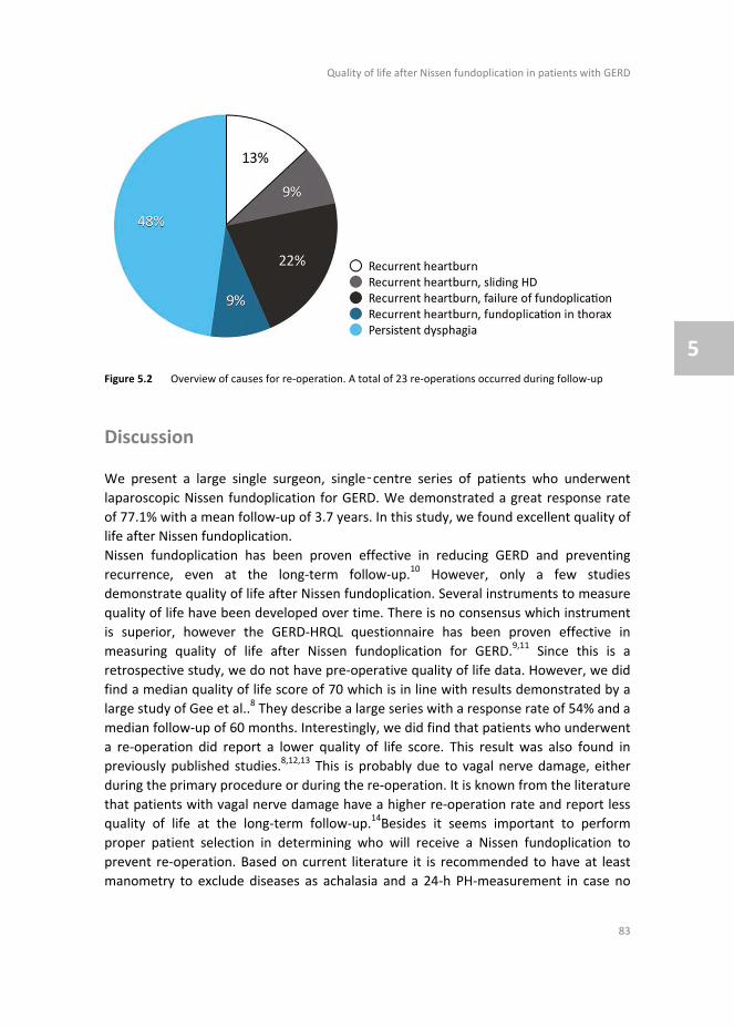
Quality of life after Nissen fundoplication in patients with GERD
83
5
Figure 5.2 Overview of causes for re‐operation. A total of 23 re‐operations occurred during follow‐up
Discussion
We present a large single surgeon, single‑centre series of patients who underwent
laparoscopic Nissen fundoplication for GERD. We demonstrated a great response rate
of 77.1% with a mean follow‐up of 3.7 years. In this study, we found excellent quality of
life after Nissen fundoplication.
Nissen fundoplication has been proven effective in reducing GERD and preventing
recurrence, even at the long‐term follow‐up.10 However, only a few studies
demonstrate quality of life after Nissen fundoplication. Several instruments to measure
quality of life have been developed over time. There is no consensus which instrument
is superior, however the GERD‐HRQL questionnaire has been proven effective in
measuring quality of life after Nissen fundoplication for GERD.9,11 Since this is a
retrospective study, we do not have pre‐operative quality of life data. However, we did
find a median quality of life score of 70 which is in line with results demonstrated by a
large study of Gee et al..8 They describe a large series with a response rate of 54% and a
median follow‐up of 60 months. Interestingly, we did find that patients who underwent
a re‐operation did report a lower quality of life score. This result was also found in
previously published studies.8,12,13 This is probably due to vagal nerve damage, either
during the primary procedure or during the re‐operation. It is known from the literature
that patients with vagal nerve damage have a higher re‐operation rate and report less
quality of life at the long‐term follow‐up.14Besides it seems important to perform
proper patient selection in determining who will receive a Nissen fundoplication to
prevent re‐operation. Based on current literature it is recommended to have at least
manometry to exclude diseases as achalasia and a 24‐h PH‐measurement in case no
Chapter 5
84
reflux oesophagitis is present on endoscopy. Besides, it is preferred that patients
should have a good response to anti‐acid medication.15,16 Unfortunately, due to the
retrospective aspect of this study, we could not find all data describing the complete
performed pre‐operative workup. Furthermore, a total of 55 patients was operated
before the introduction of the SAGES guidelines for GERD and therefore not all of them
did undergo a manometry. We were also unable to find data regarding pre‐operative
response to anti‐acid medication in all patients. We did find that pre‐operatively 90.5%
of the patients were on PPI, which was reduced to 50.3% at follow‐up (P<0.05) The high
percentage of patients who are on PPI on follow‐up may contribute to the good
reported quality of life. However, we do think that it is more important that patients
report good quality of life, rather than being completely free of PPI. Especially since
before surgery they were not satisfied with their situation on PPI.
We found a re‐operation rate of 13.6%, from which 7.1% was due to recurrent
heartburn. A Heartburn score on the GERD‐HRQL of >18 is classified as resolution of
symptoms. Based on this score we found a recurrence in heartburn of 23.7%. This is in
contrast with current literature, since the reported recurrent heartburn is about 10‐
20% and the actual re‐operation rate for recurrent complaints is about 3‐6%.17,18
Reported causes for recurrent heartburn are an inadequate indication for surgery due
to incomplete pre‑operative work‐up and failure to execute proper technical steps.19
Furthermore it is known that with increasing experience from the surgeon, the
complication and re‐operation rate will decrease.20,21 As mentioned before, it is known
that patients with vagal nerve damage receive more frequent re‐operations.14 Since this
was a retrospective study, we were unable to determine whether the patients who
underwent a re‐operation, indeed had vagal nerve injury. Our higher re‐operation rate
is most likely due to a combination of above mentioned factors.
Eleven of the 23 re‐operation were due to persistent dysphagia. This is a common side‐
effect of Nissen fundoplication and a large meta‐analysis by Tian et al. in 2015
demonstrates a post‐operative dysphagia rate of 12.6% in patients who underwent
Nissen fundoplication.22 In this study, 8.9% of the patients had moderate to severe
dysphagia, which might be an indication for re‐operation. This is comparable with the
6.5% re‐operations for dysphagia in our study. Post‑operative dysphagia is a known
and major problem and re‐operation rates between 1.8% and 10.8% for dysphagia have
been reported in literature.2 To reduce dysphagia some authors advocate to perform a
Toupet fundoplication. Despite the lower dysphagia rate, this technique seems to be
inferior in controlling GERD symptoms at the long term.23 However, there are also
studies that report comparable control of GERD symptoms with less post‑operative
complications and therefore advocate to perform a Toupet fundoplication.22 We
believe that it is important to perform the technique in which the surgeon has the most
experience. In case of persistent post‑operative dysphagia a Nissen can be converted
to a Toupet and in case of insufficient reflux control after a Toupet, this can be
Quality of life after Nissen fundoplication in patients with GERD
85
5
converted to a Nissen. The final decision to perform either Nissen or Toupet should be
tailored made.
Another outcome measurement in this study was the complication rate. We found a
complication rate of 7.1% which is in line with the overall complication rate of 6.3% that
is reported in a large meta‐analysis by Memon et al in 2015.24 All complications were
minor and could be managed during surgery without any further sequelae. The median
time to re‐operation was 380 days.
Some authors report the relevance of long‐term results after antireflux surgery.
However, if we compare quality of life after a follow‐up of <5 years with results after
>5 years, we do not see any statistically significant difference. This may suggest that
most complications and recurrences occur within the first 5 years after initial surgery.
This hypothesis is supported by the fact that almost all re‐operations 21/23 (91.3%)
were performed within 5 years after the primary procedure. Therefore, we might
conclude that follow‐up of 5 years after Nissen fundoplication might be sufficient to
evaluate its effectiveness. This is supported by the study of Kelly et al. where they
report that most re‐operations are performed within the first 2 years and that they did
not see any difference in symptomatic outcome between 5 and 10 years of follow‐up.7
Conclusion
We report a large series of single‐centre, single surgeon laparoscopic Nissen
fundoplication. Despite the re‑operation rate of 13.6% we found excellent
symptomatic outcome at a median follow‐up of 3.7 years. We did not find any
difference between short‐term and long‐term results, therefore follow‐up of 5 years
might be sufficient to evaluate the results of Nissen fundoplication since 91.3% of the
re‑operations took place within 5 years. Patients who underwent re‐operation
experienced less quality of life, this might be due to vagal nerve injury, however this
requires more research.
Chapter 5
86
References
1. Soricelli E, Basso N, Genco A, Cipriano M. Long‐term results of hiatal hernia mesh repair and antireflux laparoscopic surgery. Surgical endoscopy. 2009;23(11):2499‐504.
2. Stefanidis D, Hope WW, Kohn GP, Reardon PR, Richardson WS, Fanelli RD, et al. Guidelines for surgical
treatment of gastroesophageal reflux disease. Surgical endoscopy. 2010;24(11):2647‐69. 3. Cuschieri A, Shimi S, Nathanson LK. Laparoscopic reduction, crural repair, and fundoplication of large
hiatal hernia. American journal of surgery. 1992;163(4):425‐30.
4. Robertson AG, Patel RN, Couper GW, de Beaux AC, Paterson‐Brown S, Lamb PJ. Long‐term outcomes following laparoscopic anterior and Nissen fundoplication. ANZ journal of surgery. 2015.
5. Lundell L, Miettinen P, Myrvold HE, Pedersen SA, Liedman B, Hatlebakk JG, et al. Continued (5‐year)
followup of a randomized clinical study comparing antireflux surgery and omeprazole in gastroesophageal reflux disease. Journal of the American College of Surgeons. 2001;192(2):172‐9;
discussion 9‐81.
6. Dallemagne B, Weerts J, Markiewicz S, Dewandre JM, Wahlen C, Monami B, et al. Clinical results of laparoscopic fundoplication at ten years after surgery. Surgical endoscopy. 2006;20(1):159‐65.
7. Kelly JJ, Watson DI, Chin KF, Devitt PG, Game PA, Jamieson GG. Laparoscopic Nissen fundoplication:
clinical outcomes at 10 years. Journal of the American College of Surgeons. 2007;205(4):570‐5. 8. Gee DW, Andreoli MT, Rattner DW. Measuring the effectiveness of laparoscopic antireflux surgery:
long‐term results. Archives of surgery. 2008;143(5):482‐7.
9. Velanovich V. The development of the GERD‐HRQL symptom severity instrument. Diseases of the esophagus : official journal of the International Society for Diseases of the Esophagus / ISDE.
2007;20(2):130‐4.
10. Simorov A, Ranade A, Jones R, Tadaki C, Shostrom V, Boilesen E, et al. Long‐term patient outcomes after laparoscopic anti‐reflux procedures. Journal of gastrointestinal surgery : official journal of the
Society for Surgery of the Alimentary Tract. 2014;18(1):157‐62; discussion 62‐3.
11. Koetje JH, Nieuwenhuijs VB, Irvine T, Mayne GC, Watson DI. Measuring Outcomes of Laparoscopic Anti‐reflux Surgery: Quality of Life Versus Symptom Scores? World journal of surgery. 2016;40(5):1137‐44.
12. Khaitan L, Bhatt P, Richards W, Houston H, Sharp K, Holzman M. Comparison of patient satisfaction
after redo and primary fundoplications. Surgical endoscopy. 2003;17(7):1042‐5. 13. Draaisma WA, Rijnhart‐de Jong HG, Broeders IA, Smout AJ, Furnee EJ, Gooszen HG. Five‐year subjective
and objective results of laparoscopic and conventional Nissen fundoplication: a randomized trial.
Annals of surgery. 2006;244(1):34‐41. 14. van Rijn S, Rinsma NF, van Herwaarden‐Lindeboom MY, Ringers J, Gooszen HG, van Rijn PJ, et al. Effect
of Vagus Nerve Integrity on Short and Long‐Term Efficacy of Antireflux Surgery. The American journal of
gastroenterology. 2016;111(4):508‐15. 15. Campos GM, Peters JH, DeMeester TR, Oberg S, Crookes PF, Tan S, et al. Multivariate analysis of factors
predicting outcome after laparoscopic Nissen fundoplication. Journal of gastrointestinal surgery :
official journal of the Society for Surgery of the Alimentary Tract. 1999;3(3):292‐300. 16. Bello B, Zoccali M, Gullo R, Allaix ME, Herbella FA, Gasparaitis A, et al. Gastroesophageal reflux disease
and antireflux surgery‐what is the proper preoperative work‐up? Journal of gastrointestinal surgery :
official journal of the Society for Surgery of the Alimentary Tract. 2013;17(1):14‐20; discussion p 17. Carlson MA, Frantzides CT. Complications and results of primary minimally invasive antireflux
procedures: a review of 10,735 reported cases. Journal of the American College of Surgeons.
2001;193(4):428‐39. 18. Catarci M, Gentileschi P, Papi C, Carrara A, Marrese R, Gaspari AL, et al. Evidence‐based appraisal of
antireflux fundoplication. Annals of surgery. 2004;239(3):325‐37.
19. Patti MG, Allaix ME, Fisichella PM. Analysis of the Causes of Failed Antireflux Surgery and the Principles of Treatment: A Review. JAMA Surg. 2015;150(6):585‐90.
20. Watson DI, Baigrie RJ, Jamieson GG. A learning curve for laparoscopic fundoplication. Definable,
avoidable, or a waste of time? Annals of surgery. 1996;224(2):198‐203.

Quality of life after Nissen fundoplication in patients with GERD
87
5
21. Salminen P, Hiekkanen H, Laine S, Ovaska J. Surgeons' experience with laparoscopic fundoplication
after the early personal experience: does it have an impact on the outcome? Surgical endoscopy. 2007;21(8):1377‐82.
22. Tian ZC, Wang B, Shan CX, Zhang W, Jiang DZ, Qiu M. A Meta‐Analysis of Randomized Controlled Trials
to Compare Long‐Term Outcomes of Nissen and Toupet Fundoplication for Gastroesophageal Reflux Disease. PLoS One. 2015;10(6):e0127627.
23. Ma S, Qian B, Shang L, Shi R, Zhang G. A meta‐analysis comparing laparoscopic partial versus Nissen
fundoplication. ANZ journal of surgery. 2012;82(1‐2):17‐22. 24. Memon MA, Subramanya MS, Hossain MB, Yunus RM, Khan S, Memon B. Laparoscopic anterior versus
posterior fundoplication for gastro‐esophageal reflux disease: a meta‐analysis and systematic review.
World journal of surgery. 2015;39(4):981‐96.
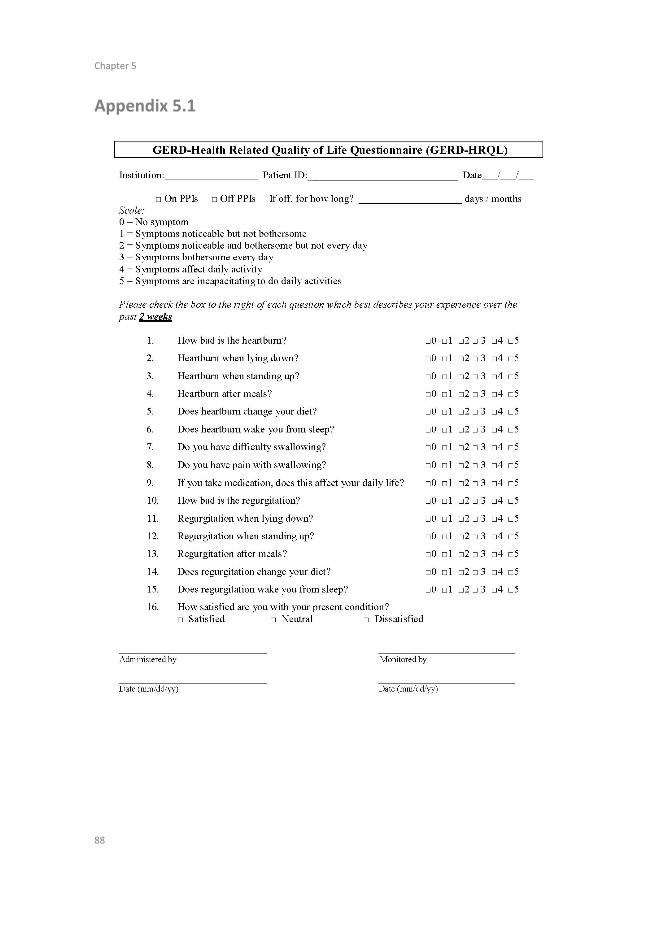
Chapter 5
88
Appendix 5.1
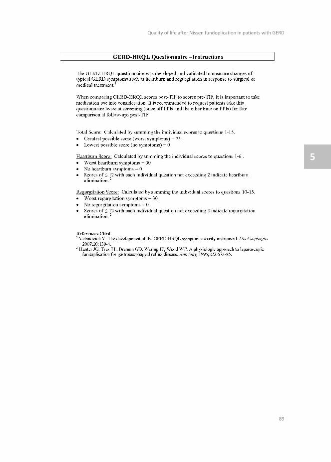
Quality of life after Nissen fundoplication in patients with GERD
89
5

Chapter 5
90
Chapter 6 7‐year follow up after transoral incisionless
fundoplication for treatment of gastroesophageal
reflux disease; results of patients who participated in
a randomized controlled trial
P.S.S. Castelijns
B.T.A. de Greef
A. Peeters
M.C.G. van de Poll
B.P.L. Witteman
G.H. Koek
J.M. Conchillo
N.D. Bouvy
Submitted

Chapter 6
92
2
Abstract
Aims Transoral incisionless fundoplication (TIF) is a minimally invasive endoscopic procedure for the treatment of gastroesophageal reflux disease (GERD) that mimics antireflux surgery, with the advantage of fewer side‐effects. This procedure might be beneficial especially for patients who would like to avoid lifelong treatment with proton pump inhibitors (PPI’s). Reports on long‐term effects after TIF are very limited. The primary objective of this study was to report the 7‐years results of patients who took part in a randomized controlled trial (RCT) comparing TIF (EsophyX‐2 device) with PPI’s. Because of a crossover option eventually all patients in the trial underwent TIF. Secondary objective was to identify pre‐operative predictors for successful outcomes. Methods All patients who underwent TIF in the previously published randomized controlled trial (between 2008 and 2012) were contacted by mail and were sent a quality of life (GERD‐HRQL) questionnaire. Additional questions regarding current PPI use and re‐operations were added to the questionnaire. Also questions about the severity of regurgitation and overall satisfaction with health were included. A clinically significant improvement in quality of life was defined as 50% improvement in GERD‐HRQL score. The original RCT database was used to perform multivariate analyses to identify potential predictors of a favourable outcome. Results All sixty patients from the original RCT were contacted. One patient died due to unrelated cause. 44 out of the remaining 59 patients, returned the questionnaire. The response rate was 74.6%, however, three returned questionnaires were blank. The mean follow‐up is 7.3 (SD 1.2) years. During the follow‐up, 9 (22%) patients underwent reoperation, 23 (56.1%) patients resumed daily oral PPI intake. At 7 years, the median quality of life score was 6.0 (0‐39) compared with 15.0 (3‐42) at baseline (P<0.05) (a lower quality of life score represents better outcome, with 0 as the maximum quality of life). 30 patients (73.2%) had a 50% improvement in GERD‐HRQL score compared to baseline. 31 patients (75.6%) had relevant improvement in regurgitation score compared to baseline. The median regurgitation score was 2.0 (0‐24) compared to 11.0 (0‐28) at baseline (P<0.001). 43.9% of the patients reported to be satisfied with their health compared with 11.7% at baseline (P<0.05). The presence of a hiatal hernia or oesophagitis were a negative predictor for successful outcome. Conclusions This is the first study reporting on 7‐year follow‐up results after TIF. It showed a significant improvement in quality of life scores and patient satisfaction. Despite the fact that some patients needed anti‐reflux surgery in addition to TIF to control their symptoms and that 56% of patients resumed PPI usage, there is a group of patients who benefit optimally from the TIF procedure even in the long term.
7‐year follow up after transoral incisionless fundoplication for treatment of GERD
93
2
6
Introduction
The main purpose of gastroesophageal reflux disease (GERD) treatment is to achieve
symptoms relief, improve quality of life, and to prevent oesophageal mucosal damage.1
It can be treated either by medication, surgery or transoral interventions. The first step
in the treatment is usually with medication, specifically with proton pump inhibitors
(PPI).2 However, this treatment is focused on symptom relief only and patients will have
to continue to use PPI’s lifelong.3,4 A long‐term use of PPI’s might be associated with an
increased risk of dementia, pneumonia, rib fractures, magnesium deficiency,
gastrointestinal infections and even a predisposition for upper gastrointestinal
malignancies.5,6
The GERD treatment with medication is not optimal as some patients experience
bothersome side‐effects, some are ‘unresponsive’ to medical therapy or are not willing
to take medication for a lifetime period. Besides, while the PPI treatment can provide
reduction in acid reflux, it has minimal effect on regurgitation. For many years,
laparoscopic antireflux surgery (LARS) has been the only treatment option for patients
who did not respond well to medication or were not satisfied with the use of PPI’s.
However, LARS can cause serious side‐effects. Several studies which reported the long‐
term results, showed a high degree of dysphagia, gas bloating, inability to belch, vagal
nerve injury and even a re‐operation rate of up to 15%.7,8 Literature shows recurrent
heartburn in 10‐20% of the patients after LARS for a median follow‐up of 6.5 years.9‐11
Therefore, alternative methods for controlling GERD have been developed. One of the
most promising techniques is the transoral incisionless fundoplication (TIF). There are
three devices for the performance of this intervention available by now, the
EsophyX® device (EndoGastric Solutions, Redmond, WA, United States), the Medigus
Ultrasonic Surgical Endostapler system (MUSE™, Medigus Ltd., Omer; Israel) and the
GERDX™ (G‐SURG GmbH, Seeon‐Seebruck, Germany).12,13 The transoral incisionless
fundoplication (TIF) technique is minimally invasive and seems effective in controlling
heartburn symptoms in a select group of patients, at least in the short term, which was
demonstrated in several studies.12,14,15 In these studies TIF was compared with no
treatment12,14 or a sham procedure.15 One randomized controlled trial has compared
TIF with the standard PPI treatment.16 The results of this RCT showed an improved
GERD‐related quality of life and an improvement of the antireflux barrier in the short
term. However, the results up to 1‐year showed an increasing PPI dependence over
time. There is only one prospective study published so far which gives the long‐term
results, up to 6 years after transoral incisionless fundoplication with EsophyX (Tif 2.0).17
In this study with 50 patients there was an elimination of daily dependence on PPI
achieved in 75‐80% of the patients after 6‐years. However, only 14 patients completed
the 6‐year follow‐up. Therefore, the conclusions of this study must be viewed with
care.
Chapter 6
94
2
The aim of the present study is to evaluate the 7 year’s results of all patients who
underwent a TIF procedure (by EsophyX device 2.0) after participating in a randomized
trial comparing TIF with PPI’s, the short‐term outcomes of which we have published
earlier.16 Also, we attempted to identify the pre‐operative predictors for successful
outcomes.
Methods
Initial study design
The initial study was designed as a prospective randomized controlled trial that was
carried out at a university hospital in the Netherlands. A total of 60 patients with
chronic GERD who were well controlled on daily PPI therapy were randomized 2:1, to
either TIF (intervention) or continuation of treatment with PPI’s (control group). After
the baseline measurements were taken, the patients discontinued the PPI use and
subsequently the measurements were taken again after a 14 days period without
medication. Included were patients with hiatal hernia ≤2 cm who had proven reflux
while off PPIs and recurrence of GERD symptoms after cessation of PPI’s (GERD‐HRQL
score difference of >10 between on and off PPI’s). After 6 months, patients in the
control group had the possibility to switch to the TIF group and to undergo the
intervention as well. All 60 patients underwent the TIF procedure. At 12 months, quality
of life remained improved after TIF compared with baseline but there was no
improvement in oesophageal acid exposure compared with baseline. Normalization of
pH was accomplished in only 29% of patients and 61% of patients resumed the use of
PPI’s, from which 44% on daily basis.
TIF procedure
All procedures were performed under general anaesthesia and in left lateral decubitus
position. The TIF procedure was performed by a team of two experienced physicians
(surgeon and gastroenterologist) under continuous endoscopic visualization using the
EsophyX‐2 device with SerosaFuse fasteners (EndoGastric Solutions, Redmond, WA).
Using a bite block, the lubricated EsophyX device was inserted over the endoscope,
used as a guide wire. If present, a small hiatal hernia was reduced. PPI was continued
for 14 days after the procedure to provide the mucosa time to heal. More details
regarding the technical aspects of the used technique have been described before.16
7‐year follow up after transoral incisionless fundoplication for treatment of GERD
95
2
6
Present study
For the present study, we contacted all 60 patients who underwent the TIF procedure
and also consented to be approachable for further study. The patients were sent the
quality of life questionnaires by post and by e‐mail. Non‐responders were contacted by
phone once, to verify the address and to ask approval for sending the questionnaire
again. The initial randomized study was approved by the Maastricht Medical Ethics
Committee (METC 07‐2‐051, NL 17303.068.07) and was registered NCT0085759
(clinicaltrials.gov). The METC of Maastricht has confirmed that the Medical Research
Involving Human Subjects Act (WMO) does not apply to the current long‐term follow‐
up study and that official approval is not required. Primary objective of this study was
to report the long‐term quality of life after a TIF procedure, secondary outcome was to
find predictors for successful outcomes regarding GERD symptoms.
Quality of life questionnaires
The GERD‐HRQL (GERD related quality of life) questionnaire, is a validated tool that
measures the symptom severity in GERD patients.18,19 Six out of the 10 questions assess
the degree of heartburn, two questions address dysphagia, one question addresses
regurgitation, and one question assesses the impact of medication use on daily living.
We have added five other questions concerning regurgitation. These questions were
included in the initial RCT study as well and are formulated in a similar way as the
questions about heartburn. Each item is scored from 0 to 5, with 0 representing no
complaints at all, while a score of 5 represents very bothersome complaints. Based on
study by Cadiere et al, a successful outcome was defined as an improvement of 50% of the quality of life score.20 The six questions about regurgitation have been
furthermore analysed in an additional analysis to calculate a regurgitation score.
Further, one additional question was added to evaluate the overall satisfaction with
current health. In addition to the questionnaire, the patients were asked to report
whether they have undergone additional surgery for GERD during follow‐up and if they
were currently using PPI’s and/or other anti‐acid drugs. In case of drug usage, the
frequency and mode of delivery were questioned.
Data handling
The results of the questionnaires were added to the initial RCT database that was
stored in an electronic database capture system (Microsoft Excel 97‐2004). In this study
only data regarding responders were analysed and are reported.
Chapter 6
96
2
Statistical analysis
Metric variables with normal distribution (tested by Shapiro‐Wilk statistic) were
univariately presented by their means and standard deviation and if not normally
distributed the median and range (minimum – maximum) were used. Categorical
variables were presented by frequencies and percentages. Standard univariable
statistics were used to examine the differences between subgroups of patients or the
association between particular parameters: Chi‐square test or Fisher’s exact test for
categorical variables, Students t‐test or Mann‐Whitney U test for continuous variables,
and the Wilcoxon signed rank test or McNemar test for paired data. Multivariable
analyses were performed by using backward logistic regression, whereby the odds ratio
(OR) with 95% confidence limits were estimated and tested with the Wald chi‐square
test. For the logistic regression, backward stepwise regression, based on Wald criteria,
was used with the covariates presence of hiatal hernia and presence of oesophagitis
were included together with the covariates with a p‐value below 0.2 in univariate
analyses. A P‐value of <0.05 was considered statistically significant. All analyses were
performed with SPSS, version 24.0 (IBM, Armonk, NY).
Results
Patient characteristics
A total of 60 patients underwent the TIF 2.0 procedure between 2008 and 2012. During
follow‐up 1 patient died, the cause of death was not related to the intervention. The
remaining 59 patients were contacted. A total of 44 patients (74.6%) returned the
questionnaire. Three of the returned questionnaires were blank, the patients refused to
answer the questions due to lack of time. Among the patients who returned the
completed questionnaire (n=41), 14 (34.1%) are female and 27 (65.9%) are male, with a
median age of 48 (20‐68). Median BMI is 24.9 (19.8‐34.8) and the mean follow‐up is 7.3
(SD 1.2) years.
Quality of life and patient satisfaction
Median quality of life score after 7 years is 6.0 (0‐39) compared with 15.0 (3‐42) at
baseline (P<0.01). The median regurgitation score is 2.0 (0‐24) at 7 years compared to
11.0 (0‐28) at baseline(P<0.01). From one year after surgery until 7 years, the quality of
life and regurgitation score remained constant (Table 6.1). After 7 years a total of 56.1%
of the patients resumed daily use of proton pump inhibitors, after 12 months this was
34.1% (P=0.267). (Table 6.2) Quality of life score of patients who resumed daily PPI
usage was 12.0 (0‐39) compared with 3.5 (0‐19) for patients who were still without PPI
medication after 7 years of follow‐up (P=0.010) (Table 6.3).
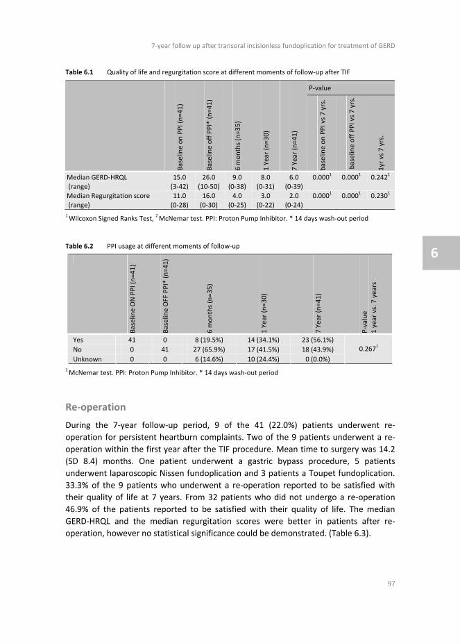
7‐year follow up after transoral incisionless fundoplication for treatment of GERD
97
2
6
Table 6.1 Quality of life and regurgitation score at different moments of follow‐up after TIF
P‐value
Baseline on PPI (n=41)
Baseline off PPI* (n=41)
6 m
onths (n=35)
1 Year (n=30)
7 Year (n=41)
baseline on PPI vs 7 yrs.
baseline off PPI vs 7 yrs.
1yr vs 7 yrs.
Median GERD‐HRQL (range)
15.0 (3‐42)
26.0 (10‐50)
9.0 (0‐38)
8.0 (0‐31)
6.0 (0‐39)
0.0001
0.0001
0.2421
Median Regurgitation score
(range)
11.0
(0‐28)
16.0
(0‐30)
4.0
(0‐25)
3.0
(0‐22)
2.0
(0‐24)
0.0001
0.0001
0.2301
1 Wilcoxon Signed Ranks Test,
2 McNemar test. PPI: Proton Pump Inhibitor. * 14 days wash‐out period
Table 6.2 PPI usage at different moments of follow‐up
Baseline ON PPI (n=41)
Baseline OFF PPI* (n=41)
6 m
onths (n=35)
1 Year (n=30)
7 Year (n=41)
P‐value
1 year vs. 7
years
Yes 41 0 8 (19.5%) 14 (34.1%) 23 (56.1%)
No 0 41 27 (65.9%) 17 (41.5%) 18 (43.9%)
Unknown 0 0 6 (14.6%) 10 (24.4%) 0 (0.0%)
0.2671
1 McNemar test. PPI: Proton Pump Inhibitor. * 14 days wash‐out period
Re‐operation
During the 7‐year follow‐up period, 9 of the 41 (22.0%) patients underwent re‐
operation for persistent heartburn complaints. Two of the 9 patients underwent a re‐
operation within the first year after the TIF procedure. Mean time to surgery was 14.2
(SD 8.4) months. One patient underwent a gastric bypass procedure, 5 patients
underwent laparoscopic Nissen fundoplication and 3 patients a Toupet fundoplication.
33.3% of the 9 patients who underwent a re‐operation reported to be satisfied with
their quality of life at 7 years. From 32 patients who did not undergo a re‐operation
46.9% of the patients reported to be satisfied with their quality of life. The median
GERD‐HRQL and the median regurgitation scores were better in patients after re‐
operation, however no statistical significance could be demonstrated. (Table 6.3).
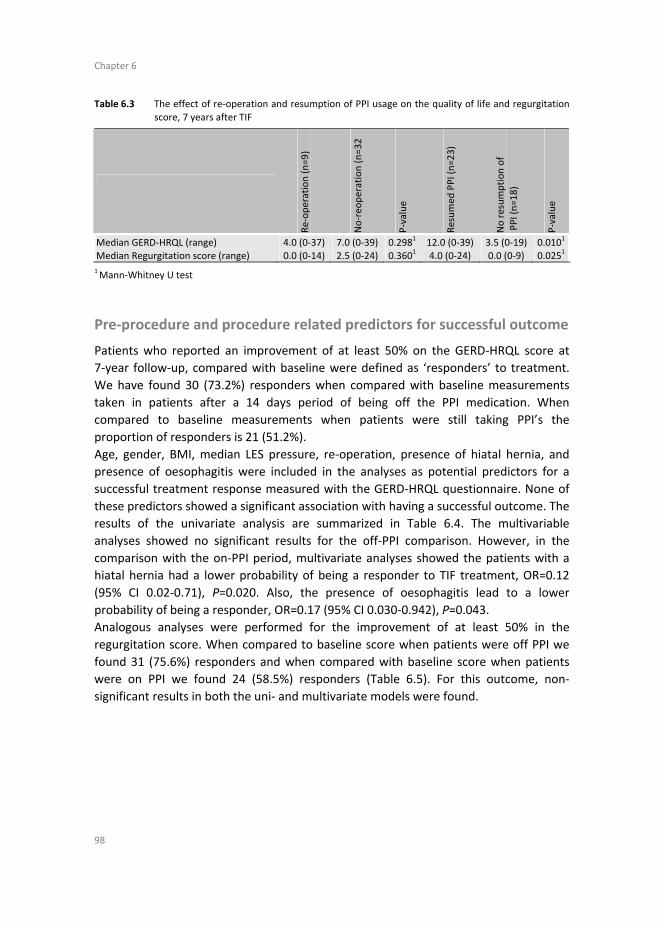
Chapter 6
98
2
Table 6.3 The effect of re‐operation and resumption of PPI usage on the quality of life and regurgitation
score, 7 years after TIF
Re‐operation (n=9)
No‐reo
peration (n=32
P‐value
Resumed
PPI (n=23)
No resumption of
PPI (n=18)
P‐value
Median GERD‐HRQL (range) 4.0 (0‐37) 7.0 (0‐39) 0.2981 12.0 (0‐39) 3.5 (0‐19) 0.010
1
Median Regurgitation score (range) 0.0 (0‐14) 2.5 (0‐24) 0.3601 4.0 (0‐24) 0.0 (0‐9) 0.025
1
1 Mann‐Whitney U test
Pre‐procedure and procedure related predictors for successful outcome
Patients who reported an improvement of at least 50% on the GERD‐HRQL score at
7‐year follow‐up, compared with baseline were defined as ‘responders’ to treatment.
We have found 30 (73.2%) responders when compared with baseline measurements
taken in patients after a 14 days period of being off the PPI medication. When
compared to baseline measurements when patients were still taking PPI’s the
proportion of responders is 21 (51.2%).
Age, gender, BMI, median LES pressure, re‐operation, presence of hiatal hernia, and
presence of oesophagitis were included in the analyses as potential predictors for a
successful treatment response measured with the GERD‐HRQL questionnaire. None of
these predictors showed a significant association with having a successful outcome. The
results of the univariate analysis are summarized in Table 6.4. The multivariable
analyses showed no significant results for the off‐PPI comparison. However, in the
comparison with the on‐PPI period, multivariate analyses showed the patients with a
hiatal hernia had a lower probability of being a responder to TIF treatment, OR=0.12
(95% CI 0.02‐0.71), P=0.020. Also, the presence of oesophagitis lead to a lower
probability of being a responder, OR=0.17 (95% CI 0.030‐0.942), P=0.043.
Analogous analyses were performed for the improvement of at least 50% in the
regurgitation score. When compared to baseline score when patients were off PPI we
found 31 (75.6%) responders and when compared with baseline score when patients
were on PPI we found 24 (58.5%) responders (Table 6.5). For this outcome, non‐
significant results in both the uni‐ and multivariate models were found.

7‐year follow up after transoral incisionless fundoplication for treatment of GERD
99
2
6
Table 6.4 Univariate analysis for predictors of a 50% improvement of the GERD‐HRQL score, 7 years
after TIF
on PPI at baseline off PPI at baseline*
Non‐
responders
(n=20)
Responders
(n=21)
P‐value
Non‐
responders
(n=11)
Responders
(n=30)
p‐value
Median age (range)
42 (20‐68) 49 (33‐67) 0.3411
55 (28‐68) 45.5 (20‐67) 0.5561
Gender male (%) 15 (75.0) 12 (57.1) 0.2282
9 (81.8) 18 (60.0) 0.2753
BMI ≥ 25 (%) 11 (55.0) 9 (42.9) 0.4372 7 (63.6) 13 (43.3) 0.249
2
Median LES
pressure (range)
12.5 (8‐25) 15 (6‐25) 0.0771
13 (8‐25) 15 (6‐25) 0.2221
Re‐operation (%) 3 (15.0) 6 (28.6) 0.4543
1 (9.1) 8 (26.7) 0.4013
Hiatal hernia (%) 17 (85.0) 11 (52.4) 0.0252
8 (72.7) 20 (66.7) 1.0003
Oesophagitis (%) 9 (45.0) 4 (19.0) 0.0742 4 (36.4) 9 (30.0) 0.719
3
1 Mann‐Whitney U,
2 Pearson Chi‐Square,
3 Fisher’s Exact Test. BMI: Body Mass Index (kg/m
2), LES pressure:
Lower Oesophageal Sphincter pressure. Oesophagitis is defined as the presence of any grade of Oesophagitis.
Responders are patients that have an improvement of at least 50% on the GERD‐HRQL score after a follow‐up
of 7 years compared with baseline. * 14 days wash‐out period
Table 6.5 Univariate analysis for predictors of a 50% improvement of the regurgitation score, 7 years after TIF
on PPI at baseline off PPI at baseline*
Non‐
responders
(n=17)
Responders
(n=24)
P‐value
Non‐
responders
(n=10)
Responders
(n=31)
P‐value
Median age (range) 43.0 (20‐68) 49.0 (33‐67) 0.2781
42.5 (23‐57) 49.0 (20‐68) 0.3871
Gender male (%) 11 (64.7) 16 (66.7) 0.8962
7 (70.0) 20 (64.5) 1.0003
BMI ≥ 25 (%) 8 (47.1) 12 (50.0) 0.8532 7 (70.0) 13 (41.9) 0.159
3
Median LES pressure (range)
15 (9‐25) 15 (6‐25) 0.4611
14 (9‐25) 15 (6‐25) 0.5601
Re‐operation (%) 4 (23.5) 5 (20.8) 1.0003
3 (30.0) 6 (19.4) 0.6623
Hiatal hernia (%) 11 (64.7) 17 (70.8) 0.6782
5 (50.0) 23 (74.2) 0.2413
Oesophagitis (%) 8 (47.1) 5 (20.8) 0.0752
4 (40.0) 9 (29.0) 0.6983
1 Mann‐Whitney U,
2 Pearson Chi‐Square,
3 Fisher’s Exact Test. BMI: Body Mass Index (kg/m
2), LES pressure:
Lower Oesophageal Sphincter pressure. Oesophagitis is defined as the presence of any grade of Oesophagitis. Responders are patients that have an improvement of at least 50% on the regurgitation score after a follow‐
up of 7 years compared with baseline. * 14 days wash‐out period

Chapter 6
100
2
Discussion
This is the first study reporting on 7‐year follow‐up results after TIF. This study reports
the quality of life of patients who participated in a randomized controlled trial
comparing TIF with PPI. The aim of this study was to evaluate the impact of TIF on
quality of life, regurgitation, PPI usage and the need for re‐operation. Secondary
outcome was to identify predictors for a successful improvement in quality of life after
a TIF procedure.
After a 7‐year follow up, quality of life and regurgitation were statistically significant
improved compared with baseline. When compared with the results at 12‐month
follow‐up, we did not see a further deterioration of symptoms and good symptom
control remained achieved. At 12 months after the initial TIF procedure, only 2 (5%)
patients underwent re‐operation, but after 7 years a total of 9 re‐operations have been
reported in 41 patients that have completed the follow‐up (22.0%). This is higher than
the re‐operation rate reported in some studies after laparoscopic anti‐reflux surgery,
which is between 3 and 14% after a follow‐up of 5 years.21‐23
Previous studies have shown that the re‐operation rate after a TIF procedure is
approximately 7%.24‐26 These studies only report on the short‐term follow‐up and in the
one study that reports a long‐term follow‐up after TIF, only 14 patients completed the
entire follow‐up. They found a re‐operation rate of 8.1%.
In this study, we included patients who were well controlled with PPI treatment, but
were unwilling to continue PPI treatment for a lifelong period. They initially did not
have an indication for laparoscopic anti‐reflux surgery, however, some still underwent
laparoscopic anti‐reflux surgery after the TIF procedure due to insufficient effect of the
TIF. Quality of life and regurgitation scores after 7 years were slightly better for patients
who underwent re‐operation compared with patients who did not. However, we could
not detect a significant difference between these groups.
Despite the re‐operation rate of 22% in our study, there are still patients who benefit
from TIF procedure at 7 years of follow‐up. 46.9% of the 32 patients who did not
undergo a re‐operation reported to be satisfied with their current quality of life. These
patients also had an improvement of at least 50% on the quality of life score.
A disadvantage of laparoscopic antireflux surgery may be that it can lead to severe
complications, such as dysphagia, gas bloating and other postoperative
complications.7,8 TIF is a safe procedure with a low complication rate and if needed
even after a previous TIF procedure, laparoscopic anti‐reflux surgery can be performed
without increased morbidity and mortality.27,28 It can be advocated that TIF might be
performed as a safe minimally invasive technique for the treatment of GERD in patients
who are not willing to take lifelong medication.
Testoni et al. tried to establish predictors for successful treatment with TIF and
concluded that for patients with a hiatal hernia <2 cm or a Hill grade I or II TIF might be

7‐year follow up after transoral incisionless fundoplication for treatment of GERD
101
2
6
a therapeutic option.17 In our study, we have performed a multivariate analysis to
assess predictors for a successful outcome. We defined responders, patients who have
a successful symptomatic outcome, as 50% improvement of GERD‐HRQL score
according to a common definition from the literature.16,20 We found that the presence
of a hiatal hernia of any size (all hernia’s ≤2cm) and the presence of oesophagitis of any
degree had a negative correlation with the probability of having a successful
symptomatic outcome after TIF. We have also evaluated the pre‐operative Hill
classification as a predictor for response to TIF therapy at long‐term follow‐up, but this
information was missing in 8 of 41 patients (19.5%). This large number of missing data
in combination with the small sample size resulted in a non‐significant result in the
univariate analysis (P=0.381). Since the Hill classification is closely related to the size of
hiatal hernia we decided to report only the presence of hiatal hernia instead of Hill
classification.29
We also performed a multivariate analysis for the effect of TIF on regurgitation,
however we could not detect any significant predictor for an improvement of at least
50% in regurgitation score. A BMI >25 might be a potential predictor, although in our
analysis no statistical significance was reached (P=0.159). It has been reported that an
increased BMI is a predictor for increased severity of GERD complaints.30‐32 The
increased intra‐abdominal pressure that comes with an increased BMI might have a
negative impact on the regurgitation. Also, the presence of oesophagitis could have a
negative effect on controlling the regurgitation symptoms (P=0.075 in the univariate
analysis).
In a large meta‐analysis with 18 studies, the number of patients that resumed PPI usage
after 12 months was 40% and 60% after 5 years. However, the results for a follow‐up
longer than 3 years, were based on one study only with a very small number of patients
that completed the follow‐up (n=24).33 We report a similar result of 56.1% of patients
that resumed using PPI’s after 7 years. We found that the patients who have resumed
to use PPI’s, reported a significant lower GERD‐HRQL score compared with patients
without the usage of PPI at 7 years. This underlines the negative impact that chronic PPI
usage has on the quality of life in these patients.
In our study, we have decided to evaluate the long‐term results after TIF by quality of
life questionnaires only.
It is proven that GERD has a negative impact on quality of life of patients.34 PH‐metry
seems unreliable in detecting recurrent GERD. Up to 16% of the patients with a positive
postoperative 24‐h PH monitoring seem to have negative results in the follow‐up PH‐
metry. Moreover, about 25% of the patients with erosive oesophagitis present with a
negative 24‐h PH metry.35 Furthermore, endoscopy and 24‐h PH monitoring are rather
invasive procedures.
A limitation of this study might be that at the 12 months follow‐up the patients who
were using PPI’s at that moment were asked to stop using PPI’s 2 weeks before
completing the quality of life questionnaires. This was done according to the protocol
Chapter 6
102
2
of the randomized controlled trial. At 7 years of follow‐up we evaluated quality of life
while some patients were using PPI’s, therefore the quality of life comparison might be
biased. However, the quality of life evaluation at 7 years represents better the daily
practice.
One cost‐effectiveness study has shown that EsophyX is more expensive and less
efficacious than Nissen procedure for the treatment of GERD.36 However, this study did
not evaluate the costs that came with additional medication usage after the
intervention, which plays a major role in the long‐term. Further studies are needed to
establish the cost‐effectiveness of TIF in the long‐term.
Conclusion
In conclusion, this is the first study reporting long‐term follow‐up results 7 years after
TIF. In patients, who had well controlled GERD symptoms with PPI medication at the
start of the study, 22% needed additional antireflux surgery within 7 years after TIF
procedure. 56.1% of the patients resumed PPI usage. Quality of life score and
regurgitation score were improved compared with baseline and good symptom control
remained when compared with 1 year follow‐up.
TIF could be performed as a first step in the treatment of GERD when patients are not
willing to take PPI’s for a lifelong period or when patients are not feasible for surgery.
The presence of a hiatal hernia or oesophagitis was associated with a worse GERD‐
HRQL score after 7 years.
7‐year follow up after transoral incisionless fundoplication for treatment of GERD
103
2
6
References
1. Vakil N, van Zanten SV, Kahrilas P, Dent J, Jones R, Global Consensus G. The Montreal definition and classification of gastroesophageal reflux disease: a global evidence‐based consensus. Am J
Gastroenterol. 2006;101(8):1900‐20; quiz 43.
2. Tytgat GN, McColl K, Tack J, Holtmann G, Hunt RH, Malfertheiner P, et al. New algorithm for the treatment of gastro‐oesophageal reflux disease. Aliment Pharmacol Ther. 2008;27(3):249‐56.
3. El‐Serag HB. Time trends of gastroesophageal reflux disease: a systematic review. Clin Gastroenterol
Hepatol. 2007;5(1):17‐26. 4. Metz DC. Managing gastroesophageal reflux disease for the lifetime of the patient: evaluating the long‐
term options. Am J Med. 2004;117 Suppl 5A:49S‐55S.
5. Corsonello A, Lattanzio F, Bustacchini S, Garasto S, Cozza A, Schepisi R, et al. Adverse events of proton pump inhibitors: potential mechanisms. Curr Drug Metab. 2017.
6. de la Coba Ortiz C, Arguelles Arias F, Martin de Argila de Prados C, Judez Gutierrez J, Linares Rodriguez
A, Ortega Alonso A, et al. Proton‐pump inhibitors adverse effects: a review of the evidence and position statement by the Sociedad Espanola de Patologia Digestiva. Rev Esp Enferm Dig. 2016; 108(4):207‐24.
7. Castelijns PSS, Ponten JEH, Vd Poll MCG, Bouvy ND, Smulders JF. Quality of life after nissen
fundoplication in patients with gastroesophageal reflux disease: Comparison between long‐ and short‐term follow‐up. J Minim Access Surg. 2018;14(3):213‐20.
8. van Rijn S, Rinsma NF, van Herwaarden‐Lindeboom MY, Ringers J, Gooszen HG, van Rijn PJ, et al. Effect
of Vagus Nerve Integrity on Short and Long‐Term Efficacy of Antireflux Surgery. Am J Gastroenterol. 2016;111(4):508‐15.
9. Carlson MA, Frantzides CT. Complications and results of primary minimally invasive antireflux
procedures: a review of 10,735 reported cases. J Am Coll Surg. 2001;193(4):428‐39. 10. Catarci M, Gentileschi P, Papi C, Carrara A, Marrese R, Gaspari AL, et al. Evidence‐based appraisal of
antireflux fundoplication. Ann Surg. 2004;239(3):325‐37.
11. Maret‐Ouda J, Wahlin K, El‐Serag HB, Lagergren J. Association Between Laparoscopic Antireflux Surgery and Recurrence of Gastroesophageal Reflux. JAMA. 2017;318(10):939‐46.
12. Testoni PA, Mazzoleni G, Testoni SG. Transoral incisionless fundoplication for gastro‐esophageal reflux
disease: Techniques and outcomes. World J Gastrointest Pharmacol Ther. 2016;7(2):179‐89. 13. von Renteln D, Schiefke I, Fuchs KH, Raczynski S, Philipper M, Breithaupt W, et al. Endoscopic full‐
thickness plication for the treatment of gastroesophageal reflux disease using multiple Plicator
implants: 12‐month multicenter study results. Surg Endosc. 2009;23(8):1866‐75. 14. Witteman BP, Strijkers R, de Vries E, Toemen L, Conchillo JM, Hameeteman W, et al. Transoral
incisionless fundoplication for treatment of gastroesophageal reflux disease in clinical practice. Surg
Endosc. 2012;26(11):3307‐15. 15. Hakansson B, Montgomery M, Cadiere GB, Rajan A, Bruley des Varannes S, Lerhun M, et al.
Randomised clinical trial: transoral incisionless fundoplication vs. sham intervention to control chronic
GERD. Aliment Pharmacol Ther. 2015;42(11‐12):1261‐70. 16. Witteman BP, Conchillo JM, Rinsma NF, Betzel B, Peeters A, Koek GH, et al. Randomized controlled trial
of transoral incisionless fundoplication vs. proton pump inhibitors for treatment of gastroesophageal
reflux disease. Am J Gastroenterol. 2015;110(4):531‐42. 17. Testoni PA, Testoni S, Mazzoleni G, Vailati C, Passaretti S. Long‐term efficacy of transoral incisionless
fundoplication with Esophyx (Tif 2.0) and factors affecting outcomes in GERD patients followed for up
to 6 years: a prospective single‐center study. Surg Endosc. 2015;29(9):2770‐80. 18. Velanovich V, Vallance SR, Gusz JR, Tapia FV, Harkabus MA. Quality of life scale for gastroesophageal
reflux disease. J Am Coll Surg. 1996;183(3):217‐24.
19. Velanovich V. The development of the GERD‐HRQL symptom severity instrument. Dis Esophagus. 2007;20(2):130‐4.
20. Cadiere GB, Buset M, Muls V, Rajan A, Rosch T, Eckardt AJ, et al. Antireflux transoral incisionless
fundoplication using EsophyX: 12‐month results of a prospective multicenter study. World J Surg. 2008;32(8):1676‐88.
Chapter 6
104
2
21. Draaisma WA, Rijnhart‐de Jong HG, Broeders IA, Smout AJ, Furnee EJ, Gooszen HG. Five‐year subjective
and objective results of laparoscopic and conventional Nissen fundoplication: a randomized trial. Ann Surg. 2006;244(1):34‐41.
22. Anvari M, Allen C. Five‐year comprehensive outcomes evaluation in 181 patients after laparoscopic
Nissen fundoplication. J Am Coll Surg. 2003;196(1):51‐7; discussion 7‐8; author reply 8‐9. 23. Pessaux P, Arnaud JP, Delattre JF, Meyer C, Baulieux J, Mosnier H. Laparoscopic antireflux surgery: five‐
year results and beyond in 1340 patients. Arch Surg. 2005;140(10):946‐51.
24. Repici A, Fumagalli U, Malesci A, Barbera R, Gambaro C, Rosati R. Endoluminal fundoplication (ELF) for GERD using EsophyX: a 12‐month follow‐up in a single‐center experience. J Gastrointest Surg.
2010;14(1):1‐6.
25. Testoni PA, Corsetti M, Di Pietro S, Castellaneta AG, Vailati C, Masci E, et al. Effect of transoral incisionless fundoplication on symptoms, PPI use, and ph‐impedance refluxes of GERD patients. World J
Surg. 2010;34(4):750‐7.
26. Muls V, Eckardt AJ, Marchese M, Bastens B, Buset M, Deviere J, et al. Three‐year results of a multicenter prospective study of transoral incisionless fundoplication. Surgical Innov. 2013;20(4):
321‐30.
27. Furnee EJ, Broeders JA, Draaisma WA, Schwartz MP, Hazebroek EJ, Smout AJ, et al. Laparoscopic Nissen fundoplication after failed EsophyX fundoplication. Br J Surg. 2010;97(7):1051‐5.
28. Perry KA, Linn JG, Eakin JL, Onders RP, Velanovich V, Melvin WS. Transoral incisionless fundoplication
does not significantly increase morbidity of subsequent laparoscopic Nissen fundoplication. J Laparoendosc Adv Surg Techn Part A. 2013;23(5):456‐8.
29. Hansdotter I, Bjor O, Andreasson A, Agreus L, Hellstrom P, Forsberg A, et al. Hill classification is superior
to the axial length of a hiatal hernia for assessment of the mechanical anti‐reflux barrier at the gastroesophageal junction. Endosc Int Open. 2016;4(3):E311‐7.
30. Ayazi S, Hagen JA, Chan LS, DeMeester SR, Lin MW, Ayazi A, et al. Obesity and gastroesophageal reflux:
quantifying the association between body mass index, esophageal acid exposure, and lower esophageal sphincter status in a large series of patients with reflux symptoms. J Gastrointest Surg.
2009;13(8):1440‐7.
31. Hajar N, Castell DO, Ghomrawi H, Rackett R, Hila A. Impedance pH confirms the relationship between GERD and BMI. Dig Dis Sci. 2012;57(7):1875‐9.
32. Park SK, Lee T, Yang HJ, Park JH, Sohn CI, Ryu S, et al. Weight loss and waist reduction is associated with
improvement in gastroesophageal disease reflux symptoms: A longitudinal study of 15 295 subjects undergoing health checkups. Neurogastroenterol Motil. 2017;29(5).
33. Huang X, Chen S, Zhao H, Zeng X, Lian J, Tseng Y, et al. Efficacy of transoral incisionless fundoplication
(TIF) for the treatment of GERD: a systematic review with meta‐analysis. Surg Endosc. 2017;31(3): 1032‐44.
34. Revicki DA, Wood M, Maton PN, Sorensen S. The impact of gastroesophageal reflux disease on health‐
related quality of life. Am J Med. 1998;104(3):252‐8. 35. Swidnicka‐Siergiejko A, Dabrowski A. Prolonged 2‐day esophageal pH‐metry with impedance
monitoring improves symptom‐reflux association analysis. Dig Dis Sci. 2013;58(9):2556‐63.
36. Funk LM, Zhang JY, Drosdeck JM, Melvin WS, Walker JP, Perry KA. Long‐term cost‐effectiveness of medical, endoscopic and surgical management of gastroesophageal reflux disease. Surgery.
2015;157(1):126‐36.

Chapter 7 A modified technique to create a standardized floppy
Nissen fundoplication without a bougie
P.S.S. Castelijns
M.C.G. van de Poll
J.F. Smulders
J Laparoendosc Adv Surg Tech A. 2018;28(7):853‐858

Chapter 7
106
Abstract
Introduction Nissen fundoplication is frequently applied in the surgical treatment of patients with gastroesophageal reflux disease (GERD). When the gastroesophageal junction remains too large or becomes too narrow persistent GERD or dysphagia may occur. To assure a correct size of the gastroesophageal junction the fundoplication can be created over a bougie. However, this increases the risk of oesophageal perforation. Therefore, we have modified a previously described technique to create a standardized fundoplication without the use of a bougie. In this paper, we describe this technique and demonstrate the initial results. Materials and methods We describe a technique to create a standardized Nissen fundoplication. After suture repair of the hiatal hernia, 3 marking sutures were placed on the gastric fundus, based on an equilateral triangle. The size of this triangle determines the final diameter of the fundoplication. With these measurements, we assure sufficient patency, minimize rotation and create a more reproducible fundoplication that may reduce postoperative dysphagia. Results We have operated 15 patients according this technique. Mean operative time was 69.5(SD 8.4) minutes, no complications occurred. There was no early dysphagia and the mean length of stay was 1.3 days. (1‐2) Quality of life after 1 year was excellent. Conclusions This modified method for standardized Nissen fundoplication is safe and might reduce postoperative dysphagia. Quality of life after one year is excellent. The effect on postoperative dysphagia and the reproducibility of this technique should be established in a large prospective study.
A modified technique to create a standardized floppy Nissen fundoplication without a bougie
107
7
Introduction
Gastroesophageal reflux disease (GERD) is a common problem in the western world
and approximately 10‐28% of the europeans experience some degree of GERD.1 When
conservative measures such as lifestyle interventions and proton‐pump‐inhibition fail,
surgical fundoplication is indicated. In the year 1992, the first report on laparoscopic
Nissen fundoplication was published and over the subsequent years, the laparoscopic
approach has become standard treatment.2,3 For gastroesophageal reflux disease
(GERD) the Nissen fundoplication is proven effective in reducing GERD related
symptoms.4 However, this technique comes with different complications and side
effects. Most reported complications are gas bloating and persistent dysphagia. Early
dysphagia is reported up to 76%5 and long‐term dysphagia is described in
approximately 8.0% of the patients.6,7 Dysphagia is a serious problem since 25% of the
patients receive an endoscopic dilatation to reduce the dysphagia and even 1.8‐10.8%
of the patients undergo a re‐operation for either recurrent reflux or persistent
dysphagia.4 Different techniques have been developed over time, all with the aim to
reduce postoperative dysphagia and to resolve the symptoms of GERD. A possible
solution is to perform a partial‐fundoplication. This technique reduces postoperative
dysphagia, but seems less effective in controlling GERD related symtoms.8,9 During
history some have tried to do not devide the gastric vessels, however this technique
seems to result in more postoperative dysphagia. This might be due to the fact that this
technique creates a fundoplication that has little rotation.10Another technique to
reduce postoperative dysphagia that is described in the literature is the use of a bougie
to assure sufficient patency during the formation of the fundopication. However, the
evidence is very little and there is no consensus regarding which size is best; reported
bougies differ between 52Fr and 60Fr.11‐13 The use of a 56Fr bougie is a proven effective
technique to reduce postoperative dysphagia, however application of such a large size
bougie has been associated with an increased risk of oesophageal perforation.
Perforation rates of 1.2% are reported; this complication may result in serious
morbidity and even mortality for the patient.14,15
Therefore, we have developed a modification to the current technique for Nissen
fundoplication to create a standardized and reproducible wrap without the use of a
bougie. With this technique, we try to assure sufficient patency and to minimize the risk
of damage to the oesophagus. This technique may minimize postoperative dysphagia.
Aim of this study is to investigate whether this technique is feasible, safe and does not
lead to a significant increased operating time.
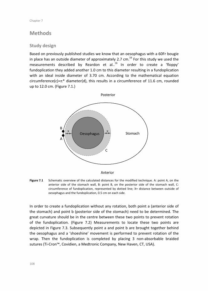
Chapter 7
108
Methods
Study design
Based on previously published studies we know that an oesophagus with a 60Fr bougie
in place has an outside diameter of approximately 2.7 cm.16 For this study we used the
measurements described by Reardon et al..16 In order to create a ‘floppy’
fundoplication they added another 1.0 cm to this diameter resulting in a fundoplication
with an ideal inside diameter of 3.70 cm. According to the mathematical equation
circumference(c)=* diameter(d), this results in a circumference of 11.6 cm, rounded
up to 12.0 cm. (Figure 7.1.)
Figure 7.1 Schematic overview of the calculated distances for the modified technique. A: point A, on the
anterior side of the stomach wall, B: point B, on the posterior side of the stomach wall, C: circumference of fundoplication, represented by dotted line; X= distance between outside of
oesophagus and the fundoplication, 0.5 cm on each side.
In order to create a fundoplication without any rotation, both point a (anterior side of
the stomach) and point b (posterior side of the stomach) need to be determined. The
great curvature should be in the centre between these two points to prevent rotation
of the fundoplication. (Figure 7.2) Measurements to locate these two points are
depicted in Figure 7.3. Subsequently point a and point b are brought together behind
the oesophagus and a ‘shoeshine’ movement is performed to prevent rotation of the
wrap. Then the fundoplication is completed by placing 3 non‐absorbable braided
sutures (Ti‐Cron™, Covidien, a Medtronic Company, New Haven, CT, USA).
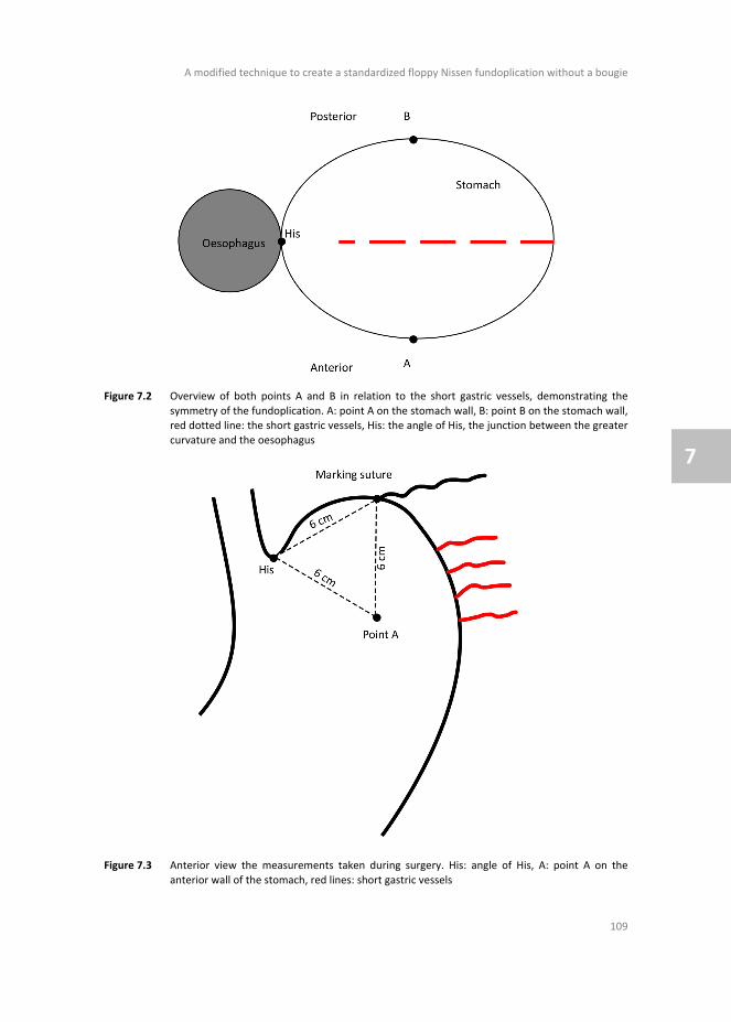
A modified technique to create a standardized floppy Nissen fundoplication without a bougie
109
7
Figure 7.2 Overview of both points A and B in relation to the short gastric vessels, demonstrating the
symmetry of the fundoplication. A: point A on the stomach wall, B: point B on the stomach wall, red dotted line: the short gastric vessels, His: the angle of His, the junction between the greater
curvature and the oesophagus
Figure 7.3 Anterior view the measurements taken during surgery. His: angle of His, A: point A on the
anterior wall of the stomach, red lines: short gastric vessels

Chapter 7
110
Patient selection
For this modified technique, we retrospectively evaluated the first 15 consecutive
patients that have been operated according this modified technique. All patients were
diagnosed with GERD according to the SAGES guideline for GERD. Minimal age was
18 years and patients were not selected for this evaluation if they have had prior anti‐
reflux surgery.
Outcome measurements
To evaluate postoperative quality of life, all patients were sent a questionnaire by mail.
Non‐responders were contact up to three times by phone, to optimize response rate.
Primary outcome of this study was the safety of this procedure, and therefore the
complication rate. Secondary outcomes were time of surgery, length of stay,
postoperative dysphagia and short‐term quality of life measured by the GERD‐HRQL
questionnaire.
Operative technique
The entire procedure will be performed laparoscopically, patients are in supine position
with the surgeon between the legs and under general anaesthesia. Pneumoperitoneum
is obtained using a Veress needle technique typically at Palmers point. Our preference,
for safety reasons, is to place the first trocar under direct vision. We therefore use a
12 mm Visiport (Visiport™, Medtronic, North Haven, CT, USA) in the supra umbilical
position. After inspection for adhesions and/or other abnormalities, two 5 mm trocars
and one 8 mm trocars are inserted. Then the liver retractor placed in position to create
enough space to perform the procedure.
The procedure starts with opening the lesser omentum and phrenoesophageal
ligament, followed by dissection of the right and left limbs of the crus of the diaphragm.
Then the short gastric vessels will be divided using an ultrasonic device. (Harmonic
ace®, Ethicon, US)
The next step is to create a window behind the oesophagus and encircle the
oesophagus with a silicon tube to retract the oesophagus without damage. At least
3 cm of the oesophagus is dissected and has to lay tension‐free in the abdomen. The
hernia sec is brought back into the abdomen and If a hiatal hernia is present, this is
narrowed with braided sutures (Ti‐Cron™, Covidien, a Medtronic Company, New Haven,
CT, USA)
We pursue patency for a 10‐mm instrument to pass besides the oesophagus. If a large
hiatal hernia is present or in case of weakness of the crus, the cruroplasty is reinforced
with a synthetic non‐absorbable mesh. (Parietex™ Composite Mesh Hiatal, Sofradim, a
Medtronic Company, Trevoux, France)
A large hiatal hernia is defined as migration of >50% of the stomach into the thorax.17

A modified technique to create a standardized floppy Nissen fundoplication without a bougie
111
7
We now measure 6 cm from the angle of His along the great curvature using the
grasper, which has a length of 2.5 cm, and place a marking suture. From this point, we
will create an equilateral triangle with sides of 6 cm. (Figure 7.2) We place a second
marking suture on the anterior side of the stomach. The next step is to flip the stomach
anteriorly and perform the same manoeuvre on the posterior side of the stomach and
place a third marking suture. All marking sutures are braided absorbable sutures.
(Polysorb™, Covidien, a Medtronic Company, New Haven, CT, USA) In order to facilitate
the mobilization of the stomach, the suture at the posterior side is kept a little longer.
Then the silicone is removed and with a grasper the posterior suture is grabbed behind
the oesophagus. A “shoeshine” movement is performed to make sure that there is no
rotation. The surgeon brings the posterior suture together with the anterior suture and
the assistant holds them in place. Then the procedure will be completed by placing
3 braided sutures (Ti‐Cron™, Covidien, a Medtronic Company, New Haven, CT, USA) to
fixate the fundoplication. The most distal suture is attached to the oesophagus to
prevent telescoping. A video of the complete procedure can be found in the
supplemental data of this article. (https://tinyurl.com/modified‐nissen)
Ethics
Since it concerns the routine implementation of a modification of a previously
described technique that is now routinely applied in our institution, informed consent
was waived.
Results
Patient characteristics
A total of 15 patients were operated according this technique between March 2015 and
January 2016. Mean age during surgery was 53.4(SD 11.8) years, 9(60%) of the patients
were male and the mean BMI was 26.5 (SD 3.7). 93.3% of the operated patients
experienced pre‐operative heartburn, 26.7% of the patients experienced regurgitation
and dysphagia was present in 20% of the patients. A complete overview of the baseline
characteristics for all patients is presented in Table 7.1.
All patients were diagnosed according to the SAGES guideline for GERD. An overview of
the pre‐operative work‐up is demonstrated in Table 7.2.
Operative results
All patients underwent a primary laparoscopic Nissen fundoplication. Mean operative
time was 69.5 (SD 8.4) minutes. We experienced no operative complications and there
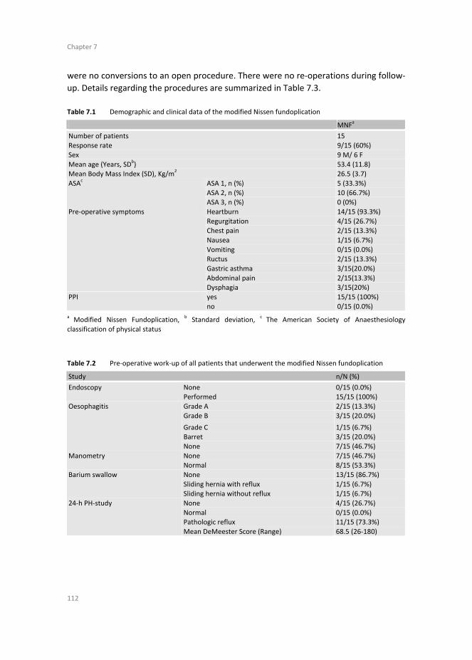
Chapter 7
112
were no conversions to an open procedure. There were no re‐operations during follow‐
up. Details regarding the procedures are summarized in Table 7.3.
Table 7.1 Demographic and clinical data of the modified Nissen fundoplication
MNFa
Number of patients 15 Response rate 9/15 (60%)
Sex 9 M/ 6 F
Mean age (Years, SDb) 53.4 (11.8)
Mean Body Mass Index (SD), Kg/m2 26.5 (3.7)
ASA 1, n (%) 5 (33.3%)
ASA 2, n (%) 10 (66.7%)
ASAc
ASA 3, n (%) 0 (0%)
Heartburn 14/15 (93.3%)
Regurgitation 4/15 (26.7%) Chest pain 2/15 (13.3%)
Nausea 1/15 (6.7%)
Vomiting 0/15 (0.0%) Ructus 2/15 (13.3%)
Gastric asthma 3/15(20.0%)
Abdominal pain 2/15(13.3%)
Pre‐operative symptoms
Dysphagia 3/15(20%)
yes 15/15 (100%) PPI
no 0/15 (0.0%) a Modified Nissen Fundoplication,
b Standard deviation,
c The American Society of Anaesthesiology
classification of physical status
Table 7.2 Pre‐operative work‐up of all patients that underwent the modified Nissen fundoplication
Study n/N (%)
Endoscopy None Performed
0/15 (0.0%) 15/15 (100%)
Grade A 2/15 (13.3%)
Grade B 3/15 (20.0%)
Grade C 1/15 (6.7%) Barret 3/15 (20.0%)
Oesophagitis
None 7/15 (46.7%)
None 7/15 (46.7%) Manometry Normal 8/15 (53.3%)
None 13/15 (86.7%)
Sliding hernia with reflux 1/15 (6.7%)
Barium swallow
Sliding hernia without reflux 1/15 (6.7%)
None 4/15 (26.7%)
Normal 0/15 (0.0%) Pathologic reflux 11/15 (73.3%)
24‐h PH‐study
Mean DeMeester Score (Range) 68.5 (26‐180)
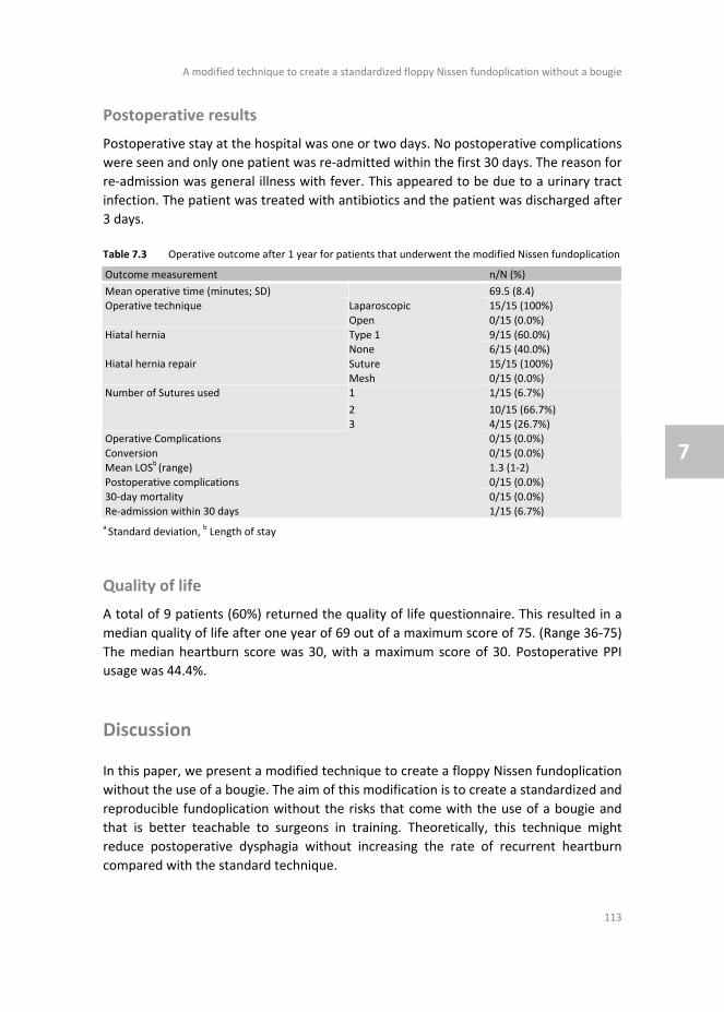
A modified technique to create a standardized floppy Nissen fundoplication without a bougie
113
7
Postoperative results
Postoperative stay at the hospital was one or two days. No postoperative complications
were seen and only one patient was re‐admitted within the first 30 days. The reason for
re‐admission was general illness with fever. This appeared to be due to a urinary tract
infection. The patient was treated with antibiotics and the patient was discharged after
3 days.
Table 7.3 Operative outcome after 1 year for patients that underwent the modified Nissen fundoplication
Outcome measurement n/N (%)
Mean operative time (minutes; SD) 69.5 (8.4) Laparoscopic 15/15 (100%) Operative technique
Open 0/15 (0.0%)
Type 1 9/15 (60.0%) Hiatal hernia None 6/15 (40.0%)
Suture 15/15 (100%) Hiatal hernia repair
Mesh 0/15 (0.0%) 1 1/15 (6.7%)
2 10/15 (66.7%)
Number of Sutures used
3 4/15 (26.7%)
Operative Complications 0/15 (0.0%)
Conversion 0/15 (0.0%) Mean LOS
b (range) 1.3 (1‐2)
Postoperative complications 0/15 (0.0%)
30‐day mortality 0/15 (0.0%) Re‐admission within 30 days 1/15 (6.7%)
a Standard deviation,
b Length of stay
Quality of life
A total of 9 patients (60%) returned the quality of life questionnaire. This resulted in a
median quality of life after one year of 69 out of a maximum score of 75. (Range 36‐75)
The median heartburn score was 30, with a maximum score of 30. Postoperative PPI
usage was 44.4%.
Discussion
In this paper, we present a modified technique to create a floppy Nissen fundoplication
without the use of a bougie. The aim of this modification is to create a standardized and
reproducible fundoplication without the risks that come with the use of a bougie and
that is better teachable to surgeons in training. Theoretically, this technique might
reduce postoperative dysphagia without increasing the rate of recurrent heartburn
compared with the standard technique.
Chapter 7
114
Based on current literature we have found an incidence of early dysphagia up to 76%5 A
retrospective study performed by Lamb et al. investigates the results of revision surgery
after prior anti‐reflux surgery for GERD.18 This study demonstrates that persistent
dysphagia is the most reported indication for revision surgery. (52%) We also know
from previous literature that patients who experience postoperative dysphagia report
suboptimal outcomes after revision surgery.19‐21 It is therefore important to reduce the
risk of postoperative dysphagia and minimize the chance for revision surgery.
Our hypothesis is that postoperative dysphagia may be, among other causes, the result
of both a too tight fundoplication and some sort of rotation in the wrap. A too tight
wrap as a cause of postoperative dysphagia has been investigated previously.22
However, there is no consensus regarding the ideal diameter of the fundoplication and
it is hard to feel the correct tightness of the wrap. Some authors therefore advocate the
use of a bougie to assure sufficient patency.11,12 Nevertheless, different bougie sizes
have been used varying between 52 and 60Fr, but the optimal size has yet to be found.
The positive effect of a bougie on postoperative dysphagia was already demonstrated
by DeMeester et al in 1986.23 This study showed a significant decrease in postoperative
dysphagia for a 60Fr bougie compared with a 36Fr bougie, especially in the first three
postoperative months. The use of a bougie comes, however, with the severe risk of
oesophageal perforation, which comes with serious morbidity and even mortality.15
Despite these possible serious complications, an anonymous survey performed in 2005
by Huttl et al in Germany, showed that still 46% of the surgeons still routinely used a
bougie during laparoscopic Nissen fundoplication at that time.24 A possible explanation
might be that it is difficult, especially for the beginning surgeon, to create a proper
fundoplication with a correct diameter. This hypothesis is supported by historical
studies, that demonstrates a learning curve of approximately 25 procedures per
surgeon.25‐27 These studies also show a prior complication rate of 24‐55% which
decreases to approximately 10% when more experience is obtained. By this modified
technique, we aim to aid surgeons who start performing laparoscopic Nissen
fundoplication, to create a standard fundoplication which ensures sufficient patency.
Further research is needed to investigate whether this technique indeed reduces the
learning curve and reduces long‐term dysphagia.
According to previously published literature, we believe that a common error made by
surgeons is to create an asymmetrical fundoplication, resulting in a ‘twist’ in the
fundoplication.16,28 With the above‐mentioned technique we assured that the
fundoplication that is created, is as symmetrical as possible. This leads to a reduced risk
of shifting the line of the greater curvature to either anteriorly or posteriorly.
(Figure 7.2.)
In this report, we demonstrate the first 15 patients that have been operated according
to this modified technique. We are aware of the fact that this is a very small group and
follow‐up is short and only 60% returned the quality of life questionnaire. However, we
did not see any perioperative complications, only one patient was admitted for an

A modified technique to create a standardized floppy Nissen fundoplication without a bougie
115
7
urinary tract infection on the third postoperative day. This is a minor complication and
in our opinion not related to the modified technique.
Since the measurement of the exact points a and b does take some extra time, we
compared the operative time with our retrospective cohort of the past 10 years. The
mean operative time in this cohort was 78.8 (SD 39.2),29 where the mean operative
time for this modified technique was 69.5 (SD 8.4) minutes. (P=0.752) There is no
statistically significant difference between both techniques in operating time. However,
we do expect that especially beginning surgeons will benefit from this modified
technique, which might even result in an even faster operating time. This hypothesis
will be subject of further research.
Conclusion
The size of the gastric wrap, created during a Nissen fundoplication can be standardized
using an atraumatic and minimal invasive approach. This modified technique is safe and
leads to good results direct postoperatively and at 1 year follow up. The possible
advantage regarding reproducibility, reduced learning curve and less postoperative
dysphagia has yet to be established is further studies.
Chapter 7
116
References
1. El‐Serag HB, Sweet S, Winchester CC, Dent J Update on the epidemiology of gastro‐oesophageal reflux disease: a systematic review. Gut 2014:63:871‐80.
2. Watson DI Evolution and development of surgery for large paraesophageal hiatus hernia. World J Surg
2011:35:1436‐41. 3. Cuschieri A, Shimi S, Nathanson LK Laparoscopic reduction, crural repair, and fundoplication of large
hiatal hernia. Am J Surg 1992:163:425‐30.
4. Stefanidis D, Hope WW, Kohn GP, Reardon PR, Richardson WS, et al. Guidelines for surgical treatment of gastroesophageal reflux disease. Surg Endosc 2010:24:2647‐69.
5. Balci D, Turkcapar AG Assessment of quality of life after laparoscopic Nissen fundoplication in patients
with gastroesophageal reflux disease. World J Surg 2007:31:116‐21. 6. Pessaux P, Arnaud JP, Delattre JF, Meyer C, Baulieux J, et al. Laparoscopic antireflux surgery: five‐year
results and beyond in 1340 patients. Arch Surg 2005:140:946‐51.
7. Triponez F, Dumonceau JM, Azagury D, Volonte F, Slim K, et al. Reflux, dysphagia, and gas bloat after laparoscopic fundoplication in patients with incidentally discovered hiatal hernia and in a control group.
Surgery 2005:137:235‐42.
8. Ma S, Qian B, Shang L, Shi R, Zhang G A meta‐analysis comparing laparoscopic partial versus Nissen fundoplication. ANZ J Surg 2012:82:17‐22.
9. Sgromo B, Irvine LA, Cuschieri A, Shimi SM Long‐term comparative outcome between laparoscopic total
Nissen and Toupet fundoplication: Symptomatic relief, patient satisfaction and quality of life. Surg Endosc 2008:22:1048‐53.
10. Leggett PL, Bissell CD, Churchman‐Winn R, Ahn C A comparison of laparoscopic Nissen fundoplication
and Rossetti's modification in 239 patients. Surg Endosc 2000:14:473‐7. 11. Allaix ME, Herbella FA, Patti MG Laparoscopic total fundoplication for gastroesophageal reflux disease.
How I do it. J Gastrointest Surg 2013:17:822‐8.
12. Auyang ED, Pellegrini CA How I do it: laparoscopic paraesophageal hernia repair. J Gastrointest Surg 2012:16:1406‐11.
13. Davis RE, Awad ZT, Filipi CJ Technical factors in the creation of a "floppy" Nissen fundoplication. Am J
Surg 2004:187:724‐7. 14. Patterson EJ, Herron DM, Hansen PD, Ramzi N, Standage BA, et al. Effect of an esophageal bougie on
the incidence of dysphagia following nissen fundoplication: a prospective, blinded, randomized clinical
trial. Arch Surg 2000:135:1055‐61. 15. Lowham AS, Filipi CJ, Hinder RA, Swanstrom LL, Stalter K, et al. Mechanisms and avoidance of
esophageal perforation by anesthesia personnel during laparoscopic foregut surgery. Surg Endosc
1996:10:979‐82. 16. Reardon PR, Matthews BD, Scarborough TK, Preciado A, Marti JL, et al. Geometry and reproducibility in
360 degrees fundoplication. Surg Endosc 2000:14:750‐4.
17. Peters JH SAGES guidelines for the management of hiatal hernia. Surg Endosc 2013:27:4407‐8. 18. Lamb PJ, Myers JC, Jamieson GG, Thompson SK, Devitt PG, et al. Long‐term outcomes of revisional
surgery following laparoscopic fundoplication. Br J Surg 2009:96:391‐7.
19. Iqbal A, Awad Z, Simkins J, Shah R, Haider M, et al. Repair of 104 failed anti‐reflux operations. Ann Surg 2006:244:42‐51.
20. Smith CD, McClusky DA, Rajad MA, Lederman AB, Hunter JG When fundoplication fails: redo? Ann Surg
2005:241:861‐9. 21. Safranek PM, Gifford CJ, Booth MI, Dehn TC Results of laparoscopic reoperation for failed antireflux
surgery: does the indication for redo surgery affect the outcome? Dis Esophagus 2007:20:341‐5.
22. Bais JE, Bartelsman JF, Bonjer HJ, Cuesta MA, Go PM, et al. Laparoscopic or conventional Nissen fundoplication for gastro‐oesophageal reflux disease: randomised clinical trial. The Netherlands
Antireflux Surgery Study Group. Lancet 2000:355:170‐4.
23. DeMeester TR, Bonavina L, Albertucci M Nissen fundoplication for gastroesophageal reflux disease. Evaluation of primary repair in 100 consecutive patients. Ann Surg 1986:204:9‐20.
A modified technique to create a standardized floppy Nissen fundoplication without a bougie
117
7
24. Huttl TP, Hohle M, Wichmann MW, Jauch KW, Meyer G Techniques and results of laparoscopic
antireflux surgery in Germany. Surg Endosc 2005:19:1579‐87. 25. Salminen P, Hiekkanen H, Laine S, Ovaska J Surgeons' experience with laparoscopic fundoplication after
the early personal experience: does it have an impact on the outcome? Surg Endosc 2007:21:1377‐82.
26. Watson DI, Baigrie RJ, Jamieson GG A learning curve for laparoscopic fundoplication. Definable, avoidable, or a waste of time? Ann Surg 1996:224:198‐203.
27. Soot SJ, Eshraghi N, Farahmand M, Sheppard BC, Deveney CW Transition from open to laparoscopic
fundoplication: the learning curve. Arch Surg 1999:134:278‐81. 28. Peters JH, DeMeester TR, Crookes P, Oberg S, de Vos Shoop M, et al. The treatment of
gastroesophageal reflux disease with laparoscopic Nissen fundoplication: prospective evaluation of 100
patients with "typical" symptoms. Ann Surg 1998:228:40‐50. 29. Castelijns PSS, Ponten JEH, Vd Poll MCG, Bouvy ND, Smulders JF Quality of life after nissen
fundoplication in patients with gastroesophageal reflux disease: Comparison between long‐ and short‐
term follow‐up. J Minim Access Surg. 2018;14(3):213‐20.

Chapter 7
118
Chapter 8 Standardized method for Nissen fundoplication:
reproducibility and effect on learning curve
P.S.S. Castelijns
M.C.G. van de Poll
B.P.A. de Greef
N.D. Bouvy
J.F. Smulders
Submitted

Chapter 8
120
Abstract
Aims Laparoscopic anti‐reflux surgery requires advanced laparoscopic skills and is associated with a substantial learning curve leading to a large variability of the size and form of the fundoplication in inexperienced hands. We have developed a modified technique to create a standardized floppy Nissen fundoplication without the use of a bougie. The aim of this study was to determine the effect of this modified technique on the variability of fundoplication in a standardized ex‐vivo model. Methods In an ex‐vivo set‐up, a laparoscopic fundoplication was performed in 20 porcine stomachs using a 2‐by‐2 factorial design. Fundoplications were performed by either a 5th year surgical resident or an experienced anti‐reflux surgeon using a conventional or modified technique. Measurements regarding time, size, symmetry and reproducibility of the fundoplication were evaluated. Results Using the modified technique symmetry of the fundoplication increased whereas variability between different fundoplications decreased significantly. This effect was seen in both experienced as well as inexperienced operators. The positive effect of the modified technique on symmetry was more profound in the inexperienced operator. Symmetry of fundoplications created with the modified technique was comparable between both operators. Conclusions This modified technique is better reproducible than the standard technique for Nissen fundoplication and leads to a more symmetrical and more standardized wrap. It may facilitate both beginning and experienced surgeons to create a standardized fundoplication and may reduce the learning curve in anti‐reflux surgery.

A modified technique to create a standardized floppy Nissen fundoplication without a bougie
121
Introduction
Laparoscopic Nissen fundoplication has been described for the first time in 1992 by
Cushieri et al.1 Despite the broad experience with this technique, there is still a learning
curve associated with this advanced laparoscopic technique. Watson et al described
their experience of 280 fundoplications in 1996 and concluded that the risk of adverse
outcomes did not stabilize until 20 procedures for each surgeon and even
50 procedures per institution was achieved.2 Another study in 1999 evaluated the
laparoscopic learning curve for surgeons experienced in open Nissen fundoplication,
but still the learning curve is approximately 20 procedures and even after
100 procedures there is still improvement in operation time, conversion rate and intra‐
operative complication rate.3 In 2005, a similar result has been described. This study
presents a re‐operation rate for dysphagia of 22% in the early experience with
laparoscopic anti‐reflux surgery compared with 4% for the more experienced surgeon.4
Even in the current era, where laparoscopic surgery is gold standard for most
treatments, the institutional experience in laparoscopic hiatal hernia repair of
<10 procedures per year remains an independent risk factor for recurrence.5 Even if a
resident is supervised by an experienced surgeon, the outcomes after anti‐reflux
surgery are slightly less.6 Several other studies report on the large learning curve of
laparoscopic Nissen fundoplication.7‐9
Dysphagia remains a serious complication of Nissen fundoplication and early dysphagia
up to 76% has been reported, and even long‐term dysphagia is present in 8.0% of the
patients.10‐12 Vagal nerve injury leads to delayed gastric emptying, which is closely
related to the feeling of dysphagia. Also several technical aspects are of great
importance in reducing dysphagia.13 In the history of anti‐reflux surgery several
techniques have been proposed, including partial fundoplications. Especially the early
postoperative dysphagia is significantly less after partial fundoplication compared with
Nissen fundoplication.14 Preservation of the gastric vessels has been attempted to
reduce postoperative dysphagia, however in fact this has led to an increase of this
complication. Possibly due to forced rotation of the fundoplication.15 This emphasizes
the importance of the construction of an adequate and symmetric fundoplication.
To reduce the risk of postoperative dysphagia we have developed a technique to create
a standardized floppy fundoplication with minimal rotation. Clinical results of this
technique have been described earlier in detail.16 It is unknown whether this technique
is indeed better reproducible, teachable and leads to a more standard fundoplication
with sufficient patency. Furthermore, this technique may reduce the learning curve for
laparoscopic Nissen fundoplication. The primary aim of this study was to investigate the
reproducibility of this modified technique compared with the conventional method.
Secondary outcome measure is the difference between an experienced surgeon and a
resident in performing this technique.
Chapter 8
122
Methods
Experimental setup
For this study, we have created an ex‐vivo laparoscopic set‐up box. We used the
stomach and esophagus of cadaver pigs obtained from the local abattoir. All
procedures were recorded on video and analyzed afterwards. We only studied the
creation of the fundoplication, the laparoscopic introduction, mobilization of the
stomach, dissecting the gastric vessels and/or the hiatal hernia repair are not subject of
this study.
Conventional technique
The conventional technique starts with grasping the fundus behind the
gastroesophageal junction and performing the “shoe‐shine maneuver” i.e. the tension
free movement of the fundus dorsal of the esophageal‐cardiac junction. Then the
fundus is plicated around the esophagus and sutured to the anterior gastric wall with
three sutures. These sutures are placed at a location that is deemed appropriate by the
operating surgeon. In general, no Bougie is used in this method.
Modified technique
For the modified technique, a marking suture is placed at the greater curvature.
Hereafter, 2 sutures are places at the anterior and posterior wall of the stomach, with
12 cm in between the sutures. Distances are measured using the laparoscopic grasper,
which has a width of 2.5 cm. Than the anterior and posterior marking sutures are
brought together, a shoe‐shine maneuver is performed and the fundoplication
completed by placing 3 sutures. This modified technique is described in detail in a
previous publication.16 The measurements that are needed to perform the modified
technique are demonstrated in Figure 8.1.
Experimental procedures
The studies were performed using a 2‐by‐2 factorial design. Ten fundoplications were
performed by an experienced anti‐reflux with over 300 laparoscopic Nissen
fundoplications and at least 50 procedures in the last year (JS) and 10 by a 5th year
resident (JP) with general experience in laparoscopic surgery. Both of them performed
5 fundoplications according the conventional method and 5 fundoplications according
the modified technique.
Afterwards the fundoplication was dismantled by the principal investigator (PC) and
several distances, including the circumference of the fundoplication, was measured.
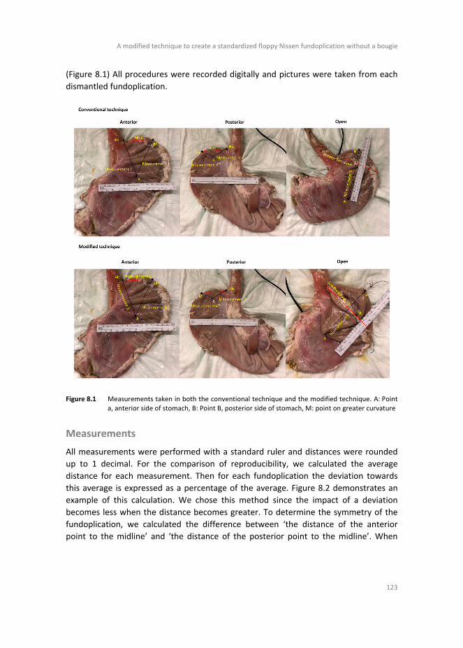
A modified technique to create a standardized floppy Nissen fundoplication without a bougie
123
(Figure 8.1) All procedures were recorded digitally and pictures were taken from each
dismantled fundoplication.
Figure 8.1 Measurements taken in both the conventional technique and the modified technique. A: Point
a, anterior side of stomach, B: Point B, posterior side of stomach, M: point on greater curvature
Measurements
All measurements were performed with a standard ruler and distances were rounded
up to 1 decimal. For the comparison of reproducibility, we calculated the average
distance for each measurement. Then for each fundoplication the deviation towards
this average is expressed as a percentage of the average. Figure 8.2 demonstrates an
example of this calculation. We chose this method since the impact of a deviation
becomes less when the distance becomes greater. To determine the symmetry of the
fundoplication, we calculated the difference between ‘the distance of the anterior
point to the midline’ and ‘the distance of the posterior point to the midline’. When
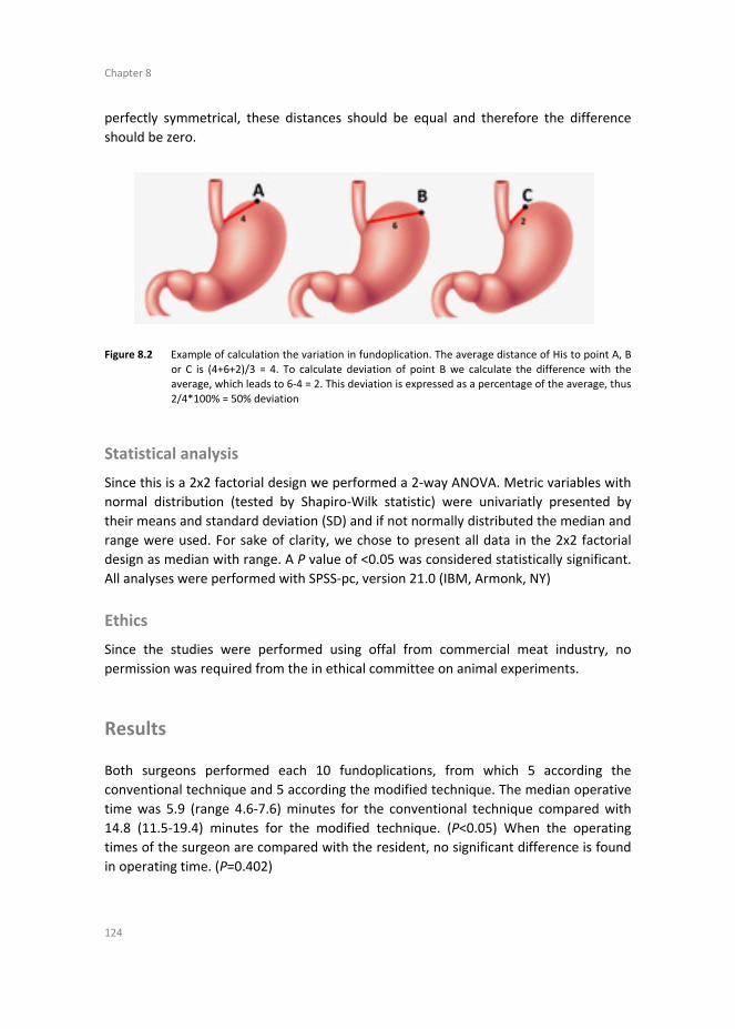
Chapter 8
124
perfectly symmetrical, these distances should be equal and therefore the difference
should be zero.
Figure 8.2 Example of calculation the variation in fundoplication. The average distance of His to point A, B
or C is (4+6+2)/3 = 4. To calculate deviation of point B we calculate the difference with the average, which leads to 6‐4 = 2. This deviation is expressed as a percentage of the average, thus
2/4*100% = 50% deviation
Statistical analysis
Since this is a 2x2 factorial design we performed a 2‐way ANOVA. Metric variables with
normal distribution (tested by Shapiro‐Wilk statistic) were univariatly presented by
their means and standard deviation (SD) and if not normally distributed the median and
range were used. For sake of clarity, we chose to present all data in the 2x2 factorial
design as median with range. A P value of <0.05 was considered statistically significant.
All analyses were performed with SPSS‐pc, version 21.0 (IBM, Armonk, NY)
Ethics
Since the studies were performed using offal from commercial meat industry, no
permission was required from the in ethical committee on animal experiments.
Results
Both surgeons performed each 10 fundoplications, from which 5 according the
conventional technique and 5 according the modified technique. The median operative
time was 5.9 (range 4.6‐7.6) minutes for the conventional technique compared with
14.8 (11.5‐19.4) minutes for the modified technique. (P<0.05) When the operating
times of the surgeon are compared with the resident, no significant difference is found
in operating time. (P=0.402)
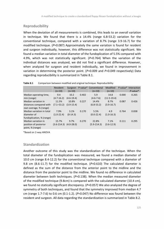
A modified technique to create a standardized floppy Nissen fundoplication without a bougie
125
Reproducibility
When the deviation of all measurements is combined, this leads to an overall variation
in technique. We found that there is a 14.4% (range 6.8‐53.2) variation for the
conventional technique, compared with a variation of 8.7% (range 3.9‐16.7) for the
modified technique. (P=0.087) Approximately the same variation is found for resident
and surgeon individually, however, this difference was not statistically significant. We
found a median variation in total diameter of the fundoplication of 5.5% compared with
4.9%, which was not statistically significant. (P=0.764) When the variation of the
individual distances was analyzed, we did not find a significant difference. However,
when analyzed for surgeon and resident individually, we found in improvement in
variation in determining the posterior point. (P=0.009 and P=0.049 respectively) Data
regarding reproducibility is summarized in Table 8.1.
Table 8.1 Comparison between modified and original technique: Reproducibility
Resident
(n=10)
Surgeon
(n=10)
P‐value* Conventional
(n=10)
Modified
(n=10)
P‐value* Interaction
P‐value*
Median operating time,
min (range)
9.4
(4.7‐16.2)
10.2
(4.6‐19.4)
0.402 5.9
(4.6‐7.6)
14.8
(11.5‐19.4)
0.000 0.265
Median variation in distances compared with
own average, % (range)
11.5% (7.1–53.2)
10.8% (3.9‐15.4)
0.227 14.4% (6.8‐53.2)
8.7% (3.9‐16.7)
0.087 0.426
Median variation in total diameter of
fundoplication, % (range)
7.9% (1.0‐22.4)
3.1% (0‐14.3)
0.170 5.5% (0.0‐22.4)
4.9 (1.0‐16.3)
0.764 0.838
Median variation in position of posterior
point, % (range)
15.7% (3.6‐214.3)
9.7% (4.0‐28.0)
0.270 22.8% (5.9‐214.3)
7.1% (3.6‐12.9)
0.111 0.295
*Based on 2‐way ANOVA
Standardization
Another outcome of this study was the standardization of the technique. When the
total diameter of the fundoplication was measured, we found a median diameter of
10.0 cm (range 8.4‐12.2) for the conventional technique compared with a diameter of
9.8 cm (8.6‐11.7) for the modified technique. (P=0.610) The calculated diameter is
defined as the sum of the distance from the anterior point to the midline and the
distance from the posterior point to the midline. We found no difference in calculated
diameter between both techniques. (P=0.138). When the median measured diameter
of the modified technique (9.8cm) is compared with the calculated diameter (10.4 cm),
we found no statically significant discrepancy. (P=0.057) We also analyzed the degree of
symmetry of both techniques, and found that the symmetry improved from median 4.7
cm (range 1.7‐7.6) to 0.6 cm (0.1‐1.2). (P=0.047) No difference was found between the
resident and surgeon. All data regarding the standardization is summarized in Table 8.2.

Chapter 8
126
Table 8.2 Comparison between modified and original technique: Standardization
Resident
(n=10)
Surgeon
(n=10)
P‐value* Conventional
(n=10)
Modified
(n=10)
P‐value* Interaction
P‐value*
Measured diameter of fundoplication (median
cm; range)
10.2 (8.5‐12.2)
9.8 (8.4‐10.4)
0.212 10.0 (8.4‐12.2)
9.8 (8.6‐11.7)
0.610 0.640
Calculated diameter of fundoplication
a (median
cm; range)
10.7 (8.6‐12.9)
10.0 (8.5‐11.7)
0.242 10.0 (8.5‐11.5)
10.4 (9.2‐12.9)
0.138 0.583
Degree of symmetryb
(median cm; range) 1.9
(0.1‐7.6) 1.5
(0.1‐5.9) 0.061 4.7
(1.7‐7.6) 0.6
(0.1‐1.2) 0.000 0.047
a Diameter is calculated by adding ‘the distance from anterior point to greater curvature’ to ‘the distance from posterior point to greater curvature’;
b Degree of symmetry is calculated by calculating the difference
between ‘anterior point to midline’ and ‘posterior point to midline’. These distances should be equal;
therefore, the difference should be close to zero when fundoplication is symmetrical; * Based on 2‐way ANOVA
Measurements
To evaluate the accuracy of the measurements that were performed in the modified
technique, we compared each distance with the targeted distance of 6 cm. These
results are demonstrated in Table 8.3. The only measurement that was statistically
different compared with the aimed distance, was from the posterior point to the
greater curvature. This distance should have been 6cm, however, we found a mean
distance of 5.3 cm (SD 0.5). (P=0.001) For the surgeon and resident individually, this
distance was not statically significant different.
Table 8.3 Correlation between measurements and actual distances for modified fundoplication
Actual distances (cm) P‐value (compared with the
ideal distance of 6.0 cm)
Overall 5.7 cm (4.9‐6.8) 0.4191
Resident 6.3 cm (4.9‐6.8) 0.5761
Measurement 1, median (range)
Surgeon 5.4 cm (4.9‐6.1) 0.0951
Overall 5.4 cm (4.5‐6.7) 0.2141
Resident 6.0 cm (4.9‐6.7) 1.0001
Measurement 2, median (range)
Surgeon 4.8 cm (4.5‐6.2) 0.0941
Overall 6.5 cm (0.8) 0.0902
Resident 6.8 cm (0.9) 0.1402
Measurement 3, mean (SD3)
Surgeon 6.2 cm (0.5) 0.5082
Overall 5.3 cm (0.5) 0.0012
Resident 5.4 cm (0.6) 0.0612
Measurement 4, mean (SD)
Surgeon 5.2 cm (0.4) 0.0112
Overall 6.6 cm (1.1) 0.1102
Resident 6.9 cm (1.2) 0.1432
Measurement 5, mean (SD)
Surgeon 6.4 cm (1.1) 0.4822
This table demonstrates the actual distances and are compared with the aimed distance (6.0 cm for each
measurement). For description of the measurements we refer to Figure 8.1. 1 Mann‐Whitney U,
2 Independent‐Samples T Test,
3 SD; Standard deviation

A modified technique to create a standardized floppy Nissen fundoplication without a bougie
127
Learning curve
To evaluate whether the fundoplication that has been created according this technique
is similar between the resident and the surgeon we have compared the measurements
between both. As is seen in Table 8.1 and 8.2 there is no difference detected between
both the resident and the surgeon. When sub‐group analyses is performed for both the
conventional and modified technique, there is still no significant difference. (Table 8.4).
Table 8.4 Comparison between resident and surgeon
Resident Surgeon P‐value
Median operating time, min (range) Original 6.2 (4.7‐443) 5.6 (4.6‐7.6) 0.754
1
Modified 14.4 (11.5‐16.2) 16.7 (12.7‐19.4) 0.3471
Median degree of symmetry, cm (range) Original 6.1 cm (2.8‐7.6) 3.0 cm (1.7‐5.9) 0.076
1
Modified 0.5 cm (0.1‐0.9) 0.6 cm (0.1‐1.2) 0.7511
Mean total diameter, cm (SD) Original 10.4 cm (1.4) 9.6 cm (0.7) 0.274
2
Modified 10.0 cm (1.2) 9.6 cm 0.7) 0.5462
Median variation in distances compared with own average, % (range)
Original 14.6% (10.2‐53.2) 14.1% (6.8‐15.4) 0.4651
Modified 9.5% (7.1‐16.7) 6.7% (3.9‐15.0) 0.3471
1 Mann‐Whitney U,
2 Independent‐Samples T Test
Discussion
Laparoscopic Nissen fundoplication comes with a substantial learning curve and one of
the most reported causes for re‐operation remains dysphagia or recurrent heartburn.
Among the many reasons for these complications, we believe that an inappropriate
construction of the fundoplication plays an essential role. To aid surgeons in performing
a proper fundoplication without the use of a bougie, we have developed a modified
technique, which is proven safe and leads to good results at the short term.16 For this
paper we have studied the actual reproducibility and standardization of this technique
in an ex‐vivo experiment. Furthermore, we have compared the results of a resident
with an experienced laparoscopic surgeon to assess whether this technique may reduce
the learning curve. A recent publication established the relevance of training
procedures to reduce the learning curve once again.17 When we compared the resident
with the surgeon we did not find any significant differences. However, it seems that
with the conventional technique the fundoplication of the resident is far more
asymmetrical that the surgeon. This difference seems to disappear when the modified
technique is used. (P=0.076 vs .P=0.751) This difference might not be significant due to

Chapter 8
128
small sample sizes, since only 10 fundoplications each were performed. The variation in
the technique did not significantly differ between the surgeon and the resident.
We have found that the variation in distances of the fundoplication is reduced from
14.4 to 8.7% (P=0.087). For the individual distances no differences were found, even for
the location of the posterior point of the fundoplication. Both the surgeon and the
resident experienced that it was difficult to determine the proper location at the
posterior side of the stomach. We noticed that in all cases, the distance from the
midline to the posterior point was less than the distance from the midline to the
anterior point. This resulted in a fundo‐rotation instead of a fundoplication. The
relevance of creating a symmetrical fundoplication is already mentioned in previous
studies.2,18 The median deviation from perfect symmetry was 4.7 cm in the
conventional technique compared to 0.6cm for the modified technique. (P=0.000)
There seems to be an interaction between the performer of the fundoplication and the
method that is been used. Most likely the conventional fundoplication of the surgeon is
more symmetrical than the one of the resident due to the experience of the surgeon.
This therefore increases the effect that is found. However, we believe that the effect of
the difference in technique is of such proportion, that this outweighs the possible effect
of the performer.
Therefore, we conclude that this modified technique leads to a significant more
symmetrical fundoplication. Since this is a technical study, we do not know whether
this effect indeed reduces postoperative dysphagia and leads to better symptomatic
outcome, but this might be subject of further study.
It is interesting that the actual diameter of the fundoplication for both techniques is
similar. Thus, the size of the wrap is comparable with the conventional technique. We
know from previous studies, that the size of the fundoplication influences
postoperative outcome. We have also learnt from studies that use a bougie, that with a
larger diameter of the fundoplication, postoperative dysphagia reduces.19 The same
effect is seen when Nissen fundoplication is compared with the partial fundoplications.
Especially the short‐term dysphagia is significant more severe after Nissen when
compared with Toupet fundoplication.14 Whether the demonstrated patency is the
proper size has yet to be established, since no consensus has been reached regarding
this subject.20 Despite the fact that it is unknown whether this technique leads to
significant less dysphagia, a great advantage is that the surgeon knows the size of the
fundoplication that is created. Therefore, when persistent dysphagia is present, it is
possible to perform a re‐do fundoplication, with an increased patency by measuring a
larger distance.
This study is based on a theoretical model with mathematical equations to determine
the distances from the midline to create a fundoplication with a diameter of
approximately 12 cm.16 We found that all measurements except the determination of
the posterior point was accurate and no statically significant difference with the aimed
distance was observed. This is supported by the fact that the variation regarding to

A modified technique to create a standardized floppy Nissen fundoplication without a bougie
129
location of the posterior point was very large. Therefore, special attention should be
paid when determining the location of the posterior point.
The distances in this study are measured by an estimation with the laparoscopic
grasper, which is 2.5 cm. The accuracy can be further improved by introducing a sterile
ruler, however this may lead to a further increasing operating time and we do not
believe that this leads to statistically improvement compared with the technique
mentioned above.
Another interesting finding in this study is the discrepancy between the calculated
diameter of the fundoplication and the actual diameter. Based on the theoretical
model, the diameter should be 12 cm. However, we measured a median distance of
9.8 cm and the calculated distance was 10.4 cm. This might be due to a standard error
which is made in the measurement, either during surgery or during analysis of the
distances by the investigator. Another explanation might be an error in the
mathematical equation to determine the diameter of the fundoplication. Since we
create 2 equilateral triangles with a base of 6 cm, we may need to determine the height
of these triangles, instead of the length of the sides. The result of this alternative
calculation is a diameter of 10.4 cm, which is more in line with the diameter that we
have found. This should be kept in mind when functional outcomes of this techniques
are evaluated.
Despite the possible advantages of this technique, which is better reproducible and
leads to a more symmetrical fundoplication, the operating time is significant longer.
The conventional technique requires a median of 5.9 minutes to complete compared
with 14.8 minutes for the modified technique. However, this is only a difference of
6.9 minutes for a small part of the entire procedure. Based on a large retrospective
cohort study of the same experience surgeon of 169 patients, we know that the mean
operative time for the entire procedure is 78.8 minutes.21 Therefore, the operative time
is only increased by 8.8% based on a mean operative time of an experienced surgeon. It
might be that when a resident of unexperienced surgeon performs the entire
procedure, the effect on operating time due to this modified technique is minimized.
Furthermore, when this technique is more frequently performed, the operative time
may further reduce and the difference may become even less.
This is this the first study that evaluates the standardization and reproducibility of a
laparoscopic fundoplication with an ex vivo porcine model. Despite maximum effort to
reduce bias and perform a reproducible concept, there are still some limitations of this
study. We operated on porcine stomachs, which are comparable with the human
stomach, but are slightly more rigid and more difficult to handle. Since this is our first
experience with this concept, only one surgeon and one resident were included. It
would be interesting to see what the results will be when more residents perform this
experiment, to actually extrapolate the results to real life. Some data seems to provide
an advantage of the modified technique over the conventional technique, but no
significance is reached. This might be due to the very small sample size, and when more

Chapter 8
130
fundoplications are performed, these differences may become statistically significant.
This study demonstrates possible advantages of this modified technique over the
modified technique and shows that even an inexperience resident is capable of creating
a proper fundoplication with this modified technique. Further studies are needed to
evaluate patient outcomes of this technique and to establish whether the learning
curve is further reduced by this technique.
Conclusion
This modified technique for Nissen fundoplication leads to a better reproducible and
more standardized fundoplication which can easily be reproduced by an inexperienced
surgeon. Further studies are need to evaluate the actual effect on patient outcomes
and learning curve.
A modified technique to create a standardized floppy Nissen fundoplication without a bougie
131
References
1. Cuschieri A, Shimi S, Nathanson LK. Laparoscopic reduction, crural repair, and fundoplication of large hiatal hernia. Am J Surg 1992;163(4):425‐30.
2. Watson DI, Baigrie RJ, Jamieson GG. A learning curve for laparoscopic fundoplication. Definable,
avoidable, or a waste of time? Ann Surg 1996;224(2):198‐203. 3. Soot SJ, Eshraghi N, Farahmand M, Sheppard BC, Deveney CW. Transition from open to laparoscopic
fundoplication: the learning curve. Arch Surg 1999;134(3):278‐81.
4. Hwang H, Turner LJ, Blair NP. Examining the learning curve of laparoscopic fundoplications at an urban community hospital. Am J Surg 2005;189(5):522‐6.
5. Antiporda M, Veenstra B, Jackson C, Kandel P, Daniel Smith C, Bowers SP. Laparoscopic repair of giant
paraesophageal hernia: are there factors associated with anatomic recurrence? Surg Endosc 2018;32(2):945‐54.
6. Brown CN, Smith LT, Watson DI, Devitt PG, Thompson SK, Jamieson GG. Outcomes for trainees vs
experienced surgeons undertaking laparoscopic antireflux surgery ‐ is equipoise achieved? J Gastrointest Surg 2013;17(7):1173‐80.
7. Zacharoulis D, O'Boyle CJ, Sedman PC, Brough WA, Royston CM. Laparoscopic fundoplication: a 10‐year
learning curve. Surg Endosc 2006;20(11):1662‐70. 8. Gill J, Booth MI, Stratford J, Dehn TC. The extended learning curve for laparoscopic fundoplication: a
cohort analysis of 400 consecutive cases. J Gastrointest Surg 2007;11(4):487‐92.
9. Dagash H, Chowdhury M, Pierro A. When can I be proficient in laparoscopic surgery? A systematic review of the evidence. J Pediatr Surg. 2003;38(5):720‐4.
10. Balci D, Turkcapar AG. Assessment of quality of life after laparoscopic Nissen fundoplication in patients
with gastroesophageal reflux disease. World J Surg 2007;31(1):116‐21. 11. Pessaux P, Arnaud JP, Delattre JF, Meyer C, Baulieux J, Mosnier H. Laparoscopic antireflux surgery: five‐
year results and beyond in 1340 patients. Arch Surg 2005;140(10):946‐51.
12. Triponez F, Dumonceau JM, Azagury D, Volonte F, Slim K, Mermillod B, et al. Reflux, dysphagia, and gas bloat after laparoscopic fundoplication in patients with incidentally discovered hiatal hernia and in a
control group. Surgery 2005;137(2):235‐42.
13. van Rijn S, Roebroek YG, Conchillo JM, Bouvy ND, Masclee AA. Effect of Vagus Nerve Injury on the Outcome of Antireflux Surgery: An Extensive Literature Review. Dig Surg 2016;33(3):230‐9.
14. Du X, Hu Z, Yan C, Zhang C, Wang Z, Wu J. A meta‐analysis of long follow‐up outcomes of laparoscopic
Nissen (total) versus Toupet (270 degrees ) fundoplication for gastro‐esophageal reflux disease based on randomized controlled trials in adults. BMC Gastroenterol 2016;16(1):88.
15. Leggett PL, Bissell CD, Churchman‐Winn R, Ahn C. A comparison of laparoscopic Nissen fundoplication
and Rossetti's modification in 239 patients. Surg Endosc 2000;14(5):473‐7. 16. Castelijns PSS, van de Poll MCG, Smulders JF. A modified technique to create a standardized floppy
Nissen fundoplication without a bougie. J Laparoendosc Adv Surg Tech A. 2018.
17. Ujiie H, Kato T, Hu HP, Bauer P, Patel P, Wada H, et al. Development of a novel ex vivo porcine laparoscopic Heller myotomy and Nissen fundoplication training model (Toronto lap‐Nissen simulator). J
Thorac Dis 2017;9(6):1517‐24.
18. Reardon PR, Matthews BD, Scarborough TK, Preciado A, Marti JL, Kamelgard JI. Geometry and reproducibility in 360 degrees fundoplication. Surg Endosc 2000;14(8):750‐4.
19. DeMeester TR, Bonavina L, Albertucci M. Nissen fundoplication for gastroesophageal reflux disease.
Evaluation of primary repair in 100 consecutive patients. Ann Surg 1986;204(1):9‐20. 20. Bais JE, Bartelsman JF, Bonjer HJ, Cuesta MA, Go PM, Klinkenberg‐Knol EC, et al. Laparoscopic or
conventional Nissen fundoplication for gastro‐oesophageal reflux disease: randomised clinical trial. The
Netherlands Antireflux Surgery Study Group. Lancet. 2000;355(9199):170‐4. 21. Castelijns PSS, Ponten JEH, Vd Poll MCG, Bouvy ND, Smulders JF. Quality of life after nissen
fundoplication in patients with gastroesophageal reflux disease: Comparison between long‐ and short‐
term follow‐up. J Minim Access Surg. 2018;14(3):213‐20.

Chapter 8
132

Chapter 9 General discussion and future perspectives

Chapter 9
134

General discussion and future perspectives
135
9
Laparoscopic treatment of intrathoracic stomach
The first reports on diaphragmatic hernias date back as far as the 16th century.1 During
the second half of the 20th century, major advancements in the surgical treatment of
this entity have been made. The increasing knowledge regarding the association
between hiatal hernias and acid reflux lead to the first fundoplication, performed by
Rudolph Nissen, in 1956.2 Several modifications to this technique were made, and with
the development of laparoscopic surgery the outcome was further improved with
fewer complications and lower morbidity and mortality. The postoperative hospital stay
is reduced by half and the complication rate is further reduced from 20.6% to 8.5%.3,4
We found the same results in chapter 4, where we evaluated the outcome of our
retrospective cohort of 86 patients who underwent laparoscopic hiatal hernia repair for
an intrathoracic stomach. Median follow‐up was 2.7 years with a maximum of 9.6 years
and complication rate was 12.5%. As we described in chapter 2, one of the
complications after intrathoracic stomach (ITS) and para‐oesophageal (PEH) repair, is a
bleeding from either large vessels or organs.5,6 We experienced one major bleeding of
the aorta in our cohort of the past 10 years, which lead to the death of the patient.
Aortic injury is rarely described in the literature, and only two cases have been
described so far.7,8 Also pleural rupture is a common complication, mostly without a
pneumothorax when positive end expiratory pressure is kept high (>10 cm H2O). If a
pneumothorax does occur, chest tube insertion may be inevitable.9 Reported incidence
of pneumothorax after Nissen fundoplication is 4%, which is comparable with the
findings in our study (6.25%). Only two out of the 5 patients with a pneumothorax
required chest tube drainage in our cohort. Another complication that often occurs, is
delayed gastric emptying (DGE), which has both organic causes (diabetes, auto‐immune
disease, neuromotor dysfunction) and mechanical causes (vagal nerve injury of
obstruction). The detection of DGE is difficult due to the changed anatomy in patients
with a large hiatal hernia. The reported incidence is 3%, however with more re‐
operations, higher incidence of DGE is found. This might be due to the increased risk of
vagal nerve injury.10 The incidence of vagal nerve injury is supposed to be as high as
10‐42%.11 This complication results in an increased recurrence of acid reflux, a
significantly decreased quality of life and a lower patient satisfaction. This worse
outcome leads to more re‐operations, with increased risk of complications and
postoperative dysphagia; the re‐do fundoplication will mostly be tighter, in some cases
to tight, in order to treat the persistent acid reflux.12 Symptoms of unintentional vagal
nerve injury are not recognized by surgeons; one explanation might be that the tools
for measuring gastric emptying and for the pancreas polypeptide test are not available
in most hospitals. Furthermore, these investigations are time consuming and
demanding for patients. It is therefore unlikely that these studies will become part of
the standard workup for recurrent antireflux surgery. Special caution is needed when
mobilizing the oesophagus to prevent vagal nerve injury and if suspicion of vagal nerve

Chapter 9
136
injury is raised, patients should be referred to an institution that performs these
measurements.
Mesh reinforcement in hiatal hernia surgery
Recurrence of hiatal hernia is one of the major problems in hiatal hernia surgery.
Current literature regarding this topic is reviewed extensively in chapter 2. The
approximation of the crural defect starts almost always with suture repair. There is only
one study that describes the difference in outcome between anterior and posterior
suture repair, however no statistically significant difference was found.13 Recurrence
rate after hiatal hernia repair is reduced by reinforcement of the cruroplasty with a
mesh from 22.5% to 12.1%.14‐17
In chapter 4, we found a symptomatic recurrence rate of 4.9% (4/81), from which
3 patients experienced an early recurrence within the first 2 weeks; Most likely due to
persistent nausea and extensive vomiting, which lead to an extreme intra‐abdominal
pressure. One late recurrence was seen after 2.4 years. We performed mesh
reinforcement in 20 of the 81 patients; all recurrences were in the non‐mesh group.
However, due to the small sample size, proper statistical analyses are not valid.
Nevertheless, it seems that mesh reinforcement reduced the risk of recurrence in our
cohort. The importance of preventing postoperative nausea and vomiting is supported
by Iqbal et al, who described the relation between failure of Nissen fundoplication and
postoperative vomiting.18 Now we have established the possible advantage of mesh
reinforcement, the question is raised which type of mesh we should use. This question
is addressed in chapter 3 of this thesis, where we performed a large literature review of
studies from the past 11 years. There are many types of meshes available nowadays,
which can be roughly be divided in synthetic and biological material. The synthetic
meshes are mostly non‐absorbable in contrast with the biological meshes that are
often absorbable. The theoretical advantage of biological meshes is fewer mesh related
complications such as erosion, migration, mesh related infection and development of
fibrotic structures. However, considering the higher costs and the possible higher
recurrence rate, the advantage of biological mesh should be established before
standard implementation. In this study, we included 16 papers, from which 5 described
repair with biological meshes. A total of 1089 patients have been described, from which
385 received a biological mesh and 704 a synthetic mesh. Due to the large
heterogeneity between the studies, we were unable to perform a proper meta‐analysis.
Combined recurrence rate after biological mesh repair is 16.1% compared with 6.8%
after synthetic mesh repair, after a median follow up of 53.4 months. (P<0.05) This
finding is line with the study of Watson et al, however in this study the difference was
not statistically significant (P=0.161), which might be the result of small sample size.19
General discussion and future perspectives
137
9
In our cohort that is described in chapter 4, we used a synthetic mesh (Parietex™,
Covidien, New Haven, CT, USA) and found no recurrences in the mesh group after a
median follow‐up of 2.7 years. In the study performed by Oelschlager et al, they
described a recurrence rate after biological mesh repair of 9% after 6 months. But after
5‐years, the recurrence rate is 54%.20,21 It might be that the follow‐up of our cohort was
too short to detect recurrence. Another explanation is that most recurrences seem
asymptomatic, and we did not perform routine clinical follow‐up with endoscopy or
barium swallow investigation. There is need for a golden standard in the definition of a
recurrence in order to perform adequate comparison between studies. For now,
regarding anatomical recurrence rate, the synthetic mesh seems in favour. However,
there is no standard method for detecting the anatomical recurrence; some studies
perform a CT‐scan, where others perform a barium swallow, endoscopy or X‐thorax.
Therefore, this conclusion should be drawn very carefully due to the large
heterogeneity in the current literature and the lack of randomized controlled trials.
Complications and symptomatic outcome after mesh reinforcement
Mesh related complications are the fear of many surgeons when using mesh, since this
may lead to long lasting morbidity and even death. In chapter 3 we describe the mesh
related complications that can be found in the literature up to 2015. 12 out of 1089
patients experienced a mesh related complication. Most reported mesh related
complication was stenosis and 2 patients experienced an erosion of the mesh. Eleven
complications occurred in the synthetic mesh group, compared with only 1 in the
biological group (P=0.065). Recent literature supports the rarity of mesh related
complications, with no mesh related complications in a cohort of 62 mesh
reinforcements.22 This is in line with our findings in chapter 4 and chapter 5, where we
used a total of 25 meshes and report no mesh related complications.
Reinforcement of the crural defect does reduce recurrence rate, but another very
important aspect is the symptomatic outcome. We know that most radiological
recurrences are asymptomatic.22 Interestingly, patients with an anatomical recurrence
seem to be less satisfied with the overall outcome of the surgery.23 Even if surgery is
performed for gastroesophageal reflux disease, objective measurements seem to have
a poor correlation with the reported quality of life.24,25 Therefore, we have assessed the
symptomatic outcome after mesh repair with both synthetic and biological mesh in
chapter 3. Since different quality of life questionnaires have been used in the studies,
we were unable to perform a meta‐analysis. Instead, we use the reported successful
symptomatic outcome and compared these data between both types of mesh. The
successful symptomatic outcome after hiatal hernia repair with synthetic mesh is 94.6%

Chapter 9
138
compared with 85.5% for the biological mesh, overall successful symptomatic outcome
is 91.6%. Similar successful symptomatic outcomes have been reported in the
literature.22,23,26 This high successful symptomatic outcome is comparable with suture
repair only, therefore the use of mesh in hiatal hernia repair does not improve and
neither deteriorate symptomatic outcome. Of course, this conclusion must be drawn
with care, since many different questionnaires are used to report on symptomatic
outcome; GERD‐HRQL, GIQLI and SF‐36. Moreover, most studies are retrospective
cohort studies and only very few randomized controlled trials with long‐term follow‐up
have been performed.
Long‐term quality of life after anti‐reflux surgery for GERD
The assessment of long‐term quality of life after the treatment for gastroesophageal
reflux disease (GERD) was one of the aims of this thesis. In chapter 5 we studied 169
patients that underwent anti‐reflux surgery for GERD in the past 10 years. All patients
except one underwent Nissen fundoplication, and only 5 patients received a mesh
reinforcement. We evaluated quality of life with the GERD‐HRQL questionnaire, which
is a validated questionnaire to assess the quality of life (QoL). Maximum score is 75,
demonstrating excellent QoL. Sub‐scores can be calculated to evaluate heartburn and
regurgitation specific QoL.27,28 Definition on the GERD‐HRQL questionnaire for
resolution of symptoms regarding heartburn, is a heartburn score >18. This is found in
76.3% of our patients. The median overall quality of life score was 70 (range 2‐75),
which is comparable with a QoL score of 72.2 that is found after 5 years in the
literature.29
In chapter 5, we also compared the long‐term outcome with short‐term outcome, since
most authors advocate for long‐term follow‐up. When quality of life for patients with a
follow‐up <5 years is compared with >5 years, we found no difference in symptomatic
outcome; PPI usage was 53.0% after follow‐up of less than 5 years, compared with
44.4% for patients with a follow‐up of more than 5 years (P=0.297). Thus, follow‐up of
5 years should represent long‐term follow‐up, since quality of life does not further
change after 5 years. This is supported by the study of Kelly et al. where they conclude
that most reoperations are performed within the first 2 years of follow‐up and that
quality of life does not change between 5 and 10 years of follow up.30 In our cohort that
is described in chapter 5, we found that 91.3% of the reoperations were performed
within the first 5 years; in the ten‐year follow‐up of the patients in chapter 4, we also
see that all re‐operations were performed within 3.2 years of follow‐up, supporting this
theory. Therefore, follow‐up of 5 years should be sufficient to evaluate the functional
outcome of anti‐reflux surgery and hiatal hernia repair.
General discussion and future perspectives
139
9
The influence of re‐operation on quality of life
As demonstrated in chapter 2, re‐operation after intrathoracic stomach repair comes
with a reduced quality of life and should therefore be minimized. The same is valid for
anti‐reflux surgery in the treatment of GERD.27,31,32 We found comparable results in
chapter 5, where the mean quality of life was 62.7 for patients without re‐operation
and 53.1 for patients that underwent re‐operation. (P=0.196) This difference is not
statistically significant probably due to the small number of patients. We found a
reoperation rate of 13.6% (23/169); the reasons were persistent dysphagia in 47.8%
and recurrent heartburn in 52.2% of the patients. According to the literature, recurrent
heartburn after Nissen fundoplication is reported in 10‐20% of the patients and 3‐6%
require re‐operation.33,34 Reported causes for this high rate of recurrence are
inadequate indication for anti‐reflux surgery due to incomplete pre‐operative work‐up,
inability to execute proper technique steps due to inexperience of the surgeon or vagal
nerve injury. Due to the retrospective aspect of our study, we are unable to determine
what causes the high re‐operation rate, most likely a combination of all causes
mentioned above. However, we can conclude that re‐operation is associated with a
reduced quality of life which might be due to the perceived risk of vagal nerve injury.
Long‐term results of EsophyX device
Due to complications that come with anti‐reflux surgery, such as dysphagia, recurrence,
bleeding, and vagal nerve injury, minimal invasive techniques have been developed.
The EsophyX device is one of the devices to create a transoral incisionless
fundoplication. The is still lack of long‐term results for the EsophyX device. Chapter 6
discusses the 7‐year outcome of patients who participated in a randomized controlled
trial with a cross‐over design between the EsophyX device and PPI treatment. After 1
year all patients crossed over and underwent TIF procedure. We found that 56.1% of
the patients resumed PPI after 7 year compared with 34.1% at one year of EsophyX.35
This suggests that EsophyX does not provide a long‐lasting solution for heartburn,
which is supported by the study of Testoni et al..36 They report a resumption to PPI of
74.3% after 6 years. However, only 14 out of 49 patients completed the 6‐year follow‐
up. PPI usage after anti‐reflux surgery is 43% at 17‐year follow‐up after laparoscopic
Nissen fundoplication.37 They also performed a multi‐variate analysis to predict
successful outcome. Presence of a hiatal hernia <2 cm or a Hill classification of 1 or 2
predicts successful outcome after 6 years. We also performed a univariate and multi‐
variate analysis and found that a hiatal hernia or oesophagitis pre‐operatively is
associated with a worse quality of life after 7 years. Quality of life remained constant
after 1 year and PPI resumption did not further decrease compared with 7 year results.
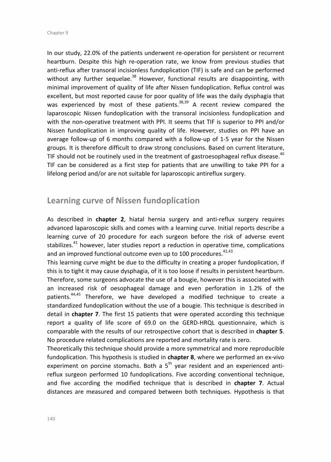
Chapter 9
140
In our study, 22.0% of the patients underwent re‐operation for persistent or recurrent
heartburn. Despite this high re‐operation rate, we know from previous studies that
anti‐reflux after transoral incisionless fundoplication (TIF) is safe and can be performed
without any further sequelae.38 However, functional results are disappointing, with
minimal improvement of quality of life after Nissen fundoplication. Reflux control was
excellent, but most reported cause for poor quality of life was the daily dysphagia that
was experienced by most of these patients.38,39 A recent review compared the
laparoscopic Nissen fundoplication with the transoral incisionless fundoplication and
with the non‐operative treatment with PPI. It seems that TIF is superior to PPI and/or
Nissen fundoplication in improving quality of life. However, studies on PPI have an
average follow‐up of 6 months compared with a follow‐up of 1‐5 year for the Nissen
groups. It is therefore difficult to draw strong conclusions. Based on current literature,
TIF should not be routinely used in the treatment of gastroesophageal reflux disease.40
TIF can be considered as a first step for patients that are unwilling to take PPI for a
lifelong period and/or are not suitable for laparoscopic antireflux surgery.
Learning curve of Nissen fundoplication
As described in chapter 2, hiatal hernia surgery and anti‐reflux surgery requires
advanced laparoscopic skills and comes with a learning curve. Initial reports describe a
learning curve of 20 procedure for each surgeon before the risk of adverse event
stabilizes.41 however, later studies report a reduction in operative time, complications
and an improved functional outcome even up to 100 procedures.42,43
This learning curve might be due to the difficulty in creating a proper fundoplication, if
this is to tight it may cause dysphagia, of it is too loose if results in persistent heartburn.
Therefore, some surgeons advocate the use of a bougie, however this is associated with
an increased risk of oesophageal damage and even perforation in 1.2% of the
patients.44,45 Therefore, we have developed a modified technique to create a
standardized fundoplication without the use of a bougie. This technique is described in
detail in chapter 7. The first 15 patients that were operated according this technique
report a quality of life score of 69.0 on the GERD‐HRQL questionnaire, which is
comparable with the results of our retrospective cohort that is described in chapter 5.
No procedure related complications are reported and mortality rate is zero.
Theoretically this technique should provide a more symmetrical and more reproducible
fundoplication. This hypothesis is studied in chapter 8, where we performed an ex‐vivo
experiment on porcine stomachs. Both a 5th year resident and an experienced anti‐
reflux surgeon performed 10 fundoplications. Five according conventional technique,
and five according the modified technique that is described in chapter 7. Actual
distances are measured and compared between both techniques. Hypothesis is that
General discussion and future perspectives
141
9
this technique leads to more symmetrical fundoplication. It is indeed found that the
modified technique leads to a larger degree of symmetry than the conventional
technique (P=0.047). Further research is needed to prove whether this technique also
reduces postoperative dysphagia and results in better clinical outcome. This technique
does increase the procedure time with a median of 8.9 minutes. However, this is only a
small part of the entire procedure and when more experience is gained, this time will
further decrease. This technique seems to help beginning surgeons to provide a
reproducible, proper fundoplication.
Future perspectives
As previously stated, there are many definitions for recurrence in hiatal hernia surgery.
These can be either anatomical or symptomatic, and the correlation between these is
very poor. As is seen in chapter 3, there was a large heterogeneity due to different
methods for determining quality of life. Since quality of life is an important outcome
measurement nowadays, there is need for a golden standard in reporting quality of life
after hiatal hernia repair and after anti‐reflux surgery. In chapter 4 and 5 we present
the results on long‐term follow‐up, which are good. However, high re‐operation rate is
reported. Also, minimal invasive techniques as described in chapter 6 result in high re‐
operation rate. This expresses the difficulty in anti‐reflux surgery and there is still need
for a long‐term solution for all patients. All treatment options (endoluminal pacing,
total fundoplication, partial fundoplication, transoral incisionless fundoplication) have
advantages and disadvantages. Probably not one technique fits for all patients and
preoperative investigations like upper‐GI endoscopy, manometry, 24‐hours impedance
and gastric emptying measurement, must help surgeons to determine the ideal
treatment for each individual patient. As mentioned in chapter 2 and also described in
chapter 7 and 8, the type of surgery that is described in this thesis is difficult and
requires advanced laparoscopic techniques. Since the learning curve is one predictor
for successful outcome more research is needed to reduce this learning curve. Maybe
with specialized training programs for beginning surgeons. Novel techniques such as 3D
view or robot surgery may provide additional value to anti‐reflux surgery and hiatal
hernia repair.
Chapter 9
142
References
1. Hochberg LA Thoracic surgery before the 20th century. New York: Vantage Press, 1960. 2. Stylopoulos N, Rattner DW The history of hiatal hernia surgery: from Bowditch to laparoscopy. Ann
Surg 2005:241:185‐93.
3. Diez Tabernilla M, Ruiz‐Tovar J, Grajal Marino R, Calero Garcia P, Pina Hernandez JD, et al. Paraesophageal hiatal hernia. Open vs. laparoscopic surgery. Rev Esp Enferm Dig 2009:101:706‐11.
4. Klinginsmith M, Jolley J, Lomelin D, Krause C, Heiden J, et al. Paraesophageal hernia repair in the
emergency setting: is laparoscopy with the addition of a fundoplication the new gold standard? Surg Endosc 2016:30:1790‐5.
5. Baigrie RJ, Watson DI, Game PA, Jamieson GG Vascular perils during laparoscopic dissection of the
oesophageal hiatus. Br J Surg 1997:84:556‐7. 6. Cano‐Valderrama O, Marinero A, Sanchez‐Pernaute A, Dominguez‐Serrano I, Perez‐Aguirre E, et al.
Aortic injury during laparoscopic esophageal hiatoplasty. Surg Endosc 2013:27:3000‐2.
7. Leggett PL, Bissell CD, Churchman‐Winn R Aortic injury during laparoscopic fundoplication: an underreported complication. Surg Endosc 2002:16:362.
8. Yano F, Omura N, Tsuboi K, Kashiwagi H, Yanaga K Thoracic aortic injury during laparoscopic
fundoplication for reflux esophagitis. Int J Surg 2008:6:490‐2. 9. Yang X, Hua R, He K, Shen Q, Yao Q Laparoscopic hernioplasty of hiatal hernia. Ann Transl Med
2016:4:343.
10. Hamrick MC, Davis SS, Chiruvella A, Coefield RL, Waring JP, et al. Incidence of delayed gastric emptying associated with revisional laparoscopic paraesophageal hernia repair. J Gastrointest Surg 2013:17:
213‐‐7.
11. van Rijn S, Roebroek YG, Conchillo JM, Bouvy ND, Masclee AA Effect of vagus nerve Injury on the outcome of antireflux surgery: an extensive literature review. Surgery 2016:33:230‐9.
12. van Rijn S, Rinsma NF, van Herwaarden‐Lindeboom MY, Ringers J, Gooszen HG, et al. Effect of Vagus
Nerve Integrity on Short and Long‐Term Efficacy of Antireflux Surgery. Am J Gastroenterol 2016:111:508‐15.
13. Antiporda M, Veenstra B, Jackson C, Kandel P, Daniel Smith C, et al. Laparoscopic repair of giant
paraesophageal hernia: are there factors associated with anatomic recurrence? Surg Endosc 2018:32:945‐54.
14. Antoniou SA, Koch OO, Antoniou GA, Pointner R, Granderath FA Mesh‐reinforced hiatal hernia repair: a
review on the effect on postoperative dysphagia and recurrence. Langenbecks Arch Surg. 2012;397(1):19‐27.
15. Puri V, Kakarlapudi GV, Awad ZT, Filipi CJ Hiatal hernia recurrence: 2004. Hernia 2004:8:311‐7.
16. Targarona EM, Bendahan G, Balague C, Garriga J, Trias M Mesh in the hiatus: a controversial issue. Arch Surg 2004:139:1286‐96.
17. Furnee E, Hazebroek E Mesh in laparoscopic large hiatal hernia repair: a systematic review of the
literature. Surg Endosc 2013:27:3998‐4008. 18. Iqbal A, Kakarlapudi GV, Awad ZT, Haynatzki G, Turaga KK, et al. Assessment of diaphragmatic stressors
as risk factors for symptomatic failure of laparoscopic nissen fundoplication. J Gastrointest Surg
2006:10:12‐21. 19. Watson DI, Thompson SK, Devitt PG, Smith L, Woods SD, et al. Laparoscopic repair of very large hiatus
hernia with sutures versus absorbable mesh versus nonabsorbable mesh: a randomized controlled trial.
Ann Surg 2015:261:282‐9. 20. Oelschlager BK, Pellegrini CA, Hunter J, Soper N, Brunt M, et al. Biologic prosthesis reduces recurrence
after laparoscopic paraesophageal hernia repair: a multicenter, prospective, randomized trial. Ann Surg
2006:244:481‐90. 21. Oelschlager BK, Pellegrini CA, Hunter JG, Brunt ML, Soper NJ, et al. Biologic prosthesis to prevent
recurrence after laparoscopic paraesophageal hernia repair: long‐term follow‐up from a multicenter,
prospective, randomized trial. J Am Coll Surg 2011:213:461‐8.
General discussion and future perspectives
143
9
22. Koetje JH, Oor JE, Roks DJ, Van Westreenen HL, Hazebroek EJ, et al. Equal patient satisfaction, quality of
life and objective recurrence rate after laparoscopic hiatal hernia repair with and without mesh. Surg Endosc 2017:31:3673‐80.
23. Tam V, Luketich JD, Levy RM, Christie NA, Awais O, et al. Mesh cruroplasty in laparoscopic repair of
paraesophageal hernias is not associated with better long‐term outcomes compared to primary repair. Am J Surg 2017:214:651‐6.
24. Mittal SK, Bikhchandani J, Gurney O, Yano F, Lee T Outcomes after repair of the intrathoracic stomach:
objective follow‐up of up to 5 years. Surg Endosc 2011:25:556‐66. 25. Swidnicka‐Siergiejko A, Dabrowski A Prolonged 2‐day esophageal pH‐metry with impedance monitoring
improves symptom‐reflux association analysis. Dig Dis Sci 2013:58:2556‐63.
26. Zhang C, Liu D, Li F, Watson DI, Gao X, et al. Systematic review and meta‐analysis of laparoscopic mesh versus suture repair of hiatus hernia: objective and subjective outcomes. Surg Endosc 2017:31:4913‐22.
27. Gee DW, Andreoli MT, Rattner DW Measuring the effectiveness of laparoscopic antireflux surgery:
long‐term results. Arch Surg 2008:143:482‐7. 28. Velanovich V, Vallance SR, Gusz JR, Tapia FV, Harkabus MA Quality of life scale for gastroesophageal
reflux disease. J Am Coll Surg 1996:183:217‐24.
29. Gunter RL, Shada AL, Funk LM, Wang X, Greenberg JA, et al. Long‐Term Quality of Life Outcomes Following Nissen Versus Toupet Fundoplication in Patients with Gastroesophageal Reflux Disease. J
Laparoendosc Adv Surg Tech A. 2017;27(9):931‐6.
30. Kelly JJ, Watson DI, Chin KF, Devitt PG, Game PA, et al. Laparoscopic Nissen fundoplication: clinical outcomes at 10 years. J Am Coll Surg 2007:205:570‐5.
31. Khaitan L, Bhatt P, Richards W, Houston H, Sharp K, et al. Comparison of patient satisfaction after redo
and primary fundoplications. Surg Endosc 2003:17:1042‐5. 32. Draaisma WA, Rijnhart‐de Jong HG, Broeders IA, Smout AJ, Furnee EJ, et al. Five‐year subjective and
objective results of laparoscopic and conventional Nissen fundoplication: a randomized trial. Ann Surg
2006:244:34‐41. 33. Carlson MA, Frantzides CT Complications and results of primary minimally invasive antireflux
procedures: a review of 10,735 reported cases. J Am Coll Surg 2001:193:428‐39.
34. Catarci M, Gentileschi P, Papi C, Carrara A, Marrese R, et al. Evidence‐based appraisal of antireflux fundoplication. Ann Surg 2004:239:325‐37.
35. Witteman BP, Conchillo JM, Rinsma NF, Betzel B, Peeters A, et al. Randomized controlled trial of
transoral incisionless fundoplication vs. proton pump inhibitors for treatment of gastroesophageal reflux disease. Am J Gastroenterol 2015:110:531‐42.
36. Testoni PA, Testoni S, Mazzoleni G, Vailati C, Passaretti S Long‐term efficacy of transoral incisionless
fundoplication with Esophyx (Tif 2.0) and factors affecting outcomes in GERD patients followed for up to 6 years: a prospective single‐center study. Surg Endosc 2015:29:2770‐80.
37. Oor JE, Roks DJ, Broeders JA, Hazebroek EJ, Gooszen HG Seventeen‐year Outcome of a Randomized
Clinical Trial Comparing Laparoscopic and Conventional Nissen Fundoplication: A Plea for Patient Counseling and Clarification. Ann Surg 2017:266:23‐8.
38. Witteman BP, Kessing BF, Snijders G, Koek GH, Conchillo JM, et al. Revisional laparoscopic antireflux
surgery after unsuccessful endoscopic fundoplication. Surg Endosc 2013:27:2231‐6. 39. Furnee EJ, Broeders JA, Draaisma WA, Schwartz MP, Hazebroek EJ, et al. Laparoscopic Nissen
fundoplication after failed EsophyX fundoplication. Br J Surg 2010:97:1051‐5.
40. Richter J, Kumar A, Lipka S, Miladinovic B, Velanovich V Efficacy of laparoscopic Nissen fundoplication vs transoral incisionless fundoplication or proton pump inhibitors in patients with gastroesophageal
reflux disease: a systematic review and network meta‐analysis. Gastroenterology 2018.
41. Watson DI, Baigrie RJ, Jamieson GG A learning curve for laparoscopic fundoplication. Definable, avoidable, or a waste of time? Ann Surg 1996:224:198‐203.
42. Soot SJ, Eshraghi N, Farahmand M, Sheppard BC, Deveney CW Transition from open to laparoscopic
fundoplication: the learning curve. Arch Surg 1999:134:278‐81. 43. Hwang H, Turner LJ, Blair NP Examining the learning curve of laparoscopic fundoplications at an urban
community hospital. Am J Surg 2005:189:522‐6.

Chapter 9
144
44. Patterson EJ, Herron DM, Hansen PD, Ramzi N, Standage BA, et al. Effect of an esophageal bougie on
the incidence of dysphagia following nissen fundoplication: a prospective, blinded, randomized clinical trial. Arch Surg 2000:135:1055‐61.
45. Lowham AS, Filipi CJ, Hinder RA, Swanstrom LL, Stalter K, et al. Mechanisms and avoidance of
esophageal perforation by anesthesia personnel during laparoscopic foregut surgery. Surg Endosc 1996:10:979‐82.

Summary

146
Summary
147
S
Summary
This thesis addresses the long‐term outcome after several treatment options for both
hiatal hernia repair and gastroesophageal reflux disease. (GERD) We have developed a
modified technique to create a standardized Nissen fundoplication without rotation to
aid beginning surgeons to perform a proper fundoplication.
In chapter 2 we have addressed several aspects of the treatment of intrathoracic
stomach. Laparoscopic approach is gold standard. The hiatal hernia should be reduced
and the crural defect should be repaired with suture repair. Mesh reinforcement seems
to reduce recurrence rate, however comes with a small risk of complications. Additional
fundoplication is not advised, except when severe heartburn complaints are present.
When intrathoracic stomach becomes incarcerated, first step should be
decompression, followed by semi‐elective repair.
Chapter 3 is a collective review comparing synthetic non‐absorbable mesh with
biological synthetic meshes. All studies between 2004 and 2015 are analysed and a
total of 16 studies were included. Median follow‐up was 53.4 months with a recurrence
rate of 6.8% for synthetic mesh compared with 16.1% for biological mesh. Complication
rate, mortality and symptomatic outcome was comparable. Due to higher costs of
biological meshes, the synthetic mesh might be advocated.
In Chapter 4 we retrospectively evaluated the results of all patients that underwent
laparoscopic hiatal hernia repair for intrathoracic stomach with a follow‐up of up to
9.6 years. 81/85 patients completed the follow‐up and reported good quality of life of
13.5. (maximum score 16) Re‐operation rate was 9.9% with all re‐operations in the non‐
mesh group.
Chapter 5 focusses on the long‐term quality of life after Nissen fundoplication for
patients with gastroesophageal reflux disease. 175 patients completed the GERD‐HRQL‐
questionnaire with an excellent median quality of life of 70.0 (maximum score 75)
Quality of life remained stable after 5 years and 91.3% of all re‐operations occurred
within the first 5 years. Patients that underwent re‐operation experienced a reduced
quality of life.
Chapter 6 describes the 7‐year follow‐up of a randomized controlled trial with cross‐
over design between transoral incisionless fundoplication (TIF) versus proton pump
inhibitors for GERD. Quality of life remained stable after 1 year, however resumption to
PPI increased from 34.1% to 56.1%. 22% of the patients underwent re‐operation for
persistent and/or recurrent complaints. We could not detect pre‐operative or

148
procedure related predictors for successful outcome at the long‐term. TIF should not be
used in the standard treatment of GERD.
In chapter 7 we described a modification of the conventional Nissen fundoplication. We
attempt to perform a standardized fundoplication with sufficient patency and less
rotation. This study describes the 1‐year results of the first 15 patients. Median quality
of life was 69.0 on the GERD‐HRQL questionnaire, which is excellent. No complications
or reoperations are reported. This technique is safe and leads to good postoperative
results at the short term.
Chapter 8 is an experimental study with an ex‐vivo porcine model. We evaluated the
standardization and reproducibility of the modified technique described in chapter 7.
The modified technique leads to a more symmetrical fundoplication (P=0.047), but
increases the operative time from 5.9 to 14.8 minutes. No difference is found between
the resident and the experienced surgeon regarding total diameter and degree of
symmetry for this modified technique.

Samenvatting

150
Samenvatting
151
S
Samenvatting
Dit proefschrift beschrijft de lange termijn resultaten van verschillende behandelopties
voor de behandeling van zowel een middenrif breuk als van gastro‐oesofageale reflux
ziekte. (GORZ) We hebben een aangepaste techniek ontwikkeld om een
gestandaardiseerde Nissen fundoplicatie te maken zonder rotatie. Het doel hiervan is
om beginnende chirurgen te helpen bij het creëren van een correcte fundoplicatie.
In hoofdstuk 2 hebben we verschillende aspecten beschreven voor de behandeling van
een intrathoracale maag. (ITM) Laparoscopie is de gouden standaard. De breuk wordt
gereduceerd en hiatale defect wordt hersteld middels hechtingen. Versteviging van het
herstel met een mat verlaagt het recidief risico, echter geeft een klein risico op
complicaties. Het verrichten van een additionele fundoplicatie wordt niet geadviseerd,
behalve wanneer iemand ernstig zuurbranden heeft. Als de intrathoracale maag
beklemd raakt, is de eerste stap decompressie gevolgd door een semi‐electief herstel.
Hoofdstuk 3 is een collectieve review die synthetische niet‐oplosbare matten vergelijkt
met biologisch oplosbare matten. Alle studies tussen 2004 en 2015 zijn bekeken en
uiteindelijk zijn er 16 geïncludeerd. Mediane follow‐up was 53.4 maanden met een
recidief percentage van 6.8% voor synthetische matten vergeleken met 16.1% voor de
biologische matten. Complicaties, mortaliteit en symptomatische uitkomst zijn
vergelijkbaar. Gezien de hogere kosten van biologische matten, zou de eerste keuze
een synthetische mat zijn.
In hoofdstuk 4 hebben we retrospectief de resultaten geëvalueerd van alle patiënten
die een laparoscopisch herstel van een intrathoracale maag hebben ondergaan met een
follow‐up tot 9.6 jaar. 81/85 patiënten hebben de follow‐up voltooid en rapporteren
een goede kwaliteit van leven van 13.5 (maximale score 16) Re‐operatie percentage
was 9.9% waarbij alle re‐operaties plaatsvonden in de groep waarbij geen mat is
gebruikt.
Hoofdstuk 5 focust op de lange‐termijn kwaliteit van leven na Nissen fundoplicatie voor
patiënten met GORZ. 175 patiënten hebben de GERD‐HRQL‐vragenlijst ingevuld en
rapporteren een zeer goede kwaliteit van leven van 70.0 (maximale score 75) Kwaliteit
van leven bleef stabiel na 5 jaar en 91.3% van alle recidieven vond plaats in de eerste
5 jaar. Patiënten die een re‐operatie ondergingen rapporteren en verminder kwaliteit
van leven.
Hoofdstuk 6 beschrijft de 7‐jaars resultaten van een gerandomiseerde trial met cross‐
over ontwerp tussen endoscopische incisie loze fundoplicatie (TIF) versus protonpomp
remmers (PPI) voor GORZ. Kwaliteit van leven blijft stabiel na 1 jaar, maar het aantal
152
mensen dat PPI’s gebruikt is toegenomen van 34.1% tot 56.1%. 22% van de patiënten
hebben een re‐operatie ondergaan vanwege blijvende of recidiverende klachten. We
hebben geen variabelen kunnen detecteren om succesvolle resultaten na 7 jaar te
kunnen voorspellen. TIF moet nog niet standaard toegepast worden in de behandeling
van GORZ.
In hoofdstuk 7 beschrijven we een aangepast techniek voor de Nissen fundoplicatie we
hebben geprobeerd om een gestandaardiseerde fundoplicatie te creëren met
voldoende doorgang en minder rotatie. Deze studie rapporteert de 1‐jaars resultaten
van de eerste 15 patiënten. Mediane kwaliteit van leven is 69.0 op de GERD‐HRQL‐
vragenlijst, wat zeer goed is. Geen complicaties of re‐operaties zijn gerapporteerd. Deze
techniek is veilig en leidt tot goede postoperatieve resultaten op de korte termijn.
Hoofdstuk 8 is een experimentele studie met een ex‐vivo model met varkensmagen.
We hebben de standaardisatie en reproduceerbaarheid van de techniek uit hoofdstuk 7
bestudeerd. De aangepaste techniek resulteert in een meer symmetrische fundoplicatie
(P=0.047), maar geeft een toename van operatieduur van 5.9 tot 14.8 minuten. Er is
geen verschil gevonden tussen de assistent en de ervaren chirurg met betrekking tot de
diameter en de mate van symmetrie in de aangepast techniek.

Valorisation addendum

154
Valorisation addendum
155
V
Valorisation addendum
Introduction
Gastroesophageal reflux disease (GERD)has an estimated prevalence of 18.1‐27.8% in
North America. GERD is currently the most common reason for outpatient
gastrointestinal visits in the United States (US), with an estimated 8.9 million outpatient
visits. With increasing number of patients, GERD has become a serious burden for the
general population. GERD reduces quality of life in patients and comes with high costs
for healthcare. Initial treatment is with life‐style interventions, but next step is medical
therapy. Medical therapy for GERD costs approximately 7.7 billion dollars annually in
the US.
Relevance of this thesis
If left untreated GERD may lead to severe complications including erosive oesophagitis,
pulmonary complaints and even Barret’s dysplasia. First step in the treatment of GERD
is medical treatment with PPI’s, however long‐term PPI usage can lead to an increased
risk of pneumonia, dementia, Vit B12 deficiency and osteoporosis. Surgery is found to
be more cost‐effective at the long‐term for the treatment of GERD, however comes
with the risk of complications. Dysphagia, recurrence and vagal nerve injury are some
of the most feared complications. To reduce complications and recurrence rates,
several modifications have been developed. Total fundoplication, partial
fundoplication, transoral incisionless fundoplication(TIF), lower oesophageal
stimulation and even oesophageal lengthening procedures are available. Chapter 2 and
3 provide an overview of current techniques of hiatal hernia repair and the treatment
of hiatal hernia with special emphasis on the use of mesh. We have shown that
synthetic mesh might be superior to biological mesh in hiatal hernia repair.
All techniques have advantages and disadvantages and for many techniques the long‐
term effects are still lacking. This thesis therefore addresses long‐term outcome of
several techniques to provide more inside in the effects of these techniques.
Since quality of life has become a more important outcome measurement over time,
we have tried to establish the quality of life after hiatal hernia repair and after Nissen
fundoplication for GERD. Chapter 4 demonstrates that quality of life is good after a
follow‐up of up to 9.6 years and re‐operation rate is 9.9%. Most re‐operations occur
within the first year after surgery and quality of life does not further change after 5
years of follow‐up, as is seen in chapter 5. Therefore, a follow‐up period of 5 years
should be sufficient to evaluate quality of life after antireflux surgery. Chapter 5 also
addresses quality of life after antireflux surgery for GERD and shows an excellent
quality of life at long‐term follow‐up. Patient who underwent re‐operation reported
worse quality of life, therefore the re‐operation rate should be as low as possible.
156
We have found that 48% of the re‐operations is due to persistent dysphagia after
Nissen fundoplication. We believe that this might be due to failure of creating a proper
fundoplication. It is difficult to create a symmetrical wrap that is neither too tight, nor
too loose. In an attempt to help, especially beginning surgeons, we have developed a
modified technique to create a more symmetrical and more reproducible
fundoplication. This technique is described in chapter 7 and further evaluated in
chapter 8. Results are promising, but further research is needed to evaluate the
possible clinical benefit.
Target population
The results of this thesis are relevant for patients with a hiatal hernia or with
gastroesophageal reflux disease who are not satisfied with their medical therapy.
Further, it offers a modified technique for beginning surgeons to create a proper Nissen
fundoplication.
Innovation and future
This thesis evaluated existing treatment options for hiatal hernia repair and anti‐reflux
surgery. However, we also studied a more experimental and less invasive technique.
The EsophyX device creates a transoral incisionless fundoplication. The results after 1
year were rather disappointing with a high recurrence rate. (22.7%‐50.0%) However,
we have studied the long‐term results in chapter 6, in attempt to detect a subgroup
that benefits from TIF procedure. We found that patients with a hiatal hernia or
oesophagitis reported a worse quality of life after 7 years. We found a failure rate of
22%. Since additional surgery after previous TIF does not come with increase morbidity
and mortality TIF can be a first step in the treatment of GERD. With this thesis, we hope
to provide a tailor‐made approach to all future GERD patients.

Dankwoord

158
Dankwoord
159
D
Dankwoord
Na 5 jaar hard werken aan mijn promotietraject naast mijn klinische werkzaamheden, is
het uiteindelijk toch gelukt. Het proefschrift is af, maar niet zonder de hulp van een
heel team. Graag wil ik iedereen bedanken die me geholpen en gesteund heeft de
afgelopen jaren, maar een aantal mensen in het bijzonder. Uiteraard is dit in de eerste
plaats mijn promotor, maar ik wil graag voor een persoon een uitzondering maken, die
voor mij een essentiële rol heeft gespeeld in mijn ontwikkeling als arts en mijn
enthousiasme voor de chirurgie heeft weten te vergroten.
Beste drs. Smulders, Frans, het zal je misschien verbazen, maar jij bent toch echt de
eerste die ik wil bedanken! Dankzij jou ben ik tijdens mijn coschappen al in aanraking
gekomen met het wetenschappelijk onderzoek naar de anti‐reflux chirurgie en je hebt
me vanaf toen gesteund en sturing gegeven. Niet alleen tijdens mijn promotie, maar
ook in de kliniek ben je een mentor voor me geweest. Je hebt me geïnspireerd en
enthousiast gemaakt voor het vak en alles daaromheen. We zijn beiden geen
onderzoeker pur sang, maar we hebben het toch mooi voor elkaar gekregen, ‘de S is
weggepoetst’. Ik heb veel van je geleerd en je zal altijd een voorbeeld voor me blijven.
Dankjewel voor alles!
Prof. dr. Bouvy, beste Nicole, op de (bijna) eerste plaats wil ik uiteraard jou bedanken!
Pas later tijdens mijn promotietraject ben je als promotor onderdeel geworden van het
team, maar wat was je belangrijk. Je weet het misschien niet meer, maar in het 3e jaar
van geneeskunde heb ik mijn mondelinge examen bij jou gedaan en al vrij snel was je
tevreden en was je reactie: ‘volgens mij heb jij het wel begrepen’. Dit vertrouwen in mij
heb je niet verloren in de daaropvolgende jaren en je hebt me gestimuleerd om in rap
tempo door te werken aan mijn promotie. Mede dankzij jouw innovatieve blik hebben
we mijn proefschrift nog kunnen uitbreiden met een mooie vervolgstudie naar de
EsophyX. Heel erg bedankt voor je hulp en begeleiding, zowel bij mijn proefschrift als
bij het bemachtigen van een opleidingsplek. Zonder jouw was dit beiden niet gelukt!
Dr. v.d. Poll, beste Marcel, mijn co‐promotor. Bedankt voor je kritische blik op mijn
artikelen en je bijdrage aan dit prachtige boekwerk. Bedankt voor de avonden die we
bij je thuis hebben gewerkt, gediscussieerd en gelachen. Je hebt mijn proefschrift naar
een wetenschappelijk hoger niveau weten te tillen en je bent onmisbaar geweest in de
totstandkoming van dit werk.
Beste Jeroen, dr(s). Ponten, vriend en paranimf. Waar moet ik beginnen met mijn
dankwoord voor jou. Mede dankzij jou ben ik enthousiast geworden voor de chirurgie
en tja, daar hoort de wetenschap nou eenmaal bij….. Maar we hebben er samen het
beste van gemaakt, ik heb een tijdje je RCT mogen waarnemen in de periode dat jij in
160
het MUMC+ zat. Daarnaast hebben we onze kennis mogen uitbreiden op de diverse
congressen, waar we ook de nodige ontspanning wisten te vinden, in het bijzonder in
Milaan. Heel erg bedankt voor je hulp bij de totstandkoming van dit proefschrift, je
begeleiding in de kliniek en mede het vele plezier dat we samen hebben gehad en
hopelijk nog gaan beleven.
Graag zou ik de leden van de beoordelingscommissie, voorzitter prof. dr. L.P.S. Stassen,
dr. E.J. Hazebroek, prof. dr. A.A.M. Masclee, prof. dr. H.J.T. Rutten, dr. J.H.M.B. Stoot
willen bedanken voor de tijd en moeite die jullie hebben genomen voor de beoordeling
van mijn proefschrift. Ik kijk uit naar de verdediging van dit proefschrift om met jullie te
discussiëren over dit onderwerp.
Dr. Nienhuijs, Simon, onder jouw supervisie ben ik gestart met mijn wetenschappelijke
en klinische carrière. Je hebt me als jonge student begeleid en gesteund. Dankjewel
hiervoor en mede dankzij jou is het gelukt om aangenomen te worden voor de
opleiding tot chirurg.
Onderzoekers uit de ‘kelder’, Yannick, Pieter, Thijs, Geert, Lindy, David, Martin, Marijn
en Dennis, dank jullie wel voor de gezelligheid en de gastvrijheid in jullie hok. Ook al
was ik geen fulltime onderzoeker, ik kon altijd bij jullie terecht voor een werkplek, een
koffietje, wat gezelligheid en ook als ik hulp nodig had bij mijn onderzoek kon ik jullie
om advies vragen! Jullie zijn een inspiratie en voorbeeld voor me geweest tijdens mijn
promotietraject.
Arts‐assistenten van de chirurgie en intensive care in het Catharina ziekenhuis
Eindhoven, met heel veel plezier heb ik gewerkt bij jullie in de kliniek en mede dankzij
jullie heb ik de energie gehad om kliniek en wetenschap te combineren. De borrels en
feestjes na het werk hebben gezorgd voor de nodige ontspanning en zorgde ervoor dat
we een hechte groep werden die voor elkaar klaar stond. Dank jullie wel en hopelijk tot
snel weer in de kliniek!
Arts‐assistenten van de chirurgie in het MUMC+, huidige collega’s, dank jullie wel voor
de gezelligheid op het werk. Ik kwam als vreemde eend in de bijt, na 5 jaar in het
Catharina, als ‘Eindhoven boy’ bij jullie binnen. Desondanks werd ik met open armen
ontvangen en heb ik het erg naar mijn zin en is het gelukt om mijn proefschrift bij jullie
af te ronden.
Niet te vergeten, mijn andere paranimf. Skip, goeie vriend, boef. Ik leerde je inmiddels
alweer 11 jaar geleden kennen als collega in de horeca. Samen met Noudje zijn we als
de Freak’a Dellen al vele borrels, avonden in ‘de minibar’ en later in ‘the little one’,
etentjes, 2 verhuizingen en een toppersconcert verder. De laatste tijd zijn we allemaal

Dankwoord
161
D
erg druk met ons werk waardoor we elkaar wat minder zien, maar als we elkaar zien
blijft het gezellig als vanouds! Een hele eer dat je mijn paranimf wilt zijn, al heb ik je
eerste even uit moeten leggen wat dit dan ook wel precies betekend. En nee, het is
geen prada‐milf. Bedankt voor alles van de afgelopen jaren en dat we samen nog maar
veel plezier mogen maken in het leven!
Mijn grote passie is skydiven en hier haal ik dan ook de nodige ontspanning uit.
Iedereen die met mij gesprongen heeft afgelopen jaren wil ik bedanken voor de
gezelligheid, ontspanning en gave sprongen die we samen gemaakt hebben! In het
bijzonder Floortje, Tom en Jan. Met jullie heb ik toch echt wel de meeste tijd in de lucht
en daarna ‘op de grond’ doorgebracht. Ik kijk er enorm naar uit om met jullie komend
jaar weer op skydive reis te gaan, laten we er weer een mooi avontuur van maken! En
ik zou zeggen, laten we er snel nog maar ‘eentje’ op drinken
Lieve Nick, broertje. Wat ben ik trots op je, met succes een eigen hoveniersbedrijf
gestart, wat kan jij werken, ongekend. Je gedrevenheid is een voorbeeld en een bron
van inspiratie voor me. Je bent altijd geïnteresseerd in mijn werk en staat altijd voor me
klaar. Ik kan altijd op je rekenen! Dankjewel voor alles en ik wens jou alle succes en
geluk.
Lieve papa en mama, bedankt dat jullie altijd voor me klaar hebben gestaan. Jullie
hebben me altijd onvoorwaardelijk gesteund en zonder jullie hulp was ik nooit
gekomen waar ik nu ben. Jullie zijn altijd betrokken en geïnteresseerd geweest in mijn
promotietraject en hebben mij altijd gestimuleerd om door te zetten en te blijven
werken voor een opleidingsplek binnen de chirurgie. Jullie zijn de beste ouders die
iemand zich kan wensen en hopelijk mag ik nog lang van jullie liefde genieten! Dank
jullie wel.
Tot slot wil ik graag nog mijn meisje bedanken, de liefde van mijn leven. Lieve Carmen,
je hebt me enorm gesteund en dankjewel dat je mij al die avonden hebt laten werken.
Het is nu eindelijk klaar, zodat we meer tijd met elkaar kunnen doorbrengen. Ik hoop
dat we samen oud worden en nog heel veel mooie dingen gaan meemaken in ons
leven! Dikke kus, ik hou van je.

162

List of publications

164

List of publications
165
P
List of publications
1. Castelijns PSS, Woensdregt K, Hoevenaars B, Nieuwenhuijzen GAP. Intra‐
abdominal esophageal duplication cyst: A case report and review of the literature.
World J Gastrointest Surg. 2014;6(6):112‐6.
2. Castelijns PSS, Boesten RHM, van der Veen AH. Case report, Het Pellegrini‐
Stiedafenomeen: casus en overzicht van de literatuur. Ned. Tijdschrift voor
traumachirurgie. 2015; (4):75‐7.
3. Castelijns PSS, Ponten JEH, Van de Poll MCG, Nienhuijs SW, Smulders JF. Subjective
outcome after laparoscopic hiatal hernia repair for intrathoracic stomach.
Langenbecks Arch Surg. 2017 May;402(3):521‐30.
4. Castelijns PSS, Ponten JEH, Bouvy ND, Smulders JF, van de Poll MCG. Intrathoracic
stomach in hiatal hernia: th.e role of laparoscopic repair. Minerva Chir.
2018;73(1):64‐76.
5. Castelijns PSS, Ponten JEH, van de Poll MCG, Nienhuijs SW, Smulders JF. A
collective review of biological versus synthetic mesh‐reinforced cruroplasty during
laparoscopic Nissen fundoplication. J Minim Access Surg. 2018;14(2):87‐94.
6. Ponten JEH, Leenders BJM, Leclercq WKG, Lettinga T, Heemskerk J, Konsten JLM,
Castelijns PSS, Nienhuijs SW. Mesh Versus Patch Repair for Epigastric and Umbilical
Hernia (MORPHEUS Trial); One‐Year Results of a Randomized Controlled Trial.
World J Surg. 2018;42(5):1312‐20.
7. Castelijns PSS, Ponten JEH, Vd Poll MCG, Bouvy ND, Smulders JF. Quality of life
after Nissen fundoplication in patients with gastroesophageal reflux disease:
Comparison between long‐ and short‐term follow‐up. J Minim Access Surg. 2018;
14(3):213‐20.
8. Castelijns PSS, van de Poll MCG, Smulders JF. A Modified Technique to Create a
Standardized Floppy Nissen Fundoplication Without a Bougie. J Laparoendosc Adv
Surg Tech A. 2018;28(7):853‐8.
9. Castelijns PSS, Ponten JEH, Van de Poll MCG, de Greef BTA, Bouvy ND, Smulders JF,
Standardized method for Nissen fundoplication: reproducibility and effect on
learning curve. Submitted.

166
10. Castelijns PSS, de Greef BTA, Peeters A, vd Poll MCG, MD, Witteman BPL, Koek GH,
Conchillo JM, Bouvy ND, 7‐year follow up after transoral incisionless fundoplication
for treatment of gastroesophageal reflux disease; results of patients who
participated in a randomized controlled trial. Submitted

Curriculum vitae

168
Curriculum vitae
169
CV
Curriculum vitae
Petrus Sebastianus Simon Castelijns was born on June 4th, 1991, in Geldrop, the
Netherlands. In 2009, he graduated from the Strabrecht College in Geldrop.
Immediately thereafter he started medical school at the Faculty of Medicine at the
Maastricht University. During the 6th year of this study he started with research under
the supervision of J.F. Smulders. After his medical training was completed, he started as
a resident (ANIOS) at the surgery department of the Catharina Hospital in Eindhoven.
(2015‐2016) He combined clinical activities with research and started an official PhD
project under supervision of Prof. dr. N.D. Bouvy. The main goal of this PhD project was
to evaluate quality of life after anti‐reflux surgery and hiatal hernia repair. Thereafter
he worked as a resident (ANIOS) at the intensive care of the same hospital. (2016‐2017)
From 2017 he started working at the surgery department of the Maastricht University
Medical Centre and started his surgical training program in June 2018 under the
supervision of L.P.S. Stassen and R.M.H. Roumen.

170