Out of the twilight zone: phylogeny and evolutionary
260
Out of the twilight zone: phylogeny and evolutionary morphology of the orb-weaving spider family Mysmenidae, with a focus on spinneret spigot morphology in symphytognathoids (Araneae, Araneoidea) LARA LOPARDO* and GUSTAVO HORMIGA Department of Biological Sciences, The George Washington University, 2023 G Street NW, Washington DC, WA 20052, USA Received 21 March 2014; revised 23 July 2014; accepted for publication 28 July 2014 This paper provides the first comprehensive comparative morphological study of symphytognathoid spiders, with an emphasis on the family Mysmenidae. Hypotheses of primary homology, particularly at the level of male geni- talia, are proposed for a total of 65 taxa (42 mysmenids), compiled into a morphological data set of more than 350 characters. Male palpal structures (paracymbium and tegular conductor), considered absent for the family by previous workers, are actually present in Mysmenidae. The pattern of interfamilial relationships based on the morphological data differs from the hypothesis based on the total evidence (morphology plus multigene sequence data) in the placement of Theridiosomatidae. We have based all formal taxonomic and nomenclatural decisions on the results of analysis of the total evidence from a previous study, except in the cases in which only morpho- logical information was available. Based on such phylogenetic results, the following generic transfers from Mysmenidae are proposed: Crassignatha, Iardinis (to Symphytognathidae); Leviola (to Zodariidae); and Phricotelus (Araneoidea incertae sedis). Mysmenidae is redelimited to include at least eight genera: Mysmena, Microdipoena, Maymena, Trogloneta, Isela, Mysmenopsis, Brasilionata, and Mysmeniola, which are re-diagnosed. Mysmenella and Anjouanella are synonymized with Microdipoena. Calodipoena, Itapua, Calomyspoena, Tamasesia, and Kekenboschiella are synonymized with Mysmena. Two mysmenid subfamilies are here proposed: Mysmenopsinae subf. nov. and Mysmeninae. In addition, diagnostic features for all symphytognathoid families are provided. One significant outcome of this comparative review is the entelegyne internal genitalic conformation for the family Anapidae (as opposed to haplogyne): all anapid representatives examined possess fertilization ducts. We provide some comments on the evolution of the morphology of spinneret spigots in symphytognathoids. © 2015 The Linnean Society of London, Zoological Journal of the Linnean Society, 2015, 173, 527–786. doi: 10.1111/zoj.12199 ADDITIONAL KEYWORDS: Anapidae – cladistics – comparative morphology – Micropholcommatinae – Mysmeninae – Mysmenopsinae – Symphytognathidae – Synaphridae – taxonomy – Theridiosomatidae. The group [Mysmenidae] lies in a twilight zone between fami- lies and presents such diluted morphological characters that placement and relationships become uncertain. Willis J. Gertsch (1960a: 1) INTRODUCTION Orbiculariae, the orb-weaving spiders (Araneoidea, Deinopoidea, and Nicodamidae), include 21 families and approximately one-quarter of the described species in the order Araneae. The exact limits and the familial composition of Orbiculariae have been the focus of nu- merous studies and have often been intensively debated (Griswold et al., 1998, 2005; Schütt, 2000; Rix, Harvey & Roberts, 2008; Blackledge et al., 2009; Miller et al., *Corresponding author. Current address: Zoologisches Institut und Museum, Ernst-Moritz-Arndt-Universität, J.-S.-Bach-Str. 11/12, D-17489 Greifswald, Germany. E-mail: [email protected] Zoological Journal of the Linnean Society, 2015, 173, 527–786. With 161 figures © 2015 The Linnean Society of London, Zoological Journal of the Linnean Society, 2015, 173, 527–786 527 Downloaded from https://academic.oup.com/zoolinnean/article/173/3/527/2449763 by guest on 22 July 2022
-
Upload
khangminh22 -
Category
Documents
-
view
3 -
download
0
Transcript of Out of the twilight zone: phylogeny and evolutionary

Out of the twilight zone: phylogeny and evolutionarymorphology of the orb-weaving spider familyMysmenidae, with a focus on spinneret spigotmorphology in symphytognathoids(Araneae, Araneoidea)
LARA LOPARDO* and GUSTAVO HORMIGA
Department of Biological Sciences, The George Washington University, 2023 G Street NW, WashingtonDC, WA 20052, USA
Received 21 March 2014; revised 23 July 2014; accepted for publication 28 July 2014
This paper provides the first comprehensive comparative morphological study of symphytognathoid spiders, withan emphasis on the family Mysmenidae. Hypotheses of primary homology, particularly at the level of male geni-talia, are proposed for a total of 65 taxa (42 mysmenids), compiled into a morphological data set of more than350 characters. Male palpal structures (paracymbium and tegular conductor), considered absent for the family byprevious workers, are actually present in Mysmenidae. The pattern of interfamilial relationships based on themorphological data differs from the hypothesis based on the total evidence (morphology plus multigene sequencedata) in the placement of Theridiosomatidae. We have based all formal taxonomic and nomenclatural decisionson the results of analysis of the total evidence from a previous study, except in the cases in which only morpho-logical information was available. Based on such phylogenetic results, the following generic transfers from Mysmenidaeare proposed: Crassignatha, Iardinis (to Symphytognathidae); Leviola (to Zodariidae); and Phricotelus (Araneoideaincertae sedis). Mysmenidae is redelimited to include at least eight genera: Mysmena, Microdipoena, Maymena,Trogloneta, Isela, Mysmenopsis, Brasilionata, and Mysmeniola, which are re-diagnosed. Mysmenella and Anjouanellaare synonymized with Microdipoena. Calodipoena, Itapua, Calomyspoena, Tamasesia, and Kekenboschiella aresynonymized with Mysmena. Two mysmenid subfamilies are here proposed: Mysmenopsinae subf. nov. andMysmeninae. In addition, diagnostic features for all symphytognathoid families are provided. One significant outcomeof this comparative review is the entelegyne internal genitalic conformation for the family Anapidae (as opposedto haplogyne): all anapid representatives examined possess fertilization ducts. We provide some comments on theevolution of the morphology of spinneret spigots in symphytognathoids.
© 2015 The Linnean Society of London, Zoological Journal of the Linnean Society, 2015, 173, 527–786.doi: 10.1111/zoj.12199
ADDITIONAL KEYWORDS: Anapidae – cladistics – comparative morphology – Micropholcommatinae –Mysmeninae – Mysmenopsinae – Symphytognathidae – Synaphridae – taxonomy – Theridiosomatidae.
The group [Mysmenidae] lies in a twilight zone between fami-lies and presents such diluted morphological characters thatplacement and relationships become uncertain.Willis J. Gertsch (1960a: 1)
INTRODUCTION
Orbiculariae, the orb-weaving spiders (Araneoidea,Deinopoidea, and Nicodamidae), include 21 families andapproximately one-quarter of the described species inthe order Araneae. The exact limits and the familialcomposition of Orbiculariae have been the focus of nu-merous studies and have often been intensively debated(Griswold et al., 1998, 2005; Schütt, 2000; Rix, Harvey& Roberts, 2008; Blackledge et al., 2009; Miller et al.,
*Corresponding author. Current address: ZoologischesInstitut und Museum, Ernst-Moritz-Arndt-Universität,J.-S.-Bach-Str. 11/12, D-17489 Greifswald, Germany.E-mail: [email protected]
bs_bs_banner
Zoological Journal of the Linnean Society, 2015, 173, 527–786. With 161 figures
© 2015 The Linnean Society of London, Zoological Journal of the Linnean Society, 2015, 173, 527–786 527
Dow
nloaded from https://academ
ic.oup.com/zoolinnean/article/173/3/527/2449763 by guest on 22 July 2022

2010; Hormiga & Griswold, 2014). Although the mostexhaustive (in terms of taxon sampling) molecular analy-sis of Orbiculariae published to date (Dimitrov et al.,2012) has recovered the monophyly of orbicularians,taxonomically expanded versions of these data (Dimitrovet al., 2013) refute orbicularian monophyly. Our morerecent work (Fernández, Hormiga & Giribet, 2014) usinga transcriptomic approach (2637 genes and 791 793amino acids) has also revealed the non-monophyly ofOrbiculariae (for a similar study using 327 genes, seealso Bond et al. 2014). The phylogenomic data placethe Deinopoidea (the cribellate orb weavers) with othergroups and not with Araneoidea (the ecribellate orbweavers), implying either independent origins of bothtypes of orb webs or a much more ancient origin ofthe orb with subsequent losses in lineages such as theRTA clade. Thus, as Hormiga & Griswold (2014) hadanticipated in light of Dimitrov et al.’s (2013) find-ings, ‘the evolution of the whole RTA clade from anorbicularian ancestor is thus conceivable’. These resultsclearly require a major reevaluation of our currentunderstanding of the spider evolutionary chronicle.
Nonetheless, the monophyly of Araneoidea (theecribellate orb weavers) is well supported by both mor-phological and molecular data (Fernández et al., 2014;Hormiga & Griswold, 2014).
Mysmenidae, a small family of minute araneoids (23genera, 131 species; Platnick, 2014) (see Table 1 here-after for authorship of taxa), are one of the least studiedfamily-level groups of orb weavers, mainly because oftheir small size (0.7–3 mm) and cryptic lifestyle. Untilrecently (Lopardo, Giribet & Hormiga, 2011), no modernphylogenetic research has been performed in thisaraneoid group, and its monophyly has never been ro-bustly established (but see below). Mysmenids belongto the so-called ‘symphytognathoid’ clade: minute orbweavers that build highly modified orb webs. Thisclade was originally delimited to include the familiesAnapidae, Mysmenidae, Symphytognathidae, andTheridiosomatidae (Fig. 150A; as delimited byGriswold et al. 1998; see also Coddington (1990). Thecomposition and the familial relationships withinsymphytognathoids, as well as the relationships of thewhole Araneoidea, have been recently challenged and
Figure 1. Isela okuncana (Mysmenidae), male left palp: A, D, E, ventral view; B, C, dorsal view; C, D, detail of tip ofpalp; E, detail of cymbial process. See Appendix 3 for the list of abbreviations.
528 L. LOPARDO AND G. HORMIGA
© 2015 The Linnean Society of London, Zoological Journal of the Linnean Society, 2015, 173, 527–786
Dow
nloaded from https://academ
ic.oup.com/zoolinnean/article/173/3/527/2449763 by guest on 22 July 2022

Table 1. Author names and list of taxa referred to in the text, matrix, and figures
Taxa Author and yearFamilyplacement Observations
Anapidae Simon, 1895(a)Mysmenidae Petrunkevitch, 1928Symphytognathidae Hickman, 1931Synaphridae Wunderlich, 1986Theridiosomatidae Simon, 1881Acrobleps Hickman, 1979 AnapidaeAcrobleps hygrophilus Hickman, 1979 AnapidaeAnapisona Gertsch, 1941 AnapidaeAnapisona kethleyi Platnick & Shadab, 1979 AnapidaeComaroma simoni Bertkau, 1889 AnapidaeCrassanapis Platnick & Forster, 1989 AnapidaeCrassanapis chilensis Platnick & Forster, 1989 AnapidaeElanapis Platnick & Forster, 1989 AnapidaeElanapis aisen Platnick & Forster, 1989 AnapidaeEpecthina Simon, 1895(a) Anapidae Synonym of AnapisEpecthinula Simon, 1903 Anapidae Synonym of AnapisMicropholcommatinae Hickman, 1944 Anapidae After Lopardo et al. (2011), but see
Rix & Harvey (2010)Minanapis casablanca Platnick & Forster, 1989 AnapidaeMinanapis palena Platnick & Forster, 1989 AnapidaeTaphiassa Simon, 1880 Anapidae After Lopardo et al. (2011), but see
Rix & Harvey (2010)Taphiassa punctata (Forster, 1959) Anapidae After Lopardo et al. (2011), but see
Rix & Harvey (2010)Tasmanapis Platnick & Forster, 1989 AnapidaeTasmanapis strahan Platnick & Forster, 1989 AnapidaeTeutoniella cekalovici Platnick & Forster, 1986 Anapidae After Lopardo et al. (2011), but see
Rix & Harvey (2010)Phricotelus Simon, 1895(a) Araneoidea incertae
sedisThis study
Phricotelus stelliger Simon, 1895(a) Araneoidea incertaesedis
This study
Linyphia triangularis (Clerck, 1757) LinyphiidaeAnjouanella comorensis Baert, 1986 MysmenidaeBrasilionata Wunderlich, 1995 MysmenidaeBrasilionata arborense Wunderlich, 1995 MysmenidaeCalodipoena Gertsch & Davis, 1936 MysmenidaeCalodipoena conica (Simon, 1895)(b) MysmenidaeCalodipoena dumoga Baert, 1988 MysmenidaeCalodipoena incredula Gertsch & Davis, 1936 MysmenidaeCalodipoena mooatae Baert, 1988 MysmenidaeCalodipoena tarautensis Baert, 1988 MysmenidaeCalomyspoena santacruzi Baert & Maelfait, 1983 MysmenidaeDominicanopsis grimaldii Wunderlich, 2004 Mysmenidae Fossil speciesEomysmenopsis spinipes Wunderlich, 2004 Mysmenidae Fossil speciesIsela Griswold, 1985 MysmenidaeIsela okuncana Griswold, 1985 MysmenidaeItapua Baert, 1984(b) MysmenidaeItapua tembei Baert, 1984(b) MysmenidaeKekenboschiella Baert, 1982 MysmenidaeKekenboschiella awari Baert, 1984(a) MysmenidaeKekenboschiella marijkeae Baert, 1982 MysmenidaeKilifina Baert & Murphy, 1987 Mysmenidae
PHYLOGENY AND MORPHOLOGY OF MYSMENIDAE 529
© 2015 The Linnean Society of London, Zoological Journal of the Linnean Society, 2015, 173, 527–786
Dow
nloaded from https://academ
ic.oup.com/zoolinnean/article/173/3/527/2449763 by guest on 22 July 2022

Table 1. Continued
Taxa Author and yearFamilyplacement Observations
Kilifina inquilina Baert & Murphy, 1987 MysmenidaeLucarachne Bryant, 1940 Mysmenidae Synonym of MysmenopsisMaymena Gertsch, 1960a MysmenidaeMaymena ambita (Barrows, 1940) MysmenidaeMaymena mayana (Chamberlin & Ivie, 1938) MysmenidaeMaymena misteca Gertsch, 1960a MysmenidaeMaymena rica Platnick, 1993 Mysmenidae in Eberhard, Platnick & Schuh
(1993)Microdipoena Banks, 1895 MysmenidaeMicrodipoena elsae Saaristo, 1978 MysmenidaeMicrodipoena guttata Banks, 1895 MysmenidaeMicrodipoena nyungwe Baert, 1989 MysmenidaeMysmena Simon, 1894 MysmenidaeMysmena dominicana Wunderlich, 1998 Mysmenidae Fossil speciesMysmena fossilis Petrunkevitch, 1971 Mysmenidae Fossil speciesMysmena groehni Wunderlich, 2004 Mysmenidae Fossil speciesMysmena grotae Wunderlich, 2004 Mysmenidae Fossil speciesMysmena leichhardti Lopardo & Michalik, 2013 Mysmenidae As Mysmena-MYSM-
017-AUST in Lopardo et al.(2011)
Mysmena leucoplagiata (Simon, 1879) MysmenidaeMysmena phyllicola (Marples, 1955) MysmenidaeMysmena tasmaniae Hickman, 1979 MysmenidaeMysmena vitiensis Forster, 1959 MysmenidaeMysmena woodwardi Forster, 1959 MysmenidaeMysmenella Brignoli, 1980 MysmenidaeMysmenella illectrix (Simon, 1895b) MysmenidaeMysmenella jobi (Kraus, 1967) MysmenidaeMysmenella samoensis (Marples, 1955) MysmenidaeMysmeninae Petrunkevitch, 1928 MysmenidaeMysmeniola Thaler, 1995 MysmenidaeMysmeniola spinifera Thaler, 1995 MysmenidaeMysmenopsinae Mysmenidae New rank, this studyMysmenopsis Simon, 1897 MysmenidaeMysmenopsis cidrelicola (Simon, 1895b) MysmenidaeMysmenopsis dipluramigo Platnick & Shadab, 1978 MysmenidaeMysmenopsis furtiva Coyle & Meigs, 1989 MysmenidaeMysmenopsis gamboa Platnick & Shadab, 1978 MysmenidaeMysmenopsis ischnamigo Platnick & Shadab, 1978 MysmenidaeMysmenopsis lissycoleyae Penney, 2000 Mysmenidae Fossil speciesMysmenopsis monticola Coyle & Meigs, 1989 MysmenidaeMysmenopsis palpalis (Kraus, 1955) MysmenidaeMysmenopsis penai Platnick & Shadab, 1978 MysmenidaeMysmenopsis
tengellacompaPlatnick, 1993 Mysmenidae in Eberhard, Platnick & Schuh
(1993)Palaeomysmena
hoffeinsorumWunderlich, 2004 Mysmenidae Fossil species
Tamasesia Marples, 1955 MysmenidaeTamasesia acuminata Marples, 1955 MysmenidaeTamasesia rotunda Marples, 1955 MysmenidaeTrogloneta Simon, 1922 MysmenidaeTrogloneta cantareira Brescovit & Lopardo, 2008 MysmenidaeTrogloneta granulum Simon, 1922 Mysmenidae
530 L. LOPARDO AND G. HORMIGA
© 2015 The Linnean Society of London, Zoological Journal of the Linnean Society, 2015, 173, 527–786
Dow
nloaded from https://academ
ic.oup.com/zoolinnean/article/173/3/527/2449763 by guest on 22 July 2022
are currently under debate (Figs 150B, 151A, B; Schütt,2003; Lopardo & Hormiga, 2008; Rix et al., 2008; Rix& Harvey, 2010; Lopardo et al., 2011; Dimitrov et al.,2012; Wood, Griswold & Gillespie, 2012; Wood et al.,2013; Hormiga & Griswold, 2014).
The monophyly of symphytognathoids has not beensupported by analyses based exclusively on DNA se-quence data using a sufficiently dense taxon sample(reviewed in Hormiga & Griswold, 2014). The recentwork on the phylogenetics of symphytognathoids hasbeen driven by studies on micropholcommatine anapids(Rix et al., 2008, 2010; Rix & Harvey, 2010) and onmysmenids (Lopardo et al., 2011). These studies haveused both morphological and molecular data. Rix &Harvey (2010) revised micropholcommatine classifica-tion and phylogeny, erecting and describing many newtaxa. In our recent molecular study (Lopardo et al., 2011),only after the inclusion of an extensive morphologicaland behavioural character matrix that we present anddiscuss here in more detail, was symphytognathoid
monophyly supported. Dimitrov et al.’s (2012) multigeneanalyses of orbicularians, using a much denser taxonsampling, suggest that Lopardo et al.’s (2011) resultsmight not simply be artefacts of outgroup sampling. InDimitrov et al.’s (2012) results only Symphytognathidae(represented by four species) were recovered asmonophyletic. Theridiosomatidae and Anapidae cameout as polyphyletic, although the support values of mostof the nodes involved in their polyphyly are very low.Mysmenidae, represented in their analysis by 15 species,were not monophyletic, but this was the result of asingle species (Trogloneta sp.) moving out of an other-wise relatively well-supported lineage with all othermysmenids. The analysis of Rix & Harvey (2010) using18S and 28S rRNA sequences, and a smaller taxonsample, did not resolve this problem either. The resultsof Lopardo et al.’s (2011) combined analysis also supportin part the monophyly and the relationships of‘symphytognathoids’ proposed by Griswold et al. (1998),as modified by Schütt (2003).
Table 1. Continued
Taxa Author and yearFamilyplacement Observations
Trogloneta paradoxa Gertsch, 1960a MysmenidaeCrassignatha Wunderlich, 1995 Symphytognathidae Miller et al. (2009), also this studyCrassignatha haeneli Wunderlich, 1995 Symphytognathidae Miller et al. (2009), also this studyIardinis Simon, 1899 Symphytognathidae This studyIardinis martensi Brignoli, 1978 Symphytognathidae This studyIardinis mussardi Brignoli, 1980 Symphytognathidae This studySymphytognatha picta Harvey, 1992 SymphytognathidaeCepheia Simon, 1894 SynaphridaeCepheia longiseta (Simon, 1881) SynaphridaeSynaphris Simon, 1894 SynaphridaeSynaphris saphrynis Lopardo et al., 2007 SynaphridaeTengella Dahl, 1901 TengellidaeLeucauge venusta (Walckenaer, 1841) TetragnathidaeTetragnatha versicolor Walckenaer, 1841 TetragnathidaeAsagena americana (Emerton, 1882) Theridiidae Previously on Steatoda, see
Wunderlich (2008)Steatoda Sundevall, 1833 TheridiidaeSteatoda bipunctata (Linnaeus, 1758) TheridiidaeSteatoda grossa (C.L. Koch, 1838) TheridiidaeTheonoe Simon, 1881 TheridiidaeCoddingtonia euryopoides Miller, Griswold &
Yin, 2009Theridiosomatidae See also Labarque & Griswold,
2014Theridiosoma gemmosum (L. Koch, 1877) TheridiosomatidaeAkytara Jocqué, 1987 ZodariidaeDiores Simon, 1893 ZodariidaeLeviola Miller, 1970 Zodariidae Presumably, this studyLeviola termitophila Miller, 1970 Zodariidae Presumably, this study
Taxa names are sorted by family. Familial placement refers to taxonomic changes from this study and Lopardo et al.(2011) (noted under ‘Observations’, see Appendix 5), or are otherwise taken from Platnick (2014).
PHYLOGENY AND MORPHOLOGY OF MYSMENIDAE 531
© 2015 The Linnean Society of London, Zoological Journal of the Linnean Society, 2015, 173, 527–786
Dow
nloaded from https://academ
ic.oup.com/zoolinnean/article/173/3/527/2449763 by guest on 22 July 2022

Figure 2. Isela okuncana (Mysmenidae): A, D–F, female; B, C, G, H, male. A, B, carapace, lateral view; C, carapace,ventral view; D, same, detail of labium–sternum junction; E, detail of female palpal tibia; F, detail of mouthparts;G, abdomen, ventral view; H, same detail of pedicel area.
532 L. LOPARDO AND G. HORMIGA
© 2015 The Linnean Society of London, Zoological Journal of the Linnean Society, 2015, 173, 527–786
Dow
nloaded from https://academ
ic.oup.com/zoolinnean/article/173/3/527/2449763 by guest on 22 July 2022

Mysmenids are distributed worldwide, but remainpoorly studied from all aspects. About half of the de-scribed mysmenid genera (ten out of 23) are monotypic.The taxonomic diversity of mysmenids is grossly under-studied. For example, no mysmenid species has everbeen described from Argentina or Chile, just seven
species have been described from Brazil (Banks, 1895;Platnick & Shadab, 1978; Wunderlich, 1995; Brescovit& Lopardo, 2008), the family was first reported in His-paniola in 2007 (Hormiga, Álvarez-Padilla & Benjamin,2007), and only two species of mysmenid has been de-scribed from Australia (Hickman, 1979; Lopardo &
Figure 3. Isela okuncana (Mysmenidae), legs: A, B, male; C–F, female; A, left leg I, prolateral view, tibia–metatarsusjunction bearing clasping spines; B, detail of metatarsal clasping spine; C, right leg I, femur, ventral surface; D, leftleg IV, tarsus, prolateral view; E, right leg I, prolateral view, detail of claws; F, right leg I, prolateral view, detail of tarsalorgan.
PHYLOGENY AND MORPHOLOGY OF MYSMENIDAE 533
© 2015 The Linnean Society of London, Zoological Journal of the Linnean Society, 2015, 173, 527–786
Dow
nloaded from https://academ
ic.oup.com/zoolinnean/article/173/3/527/2449763 by guest on 22 July 2022

Figure 4. Kilifina-MYSM-002-KENYA (Isela sp., Mysmenidae) from Kwale, Kenya, male left palp: A, B, F, prolateralview; C, dorsal view; D, retrolatero–dorsal view; E, J, retrolateral view; G, I, ventral view; F, detail from figure B;I, detail from figure G. See Appendix 3 for the list of abbreviations.
534 L. LOPARDO AND G. HORMIGA
© 2015 The Linnean Society of London, Zoological Journal of the Linnean Society, 2015, 173, 527–786
Dow
nloaded from https://academ
ic.oup.com/zoolinnean/article/173/3/527/2449763 by guest on 22 July 2022

Michalik, 2013), although numerous undescribed speci-mens of this family have been collected and/or existin some museum collections in the aforementioned andother countries. Traditionally, mysmenids have beendistinguished from other orbicularians by: the pres-ence of at least one prolateral clasping spine on the
male metatarsus or tibia I, or both (Figs 3A, 26C, 57G,140C, E, J, K, 141K–O, 142G); a ventral, subapical,sclerotized spot on the femur of at least leg I on mostfemales and some males (Figs 34A, 39D, 141C, 143N);the ‘apically twisted’ cymbium (Figs 14A, D, 17C, 22F,30D) (Platnick & Shadab, 1978; Brignoli, 1980;
Figure 5. Kilifina-MYSM-002-KENYA (Isela sp., Mysmenidae) from Kwale, Kenya; opisthosoma: A, B, D–F, female;C, male. A, B, epigynum; A, ventral view; B, lateral view. C, abdomen, ventral view; D–F, digested abdomen; D, detail ofvulva; E, posterior respiratory system; F, detail of internal posterior tracheae on spiracular area. See Appendix 3 for thelist of abbreviations.
PHYLOGENY AND MORPHOLOGY OF MYSMENIDAE 535
© 2015 The Linnean Society of London, Zoological Journal of the Linnean Society, 2015, 173, 527–786
Dow
nloaded from https://academ
ic.oup.com/zoolinnean/article/173/3/527/2449763 by guest on 22 July 2022

Wunderlich, 1995; Griswold et al., 1998; Schütt, 2003);and the highly elevated carapace on males of somespecies (compare Figs 27F, G, 63G, 64E, 141M, N)(Lopardo & Coddington, 2005). Although some of themodern descriptions of mysmenids are greatly de-tailed in terms of genitalic morphology, most of thespecies have been insufficiently described, and havebeen diagnosed by the general appearance of the geni-
talia, by measurements of eyes, and their interdistances,or by the somatic coloration patterns. Furthermore, therehas been no monographic work for the family and mosttaxonomic work on this family has been regionallyfocused. Differential diagnoses of mysmenid genera arealmost non-existent, generic circumscriptions are vague,and some of the current genera share the same diag-nostic features (but see below).
Figure 6. Kilifina-MYSM-002-KENYA (Isela sp., Mysmenidae) from Kwale, Kenya; opisthosoma: A–F, female; G, male.A, right anterior lateral spinneret; B, same, left, detail; C, posterior median spinnerets; D, G, posterior lateral spinnerets;E, abdomen, posterior view; F, detail of minor ampullate spigot.
536 L. LOPARDO AND G. HORMIGA
© 2015 The Linnean Society of London, Zoological Journal of the Linnean Society, 2015, 173, 527–786
Dow
nloaded from https://academ
ic.oup.com/zoolinnean/article/173/3/527/2449763 by guest on 22 July 2022

Figure 7. Kilifina-MYSM-002-KENYA (Isela sp., Mysmenidae) from Kwale, Kenya; prosoma: A, C, E–J, female; B, D,male. A, lateral view; B, dorsal view; C, ventral view; D, same, detail of labium–sternum junction; E, mouthparts, anteroventralview; F, same, detail of cheliceral fangs and promarginal teeth; G, same, detail of cheliceral denticles; H, detail of promarginalteeth; I, cheliceral fang and surrounding setae; J, cheliceral retromargin. See Appendix 3 for the list of abbreviations.
PHYLOGENY AND MORPHOLOGY OF MYSMENIDAE 537
© 2015 The Linnean Society of London, Zoological Journal of the Linnean Society, 2015, 173, 527–786
Dow
nloaded from https://academ
ic.oup.com/zoolinnean/article/173/3/527/2449763 by guest on 22 July 2022

Mysmenids live mainly in leaf litter, caves, and othercryptic places in highly humid habitats (Banks, 1895;Bishop & Crosby, 1926; Barrows, 1940; Levi, 1956;Gertsch, 1960a; Lopardo & Coddington, 2005; Miller,Griswold & Yin, 2009; Lopardo & Michalik, 2013; alsoL. Lopardo and G. Hormiga, pers. observ.). Web-spinning mysmenids usually prefer the interstices ofleaf litter or small cavities created by the top layerof leaves (∼5–15 cm in diameter, depending on the sizeof the spider). They can be collected by beating foliage,pitfall traps, Berlese funnels, Winkler extractors, byfogging the tree canopy with insecticides, or just manu-
ally (see above, see also Wunderlich, 1995; Lin & Li,2008). Little is known about the biology and naturalhistory of mysmenids, with few exceptions (e.g. Mysmenatasmaniae; see Hickman, 1979; and Troglonetagranulum; see Fage, 1931; Gertsch, 1960a; Hajer, 2000,2002; Hajer & Reháková, 2003). In addition, 11 speciesin three mysmenid genera have been reported to bekleptoparasites on the webs of other spiders (Platnick& Shadab, 1978; Griswold, 1985; see also Baert &Murphy, 1987; Eberhard, Platnick & Schuh, 1993; re-viewed in Lopardo et al., 2011). It has been recentlyhypothesized that the kleptoparasitic lifestyle has a
Figure 8. Kilifina-MYSM-002-KENYA (Isela sp., Mysmenidae) from Kwale, Kenya; male right leg I: A, metatarsus, dorsalview; B, same, detail of clasping spine; C, same prolateral view; D, tibia, ventral view; E, tarsus, dorsal view; F, sameprolateral view.
538 L. LOPARDO AND G. HORMIGA
© 2015 The Linnean Society of London, Zoological Journal of the Linnean Society, 2015, 173, 527–786
Dow
nloaded from https://academ
ic.oup.com/zoolinnean/article/173/3/527/2449763 by guest on 22 July 2022
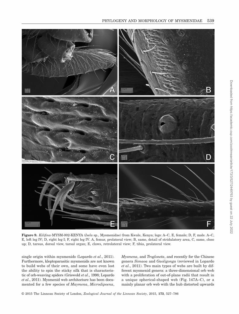
single origin within mysmenids (Lopardo et al., 2011).Furthermore, kleptoparasitic mysmenids are not knownto build webs of their own, and some have even lostthe ability to spin the sticky silk that is characteris-tic of orb-weaving spiders (Griswold et al., 1998; Lopardoet al., 2011). Mysmenid web architecture has been docu-mented for a few species of Maymena, Microdipoena,
Mysmena, and Trogloneta, and recently for the Chinesegenera Simaoa and Gaoligonga (reviewed in Lopardoet al., 2011). Two main types of webs are built by dif-ferent mysmenid genera: a three-dimensional orb webwith a proliferation of out-of-plane radii that result ina unique spherical-shaped web (Fig. 147A–C), or amainly planar orb web with the hub distorted upwards
Figure 9. Kilifina-MYSM-002-KENYA (Isela sp., Mysmenidae) from Kwale, Kenya; legs: A–C, E, female; D, F, male. A–C,E, left leg IV; D, right leg I; F, right leg IV. A, femur, prolateral view; B, same, detail of stridulatory area, C, same, closeup; D, tarsus, dorsal view, tarsal organ; E, claws, retrolateral view; F, tibia, prolateral view.
PHYLOGENY AND MORPHOLOGY OF MYSMENIDAE 539
© 2015 The Linnean Society of London, Zoological Journal of the Linnean Society, 2015, 173, 527–786
Dow
nloaded from https://academ
ic.oup.com/zoolinnean/article/173/3/527/2449763 by guest on 22 July 2022

Figure 10. Maymena ambita (Mysmenidae), male left palp: A, dorsal view; B, prolateral–dorsal view; C, prolateral view;D, prolateral–ventral view; E, ventral view; F, retrolateral–ventral view; H, retrolateral view, detail of tip of embolusand housing cymbial conductors; G, same, retrolateral–dorsal view; I, same, retrolateral–dorsal view; J, detail of squaredarea from I; K, detail of tibia from C. See Appendix 3 for the list of abbreviations.
540 L. LOPARDO AND G. HORMIGA
© 2015 The Linnean Society of London, Zoological Journal of the Linnean Society, 2015, 173, 527–786
Dow
nloaded from https://academ
ic.oup.com/zoolinnean/article/173/3/527/2449763 by guest on 22 July 2022

Figure 11. Maymena ambita (Mysmenidae), female: A, abdomen, ventral view; B, same, detail of epigynum; C, same,posterior view; D, digested abdomen, vulva; E, left anterior lateral spinneret; F, posterior median spinnerets; G, H, pos-terior lateral spinnerets (PLS); G, right PLS; H, left PLS. See Appendix 3 for the list of abbreviations.
PHYLOGENY AND MORPHOLOGY OF MYSMENIDAE 541
© 2015 The Linnean Society of London, Zoological Journal of the Linnean Society, 2015, 173, 527–786
Dow
nloaded from https://academ
ic.oup.com/zoolinnean/article/173/3/527/2449763 by guest on 22 July 2022

by between one and several out-of-plane radial linesthat attach to substrate above the web (Fig. 147D, E).
The respiratory system of Mysmenidae, as well asother members of the symphytognathoids, is greatlydiverse, but also remains relatively poorly studied (e.g.Forster, 1959, 1980; Gertsch, 1960a; Levi, 1967; Levi& Kirber, 1976). No detailed comparative study of eitherfemale or male mysmenid genitalia exists, and the ho-mologies of male palpal sclerites are poorly under-stood. Mysmenids are entelegynes: their epigyna canbe weakly sclerotized (e.g. Microdipoena, Brasilionata,Itapua, Mysmenella, and Calodipoena), or can have a
sclerotized and protruding epigynal plate (e.g. Trogloneta,Mysmenopsis, and Maymena). In some species a finger-like scape extends posteriorly (e.g. the species in thegenera Calodipoena and Mysmenella). The morphol-ogy of the male palp is highly intricate. The emboluscan be long and straight, long and coiled, short, bifid,and/or with apophyses, and it can also be distallytwisted. The cymbium is highly complex, with lobesor apophyses related to the embolus, forming an apicalcymbial ‘conductor’. Although the details of the malepalp morphology have been insufficiently studied, theconductor, median apophysis, and paracymbium appear
Figure 12. Maymena mayana (Mysmenidae): A, B, male; C–H, female. A, left palp, dorsal–retrolateral view; B, abdomen,detail of epiandrous spigots; C, digested abdomen, detail of spermathecae; D, epigynum, ventral view; E, same, detail ofcopulatory openings; F, G, right leg I; F, claws; G, tarsus–metatarsus junction; H, left leg IV, claws. See Appendix 3 forthe list of abbreviations.
542 L. LOPARDO AND G. HORMIGA
© 2015 The Linnean Society of London, Zoological Journal of the Linnean Society, 2015, 173, 527–786
Dow
nloaded from https://academ
ic.oup.com/zoolinnean/article/173/3/527/2449763 by guest on 22 July 2022
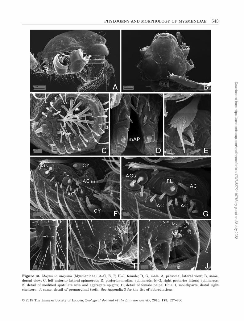
Figure 13. Maymena mayana (Mysmenidae): A–C, E, F, H–J, female; D, G, male. A, prosoma, lateral view; B, same,dorsal view; C, left anterior lateral spinnerets; D, posterior median spinnerets; E–G, right posterior lateral spinnerets;E, detail of modified spatulate seta and aggregate spigots; H, detail of female palpal tibia; I, mouthparts, distal rightchelicera; J, same, detail of promarginal teeth. See Appendix 3 for the list of abbreviations.
PHYLOGENY AND MORPHOLOGY OF MYSMENIDAE 543
© 2015 The Linnean Society of London, Zoological Journal of the Linnean Society, 2015, 173, 527–786
Dow
nloaded from https://academ
ic.oup.com/zoolinnean/article/173/3/527/2449763 by guest on 22 July 2022

to be absent (Coddington, 1990; Griswold et al., 1998).In addition, the spinning organs of Mysmenidae havebeen studied in only a few species (Griswold, 1985;Griswold et al., 1998; Brescovit & Lopardo, 2008; Milleret al., 2009). Mysmenidae seem to possess the typicalsymphytognathoid and higher araneoid gland spigotconformation. The anterior lateral spinnerets (ALS, seeAppendix 3 for abbreviations) have few piriform glandspigots (with small bases) and a deep groove betweenthe major ampullate and the piriform field. The pos-terior spinnerets (PMS and PLS) have few aciniformgland spigots. No additional data are available on thespinning organs of mysmenids.
Ten mysmenid species in five genera have been re-ported from the fossil record (all of them described fromCenozoic amber; Dunlop, Penney & Jekel, 2014). Fiveof the oldest fossil species have been reported from theEocene (Palaeogene; 44 Mya): three species from Balticamber (Mysmena grotae, Mysmena curvata, andPalaeomysmena hoffeinsorum) and two species fromBaltic and Bitterfeld ambers (Eomysmenopsis spinipesand Mysmena groehni) (Wunderlich, 2004, 2011). Onespecies has been reported from the Miocene–Oligocene(Neogene and Palaeogene; 19–27 Mya), from Chiapas
amber: Mysmena fossilis (see Petrunkevitch, 1971). Twospecies have been reported from the Miocene (Neogene;15–20 Mya), from Dominican amber: Dominicanopsisgrimaldii and Mysmenopsis lissycoleyae (see Penney,2000; Wunderlich, 2004). Two relatively young fossilspecies, Mysmena dominicana and Mysmena (s.l.)copalis, were described by Wunderlich (1998, 2011) fromMadagascan copal (a semi-fossilized resin of less thantwo million years old), dating to Early Pleistocene(Neogene).
TAXONOMIC HISTORY
Mysmenidae were proposed by Simon in 1922 as a groupwithin Theridiidae, under the name ‘Mysmeneae’(Simon, 1922, 1926). The elusive circumscription ofthis family is illustrated by the fact that several speciesoriginally placed in Mysmenidae have now beentransferred to a diverse array of families, such asTetragnathidae, Theridiidae, Anapidae, Symphytog-nathidae, Synaphridae, and Theridiosomatidae, and onespecies was even transferred to Acari! The followingquotations of Mysmenidae, as well as the one preced-ing the Introduction, illustrate the traditionally obscure
Figure 14. Maymena rica (Mysmenidae): A, B, D, male left palp; C, E, female. A, dorsal view; B, same, prolateralview, detail of tip of embolus and housing cymbial conductors; C, epigynum, ventral view; D, same as B, dorsal view;E, digested abdomen, detail of vulva. See Appendix 3 for the list of abbreviations.
544 L. LOPARDO AND G. HORMIGA
© 2015 The Linnean Society of London, Zoological Journal of the Linnean Society, 2015, 173, 527–786
Dow
nloaded from https://academ
ic.oup.com/zoolinnean/article/173/3/527/2449763 by guest on 22 July 2022

Figure 15. Maymena rica (Mysmenidae), prosoma: A, C, F, G, female; B, D, E, H, I, male. A, B, lateral view; C, dorsalview; D, carapace, frontal view; E, prosoma, posterior view; F, ocular area, detail from panel C; G–I, mouthparts, detailof cheliceral fang and teeth; G, distal promargin of left chelicera; H, distal promargin of right chelicera; I, distal rightchelicera.
PHYLOGENY AND MORPHOLOGY OF MYSMENIDAE 545
© 2015 The Linnean Society of London, Zoological Journal of the Linnean Society, 2015, 173, 527–786
Dow
nloaded from https://academ
ic.oup.com/zoolinnean/article/173/3/527/2449763 by guest on 22 July 2022

Figure 16. Maymena rica (Mysmenidae), abdomen and legs: A, B, G, H, male; C–F, female. A, epiandrous spigots;B, left anterior lateral spinnerets; C, abdomen, ventral view. D, G, H, left leg I; E, F, left leg IV. D, femur, ventral view;E, tibia, dorsal view; F, claws, retrolateral view; G, tibia–metatarsus junction, prolaterodorsal view; H, tarsus, prolaterodorsalview. See Appendix 3 for the list of abbreviations.
546 L. LOPARDO AND G. HORMIGA
© 2015 The Linnean Society of London, Zoological Journal of the Linnean Society, 2015, 173, 527–786
Dow
nloaded from https://academ
ic.oup.com/zoolinnean/article/173/3/527/2449763 by guest on 22 July 2022

status of mysmenids. Concerning the monophylyof the family, Paolo Brignoli (1980: 727) statedthat
. . . what we call Mysmenidae is equal to what has remainedof the ‘Symphytognathidae’ (sensu Forster 1959 and Levi &Levi, 1962) after the removal of the Anapidae, Textricellidaeand Symphytognathidae s.s.
Also, as Karin Schütt (2003: 137) declared,
Every large taxon seems to have a polyphyletic waste dispos-al group. In the case of symphytognathoids, the familyMysmenidae, into which all unassignables are swept togeth-er, has apparently been used for this purpose.
Simon’s ‘Mysmeneae’ (Simon, 1922, 1926) included fivegenera: Mysmena, Mysmenopsis, Cepheia, Synaphris,and Trogloneta (Simon, 1926: 315). ‘Mysmeneae’ wasdiagnosed mainly based on the absence of the femalepalpal claws, the presence of a voluminous male bulbwith a long embolus, the globular or conical abdomenwith sparse, rather long hairs, and the median tarsalclaw being as long as the superior claws. In a revi-sion of Theridiidae, Petrunkevitch (1928) elevated thegroup to the subfamily level. He united Simon’sMysmeneae and Theonoeae in the theridiid subfam-ily Mysmeninae, thus adding four genera to theoriginal group: Epecthina, Epecthinula, Iardinis, andTheonoe.
Figure 17. Microdipoena elsae (Mysmenidae), male right palp, inverted images: A, prolateral view; B, ventral view;C, retrolateral view; D, retroventral view. See Appendix 3 for the list of abbreviations.
PHYLOGENY AND MORPHOLOGY OF MYSMENIDAE 547
© 2015 The Linnean Society of London, Zoological Journal of the Linnean Society, 2015, 173, 527–786
Dow
nloaded from https://academ
ic.oup.com/zoolinnean/article/173/3/527/2449763 by guest on 22 July 2022

Tab
le2.
Gen
eric
list
and
info
rmat
ion
ofm
ysm
enid
desc
ribe
dsp
ecie
sin
clu
ded
inth
em
orph
olog
ical
data
set
Gen
us
Tota
lsp
ecie
s
Tota
lsp
ecie
ssc
ored
for
mor
phol
ogy
Spe
cies
incl
ude
dS
peci
esex
clu
ded
Rea
son
for
excl
usi
on
An
jou
anel
la(=
Mic
rod
ipoe
na)
11
A.c
omor
ensi
s*B
rasi
lion
ata
11
B.a
rbor
ense
*C
alod
ipoe
na
(=M
ysm
ena)
103
C.i
ncr
edu
la*,
C.m
ooat
ae,
C.t
arau
ten
sis
1(C
.tar
aute
nsi
s)N
ode
tail
edob
serv
atio
n,
>78
%m
issi
ng
data
Cal
omys
poen
a(=
Mys
men
a)1
1C
.san
tacr
uzi
*C
rass
ign
ath
a(S
ymph
ytog
nat
hid
ae)†
81
C.h
aen
eli*
1T
ype
and
only
spec
imen
not
avai
labl
e,>
78%
mis
sin
gda
taIa
rdin
is(S
ymph
ytog
nat
hid
ae)
3(2
)‡2
I.m
arte
nsi
,I.
mu
ssar
di
1(I
.mar
ten
si)
Typ
ean
don
lysp
ecim
enlo
st,
>78
%m
issi
ng
data
Isel
a1
1I.
oku
nca
na*
Itap
ua
(=M
ysm
ena)
11
I.te
mbe
i*K
eken
bosc
hie
lla
(=M
ysm
ena)
42
K.a
war
i,K
.mar
ijke
ae*
Kil
ifin
a(=
Isel
a)1
(1)
Lev
iola
(Zod
arii
dae)
11
L.t
erm
itop
hil
a*1
Typ
ean
don
lysp
ecim
enn
otav
aila
ble,
>78
%m
issi
ng
data
May
men
a13
3M
.am
bita
,M
.may
ana*
,M
.ric
aM
icro
dip
oen
a4
3M
.els
ae,
M.g
utt
ata*
,M
.nyu
ngw
eM
ysm
ena
263
M. l
eich
har
dti
,M
.leu
copl
agia
ta*,
M.t
asm
ania
eM
ysm
enel
la(=
Mic
rod
ipoe
na)
113
M.i
llec
trix
*,M
.job
i,M
.sam
oen
sis
Mys
men
iola
11
M.s
pin
ifer
a*M
ysm
enop
sis
274
M.c
idre
lico
la,
M.d
iplu
ram
igo,
M.p
alpa
lis,
M.p
enai
Ph
rico
telu
s(A
ran
eoid
eain
cert
aese
dis
.)1
1P.
stel
lige
r*1
No
deta
iled
obse
rvat
ion
,>78
%m
issi
ng
data
Tam
ases
ia(=
Mys
men
a)3
2T.
acu
min
ata,
T.ro
tun
da*
Trog
lon
eta
112
T.ca
nta
reir
a,T.
gran
ulu
m*
*Typ
esp
ecie
s.†C
urr
entl
yu
nde
rS
ymph
ytog
nat
hid
ae,
see
Mil
ler
etal
.(2
009)
and
this
stu
dy.
‡Typ
ean
don
lysp
ecim
enof
type
spec
ies
lost
:n
omen
du
biu
m.
548 L. LOPARDO AND G. HORMIGA
© 2015 The Linnean Society of London, Zoological Journal of the Linnean Society, 2015, 173, 527–786
Dow
nloaded from https://academ
ic.oup.com/zoolinnean/article/173/3/527/2449763 by guest on 22 July 2022

In a revision of Symphytognathidae s.l. (withAnapidae, ‘Micropholcommatidae’, ‘Textricellidae’,Mysmenidae, and Symphytognathidae s.s. as subfami-lies), Forster (1959) transferred Mysmena (and con-sequently the entire subfamily) from Theridiidae toSymphytognathidae, and described the respiratory
system of many Mysmena species, some of them con-sidered to now belong to Mysmena itself (Microdipoena,Calodipoena, Tamasesia, and Mysmenella). Gertsch(1960a) summarized the taxonomic revisions ofSymphytognathidae by Forster (1959) and of Mysmenaby Levi (1956), and proposed new diagnostic features
Figure 18. Microdipoena guttata (Mysmenidae): A–F, male left palp; G, female. A, ventral–proximal view; B, retrolateral–distal view; C, dorsal–retrolateral view; D, expanded bulb, prolateral view; E, prolateral view; F, expanded bulb,detail of tip of embolus, retrolateral–distal view; G, digested abdomen, detail of vulva. See Appendix 3 for the list ofabbreviations.
PHYLOGENY AND MORPHOLOGY OF MYSMENIDAE 549
© 2015 The Linnean Society of London, Zoological Journal of the Linnean Society, 2015, 173, 527–786
Dow
nloaded from https://academ
ic.oup.com/zoolinnean/article/173/3/527/2449763 by guest on 22 July 2022

for mysmenines, currently shared by many othersymphytognathoid species (e.g. their small to minutesize, with or without book lungs, pedipalps present andof normal size in females, lack of comb in the hindtarsi, and metatarsi longer or of equal size than tarsi).
Gertsch (1960a) transferred Lucarachne, Mysmenopsis,Iardinis, and Trogloneta from Theridiidae toSymphytognathidae, and stated that Mysmeninae com-prised the following genera: Mysmena, Mysmenopsis,Lucarachne, Maymena, Cepheia, Synaphris, Iardinis,
Figure 19. Microdipoena guttata (Mysmenidae). A, B, D–F, male; C, female. A, prosoma, frontal view; B, C, same, lateralview; D, same, ventral view, detail of labium–sternum junction; E, mouthparts, detail of tip of left chelicera; F, posteriormedian spinnerets. See Appendix 3 for the list of abbreviations.
550 L. LOPARDO AND G. HORMIGA
© 2015 The Linnean Society of London, Zoological Journal of the Linnean Society, 2015, 173, 527–786
Dow
nloaded from https://academ
ic.oup.com/zoolinnean/article/173/3/527/2449763 by guest on 22 July 2022

and Trogloneta. Levi & Levi (1962), in a genericrevision of Theridiidae, transferred Taphiassa toSymphytognathidae s.l. and placed Iardinis as incertaesedis within the latter family.
Forster & Platnick (1977) concluded that mysmenidswere sufficiently distinct from Symphytognathidae towarrant family rank, without providing further jus-tification. A year later, Platnick & Shadab (1978), in
a revision of the genus Mysmenopsis, provided an ex-plicit, although tentative, diagnosis of Mysmenidae,based only on New World genera. According to Platnick& Shadab (1978), four features diagnosed Mysmenidae:a clasping spur on the male metatarsus I (occasion-ally also on tibia I), the presence of lobes or apophyseson the cymbium, a ventral sclerotized spot on distalfemur I of females, and a series of tiny denticles
Figure 20. Microdipoena guttata (Mysmenidae), abdomen: A, female; B–G, male. A, ventral view; B, detail of booklungcover; C, detail of abdominal cuticular pattern; D, pedicel area and booklung covers; E, detail of area above pedicel;F, posterior–ventral view, detail of epigastric furrow, epiandrous spigots and spiracular openings; G, same, ventral view.
PHYLOGENY AND MORPHOLOGY OF MYSMENIDAE 551
© 2015 The Linnean Society of London, Zoological Journal of the Linnean Society, 2015, 173, 527–786
Dow
nloaded from https://academ
ic.oup.com/zoolinnean/article/173/3/527/2449763 by guest on 22 July 2022

scattered between the cheliceral teeth. Brignoli (1980)questioned the validity of the new family rank ofMysmenidae and its diagnosis, and reviewed some ofits genera, revalidating some, creating others, and thussplitting the genera previously placed in the family.Still, Brignoli accepted the group diagnosis. Herevalidated Microdipoena (previously revalidated bySaaristo, 1978), Calodipoena, and Tamasesia, anderected the genus Mysmenella. Wunderlich (1986; contraForster & Platnick, 1977) defended the monophyly ofSymphytognathidae s.l. (i.e. sensu Forster, 1959), thusreverting Mysmenidae to subfamily level, and split-ting this subfamily into two: Mysmeninae andSynaphrinae. Synaphrinae included the mysmenidgenera Synaphris, Iardinis, and Cepheia. Further-more, Wunderlich (1986) proposed a hypothesis of re-lationships between symphytognathoid families (asAnapidae s.l.), although no data set or thorough andexplicit phylogenetic rationale for inferring such re-lationships was provided (as discussed by Schütt, 2002).
More than 90 years after its erection, and despitethe many arguments about its circumscription andphylogenetic placement, Mysmenidae still lack a modernphylogenetic morphological revision. Only recently themonophyly of Mysmenidae has been robustly testedin a comprehensive combined phylogenetic analysis of
symphytognathoid spiders using morphological and mo-lecular data (Lopardo et al., 2011, and see referencestherein for a review of previous phylogenetic analy-sis including mysmenid representatives; see alsoDimitrov et al., 2012 for a comprehensive orbicula-rian analysis and the problematic monophyly ofsymphytognathoids).
GOALS
The goals of this study are several. First, to performthe first comparative morphological study of mysmenidsand their close relatives, and to propose and testhypotheses of primary homology. These primary ho-mology hypotheses have been compiled as charactersinto a morphological data set. Second, to explorethe phylogenetic signal of the morphological (andbehavioural) data partition by means of a generic-level cladistic analysis of Mysmenidae, to test themonophyly of the family and its genera, and toplace Mysmenidae within symphytognathoids. Themorphological characters were also included aspart of a larger, combined analysis of mysmenid andother symphytognathoids elsewhere, and thereforeproper taxonomic and nomenclatorial actions fromthe phylogenetic classification for the family arebased on the results of our combined analyses, whichhave been presented and discussed elsewhere (Figs 160,161; see also Lopardo et al., 2011: fig. 12). Thirdly,to discuss the evolutionary implications for thespinneret silk gland spigot conformations in Mysme-nidae and other symphytognathoids, based on theevolutionary and comparative framework providedby the total-evidence phylogenetic hypothesis of thecombined analysis (Fig. 160; see also Lopardo et al.,2011, fig. 12).
MATERIAL AND METHODSSPECIMENS
Taxon samplingSpecimens for this study were borrowed from museumcollections, kindly loaned or donated by colleagues, orcollected in the field (see Acknowledgements). SeeAppendix 1 for a list of material examined and voucherinformation.
IngroupThe ingroup for the morphological and behavioural dataset (hereafter referred to as ‘morphological’ data set)consists of 47 mysmenid species: 36 species belong todescribed taxa, representing 18 of the 23 mysmenidgenera as currently defined (see Table 2). Describedspecies (see Table 1 for authorship of taxa) scored inthis data set include Anjouanella comorensis,
Table 3. List of described and undescribed outgroup (i.e.non-mysmenid) taxa
Family Species
Anapidae Acrobleps hygrophilusAnapisona kethleyiCrassanapis chilensisComaroma simoniElanapis aisenMinanapis casablancaMinanapis palenaTasmanapis strahan
(Micropholcommatinae) Taphiassa punctataTeutoniella cekalovici
Linyphiidae Linyphia triangularisSymphytognathidae Crassignatha haeneli
Patu-SYMP-001-DRSYMP-002-MADSYMP-006-AUSTSYMP-007-AUSTSymphytognatha picta
Synaphridae Cepheia longisetaSynaphris saphrynis
Tetragnathidae Leucauge venustaTetragnatha versicolor
Theridiidae SteatodaTheridiosomatidae Theridiosoma gemmosum
Coddingtonia euryopoides
552 L. LOPARDO AND G. HORMIGA
© 2015 The Linnean Society of London, Zoological Journal of the Linnean Society, 2015, 173, 527–786
Dow
nloaded from https://academ
ic.oup.com/zoolinnean/article/173/3/527/2449763 by guest on 22 July 2022

Figure 21. Microdipoena guttata (Mysmenidae), legs: A, D, F–I, male; B, C, E, female. A, left leg I, detail of femoralspot; B, right leg I, metatarsus–tarsus junction; C, right femur II, dorsal view; D, left patella I, dorsal view; E, rightfemur II, prolateral view; F, left tibia I, prolateral view; G, right metatarsus III, dorsal view; H, right femur II, ventralview; I, right leg III, metatarsal trichobothrium.
PHYLOGENY AND MORPHOLOGY OF MYSMENIDAE 553
© 2015 The Linnean Society of London, Zoological Journal of the Linnean Society, 2015, 173, 527–786
Dow
nloaded from https://academ
ic.oup.com/zoolinnean/article/173/3/527/2449763 by guest on 22 July 2022

Figure 22. Microdipoena nyungwe (Mysmenidae): A, B, E, female; C, D, F, G, male left palp. A, digested abdomen, res-piratory system and vulva; B, same, detail of vulva; C, distal–retrolateral view, detail of tip of embolus and primarycymbial conductor; D, ventral–proximal view; E, tip of palp and tarsal organ; F, dorsal view; G, retrolateral view. SeeAppendix 3 for the list of abbreviations.
554 L. LOPARDO AND G. HORMIGA
© 2015 The Linnean Society of London, Zoological Journal of the Linnean Society, 2015, 173, 527–786
Dow
nloaded from https://academ
ic.oup.com/zoolinnean/article/173/3/527/2449763 by guest on 22 July 2022

Figure 23. Microdipoena nyungwe (Mysmenidae), spinnerets: A, B, female; C–H, male. A, C, anterior lateral spinnerets(ALS); B, E, left posterior lateral spinnerets; D, posterior median spinnerets; F, ALS, detail of major ampullate field;G, H, ALS, detail of intersegmental lobe. See Appendix 3 for the list of abbreviations.
PHYLOGENY AND MORPHOLOGY OF MYSMENIDAE 555
© 2015 The Linnean Society of London, Zoological Journal of the Linnean Society, 2015, 173, 527–786
Dow
nloaded from https://academ
ic.oup.com/zoolinnean/article/173/3/527/2449763 by guest on 22 July 2022

Brasilionata arborense, Calodipoena incredula,Calodipoena mooatae, Calodipoena tarautensis,Calomyspoena santacruzi, Crassignatha haeneli (butsee below), Iardinis martensi, Iardinis mussardi, Iselaokuncana, Itapua tembei, Kekenboschiella awari,Kekenboschiella marijkeae, Leviola termitophila,Maymena ambita, Maymena mayana, Maymena rica,Microdipoena elsae, Microdipoena guttata, Microdipoenanyungwe, Mysmena leichhardti, Mysmena leucoplagiata,Mysmena tasmaniae, Mysmenella illectrix, Mysmenella
jobi, Mysmenella samoensis, Mysmeniola spinifera,Mysmenopsis cidrelicola, Mysmenopsis dipluramigo,Mysmenopsis palpalis, Mysmenopsis penai, Phricotelusstelliger, Tamasesia acuminata, Tamasesia rotunda,Trogloneta cantareira, and Trogloneta granulum. Thecurrently monotypic genus Kilifina was represented byan undescribed species sharing apomorphies with thetype species Kilifina inquilina, also from Kenya,the country from which the type species was collect-ed. This undescribed species has also been collected
Figure 24. Microdipoena nyungwe (Mysmenidae), abdomen: A–D, female; E–G, male. A, epigastric furrow, epigynal areaand spiracular openings, posterior–ventral view; B, same, detail of epigynal area; C, pedicel area and booklung covers;D, epiandrous spigots, detail from figure F; E, posterior respiratory spiracle and colulus, ventral view; F, pedicel area,booklung covers and epiandrous spigots.
556 L. LOPARDO AND G. HORMIGA
© 2015 The Linnean Society of London, Zoological Journal of the Linnean Society, 2015, 173, 527–786
Dow
nloaded from https://academ
ic.oup.com/zoolinnean/article/173/3/527/2449763 by guest on 22 July 2022
in Cameroon and São Tome (C. Griswold, pers. comm.).The monotypic genus Crassignatha was recently trans-ferred from Mysmenidae to Symphytognathidae basedon morphology by Miller et al. (2009), and this newlyproposed familial placement is tested in the presentstudy. The remaining ten taxa correspond to undescribedmysmenid species (see Appendix 1). When possible,undescribed species were identified (i.e. tentatively
assigned to a genus) so that generic (and familial)membership could also be tested. The exemplar ap-proach was followed as much as possible when scoringcharacters (i.e. morphological characters were scoredfollowing direct observation of specimens). When speci-mens from described species were not available for study,or insufficient material existed or was suitable for de-tailed and thorough comparative observations, scoring
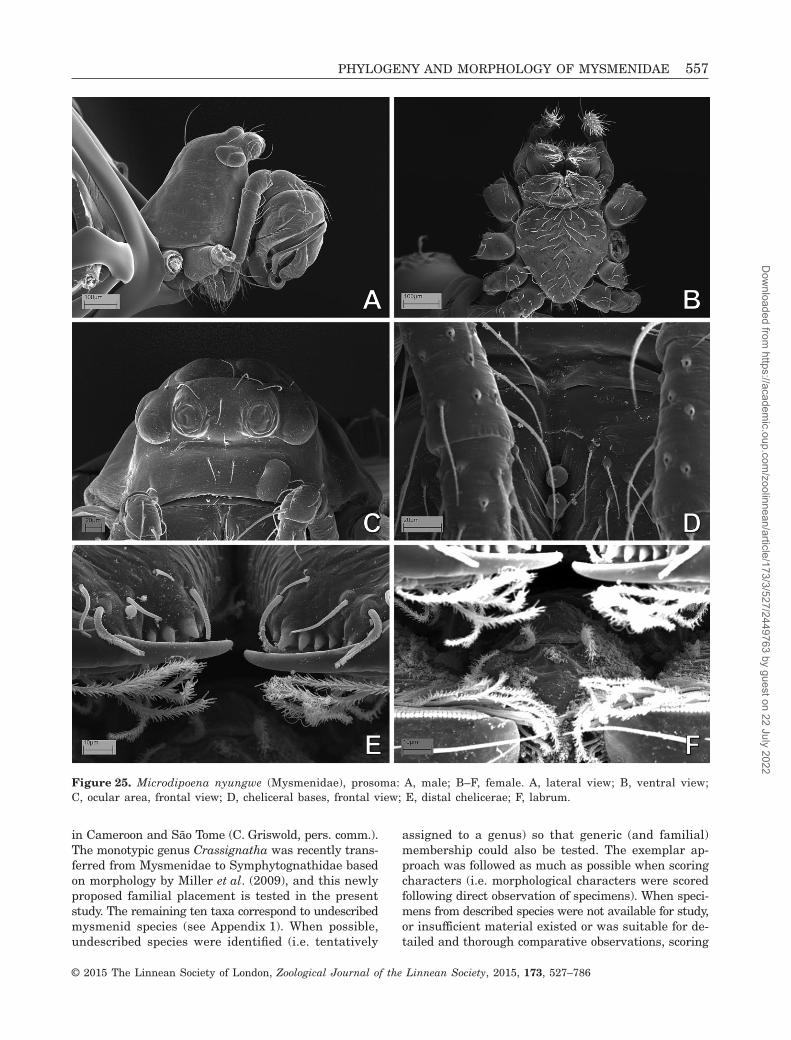
Figure 25. Microdipoena nyungwe (Mysmenidae), prosoma: A, male; B–F, female. A, lateral view; B, ventral view;C, ocular area, frontal view; D, cheliceral bases, frontal view; E, distal chelicerae; F, labrum.
PHYLOGENY AND MORPHOLOGY OF MYSMENIDAE 557
© 2015 The Linnean Society of London, Zoological Journal of the Linnean Society, 2015, 173, 527–786
Dow
nloaded from https://academ
ic.oup.com/zoolinnean/article/173/3/527/2449763 by guest on 22 July 2022

was based on the original species descriptions or otherdescriptive literature, or on previous phylogenetic work.As a result of a lack of detailed observations, the levelof missing data for five of the ‘literature-based taxa’was higher than 78% in the current data set, and theywere not included in the final analyses. Instead, thesefive species were included in a preliminary analysiscomprising all taxa to test their familial placement (for
the inclusion/exclusion of taxa and reasons for exclu-sion, see Table 2). The five taxa removed from the finalanalyses include three species not available for study,and scored entirely from the literature (Crassignathahaeneli, Iardinis martensi, and Leviola termitophila),and two species without adequate material for directdetailed observation, and therefore scored mainly fromliterature (Phricotelus stelliger and Calodipoena
Figure 26. Microdipoena nyungwe (Mysmenidae), left legs: A, C, D, G, male; B, E, F, female. A, tarsus I, prolateral view;B, claws I, retrolateral view; C, leg I, tibia–metatarsal junction, prolateral view; D, metatarsus I, dorsal view; E, leg I,metatarsus–tarsus junction, dorsal view; F, tarsus IV, prolateral view; G, tibia IV, prolateral view.
558 L. LOPARDO AND G. HORMIGA
© 2015 The Linnean Society of London, Zoological Journal of the Linnean Society, 2015, 173, 527–786
Dow
nloaded from https://academ
ic.oup.com/zoolinnean/article/173/3/527/2449763 by guest on 22 July 2022

Figure 27. Microdipoena (= Mysmenella) samoensis (Mysmenidae), syntypes: A–C, F, H, I, male; D, E, G, female. A, leftpalp, prolateral–ventral view; B, same, retrolateral–ventral view; C, same, detail of tip of embolus and interaction withprimary cymbial conductor, prolateral view; D, digested abdomen, vulva, and part of respiratory system; E, same, detailof vulva; F, G, prosoma, lateral view; H, mouthparts, ventral view, detail of retromargin of chelicerae and labium; I, lefttibia I, dorsal view. See Appendix 3 for the list of abbreviations.
PHYLOGENY AND MORPHOLOGY OF MYSMENIDAE 559
© 2015 The Linnean Society of London, Zoological Journal of the Linnean Society, 2015, 173, 527–786
Dow
nloaded from https://academ
ic.oup.com/zoolinnean/article/173/3/527/2449763 by guest on 22 July 2022
tarautensis). The ingroup includes the type species ofmost genera, plus up to three other described speciesfor those non-monotypic genera. Although the repre-sentative ingroup species were initially selected takinginto account the morphological diversity of each genus,the availability of material is always the final arbiterfor the taxon selection used. The final ingroup sample
includes 42 mysmenid species representing 16 genera(see Appendix 1 for a list of studied specimens).
OutgroupsThe phylogenetic relationships among araneoid fami-lies have yet to be satisfactorily resolved (Hormiga &Griswold, 2014), and thus when designing the
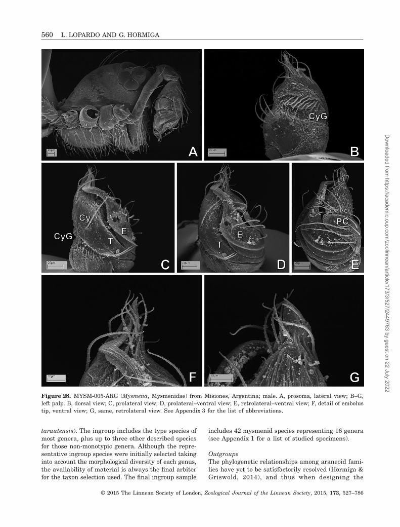
Figure 28. MYSM-005-ARG (Mysmena, Mysmenidae) from Misiones, Argentina; male. A, prosoma, lateral view; B–G,left palp. B, dorsal view; C, prolateral view; D, prolateral–ventral view; E, retrolateral–ventral view; F, detail of embolustip, ventral view; G, same, retrolateral view. See Appendix 3 for the list of abbreviations.
560 L. LOPARDO AND G. HORMIGA
© 2015 The Linnean Society of London, Zoological Journal of the Linnean Society, 2015, 173, 527–786
Dow
nloaded from https://academ
ic.oup.com/zoolinnean/article/173/3/527/2449763 by guest on 22 July 2022

out-group sampling it is very difficult, if not impos-sible, not to fall down the slippery slope that bringsthe study into the orbicularian phylogeny abyss. Thechoice of which families to represent quickly becomesa question of which families will not be representedin the analysis. Our taxon selection has emphasizedthe symphytognathoid families, and is largely basedon the phylogenetic hypotheses of Coddington (1990),Griswold et al. (1998), Schütt (2003), and Lopardo &Hormiga (2008; for a detailed rationale for outgrouptaxon sampling, see also Lopardo et al., 2011).Cyatholipidae were not represented; retrospectively, theirinclusion along with Synotaxidae would have been rel-evant to the problem finding the closest relatives ofsymphytognathoids. But ultimately a line must bedrawn across this slippery slope, otherwise our studywould grow out of proportion to become an analysisof the araneoid families. Although Mysmenidae seemto be related to other symphytognathoid families, thelimits and diagnoses of these families appear prob-lematic, except for Theridiosomatidae (see Coddington,1986a; Labarque & Griswold, 2014; see also Lopardoet al., 2011, for current re-delimitation and diagnosesof symphytognathoid families). The choice of out-group taxa therefore focused on symphytognathoid rep-resentatives, in particular Symphytognathidae andAnapidae, and was also based on, and limited by, speci-men availability. The outgroup taxon partition con-sists of 23 species representing seven araneoid families(18 species correspond to described taxa, see Table 3):Anapidae [ten species: Acrobleps hygrophilus, Anapisonakethleyi, Crassanapis chilensis, Comaroma simoni,Elanapis aisen, Minanapis casablanca, Minanapispalena, and Tasmanapis strahan, plus two species ofthe subfamily Micropholcommatinae sensu Lopardo et al.
(2011), Taphiassa punctata and Teutoniella cekalovici],Symphytognathidae [five species: Symphytognathapicta plus four undescribed species and Crassignathahaeneli, see Ingroup above), Theridiosomatidae (twospecies: Theridiosoma gemmosum and Coddingtoniaeuryopoides), Synaphridae (two species: Cepheialongiseta and Synaphris saphrynis); and the non-symphytognathoid families Theridiidae (Steatoda, seebelow), Linyphiidae (Linyphia triangularis), andTetragnathidae (two species: Leucauge venusta andTetragnatha versicolor) (see Table 3). Most of the ob-servations are based on our study of the relevant speci-mens, using an exemplar approach (e.g. Prendini, 2001),rather than inferring basal states for genera; other-wise the scoring was based on the literature. Thetheridiid genus Steatoda is the only chimeric taxon inthis data set. This genus has also been included as asingle chimeric representative in previous studies, in-cluding a phylogenetic analysis of the same Theridiidae(see also Griswold et al., 1998; Agnarsson, 2004), wheremore than one species was examined. The general mor-phology of Steatoda appears to be sufficiently invari-able, and following Agnarsson’s (2004) approach, acombination of species was included to represent thegroundplan character vector of the genus (Steatodaamericana, Steatoda bipunctata, and Steatoda grossa).Specimens of S. americana were examined here, al-though the character scoring was complemented withinformation from the data sets of Griswold et al. (1998;S. grossa) and Agnarsson (2004; S. americana,S. bipunctata, and S. grossa) (but see Wunderlich, 2008for the transfer of S. americana to Asagena). All re-sulting trees were rooted using the tetragnathidTetragnatha versicolor. See Appendix 1 for a list of allexamined material.
Table 4. List of character sets reporting number of corresponding characters and their relative proportions (see Fig. 152)
Character setNumber ofcharacters Percentage
Number of continuouscharacters in the set
Abdomen (including respiratory system) 35 9.80% 1General body 1 0.28%Cephalothorax 20 5.60%Egg sacs 1 0.28%Epiandrous spigots 3 0.84%Epigynum (internal and external female genitalia) 29 8.12%Eyes 9 2.52%Legs 58 16.25% 4Male palp 111 31.09%Mouthparts 35 9.80%Palp (female) 7 1.96%Spinnerets 39 10.92% 2Web building (and other behavioural characters) 9 2.52%Total 357 100.00% 7
PHYLOGENY AND MORPHOLOGY OF MYSMENIDAE 561
© 2015 The Linnean Society of London, Zoological Journal of the Linnean Society, 2015, 173, 527–786
Dow
nloaded from https://academ
ic.oup.com/zoolinnean/article/173/3/527/2449763 by guest on 22 July 2022

METHODS OF STUDY
Morphological and behavioural methodsSpecimens were studied using standard morphologi-cal techniques in arachnology. Morphological methodsof study follow Lopardo (2005) and Lopardo, Hormiga
& Melic (2007). Specimens were initially examinedin 80% ethanol using Leica MZAPO or MZ16Astereomicroscopes. Because of their minute size, furtherdetailed observations and illustrations were per-formed using a Leica DMRM compound microscope witha drawing tube, and Scanning Electron Microscopy
Figure 29. MYSM-005-ARG (Mysmena, Mysmenidae) from Misiones, Argentina. A–C, E, F, female; D, male. A, digestedabdomen, detail of left spermatheca; B, same, detail of posterior tracheal system; C, epigynal area and scapus; D, leftfemur I, retrolateral view; E, left tibia IV, prolateral view; F, left metatarsus IV, prolateral view. See Appendix 3 for thelist of abbreviations.
562 L. LOPARDO AND G. HORMIGA
© 2015 The Linnean Society of London, Zoological Journal of the Linnean Society, 2015, 173, 527–786
Dow
nloaded from https://academ
ic.oup.com/zoolinnean/article/173/3/527/2449763 by guest on 22 July 2022

(SEM; see below). Measurements are in millimetres.Carapace height was measured at the highest point,from carapace lateral edge. Carapace length and heightwere measured in lateral view; carapace width wastaken at its widest point in dorsal view. Abdominallength and height were measured in lateral view, andwidth at widest point in dorsal view. In mostsymphytognathoids the abdomen is globular and theposition of the pedicel seems to have advanced towardsthe spinnerets (or vice versa), so the length of theabdomen is measured here from the spinnerets to theopposite point in the abdomen, and height is meas-ured as the longest section perpendicular to the length(for a detailed explanation, see characters related tothe abdominal morphology in Appendix 2). Leg articlelengths were mostly measured using SEM micros-copy, and measurements were taken in lateral or dorsalviews. Left structures (mostly male palps) are depict-ed unless otherwise stated. If right palps were usedand/or illustrated, images were reversed to facilitatecomparisons (and noted in the figure legend). Most hairsand macrosetae are usually not depicted in final palp
and epigynum drawings, unless they provide puta-tive phylogenetic information. As a convention, rela-tive position of sclerites in male palp is stated as ifthe cymbium were dorsal, regardless of the relativeposition of the cymbium to the whole palp or theprosoma.
For observation of respiratory structures and femaleinternal genitalia, we used the method of Álvarez-Padilla & Hormiga (2008). Abdomens were bisectedhorizontally and digested with SIGMAPancreatin P1750enzyme complex, in a solution of sodium borate pre-pared using the concentrations described by Dingerkus& Uhler (1977), as modified by Álvarez-Padilla & Hormiga(2008). Bisected abdomens were left in this solution atroom temperature (i.e., 20–25ºC) overnight or for a fewhours. After enzymatic digestion, abdomens were trans-ferred to distilled water, and then to ethanol.
Digital images of spider habitus and other detailswere taken with a Leica DFC 500 camera. A compo-site of multiple digital images taken at varyingfocal lengths along the z-axis was assembled usingthe software package Leica Application Suite. Most
Figure 30. MYSM-007-MEX (Mysmena, Mysmenidae) from Chiapas, Mexico; male left palp. A, retroventral view; B, ventralview; C, prolateral view; D, dorsal view; E, retrolateral view; F, detail of tip of palp, ventral–prolateral view. SeeAppendix 3 for the list of abbreviations.
PHYLOGENY AND MORPHOLOGY OF MYSMENIDAE 563
© 2015 The Linnean Society of London, Zoological Journal of the Linnean Society, 2015, 173, 527–786
Dow
nloaded from https://academ
ic.oup.com/zoolinnean/article/173/3/527/2449763 by guest on 22 July 2022

Tab
le5.
Mor
phol
ogic
alda
tam
atri
x
0000000000111111111122222222223333333333444444444455555555556666666666777777777788888888889999999999
0123456789012345678901234567890123456789012345678901234567890123456789012345678901234567890123456789
AN
AP
IDA
EA
crob
leps
hyg
roph
ilu
s00000100---0a001001012011--??-----20001000-110010010210?1--00-1---001101001000111000-00110201?000201
An
apis
ona
keth
leyi
20110100---00000001??2??011011???024010100-110410010311?1--00-????000-?10-100?111000-0010----?010211
Com
arom
asi
mon
i21110200---00010001001-001100--000200000021100030010412?01100-1---0011000000-0001000-0--a000-?000001
Cra
ssan
apis
chil
ensi
s211101010-100000001001-?01100--00024001100-110520010311?1--00-1---0011010010-020000d-00?10001?010011
Ela
nap
isai
sen
001001010-120001001001-101100--00020010000-11001?010210?1--00-1---00110100??-0?11105-?1110001?010201
Min
anap
isca
sabl
anca
22110100---00001001001-101100--000200111031100420010311?1--00-1---0011011000-000000d-01110001?010201
Min
anap
ispa
len
a22110100---00001001001-101100--000200111031100420010h11?1--00-1---0011011000-000000d-01110001?010201
Tas
man
apis
stra
han
21110100---2000100100e-001100--00025001100-110620010312?1--00-1---000-011-00-00?1?00-11110001?010201
Tap
hia
ssa
pun
ctat
a11110100---001100010021?011???????23010000-113340010412?1--10-00??0?0-?11-00-0001002-00011001?010201
Teu
ton
iell
ace
kalo
vici
111101010-100?10011001-101100--000230100011100440010i10?1--00-0011000-010-00-0?01000-?000----?010201
LIN
YP
HII
DA
EL
inyp
hia
tria
ngu
lari
s00000001a0101001010010-?011010000001000000-001110000500000011100000110010-100?101004-?01100010010100
MY
SM
EN
IDA
EA
njo
uan
ella
com
oren
sis
0000?1010-1210?????0??????????????2?00100???0???a010?20????010????0011?12110-?21100a-11111?0??0?0?01
Bra
sili
onat
aar
bore
nse
0?0??100---210????????????????????2????00??10???1010?e0?????????????????????????????????11?11???0?01
Cal
odip
oen
ain
cred
ula
000011210-1e10010110120100000--1122a?01000-10000a010020?0110111---0011?12010-?211002-000111a1?010101
Cal
odip
oen
am
ooat
ae0000?1010-1010???????2??000????11?2?00000???0???0010?20????011????0011?12110-?21100g-?0011101?0?0?01
Cal
odip
oen
ata
rau
ten
sis
?0?0?1010-1210???????2??000????11?2?00?00???0???1010?20????011????0011?12110-?211001-10111?01?0?0?01
Cal
omys
poen
asa
nta
cru
zi0000?1210-1210????????????????????2??0000???0???1010?20????011????0011?12?10-?211002-???111a1?0?0?01
Cra
ssig
nat
ha
hae
nel
i0????1011?1???????????????????????2????10???????0??0?e??????????????????????????????????0----?0?0??1
Iard
inis
mar
ten
si0?0???0??????1????????????????????2?????????????0????e??????????????????????????????????0----???0??1
Iard
inis
mu
ssar
di
0?0??1010-220?????????????????????2????00??10???0010?10?????????????????????????????????0----?0?0?01
Isel
aok
un
can
a000011010-1012010111-1-?011010000?21000000-10aa01010020-00110-????000-?11-00-0001003100011101?010100
Itap
ua
tem
bei
0000110????110??????????000???????2?00100??10???1010??0????0111---0011?12110-?211000-011111a1?0?0?01
Kek
enbo
sch
iell
aaw
ari
0?0??1010-111?????????????????????2????00??10???1010?20?????????????????????????????????11?11?0?0?01
Kek
enbo
sch
iell
am
arij
keae
0?0??1010-111???????????00????????2?0??00??10???1010?20?????????????????????????????????11111?0?0?0?
Lev
iola
term
itop
hil
a?2?0?200---2?1????????????????????2?00??0???0???101??e?????00-????02????1-00-?0?000?-???12001?001???
May
men
aam
bita
00000100---01001011010-101100--0002??0000??10???0010?001???10-000?00??111-00-0a01000-0a111?010010?00
May
men
am
ayan
a0000110a1110?001011010-101100--00011000000-10101?000100?00110-0000020-111-00-0001005-01011101?010100
May
men
ari
ca000011110-10000100101e-201100--00021000000-100000010020?00100-????000-011-00-0001001-0a111101?010100
Mic
rod
ipoe
na
elsa
e000011010-11100101101???000???????2000100??100001010020????00-1---00??01??10-?211004-1?011101?010101
Mic
rod
ipoe
na
gutt
ata
000011010-1110010110121100000--11221001000-1000a1010020?01100-????0011012111-1211000-11111101?010101
Mic
rod
ipoe
na
nyu
ngw
e000011010-1110010110121100000--11221001000-1000a0010020?0110101---0011012111-?21100a-11111101?010101
Kil
ifin
a-M
YS
M-0
02-K
EN
YA
00001100---0120101101e-1011010000221000000-100001010120?00110-000101??012010-12a1003100011101?010100
MY
SM
-005
-AR
G00001100---110000110121100000--11221000000-10000101002010110111---0011112000-??11001-100111a1?0?0101
MY
SM
-007
-ME
X000011210-1110010110121200000--11221001000-100001010020????0111---0011012110-1211002-0?011101?010101
Mys
men
a-M
YS
M-0
15-M
AD
000011010-10100?01101201000???-11?20001000-10000a010220?01101?1---001111111000101000-00111101?010201
Mys
men
a-M
YS
M-0
18-M
AD
000011010-1e100?01101201000????11?20?0100??100001010020?0110111---0011112000-??11002-0?011101?010101
MY
SM
-019
-MA
D000011010-1210010110120?00000--11220000000-100010010020?011011????00???12000-1211002-0?011101?010101
MY
SM
-020
-MA
D000011010-1110010110120100000--11221???000-100000010210?011?????????????????????????????11101?010101
MY
SM
-023
-MA
D000011010-11100?0110120200000--1122a00100??100001010be0?01110-010?0111112010-1211103000011101?010201
MY
SM
-028
-MA
D?0?011010-1??001011012?100000--1122000?000-100001010010????0111--?0011012000-?-11002-00011101?0102?1
MY
SM
-029
-MA
D?0?011010-11100101101???000?????1?2000?000-100001010020????00-1---0011112111-?211001-11111101?0102?1
564 L. LOPARDO AND G. HORMIGA
© 2015 The Linnean Society of London, Zoological Journal of the Linnean Society, 2015, 173, 527–786
Dow
nloaded from https://academ
ic.oup.com/zoolinnean/article/173/3/527/2449763 by guest on 22 July 2022

Tab
le5.
Con
tin
ued
0000000000111111111122222222223333333333444444444455555555556666666666777777777788888888889999999999
0123456789012345678901234567890123456789012345678901234567890123456789012345678901234567890123456789
MY
SM
-034
-MA
D?0?011010-1210000110120100000--1122000?000-100001010020????0101---00-?11??00-?001000-01111101?0101?1
Mys
men
ale
ich
har
dti
000011010-1110010110120100000--11220001000-100001010020?0110101---0011112011-?211001-00011101?010101
Mys
men
ale
uco
plag
iata
0000?1010-1e10????????????????????2?00100??10???1010?20????011????0011?12?10-?211002-?1?11101???0???
Mys
men
ata
sman
iae
000011010-11100?0110120200000--1122?00000??10???1010?201???010????0011?12001-?211002-0001110100?0?01
Mys
men
ella
ille
ctri
x0?0??1010-11-0????????????????????2????00??10????010?20?????????????????????????????????11101?010?01
Mys
men
ella
jobi
0000?1010-11-0???????2??000????11?2?00100???0???1010?20????011????0011?12111-?211001-11111?01?010?01
Mys
men
ella
sam
oen
sis
000011010-11-00101101202000????11?21001000-100000010020????0111---0011012111-?211001-11111101?010201
Mys
men
iola
spin
ifer
a0?0??1210-121?????????????????????2????00??10???0010??0?????????????????????????????????0----?010?01
Mys
men
opsi
sci
dre
lico
la000011010-111?00011??21?001???????22?00000-100001010020?01110-??????????1?????00100?-???11101?010100
Mys
men
opsi
sd
iplu
ram
igo
000011010-1?120001101212001????1??21000000-1000010?0020?01110-000?0110011-0000101000-00011101?010100
Mys
men
opsi
spa
lpal
is000011010-1?120?0?10121?0011---10-20000000-10000a010020?01110-000?1210011-11-0201000-01111101?010100
Mys
men
opsi
spe
nai
000011010?1?1201011012120011---10-21000000-100001010020?01110-000?010-011-01-0001004-01111101?010101
Ph
rico
tell
us
stel
lige
r?0?0?120---01???????????011???????2??0?00??10????0?0?e0????011?????0???1????-0?0000?-00?11?0???????1
Tam
ases
iaac
um
inat
a0000?1210-101?0??????20e000????11?2?00000??1????1010??01???011????0011?11010-?201002-?0011101?0?0?01
Tam
ases
iaro
tun
da
00001100---1100??????20e000????11?2?00000??1????1010?20????011????0011?100?????01005-?0011101?0?0?01
Trog
lon
eta
can
tare
ira
00001120---2?000011011-101100--0032b001000-102e0?0100e0????10-????010-110-11-0200004-11010201?011101
Trog
lon
eta
gran
ulu
m00001100---21000011011-1011????00?2a001000-10000?010020101110-????010-110-11-020000d-11010201?011101
SY
MP
HY
TO
GN
AT
HID
AE
Pat
u-S
YM
P-0
01-D
R00001100---01e01011??21101100--00320011000-10001a010210?1--00-0011000-011-01-000100c10110----?010001
SY
MP
-002
-MA
D00001120---211010111-2111--1------200?0000-100011010b10?1--10-0011000-011-01-0a01000-0000----?010001
SY
MP
-006
-AU
ST
00001120---011010111-2111--1------20001000-10001101031011--10-0010000-011-1?-0201004-0?00----?010001
SY
MP
-007
-AU
ST
?0?01120---2100???1??2111--??-?---200??000-1000100100?0????10-000?020-?11-1?10101000-0000----?0100?1
Sym
phyt
ogn
ath
api
cta
000011010-10110?0110121?1--??-?---20?10000-110010110210?1--00-01????????1?1?10101?0?-???0----?010001
SY
NA
PH
RID
AE
Cep
hei
alo
ngi
seta
00000200---01?010011-211000011111121100000-100010010210?00100-00100110011-1?10101000-00011001?010101
Syn
aph
ris
saph
ryn
is0?0?0000---0100????1-211000011111121???000-100010010110?001?????????????????????????????11001?010201
TH
ER
IDII
DA
ES
teat
oda
amer
ican
a000002010-100101111000-101100--000100000110004530000300001110-00010010010-1000e01000-000110010010111
TH
ER
IDIO
SO
MA
TID
AE
Th
erid
ioso
ma
gem
mos
um
000001010-00??00011010-101100--0002a000000-1000110110101???10-000?01???10?1000?01010-000111010010100
Cod
din
gton
iaeu
ryop
oid
es000001210-10110???1??1-101100--00021000000-100011011110????10-00??0????10?1100101004-00011101?0101?1
TE
TR
AG
NA
TH
IDA
EL
euca
uge
ven
ust
a0000120110001001011010-?011010000000000000-00001?000200001010-0010000-011-00-0201124-001110000010100
Tetr
agn
ath
ave
rsic
olor
0000100111000?01010000-?011???????00000010-001110000200001000-0001000-00??00-0000000-0--110001100100
1111111111111111111111111111111111111111111111111111111111111111111111111111111111111111111111111111
0000000000111111111122222222223333333333444444444455555555556666666666777777777788888888889999999999
0123456789012345678901234567890123456789012345678901234567890123456789012345678901234567890123456789
AN
AP
IDA
EA
crob
leps
hyg
roph
ilu
s00--0-?00001100------000010002210001000--00--00010001---------00001-000----0-???120--0-0---0--1120-0
An
apis
ona
keth
leyi
?0--0-0000011?10e---1110010002210001000--00--100?1001---------035-00000----110?-0-11-0-0---0--1120-0
Com
arom
asi
mon
i00--0-0000111?0------000110200000001120--10--000?10002001??000000000000----0----0-0--0-0---0--10010-
Cra
ssan
apis
chil
ensi
s00--0-0?00011?102---010001101?210001020--00--000?000020?111?01020000000----0----0-0--0-0---0--0020--
PHYLOGENY AND MORPHOLOGY OF MYSMENIDAE 565
© 2015 The Linnean Society of London, Zoological Journal of the Linnean Society, 2015, 173, 527–786
Dow
nloaded from https://academ
ic.oup.com/zoolinnean/article/173/3/527/2449763 by guest on 22 July 2022

Tab
le5.
Con
tin
ued
1111111111111111111111111111111111111111111111111111111111111111111111111111111111111111111111111111
0000000000111111111122222222223333333333444444444455555555556666666666777777777788888888889999999999
0123456789012345678901234567890123456789012345678901234567890123456789012345678901234567890123456789
Ela
nap
isai
sen
00--0-0?0001100------000010002210001020--00--000?11001?00000010000??000----0----?????0-0---0--0020--
Min
anap
isca
sabl
anca
00--0-00?011100------000010002210001020--00--000?010020010?011000-1-000----121020-0--0-0---0--10e3--
Min
anap
ispa
len
a00--0-00?011100------000010002210001020--00--000?010020010?011000-1-000----121020-0--0-0---0--10e3--
Tas
man
apis
stra
han
00--0-0000011?10e---1000010012210001100--00--000?20001100000010c001-000----0----0-0--0-0---0--100??0
Tap
hia
ssa
pun
ctat
a00--0-?0?0011?0------0000100000?0a01000--00--000?1001---------10001-000----0----0-0--0-0---0--1000--
Teu
ton
iell
ace
kalo
vici
?0--0-1000010?0------000010002200001000--00--000?2000000000111000000000----0----0-0--0-0---0--0010--
LIN
YP
HII
DA
EL
inyp
hia
tria
ngu
lari
s00--0-00??01?00------00000000100010??0???00--00??0?01---------120000000----0----0-0--0-0---0--0010-0
MY
SM
EN
IDA
EA
njo
uan
ella
com
oren
sis
?10110????????11a100-00001000????????0???011000????0030?10?001013-???01a??01?2?-??0-?0-1110???1120-0
Bra
sili
onat
aar
bore
nse
????0-?????10011e?00-?000100?????????0???00--00????0030?10?00101?-???0????????????0-?101100???01?h-1
Cal
odip
oen
ain
cred
ula
0100100000010?111010-0000100022100000010100--0?0?000030?10?011213-??00111111200-110-?1110-01111120-0
Cal
odip
oen
am
ooat
ae?1011?????????11e??0-0??01000????????????00--0?????0030?10?011??3-???0?????1??????0-??????????112??0
Cal
odip
oen
ata
rau
ten
sis
?101?????????????????0??0100????????????????????????????????????????????????????????????????????????
Cal
omys
poen
asa
nta
cru
zi?1000-????????111000-00101000????????????00--00????0030?10?0?1013-??0??????1?2????0-?121???????120-0
Cra
ssig
nat
ha
hae
nel
i0???0-?????1100------?00010??????????????00--01?????0??????????2??????10???0----?????0-???????0?10-?
Iard
inis
mar
ten
si??????????????0------??0?1????????????????0--00?????001????????0??????0----???????????????????00?0-?
Iard
inis
mu
ssar
di
????0-????????0------?000100?????????????00--01????000111?00000000???0?----0----0-0--0-0---0--0120-0
Isel
aok
un
can
a01010-?1100110111100-000011001210a0010110010000000001---------113-01000----1000-100-11011100--1120-0
Itap
ua
tem
bei
?10???????????111100-00001000????????????00--00????0030?10?011013-???010??01?0?-1?0-10-1110???112b-0
Kek
enbo
sch
iell
aaw
ari
????0-????????11?000-?000100?????????????00--00????0030?10?0?1013-?????????1?0?-??0-???111????0120-0
Kek
enbo
sch
iell
am
arij
keae
????0-????????11?000-?0??10???????????????0--0?????0030?10?0?1013-?????????1?0?-??0-?10???????00210-
Lev
iola
term
itop
hil
a??????????1120???????0????0??0?2?????2??????????????????????????????????????????????????????????????
May
men
aam
bita
010111????????110000-00000000?210????????010000?0??01---------030-00000----1030-110-00-10-00--102110
May
men
am
ayan
a010110?00001000------000000001?1000100???00--000?0001---------110-00100----10c0-0-0--0-0---100?020-0
May
men
ari
ca0101100000010011000--0000?0002?00001001000100100?2001---------a30-00000----10c0-110-00-10-00--102110
Mic
rod
ipoe
na
elsa
e010110??00010?111100-00001000??1000100???0110000??00030?10100121?-??0010110121010-0--0-1110111112101
Mic
rod
ipoe
na
gutt
ata
01011000000100111100-000010002?100010010101100000?00030?10100121?-??0010110121010-0--0-1110111112101
Mic
rod
ipoe
na
nyu
ngw
e010110?000010?111100-000010002210001001010110000?100030?10100121?-??0010110121010-0--0-1110111112101
Kil
ifin
a-M
YS
M-0
02-K
EN
YA
?1010-01100110111100-00001100121010?101100100000?0001---------113-0100100--1000-100-11011110--1120-0
MY
SM
-005
-AR
G00--0-0000010?111000-0000100022100?10010100--00??1001---------010-??00101a01201-110-11110-01111010-0
MY
SM
-007
-ME
X?100???000010?1111?0-000010002?1001100???00--00??000030?10?011013-??00111001201-111010-11001110120-0
Mys
men
a-M
YS
M-0
15-M
AD
?101100000010?111000-0000100022100010010100--000?100030?10?011013-0100100--1200-110-?1011001001120-0
Mys
men
a-M
YS
M-0
18-M
AD
01010-0000?10?111000-0000100022100010010100--000?100030?10?011013-00000--??1200-110-?1110-01100120-0
MY
SM
-019
-MA
D01010-0000010?111000-000010012?100?10010100--00??000030?10?011013-0000100--1200-0-0--1110-01100120-0
MY
SM
-020
-MA
D0???0-00?0010?112000-00001?0??2?00010010100--001?100030?10?011010-0100100--1200-111011010-00--0120-0
MY
SM
-023
-MA
D?1010-0000010?112000-0000100022100010010100--001?100030?10?011023-??00100--1200-1?1001010-00--0120-0
MY
SM
-028
-MA
D?101????0??10????????0??0100?2???0010????0??????????????????????????????????????????????????????????
MY
SM
-029
-MA
D0101??0000010????????0??0100?2?100?1?0???0????????0?????????????????????????????????????????????????
MY
SM
-034
-MA
D?101???000?10????????0??0100?2?1000100???0???????10?????????????????????????????????????????????????
Mys
men
ale
ich
har
dti
010110?000010?111000-0000100022100010010100--00??000030?10?011213-??00100--1200-110-11211000--1120-0
Mys
men
ale
uco
plag
iata
?101??????????112000-00?010?0?????????????0--00????0030?10?011213-???010???1200-??0-0111110???0120-0
Mys
men
ata
sman
iae
010111?????100112000-00001000?2?00???????00--00????0030?10?011013-??000--??1200-110-010110010?1120-0
566 L. LOPARDO AND G. HORMIGA
© 2015 The Linnean Society of London, Zoological Journal of the Linnean Society, 2015, 173, 527–786
Dow
nloaded from https://academ
ic.oup.com/zoolinnean/article/173/3/527/2449763 by guest on 22 July 2022

Tab
le5.
Con
tin
ued
1111111111111111111111111111111111111111111111111111111111111111111111111111111111111111111111111111
0000000000111111111122222222223333333333444444444455555555556666666666777777777788888888889999999999
0123456789012345678901234567890123456789012345678901234567890123456789012345678901234567890123456789
Mys
men
ella
ille
ctri
x????10????????1111?0-?000100?????????????011000????0030?101001213-???010??0??2?-??0-?0-0---???1122-1
Mys
men
ella
jobi
?10110????01?01111?0-000010002???0???????011000????0030?101001013-???010??01?20-??0-?10???????1122-1
Mys
men
ella
sam
oen
sis
?1010-00?00100111110-000010002?10001001010110000?0?0030?101001013-??00100--1220-??0-011111?1001122-1
Mys
men
iola
spin
ifer
a0???0-????0110111100-?000100?????0???01?000--00?0??0030?10?001023-???011100??2?-??0-?10????1110120-0
Mys
men
opsi
sci
dre
lico
la?1100-01?0?11?112201-?0001???12?0100?01110101000?0?01---------1?31??000----0----??0--0-0---0--1000--
Mys
men
opsi
sd
iplu
ram
igo
01100-?1100100111201-000001?01??0100101110111000?0001---------123100000----0----0-0--0-0---0--1000--
Mys
men
opsi
spa
lpal
is00--0-?110?10?112201-000011?012?010110111010000???001---------1?3100000----0----0-0--0-0---0--1000--
Mys
men
opsi
spe
nai
?0--0-?110?10?111000-000010?01?10100101110100000?1001---------12310?000----0----120--100---0--1000--
Ph
rico
tell
us
stel
lige
r?0--??????????????????0??10???????????????????????????-------???????????????????????????????????????
Tam
ases
iaac
um
inat
a?1010-0???01?0111000-000010003?1?01?00???00--00??????---------013-?????????1?00???0-?10???????01?0-0
Tam
ases
iaro
tun
da
?1010-0???01?0111000-000010002?1?00?00???00--00??????---------013-?????????1?00???0-?10???????01?0-0
Trog
lon
eta
can
tare
ira
?10010000??10?111000-00?010002?100010b11000--0???000030010?100014-01a0100--101000-0--1010-00--1020-0
Trog
lon
eta
gran
ulu
m010010?0000100111000-000010002?100010b11000--000?200030010?100014-01a0100--101000-0--1010-00--100110
SY
MP
HY
TO
GN
AT
HID
AE
Pat
u-S
YM
P-0
01-D
R?0--0-?0??010?0------0000100022?000100???00--000?2001---------??41??00101100----110--100---0--0110-0
SY
MP
-002
-MA
D?0--0-0?01010?0------0000100022?00??000--00--010?20002101000000201??010----0----0-0--0-0---0--0000--
SY
MP
-006
-AU
ST
?0--0-0001011?0------0000100022100100b0--00--010?20000100001?1024?1-010----0----0-0--0-0---0--1000-0
SY
MP
-007
-AU
ST
?0--0-???1011???????-0??0100?2?100?000???0???????20?????????????????????????????????????????????????
Sym
phyt
ogn
ath
api
cta
?0--0-00??010?0------?0?0100002?0010020--00--000?00001000100010e001-000----0----0-0--0-0---0--1000--
SY
NA
PH
RID
AE
Cep
hei
alo
ngi
seta
?0--0-?0?001110------000110101000110000--10--000?10001010111?1201-00000----0----0-0--0-0---0--0020-0
Syn
aph
ris
saph
ryn
is????0-??0001110------?00?101?10?0100000--10--000?000010001011103001-00100-00----0-0--0-0---0--0020--
TH
ER
IDII
DA
ES
teat
oda
amer
ican
a00--0-00???1100------000?10001??010001???00--00????10001000010000?00000----0----0-0--0-0---0--0010-0
TH
ER
IDIO
SO
MA
TID
AE
Th
erid
ioso
ma
gem
mos
um
00--0-000001000------00001000h21?00002???00--00??20000010010?12000??0010---0----0-0--0-0---0--100f1-
Cod
din
gton
iaeu
ryop
oid
es00--0-?000010?0------0??0100?2?1000112???0???0???2000001001011221-000010---0----0-0--0-0---0--0020-0
TE
TR
AG
NA
TH
IDA
EL
euca
uge
ven
ust
a10--0-00??????0------00000001???1????????00--00????00001000??1100000000----0----0-0--0-0---0--1000--
Tetr
agn
ath
ave
rsic
olor
10--0-00??00-00------?000000?1101????0???00--00????0000100001110210000111?00----110--0-0---0--1110-0
2222222222222222222222222222222222222222222222222222222222222222222222222222222222222222222222222222
0000000000111111111122222222223333333333444444444455555555556666666666777777777788888888889999999999
0123456789012345678901234567890123456789012345678901234567890123456789012345678901234567890123456789
AN
AP
IDA
EA
crob
leps
hyg
roph
ilu
s001000000-000-----001110-00-1?01010000------100-00112-0001-1-010010001110100002000?2?001001010-0012-
An
apis
ona
keth
leyi
000100010-110-----000----0120?0110-100------101110000--001-1-010020001e1110000100012200??0?1?11?0101
Com
arom
asi
mon
i000101010-001011?0001110-0110?00-0-001100200000-000000-0-021-010010001110100001000121001?01010-?1100
Cra
ssan
apis
chil
ensi
s00-000000-0011?110001000-0200?0110-000------100-00000--001-1-010020001110100001000123001001011000101
Ela
nap
isai
sen
00-000000-000-----001110-00-0?0110-000------100-00000--0?1-1-110000001110100100000?2?001001011000101
Min
anap
isca
sabl
anca
011?0?0?00000-----001101000-0?00-11010------100-10000--021-1-110010001110100102000?2?001001010-0013-
Min
anap
ispa
len
a011?0?0?00000-----001101000-0?00-11010------100-10000--021-1-110010001110100102000?2?001001010-0013-
PHYLOGENY AND MORPHOLOGY OF MYSMENIDAE 567
© 2015 The Linnean Society of London, Zoological Journal of the Linnean Society, 2015, 173, 527–786
Dow
nloaded from https://academ
ic.oup.com/zoolinnean/article/173/3/527/2449763 by guest on 22 July 2022

Tab
le5.
Con
tin
ued
2222222222222222222222222222222222222222222222222222222222222222222222222222222222222222222222222222
0000000000111111111122222222223333333333444444444455555555556666666666777777777788888888889999999999
0123456789012345678901234567890123456789012345678901234567890123456789012345678901234567890123456789
Tas
man
apis
stra
han
0???????0?0011?000000----10-0?00-11000------0010000002-001-1-01002000111010000100002-00100101100013-
Tap
hia
ssa
pun
ctat
a000000000-000-----000----00-1?0110-100------101000000--021-1-010010001000101---11-12200100011100011?
Teu
ton
iell
ace
kalo
vici
00-001000-00110010001110-0101?00-11000------101000010--021-1-010020001110100001000123001001011000101
LIN
YP
HII
DA
EL
inyp
hia
tria
ngu
lari
s00-00?010-000-----000----20-0000-0-001?0200-b00-000000-0-01010210a0110--a100000000110101000000-00000
MY
SM
EN
IDA
EA
njo
uan
ella
com
oren
sis
????0?0???000-----101000-?????00-0-0?1?30100200-000002-?01-1-010??0??????????????????0???????????10?
Bra
sili
onat
aar
bore
nse
??-?0???1?000-----101010-10-0?00-0-0012301002?0-000001-?0????01???0??????10??????????0110???????????
Cal
odip
oen
ain
cred
ula
?1??0?0?11000-----001001110-0000-0-001030100200-00000?-1?1-1-01000000122010000100012?011000000-00101
Cal
odip
oen
am
ooat
ae????0???1?000-----0?100???????00-0-00???????200-00000?-??????010??0??????????????????0???????????10?
Cal
odip
oen
ata
rau
ten
sis
??????????????????????????????????????????????????????????????1???0??????????????????0?1?????????10?
Cal
omys
poen
asa
nta
cru
zi????0?0???000-----?010111?????00-0-001030100200-000000-?-1-1-010??0???????00??e???1?f0???????????10?
Cra
ssig
nat
ha
hae
nel
i????????0-001??????????????????0-0-0?0------100-0?00?-???????0??0?0??????1?01?2??????0?1????????????
Iard
inis
mar
ten
si??????????001???????1?????????00-0-0?0------?00-000??????????0?????0???????0??3??-0--?01????????????
Iard
inis
mu
ssar
di
0?-1010?0-000-----?2????????0?00-0-001030000a00-00010?-??1-1-0?????????????0??3??-0--001?????0-?????
Isel
aok
un
can
a00-000000-000-----001010-0100?00-0-00???????210-000011-1c1-00010010001220100002000123011000000-00100
Itap
ua
tem
bei
????0?0?1?000-----001001010-0?00-0-001230100200-00000?-??????010??0??????????????????0???????????10?
Kek
enbo
sch
iell
aaw
ari
??-?0?0?10000-----?01001120-0?00-0-0?1?3010?200-000000-1-1-1-01???0??????1000?10??1?201?????????????
Kek
enbo
sch
iell
am
arij
keae
??-?0?0?10000-----?01001020-0?00-0-00???????200-000000-1-1-1-01???0??????1000?10??1?101?????????????
Lev
iola
term
itop
hil
a??????????????????????????????????????????????????????????????1?010?????????????????????1????????00?
May
men
aam
bita
001000000-000-----001000-00-0?00-0-001130100000-000002-101-010100?000???01000010?0???0?10????????10?
May
men
am
ayan
a0??000?00-000-----001110-00-0?00-0-001030100000-000000-1-020101001000111010000100012?011000000-0?100
May
men
ari
ca011000000-000-----001010-00-0?00-0-000------000-000002-001-01010?10001010100001000120011000000-00100
Mic
rod
ipoe
na
elsa
e?00?0?1?10000-----101000-???0?00-0-001130100200-000001-101-1-010?10001220100000000?2?0?1000000-00101
Mic
rod
ipoe
na
gutt
ata
000?001?10000-----101000-?100000-0-001130100200-000001-101-1-010?10001220100000000?2?011000000-00101
Mic
rod
ipoe
na
nyu
ngw
e?00?0?1?10000-----101000-?100?00-0-001130100200-000001-101-1-01001000122010000a00??2?011000000-00101
Kil
ifin
a-M
YS
M-0
02-K
EN
YA
00?000?00-000-----001110-10-0?00-0-000------210-000011-1c1-00010010001220100100001?23011000000-0a10?
MY
SM
-005
-AR
G011000000-000-----001010-00-0?00-0-001030100200-000000-1-1-1-010?00001220??0???????2?0?10?0000-0?101
MY
SM
-007
-ME
X?1-?0?0?10000-----001001?1100?00-0-001230100200-00000?-111-1-010?100?122010000a000?2?011000000-00101
Mys
men
a-M
YS
M-0
15-M
AD
?1??0?0?10000-----0010?0-e????00-0-001030100200-00000?-111-1-0?0?00001220100001000122011000000-00101
Mys
men
a-M
YS
M-0
18-M
AD
?1-?0?0?11000-----001011120-0?00-0-0010301002?0-000000-1-1-1-010?000012201000010001230?1000000-00101
MY
SM
-019
-MA
D?1-?0?0?11000-----001011120-0000-0-001030100200-000000-1-1-1-010?10001220100000000123011010000-00101
MY
SM
-020
-MA
D?0-?0?0???000-----021210-2200?00-0-001030100200-000000-1-1-1-01?00000122010000100012?011000000-00???
MY
SM
-023
-MA
D?1??0?0???000-----021210-2200000-0-001030100200-000000-1-1-1-010?10001220100001000122011010000-00101
MY
SM
-028
-MA
D??????????????????????????????????????????????????????????????1?000?0122010000?00012?0?1000000-00101
MY
SM
-029
-MA
D??????????????????????????????????????????????????????????????1?010?01220100001000122011000000-00101
MY
SM
-034
-MA
D??????????????????????????????????????????????????????????????1?000?0122010000a00?122011000000-00101
Mys
men
ale
ich
har
dti
?0??0?0?1?000-----0010011e??0000-0-001030100200-000000-1-1-1-01000000122010000100012?011000000-00101
Mys
men
ale
uco
plag
iata
??-?0?0???000-----00100101????00-0-001?30100200-00000?-??1-1-010??0??????1000?1???1?30???????????10?
Mys
men
ata
sman
iae
????0?0?10000-----0010010e??0000-0-001030100200-000000-1-1-1-010??00???????00????????0111????0-??10?
Mys
men
ella
ille
ctri
x????0???10000-----1b1000-0110?00-0-0?1?30100200-000000-?-1-1-01???0??????????????????0??????????????
Mys
men
ella
jobi
????0???10000-----1b1000-0110?00-0-001?30100200-000000-0-1-1-010??0?????01000000?01?30010????????101
Mys
men
ella
sam
oen
sis
?0??0?0?10000-----1b1000-e110?00-0-001130100200-000000-1-1-1-01001000122010????0??12?011000010-00101
568 L. LOPARDO AND G. HORMIGA
© 2015 The Linnean Society of London, Zoological Journal of the Linnean Society, 2015, 173, 527–786
Dow
nloaded from https://academ
ic.oup.com/zoolinnean/article/173/3/527/2449763 by guest on 22 July 2022

Tab
le5.
Con
tin
ued
2222222222222222222222222222222222222222222222222222222222222222222222222222222222222222222222222222
0000000000111111111122222222223333333333444444444455555555556666666666777777777788888888889999999999
0123456789012345678901234567890123456789012345678901234567890123456789012345678901234567890123456789
Mys
men
iola
spin
ifer
a??-?0?0?10000-----101010-10-0?00-0-001230100200-00100?-0?1-1-01??????????????????????0??????????????
Mys
men
opsi
sci
dre
lico
la100110010-000-----001110-00-0?00-0-001100101h10-010021010011-01?0000?122010000100-02-011001000-0110?
Mys
men
opsi
sd
iplu
ram
igo
100110010-000-----001110-00-0?00-0-001100101310-01002101001010100100?12?0100001000?2?011001000-0110?
Mys
men
opsi
spa
lpal
is100110010-000-----0???????????00-0-001?00101310-010021110011-0100000?1220100001000?2?011001000-0110?
Mys
men
opsi
spe
nai
100110010-000-----001110-10-0?00-0-001100101310-010021110011-010000?01e1010000100-02-011001000-01100
Ph
rico
tell
us
stel
lige
r??????????????????????????????????????????????????????????????0????????????????????????1?????????10?
Tam
ases
iaac
um
inat
a????0?0?1?000-----??10?1?e?-0?00-0-00???????200-00000?-??1-1-010??0??????100??1???1?10110????????10?
Tam
ases
iaro
tun
da
????0?0?1?000-----??1200-e0-0?00-0-001?3010?200-00000?-??1-1-010??0?????01000?10?01?30110????????101
Trog
lon
eta
can
tare
ira
000?1?0?0-000-----100----0110?00-0-001030000200-000000-0-021-010?000012101000010?0?2?011100100-00100
Trog
lon
eta
gran
ulu
m000?1?0?0-000-----100----10-0?00-0-001030000200-000002-02021-010?00001210100002000122011100100-00100
SY
MP
HY
TO
GN
AT
HID
AE
Pat
u-S
YM
P-0
01-D
R01-0000?0-000-----100----2110000-0-00110020?10??00000--0?1-1-010100001110100100000120001100100-0013-
SY
MP
-002
-MA
D00-0010?0-0011?0?0?00----??10?00-0-?01??020?100-00??0--001-1-110?10000--010011200002-001010000-1013-
SY
MP
-006
-AU
ST
?0??0?0???00???????0???0-???0?00-0-000------100-00010--001-1-010?20000--010011200002-001110010-0?13-
SY
MP
-007
-AU
ST
??????????????????????????????????????????????????????????????1??10?00--010011200002-0011?0010-1013-
Sym
phyt
ogn
ath
api
cta
000000??0-000-----000----1110?00-0-00102000-a00-00000?-001-1-0102000?0--000010310-12?000010100-1?13-
SY
NA
PH
RID
AE
Cep
hei
alo
ngi
seta
20-10?010-00100010000----20-0?00-0-001100100200-000002-031-01000010000--011--------0-0-1110100-?1100
Syn
aph
ris
saph
ryn
is20-001000-000-----000----10-0?00-0-001130100200-000002-031-0101?0100?0--011--------0-0-1110000-01???
TH
ER
IDII
DA
ES
teat
oda
amer
ican
a00-1000?1000110101001?????0?1110-0-000------200-000001-13020101000000100010000300-00-001000100-00000
TH
ER
IDIO
SO
MA
TID
AE
Th
erid
ioso
ma
gem
mos
um
00?11?000-??10?010001010-02-0?00-0-0?1100010??????????????????10010?01010100000000113011000000-0?100
Cod
din
gton
iaeu
ryop
oid
es00-100000-0010?0100???????????00-0-00110?010200-000002-00020101?000?010001000000001?1011000000-01100
TE
TR
AG
NA
TH
IDA
EL
euca
uge
ven
ust
a?0??00010-000-----001010-00-0?00-0-001100000010-000000-0-000001001000111a100001000110001000000-00000
Tetr
agn
ath
ave
rsic
olor
000000010-000-----010----20-1000-0-00101100-b110000000-0-001-021001000--1100000000110101000000-00000
33333333333333333333333333333333333333333333333333
00000000001111111111222222222233333333334444444444
01234567890123456789012345678901234567890123456789
AN
AP
IDA
EA
crob
leps
hyg
roph
ilu
s----021001111200101111100111001101010100?10??010??
An
apis
ona
keth
leyi
-101021000111200101111110111001101010100?10??010??
Com
arom
asi
mon
i3110-21001?11000001111110101001111010100??01-2--??
Cra
ssan
apis
chil
ensi
s-10101100?111200101111110111001101010100?10??010??
Ela
nap
isai
sen
-1?1021001111200101111100111001101010100?10??00-??
Min
anap
isca
sabl
anca
-----21000111200101111110111001101010100??????????
Min
anap
ispa
len
a-----21000111e00101111110111001101010100??????????
Tas
man
apis
stra
han
----0210001112001011111101?1001101010100?10??00-??
Tap
hia
ssa
pun
ctat
a????021000011000101100010001001100-0----??????????
Teu
ton
iell
ace
kalo
vici
-111?21001110---1011111101?1001101010100??????????
PHYLOGENY AND MORPHOLOGY OF MYSMENIDAE 569
© 2015 The Linnean Society of London, Zoological Journal of the Linnean Society, 2015, 173, 527–786
Dow
nloaded from https://academ
ic.oup.com/zoolinnean/article/173/3/527/2449763 by guest on 22 July 2022

Tab
le5.
Con
tin
ued
33333333333333333333333333333333333333333333333333
00000000001111111111222222222233333333334444444444
01234567890123456789012345678901234567890123456789
LIN
YP
HII
DA
EL
inyp
hia
tria
ngu
lari
s111000100?01100110101010011100100100-0000101-1----
MY
SM
EN
IDA
EA
njo
uan
ella
com
oren
sis
???1?????????????01????00?????????????????????????
Bra
sili
onat
aar
bore
nse
???????????????????????00?????????????????????????
Cal
odip
oen
ain
cred
ula
-1?11120113111001011???0011101?0???0-?????????????
Cal
odip
oen
am
ooat
ae?????????????????01????00?????????????????????????
Cal
odip
oen
ata
rau
ten
sis
?????????????????01????00?????????????????????????
Cal
omys
poen
asa
nta
cru
zi?????????????????01????00?????????????????????????
Cra
ssig
nat
ha
hae
nel
i?????????????????1--????0?????????????????????????
Iard
inis
mar
ten
si?????????????????1--????0?????????????????????????
Iard
inis
mu
ssar
di
???????????????????????00?????????????????????????
Isel
aok
un
can
a1110?1201?3110001011112001110000-0-0-----?2-------
Itap
ua
tem
bei
????112011311100101111200111011?010??100??????????
Kek
enbo
sch
iell
aaw
ari
???????????????????????00?????????????????????????
Kek
enbo
sch
iell
am
arij
keae
???????????????????????00?????????????????????????
Lev
iola
term
itop
hil
a?0???????????????1--???1??????????????????1???????
May
men
aam
bita
????112021211200101111000111011?010??100?101101010
May
men
am
ayan
a2110112021211200101a11000111011101010100??0??010??
May
men
ari
ca31101120212112?010111100011101100100-100??b??01010
Mic
rod
ipoe
na
elsa
e-?11?12011311?00101111200111011?010??100??????????
Mic
rod
ipoe
na
gutt
ata
-11111e01131110010111120011101100100-100?101?011??
Mic
rod
ipoe
na
nyu
ngw
e-11111e01131110010111120011101100100-100??????????
Kil
ifin
a-M
YS
M-0
02-K
EN
YA
????0120213110001011112001110000-0-0-----?2?------
MY
SM
-005
-AR
G-1?1?1201?311e?01011?1200111011101010100?10??011??
MY
SM
-007
-ME
X-??1112011311100101111200111011?010??100?10??011??
Mys
men
a-M
YS
M-0
15-M
AD
-11111201131110010111120011101100100-100??????????
Mys
men
a-M
YS
M-0
18-M
AD
-11111201131110010111120011101100100-100??????????
MY
SM
-019
-MA
D-111112011311100101111200?11011?010??100??????????
MY
SM
-020
-MA
D????11e01131111?1011???0011101?0???0-?????????????
MY
SM
-023
-MA
D-11111?01131111010111120011101100100-100??????????
MY
SM
-028
-MA
D-111112011311100101111200111011?010??100??????????
MY
SM
-029
-MA
D-1?1112011311100101111200111011?010??100??????????
MY
SM
-034
-MA
D-111112011311100101111200111011?010??100??????????
Mys
men
ale
ich
har
dti
-111112011311110101111?00111011101010100?10??011??
Mys
men
ale
uco
plag
iata
???????????????????????00?????????????????????????
Mys
men
ata
sman
iae
???1?1201??11100101111?00111011?010??100?101101110
Mys
men
ella
ille
ctri
x?????????????????01????00?????????????????????????
Mys
men
ella
jobi
-?1??????????????0?????00?????????????????????????
Mys
men
ella
sam
oen
sis
-11111e01131110010111120011101100100-100??????????
Mys
men
iola
spin
ifer
a???????????????????????00?????????????????????????
Mys
men
opsi
sci
dre
lico
la????01201?31100?1010???0011100?10??0----??2-------
Mys
men
opsi
sd
iplu
ram
igo
????012011311000101011200111001100-0----??2-------
570 L. LOPARDO AND G. HORMIGA
© 2015 The Linnean Society of London, Zoological Journal of the Linnean Society, 2015, 173, 527–786
Dow
nloaded from https://academ
ic.oup.com/zoolinnean/article/173/3/527/2449763 by guest on 22 July 2022

Tab
le5.
Con
tin
ued
33333333333333333333333333333333333333333333333333
00000000001111111111222222222233333333334444444444
01234567890123456789012345678901234567890123456789
Mys
men
opsi
spa
lpal
is????01201?311000101111200111001100-0----??2-------
Mys
men
opsi
spe
nai
1111012011311000101011200111001100-0----??????????
Ph
rico
tell
us
stel
lige
r?????????????????001???00?????????????????????????
Tam
ases
iaac
um
inat
a???????0?????????0?????00?????????????????0??011??
Tam
ases
iaro
tun
da
-????1e011?11e00101111?00111001??10???00??0??011??
Trog
lon
eta
can
tare
ira
3111?1212?3111001001???00111001??10???00??????????
Trog
lon
eta
gran
ulu
m311101212?311100100111000111001101010100?101-1--??
SY
MP
HY
TO
GN
AT
HID
AE
Pat
u-S
YM
P-0
01-D
R----0210b011121011--11100211001101010100??0??00-??
SY
MP
-002
-MA
D----02100011120011--11100211001101010100??????????
SY
MP
-006
-AU
ST
----?2100011120011--11100211001101010100?10??00-??
SY
MP
-007
-AU
ST
----02100?11120011--11100211001?010??100?10??00-??
Sym
phyt
ogn
ath
api
cta
----?2100011121?11--???0021110?1???1??????????????
SY
NA
PH
RID
AE
Cep
hei
alo
ngi
seta
31?1010000010---101a0-0010?100???1111?-0??????????
Syn
aph
ris
saph
ryn
is??????000001????1011???010?100?????11?-0??0??1--??
TH
ER
IDII
DA
ES
teat
oda
amer
ican
a201102100?011011?010110001110010?100-2111100-2----
TH
ER
IDIO
SO
MA
TID
AE
Th
erid
ioso
ma
gem
mos
um
2111002020211110101111000111001?010??000?101101010
Cod
din
gton
iaeu
ryop
oid
es211100?020?1121?101111000111001?010???00??0??010??
TE
TR
AG
NA
TH
IDA
EL
euca
uge
ven
ust
a011000102?00120010101100011000?0?100-0???000000-01
Tetr
agn
ath
ave
rsic
olor
0?1000102?01120010101100011000100100-0000001000-01
01
23
45
6
AN
AP
IDA
EA
crob
leps
hyg
roph
ilu
s1.
61.
170.
512.
782.
092.
09.
0A
nap
ison
ake
thle
yi1.
21.
310.
692.
371.
763.
013
.0C
omar
oma
sim
oni
1.8
0.95
0.99
1.66
1.37
1.0
10.0
Cra
ssan
apis
chil
ensi
s1.
51.
170.
45?
?2.
011
.0E
lan
apis
aise
n1.
3?
??
?2.
011
.0M
inan
apis
casa
blan
ca?
1.0
0.62
2.14
1.57
1.0
9.0
Min
anap
ispa
len
a1.
80.
970.
591.
991.
611.
08.
0T
asm
anap
isst
rah
an1.
21.
280.
483.
162.
392.
08.
0T
aph
iass
apu
nct
ata
1.3
1.12
0.82
1.93
1.5
2.0
5.0
Teu
ton
iell
ace
kalo
vici
1.5
1.14
0.55
2.43
2.12
?6.
0L
INY
PH
IID
AE
Lin
yph
iatr
ian
gula
ris
1.5
1.2
1.76
1.03
0.93
7.0
52.0
MY
SM
EN
IDA
EA
njo
uan
ella
com
oren
sis
1.0
1.2
0.89
1.84
1.2
??
PHYLOGENY AND MORPHOLOGY OF MYSMENIDAE 571
© 2015 The Linnean Society of London, Zoological Journal of the Linnean Society, 2015, 173, 527–786
Dow
nloaded from https://academ
ic.oup.com/zoolinnean/article/173/3/527/2449763 by guest on 22 July 2022

Tab
le5.
Con
tin
ued
01
23
45
6
Bra
sili
onat
aar
bore
nse
1.3
??
??
??
Cal
odip
oen
ain
cred
ula
?1.
210.
791.
971.
35.
0?
Cal
odip
oen
am
ooat
ae1.
41.
230.
961.
831.
17?
?C
alod
ipoe
na
tara
ute
nsi
s1.
01.
261.
041.
71.
19?
?C
alom
yspo
ena
san
tacr
uzi
1.1
1.16
0.9
1.78
1.33
??
Cra
ssig
nat
ha
hae
nel
i1.
0?
??
??
?Ia
rdin
ism
arte
nsi
??
??
??
?Ia
rdin
ism
uss
ard
i?
??
??
??
Isel
aok
un
can
a1.
41.
041.
051.
71.
335.
010
.0It
apu
ate
mbe
i?
1.04
0.76
2.0
1.23
3.0
7.0
Kek
enbo
sch
iell
aaw
ari
1.1
??
??
??
Kek
enbo
sch
iell
am
arij
keae
1.2
??
??
??
Lev
iola
term
itop
hil
a?
0.86
1.03
1.37
1.09
??
May
men
aam
bita
1.2
1.71
1.22
1.97
1.44
7.0
14.0
May
men
am
ayan
a1.
31.
51.
211.
511.
2110
.022
.0M
aym
ena
rica
1.3
1.46
1.08
?1.
457.
014
.0M
icro
dip
oen
ael
sae
1.1
1.27
0.89
1.94
1.26
5.0
11.0
Mic
rod
ipoe
na
gutt
ata
1.1
1.19
0.86
1.94
1.18
5.0
11.0
Mic
rod
ipoe
na
nyu
ngw
e1.
11.
150.
91.
671.
155.
012
.0K
ilifi
na-
MY
SM
-002
-KE
NY
A?
??
??
5.0
12.0
MY
SM
-005
-AR
G1.
11.
10.
841.
861.
184.
07.
0M
YS
M-0
07-M
EX
1.0
1.13
0.87
1.8
1.19
4.0
9.0
Mys
men
a-M
YS
M-0
15-M
AD
1.2
1.09
0.85
1.82
1.06
3.0
9.0
Mys
men
a-M
YS
M-0
18-M
AD
1.2
1.33
0.86
2.01
1.44
5.0
8.0
MY
SM
-019
-MA
D1.
31.
240.
981.
871.
565.
011
.0M
YS
M-0
20-M
AD
1.5
??
??
5.0
?M
YS
M-0
23-M
AD
1.0
1.2
0.88
2.13
1.35
5.0
11.0
MY
SM
-028
-MA
D?
?1.
041.
91.
375.
010
.0M
YS
M-0
29-M
AD
1.1
1.17
0.79
1.61
1.2
5.0
11.0
MY
SM
-034
-MA
D1.
21.
20.
911.
691.
154.
09.
0M
ysm
ena
leic
hh
ard
ti1.
3?
0.95
1.7
1.17
4.0
10.0
Mys
men
ale
uco
plag
iata
1.1
1.09
0.83
2.16
1.32
??
Mys
men
ata
sman
iae
1.2
1.09
0.95
1.83
1.28
?7.
0M
ysm
enel
lail
lect
rix
??
??
??
?
572 L. LOPARDO AND G. HORMIGA
© 2015 The Linnean Society of London, Zoological Journal of the Linnean Society, 2015, 173, 527–786
Dow
nloaded from https://academ
ic.oup.com/zoolinnean/article/173/3/527/2449763 by guest on 22 July 2022

Tab
le5.
Con
tin
ued
01
23
45
6
Mys
men
ella
jobi
1.1
1.13
0.83
1.95
1.25
??
Mys
men
ella
sam
oen
sis
?1.
211.
021.
81.
196.
011
.0M
ysm
enio
lasp
inif
era
1.4
??
??
??
Mys
men
opsi
sci
dre
lico
la1.
21.
10.
921.
671.
356.
0?
Mys
men
opsi
sd
iplu
ram
igo
?1.
211.
12.
061.
536.
015
.0M
ysm
enop
sis
palp
alis
?1.
21.
251.
591.
046.
011
.0M
ysm
enop
sis
pen
ai1.
21.
311.
061.
391.
136.
011
.0P
hri
cote
llu
sst
elli
ger
1.1
??
??
??
Tam
ases
iaac
um
inat
a1.
11.
441.
081.
741.
3?
?T
amas
esia
rotu
nd
a1.
11.
281.
091.
631.
214.
09.
0Tr
oglo
net
aca
nta
reir
a1.
31.
01.
111.
761.
32?
11.0
Trog
lon
eta
gran
ulu
m1.
41.
01.
221.
550.
963.
011
.0S
YM
PH
YT
OG
NA
TH
IDA
EP
atu
-SY
MP
-001
-DR
1.2
1.25
0.67
?1.
283.
09.
0S
YM
P-0
02-M
AD
1.2
?0.
831.
931.
283.
05.
0S
YM
P-0
06-A
US
T1.
11.
170.
812.
051.
393.
08.
0S
YM
P-0
07-A
US
T1.
11.
110.
82.
011.
373.
08.
0S
ymph
ytog
nat
ha
pict
a?
0.95
0.94
1.81
1.0
3.0
?S
YN
AP
HR
IDA
EC
eph
eia
lon
gise
ta1.
20.
970.
911.
651.
2?
4.0
Syn
aph
ris
saph
ryn
is?
??
??
??
TH
ER
IDII
DA
ES
teat
oda
amer
ican
a1.
51.
021.
71.
331.
125.
020
.0T
HE
RID
IOS
OM
AT
IDA
ET
her
idio
som
age
mm
osu
m1.
31.
321.
661.
621.
168.
019
.0C
odd
ingt
onia
eury
opoi
des
?1.
271.
121.
861.
154.
016
.0T
ET
RA
GN
AT
HID
AE
Leu
cau
geve
nu
sta
1.8
1.35
2.92
1.11
1.05
15.0
63.0
Tetr
agn
ath
ave
rsic
olor
2.8
?3.
2?
?22
.065
.0
Fir
st35
0ch
arac
ters
corr
espo
nd
todi
scre
tech
arac
ters
,las
tse
ven
char
acte
rsar
eco
nti
nu
ous.
Pol
ymor
phic
term
inal
sar
eco
ded
asfo
llow
s:a
=(0
1);
b=
(02)
;c=
(03)
;d=
(04)
;e
=(1
2);
f=
(13)
;g
=(1
4);
h=
(23)
;i=
(34)
.In
appl
icab
lech
arac
ters
are
scor
edas
‘–’;
mis
sin
gda
taas
‘?’.
Tofa
cili
tate
mat
rix
expl
orat
ion
,di
scre
tech
arac
ter
scor
ing
ish
igh
-li
ghte
din
bold
ever
yte
nth
char
acte
r.
PHYLOGENY AND MORPHOLOGY OF MYSMENIDAE 573
© 2015 The Linnean Society of London, Zoological Journal of the Linnean Society, 2015, 173, 527–786
Dow
nloaded from https://academ
ic.oup.com/zoolinnean/article/173/3/527/2449763 by guest on 22 July 2022

morphological data used for character scoring wasgathered by means of SEM observations. More than7000 SEM images were taken for all ingroup and mostoutgroup taxa (∼120 images/species). For SEM study,the specimens were dissected; all structures includ-ing female digested abdomens were dried using anAutosamdri-815 (Tousimis®, Rockville, MD) critical-point drier. To avoid damage to tracheal and internal
female genitalia, digested abdomens were critical-point dried in separate porous capsules. Specimenswere then mounted on aluminum rivets with adhe-sive copper conductive tape. If the preparation wasrelatively large, it was attached to the tape with thehelp of a surrounding fine thread of glue made ofan acetone solution of polyvinyl resin (as also ex-plained in Álvarez-Padilla & Hormiga, 2008), and then
Figure 31. MYSM-007-MEX (Mysmena, Mysmenidae) from Chiapas, Mexico; A–D, male left palp; E–G, female. A, detailof proximal palp, prolateral basal expansion and paracymbium, retroventral view; B, detail of dorsal–retrolateral tip ofcymbium; C, same, prolateral view; D, same, retrolateral view; E, cheliceral bases, frontal view; F, abdomen, lateral view;G, abdomen, lateral view, detail of scapus and spinnerets. See Appendix 3 for the list of abbreviations.
574 L. LOPARDO AND G. HORMIGA
© 2015 The Linnean Society of London, Zoological Journal of the Linnean Society, 2015, 173, 527–786
Dow
nloaded from https://academ
ic.oup.com/zoolinnean/article/173/3/527/2449763 by guest on 22 July 2022

sputter-coated with gold-palladium using a Desk IILLC Cold Sputter Coater (Denton Vacuum, Moorestown,NJ, USA). Usually, a maximum of eight preparations(i.e. rivets) were made for each species: four for females(digested abdomen, abdomen, cephalothorax, leftlegs I + IV) and four for males (abdomen, cephalotho-rax with right palp attached, left palp, left legs I + IV)(see Appendix 1). Images were taken with a LEO
1430VP scanning microscope at the Department ofBiological Sciences SEM facility (George WashingtonUniversity, GWU).
About 190 camera lucida schematic drawings offemale and male genitalia, representing most taxa, wereproduced in order to visualize complex structures, in-ternal ducts, and to propose hypotheses of primary ho-mology. For observation of internal genitalia, male palps
Figure 32. Mysmena (= Calodipoena) incredula (Mysmenidae); male palp. A–C, E–H, left palp; D, right palp, subtly ex-panded, inverted. A, D retrolateral view; B, ventral–prolateral view; C, detail of proximal palp, retrolateral view; E, detailof tibia and prolateral basal expansion, ventral view; F, same, close up; G, detail of tip of palp, ventral view; H, same,retrolateral view. See Appendix 3 for the list of abbreviations.
PHYLOGENY AND MORPHOLOGY OF MYSMENIDAE 575
© 2015 The Linnean Society of London, Zoological Journal of the Linnean Society, 2015, 173, 527–786
Dow
nloaded from https://academ
ic.oup.com/zoolinnean/article/173/3/527/2449763 by guest on 22 July 2022

and female epigyna were dissected and/or digested, andobserved under clove oil. Interpretation and drawingof genitalic structures was also facilitated after ob-servation of the same structures under SEM. The pres-ence and details of respiratory systems were directlyobserved in digested abdomens, preferably under SEM,or when no sufficient material was available, throughcompound microscopy.
Behavioural data are limited to a few field obser-vations and data from the literature, except for thegenus Synaphris (Synaphridae). The web architec-ture of only one synaphrid species is known (Synaphrislehtineni). This Ukrainian species builds a small, thinsheet web underneath stones in hollow depressions(Marusik, Gnelitsa & Kovblyuk, 2005: 129). In orderto account for web architecture in at least one synaphridgenus (the web of Cepheia remains unknown), we scoredthe web of Synaphris saphrynis to be like that ofS. lehtineni, even though the web architecture of theformer species is not known. The web-building behav-iour of synaphrids is still unknown. For photography,and to facilitate detailed observations in the field, con-trast was increased by dusting webs with cornstarch(Eberhard, 1976; Carico, 1977). Drawing and produc-tion of images were performed in Adobe PHOTOSHOP.For definitions of the abbreviations used throughoutthe figures and text, see Appendix 3.
Morphological and behavioural dataThe morphological data set combines 101 new char-acters proposed here with 256 characters used in fiveprevious studies (in chronological order): Griswold et al.
(1998), hereafter referred to as G98 (93 characters);Schütt (2002), hereafter referred to as S02 (156 char-acters); Schütt (2003), hereafter referred to as S03 (120characters); Agnarsson (2004), hereafter referred to asA04 (241 characters); and Griswold et al. (2005), here-after referred to as G05 (154 characters). Our selec-tion of previously published data sets was guided mainlyby two criteria: a relevant taxonomic sample and a de-tailed and extensive morphological examination of thestudy taxa. Two morphological phylogenetic studiesincluded at least two representatives of eachsymphytognathoid family: Griswold et al. (1998) andSchütt (2003). The matrix of the latter study is es-sentially a subset of the author’s previous work onAraneoidea (Schütt, 2002), and therefore all charac-ters investigated by this author were included. Assymphytognathoids are of fairly small size (on the orderof 1 mm), traditional morphological characters (i.e. atstereomicroscope and compound microscope levels) werenot comprehensive enough to encompass their mor-phological diversity, especially for genitalic features.Only recently has the number of phylogenetic analy-ses that rely extensively on SEM data been increas-ing for representatives across spider families. Amongthose, the data matrix of theridiid spiders by Agnarsson(2004) comprises a large number of characters that focuson spider morphology at the SEM level. Griswold et al.(2005) focuses on spinneret gland spigot morphologyfor a wide variety of entelegyne families. Thedetailed study of Rix & Harvey (2010) on micro-pholcommatine anapids was published after the com-pletion of our analyses, and it largely relied on Griswold
Table 6. Summary of analyses performed in this study
Morphologicaldata set
Discretemorphologicaldata set
Morphological data set(including taxa with78% missing data)
Root T. versicolor T. versicolor T. versicolorNumber of characters 357 350 357Number of taxa 65 65 70Number of MPTs 3 3835 3MPT length 1.512.169 1323 1.545.559CI 0.366 0.349 0.36RI 0.666 0.663 0.664Hits 300 895 21Informative characters 311 304 312% informative characters 87.1% 86.9% 87.4%% uninformative characters 0.0% 0.0% 0.0%% autapomorphic characters 12.9% 13.1% 12.6%
Composition, resulting statistics, as well as informative character proportions are reported for each data set. Abbrevia-tions: CI, consistency index; MPT, most-parsimonious tree; RI, retention index. ‘Informative characters’ and their per-centages exclude autapomorphic characters; ‘Uninformative characters’ refer to characters only scored for one constantstate.
576 L. LOPARDO AND G. HORMIGA
© 2015 The Linnean Society of London, Zoological Journal of the Linnean Society, 2015, 173, 527–786
Dow
nloaded from https://academ
ic.oup.com/zoolinnean/article/173/3/527/2449763 by guest on 22 July 2022

Figure 33. Mysmena (= Calodipoena) incredula (Mysmenidae); abdomen. A, B, female digested abdomen; C–H, male spinnerets.A, tracheal system and vulva; B, same, detail of vulva; C, spinning field, ventral–posterior view; D, posterior medianspinnerets; E, right anterior lateral spinnerets (ALS); F, left ALS; G, right posterior lateral spinnerets; H, same, detail.See Appendix 3 for the list of abbreviations.
PHYLOGENY AND MORPHOLOGY OF MYSMENIDAE 577
© 2015 The Linnean Society of London, Zoological Journal of the Linnean Society, 2015, 173, 527–786
Dow
nloaded from https://academ
ic.oup.com/zoolinnean/article/173/3/527/2449763 by guest on 22 July 2022

et al. (1998) and Lopardo & Hormiga (2008) forcharacters addressing non-micropholcommatinesymphytognathoid relationships.
As most of these five published data sets include taxanot represented in the current study, all published char-acters (764 characters in total) were evaluated in orderto assess their relevance to our taxon sampling. Char-
acter overlap among matrices decreased the numberof characters to 530: 256 of those were relevant (i.e.phylogenetically informative or autapomorphic) for ourtaxonomic sample, including (some with modifica-tions) 249 discrete and seven continuous characters.The remaining 274 characters were inapplicable or un-informative, and are not reported here. In summary,
Figure 34. Mysmena (= Calodipoena) incredula (Mysmenidae); legs. A, B, female right leg I; C–F, male left legs. A, femoralspot, retrolateral view; B, metatarsal trichobothrium, dorsal view; C, metatarsus I, prolateral view; D, tarsus I, prolateralview; E, femur IV, prolateral view; F, claws IV, retrolateral view.
578 L. LOPARDO AND G. HORMIGA
© 2015 The Linnean Society of London, Zoological Journal of the Linnean Society, 2015, 173, 527–786
Dow
nloaded from https://academ
ic.oup.com/zoolinnean/article/173/3/527/2449763 by guest on 22 July 2022

the morphological matrix presented here includes 357characters: 101 new hypotheses of homology plus 256previously published ones, some of them modified todescribe symphytognathoid diversity. All characters com-prise features related to the somatic morphology ofmales and females, internal anatomy, internal and ex-ternal genitalic structures, and natural history. Char-
acters were grouped into 13 non-overlapping sets:abdomen (including respiratory system); general somaticmorphology; cephalothorax; egg sacs; epiandrous fusules;epigynum (internal and external female genitalia); eyes;legs; male palp; mouth parts; palp (female); spinnerets;and web building and other behavioural characters.The morphological data set is summarized in Table 5.
Figure 35. Mysmena (= Calodipoena) incredula (Mysmenidae): A, E, F, male; B–D, female; A, B, ocular area, frontalview; C, prosoma, ventral view; D, same, detail of anterior sternum and labium; E, abdomen, pedicel area and epigas-tric furrow; F, epiandrous spigots.
PHYLOGENY AND MORPHOLOGY OF MYSMENIDAE 579
© 2015 The Linnean Society of London, Zoological Journal of the Linnean Society, 2015, 173, 527–786
Dow
nloaded from https://academ
ic.oup.com/zoolinnean/article/173/3/527/2449763 by guest on 22 July 2022

All morphological and behavioural character state defi-nitions used in this study are listed and summarizedin Appendix 2, with a brief description of theirevolution based on the total-evidence phylogenetichypothesis. See also Table 4 and Figure 152 for thenumber of characters for each set and their relativeproportions.
PHYLOGENETIC ANALYSES
Evaluation of cladistic hypotheses: search formost-parsimonious treesThree different phylogenetic analyses were per-formed. First, the morphological data set (65 taxa and357 characters) was analysed performing heuristicsearches with parsimony under equal weights usingTNT 1.1 (Goloboff, Farris & Nixon, 2003b, 2008). Theshortest trees were found using the parsimony ratchet(Nixon, 1999a), as implemented in TNT (Goloboff et al.,2003b: program documentation). Heuristic searchesconsisted of 1000 replicates of random-addition se-quences (RASs), followed by 500 iterations of tree bi-
section and reconnection branch swapping (TBR) andthe parsimony ratchet (alternating search and per-turbation phases, with periodic rounds of originalweights), retaining ten trees per replication (com-mands ratchet: iter 500 equal; mult = ratchet repl 1000tbr hold 10;). To evaluate the familial placement of taxawith more than 78% of missing data in the morpho-logical partition (see Ingroup above), a second analy-sis was performed, with all scored taxa included intoa complete morphological data set (70 taxa, 357 char-acters). This complete data set was analysed under thesame parameters as above, and both resultingphylogenetic hypotheses were compared.
Morphological continuous characters in this data setwere treated as ordered, and analysed as such (Goloboff,Mattoni & Quinteros, 2006) with the algorithms re-cently incorporated into TNT. Continuous characterscarry phylogenetic information (e.g. Thiele, 1993; Rae,1998; Wiens, 2001; Humphries, 2002; Goloboff et al.,2006; González-José et al., 2008), and this treatmentavoids the problems with discretization (e.g. loss of in-formation; assignment of different discrete states to
Figure 36. Mysmena-MYSM-015-MAD (Mysmena, Mysmenidae) from Antananarivo, Madagascar; male left palp; A, retrolateralview; B, distal view; C, prolateral view; D, ventral view; E, dorsal view. See Appendix 3 for the list of abbreviations.
580 L. LOPARDO AND G. HORMIGA
© 2015 The Linnean Society of London, Zoological Journal of the Linnean Society, 2015, 173, 527–786
Dow
nloaded from https://academ
ic.oup.com/zoolinnean/article/173/3/527/2449763 by guest on 22 July 2022

taxa that do not differ significantly and/or vice versa;difficulty of state delimitation with overlapping dis-tributions of measurements) (e.g. Farris, 1990; Wiens,2001; Humphries, 2002; Clouse, de Bivort & Giribet,2009; de Bivort, Clouse & Giribet, 2010). Measure-ments used in the morphological data set appear tocorrelate well with taxonomic groups (see Fig. 155A–G); they were taken from one specimen and assumedto represent the species, although when available,several specimens of each species were compared inorder to ensure the constancy of measurements. Thedata set includes seven continuous characters, all ofthem originally proposed as discrete. Two continuouscharacters represent meristic counts of large ranges(original character A04 – 211, 205); and the remain-ing five characters correspond to ratio characters (i.e.
not direct measurements; original characters S02 – 61,64, 89; S03 – 27, 29; and A04 – 184, 185, 186, 187).Scoring of characters based on ratios is problematicas they may conceal information about which of thetwo features measured is actually undergoing change(i.e. changes in either feature can produce identicalratios, and therefore similar ratios may originate indifferent ways; e.g. Hormiga, Scharff & Coddington,2000). As identical ratios may require different evo-lutionary explanations, each of the measured fea-tures should ideally be evaluated independently. Inaddition, all five ratio characters in this data set arerelated to shape or size (as either abdominal shapeor leg segment lengths). Our taxon sample varies greatlyin body size, from 7 mm to less than 1 mm (body lengthscored in character 34), and therefore all features
Figure 37. Mysmena-MYSM-015-MAD (Mysmena, Mysmenidae) from Antananarivo, Madagascar; female abdomen;A, epigynal area and scapus, ventral view; B, left posterior spinnerets (PS); C, same as A, posterolateral view;D, digested abdomen, detail of vulva; E, right PS. See Appendix 3 for the list of abbreviations.
PHYLOGENY AND MORPHOLOGY OF MYSMENIDAE 581
© 2015 The Linnean Society of London, Zoological Journal of the Linnean Society, 2015, 173, 527–786
Dow
nloaded from https://academ
ic.oup.com/zoolinnean/article/173/3/527/2449763 by guest on 22 July 2022

Figure 38. Mysmena-MYSM-015-MAD (Mysmena, Mysmenidae) from Antananarivo, Madagascar; prosoma: A, B, male;C–H, female; A–C, lateral view; D, carapace, frontal view; E, chelicerae and palps, frontal view; F, mouthparts, ventralview; G, right palp, detail of palpal tip; H, distal promargin of right chelicera.
582 L. LOPARDO AND G. HORMIGA
© 2015 The Linnean Society of London, Zoological Journal of the Linnean Society, 2015, 173, 527–786
Dow
nloaded from https://academ
ic.oup.com/zoolinnean/article/173/3/527/2449763 by guest on 22 July 2022
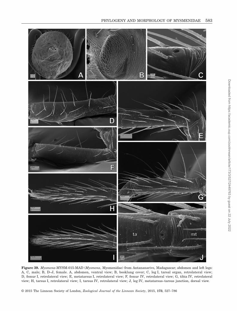
Figure 39. Mysmena-MYSM-015-MAD (Mysmena, Mysmenidae) from Antananarivo, Madagascar; abdomen and left legs:A, C, male; B, D–J, female. A, abdomen, ventral view; B, booklung cover; C, leg I, tarsal organ, retrolateral view;D, femur I, retrolateral view; E, metatarsus I, retrolateral view; F, femur IV, retrolateral view; G, tibia IV, retrolateralview; H, tarsus I, retrolateral view; I, tarsus IV, retrolateral view; J, leg IV, metatarsus–tarsus junction, dorsal view.
PHYLOGENY AND MORPHOLOGY OF MYSMENIDAE 583
© 2015 The Linnean Society of London, Zoological Journal of the Linnean Society, 2015, 173, 527–786
Dow
nloaded from https://academ
ic.oup.com/zoolinnean/article/173/3/527/2449763 by guest on 22 July 2022

measured in ratio characters are dependent on bodysize, but not on their ratios. If measurements scoredin each of the ratio characters are included as sepa-rate characters, then these characters would be highlycorrelated and size variation would be scored manytimes under different names. Consequently, thesemeasurements are included here as ratio characters,in spite of missing evolutionary information. Given the
subjectivity of the discretization and the lack of statedefinitions in the original discrete ratio characters, thiscoding represents an improvement in the objectivityof their definition, and can further elucidate a ten-dency (if any) of change in proportion throughout thephylogenetic history of these features. We carried outa third phylogenetic analysis to explore the influenceof the phylogenetic signal of the seven continuous
Figure 40. Mysmena-MYSM-015-MAD (Mysmena, Mysmenidae) from Antananarivo, Madagascar; male left palp, details.A, ventral-distal view; B, prolateral view; C, same, close up; D, ventral view; E, retrolateral view; F, dorsal view. SeeAppendix 3 for the list of abbreviations.
584 L. LOPARDO AND G. HORMIGA
© 2015 The Linnean Society of London, Zoological Journal of the Linnean Society, 2015, 173, 527–786
Dow
nloaded from https://academ
ic.oup.com/zoolinnean/article/173/3/527/2449763 by guest on 22 July 2022

characters by removing them from the original mor-phological data set. The modified data set containingonly discrete characters (65 taxa, 350 characters) wasanalysed under the same heuristic search param-eters. In addition, we compared the changes in the sym-metric resampling frequencies (see below) for the cladesshared between the resulting cladograms from bothanalyses (Goloboff et al., 2006).
Internal branches were considered unsupported andcollapsed during searches if they were supported am-biguously (when some optimization lacks support, ‘rule 1’of TNT; i.e. the minimum length is zero; see discus-sion in Coddington & Scharff, 1994). All discrete multi-state characters were treated as unordered (non additive;Fitch, 1971). For complex characters, reductive (binary)coding was maximized as much as possible in the dataset. In this coding, a multistate character is usuallysplit into a number of characters, where generallyabsence and presence are treated separately fromthe remaining qualities. This method introducesinapplicable scores in the matrix (which are
treated as missing data by the current phylogeneticsoftware), with the known risk associated withinapplicable characters for optimization (see Maddison,1993). Nevertheless, it minimizes redundancy anddependency among characters (Maddison, 1993; for areview and discussion of coding methods and theproblem of inapplicable character states, see alsoStrong & Lipscomb, 1999). State transformationsare considered synapomorphies for a given node onlyif they are unambiguous and shared by all dichoto-mous most-parsimonious trees; however, commentson putative (i.e. ambiguously optimized) synapomorphiesfor clades and for taxa are also discussed. Putativesynapomorphies may result from inapplicable scoring,from abundant missing information, or simply fromambiguity resulting from scoring of different statesaround the node of interest. Nevertheless, whenthese features occur exclusively within a clade ofinterest (or at least in most of its representatives),they are reported here as putative synapomorphies.Resulting cladograms are summarized in strict
Figure 41. Mysmena leichhardti (Mysmenidae) from Queensland, Australia; male left palp. A, prolateral–ventral view;B, dorsal view; C, retrolateral–ventral view; D, prolateral view; E, retrolateral view. See Appendix 3 for the list ofabbreviations.
PHYLOGENY AND MORPHOLOGY OF MYSMENIDAE 585
© 2015 The Linnean Society of London, Zoological Journal of the Linnean Society, 2015, 173, 527–786
Dow
nloaded from https://academ
ic.oup.com/zoolinnean/article/173/3/527/2449763 by guest on 22 July 2022

Figure 42. Mysmena leichhardti (Mysmenidae) from Queensland, Australia: A, B, E, G, H, male; C, D, F, female. A,prosoma, lateral view; B, same, lateral–frontal view; C, epigynal area and scapus, ventral view; D, digested abdomen,detail of vulva, tracheae removed; E, mouthparts, distal view; right femur I, prolateral–dorsal view; G, left femur I, ventralview; H, left metatarsus I, prolateral view. See Appendix 3 for the list of abbreviations.
586 L. LOPARDO AND G. HORMIGA
© 2015 The Linnean Society of London, Zoological Journal of the Linnean Society, 2015, 173, 527–786
Dow
nloaded from https://academ
ic.oup.com/zoolinnean/article/173/3/527/2449763 by guest on 22 July 2022

consensus for each analysis. The character matrix(Table 5) was edited and managed using MES-QUITE 2.01 (Maddison & Maddison, 2007). Charac-ter state optimizations and tree editing were performedin WINCLADA (Nixon, 1999b).
SENSITIVITY ANALYSES
To explore the sensitivity of the data to variation inanalytical parameters, i.e. the effect of data pertur-bation on phylogenetic results (‘sensitivity analysis’ sensuWheeler, 1995; see also Giribet, 2003), the morpho-logical data set (65 taxa, 357 characters) wasanalysed under parsimony using differential character-weighting schemes. Although the analyses were per-formed here solely to explore the stability ofrelationships proposed in the preferred phylogenetichypotheses, the sensitivity of groups to changes in theanalytical parameters might also provide an insightto the support of groups (Giribet, 2003). Stable cladesand taxa relationships remain invariable under a widerange of parameters, whereas unstable relationshipsremain only under fewer particular parameters. The
data set was analysed under different weighting regimesagainst homoplasy, using implied weighting (Goloboff,1993). Sensitivity of the results to variations in thestrength of the weighting function was assessed per-forming heuristic searches (same commands as above)using integer values of the constant of concavity (k).The selection of 11 different concavities representinga range of 100 values was based on preliminary analy-ses of the morphological data set under all concavi-ties (1–100, not shown). The k values chosen were: 1,2, 3, 6, 8, 10, 12, 20, 35, 50, and 99. We plotted thestability of clades as ‘Navajo rugs’ (sensitivity plots;Giribet & Edgecombe, 2006) at the nodes in the strictconsensus of the equal-weights hypotheses.
SUPPORT VALUES: CLADE SUPPORT
We calculated three clade support measures using TNT:Bremer support, relative Bremer support, and sym-metric resampling frequencies.
Bremer support measuresThe Bremer support (BS; Bremer, 1988, 1994) of aclade represents the number of extra steps a
Figure 43. Mysmena leichhardti (Mysmenidae) from Queensland, Australia; male left palp, details. A, B, embolus–conductor interaction; C, distal palp, distal–prolateral view; D, dorsal cymbial tip; E, ventral–prolateral cymbial tip. SeeAppendix 3 for the list of abbreviations.
PHYLOGENY AND MORPHOLOGY OF MYSMENIDAE 587
© 2015 The Linnean Society of London, Zoological Journal of the Linnean Society, 2015, 173, 527–786
Dow
nloaded from https://academ
ic.oup.com/zoolinnean/article/173/3/527/2449763 by guest on 22 July 2022

most-parsimonious tree requires to collapse the mono-phyly of that clade. BS was calculated heuristicallysearching for suboptimal trees using the optimal treesas a starting point. TBR branch swapping was per-formed until the tree-buffer was filled, gradually in-creasing the number of steps of suboptimals by one(from 1 to 12) then by two (from 12 to 22), expandingthe numbers of retained trees in intervals of 3000 eachtime, (from 2000 to 50 000, with a series of com-mands similar to sub 1 hold 2000; bb = tbr fillonly; sub2 hold 5000; bb = tbr fillonly;). The lowest values ofBS are reported. The relative Bremer support (rela-tive fit difference, RFD; Goloboff & Farris, 2001), takesinto account the relative levels of evidence, contradic-tory and favourable, for a clade (ranging from 0 to 100%,for highly contradicted and highly supported groups,respectively). The RFD was calculated in the samemanner as the BS using only suboptimal values in anumber of steps no greater than the BS of the group(i.e. only suboptimal trees within absolute support;command bsupport];).
Symmetric resampling frequenciesAbsolute symmetric resampling frequencies (SFqs) werecalculated by computing 4000 pseudoreplicates (prob-ability of character elimination: 0.33), performing heu-ristic searches consisting of ten random-additionsequences (RASs), followed by ten iterations of TBR,holding one tree (commands mult: noratchet repl 10tbr hold 1; resample sym repl 4000 freq from 0 [mult);).SFq and frequency differences (GC, Goloboff et al.,2003a) are reported in the strict consensus of Figs 154,157, and 158. The GC is the absolute frequency of agroup, minus the frequency of the most frequent contra-dictory group (ranging from 100 to −100% for best toworst support, respectively). Both GC and absolute sym-metric frequencies have been shown to be less biasedthan traditional bootstrap or jackknifing estimations(Goloboff et al., 2003a). In addition to symmetricresampling, we also calculated group frequencies undertraditional bootstrap (Felsenstein, 1985) and jack-knife (Farris et al., 1996; Farris, 1997; Goloboff et al.,2003a) resampling schemes (same search strategies,
Figure 44. Mysmena-MYSM-018-MAD (Mysmena, Mysmenidae) from Mahajanga, Madagascar: A, female epigynal areaand scapus, ventral–posterior view; B–D, male left palp; B, prolateral view; C, detail of embolus and pars pendula, retrolateral–ventral view; D, cymbial distal tip. See Appendix 3 for the list of abbreviations.
588 L. LOPARDO AND G. HORMIGA
© 2015 The Linnean Society of London, Zoological Journal of the Linnean Society, 2015, 173, 527–786
Dow
nloaded from https://academ
ic.oup.com/zoolinnean/article/173/3/527/2449763 by guest on 22 July 2022

Figure 45. MYSM-019-MAD (Mysmeninae, Mysmenidae) from Toliara, Madagascar. A–C, male right palp, inverted;A, dorsal view; B, retrolateral view; C, prolateral view. D, E, male left palp, expanded; D, prolateral–ventral view;E, same, detail of embolus and pars pendula; F, female posterior spinnerets; G–I, male left leg I, prolateral view;G, femur and patella; H, metatarsus; I, tarsus. See Appendix 3 for the list of abbreviations.
PHYLOGENY AND MORPHOLOGY OF MYSMENIDAE 589
© 2015 The Linnean Society of London, Zoological Journal of the Linnean Society, 2015, 173, 527–786
Dow
nloaded from https://academ
ic.oup.com/zoolinnean/article/173/3/527/2449763 by guest on 22 July 2022

with commands boot or jak instead of sym for boot-strap or jackknife resampling, respectively), ex-pressed as absolute frequencies or frequency differences(not shown). Because all values were highly correlat-ed, we only report the symmetric resampling values.
For the support values calculated in the analysesand reported below, we refer to low support for values0.01–2.50 (BS), 0–39 (RFD), and 50–73% (SFq); inter-
mediate support for values 2.51–8 (BS), 40–79 (RFD),and 74–85% (SFq); and high support for values 8.01or higher (BS), 80–100 (RFD), and 86–100% (SFq).
RESULTS
The following section reports the results of all mor-phological cladistic analyses performed in this study.
Figure 46. MYSM-019-MAD (Mysmeninae, Mysmenidae) from Toliara, Madagascar. A, C, D, F, G, female; B, E, male.A, prosoma, ventral view; B, same, frontal view; C, labrum, lateral view; D, ocular area, frontal view; E, same, lateralview; F, cheliceral bases, frontal view; G, right leg I, tarsal organ and tarsus–metatarsus junction.
590 L. LOPARDO AND G. HORMIGA
© 2015 The Linnean Society of London, Zoological Journal of the Linnean Society, 2015, 173, 527–786
Dow
nloaded from https://academ
ic.oup.com/zoolinnean/article/173/3/527/2449763 by guest on 22 July 2022

Names of taxa refer to taxonomic or nomenclatural de-cisions from the classification based on the preferredtotal-evidence hypothesis of relationships (Lopardo et al.,2011, fig. 12; see Figs 160, 161B, Appendix 5, and below).Node numbers from the phylogenetic hypothesis basedon morphological data (Fig. 153) begin with ‘M’; nodesfrom the combined total-evidence hypothesis (Figs 160,161) begin with ‘C’.
MORPHOLOGICAL DATA SET (65 TAXA,357 CHARACTERS)
The cladistic analysis of the morphological data set of65 taxa (357 characters) resulted in three most-parsimonious trees (MPTs) of 1512.169 steps (consist-ency index, CI = 0.366; retention index, RI = 0.666; seestrict consensus in Figs 153, 154). A total of 311 char-acters (87.1%) are phylogenetically informative, the re-
maining 12.9% characters are autapomorphic (for asummary of analysis of statistics and character com-position, see Table 6). Conflicting relationships causedone node within Anapidae to collapse in the strict con-sensus. Major clades, and support and stability values,are reported below. Synapomorphies for clades (exceptfor Mysmenidae, see below), and the description andimages of characters and character states, are listedin Appendices 2 and 4.
‘Symphytognathoids’ (node M69) and clades withinsymphytognathoidsResults of the morphological analysis support the com-position of symphytognathoids as proposed by Griswoldet al. (1998), and modified in Schütt (2003). That is,it includes Anapidae, Mysmenidae, Symphytognathidae,Theridiosomatidae, and also Synaphridae andMicropholcommatinae sensu Lopardo et al. (2011).
Figure 47. MYSM-020-MAD (Mysmeninae, Mysmenidae) from Toamasina, Madagascar, male. A–E, left palp; A, retrolateralview; B, prolateral–ventral view; C, same, detail of tip of embolus and cymbial conductors; D, same, ventral–retrolateralview; E, same, prolateral view. F, digested abdomen, tracheal system, dorsal view; G, same, detail of posterior trachealsystem. See Appendix 3 for the list of abbreviations.
PHYLOGENY AND MORPHOLOGY OF MYSMENIDAE 591
© 2015 The Linnean Society of London, Zoological Journal of the Linnean Society, 2015, 173, 527–786
Dow
nloaded from https://academ
ic.oup.com/zoolinnean/article/173/3/527/2449763 by guest on 22 July 2022

Although the implied weighting sensitivity analysis re-covered symphytognathoids as a natural group underall concavities studied, group support values are inter-mediate to low (BS 4.560; SFq 68%; RFD 20%; GC 45%).Interfamilial relationships are stable except for the po-sition of Synaphridae, with an alternative placementas sister to all other symphytognathoid families (see
below). Two main symphytognathoid clades resultedfrom the analysis.
First, a re-delimited Mysmenidae (see below) sisterto Theridiosomatidae (node M95). Both theridiosomatidand mysmenid representatives in this data set groupedtogether, although with low support values (BS 0.960;SFq 38%; RFD 10%; GC 7%). Mysmenidae are sister
Figure 48. MYSM-020-MAD (Mysmeninae, Mysmenidae) from Toamasina, Madagascar, male: A, epiandrous spigots;B, cheliceral teeth and fang; C, D, right tibia I, detail of ventral paired setae with protruded bases, prolateral view;E, right tibia I, ventral view; F, right tarsus I, detail of tarsal organ, retrolateral view; G, right leg IV, claws.
592 L. LOPARDO AND G. HORMIGA
© 2015 The Linnean Society of London, Zoological Journal of the Linnean Society, 2015, 173, 527–786
Dow
nloaded from https://academ
ic.oup.com/zoolinnean/article/173/3/527/2449763 by guest on 22 July 2022

Figure 49. MYSM-023-MAD (Mysmeninae, Mysmenidae) from Antananarivo, Madagascar: A, D, E, female; B, C, F–H,male. A, digested abdomen, detail of vulva; B, left palp, prolateral view, bulb displaced; C, same, detail of cymbial tip,retrolateral–dorsal view; D, abdomen, detail of epigynal area and respiratory spiracles, posterior view; E, same, ventralview; F, colulus and spinnerets, ventral view; G, posterior median spinnerets; H, prosoma, detail of sternum, labiumand maxillae, ventral view. See Appendix 3 for the list of abbreviations.
PHYLOGENY AND MORPHOLOGY OF MYSMENIDAE 593
© 2015 The Linnean Society of London, Zoological Journal of the Linnean Society, 2015, 173, 527–786
Dow
nloaded from https://academ
ic.oup.com/zoolinnean/article/173/3/527/2449763 by guest on 22 July 2022

Figure 50. MYSM-023-MAD (Mysmeninae, Mysmenidae) from Antananarivo, Madagascar: A, D, E, female; B, C, F–H,male. A, B, prosoma, lateral view; C, left chelicera, retromarginal view. D–H, right leg I, prolateral view; D, tarsus–metatarsus junction, prolateral–dorsal view; E, F, tarsus; G, tibia; H, metatarsus.
594 L. LOPARDO AND G. HORMIGA
© 2015 The Linnean Society of London, Zoological Journal of the Linnean Society, 2015, 173, 527–786
Dow
nloaded from https://academ
ic.oup.com/zoolinnean/article/173/3/527/2449763 by guest on 22 July 2022

to Theridiosomatidae under all implied weightingconcavities, except k = 1. Within this group, Theri-diosomatidae (node M127) have intermediate to lowsupport values (BS 3.410; SFq 79%; RFD 20%; GC 73%),but appears highly stable (monophyletic under allimplied weighting concavities).
The second clade comprises Synaphridae sister toAnapidae + Symphytognathidae (node M68), with inter-mediate to low support values (BS 4.160; SFq 65%;RFD 34%; GC 44%). This clade is one of the mostunstable (at the interfamilial level) withinsymphytognathoids, recovered by two weighting schemes(EW and IW at k = 99). Synaphridae (node M126),a highly supported and stable family (BS 13.000;SFq 100%; RFD 76%; GC 100%; recovered under allimplied weighting concavities), are alternatively placedas sister to all other symphytognathoids (recoveredby all other concavities). Support values for the cladecomprising Anapidae and Symphytognathidae (nodeM67) are intermediate to low (BS 3.170; SFq 67%;RFD 51%; GC 56%), with all concavities recoveringthis clade.
Symphytognathidae (node M103) are relatively wellsupported (intermediate support) and stable, and com-prise all representatives in this data set, includingthe enigmatic Iardinis mussardi (see below) (BS 6.000;SFq 79%; RFD 60%; GC 70%). Symphytognathidae aremonophyletic under all implied weighting concavi-ties. The resulting pectinated pattern of relation-ships encumbers the proposal of groups within thefamily.
Anapidae (node M66) include all its represen-tatives. In addition, the analysis places all microphol-commatine representatives within this family (Taphiassaand Teutoniella, node M79), which group together dis-tally within the family, sister to the controversialComaroma (node M76, recovered under all concavi-ties, see below). This hypothesis (i.e. Micropholcommatinaeas a subfamily of Anapidae) is not new (Brignoli, 1970;Schütt, 2003), and is in agreement with the hypothesisrendered by the morphological and molecular com-bined analysis (Fig. 160; Lopardo et al., 2011; but seeRix & Harvey, 2010). Although seemingly stable,Micropholcommatinae is weakly supported (BS 1.800;
Figure 51. Mysmena tasmaniae (Mysmenidae). A–C male left palp; A, dorsal view; B, retrolateral–dorsal view; C, ex-panded palp, distal–ventral view. D, female digested abdomen, detail of vulva, tracheae removed. See Appendix 3 for thelist of abbreviations.
PHYLOGENY AND MORPHOLOGY OF MYSMENIDAE 595
© 2015 The Linnean Society of London, Zoological Journal of the Linnean Society, 2015, 173, 527–786
Dow
nloaded from https://academ
ic.oup.com/zoolinnean/article/173/3/527/2449763 by guest on 22 July 2022

SFq 67%; RFD 18%; GC 54%; recovered under all impliedweighting concavities). Support and stability values forAnapidae (including Micropholcommatinae) are inter-mediate to relatively high (BS 6.500; SFq 90%; RFD42%; GC 87%; all implied weighting concavities recover
this clade). Relationships within Anapidae aremostly unresolved and have relatively low support(except Minanapis). The familial placement ofthe controversial genus Acrobleps as an anapid iscorroborated (as hypothesized in Lopardo & Hormiga,
Figure 52. A, MYSM-034-MAD (Mysmena, Mysmenidae), female; B–D, MYSM-029-MAD (Mysmeninae, Mysmenidae),female; E, F, MYSM-028-MAD (Mysmena, Mysmenidae), female. A, prosoma, lateral view; B, right anterior lateral spin-neret; C, right posterior lateral spinneret; D, right leg I, claws, retrolateral view; E, chelicerae and palps, frontal view;F, epigynal area, ventral view. See Appendix 3 for the list of abbreviations.
596 L. LOPARDO AND G. HORMIGA
© 2015 The Linnean Society of London, Zoological Journal of the Linnean Society, 2015, 173, 527–786
Dow
nloaded from https://academ
ic.oup.com/zoolinnean/article/173/3/527/2449763 by guest on 22 July 2022

Figure 53. Mysmenopsis cidrelicola (Mysmenidae), male paralectotype. A–F, left palp; A, retrolateral view; B, dorsalview; C, retrolateral–ventral view; D, bulb and cymbium, ventral view; E, detail of tibial hollow area; F, bulb andcymbium, retrolateral–ventral view. G, abdomen, pedicel area; H, posterior spinnerets. See Appendix 3 for the list ofabbreviations.
PHYLOGENY AND MORPHOLOGY OF MYSMENIDAE 597
© 2015 The Linnean Society of London, Zoological Journal of the Linnean Society, 2015, 173, 527–786
Dow
nloaded from https://academ
ic.oup.com/zoolinnean/article/173/3/527/2449763 by guest on 22 July 2022

2008; also recovered by Lopardo et al., 2011).Acrobleps is placed basally in Anapidae, sister to theremaining family representatives. Minanapis ismonophyletic.
Mysmenidae (node M94)The results of this analysis support the monophyly ofMysmenidae, which are here re-delimited to excludethe genus Iardinis, an enigmatic taxon of uncertain
Figure 54. Mysmenopsis cidrelicola (Mysmenidae), male paralectotype. A, ocular area, frontal view; B, mouthparts, ventralview. C–G, left leg I; C, tibia and metatarsus, prolateral view; D, metatarsal clasping spine, dorsal view; E, tarsus, dorsalview; F, same, detail of tarsal organ; G, tarsus, prolateral view. H, femur, prolateral–ventral view.
598 L. LOPARDO AND G. HORMIGA
© 2015 The Linnean Society of London, Zoological Journal of the Linnean Society, 2015, 173, 527–786
Dow
nloaded from https://academ
ic.oup.com/zoolinnean/article/173/3/527/2449763 by guest on 22 July 2022

familial placement, recently disputed betweenMysmenidae and Synaphridae (Gertsch, 1960a; Levi& Levi, 1962; Forster & Platnick, 1977; Brignoli, 1978,1980; Wunderlich, 1986; Schütt, 2003; Miller, 2007;Platnick, 2014). Iardinis mussardi, our study species,is placed within Symphytognathidae in a distal cladeof relatively high support and stability. As a result, aredefinition of Mysmenidae to exclude Iardinis is needed.
Mysmenidae, comprising all current mysmenid generaincluded in this analysis, with the exception of Iardinis,form a monophyletic group. Support for the familyis intermediate to low (BS 4.010; SFq 58%; RFD57%; GC 31%); however, Mysmenidae are monophyleticunder 11 out of the 12 implied weighting concavitiesexplored (all concavities except the most severe weight-ing, k = 1).
Figure 55. Mysmenopsis dipluramigo (Mysmenidae), male left palp. A, prolateral view; B, dorsal view; C, retrolateralview; D, tibia, detail from figure A, prolateral view; E, same, detail from figure C, retrolateral view; F, distal view;G, embolus and tip of cymbium, detail from figure A, prolateral view; H, detail of tibial hollow area, ventral view;I, same, detail of spur. See Appendix 3 for the list of abbreviations.
PHYLOGENY AND MORPHOLOGY OF MYSMENIDAE 599
© 2015 The Linnean Society of London, Zoological Journal of the Linnean Society, 2015, 173, 527–786
Dow
nloaded from https://academ
ic.oup.com/zoolinnean/article/173/3/527/2449763 by guest on 22 July 2022

Under the phylogenetic hypothesis based on mor-phological data, Mysmenidae are diagnosed by the fol-lowing combination of unambiguous synapomorphies,present in most of their representatives (but see Ap-pendix 5 and Figs 160, 161B for the diagnosis and taxo-nomic decisions of the family based on the combinedtotal-evidence hypothesis; see also Lopardo et al., 2011
for the diagnosis of Mysmenidae based on total-evidence analyses): cymbium oriented ventrally orprolatero-ventrally (character 163, state 1; retrolateral–dorsal or fully prolateral in few mysmenid taxa);cymbium with a distinct prolateral and apical inter-nal cymbial conductor (CyC1; characters 175, state 1;176, state 0; strictly apical in Mysmeninae, absent in
Figure 56. Mysmenopsis dipluramigo (Mysmenidae): A, B, E–G, male; C, D, female. A, left patella III, dorsal view; B,left leg II, claws; C, abdomen, epigynal area, ventral view; D, same, colulus, posterior respiratory spiracle and spinnerets,ventral view; E, epiandrous spigots; F, left tarsus I, dorsal view; G, mouthparts, ventral view.
600 L. LOPARDO AND G. HORMIGA
© 2015 The Linnean Society of London, Zoological Journal of the Linnean Society, 2015, 173, 527–786
Dow
nloaded from https://academ
ic.oup.com/zoolinnean/article/173/3/527/2449763 by guest on 22 July 2022

Figure 57. Mysmenopsis dipluramigo (Mysmenidae), left legs I: A–E, female; F–J, male. A, femur, prolateral view;B, same, retrolateral view; C, same, detail of stridulatory area; D, same, detail from panel C; E, femoral projection, detailfrom panel A; F, dorsal view; G, tibial and metatarsal clasping spines, prolateral view; H, metatarsal clasping spines;I, same, detail of strongest clasping spine; J, tibial clasping spines.
PHYLOGENY AND MORPHOLOGY OF MYSMENIDAE 601
© 2015 The Linnean Society of London, Zoological Journal of the Linnean Society, 2015, 173, 527–786
Dow
nloaded from https://academ
ic.oup.com/zoolinnean/article/173/3/527/2449763 by guest on 22 July 2022

Figure 58. Mysmenopsis palpalis (Mysmenidae): A–G, male; H, female. A–E, left palp; A, prolateral view; B, dorsal view;C, distal view; D, embolus and distal cymbium, ventral view; E, detail of tibial spurs. F, posterior median spinnerets;G, H, posterior lateral spinneret. See Appendix 3 for the list of abbreviations.
602 L. LOPARDO AND G. HORMIGA
© 2015 The Linnean Society of London, Zoological Journal of the Linnean Society, 2015, 173, 527–786
Dow
nloaded from https://academ
ic.oup.com/zoolinnean/article/173/3/527/2449763 by guest on 22 July 2022
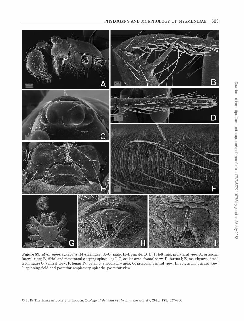
Figure 59. Mysmenopsis palpalis (Mysmenidae): A–G, male; H–I, female. B, D, F, left legs, prolateral view. A, prosoma,lateral view; B, tibial and metatarsal clasping spines, leg I; C, ocular area, frontal view; D, tarsus I; E, mouthparts, detailfrom figure G, ventral view; F, femur IV, detail of stridulatory area; G, prosoma, ventral view; H, epigynum, ventral view;I, spinning field and posterior respiratory spiracle, posterior view.
PHYLOGENY AND MORPHOLOGY OF MYSMENIDAE 603
© 2015 The Linnean Society of London, Zoological Journal of the Linnean Society, 2015, 173, 527–786
Dow
nloaded from https://academ
ic.oup.com/zoolinnean/article/173/3/527/2449763 by guest on 22 July 2022

Figure 60. Mysmenopsis penai (Mysmenidae): A–F, male right palp, inverted; G, H, female digested abdomen. A, ventralview; B, same, detail of tibial hollow area; C, distal view; D, same, detail of embolus and cymbium; E, tibial spurs, detailfrom panel B; F, embolus and distal cymbium; G, posterior tracheal system; H, anterior tracheal system and vulva. SeeAppendix 3 for the list of abbreviations.
604 L. LOPARDO AND G. HORMIGA
© 2015 The Linnean Society of London, Zoological Journal of the Linnean Society, 2015, 173, 527–786
Dow
nloaded from https://academ
ic.oup.com/zoolinnean/article/173/3/527/2449763 by guest on 22 July 2022

Mysmenopsis), and a flat, rounded paracymbium (char-acter 239, state 3; hook-shaped in Mysmenopsis).Mysmenid females have a distinct structure on theapical ventral surface of at least femur I, either asclerotized spot or a projection (character 101, state 1;absent in some Mysmenopsis species and MYSM-005-ARG). Other unambiguous synapomorphies for thefamily include a lobe on the intersegmental groove ofthe anterior lateral spinnerets (ALS; character 309,state 1; convergently present in few anapid repre-sentatives); abdomen with fingerprint cuticle pattern(character 4, state 1; independently synapomorphic forSymphytognathidae, imbricate in Maymena ambita);and cheliceral promargin with three teeth (charac-ter 278, state 1, although either four or two teeth canoccur within the family). Putative synapomorphies forMysmenidae (see Discussion below for details in theevolution of these characters): intermediate sternumposterior margin (character 53, state 2; pointed in someMaymena species); prolateral row of modified setae ontarsus I (character 138, state 1); sparse imbricate cu-ticular pattern on carapace border (character 47, state 0;
smooth in Maymena mayana); two aciniform glandspigots on posterior median spinnerets (PMS AC; char-acter 305, state 1); male palpal tibial rim setae longerthan remaining tibial setae and arranged distally ina row or two (character 255, state 1; equally short andof irregular conformation arising convergently inTrogloneta and few other mysmenid taxa); medialparacymbium (PC; character 241, state 1; basal inTrogloneta and in Theridiosomatidae; median also inSynaphridae); cymbial fold (character 187, state 1; absentin Maymena mayana and Mysmenopsis); metatarsalclasping spine in males (character 115, state 1; thepresence of this distinct metatarsal structure –character 114, state 1 – optimized as ambiguous inMysmenidae because of the absence of such struc-ture in Maymena mayana); female ventral distal spot(character 102, state 0; projection in Mysmenopsis) onfemora I and II (character 103, state 1; on femur I inMysmenopsis, Trogloneta, and node M96); epigynalmedian plate projecting from epigastric furrow (char-acter 70, state 1; ambiguously optimized because ofunknown information in theridiosomatids, some
Figure 61. Mysmenopsis penai (Mysmenidae), female. A, epigynum, ventral view; B, same, posterior view; C, left ante-rior lateral spinneret; D, right posterior median spinneret. See Appendix 3 for the list of abbreviations.
PHYLOGENY AND MORPHOLOGY OF MYSMENIDAE 605
© 2015 The Linnean Society of London, Zoological Journal of the Linnean Society, 2015, 173, 527–786
Dow
nloaded from https://academ
ic.oup.com/zoolinnean/article/173/3/527/2449763 by guest on 22 July 2022

Figure 62. Mysmenopsis penai (Mysmenidae): A–D, female; E–H, male left leg I. A, right palpal tibia, dorsal view;B, right maxilla; C, labrum; D, right chelicera, distal view. E, tibial and metatarsal clasping spines, prolateral view;F, same, detail of tibial clasping spine; G, metatarsus–tarsus junction, dorsal view; H, tarsal organ. See Appendix 3 forthe list of abbreviations.
606 L. LOPARDO AND G. HORMIGA
© 2015 The Linnean Society of London, Zoological Journal of the Linnean Society, 2015, 173, 527–786
Dow
nloaded from https://academ
ic.oup.com/zoolinnean/article/173/3/527/2449763 by guest on 22 July 2022

Figure 63. Trogloneta cantareira (Mysmenidae), male. A–F, right palp, inverted; A, retrolateral view; B, prolateral view;C, dorsal view; D, ventral–retrolateral view; E, ventral view; F, detail of embolic base and primary cymbial conductor.G, prosoma, lateral view; H, ocular area, frontal view; I, mouthparts, lateral–ventral view. See Appendix 3 for the list ofabbreviations.
PHYLOGENY AND MORPHOLOGY OF MYSMENIDAE 607
© 2015 The Linnean Society of London, Zoological Journal of the Linnean Society, 2015, 173, 527–786
Dow
nloaded from https://academ
ic.oup.com/zoolinnean/article/173/3/527/2449763 by guest on 22 July 2022

Figure 64. Trogloneta cantareira (Mysmenidae), female: A, digested abdomen, detail of vulva; B, same, detail of pos-terior respiratory system; C, right anterior lateral spinneret; D, abdomen, detail of colulus and posterior respiratory spira-cle; E, prosoma, lateral view; F, same, anterodorsal view; G, left palpal tibia, dorsal view; H, sternal cuticular pattern;I, carapace dorsal cuticular pattern, detail from panel F. See Appendix 3 for the list of abbreviations.
608 L. LOPARDO AND G. HORMIGA
© 2015 The Linnean Society of London, Zoological Journal of the Linnean Society, 2015, 173, 527–786
Dow
nloaded from https://academ
ic.oup.com/zoolinnean/article/173/3/527/2449763 by guest on 22 July 2022

Figure 65. A, Mysmena (= Tamasesia) rotunda (Mysmenidae), female syntype, left posterior lateral spinneret. B–H, Troglonetacantareira (Mysmenidae). B, female left leg I; C–F, male left leg I; G, H, female left leg IV. B, femur, patella and tibia;C, metatarsus, dorsal view; D, tarsus, prolateral view; E, proximal metatarsal cuticular pattern, dorsal view; F, claws,prolateral view; G, tarsus, retrolateral view; H, metatarsus, retrolateral view. See Appendix 3 for the list of abbreviations.
PHYLOGENY AND MORPHOLOGY OF MYSMENIDAE 609
© 2015 The Linnean Society of London, Zoological Journal of the Linnean Society, 2015, 173, 527–786
Dow
nloaded from https://academ
ic.oup.com/zoolinnean/article/173/3/527/2449763 by guest on 22 July 2022

Figure 66. Trogloneta granulum (Mysmenidae), male. A, prosoma, lateral view. B–E, left palp; B, retrolateral view;C, prolateral view; D, prolateral–dorsal view; E, embolus. F, anterior lateral spinnerets; G, mouthparts, lateral view;H, labium, ventral view. See Appendix 3 for the list of abbreviations.
610 L. LOPARDO AND G. HORMIGA
© 2015 The Linnean Society of London, Zoological Journal of the Linnean Society, 2015, 173, 527–786
Dow
nloaded from https://academ
ic.oup.com/zoolinnean/article/173/3/527/2449763 by guest on 22 July 2022

Figure 67. Trogloneta granulum (Mysmenidae), female. A, epigynum, ventral view; B, same, posterior view; C, digestedabdomen, anterior respiratory system and vulva; D, left posterior spinnerets; E, booklung cover; F, abdomen, detail ofcolulus and posterior spiracle, ventral view; G, ocular area, frontal view; H, same, dorsal view. See Appendix 3 for thelist of abbreviations.
PHYLOGENY AND MORPHOLOGY OF MYSMENIDAE 611
© 2015 The Linnean Society of London, Zoological Journal of the Linnean Society, 2015, 173, 527–786
Dow
nloaded from https://academ
ic.oup.com/zoolinnean/article/173/3/527/2449763 by guest on 22 July 2022

reversions within the family); and either a more or lessstraight trajectory or a convoluted trajectory of thecopulatory ducts (character 78, states 0 and 2, highlyhomoplastic basally).
Clades within MysmenidaeMysmenidae are fully resolved in the consensus of theMPTs. Monophyletic genera include: Kekenboschiella,Microdipoena, Maymena, Mysmenopsis, and Trogloneta,whereas non-monophyletic mysmenid genera includeCalodipoena, Mysmenella, Tamasesia, and the typegenus Mysmena (but see Appendix 5 for generic syn-onymies). Below we report major clades withinMysmenidae in reference to the strict consensus ofFigures 153, 154. For a complete list of unambiguoussynapomorphies for each node and taxa, see Appen-dix 4. See Appendix 5 for taxonomic changes and genericdiagnoses based on the hypothesis of the combined total-evidence analysis from Lopardo et al. (2011; refer toFig. 161B).
Maymena (node M110) is monophyletic, sister to theremaining mysmenid representatives (BS 3.070; SFq58%; RFD 22%; GC 32%). In the implied weightingsensitivity analysis, the genus is monophyletic underall concavities except the most extreme (k = 1; seeFig. 154). The clade comprising all remaining mysmenid
representatives (node M93, excluding Maymena) is oneof the few clades diagnosed by several unambiguousand non-homoplastic synapomorphies (see Appen-dix 4); however, support values for this node are rela-tively intermediate to low (BS 3.050; SFq 46%; RFD25%; GC 31%). The implied weighting sensitivityscheme, however, consistently recovers this cladeunder all but the two most extreme concavities (k = 1and 2).
Trogloneta (node M123) is monophyletic; supportvalues for this genus are among the highest for thisdata set (BS 13.000; SFq 100%; RFD 93%; GC 100%).Accordingly, the implied weighting sensitivity schemerecovers Trogloneta under all concavities explored. Theclade comprising the remaining mysmenids, exclud-ing both Maymena and Trogloneta (i.e. node M92), in-cludes the subfamilies Mysmenopsinae (node M105) andMysmeninae (node M91) (see Appendix 5). This cladehas support values among the lowest for this data set(BS 0.040; SFq 35%; RFD 1%; GC −8%). In the impliedweighting sensitivity scheme, however, this group isconsistently monophyletic under all but the two mostextreme concavities (k = 1 and 2).
The subfamily Mysmenopsinae (node M105; see Ap-pendix 5) comprises all studied kleptoparasiticmysmenids. This subfamily is monophyletic under all
Figure 68. Trogloneta granulum (Mysmenidae), male. A, left tarsus I, prolateral view; B, left metatarsus I, prolateralview; C, abdomen, detail of colulus, posterior spiracle and spinnerets, ventral view; D, left tibia III, prolateral view.
612 L. LOPARDO AND G. HORMIGA
© 2015 The Linnean Society of London, Zoological Journal of the Linnean Society, 2015, 173, 527–786
Dow
nloaded from https://academ
ic.oup.com/zoolinnean/article/173/3/527/2449763 by guest on 22 July 2022

concavities explored, and support values for this nodeare fairly high (BS 7.020; SFq 98%; RFD 50%; GC 98%).Isela (node M104), including the Kilifina representa-tive (Kilifina-MYSM-002-KENYA) is monophyletic underall concavities explored (not shown; see Appendix 5 forgeneric synonymy). Frequency values for this genusare high (SFq 95%; GC 92%) although Bremer support
is low (BS 1.990; RFD 33%). Mysmenopsis (node M121)has the highest clade support within symphytognathoids(BS 14.160; SFq 100%; RFD 67%; GC 100%). Accord-ingly, the implied weighting sensitivity scheme recov-ers this node under all concavities explored (not shown).
The subfamily Mysmeninae (node M91; seeAppendix 5) comprises the remaining mysmenid
Figure 69. Acrobleps hygrophilus (Anapidae), female. A, carapace, frontal view; B, left palp and carapace lateral de-pression; C, abdomen, pedicel area, ventral view; D, same, detail of suprapedicellate nubbins; E, digested abdomen, detailof vulva, tracheae removed; F, right leg II, claws. See Appendix 3 for the list of abbreviations.
PHYLOGENY AND MORPHOLOGY OF MYSMENIDAE 613
© 2015 The Linnean Society of London, Zoological Journal of the Linnean Society, 2015, 173, 527–786
Dow
nloaded from https://academ
ic.oup.com/zoolinnean/article/173/3/527/2449763 by guest on 22 July 2022
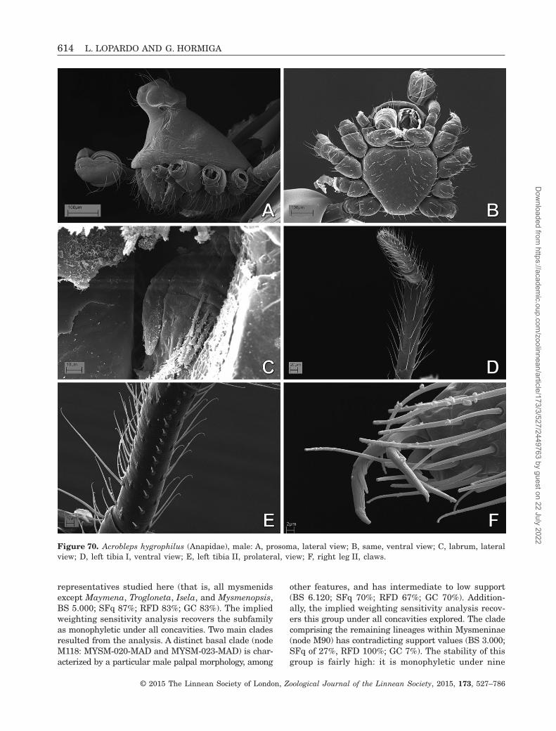
representatives studied here (that is, all mysmenidsexcept Maymena, Trogloneta, Isela, and Mysmenopsis,BS 5.000; SFq 87%; RFD 83%; GC 83%). The impliedweighting sensitivity analysis recovers the subfamilyas monophyletic under all concavities. Two main cladesresulted from the analysis. A distinct basal clade (nodeM118: MYSM-020-MAD and MYSM-023-MAD) is char-acterized by a particular male palpal morphology, among
other features, and has intermediate to low support(BS 6.120; SFq 70%; RFD 67%; GC 70%). Addition-ally, the implied weighting sensitivity analysis recov-ers this group under all concavities explored. The cladecomprising the remaining lineages within Mysmeninae(node M90) has contradicting support values (BS 3.000;SFq of 27%, RFD 100%; GC 7%). The stability of thisgroup is fairly high: it is monophyletic under nine
Figure 70. Acrobleps hygrophilus (Anapidae), male: A, prosoma, lateral view; B, same, ventral view; C, labrum, lateralview; D, left tibia I, ventral view; E, left tibia II, prolateral, view; F, right leg II, claws.
614 L. LOPARDO AND G. HORMIGA
© 2015 The Linnean Society of London, Zoological Journal of the Linnean Society, 2015, 173, 527–786
Dow
nloaded from https://academ
ic.oup.com/zoolinnean/article/173/3/527/2449763 by guest on 22 July 2022

Figure 71. Acrobleps hygrophilus (Anapidae), male left palp: A, ventral view; B, ventral–prolateral view; C, prolateralview; D, prolaterodorsal view; E, dorsal view; F, retrolateral view; G, detail of tibial structures, detail from panel D;H, detail of patellar apophyses, retrolateral view, detail from panel F. See Appendix 3 for the list of abbreviations.
PHYLOGENY AND MORPHOLOGY OF MYSMENIDAE 615
© 2015 The Linnean Society of London, Zoological Journal of the Linnean Society, 2015, 173, 527–786
Dow
nloaded from https://academ
ic.oup.com/zoolinnean/article/173/3/527/2449763 by guest on 22 July 2022

Figure 72. Anapisona kethleyi (Anapidae): A–D, female; E, F, male. A, prosoma, dorsal view; B, same, frontal–lateralview; C, same, lateral view; D, same detail of carapace cuticular pattern; E, abdomen, lateral view; F, same, detail ofpedicel area, ventral view.
616 L. LOPARDO AND G. HORMIGA
© 2015 The Linnean Society of London, Zoological Journal of the Linnean Society, 2015, 173, 527–786
Dow
nloaded from https://academ
ic.oup.com/zoolinnean/article/173/3/527/2449763 by guest on 22 July 2022

concavities (all concavities except k = 3, 6, and 8). Inaddition, low support and low stability values occurin most of the internal nodes of this clade.
DISCRETE MORPHOLOGICAL DATA SET
(65 TAXA, 350 CHARACTERS)
The cladistic analysis of the discrete morphological dataset (65 taxa scored for 350 characters: i.e. excludingall seven continuous characters) resulted in 3835 MPTsof 1323 steps (CI = 0.349; RI = 0.663; see strictconsensus in Fig. 157; see Table 6 for summary of analy-sis statistics and character composition). The family-level signal of the discrete characters analysed aloneis essentially identical to the combined data set in-cluding continuous characters: each family represent-ed by more than one taxon is monophyletic, andMysmenidae are monophyletic excluding Iardinis(which falls within Symphytognathidae). Relation-
ships among families and among mysmenid genera differslightly from the analysis of the complete data set, asfollows. Synaphridae are no longer sister to theAnapidae + Symphytognathidae clade. Instead,Synaphridae are placed basally, sister to all othersymphytognathoids, a relationship recovered under mostconcavity values in the implied weighting sensitivityanalyses of the complete morphological data set.As in the complete analysis, Mysmenidae andTheridiosomatidae are sister taxa. Within Mysmenidae,clades (and correspondent subclades) common to bothanalyses include (node numbers refer to consensusof the complete morphological analysis, Fig. 153):Maymena, Mysmenopsis, Isela, Mysmenopsinae,Trogloneta, Mysmeninae, Kekenboschiella, and nodeM118 (compare Figs 153, 157). Trogloneta is sister toMysmeninae when continuous characters are exclud-ed. Few clades within Mysmeninae are recovered.MYSM-005-ARG is the basal taxon, sister to all
Figure 73. Anapisona kethleyi (Anapidae), left legs: A, male; B–F, female. A, tibia I, ventral view; B, leg I, tarsal organ,dorsal view; C, same, claws, prolateral view; D, leg IV, metatarsus–tarsus junction; E, tibia IV, retrolateral view; F, same,prolateral view.
PHYLOGENY AND MORPHOLOGY OF MYSMENIDAE 617
© 2015 The Linnean Society of London, Zoological Journal of the Linnean Society, 2015, 173, 527–786
Dow
nloaded from https://academ
ic.oup.com/zoolinnean/article/173/3/527/2449763 by guest on 22 July 2022

remaining mysmenines, which comprise a mostly un-resolved clade.
In order to assess the effect of continuous charac-ters on the phylogenetic signal of the discrete char-acters alone (i.e. conflict or agreement between the twosubsets of data), support differences for common cladesbetween the discrete and the complete morphologicaldata sets were examined (see also Goloboff et al., 2006).Differences on the symmetric resampling frequenciesand GC were compared and plotted on the consensusof the discrete analysis (Fig. 158, compare values onFigs 154, 157). Out of a total of 27 common cladesbetween the two consensus trees, support when in-cluding the continuous characters decreased in 14 groupsfor GC (13 groups for absolute frequencies), with a sum
of support differences of 254 and 144 (respectively);increased in nine groups for GC (eight groups for ab-solute frequencies), with a sum of support differencesof 118 and 77 (respectively); and remained un-changed in four groups for GC (six groups for abso-lute frequencies). Unexpectedly, it appears that in ouranalyses the tendency when adding continuous char-acters is an overall decrease in support of approxi-mately 50%.
FAMILIAL PLACEMENT OF TAXA OF UNCERTAIN
AFFINITIES (70 TAXA, 357 CHARACTERS)
The cladistic analysis of the complete morphologicaldata set, including five taxa with more than 78% of
Figure 74. Anapisona kethleyi (Anapidae), left legs: A, B, D, E, male; C, F, G, female. A, tarsus I, prolateral view;B, metatarsus IV, retrolateral view; C, tarsus IV, retrolateral view; D, G, metatarsus I, prolateral view; E, F, femur I,retrolateral view.
618 L. LOPARDO AND G. HORMIGA
© 2015 The Linnean Society of London, Zoological Journal of the Linnean Society, 2015, 173, 527–786
Dow
nloaded from https://academ
ic.oup.com/zoolinnean/article/173/3/527/2449763 by guest on 22 July 2022

missing data (i.e. Calodipoena tarautensis, Crassignathahaeneli, Iardinis martensi, Leviola termitophila, andPhricotelus stelliger; 70 taxa, 357 characters) ren-dered three MPTs of 1545.559 steps (CI = 0.360;RI = 0.664; see strict consensus in Fig. 159). A total of312 characters (87.4%) are phylogenetically informa-tive, and the remaining 12.6% characters areautapomorphic (for a summary of the analysis of sta-
tistics and character composition, see Table 6). Overall,the resulting pattern of relationships is essentiallysimilar to that of the complete morphological data setincluding 65 taxa. Each family represented by morethan one taxon is monophyletic and, as previously found,Iardinis mussardi is placed within Symphytognathidae.All three MPTs support the symphytognathoidsinterfamilial relationships, with Theridiosomatidae sister
Figure 75. Anapisona kethleyi, spinnerets: A, D, E, G, female; B, C, F, male. A, B, anterior lateral spinnerets; C, D,posterior median spinnerets; E–G, posterior lateral spinnerets. See Appendix 3 for the list of abbreviations.
PHYLOGENY AND MORPHOLOGY OF MYSMENIDAE 619
© 2015 The Linnean Society of London, Zoological Journal of the Linnean Society, 2015, 173, 527–786
Dow
nloaded from https://academ
ic.oup.com/zoolinnean/article/173/3/527/2449763 by guest on 22 July 2022

Figure 76. Anapisona kethleyi (Anapidae), male right palp, inverted. A, retrolateral view; B, dorsal view; C, prolateral-dorsal view; D, prolateral view; E, retrolateral–dorsal view; F, detail of femur, retrolateral view; G, detail of embolicbase, retrolateral view; H, detail of tip of cymbial conductor; I, detail of patella, retrolateral view; J, cymbial tarsal organ,retrolateral view. See Appendix 3 for the list of abbreviations.
620 L. LOPARDO AND G. HORMIGA
© 2015 The Linnean Society of London, Zoological Journal of the Linnean Society, 2015, 173, 527–786
Dow
nloaded from https://academ
ic.oup.com/zoolinnean/article/173/3/527/2449763 by guest on 22 July 2022

Figure 77. Anapisona kethleyi (Anapidae), female prosoma: A, clypeus and cheliceral bases, frontal view; B, ocular area,frontal view; C, same, dorsal view; D, left palpal tarsus; E, labrum, lateral view; F, detail of cheliceral lateral condyle–boss; G, distal chelicerae, cheliceral fang and teeth; H, palpal tarsal organ; I, carapace lateral depression.
PHYLOGENY AND MORPHOLOGY OF MYSMENIDAE 621
© 2015 The Linnean Society of London, Zoological Journal of the Linnean Society, 2015, 173, 527–786
Dow
nloaded from https://academ
ic.oup.com/zoolinnean/article/173/3/527/2449763 by guest on 22 July 2022

Figure 78. Comaroma simoni (Anapidae), female abdomen: A, pedicel area, ventral–posterior view; B, spinning field,colulus, and posterior spiracle, ventral–posterior view; C, right anterior lateral spinneret; D, right posterior median spin-neret (PMS); E, left PMS; F, right posterior lateral spinneret (PLS), lateral view; G, same, posterior view; H, left PLS.See Appendix 3 for the list of abbreviations.
622 L. LOPARDO AND G. HORMIGA
© 2015 The Linnean Society of London, Zoological Journal of the Linnean Society, 2015, 173, 527–786
Dow
nloaded from https://academ
ic.oup.com/zoolinnean/article/173/3/527/2449763 by guest on 22 July 2022

to Mysmenidae, Anapidae sister to Symphytogna-thidae, and Synaphridae sister to (Anapidae +Symphytognathidae); however, because of the alter-native placements of Phricotelus (see below), relation-ships among the latter three families are unresolved.Regarding the familial placement of taxa with a highproportion of missing data, we suggest the following:
CALODIPOENA TARAUTENSIS BAERT 1988 (FIG. 130A)
Calodipoena tarautensis Baert, 1988: 17, fig. 21 [femaleholotype and female paratype from Sulawesi (Indo-nesia), Sulawesi Utara, Dumoga Bone National Park,Toraut, Grassland, 200 m, 27.x.1985, R. Bosman &
J. Van Stalle, in IRSN (no. IG 26977), male unknown,female paratype examined].
Familial placement: Mysmenidae. As expected, thisspecies grouped within one of the largest clades withinMysmeninae, which includes Calodipoena mootae,Mysmena leichhardti, Anjouanella, Microdipoena s.s.,Mysmenella, and three undescribed species [Mysmena-MYSM-015-MAD, MYSM-(029 034)-MAD]. Calodipoenatarautensis is known only from females, and shareswith mysmenines several features, such as femoral spotson legs I and II, wide posterior tracheal spiracle, ante-rior tracheae, absence of epigynal plate, long scape,highly membranous and complex pattern of ducts in
Figure 79. Comaroma simoni (Anapidae), female. A, digested abdomen, anterior respiratory system and vulva; B, mouth-parts, ventral–frontal view; C, right palpal tarsus, retrolateral view; D, distal chelicerae, frontal view; E, left palpal tibia,dorsal view; F, labrum.
PHYLOGENY AND MORPHOLOGY OF MYSMENIDAE 623
© 2015 The Linnean Society of London, Zoological Journal of the Linnean Society, 2015, 173, 527–786
Dow
nloaded from https://academ
ic.oup.com/zoolinnean/article/173/3/527/2449763 by guest on 22 July 2022

the vulva, absence of palpal claw but palp with all seg-ments, and absence of pore-bearing depressions.
GENUS CRASSIGNATHA WUNDERLICH 1995
Crassignatha Wunderlich, 1995: 546. Type species byoriginal designation and monotypy: Crassignatha haeneliWunderlich 1995: 547 (type and only specimen not ex-amined, scored from literature). Miller et al. 2009: 68(transfer from Mysmenidae to Symphytognathidae).
Familial placement: Symphytognathidae. The place-ment of this genus within Symphytognathidae was re-cently proposed by Miller et al. (2009) based exclusivelyon morphological comparative observations (as here,the type species was also not examined). This place-ment is corroborated by the results of our phylogeneticanalysis. Crassignatha nested within Symphytog-nathidae as sister to a distal clade comprising threeundetermined symphytognathid species (SYMP-002-MAD, SYMP-006-AUST, and SYMP-007-AUST). The
Figure 80. Comaroma simoni (Anapidae), female left legs: A–E, left leg I; F–H, left leg IV. A, metatarsus–tarsus junc-tion, dorsal view; B, patella, tibia, dorsal view; C, metatarsal trichobothrial base; D–F, claws, prolateral view; G, tarsus,prolateral view; H, tibia, prolateral view. See Appendix 3 for the list of abbreviations.
624 L. LOPARDO AND G. HORMIGA
© 2015 The Linnean Society of London, Zoological Journal of the Linnean Society, 2015, 173, 527–786
Dow
nloaded from https://academ
ic.oup.com/zoolinnean/article/173/3/527/2449763 by guest on 22 July 2022

type species of Crassignatha, Crassignatha haeneli, isknown only from a male specimen (holotype), has noneof the synapomorphic features of Mysmenidae, and con-sequently its placement in a different family is hardlysurprising. It differs from Mysmenidae in the absenceof femoral spots, absence of tibial or metatarsal clasp-ing spines on leg I, prolateral (instead of ventral)
cymbium, absence of cymbial structures (e.g. primaryconductor, process, and paracymbium), absence of parspendula, and presence of median apophysis. In addi-tion, Crassignatha shares with Symphytognathidae theloss of the anterior median eyes, promarginal cheliceralteeth originating from a common base or raised plate,and loss of the colulus. It also shares with several
Figure 81. Comaroma simoni (Anapidae), male left palp: A, retrolateral view; B, ventral view; C, ventral–distalview; D, prolateral view; E, distal view; F, bulb, detail from panel B. See Appendix 3 for the list of abbreviations. SeeAppendix 3 for the list of abbreviations.
PHYLOGENY AND MORPHOLOGY OF MYSMENIDAE 625
© 2015 The Linnean Society of London, Zoological Journal of the Linnean Society, 2015, 173, 527–786
Dow
nloaded from https://academ
ic.oup.com/zoolinnean/article/173/3/527/2449763 by guest on 22 July 2022

Figure 82. Comaroma simoni (Anapidae), male: A, prosoma, lateral view; B, same, dorsal view; C, ocular area, frontalview; D, distal chelicerae, frontal view; E, abdomen, ventral view; F, epiandrous spigots; G, posterior median spinnerets;H, left posterior lateral spinneret. See Appendix 3 for the list of abbreviations.
626 L. LOPARDO AND G. HORMIGA
© 2015 The Linnean Society of London, Zoological Journal of the Linnean Society, 2015, 173, 527–786
Dow
nloaded from https://academ
ic.oup.com/zoolinnean/article/173/3/527/2449763 by guest on 22 July 2022

symphytognathids in this data set a clasping spinelocated ventrally on tibia II and the absence of palpalpatellar or tibial apophyses.
GENUS IARDINIS SIMON 1899 (FIGS 135A, B, 144C)
Iardinis Simon, 1899: 87. Type species by originaldesignation: Iardinis weyersi Simon 1899: 87 (nomen
dubium, female type and only specimen lost); Levi &Levi, 1962: 22 (considered incertae sedis); Forster &Platnick, 1977: 5 (considered nomen dubium); Brignoli,1970: 1426; 1978: 250; 1980: 731 (provisional trans-fer to Mysmenidae).
Iardinus, Gertsch, 1960a: 8 (lapsus calami, trans-ferred from Theridiidae to Symphytognathidaes.l.).
Figure 83. Comaroma simoni (Anapidae), male left legs: A, tibia I, retrolateral; B, metatarsus I, retrolateral; C, meta-tarsus, tarsus IV, prolateral; D, patella, tibia I, prolaterodorsal; E, leg I, tarsal organ; F, leg IV, claws, prolateral.
PHYLOGENY AND MORPHOLOGY OF MYSMENIDAE 627
© 2015 The Linnean Society of London, Zoological Journal of the Linnean Society, 2015, 173, 527–786
Dow
nloaded from https://academ
ic.oup.com/zoolinnean/article/173/3/527/2449763 by guest on 22 July 2022

Familial placement: Symphytognathidae. The type andonly specimen of the type species (Iardinus weyersi)from Sumatra was described by Simon in 1899, buthas been considered lost by the arachnologists thathave tried to examine the type material (Gertsch,1960a; Levi & Levi, 1962; Brignoli, 1970, 1978, 1980;Forster & Platnick, 1977). The vial from the ParisMuseum (MNHN) with the original label that
should have housed the type material is actuallyempty (L. Lopardo & G. Hormiga, pers. observ.).Nevertheless, two additional species have been de-scribed for the genus: Iardinus martensi Brignoli, 1978from Nepal, and Iardinus mussardi Brignoli 1980 fromIndia (holotype examined, see also Figs 135A, B, 144C).Both species are exclusively known from their maletype specimens. To add to the enigmatic status of
Figure 84. Crassanapis chilensis (Anapidae), right legs: A–E, right legs I; F, G, right legs IV. A, B, D, G, male; C, E, F,female. A, femur and patella, retrolateral; B, same, detail; C, metatarsus, prolateral; D, detail of setal bases on ventralfemur; E, metatarsal clasping spur, detail from panel C; F, detail of tarsal ventral setae, retrolateral view; G, tibia, retrolateralview.
628 L. LOPARDO AND G. HORMIGA
© 2015 The Linnean Society of London, Zoological Journal of the Linnean Society, 2015, 173, 527–786
Dow
nloaded from https://academ
ic.oup.com/zoolinnean/article/173/3/527/2449763 by guest on 22 July 2022

the genus, the original vial containing the type andonly specimen of I. martensi instead contained a femaleanapid (L. Lopardo & G. Hormiga, pers. observ.), andtherefore its morphology had to be scored from theliterature.
Nonetheless, both Iardinis species included inthe 65- and 70-taxa data sets (i.e. I. martensi andI. mussardi) are more closely related to Symphy-tognathidae than to Mysmenidae. Morphologically thesespecies have none of the synapomorphic features ofMysmenidae, and thus, as with Crassignatha, their
placement in a different family was expected. They differfrom Mysmenidae in the absence of femoral spots (atleast in I. mussardi), absence of tibial or metatarsalclasping spines on leg I, dorsal (instead of ventral)cymbium, absence of cymbial structures (e.g. primaryconductor, process, and paracymbium), and presenceof median apophysis. They further share withSymphytognathidae the loss of anterior median eyes,thick setae on the dorsal part of the abdomen, absenceof cheliceral denticles, loss of colulus, and absence ofpalpal patellar or tibial apophyses.
Figure 85. Crassanapis chilensis (Anapidae), female: A, prosoma, lateral view; B, digested abdomen, anterior respira-tory system and vulva, dorsal view; C, same, posterior respiratory system; D, same, anterior respiratory system andvulva, dorsal–anterior view; E, labium, ventral view; F, carapace, lateral cuticular pattern; G, left palpal tarsus.
PHYLOGENY AND MORPHOLOGY OF MYSMENIDAE 629
© 2015 The Linnean Society of London, Zoological Journal of the Linnean Society, 2015, 173, 527–786
Dow
nloaded from https://academ
ic.oup.com/zoolinnean/article/173/3/527/2449763 by guest on 22 July 2022

Figure 86. Crassanapis chilensis (Anapidae), male: A, prosoma, frontal–lateral view; B, labrum, lateral view; C, booklungcover; D, abdominal cuticle; E, carapace lateral depression. F, H, right palp, inverted; G, I, left palp. F, ventral view;G, ventral–proximal view; H, detail of patella and median apophysis; I, detail of median apophysis, conductor, and tip ofembolus. See Appendix 3 for the list of abbreviations.
630 L. LOPARDO AND G. HORMIGA
© 2015 The Linnean Society of London, Zoological Journal of the Linnean Society, 2015, 173, 527–786
Dow
nloaded from https://academ
ic.oup.com/zoolinnean/article/173/3/527/2449763 by guest on 22 July 2022

GENUS PHRICOTELUS SIMON, 1895(FIGS 135C, 144B)
Phricotelus Simon, 1895a: 919. Type species by origi-nal designation and monotypy: Phricotelus stelligerSimon, 1895a: 919 (type specimen examined); Levi,1972: 534 (transferred from Theridiosomatidae toSymphytognathidae); Brignoli, 1980: 731, 1981: 14 (pro-visional transfer to Mysmenidae).
Familial placement: Araneoidea incertae sedis. Alter-native equally parsimonious placements of Phricotelusimply various relationships to Synaphridae, Anapidae,or Symphytognathidae; therefore, its placement becomesunresolved in the strict consensus cladogram. In theresulting three MPTs, Phricotelus is placed as themost basal species of Anapidae, Anapidae +Symphytognathidae, or Synaphridae. Although muchof the morphology for P. stelliger needs to be properly
Figure 87. Elanapis aisen (Anapidae), female: A, prosoma, ventral view; B, left tarsus I, detail of tarsal organ; C, leftposterior lateral spinneret; D, digested abdomen, detail of vulva; E, abdomen, lateral view; F, mouthparts, ventral view;G, abdomen, anterior pedicel area, ventral view. See Appendix 3 for the list of abbreviations.
PHYLOGENY AND MORPHOLOGY OF MYSMENIDAE 631
© 2015 The Linnean Society of London, Zoological Journal of the Linnean Society, 2015, 173, 527–786
Dow
nloaded from https://academ
ic.oup.com/zoolinnean/article/173/3/527/2449763 by guest on 22 July 2022

Figure 88. Elanapis aisen (Anapidae), male: A, prosoma, lateral view; B, detail of carapace lateral depression; C, chelicerae,frontal view; D, right palp, inverted, ventral view; E, same, detail of tip of palp, retrolateral view; abdomen, detail ofcolulus, posterior spiracle and spinnerets, ventral view; G, posterior median spinnerets; H, tibia III; I, leg IV, claws. SeeAppendix 3 for the list of abbreviations.
632 L. LOPARDO AND G. HORMIGA
© 2015 The Linnean Society of London, Zoological Journal of the Linnean Society, 2015, 173, 527–786
Dow
nloaded from https://academ
ic.oup.com/zoolinnean/article/173/3/527/2449763 by guest on 22 July 2022

scored, the type species of this genus, only known byfemales, was not placed within or closely relatedto Mysmenidae. Furthermore, its placementwithin symphytognathoids also remains uncertain.Morphologically, Phricotelus has none of thesynapomorphic features that diagnose Mysmenidae,and thus its placement in a different family wasexpected. Although females have long scape, no
epigynal plate (Fig. 135C), no pore-bearing depres-sion on lateral edges of the carapace, and a narrowposterior respiratory spiracle located in front of thespinnerets, the species differs from Mysmenidae inthe absence of femoral spots, two pairs of spermathecae,and abdomen extremely projected posteriorly (seeFigs 135C, 144B; further morphological details notobserved).
Figure 89. Minanapis casablanca (Anapidae), female: A, prosoma, dorsal view; B, labium–sternum junction, ventral view;C, left anterior lateral spinneret; D, right posterior lateral spinneret. E–G, right leg I, prolateral view; E, tarsus; F, meta-tarsus; G, tibia and patella. See Appendix 3 for the list of abbreviations.
PHYLOGENY AND MORPHOLOGY OF MYSMENIDAE 633
© 2015 The Linnean Society of London, Zoological Journal of the Linnean Society, 2015, 173, 527–786
Dow
nloaded from https://academ
ic.oup.com/zoolinnean/article/173/3/527/2449763 by guest on 22 July 2022

GENUS LEVIOLA MILLER, 1970
Leviola Miller, 1970: 155. Type species by original des-ignation and monotypy: Leviola termitophila Miller 1970(female type and only specimen not examined, pre-sumably lost or in Museu do Dundo, Angola; scoredfrom literature). Levi, 1972: 534 (transfer fromTheridiidae to Symphytognathidae); Brignoli, 1980: 731(provisional transfer from Symphytognathidae toMysmenidae).
Familial placement: Zodariidae. The placement of thisenigmatic genus, known only from females, has beencontroversial, and the rationale for its various familyplacements tenuous at best. Based on the original de-
scription (Miller, 1970), the morphology of L. termitophilais highly different from that of symphytognathoids (oreven araneoids), including features such as: presenceof a palpal claw, which is flat and comb-like; large ante-rior median eyes; lateral eyes not juxtaposed, tarsaland metatarsal trichobothrium in all legs, etc. (seeGriswold et al., 1998). Furthermore, its peculiar mor-phology resembles that of a few zodariid genera, suchas the African genera Akyttara or Diores. Leviola shareswith the latter zodariid genera (and with the familyZodariidae in general) robust chelicerae, large ante-rior median eyes, female palpal claw with roughly tenteeth on the prolateral side, comb-like teeth arisingfrom the side on the lateral tarsal claws, minute median
Figure 90. Minanapis casablanca (Anapidae), male: A, prosoma, frontal view; B, left tibia IV, retrolateral view; C, leftpalp, detail of fused tibia–patella, retrolateral view; D, labrum; E, left tibia I, prolateral view; F, left metatarsus I, prolateralview.
634 L. LOPARDO AND G. HORMIGA
© 2015 The Linnean Society of London, Zoological Journal of the Linnean Society, 2015, 173, 527–786
Dow
nloaded from https://academ
ic.oup.com/zoolinnean/article/173/3/527/2449763 by guest on 22 July 2022

Figure 91. Minanapis palena (Anapidae), prosoma: A–D, G, female; E, F, H, male. A, C, E, lateral view; B, F, lateral–frontal view; D, ventral view; G, detail of lateral prosoma; H, ocular area, frontal view.
PHYLOGENY AND MORPHOLOGY OF MYSMENIDAE 635
© 2015 The Linnean Society of London, Zoological Journal of the Linnean Society, 2015, 173, 527–786
Dow
nloaded from https://academ
ic.oup.com/zoolinnean/article/173/3/527/2449763 by guest on 22 July 2022

claw, trichobothrium on all tarsi and metatarsi, colulusimperceptible, strongly serrated setae on tarsi, meta-tarsi and tibiae on all legs, and dorsal abdominal scutum(e.g. Jocqué, 1987, 1991; Dippenaar-Schoeman & Jocqué,1997). Details of the general morphology of thespinnerets and female genitalia are lacking. The species
has been collected in termite nests in Angola, an as-sociation previously reported in some zodariines as well(see Jocqué, 1991; Dippenaar-Schoeman & Jocqué, 1997,and references therein), which lends further supportto our conjecture that Leviola is in fact a member ofthe family Zodariidae.
Figure 92. Minanapis palena (Anapidae): A–E, male left palp; A, retrolateral–ventral view; B, prolateral view; C, con-ductor and distal embolus, detail from panel A; D, same, detail from panel B; E, detail of tip of embolus. F, female di-gested abdomen, anterior respiratory system, and vulva. See Appendix 3 for the list of abbreviations.
636 L. LOPARDO AND G. HORMIGA
© 2015 The Linnean Society of London, Zoological Journal of the Linnean Society, 2015, 173, 527–786
Dow
nloaded from https://academ
ic.oup.com/zoolinnean/article/173/3/527/2449763 by guest on 22 July 2022

Figure 93. Minanapis palena (Anapidae): A, B, D, I, female; C, E–H, male. D–I, left legs. A, reduced right palp; B, mouth-parts, ventral view; C, booklung cover; D, tibia IV, retrolateral view; E, leg I, prolateral view; F, metatarsus IV, dorsalview; G, metatarsus I, prolateral view; H, tarsus IV, retrolateral view; I, tarsus, metatarsus IV, retrolateral view.
PHYLOGENY AND MORPHOLOGY OF MYSMENIDAE 637
© 2015 The Linnean Society of London, Zoological Journal of the Linnean Society, 2015, 173, 527–786
Dow
nloaded from https://academ
ic.oup.com/zoolinnean/article/173/3/527/2449763 by guest on 22 July 2022
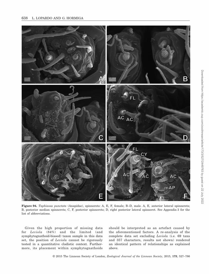
Given the high proportion of missing datafor Leviola (84%) and the limited (andsymphytognathoid-biased) taxon sample in this dataset, the position of Leviola cannot be rigorouslytested in a quantitative cladistic context. Further-more, its placement within symphytognathoids
should be interpreted as an artefact caused bythe aforementioned factors. A re-analysis of thecomplete data set excluding Leviola (i.e. 69 taxaand 357 characters, results not shown) renderedan identical pattern of relationships as explainedabove.
Figure 94. Taphiassa punctata (Anapidae), spinnerets: A, E, F, female; B–D, male. A, E, anterior lateral spinnerets;B, posterior median spinnerets; C, F, posterior spinnerets; D, right posterior lateral spinneret. See Appendix 3 for thelist of abbreviations.
638 L. LOPARDO AND G. HORMIGA
© 2015 The Linnean Society of London, Zoological Journal of the Linnean Society, 2015, 173, 527–786
Dow
nloaded from https://academ
ic.oup.com/zoolinnean/article/173/3/527/2449763 by guest on 22 July 2022
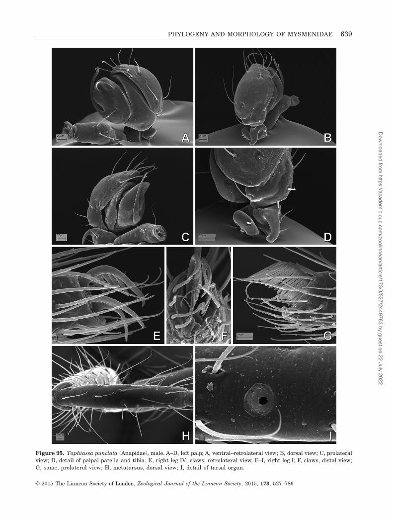
Figure 95. Taphiassa punctata (Anapidae), male. A–D, left palp; A, ventral–retrolateral view; B, dorsal view; C, prolateralview; D, detail of palpal patella and tibia. E, right leg IV, claws, retrolateral view. F–I, right leg I; F, claws, distal view;G, same, prolateral view; H, metatarsus, dorsal view; I, detail of tarsal organ.
PHYLOGENY AND MORPHOLOGY OF MYSMENIDAE 639
© 2015 The Linnean Society of London, Zoological Journal of the Linnean Society, 2015, 173, 527–786
Dow
nloaded from https://academ
ic.oup.com/zoolinnean/article/173/3/527/2449763 by guest on 22 July 2022

Figure 96. Taphiassa punctata (Anapidae), male prosoma: A, dorsal view; B, ventral view; C, lateral view; D, lateral–frontal view; E, detail of prosoma, lateral view; F, detail of anterior prosoma, ventral–lateral view; G, detail of carapacelateral depression; H, detail of sternal edge.
640 L. LOPARDO AND G. HORMIGA
© 2015 The Linnean Society of London, Zoological Journal of the Linnean Society, 2015, 173, 527–786
Dow
nloaded from https://academ
ic.oup.com/zoolinnean/article/173/3/527/2449763 by guest on 22 July 2022

Figure 97. Taphiassa punctata (Anapidae), male: A, sternal cuticle; B, detail of distal chelicerae, frontal view; C, mouth-parts, ventral view; D, detail of distal chelicerae, frontal–lateral view; E, labrum, lateral view; F, detail of gland mound,from panel D; G, abdomen, pedicel area; H, spinning field.
PHYLOGENY AND MORPHOLOGY OF MYSMENIDAE 641
© 2015 The Linnean Society of London, Zoological Journal of the Linnean Society, 2015, 173, 527–786
Dow
nloaded from https://academ
ic.oup.com/zoolinnean/article/173/3/527/2449763 by guest on 22 July 2022
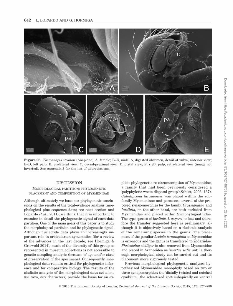
DISCUSSIONMORPHOLOGICAL PARTITION: PHYLOGENETIC
PLACEMENT AND COMPOSITION OF MYSMENIDAE
Although ultimately we base our phylogenetic conclu-sions on the results of the total-evidence analysis (mor-phological plus sequence data; see next section andLopardo et al., 2011), we think that it is important toexamine in detail the phylogenetic signal of each datapartition. One of the main goals of this paper is to studythe morphological partition and its phylogenetic signal.Although nucleotide data plays an increasingly im-portant role in orbicularian systematics (for a reviewof the advances in the last decade, see Hormiga &Griswold 2014), much of the diversity of this group asrepresented in museum collections is not accessible togenetic sampling analysis (because of age and/or stateof preservation of the specimens). Consequently, mor-phological data remain critical for phylogenetic infer-ence and for comparative biology. The results of thecladistic analysis of the morphological data set alone(65 taxa, 357 characters) provide the basis for an ex-
plicit phylogenetic re-circumscription of Mysmenidae,a family that had been previously considered a‘polyphyletic waste disposal group’ (Schütt, 2003: 137).Calodipoena tarautensis was placed within the sub-family Mysmeninae and possesses several of the pro-posed synapomorphies for the family. Crassignatha andIardinis, on the other hand, are both excluded fromMysmenidae and placed within Symphytognathidae.The type species of Iardinis, I. weyersi, is lost and there-fore the transfer suggested here is preliminary, al-though it is objectively based on a cladistic analysisof the remaining species in the genus. The place-ment of the peculiar Leviola termitophila in Mysmenidaeis erroneous and the genus is transferred to Zodariidae.Phricotelus stelliger is also removed from Mysmenidaeand placed in Araneoidea as incertae sedis until a thor-ough morphological study can be carried out and itsplacement more rigorously tested.
Previous morphological phylogenetic analyses hy-pothesized Mysmenidae monophyly based on two orthree synapomorphies: the ‘distally twisted and notchedcymbium’, the sclerotized spot subapically on ventral
Figure 98. Tasmanapis strahan (Anapidae). A, female; B–E, male. A, digested abdomen, detail of vulva, anterior view;B–D, left palp; B, prolateral view; C, dorsal-proximal view; D, distal view; E, right palp, retrolateral view (image notinverted). See Appendix 3 for the list of abbreviations.
642 L. LOPARDO AND G. HORMIGA
© 2015 The Linnean Society of London, Zoological Journal of the Linnean Society, 2015, 173, 527–786
Dow
nloaded from https://academ
ic.oup.com/zoolinnean/article/173/3/527/2449763 by guest on 22 July 2022

Figure 99. Tasmanapis strahan (Anapidae), prosoma: A, E, G, male; B–D, F, H, female. A, frontal view; B, lateral view;C, ventral view; D, dorsal view; E, ocular area and clypeus, frontal view; F, mouthparts, lateral–frontal view; G, detailof sternum and labium, ventral view; H, mouthparts, ventral view.
PHYLOGENY AND MORPHOLOGY OF MYSMENIDAE 643
© 2015 The Linnean Society of London, Zoological Journal of the Linnean Society, 2015, 173, 527–786
Dow
nloaded from https://academ
ic.oup.com/zoolinnean/article/173/3/527/2449763 by guest on 22 July 2022

Figure 100. Tasmanapis strahan (Anapidae): A–C, E, female; D, F–H, male. A, right anterior laeral spinneret; B, pos-terior median spinnerets; C, D, right posterior lateral spinneret; E, reduced palp and carapace lateral depression;F, labrum, lateral view; G, abdomen, epigastric furrow area, ventral view; H, same, posterior view. See Appendix 3 forthe list of abbreviations.
644 L. LOPARDO AND G. HORMIGA
© 2015 The Linnean Society of London, Zoological Journal of the Linnean Society, 2015, 173, 527–786
Dow
nloaded from https://academ
ic.oup.com/zoolinnean/article/173/3/527/2449763 by guest on 22 July 2022

Figure 101. Tasmanapis strahan (Anapidae): A, B, D, female; C, E–H, male. A, sternum, detail of sternal edge. B–H,left legs; B, leg IV, tarsal organ; C, patella–tibia IV, retrolateral view; D, patella–tibia I, prolateral view; leg I, tarsalorgan; F, metatarsus I, prolateral view; G, leg IV, claws, retrolateral view; H, clasping spurs, detail from panel F.
PHYLOGENY AND MORPHOLOGY OF MYSMENIDAE 645
© 2015 The Linnean Society of London, Zoological Journal of the Linnean Society, 2015, 173, 527–786
Dow
nloaded from https://academ
ic.oup.com/zoolinnean/article/173/3/527/2449763 by guest on 22 July 2022

femur I and II, and the male metatarsus I clasping spine(Griswold et al., 1998), or just the ‘apical lobes on thecymbium of the male palp and femoral spots on thefirst or first two pairs of legs’ (Schütt, 2003: 134). Ourstudy provides a more in depth understanding of theprimary homology hypotheses of structures such as‘lobes, twists, or notches’ of the cymbium (see belowand Appendix 2). Mysmenidae are here diagnosed byseven unambiguous and twelve ambiguously optimizedsynapomorphies not only related to legs and male palp,but also related to female genitalia, spinnerets, cuticlepatterns, chelicerae, and sternum morphology (seeResults above; see also Lopardo et al., 2011).
The morphological data set places Mysmenidae assister to Theridiosomatidae, a relationship recovered
in only one recent study performing a modifiedphylogenetic analysis of a previous data set (seeFig. 151B; Lopardo & Hormiga, 2008). Previousphylogenetic hypotheses have placed Mysmenidaeas sister to a clade comprising Anapidae andSymphytognathidae s.s. (Fig. 150A, B; Griswold et al.,1998; Lopardo & Hormiga, 2008), or a redelimitedMysmenidae sister to Symphytognathidae s.s. (Fig. 151A;Schütt, 2003; but for the best-informed phylogeneticplacement of Mysmenidae based on a total-evidenceapproach, see Lopardo et al., 2011). Also, the patternof familial relationships within symphytognathoidsagrees with that proposed by Griswold et al. (1998),but circumscribed as in Schütt (2003), with twomain symphytognathoid clades resulting from the
Figure 102. Teutoniella cekalovici (Anapidae), male left palp: A, ventral view; B, dorsal–proximal view; C, detail of dorsaltibia and patella; D, detail of apical palp, ventral view; E, same, prolateral view; F, same, retrolateral–distal view. SeeAppendix 3 for the list of abbreviations.
646 L. LOPARDO AND G. HORMIGA
© 2015 The Linnean Society of London, Zoological Journal of the Linnean Society, 2015, 173, 527–786
Dow
nloaded from https://academ
ic.oup.com/zoolinnean/article/173/3/527/2449763 by guest on 22 July 2022

Figure 103. Teutoniella cekalovici (Anapidae), abdomen: A–D, H, male; E–G, I, female. A, B, anterior lateral spinnerets;C, E, posterior median spinnerets; D, F, posterior lateral spinnerets; G, abdomen, ventral view; H, abdomen, detail ofepigastric area, ventral view; I, same as H, detail of pedicel area. See Appendix 3 for the list of abbreviations.
PHYLOGENY AND MORPHOLOGY OF MYSMENIDAE 647
© 2015 The Linnean Society of London, Zoological Journal of the Linnean Society, 2015, 173, 527–786
Dow
nloaded from https://academ
ic.oup.com/zoolinnean/article/173/3/527/2449763 by guest on 22 July 2022

Figure 104. Teutoniella cekalovici (Anapidae), prosoma: A–C, F–H, female; D, E, male. A, lateral view; B, lateral–frontal view; C, dorsal view; D, frontal view; E, F, carapace, frontal view; G, labrum, lateral view; H, detail of prosomaldepression between coxae II and III.
648 L. LOPARDO AND G. HORMIGA
© 2015 The Linnean Society of London, Zoological Journal of the Linnean Society, 2015, 173, 527–786
Dow
nloaded from https://academ
ic.oup.com/zoolinnean/article/173/3/527/2449763 by guest on 22 July 2022

Figure 105. Teutoniella cekalovici (Anapidae): A, B, E–H, female; C, D, male. A, chelicerae and palps, frontal view;B, same, lateral view; C, mouthparts, ventral view; D, same, lateral–ventral view; E, left metatarsus I, dorsal view;F, left femur IV, retrolateral view; G, left metatarsus IV, retrolateral view; H, left leg IV, retrolateral view.
PHYLOGENY AND MORPHOLOGY OF MYSMENIDAE 649
© 2015 The Linnean Society of London, Zoological Journal of the Linnean Society, 2015, 173, 527–786
Dow
nloaded from https://academ
ic.oup.com/zoolinnean/article/173/3/527/2449763 by guest on 22 July 2022

analysis. Mysmenidae are sister to Theridiosomatidae,and Synaphridae are sister to Anapidae +Symphytognathidae. Symphytognathidae are here re-delimited to include the enigmatic mysmenid genusIardinis. The family Anapidae includes a basal Acroblepsand a distal clade comprising Comaroma simoni sisterto the remaining micropholcommatine taxa (nodeM76, see Lopardo et al., 2011). The monophyly of
Micropholcommatinae (formally ranked as a subfam-ily by Lopardo et al., 2011) has already been recov-ered in a previous analysis (Schütt, 2003), althoughits placement within Anapidae has been questioned ina catalogue, rooted on the opinion that those results‘were based on analyses of very few genera’ (Platnick,2014). It should be noted, however, that Lopardo et al.(2011) studied other taxa relevant to the monophyly
Figure 106. Cepheia longiseta (Synaphridae), male paralectotype, right palp, inverted: A, apical view; B, dorsal view;C, retrolateral view; D, prolateral view; E, retrolateral–dorsal view; F, detail of palpal tibia and tip of conductor; G, detailof dorsal–apical view. See Appendix 3 for the list of abbreviations.
650 L. LOPARDO AND G. HORMIGA
© 2015 The Linnean Society of London, Zoological Journal of the Linnean Society, 2015, 173, 527–786
Dow
nloaded from https://academ
ic.oup.com/zoolinnean/article/173/3/527/2449763 by guest on 22 July 2022

issue that had not been included in their charactermatrix: ‘. . . based on the results of our study, pub-lished descriptions of other micropholcommatines (e.g.Hickman, 1944, 1945; Forster, 1959; Rix, 2008) showno character evidence that contradicts our hypo-thesis’. The results of the current analysis involve amuch more comprehensive taxon sampling of
symphytognathoids in general, and support the place-ment of the subfamily Micropholcommatinae as a distalclade within Anapidae, challenging the monophyly ofAnapidae exclusive of micropholcommatines (Forster,1959; Brignoli, 1970; but see Forster & Platnick, 1984;Platnick et al., 1991; Schütt, 2003; Rix et al., 2008; Rix& Harvey, 2010). Whereas our classification is based
Figure 107. Cepheia longiseta (Synaphridae), paralectotypes, abdomen: A, B, D, F, G, female; C, E, male. A, pediceland epigastric area; B, E, detail of epigastric–genital area; C, abdomen, ventral view; D, digested abdomen, detail ofvulva; F, spinning field, colulus and posterior spiracle; G, left anterior lateral spinneret. See Appendix 3 for the list ofabbreviations.
PHYLOGENY AND MORPHOLOGY OF MYSMENIDAE 651
© 2015 The Linnean Society of London, Zoological Journal of the Linnean Society, 2015, 173, 527–786
Dow
nloaded from https://academ
ic.oup.com/zoolinnean/article/173/3/527/2449763 by guest on 22 July 2022

Figure 108. Cepheia longiseta (Synaphridae), paralectotypes, prosoma: A–E, female; F–H, male. A, dorsal view; B, ventralview; C, mouthparts and palp, lateral view; D, same, lateral–ventral view; E, same, ventral–frontal view; F, detail ofmaxilla and labium; G, prosoma, lateral view; H, same, detail of mouthparts.
652 L. LOPARDO AND G. HORMIGA
© 2015 The Linnean Society of London, Zoological Journal of the Linnean Society, 2015, 173, 527–786
Dow
nloaded from https://academ
ic.oup.com/zoolinnean/article/173/3/527/2449763 by guest on 22 July 2022

on the data at hand, it seems clear that further re-search is required to further test the phylogeny of theextremely diverse Anapidae, using a still wider selec-tion of taxa from across the world.
Given the nature of measurements (i.e. decimalnumbers) as characters and the concomitant treat-ment of such continuous characters as additive inthe phylogenetic analyses, an increase in the resolu-tion of the phylogenetic hypothesis was expected(compare cladograms from Figs 154, 157, see optimi-zation of the seven continuous characters into the mostparsimonious cladogram in Fig. 155A–G). Our resultsshow a tendency towards an overall decrease in cladesupport of approximately 50% when continuous char-acters are included in the analyses (Fig. 158). Cladogramlength and continuous character changes are conse-quently measured not as entire ‘steps’, but as frac-tions of steps. Support values calculated based on stepnumbers (e.g. absolute Bremer support, BS) can there-fore yield decimal numbers as total clade support values.Typically in analyses with exclusively discrete char-
acters, the minimum BS value for a clade is one step,meaning that only one extra step is required by thathypothesis to collapse the monophyly of that clade. Themeaning of decimal values in Bremer support calcu-lations is not entirely clear, especially when such valuesare close to zero (see Fig. 154), even though such cladescan be supported by two or more discrete changes.Decimal BS values are the result of subtle changes inthe continuous characters. If all clades with BS lowerthan 0.95 are collapsed, the loss of resolution is dis-tributed in three main clades within Mysmenidae(Fig. 156): relationships within Mysmenopsis col-lapse, as well as within clades M86 (including all itsinternal nodes) and M93. Even though the phylogenetichypothesis from the complete morphological analysiscontains unstable clades and decreased support, whencompared with a discrete-only analysis, the overallhomoplasy in the discrete characters alone is also rela-tively high, and we see no epistemological reason toexclude such continuous characters (see also Rae, 1998;Humphries, 2002).
Figure 109. Cepheia longiseta (Synaphridae), paralectotypes, legs: A, B, female left legs; C–E, male right legs. A, leg II,metatarsus–tarsus junction, dorsal view; B, leg I, tarsal organ, lateral view; C, leg I, tibia–patella, dorsal view; D, meta-tarsus III, dorsal view; E, leg I, claws.
PHYLOGENY AND MORPHOLOGY OF MYSMENIDAE 653
© 2015 The Linnean Society of London, Zoological Journal of the Linnean Society, 2015, 173, 527–786
Dow
nloaded from https://academ
ic.oup.com/zoolinnean/article/173/3/527/2449763 by guest on 22 July 2022

COMBINED ANALYSIS: PHYLOGENETIC IMPLICATIONS
AND EVOLUTIONARY MORPHOLOGY
To study and report morphological and behaviouralcharacter evolution in Mysmenidae (below) and
symphytognathoids (in part below, see Appendix 2), weuse the preferred optimal tree that results from thetotal evidence analysis, which combines morphology,behaviour, and multigene sequence data (see Figs 160,161, and Appendices 2, 5; see also Lopardo et al., 2011).
Figure 110. Synaphris saphrynis (Synaphridae), male paratype. A–E, left palp; A, prolateral–apical view; B, apical view;C, retrolateral view; D, dorsal–proximal view; E, prolateral–proximal view. F, digested abdomen, detail of posterior res-piratory atrium and tracheae; G, epiandrous spigots. See Appendix 3 for the list of abbreviations.
654 L. LOPARDO AND G. HORMIGA
© 2015 The Linnean Society of London, Zoological Journal of the Linnean Society, 2015, 173, 527–786
Dow
nloaded from https://academ
ic.oup.com/zoolinnean/article/173/3/527/2449763 by guest on 22 July 2022

Figure 111. Synaphris saphrynis (Synaphridae), male paratype: A, prosoma, frontal view; B, sternum, ventral view;C, mouthparts, ventral view; D, mouthparts, lateral view; E, mouthparts, posterior view; F, right tibia I, dorsal view;G, left leg I, metatarsus–tarsus junction, dorsal view; H, right leg I, claws, retrolateral view. See Appendix 3 for the listof abbreviations.
PHYLOGENY AND MORPHOLOGY OF MYSMENIDAE 655
© 2015 The Linnean Society of London, Zoological Journal of the Linnean Society, 2015, 173, 527–786
Dow
nloaded from https://academ
ic.oup.com/zoolinnean/article/173/3/527/2449763 by guest on 22 July 2022

Figure 112. Patu-SYMP-001-DR (Symphytognathidae), from Barahona, Dominican Republic; prosoma: A, C, E–H, female;B, D, male. A, B, lateral view; C, D, ocular area, frontal view; E, ventral view; F, same, detail of labium–sternum junc-tion; G, mouthparts, frontal view; H, sternal cuticular pattern, detail from panel F.
656 L. LOPARDO AND G. HORMIGA
© 2015 The Linnean Society of London, Zoological Journal of the Linnean Society, 2015, 173, 527–786
Dow
nloaded from https://academ
ic.oup.com/zoolinnean/article/173/3/527/2449763 by guest on 22 July 2022

Figure 113. Patu-SYMP-001-DR (Symphytognathidae), from Barahona, Dominican Republic: A–D, G, female abdomen;E, F, H, male. A, right anterior lateral spinneret (ALS); B, left ALS; C, right posterior median spinneret; D, left pos-terior lateral spinneret. E, right palp, inverted, bulb detaching from cymbium, retrolateral–ventral (bulb) and apical (cymbium)views; F, same, ventral (bulb) and dorsal (cymbium) views. G, pedicel area; H, left tibia IV, retrolateral view. SeeAppendix 3 for the list of abbreviations.
PHYLOGENY AND MORPHOLOGY OF MYSMENIDAE 657
© 2015 The Linnean Society of London, Zoological Journal of the Linnean Society, 2015, 173, 527–786
Dow
nloaded from https://academ
ic.oup.com/zoolinnean/article/173/3/527/2449763 by guest on 22 July 2022

Taxon names used throughout the discussion includethe taxonomic decisions reported in Appendix 5. Wewill discuss the comparative morphology of therespiratory system in mysmenids, and its evolution-ary implications within symphytognathoids, in a sepa-rate paper.
MYSMENIDAE COMPARATIVE MORPHOLOGY
Male palp (refer to characters 151–261)The diversity and sclerite homologies of mysmenid malepalps are only superficially understood. Mysmenidaehave been recognized and even diagnosed by the
Figure 114. SYMP-002-MAD (Symphytognathidae), from Mahajanga, Madagascar: A–C, female abdomen; D–F, male leftpalp. A, posterior median spinnerets; B, left posterior lateral spinneret; C, digested abdomen, showing respiratory systemand vulva; D, left palp, ventral–apical view; E, same, prolateral view, arrow to prolateral distal expansion; F, same, detailfrom panel E, arrow to embolic basal expansion. See Appendix 3 for the list of abbreviations.
658 L. LOPARDO AND G. HORMIGA
© 2015 The Linnean Society of London, Zoological Journal of the Linnean Society, 2015, 173, 527–786
Dow
nloaded from https://academ
ic.oup.com/zoolinnean/article/173/3/527/2449763 by guest on 22 July 2022

generalized shape of the cymbium, described as ‘api-cally twisted and with lobes’ (Platnick & Shadab, 1978;Brignoli, 1980; Coddington, 1990; Wunderlich, 1995;Griswold et al., 1998; Schütt, 2003). Despite the factthat several relatively modern descriptions of mysmenidshave included detailed illustrations of genitalic mor-phology (e.g. Kraus, 1967; Thaler, 1975, 1995; Saaristo,1978; Baert & Maelfait, 1983; Baert, 1984a, 1990; Lin& Li, 2008; Miller et al., 2009), most mysmenid speciesremain poorly described. The details of the palp mor-
phology are also insufficiently studied, especially interms of explicit hypotheses of homology. For example,it has been suggested that a tegular conductor, medianapophysis, and paracymbium are absent (Coddington,1990; Griswold et al., 1998; Miller et al., 2009); however,a paracymbium had previously been identified inMysmenidae (Kraus, 1967). The mysmenid male palpappears highly complex and morphologically vari-able, and in most species it is greatly translucent, sothat the cymbium, conductor, and other sclerites are
Figure 115. SYMP-002-MAD (Symphytognathidae), from Mahajanga, Madagascar: A–C, male prosoma; D–G, female.A, C, lateral views; B, frontal view. D, mouthparts, ventral view; E, same, detail of cheliceral fangs and teeth. F, pediceland epigynal areas; G, same, detail of epigynal area.
PHYLOGENY AND MORPHOLOGY OF MYSMENIDAE 659
© 2015 The Linnean Society of London, Zoological Journal of the Linnean Society, 2015, 173, 527–786
Dow
nloaded from https://academ
ic.oup.com/zoolinnean/article/173/3/527/2449763 by guest on 22 July 2022

difficult to distinguish and to delineate precisely underlight microscopy. In summary, mysmenid male palpshave distinct cymbial structures, including aparacymbium, and can also have a tegular conduc-tor. A median apophysis or any other tegular scleritesare lacking, however. As in the details of the differ-
ent respiratory organs in Mysmenidae, the diversityof palpal structures is great within the family, al-though each particular arrangement seems character-istic at the genus or sometimes subfamily level (thisis simply a consequence of how taxonomists have cir-cumscribed higher taxa in Mysmenidae). In the
Figure 116. SYMP-002-MAD (Symphytognathidae), from Mahajanga, Madagascar, left legs. A, C, E, female; B, D, F,male. A, femur, patella and tibia I, prolateral view; B, patella and tibia II, retrolateral view; C, femur, patella and tibia I,retrolateral view; D, detail of tibia II clasping spine, from panel B; E, leg I, tarsal organ; F, patella and tibia IV, retrolateralview.
660 L. LOPARDO AND G. HORMIGA
© 2015 The Linnean Society of London, Zoological Journal of the Linnean Society, 2015, 173, 527–786
Dow
nloaded from https://academ
ic.oup.com/zoolinnean/article/173/3/527/2449763 by guest on 22 July 2022

sections below, we address the large diversity ofmysmenid palpal morphology.
Palpal femur, patella and tibiaMysmenids lack any modifications on the palpal femurand patella. Conversely, and across symphytognathoids,
varying shapes of the male palpal tibia can be found.A distally broad tibia (i.e. wider distally, usuallymore than two times its basal width) may besymplesiomorphic for symphytognathoids, as it alsooccurs in Theridiidae. Distally broad tibiae occur inTheridiosomatidae, Synaphridae, and most Mysmenidae
Figure 117. SYMP-006-AUST (Symphytognathidae), from Queensland, Australia: A, B, F, female; C–E, G, male. A, pediceland epigynal areas; B, digested abdomen, detail of vulva. C, left palp, dorsal view; D, same, ventral-apical view; E, same,detail of embolus and conductor. F, ventral abdomen and anterior spinnerets, note absence of colulus and posterior spira-cle; G, left posterior lateral spinneret. See Appendix 3 for the list of abbreviations.
PHYLOGENY AND MORPHOLOGY OF MYSMENIDAE 661
© 2015 The Linnean Society of London, Zoological Journal of the Linnean Society, 2015, 173, 527–786
Dow
nloaded from https://academ
ic.oup.com/zoolinnean/article/173/3/527/2449763 by guest on 22 July 2022

(Figs 4A, 17D, 28A, 30E, 32E, 38A, 42A, 47B,63C). Within symphytognathoids, a flat tibia (i.e.flattened from the base, and usually with irregular,not circular, distal section outline) is synapomorphicfor the clade comprising Symphytognathidae plusAnapidae (Figs 91F, 95C, D, 102C). Within Mysmenidae,broad male palpal tibiae are widespread, occurring
in Trogloneta, Isela, and all Mysmeninae. Moreover,a cylindrical tibia (distal width similar to or less thantwo times proximal width) is synapomorphic forMaymena, and convergently present in Comaroma andTasmanapis (Figs 10A, 81D, 98B,C). A globose tibia isa synapomorphy of Mysmenopsis (Figs 55A, C, 58A,60A).
Figure 118. SYMP-006-AUST (Symphytognathidae), from Queensland, Australia, prosoma: A, C, female; B, D–F, male.A, B, lateral view; C, mouthparts, ventral view; D, same, lateral view, detail from panel B; E, detail of cheliceral fangsand teeth; F, sternum and labium, ventral view.
662 L. LOPARDO AND G. HORMIGA
© 2015 The Linnean Society of London, Zoological Journal of the Linnean Society, 2015, 173, 527–786
Dow
nloaded from https://academ
ic.oup.com/zoolinnean/article/173/3/527/2449763 by guest on 22 July 2022

Figure 119. SYMP-006-AUST (Symphytognathidae), from Queensland, Australia, left legs. A–G, female; H–J, male.A, leg I, retrolateral; B, femur I, prolateral; C, metatarsus IV, retrolateral; D, leg I, tarsal organ and metatarsus–tarsusjunction, dorsal; E, tibia IV, retrolateral; F, metatarsus I, detail of trichobothrial base, dorsal; G, tarsus IV, retrolateral;H, tibia II, retrolateral view; I, same, detail of clasping spine; J, same, ventral view.
PHYLOGENY AND MORPHOLOGY OF MYSMENIDAE 663
© 2015 The Linnean Society of London, Zoological Journal of the Linnean Society, 2015, 173, 527–786
Dow
nloaded from https://academ
ic.oup.com/zoolinnean/article/173/3/527/2449763 by guest on 22 July 2022

Most mysmenid palpal tibiae are small, shorter thancymbium, as in other symphytognathoids (Figs 10A,27B, 30D, 47A, B, 63B, C, 71D, 106C, 110E). A largetibia is synapomorphic for Mysmenopsinae (Figs 1A,4A, 55A, C). In addition, no distinct tibial processesoccur in Mysmenidae, except for a prolateral exten-sion in Mysmeniola (Fig. 134D; see also Thaler, 1995:
figs 5, 7) and an apical ventral (sometimes ventro-retrolateral) excavation usually bearing spurs inMysmenopsis (Figs 53A, 58A, 60A). Mysmenopsines havemodified setae distally on the tibiae, such as spurs inMysmenopsis (as mentioned above, Figs 53E, 55H, 58E,60B), or spine-like, strong setae in Isela (Figs 1A, B,4A, E). Mysmenid tibial rim setae are longer than
Figure 120. SYMP-007-AUST (Symphytognathidae), from Queensland, Australia, female. A, ocular area, frontal view;B, mouthparts, ventral view; C, spinning field, details to absence of colulus; D, mouthparts, detail of cheliceral fangsand teeth; E, digested abdomen, respiratory system and vulva; F, same, detail of vulva.
664 L. LOPARDO AND G. HORMIGA
© 2015 The Linnean Society of London, Zoological Journal of the Linnean Society, 2015, 173, 527–786
Dow
nloaded from https://academ
ic.oup.com/zoolinnean/article/173/3/527/2449763 by guest on 22 July 2022

Figure 121. Symphytognatha picta (Symphytognathidae), from Western Australia, Australia, male prosoma: A, frontalview; B, lateral–frontal view; C, ventral view; D, chelicerae, ventral view, detail from panel A; E, detail anterior prosoma,lateral view; F, carapace lateral depression, detail from panel B; G, labium–sternum junction, detail from panel C;H, maxillae, ventral view.
PHYLOGENY AND MORPHOLOGY OF MYSMENIDAE 665
© 2015 The Linnean Society of London, Zoological Journal of the Linnean Society, 2015, 173, 527–786
Dow
nloaded from https://academ
ic.oup.com/zoolinnean/article/173/3/527/2449763 by guest on 22 July 2022

Figure 122. Symphytognatha picta (Symphytognathidae), from Western Australia, Australia, male: A, cheliceral fangsand teeth, frontal view; B, same, lateral view; C, left palp, retrolateral–ventral view; D, same, retrolateral view; E, pos-terior median spinnerets; F, right posterior lateral spinneret; G, left leg I, claws, prolateral view; H, left leg IV, claws,retrolateral view. See Appendix 3 for the list of abbreviations.
666 L. LOPARDO AND G. HORMIGA
© 2015 The Linnean Society of London, Zoological Journal of the Linnean Society, 2015, 173, 527–786
Dow
nloaded from https://academ
ic.oup.com/zoolinnean/article/173/3/527/2449763 by guest on 22 July 2022

Figure 123. Theridiosoma gemmosum (Theridiosomatidae), female: A, digested abdomen, detail of anterior respiratorysystem and vulva; B, mouthparts, ventral–frontal view; C, same, detail of cheliceral fang and teeth; D, labium, ventralview; E, posterior median spinnerets; F, right posterior lateral spinneret; G, left leg I, claws, prolateral view; H, left leg IV,claws, retrolateral view. See Appendix 3 for the list of abbreviations.
PHYLOGENY AND MORPHOLOGY OF MYSMENIDAE 667
© 2015 The Linnean Society of London, Zoological Journal of the Linnean Society, 2015, 173, 527–786
Dow
nloaded from https://academ
ic.oup.com/zoolinnean/article/173/3/527/2449763 by guest on 22 July 2022
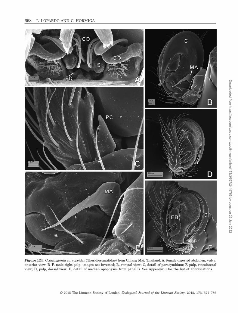
Figure 124. Coddingtonia euryopoides (Theridiosomatidae) from Chiang Mai, Thailand. A, female digested abdomen, vulva,anterior view. B–F, male right palp, images not inverted; B, ventral view; C, detail of paracymbium; F, palp, retrolateralview; D, palp, dorsal view; E, detail of median apophysis, from panel B. See Appendix 3 for the list of abbreviations.
668 L. LOPARDO AND G. HORMIGA
© 2015 The Linnean Society of London, Zoological Journal of the Linnean Society, 2015, 173, 527–786
Dow
nloaded from https://academ
ic.oup.com/zoolinnean/article/173/3/527/2449763 by guest on 22 July 2022

remaining tibial setae, and are arranged distally inone or two rows (Figs 4E, 10D, 18A, 32E, 36D, 42B,45C, 53B, 55C), except in Trogloneta and a few othermysmenids where the setae are equally short and dis-persed in an irregular conformation (Figs 63C, 66A).
Cymbium: general morphology(see Fig. 126 for reference)The size of the cymbium and bulb (relative to the sizeof the carapace, in lateral view) varies across mysmenidtaxa. Although small male palps are common in adult
Figure 125. Coddingtonia euryopoides (Theridiosomatidae) from Chiang Mai, Thailand; female: A, sternum and labium,ventral view; B, mouthparts, detail of maxillary setae; C, right posterior median spinneret; D, right anterior lateral spin-neret; E, palpal tibia, dorsal view; F, left metatarsus I, prolateral view; G, leg I, tarsal organ. See Appendix 3 for the listof abbreviations.
PHYLOGENY AND MORPHOLOGY OF MYSMENIDAE 669
© 2015 The Linnean Society of London, Zoological Journal of the Linnean Society, 2015, 173, 527–786
Dow
nloaded from https://academ
ic.oup.com/zoolinnean/article/173/3/527/2449763 by guest on 22 July 2022

spiders, medium-sized palps are widespread withinsymphytognathoids, including mysmenids (i.e. thecymbium-bulb complex is about half the size of the cara-pace; Figs 27F, 66A, 141K). The cymbium bulb becomessecondarily small (about one-fifth the size of the cara-pace) in Maymena mayana and mysmenopsines,however (Figs 2B, 140E, 141D). A very large cymbium-
bulb complex (i.e. as large as the prosoma) has evolvedindependently in some distal clades within Microdipoenaand Mysmena, including Mysmena leucoplagiata(Figs 19B, 142A), in Theridiosomatidae, and thesynaphrid genus Cepheia. Superficially, there appearsto be a tendency towards an increase in palpal size,which suggests a correlation with the reduction ofbody size in symphytognathoids; however, mostsymphytognathoids are equally minute in body size,regardless of their familial placement or size of thepalp. For example, most Microdipoena species (and alsotheridiosomatids) with huge palps are as small as anyother mysmenine or even larger than members ofSymphytognathidae, which possess medium-sized palps.Taphiassa, a small micropholcommatine anapid, alsohas small palps.
The cymbium in most mysmenids is uniquely ori-ented ventrally or prolatero-ventrally in the palp(Figs 38A,B, 42B, 66A). The cymbium evolved inde-pendently to a prolateral position in Mysmenopsis,Mysmeniola, and the mysmenine MYSM-023-MAD(Fig. 50B), or to a retrolateral–dorsal position in someMaymena species (Fig. 15B).
In some symphytognathoids the cymbium is some-times modified relative to the typical cymbium of mostaraneoids, which is scoop-shaped and round to ovalin dorsal view (e.g. Griswold et al., 1998: figs 16A, 18F).This is especially so in mysmenids. Without taking intoaccount any other cymbial structures (such as cymbialconductors, apophyses, paracymbia, expansions, etc.;see comments below), an oval cymbium is plesiomorphic
Figure 126. Schematic drawings showing cymbial structures on the male palp of Mysmenidae. Left cymbium is depict-ed: A, ventral view; B, dorsal view. See Appendix 3 for the list of abbreviations.
Figure 127. Schematic drawing of the trajectory of the sper-matic duct on male left palp, in prolateral view. See Ap-pendix 3 for the list of abbreviations.
670 L. LOPARDO AND G. HORMIGA
© 2015 The Linnean Society of London, Zoological Journal of the Linnean Society, 2015, 173, 527–786
Dow
nloaded from https://academ
ic.oup.com/zoolinnean/article/173/3/527/2449763 by guest on 22 July 2022

Figure 128. Mysmenidae female genitalia, cleared: A, Isela okuncana, dorsal view; B, Maymena mayana, dorsal view;C, Kilifina-MYSM-002-KENYA (Isela sp.), from Kwale, Kenya, dorsal view; D, Maymena ambita, ventral view; E, Maymenarica, dorsal view; F, Trogloneta granulum, ventral view; G, Mysmenopsis dipluramigo, dorsal view. See Appendix 3 forthe list of abbreviations.
PHYLOGENY AND MORPHOLOGY OF MYSMENIDAE 671
© 2015 The Linnean Society of London, Zoological Journal of the Linnean Society, 2015, 173, 527–786
Dow
nloaded from https://academ
ic.oup.com/zoolinnean/article/173/3/527/2449763 by guest on 22 July 2022

Figure 129. Mysmenidae female genitalia, cleared: A, Microdipoena guttata, ventral view; B, Microdipoena nyungwe,ventral view; C, MYSM-007-MEX (Mysmena), from Chiapas, Mexico, ventral view, arrow to diverticle of copulatory duct;D, Microdipoena elsae, ventral copulatory duct; E, Microdipoena (= Mysmenella) samoensis, syntype, ventral view; F,Microdipoena (= Mysmenella) jobi, paratype, ventral view; G, Mysmena-MYSM-015-MAD (Mysmena), from Antananari-vo, Madagascar, dorsal view; H, MYSM-029-MAD (Mysmeninae), from Antsiranana, Madagascar, ventral view. SeeAppendix 3 for the list of abbreviations.
672 L. LOPARDO AND G. HORMIGA
© 2015 The Linnean Society of London, Zoological Journal of the Linnean Society, 2015, 173, 527–786
Dow
nloaded from https://academ
ic.oup.com/zoolinnean/article/173/3/527/2449763 by guest on 22 July 2022

Figure 130. Mysmenidae female genitalia, cleared. A, Mysmena (= Calodipoena) tarautensis, paratype, dorsal view, arrowto broken scapus; B, Mysmena (= Calodipoena) incredula, dorsal view; C, Mysmena leichhardti, from Queensland, Aus-tralia, ventral view; D, MYSM-023-MAD (Mysmeninae), from Antananarivo, Madagascar, ventral view; E, Mysmena tasmaniae,ventral view; F, MYSM-005-ARG (Mysmena), from Misiones, Argentina, ventral view; G, MYSM-019-MAD (Mysmeninae),from Toliara, Madagascar, ventral view. See Appendix 3 for the list of abbreviations.
PHYLOGENY AND MORPHOLOGY OF MYSMENIDAE 673
© 2015 The Linnean Society of London, Zoological Journal of the Linnean Society, 2015, 173, 527–786
Dow
nloaded from https://academ
ic.oup.com/zoolinnean/article/173/3/527/2449763 by guest on 22 July 2022

Figure 131. Mysmenidae male left palp (unless otherwise stated), cleared: A, Mysmenopsis penai, retrolateral view, rightpalp, inverted; B, Mysmenopsis dipluramigo, retrolateral view; C, Mysmenopsis cidrelicola, presumably paralectotype,retrolateral view, right palp, inverted; D, Kilifina-MYSM-002-KENYA (Isela sp.), from Kwale, Kenya, ventral view, rightpalp, inverted; E, Trogloneta granulum, ventral view; F, Trogloneta cantareira, ventral view; G, Kilifina-MYSM-002-KENYA (Isela sp.), from Kwale, Kenya, dorsal view, right palp, inverted; H, Isela okuncana, ventral view; I, Maymenaambita, ventral view; J, Maymena mayana, ventral view. See Appendix 3 for the list of abbreviations.
674 L. LOPARDO AND G. HORMIGA
© 2015 The Linnean Society of London, Zoological Journal of the Linnean Society, 2015, 173, 527–786
Dow
nloaded from https://academ
ic.oup.com/zoolinnean/article/173/3/527/2449763 by guest on 22 July 2022

Figure 132. Mysmenidae male left palp, cleared: A, Microdipoena elsae, prolateral view, arrow to spine of basal prolateralcymbial expansion; B, Microdipoena guttata, prolateral view, arrow to spine of basal prolateral cymbial expansion;C, Microdipoena (= Mysmenella) illectrix, type, prolateral view; D, Microdipoena (= Mysmenella) samoensis, syntype, prolateralview; E, same, retrolateral view; F, Microdipoena (= Mysmenella) jobi, holotype, retrolateral view. See Appendix 3 forthe list of abbreviations.
PHYLOGENY AND MORPHOLOGY OF MYSMENIDAE 675
© 2015 The Linnean Society of London, Zoological Journal of the Linnean Society, 2015, 173, 527–786
Dow
nloaded from https://academ
ic.oup.com/zoolinnean/article/173/3/527/2449763 by guest on 22 July 2022

Figure 133. Mysmenidae male left palp (unless otherwise stated), cleared. A, MYSM-005-ARG (Mysmena), from Misiones,Argentina, dorsal view, right palp, inverted, arrow to cymbial groove (CyG); B, same, ventral view; C, MYSM-007-MEX(Mysmena), from Chiapas, Mexico, retrolateral view, arrows to CyG; D, Mysmena (= Kekenboschiella) awari, paratype,prolateral view; E, same, dorsal–retrolateral view; F, Mysmena (= Kekenboschiella) marijkeae, holotype, retrolateralview, right palp, inverted; G, Brasilionata arborense, holotype, retrolateral view, right palp, inverted; H, Mysmena(= Tamasesia) rotunda, type, prolateral view; I, same, retrolateral view, schematic drawing. See Appendix 3 for the listof abbreviations.
676 L. LOPARDO AND G. HORMIGA
© 2015 The Linnean Society of London, Zoological Journal of the Linnean Society, 2015, 173, 527–786
Dow
nloaded from https://academ
ic.oup.com/zoolinnean/article/173/3/527/2449763 by guest on 22 July 2022

Figure 134. Mysmenidae male left palp (unless otherwise stated), cleared: A, Mysmena leucoplagiata, type, prolateralview, right palp, inverted; B, Mysmena leichhardti, from Queensland, Australia, prolateral–ventral view; C, Mysmenatasmaniae, prolateral–ventral view, right palp, inverted; D, Mysmeniola spinifera, holotype, prolateral view, right palp,inverted; E, MYSM-019-MAD (Mysmeninae), from Toliara, Madagascar, prolateral view; F, MYSM-023-MAD (Mysmeninae),from Antananarivo, Madagascar, retrolateral view, right palp, inverted; G, MYSM-020-MAD (Mysmeninae), from Toamasina,Madagascar, dorsal view, right palp, inverted; H, same, ventral view. See Appendix 3 for the list of abbreviations.
PHYLOGENY AND MORPHOLOGY OF MYSMENIDAE 677
© 2015 The Linnean Society of London, Zoological Journal of the Linnean Society, 2015, 173, 527–786
Dow
nloaded from https://academ
ic.oup.com/zoolinnean/article/173/3/527/2449763 by guest on 22 July 2022
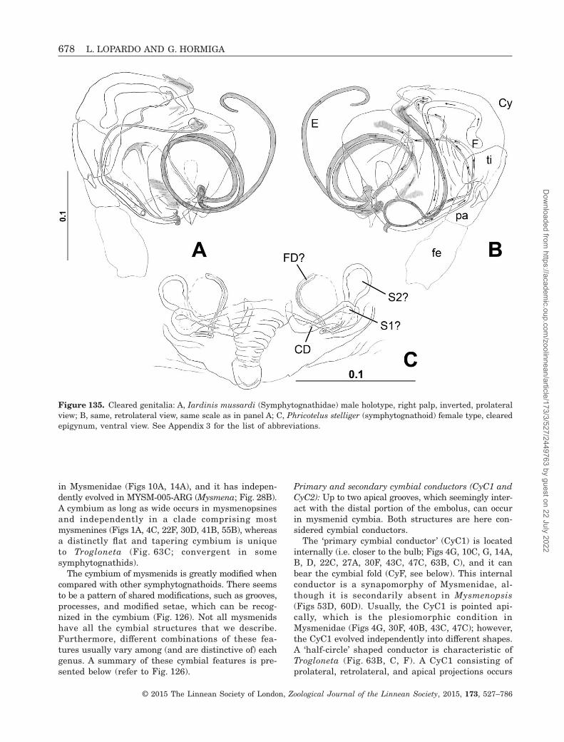
in Mysmenidae (Figs 10A, 14A), and it has indepen-dently evolved in MYSM-005-ARG (Mysmena; Fig. 28B).A cymbium as long as wide occurs in mysmenopsinesand independently in a clade comprising mostmysmenines (Figs 1A, 4C, 22F, 30D, 41B, 55B), whereasa distinctly flat and tapering cymbium is uniqueto Trogloneta (Fig. 63C; convergent in somesymphytognathids).
The cymbium of mysmenids is greatly modified whencompared with other symphytognathoids. There seemsto be a pattern of shared modifications, such as grooves,processes, and modified setae, which can be recog-nized in the cymbium (Fig. 126). Not all mysmenidshave all the cymbial structures that we describe.Furthermore, different combinations of these fea-tures usually vary among (and are distinctive of) eachgenus. A summary of these cymbial features is pre-sented below (refer to Fig. 126).
Primary and secondary cymbial conductors (CyC1 andCyC2): Up to two apical grooves, which seemingly inter-act with the distal portion of the embolus, can occurin mysmenid cymbia. Both structures are here con-sidered cymbial conductors.
The ‘primary cymbial conductor’ (CyC1) is locatedinternally (i.e. closer to the bulb; Figs 4G, 10C, G, 14A,B, D, 22C, 27A, 30F, 43C, 47C, 63B, C), and it canbear the cymbial fold (CyF, see below). This internalconductor is a synapomorphy of Mysmenidae, al-though it is secondarily absent in Mysmenopsis(Figs 53D, 60D). Usually, the CyC1 is pointed api-cally, which is the plesiomorphic condition inMysmenidae (Figs 4G, 30F, 40B, 43C, 47C); however,the CyC1 evolved independently into different shapes.A ‘half-circle’ shaped conductor is characteristic ofTrogloneta (Fig. 63B, C, F). A CyC1 consisting ofprolateral, retrolateral, and apical projections occurs
Figure 135. Cleared genitalia: A, Iardinis mussardi (Symphytognathidae) male holotype, right palp, inverted, prolateralview; B, same, retrolateral view, same scale as in panel A; C, Phricotelus stelliger (symphytognathoid) female type, clearedepigynum, ventral view. See Appendix 3 for the list of abbreviations.
678 L. LOPARDO AND G. HORMIGA
© 2015 The Linnean Society of London, Zoological Journal of the Linnean Society, 2015, 173, 527–786
Dow
nloaded from https://academ
ic.oup.com/zoolinnean/article/173/3/527/2449763 by guest on 22 July 2022

Figure 136. Anapidae female genitalia, cleared. A, Acrobleps hygrophilus, ventral view; B, Anapisona kethleyi, dorsalview; C, Crassanapis chilensis, dorsal view, arrow to broken inserted embolus; D, Minanapis palena, dorsal view; E, Elanapisaisen, dorsal view; F, Tasmanapis strahan, dorsal view. See Appendix 3 for the list of abbreviations.
PHYLOGENY AND MORPHOLOGY OF MYSMENIDAE 679
© 2015 The Linnean Society of London, Zoological Journal of the Linnean Society, 2015, 173, 527–786
Dow
nloaded from https://academ
ic.oup.com/zoolinnean/article/173/3/527/2449763 by guest on 22 July 2022

in the clade comprising Microdipoena and Mysmeniola,independently occurring in Mysmena (= Calomyspoena)santacruzi (Fig. 27A). A spiral cymbial conductor occursin Microdipoena s.s. (Fig. 22C). In Maymena, the char-acteristic CyC1 comprises a particular apical cymbiumthat is bent over the ventral side (Fig. 10D, G).
The ‘secondary cymbial conductor’ (CyC2) is exter-nal, located on the edge or dorsally on the cymbium(e.g. Figs 30F, 31C, 43C). This external conductor isplesiomorphically absent in Trogloneta, Maymenamayana, and in most Mysmenopsis species (Fig. 63C).Within Mysmenidae, the CyC2 has evolved convergently
in Maymena (excluding M. mayana), Isela, Mysmenopsispenai, and Mysmeninae (Figs 4G, 10G, 31C, 40A, 41D,43C, 60D). The CyC2 has been secondarily lost in theclade comprising Microdipoena and MYSM-019-MAD(Fig. 22F).
Cymbial fold (CyF): The external cuticle of the cymbiumis usually hirsute, the internal one is glabrous. Al-though the external cuticle can also cover the inter-nal side of the cymbium, both cuticles are delimitedby a well-defined border (e.g. Figs 86G, 102D). In somemysmenids the delimitation or border between
Figure 137. Female genitalia, cleared: A, Cepheia longiseta (Synaphridae) paralectotype, ventral view; B, same, dorsalview, same scale as in panel A; C, Taphiassa punctata (Anapidae), ventral view; D, Coddingtonia euryopoides(Theridiosomatidae), from Chiang Mai, Thailand, ventral view; E, SYMP-006-AUST (Symphytognathidae), from Queens-land Australia, dorsal view. See Appendix 3 for the list of abbreviations.
680 L. LOPARDO AND G. HORMIGA
© 2015 The Linnean Society of London, Zoological Journal of the Linnean Society, 2015, 173, 527–786
Dow
nloaded from https://academ
ic.oup.com/zoolinnean/article/173/3/527/2449763 by guest on 22 July 2022
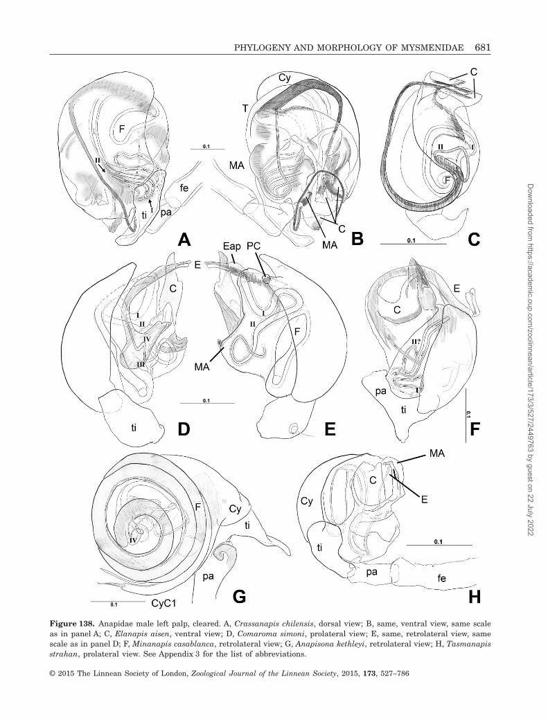
Figure 138. Anapidae male left palp, cleared. A, Crassanapis chilensis, dorsal view; B, same, ventral view, same scaleas in panel A; C, Elanapis aisen, ventral view; D, Comaroma simoni, prolateral view; E, same, retrolateral view, samescale as in panel D; F, Minanapis casablanca, retrolateral view; G, Anapisona kethleyi, retrolateral view; H, Tasmanapisstrahan, prolateral view. See Appendix 3 for the list of abbreviations.
PHYLOGENY AND MORPHOLOGY OF MYSMENIDAE 681
© 2015 The Linnean Society of London, Zoological Journal of the Linnean Society, 2015, 173, 527–786
Dow
nloaded from https://academ
ic.oup.com/zoolinnean/article/173/3/527/2449763 by guest on 22 July 2022

Figure 139. Male left palp, cleared. A, Teutoniella cekalovici (Anapidae), retrolateral view; B, Cepheia longiseta (Synaphridae),paralectotype, retrolateral view; C, Symphytognatha picta (Symphytognathidae), ventral view; D, Teutoniella cekalovici(Anapidae), prolateral view, same scale as A; E, Taphiassa punctata (Anapidae), retrolateral view. See Appendix 3 for thelist of abbreviations.
682 L. LOPARDO AND G. HORMIGA
© 2015 The Linnean Society of London, Zoological Journal of the Linnean Society, 2015, 173, 527–786
Dow
nloaded from https://academ
ic.oup.com/zoolinnean/article/173/3/527/2449763 by guest on 22 July 2022

Figure 140. Composite images of Mysmenidae species: Mysmenopsinae and Maymena. A–C, Isela okuncana; A, female,lateral view; B, female, ventral view; C, male, ventral view. D–F, Kilifina-MYSM-002-KENYA (Isela sp.), from Kwale,Kenya; D, female, lateral view; E, male, lateral view; F, male, ventral view. G, H, Mysmenopsis dipluramigo; G, female,lateral view; H, male, lateral view. I, Mysmenopsis palpalis, female, ventral view. J, K, Mysmenopsis cidrelicola, maleparalectotype; J, lateral view; K, ventral view. L, Mysmenopsis penai, female, dorsal view. M–O, Maymena ambita;M, male, lateral view; N, female, lateral view; O, female, dorsal view. Scale bars: A–I, L–O, 0.5 mm; J, K, 1 mm.
PHYLOGENY AND MORPHOLOGY OF MYSMENIDAE 683
© 2015 The Linnean Society of London, Zoological Journal of the Linnean Society, 2015, 173, 527–786
Dow
nloaded from https://academ
ic.oup.com/zoolinnean/article/173/3/527/2449763 by guest on 22 July 2022

Figure 141. Composite images of Mysmenidae species: Maymena and Mysmeninae. A, B, Maymena rica, female allotype;A, lateral view; B, dorsal view. C–F, Maymena mayana; C, female, frontal view; D, male, lateral view; E, male, dorsalview; F, male, ventral view. G–I, Maymena species, female, abdomen ventral; G, Maymena ambita; H, Maymena mayana;I, Maymena rica, female allotype. J–L, Microdipoena (= Anjouanella) comorensis; J, female paratype, lateral view;K, male holotype, lateral view; L, male holotype, ventral view. M, N, Microdipoena elsae; M, male holotype, lateral view;N, female allotype, lateral view. O, Microdipoena (= Mysmenella) illectrix, male, ventral view. Scale bars: A, B, G, I–O,0.5 mm; C–F, H, 1 mm.
684 L. LOPARDO AND G. HORMIGA
© 2015 The Linnean Society of London, Zoological Journal of the Linnean Society, 2015, 173, 527–786
Dow
nloaded from https://academ
ic.oup.com/zoolinnean/article/173/3/527/2449763 by guest on 22 July 2022

Figure 142. Composite images of Mysmenidae species: Mysmeninae and Trogloneta. A, B, Microdipoena nyungwe; A,male, lateral view; B, female, ventral view. C, Trogloneta granulum, female, ventral view. D, Mysmena (= Calodipoena)incredula, female, lateral view, inverted. E, Mysmena (= Calodipoena) mootae, female holotype, dorsal view. F, Mysmena(= Calomyspoena) santacruzi, female paratype, dorsal view. G–I, Mysmena (= Itapua) tembei; G, male holotype, lateralview, inverted; H, male holotype, frontal view; I, female paratype, frontal view. J–L, Mysmena (= Kekenboschiella) awari,male paratype; J, lateral view; K, dorsal view; L, ventral view. M, N, Mysmeniola spinifera, male holotype; M, lateralview; N, ventral view. O, Brasilionata arborense, male holotype, dorsal view, abdomen detached. Scale bars: 0.5 mm.
PHYLOGENY AND MORPHOLOGY OF MYSMENIDAE 685
© 2015 The Linnean Society of London, Zoological Journal of the Linnean Society, 2015, 173, 527–786
Dow
nloaded from https://academ
ic.oup.com/zoolinnean/article/173/3/527/2449763 by guest on 22 July 2022

Figure 143. Composite images of Mysmeninae species. A–C, Mysmena tasmaniae; A, female, lateral view; B, female,ventral view; C, male, ventral view. D–F, MYSM-019-MAD (Mysmeninae); D, female, lateral view; E, female, ventralview; F, male, lateral view. G–I, MYSM-005-ARG (Mysmena), female; G, lateral view; H, dorsal view; I, ventral view.J–L, MYSM-007-MEX (Mysmena); J, female, lateral view; K, female, dorsal view; L, male, ventral view. M, Mysmena-MYSM-015-MAD (Mysmena), female, lateral view. N, Mysmena leichhardti, female, lateral view. O, MYSM-028-MAD (Mysmena),female prosoma, ventral view. Scale bars: 0.5 mm.
686 L. LOPARDO AND G. HORMIGA
© 2015 The Linnean Society of London, Zoological Journal of the Linnean Society, 2015, 173, 527–786
Dow
nloaded from https://academ
ic.oup.com/zoolinnean/article/173/3/527/2449763 by guest on 22 July 2022

external and internal cuticles (or cymbial areas) is ratherclear, often corresponding to the outline of the cymbium;however, the internal cuticle on the tip of the cymbiumbears setae (and can also bear the tarsal organ), andit frequently appears flattened against the outer cuticle.
The internal cuticle can also be modified into a primarycymbial conductor. This suggests that the innercymbium, at least apically, might be composed of partof the external cuticle. This condition is here re-ferred to as a ‘fold’, and it is different from a twisted
Figure 144. Composite images of Mysmenidae species and outgroups. A, MYSM-023-MAD (Mysmeninae, Mysmenidae),male, lateral view. B, Phricotelus stelliger (Araneoidea), female, type, lateral view. C, Iardinis mussardi (Symphytognathidae),male holotype, dorsal view. D–F, Leucauge venusta (Tetragnathidae); D, male, lateral view; E, female, lateral view;F, female, dorsal view. G–J, Linyphia triangularis (Linyphiidae); G, male, lateral view; H, male, ventral view; I, female,dorsal view; J, female, lateral view. K, Elanapis aisen (Anapidae), male, ventral view; L, Mysmena (= Tamasesia) rotunda,female, dorsal view. Scale bars: A–C, K, 0.5 mm; D–J, 1 mm.
PHYLOGENY AND MORPHOLOGY OF MYSMENIDAE 687
© 2015 The Linnean Society of London, Zoological Journal of the Linnean Society, 2015, 173, 527–786
Dow
nloaded from https://academ
ic.oup.com/zoolinnean/article/173/3/527/2449763 by guest on 22 July 2022

Figure 145. Composite images of Anapidae species. A–D, Anapisona kethleyi; A, female, lateral view; B, female, ventralview; C, male prosoma, frontal–lateral view; D, male prosoma, lateral view. E, F, Tasmanapis strahan; E, female, lateralview; F, male, lateral view. G–I, Comaroma simoni, female; G, lateral view; H, dorsal view; I, ventral view. J–L, Crassanapischilensis; J, female, lateral view; K, male, lateral view; L, male, ventral view. M–O, Minanapis palena; M, female, lateralview; N, female, dorsal view; O, male, ventral view. Scale bars: 0.5 mm.
688 L. LOPARDO AND G. HORMIGA
© 2015 The Linnean Society of London, Zoological Journal of the Linnean Society, 2015, 173, 527–786
Dow
nloaded from https://academ
ic.oup.com/zoolinnean/article/173/3/527/2449763 by guest on 22 July 2022

tip of the cymbium, where the same external cuticleis bent inwards, i.e. ventrally (compare with Figs 10G,14B). The ‘cymbial fold’ is a synapomorphy ofMysmenidae (Figs 4G, 18E), secondarily and indepen-dently lost in Maymena mayana, Mysmenopsis, andMicrodipoena (= Mysmenella) illectrix (Fig. 60D).
On the cymbial fold cuticle, a distinct row of setaecan be present, usually associated with the primarycymbial conductor (CyFs). Fold setae arise indepen-dently in Isela, the clade comprising Brasilionata andMicrodipoena, and in a large clade within Mysmena(Figs 4G, 43E). The row setae can be similar to the
Figure 146. Composite images of other symphytognathoid species. A, B, Taphiassa punctata (Anapidae); A, female, lateralview; B, male, lateral view. C, Teutoniella cekalovici (Anapidae), male, dorsal view. D, E, Cepheia longiseta (Synaphridae),female paralectotype; D, lateral view; E, dorsal view. F, Synaphris saphrynis (Synaphridae), male holotype, lateral view.G, Patu-SYP-001-DR (Symphytognathidae), female, lateral view. H, I, Symphytognatha picta (Symphytognathidae), maleprosoma; H, frontal view; I, ventral view. J, SYMP-006-AUST (Symphytognathidae), female, lateral view. K, L, SYMP-007-AUST (Symphytognathidae), female; K, lateral view; L, dorsal view. Scale bars: 0.5 mm.
PHYLOGENY AND MORPHOLOGY OF MYSMENIDAE 689
© 2015 The Linnean Society of London, Zoological Journal of the Linnean Society, 2015, 173, 527–786
Dow
nloaded from https://academ
ic.oup.com/zoolinnean/article/173/3/527/2449763 by guest on 22 July 2022
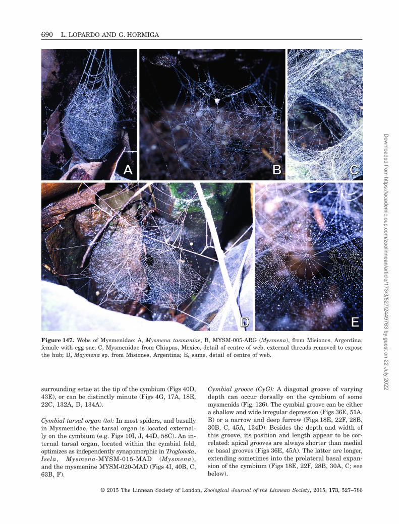
surrounding setae at the tip of the cymbium (Figs 40D,43E), or can be distinctly minute (Figs 4G, 17A, 18E,22C, 132A, D, 134A).
Cymbial tarsal organ (to): In most spiders, and basallyin Mysmenidae, the tarsal organ is located external-ly on the cymbium (e.g. Figs 10I, J, 44D, 58C). An in-ternal tarsal organ, located within the cymbial fold,optimizes as independently synapomorphic in Trogloneta,Isela, Mysmena-MYSM-015-MAD (Mysmena),and the mysmenine MYSM-020-MAD (Figs 4I, 40B, C,63B, F).
Cymbial groove (CyG): A diagonal groove of varyingdepth can occur dorsally on the cymbium of somemysmenids (Fig. 126). The cymbial groove can be eithera shallow and wide irregular depression (Figs 36E, 51A,B) or a narrow and deep furrow (Figs 18E, 22F, 28B,30B, C, 45A, 134D). Besides the depth and width ofthis groove, its position and length appear to be cor-related: apical grooves are always shorter than medialor basal grooves (Figs 36E, 45A). The latter are longer,extending sometimes into the prolateral basal expan-sion of the cymbium (Figs 18E, 22F, 28B, 30A, C; seebelow).
Figure 147. Webs of Mysmenidae: A, Mysmena tasmaniae, B, MYSM-005-ARG (Mysmena), from Misiones, Argentina,female with egg sac; C, Mysmenidae from Chiapas, Mexico, detail of centre of web, external threads removed to exposethe hub; D, Maymena sp. from Misiones, Argentina; E, same, detail of centre of web.
690 L. LOPARDO AND G. HORMIGA
© 2015 The Linnean Society of London, Zoological Journal of the Linnean Society, 2015, 173, 527–786
Dow
nloaded from https://academ
ic.oup.com/zoolinnean/article/173/3/527/2449763 by guest on 22 July 2022

Cymbial process (CyP) (Kraus, 1967: ‘Kegeldorn desparacymbium’; Baert & Maelfait, 1983: ‘cymbial thorn’):In most mysmenids there is a process, often pointed,on the dorsal surface of the cymbium. The process islocated often apically, retrolateral to the cymbial tip(Figs 1A, E, 4C, 40F, 51A, 63C, 133G, 134G, H), orbasally and prolaterally, at the end of the cymbial groove(Figs 45A, 132D, E), or it can be located apically, butprolateral to the cymbial tip (Fig. 43C, D).
Paracymbium (PC): As in most araneoids (exceptTheridiidae), a retrolateral paracymbium is present inall symphytognathoid families, except for Anapidae(Figs 71F, 92A). Although the loss of the paracymbiumis synapomorphic for Anapidae, it has secondarilyevolved in Comaroma (Fig. 81B). Almost all mysmenidshave a paracymbium; within this data set, it is sec-ondarily absent in Maymena rica and Isela (Fig. 4D,E). Although in recent studies the paracymbium was
Figure 148. Symphytognathoids webs. A, Tasmanapis strahan (Anapidae), spider not collected; B, C, Theridiosomatidaefrom Mexico; D, Anapisona kethleyi (Anapidae), male; E, potential web and egg sac of a Symphytognatha species(Symphytognathidae), from Tasmania, Australia, spider not collected.
PHYLOGENY AND MORPHOLOGY OF MYSMENIDAE 691
© 2015 The Linnean Society of London, Zoological Journal of the Linnean Society, 2015, 173, 527–786
Dow
nloaded from https://academ
ic.oup.com/zoolinnean/article/173/3/527/2449763 by guest on 22 July 2022

considered to be absent in mysmenids (Coddington, 1990;Griswold et al., 1998; Miller et al., 2009), it had beenpreviously reported as present by some authors (e.g.Kraus, 1967).
Our phylogenetic hypothesis suggests that themysmenid paracymbium evolved from a basal hook-shaped paracymbium into a characteristic shape andposition (e.g. as in Mysmena tasmaniae, Fig. 51B). Inmysmenids the paracymbium is flat and rounded, witha uniform transition to the cymbium, and it is locatedmedially, not basally (i.e. as a medial flat extensionof cymbial edge; Figs 18B, 22G, 27B, 30E, 32A, 36A,45B, 63A). A flat, rounded paracymbium evolved in-dependently as synapomorphic for Mysmenidae, butalso in Iardinis mussardi and in Synaphris. Theparacymbium becomes secondarily basal in Trogloneta(Fig. 63A), and secondarily hook-shaped in Mysmenopsis,where it is a thick (i.e. not flat), short distinct process,usually as long as wide (Figs 53D, 60D). Further-more, in Mysmenopsis, the paracymbium is bent inwards
and is seemingly interacting with a tegular groovelocated dorsally on the bulb (Figs 53F, 55F, 58D, 60D).The interaction is here considered tentative. The dorsaltegular groove does not appear to have a ‘conductor’function related to the embolus, and the paracymbium–bulb interaction as a locking mechanism is not evident,as in the case of theridiids (Levi, 1961; Saaristo, 1978;Agnarsson, 2004; and references therein).
Prolateral basal expansion: This prolonged cuticle onthe prolateral basal edge of the cymbium was origi-nally observed in Theridiosomatidae, and has beentermed the ‘prolateral basal paracymbium’ by Schütt(2003). The term ‘paracymbium’, however, has beenproposed and long used for the classical araneoidretrolateral process on the cymbium (e.g. Comstock,1910; Coddington, 1986b, 1990; Griswold et al., 1998;and references therein). Here, this prolateral struc-ture is simply referred to as ‘prolateral basal expan-sion’. Furthermore, both the paracymbium and the
Figure 149. Webs of Symphytognathidae from Tasmania, Australia: A, SYMP-006-AUST, female with egg sacs; B, same,detail of edge of web where egg sacs are attached, note female spider close to one of the egg sacs; C, SYMP-007-AUST,female?; D, same, detail of hub.
692 L. LOPARDO AND G. HORMIGA
© 2015 The Linnean Society of London, Zoological Journal of the Linnean Society, 2015, 173, 527–786
Dow
nloaded from https://academ
ic.oup.com/zoolinnean/article/173/3/527/2449763 by guest on 22 July 2022
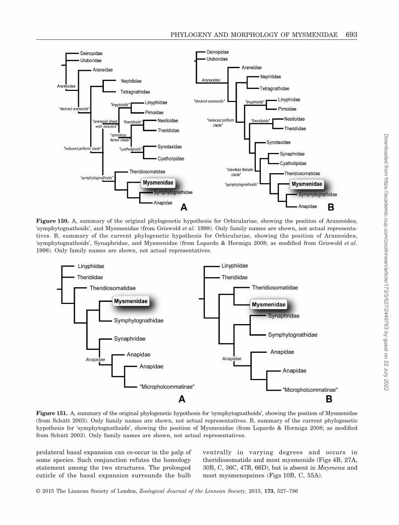
prolateral basal expansion can co-occur in the palp ofsome species. Such conjunction refutes the homologystatement among the two structures. The prolongedcuticle of the basal expansion surrounds the bulb
ventrally in varying degrees and occurs intheridiosomatids and most mysmenids (Figs 4B, 27A,30B, C, 36C, 47B, 66D), but is absent in Maymena andmost mysmenopsines (Figs 10B, C, 55A).
Figure 150. A, summary of the original phylogenetic hypothesis for Orbiculariae, showing the position of Araneoidea,‘symphytognathoids’, and Mysmenidae (from Griswold et al. 1998). Only family names are shown, not actual representa-tives. B, summary of the current phylogenetic hypothesis for Orbiculariae, showing the position of Araneoidea,‘symphytognathoids’, Synaphridae, and Mysmenidae (from Lopardo & Hormiga 2008; as modified from Griswold et al.1998). Only family names are shown, not actual representatives.
Figure 151. A, summary of the original phylogenetic hypothesis for ‘symphytognathoids’, showing the position of Mysmenidae(from Schütt 2003). Only family names are shown, not actual representatives. B, summary of the current phylogenetichypothesis for ‘symphytognathoids’, showing the position of Mysmenidae (from Lopardo & Hormiga 2008; as modifiedfrom Schütt 2003). Only family names are shown, not actual representatives.
PHYLOGENY AND MORPHOLOGY OF MYSMENIDAE 693
© 2015 The Linnean Society of London, Zoological Journal of the Linnean Society, 2015, 173, 527–786
Dow
nloaded from https://academ
ic.oup.com/zoolinnean/article/173/3/527/2449763 by guest on 22 July 2022

Figure 152. Chart representing the proportion of each of the thirteen characters sets (see Table 4).
A
Figure 153. Strict consensus of the three most-parsimonious trees (MPTs) that resulted from the analysis of the mor-phological data set. See tree statistics in Table 6. Unambiguous character optimizations are shown for every branch inthe tree. Numbers below each node indicate node numbers. Empty and filled boxes represent homoplasious and non-homoplasious transformations, respectively. Family codes used for unidentified species are as follows: MYSM, Mysmenidae;SYMP, Symphytognathidae; TSMD, Theridiosomatidae.
694 L. LOPARDO AND G. HORMIGA
© 2015 The Linnean Society of London, Zoological Journal of the Linnean Society, 2015, 173, 527–786
Dow
nloaded from https://academ
ic.oup.com/zoolinnean/article/173/3/527/2449763 by guest on 22 July 2022

Mysmenid bulb: general morphologyThe median apophysis is absent in all mysmenids. InTrogloneta and independently in the clade compris-ing Brasilionata, Mysmeniola, and Microdipoena, ategular groove housing the embolus can occur (Figs 27A,63E, 66C; see below).
Embolus: The general shape of the embolus variesgreatly within Mysmenidae, and no general patterncan be proposed. The embolus of mysmenids can bethin (or filiform) and coiled (Figs 47B, 134G), or thick(and flattened) and either coiled (Figs 4H, 27A, 36B,131H, 132B, D, E), or straight (Figs 10E, 28D, 60F,
B
Figure 153. Continued.
PHYLOGENY AND MORPHOLOGY OF MYSMENIDAE 695
© 2015 The Linnean Society of London, Zoological Journal of the Linnean Society, 2015, 173, 527–786
Dow
nloaded from https://academ
ic.oup.com/zoolinnean/article/173/3/527/2449763 by guest on 22 July 2022
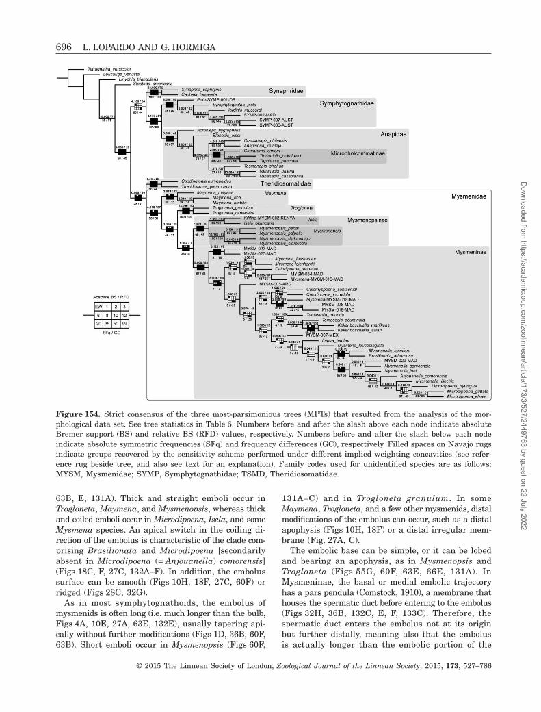
63B, E, 131A). Thick and straight emboli occur inTrogloneta, Maymena, and Mysmenopsis, whereas thickand coiled emboli occur in Microdipoena, Isela, and someMysmena species. An apical switch in the coiling di-rection of the embolus is characteristic of the clade com-prising Brasilionata and Microdipoena [secondarilyabsent in Microdipoena (= Anjouanella) comorensis](Figs 18C, F, 27C, 132A–F). In addition, the embolussurface can be smooth (Figs 10H, 18F, 27C, 60F) orridged (Figs 28C, 32G).
As in most symphytognathoids, the embolus ofmysmenids is often long (i.e. much longer than the bulb,Figs 4A, 10E, 27A, 63E, 132E), usually tapering api-cally without further modifications (Figs 1D, 36B, 60F,63B). Short emboli occur in Mysmenopsis (Figs 60F,
131A–C) and in Trogloneta granulum. In someMaymena, Trogloneta, and a few other mysmenids, distalmodifications of the embolus can occur, such as a distalapophysis (Figs 10H, 18F) or a distal irregular mem-brane (Fig. 27A, C).
The embolic base can be simple, or it can be lobedand bearing an apophysis, as in Mysmenopsis andTrogloneta (Figs 55G, 60F, 63E, 66E, 131A). InMysmeninae, the basal or medial embolic trajectoryhas a pars pendula (Comstock, 1910), a membrane thathouses the spermatic duct before entering to the embolus(Figs 32H, 36B, 132C, E, F, 133C). Therefore, thespermatic duct enters the embolus not at its originbut further distally, meaning also that the embolusis actually longer than the embolic portion of the
Figure 154. Strict consensus of the three most-parsimonious trees (MPTs) that resulted from the analysis of the mor-phological data set. See tree statistics in Table 6. Numbers before and after the slash above each node indicate absoluteBremer support (BS) and relative BS (RFD) values, respectively. Numbers before and after the slash below each nodeindicate absolute symmetric frequencies (SFq) and frequency differences (GC), respectively. Filled spaces on Navajo rugsindicate groups recovered by the sensitivity scheme performed under different implied weighting concavities (see refer-ence rug beside tree, and also see text for an explanation). Family codes used for unidentified species are as follows:MYSM, Mysmenidae; SYMP, Symphytognathidae; TSMD, Theridiosomatidae.
696 L. LOPARDO AND G. HORMIGA
© 2015 The Linnean Society of London, Zoological Journal of the Linnean Society, 2015, 173, 527–786
Dow
nloaded from https://academ
ic.oup.com/zoolinnean/article/173/3/527/2449763 by guest on 22 July 2022

Figure 155. A, strict consensus of the three most-parsimonious trees (MPTs) that resulted from the analysis of the mor-phological data set. Continuous character optimization. Character 0: shape opisthosoma, lateral view. Family codes usedfor unidentified species are as follows: MYSM, Mysmenidae; SYMP, Symphytognathidae; TSMD, Theridiosomatidae.
PHYLOGENY AND MORPHOLOGY OF MYSMENIDAE 697
© 2015 The Linnean Society of London, Zoological Journal of the Linnean Society, 2015, 173, 527–786
Dow
nloaded from https://academ
ic.oup.com/zoolinnean/article/173/3/527/2449763 by guest on 22 July 2022

Figure 155. B, strict consensus of the three MPTs that resulted from the analysis of the morphological data set. Con-tinuous character optimization. Character 1: legs I and IV relative size. Family codes used for unidentified species areas follows: MYSM, Mysmenidae; SYMP, Symphytognathidae; TSMD, Theridiosomatidae.
698 L. LOPARDO AND G. HORMIGA
© 2015 The Linnean Society of London, Zoological Journal of the Linnean Society, 2015, 173, 527–786
Dow
nloaded from https://academ
ic.oup.com/zoolinnean/article/173/3/527/2449763 by guest on 22 July 2022

Figure 155. C, strict consensus of the three MPTs that resulted from the analysis of the morphological data set. Con-tinuous character optimization. Character 2: metatarsus–tarsus relative size. Family codes used for unidentified speciesare as follows: MYSM, Mysmenidae; SYMP, Symphytognathidae; TSMD, Theridiosomatidae.
PHYLOGENY AND MORPHOLOGY OF MYSMENIDAE 699
© 2015 The Linnean Society of London, Zoological Journal of the Linnean Society, 2015, 173, 527–786
Dow
nloaded from https://academ
ic.oup.com/zoolinnean/article/173/3/527/2449763 by guest on 22 July 2022

Figure 155. D, strict consensus of the three MPTs that resulted from the analysis of the morphological data set. Con-tinuous character optimization. Character 3: femur–metatarsus relative size. Family codes used for unidentified speciesare as follows: MYSM, Mysmenidae; SYMP, Symphytognathidae; TSMD, Theridiosomatidae.
700 L. LOPARDO AND G. HORMIGA
© 2015 The Linnean Society of London, Zoological Journal of the Linnean Society, 2015, 173, 527–786
Dow
nloaded from https://academ
ic.oup.com/zoolinnean/article/173/3/527/2449763 by guest on 22 July 2022

Figure 155. E, strict consensus of the three MPTs that resulted from the analysis of the morphological data set. Con-tinuous character optimization. Character 4: tibia–metatarsus relative size. Family codes used for unidentified speciesare as follows: MYSM, Mysmenidae; SYMP, Symphytognathidae; TSMD, Theridiosomatidae.
PHYLOGENY AND MORPHOLOGY OF MYSMENIDAE 701
© 2015 The Linnean Society of London, Zoological Journal of the Linnean Society, 2015, 173, 527–786
Dow
nloaded from https://academ
ic.oup.com/zoolinnean/article/173/3/527/2449763 by guest on 22 July 2022

Figure 155. F, strict consensus of the three MPTs that resulted from the analysis of the morphological data set. Con-tinuous character optimization. Character 5: posterior lateral spinnerets aciniform gland spigot number. Family codesused for unidentified species are as follows: MYSM, Mysmenidae; SYMP, Symphytognathidae; TSMD, Theridiosomatidae.
702 L. LOPARDO AND G. HORMIGA
© 2015 The Linnean Society of London, Zoological Journal of the Linnean Society, 2015, 173, 527–786
Dow
nloaded from https://academ
ic.oup.com/zoolinnean/article/173/3/527/2449763 by guest on 22 July 2022

Figure 155. G, strict consensus of the three MPTs that resulted from the analysis of the morphological data set. Con-tinuous character optimization. Character 6: anterior lateral spinnerets piriform spigot number. Family codes used forunidentified species are as follows: MYSM, Mysmenidae; SYMP, Symphytognathidae; TSMD, Theridiosomatidae.
PHYLOGENY AND MORPHOLOGY OF MYSMENIDAE 703
© 2015 The Linnean Society of London, Zoological Journal of the Linnean Society, 2015, 173, 527–786
Dow
nloaded from https://academ
ic.oup.com/zoolinnean/article/173/3/527/2449763 by guest on 22 July 2022

Figure 156. Strict consensus of the three most-parsimonious trees (MPTs) that resulted from the analysis of the mor-phological data set. Nodes collapsed if Bremer support values are smaller than 0.95 steps. Family codes used for un-identified species are as follows: MYSM, Mysmenidae; SYMP, Symphytognathidae; TSMD, Theridiosomatidae.
704 L. LOPARDO AND G. HORMIGA
© 2015 The Linnean Society of London, Zoological Journal of the Linnean Society, 2015, 173, 527–786
Dow
nloaded from https://academ
ic.oup.com/zoolinnean/article/173/3/527/2449763 by guest on 22 July 2022

Figure 157. Strict consensus of 3835 most-parsimonious trees (MPTs) that resulted from the analysis of the morpho-logical data set including only discrete characters. See tree statistics in Table 6. Numbers before and after the slashabove each node indicate absolute Bremer support (BS) and relative BS (RFD) values, respectively. Numbers before andafter the slash below each node indicate absolute symmetric frequencies (SFq) and frequency differences (GC), respec-tively. Family codes used for unidentified species are as follows: MYSM, Mysmenidae; SYMP, Symphytognathidae; TSMD,Theridiosomatidae.
PHYLOGENY AND MORPHOLOGY OF MYSMENIDAE 705
© 2015 The Linnean Society of London, Zoological Journal of the Linnean Society, 2015, 173, 527–786
Dow
nloaded from https://academ
ic.oup.com/zoolinnean/article/173/3/527/2449763 by guest on 22 July 2022
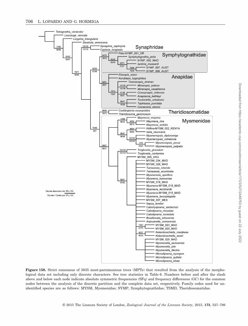
Figure 158. Strict consensus of 3835 most-parsimonious trees (MPTs) that resulted from the analysis of the morpho-logical data set including only discrete characters. See tree statistics in Table 6. Numbers before and after the slashabove and below each node indicate absolute symmetric frequencies (SFq) and frequency differences (GC) for the commonnodes between the analysis of the discrete partition and the complete data set, respectively. Family codes used for un-identified species are as follows: MYSM, Mysmenidae; SYMP, Symphytognathidae; TSMD, Theridiosomatidae.
706 L. LOPARDO AND G. HORMIGA
© 2015 The Linnean Society of London, Zoological Journal of the Linnean Society, 2015, 173, 527–786
Dow
nloaded from https://academ
ic.oup.com/zoolinnean/article/173/3/527/2449763 by guest on 22 July 2022

Figure 159. Strict consensus of the three most-parsimonious trees (MPTs) that resulted from the analysis of the mor-phological data set including all taxa. The five taxa represented by 78% or more missing data are in bold. See tree sta-tistics in Table 6. Numbers above and below each node indicate absolute Bremer support (BS) and relative BS (RFD)values, respectively. Family codes used for unidentified species are as follows: MYSM, Mysmenidae; SYMP, Symphytognathidae;TSMD, Theridiosomatidae.
PHYLOGENY AND MORPHOLOGY OF MYSMENIDAE 707
© 2015 The Linnean Society of London, Zoological Journal of the Linnean Society, 2015, 173, 527–786
Dow
nloaded from https://academ
ic.oup.com/zoolinnean/article/173/3/527/2449763 by guest on 22 July 2022

ejaculatory duct. Although found in other spider fami-lies (e.g. in some theridiids, linyphiids, cyatholipids,and agelenids), the pars pendula is an ambiguouslyoptimized synapomorphy for Mysmeninae (ambigu-ously optimizes at its node because of missinginformation on the basal clade of this group),and it is secondarily absent in MYSM-005-ARG(Mysmena).
Tegular conductor: As in most symphytognathoids, mostmysmenids have a conductor. Mysmenid conductor isdistinctly voluminous and membranous, and origi-nates subterminally from the tegulum, close to theembolic base (Figs 17A, 18D, 27A, B, 30E, 36C, 41D,63B, D). This structure has been named ‘bulbal shield’(e.g. Baert, 1984a; Schütt, 2003), and other than theembolus, it appears to be the only tegular sclerite. Inmysmenids, the conductor often embraces and evencovers the embolic base (Figs 41C, 43A, B). In somespecies there is a groove on the conductor surfacehousing the basal portion of the exposed embolus(Figs 36B, C, 47E); however, a groove housing the distalportion of embolus is absent in the tegular conductorof mysmenids, and the tip of the embolus is insteadhoused by one of the two conductors on the cymbium(see above). Within symphytognathoids, the occur-rence of a tegular conductor is symplesiomorphicand widespread, with few independent losses of thissclerite occurring in anapids, symphytognathids, andmysmenids. Within Mysmenidae, the conductor is losttwo times: in the clade that includes Maymena andMysmenopsinae, and in Mysmena MYSM-005-ARG(Figs 4A, 10E, 28D).
Spermatic duct trajectory (SDT; refer to Fig. 127 andcharacters 220–228): As with most of the aforemen-tioned palpal features, there does not seem to be a con-sistent pattern in the trajectory of the spermatic ductacross the family, although there is some regularitywithin clades. The spermatic duct usually travels clock-wise from the fundus (in left palp). If a switchback(SB) occurs, it alters this direction to travel counter-clockwise. Usually, a counter-switch also occurs to returnthe duct trajectory to its original clockwise direction.Therefore, when SBs are present, they usually occurin pairs of switchbacks. The trajectory of the sper-matic duct in Trogloneta differs from all othermysmenids in that the pair of switchbacks that occurbefore the spermatic duct completes one loop from thefundus (i.e. SB I and II, see Fig. 127) are absent, there-fore the trajectory is completely spiralling or has onecomplete loop before the ‘second’ pair of switchbacks(SB III and IV) occurs (Fig. 131E, F). Although thegeneral arrangement of the spermatic duct in all othermysmenid species examined in this study (exceptTrogloneta) is not necessarily similar among clades, all
have the first pair of switchbacks on their trajec-tories (SB I and II; Figs 131I, 132B, D).
The position of switchback I (SB I) varies withinMysmenidae also. A distal SB I (apart from basal fundus,on the opposite area of the bulb) occurs in mostmysmenines, most Maymena, and in Isela okuncana(Figs 131H, I, 132B, E, 133A, B, 134B, C). A basal SB Ithat does not reach the distal part of the bulb can occurin other mysmenines, however (Figs 133H, I, 134F–H); whereas in most mysmenopsines and Maymenamayana the SB I occurs close to the fundus, after thespermatic duct has reached the distal wall of the bulb(i.e. ‘beyond distal’; Fig. 131C, G, J).
In most mysmenids, the portions of spermaticduct forming the SB I are divergent, and SB IIoccurs relatively close to SB I (Figs 131B, H, J, 133A,B, 134D–G). In Microdipoena and most Mysmenaspecies, the portions of spermatic duct comprising SB Irun close to each other, and the SB II occurs close tothe midpoint between SB I and the fundus, or evencloser to the fundus (Figs 131I, 132B–D, 133D, E,134A–C).
The plesiomorphic condition in Mysmenidae is to havetwo or more ascending loops in the last portion of thecoiling reservoir before entering the embolus. This occursbasally within Mysmeninae (Figs 133D, E, 134E, H).Within the family, the number of loops decreases in-dependently in distal clades (no loops or less than oneentire loop, Figs 131B, E, H, I, 132F, 133A, B, 138C,G, 139E; or about one and one and a half loops,Figs 131D, 139C).
A pair of switchbacks (SB III and IV) can occureither after SB II, or if SB I and II are absent, aftera complete loop of the spermatic duct (e.g. as inSymphytognatha picta; Fig. 139C). In this data set, anabsence of pairs of extra switches (i.e. SB III and IV)is plesiomorphic for both symphytognathoidsand Mysmenidae (Figs 131D, E, J, 133A, B, D, E, H,I, 134E). Switchbacks III and IV evolve independent-ly in Microdipoena, Mysmena MYSM-007-MEX, Iselaokuncana, and Trogloneta cantareira (Figs 131F, H,132B–E). Furthermore, a distinct trajectory of the sper-matic duct where several pairs of switchbacks occurevolved convergently in the mysmenines MYSM-020-MAD and MYSM-023-MAD (Figs 134F–H).
Epigynum (refer to characters 59–87)Mysmenids are entelegyne spiders. That is, they havefertilization ducts leading from the spermathecae tothe uterus externus and copulatory ducts connectingexternal openings to the spermathecae (e.g. Wiehle,1967; Uhl, 2002). No comparative study of mysmenidfemale genitalia has ever been performed. In mostfemale mysmenids, particularly mysmenines, theducts and sometimes even the spermathecae areextremely membranous, almost invisible under light
708 L. LOPARDO AND G. HORMIGA
© 2015 The Linnean Society of London, Zoological Journal of the Linnean Society, 2015, 173, 527–786
Dow
nloaded from https://academ
ic.oup.com/zoolinnean/article/173/3/527/2449763 by guest on 22 July 2022

microscopy. A few authors describing mysmenid specieshave published detailed illustrations and have at-tempted to identify the different components of femalegenitalia in as much detail as possible (e.g. Kraus, 1967;Loksa, 1973; Thaler, 1975; Baert, 1984a, b, 1986, 1988;Snazell, 1986; Baert & Murphy, 1987). Given themembranous and almost undetectable nature ofmysmenid female genitalia, the interpretation of thesestructures is often difficult. Furthermore, the greatdiversity of mysmenid female genitalic morphology(see below) makes diagnosis of the family a challeng-ing task if only based on this system of characters.Our interpretations and homology statements of femalegenitalic structures are based on light and SEMmicroscopy data.
External genitaliaThe epigynum, as a sclerotized modification of thecuticle, is absent in most of the members of the sub-family Mysmeninae. In addition, the copulatory open-ings are located within the epigastric furrow (i.e. theepigynal area containing the copulatory openings ishidden within it; Fig. 24A). This latter trait hasconvergently evolved in most anapids. All representa-tives of Mysmeninae here examined have a membra-nous atrium (Figs 59H, 129A, E, G, 130B). The atriumhas been defined as a ‘widened cavity into the copu-latory ducts’ (Sierwald, 1989: 2), and can occur inde-pendently of the location (external or internal) of thecopulatory openings. In most mysmenids, however, theseemingly epigynal area located centrally betweenthe copulatory openings (here regarded as the ‘dorsalplate’ sensu Millidge, 1984; ‘middle field’ on Sierwald,1989) is projecting, i.e. it is exposed or protruding fromthe epigastric furrow (Figs 11B, C, 12D, E, 37C, 42C,44A, 49D, 67A, B). The dorsal plate is secondarilyinternal (i.e. neither exposed nor projecting) inMysmenopsinae, Maymena rica, Microdipoena, and fewother mysmenines (e.g. Fig. 24B).
Trogloneta, Maymena, Mysmenopsinae, and themysmenine MYSM-023-MAD have a modified copula-tory area or epigynum in the form of a sclerotized plateor a protruding modification of the cuticle, usuallybearing setae and the copulatory openings (Figs 5A,B, 11B, C, 12D, E, 49D, E, 59H, 61A, 67A, B, 140D,I, 141G, H, 142C). The copulatory openings are exposedcaudally (i.e. posteriorly) in the epigynal area (Figs 49D,59H, 61B). A sclerotized external atrium is present ina few Mysmenopsis species (e.g. Fig. 59H). On the otherhand, in most Mysmeninae the epigynal area is weaklymodified or even absent (i.e. the cuticle in this areais similar to surrounding abdominal cuticle), and it isusually translucent (spermathecae can be observedbeneath it; Figs 14C, 37A, 52F, 141I). Although seem-ingly widespread among araneoids (Levi & Levi, 1962;Millidge, 1984; Scharff & Coddington, 1997; Griswold,
2001), the ventral scape of most mysmenines is uniquewithin symphytognathoids (Figs 24B, 29C, 31G, 37C,42C, 129E).
Internal genitaliaCopulatory ducts: Within Mysmenidae, the copula-tory ducts show varying degrees of sclerotization andwidth. In Maymena and most Mysmenopsinae the copu-latory ducts are short, relatively sclerotized, narrow,and of invariable diameter (Figs 11D, 37D, 128A, D,G, 129G). In Trogloneta the walls of the distal portionof the long copulatory ducts are rather smooth, rela-tively sclerotized, and uniform in diameter; however,the proximal portion of the ducts of Trogloneta is highlymembranous and has a larger diameter than the distalpart (Figs 64A, 128F). In the aforementioned taxa (i.e.Trogloneta, Maymena, and Mysmenopsinae), the tra-jectory of the copulatory ducts is in most cases rec-ognizable. Conversely, the copulatory ducts ofMysmeninae differ from all other mysmenids (with theexception of a few taxa). The walls of the ducts areextremely membranous, imperceptible under light mi-croscopy, and are of uneven diameter. These irregu-lar membranous ducts follow a convoluted and longtrajectory of unclear course. The ducts seem to extendventrally and anteriorly to the spermathecae, al-though without a definite pattern (Figs 18G, 27D, 129A,C, E, H, 130B). In some species the ducts can be subtlymore sclerotized and definite, and a coiled trajectorycan be observed (Figs 37D, 129G). The increase in thediameter of the proximal portion of the copulatory ducts(i.e. the first half of the ducts from the copulatory open-ings) has been termed ‘bursae’ by Schütt (2003: char-acter 77), although it is not clear from that studywhether the increase in diameter refers to the copu-latory ducts or to the membranous atrium (see above).
Within Mysmeninae, there is a particular turn oc-curring proximally in the convoluted and membra-nous copulatory ducts, seemingly originating close tothe internal atrium, but immediately before becom-ing widened and convoluted. This duct turn is char-acterized by a subtle but consistent sclerotization. Thisfeature occurs in Microdipoena, and also evolved in-dependently in two clades within Mysmena and inMYSM-029-MAD (Figs 129A, B, 130A).
A convoluted trajectory of the copulatory ducts char-acterizes Mysmenidae, although the ducts can varygreatly in terms of sclerotization and diameter. Straightducts evolved independently twice: once in the cladecomprising Maymena and Mysmenopsinae and once inMysmena MYSM-034-MAD. Although rare in the currentmysmenid taxon sample, coiled and more distinct copu-latory ducts of mysmenines (as in Figs 37D, 129G)appear to be more common than represented here (e.g.Lopardo, Dupérré & Paquin, 2008; Miller et al., 2009;L. Lopardo, A. Janzen, C. Griswold & P. Michalik,
PHYLOGENY AND MORPHOLOGY OF MYSMENIDAE 709
© 2015 The Linnean Society of London, Zoological Journal of the Linnean Society, 2015, 173, 527–786
Dow
nloaded from https://academ
ic.oup.com/zoolinnean/article/173/3/527/2449763 by guest on 22 July 2022
unpubl. data). These distinct ducts might represent aplesiomorphic but intermediate condition (i.e. betweensclerotized and fully membranous ducts) in the evo-lution of convoluted and highly membranous copula-tory ducts, and might help in elucidating our currentinterpretation of their trajectory, as well as theiridentification.
Spermathecae and accessory glands: Most mysmenidshave one sperm-storage compartment in eachspermatheca (e.g. Fig. 33A). Trogloneta and someanapids have two pairs of compartments in eachspermatheca (Fig. 128F). The spermathecae are usuallydefined by a thick sclerotized wall (Figs 129A, 130B),although exceptions occur (e.g. in mysmenine MYSM-023-MAD and a few anapids; Figs 49A, 130D).
Ovoid spermathecae are plesiomorphic for bothsymphytognathoids and Mysmenidae. Nevertheless, thediversity of spermathecal shapes within Mysmenidaeis immense, seemingly following no particularphylogenetic pattern, not even at the genus level.Spermathecae can be ovoid (Figs 11D, 18G, 37D, 128D,129A, G), C- or cup-shaped (Figs 27D,E, 42D, 128E,130C), coiled within the same spermathecal struc-ture (Figs 33A, B, 51D, 129C, 130B, G), tubuliform (asone large tube, sometimes like tracheoles; Figs 5D, 49A,128A, C, 130D), clavate (Figs 12C, 128B), or even ir-regular (although consistent within each species), whereno particular shape can be defined (Fig. 129D).
An additional paired structure occurs in the inter-nal genitalia of Trogloneta, the mysmenine MYSM-029-MAD, Mysmena MYSM-005-ARG, Microdipoena,and the anapid Tasmanapis. This structure resem-bles either an apodeme or a glandular structure, andappears related to the copulatory ducts or thespermathecae (Figs 22B, 27D, 29A, 64A, 129B, E, H,130A, F). It is better observed by SEM, although itcan be distinguished in transparent preparations of thevulva by a higher degree of sclerotization, compa-rable with that of the spermathecae (Figs 129B, E, H,130A, F; see also Brescovit & Lopardo, 2008: fig. 6C–E). Whether these structures are functional glands,muscle attachment points, or perform other func-tions remains unknown. They are regarded here asaccessory glands.
Fertilization ducts: The morphology of fertilization ductsis also variable within the family. Fertilization ductswere identified in Mysmenidae by discerning theducts connected to and from the spermathecae, and,with the help of SEM, an attempt was made to followthe trajectory of these ducts. Usually fertilization ductsare located either dorsal to or on the central internallateral side of the spermathecae. As a convention, whenthe genital system was mainly composed by mem-branes, highly developed and convoluted copulatory
ducts were first identified, and then fertilization ductswere distinguished by elimination. The degree ofsclerotization of the fertilization ducts appears highlyhomoplastic. Weakly sclerotized fertilization ducts witha distinguishable wall are plesiomorphic for Mysmenidae(although ambiguously optimized), and occur in Isela,mysmenine MYSM-023-MAD, and most Mysmena(Figs 42D, 49A, 128A, 129G); however, membranous,translucent, and almost imperceptible fertilization ductsalso occur within the family, in Trogloneta, mostMysmenopsis, and all members of Microdipoena(Figs 18G, 27D, 51D, 60H, 64A, 128F, 136D).
Small, short fertilization ducts providing a direct con-nection between the spermathecae and the uterusexternus (usually in straight fashion and unmodified)are plesiomorphic and widespread within Mysmenidae(Figs 42D, 49A, 64A, 128A, F). Large, long fertiliza-tion ducts, most often longer than the size of thespermathecae, might be provided with modificationsor expansions (Figs 11D, 18G, 27D, 51D, 60H, 92F, 129G,136D). They occur independently in most Maymena,most Mysmenopsis, most species of Microdipoena, andfew Mysmena.
Spinneret silk gland spigot morphology (refer tocharacters 304–340)The spinning organs of Mysmenidae have been de-scribed for a few species, including the kleptoparasiticIsela okuncana (Griswold, 1985), an undescribed Iselafrom Cameroon, Mysmenopsis penai, the AustralianMysmena tasmaniae and M. leichhardti (Lopardo &Michalik, 2013) and Maymena mayana (Griswold et al.,1998). Recently, the gland spigot patterns of Troglonetacantareira (Brescovit & Lopardo, 2008) and severalChinese mysmenids (Mysmena, Gaoligonga, Chanea,and Maymena, Miller et al., 2009) were alsodescribed. The data in the aforementioned works suggestthat mysmenids have the typical symphytognathoidand higher araneoid silk gland spigot conformationon the anterior lateral and the posterior medianspinnerets (ALS and PMS, respectively): few ALSpiriform gland spigots, few aciniform gland spigots onposterior (median and lateral) spinnerets, a furrowbetween major ampullate and piriform fields, andreduced piriform bases. In this study we examinedin detail the spinneret gland spigot conformation of30 mysmenid species. In the following section, thegeneral arrangement of mysmenid spinneret glandspigots is described. Exceptions, singular features, andsynapomorphies for the main mysmenid clades arenoted below. See Appendix 2 for an explanation of cu-ticular textures.
In general, the colulus is fleshy and usually rela-tively large, bearing three or less setae (Figs 24E, 33C,56D, 59I). On the anterior lateral spinnerets, a gla-brous tuberculate intersegmental cuticle occurs (Figs 6A,
710 L. LOPARDO AND G. HORMIGA
© 2015 The Linnean Society of London, Zoological Journal of the Linnean Society, 2015, 173, 527–786
Dow
nloaded from https://academ
ic.oup.com/zoolinnean/article/173/3/527/2449763 by guest on 22 July 2022

16B, 23A, C, 33F, 52B, 61C). The major ampullate glandspigot is accompanied by a nubbin and a tartipore(Figs 6B, 23A, C, 61C). The base of the piriform glandspigots is reduced (Figs 6B, 52B, 61C), and the cuticlesurrounding those piriform gland spigots can be eitherfingerprint (in Mysmeninae and most Mysmenopsinae)or rugose (in Maymena, Trogloneta, and the Isela rep-resentative Kilifina-MYSM-002-KENYA; Fig. 6B). Twoaciniform, one posterior minor ampullate, and (infemales) one cylindrical gland spigot occur on theposterior median spinnerets (Figs 11F, 33D, 58F). Theminor ampullate can be accompanied by a tartipore,a tartipore plus a nubbin, or by none of these. Theposterior lateral spinnerets bear two cylindrical glandspigots, where both (Figs 11G, H, 13F, 67D) or onlythe posterior spigot is peripheral to the spinning field(the anterior spigot on the edge of the field; Figs 23B,37B, 58H), and some aciniform gland spigots. The triad(the assemblage of one flagelliform and two aggre-gate gland spigots producing sticky silk in araneoids)is present in most female mysmenids (Figs 11G, 23B,37B, 52C, 67D), as is usually the case in Araneoidea.Some exceptions occur, however (see below). Both ag-gregate and flagelliform gland spigots are similar insize (Figs 13F, G, 37B, 52C). In most species, the triadon males is vestigial, were remnants of previously func-tional gland spigots can be observed (Figs 23E, 33H).In some species, the triad is retained in adult males(Fig. 13G; this change is ambiguously optimized at thebase of Mysmenidae).
The spinnerets of mysmenids differ from that of allother families represented in this data set by the pres-ence of a lobe on the intersegmental groove of the ALS(Figs 23A, C, G, H, 52B). This lobe is also present withinAnapidae, although with high homoplasy. Mysmenidsalso differ from other families in the separation of themajor ampullate and piriform fields by a subtle (shallow)groove (Figs 13C, 23C, 33F, 52B, 61C, 64C), where theconnection between both fields is distinctly evident proxi-mally within the ALS segment (ambiguously occur-ring in Theridiosomatidae). Finally by the characteristicshape of a seta on the major ampullate field, with eitherone or two rows of long ‘branches’ (Figs 6A, 23F, 33F,52B, 61C).
Trogloneta is the only symphytognathoid so far ex-amined with minute but distinguishable colulus(Figs 64D, 67F, 68C; as opposed to the remnant colulusof Patu, see character 317). An additional anterior dis-crete cluster of cuticular protuberances of unknown func-tion also occurs in the ALS of Trogloneta (Figs 64C,66F). Other attributes occurring in Trogloneta, al-though not exclusive to this genus, include: a rugosecuticle on the piriform field on ALS (Fig. 64C); minorampullate gland spigot accompanied solely by a tartiporeon PMS; both PLS cylindrical gland spigots equallylarge and larger than the flagelliform gland spigot
(Fig. 67D); and triad spigots retained in adult males(at least in T. granulum).
In Maymena, the shape of the seta on the majorampullate field has a distinct single row of long‘branches’ (Figs 11E, 13C, 16B); in other mysmenids,two rows occur. The PMS minor ampullate gland spigotis accompanied by a nubbin and a tartipore (Fig. 11F).The following features occur independently in bothMaymena and Mysmeninae, and were not observed inany other taxa examined in this data set: an anteri-or distinctly flat spatulate modified seta on PLS(Figs 11G,H, 13E) and aciniform gland spigots of dif-ferent shape in both the posterior spinnerets (medianand lateral, Figs 11F, G, 13F, G; see character 304).Other attributes occurring in Maymena, although notexclusive of the genus, include: rugose cuticle on piriformfield on ALS (Figs 11E, 16B), and both PLS cylindri-cal gland spigots equally large and larger than theflagelliform gland spigot (Figs 11G, H, 13F). Triad spigotsare retained in adult males of M. mayana (Fig. 13G),but this retention appears autapomorphic for this speciesrather than a generic condition, given that the triadis vestigial in at least M. rica.
In Mysmenopsinae the adult male aggregate glandspigots are absent, and those of the females are dis-tinctly absent as well (see below). Other spinneret fea-tures of this subfamily include: a fingerprint cuticleon ALS piriform field; PMS minor ampullate glandspigot accompanied by neither nubbin nor tartipore(Figs 6C, F, 58F, 61D); and both PLS cylindrical glandspigots slim as other gland spigots (not larger) andsubequal to flagelliform gland spigot (Fig. 58H). InMysmenopsis, the colulus bears four or more setae(Figs 56D, 59I). Although both aggregate gland spigotsare absent in males and females, the flagelliform glandspigot in the representatives of Mysmenopsis studiedhas been retained, and seems to be functional in bothsexes (Fig. 58G, H). In Isela both flagelliform and ag-gregate gland spigots are distinctly absent in both sexes(Fig. 6D, G).
Finally, the subfamily Mysmeninae shares withMaymena the anterior distinctly flat spatulate modi-fied seta on PLS (Figs 23B, E, 33G, H, 37B, 52C) andaciniform gland spigots of different shape in both pos-terior spinnerets (median and lateral, Figs 23D, 33D,37B; see character 304). These two features evolvedindependently in the two clades. Other features oc-curring in mysmenines include a fingerprint cuticle onALS piriform field (Figs 23C, 52B); PMS minorampullate gland spigot accompanied solely by a tartipore(Figs 19F, 23D, 33D); both PLS cylindrical gland spigotsslim as other gland spigots (not larger) and subequalto flagelliform gland spigot (Figs 23B, 37B, E, 52C);and vestigial triad in males (Fig. 23E), independent-ly functional in Mysmena MYSM-005-ARG andMysmena tasmaniae.
PHYLOGENY AND MORPHOLOGY OF MYSMENIDAE 711
© 2015 The Linnean Society of London, Zoological Journal of the Linnean Society, 2015, 173, 527–786
Dow
nloaded from https://academ
ic.oup.com/zoolinnean/article/173/3/527/2449763 by guest on 22 July 2022

Other morphological features of Mysmenidae
Clasping spinesThe males of all mysmenids except Maymena mayanahave a prolateral metatarsal clasping spine (macroseta)on leg I (e.g. Fig. 34C). The phylogenetic hypothesis fromthe total-evidence analysis agrees with the morpho-logical hypothesis from this study and with previousstudies that have suggested this macroseta as asynapomorphy (or diagnostic) for the family (Thaler,1975; Platnick & Shadab, 1978; Brignoli, 1980; Griswold,1985; Wunderlich, 1995; Griswold et al., 1998; Schütt,2003). Although some members of Anapidae have aclasping structure on the first legs, these structuresare not spines (macrosetae) but spurs (short and stoutseta), and can occur in both sexes. The clasping spinesof mysmenids are sexually dimorphic, occurring inmales, are unique for the family, and might be in-volved in mating behaviour (Schütt, 2003). The wide-spread condition is a medial and straight metatarsalclasping spine (Figs 34C, 42H, 45H, 65C), but the spinecan be basal (e.g. Maymena; Figs 16G, 141K), apical(few Mysmenopsis and mysmenines species; Figs 50H,59B, 143C, 144A), twisted (Isela and Microdipoena;Figs 3B, 8B, C, 26C, 140E, F, 141K, L, 142N), or strong-ly curved proximally and accompanied by adjacentstrong setae (most Mysmenopsis; Figs 54D, 57I, 59B).Additional apical tibial clasping spines can also be foundin some mysmenids, as is the case of one tibial clasp-ing spine occurring in all Maymena species (exceptM. mayana) and in Mysmenopsinae (Figs 3A, 8D, 16G,54C, 59B, 62E, F, 140E, J, K); or two clasping spinesin Mysmenopsis dipluramigo (Fig. 140H) andMicrodipoena (Figs 26C, 27I, 57F, G, 141L, O).
Femoral spot or other femoral structuresThe femoral spot has been previously suggested as di-agnostic or synapomorphic for the family (Thaler, 1975;Platnick & Shadab, 1978; Brignoli, 1980; Griswold, 1985;Wunderlich, 1995; Griswold et al., 1998; Schütt, 2003),as this cuticular structure is unique to mysmenids. Theresults of our study corroborate this hypothesis. Theadult females of all mysmenid species have either asclerotized spot (Figs 34A, 39D, 140G, 141C, 143N) ora cuticular projection (Fig. 57A, E) on the apical ventralsurface of at least femur I, although a few exceptionsoccur (e.g. Mysmena MYSM-005-ARG and a fewMysmenopsis species). The femoral structure of mostfemale mysmenids occurs in both femora I and II(symplesiomorphic in this data set, Figs 141C, 142B).The occurrence of this feature only on femur I is con-vergent in Trogloneta, Mysmenopsis (when present),and a number of times within Mysmena (Fig. 140G).
The femoral sclerotization (i.e. spot) was first de-scribed by Marples (1955) for Tamasesia (= Mysmena).Its function remains unknown. The absence of pores
in its surface indicates that we should rule out a glan-dular function. It has been suggested that the spot couldbe a ‘. . . functionless remnant of an unknown struc-ture, because it shows a great variability in size andshape among specimens and can be differently pro-nounced on the two first femora of a single specimen.It is too large to be the socket of a former spine andapart from this there is no space for a large spine soclose to the patella . . .’ (Schütt, 2003: 143). Based onthe results of the combined analysis, the spot origi-nates at the node of Mysmenidae, becomes a femoralprojection in Mysmenopsis, and is lost distally in somespecies. Whether the femoral spot actually has a func-tion (e.g. a behavioural function), or is just a remnantof a functional structure, remains an unsolved puzzle.
All femoral structures are absent in juvenilemysmenids (which argues against the remnant hy-pothesis), although they can sometimes be perceivedin subadult stages. The femoral projection occurs onlyin females of some species of Mysmenopsis. The femoralspot, however, can also occur on males (Fig. 21A). Malefemoral spots evolved ambiguously in Trogloneta andMaymena, independently in Microdipoena [excludingits basal species M. (= Mysmenella) samoensis], andin two instances within Mysmena. In contrast withfemales, most mysmenid males have the femoral spot(when present) only on femur I (Fig. 141O), althoughin two cases the spot occurs in both femora I and II(Maymena ambita and Mysmena tasmaniae; Fig. 140M).
Prolateral row of modified setae in the malefirst leg tarsiFirst observed and described as a ventral row of modi-fied setae by Thaler (1975, 1995) for males of Troglonetagranulum and Mysmeniola spinifera (respectively), thisprolateral row of modified setae (see below) is an am-biguously optimized synapomorphy for Mysmenidae.As is the case of most morphological features withinMysmenidae, and even though this row of modified setaeoccurs in all examined species, the particular detailsof this feature differ among clades. The modified setaeare usually shorter than the surrounding tarsal setae,and can be slimmer and curved or stouter and straight.These modified setae can also be distributed along theentire length of the tarsus or just on the distal half.In Mysmeninae, the setae are slimmer and the rowoccupies the distal half of the tarsus (except inMysmeniola, see below; Figs 26A, 34D, 45I, 50F). InMaymena rica and in Mysmeniola spinifera the setaeare also slimmer but the row is distributed all alongthe segment (Fig. 16H). Trogloneta and Isela both havestout setae distributed along the tarsus (Figs 8F, 65D,68A). And lastly, Mysmenopsis also have stout setaebut, as in Mysmeninae, the row occupies the distal halfof the tarsus (Figs 54G, 59D). Although the functionof this row of modified setae remains unknown, the
712 L. LOPARDO AND G. HORMIGA
© 2015 The Linnean Society of London, Zoological Journal of the Linnean Society, 2015, 173, 527–786
Dow
nloaded from https://academ
ic.oup.com/zoolinnean/article/173/3/527/2449763 by guest on 22 July 2022

fact that these setae are found only in the males sug-gests that it may be involved in mating behaviour.
Other morphological features of MysmenidsSize of the tarsal organ opening on leg I: A large openingof the tarsal organ (i.e. subequal or larger than setalsockets) evolved independently in Mysmenidae andAnapidae (see Figs 26E, 39C, 48F, 50D, 73B, 83E, 95I,101E, respectively). An opening distinctly smaller thansetal sockets is the plesiomorphic condition insymphytognathoids. Within Mysmenidae, the tarsalorgan opening becomes secondarily small inMysmenopsinae, with a reversal in Mysmenopsispalpalis (Figs 3F, 54F, 62H).
Distinctly thick distal promarginal curved seta onchelicerae: Most taxa in this data set have a particu-larly distinct curved seta located distally at thepromarginal edge of chelicerae, near the fang base. Thisseta differs from surrounding promarginal setae by itsthickness and/or serration. Although a distinctly curvedseta can also occur on the cheliceral retromargin ofsome araneomorph spiders (see Griswold et al., 2005,character 34), retromarginal setae of the taxon sampleexamined in this study do not differ from surround-ing setae (e.g. Fig. 7J). Within symphytognathoids, thedistinctly curved promarginal seta is lost in Synaphridaeand most Symphytognathidae (Figs 108E, 118E, 122A;the optimization of this character under parsimony isambiguous). All examined mysmenid representativeshave a uniquely thicker distal promarginal curved seta(Figs 19E, 25E, 38H, 48B), except Maymena. In somemysmenids this seta is equally serrated as surround-ing seta, which is the plesiomorphic condition for thefamily; however, the thicker seta is strongly serratedon one side independently in Mysmeninae and mostMysmenopsinae (Figs 19E, 38H, 42E, 48B).
Intermediate sternum posterior margin: Pointed andtruncate sternal margins are quite distinct in the taxarepresented here, although the systematic value of thischaracter has been questioned because of impreci-sion in shape definition, reliability of observation,homoplasy, and possible influence of overall body pro-portions on sternum shape (Coddington, 1986a; Griswoldet al., 1998; Schütt, 2003). A truncate sternum is asynapomorphy of symphytognathoids, occurring in allfamilies except Mysmenidae. In Mysmenidae, an inter-mediate condition between pointed and truncateposterior sternal margin is consistently found (Figs 2C,7C, 25B, 35C, 46A, 59G, 140B, 143B, I); however, anambiguously optimized reversal to pointed posteriorsternum occurs in Maymena (Fig. 141F), and two in-dependent instances of truncate sternum occur withinmysmenines (Fig. 143O).
Sparse imbricate cuticular pattern on carapace border:Almost all mysmenids here examined have a typicalcuticle pattern on the carapace lateral edges thatwas not observed in other taxa. It consists of slender(i.e. not prominent) ridges running mostly parallelwith each other and with the edge of the carapace,delimiting elongated scales (Fig. 50A). A smoothcuticle is widespread in both outgroup taxa andsymphytognathoids (Figs 118A, B, 121B, E). WithinMysmenidae, a smooth cuticle is secondarily and in-dependently present in Maymena mayana and themysmenine MYSM-019-MAD.
Cheliceral fang furrow denticles: Minute denticles inthe cheliceral fang furrow (Figs 7G, 15H, 48B) occurin almost all mysmenids studied (e.g. Forster, 1959;Brignoli, 1974; Thaler, 1975; absent in Microdipoenajobi; Platnick & Shadab, 1978; Griswold et al., 1998;Schütt, 2003), and have been proposed as synapomorphicfor the family (Platnick & Shadab, 1978). Althoughnot unique for symphytognathoids (cheliceral denticleshave been reported at least in nesticids, Wiehle, 1963;uloborids, Peters, 1982; and araneids and nephilids,Hormiga, Eberhard & Coddington, 1995), similardenticles also occur in Theridiosomatidae andsome anapids (Coddington, 1986a; see also Schütt,2003). Within symphytognathoids, denticles are anambiguously optimized synapomorphy for bothTheridiosomatidae and Mysmenidae.
Anterior median eyes on protruded area: Another par-ticular feature shared between mysmenids andtheridiosomatids is the arrangement of the anteriormedian eyes. Character optimization under parsimo-ny for this feature is ambiguous. Both sexes ofmysmenids (except Trogloneta, see below) andtheridiosomatids have a depression around the ante-rior median eyes defining a protruded area (Figs 15B,D, 25C, 46D, E, 59C). This area is clearly protruded,not just smoothly raised from the rest of the cara-pace, and can be best observed with SEM and in frontalview. In Trogloneta, males have all eyes in a tubercleor a narrow elevation of the ocular area (Figs 63G,H, 66A).
THE BIGGER PICTURE: SYMPHYTOGNATHOID SILK
GLAND SPIGOT CONFORMATION AND EVOLUTIONARY
IMPLICATIONS
The spinneret silk gland spigots of symphytognathoidshave been studied at varying levels of detail by anumber of arachnologists (Forster & Platnick, 1984;Griswold, 1985; Coddington, 1986a; Platnick et al., 1991;Griswold et al., 1998; Schütt, 2000, 2003; Ramírez,Lopardo & Platnick, 2004; Lopardo & Hormiga, 2007,2008; Lopardo et al., 2007; Brescovit & Lopardo, 2008;
PHYLOGENY AND MORPHOLOGY OF MYSMENIDAE 713
© 2015 The Linnean Society of London, Zoological Journal of the Linnean Society, 2015, 173, 527–786
Dow
nloaded from https://academ
ic.oup.com/zoolinnean/article/173/3/527/2449763 by guest on 22 July 2022

Miller et al., 2009; Rix & Harvey, 2010). In general,most symphytognathoids have the typical higher(‘derived’) araneoid silk gland spigot conformation, con-sisting of anterior lateral spinnerets (ALSs) with a fieldof piriform gland spigots (Pis) of reduced bases andone peripheral major ampullate gland spigot (MAP),accompanied by a nubbin and a tartipore in both sexes,with the MAP and Pi fields separated by a groove. Thefemale posterior median spinnerets (PMSs) haveaciniform gland spigots (ACs), one posterior minorampullate gland spigot (mAP), and one or two cylin-drical gland spigots (CYs; cylindrical gland spigots areabsent in males). The female posterior lateral spinnerets(PLSs) have a triplet of two aggregate (AGs) and oneflagelliform (FL) gland spigots, several aciniform glandspigots (between one and ten), and two CY gland spigots(Coddington, 1989; Griswold et al., 1998, 2005). Typicalsymphytognathoid gland spigot conformations differ fromthat of derived araneoids in the decrease in numberof the multiple gland spigots, however: i.e. fewer Pison ALS and fewer ACs on both PMS and PLS; a groovewith varying degrees of depth between MAP and Pifields; mAP gland spigots accompanied by a nubbin anda tartipore, just a tartipore, or no gland spigot remnantat all; and in some symphytognathoids, the triplet canbe present in males (this study, but see also Griswoldet al., 1998; Schütt, 2003).
The evolutionary pattern of the spinneret featuresdescribed in characters 304–340 fits and supportsthe family-level phylogenetic hypothesis that resultsfrom the total-evidence analysis (e.g. 24 out of 29informative characters have retention index higher than60%). Besides the typical araneoid gland spigot ar-rangement, the general symphytognathoid spinneretand gland spigot conformation is as follows. The colulusis fleshy and relatively large, bearing three or less setae(Figs 33C, 49F, 68C, 88F). The colulus becomes reducedin Trogloneta (Figs 64D, 67F, 68C), and is lost inSymphytognathidae (Figs 117F, 120C). A reversal to fouror more colular setae occurred in Mysmenopsis(Fig. 56D).
ANTERIOR LATERAL SPINNERETS (ALSS)
Commonly in Araneoidea, the furrow between MAP andPi fields on the ALS is deep, conspicuously separat-ing the fields, which appear as two distinct distalsegments. Within symphytognathoids, this conditionoccurs in the clade comprising Anapidae plusSymphytognathidae (Figs 75A, B, 103B, 113A). Alter-natively, MAP and Pi fields are separated by asubtle or shallow groove in Mysmenidae andTheridiosomatidae, where the connection between bothfields is distinctly evident basally (Figs 6A, 13C, 33F,61C). As previously reported, no distinct separationbetween the fields occurs in Synaphridae (Fig. 107G),
i.e. no deep furrow can be observed, as in other non-araneoid spiders (Lopardo et al., 2007).
The shape of the single seta located on the MAP fieldalso differs within symphytognathoids. The ancestralcondition, occurring in Synaphridae and the non-symphytognathoid outgroup representatives (Fig. 107G;with a reversal in Taphiassa, see Fig. 94E), is a setasimilar to surrounding setae on ALS (i.e. smooth orsimilarly serrated). A seta with a row (or two, as inTasmanapis) of minute branches is synapomorphic forthe clade comprising Anapidae plus Symphytognathidae(Figs 89C, 103B, 113A), reversed in Taphiassa. Two rowsof long branches in the MAP seta evolved indepen-dently from the ancestral condition in most Mysmenidae(Figs 6B, 23F, 33F, 52B; changing to one row of longbranches in Maymena, see Figs 11E, 13C, 16B) andTheridiosomatidae. The function of this seta is unknown,but its shape appears to be distinct amongsymphytognathoid families or groups of families.
Glabrous tuberculate intersegmental cuticle is theancestral and widespread cuticular pattern insymphytognathoids, occurring in other outgroup taxaas well (Figs 6A, 23C, 66F, 75A; see Appendix 2 for anexplanation of cuticular textures). This cuticular patternmight represent at least a common pattern forAraneoidea. Fingerprint cuticle is synapomorphic forSymphytognathidae (Fig. 113A). Smooth cuticle is in-dependently synapomorphic for Synaphridae, conver-gent in Taphiassa (Fig. 94A, E). The cuticle patternwithin the Pi field also differs among symphytognathoidfamilies or groups of them. This cuticle around Pioptimizes ambiguously at the base of symphytog-nathoids, because of a diversity of patterns both withinsymphytognathoids but also among the non-symphytognathoid outgroup representatives. Smoothcuticle occurs in the clade comprising Anapidae,Symphytognathidae, and Synaphridae (Figs 75A, 94A,100A, 103A), but also in the Linyphiidae and Theridiidaerepresentatives. Rugose cuticular pattern (just one ringaround Pi gland spigots) occurs in the Tetragnathidaerepresentatives, in Theridiosomatidae, and in themysmenids Trogloneta, Maymena, and Isela sp. (Kilifina-MYSM-002-KENYA) (Figs 16B, 125D). Fingerprintpattern (i.e. concentric rings around Pi), occurs onlywithin symphytognathoids, in Mysmeninae, and mostMysmenopsinae (see above; Figs 6B, 23A,C, 33E, 61C).
Although the function of this structure is unknown,the cuticular lobe on the ALS intersegmental grooveis synapomorphic of Mysmenidae (Figs 23C, G, H, 33F,52B), with homoplasy in the anapids Acrobleps,Elanapis, Teutoniella, and Comaroma (Fig. 103A).
POSTERIOR MEDIAN SPINNERETS (PMSS)
There appears to be a tendency within symphy-tognathoids towards a decrease in number of PMS
714 L. LOPARDO AND G. HORMIGA
© 2015 The Linnean Society of London, Zoological Journal of the Linnean Society, 2015, 173, 527–786
Dow
nloaded from https://academ
ic.oup.com/zoolinnean/article/173/3/527/2449763 by guest on 22 July 2022

aciniform (AC) gland spigots. Three ACs occur inTheridiosomatidae (Fig. 123E) and most outgrouptaxa in this matrix (except Theridiidae, with oneAC). A change to two ACs occurs at the base ofsymphytognathoids excluding Theridiosomatidae (i.e.in the ANTS clade; Figs 6C, 11F, 13D, 19F, 33D, 61D),and a further change to one AC occurs in the Anapidaeplus Symphytognathidae clade (Figs 75D, 78D, 82G,94B, F, 103C, E, 114A), with a secondary reversal totwo ACs in Crassanapis.
Variation in combinations of nubbin and tartiporeaccompanying the mAP gland spigot is not exclusiveto Symphytognathidae. The nubbin and tartipore aresecondarily and independently absent in the anapidTeutoniella and in the synaphrid Cepheia (see Fig. 103C,E). Although the evolution of these structures is am-biguously optimized, the common araneoid condition(mAP accompanied by nubbin plus tartipore) occursin tetragnathid representatives on this data set, thetheridiosomatid Coddingtonia euryopoides (TSMD-002-THAI), Symphytognathidae, most Anapidae (except thedistal clade comprising Taphiassa and Comaroma), andMaymena (Figs 11F, 75D, 88G, 113C, 122E, 125C). AnmAP gland spigot accompanied only by a tartiporeoccurs in Theridiosoma gemmosum, Trogloneta, andMysmeninae (Figs 19F, 23D, 33D, 123E), whereasan mAP alone occurs in Linyphia, Steatoda,Mysmenopsinae, and the distal anapid clade compris-ing Taphiassa plus Comaroma (Figs 6C, F, 58F, 61D,78E, 82G, 94B, F).
POSTERIOR LATERAL SPINNERETS (PLSS)
The PLSs in most taxa in our data set, bothsymphytognathoids and outgroup taxa, bear two cy-lindrical gland spigots (CYs), where both or only theposterior one is peripheral to the spinning field (theanterior one on the edge of the field; Figs 13F, 45F,58H, 65A, 78H, 103F, 113D, 123F). The loss of one PLSCY occurred independently in Cepheia and our exem-plar Taphiassa (Fig. 94F), although other Taphiassaspecies do possess both PLS CY gland spigots (see Rix& Harvey, 2010: fig. 162E). Ancestrally, both CY glandspigots are equally large and larger than the flagelliformgland spigot, a condition found in most outgrouptaxa (except Linyphia, see below), and also inTheridiosomatidae, Synaphridae, Maymena, andTrogloneta (Figs 11G, H, 13F, 67D, 94F, 123F). Hugeposterior CY gland spigot, much larger than the ante-rior one (both still larger than FL) diagnoses the cladecomprising Anapidae plus Symphytognathidae, with areversal in Taphiassa (but not in all congeners; Rix& Harvey, 2010) (Figs 75E, 78H, 89D, 100C, 103F, 113D,114B). The presence of such enlarged CY gland spigotsdefined the so-called ‘enlarged basal cylindrical gland’(EbCY) clade of Rix & Harvey (2010: 11), which in-
cludes Anapidae and Symphytognathidae, and also‘teutoniellids’ and Micropholcommatidae sensu Rix &Harvey (2010). An enlarged posterior CY gland spigotalso occurs independently in Linyphia as well as inother araneoids not studied here (e.g. Álvarez-Padillaet al., 2009). Slim CY gland spigots, subequal toFL, evolved independently in Mysmeninae andMysmenopsinae (Figs 23B, 37B, E).
The number of PLS AC gland spigots varies acrossfamilies, and it appears uncorrelated, or weaklycorrelated, with size. For example, even thoughlargest symphytognathoids such as theridiosomatidshave between seven and 12 PLS AC gland spigots,and Mysmenidae have between four and ten(tiniest mysmenids have four ACs), the large anapidgenus Crassanapis has only two ACs (anapids havebetween one and four ACs), whereas the smallestsymphytognathoids, the minute Symphytognathidae,consistently have three ACs on their PLS. That bodysize is a poor predictor of the quantity of aciniformgland spigots is also suggested by other comparativestudies of araneoid gland spigot morphology (e.g.Hormiga, 1994, 2000).
Finally, the triad (or triplet, the assemblage of oneFL and two AG gland spigots producing the viscidsticky silk of araneoids) is present in most femalesymphytognathoids, as is usually the case in araneoids(Figs 13F, 23B, 58H, 67D, 75G, 78H, 103F, 113D, 123F),although as mentioned above, exceptions occur. Insymphytognathoids, both AGs and FL gland spigotsare similar in size (Figs 13G, 75F, 78H, 100D), exceptTheridiosomatidae (Fig. 123F). This observation is notapplicable to Synaphridae, as only one gland spigotfrom the triad has been retained (Lopardo et al., 2007).In most species, the triad is vestigial on males, andremnants of previously functional gland spigots fromthe subadult stage can often be observed (Figs 23E,33H). In some species, the triad is retained in adultmales. A complete and seemingly functional triad isretained in males of the clade comprising Anapidae plusSymphytognathidae (except Taphiassa, see below),Trogloneta, Maymena mayana, Mysmena leichhardti,and Mysmena species MYSM-005-ARG (Figs 75F, 82H,103D, 117G; see also Forster & Platnick, 1984; Schütt,2000). In Synaphridae, one triad gland spigot is re-tained in the adult males (Lopardo et al., 2007). InMysmenopsis and Taphiassa, the triad is partially lostin both sexes (or put differently, partially lost in femalesand partially gained in males), as the FL gland spigotappears as the only (and presumably functional) triadspigot (Figs 53H, 58G, H, 94D, F). Homology with FLis tentative though, as no remnant of aggregate glandspigots remains in the PLS (e.g. as in other males ofmysmenids; Fig. 23E). The complete triad is furtherlost in Isela, where neither a functional or remnantFL nor an AG is present in either sex (Fig. 6D,G).
PHYLOGENY AND MORPHOLOGY OF MYSMENIDAE 715
© 2015 The Linnean Society of London, Zoological Journal of the Linnean Society, 2015, 173, 527–786
Dow
nloaded from https://academ
ic.oup.com/zoolinnean/article/173/3/527/2449763 by guest on 22 July 2022

The absence of complete or partial PLS triad onMysmenopsinae and the concomitant loss of the abilityto spin sticky silk has been proposed as a result ofthe kleptoparasitic lifestyle of the members of this clade(Griswold et al., 1998; see also Lopardo et al., 2011).Our study supports this latter hypothesis. According-ly, partial loss of the triad in both sexes of themicropholcommatine Taphiassa punctata, where onlya seemingly functional FL gland spigot occurs, couldtherefore be indicative of the inability of this speciesto build typical capture webs, or at least webs withsticky silk. Unfortunately, the natural history of thisspecies remains unknown. Conversely, females of themysmenid Maymena rica, which have been reportedas occasional kleptoparasite of Tengella webs (Eberhardet al., 1993), have a completely functional triad (ves-tigial on males) and build the typical three-dimensionalwebs of many Maymena species (see Eberhard, 1987:fig. 11). Given the ability of M. rica to build capturewebs, the alleged kleptoparasitic behaviour of thisspecies is suspected to be accidental (Eberhard et al.,1993).
As mentioned by Griswold et al. (1998), it appearsthat within Araneoidea the evolution of single pairsof glands remains relatively constant, whereas the multi-ple glands evolve towards a decrease in number. Eventhough reduction in size is a tempting explanation formost features observed in these minute spiders, subtledifferences in the spinning organs as well as respira-tory arrangements observed indicate that some otherfactors might act in concert to promote such diversi-fication (L. Lopardo, P. Michalik & G. Hormiga, unpubl.data). Schütt (2003) convincingly argued that mor-phological characters thought to relate to miniaturi-zation might after all provide phylogenetic information,in the sense that they are the result of common an-cestry rather than mere convergences linked with small-ness. Her discussion on the systematic value of thesecharacters focused on whether other small-sized spidersalso show these reductions, and whether spiders withsimilar morphologies are similarly small. Charactersdiscussed by Schütt (2003) include eye number, femalepedipalp, metatarsi length, and respiratory system. Themale PLS triad was present in all symphytognathoidspecies included in her data set; however, the muchmore extensive taxon sample of our data set shows thatthe male triad is actually absent in several species ofMysmenidae, particularly the minute Mysmeninae, andalso in Theridiosomatidae, therefore validating theargument that the male triad retention is not neces-sarily a consequence of smallness (Hormiga, 2002;Ramírez et al., 2004).
As with respiratory system arrangements (L. Lopardo,P. Michalik & G. Hormiga, unpubl. data), no evidentcorrelated pattern of evolution is perceived among theseminute families. Regarding silk gland spigot confor-
mation, one would expect similarly arranged glandspigots in species building similar webs (see Lopardoet al., 2011, for a review and phylogenetic implica-tions regarding symphytognathoid web architec-tures). Similar webs are built by Maymena and mostanapids; however, the silk gland spigot arrangementof Maymena resembles that of Mysmeninae rather thanthat of Anapidae. Accordingly, webs of Elanapis and,to a certain extent, Tasmanapis resemble the typicalSymphytognathidae web; however, both Anapidae andSymphytognathidae have similar silk gland spigotarrays, yet the webs of most anapids have differentweb architectures.
CONCLUSION
This study presents the first comparative morphologi-cal review of mysmenid spiders and their close rela-tives, and explores the phylogenetic signal of themorphological data partition. The phylogenetic hy-pothesis based exclusively on morphological datare-delimits Mysmenidae, placing it as sister toTheridiosomatidae, a sister relationship recoveredin only one recent phylogenetic study (Fig. 153, seealso Fig. 151B; Lopardo & Hormiga, 2008). In addi-tion, Synaphridae are hypothesized to be sister ofAnapidae + Symphytognathidae. Our results supportthe placement of Micropholcommatinae as a subfam-ily of Anapidae, as it has been previously proposed(Forster, 1959; Brignoli, 1970; Schütt, 2003; Lopardo& Hormiga, 2008; but see Forster & Platnick, 1984;Platnick et al., 1991; Rix et al., 2008; Rix & Harvey,2010). Recognition of micropholcommatines at the familyrank would have the undesirable consequence ofrendering Anapidae paraphyletic. Future researchshould test our hypothesis of Anapidae monophylywith an expanded selection of both anapid andmicropholcommatine exemplars. A significant outcomeof our comparative review is the entelegyne condi-tion of the family Anapidae and the mysmenid genusTrogloneta: presence of fertilization ducts was ob-served in all examined anapids and in the Troglonetarepresentatives, suggesting an entelegyne (rather thanhaplogyne, as previously proposed; Platnick & Forster,1989; see also Brescovit & Lopardo, 2008; Lopardo &Hormiga, 2008) internal genitalic conformation for thefamily.
A redefined monophyletic Mysmenidae are herediagnosed not only by the three traditionalsynapomorphies (femoral spot, clasping spines, and‘modified’ cymbium), but also by more than 15 newsynapomorphies (both unambiguous and ambiguous-ly optimized) here proposed, related to various char-acter systems. Our phylogenetic circumscription ofMysmenidae comprises the removal of at least three
716 L. LOPARDO AND G. HORMIGA
© 2015 The Linnean Society of London, Zoological Journal of the Linnean Society, 2015, 173, 527–786
Dow
nloaded from https://academ
ic.oup.com/zoolinnean/article/173/3/527/2449763 by guest on 22 July 2022

genera. Obscure and/or little-known taxa examined inthe present study include: Calodipoena tarautensis(placed in Mysmeninae); Iardinis (transferred toSymphytognathidae); the peculiar Leviola (trans-ferred to Zodariidae); and Phricotelus (classified as‘Araneoidea incertae sedis’ until specimens become avail-able for a thorough morphological study, and its place-ment can be more rigorously tested). This study alsoconcurs with the transfer of Crassignatha toSymphytognathidae by Miller et al. (2009).
The morphological character matrix presentedhere is part of a larger combined analysis ofsymphytognathoids. The phylogenetic hypothesis re-sulting from the total evidence analysis (which com-bines morphological, behavioural, and nucleotidesequence data) provides a comparative framework tostudy the evolution and diversification of mysmenidsand their relatives, and the ultimate test of all hy-pothesis of synapomorphy. In light of the cladogramsresulting from the total evidence analyses we have dis-cussed the evolution of male and female genitalia, spin-neret silk gland spigot morphology, leg structures, andother somatic features. For example, mysmenid malepalps have distinct cymbial structures, including aparacymbium and a tegular conductor. The medianapophysis or any other tegular sclerites are absent.The diversity of arrangements related to palpal struc-tures and spinneret silk gland spigots is great withinthe family, although each particular arrangement seemscharacteristic at a higher level (genus, or sometimessubfamily). In addition, we have also discussed the evo-lutionary implications of the comparative spinneretspigot conformations within symphytognathoids.
This study is a first step towards a more thoroughassessment of comparative morphology and evolution-ary hypotheses of Mysmenidae and symphytognathoids(see also Rix & Harvey, 2010). We also hope to en-courage researchers to pursue larger and more rigor-ous studies on these minute but extremely complex andamazing animals. Much more work remains to be done.The richness and diversity of this group remains largelyundiscovered and numerous new species await de-scription. This in turn will provide a broader spec-trum of morphological variation and will hopefullyfacilitate our understanding of the evolution of the dif-ferent character systems reported here. Further-more, histological sections, micro computed tomographyscanning, and other techniques are needed to explorethe internal anatomy of mysmenids, which still remainpoorly understood.
ACKNOWLEDGEMENTS
This work is part of a PhD thesis at The George Wash-ington University (GWU) by the senior author. L.L.wishes to thank her dissertation committee members
(Gonzalo Giribet, Charles E. Griswold, James M. Clark,Diana L. Lipscomb, and Marc W. Allard) for theirsupport, critical reading, comments, and suggestionsthat greatly improved her work. Special thanks go toFernando Álvarez-Padilla for kindly sharing his un-published SEM images and other morphological in-formation from his own research, to Peter Michalik fortranslating otherwise obscure spider papers in German,and to Christian Kropf, Jaromir Hajer, Mark Harvey,and Charles E. Griswold for supplying inaccessiblepapers. Several friends and colleagues joined us in fieldtrips around the world. We are grateful to CharlesGriswold, Martín J. Ramírez, Robert Raven, NikolajScharff, Diana Silva-Dávila, Lisa Joy Boutin, and TamásSzüts (Australia); Cristian J. Grismado, Ana Quaglino,Luis Piacentini, and Gonzalo Rubio (Argentina); Fer-nando Álvarez-Padilla and José Luis Castelo-Calvillo(Mexico); and Dimitar S. Dimitrov (USA). The field tripto Misiones (Argentina) was funded by the Cosmos ClubFoundation Program of Grants-In-Aid to Young Schol-ars (to L.L.) and the NSF-AToL spider project. Wefurther thank the authorities at the Administraciónde Parques Nacionales (APN, Argentina) and Ministeriode Ecología, R.N.R y Turismo de la Provincia deMisiones (Misiones, Argentina) for collecting permitsin protected areas, the park rangers for assistance inthe field, and Martín J. Ramírez for logistic help. Thefield trip to Mexico was funded by the NSF-PEET grant(see below). We also thank Tila Pérez, G. Montiel andthe IBUNAM staff (Mexico) for logistic support and re-sources, and the Álvarez-Padilla family for their hos-pitality. The Australian field trip was funded by theNSF-AToL spider project and the NSF-PEET grant. Wefurther thank Robert Raven (QM) for logistic help, andthe Parks and Wildlife Service (Department of PrimaryIndustries, Water and Environment, Tasmania, Aus-tralia) and the Department of Conservation and LandManagement (Western Australia) for spider-collectingpermits. Mark Harvey (Western Australian Museum,Perth) and Lisa Joy Boutin (Queen Victoria Museumand Art Gallery, Launceston, Australia) provided valu-able help for our fieldwork in Western Australia andTasmania, respectively. We would also like to thankthe Sheridan School and their staff at the SheridanSchool Mountain Campus (Virginia, USA) for allow-ing us to collect at their property. For specimen col-lection and donation, we are immensely grateful toJaromir Hajer (Department of Biology, University J.E.Purkinje, Ustí nad Labem, Czech Republic), RichardHoffman (Virginia Museum of Natural History, VMNH,Martinsville, VA, USA), Christian Kropf (Depart-ment of Invertebrates, Natural History Museum Bern,Switzerland), Antonio Melic (Zaragoza, Spain), MartínJ. Ramírez (Museo Argentino de Ciencias Naturales‘Bernardino Rivadavia’, MACN-Ar, Buenos Aires, Ar-gentina), and Robert Raven (Queensland Museum, QM,
PHYLOGENY AND MORPHOLOGY OF MYSMENIDAE 717
© 2015 The Linnean Society of London, Zoological Journal of the Linnean Society, 2015, 173, 527–786
Dow
nloaded from https://academ
ic.oup.com/zoolinnean/article/173/3/527/2449763 by guest on 22 July 2022
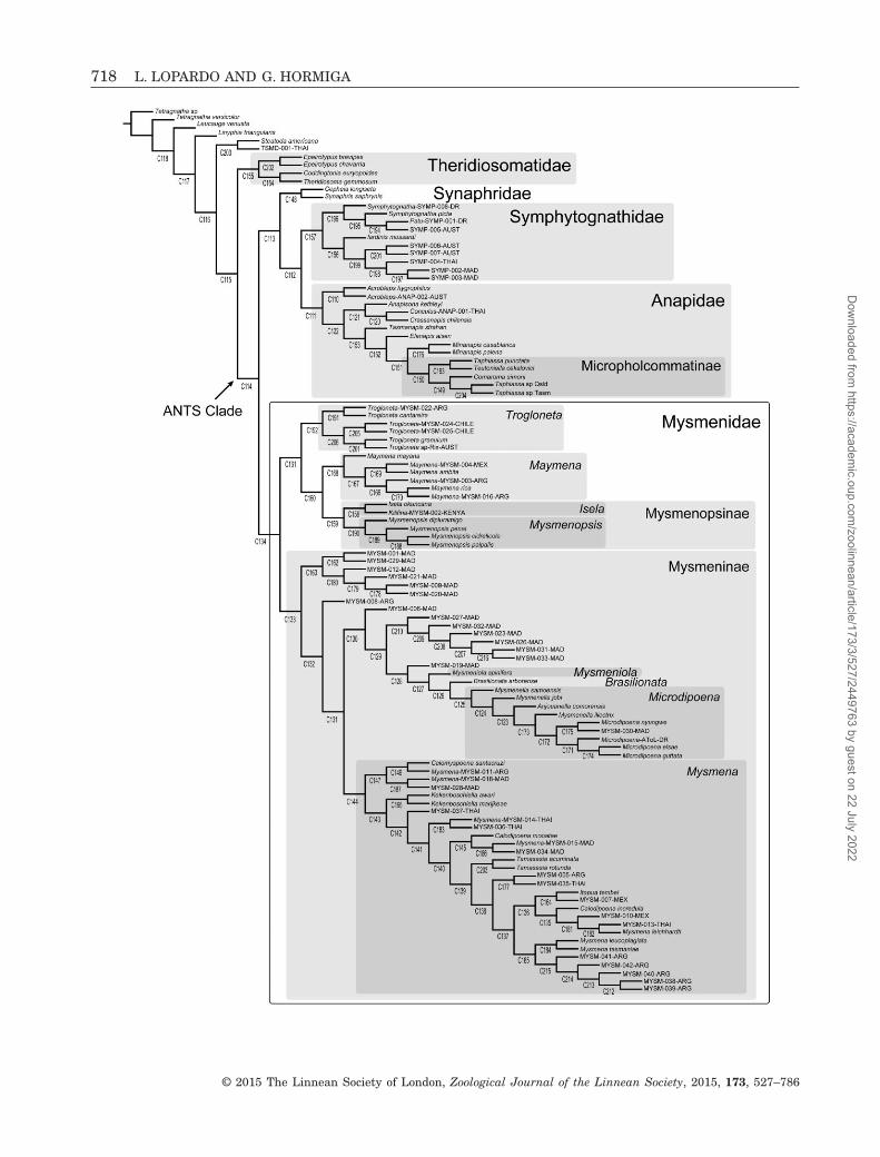
718 L. LOPARDO AND G. HORMIGA
© 2015 The Linnean Society of London, Zoological Journal of the Linnean Society, 2015, 173, 527–786
Dow
nloaded from https://academ
ic.oup.com/zoolinnean/article/173/3/527/2449763 by guest on 22 July 2022

Brisbane, Australia). Our thanks extend to the fol-lowing curators and assistants for kindly loaning pre-cious type and non-type material (in alphabetical order):Abigail Blair (Otago Museum, OMNZ, Dunedin, NewZealand), Alistair Ramsdale (Bernice P. Bishop Museum,BPBM, Hawaii, USA), Ansie Dippenaar-Schoeman (PlantProtection Research Institute, PPRI, Queenswood, SouthAfrica), Antonio D. Brescovit (Instituto Butantan, IB,São Paulo, Brazil), Charles Griswold (CaliforniaAcademy of Sciences, CAS, San Francisco, CA, USA),Christine Rollard and Elise-Anne Leguin (Muséum Na-tional d’Histoire Naturelle, MNHN, Paris, France),Cristina Scioscia and Martín J. Ramírez (MACN-Ar),Debbie Jennings (Natal Museum, NMSA, Pietermar-
itzburg, South Africa), Diana Silva-Dávila (Museo deHistoria Natural, Universidad Nacional Mayor de SanMarcos, MUSM, Lima, Perú), Gonzalo Giribet and LauraLeibesperger (Museum of Comparative Zoology, HarvardUniversity, MCZ, Cambridge, MA, USA), GrahamMilledge (Australian Museum, AMS, Sydney, Aus-tralia), Hieronymus Dastych (Zoologisches Institut undMuseum, Universitat Hamburg, ZMH, Germany), JanetBeccaloni (The Natural History Museum, BMNH,London, UK), John Marris (Lincoln University, LUNZ,Canterbury, New Zealand), Jonathan Coddington (Na-tional Museum of Natural History, Smithsonian In-stitution, USNM, Washington DC, USA), Jürgen Gruber(Naturhistorisches Museum, NMW, Vienna, Austria),
Figure 160. Phylogenetic hypothesis rendered by the complete morphological and molecular data set analysed underequal-weights parsimony using the dynamic homology criterion. Cladogram taken from Lopardo et al. (2011: fig. 12). Numbersbelow each node indicate node numbers. Note that the micropholcommatine Taphiassa punctata and the theridiosomatidCoddingtonia euryopoides were referred to as Parapua punctata and TSMD-002-THAI (respectively) in Lopardo et al.(2011). Family codes used for unidentified species are as follows: ANAP, Anapidae; MYSM, Mysmenidae; SYMP,Symphytognathidae; TSMD, Theridiosomatidae.◀
A
Figure 161. Hypothesis of relationships within Mysmenidae, taken from the results of the combined analysis from Lopardoet al. (2011) (see Fig. 160). New taxonomic (i.e. generic) status as proposed in our study is assigned to each representa-tive; new combinations are emphasized in bold. Unambiguous morphological character optimizations are shown for everybranch in the tree. Numbers below each node indicate node numbers. Empty and filled boxes represent homoplasiousand non-homoplasious transformations, respectively. Major clades representing taxonomic decisions discussed in the textare highlighted in grey boxes.
PHYLOGENY AND MORPHOLOGY OF MYSMENIDAE 719
© 2015 The Linnean Society of London, Zoological Journal of the Linnean Society, 2015, 173, 527–786
Dow
nloaded from https://academ
ic.oup.com/zoolinnean/article/173/3/527/2449763 by guest on 22 July 2022

B
Figure 161. Continued
720 L. LOPARDO AND G. HORMIGA
© 2015 The Linnean Society of London, Zoological Journal of the Linnean Society, 2015, 173, 527–786
Dow
nloaded from https://academ
ic.oup.com/zoolinnean/article/173/3/527/2449763 by guest on 22 July 2022

Leon Baert (Institut Royal des Sciences Naturelles,IRSN, Brussels, Belgium), Lisa Joy Boutin (Queen Vic-toria Museum and Art Gallery, QVM, Launceston, Aus-tralia), Nikolaj Scharff (Zoological Museum, Universityof Copenhagen, ZMUC, Denmark), Norman Platnickand Lou Sorkin (American Museum of Natural History,AMNH, New York, USA), Pablo Goloboff (InstitutoMiguel Lillo, IML, Tucumán, Argentina), Peter J.Schwendinger (Museum d’Histoire naturelle, MHNG,Geneve, Switzerland), Peter Jäger and Julia Altmann(Natur-Museum und Forschung-Institut, Senckenberg,SMF, Frankfurt am Main, Germany), Phil Sirvid(Museum of New Zealand Te Papa Tongarewa, MONZ,Wellington, New Zealand), Robert Raven (QM), RowleySnazell (personal collection, RGS, UK), Rudy Jocque(Musee Royal Afrique Centrale, MRAC, Tervuren,Belgium), Seppo Koponen (Zoological Museum, Uni-versity of Turku, ZMUT, Finland), and TorbjornKronestedt (Naturhistoriska Riksmuseet, NRS, Stock-holm, Sweden). L.L. immensely thanks Gonzalo Giribet,Martín J. Ramírez, Fernando Álvarez-Padilla, DimitarS. Dimitrov, Ligia R. Benavides Silva, Matjaž Kuntner,Jeremy A. Miller, Ingi Agnarsson, Anahita Shaya, Anastasia Kondakova, Vanessa Degrassi, Joana Zanol P.Silva, Vinita Gowda, Maria del Rosario Castañeda, Al-exandra Herrera Martínez, Claudio D. Gutman, herfamily, friends, and Peter for their continuous support,encouragement, advice, and help. We are grateful toGabriele Uhl for her support. Michael G. Rix, associ-ate editor Ingi Agnarsson, and an anonymous reviewerare greatly thanked for their useful comments and criti-cal reading of the article. Funding for this research hasbeen provided by grants from the US National ScienceFoundation (DEB-0328644, DEB 1144492 and DEB114417) to G.H. and Gonzalo Giribet, by a WeintraubFellowship and a Research Enhancement Fund from TheGeorge Washington University (to GH), and by a BSFAToL grant (EAR-0228699) to W. Wheeler, J. Coddington,G. Hormiga, L. Prendini, and P. Sierwald.
REFERENCES
Agnarsson I. 2004. Morphological phylogeny of cobweb spidersand their relatives (Araneae, Araneoidea, Theridiidae). Zoo-logical Journal of the Linnean Society 141: 447–626.
Agnarsson I, Coddington J, May-Collado LJ. 2007.Elongated pedicillate setae: a putative sensory systemand synapomorphy of spiders. Journal of Arachnology 35:411–426.
Álvarez-Padilla F, Dimitrov D, Giribet G, Hormiga G. 2009.Phylogenetic relationships of the spider family Tetragnathidae(Araneae, Araneoidea) based on morphological and DNAsequence data. Cladistics 25: 109–146.
Álvarez-Padilla F, Hormiga G. 2008. A protocol for digest-ing internal soft tissues and mounting spiders for scanning
electron microscopy. Journal of Arachnology 35: 538–542.
Baert L. 1982. Spiders (Araneae) from Papua New Guinea III.Mysmenidae (Symphytognathoidea). Bulletin of the BritishArachnological Society 5: 303–308.
Baert L. 1984a. Spiders (Araneae) from Papua New Guinea.IV. Ochyroceratidae, Telemidae, Hadrotarsidae andMysmenidae. Indo-Malayan Zoology 2: 225–244.
Baert L. 1984b. Mysmenidae and Hadrotarsidae from theNeotropical Guaraní zoogeographical province (Paraguay andsouth Brasil) (Araneae). Revue Suisse De Zoologie 91: 603–616.
Baert L. 1985. Telemidae, Mysmenidae and Ochyroceratidaefrom Cameroon (Araneae): scientific report of the BelgianMount Cameroon Expeditions 1981 and 1983 (no. 13). BiologJaarb Dodonaea 53: 44–57.
Baert L. 1986. Mysmenidae from the Comoro Islands (Araneae).Revue de Zoologie Africaine 100: 265–267.
Baert L. 1988. The Ochyroceratidae and Mysmenidae fromSulawesi (Araneae). Indo-Malayan Zoology 5: 9–22.
Baert L. 1989. Mysmenidae from Rwanda (Araneae). Revuede Zoologie Africaine 103: 29–33.
Baert L. 1990. Mysmenidae (Araneae) from Peru. Bulletin del’Institut Royal des Sciences Naturelles de Belgique Entomologie60: 5–18.
Baert L, Maelfait J-P. 1983. Spiders of the Galápagos Islands.I. Mysmenidae (Araneae). Bulletin of the British ArachnologicalSociety 6: 102–108.
Baert L, Murphy J. 1987. Kilifia inquilina, a new mysmenidspider from Kenya (Araneae, Mysmenidae). Bulletin of theBritish Arachnological Society 7: 194–196.
Baert L, Murphy J. 1992. Kilifina, new generic name forKilifia, preoccupied (Araneae, Mysmenidae). Bulletin of theBritish Arachnological Society 9: 104.
Banks N. 1895. A list of the spiders of Long Island; with de-scriptions of new species. Journal of the New York Entomo-logical Society 3: 76–93.
Barrows W. 1940. New and rare spiders from the Great SmokyMountain National Park region. Ohio Journal of Science 40:130–138.
Bertkau P. 1889. Interessante Tiere aus der Umgebung vonBonn. Verhandlungen des Naturhistorischen Vereins derPreussischen Rheinlande und Westfalens 46: 69–82.
Bishop S, Crosby C. 1926. Notes on the spiders of the south-eastern United States with descriptions of new species. Journalof the Elisha Mitchell Scientific Society 41: 163–212.
de Bivort BL, Clouse RM, Giribet G. 2010. A morphometrics-based phylogeny of the temperate Gondwanan mite harvest-men (Opiliones, Cyphophthalmi, Pettalidae). Journal ofZoological Systematics and Evolutionary Research 48: 294–309.
Blackledge T, Scharff N, Coddington J, Szüts T, WenzelJ, Hayashi C, Agnarsson I. 2009. Reconstructing webevolution and spider diversification in the molecular era.Proceedings of the National Academy of Sciences of the UnitedStates of America 106: 5229–5234.
Bond JE, Garrison NL, Hamilton CA, Godwin RL, HedinM, Agnarsson I. 2014. Phylogenomics resolves a spider
PHYLOGENY AND MORPHOLOGY OF MYSMENIDAE 721
© 2015 The Linnean Society of London, Zoological Journal of the Linnean Society, 2015, 173, 527–786
Dow
nloaded from https://academ
ic.oup.com/zoolinnean/article/173/3/527/2449763 by guest on 22 July 2022

backbone phylogeny and rejects a prevailing paradigm fororb web evolution. Current Biology 24: 1765–1771. http://dx.doi.org/10.1016/j.cub.2014.06.034.
Bremer K. 1988. The limits of aminoacid sequence data inangiosperm phylogenetic reconstruction. Evolution 42: 795–803.
Bremer K. 1994. Branch support and tree stability. Cladistics10: 295–304.
Brescovit A, Lopardo L. 2008. The first record on the spidergenus Trogloneta Simon in the southern hemisphere (Araneae,Mysmenidae), with descriptions of three new species fromBrazil and remarks on the morphology. Acta Zoologica (Stock-holm) 89: 93–106.
Breuss W. 2001. Bemerkenswerte Spinnen aus Vorarlberg(Österreich) – I (Arachnida: Araneae: Lycosidae, Theridiidae,Mysmenidae, Gnaphosidae, Salticidae). Berichte desNaturwissenschaftlich – Medizinischen Vereins in Inns-bruck 88.
Brignoli P. 1970. Contribution à la connaissance desSymphytognathidae paléarctiques (Arachnida, Araneae).Bulletin du Museum National d’Histoire Naturelle, Paris 41:1403–1420.
Brignoli P. 1974. Notes on spiders, mainly cave-dwelling, ofsouthern Mexico and Guatemala (Araneae). Quadernidell’Accademia Nazionale dei Lincei 171: 195–238.
Brignoli P. 1978. Spinnen aus Nepal, IV. Drei neueSymphytognathidae (Arachnida: Araneae). SenckenbergianaBiologica 59: 247–252.
Brignoli P. 1980. On few Mysmenidae from the Oriental andAustralian regions (Araneae). Revue Suisse De Zoologie 87:727–738.
Brignoli P. 1981. Spiders from the Philippines IV. A newOgulnius and notes on some other Oriental and JapaneseTheridiosomatidae (Araneae). Acta Arachnologica, Tokyo 30:9–19.
Brignoli P. 1983. A catalogue of the Araneae described between1940 and 1981. Manchester: Manchester Univ. Press.
Bryant E. 1940. Cuban spiders in the Museum of Compara-tive Zoology. Bulletin of the Museum of Comparative Zoology,Harvard 86: 247–554.
Carico J. 1977. A simple dusting device for coating orb websfor field photography. Bulletin of the British ArachnologicalSociety 4: 100.
Chamberlin RV, Ivie W. 1938. Araneida from Yucatan.Publications, Carnegie Institution of Washington 491: 123–136.
Chickering A. 1960. The female of Lucarachne beebei Gertsch(Araneae: Symphytognathidae). Psyche (Cambridge, Massa-chusetts) 67: 95–97.
Clerck C. 1757. Svenska spindlar, uti sina hufvud-slågter indeltesamt under några och sextio särskildte arter beskrefne ochmed illuminerade figurer uplyste. Stockholm: Stockholmiae,Literis Laur. Salvii.
Clouse RM, de Bivort BL, Giribet G. 2009. A phylogeneticanalysis for the South-east Asian mite harvestman familyStylocellidae (Opiliones, Cyphophthalmi) – a combined analy-sis using morphometric and molecular data. InvertebrateSystematics 23: 515–529.
Coddington J. 1986a. The genera of the spider familyTheridiosomatidae. Smithsonian Contributions to Zoology 422:1–96.
Coddington J. 1986b. The monophyletic origin of the orb web.In: Shear WA, ed. Spider webs, behavior and evolution.Redwood City, CA: Stanford University Press, 319–363.
Coddington J. 1989. Spinneret silk spigot morphology: evi-dence for the monophyly of orbweaving spiders, Cyrtophorinae(Araneae), and the group Theridiidae plus Nesticidae. Journalof Arachnology 17: 71–95.
Coddington J. 1990. Ontogeny and homology in the malepalpus of orb-weaving spiders and their relatives, with com-ments on phylogeny (Araneoclada: Araneoidea, Deinopoidea).Smithsonian Contributions to Zoology 496: 1–52.
Coddington J, Scharff N. 1994. Problems with zero-lengthbranches. Cladistics 10: 415–423.
Comstock JH. 1910. The palpi of male spiders. Annals of theEntomological Society of America 3: 161–185.
Coyle F, Meigs T. 1989. Two new species of kleptoparasiticMysmenopsis (Araneae, Mysmenidae) from Jamaica. Journalof Arachnology 17: 59–70.
Dahl F. 1901. Nachtrag zur Uebersicht der Zoropsiden.Sitzungsberichte der Gesellschaft Naturforschender Freundezu Berlin 1901: 244–255.
Davies V. 1985. Araneomorphae (in part). Zoological Cata-logue of Australia 3: 49–125.
Dimitrov D, Benavides L, Giribet G, Arnedo MA, GriswoldCE, Scharff N, Hormiga G. 2013. Squeezing the last dropsof the lemon: molecular phylogeny of Orbiculariae (Araneae).In: 19th International Congress of Arachnology, Abstract Book.Taiwan: Kenting National Park.
Dimitrov D, Lopardo L, Giribet G, Arnedo MA, Álvarez-Padilla F, Hormiga G. 2012. Tangled in a sparse spiderweb: single origin of orb weavers and their spinning workunravelled by denser taxonomic sampling. Proceeding of theRoyal Society B 279: 1341–1350.
Dingerkus G, Uhler L. 1977. Enzyme clearing of alcian bluestained whole small vertebrates for demonstration of carti-lage. Stain Technology 52: 229–232.
Dippenaar-Schoeman A, Jocqué R. 1997. Family Zodariidae.In: African spiders: an identification manual. (Handbook,PPRI). Pretoria. (9): 392p.
Dunlop JA, Penney D, Jekel D. 2014. A summary list offossil spiders and their relatives. In: Platnick NI, ed. Theworld spider catalog, version 14.5. New York: AmericanMuseum of Natural History, 1–75. Available at: http://research.amnh.org/entomology/spiders/catalog/index.html
Eberhard W. 1976. Photography of orb webs in the field.Bulletin of the British Arachnological Society 3: 200–204.
Eberhard W. 1987. Orb webs and construction behavior inAnapidae, Symphytognathidae, and Mysmenidae. Journal ofArachnology 14: 339–356.
Eberhard W, Platnick NI, Schuh R. 1993. Natural historyand systematics of arthropod symbionts (Araneae; Hemiptera;Diptera) inhabiting webs of the spider Tengella radiata(Araneae, Tengellidae). American Museum Novitates 3065:1–17.
722 L. LOPARDO AND G. HORMIGA
© 2015 The Linnean Society of London, Zoological Journal of the Linnean Society, 2015, 173, 527–786
Dow
nloaded from https://academ
ic.oup.com/zoolinnean/article/173/3/527/2449763 by guest on 22 July 2022

Emerton JH. 1882. New England spiders of the familyTheridiidae. Transactions of the Connecticut Academy of Artsand Sciences 6: 1–86.
Fage L. 1931. Araneae, 5e série, précédée d’un essai surl’évolution souterraine et son déterminisme. Biospeologica(expér, LAZ) 71: 91–291.
Farris J. 1990. Phenetics in camouflage. Cladistics 6: 91–100.
Farris J. 1997. The future of phylogeny reconstruction. ZoologicaScripta 26: 303–311.
Farris J, Albert V, Källersjö M, Lipscomb D, Kluge A. 1996.Parsimony jackknifing outperforms neighbor-joining. Cladistics12: 99–124.
Felsenstein J. 1985. Confidence limits on phylogenies: anapproach using the bootstrap. Evolution 39: 783–791.
Fernández R, Hormiga G, Giribet G. 2014. Phylogenomicanalysis of spiders reveals nonmonophyly of orb weavers.Current Biology 24: 1772–1777. http://dx.doi.org/10.1016/j.cub.2014.06.035.
Fitch WM. 1971. Toward defining the course of evolution:minimum change for a specific tree topology. SystematicZoology 20: 406–416.
Forster R. 1959. The spiders of the family Symphytognathidae.Transactions of the Royal Society of New Zealand 86: 269–329.
Forster R. 1977. Fam. Symphytognathidae. In: La fauneterrestre de l’île de Saite-Hélène IV. Annales du Musée Royalde l’Afrique Centrale (Zoologique, Sn) 220: 129–131.
Forster R. 1980. Evolution of the tarsal organ, the respira-tory system, and the female genitalia in spiders. In: GruberJ, ed. Proceedings of the 8th International Congress of Arach-nology. Vienna: Egermann H, 269–284.
Forster R, Platnick NI. 1977. A review of the spider familySymphytognathidae (Arachnida, Araneae). American MuseumNovitates 2619: 1–29.
Forster R, Platnick NI. 1984. A review of the archaeid spidersand their relatives, with notes on the limits of the super-family Palpimanoidea (Arachnida, Araneae). Bulletin of theAmerican Museum of Natural History 178: 1–106.
Gertsch WJ. 1941. Report on some arachnids from Barro Colo-rado Island, Canal Zone. American Museum Novitates 1146:1–14.
Gertsch W. 1960a. Descriptions of American spiders of thefamily Symphytognathidae. American Museum Novitates 1981:1–40.
Gertsch W. 1960b. The fulva group of the spider genus Steatoda(Araneae, Theridiidae). American Museum Novitates 1982:1–48.
Gertsch W. 1971. A report on some Mexican cave spiders.Bulletin of the Association for Mexican Cave Studies 4:47–111.
Gertsch W, Davis L. 1936. New spiders from Texas. Ameri-can Museum Novitates 881: 1–21.
Giribet G. 2003. Stability in phylogenetic formulations andits relationship to nodal support. Systematic Biology 42: 554–564.
Giribet G, Edgecombe G. 2006. Conflict between datasetsand phylogeny of centipedes: an analysis based on seven genes
and morphology. Proceedings of the Royal Society B 273: 531–538.
Goloboff PA. 1993. Estimating character weights during treesearch. Cladistics 9: 83–91.
Goloboff PA, Farris J. 2001. Methods for quick consensusestimation. Cladistics 17: S26–S34.
Goloboff PA, Farris J, Kälersjö M, Oxelman B, RamírezMJ, Szumik C. 2003a. Improvements to resampling meas-ures of group support. Cladistics 19: 324–332.
Goloboff PA, Farris J, Nixon K. 2003b. T.N.T.: tree analy-sis using new technology. Version 1.1 (August, 2008). WilliHennig Society Edition. Available at: http://www.zmuc.dk/public/phylogeny
Goloboff PA, Farris J, Nixon K. 2008. TNT, a free programfor phylogenetic analysis. Cladistics 24: 774–786.
Goloboff PA, Mattoni C, Quinteros A. 2006. Continuous char-acters analyzed as such. Cladistics 22: 589–601.
González-José R, Escapa I, Neves W, Cúneo R, PucciarelliH. 2008. Cladistic analysis of continuous modularized traitsprovides phylogenetic signals in Homo evolution. Nature 453:775–778.
Griswold CE. 1985. Isela okuncana, a new genus and speciesof kleptoparasitic spider from southern Africa (Araneae:Mysmenidae). Annals of the Natal Museum 27: 207–217.
Griswold CE. 2001. A monograph of the living world generaand Afrotropical species of cyatholipid spiders (Araneae,Orbiculariae, Araneoidea, Cyatholipidae). Memoirs of the Cali-fornia Academy of Sciences 26: 1–251.
Griswold CE, Coddington J, Hormiga G, Scharff N. 1998.Phylogeny of the orb-web building spiders (Araneae,Orbiculariae: Deinopoidea, Araneoidea). Zoological Journalof the Linnean Society 123: 1–99.
Griswold CE, Ramírez MJ, Coddington J, Platnick NI.2005. Atlas of phylogenetic data for entelegyne spiders(Araneae, Araneomorphae, Entelegynae), with comments ontheir phylogeny. Proceedings of the California Academy ofSciences 4th Series (56, Suppl. II): 1–324.
Griswold CE, Yan H-M. 2003. On the Egg-guarding Behav-ior of a Chinese Symphytognathid Spider of the Genus PatuMarples, 1951 (Araneae, Araneoidea, Symphytognathidae).Proceedings of the California Academy of Sciences 54: 356–360.
Gruia M. 1977. Sur quelques Theridiidae et Symphytognathidae(Aranea) recueillis par la deuxième expédition biospéologiquecubano-roumaine à Cuba. Rés Expédition BiospéologiqueCubano-Roumaine, Cuba 2: 159–163.
Hajer J. 2000. Web of Trogloneta granulum Simon (Araneae,Mysmenidae). Bulletin of the British Arachnological Society11: 333–338.
Hajer J. 2002. The spinning activity of the spider Troglonetagranulum Simon, 1922 (Araneae, Mysmenidae). ActaUniversitatis Purkynianae Studia Biologica VI: 21–38.
Hajer J, Reháková D. 2003. Spinning activity of the spiderTrogloneta granulum (Araneae, Mysmenidae): web, cocoon,cocoon handling behaviour, draglines and attachment discs.Zoology 106: 223–231.
PHYLOGENY AND MORPHOLOGY OF MYSMENIDAE 723
© 2015 The Linnean Society of London, Zoological Journal of the Linnean Society, 2015, 173, 527–786
Dow
nloaded from https://academ
ic.oup.com/zoolinnean/article/173/3/527/2449763 by guest on 22 July 2022

Harris R. 1979. A glossary of surface sculpturing. Occasion-al Papers in Entomology 28: 1–31.
Harvey MS. 1992. A new species of Symphytognatha Hickman(Araneae: Symphytognathidae) from Western Australia. Recordsof the Western Australian Museum 15: 685–689.
Heimer S, Nentwig W. 1991. Spinnen Mitteleuropas: EinBestimmungsbuch. Berlin: Verlag Paul Parey.
Hickman VV. 1931. A new family of spiders. Proceedings ofthe Zoological Society of London (B) 1931: 1321–1328.
Hickman VV. 1944. On some new AustralianApneumonomorphae with notes on their respiratory system.Papers and Proceedings of the Royal Society of Tasmania 1943:179–195.
Hickman VV. 1945. A new group of apneumone spiders. Trans-actions of the Connecticut Academy of Arts and Sciences 36:135–157.
Hickman V. 1979. Some Tasmanian spiders of the familiesOonopidae, Anapidae and Mysmenidae. Papers and Proceed-ings of the Royal Society of Tasmania 113: 53–79.
Hormiga G. 1994. Cladistics and the comparative morphol-ogy of linyphiid spiders and their relatives (Araneae,Araneoidea, Linyphiidae). Zoological Journal of the LinneanSociety 111: 1–71.
Hormiga G. 1999. Cephalothoracic sulci in linyphiine spiders(Araneae, Araneoidea, Linyphiidae). Journal of Arachnol-ogy 27: 94–102.
Hormiga G. 2000. Higher level phylogenetics of erigonine spiders(Araneae, Linyphiidae, Erigoninae). Smithsonian Contribu-tions to Zoology 609: 1–160.
Hormiga G. 2002. Orsonwelles, a new genus of giant linyphiidspiders (Araneae) from the Hawaiian Islands. InvertebrateSystematics 16: 369–448.
Hormiga G, Álvarez-Padilla F, Benjamin S. 2007. Firstrecords of extant Hispaniolan spiders of the familiesMysmenidae, Symphytognathidae and Ochyroceratidae(Araneae), including a new species of Ochyrocera. Ameri-can Museum Novitates 3577: 1–21.
Hormiga G, Eberhard W, Coddington J. 1995. Web-construction behaviour in Australian Phonognatha and thephylogeny of nephiline and tetragnathid spiders (Araneae:Tetragnathidae). Australian Journal of Zoology 43: 313–364.
Hormiga G, Griswold CE. 2014. Systematics, Phylogeny andEvolution of Orb-weaving Spiders. Annual Review of Ento-mology 59: 487–512.
Hormiga G, Scharff N, Coddington J. 2000. The PhylogeneticBasis of Sexual Size Dimorphism in Orb-Weaving Spiders(Araneae, Orbiculariae). Systematic Biology 49: 435–462.
Humphries C. 2002. Homology, characters and continuous vari-ables. In: MacLeod N, Forey P, eds. Morphology, shape andphylogeny. London: Taylor and Francis, 8–26.
Jocqué R. 1987. Descriptions of new genera and species ofAfrican Zodariinae with a revision of the genus Heradida(Araneae, Zodariidae). Revue de Zoologie Africaine 101: 143–163.
Jocqué R. 1991. A generic revision of the spider familyZodariidae (Araneae). Bulletin of the American Museum ofNatural History 201: 1–160.
Jocqué R, Dippenaar-Schoeman A. 2006. Spiderfamilies of the world. Tervuren: Musée Royal de l’AfriqueCentral.
Kasal P. 1982. Theridion antusi sp.n. and Mysmenajobi from Czechoslovakia (Araneida, Theridiidae andSymphytognathidae). Acta entomologica Bohemoslovaca 79:73–76.
Kaston B. 1978. How to know the spiders, 3rd edn. Dubuque,IA: Brown WC.
Koch CL. 1838. Die Arachniden. Nürnberg In: der C.H.Zeh’schen Buchhandlung, Vierter Band, 109–144, FunfterBand, 1–124.
Koch L. 1877. Verzeichniss der bei Nürnberg bis jetztbeobachteten Arachniden (mit Ausschluss der Ixodiden undAcariden) und Beschreibungen von neuen, hier vorkommendenArten. Abhandlungen der Naturhistorischen Gesellschaft zuNürnberg 6: 113–198.
Kraus O. 1955. Spinnen aus El Salvador (Arachnoidea, Araneae).Abhandlungen der senckenbergisch-naturforschendenGesellschaft 493: 1–112.
Kraus O. 1967. Zur Spinnenfauna Deutschlands, II. Mysmenajobi n. sp, eine Symphytognathide in Mitteleuropa (Arachnida:Araneae: Symphytognathidae). Senckenbergiana Biologica 48:387–399.
Kropf C. 1990. Web construction and prey capture of Comaromasimoni Bertkau (Araneae). Acta Zoologica Fennica 190: 229–233.
Labarque FM, Griswold CE. 2014. New ray spiders fromSoutheast Asia: the new Philippine genus Tagalogonia gen.nov. and continental genus Coddingtonia Miller, Griswold andYin, 2009 (Araneae: Theridiosomatidae), with comments ontheir intergeneric relationships. California Academy of Sci-ences Special Publication, The Coral Triangle: The 2011 HearstPhilippine Biodiversity Expedition, 406–26.
Lee Y, Yoo J, Lee D, Kim J. 2004. Ground dwelling spiders.Korean Arachnology 20: 97–115.
Levi H. 1956. The spider genus Mysmena in the Americas(Araneae, Theridiidae). American Museum Novitates 1801:1–13.
Levi H. 1961. Evolutionary trends in the development ofpalpal sclerites in the spider family theridiidae. Journal ofMorphology 108: 1–9.
Levi H. 1967. Adaptations of respiratory systems of spiders.Evolution 21: 571–583.
Levi H. 1972. Taxonomic-nomenclatural notes on misplacedtheridiid spiders (Araneae: Theridiidae), with observationson Anelosimus. Transactions of the American MicroscopicalSociety 91: 533–538.
Levi H, Kirber W. 1976. On the evolution of tracheae inArachnids. Bulletin of the British Arachnological Society 3:187–188.
Levi H, Levi L. 1962. The genera of the spider familyTheridiidae. Bulletin of the Museum of Comparative Zoology,Harvard 127: 1–71.
Lin Y, Li S. 2008. Mysmenid spiders of China (Araneae:Mysmenidae). Annales Zoologici (Warszawa) 58: 487–520.
Linnaeus C. 1758. Systema naturae per regna tria naturae,secundum classes, ordines, genera, species cum characteribus
724 L. LOPARDO AND G. HORMIGA
© 2015 The Linnean Society of London, Zoological Journal of the Linnean Society, 2015, 173, 527–786
Dow
nloaded from https://academ
ic.oup.com/zoolinnean/article/173/3/527/2449763 by guest on 22 July 2022
differentiis, synonymis, locis. Editio decima, reformata. Holmiae:Impensis Direct. Laurentii Salvii, 821 (Araneae, 619–624).
Loksa I. 1973. On the morphology and systematical positionof Mysmena leucoplagiata (Simon, 1879) (Araneae:Symphytognathidae). Acta Zoologica Academiae ScientiarumHungaricae 19: 283–287.
Lopardo L. 2005. Phylogenetic revision of the spider genusNegayan (Araneae, Anyphaenidae, Amaurobioidinae). ZoologicaScripta 34: 245–277.
Lopardo L, Coddington J. 2005. Chapter 40: Mysmenidae.In: Ubick D, Cushing P, Paquin P, eds. Spiders of NorthAmerica: an identification manual. Poughkeepsie, NY: Ameri-can Arachnology Society, 175–177.
Lopardo L, Dupérré N, Paquin P. 2008. Expanding hori-zons . . . The first report of the genus Mysmena (Araneae,Mysmenidae) from continental North America, with the de-scription of a new species. Zootaxa 1718: 36–44.
Lopardo L, Giribet G, Hormiga G. 2011. Morphology tothe rescue: molecular data and the signal of morphologicalcharacters in combined phylogenetic analyses – a case studyfrom mysmenid spiders (Araneae, Mysmenidae), with com-ments on the evolution of web architecture. Cladistics 27:278–330.
Lopardo L, Hormiga G. 2007. On the Synaphrid SpiderCepheia longiseta (Simon 1881) (Araneae, Synaphridae). Ameri-can Museum Novitates 3575: 1–18.
Lopardo L, Hormiga G. 2008. Phylogenetic placement of theTasmanian spider Acrobleps hygrophilus (Araneae, Anapidae)with comments on the evolution of the capture web inAraneoidea. Cladistics 24: 1–33.
Lopardo L, Hormiga G, Melic A. 2007. Spinneret spigot mor-phology in synaphrid spiders (Araneae, Synaphridae), withcomments on the systematics of the family and descriptionof a new species of Synaphris Simon 1894 from Spain. Ameri-can Museum Novitates 3556: 1–26.
Lopardo L, Michalik P. 2013. First description of a mysmenidspider species from mainland Australia and new data forMysmena tasmaniae Hickman 1979 (Araneae, Mysmenidae).Memoirs of the Queensland Museum/Nature 58: 381–396.
Maddison WP. 1993. Missing data versus missing charac-ters in phylogenetic analysis. Systematic Biology 42:576–581.
Maddison WP, Maddison D. 2007. Mesquite: a modular systemfor evolutionary analysis. Version 2.01 (build j28). Avail-able at: http://mesquiteproject.org
Marples B. 1955. Spiders from western Samoa. Journal of theLinnean Society of London (Zoology) 42: 453–504.
Marusik Y, Gnelitsa V, Kovblyuk M. 2005. A new speciesof Synaphris (Araneae, Synaphridae) from Ukraine. Bul-letin of the British Arachnological Society 13: 125–130.
Miller F. 1970. Spinnenarten der Unterfamilie Micryphantinaeund der Familie Theridiidae aus Angola. Publicações Culturaisda Companhia de Diamantes de Angola 82: 75–166.
Miller JA. 2007. Synaphridae of Madagascar (Araneae:Araneoidea): a new family record for the Afrotropical region.Proceedings of the California Academy of Sciences 58: 21–48.
Miller JA, Carmichael A, Ramírez MJ, Spagna J, HaddadCR, Rezác M, Johannesen J, Král J, Wang X-P, GriswoldCE. 2010. Phylogeny of entelegyne spiders: affinities of thefamily Penestomidae (NEW RANK), generic phylogeny ofEresidae, and asymmetric rates of change in spinning organevolution (Araneae, Araneoidea, Entelegynae). MolecularPhylogenetics and Evolution 55: 786–804.
Miller JA, Griswold CE, Yin C. 2009. The symphytognathoidspiders of the Gaoligongshan, Yunnan, China (Araneae:Araneoidea): systematics and diversity of micro-orbweavers.ZooKeys 11: 9–195.
Millidge AF. 1984. The taxonomy of the Linyphiidae, basedchiefly on the epigynal and tracheal characters (Araneae:Linyphiidae). Bulletin of the British Arachnological Society6: 229–267.
Müller H-G. 1987. Spiders from Colombia V. A new Mysmenopsisfrom the Ciénaga Grande de Santa Marta, northern Colom-bia (Araneida: Mysmenidae). Bulletin of the BritishArachnological Society 7: 185.
Namkung J. 2002. The spiders of Korea, 1st edn. Seoul:Kyo-Hak Publishing Co.
Namkung J. 2003. The spiders of Korea, 2nd edn. Seoul:Kyo-Hak Publishing Co.
Namkung J, Lee K. 1987. A new record spider of the genusMysmenella Brignoli, 1980 (Araneae: Mysmenidae) from Korea.Korean Arachnology 3: 45–49.
Nixon K. 1999a. The parsimony ratchet, a new method forrapid parsimony analysis. Cladistics 15: 407–414.
Nixon K. 1999b. WINCLADA, program and documentation.Available at: http://www.cladistics.com
Ono H. 2007. Eight new species of the families Hahniidae,Theridiidae, Linyphiidae and Anapidae (Arachida, Araneae)from Japan. Bulletin of the National Science Museum, Tokyo33: 153–173.
Ono H, Chang Y, Tso I. 2007. Three new spiders of thefamilies Theridiidae and Anapidae (Araneae) from south-ern Taiwan. Memoirs of the National Science Museum, Tokyo44: 71–82.
Penney D. 2000. Miocene spiders in Dominican amber(Oonopidae, Mysmenidae). Palaeontology 43: 343–357.
Peters H. 1982. Wie spinnen der Familie Uloboridae ihrebeute einspinnen und verzehren. Verhandlungen desNaturwissenschaftlichen Vereins in Hamburg 25: 147–167.
Petrunkevitch A. 1928. Systema Aranearum. Transactionsof the Connecticut Academy of Arts and Sciences 29: 1–270.
Petrunkevitch A. 1971. Chiapas amber spiders, II.University of California Publications in Entomology 63:1–44.
Platnick NI. 2014. The world spider catalog, version 14.5.American Museum of Natural History. Available at: http://research.amnh.org/entomology/spiders/catalog/index.html
Platnick NI, Coddington J, Forster R, Griswold CE. 1991.Spinneret Morphology and the Phylogeny of Haplogyne Spiders(Araneae, Araneomorphae). American Museum Novitates 3016:1–73.
Platnick NI, Forster R. 1986. On Teutoniella, an AmericanGenus of the Spider Family Micropholcommatidae (Araneae,Palpimanoidea). American Museum Novitates 2854: 1–23.
PHYLOGENY AND MORPHOLOGY OF MYSMENIDAE 725
© 2015 The Linnean Society of London, Zoological Journal of the Linnean Society, 2015, 173, 527–786
Dow
nloaded from https://academ
ic.oup.com/zoolinnean/article/173/3/527/2449763 by guest on 22 July 2022

Platnick NI, Forster R. 1987. On the first American spidersof the subfamily Sternodinae (Araneae, Malkaridae). Ameri-can Museum Novitates 2894: 1–12.
Platnick NI, Forster R. 1989. A revision of the temperateSouth American and Australasian spiders of the familyAnapidae (Araneae, Araneoidea). Bulletin of the AmericanMuseum of Natural History 190: 1–139.
Platnick NI, Shadab M. 1978. A review of the spider genusMysmenopsis (Araneae, Mysmenidae). American MuseumNovitates 2661: 1–22.
Platnick NI, Shadab MU. 1979. A review of the spider generaAnapisona and Pseudanapis (Araneae, Anapidae). Ameri-can Museum Novitates 2672: 1–20.
Prendini L. 2001. Species or supraspecific taxa as terminalsin cladistic analysis? Groundplans versus exemplars revis-ited. Systematic Biology 50: 290–300.
Purcell W. 1909. Development and origin of respiratory organsin Araneae. Quarterly Journal of Microscopical Science 54:1–110.
Rae T. 1998. The logical basis for the use of continuous char-acters in phylogenetic systematics. Cladistics 14: 221–228.
Ramírez MJ. 2000. Respiratory system morphology and thephylogeny of haplogyne spiders (Araneae, Araneomorphae).Journal of Arachnology 28: 149–157.
Ramírez MJ, Lopardo L, Platnick NI. 2004. Notes on Chileananapids and their webs (Araneae, Anapidae). AmericanMuseum Novitates 3428: 1–13.
Rix MG. 2008. A new species of Micropholcomma (Araneae:Araneoidea: Micropholcommatidae) from Western Aus-tralia. Records of the Western Australian Museum 24: 343–348.
Rix MG, Harvey MS. 2010. The spider familyMicropholcommatidae (Arachnida, Araneae, Araneoidea): arelimitation and revision at the generic level. ZooKeys 36:1–321.
Rix MG, Harvey MS, Roberts J. 2008. Molecular phylogeneticsof the spider family Micropholcommatidae (Arachnida: Araneae)using nuclear rRNA genes (18S and 28S). MolecularPhylogenetics and Evolution 46: 1031–1048.
Rix MG, Harvey MS, Roberts JD. 2010. A revision of thetricellin spider genus Raveniella (Araneae: Araneoidea:Micropholcommatidae): exploring patterns of phylogeny andbiogeography in an Australian biodiversity hotspot. Inver-tebrate Systematics 24: 209–237.
Roberts M. 1978. Contributions à l’étude de la faune terrestredes îles granitiques de l’archipel des Séchelles (Mission PLG Benoit – J J Van Mol 1972) Theridiidae, Mysmenidae andgen Theridiosoma (Araneidae) (Araneae). Revue de ZoologieAfricaine 92: 902–939.
Saaristo M. 1978. Spiders (Arachnida, Araneae) from theSeychelle islands, with notes on taxonomy. Annales ZoologiciFennici 15: 99–126.
Scharff N, Coddington J. 1997. A phylogenetic analysisof the orb-weaving spider family Araneidae (Arachnida,Araneae). Zoological Journal of the Linnean Society 120: 355–434.
Schütt K. 2000. The limits of the Araneoidea (Arachnida:Araneae). Australian Journal of Zoology 48: 135–153.
Schütt K. 2002. The limits and phylogeny of the Araneoidea(Arachnida, Araneae). Unpublished D. Phil. Thesis, Univer-sity of Berlin.
Schütt K. 2003. Phylogeny of Symphytognathidae s.l. (Araneae,Araneoidea). Zoologica Scripta 32: 129–151.
Shinkai E. 1977. Spiders of Tokyo III. Acta Arachnologica,Tokyo 27: 321–336.
Sierwald P. 1989. Morphology and Ontogeny of Female Copu-latory Organs in American Pisauridae, with Special Refer-ence to Homologous Features (Arachnida: Araneae).Smithsonian Contributions to Zoology 484: 1–24.
Simon E. 1879. Arachnides nouveau de France, d’Espagne etd’Algérie. Premier mémoire. Bulletin de la Société Zoologiquede France 4: 251–263.
Simon E. 1880. Matériaux pour servir à une faunarachnologique de la Nouvelle Calédonie. Annales de la SociétéEntomologique de Belgique 23(C.R.): 164–175.
Simon E. 1881. Les arachnides de France. Paris 5: 1–180.Simon E. 1893. Histoire naturelle das araignées. Paris 1: 257–
488.Simon E. 1894. Histoire naturelle des araignées. Paris. 1:
489–760.Simon E. 1895a. Histoire naturelle des araignées. Paris. 1:
761–1084.Simon E. 1895b. Etudes arachnologiques. 26e XLI Descrip-
tions d’espèces et de genres nouveaux de l’ordre des Araneae.Annales de la Societe Entomologique de France 64: 131–160.
Simon E. 1897. On the spiders of the island of St Vincent,III. Proceedings of the Zoological Society of London 1897: 860–890.
Simon E. 1899. Contribution à la faune de Sumatra. Arachnidesrecueillis par M J L Weyers, à Sumatra (Deuxiéme mémoire).Annales de la Societe Entomologique de Belgique 43: 78–125.
Simon E. 1903. Descriptions d’arachnides nouveaux. Annalesde la Société Entomologique de Belgique 47: 21–39.
Simon E. 1922. Description de deux arachnides cavernicolesdu midi de la France. Bulletin de la Societe Entomologiquede France 1922: 199–200.
Simon E. 1926. Les arachnides de France. Synopsis généraleet catalogue des espèces françaises de l’ordre des Araneae; 2epartie. Paris. 6: 309–532.
Snazell R. 1986. A new Mysmena (Araneae: Mysmenidae)from Spain. Bulletin of the British Arachnological Society 7:62–64.
Strong E, Lipscomb D. 1999. Character coding and inappli-cable data. Cladistics 15: 363–371.
Sundevall CJ. 1833. Conspectus Arachnidum. LondiniGothorum, 1–39.
Thaler K. 1975. Trogloneta granulum Simon, eine weitereReliktart der Nordostalpen (Arachnida, Aranei,‘Symphytognathidae’). Revue Suisse De Zoologie 82: 283–291.
Thaler K. 1995. Mysmeniola spinifera n.gen. n.sp., einemerkwürdige Kleinspinne aus Venezuela (Arachnida, Araneae:Mysmenidae). Mitteilungen der Schweizerischen Entomolo-gischen Gesellschaft 68: 429–433.
726 L. LOPARDO AND G. HORMIGA
© 2015 The Linnean Society of London, Zoological Journal of the Linnean Society, 2015, 173, 527–786
Dow
nloaded from https://academ
ic.oup.com/zoolinnean/article/173/3/527/2449763 by guest on 22 July 2022

Thaler K, Noflatscher M-T. 1990. Neue und bemerkenswerteSpinnenfunde in Südtirol (Arachnida: Aranei).Veröffentlichungen des Museum Ferdinandeum in Inns-bruck 69: 169–190.
Thiele K. 1993. The Holy Grail of the perfect character: thecladistic treatment of morphometric data. Cladistics 9: 275–304.
Trotta A. 2005. Introduzione al ragni italiani (ArachnidaAraneae). Memorie della Societa Entomologica Italiana 83:3–178.
Uhl G. 2002. Female genital morphology and sperm prioritypatterns in spiders (Araneae). In: Toft S, Scharff N, eds. Euro-pean arachnology 2000. Aarhus: Aarhus University Press,145–156.
Walckenaer CA. 1841. Histoire naturelle des Insects. Aptères.Paris 2: 1–549.
Wheeler W. 1995. Sequence alignment, parameter sensitiv-ity, and the phylogenetic analysis of molecular data.Systematic Biology 44: 321–331.
Wiehle H. 1963. Über Nesticus borutzkyi Reimoser (Arach.,Araneae). Senckenbergiana Biologica 44: 431–435.
Wiehle H. 1967. Meta, -eine semientelegyne Gattung derAraneae (Arach.). Senckenbergiana Biologica 48: 183–196.
Wiens J. 2001. Character analysis in morphologicalphylogenetics: problems and solutions. Systematic Biology 50:689–699.
Wood HM, Griswold CE, Gillespie RG. 2012. Phylogeneticplacement of pelican spiders (Archaeidae, Araneae), withinsight into evolution of the ‘neck’ and predatory behaviorsof the superfamily Palpimanoidea. Cladistics 28: 598–626.
Wood HM, Matzke NJ, Gillespie RG, Griswold CE. 2013.Treating fossils as terminal taxa in divergence time estima-tion reveals ancient vicariance patterns in the palpimanoidspiders. Systematic Biology 62: 264–284.
Wunderlich J. 1978. Zu Taxonomie und Synonymie der TaxaHadrotarsidae, Lucarachne Bryant 1940 und Flegia CL Koch& Berendt 1854 (Arachnida: Araneida: Theridiidae). ZoologischeBeiträge 24: 25–31.
Wunderlich J. 1980a. Sternal-organe der Theridiosomatidae– eine bisher iibersehene Autapomorphie (Arachnida: Araneae).Verhandlungen des Naturwissenschaftlichen Vereins inHamburg 23: 255–257.
Wunderlich J. 1980b. Über europäische Symphytognathidae(Arach: Araneae). Verhandlungen des NaturwissenschaftlichenVereins in Hamburg 23: 259–273.
Wunderlich J. 1986. Die Beziehungen fossiler und rezenterRadnetzspinnen-Verwandter (überfamilie Araneoidea. In: MeyerQ, ed. Spinnenfauna gestern und heute: Fossile Spinnen inBernstein und ihre heute lebenden Verwandten. Wiesbaden:Quelle & Meyer, 93–135.
Wunderlich J. 1987. Die Spinnen der Kanarischen Inseln undMadeiras: adaptive radiation, biogeographie, revisionen undneubeschreibungen. Langen: Verlag T.
Wunderlich J. 1995. Drei bisher unbekannte Arten undGattungen der Familie Anapidae (s.l.) aus Süd-Afrika, Brasilienund Malaysia (Arachnida: Araneae). Beiträge zur Araneologie4: 543–551.
Wunderlich J. 1998. Beschreibung der ersten fossilen Spinnender Unterfamilien Mysmeninae (Anapidae) und Erigoninae(Linyphiidae) im Dominikanischen Bernstein (Arachnida:Araneae). Entomologische Zeitschrift, Frankfurt am Main 108:363–367.
Wunderlich J. 2004. The fossil spiders of the family Anapidaes. l. (Aeaneae) [sic] in Baltic, Dominican and Mexican amberand their extant relatives, with the description of the newsubfamily Comarominae. Beiträge zur Araneologie 3: 1020–1111.
Wunderlich J. 2008. On extant and fossil (Eocene) Euro-pean comb-footed spiders (Araneae: Theridiidae), with noteson their subfamilies, and with descriptions of new taxa.Beiträge zur Araneologie 5: 140–469.
Wunderlich J. 2011. Some subrecent spiders (Araneae) in copalfrom Madagascar. Beiträge zur Araneologie 6: 445–460.
Yin C, Peng X, Bao Y. 2004. A new species of the genusMysmenella from China (Araneae, Mysmenidae). ActaZootaxonomica Sinica 29: 80–82.
APPENDIX 1
List of specimens examined. The generic assignmentof unidentified mysmenid species (in parentheses afterthe identification code) is based on the phylogenetichypothesis from the total-evidence analysis of Lopardoet al. (2011) (see Fig. 161B).
ANAPIDAE
Acrobleps hygrophilusAustralia: Tasmania; (SW of Hobart), Myrtle Gully (?),The Cascades, 13.xii.1945, under stone at side of creek,in fern gullies, manual, V.V. Hickman, � holotype(MYSM-0147); � allotype (MYSM-0148) (AMS);10.iv.1961, 2� � sub� (AMS, MYSM-0149); in ferngullies, 13.xii.1945, � 4� 4sub� (AMS, MYSM-0150); Cascades Myrtle Gully, 13.xii.1945, V.V. Hickman,1sub� (AMS-KS 30738, sub� SEM); Lake St. ClairNP. LSC-SFT, Flood Creek, 42°10′02″S, 146°11′35″E,(AGD 66), 16.ii.2004, buttongrass moorland, Pitfall: 3,−42.16722, 146.19306 (coll. M. Driessen), � (GWU, giftfrom QM, MYSM-0085, � SEM); Cradle Mountain-Lake St. Clair N.P., near Waldheim cabins, 22.6km 202°SWS Moina, 3.iii.2006, Nothofagus forest, S41°38′28.5″E145°56′26.5″, 926 m, G. Hormiga, L. Lopardo, � (GWU,95% ethanol, LL-AU-02, LLS-088, � sequenced);3–5.iii.2006, 4� � (GWU, MYSM-0179, � SEM);manual, � (GWU, MYSM-0181); 12� 6� sub� 1 juv(GWU, MYSM-0180, �� SEM, �� composite, � geni-talic drawing); Newall Creek, Franklin-Gordon WildRivers N.P., 9.57 km 177° S Queenstown, 14.iii.2006,Nothofagus rainforest, manual, 159 m, G. Hormiga,L. Lopardo, � (GWU, MYSM-0182).
PHYLOGENY AND MORPHOLOGY OF MYSMENIDAE 727
© 2015 The Linnean Society of London, Zoological Journal of the Linnean Society, 2015, 173, 527–786
Dow
nloaded from https://academ
ic.oup.com/zoolinnean/article/173/3/527/2449763 by guest on 22 July 2022

Anapisona kethleyiMexico: Chiapas, Ocosingo, Monumento NaturalBonampak, 16°43′32.5″ N, 91°04′43.4″ W, EPE07 207 m.26.x–2.xi.2005. F. Alvarez, L. Lopardo & J. Castelo leg(BONA-01; 28.x.2005) �� 3sub� 1juv (� R1-f28-31, �� composite, � genitalic drawing); sub� (80%ethanol, LLS-064, sequenced); sub� (R3-f13-16, SEM);� (SEM, genitalic drawing); � (SEM) (GWU/MCZ).
Comaroma simoniAustria: Carinthia, Weissensee, Kleiner Silbergraben,1150 m asl; July 7, 1991; C. Komposch leg., �� (GWU,Kropf donation, � SEM, �� composite, � genitalicdrawing); no data, H. Franz leg, slg. H. Wiehle, � (SMF11456/5, SEM).
Crassanapis chilensisArgentina: P. Nac. Lanín, Lago Queñi, 15.ii.96, Ramírez,3� 2� 4juvs (MACN-Ar 10382, �� composite, � geni-talic drawing).
Chile: Región X (Los Lagos): Osorno Prov.: ParqueNacional Puyehue, 12.xii.2000–2.i.2001, J. Miller,I. Agnarsson, Alvarez, J. Coddington, G. Hormiga, AguasCalientes, 40°44′0″S 72°18′45″W, 450 m, forest, (pitfall515T1, 15 Dec 2000), 27� 5� (USNM, 75% ethanol,LLS-006, � sequenced); Berlese, � (USNM, 75%ethanol, LLS-007, � SEM, � sequenced); Antillancaroad, 40°46′30″S, 72°12′00″W, 1000 m, � (USNM, geni-talic drawing); Nothofagus pumilio forest (pitfall 512T3,29.Dec.2000), � (USNM, SEM); � (USNM, SEM).
Elanapis aisenArgentina: Neuquén Prov.: P. Nac. Nahuel Huapi, PuertoBlest, 10.i.1998, Ramírez, 4� 2� 8juv (MACN-Ar 10380,�� SEM, � composite, �� genitalic drawing).
Chile: Reg. X: Llanquihue, PN Alerce Andino,Correntoso, sendero ‘Huillifoten’, bosque humedo, 135 m,S41°27′53.0″ W72°38′43.4″, 3.ii.05, MJ Ramírez & FLabarque, � (MACN-Ar, � SEM, � composite).
Minanapis casablancaChile: Región X (Los Lagos): Osorno Prov.: ParqueNacional Puyehue: Antillanca, 12.xii.2000–2.i.2001,J. Miller, I. Agnarsson, Alvarez, J. Coddington,G. Hormiga, 40°46′30″S, 72°12′00″W, 1150 m, Nothofaguspumilio forest, (pitfall 58T1, 2 Jan 2001) 6� 3� (USNM,75% ethanol, LLS-002(�), LLS-001(�), �� SEM, ��composite, � genitalic drawing, �� sequenced); 700 m,forest (pitfall 513T1, 18 Dec 2000) � (USNM, SEM);40°46′30″S, 72°11′30″W, 1050–1350 m, alpine meadow,
(pitfall 57T1, 12–15 Dec 2000), 4� (USNM, 75% ethanol,LLS-008, genitalic drawing, sequenced).
Minanapis palenaChile: Región X (Los Lagos): Osorno Prov.: ParqueNacional Puyehue, J. Miller, I. Agnarsson, Alvarez,J. Coddington, G. Hormiga, 12.xii.2000–2.i.2001,40°46′30″S 72°12′00″W, 700 m, (pitfall 513T1, 29 Dec2000), 19� 13� 2juv (USNM, �� SEM, � compo-site, � genitalic drawing); Aguas Calientes, 40°44′0″S72°18′45″W, 450 m, forest, Berlese, �� [USNM, 75%ethanol, LLS-004(�), LLS-005(�), �� sequenced].
Taphiassa punctataNew Zealand: South Island, Canterbury, Lincoln, Vortissuction sample, ARG McLachland, 22.x.1994, in grazedpasture, � (LUNZ, MYSM-0057, composite); 30.v.1994,in grass under pine shelterbelt, 2� (LUNZ, MYSM-0055, SEM, composite, genitalic drawing); 22.xi.1995,in long grass under Eucalyptus trees, � (LUNZ, MYSM-0056); 15.iv.1996, in grass under shelterbelt beside dairypasture, � (LUNZ, MYSM-0054); 15.v.1996, in longgrass beside sheep pasture, � (LUNZ, MYSM-0058,SEM, genitalic drawing).
Tasmanapis strahanAustralia: Tasmania: Franklin-Gordon Wild Rivers N.P.,Nothofagus rainforest, Nelson Falls, 15.0km 089° EQueenstown, S42°06′13.9″ E145°44′10.0″, 338 m,9.iii.2006, Ramírez, Griswold, Hormiga, Lopardo, Scharff,Silva, Boutin, Szüts, � (MACN-Ar 11536, SEM, com-posite); Newall Creek, 9.57km 177° S Queenstown,S42°09′37.1″ E145°32′20.1″, 159 m, 10.iii.2006,G. Hormiga, L. Lopardo, � (GWU, �� SEM, � com-posite, �� genitalic drawing).
Teutoniella cekaloviciChile: Concepcion Province: Estero Nonquen, 90 m,16.xi.1981, N.I. Platnick & R.T. Schuh, conc. berlese,modified forest, floor litter, �� (AMNH, �� SEM, ��composite, �� genitalic drawing).
LINYPHIIDAE
Linyphia triangularisDenmark: Hestehaven, Ronde, 22 km NE of Arhus,56°17.46′ N 10°28.50′ E, 30-viii-1994, Bjorn,Christiansen, Coddington, Griswold, Hormiga, Krat,Langemark, Scharff & Sorensen leg., �� (USNM,several of both sexes, �� composite).
MYSMENIDAE
Anjouanella comorensisComoros: Anjouan, Sentier Hombo-N’Thindi, 3.xii.1983,Leaf litter, Litiere, tamisage, 600, R Jocque, 600 m, �
728 L. LOPARDO AND G. HORMIGA
© 2015 The Linnean Society of London, Zoological Journal of the Linnean Society, 2015, 173, 527–786
Dow
nloaded from https://academ
ic.oup.com/zoolinnean/article/173/3/527/2449763 by guest on 22 July 2022

holotype (MRAC, MYSM-0151, composite); 900 m, 3�paratypes (MRAC, MYSM-0152, � composite).
Brasilionata arborenseBrazil: Amazonas Central(?), near Manaus, TarumaMirim, IX, 10–24.ix.1991, tree trap, U. Irmler leg., �holotype (AMNH, MYSM-0153, composite, genitalicdrawing).
Calodipoena incredulaPanama: Cerro Galera, S. Heimer col., xii.1983, �(MHNG, MYSM-0159); i.1984, 2� 6� 1 juv (MHNG,MYSM-0156, �� SEM); Colon, near Gamboa, S.Heimer col., xi.1983, �� (MHNG, MYSM-0157); i.1984,4� � (MHNG, MYSM-0158, � SEM); vi.1983, ��(MHNG, MYSM-0160, �� composite); ii.1984, �(MHNG, MYSM-0161). USA: Texas, Cameron Co., PalmGrove, 16.iii.1936, Cornell University Coll., 4� �(AMNH, MYSM-0164).
Calodipoena mooataeSulawesi (Indonesia): Sulawesi Utara, Gunung (Mount)Mooat, Danau (Lake) Mooat, 29.x.1985, primary rainforest, 1050 m, R. Bosman & J. Van Stalle, � holotype(IRSN, Cel. 064 BV, MYSM-0162, composite); S. deKotamobago, 30.x.1985, primary rain forest, 1000 m,R. Bosman & J. Van Stalle, � paratype (IRSN, MYSM-0163).
Calodipoena tarautensisSulawesi (Indonesia): Sulawesi Utara, Dumoga BoneNational Park, Toraut, 27.x.1985, Grassland, 200 m,R. Bosman & J. Van Stalle, � paratype (IRSN, MYSM-0165, composite, genitalic drawing).
Calomyspoena santacruziEcuador: Galapagos Islands, Isla Isabela, Volcan SierraNegra, 23.iii.1982, stems of grass clumps, manual,160 m, L. Baert & J.-P. Maelfait, � paratype 1 juv(IRSN, MYSM-0166, � composite); Isla Santa Cruz,Caseta Occidente, 17–18.iii.1982, stems of grass clumps,manual, 170 m, L. Baert & J.-P. Maelfait, 3� paratype1 juv (IRSN, MYSM-0167, � composite).
Iardinis martensiNepal: Ost-Nepal: Jiri bei Those, 1800–2000 m, Martensleg., i.1970; Brignoli det. 1977, ‘male holotype’ (SMF-29597, female anapid found in vial, no male holotype,composite).
Iardinis mussardiIndia: INDE (Madras): Madurai, 2.xi.1972, leg.C. Besuchet/I. Löbl (In 72/4), � holotype (MHNG, com-posite, genitalic drawing).
Isela okuncanaSouth Africa: KwaZulu Natal, Mhlopeni Nature Reserve,6Km SE Muden, 11.vi.1984, in sheet web of Allothele
teretis, 900 m, −29.02, 30.21, CE Griswold & T Meikle,�� paratypes (CAS, MYSM-0014); 3.ii.1984, 2� (CAS,MYSM-0015); 10Km SE of Muden, 2800ft, 29°01′12″S030°12′36″E, 3–4.ii.1984, T&C Griswold, PMC Croeser,6� 3� 1sub� (CAS, MYSM-0016, �� SEM, �� com-posite, �� genitalic drawing).
Itapua tembeiParaguay: Itapua Prov., Salto Tembey, 1.xi.1982,Manhert et al. col, � holotype (MHNG I.G.: 26619 (Py-82/20); A.LB 1200, Index 35, Arachn. Mod. II; compo-site); � paratype (MHNG I.G.: 26619 (Py-20/82); A.LB1200, Index 35, Arachn. Mod. II; SEM, composite).
Kekenboschiella awariPapua New Guinea: Awar air strip, 18.vi.1982, P.Grootaert, � paratype (IRSN-KBIN IG 26480 (1350)A.L.B. 1211, composite, genitalic drawing).
Kekenboschiella marijkeaePapua New Guinea: Beri Village (Berlese #165),27.v.1978, Y. Von Goethem, � holotype (IRSN-KBINIG 25848/1, composite, genitalic drawing); Bogia (?)(Berlese #227), 05.vi.1978, Y. Von Goethem, � allotype(IRSN-KBIN IG 25848/2, composite).
Kilifina inquilinaKenya: Kilifi, 9.viii.1980, JA Murphy, � paratype (IRSN);Betty’s Garden, 8.ix.1977, JA Murphy, � paratype(IRSN); no collector, 16.ix.1977, �� paratypes (MRAC174.634).
Maymena ambitaUSA: Missouri, Rolla, Dry Fork Cr., H. Exline-Frizzell, 7.vii.1951, � (CAS, composite); Virginia, Staf-ford Co., Falmouth, mixed deciduous forest, 27.vi.1978,H,L&F Levi, � (MCZ 51259, SEM, composite, geni-talic drawing); Alabama: Blount Co., Firelighter Cave#1, 33°54′N 86°47′W, 3.ix.2004, col. P. Paquin, J. Miller,N. Dupérré, D. Caudle, 2� (USNM, 95% ethanol, LLS-079–080, sequenced); Madison Co., Aladdin Cave,Sharp’s Cave, AF Archer, 1.xii.1939, � (AMNH, SEM);Tuscaloosa Co., Tuscaloosa, River Rd & Guild WoodoRd, hollow logs, 4.vii.1981, J Coddington, 1sub� 1juv(MCZ 51256, SEM); � (MCZ 51254, genitalic drawing).
Maymena mayanaGuatemala: Alta Vera Paz, Gruta de Lanquin, 6.ii.1980,B.-V. Roth, 4� � (CAS, �� SEM).
Mexico: Veracruz, Municipio de Atoyac, Grutas deAtoyac, 18.9215°N 96.7653°W, A. Gluesenkamp C. SavvasP. Sprouse E. Gonzalez O. Francke, 22.ix.2004, 460 m,in cave, 5� 2� (AMNH, AGG 951, ATOL, 95% etOH,� SEM); Tabasco, 2.4 km E of Teapa, Grutas de Cocona,
PHYLOGENY AND MORPHOLOGY OF MYSMENIDAE 729
© 2015 The Linnean Society of London, Zoological Journal of the Linnean Society, 2015, 173, 527–786
Dow
nloaded from https://academ
ic.oup.com/zoolinnean/article/173/3/527/2449763 by guest on 22 July 2022

ca. 92°56′W 17°33′N, 7.vii.1983, W. Maddison, 83–098, ca. 250ft. � 4� (MCZ 51264, �� genitalicdrawing, composite).
Maymena ricaCosta Rica: Heredia, Organization for Tropical Studiesfield station at Finca La Selva, near Puerto Viejo, onWest River Road, 13.i.1982, J. Coddington, � holotype(MCZ 22902, composite); � allotype (MCZ 24973, com-posite); general, 10.i.1982, J. Coddington, 4� 2� (MCZ53351, �� SEM, � genitalic drawing); West River Roadat OTS, 10.i.1982, J. Coddington, � (MCZ 53358, geni-talic drawing).
Microdipoena elsaeSeychelles: Mahe near La Misére, ∼600 m elevation,30.x.1975, M. Saaristo leg., � holotype (ZMTU, AA0.045, composite); � allotype (ZMTU, AA 0.046, com-posite); �� (ZMTU, AA 0.273, �� SEM, �� geni-talic drawing); Big Sister Island, no specific locality,10.ix.1975, M Muhlenberg, � (MRAC, MYSM-0063).
Comoros: Grande Comore, Boboni, sentier (path)Kartala, 24.xi.1983, tamisage (shifting), 1000 m, RJocque, � (MRAC, MYSM-0064).
Congo (DRC): Nord Kivu, PNA, sect. Tshiaberimu,riv. Musavaki, affl. Talya Nord, 15–21.iv.1955,2720, PVanschuytbroeck RFonteyn, � (MRAC,MYSM-0084).
Microdipoena guttataUSA: Several syntypes in vial with no label (MCZ, pre-sumably from New York, Long Island, under dead leavesin dry woods Levi, 1956). Alabama: Blount Co., RickleCave, 32°27′N 83°41′W, 3.ix.2004, col. P. Paquin, J. Miller,N. Dupérré, D. Caudle �� (USNM, 95% ethanol, LLS-077, � sequenced); Firelighter Cave #1, 33°54′N 86°47′W,3.ix.2004, col. P. Paquin, J. Miller, N. Dupérré, D. Caudle,� (USNM, 95% ethanol, LLS-078, � sequenced);Florida, Alachua Co., Forest nr Devil’s Millhopper,Gainesville, 1.viii.1994, from 3-D orb webs,C.E. Griswold, 17� 2� 2 juvs (CAS, MYSM-0025, �genitalic drawing); Ohio, Champaign Co., no specificdata, 26.i.1974, cedar bog – wood, K Menders, 1 juv(MCZ, MYSM-0029); Warren Co., Camp Kern,17.vii.1980, Beech forest – leaf litter, Tom Bultman,� (MCZ, MYSM-0030); � (MCZ, MYSM-0031);Tennessee, Sullivan Co., Warrior’s Path State Park,16.vi.1991, mixed hardwoods, 36.53333, −82.48333,C.E.Griswold, 2� � (CAS, MYSM-0026); Virginia,Suffolk Co., South Quay, 6mi SSE Franklin, 100 m NCanal, 6.viii-16.ix.2003, SM Roble DFPF, � (VMNH,MYSM-0168); Page Co. Rileyville, Sheridan School Mtn.Campus N38°43′49.2″: W 78°22′58.8″, 300 m, 29.vii.2006,
L. Lopardo, D. Dimitrov, �� (MCZ, several speci-mens, �� SEM, � genitalic drawing).
Comoros: Mayotte, Majimbini, maison de la Conva-lescence, ruines, 21.ii.1998, forest, sieved litter, R Jocque,2� (MRAC, MYSM-0067, � SEM).
Congo (DRC): Nord Kivu, PNA, sect. Tshiaberimu, riv.Talya Nord, aff. g. de la Semliki, 26.iii.1954, 2340,PVanschuytbroeck HSynave, � (MRAC, MYSM-0083).
Côte d’Ivoire: Appouesso, forêt classée de la Bossematié,station 5B, 2.i.1995, forest, pitfall, R Jocque & RG Tanoh,� (MRAC, MYSM-0069); station 4F, 29.i.1995, forest,pitfall, R Jocque & RG Tanoh, � (MRAC, MYSM-0071); rain forest, pitfall traps, R Jocque & N Séabé,station A,C,F, 1.xii.1994, 4� 2� (MRAC, MYSM-0073); station B 9-1-0, 29.xi.1993, 2� (MRAC,MYSM-0074); station B,C,D,F, 19.ix.1994, 6� (MRAC,MYSM-0076, � SEM).
Microdipoena nyungweRwanda: Forêt de Nyungwe, 7,5 km S de Pindura,rivière Nyungwe, 1850 m, litière de forêt, tamisage,Jocqué, Nsengimana & Michiels, 9.xi.1985, � holotype(MRAC 164.921).
Tanzania: Tanga, E. Usambara Mtns., Amani, forest,5°5.7′S 38°38′E, 950 m, 27.x–9.xi.1995, Griswold, Scharff,Ubick, �� (ZMUC 5599, �� SEM, �� composite,�� genitalic drawing).
Madagascar: Fianarantsoa: Forêt d’Atsirakambiaty,7.6 km 285° WNW Itremo, 22°35′36″S, 46°33′48″E,1550 m, 22–26.i.2003, montane rainforest, general col-lecting day spiders, Fisher, Griswold et al., BLF7156,10� 4� 13juv (CASENT 9017312, �� SEM, �genitalic drawing). Mahajanga: Parc Nationald’Ankarafantsika, Forêt de Tsimaloto, 18.3 km 46° NEde Tsaramandroso, 2–8.iv.2001, 16°13′41″S, 46°8′37″E,elev 135 m, tropical dry forest, EF19 sifted litter, Fisher,Griswold et al., BLF3599, 12� 4� 11juv (CASENT9007648, 75% ethanol, LLS-036(�)-037(�), �� se-quenced); Parc National Tsingy de Bemaraha, 3.4 km93°E Bekopaka, Tombeau Vazimba, 6–10.XI.2001,19°8′31″S, 44°49′41″E, elev. 50 m, tropical dry forest,EF19 sifted litter (leaf mold, rotten wood), B.L. Fisheret al. BLF4232, � (CASENT 9008782). Antsiranana:Nosy Be, Parc National de Lokobe, 4.95 km 125°ESEHellville, 13°24′56″S, 048°18′27.3″E, elev 0–200 m,lowland rainforest, 13–16.ii.2003, D. Andriamalala,D. Silva, C. Griswold, H. Ratsirarson, General collect-ing, day BLF7999, � (CASENT 9005495); � (CASENT9005496); Reserve Speciale d’Ambre, 3.5 km 235°SWSakaramy, 26–31.i.2001, 12°28′8″S, 49°14′32″E, Elev.
730 L. LOPARDO AND G. HORMIGA
© 2015 The Linnean Society of London, Zoological Journal of the Linnean Society, 2015, 173, 527–786
Dow
nloaded from https://academ
ic.oup.com/zoolinnean/article/173/3/527/2449763 by guest on 22 July 2022

325 m, tropical dry forest, EF22 pitfall trap, generalcoll. Day, Fisher, Griswold et al., BLF2655, 3��(CASENT 9006768); J.J. Rafanomezantsoa et al.,JJR0115, 2� 2� (CASENT 9004613); Montagne desFrançais, 7.2 km, 142° SE Antsiranana (= Diego Suarez),12°19′22″S, 49°20′17″E, Elev 180 m, 22–28.ii.2001, tropi-cal dry forest, EF19 sifted litter, Fisher, Griswold et al.,BLF3128, � (CASENT 9007145). Toamasina: Res.Analamazaotra, Parc National Andasibe, 23 road kmE Moramanga, 18°56′38″S, 48°25′3″E, 18°56′38.2″S,48°25′03.2″E, elev 960 m, 16–18.i.2003, general col-lecting day, rainforest, C. Griswold, D. Silva,D. Andriamalala, BLF7993, � juv (CASENT 9018286);Toliara: Parc National de Zombitse, 19.8 km 84° ESakaraha, 22°50′36″S, 44°42′36″E, el. 770 m, 5–9.ii.2003,dry forest on sandy soil, general collecting day spiders,Fisher, Griswold et al., BLF7512, � (CASENT 9005786).Antananarivo: R.S. d’Ambohitantely, Forêtd’Ambohitantely, primary forest, ca. 20.9 km 72° NEd’Ankazobe, 18°13′30.3″S, 47°16′44″E, Elev. 1574 m,montane rainforest, Ludd/raking, 19.iii.2003,D. Andriamalala, D. Silva, et al., DSD0037, 4� � 12juv(CASENT 9014984).
Kilifina-MYSM-002-KENYA (Isela)Kenya: Coastal Prov., Kilifi (?), 20 Km S Malindi, Kilifi,nr. Gede, 16.xi.1992, on diplurids webs, −3.33333,40.01667, V&B Roth, 4� 2 juvs (CAS, MYSM-0018);Kwale, 30 Km S Mombasa, 12.xi.1992, on diplurids webs,−4.16667, 39.66667, V&B Roth, � 2� 1 juv (CAS,MYSM-0019); 3� 5� (CAS, MYSM-0021); 39°40′E4°10′S, 8� 7� 2 juvs (CAS, MYSM-0020, �� SEM,�� composite, �� genitalic drawing). Tsavo, TaitaDiscovery Center, gala camp, 29.iii.2000, wet area withdd trees, R Jocque, � (MRAC, MYSM-0062).
MYSM-005-ARG (Mysmena)Argentina: Misiones, San Pedro, P. Prov. Cruce Ca-ballero, 13–16.i.2005, CC-Salto, manual, −26.46667,−53.96667, Grismado, Lopardo, Piacentini, Quaglino,Rubio, 2� (MACN-Ar, MYSM-0088); � (MACN-Ar,MYSM-0093); � (MACN-Ar, MYSM-0095); � (MACN-Ar, MYSM-0096); 6� (MACN-Ar, MYSM-0104); �(MACN-Ar, MYSM-0107); 26°28′00″S, 053°58′00″W, �(MACN-Ar, 95% ethanol, MYSM-0173, LLS-069, se-quenced). P.N. Iguazu: Sendero Macuco y Picadasaledañas, 18–21.i.2005, manual (coll. Grismado, Lopardo,Piacentini, Quaglino, Rubio), 25°40′43″S, 054°26′57″W,� (MACN-Ar, MYSM-0099, � composite); 5� (MACN-Ar, MYSM-0100, � composite); 2� 3� 1 juv (MACN-Ar, MYSM-0101, �� SEM, �� genitalic drawing);� (MACN-Ar, 95% ethanol, MYSM-0172, LLS-068, �sequenced); −25.67861, −54.44917, 1 juv (MACN-Ar,MYSM-0102); 3� 3 juv (MACN-Ar, MYSM-0103);sendero macuco, 8–15 Feb 1995, manual, M.J. Ramírez,� (MACN-Ar, MYSM-0123); RN 101, 6Km E seccional
Yacuy, 14–16 Dec 1999, manual, M.J. Ramírez yL. Lopardo, 2� (MACN-Ar, MYSM-0126); areaCataratas, 11–16 Dec 1999, manual, M.J. Ramírez yL. Lopardo, 2� (MACN-Ar, MYSM-0133).
MYSM-007-MEX (Mysmena)Mexico: Chiapas, Ocosingo, Monumento NaturalBonampak, 16°43′ 32.5″ N, 91°04′ 43.4″ W, EPE07207 m. 26.x–2.xi.2005. F. Alvarez, L. Lopardo & J. Casteloleg (BONA-01; 28.x.2005), 6� 3� 1sub� (GWU, 80%ethanol, LLS-066, � SEM, �� genitalic drawing, �sequenced); Arroyo Nayte Loc 1, Sierra de la Cojolita,16°47′ 36.2″ N, 91°02′ 35.3″ W EPE06, 202 m. 26.x–2.xi.2005. F. Alvarez, L. Lopardo & J. Castelo leg.(NAYTE-01; 27.x.2005), � (GWU/MCZ, composite); �(GWU/MCZ, R2:f13-21, composite); � (GWU/MCZ, R2-f6-12, SEM); Ejido Nueva Argentina, Laguna Miramar,Reserva de la Biósfera Montes Azules, 16°23′ 36.2″ N,91°14′ 29.6″ W, EPE07 150 m. 23–25.x.2005. F. Alvarez,L. Lopardo & J. Castelo leg (MIRA-01; 23.x.2005), �(GWU/MCZ, R1-f10-14, SEM).
Mysmena-MYSM-015-MAD (Mysmena)Madagascar: Antananarivo: R.S. d’Ambohitantely, Forêtd’Ambohitantely, primary forest, ca. 20.9 km 72° NEd’Ankazobe, 18°13′30.3″S, 47°16′44″E, Elev. 1574 m,montane rainforest, General coll., 20–21.iii.2003, Ldd,D.Andriamalala, D.Silva, et al., DSD0040, 15� 13�5juv (CASENT 9015033, 75% ethanol, LLS-020(�), LLS-021(�), �� SEM, �� composite, � genitalic drawing,�� sequenced).
Mysmena-MYSM-018-MAD (Mysmena)Madagascar: Mahajanga: Parc National Tsingy deBemaraha, 3.4 km 93°E Bekopaka, Tombeau Vazimba,6–10.XI.2001, 19°8′31″S, 44°49′41″E, elev. 50 m, tropi-cal dry forest, EF19 sifted litter (leaf mold, rotten wood),B.L. Fisher et al. BLF4232, �� (CASENT 9008782,several specimens, �� SEM, �� composite);Antsiranana: Reserve Speciale d’Ambre, 3.5 km 235°SWSakaramy, 26–31.i.2001, 12°28′8″S, 49°14′32″E, Elev.325 m, tropical dry forest, general coll. Day,J.J. Rafanomezantsoa et al., JJR0115, 3� 3� 3juv, 2�� 2juv, 2� 2� 3juv, 4� 2� 2juv, 59� 25�, 14� 3�7juv, � 3�, � 3� (CASENT 9004612, 75% ethanol,LLS-038(�), LLS-039(�), �� sequenced).
MYSM-019-MAD (Mysmeninae)Madagascar: Mahajanga: Réserve d’Ankoririka, 10.6km13° NE de Tsaramandroso, 9–14.iv.2001, 16°16′2″S,46°2′55″E, Elev 210 m, tropical dry forest, EF22 pitfalltrap, Fisher, Griswold et al., BLF3662, � (CASENT9007770, composite). Toliara: Forêt de Mite, 20.7 km29° WNW Tongobory, 23°31′27″S, 44°7′17″E, el. 75 m,27.ii–3.iii.2002, gallery forest, ER27 pitfall trap, coll.B.L.Fisher et al., BLF5848, 1� 4�, 1� 2� (CASENT
PHYLOGENY AND MORPHOLOGY OF MYSMENIDAE 731
© 2015 The Linnean Society of London, Zoological Journal of the Linnean Society, 2015, 173, 527–786
Dow
nloaded from https://academ
ic.oup.com/zoolinnean/article/173/3/527/2449763 by guest on 22 July 2022

9014427, 75% ethanol, LLS-034(�), LLS-035(�),�� SEM, � composite, �� genitalic drawing, �(�)sequenced).
MYSM-020-MAD (Mysmeninae)Madagascar: Toamasina: Res. Analamazaotra, Parc Na-tional Andasibe, 23 road km E Moramanga, 18°56′38″S,48°25′3″E, el 960 m, 16–18.i.2003, general collectingday, rainforest, C. Griswold, D. Silva, D. Andriamalala,BLF7993, 6� (CASENT 9018285, 75% ethanol, LLS-016, � SEM, � composite, � genitalic drawing,� sequenced).
MYSM-023-MAD (Mysmeninae)Madagascar: Antananarivo: 3 km 41° NE Andranomay,11.5 km 147° SSE Anjozorobe, 5–13.xii.2000, 18°28′24″S,47°57′36″E, Elev. 1300 m, montane rainforest, generalcollecting, Griswold et al., BLF2543, 26� 17� 6juv, 2�,24�, 1� 1juv (CASENT 9004228, 75% ethanol, LLS-033(�), LLS-032(�), �� SEM, �� composite, ��genitalic drawing, �� sequenced).
MYSM-028-MAD (Mysmena)Madagascar: Antsiranana: Reserve Speciale d’Ambre,3.5 km 235°SW Sakaramy, 26–31.i.2001, 12°28′8″S,49°14′32″E, Elev. 325 m, tropical dry forest, generalcoll. Day, J.J. Rafanomezantsoa et al., JJR0115 (CAS),5� (CASENT 9004610, 75% ethanol, LLS-025, � SEM,� composite, � sequenced).
MYSM-029-MAD (Mysmeninae)Madagascar: Antsiranana: Nosy Be, Parc National deLokobe, 4.95 km 125°ESE Hellville, 13°24′56″S,048°18′27.3″E, elev 0–200 m, lowland rainforest, 13–16.ii.2003, D. Andriamalala, C. Griswold, H. Ratsirarson,D. Silva; General collecting, day BLF7999, �� (CASENT9005464, 75% ethanol, LLS-027, � SEM, � compo-site, � genitalic drawing, � sequenced).
MYSM-034-MAD (Mysmena)Madagascar: Antsiranana: Nosy Be, Parc National deLokobe, 4.95 km 125°ESE Hellville, 13°24′56″S,048°18′27.3″E, elev 0–200 m, lowland rainforest, 13–16.ii.2003, D. Andriamalala, C. Griswold, H. Ratsirarson,D. Silva; General collecting, day BLF7999, 7� (CASENT9005464, 75% ethanol, LLS-031, � SEM, � compo-site, � sequenced).
Mysmena leichhardti (as Mysmena-MYSM-017-AUST in Lopardo et al., 2011)Australia: Queensland: Atherton Plateau, Rose GumsWilderness Retreat, waterfall trail, around waterfall,rainforest, S17°18′51.1″ E145°42′08.6″, 770 m, 15.iii.2006,G. Hormiga, L. Lopardo, �� (GWU, many speci-mens, ATOL sequence: ARAGH000063 (GH0154), 95%
ethanol, LL-AU-13, LLS-062, �� SEM, �� compo-site, �� genitalic drawing, � sequenced).
Mysmena leucoplagiataFrance: Gallia merid., (Hierzu G. Mikro-Pràp.), � type(MNHN AR 10982, composite); � type (MNHN AR10979, composite, genitalic drawing).
Mysmena tasmaniaeAustralia: Tasmania: Maxwell River Valley, SW Tas-mania, 42°38′S 145°54′E, 5.i.1978, L. Hill et al. �holotype (AMS KS 2711), � allotype (AMS KS 9625);SW Tasmania, 31.i.1977, L. Hill et al. � (AMS KS34486, SEM, genitalic drawing); Newall Creek, Franklin-Gordon Wild Rivers N.P., 9.57km 177° S Queenstown,Nothofagus rainforest, S42°09′37.1″ E145°32′20.1″,159 m, 10.iii.2006, G. Hormiga, L. Lopardo, 2� ∼10�(GWU, �� SEM, �� composite, � genitalic drawing);Cradle Mountain-Lake St. Clair N.P., near Waldheimcabins, 22.6 km 202° SWS Moina, Nothofagus forest,S41°38′28.5″ E145°56′26.5″, 926 m, 5.iii.2006,G. Hormiga, L. Lopardo, sub� (GWU, ATOL seqARAGH000062 (GH0153), 95% ethanol, LL-AU-04, LLS-061, sequenced); Lottah Road, 25.1km 284° WNW StHelens, disturbed Nothofagus forest, S41°13′04.0″E147°59′06.2″, 550 m, 7.iii.2006, G. Hormiga, L. Lopardo,� (GWU, 95% ethanol, LL-AU-xx, LLS-089,sequenced).
Mysmenella illectrixPhilippines: Manila, E Simon, no date, � holotype(MNHN 11461, AR 10983).
Northern Mariana Islands: SAIPAN, As Lito. (Site#101),15 07 35 N, 145 43 30 E, 60 m asl, 09.2001–02.2002,E. Benjamin leg., � (ZMTU, composite, genitalicdrawing).
Mysmenella jobiGermany: Mainz-Gonsenheim, Gonsenheimer Wald,V. Job leg, 20.iv.1967, � holotype (SMF 12958, geni-talic drawing); v–vii.1967, pitfall trap, � paratype (SMF12959, genitalic drawing).
France: No locality data, � (MNHN AR 10984, com-posite, misidentified specimen: ‘3036 Mysmenaleucoplagiata, � type (?) – Gall.merid. – Dresco rev.’).
Italy: Sudtirol, Guntschna, 470 m, b. Bozen.;D.F. Noflatscher, 1988, � (NMW 14995, composite).
Mysmenella samoensisSamoa: Upolu, under stones and vegetation, 1974,BJ Marples, �� syntypes (BMNH 1974.159, �� SEM,�� composite, �� genitalic drawing).
732 L. LOPARDO AND G. HORMIGA
© 2015 The Linnean Society of London, Zoological Journal of the Linnean Society, 2015, 173, 527–786
Dow
nloaded from https://academ
ic.oup.com/zoolinnean/article/173/3/527/2449763 by guest on 22 July 2022

Mysmeniola spiniferaVenezuela: San Carlos de Rio Negro, Amazonas-Regenwald, 110 m, Bodenfalle Kübelböck, 21–28.i.1980,� holotype (MHNG, composite, genitalic drawing).
Mysmenopsis cidrelicolaVenezuela: Colonia Tovar, E Simon(?), 1895(?), �lectotype (presumably) and 2� paralectotypes (pre-sumably, MNHN AR-2407, � SEM, � composite, �genitalic drawing).
Mysmenopsis dipluramigoColombia: Chocó, Km 15 carretera Quibdó-Yuto,24.iv.1984, Kleptoparasites of Uruchus – Dipluridae,85 m, N. Paz S., 4� � (MCZ 51286, MYSM-0040).
Panama: Bayano Region, upper Rio Maje, 11.vi.1976,in webs of Dipluridae sp., L. Kirkendal, �� (MCZ51287, MYSM-0039); Coclé, 5 mi. SO. of El Valle,11.i.1958, AM Chickering, 2� (MCZ 51286, MYSM-0041); 9� 4� 2 juvs (MCZ 51292, MYSM-0043, ��SEM, �� composite, �� genitalic drawing); ElValle, vii.1936, AM Chickering, � (MCZ 51288,MYSM-0042).
Mysmenopsis femoralisSaint Vincent: British West Indies, 2 � syntypes(BMNH BM 1897.9.18.461–464); 2 � (possibly syntypes,MNHN AR 1062).
Mysmenopsis palpalisHonduras: Copán, H Peters, 9.ix.1951, � holotype(SMF 8700/1); 3� 7� paratypes (SMF 8701).
Guatemala: Alta Verapaz, Lanquin nr Gruta, 5.ii.1980,V&B Roth, � (CAS, MYSM-0022).
Mexico: Chiapas, Tapachula, viii.1909, no collector, ��(MCZ 51294, MYSM-0036); Veracruz, La Buena Ventura,7-?-09?, �� (AMNH, �� SEM, �� composite).
Mysmenopsis penaiEcuador: Napo, Coca, Río Napo, v.1965, L. Peña, �holotype (MCZ).
Colombia: Amazonas, Rio Pira and Apaporis, 00°25′00″S,070°15′00″W, 7–16.ii.1989 (coll. V&B Roth), 11� 2�1sub� (CAS, MYSM-0023, �� SEM, �� compo-site, � genitalic drawing).
Phricotelus stelligerSri Lanka: Ins. Taprobane/Ceylan, � type (MNHN AR1068, composite, genitalic drawing).
Tamasesia acuminataSamoa: Upolu, at 1500 ft. from epiphytes in forest, col.BJ Marples, �� syntypes (BMNH 1974.192, � SEM,� composite); Upolu (same data as types? no otherlabel in vial), � (AMNH, composite).
Tamasesia rotundaSamoa: Upolu, under stones, col. BJ Marples, ��syntypes (BMNH 1974.194, � SEM, � composite, �genitalic drawing); Upolu (same data as types? no otherlabel in vial), � (AMNH, composite).
Trogloneta cantareiraBrazil: Sao Paulo, Cotia, Reserva do Morro Alto,18–28.vi.2002, Equipe Biota col., �� (USNM, ex IBSP59785, MYSM-0169, �� SEM, � genitalic drawing);� (USNM, ex IBSP 59786, MYSM-0170, � SEM).
Trogloneta granulumFrance: Dépt du Lot: Grotte de la Finou, com. et cant.de Livernon, lectotypes designated by Brignoli (1970)(MNHN AR 10974, 25370, one presumably juvenile onvial, no � or �).
Czech Republic: South Bohemia (Jihoceský Kraj),Blanský Les Protected Landscape Area, Vysoká Beta,780–790 m, 5.ix.2006, J. Hajer, V. Ružicka, �� (GWU,J. Hajer donation, �� SEM, �� composite, �� geni-talic drawing).
Austria: Styria: Ennstaler Alpen, Gesäuse, 700 m,2.x.1973, K. Thaler, 2� (NMW 4529).
SYMPHYTOGNATHIDAE
Patu-SYMP-001-DRDominican Republic: Barahona Prov., Paraíso, ReservaNatural Cachote, cloud forest and secondary growth.N 18°05′54.8″: W 71°11′22.0″, 1220 m, 6–9.IV.2005.G. Hormiga, F. Alvarez & S. Benjamin, 44� 7� (GWU/MCZ, 80% ethanol, LLS-083, �� SEM, �� compo-site, � sequenced).
SYMP-002-MADMadagascar: Mahajanga: Réserve d’Ankoririka, 10.6km13° NE de Tsaramandroso, 9–14.iv.2001, 16°16′2″S,46°2′55″E, Elev 210 m, tropical dry forest, EF19 siftedlitter, Fisher, Griswold et al., BLF3664, 6� 9� 6juv(CASENT 9007800, 75% ethanol, LLS-009(�), LLS-010(�), �� SEM, �� composite, �� sequenced).
PHYLOGENY AND MORPHOLOGY OF MYSMENIDAE 733
© 2015 The Linnean Society of London, Zoological Journal of the Linnean Society, 2015, 173, 527–786
Dow
nloaded from https://academ
ic.oup.com/zoolinnean/article/173/3/527/2449763 by guest on 22 July 2022

SYMP-006-AUSTAustralia: Queensland: Atherton Plateau, Rose GumsWilderness Retreat, waterfall trail, around waterfall,rainforest, S17°18′51.1″ E145°42′08.6″, 770 m, 15.iii.2006,G. Hormiga, L. Lopardo, 2�, �, �, � sub�, �, �, �(GWU, 95% ethanol, LL-AU-11, LLS-087, �� SEM,�[R4.f15-19,24] �[R6.f3-9] composite, � genitalicdrawing, � sequenced).
SYMP-007-AUSTAustralia: Queensland: Atherton Plateau, Rose GumsWilderness Retreat, waterfall trail, around waterfall,rainforest, S17°18′51.1″ E145°42′08.6″, 770 m, 15.iii.2006,G. Hormiga, L. Lopardo, 3� (GWU, 95% ethanol, LL-AU-12, LLS-084, SEM, composite, sequenced).
Symphytognatha pictaAustralia: Western Australia: Tinglewood, near cabins,6.98km 5° N Walpole, disturbed eucalypt forest,S34°54′51.0″ E116°43′50.9″, 185 m, 24.ii.2006,G. Hormiga, L. Lopardo, � (GWU, ATOL seqARAGH000064 (GH0155), 95% ethanol, LL-AU-01, LLS-063, composite, genitalic drawing, sequenced); GiantRed Tingle Tree, Walpole-Nornalup N.P., 5.29km104° ESE Walpole, eucalypt forest, S34°58′57.3″E116°47′23.3″, 87 m, 25.ii.2006, C.E. Griswold, � (GWU,OZCG-02, SEM).
SYNAPHRIDAE
Cepheia longisetaFrance: Gallia; coll. Simon; 4538, b.849, 14� 18� 3juv paralectotypes (MNHN-AR1059, MYSM-0171, ��SEM, �� composite, �� genitalic drawing); Banyuls,no collector, � sub� (MNHN, MYSM-0177).
Italy: South Tirol, Bolzano Province, Bolzano/Guntschna( = Guncinà), 27.vi.1988, 470, Noflatscher, 2� � (NMW14994, MYSM-0176). No locality data, no collector, �(MNHN, MYSM-0178).
Synaphris saphrynisSpain: Toledo, Huecas, 29.v.2003, Antonio Melic, 5739B-AM, 30T-395937, � holotype (MNCN) 7� paratypes(MCNC, AMNH, MCZ, CAS, MYSM-0027, � SEM, �composite).
TETRAGNATHIDAE
Leucauge venustaUSA: District of Columbia, near Rock Creek Park,18.v.2007, Lopardo, Dimitrov, Álvarez-Padilla, ��(GWU, composite).
Tetragnatha versicolorUSA: Georgia: Rabun Co., Ellicott Rock WilldernessArea, 1 km SW Ellicott Rock, cove hardwood for., 750–
800 m., 22.v.1993, 34°59′ 46″N 83°06′ 54″W, Bond,Dellinger, Dobyns, Hour 1, R. site 2, beating, day, �(USNM, composite); Hour 6, R. site 3, beating, day, �(USNM, composite).
THERIDIIDAE
Steatoda americanaUSA: West Virginia, Berkeley County, Sleepy CreekHunt & Fish Area, 6–13.VI.1986, Third hill Mtn. Oak-Pine Forest. P.J. Martinat unbaited pitfall trap 4 NEWN.E., 4� (USNM, composite); pitfall trap 5 NEW N.W.,� (USNM, composite).
THERIDIOSOMATIDAE
Theridiosoma gemmosumUSA: District of Columbia, near Rock Creek Park,18.v.2007, Lopardo, Dimitrov, Álvarez-Padilla, � (GWU,several specimens), � (SEM), � (composite).
Coddingtonia euryopoidesThailand: Chiang Mai Prov., Doi Inthanon NP, cloudforest, Kew Mae Pan Nature trail, N 18°33′19.9″;E 98°28′56.4″, 2170 m, 4–5.X.2003, ATOL Expedition2003, 5� � (USNM, �� SEM, � composite); 2sub�(USNM, 95% ethanol, LLS-043, sub� sequenced); 1 ha.inventory, ca. 500 m from checkpoint at intersect. rd.summit/Mae Chaem, wet primary forest, N 18°31′47.9″;E 98°30′9.0″, ca. 1800 m, 6–7.X.2003, ATOL Expedi-tion 2003, � (USNM, 95% ethanol, LLS-052, � geni-talic drawing, � sequenced).
APPENDIX 2: MORPHOLOGICALCHARACTERS
This appendix provides definitions and illustrations foralmost all morphological and behavioural charactersand states scored in the current morphological dataset, with a brief discussion of their evolution withinsymphytognathoids as optimized on the preferredworking hypothesis of relationships based on the total-evidence analysis of Lopardo et al. (2011) (see Figs 160,161) (for a detailed report on Mysmenidae characterevolution, see the Discussion in the main text). Almostall character states refer to at least one image as ex-planation. Usually more than one image was used inorder to show morphological diversity among differ-ent groups for traits (i.e. character states) that wereconsidered homologous and scored as being the same.Given the large number of images presented, a com-promise between visual and written definitions wasmade, where images were preferred over long writtendescriptions. In this manner, character and state de-scriptions are highly succinct, whereas character and
734 L. LOPARDO AND G. HORMIGA
© 2015 The Linnean Society of London, Zoological Journal of the Linnean Society, 2015, 173, 527–786
Dow
nloaded from https://academ
ic.oup.com/zoolinnean/article/173/3/527/2449763 by guest on 22 July 2022

state names are as descriptive as possible. Lengthyexplanations are provided only when the images lackdetails, and/or when the original characters are missinga proper definition. All characters are referred to theiroriginal statements (except for new characters) in theabbreviated format (see below), followed by the origi-nal character number (note that reference to originalstatements does not necessarily mean character author-ship). Characters from the literature have been takenfrom: Griswold et al. (1998), hereafter G98; Schütt (2002),S02; Schütt (2003), S03; Agnarsson (2004), A04; andGriswold et al. (2005), G05. Most of the characters takenfrom the literature have been reinterpreted here toaccomodate symphytognathoid morphological vari-ation and diversity. These changes in character andstate names or interpretations are not indicated. In-formation for behavioural characters is scarce and hasbeen mostly taken from the literature. Each morpho-logical character set (continuous and discrete) is num-bered independently of the other. Seven continuouscharacters were scored for abdomen (character 0), legs(characters 1–4), and spinnerets (characters 5–6). Dis-crete characters (a total number of 350) covering mostof somatic and some internal morphology were scoredas follows (Fig. 152): abdomen (including respiratorysystem), characters 0–33 (total 34); general body, char-acter 34; cephalothorax, characters 35–54 (total 20); eggsacs, character 55; epiandrous fusules, characters 56–58 (total three); epigynum (internal and external femalegenitalia), characters 59–87 (total 29); eyes, charac-ters 88–96 (total nine); legs, characters 97–150 (total54); male palp: characters 151–261 (total 111); mouth-parts: characters 262–296 (total 35); palp (female), char-acters 297–303 (total seven); spinnerets, characters 304–340 (total 37); web building (and other behaviouralcharacters), characters 341–349 (total nine). All dis-crete characters are treated as non-additive or unor-dered, all continuous characters are treated as additiveor ordered (for an explanation, see Material andmethods). Node or clade numbers from the phylogenetichypothesis based on morphological data (Fig. 153) beginwith ‘M’; nodes from the combined total-evidencehypothesis (Figs 160, 161) begin with ‘C’.
CUTICLE
The details of the cuticular surface of the study taxaare in the majority of cases not visible with standardlight microscopy. Therefore, cuticular patterns can onlybe studied under SEM. The cuticle texture shows animmense diversity, not only across families but alsowithin them. The observed heterogeneity also occurswithin and between the different regions of the body(carapace, sternum, legs, and spinnerets). For example,to account for such a diversity of patterns, the cara-pace was virtually divided into four regions showing
different combinations of cuticle textures: clypeus, ante-rior dorsal carapace, posterior dorsal carapace, andlateral margins. The following list of cuticular tex-tures is an attempt to enclose such diversity intoscorable states, although not all textures are present(or look the same) in all regions of the body (see eachcuticle description). Names and descriptions of cuticu-lar patterns follow the illustrated glossary of Harris(1979).
Imbricate (cf. squamate, scaly): Slender ridges delim-iting partly overlapping ‘scales’ or small irregular areas:In the abdomen, the ridges may also be relatively par-allel, in a strigulate manner that resembles the finger-print cuticular pattern (see below), although in the latterthe ridges are markedly elevated (compare Figs 86D,20E, C). This cuticular pattern was observed on:abdomen (character 4, Fig. 86D), carapace (charac-ter 35, Fig. 15D; character 45, Fig. 13B); sternum (char-acter 52, Fig. 111B); legs (character 97, Figs 21H, I, 34B,65E, H); and posterior spinnerets (character 326,Figs 11G, 19F, 89D).
Fingerprint (cf. ridged, finely strigulate): Conflected el-evated ridges running parallel with each other and pro-ducing a fingerprint pattern. In the piriform field ofthe ALS, this type of cuticle shows a fingerprint patternaround the piriform spigots, creating nearly concen-tric rings surrounding their bases. This cuticular patternwas observed on: abdomen (character 4, Fig. 20E, C);intersegmental cuticle on ALS (character 325, Fig. 113A);and piriform field on ALS (character 308, Figs 6B, 23A,C, 33E, 61C).
Smooth unsculptured surface: No evident ridges or otherstructures. In some cases subtle grooves, scales, or someother non-prominent cuticle pattern can be observed,but not as definite or evident as the other cuticularpatterns. This unsculptured cuticle wasobserved on: carapace (character 35, Fig. 38D;character 45, Figs 72A, 85A; character 46, Fig. 50A; char-acter 47, Figs 118A, B, 121B, E); sternum (charac-ter 52, Figs 112E, H, 121C, G); legs (character 97,Figs 80C, 119F, H); posterior spinnerets (charac-ter 326, Figs 78F, 82H, 94C, D); intersegmental cuticleon ALS (character 325, Fig. 94A, E); and piriform fieldon ALS (character 308, Figs 75A, 94A, 100A, 103A).
Imbricate–fingerprint: An imbricate pattern of slenderwavy ridges delimiting scales. Within the scales, lowerand shorter ridges are arranged in a fingerprint fashion.This cuticular pattern was observed on: carapace (char-acter 35, Fig. 54A; character 45, Fig. 64F, I; charac-ter 46, Fig. 64E, F); and sternum (character 52, Figs 19D,64H).
PHYLOGENY AND MORPHOLOGY OF MYSMENIDAE 735
© 2015 The Linnean Society of London, Zoological Journal of the Linnean Society, 2015, 173, 527–786
Dow
nloaded from https://academ
ic.oup.com/zoolinnean/article/173/3/527/2449763 by guest on 22 July 2022
Punctate (cf. pitted, foveate): Large rounded pits inter-spersed. The pits do not house setal bases. This cu-ticular pattern was observed on: carapace (characters 35,45, 46, and 47: Fig. 96A, E); and sternum (charac-ter 52, Figs 96B, E, 97A).
Rugose (cf. irregular, asperous, bumpy): Not a regularpattern. In the piriform field of the ALS, this type ofcuticle shows a distinct ring around the Pi bases andgranulate pattern surrounding these rings. This rugosecuticular pattern was observed on: carapace (charac-ter 35, Fig. 77A; character 45, Fig. 85A; character 46,Fig. 85A, F); sternum (character 52, Figs 99G, 118F);and piriform field on ALS (character 308, Figs 16B, 113B,125D).
Prominent imbricate (cf. alveolate): Prominent (i.e. highlyelevated, more marked) ridges delimiting regular, deep,angular ‘scale-like’ cavities. The ridges are not paral-lel. This cuticular pattern was observed only on cara-pace (character 35, Fig. 99E; character 46, Fig. 99D;character 47, Figs 91A, G, 99B).
Glabrous tuberculate: Densely covered or furnished withrounded, projecting lobes not related to setae. This cu-ticular pattern was observed on: carapace (charac-ter 46, Fig. 72A, D); sternum (character 52, Linyphia);and intersegmental cuticle on ALS (character 325,Figs 6A, 23C, 66F, 75A).
Sparse imbricate: This cuticular pattern occurs onlyon the borders of the carapace, and it consists of slender(i.e. not prominent) ridges, running mostly parallel witheach other and with the edge of the carapace, and de-limiting elongated scales (character 47, Fig. 50A).
Hirsute tuberculate–punctate: This cuticular pattern con-sists of scattered fine pits surrounding a row of sparsetubercles, each bearing a strong seta. These elevatedsetal bases were scored as carapace tubercles by Schütt(2002: characters 13, 14). This cuticle occurs only onthe borders of the carapace (character 47, Fig. 82A).
Fatiscent (cf. imbricate–smooth): This cuticular pattern,observed only on legs, does not fit the definition of eitherimbricate or smooth cuticle (both also observed on legs),but instead presents an intermediate condition. As thiscuticular pattern has been consistently observed acrosstaxa, it is considered as a distinct kind of cuticle (i.e.a distinct state), otherwise only possibly scored as apolymorphism, therefore losing its potential phylogeneticinformation (character 97, Figs 73D, 87B, 93F).
CONTINUOUS CHARACTERS
The following continuous characters are based on meas-urements taken from only one specimen and assumedto be representative of the species, although when avail-able, several specimens of each species were superfi-cially examined in order to ensure constancy in themeasurements (for details of measurements, refer toMaterial and methods). The choice of a more objec-tive approach in the treatment of continuous charac-ters (i.e. as such) was preferred after evaluatingsubjectivity of character state definitions in the origi-nal discrete character statements (see information atthe end of each character explanation, see alsoFig. 155A–G). Only ratio characters (i.e. not direct meas-urements) or counts were scored as continuous. Char-acters based on counts were treated as continuous (i.e.additive) only when comprising more than five states,otherwise they were treated as discrete characters. Allmeasurements were arbitrarily taken from females, andin most of the cases no sexual dimorphism was ob-served in the measured variables.
0. Shape opisthosoma, lateral view (female, seeFig. 155A): range of measurements: 1.0–2.8 (N = 55).The shape of the female opisthosoma is here definedas the ratio length/height (L/H), where length is definedas the distance from the base of the spinnerets(spinnerets not included in the measurements) totheir opposite side (i.e. if the spinnerets are ventral,then length is the distance to the highest point ofthe abdomen; if the spinnerets are posterior, thenlength is the distance to the anterior point). Theperpendicular section to the length is taken as theheight. Under this definition, it is assumed that, asdiscussed in character 5 (see below), the pedicel mightshift positions across taxa. As this shift in positionof the pedicel is related to a shift in the generalorientation of the abdomen (where the spinneretsbecome ventral), then the ‘length’ orientation is alsoshifted to become perpendicular in some taxa, andcan therefore be mistaken as height. Thus, the lengths(and consequently the heights) as defined here areassumed to be homologous, regardless of their ori-entation. In taxa with globular abdomens (wherethe height, length, and width are similar) and pre-senting bumps, those bumps are ignored in the meas-urements, and as an approximation, the height istaken to be similar to the width. The ratio meas-ured in this character provides a tentative notionin an attempt to define abdominal shape, given thatthe shape of the abdomen may vary because of physio-logical conditions of the specimen at time of fixa-tion, and whether the L/H ratio remains the samein live and fixed specimens is unknown. (S02 – 89;original states and definitions, ‘oval or rectangularand long’, ‘subtriangular or globular and high’).
736 L. LOPARDO AND G. HORMIGA
© 2015 The Linnean Society of London, Zoological Journal of the Linnean Society, 2015, 173, 527–786
Dow
nloaded from https://academ
ic.oup.com/zoolinnean/article/173/3/527/2449763 by guest on 22 July 2022

1. Legs I and IV relative size (female, see Fig. 155B):range of measurements: 0.86–1.71 (N = 53). The rela-tive length of leg IV is taken as the ratio of thelengths of leg I/leg IV. The total length of legs is,in most of the examined taxa, taken as the sumof the lengths of femur, tibia, metatarsus, and tarsus(except when the lengths were taken from litera-ture). The ratio of the legs is assumed to be similarirrespective of the length of the patella (S02 – 61;S03 – 27; A04 – 184, 185; original states and defi-nitions, S02 – 61 and S03 – 27: ‘longer’, ‘subequal’,‘shorter’; A04 – 184, 185; ‘leg IV 3rd longest’, ‘legIV 2nd longest’, and ‘leg IV longest’).
2. Metatarsus–tarsus relative size (female, seeFig. 155C): range of measurements: 0.45–3.2 (N = 57).This character is defined as the ratio of the lengthsof metatarsus I/tarsus I. The lengths of these seg-ments were taken from female leg I, and it appearsrelatively constant across all legs and sexes. Ex-ceptions occur, as in Mysmeniola spinifera (scoredas ‘?’, as only males are known), where the meta-tarsus I is subequal to tarsus I (ratio = 0.81), butmetatarsus IV is shorter than tarsus IV (ratio = 0.55);and in Leviola termitophila (only known fromfemales), where the metatarsus I is subequal totarsus I (ratio = 1.03, scored), but metatarsus IVis longer than tarsus IV (ratio = 1.39) (S02 – 64;S03 – 29; original states and definitions, ‘Mt >> Ta’,‘subequal’, ‘Mt << Ta’).
3. Femur–metatarsus relative size (female, seeFig. 155D): range of measurements: 1.03–3.16(N = 53). As for continuous character 2, this char-acter represents the ratio between the lengths offemur I/metatarsus I. (A04 – 186; original states anddefinitions: ‘metatarsus longer’, ‘metatarsus shorter’).
4. Tibia–metatarsus relative size (female, see Fig. 155E):range of measurements: 0.93–2.39 (N = 55). As inthe previous two characters, this character repre-sents the ratio of the lengths of tibia I/metatar-sus I. (A04 – 187; original states and definitions:‘metatarsus longer’, ‘metatarsus shorter’).
5. PLS AC spigot number (see Fig. 155F): range ofmeasurements: 1–22 (N = 48). The range in thenumber of AC spigots on the PLS of the repre-sentatives of this data set appears large enough tobe lumped into just two character states (A04 – 211;original states: ‘five or more’, ‘four or less’).
6. ALS Pi spigot number (see Fig. 155G): range of meas-urements, 4–65 (N = 48). The largest number ofpiriform spigots on the ALS occurring in the speci-mens studied was about 100. Instead, the maximumnumber was scored as 65, which is the upper limitof state value for continuous characters in theprogram TNT (Goloboff et al., 2003b, 2008) (A04 –205; original states and definitions: ‘large, over 40spigots’, ‘small, less than 35 spigots’).
DISCRETE CHARACTERS
Abdomen
0. Male dorsal surface: (0) soft or coriaceous (Fig. 140E);(1) scattered with sclerotized spots (Fig. 146C); (2)with a scutum (Fig. 145F). Male dorsal abdominalscutum is a synapomorphy of clade C122 withinAnapidae, changing to sclerotized spots in themicropholcommatine representatives (clade C193)(S02 – 91; S03 – 41; G98 – 47; A04 – 166).
1. Female dorsal surface: (0) soft or coriaceous(Figs 142D, 146D); (1) scattered with sclerotizedspots (Fig. 145H); (2) with a scutum (Fig. 145M).Female dorsal sclerotized spots on abdomenoptimize ambiguously at the base of anapid cladeC122. A dorsal scutum is synapomorphic ofMinanapis (clade C176) (S02 – 92; S03 – 42).
2. Male ventral surface: (0) soft or coriaceous(Fig. 141L); (1) with epigastric scutum (Fig. 145O).Synapomorphy of clade C122 within Anapidae(S02 – 93; S03 – 43).
3. Female ventral surface: (0) soft or coriaceous(Fig. 141H); (1) epigastric scutum (Fig. 145I).Synapomorphy of clade C122 within Anapidae(S02 – 94; S03 – 44).
4. Abdominal cuticle pattern: (0) imbricate (Fig. 86D);(1) fingerprint (Fig. 20E, C). The abdominal cuticlewas observed on the sides of the abdomen in rep-resentatives with dorsal scutum. Only two cuticlepatterns are present in the abdomen. Finger-print abdominal cuticle arises convergently inMysmenidae and Symphytognathidae (S02 – 104;S03 – 46).
5. Pedicel location: (0) anterior (Figs 144G, J, 146F);(1) central (Figs 11A, 16C, 31F, 87E, 140N); (2) inter-mediate (Figs 82E, 107C, 145G, 146D). The statesof this character can be observed from lateral viewin the entire specimen (e.g. Figs 140N, 144G, J,145G, 146D, F), or from either lateral or ventralview in the dissected abdomen (e.g. Figs 2G, 5C,16C, 31F, 39A, 72E, 82E, 87E, 107C). An anteri-or pedicel (state 0) entails posteriorly locatedspinnerets, whereas the central (state 1) pedicelis located distinctly in the centre of the abdomenwhen observed from ventral view (as in Figs 2G,5C, 16C, 31F, 39A, 72E, 87E) and the spinneretsare located ventrally in a seemingly verticalabdomen (e.g. Fig. 140N). An intermediate pedicel(state 2) is usually located in between the posi-tions on the previous states, but it does not, in aconsistent manner, fit into any of them, as inventral view, it is neither central nor completelyanterior (Figs 82E, 107C, 144D, E, 145G, 146D).The position where the pedicel inserts in theabdomen seems to have changed from anterior
PHYLOGENY AND MORPHOLOGY OF MYSMENIDAE 737
© 2015 The Linnean Society of London, Zoological Journal of the Linnean Society, 2015, 173, 527–786
Dow
nloaded from https://academ
ic.oup.com/zoolinnean/article/173/3/527/2449763 by guest on 22 July 2022

towards central (i.e. ventral) in most symphy-tognathoids. A central pedicel is located closer tothe epigastric groove and the spinnerets thanto the anterior border. This condition appears toaffect the relative position of the abdomen, wherethe spinnerets point ventrally and the anteriorportion of the abdomen points dorsally (see dis-cussion of characters 140 and 201 in Agnarsson,2004). An alternative modification of potentiallysimilar consequences could be a shift in thespinnerets relative position (from posterior toventral). As both transitions could be equally pos-sible and scoring either one of them accounts forthe same variation observed, here we interpret thismodification as a shift in the pedicel location, andassume that the same change has occurred acrossall the observed taxa. A central pedicel (state 1)optimizes ambiguously at the base of symphytogna-thoids (clade C115) (A04 – 140).
6. Abdominal humps: (0) abdomen smooth, withouthumps (Figs 140O, 146E); (1) with paired dorsalhumps (Figs 16C, 141B); (2) with a dorsal–posterior single hump (Figs 142D, 143J–L, 144B,146J–L). When present, the dorsal single hump(state 2) shows a high degree of protrusion betweenspecies, although consistent within a species. Specieswith a subtle posterior bulge were scored as smooth(state 0, e.g. Microdipoena comorensis, Fig. 141J,K). A pair of dorsal humps (state 1) is anautapomorphy of Maymena rica (S02 – 90; G98– 46; A04 – 142).
7. Abdominal dorsal colour pattern (in alcohol): (0)absent, uniform or unpigmented (Figs 144L, 143H);(1) present (Figs 140L, 141B, 142F). Homoplasticcharacter (S02 – 99; A04 – 143).
8. Abdominal dorsal colour pattern: (0) symmetri-cal bilateral pattern as white spots, blotches, ortransversal lines (Figs 140L, 141B, 142F, 144C);(1) distinct central band (Fig. 141E). Only appli-cable to taxa with abdominal dorsal colour pattern.Within symphytognathoids, a central band isautapomorphic for Maymena mayana (A04 – 144).
9. Abdominal central band: (0) dark with white edge(Fig. 144F, I); (1) light with dark edge (Fig. 141E).Only applicable to taxa with abdominal centraldorsal band (A04 – 145).
10. Abdominal dorsal pigment: (0) silver (Fig. 144F);(1) non-reflective, whitish (Fig. 140L); (2) dark(Fig. 144C). Only applicable to taxa with abdomi-nal dorsal colour pattern. Within symphy-tognathoids, silver and black patterns areautapomorphies of Theridiosoma gemmosum andIardinis mussardi, respectively (A04 – 146).
11. Abdominal ventral colour pattern: (0) uniform, noclear pattern (Figs 140A, B, 145A, B, 146D); (1)with distinct white or lighter ring, restricted to
surrounding the spinnerets (Figs 142J, L, 143A,B, G, I, L); (2) ventral abdominal area lighter(Figs 141K, L, 143D, E). Uniform ventral abdomen(state 0) presents the same coloration pattern asdorsally, including no change in coloration(Fig. 145A, B) as well as no change in the irregu-lar pattern of dots (Fig. 140A, B). A distinct whitering (state 1) encircles the spinnerets posteriorlyfrom the epigastric furrow area (Fig. 143I), but canalso extend dorsally, beyond the anal tubercle(Fig. 143A, B). State 2 includes a distinctly lighterventral abdomen, and/or a whitish extended col-oration on the posterior abdomen. Greatly homo-plastic, although ambiguously optimized andhomoplastic, a white ring around the spinnerets(state 1) occurs only within Mysmenidae. A ventrallighter area (state 2) evolved convergently inTrogloneta, node C156 within Symphytognathidae,and a few other taxa.
12. Sigilla: (0) conspicuous, deep, contrastingly colored(Fig. 146C); (1) inconspicuous, superficial (Fig. 143H).In some spiders with sclerotized dorsal abdomen(i.e. scutum), the sigilla is visible through thescutum (e.g. Fig. 145N). Conspicuous sigillaoptimizes ambiguously at the base of Anapidae (A04– 167).
13. Opisthosoma vestiture: (0) uniform thin and shortsetae (Figs 31F, 39A, 72E, 141A); (1) uniform thicksetae (Fig. 146A, B, J); (2) long and thick setaeinterspersed among shorter and thinner setae(Figs 2G, 5C, 140A). The abdomen of mostsymphytognathids is covered by distinctly thick andlong setae (state 1), whereas in most kleptoparasiticmysmenids the setae arrangement is a mixture ofboth short and thin, and longer thicker setae, es-pecially anteriorly (state 2). The abdomen of mostanapids and non-kleptoparasitic mysmenids iscovered by short and thin setae (state 0). Steatoda(state 1) presents thick setae covering the abdomen,although not as long as the characteristic setaeof symphytognathids (S03 – 45).
14. Strong setae with plates at base: (0) absent(Fig. 20A); (1) present (Figs 78A, 103G, I). Thereis no original definition of this character; the onlystatement including some clarification was its char-acter name. Therefore, it was not clear what themeanings of the states were exactly, or the loca-tion of these bases. If these setal bases are scoredaccording to the thickness of the setae they bear,then this character would not be independent ofcharacter 13 (opisthosoma vestiture). Independent-ly of the type of seta borne, stronger (i.e. moreevident, salient) setal bases with an apparent homo-geneous distribution are observed in Comaroma,Teutoniella, and in some degree in Taphiassa (i.e.clade C150) (S02 – 97).
738 L. LOPARDO AND G. HORMIGA
© 2015 The Linnean Society of London, Zoological Journal of the Linnean Society, 2015, 173, 527–786
Dow
nloaded from https://academ
ic.oup.com/zoolinnean/article/173/3/527/2449763 by guest on 22 July 2022
15. Abdominal suprapedicellate nubbins: (0) absent(Fig. 78A); (1) present (Figs 20E, 69C, D, 87G).These presumably stridulatory nubbins located onthe anterior abdominal area above the pedicel werefirst described by Agnarsson (2004: character 149),who suggested that they were synapomorphicfor Araneoidea. These nubbins seem absent inTrogloneta, Micropholcommatinae, the anapidclade C121, and some other mysmenids andtheridiosomatids.
16. Stridulatory pick row (SPR): (0) absent (Fig. 20D,E); (1) present (see Agnarsson, 2004: fig. 72A, B).The presence of rows of modified setal bases asstridulatory picks is autapomorphic for Theridiidaein this data set (A04 – 150).
17. Suprapedicillate proprioreceptors: (0) absent(Figs 72F, 78A, 87G); (1) present (Figs 20D, E, 24C,F, 35E, 113G). Agnarsson (2004) defined two char-acters (his characters 163 and 164) concerning theposition of these presumed proprioreceptors sur-rounding the pedicel area (‘elongated pedicillatesetae’of Agnarsson, Coddington & May-Collado 2007),which appear to be a synapomorphy of spiders. Thepositions were originally described as ‘11 o’clock’(i.e. dorsal) and ‘4 o’clock’ (i.e. ventrolateral), butthe relative position of these modified setae waslater standardized to fit into one of six paired sectors(S1–S6) (Agnarsson et al., 2007: fig. 1). In these pre-vious studies, no symphytognathoid representa-tive was studied. Most symphytognathoids presentat least one pair of elongated setae that fit neitherthe 11 nor the 4 o’clock states, and fit between sectorsS2 and S3. Therefore, the positional homology ofindividual setae for symphytognathoids remainsambiguous, although consistent within this group.Because of this ambiguity, only the presence of elon-gated pedicillate setae is scored here. The absenceof proprioreceptors occurs in at least Cepheia andmost anapids, and is optimized ambiguously atthe base of the clade containing Anapidae,Symphytognathidae, and Synaphridae.
18. Abdominal suprapedicellate apodemes: (0) dis-tinctly disc-like, strongly rugose; (1) inconspicu-ous (Fig. 82E), weakly rugose (Figs 53G, 72F), orsmooth (Figs 20E, 24C). As in theridiids, allsymphytognathoids have weak suprapedicellateapodemes (A04 – 165).
19. Booklungs cover: (0) present (Figs 20D, 39A, 103G);(1) absent (Figs 2H, 107A, 115F, 117A). Althoughhomoplastic within this data set, the absence ofbooklungs covers is synapomorphic for Synaphridaeand a putative synapomorphy for clade C199 withinSymphytognathidae (S03 – 47).
20. Booklungs cover cuticle: (0) smooth (Figs 86C, 93C,97G, 103H); (1) imbricate (Figs 20B, 39B, 67E).Within symphytognathoids, smooth cuticle is
synapomorphic for a clade comprising all anapidsexcept its basal genus Acrobleps (node C122) (S02– 100; G98 – 49).
21. Anterior respiratory system: (0) booklungs withmore than 20 leaves (Fig. 123A); (1) booklungs withless than 15 leaves (Figs 67C, 79A, 85B); (2) tra-cheae (Figs 33A, 60H, 114C, 120E). The distinc-tion between booklungs and ‘reduced’ booklungsis defined here as a clear difference in the numberof leaves, where usually state 0 includes more than20 leaves, whereas state 1 includes 15 or less,usually seven or eight leaves (for the taxa in thisdata set). In the few cases where the anterior res-piratory system consisted of a combination ofreduced booklungs plus tracheae, it was coded aspolymorphic (states 1 and 2). In Tasmanapisstrahan, Kilifina-MYSM-002-KENYA, and Maymenarica, the scoring of characters related to the ante-rior respiratory system was based on the reducedbooklungs, not the tracheae. Conversely, inMysmenopsis dipluramigo and M. penai, scoringwas based on the predominant tracheal system (seeL. Lopardo, P. Michalik & G. Hormiga, unpubl. data).Anterior tracheae evolved from booklungs in theANTS clade (node C114), changing distally intoreduced booklungs within anapids (ambiguouslyoptimized). Within Mysmenidae, the evolution ofboth tracheae and reduced booklungs optimize asambiguous in node C161 (i.e. all mysmenids exceptMysmeninae) (S02 – 101; S03 – 48; G98 – 48).
22. Anterior tracheae: (0) restricted to opisthosoma(Fig. 33A); (1) extending into prosoma (Fig. 120E).Scoring of state 1 was based on preparations ofdigested abdomens and subsequent observationsof the anterior tracheae actually entering into thepedicel. If tracheae were not observed enteringthe pedicel, state 0 was scored instead. Tracheaerestricted to opisthosoma evolved in Mysmeninae(with some reversals) and convergently in Acrobleps(S03 – 49).
23. Anterior atria: (0) unconnected (Fig. 79A); (1) inter-connected by transverse duct (Figs 33A, 123A); (2)connected by membranous duct (Fig. 60H). A trans-verse duct connecting both atria has the appear-ance of a more rigid tube (state 1). In contrast, amembranous connection between atria (state 2) lacksthe tube-like form and seems flattened or irregu-lar. A highly homoplastic character (S03 – 50).
24. Posterior respiratory system: (0) present (Figs 5E,78B, 85C, 110F); (1) absent (Figs 114C, 117F, 120E).In cases where neither SEM nor light micros-copy observations for the inner respiratory struc-tures were available, an external posterior spiracle(observed in SEM preparations of non-digested ab-domens) was taken as indicative for the presenceof a posterior respiratory system. As far as
PHYLOGENY AND MORPHOLOGY OF MYSMENIDAE 739
© 2015 The Linnean Society of London, Zoological Journal of the Linnean Society, 2015, 173, 527–786
Dow
nloaded from https://academ
ic.oup.com/zoolinnean/article/173/3/527/2449763 by guest on 22 July 2022

observed in these minute spiders, taxa with pos-terior spiracle possess posterior tracheae, and viceversa. Similarly, taxa without a posterior spira-cle lack posterior tracheae and vice versa. This way,the posterior respiratory system of Taphiassapunctata has been scored as present based on thepresence of a posterior single spiracle (Fig. 97H),although the internal tracheae were not directlyobserved or were suggested to be absent (Forster,1959 – 301; and Platnick & Forster, 1986 – 7). Theposterior respiratory system has been lost at leastonce (i.e. it is ambiguously optimized), and it isabsent in most Symphytognathidae and in Acrobleps(S03 – 51; G05 – 61).
25. Posterior spiracle: (0) wide spiracular opening(Figs 24E, 59I); (1) narrow spiracle (Figs 64D, 78B,97H). A wide spiracle opening consists of two distantspiracular openings exteriorly connected by athin ridge, and is usually almost as wide or widerthan the spinnerets, whereas a narrow spiracleconsists of a single central opening. Wide post-erior spiracle originated three times, and issynapomorphic for Mysmenopsis, Mysmeninae,and Synaphridae (S02 – 102; S03 – 52; G98 – 50;G05 – 60).
26. Posterior spiracle opening: (0) advanced, midwaybetween the spinnerets and epigastric groove(Fig. 24A, E); (1) adjacent to the spinnerets (Figs 11A,59I, 64D, 78B). An advanced posterior spiracle isconvergently synapomorphic for Mysmeninae andSynaphridae (S02 – 103; S03 – 53).
27. Median structures of posterior respiratory system(as either tracheae or third entapophyses): (0)present (Figs 29B, 64B, 85C); (1) absent (Fig. 60G).The third abdominal entapophyses are muscularapodemes located between the posterior respira-tory structures (i.e. lateral tracheae) in spiders.These apodemes have developed into median tra-cheae in the Entelegynae (and convergently inaustrochiline spiders, see character 28; Purcell, 1909;Forster, 1980; Ramírez, 2000). The thirdentapophyses have been lost in at least someMysmenopsis species. In these cases, there ap-peared to be no evidence of these structures, andthe lateral tracheae (although connected throughan intertracheal duct) are well separated from eachother and arising at each end of the wide posteri-or spiracle (Fig. 60G). In Synaphridae, two bundlesof posterior tracheae (each containing both lateraland median tracheae) arise from a deep, flat, mem-branous atrium, with an intertracheal U-shapedduct connecting them. In this latter family the twotracheal bundles arise centrally and relatively closerto each other than in Mysmenopsis, and there-fore it is here assumed that the median tracheaeare present and arising together with their cor-
respondent lateral tracheae in a branched fashion(G05 – 63).
28. Median structure of posterior respiratory system:(0) apodemes, non-tubular (third entapophyses;Figs 64B, 85C); (1) tubular (median tracheae;Figs 5E, 110F). Third apodemal entapophyses aredistinguished here as short fibrous structures thatseemingly lack a lumen and have a clear muscu-lar attachment point at their tips (e.g. Fig. 85C).Median tubes similar to, and as long as, lateraltracheae are considered here as functional mediantracheae (see e.g. Fig. 5E), and are synapomorphicfor Isela, convergent in Synaphridae and Anapisonakethleyi (G05 – 63).
29. Posterior median tracheae: (0) single tubes (un-branched; Fig. 5E); (1) branching into severaltracheoles (Fig. 110F). Branching posterior tra-cheae optimizes at the base of the clade compris-ing anapids, symphytognathids, and synaphrids (i.e.node C113) (G05 – 64).
30. Posterior median tracheae: (0) restricted to abdomen(Fig. 5E); (1) extending into prosoma (Fig. 110F).Median tracheae extending into prosoma is an am-biguously optimized synapomorphy of Synaphridae.
31. Posterior lateral tracheae: (0) single tubes (un-branched, Figs 5E, 85C); (1) branching into severaltracheoles (Figs 33A, 60G, 110F). Branching lateraltracheae are synapomorphic for Mysmeninae,Mysmenopsis, and Synaphridae (G05 – 62).
32. Posterior lateral tracheae: (0) restricted to abdomen(Figs 5E, 60G, 85C); (1) extending into prosoma(Figs 37D, 110F). Lateral tracheae extending intoprosoma is convergently synapomorphic forMysmeninae and Synaphridae.
33. Posterior respiratory system arrangement: (0)median and lateral structures separated, result-ing in four trunks usually arising independentlyfrom the posterior spiracle (Fig. 85C); (1) medianand lateral structures sharing a common basaltrunk, resulting in two bundles arising from theatrium (Fig. 110F); (2) median structures (usuallyas minute apodemes) surrounded by lateral tra-cheae (Fig. 29B); (3) a single median apodemalstructure and two lateral tracheae, resulting inthree trunks arising from the posterior spiracle(Fig. 64B). The Kilifina representative (Kilifina-MYSM-002-KENYA; Fig. 5E, F) has a pair of longmedian tracheae each arising from the same lo-cation as the lateral tracheae, but as the medianand lateral tracheae do not share a common trunk,as in the characteristic arrangement of synaphrids(see Fig. 110F), Kilifina was coded as having state 2.This character applies only to taxa with posteri-or median structures. Four independent medianand lateral structures (state 0) is a widespreadcondition in spiders (e.g. Purcell, 1909; Forster,
740 L. LOPARDO AND G. HORMIGA
© 2015 The Linnean Society of London, Zoological Journal of the Linnean Society, 2015, 173, 527–786
Dow
nloaded from https://academ
ic.oup.com/zoolinnean/article/173/3/527/2449763 by guest on 22 July 2022

1980; Ramírez, 2000), and therefore resultedsymplesiomorphic in this data set, occurring alsoin all anapids examined here (Comaroma,Crassanapis, Elanapis, Minanapis, andTasmanapis), including the micropholcommatinerepresentatives (Teutoniella), theridiosomatids, andall examined Maymena species (see L. Lopardo,P. Michalik & G. Hormiga, unpubl. data). Twobundles of median and lateral tracheae (state 1)are synapomorphic for Synaphridae, a single medianstructure (state 3) is (ambiguously) autapomorphicwithin Trogloneta and Symphytognathidae, andan arrangement of lateral tracheae surround-ing a minute median apodeme is a putativesynapomorphy of Mysmeninae.
Body
34. Body length: (0) medium, 5.0–8.0 mm; (1) small,2.5–4.0 mm; (2) minute, < 2.0 mm. This charac-ter represents the length of adult spiders, al-though a justification for the delimitations of thestates as originally defined was not provided. Never-theless, the character provides a general notionof size, and each representative in this data setfalls into one of the three distinct states here de-limited. All species scored are monomorphic, or areassumed as such when only one sex is known. Thelength of adult Trogloneta cantareira was de-scribed mistakenly as larger than 2 mm (Brescovit& Lopardo, 2008), but the actual length is about1 mm (compare scale bars on their figs 1A, B, 3A,B). All symphytognathoids studied here are minute,except the small Maymena mayana. Synapomorphyof symphytognathoids, although ambiguouslyoptimized because of the small size of Steatoda (S02– 2; S03 – 1).
Cephalothorax
35. Clypeus cuticle: (0) smooth (Fig. 38D); (1) imbri-cate (Fig. 15D); (2) imbricate–fingerprint (Fig. 54A);(3) punctate (Fig. 96E); (4) rugose (Fig. 77A); (5)prominent imbricate (Fig. 99E). Smooth clypealcuticle is the plesiomorphic condition; imbricateclypei are highly homoplastic in this data set. Punc-tate clypeus is synapomorphic for the micro-pholcommatine representatives (node C193),whereas rugose clypeus is a synapomorphy of nodeC121 within Anapidae. A prominent imbricateclypeal cuticle is an autapomorphy of Tasmanapis,whereas imbricate–fingerprint cuticle is autapo-morphic of Mysmenopsis cidrelicola.
36. Carapace dorsal view (female): (0) anteriorly nar-rowed (Figs 15C, 72A, 104C); (1) round, circular,subequal (i.e. ocular area not narrowed; Figs 108A,
146E). This character is based on the general shapeof female carapace, not on measurements or ratios.The shape of the male carapace in dorsal view isusually the same as females, except when cara-pace dimorphism is present (see character 38). Di-morphic male carapace is mostly circular in dorsalview because of the elevation of the ocular area.Carapace with narrowed ocular area (state 0) isnot necessarily longer than wide and shapes canvary from oval (Figs 104C, 142E), to triangular(Figs 15C, 141B), to even pear-shaped (Figs 72A,145H). Autapomorphy of Cepheia (S02 – 4; S03 –13.2; G05 – 29; A04 – 126).
37. Outline carapace lateral (pars cephalica eleva-tion; female): (0) not elevated, height is less thanhalf carapace length (Figs 38C, 99B, 143M, 145E);(1) elevated, height is more than half carapacelength (Figs 91A, 112A, 145M, 146G). This char-acter describes the height of the female cara-pace. See character 38 for height dimorphism. Highcarapace occurs in few clades within Anapidae andSymphytognathidae (S02 – 5; S03 – 2; G05 – 31;A04 – 124).
38. Carapace height dimorphism: (0) absent, both sexeswith same height (compare Figs 2A and B, 15A andB); (1) present, male higher than female (compareFigs 27F and G, 63G and 64E, 112A and B, 141Mand N). This character accounts for difference inheight between males and females. Absent dimor-phism include species in which both sexes presentthe same height on either elevated or not elevat-ed carapaces. In dimorphic species, the male cara-pace is always the highest (and never vice versa).Dimorphic carapaces have evolved independentlyin many instances in the taxa studied.
39. Cephalic carapace transition: (0) smooth (Figs 7B,38C); (1) cervical groove, U-shaped constrictionseparating cephalic from thoracic region of the cara-pace (Figs 72A, 91A, 99B, D). A cervical grooveoptimizes ambiguously at the base of node C122within Anapidae (S02 – 6; S03 – 3).
40. Carapace hairiness: (0) sparsely or patchily hirsute(Fig. 50A); (1) uniformly hirsute (e.g. Steatoda,see Agnarsson, 2004: fig. 71D; and Tetragnatha)(A04 – 127).
41. Carapace pars stridens (pedicel area): (0) irregu-lar (Fig. 15E); (1) regular parallel ridges (Fig. 104C);(2) elevated setal bases (Fig. 82B); (3) minute dorsalbumps (Fig. 89A). This character represents cara-pace structures on and around the pedicel area thatpresumably interact with the correspondent areaon the abdomen, although the existence of astridulatory mechanism between these two areasis unknown. Irregular pedicel area (state 0) in-cludes areas with no structures (i.e. smooth) aswell as any irregular cuticular pattern (Fig. 15E).
PHYLOGENY AND MORPHOLOGY OF MYSMENIDAE 741
© 2015 The Linnean Society of London, Zoological Journal of the Linnean Society, 2015, 173, 527–786
Dow
nloaded from https://academ
ic.oup.com/zoolinnean/article/173/3/527/2449763 by guest on 22 July 2022

Regular parallel ridges (state 1) comprise ridgeslateral to the pedicel (e.g. in theridiids, as definedin the original character), as well as parallel ridgesaround the pedicel, as in Teutoniella (Fig. 104C).Elevated setal bases are an autapomorphy ofComaroma, minute dorsal bumps occur inMinanapis (A04 – 128).
42. Carapace pars stridens: (0) separate, i.e. two lateralpatches (Agnarsson, 2004: fig. 71F); (1) continu-ous or central (Fig. 82B). Applies only to taxa withmodified pars stridens (character 41, states 1, 2,and 3) (A04 – 129).
43. Fovea: (0) present (Fig. 144F, I); (1) absent(Fig. 141E). The absence of a distinct apodemalgroove on the thoracic region of the carapaceis a synapomorphy of symphytognathoids (S02 –11; S03 – 7).
44. Pore bearing prosomal depression: (0) absent(Fig. 19C); (1) present (Figs 72C, 77I, 86A, E). Pore-bearing depressions on the anterolateral marginsof the carapace have been proposed as an anapidsynapomorphy (Platnick & Forster, 1989); however,these glandular depressions are not found in allanapids and are not unique to Anapidae (Platnick& Forster, 1989; Schütt, 2003). In this data setthe anapids Minanapis (Fig. 91B) and Comaromalack these depressions, whereas Taphiassa(Micropholcommatinae) has similar cuticular ex-cavations (Fig. 96D, G; see also Schütt, 2003). Thesedepressions have been reported to also occur inmalkarids (Platnick & Forster, 1987) and in somelinyphiids (Hormiga, 1999: and references therein).The depressions in Elanapis aisen (Fig. 88A, B)are weakly excavated and appear similar to thosefound in Symphytognatha picta (scored here aspresent; Fig. 121A, F). The pore-bearing depres-sions in S. picta, although weak, represent the firstreport of these pits in non-anapid symphy-tognathoids. In some cases, when the anterolateral‘corners’ of the carapace bear no conspicuous glan-dular areas, some minute depressions might occuron a separate sclerite adjacent to the pedipalp, asin the Australian anapid genus Maxanapis (Platnick& Forster, 1989 – 95, figs 271, 272; see also Rixet al., 2008 – 1044). In Teutoniella these hiddencuticular depressions occur at least between coxae IIand III, but are presumably distributed betweenall leg and palpal coxae (Fig. 104A, H). As homol-ogy of this serial feature with the carapace exca-vations is dubious, Teutoniella was scored as absent.The pore-bearing depressions are a synapomorphyof Anapidae (with a reversal in node C151 and aregain in Taphiassa), and an autapomorphy ofSymphytognatha picta (S02 – 15; S03 – 8).
45. Cuticle pattern on anterior (dorsal) carapace: (0)smooth (Figs 72A, 85A); (1) imbricate (Fig. 13B);
(2) imbricate–fingerprint (Fig. 64F, I); (3) punc-tate (Fig. 96A, E); (4) rugose (Steatoda). Putativesynapomorphy of symphytognathoids, although mosttaxa in this data set have smooth cuticle anteri-orly on the carapace. Within symphytognathoids,punctate anterior cuticle is an autapomorphy ofTaphiassa, imbricate–fingerprint cuticle is anautapomorphy of Trogloneta cantareira, and im-bricate cuticle is an autapomorphy of Maymenamayana (S02 – 12; S03 – 6; A04 – 123).
46. Cuticle pattern on posterior (dorsal) carapace: (0)smooth (Fig. 50A); (1) imbricate (Linyphia andTetragnatha); (2) imbricate–fingerprint (Fig. 64E,F); (3) punctate (Fig. 96A, E); (4) glabrous tuberculate(Fig. 72A, D); (5) rugose (Fig. 85A, F); (6) promi-nent imbricate (Fig. 99D). Potential synapomorphyof symphytognathoids, although most taxa in thisdata set have smooth cuticle posteriorly on thecarapace (state 0). Within symphytognathoids, aglabrous tuberculate cuticle pattern (state 4) is anambiguously optimized synapomorphy of the anapidclade C122; however, within this clade, punctateposterior cuticle pattern (state 3) is an autapomorphyof Taphiassa, rugose cuticle (state 5) is anautapomorphy of Crassanapis, and prominentimbricate cuticle (state 6) is an autapomorphy ofTasmanapis (S02 – 12; S03 – 6; A04 – 123).
47. Cuticle pattern on carapace edge: (0) sparse im-bricate (Fig. 50A); (1) smooth (Figs 118A, B, 121B,E); (2) prominent imbricate (Figs 91A, G, 99B);(3) hirsute tuberculate–punctate (Fig. 82A); (4) punc-tate (Fig. 96E). Sparse imbricate cuticle patternis a synapomorphy of Mysmenidae. A diversity ofcuticular patterns on the lateral borders of the cara-pace has evolved within Anapidae.
48. Carapace coloration: (0) uniform (Figs 140O, 142M,144C, 145H, 146C); (1) radial pattern (Figs 140L,143K). Although highly homoplastic and of am-biguous optimization, a radial pattern of cara-pace coloration occurs in theridiosomatids and inmost mysmenids (A04 – 125).
49. Sternum shape: (0) scutiform, wider anteriorly,tapering posteriorly (Figs 59G, 91D, 108B); (1)rectangular, anterior, and posterior width similar(Figs 121C, 146I). Autapomorphy of Symphytognathapicta (S03 – 13.1).
50. Lateral sternum profile: (0) flat (Fig. 13A); (1) domed(Figs 15B, 19B, 72C, 96C). The sternum of Anapisonakethleyi appears flat but is actually anteriorly bulged(Fig. 72C), and is here scored as domed, althoughit might represent a different character condition.Domed sternum is a synapomorphy of symphy-tognathoids, with a reversal to flat in Maymenamayana (S03 – 13).
51. Sternal pits: (0) absent (Fig. 35D); (1) present(Figs 123D, 125A). A (potential) synapomorphy of
742 L. LOPARDO AND G. HORMIGA
© 2015 The Linnean Society of London, Zoological Journal of the Linnean Society, 2015, 173, 527–786
Dow
nloaded from https://academ
ic.oup.com/zoolinnean/article/173/3/527/2449763 by guest on 22 July 2022

Theridiosomatidae (see also Wunderlich, 1980a;Coddington, 1986a) (S03 – 10.3; G98 – 42).
52. Sternum cuticle: (0) imbricate–fingerprint (Figs 19D,64H); (1) imbricate (Fig. 111B); (2) smooth(Figs 112E, H, 121C, G); (3) rugose (Figs 99G, 118F);(4) punctate (Figs 96B, E, 97A); (5) glabroustuberculate (Linyphia). An imbricate–fingerprintcuticle pattern on the sternum is a potentialsynapomorphy for Mysmenidae. Rugose and punc-tate cuticle patterns occur within Anapidae.
53. Sternum posterior margin (in ventral view): (0)pointed (Figs 141F, 144H); (1) truncate (Figs 70B,87A, 91D, 96B, 99C, 112E, 121C, 143O, 144K); (2)intermediate (Figs 2C, 7C, 25B, 35C, 46A, 59G, 140B,C, 143B). Pointed and truncate sternal margins arequite distinct in the taxa represented here, and thesecharacter states were more objectively defined byGriswold et al. (1998). Nevertheless, a spectrum fromone extreme condition to the other is found, par-ticularly in mysmenids. The systematic value of thischaracter has already been questioned (Coddington,1986a; Griswold et al., 1998; Schütt, 2003). Eventhough the homology statement is compromised instate 2, no means were found to quantify the varyingshapes of the posterior margins of the sternum,and therefore a first attempt is made here to ac-commodate those intermediate conformations. A trun-cate sternum optimizes as a synapomorphy ofsymphytognathoids (occurring in theridiosomatids,anapids, symphytognathids, and synaphrids), whereasan intermediate margin is synapomorphic ofMysmenidae, with a few reversals (S02 – 29; S03– 11; G98 – 43).
54. Sternum–carapace junction: (0) junction free, pleuraexposed (Figs 2A, 13A, 19C, 146F); (1) joined by‘strips’ leaving the pleura partly visible, or pos-terior carapace fused with sternum (i.e. betweencoxae II–IV; Figs 72C, 91C, 145G); (2) one unit,pleura not visible, carapace and sternum com-pletely fused (Figs 96E, H, 99B, 101A, 146B). Thedifferent degrees of junction between carapace andsternum occur within Anapidae (in clade C122),but the details of their evolution are ambiguous(S02 – 30; S03 – 12).
Eggsacs
55. Egg sac doubly attached: (0) absent, one thread;(1) present, double attachment (Figs 147B, 149A,B).Based on its presence in Mysmenidae, Theridio-somatidae, and Anapidae, Griswold et al. (1998)proposed the double attachment of egg sacs as asynapomorphy of symphytognathoids. Griswold &Yan (2003) reported the same behaviour for anundescribed Patu species (Symphytognathidae) fromChina. The symphytognathid egg sacs were at-
tached to frame lines on the periphery of the webs,attached to the web by one or a few silken lines,with the females hanging close to them. We havealso observed similar behaviour in an undescribedsymphytognathid from Australia (SYMP-006-AUST; Fig. 149A, B). Double attachment optimizesas a synapomorphy of symphytognathoids, as pre-viously proposed. Females of the kleptoparasiticmysmenid species Isela okuncana carry the egg sacattached to the spinnerets, supported by the fourthlegs (Griswold, 1985: 216) (G98 – 91).
Epiandrous fusules
56. Epiandrous fusules: (0) present (Fig. 20F, G); (1)absent (Fig. 100G, H). The loss of epiandrous fusulesis synapomorphic of Symphytognathidae plusAnapidae, with a regain in Comaroma (G05 – 66;A04 – 168).
57. Epiandrous fusule distribution: (0) dispersed ina row (Figs 12B, 16A, 110G); (1) grouped in twobunches or pair of sockets (Figs 24D, 35F, 48A, 56E,82F). When only two epiandrous fusules are present(e.g. in Cepheia; Fig. 107E), they were consid-ered dispersed (state 0), given that no group ofspigots is evident on either side. Dispersed spigotsoccur independently in synaphrids and withinMysmenidae, (ambiguously optimized) in Maymenaand Isela (G05 – 67; A04 – 169).
58. Epiandrous fusule pair number: (0) more than ten;(1) eight or fewer (Figs 24D, 48A). More than tenepiandrous fusules occur in outgroup taxa Linyphiatriangularis (see Agnarsson, 2004: fig. 3C), Leucaugevenusta, and Tetragnatha versicolor (A04 – 170).
Epigynum
59. Epigynal area: (0) weak or absent (Figs 14C, 37A,52F, 141I); (1) modified copulatory area (Figs 5A,B, 11B, C, 12D, F, 49D, E, 59H, 61A, 67A, B, 140D,I, 141G, H, 142C). The epigynal area is consideredas weak or even ‘absent’ (state 0) if the cuticle inthis area does not differ from the surroundingabdominal cuticle, and/or is usually translucent sothat the spermathecae can be observed beneath it.A sclerotized plate or any protruding structure ofthe cuticle (state 1) usually bearing setae and thecopulatory openings is here defined as a ‘trueepigynum’. The loss of epigynum is ambiguouslyoptimized within symphytognathoids: withinMysmenidae (in Mysmeninae), and in the cladecomprising Synaphridae, Symphytognathidae,and Anapidae, with one regain in each of thelatter two families (S02 – 146; S03 – 74; G05 –131; A04 – 2).
60. Ventral scapus: (0) absent (Figs 14C, 56C); (1) present(Figs 24B, 29C, 31G, 37C, 42C, 129E). A scapus is
PHYLOGENY AND MORPHOLOGY OF MYSMENIDAE 743
© 2015 The Linnean Society of London, Zoological Journal of the Linnean Society, 2015, 173, 527–786
Dow
nloaded from https://academ
ic.oup.com/zoolinnean/article/173/3/527/2449763 by guest on 22 July 2022
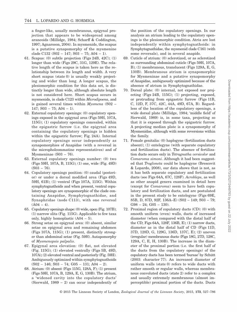
a finger-like, usually membranous, epigynal pro-jection that appears to be widespread amongaraneoids (Millidge, 1984; Scharff & Coddington,1997; Agnarsson, 2004). In mysmenids, the scapusis a putative synapomorphy of the mysmenineclade C132 (S02 – 147; S03 – 75; A04 – 1).
61. Scapus: (0) subtle projection (Figs 24B, 42C); (1)longer than wide (Figs 29C, 31G, 129E). The rela-tive length of the scapus is taken here as the re-lationship between its length and width. A veryshort scapus (state 0) is usually weakly project-ing and wider than long. A longer scapus, theplesiomorphic condition for this data set, is dis-tinctly longer than wide, although absolute lengthis not considered here. Short scapus occurs inmysmenids, in clade C123 within Microdipoena, andis gained several times within Mysmena (S02 –147; S03 – 75; A04 – 1).
62. External copulatory openings: (0) copulatory open-ings exposed in the epigynal area (Figs 59H, 107A,115G); (1) copulatory openings concealed, withinthe epigastric furrow (i.e. the epigynal areacontaining the copulatory openings is hiddenwithin the epigastric furrow; Fig. 24A). Internalcopulatory openings occur independently assynapomorphies of Anapidae (with a reversal inthe micropholcommatine representatives) and ofMysmeninae (S03 – 76).
63. External copulatory openings number: (0) two(Figs 59H, 107A, B, 115G); (1) one, wide (Fig. 49D)(S03 – 76).
64. Copulatory openings position: (0) caudal (posteri-or) or under a dorsal modified area (Figs 49D,59H, 61B); (1) ventral (Figs 107A, 115G). Withinsymphytognathoids and when present, ventral copu-latory openings are synapomorphic of the clade con-taining Anapidae, Symphytognathidae, andSynaphridae (node C113), with one reversal(A04 – 4).
65. Copulatory openings shape: (0) wide, open (Fig. 107B);(1) narrow slits (Fig. 115G). Applicable to few taxaonly, highly homoplastic (A04 – 5).
66. Strong setae on epigynal area: (0) absent, similarsetae on epigynal area and remaining abdomen(Figs 107A, 115G); (1) present, distinctly strong-er than abdominal setae (Fig. 59H). Autapomorphyof Mysmenopsis palpalis.
67. Epigynal area elevation: (0) flat, not elevated(Fig. 115G); (1) elevated ventrally (Figs 5B, 49D,107A); (2) elevated ventral and posteriorly (Fig. 59H).Ambiguously optimized within symphytognathoids(S02 – 146; S03 – 74; G05 – 131; A04 – 2).
68. Atrium: (0) absent (Figs 115G, 128A, F); (1) present(Figs 59H, 107A, B, 129A, E, G, 130B). The atrium,a ‘widened cavity into the copulatory ducts’(Sierwald, 1989 – 2) can occur independently of
the position of the copulatory openings. In ouranalysis an atrium leading to the copulatory open-ings is the plesiomorphic condition. Atria are lostindependently within symphytognathoids: inSymphytognathidae, the mysmenid clade C161 (withsome reversals), and in several anapids.
69. Cuticle of atrium: (0) sclerotized, or as sclerotizedas surrounding abdominal cuticle (Figs 59H, 107A,B); (1) membranous, translucent (Figs 129A, E, G,130B). Membranous atrium is synapomorphicfor Mysmeninae and a putative synapomorphyof Anapidae, ambiguously optimized because of theabsence of atrium in Symphytognathidae.
70. Dorsal plate: (0) internal, not exposed nor proj-ecting (Figs 24B, 115G); (1) projecting, exposed,or protruding from epigastric furrow (Figs 11B,C, 12D, F, 37C, 42C, 44A, 49D, 67A, B). Regard-less of the location of the copulatory openings, awide dorsal plate (Millidge, 1984; ‘middle field’ ofSierwald, 1989) is, in some taxa, projecting sothat it is exposed through the epigastric furrow.A projecting median plate is a synapomorphy ofMysmenidae, although with some reversions withinthe family.
71. Female genitalia: (0) haplogyne (fertilization ductsabsent); (1) entelegyne (with separate copulatoryand fertilization ducts). The absence of fertiliza-tion ducts occurs only in Tetragnatha versicolor andComaroma simoni. Although it had been suggest-ed that Trogloneta could be haplogyne (Brescovit& Lopardo, 2008), our data shows that is not, asit has both separate copulatory and fertilizationducts (see Figs 64A, 67C, 128F). Acrobleps, as wellas other anapid genera examined in detail here(except for Comaroma) seem to have both copu-latory and fertilization ducts, and are postulatedin the present study to be entelegyne (Figs 69E,85B, D, 87D, 92F, 136A–E) (S02 – 149; S03 – 79;G98 – 24; G05 – 130).
72. Proximal region of copulatory ducts (CD): (0) withsmooth uniform (even) walls, ducts of increaseddiameter (when compared with the distal half ofthe CD; Figs 64A, 128F, 136B, E); (1) narrow ducts,diameter as in the distal half of CD (Figs 11D,37D, 128D, G, 129G, 136D, 137C, E); (2) uneven(irregular) membranous ducts (Figs 18G, 27D, 128C,129A, C, E, H, 130B). The increase in the diam-eter of the proximal portion (i.e. the first half ofthe ducts from the copulatory openings) of thecopulatory ducts has been termed ‘bursae’ by Schütt(2003: character 77). An increased diameter ofuniform walls (state 0) refers to wide ducts withrather smooth or regular walls, whereas membra-nous convoluted ducts (state 2) refer to a complexand usually extremely membranous (almost im-perceptible) proximal portion of the ducts. Ducts
744 L. LOPARDO AND G. HORMIGA
© 2015 The Linnean Society of London, Zoological Journal of the Linnean Society, 2015, 173, 527–786
Dow
nloaded from https://academ
ic.oup.com/zoolinnean/article/173/3/527/2449763 by guest on 22 July 2022

with uniform diameter (regardless of width) presentno widening from the copulatory openings tothe spermathecae. The latter condition (state 1)is widespread among symphytognathoids and evenwithin Araneomorphae (L. Lopardo & G. Hormiga,pers. observ.), although ambiguously optimized here.An increased diameter of uniform walls (state 0)occurs in Trogloneta as well as theridiosomatidsand many anapid representatives. Irregular copu-latory ducts occur in most mysmenines and inthe Isela representative Isela-MYSM-002-KENYA(S03 – 77).
73. Proximal turn of CD: (0) sclerotized as the restof CD (Fig. 130B, G); (1) distinctly sclerotized(Figs 129A, B, 130A). There is a particular turnof the proximal membranous copulatory ducts inmysmenids, from the internal atrium, and im-mediately before becoming widened and convolut-ed, that is characterized by a subtle sclerotization.Only applicable to taxa with membranous inter-nal atrium and copulatory ducts. This distinctsclerotization occurs convergently withinMysmeninae, in Microdipoena, and in several othermysmenine nodes.
74. Copulatory duct length: (0) short, length equal orshorter than the size of the spermathecae(Figs 128A, 130G, 136D, F); (1) long, length at leasttwo times the size of spermathecae (Figs 129A,130B, 136B, 137D) (S02 – 148; S03 – 78; G05 –134; A04 – 8).
75. Copulatory duct–spermatheca junction: (0) pos-terior (Figs 128A, D, 129A, B, 136B, D, 137C); (1)lateral or anterior (Figs 18G, 27D, 42D, 51D, 129A,B, F, 130C, E). In most of the taxa here exam-ined the copulatory ducts connect with thespermathecae posteriorly. Lateral or anterior junc-tion is greatly homoplastic, occurring inSymphytognathidae, Trogloneta, Mysmenopsis,several mysmenines, and within theridiosomatids(A04 – 7).
76. Copulatory duct loops relative to spermathecae: (0)apart (Figs 37D, 124A, 129G, 136B, 137D); (1) copu-latory ducts encircling spermathecae (Figs 107D,120E, F, 137A, B). Only applicable to taxa withcoiled copulatory ducts (see character 78, state 1).Encircling copulatory ducts occur in Synaphridaeand Symphytognathidae, although homology of thiscondition is dubious because of ambiguous opti-mization (A04 – 9).
77. Copulatory ducts diverticulum: (0) absent(Figs 128A, F, G, 129G); (1) present (Figs 49A,129C, 130D, G). A ventral membranous pouch di-verting from the initial portion of the CD seemsto be present in some mysmenids. This characteris difficult to assess because of the membra-nous and translucent nature of most ducts in
many mysmenids. Nevertheless, this diverticu-lum appears to have evolved distally withinMysmenidae.
78. Copulatory ducts trajectory: (0) straight (Figs 128A,E, 137C); (1) coiled (Figs 37D, 129G, 136B); (2) con-voluted, switches but not coiled (Figs 18G, 22A,B, 27D, E, 42D, 49A, 51D, 67C, 136C). Homoplas-tic. Coiled copulatory ducts are plesiomorphicin symphytognathoids. Convoluted ducts aresynapomorphic for Mysmenidae, although straightducts occur in the mysmenid node C160, and alsoin node C153 within anapids.
79. Distal half of copulatory ducts: (0) sclerotized dis-tinct duct; (1) membranous. A CD with proximalwalls of increased diameter (character 71, state 0),can be followed distally by either a typicalsclerotized copulatory duct (e.g. Figs 128F, 136C),or a membranous duct (e.g. Fig. 136B). Narrow CDswith constant diameter (character 71, state 1)remain as sclerotized ducts (state 0 of this char-acter, e.g. Figs 128A, B, D, G, 129G, 137C), andCDs with membranous uneven ducts (charac-ter 71, state 2) remain as membranous (state 1 ofthis character, e.g. Figs 129A, B, C, 130A–G). Am-biguously optimized at the base of Anapidae, mem-branous distal ducts are synapomorphic forMysmeninae, although reversals occur in cladesC186 and C203.
80. Spermathecae compartment number: (0) two sperm-storage compartments per spermatheca (Figs 128F,135C, 136C); (1) one compartment (Figs 33A, 136B,137C). Two compartments evolve independently inthe spermathecae of Trogloneta, Minanapis, andCrassanapis (S02 – 150; G98 – 26; A04 – 10).
81. Spermathecae wall: (0) defined by a thick, well-sclerotized wall (Figs 129A, 130B, 136B, 137C); (1)membranous, weakly sclerotized wall (Figs 49A,87D, 130D, 136E). Membranous spermathecaeevolved independently in the mysmenine MYSM-023-MAD and the anapid Elanapis (S02 – 151; G05– 133).
82. Spermathecae: (0) separate (Figs 33A, 129A, 130B,136B, 137C); (1) connate, fused along the midline,sharing median wall (Fig. 123A); (2) touching(Leucauge venusta). Originally proposed as a di-agnostic feature of Theridiosomatidae (Coddington,1986a), connate spermathecae is here an autapo-morphy of Theridiosoma gemmosum (S03 – 79.1;G98 – 25).
83. Spermathecal shape: (0) ovoid (Figs 11D, 18G, 37D,128D, 129A,G, 136B); (1) C- or cup-shaped(Figs 27D, E, 42D, 128E, 130C); (2) coiled withinthe same spermathecal structure (Figs 33A, B, 51D,129C, 130B, G, 137C); (3) tubuliform, one largetube, sometimes like tracheoles (Figs 5D, 49A, 128A,C, 130D); (4) irregular, no particular shape
PHYLOGENY AND MORPHOLOGY OF MYSMENIDAE 745
© 2015 The Linnean Society of London, Zoological Journal of the Linnean Society, 2015, 173, 527–786
Dow
nloaded from https://academ
ic.oup.com/zoolinnean/article/173/3/527/2449763 by guest on 22 July 2022

(Figs 117B, 129D, 137E); (5) clavate (Figs 12C, 87D,128B, 136E). We have attempted to homologize thevariety of spermathecal shapes in as few statesas possible to avoid the proliferation of similarshapes with no grouping information. Althoughthere was ambiguous optimization within the out-groups, ovoid spermathecae is the plesiomorphicshape in symphytognathoids and Mysmenidae.Within Mysmenidae, spermathecal shape is ho-moplastic and mostly ambiguously optimized (A04– 11).
84. Tubuliform spermathecae: (0) relatively straight(Figs 49A, 130D); (1) coiled (Figs 5D, 128A, C).Applies only to taxa with tubuliform spermathecae(character 83, state 3).
85. Accessory glands: (0) absent; (1) present (Figs 22B,27D, E, 29A, 64A, 98A, 129B, E, H, 130A, F, 136F).In the internal genitalia of the mysmenids,Trogloneta, Microdipoena, Mysmena-MYSM-005-ARG, the mysmenine MYSM-029-MAD, as well asin the anapid Tasmanapis, there is an additionalpaired structure that resembles either a muscularapodeme or a glandular structure. This structure,probably related to the copulatory ducts or thespermathecae, is better observed under SEM, al-though it can be distinguished in transparent prepa-rations of the vulva by a higher degree ofsclerotization, comparable with that of thespermathecae (Figs 129B, E, H, 130A, F, 136F; seealso Brescovit & Lopardo, 2008: fig. 6C–E). Whetherthese structures are functional glands, muscle at-tachment points, or perform other functions remainsunknown. As the appearance of these structuresseems consistent with a glandular function, theyare regarded here as accessory glands. Given theabsence of palpimanoids in our data set (for whichthis character was originally established), the ho-mology of the palpimanoid glands with these ac-cessory glands of symphytognathoids remains tobe tested (S02 – 152).
86. Fertilization duct sclerotization: (0) weaklysclerotized but with a distinguishable wall(Figs 42D, 49A, 51D, 128A, 129G); (1) membra-nous, translucent, almost imperceptible (Figs 18G,27D, E, 60H, 64A, 128F, 136D). Highly homoplas-tic (A04 – 13).
87. Fertilization ducts: (0) small, shorter thanspermathecae length (Figs 42D, 49A, 64A, 128A,F); (1) large, longer than spermathecae length(Figs 11D, 14E, 18G, 27D, E, 51D, 60H, 92F, 129G,136D). Short fertilization ducts provide a directconnection between the spermathecae to the uterusexternus, usually in a straight fashion and withoutmodifications. Long fertilization ducts can havemodifications or expansions. Long fertilizationducts optimize ambiguously at the base of the
Symphytognathidae plus Anapidae node (mostsymphytognathids have short ducts). WithinMysmenidae, long ducts originated independent-ly six times.
Eyes
88. Anterior median eyes (AME): (0) absent (Figs 77B,104F, 112C, D, 120A, 142M, 145C, D, 146H);(1) present (Figs 35B, 99E). Anterior median eyesare absent in all Symphytognathidae studied here(including Crassignatha and Iardinis), in someanapids, and in the mysmenid Mysmeniola (S02– 20; S03 – 9; G98 – 27).
89. AME size: (0) minute (Figs 67G, 99E); (1) sameas others (Fig. 35B); (2) largest (at least two timesALE). Minute eyes (state 0) are distinctly smallerthan any other eyes, and are characteristic of alleight-eyed anapids (although ambiguously optimizedbecause of the loss of AME in Symphytognathidae)and Trogloneta. Similar AMEs (state 1) can en-compass subtly larger eyes, but differences in sizeare subtle. Largest eyes (state 2) refer to eyes twotimes larger than ALE, as reported for the mis-placed zodariid Leviola (see Miller, 1970; S02 – 20;S03 – 9; G98 – 27; A04 – 104).
90. Eyes elevation (male): (0) AME flush with cara-pace (Figs 91H, 96C); (1) AME on protruded area(Figs 15B, D, 25C, 46D, E, 59C); (2) all eyes ontubercle (Figs 63G, H, 66A). Anterior median eyeslevel with carapace (state 0) occuring in all non-symphytognathoid representatives, as well as inall eight-eyed anapids (including Taphiassa). Bothmale and female mysmenids (except Trogloneta)and theridiosomatids have a groove or depres-sion around the AME that to some extent delim-its a protruded area (state 1, ambiguously optimized,potentially synapomorphic for Theridiosomatidaeand for Mysmenidae). This area is clearly pro-truded, not just smoothly raised from the rest ofthe carapace, and is best observed in frontal viewusing SEM. Males of Acrobleps and Troglonetaspecies have all eyes in a tubercle or a narrow el-evation of the ocular area (state 2) (S02 – 21; A04– 103).
91. Distance between AME: (0) AME close (Figs 25C,35B, 99E); (1) AME separate (Fig. 142K). The origi-nal character states considered the distance betweenAME in relation to the distance between AME andtheir correspondent ALE. As the size of AME candiffer across taxa, and at the same time this dif-ference in size can affect their relative distanceto the ALE, the same AME diameters were usedas a criterion to define the distance between AME.Here, close AME (state 0) refers to AME separat-ed by about one or less their diameter. On the other
746 L. LOPARDO AND G. HORMIGA
© 2015 The Linnean Society of London, Zoological Journal of the Linnean Society, 2015, 173, 527–786
Dow
nloaded from https://academ
ic.oup.com/zoolinnean/article/173/3/527/2449763 by guest on 22 July 2022

hand, distant AME (state 1) refers to AME sepa-rated by two or more their diameter, and in thisdata set it evolves independently in Brasilionataand node C165 within Mysmena (M. awari andM. marijkeae). AME separation is similar in bothsexes, except for a few species in which males havedistant AME (state 1) and females have close AME(state 0). Sexually dimorphic taxa were scored as‘polymorphic’ and include Mysmena incredula(Fig. 35A, B), M. tembei (Fig. 142H, I), M. santacruzi,and Mysmena-MYSM-005-ARG (S02 – 26).
92. AME-ALE separation (females): (0) distant; (1) close(Figs 35B, 99E). The original character states con-sidered the distance between AME and ALE inrelation to the AME diameter. As explained in Char-acter 91, the difference in dimensions of the AMEwith respect to other eyes in some taxa could distortthe relative distances, and consequently potential-ly alter state assignation. Therefore, AME–ALE sepa-ration was measured as proportional to the numberof ALE diameters between them. This way, distantanterior median and lateral eyes (state 0) are eyesseparated by more than two ALE diameters, whereasclose eyes (state 1) are separated by less than twoALE diameters. Distant AME–ALE occurs in thetetragnathid representatives Leucauge venusta andTetragnatha versicolor (A04 – 105).
93. Secondary eyes tapetum: (0) present; (1) absent.Loss of tapeta (state 1) is autapomorphic forTetragnatha versicolor, although detailed infor-mation is missing for most of the taxa. Our scoringwas taken from (Griswold et al., 1998: charac-ter 28) (S02 – 27; G98 – 28; G05 – 47).
94. Lateral eyes (male): (0) juxtaposed (Fig. 72C); (1)separate. Separate lateral eyes are autapomorphicfor Tetragnatha versicolor (S02 – 23; S03 – 10.1;G98 – 30; A04 – 102).
95. Lateral eyes: (0) level with carapace (Figs 69A, 82C);(1) common elevation (Figs 46D, 77B, 104E). Levellateral eyes occur independently in Acrobleps,Comaroma, Tetragnatha versicolor, and presum-ably Leviola (S02 – 24; S03 – 10).
96. PME separation: (0) PME close (Figs 15F, 77C);(1) PME separated (Fig. 67H). Close PME (state 0)defined as distance between PME less or equal tothe distance between PME–PLE, occur in most ofthe examined taxa. Separate PME (state 1) havea distance of separation greater than with theircorrespondent PLE, and are synapomorphic forTrogloneta. Separated eyes are also presumablyfound in the zodariid Leviola (S03 – 10.2).
Legs
97. Leg cuticle texture: (0) smooth (Figs 80C, 119F,H); (1) imbricate (Figs 21H, I, 34B, 65E, H); (2)
fatiscent (Figs 73D, 87B, 93F). Imbricate leg cuticleis the widespread condition in the taxa compris-ing this data set. Fatiscent leg cuticle evolvesconvergently in several mysmenids, and is a pu-tative synapomorphy of Anapidae (ambiguouslyoptimized because of the fatiscent leg cuticle ofSynaphris). Smooth leg cuticle is an ambiguous-ly optimized synapomorphy of Symphytognathidae(S02 – 85; G05 – 10).
98. Ventral elevated setal bases on femur I (male):(0) absent (Figs 42G, 93E); (1) present (Figs 74E,84A, B, D, 145K). Schütt’s (2003) original defi-nition of this character did not specify which sideof the femur it was referring to, or whether thetubercles were just cuticular protrusions or setalbases. Here, the character is redefined to scoreventral elevated setal bases on femur I, asynapomorphy of anapid clade C121. In Steatodathe bases are seemingly present and in all legs(S03 – 32.2).
99. Femoral macrosetae: (0) present (Figs 16D, 57B,141A, D, 144F, J); (1) absent (Figs 21C, E, 42F,45G). Most of the taxa in this data set lack spinesor strong macrosetae. Macrosetae are consid-ered here as distinct spines and also as setae thatare stronger than most of the setae covering thelegs, like those macrosetae located distally on thedorsal patellae. Within symphytognathoids, femoralmacrosetae occur convergently on the clade in-cluding Maymena and Mysmenopsinae (i.e.node C160, with a reversal in Mysmenopsis penai)and in Theridiosoma gemmosum (S02 – 67; S03– 33; G98 – 59; A04 – 183).
100. Fe IV trichobothria: (0) absent; (1) present. Femoraltrichobothria are restricted to the tetragnathidrepresentatives of this data set (S02 – 76; G98– 58; G05 – 1).
101. Female femur I ventro-apical area (‘femoral spotor projection’): (0) cuticle unmodified, as the restof the femur (Figs 74F, 144J); (1) cuticle with eithera sclerotized spot or a projection (Figs 34A, 39D,57A, E, 140G, 141C, 143N). With just a few ex-ceptions (Mysmena-MYSM-005-ARG, Mysmenopsispenai, and M. palpalis in this data set), mostmysmenid species have either a sclerotized spotor a cuticular projection on the apical ventralsurface of at least femur I. This so called ‘femoralspot’ is a synapomorphy of Mysmenidae (see char-acter 102) (S03 – 32; G98 – 56).
102. Female femur I structure: (0) femoral spot(Fig. 34A); (1) femoral projection (Fig. 57E). Afemoral projection is a synapomorphy ofMysmenopsis, although lost in some species (seecharacter 101) (S03 – 32; G98 – 56).
103. Location of female femoral structure: (0) femur Ionly (Fig. 140G); (1) femora I and II (Figs 141C,
PHYLOGENY AND MORPHOLOGY OF MYSMENIDAE 747
© 2015 The Linnean Society of London, Zoological Journal of the Linnean Society, 2015, 173, 527–786
Dow
nloaded from https://academ
ic.oup.com/zoolinnean/article/173/3/527/2449763 by guest on 22 July 2022

142B). The femoral structure of most femalemysmenids occurs in both femora I and II. Theoccurrence of this feature only on femur I is con-vergent in Trogloneta and Mysmenopsis (whenpresent), Mysmena-MYSM-007-MEX, M. incredula,and M. santacruzi (S03 – 32.1).
104. Femur I ventro-apical sclerotized spot on males:(0) absent; (1) present (Fig. 21A). A distal ventralfemoral structure on males is not as wide-spread within mysmenids as it is for females. Asclerotized spot in males, however, occurs inde-pendently in several relatively large groups withinMysmenidae: Maymena and Trogloneta (ambigu-ously optimized at their bases because of anabsence of spot in mysmenopsine males), nodeC124 within Microdipoena, and nodes C137 andC145 within Mysmena.
105. Location of sclerotized spot on males: (0) femur Ionly (Fig. 141O); (1) femora I and II (Fig. 140M).Contrary to females, most mysmenid males havethe femoral spot (when present) only on femur I,only in two occasions the spot occurs in bothfemora I and II (Maymena ambita and Mysmenatasmaniae).
106. Stridulatory field on retrolateral femur IV: (0)absent (Fig. 39F); (1) present (Fig. 105F, H). Thismodified field was observed only in females ofTeutoniella cekalovici. It is presumably stridulatory,as is usually suspected in any area with modi-fied cuticle, especially on leg segments. Autapo-morphy of Teutoniella.
107. Stridulatory field on prolateral femur IV: (0) absent(Fig. 34E); (1) present (Figs 9A–C, 54H, 59F).Synapomorphy of Mysmenopsinae.
108. Stridulatory field on retrolateral femur I (female):(0) absent (Fig. 29D); (1) present (Figs 3C, 57B–D). This presumably stridulatory field was ob-served and scored at least in females, althoughit can also be present in some males. Synapo-morphy of Mysmenopsinae.
109. Modified area on prolateral femur I: (0) absent;(1) present (compare Figs 116A, C, 119A,B). Thecuticle on dorsal femur I can be weakly ormore distinctly modified (when compared withthe retrolateral cuticle) in some members ofSymphytognathidae (putative synapomorphy).
110. Metatarsi preening setae: (0) ventral and dorsalsetae identical (Figs 29F, 74B, 119C); (1) ventralsetae strongly serrated (Figs 83B, C, 93G, I). Meta-tarsal preening setae are defined here as ventralsetae that are more strongly serrated than thedorsal setae. They usually occur in both sexes inall metatarsi, not only on metatarsi III and IV.Whether these specialized setae actually performa preening function is not known. The distribu-tion of these setae along the metatarsi can be
lengthwise (e.g. in Comaroma) or distal (e.g.Minanapis). The presumed zodariid genus Leviolahas been reported to have a preening comb notonly in all metatarsi, but also in all tibiae andtarsi (Miller, 1970 – 156), and is here scored aspresent (state 1). In Teutoniella, at least on meta-tarsus IV, the ventral setae appear less serrat-ed than the dorsal setae (Fig. 105G), and is herescored as absent (state 0) (S02 – 73; G05 – 19).
111. Metatarsus I trichobothrium: (0) absent; (1)present. Absence of trichobothria on metatar-sus I is an autapomorphy of Tetragnatha versicolor(S02 – 77; S03 – 35.5; G05 – 5).
112. Metatarsus I trichobothrium position: (0) proxi-mal, at or within the proximal third (0.33) of meta-tarsus (Figs 26D, 39E, 105E); (1) median, withinthe middle third (usually at midpoint) of meta-tarsus (0.5) (Figs 8A, 95H, 101F); (2) distal, withindistal third (Leviola). Ambiguously optimized,proximal metatarsal trichobothrium occursin Mysmenidae (with few distal reversals),Theridiosomatidae, some Symphytognathidae, andTeutoniella (A04 – 189).
113. Metatarsus III trichobothrium: (0) present(Fig. 21G); (1) absent (Fig. 109D). Absence oftrichobothrium on metatarsus III is a synapo-morphy of Synaphridae (A04 – 190).
114. Metatarsus I prolateral clasping structure onmales: (0) absent (Figs 90F, 125F); (1) present(Figs 3A, 16G, 26C, 42H, 45H, 59B, 74D, 140C,141G, K, 142N). These metatarsal clasping struc-tures are a synapomorphy of Mysmenidae (absentin Maymena mayana). Similar structures haveindependently evolved in some anapids (S03 – 35.3;G98 – 57; G05 – 22).
115. Metatarsus I prolateral clasping structure form(males): (0) spur (Figs 74D, 101F, H, 145D, L); (1)clasping spine (Figs 3A, B, 8C, 26C, 57I, 140C,141K, 142G). Clasping spines are mostly prolateraland spurs appear to be prolateral–ventral. Never-theless, these two types of modified setae arisefrom a cuticular socket and have been consideredas homologous in a previous study (see Lopardo& Hormiga, 2008 – 28, character 57). Here, clasp-ing spurs and spines are also regarded as ho-mologous, and spurs are hereafter referred to as‘prolateral’. Clasping spurs occur in Anapidae. Allmale mysmenids, except Maymena mayana, havea prolateral clasping spine on the first metatar-sus (S03 – 35.3; G98 – 57; G05 – 22).
116. Metatarsus I prolateral clasping structure posi-tion (males): (0) basal, extremely proximal or onbasal third of metatarsus (Figs 16G, 141K); (1)medial, on middle third of metatarsus, includ-ing its borders (Figs 8A, 26C, D, 140C, 142G, N);(2) apical, beyond middle third (i.e. on distal third)
748 L. LOPARDO AND G. HORMIGA
© 2015 The Linnean Society of London, Zoological Journal of the Linnean Society, 2015, 173, 527–786
Dow
nloaded from https://academ
ic.oup.com/zoolinnean/article/173/3/527/2449763 by guest on 22 July 2022

of metatarsus (Figs 50H, 59B, 143C, 144A). Am-biguously optimizing at the basal node ofMysmenidae, basal clasping spine is a putativesynapomorphy of Maymena. Medial spine occursin the majority of mysmenids. Apical claspingspines evolve independently on several occa-sions (S03 – 35.4).
117. Metatarsus I prolateral clasping spine shape (males):(0) relatively straight or weakly curved (Figs 16G,34C, 62E, 65C, 68B); (1) twisted (proximally straightand curved at middle angle, Figs 3B, 8B, C, 26C,140E, F, 141K, L, 142N); (2) strongly curved proxi-mally (Figs 54D, 57I, 59B). This character appliesonly to the shape of the metatarsal clasping spines.Twisted spine is synapomorphic of mysmenid nodesC127 and C164, and ambiguously optimized in Isela.Most Mysmenopsis representatives have stronglycurved metatarsal clasping spines, although opti-mization is ambiguous because of straight spineof Mysmenopsis penai.
118. Metatarsus I distal spine or stronger setae nearto clasping spine (males): (0) absent (Fig. 62E);(1) present (Fig. 34C). Applicable only to meta-tarsal clasping spines. A stronger seta located closeto the clasping spine occurs independently in twomysmenids (Mysmena incredula and Microdipoenasamoensis).
119. Metatarsus I proximal row of between five andeight spines or strong setae near clasping spine(males): (0) absent (Fig. 62E); (1) present (Figs 57H,59B, 140H, J). Applicable only to taxa with meta-tarsal clasping spines. Ambiguously optimizedsynapomorphy of Mysmenopsis.
120. Metatarsus I clasping spur number (males): (0)one (Fig. 145L); (1) two (Figs 74D, 101F, 145C,D). Applicable only to metatarsal clasping spurs.
121. Metatarsus I apical prolateral spur (females): (0)absent (Fig. 89F); (1) present (Figs 74G, 84C, E).An apical spur on metatarsus I occurs in malesbut also in females of Anapisona kethleyi andCrassanapis chilensis (node C121).
122. Metatarsus I apical retrolateral spur (males): (0)absent; (1) present (Fig. 73A). Males of Anapisonakethleyi have not only two spurs prolaterally onthe first metatarsus but also one distal retrolateralspur.
123. Metatarsus II prolateral clasping spine (males):(0) absent; (1) present. The presence of a secondclasping spine on metatarsus II of males is anautapomorphy of Mysmena santacruzi (Baert &Maelfait, 1983 – 104).
124. Patella dorsal macrosetae: (0) present (Figs 21D,56A, 65B, 89G, 101D); (1) absent (Figs 80B, 83D,109C). Dorsal distal patellar macrosetae (i.e.stronger setae) is independently lost (state 1) inComaroma and Cepheia (S02 – 68; S03 – 34).
125. Leg spination: (0) three or more macrosetae dor-sally on tibiae, or macrosetae present also onfemora, metatarsi, and/or ventral tibiae (Figs 140H,M, 141C, 144E, J); (1) ‘reduced’, only one or twomacrosetae dorsally on tibiae, none on femora,metatarsi, and/or ventral tibiae (Figs 140E, 142E,145G, K). ‘Reduced’ leg spination occurs in mostsymphytognathoids. When strong spines also occur,they are scored here as state 0. ‘Strong’ spinationis synapomorphic for Maymena, convergent inMysmenopsis dipluramigo (G05 – 21).
126. Leg I (females): (0) with femur as wide as the otherlegs’ femora (Figs 140N, 145G, 146A); (1) femurtwo times as wide as the other legs’ femora(Figs 140A, D, G, 145J). This character was scoredfor females to avoid dependency of characters, asstronger leg I can be sexually dimorphic (i.e.stronger legs in males but not females, see char-acter 128). Strong legs are a synapomorphy ofMysmenopsinae, also convergently occurring inCrassanapis (S02 – 63; G98 – 55; A04 – 182).
127. Metatarsus–tarsus joint: (0) both segment tips withsame width as rest of the segments (straight,square), i.e. not constricted (Figs 12G, 21B, 39J,62G, 101B); (1) both segment tips constricted(Figs 109A, 111G); (2) tarsal tip constricted, meta-tarsal tip straight or square (Fig. 80A). Constric-tion of both metatarsus and tarsus optimizes hereas a synapomorphy of Synaphridae.
128. Leg I dimorphism: (0) absent (Figs 140D, E, M,N, 146A, B); (1) present, male leg I stronger orlonger (Figs 143D, F, 144D, E, 145E, F, J, K). Legdimorphism occurs independently on few species(S03 – 30).
129. Tarsus I superior claws: (0) with comb of per-pendicular long teeth (five or more separatedteeth), teeth equally long or increasing in length(Figs 80D, E, 95F, G, 122G); (1) with a distinctrow of four or more teeth of distally increasinglength, oriented forward and usually touching(teeth can be short; Figs 3E, 12F, 56B, 109E,111H); (2) with a row of between one and threeshort teeth oriented forward (Figs 26B, 52D, 65F,69F, 70F, 73C, 123G); (3) smooth, teeth absent.Smooth paired claws (state 3) are autapomorphicof Mysmena acuminata (Marples, 1955). Pairedclaws with few short teeth (state 2) occur in mostsymphytognathoids and can be considered as asynapomorphy for the group. Claws with four ormore teeth of decreasing length (state 1) aresynapomorphic (and secondarily gained) forSynaphridae and ambiguously optimized forMysmenopsinae (S02 – 81; S03 – 37).
130. Tarsus IV median claw (male): (0) short, at mostsubequal than superior claws (Figs 83F, 95E); (1)long and strong, as superior claws (Tetragnatha);
PHYLOGENY AND MORPHOLOGY OF MYSMENIDAE 749
© 2015 The Linnean Society of London, Zoological Journal of the Linnean Society, 2015, 173, 527–786
Dow
nloaded from https://academ
ic.oup.com/zoolinnean/article/173/3/527/2449763 by guest on 22 July 2022

(2) long and slender (Figs 34F, 48G, 88I, 101G,122H). Long and slender median claw IV inmales (state 2) is a potential synapomorphy ofsymphytognathoids. Synaphridae has shortmedian claw (state 0) (S02 – 82; S03 – 38; G98– 63; A04 – 199).
131. Tarsus IV median claw (female): (0) short, at mostsubequal to superior claws (Figs 16F, 80F); (1) longand slender (Figs 9E, 12H, 123H); (2) minute(Leviola). Long and slender median claw IV infemales (state 1) is a potential synapomorphy ofsymphytognathoids, with a reversal in Maymenarica and node C150 within Anapidae (A04 – 200).
132. Sustentaculum: (0) absent (Fig. 48G); (1) present.A sustentaculum occurs in the tetragnathid rep-resentatives of this data set (S02 – 84; G98 – 64).
133. Tarsal organ position: (0) proximal, by the junc-tion with metatarsus (Figs 39C, H, 50D, E, 119D,125G); (1) median, on the middle third of tarsus(Figs 8E, 54E, 56F). The position of the tarsal organis constant in all legs in most examined taxa, exceptfor (at least) Isela okuncana and Taphiassa, wherethe tarsal organ is medial on tarsus I, but basalon tarsus IV, and were scored here as polymor-phic. Basal tarsal organ is a synapomorphy ofsymphytognathoids, with a reversal in Synaphridaeand Mysmenopsinae (S03 – 39).
134. Shape of tarsal organ pocket: (0) domed (Figs 9D,26E, 48F, 50D, 54F, 73B, 95I, 101E); (1) flat(Figs 109B, 119D). Scored for leg I. Some taxa havean intermediate shape of the capsulate tarsalorgan (e.g. Figs 46G, 116E), where it is not clearlydomed but is neither flat. These dubious situa-tions were scored as ‘?’. Flat tarsal organ occurin Cepheia (Synaphridae), independently inMysmena MYSM-007-MEX and M. acuminata(Mysmenidae), and ambiguously within Symphy-tognathidae (S03 – 40).
135. Size of tarsal organ opening: (0) distinctly smallerthan setal sockets (Figs 3F, 54F, 62H, 109B, 119D);(1) subequal or larger than setal sockets (Figs 26E,39C, 48F, 50D, 73B, 83E, 95I, 101E). This char-acter was scored only for leg I, as the tarsal organopening on leg IV is usually smaller than setalsockets. Small opening is the plesiomorphiccondition; occurring in Synaphridae, mostsymphytognathids, and Theridiosoma gemmosum.Larger opening is independently synapomorphicfor Anapidae and Mysmenidae (with a reversalin Mysmenopsinae and Mysmena incredula) (A04– 198).
136. Shape of tarsal organ opening: (0) round (Figs 26E,48F, 73B, 95I, 119D); (1) teardrop (Figs 3F, 9D,62H, 101E, 125G). Within Mysmenidae, the tear-drop tarsal organ opening is a synapomorphy ofMysmenopsinae. This shape of the opening
also occurs independently in a few othersymphytognathoids.
137. Tarsus IV ventral setae: (0) same as dorsal setae(Figs 3D, 26F, 39I, 74C); (1) serrated tarsal comb(i.e. theridiid comb); (2) sparse strongly serrat-ed bristles (not a comb) (Figs 80G, 84F, 93H). Thegenus Leviola has been reported to have a ser-rated setae not only in all metatarsi (preeningcomb, see character 110), but also in all tibiae andtarsi (Miller, 1970 – 156), and is here scored aspresent (state 2). In Isela okuncana, both ventraland dorsal setae are serrated (Fig. 3D), and it istherefore scored as state 0. Trogloneta (Fig. 65G)and the symphytognathid SYMP-006-AUST(Fig. 119G) have weakly serrated ventral setae,but stronger than dorsal setae (although not‘strongly serrated’), and therefore were scored aspolymorphic (states 0 and 2). Strongly serratedventral setae occur in Theridiosomatidae, someanapids, and Symphytognatha picta (S03 – 36;G98 – 62; G05 – 25; A04 – 193).
138. Tarsus I prolateral row of modified setae (male):(0) absent (Figs 74A, 89E); (1) present (Figs 16H,34D, 59D, 65D). First observed and described asa ventral row of modified setae by Thaler (1975,1995) for Trogloneta granulum and Mysmeniolaspinifera, this prolateral row of modified setae(see below) is a potential synapomorphy forMysmenidae.
139. Tarsus I prolateral row, type of setae (male): (0)shorter than surrounding setae, slim and curved(Figs 16H, 26A, 34D, 45I, 50F); (1) shorter, butstout and straight (Figs 8F, 54G, 59D, 65D, 68A).Applicable only to taxa with prolateral row ofmodified setae. Ambiguous optimization. Stoutsetae occur in Trogloneta and Mysmenopsinae,whereas slim setae are present in Maymena andMysmeninae.
140. Tarsus I prolateral row of setae distribution (male):(0) along tarsus (Figs 8F, 16H, 65D, 68A); (1) distalhalf of tarsus (Figs 26A, 34D, 45I, 50F, 54G, 59D).Ambiguous optimization. Setal row along tarsusoccurs in Maymena, Trogloneta, and Isela, andindependently in Mysmeniola. Distal row is presentconvergently in Mysmenopsis and Mysmeninae.
141. Tibia dorsal macrosetae: (0) present (Figs 9F, 39G,73E, 90E, 101C, 119H); (1) absent (Figs 80H, 83A,111F). Generally at least one macrosetae, similarto that of the distal patella, is present on thedorsal tibiae. Tibial macrosetae are lost indepen-dently (state 1) in Comaroma and Synaphridae(S02 – 69; S03 – 35).
142. Tibia I prolateral apical clasping spine on males:(0) absent (Figs 45H, 90E, 143L); (1) present(Figs 3A, 26C, 140E, J, K, 141K, L, O). In addi-tion to the prolateral metatarsal clasping spine,
750 L. LOPARDO AND G. HORMIGA
© 2015 The Linnean Society of London, Zoological Journal of the Linnean Society, 2015, 173, 527–786
Dow
nloaded from https://academ
ic.oup.com/zoolinnean/article/173/3/527/2449763 by guest on 22 July 2022

some male mysmenids also have one or twoprolateral clasping spines on tibia I. Tibialclasping spines evolved convergently withinMysmenidae: in Microdipoena, and ambigu-ously optimized at node C160 (Maymena +Mysmenopsinae, ambiguous because of its absencein M. mayana) (S03 – 35.1).
143. Tibia I prolateral apical clasping spines number(males): (0) one (Figs 3A, 8D, 16G, 54C, 59B, 62E,F, 140E, J, K); (1) two (Figs 26C, 27I, 57F, G,140H, 141L, O). Only applicable to taxa with tibialclasping spines. Basal mysmenids (i.e. Maymenaand Mysmenopsinae) have one tibial clasping spine(except for Mysmenopsis dipluramigo), whereasdistal mysmenids (i.e. Microdipoena) have twospines.
144. Row of between two and five stout setae basalto tibial clasping spines (males): (0) absent(Figs 3A, 21F, 59B, 62E, 140E); (1) present(Figs 54C, 57G, J, 140H, J, K). Additional to thetibial clasping spine, stout setae occur in two in-stances within Mysmenopsis.
145. Tibia I prolateral median clasping structure onmales: (0) absent (Fig. 21F); (1) present (Figs 16G,73A). A median clasping structure occurs inde-pendently in Maymena rica and in Anapisona.
146. Ventral apical clasping spine on tibia II (males):(0) absent; (1) present (Figs 116B, D, 119H–J).A ventral clasping spine on tibia II of males occurswithin symphytognathids (node C156), includ-ing taxa previously placed in Mysmenidae asIardinis mussardi and Crassignatha.
147. Tibia I ventral paired setae with protruded bases(males): (0) absent (Fig. 70D); (1) present(Figs 48C–E, 50G). Evolves independently withinMysmenidae, in the Malagasy species MYSM-020-MAD and MYSM-023-MAD.
148. Ventral–prolateral row of short spines on tibia II:(0) absent; (1) present (Fig. 70E). Autapomorphyof Acrobleps.
149. Tibia III–IV trichobothrial length: (0) short, lengthequal or less than two times tibia diameter(Figs 84G, 90B, 93D); (1) median, length betweenmore-than-two but less-than-three tibia diam-eters (Figs 26G, 29E, 39G, 73F, 88H); (2) long,length equal or more than three times tibia di-ameter (Figs 16E, 68D, 113H, 116F, 119E). As thetrichobothria on the same tibial segment may differin length, this character was scored based on thelongest of them. Although long trichobothria ontibia III and IV (state 2) was proposed as asynapomorphy of Theridiosomatidae (Coddington,1986a; Griswold et al., 1998), here long trichobothriais also found in some anapids, micropholcommatines,mysmenids, and symphytognathids as well.Although highly homoplastic, short-length
trichobothria appear to be the ancestral state insymphytognathoids (S03 – 35.2; G98 – 61).
150. Tibia IV trichobothrial shaft: (0) smooth or ser-rated (Figs 16E, 113H); (1) distinctly plumose(Figs 90B, 93D). Plumose trichobothrial shaft isambiguously optimized within Anapidae becauseof its occurrence in Elanapis and Minanapis.
Male palp
151. Bulb–cymbium lock mechanism (B–C lock): (0)absent; (1) present. B–C lock mechanism is anautapomorphy of Steatoda in this data set (A04– 31; G98 – 12).
152. Conductor: (0) present (Figs 18D, 81B, 88D, 102A,110A, 117D, 124F); (1) absent (Figs 4A, 10E, 28D,76A, 95A, 113F). A convention discussed inGriswold et al. (1998) and followed here, contem-plates that when only one sclerotized tegularsclerite occurs in the male palp (in addition tothe embolus), which cannot be homologized to anyof the palpal sclerites because of its uncertainstructure and/or origin, it is regarded as the con-ductor. If two or more sclerites are present, thenthe one that originates closer to, or with a clearinteraction with the embolus, is regarded as theconductor, the second is referred to as the MA,etc. Some mysmenids have a voluminous andmembranous structure of the tegulum, whichappears to originate close to the embolic base(Fig. 18D). This structure has been named ‘bulbalshield’ (e.g. Baert, 1984a; Schütt, 2003), andbesides the embolus, it appears to be the onlystructure of the bulb. This structure often em-braces and/or covers the embolic base, some-times even a groove housing the embolus canoccur. Even though the general structure of thisfeature is distinctive in most mysmenids (whenpresent), it is regarded as a conductor here basedon its origin and association with the embolus.Also, this homology statement avoids the prolif-eration of taxon-specific terms, otherwise ofuntestable homology. Here, the occurrence of con-ductor is symplesiomorphic and widespread, withindependent losses of this sclerite occurring withinsymphytognathoids in anapids, symphytognathids,and mysmenids. Within Mysmenidae, the con-ductor is lost two times: in the node comprisingMaymena and Mysmenopsinae (node C160), andin MYSM-005-ARG (G98 – 14; G05 – 118; A04– 62; S03 – 69.2).
153. Conductor shape: (0) single, simple (Figs 102A,117D, 124B); (1) bifid or with apophysis (i.e. twomain peaks; Figs 88D, 106C, 110A, 122C); (2)complex, three or more peaks (Figs 81B, 86G, 92B,114E); (3) globose and voluminous membrane
PHYLOGENY AND MORPHOLOGY OF MYSMENIDAE 751
© 2015 The Linnean Society of London, Zoological Journal of the Linnean Society, 2015, 173, 527–786
Dow
nloaded from https://academ
ic.oup.com/zoolinnean/article/173/3/527/2449763 by guest on 22 July 2022

(Figs 17A, B, D, 18D, E, 27A, B, 30A, E, 36C, 41D,63A, D). A simple conductor is the plesiomorphiccondition of symphytognathoids, occurring also inTheridiosomatidae. Bifid conductor optimizesas a putative synapomorphy of clade C113(Synaphridae + Anapidae + Symphytognathidae),with ambiguously optimized distal transitions(to both simple and complex conductors) inSymphytognathidae and Anapidae. A volumi-nous and membranous conductor is a putativesynapomorphy of Mysmenidae (S02 – 141; S03– 72; G98 – 15; G05 – 121).
154. Conductor size: (0) large, about half the size orlength of bulb, or larger (Figs 88D, 102A, 106D);(1) small, considerably smaller than half the sizeof the bulb (Figs 114E, 117D). Small conductoroccurs independently in Tasmanapis and thesymphytognathid node C156 (S02 – 142).
155. Conductor position (origin): (0) subterminal, medialon ventral view (Figs 81B, 88D); (1) terminal, origi-nating on the apical border of tegulum (Fig. 106C).Subterminal conductor is synapomorphic for theANTS clade, with a reversal in Cepheia longisetaand Iardinis mussardi (G05 – 119; A04 – 69).
156. Conductor distal groove: (0) with a groove for thedistal portion of the embolus (Figs 106C, 117D,124F); (1) entire, without groove (Figs 81C, 114E).A groove for the distal embolus is lost indepen-dently in Mysmenidae, in some anapids, and ina few symphytognathids (A04 – 64).
157. Conductor tip sclerotization: (0) like base(Figs 138C, 139D); (1) more than base (Figs 138B,139B,C). Ambiguously optimized. A sclerotizeddistal conductor occurs in Synaphridae,Symphytognatha, and Crassanapis (A04 – 67).
158. Conductor apical portion: (0) width subequal tobase (Figs 81C, 88D); (1) at least a portion is largerthan the base width (Figs 86F, 124B). Ambigu-ously optimized. A large conductor occurs inMicrodipoena, Crassanapis, theridiosomatids, andCepheia. Subequal conductor occurs in Synaphris,symphytognathids, and most anapids. In mostmysmenids this condition is unknown (i.e. scoredas ‘?’) (A04 – 63).
159. Conductor surface: (0) smooth (Fig. 124B); (1) withsmall ridges or other cuticular structures(Figs 102A, D, 106D, 117D, E). Ridged conduc-tor occurs independently in Trogloneta, Teutoniella,SYMP-006-AUST, and Synaphridae (A04 – 65).
160. Conductor groove housing proximal and/or medianembolus: (0) entire, without a groove (Figs 17A,B, D, 27A, B, 88D, 110A); (1) with a groovehousing the proximal and/or median portions ofthe embolus (Figs 36B, C, 47E, 92B, 106D).Grooved conductor housing the embolus proxi-mally is plesiomorphic for symphytognathoids.
Entire conductor evolved independently threetimes: in Trogloneta, in the mysmenine clade C127,and in the clade comprising Symphytognathidaeplus Anapidae (A04 – 68).
161. Conductor (C) and embolus (E) interaction: (0)separate, no association (Figs 63A, D, 81C, 114E);(1) conductor embraces embolus (Figs 41C, 43A,B, 86G, 117D). An association between C and E(state 1) is regarded as present when the C em-braces the E completely or at least partially (e.g.in a groove), or when both C and E appear as-sociated (usually adjacent through some lengthof the embolus). Although the presence of a distalor a basal groove on the conductor (charac-ters 159 and 164 respectively) constrains thescoring of state 1 in this character, its absenceis unspecific about the association between thetwo sclerites. A separate conductor occurs inde-pendently in Trogloneta, Comaroma, and a fewsymphytognathids (S02 – 143; S03 – 73; G98 –19; G05 – 120).
162. Cymbium and bulb size relative to carapace: (0)medium, about half size of carapace in lateral view(Figs 27F, 66A, 118B, 121B, 141K); (1) small, aboutone-fifth the size of carapace in lateral view(Figs 2B, 96D, 140E, 141D, 144D, G); (2) large,as large as prosoma (Figs 19B, 108G, 142A). Therelative size of the male palp (i.e. excluding thepalpal tibia) of the mysmenid genera Isela andMysmenopsis was scored as small, as the largetibia is not taken into account for the general sizeof the palp. Although small male palps arecommon in spiders, medium-sized palps are wide-spread within symphytognathoids, becoming sec-ondarily small in Mysmenopsinae (ambiguousoptimization because of its small size in Maymenamayana). Huge male palps evolve independent-ly in Theridiosomatidae, Cepheia, node C173within Microdipoena, and twice distally withinMysmena (S03 – 61).
163. Cymbium orientation: (0) dorsal, prolateral–dorsal (Figs 90A, 104D); (1) ventral, prolateral–ventral (Figs 38A, B, 42B, 66A); (2) strictly prolateral(Figs 50B, 115B, C); (3) retrolateral–dorsal(Figs 15B, 76B, 111A). Dorsal cymbia is theplesiomorphic condition for this data set. A ventralcymbium is a synapomorphy of Mysmenidae, furtherchanging to prolateral in some taxa within thefamily, including Mysmenopsis. Retrolateralcymbium occurs in clade C167 within Maymena,and in Synaphris (S03 – 66; G98 – 2).
164. Cymbium shape in dorsal view: (0) oval, longerthan wide, cup-shaped, length of cymbium about1.5 width or more (Figs 10A, 14A, 28B, 71E);(1) curved thin stripe without median constric-tion, length about five times cymbial width
752 L. LOPARDO AND G. HORMIGA
© 2015 The Linnean Society of London, Zoological Journal of the Linnean Society, 2015, 173, 527–786
Dow
nloaded from https://academ
ic.oup.com/zoolinnean/article/173/3/527/2449763 by guest on 22 July 2022

(Figs 106E, 124D); (2) curved thin stripe withmedian constriction (Tetragnatha versicolor);(3) as long as wide (Figs 1A, 4C, 22F, 30D, 41B,55B); (4) distinctly flat and tapering distally withirregular edges (Figs 63C, 113E, 117C); (5) abouttwo times wider than long (Fig. 76B). The cymbiaof some symphytognathoids are usually modi-fied compared with the typical cymbium of spiders,which is round to oval in dorsal view (see, e.g.Griswold et al., 1998, figs 16A, 18F). This is es-pecially so in mysmenids. The various morphol-ogies of the cymbia (in dorsal view) were roughlyaccommodated into the different states pro-posed here. The general outline of the cymbiumdoes not take into account any other structureconsidered by other characters (e.g. cymbial con-ductors, apophyses, paracymbia, expansions,etc). In this data set, an oval cymbium is theplesiomorphic and also widespread conditionin symphytognathoids. A cymbium as long aswide (state 3) occurs independently onlyin Mysmenopsinae and most Mysmeninae(node C131), whereas a distinctly flat and taper-ing cymbium (state 4) occurs in Trogloneta andconvergently in some symphytognathids (S03 –65.2; G98 – 4, 5; A04 – 21).
165. Cymbium tip: (0) blunt (Figs 71B, 102D, 110B);(1) with distinct distal tip (Figs 55B, 58B, 114D).Not applicable in species with cymbial conduc-tor. A distinct cymbial distal end is an ambigu-ously optimized synapomorphy of Mysmenopsis,convergent in some symphytognathids (S03 – 65.3).
166. Cymbial tarsal organ: (0) present (Figs 4I, 76J,102F); (1) absent. Although homoplastic andoptimizing ambiguously, an apparent loss of thecymbial tarsal organ occurred at the base of theclade comprising anapids, symphytognathids, andsynaphrids.
167. Location of cymbial tarsal organ: (0) external(Figs 10I, J, 44D, 58C, 76A, J, 102F); (1) inter-nal (Figs 4I, 40B, C, 63B, F). An internal tarsalorgan optimizes as convergent for Trogloneta, Isela,and the mysmenines Mysmena-MSM-015-MADand MYSM-020-MAD.
168. Cymbium dorsobasal margin: (0) entire (Fig. 10A);(1) strongly incised (Fig. 12A). Incised dorsobasalcymbial margin is an autapomorphy of Maymenamayana, although an uncertain condition is alsofound in Trogloneta (Fig. 66B) (A04 – 23).
169. Cymbial prolateral distal salience: (0) absent(Fig. 113E); (1) present (Figs 114E, 117C).Synapomorphy of the symphytognathid clade C199(A04 – 24).
170. Cymbial prolateral basal expansion: (0) absent(Figs 10B, C, 55A); (1) present (Figs 4B, 27A, 30B,C, 36C, 47B, 66D, 110E). This character was origi-
nally named ‘prolateral basal paracymbium’ bySchütt (2003). The term ‘paracymbium’ has beenproposed for the classical araneoid retrolateralprocess on the cymbium (e.g. Comstock, 1910;Coddington, 1986b, 1990; Griswold et al., 1998,and references therein). In order to avoid mis-interpretations, the prolateral structure exam-ined in this character is simply referred to as‘prolateral basal expansion’. This expansion sur-rounds the bulb ventrally in varying degrees (seenext characters), and occurs in theridiosomatidsand most mysmenids. Ambiguous optimization.Absent in Maymena and most mysmenopsines(S03 – 69.1).
171. Cymbial prolateral basal expansion size: (0) small,from a subtle expansion to covering (surround-ing) half the bulb (Figs 4B, 27A, 36C, 47B, 66D,110E); (1) large, surrounds the bulb ending closeto the paracymbium on the retrolateral side(Figs 30A, B, 32A–C). An expansion surround-ing the bulb evolves convergently in Mysmenaincredula, Mysmeniola spinifera, and MYSM-007-MEX (S03 – 69.1).
172. Cymbial prolateral basal expansion setal patternat tip: (0) no setae (Figs 4B, 27A, 36C, 47B, 66D,110E); (1) setae present (Figs 31A, 32F). Only ap-plicable in species with prolateral basal expan-sion. Within Mysmenidae, the presence of setaeat the distal end of this expansion occurs in cladeC138 within Mysmena, and also in Mysmeniolaand clade C172 within Microdipoena (ambiguousoptimization).
173. Cymbial prolateral basal expansion setae at tip:(0) minute, almost imperceptible (Figs 30A, 31A);(1) half the length of surrounding cymbial setae(Figs 18E, 32B, F). Only applicable in species withprolateral basal expansion. Minute setae occurindependently in Mysmeniola and MYSM-007-MEX.
174. Stout short spines on cymbial prolateral basalexpansion, below cymbial groove: (0) absent(Fig. 18E); (1) present (Fig. 32F). Autapomorphyof Mysmena incredula.
175. Primary cymbial conductor (CyC1): (0) absent(Figs 53D, 60D, 81C, 117C); (1) present (Figs 4G,10C, 14B, D, 22F,G, 27A–C, 30F, 47C, 63C). Upto two apical cymbial grooves seemingly inter-acting with the distal portion of the embolus canoccur on male cymbia. Both structures are hereconsidered cymbial conductors. The ‘primarycymbial conductor’ (CyC1) is located internally(i.e. closer to the bulb) and, when present, canbear the cymbial fold (CyF). The ‘secondarycymbial conductor’ (CyC2) is external, located onthe edge or dorsally on the cymbium (Figs 30F,31C, 43C). The primary cymbial conductor is a
PHYLOGENY AND MORPHOLOGY OF MYSMENIDAE 753
© 2015 The Linnean Society of London, Zoological Journal of the Linnean Society, 2015, 173, 527–786
Dow
nloaded from https://academ
ic.oup.com/zoolinnean/article/173/3/527/2449763 by guest on 22 July 2022

synapomorphy of Mysmenidae, with a second-ary loss in Mysmenopsis. An internal conductorcan also occur in some anapids (Anapisona andMinanapis in this data set).
176. Primary cymbial conductor position: (0) prolateral–apical (Figs 4G, 10G, 14B, 63B, C); (1) retrolateral–apical (Fig. 76A); (2) strictly apical (Figs 22F, 27A,30F, 41D, 43C, 47E, 92B). All cymbial conduc-tors are located apically on the cymbium, but theydiffer on how far they extend into the margin ofthe cymbium. A prolateral CyC1 (state 0,synapomorphy of the clade C161 comprisingTrogloneta, Maymena, and Mysmenopsinae) runsalong about half or the entire prolateral edge ofthe cymbium, ending apically. In Trogloneta thelongitudinal axis of the cymbium is at an anglerelative to the axis of the pedipalp (displacedprolaterally) and the conductor is in prolateralposition (Fig. 63C). Anapisona has a retrolateralprimary conductor (state 1). The apical conduc-tor (state 2, optimizing as the ancestral state forsymphytognathoids) does not extend to either sideof the cymbium.
177. Primary cymbial conductor apical end: (0) pointed,or extending vertically (Figs 4G, 30F, 40B, 43C,47C, 76A); (1) blunt, extending transversally(Figs 22F, G, 63B, C, 92A,B); (2) irregular, ex-pansions prolateral, retrolateral, and apically(Fig. 27A–C); (3) square apical end, apical cymbiumbent over ventrally (Fig. 10D, G). Blunt (trans-versal, state 1) primary conductor occurs inTrogloneta, Microdipoena s.s. (i.e. node C172), andMinanapis. Most mysmenids have a pointed(state 0) cymbial conductor, which changed to api-cally irregular (state 2) in clade C127 and inMysmena santacruzi. The primary cymbial con-ductor of Maymena ambita is apically square(state 3), with the apical cymbium bent over theventral side. A similar cymbial arrangement isalso found on the other Maymena representa-tives in this data set, although the cymbial con-ductor is also apically pointed, and was thereforescored as polymorphic.
178. Prolateral setal pattern on primary cymbial con-ductor: (0) setae as in the rest of cymbium (Figs 4F,G, 10G, H, 14B, D, 22C, 40A, 43C, 92A); (1) withone or two setae distinctly thicker than surround-ing setae (Figs 28G, 30F, 31B). Distinct prolateralsetae occur independently in MYSM-005-ARG andMYSM-007-MEX.
179. Shape of blunt primary cymbial conductor: (0) halfcircle (Fig. 63B, C); (1) spiral, one loop (Fig. 22C);(2) subtle depression (Fig. 92A, B). This charac-ter applies only to taxa with blunt primary cymbialconductor (character 177, state 1). This charac-ter optimizes ambiguously because of the few rep-
resentatives with blunt primary cymbial conductor.Spiral conductor occurs in Microdipoena s.s. (nodeC172), half-circle shaped conductor in Trogloneta,and a subtle depression occurs in Minanapis.
180. Secondary (external) cymbial conductor (CyC2):(0) absent (Figs 22F, 63C); (1) present (Figs 4F,G, 10G, I, 31C, 40A, 43C, 60D, 71F). See char-acter 175. Highly homoplastic. Within Mysmenidae,a secondary cymbial conductor occurs indepen-dently in most Maymena (node C167), in Isela,Mysmenopsis penai, and in Mysmeninae (becom-ing secondarily absent within the latter at nodeC128). An external conductor also occurs inde-pendently in Acrobleps, Patu-SYMP-001-DR, andTetragnatha versicolor.
181. Secondary cymbial conductor position: (0)prolateral–apical (Fig. 4F–H); (1) apical (Figs 10E,14B, 31C, 40A, 43C, 47C); (2) retrolateral–apical (Figs 60D, 71F). See character 176. Mostmysmenids have an apical secondary conductor(state 1), which is the plesiomorphic condition forMysmenidae (and the entire data set). Prolateralsecondary conductor occurs in Isela; retrolateralconductor in Mysmenopsis penai.
182. Retrolateral apical setal pattern: (0) setae as inthe rest of cymbium (Figs 14B, 40A, E, 60D); (1)with one or two setae distinctly thicker than sur-rounding setae (Figs 47D, 76H). Thicker setae arepresent convergently in the mysmenids MYSM-020-MAD, MYSM-023-MAD, MYSM-007-MEX, andin the anapid Anapisona.
183. Retrolateral apical modified seta/ae: (0) distallyserrated and extremely thick single seta, four timesthicker than surrounding setae (mysmenines,Figs 31D, 47D, 49C); (1) two smooth thick setae,about two times thicker than surrounding setae(Anapisona, Fig. 76H).
184. Cymbial conductor housing embolus (mostly SEMobservation): (0) primary cymbial conductor(Fig. 14D); (1) secondary cymbial conductor(Figs 4G, H, 30F, 47C). Applies to taxa with twocymbial conductors, because of logical depend-ency when only one conductor occurs. Homoplas-tic. Embolus fitting on the secondary cymbialconductor is the ancestral condition forMysmenidae. Embolus fitting on the primary con-ductor evolves independently in Maymena and intwo clades within Mysmeninae (nodes C129 andC184).
185. Dorsal cymbial process (CyP): (0) absent (Fig. 22F,G); (1) present (Figs 4C–F, J, 43C, 45A). In mostmysmenids there is a process, often pointed, onthe dorsal surface of the cymbium. Homoplas-tic. The cymbial process is a potential synapo-morphy of Mysmenidae, although secondarily lostin some clades.
754 L. LOPARDO AND G. HORMIGA
© 2015 The Linnean Society of London, Zoological Journal of the Linnean Society, 2015, 173, 527–786
Dow
nloaded from https://academ
ic.oup.com/zoolinnean/article/173/3/527/2449763 by guest on 22 July 2022

186. Dorsal cymbial process position: (0) apical,retrolateral to cymbial tip (Figs 1A, E, 4C, D, 40F,51A, 63C, 133G, 134G); (1) basal prolateral, atdistal ending of cymbial groove (Figs 45A, 132D,E); (2) apical, prolateral to cymbial tip (Fig. 43C,D). Apical retrolateral process is the plesiomorphicform within Mysmenidae. High homoplasy.
187. Cymbial fold (CyF): (0) absent (Fig. 60D); (1)present (Figs 4G, 18E). Whereas the externalcuticle of the cymbium is usually hirsute, the in-ternal one is glabrous (see e.g. Figs 86G, 102D).In some mysmenids, although the delimitationof external and internal cuticle appears clear, theinternal cuticle of the tip of the cymbium bearssetae (and can bear the tarsal organ as well), andit frequently appears flattened against the outercuticle. The internal cuticle is also usually modi-fied into a primary cymbial conductor. This sug-gests that it might be formed as an extension(‘fold’) of the external cuticle. This condition isdifferent from a folded tip of the cymbium, wherethe same external cuticle is bent inwards, ven-trally (compare Figs 36B, 47B and Figs 10E,G, H, 14B). The cymbial fold is a synapomorphyof Mysmenidae, secondarily and independentlylost in Maymena mayana, Mysmenopsis, andMicrodipoena illectrix.
188. Row of setae on cymbial fold (CyFs): (0) absent(Figs 10G, 63C, F); (1) present (Figs 4G, 43E). Adistinct row of setae can be present on the cymbialfold, usually associated with the primary cymbialconductor. Fold setae arise independently in Isela,node C143 within Mysmena, and node C126(Brasilionata + Microdipoena).
189. Setal size of row on cymbial fold: (0) row setaesimilar to surrounding setae on tip of cymbium(Figs 40D, 43E); (1) row of minute setae (Figs 4G,17A, 18E, 22C, 132A, D, 134A). A row of minutesetae is the plesiomorphic setal size, changing tothe size of surrounding setae on clade C140 withinMysmena, and Brasilionata.
190. Process on cymbial fold (CyPF): (0) absent(Fig. 1C); (1) present (Figs 4G, J). Autapomorphyof the Isela representative Isela-MYSM-002-KENYA.
191. Cymbial groove (CyG): (0) absent (Figs 1A, 4C,D, 10A, 41B); (1) present (Figs 22F, 30C, 36E).A dorsal diagonal furrow of varying depth canoccur on the cymbium of some mysmenids. Thegroove is present in most mysmenines (ambigu-ous optimization), independently occurring inMaymena mayana.
192. Cymbial groove depth: (0) a shallow and wide ir-regular depression on dorsal cuticle (Figs 36E, 51A,B); (1) a narrow and deep furrow (Figs 18E, 22F,28B, 30B, C, 45A, 134D). Narrow and deep groove
on dorsal cymbium occurs in most mysmenines,independently reverting to shallow in Microdipoenasamoensis, Mysmena tasmaniae, and Mysmena-MYSM-015-MAD (occurring convergently inMaymena mayana).
193. Cymbial groove position/length: (0) short/apical,restricted to the apical half of the dorsal cymbium(Figs 36E, 45A); (1) long/median, extending intoprolateral cymbial expansion (Figs 18E, 22F,28B, C, 30A, C). The position and the length ofthe cymbial groove appear to be correlated:apical grooves are always shorter than medialor basal grooves. The latter are longer, extend-ing sometimes into the prolateral basal expan-sion of the cymbium. These two conditions arecombined into a single character to avoid puta-tive character dependency. Long groove issynapomorphic of clade C138 within Mysmena,and is ambiguously optimized at the base of nodeC127 (Mysmeniola + Brasilionata + Microdipoena)because of the short groove in Microdipoenasamoensis.
194. Embolus width: (0) thin, filiform, most length ofembolus as thin as tip (Figs 47B, 86G, 106D, 134F,G); (1) thick, usually flattened, most of length ofembolus thicker than tip (even though tip mightbe thin; Figs 10E, 27A, B, 28D, 36B, 63B, E, 71C,132B, D). Highly homoplastic. Thick embolus isa synapomorphy for Microdipoena, for the cladecomprising Mysmenopsinae, Trogloneta, andMaymena (node C161), and independently forAnapidae (S02 – 139; A04 – 92).
195. Embolus curvature: (0) straight, irregular, orweakly curved (less than one loop; Figs 10E, 28D,E, 60F, 86G, 92B, 131A); (1) coiled, one loop ormore (Figs 4H, 27A, 47A, B, 71C, 131H, 132D,E, 134G). A straight embolus is the plesiomorphicand widespread condition on this data set. Coiledembolus occurs independently in Isela andMysmeninae, the latter with a few reversals (S02– 140; S03 – 71).
196. Embolus length: (0) short, shorter than bulblength (Figs 60F, 81C, 131A–C); (1) medium, aboutbulb length (Fig. 28D); (2) long, much longerthan bulb length (Figs 4A, 10E, 27A, 63E, 71C,86G, 132E). Long embolus is synapomorphic ofsymphytognathoids, with some reversions to eithershort or medium embolus (S02 – 139; S03 – 71;A04 – 91).
197. Embolus tip: (0) without modifications, usuallytapering (Figs 1D, 36B, 60F, 63B, C, 88E); (1) withdistal apophysis (Figs 10H, 18F, 81E); (2) withdistal irregular membrane (Figs 27A–C); (3) thick-ened tip (Fig. 92E). Absence of structure on theembolus is the widespread condition on most taxain this data set. A distal apophysis on the embolus
PHYLOGENY AND MORPHOLOGY OF MYSMENIDAE 755
© 2015 The Linnean Society of London, Zoological Journal of the Linnean Society, 2015, 173, 527–786
Dow
nloaded from https://academ
ic.oup.com/zoolinnean/article/173/3/527/2449763 by guest on 22 July 2022

evolved independently from emboli without struc-tures in a clade within Maymena (node C167), inTrogloneta granulum, in Comaroma, and inMysmena marijkeae. The distal apophysis ofMicrodipoena s.s. (node C172), however, evolvedfrom an embolic distal membrane (optimizing atthe base of node C126). Thickened tip of the embolusis synapomorphic for Minanapis (A04 – 88).
198. Shape of embolic distal apophysis: (0) single,pointed (Figs 18F, 81E); (1) complex, flat (Fig. 10H).This character applies only to taxa with a distalembolic apophysis (character 197, state 1). Am-biguous optimization. Complex, usually flat embolicapophysis occurs in Trogloneta and Maymena,whereas a single pointed apophysis occurs withinMysmeninae.
199. Coiling direction of embolus: (0) embolus coilingin a single direction (Figs 4H, 10H, 28D, E, 36B,47B, 71C); (1) apical switch in the coiling direc-tion (Figs 18C, D, F, 27C, 132B, D, E). An apicalswitch of the embolus is characteristic ofMicrodipoena and Brasilionata (clade C126), witha secondary loss in Microdipoena comorensis.
200. Embolus origin: (0) retrolateral–ventral or ventral(Figs 10E, 63E); (1) apical (Figs 55G, 60F, 131A–C); (2) prolateral–ventral (Fig. 106D). Prolateralorigin of embolus (state 2) is a synapomorphy ofSynaphridae, whereas an embolus originating api-cally (state 1) is synapomorphic of Mysmenopsis(A04 – 89).
201. Embolus surface: (0) smooth (Figs 10H, 18F, 27C,60F, 71C); (1) ridged (Figs 28F, 32G, 40F, 92C).This character considers only the exposed partof the embolus (e.g. in the case of mysmenids),and is usually observable by means of SEM.Highly homoplastic (A04 – 90).
202. Embolus rim: (0) entire (Figs 18F, 60F, 63E); (1)deeply grooved (Figs 10H, 28C, 71A, 92D). Usuallyapplicable (and observable) in taxa with thickembolus. Grooved embolus occurs independent-ly in MYSM-005-ARG, Maymena, and the anapidsMinanapis and Acrobleps (A04 – 95).
203. Embolic base: (0) smooth, as embolus (Figs 10E,71A, 86G); (1) lobed, weakly or irregularlyprojected (Figs 60F, 76G, 81C, 106D, 131A).Within Mysmenidae, a lobed embolic base issynapomorphic of Mysmenopsis. Lobed bases ariseconvergently also in Theridiosomatidae, and a fewother symphytognathoids (A04 – 98).
204. Embolic basal apophysis: (0) absent (Figs 10E, 28D,71A, 81C, 106D); (1) present, sclerotized basalapophysis as large as embolus (i.e. the embolusappears bifid; Figs 55G, 60F, 63E, 66E, 131A). Abasal embolic apophysis arises independently inMysmenopsis, Trogloneta, and Theridiosomagemmosum (A04 – 96).
205. Embolic membranous basal expansion: (0) absent(Figs 10E, 28D, 60F, 71A, 86G, 106D, 131A, H);(1) present (Figs 81C, 110A, 114E). An embolicbasal expansion is present in Synaphris, aswell as independently in other anapids andsymphytognathids (A04 – 99).
206. Embolus inserted piece: (0) entire (Figs 1D, 10G,H,32G, 36B, 60F, 63B, 71A, 86G, 92D); (1) withpotential break-off point (Fig. 18C, D, F). Al-though no details from behaviour are available andbroken emboli were not observed in either malesor females, the embolus in Microdipoena s.s. (nodeC172) species have a clear constriction, which isassumed here to be a detaching point (A04 – 100).
207. Embolus–tegulum junction: (0) fixed, smoothtransition (Figs 10E, 71A, 86G, 131H–J); (1) mem-branous, flexible (Figs 55G, 60F, 81C, 106D). Mem-branous junction (state 1) was scored when therewas a clear division (such as a furrow) betweenthe embolus and tegulum. A fixed embolus appearsas a putative synapomorphy of symphytogna-thoids. A flexible junction is a synapomorphy ofMysmenopsis, evolving independently in Cepheia,Anapisona, and Comaroma (G98 – 22; A04 – 97).
208. Pars pendula: (0) absent (Figs 131A, F, H, I, 133B,138B–D); (1) present (Figs 32H, 36B, 132C–F,133C). In some mysmenids the embolus has amembrane along part of its median trajectory thatcontains the spermatic duct before entering to theembolus. This membrane holding the spermaticduct was termed ‘pars pendula’ (Comstock, 1910).Therefore, the spermatic duct enters the embolusnot at its origin but more distally, also meaningthat the embolus as a sclerite is actually longerthan the portion containing the ejaculatory duct.The pars pendula is a putative synapomorphyof Mysmeninae, ambiguously optimizes at itsnode because of missing information on the basalclade of this group. Within mysmenines, the parspendula is absent in MYSM-005-ARG (A04 – 93).
209. Pars pendula apophysis: (0) absent; (1) present(Figs 32H, 44C, 45E). An apophysis on the parspendula occurs three times independently withinMysmeninae.
210. Male palpal femur distal dorsal apophysis: (0)absent (Figs 86G, 91F); (1) present (Fig. 76A, F).A distal dorsal apophysis on the male palpal femuris an autapomorphy of Anapisona.
211. Male palpal femur outline: (0) straight (Figs 86G,91F); (1) curved, right angle towards palp(Fig. 76A, F). A curved male palpal femur is anautapomorphy of Anapisona.
212. Median apophysis: (0) absent; (1) present(Figs 81B, C, 86F, G, 98D, 102D, 106A, 114E,124B). Although absent in most taxa in thisdata set (including Mysmenidae), the median
756 L. LOPARDO AND G. HORMIGA
© 2015 The Linnean Society of London, Zoological Journal of the Linnean Society, 2015, 173, 527–786
Dow
nloaded from https://academ
ic.oup.com/zoolinnean/article/173/3/527/2449763 by guest on 22 July 2022

apophysis optimizes ambiguously at the base ofsymphytognathoids because of its presence in thetheridiid Steatoda, and in Theridiosomatidae.Within symphytognathoids, the median apophysisevolves convergently in Cepheia, the symphytog-nathid SYMP-002-MAD, and in some anapids (S02– 144; S03 – 70; G98 – 16; G05 – 123; A04 – 71).
213. Median apophysis shape: (0) single (Figs 81C, F,106G, 124E); (1) with two or more processes orlobed base (Figs 86I, 98D, 102D–F, 114F). Am-biguous optimization. Applies only to taxa withmedian apophysis (G05 – 124).
214. Median apophysis–tegulum attachment: (0) mem-branous, flexible (Fig. 102E); (1) fused, fixed,smooth transition (Fig. 81F). Applies only to taxawith median apophysis. Applicable only to a fewtaxa (G05 – 125; A04 – 73).
215. Median apophysis and sperm duct: (0) sperm ductloop not inside MA (Figs 138H, 139A, D); (1) spermduct loop inside MA (Fig. 138B, E). Coddington(1990) initially observed that the sperm duct mayloop inside the theridiid tegular apophysis (TTA).In a morphological comparative study of Theridiidae,Agnarsson (2004) proposed that some of the TTAassigned by Coddington were actually MA, andtherefore the character related to the inclusion ofthe sperm duct (character 72) refers to such sclerites.Applies only to taxa with median apophysis.Applicable only to a few taxa (A04 – 72).
216. Apophysis on median apophysis: (0) absent, entire(Fig. 98D); (1) with a pointed apophysis (Figs 86I,102E, F, 106G, 124E). Applies only to taxa withmedian apophysis. Applicable only to a few taxa(A04 – 76).
217. Median apophysis distal tip: (0) entire (Fig. 98D);(1) hooded (Steatoda). Applies only to taxa withmedian apophysis. Applicable only to a few taxa(A04 – 78).
218. Tegular groove housing the embolus: (0) absent(Figs 28C, 36C); (1) present (Figs 27A, B, 63E, 66C,113F). A tegular groove occurs independently inTrogloneta, the mysmenid clade comprisingMicrodipoena, Mysmeniola, and Brasilionata (cladeC127), and in the symphytognathid Patu-SYMP-001-DR (G05 – 117; A04 – 47).
219. Reservoir (SD): (0) more or less tapering fromfundus towards ejaculatory duct (Figs 131A, H,132B, D); (1) wide (Tetragnatha); (2) extremelynarrow (Fig. 134F, G). An extremely narrow res-ervoir evolves independently in the mysmenidsMYSM-020-MAD and MYSM-023-MAD, conver-gent also in Iardinis mussardi (G98 – 13).
Spermatic duct trajectory (SDT)Agnarsson (2004: characters 51–61) studied the tra-jectory of the SD in theridiids and attempted to code
its variation. He identified consistent portions of thetrajectory (such as loops and switchbacks) and pro-posed a set of characters where these regions werehomologized. Such an approach had been previouslyused in theridiosomatids (Coddington, 1986a), and thehomology statements provided in these studies werephylogenetically informative. The following set of ninecharacters code consistent regions of the SDT withinsymphytognathoids (Fig. 127). Several characters origi-nally proposed by Agnarsson (2004) are modified orreplaced by new ones to accommodate the diversity oftrajectories within symphytognathoids. Iardinis mussardiwas not scored for any character related to SDT (exceptcharacter 228, other characters were scored as ‘?’), asits trajectory is extremely complex and it does not seempossible to homologize it objectively (see Fig. 135A, B).
220. SDT, switchback I (SB I) and SB II: (0) absent,spiral (Figs 131E, F, 138G, H, 139C, E); (1) present(Figs 131I, 132B, D). The switchbacks here re-ferred to as SB I and SB II are considered asabsent if the SDT is completely spiralling (e.g.Fig. 139E), or when the duct completes at leastone entire loop before switching direction (e.g.Fig. 139C). Both switches occur relatively closeto each other and seem to be dependent; there-fore, they are scored in a single character. Theoccurrence of this pair of switches is the ances-tral condition for symphytognathoids, lost inTrogloneta, and independently in all synaphridand symphytognathid representatives in this dataset, and in some anapids (ambiguous optimiza-tion) (A04 – 51).
221. SDT, SB I position: (0) distal, apart from basalfundus, on opposite area of bulb (Figs 131H, I,132B, D, 133A, B, 134B, C, 138A); (1) ‘beyonddistal’, i.e. basal or close to fundus, but afterpassing through the distalmost wall of the bulb(Figs 131C, G, J, 138C, D, 139A); (2) medial orbasal (i.e. ‘before-distal’), close to fundus, not reach-ing the apical wall of the bulb (Figs 133H, I, 134F–H). Ambiguous optimization. In most mysmenids,in Theridiosoma, and in Crassanapis, the SB Ioccurs distally on the bulb (state 0). A basal SB Inot reaching the distal part of the bulb (state 2)occurs ambiguously in MYSM-020-MAD andMYSM-023-MAD, independently occurring alsoin Mysmena rotunda. Most mysmenopsines,Maymena mayana, and most anapids have an SB Ioccurring close to the fundus, after the sper-matic duct reached the distal wall of the bulb (i.e.‘beyond distal’, state 1).
222. SDT, SB I arrangement: (0) parallel, close switch;the portions of the spermatic duct before and afterswitch SB I run close to each other, and SB IIoccurs near midpoint between SB I and the fundus,
PHYLOGENY AND MORPHOLOGY OF MYSMENIDAE 757
© 2015 The Linnean Society of London, Zoological Journal of the Linnean Society, 2015, 173, 527–786
Dow
nloaded from https://academ
ic.oup.com/zoolinnean/article/173/3/527/2449763 by guest on 22 July 2022

or closer to fundus (the portion of the ductcomprising the ‘right angle’ of SB I – see char-acter 223 – is not taken into account in this ductarrangement; Figs 131I, 132B–D, 133D, E, 134A–C, 138A); (1) divergent, open; the portions of thespermatic duct of SB I diverge, and SB II occursdistally, relatively close to SB I (Figs 131B, H, J,133A, B, 134D, E, H, 139A). This character onlyscores the arrangement of the portions of the sper-matic duct involved in the SB I, because SB IIis always divergent (i.e. it is an open switch).Within Mysmenidae, a parallel SB I evolved in-dependently from a divergent switchback inMicrodipoena, in clade C143 within Mysmena, andin Maymena ambita.
223. SDT, SB I distally bending in right angle: (0)absent (Figs 131C, G–I, 132B, D, 133H, I, 134D);(1) present (Figs 133D–F, 134A, B, E, 138F). Theportions of the spermatic duct constituting SB Ican bend together distally in a near right anglebefore the SB I occurs. This angled SB I is a pu-tative synapomorphy of Mysmena, occurringconvergently in MYSM-019-MAD. A similar sortof angled SB I occurs independently in Minanapis.
224. SDT, SB I distal right angle arrangement: (0)closed, portions of the duct run relatively paral-lel (i.e. close to each other; Figs 133F, 134A, C,138F); (1) open, circular (Figs 133D, E, 134B, E).Ambiguous optimization.
225. SDT, number of ascending loops before enteringthe embolus, after the most distal SB (either IIor IV): (0) no loops, or less than one (Figs 131B,E, H, I, 132F, 133A, B, 138C, G, 139E); (1) betweenone and 1.5 loops (Figs 131D, 139C); (2) two ormore than two loops (Figs 133D, E, 134E, H). Thischaracter refers to the last portion of the coilingreservoir before entering the embolus; it reportsthe number of ascending loops (i.e. towards theembolus) occurring after the last switch (i.e. SB IVwhen present, or SB II if SB IV is absent). Thisportion of the trajectory is regarded as tenta-tive homologous based on position (i.e. the portion‘nearest to the embolus’), despite the fact that itmight be situated after different (i.e. non-homologous) switches in different taxa. If bothSB II and SB IV are absent, then it reports thenumber of loops in the entire spiral, here assumedhomologous also. Highly homoplasious. Two ormore loops (state 2) in the spermatic duct beforeentering the embolus is the plesiomorphic con-dition, changing convergently to less than oneloop (state 0) in the mysmenid clade C161(Mysmenopsinae, Trogloneta, Maymena), inAnapidae, and in Theridiosomatidae. WithinMysmeninae, the number of loops gradually de-creases in the clade comprising Mysmeniola,
Brasilionata, and Microdipoena (C127; change tostate 1; distally changing to state 0 in clade C124within Microdipoena). Ambiguous pattern withinMysmena.
226. SDT, extra switchbacks after SB II: (0) absent,no further switches (Figs 131D, E, J, 133A, B,D, E, H, I, 134E, 138C); (1) one pair of switches(SB III and IV, Figs 131F, H, 132B–E, 138G, 139C);(2) more than one pair, usually several pairs ofswitches (Figs 134F–H, 138A, B). The directionof the spermatic duct trajectory is usuallyclockwise from the fundus (in left palp). If aswitchback occurs, it alters this direction tocounterclockwise. Usually, a counter-switch alsooccurs to return the duct trajectory to its origi-nal clockwise direction. Therefore, when SBs arepresent, they usually occur in pairs of switch-backs, as is the case with SB I and II or, as inthis character, SB III and IV. SB III and IV canoccur either after SB II or, if SB I and II areabsent, after a complete loop of the spermatic duct(e.g. as in Symphytognatha picta; Fig. 139C). Inthis data set, the absence of pairs of extra switches(state 0) is the plesiomorphic condition. Highlyhomoplasious. Within Mysmenidae, one extra pairof switchbacks (i.e. SB III and IV, state 1) evolvesindependently in Microdipoena, Troglonetacantareira, Isela okuncana, and the MysmenaMYSM-007-MEX. Several pairs of switchbacks(state 2) occur convergently in the mysmeninesMYSM-020-MAD and MYSM-023-MAD.
227. SDT, loops before extra switches III and IV: (0)absent (Figs 131H, 132B, 134F, G, 138A, B, 139A);(1) one loop (Figs 131F, 132E, F, 138D, 139C); (2)more than one loop (Fig. 138G). This characterrefers to loops before SB III, when either both pairsof switchbacks occur (SB I–IV) or only the pairSB III–IV. Specifically, this character applies onlyto taxa with SB III. When no switchback is presentin the SDT, the number of ascending loops is ac-counted for in character 225, and here scored asinapplicable. Although scarce taxa were scored,the absence of these loops is the ancestralcondition. One loop (state 1) occurs withinMicrodipoena, and independently in Troglonetacantareira, Symphytognathidae, and Comaroma.Two or more loops (state 2) are autapomorphicfor Anapisona.
228. SDT, constriction near embolus: (0) relativelygradual (Fig. 138G); (1) spermatic duct narrowsabruptly before entering embolus (Fig. 139A, E).A sudden narrowing of the spermatic duct occursindependently in Acrobleps, Steatoda, and themicropholcommatine representatives (A04 – 61).
229. Alveolar place of attachment of basalhaematodocha: (0) central and basal (Figs 32D,
758 L. LOPARDO AND G. HORMIGA
© 2015 The Linnean Society of London, Zoological Journal of the Linnean Society, 2015, 173, 527–786
Dow
nloaded from https://academ
ic.oup.com/zoolinnean/article/173/3/527/2449763 by guest on 22 July 2022

41E, 45D, 49B, 51C); (1) prolateral (Steatoda). Onlyobserved in few specimens with available ex-panded palps. A prolateral attachment of the bulbis an autapomorphy of Steatoda (A04 – 41).
230. Additional tegular processes: (0) absent; (1)present. An additional tegular sclerite (i.e. TTA)is autapomorphic of Steatoda (S02 – 145; G98 –18; G05 – 126; A04 – 80).
231. Apical retrolateral patellar apophysis: (0) absent(Figs 98E, 102B); (1) present (Figs 71H, 76I, 95D).The retrolateral apophysis on the anapid Elanapisis here considered an apical patellar apophysis,given that the male palpal tibia and patella arefused. Ambiguously optimized synapomorphy ofAnapidae, although highly homoplastic within thefamily (S02 – 130; S03 – 62).
232. Size of apical retrolateral patellar apophysis: (0)minute, distinguishable with SEM (Fig. 71H); (1)large (Figs 76I, 86H, 95D). Applicable to taxa withapical retrolateral patellar apophysis. Minuteapical retrolateral apophysis is an autapomorphyof Acrobleps (S03 – 62.1).
233. Subapical retrolateral patellar apophysis: (0) absent(Fig. 76I); (1) present (Figs 71H, 90C, 98E, 102B).This apophysis occurs within Anapidae, evolvingindependently four times in Acrobleps, Tasmanapis,Minanapis, and Teutoniella (S03 – 63).
234. Size of subapical retrolateral patellar apophysis:(0) minute, distinguishable with SEM (Fig. 71H);(1) large (Figs 98E, 102B). Minute subapicalretrolateral apophysis is an autapomorphy ofAcrobleps.
235. Apical prolateral patellar apophysis: (0) absent(Fig. 71D); (1) present (Figs 76C, 95D). A prolateralapical patellar apophysis occurs independently inAnapisona and Taphiassa.
236. Setae with large bases on retrolateral patella: (0)absent (Figs 76I, 86H, 98E); (1) present (Fig. 90C).Synapomorphy of Minanapis.
237. Retrolateral paracymbium (PC): (0) absent (Figs 71F,92A); (1) present (Figs 27B, 60C, 81B, 122D, 124C).A retrolateral paracymbium occurs in most taxaexamined here. This cymbial structure has beenlost independently on several occasions: intwo mysmenids (Maymena rica, Isela-MYSM-002-KENYA), Steatoda (theridiids), onesymphytognathid (SYMP-006-AUST), and inAnapidae. Although the loss of the paracymbiumoptimizes here as synapomorphic for Anapidae,it has been secondarily regained in Comaroma(S02 – 136; S03 – 67; G98 – 7; G05 – 112; A04 –29).
238. Paracymbium size: (0) large, width of PC aboutone-third or more the length of cymbium, or ex-tremely long PC (e.g. as in Tetragnatha) (Figs 28E,32A, 36A, 41B, E, 47A, 63A, 122D); (1) small, width
of PC about one-tenth of cymbium and short(Figs 10F, 17C, 22G, 27B, 53C, 60C, 81E); (2) inter-mediate, not as narrow or as wide (Fig. 30E). Anobjective definition of the size of the PC was notprovided in Schütt’s description of this character.The size of the paracymbium is difficult to define,especially in taxa with a fixed junction of the PCto the cymbium, as is the case in Mysmenidae.Here, a tentative definition is provided, takinginto account the width and/or length of the PCcompared with the total length of the cymbium.Greatly homoplastic.Asmall paracymbium (state 1)is plesiomorphic in this data set. A largeparacymbium (state 0) is a potential synapomorphyof Mysmenidae (ambiguously optimized becauseof the small paracymbium of Mysmenopsis andMaymena ambita). Within Mysmeninae, thereseems to be a tendency in decreasing size of theparacymbium in the lineage comprisingMicrodipoena (large paracymbium changes to inter-mediate – state 2 – in node C127, and distally tosmall – state 1 – in Microdipoena) (S02 – 137).
239. Paracymbium shape: (0) hook shaped, thick (i.e.not flat), short distinct process, usually as longas wide (Figs 53D, 60D, 81E, 124C); (1) long, morethan five times its width (Tetragnatha); (2) massive(Fig. 122C, D); (3) flat and rounded, uniform tran-sition with cymbium (i.e. flat extension of cymbialedge; Figs 18B, 22G, 27B, 30E, 32A, 36A, 45B,63A). A hook-shaped paracymbium (state 0) is theplesiomorphic shape. Massive and long paracymbiaare autapomorphies of Symphytognatha pictaand Tetragnatha versicolor, respectively, a flat,rounded paracymbium evolved independentlyas synapomorphic for Mysmenidae, but also inIardinis mussardi and in Synaphris. WithinMysmenidae, the paracymbium becomes second-arily hook-shaped in Mysmenopsis (ambiguouslyoptimized because of the absence of paracymbiumin Isela) (S02 – 137; G98 – 9; A04 – 30).
240. Paracymbium attachment: (0) integral, fixed(Figs 27B, 60D, 81E, 122C, D); (1) flexible, move-able or separated, free (Tetragnatha); (2)intersegmental (Linyphia) (S02 – 138; S03 – 67.1;G98 – 8).
241. Paracymbium position: (0) basal (Figs 63A, 122D,124C); (1) medial (Figs 10F, 17C, 30E, 36A, 53F,110C); (2) apical (Fig. 81B). Apical PC (state 2)does not take into account the typical bulb–cymbium lock of theridiids, as these featuresmight not be homologous, and such interactionof the distal PC of Comaroma (and somesymphytognathids) with its bulb seems absent (seealso A04 – 29). Although originally defined as aretrolateral basal process (i.e. the plesiomorphicposition in this data set), a medial paracymbium
PHYLOGENY AND MORPHOLOGY OF MYSMENIDAE 759
© 2015 The Linnean Society of London, Zoological Journal of the Linnean Society, 2015, 173, 527–786
Dow
nloaded from https://academ
ic.oup.com/zoolinnean/article/173/3/527/2449763 by guest on 22 July 2022

optimizes as a synapomorphy of the ANTS clade,further becoming distal at node C112 (Anapidae +Symphytognathidae). Basal paracymbia there-fore occurs in Theridiosomatidae, and it evolvessecondarily in Trogloneta, Symphytognatha picta,and Iardinis mussardi (S03 – 68; G98 – 11).
242. Process in Paracymbium: (0) absent (Figs 63A,81E); (1) present (Fig. 124C). Synapomorphy ofTheridiosomatidae (G98 – 10).
243. Paracymbium–bulb interaction: (0) absent(Figs 17C, D, 27B, 81E); (1) present, paracymbiumbent inwards and seemingly interacting with ategular groove (Figs 53F, 55F, 58D, 60D). The inter-action considered in this character is tentative.The paracymbium is bent inwards and appearsto interact with a tegular groove located dor-sally on the bulb. This dorsal tegular groove doesnot appear to have a ‘conductor’ function relatedto the embolus (see character 218), and theparacymbium–bulb interaction as a locking mecha-nism is not evident, as in the case of theridiids(A04 – 29). As a homology statement of this inter-action acting as a locking mechanism (as presentin Theridiidae) is dubious, they were scoredas separate characters. This interaction is anambiguously optimized synapomorphy ofMysmenopsis.
244. Male palpal tibia shape: (0) cylindrical (Figs 10A,81D, 98B, C); (1) flat (Figs 91F, 95C, D, 102C);(2) broad distally (Figs 4A, D, 17D, 28A, 30E, 32E,38A, 42A, 47B, 63C); (3) globose (Figs 55A, C, 58A,60A). Flat tibia (state 1) is flattened from the base,and usually has an irregular (i.e. not circular)distal outline. Cylindrical tibia (state 0) is dis-tally as wide as basally or less than two timesits basal width. Those tibiae flattened or pro-truding only distally (i.e. with a cylindrical base)are considered cylindrical. A broad tibia (state 2)is wide distally, usually more than two times itsbasal width, and usually have a circular distaloutline; it optimizes as the plesiomorphic condi-tion in this data set, occurring in the theridiidSteatoda, in Theridiosomatidae, Synaphridae,and most mysmenids (Mysmeninae, Isela, andTrogloneta). A cylindrical tibia is synapomorphicfor Maymena in this data set, although conver-gent in Comaroma and Tasmanapis. Flat tibiaoptimizes as a synapomorphy of the clade com-prising Symphytognathidae plus Anapidae. Aglobose tibia is a synapomorphy of Mysmenopsis(S02 – 131, 132; S03 – 65; A04 – 14).
245. Male palpal tibia size: (0) small, shorter thancymbium, i.e. less than one-tenth the size of cara-pace in lateral view (Figs 10A, 27A, B, 30D, 47B,63B, C, 71D, 106C, 110E); (1) large, equal, orlarger than cymbium, i.e. about one-fifth the size
of carapace in lateral view (Figs 1A, 4A, 55A, C).Within symphytognathoids, a large tibia issynapomorphic for Mysmenopsinae (S02 – 131,132; S03 – 65; A04 – 14).
246. Male palpal tibial retrolateral apophysis: (0) absent(Figs 28A, 38A, 42A, 45C, 47B, 55D); (1) present(Figs 76E, 95B, 98C, 102C). Within symphytog-nathoids, a retrolateral apophysis on the malepalpal tibia evolves independently three timeswithin Anapidae: in Anapisona, Tasmanapis, andthe micropholcommatine representatives (RTA; S02– 133; S03 – 65.1; G05 – 105).
247. Male palpal tibial retrolateral apophysis posi-tion: (0) apical (Figs 95B, 98C, 102C); (1) basal(Fig. 76E). A basal retrolateral apophysis is anautapomorphy of Anapisona (S02 – 133; S03 –65.1; G05 – 105).
248. Male palpal tibia dorsal process: (0) absent(Figs 4E, 17D, 81D, 110D); (1) present (Figs 76D,90C). A dorsal process (or apophysis) on the malepalpal tibia occurs independently in the anapidgenera Anapisona and Minanapis (DTA; S02 –134; G05 – 108).
249. Male palpal tibial apical hollow area: (0) absent(Figs 1A, 32A); (1) present (Figs 53A, 58A, 60A).The hollow area is a sort of excavation locateddistally on ventral (sometimes ventral–retrolateral)tibia, usually bearing spurs. Synapomorphy ofMysmenopsis (VTA; G05 – 107).
250. Male palpal tibia prolateral apical process: (0)absent (Figs 76D, 110E); (1) present (Fig. 71D).This distal prolateral apophysis occurs indepen-dently in Acrobleps and in Mysmeniola (PTA; G05– 111).
251. Male palpal tibia small prolateral basal process:(0) absent (Figs 76D, 110E); (1) present (Figs 71D,102C). A small basal prolateral tibial apophysisoccurs convergently in a clade within Symphyto-gnathidae (clade C156), and in the anapidsAcrobleps, and Teutoniella.
252. Male palpal tibia distal setae: (0) as in the restof the tibia (Figs 22D, 30A, 36D, 41A, 47B, 76D,110D); (1) spine-like or strong setae (Figs 1A, B,4A, E); (2) spur (Figs 53E, 55H, 58E, 60B, 71G).Tibial spurs occur independently in Acrobleps andas an ambiguously optimized synapomorphy forMysmenopsis. Strong tibial distal spines are a pu-tative synapomorphy of Isela (G98 – 1).
253. Male palpal tibial rim: (0) uniform or only subtlyasymmetric, similar tibial length on all sides(Figs 27F, 30A, E, 38A, 42A, 44B, 45C, 47B, 63C);(1) scoop-shaped, sudden strong and asymmet-rical protrusion on one side (Figs 1A, 4A, 18A,19A, 25A, 53B, 55A, C, 58A); (2) uniformlyprotruding, smooth transition between sidesof different length (Figs 10D, K, 106B, 110E).
760 L. LOPARDO AND G. HORMIGA
© 2015 The Linnean Society of London, Zoological Journal of the Linnean Society, 2015, 173, 527–786
Dow
nloaded from https://academ
ic.oup.com/zoolinnean/article/173/3/527/2449763 by guest on 22 July 2022

Not applicable in taxa with flat palpal tibia(character 244, state 1). Highly homoplastic. Scoop-shaped tibia is an ambiguously optimizedsynapomorphy of Mysmenopsinae, convergentlysynapomorphic for Brasilionata and Micro-dipoena s.s. (node C172) (A04 – 15).
254. Number of spurs on hollow area: (0) four or less(Figs 53E, 55H, I, 71G); (1) five or more (Figs 58E,60B, E). Applicable only to taxa with palpal tibialapical hollow area (character 249, state 1). Fiveor more spurs occur in Mysmenopsis penai andM. palpalis (ambiguous optimization).
255. Male palpal tibial rim setal conformation: (0)irregular, few dispersed short setae (Figs 63C,66A, 81D, 95C, 98C, 102C, 106F, 110D); (1) rimsetae longer and arranged distally in a row ortwo (Figs 4E, 10D, 18A, 32E, 36D, 42B, 45C,53B, 55C). Putative synapomorphy of Mysmenidae;optimizes ambiguously at its base because ofthe irregular setal conformation (state 0) inTrogloneta. Secondarily irregular in Maymenarica, Mysmeniola spinifera, and Microdipoena jobi(A04 – 16).
256. Male palpal tibial rim orientation: (0) dorsal(Figs 10C, D, 17B, 19A, 25A, 55A, 71E, 76D, 98C,115A); (1) ventral (Fig. 30D); (2) prolateral(Fig. 95C); (3) retrolateral (Figs 106B, 110E). Thischaracter refers to the orientation where the pro-truding or flat tibia leans on the palp. Not ap-plicable for taxa with uniform tibia (character 253,state 0). Ambiguously optimized at the node ofsymphytognathoids, the widespread condition isa dorsal rim. Synaphrid rim is retrolateral as inSteatoda. Prolateral rim occurs in Troglonetagranulum and in clade C151 within Anapidae.A ventral rim is a putative synapomorphy ofMysmena (A04 – 17).
257. Male palpal tibia retrolateral–dorsal trichobothria:(0) present (Figs 53B, 81A); (1) absent (Figs 4E,30B, 41A, 71E, 95D, 102C). Loss of retrolateraltrichobothria is a potential synapomorphy of theANTS clade, ambiguously optimized because ofthe presence of such trichobothria in Trogloenta,Mysmenopsis, and Maymena mayana (A04 – 18).
258. Number of tibial retrolateral–dorsal trichobothria:(0) three or more (tetragnathids); (1) two (Figs 53B,55C); (2) one (Fig. 81A). When present, onetrichobothrium (state 2) is the plesiomorphicnumber within symphytognathoids. Twotrichobothria (state 1) is an ambiguously optimizedsynapomorphy of Mysmenopsis (A04 – 18).
259. Male palpal tibia dorsal, prolateral–dorsaltrichobothria: (0) present (Figs 4E, 110D); (1)absent (Figs 22D, 30A, E, 53B, 71E, 81D). Lossof prolateral trichobothria is a potentialsynapomorphy of the ANTS clade, ambiguously
optimized because of the presence of suchtrichobothria in Maymena, Isela, Mysmenopsisdipluramigo, and Synaphridae (A04 – 19).
260. Number of tibial dorsal prolateral–dorsaltrichobothria: (0) two or more (Figs 1B, 4E); (1)one (Figs 10B, 55E, 106F, 110D). Two trichobothriaare synapomorphic for Isela (A04 – 19).
261. Male palpal tibia–patella: (0) separate as two dis-tinct segments (Figs 25A, 28A, 38A, 42A, 66A);(1) fused into one single segment (Figs 90C, 91F,115A). A presumable fusion of the two palpalsegments has occurred in the anapids Elanapisand Minanapis (ambiguous optimization),and independently in the symphytognathidSYMP-002-MAD.
Mouthparts
262. Cheliceral paturon length: (0) short, not nearlyreaching tip of endites (Fig. 108C, H); (1) reach-ing tip of endites, or nearly so (Figs 2A, 82A); (2)long, more than four times longer than wide, beyondtip of endites (Linyphia, Tetragnatha). Short paturonis an autapomorphy of Cepheia in this data set(S02 – 31; G98 – 35; G05 – 36; A04 – 117).
263. Cheliceral length dimorphism: (0) absent,chelicerae subequal (Fig. 2A, B); (1) present, malechelicerae much longer (Linyphia, Tetragnatha)(S02 – 32; S03 – 14.1; G98 – 33; A04 – 113).
264. Chelicerae: (0) free (Figs 31E, 79B); (1) basallyfused (Fig. 112G); (2) fused almost entire length(Fig. 121B, D). The chelicerae of some mysmenidshave a dubious fusion at their base, which wasfirst observed and reported by Schütt (2003: char-acter 14). This basal subtle fusion was reportedas occurring in Trogloneta, although absent inMicrodipoena. Both genera as well as othermysmenid species have a similar cheliceralcondition (compare Schütt, 2003: fig. 3Cwith Figs 25D, 46B, 46F in this study).Symphytognathid fused chelicerae are difficult todiscern under light microcopy, because of thesuture between the two chelicerae, which is ob-servable even in taxa with completely fusedchelicerae (e.g. Symphytognatha picta, seeFig. 146H). Nevertheless, under SEM observa-tion, those chelicerae have an evident connec-tion; the suture is no longer observable (seeFigs 112G, 121B). Mysmenid chelicerae, on theother hand, appear separate under light micros-copy, but are neither distinctly separate nor basallyfused under SEM. As all chelicerae are proxi-mally connected by a membrane, SEM imagesare sometimes misleading, and the cheliceraeappear fused basally. The removal of one cheliceraas a test for the fused condition is sometimes
PHYLOGENY AND MORPHOLOGY OF MYSMENIDAE 761
© 2015 The Linnean Society of London, Zoological Journal of the Linnean Society, 2015, 173, 527–786
Dow
nloaded from https://academ
ic.oup.com/zoolinnean/article/173/3/527/2449763 by guest on 22 July 2022

impractical in minute taxa where the fusion issubtle. Given this dubious condition in somemysmenids, they were scored here as ‘?’. In thisdata set, completely fused chelicerae areautapomorphic for Symphytognatha picta, whereasbasally fused chelicera are an autapomorphy ofPatu-SYMP-001-DR (S02 – 33; S03 – 14; G98 –38; G05 – 38).
265. Cheliceral outline: (0) evenly outlined, cylindri-cal or conical (Figs 52A, 59A, 66G); (1) constrict-ed basally, i.e. salient anteriorly (Figs 7A, 70A,91E, 111D); (2) distinctly doomed or with a dis-tinct knob (at least on males, Figs 72B, 99A, 104B,118D). This character is better observed in ananterolateral view. The scoring of this charac-ter is tentative, given that on occasion the spidercan be fixed with the cheliceral base not exposed(i.e. retracted behind the clypeus), therefore cov-ering the actual outline. Greatly homoplastic, thischaracter optimizes ambiguously at the node of,and within, symphytognathoids (S02 – 35, 36; A04– 115).
266. Cheliceral direction: (0) vertical (Fig. 2B); (1)oblique frontal (Tetragnatha). Oblique cheliceraeare an autapomorphy of Tetragnatha (S02 – 34).
267. Male retrolateral stridulatory ridges: (0) absent(Figs 96F, 121E); (1) present (Linyphia).Autapomorphy of Linyphia (S02 – 38; S03 – 14.2;G98 – 37; G05 – 45; A04 – 114).
268. Female retrolateral stridulatory ridges: (0) absent(Fig. 99F); (1) present (Linyphia). Autapomorphyof Linyphia (S02 – 39).
269. Distal promarginal curved seta: (0) absent(Figs 108E, 118E, 122A); (1) present (Figs 19E,79D, 97D). This character refers to a particular-ly distinct curved seta located distally at thepromarginal edge of the chelicerae, near the fangbase. Curved setae can occur on the cheliceraeretromargin as well, although not different fromthe surrounding setae (e.g. Fig. 7J). The origi-nal character (G05 – 34) considered a distinctthicker seta on the retromargin. Here, repre-sentatives in this data set have distinct setaon the promargin. For retromarginal setae,see character 283. Within symphytognathoids,this particular seta is present in most taxa,but is ambiguously lost in Synaphridae andSymphytognathidae (G05 – 34).
270. Distal promarginal curved seta size: (0) minute,thinner than surrounding setae (Figs 15G, 97D);(1) as other setae (Figs 13I, 79D, 87F, 105D);(2) distinctly thicker than surrounding setae(Figs 19E, 25E, 38H, 48B). Optimizing ambigu-ously at the node of symphytognathoids, a thickseta (state 2) is a putative synapomorphy ofMysmenidae.
271. Serration of distal promarginal curved seta: (0)smooth, weakly serrated (Fig. 97D); (1) serratedas other setae, or plumose (Figs 79D, 87F, 105D);(2) strongly serrated on one side (Figs 19E, 38H,42E, 48B). Similar serration as surroundingsetae (state 1) is the plesiomorphic state insymphytognathoids. Smooth seta occurs indepen-dently in Taphiassa and Coddingtonia. Strongserration is a synapomorphy of Mysmeninae,and a putative independent synapomorphy ofMysmenopsinae.
272. Cheliceral lateral condyle-boss: (0) absent(Figs 90A, 99A); (1) present (Fig. 77A, F). Withinsymphytognathoids, autapomorphy of Anapisona(S02 – 37; G05 – 43; A04 – 122).
273. Median lamella: (0) present (Fig. 122A, B); (1)absent (Figs 97D, 115E). As discussed in Lopardoet al. (2007 – 19) and as defined by Kaston (1978– 269), here the cheliceral lamella is interpret-ed as ‘a triangular plate on the promargin of thecheliceral fang furrow in some spiders, resem-bling a broad tooth, and forms a kind of chelawith the fang’. Autapomorphy of Symphytognathapicta (S02 – 41).
274. Cheliceral keel ending in single promarginal tooth:(0) absent (Fig. 7E); (1) present (Fig. 108D). Asdiscussed in Lopardo et al. (2007 – 19), thischeliceral structure has been defined as a dis-tinct sclerotized ridge on the inner side of thepaturon over almost the entire length (Schütt,2003 – 141). As the strong promarginal tooth isconsidered as part of the keel, it is not appli-cable in promarginal teeth characters, and thefang furrow in taxa with keel is also consideredabsent, and therefore all related characters arescored as inapplicable. Synapomorphy ofSynaphridae (S02 – 42; S03 – 14.3).
275. Promargin of furrow: (0) with teeth (Figs 7F, 120D);(1) smooth (Fig. 97D). Smooth cheliceral promarginis an autapomorphy of Taphiassa (S02 – 46; S03– 14.5; G05 – 39; A04 – 106).
276. Promargin cheliceral teeth origin: (0) distinct,sessile (Figs 13I, 77G, 123C); (1) common base,raised plate (Figs 7H, 88C, 120D, 122B).Promarginal teeth on a common plate evolved in-dependently in Symphytognathidae, Isela-MYSM-002-KENYA, and ambiguously in Elanapis andMinanapis (S03 – 16; G98 – 36).
277. Promargin cheliceral teeth shape: (0) blunt(Figs 7G, 13J, 123C); (1) pointed, sharp (Fig. 120D).Sharp promarginal teeth are an ambiguouslyoptimized synapomorphy for a clade withinSymphytognathidae (node C199) (A04 – 107).
278. Promargin cheliceral teeth number: (0) four ormore (Figs 7F, 88C, 123C); (1) three (Figs 15H,77G, 82D); (2) two (Figs 2F, 120D); (3) one
762 L. LOPARDO AND G. HORMIGA
© 2015 The Linnean Society of London, Zoological Journal of the Linnean Society, 2015, 173, 527–786
Dow
nloaded from https://academ
ic.oup.com/zoolinnean/article/173/3/527/2449763 by guest on 22 July 2022
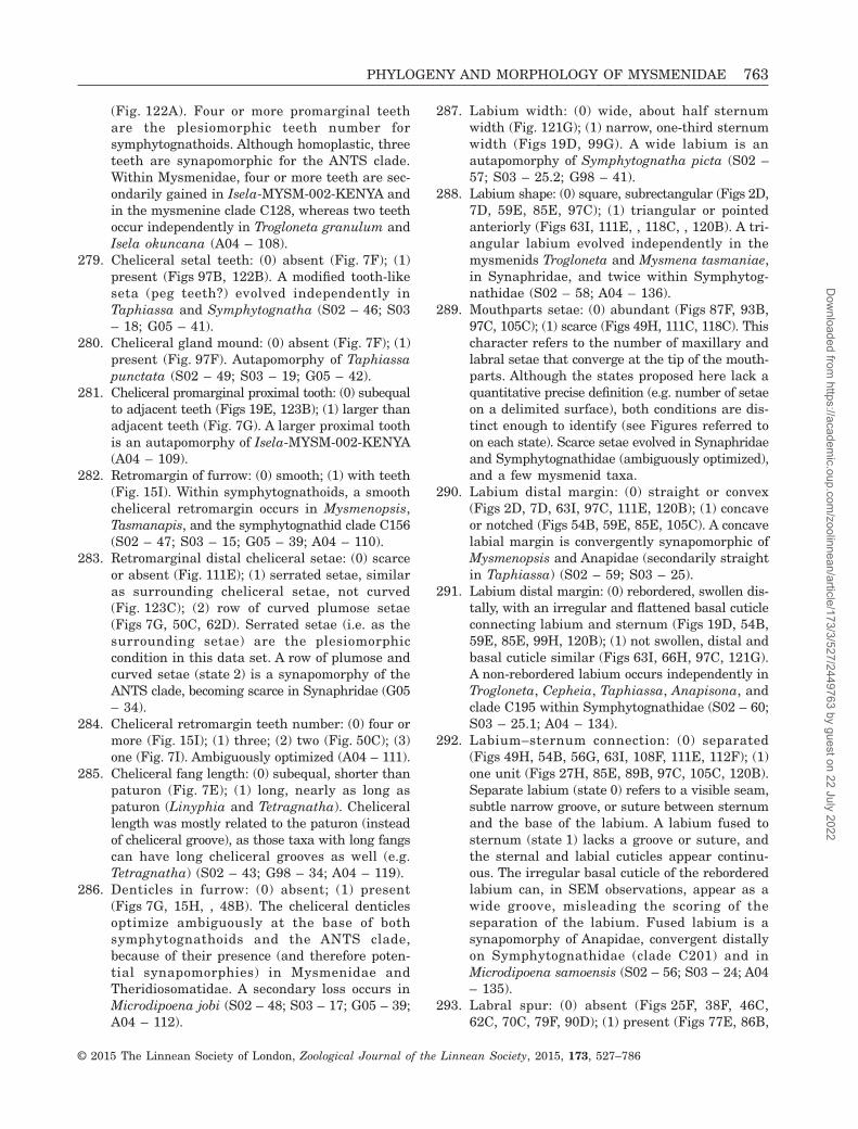
(Fig. 122A). Four or more promarginal teethare the plesiomorphic teeth number forsymphytognathoids. Although homoplastic, threeteeth are synapomorphic for the ANTS clade.Within Mysmenidae, four or more teeth are sec-ondarily gained in Isela-MYSM-002-KENYA andin the mysmenine clade C128, whereas two teethoccur independently in Trogloneta granulum andIsela okuncana (A04 – 108).
279. Cheliceral setal teeth: (0) absent (Fig. 7F); (1)present (Figs 97B, 122B). A modified tooth-likeseta (peg teeth?) evolved independently inTaphiassa and Symphytognatha (S02 – 46; S03– 18; G05 – 41).
280. Cheliceral gland mound: (0) absent (Fig. 7F); (1)present (Fig. 97F). Autapomorphy of Taphiassapunctata (S02 – 49; S03 – 19; G05 – 42).
281. Cheliceral promarginal proximal tooth: (0) subequalto adjacent teeth (Figs 19E, 123B); (1) larger thanadjacent teeth (Fig. 7G). A larger proximal toothis an autapomorphy of Isela-MYSM-002-KENYA(A04 – 109).
282. Retromargin of furrow: (0) smooth; (1) with teeth(Fig. 15I). Within symphytognathoids, a smoothcheliceral retromargin occurs in Mysmenopsis,Tasmanapis, and the symphytognathid clade C156(S02 – 47; S03 – 15; G05 – 39; A04 – 110).
283. Retromarginal distal cheliceral setae: (0) scarceor absent (Fig. 111E); (1) serrated setae, similaras surrounding cheliceral setae, not curved(Fig. 123C); (2) row of curved plumose setae(Figs 7G, 50C, 62D). Serrated setae (i.e. as thesurrounding setae) are the plesiomorphiccondition in this data set. A row of plumose andcurved setae (state 2) is a synapomorphy of theANTS clade, becoming scarce in Synaphridae (G05– 34).
284. Cheliceral retromargin teeth number: (0) four ormore (Fig. 15I); (1) three; (2) two (Fig. 50C); (3)one (Fig. 7I). Ambiguously optimized (A04 – 111).
285. Cheliceral fang length: (0) subequal, shorter thanpaturon (Fig. 7E); (1) long, nearly as long aspaturon (Linyphia and Tetragnatha). Chelicerallength was mostly related to the paturon (insteadof cheliceral groove), as those taxa with long fangscan have long cheliceral grooves as well (e.g.Tetragnatha) (S02 – 43; G98 – 34; A04 – 119).
286. Denticles in furrow: (0) absent; (1) present(Figs 7G, 15H, , 48B). The cheliceral denticlesoptimize ambiguously at the base of bothsymphytognathoids and the ANTS clade,because of their presence (and therefore poten-tial synapomorphies) in Mysmenidae andTheridiosomatidae. A secondary loss occurs inMicrodipoena jobi (S02 – 48; S03 – 17; G05 – 39;A04 – 112).
287. Labium width: (0) wide, about half sternumwidth (Fig. 121G); (1) narrow, one-third sternumwidth (Figs 19D, 99G). A wide labium is anautapomorphy of Symphytognatha picta (S02 –57; S03 – 25.2; G98 – 41).
288. Labium shape: (0) square, subrectangular (Figs 2D,7D, 59E, 85E, 97C); (1) triangular or pointedanteriorly (Figs 63I, 111E, , 118C, , 120B). A tri-angular labium evolved independently in themysmenids Trogloneta and Mysmena tasmaniae,in Synaphridae, and twice within Symphytog-nathidae (S02 – 58; A04 – 136).
289. Mouthparts setae: (0) abundant (Figs 87F, 93B,97C, 105C); (1) scarce (Figs 49H, 111C, 118C). Thischaracter refers to the number of maxillary andlabral setae that converge at the tip of the mouth-parts. Although the states proposed here lack aquantitative precise definition (e.g. number of setaeon a delimited surface), both conditions are dis-tinct enough to identify (see Figures referred toon each state). Scarce setae evolved in Synaphridaeand Symphytognathidae (ambiguously optimized),and a few mysmenid taxa.
290. Labium distal margin: (0) straight or convex(Figs 2D, 7D, 63I, 97C, 111E, 120B); (1) concaveor notched (Figs 54B, 59E, 85E, 105C). A concavelabial margin is convergently synapomorphic ofMysmenopsis and Anapidae (secondarily straightin Taphiassa) (S02 – 59; S03 – 25).
291. Labium distal margin: (0) rebordered, swollen dis-tally, with an irregular and flattened basal cuticleconnecting labium and sternum (Figs 19D, 54B,59E, 85E, 99H, 120B); (1) not swollen, distal andbasal cuticle similar (Figs 63I, 66H, 97C, 121G).A non-rebordered labium occurs independently inTrogloneta, Cepheia, Taphiassa, Anapisona, andclade C195 within Symphytognathidae (S02 – 60;S03 – 25.1; A04 – 134).
292. Labium–sternum connection: (0) separated(Figs 49H, 54B, 56G, 63I, 108F, 111E, 112F); (1)one unit (Figs 27H, 85E, 89B, 97C, 105C, 120B).Separate labium (state 0) refers to a visible seam,subtle narrow groove, or suture between sternumand the base of the labium. A labium fused tosternum (state 1) lacks a groove or suture, andthe sternal and labial cuticles appear continu-ous. The irregular basal cuticle of the reborderedlabium can, in SEM observations, appear as awide groove, misleading the scoring of theseparation of the labium. Fused labium is asynapomorphy of Anapidae, convergent distallyon Symphytognathidae (clade C201) and inMicrodipoena samoensis (S02 – 56; S03 – 24; A04– 135).
293. Labral spur: (0) absent (Figs 25F, 38F, 46C,62C, 70C, 79F, 90D); (1) present (Figs 77E, 86B,
PHYLOGENY AND MORPHOLOGY OF MYSMENIDAE 763
© 2015 The Linnean Society of London, Zoological Journal of the Linnean Society, 2015, 173, 527–786
Dow
nloaded from https://academ
ic.oup.com/zoolinnean/article/173/3/527/2449763 by guest on 22 July 2022

97E, 100F, 104G). For a detailed discussion onthe problem of the labral spur, see Miller et al.(2009). This labral sclerite was considered aspresent (state 1) when there is at least a subtleprotrusion occurring at the base of the labralflap. Micropholcommatines have a weak but pro-truding labral sclerite compared with those anapids(or other symphytognathoids) lacking spur (e.g.Acrobleps or Comaroma), and were scored hereas ‘present’ (state 1). Minanapis species have aflap with saliencies, but it is not protruding andwas therefore considered as absent (state 0) inthis data set. The labral spur is synapomorphicfor Anapidae excluding its basal genus Acrobleps(i.e. node C122), and is secondarily lost inComaroma and Minanapis (ambiguous optimi-zation) (S02 – 51; S03 – 20; G98 – 40).
294. Labral spur projection: (0) subtle but distinct el-evation (Figs 86B, 97E, 100F, 104G); (1) greatlyprojecting (Fig. 77E). A distinctly protruding labralspur is, in this taxon sample, an autapomorphyof Anapisona (S02 – 51; S03 – 20; G98 – 40).
295. Maxillary inner and distal border: (0) sclerotizedas the remaining maxilla (Figs 27H, 49H, 59E);(1) membranous (Figs 115D, 120B, 121H). A mem-branous maxillary border occurs in somesymphytognathids and is a putative synapomorphyof the family.
296. Maxillary setae: (0) mostly serrated/plumose setae,relatively absent clavate setae (Figs 87F, 105C);(1) mostly clavate setae, relatively absent plumosesetae (Figs 54B, 62B, 125B). The maxillary setaeare composed of at least two types of seta: ser-rated and clavate. This character accounts for therelative proportion of each type of seta. A higherproportion of serrated setae on the maxilla is thewidespread condition for this data set, and istherefore ancenstral for symphytognathoids.A higher proportion of clavate setae occursindependently in Mysmenopsis, Synaphridae,Comaroma, and Coddingtonia.
Palp female
297. Palpal claw: (0) present (tetragnathids, linyphiids,theridiids, and Leviola); (1) absent (Figs 22E, 69B,79C, 93A, 108C). Absent claws were also scoredfor taxa with reduced palp. Synapomorphy ofsymphytognathoids (S02 – 55; G98 – 53; A04 –176).
298. Female palp segment composition: 0) ‘cx+5’, coxaand five segments (Figs 38C, 72B, 104B, 108C);(1) ‘cx+4’, coxa and four segments (Taphiassa);(2) ‘cx + nub’, coxa and nubbin (Acroblepshygrophilus, Fig. 69B); (3) coxa (Figs 91B, 93A,100E, 118A). In most symphytognathoids, the female
palp has all six segments, lacking onlythe palpal claw; however, a complete reduction ofthe palp (where only the coxa is present, state 3)is synapomorphic of Symphytognathidae. WithinAnapidae, the reduction of the female palp occursas independent events from the five-segmentedpalp: in Taphiassa (state 1), Acrobleps (state 2),and Minanapis and Tasmanapis (state 3, con-vergent with Symphytognathidae) (S03 – 23;G98 – 52).
299. Palpal tibial trichobothria (female): (0) present(Figs 2E, 13H, 64G); (1) absent (Figs 38E,52E). Loss of female palpal trichobothria issynapomorphic for Mysmeninae and ambiguous-ly optimized at the base of Anapidae (excludingits basal genus Acrobleps), although secondarilygained in Comaroma (A04 – 179).
300. Palpal tibial trichobothria number (female): (0)numerous, more than six (tetragnathids); (1)between three and five (Figs 2E, 62A); (2) two(Figs 13H, 125E); (3) one (Figs 64G, 79E). Appli-cable only to taxa with palpal trichobothria(character 299, state 0). One female palpaltrichobothrium (state 3) optimizes as synapo-morphic for the ANTS clade, reverting to two(state 2) in Maymena mayana, and between threeand five (state 1) in Mysmenopsinae. Two palpaltrichobothria (state 2) is the ancestral conditionfor symphytognathoids, occurring in Steatoda andTheridiosomatidae (A04 – 179).
301. Palpal tarsal setae (female): (0) strongly serrat-ed (Steatoda and Leviola); (1) serrated as othersetae (Figs 22E, 79C, 105B) (A04 – 180).
302. Female palpal tarsal organ location: (0) basal, i.e.proximal (Figs 77D, H, 85G); (1) medial–distal(Figs 22E, 38G, 79C, 105B). A proximal femalepalpal tarsal organ is a synapomorphy of the anapidclade C121 (Anapisona, Conculus, and Crassanapis).
303. Female palpal tarsus macrosetae: (0) present(Figs 2A, 13A, 15A, 79C); (1) absent (Figs 77D,105A, B, 108C). Given that the taxa in this dataset usually possess reduced macrosetae insteadof strong spines (see character 125), female palpaltarsus macrosetae were scored as present (state 0)if occurring dorsally and/or ventrally. Female palpalmacrosetae are ambiguously optimized at the baseof node C160 (Maymena plus Mysmenopsinae),evolving convergently in Comaroma (A04 – 181).
Spinnerets
304. PS aciniform spigots: (0) same size and shape,uniform (Figs 6D, 61D, 75G, 94F, 113D, 123E, F);(1) two shapes (Figs 13D, 19F, 33D, 37B, E). Allsymphytognathoids have the typical araneoidspigot arrangement in both lateral and median
764 L. LOPARDO AND G. HORMIGA
© 2015 The Linnean Society of London, Zoological Journal of the Linnean Society, 2015, 173, 527–786
Dow
nloaded from https://academ
ic.oup.com/zoolinnean/article/173/3/527/2449763 by guest on 22 July 2022

posterior spinnerets (see Griswold et al., 1998);however, there is one spigot located close to otheraciniform spigots in the posterior spinnerets thatcannot be assigned to any of the described func-tional araneoid spigots. When compared with otheraciniform spigots, this spigot has a relativelythinner shaft and a marked ring around its base.Based on its location, it is tentatively assignedhere to the aciniform gland spigot group. Al-though this character scores the presence ofvarying shapes within aciniform spigots, it doesnot state which of the shapes is the plesiomorphiccondition in symphytognathoids. Moreover, thischaracter could also be defined in terms of pres-ence or absence of an additional aciniform spigotof different shape. Scoring such alternative defi-nition is, however, identical. Aciniform spigots ofdifferent shape occur within Mysmenidae,convergently in Maymena, and in Mysmeninae.
305. PMS Aciniform spigots number: (0) three(Fig. 123E); (1) two (Figs 6C, 11F, 13D, 19F, 33D,61D); (2) one (Figs 75D, 78D, 82G, 94B, F, 103C,E, 114A). Three aciniform glands are the ances-tral condition for symphytognathoids (presentin the outgroup and Theridiosomatidae). Withinsymphytognathoids, a reduction in AC spigotnumber occurs. Two AC spigots are synapomorphicfor the ANTS clade (occurring in Mysmenidae andCepheia). One AC spigot optimizes at the baseof clade C112 (Anapidae plus Symphytgnathidae),reverting to two in Crassanapis (S02 – 116; G05– 84; A04 – 222).
306. ALS major ampullate–piriform (MAP–Pi) field sepa-ration: (0) no distinct separation between MAPand piriform field (Fig. 107G); (1) MAP and piriformfield conspicuously separated by deep furrow, ap-pearing as two distinct distal segments (Figs 75A,B, 103B, 113A); (2) MAP and piriform field sepa-rated by a subtle or shallow furrow (connectionbetween both fields is distinctly evident basally;Figs 6A, 13C, 33F, 61C). Ambiguously optimized,a shallow furrow between the MAP and the piriformfields occurs in Mysmenidae and Theridiosomatidae,whereas an absence of furrow between the fieldsis a synapomorphy of Synaphridae (S03 – 54.2;G05 – 77).
307. ALS third additional anterior (spinning?) field:(0) absent (Figs 61C, 75A); (1) present (Figs 64C,66F). Synapomorphy of Trogloneta (S03 – 54.3).
308. ALS cuticle on piriform field: (0) smooth (Figs 75A,94A, 100A, 103A); (1) fingerprint (with concen-tric rings around Pi; Figs 6B, 23A, C, 33E, 61C);(2) rugose (just one ring around Pi; Figs 16B,113B, 125D). Ambiguous optimization. Withinsymphytognathoids, smooth cuticle on the piriformfield is a putative synapomorphy of the clade com-
prising Synaphridae, Anapidae, and Symphytog-nathidae (clade C113). A rugose cuticle occurs inTrogloneta, Maymena, and Theridiosomatidae.Within Mysmenidae, a fingerprint cuticle optimizesambiguously at the base of Mysmeninae and ofMysmenopsinae.
309. ALS lobe on intersegmental groove: (0) absent(Figs 75A, 94A, 113A); (1) present (Figs 23C,G, H, 33F, 52B, 103A). Synapomorphy ofMysmenidae. This lobe is convergently presentin some representatives of Anapidae, although inthis family the lobe is homoplastic and ambigu-ously optimized.
310. ALS seta on major ampullate field: (0) as othersetae surrounding the spinning field, i.e. smoothor similarly serrated (Figs 94E, 107G); (1) witha row (or two, as in Tasmanapis) of minute‘branches’ (Figs 89C, 103B, 113A); (2) with a rowof long ‘branches’ (Figs 11E, 13C, 16B); (3) withtwo rows of long ‘branches’ (Figs 6B, 23F, 33F,52B). The ancestral condition in symphy-tognathoids, occurring in the outgroup, inSynaphridae, and convergently in Taphiassa isa seta similar to the surrounding setae on theALS (state 0). Two rows of long branches are asynapomorphy of Mysmenidae. One row of longbranches in the MAP seta is synapomorphic forMaymena, convergently evolving in Theridioso-matidae. A seta with a row of minute branches(state 1) is a synapomorphy of the clade com-prising Anapidae plus Symphytognathidae (nodeC112, with a reversal in Taphiassa).
311. ALS middle fold: (0) present (Leucauge); (1) absent(Figs 33C, 78B, 97H). Leucauge venusta seems tohave a subtle fold, which appears like a relict ofa middle segment (L. Lopardo & G. Hormiga, pers.observ.). This character scores this fold, al-though its homology with a third segment remainsunknown (S02 – 108; G05 – 75).
312. PMS minor ampullate (mAP): (0) absent (Fig. 103C,E); (1) present (Figs 6C, 19F, 125C). A loss of themAP spigot occurred independently in Cepheiaand Teutoniella (S02 – 113; G05 – 81).
313. PMS mAP tartipore and nubbin: (0) mAP only(Figs 6C, F, 58F, 61D, 78E, 82G, 94B, F); (1) mAPplus tartipore (Figs 19F, 23D, 33D, 123E); (2) mAPplus tartipore and nubbin (Figs 11F, 75D, 88G,113C, 122E, 125C). The presence of a tartipore,or a nubbin plus tartipore accompanying the minorampullate spigot optimizes ambiguously at thebase of symphytognathoids, therefore, a patternof loss of the nubbin, or the nubbin plus tartipore,is unclear. Within symphytognathids, the pres-ence of a nubbin and a tartipore occurs insymphytognathids, most anapids, Coddingtonia,and Maymena. The loss of the mAP nubbin
PHYLOGENY AND MORPHOLOGY OF MYSMENIDAE 765
© 2015 The Linnean Society of London, Zoological Journal of the Linnean Society, 2015, 173, 527–786
Dow
nloaded from https://academ
ic.oup.com/zoolinnean/article/173/3/527/2449763 by guest on 22 July 2022

(although not the tartipore) optimizes ambigu-ously as a potential synapomorphy for Troglonetaand for Mysmeninae. The complete loss of boththe nubbin and the tartipore is ambiguouslyoptimized in Mysmenopsinae, independentlyoccurring in the anapid subfamily Micro-pholcommatinae (clade C150). (S02 – 115; S03 –55; G98 – 71; A04 – 220).
314. PMS mAP shaft: (0) proximal diameter of shaftnarrower than distal spigot base, shaft usuallycylindrical and longer than wide (although it canbe short; Figs 6C, F, 33D, 58F, 75C, 78D, 100B);(1) width of proximal shaft as wide as apical spigotbase, shaft as long as wide (Figs 49G, 113C, 125C).This character optimizes ambiguously at thesymphytognathoid node. A wide mAP base andshaft occur on Theridiosomatidae, independentlyin the symphytognathid clade C195, and themysmenids Mysmena leichhardti, MYSM-020-MAD, and MYSM-023-MAD (S03 – 56).
315. PMS mAP spigot shaft length: (0) short, subequalto CY shaft (Figs 6C, 61D, 75D, 94F, 114A, 123E);(1) clearly longer than any CY shaft (Linyphiaand Steatoda) (A04 – 223).
316. Major ampullate tartipores: (0) absent (Fig. 78C);(1) present (Fig. 23F). Absence of tartipores onMAP field (state 0) is an autapomorphy ofComaroma (G05 – 70).
317. Anterior median spinnerets (AMS): (0) colulus(Figs 33C, 49F, 68C, 88F); (1) absent (Figs 117F,120C). The absence of colulus is synapomorphicfor Symphytognathidae. The genus Patu has beenreported to have an extremely minute colulus (e.g.in Patu digua; see Griswold et al., 1998, fig. 37A),suggesting that the absence of colulus mightbe synapomorphic for at least a clade withinSymphytognathidae. The cuticle in the colular areaof the Patu representative in this data set (Patu-SYMP-001-DR) has a slit and bears two setae,although there is no evidence of protruding colulus(image not shown), and it is here considered asabsent (S02 – 106; S03 – 54; G98 – 66; G05 – 71;A04 – 172).
318. Colulus size: (0) minute, about one-eighth widthof ALS (Figs 64D, 67F, 68C); (1) large, fleshy, widthabout one-quarter width of ALS (Figs 33C, 49F,88F). Minute colulus is a synapomorphy ofTrogloneta (S02 – 107; S03 – 54; G98 – 65; A04– 172, 173).
319. Colular setae number: (0) four or more (Fig. 56D);(1) three or less (Figs 67F, 78B). Three orless setae on colulus are synapomorphic ofsymphytognathoids. Within Mysmenidae, fouror more colular setae are a synapomorphy ofMysmenopsis (with a reversal in M. palpalis) (G98– 65; A04 – 175).
320. PLS Cylindrical (CY) spigot number: (0) one(Fig. 94F); (1) two (Figs 13F, 45F, 58H, 65A, 78H,103F, 113D, 123F). Although usually one or twoCY spigots occur on the PLS, in a few instancesthree CY can occur, as anomalies on one of thePLS (e.g. compare both PLS of Comaroma;Fig. 78G and H). These anomalies are not takeninto account when scoring. One cylindrical glandspigot on the posterior lateral spinnerets evolvedindependently in Cepheia and Taphiassa (S02 –119; G98 – 74; A04 – 210).
321. PLS anterior CY position: (0) below FL, on spin-ning field (Fig. 94F; also Linyphia); (1) periph-eral, or above FL if on spinning field (Figs 58H,78H, 113D, 123F). This character refers to theanterior CY spigot, i.e. the CY spigot closer tothe ALS (as both spigots are usually mesal). Whentwo CY spigots are present, the posterior one isalways peripheral. A cylindrical spigot located onthe spinning field (i.e. state 0, not peripheral)occurs independently in Taphiassa and Linyphia(G98 – 76).
322. PLS CY relative size: (0) both CY equally largeand larger than FL (Figs 11G, H, 13F, 67D, 94F,123F); (1) posterior CY huge, larger than ante-rior CY (both larger than FL; Figs 75E, 78H, 89D,100C, 103F, 113D, 114B); (2) all slim as otherspigots, not larger, but subequal to FL (Figs 23B,37B, E). Slim cylindrical spigots (state 2) are con-vergent synapomorphies of Mysmenopsinae andMysmeninae. A huge posterior cylindrical spigot(state 1) is a synapomorphy of Anapidae plusSymphytognathidae (node C112, reverting tostate 0 in Taphiassa).
323. Sclerotized ring around spinnerets: (0) absent(Figs 6E, 59I, 107F, 117F, 140F); (1) present(Figs 78B, 97H, 145F, 146B). Partial sclerotizedring (heavily sclerotized cuticle not completely sur-rounding spinnerets) are considered present (e.g.in Crassanapis), whereas extremely weaklysclerotized rings were considered absent (e.g. inElanapis). Synapomorphy of the anapid cladeC122, with a reversal in Elanapis (S02 – 96; A04– 202).
324. PLS distal segment: (0) short conical, as distalALS segment (Figs 33C, 97H, 145F, 146B); (1) longcylindrical, distal PLS segment two times longerthan distal ALS segment, usually visible on dorsalview (Figs 107F, 146E, F). Synapomorphy ofSynaphridae (S03 – 54.1).
325. ALS intersegmental cuticle: (0) smooth (Fig. 94A,E); (1) glabrous tuberculate (Figs 6A, 23C, 66F,75A); (2) fingerprint (Fig. 113A). A glabroustuberculate cuticle (state 1) is the ancestral andwidespread cuticular pattern in symphytogna-thoids, occurring in the outgroup taxa as well.
766 L. LOPARDO AND G. HORMIGA
© 2015 The Linnean Society of London, Zoological Journal of the Linnean Society, 2015, 173, 527–786
Dow
nloaded from https://academ
ic.oup.com/zoolinnean/article/173/3/527/2449763 by guest on 22 July 2022

A fingerprint cuticle (state 2) optimizes as asynapomorphy of Symphytognathidae. Smoothcuticle (state 0) is synapomorphic for Synaphridae,convergent in Taphiassa.
326. PS cuticle on spigot bases: (0) smooth (Figs 78F,82H, 94C, D); (1) imbricate (Figs 11G, 23B, E,89D). Scored for both PMS and PLS. Smoothcuticle is synapomorphic of the anapid subfam-ily Micropholcommatinae (clade C150) (G05 – 69).
327. ALS Pi spigot bases: (0) large (tetragnathids); (1)‘reduced’ (Figs 33F, 75A) (S02 – 110; G98 – 69;A04 – 204).
328. PLS posterior setae: (0) slender setae (Figs 23B,123F); (1) posterior row of clavate setae (Fig. 122F).The setae on the posterior distal border of thePLS of the symphytognathid SYMP-006-AUSTappear subtly thicker than the surrounding setae(see Fig. 117G), but are not considered similar tothe clavate setae of Symphytognatha picta, andwere therefore scored as slender (state 0).Autapomorphy of Symphytognatha picta (S03 –60.1).
329. PLS anterior flat spatulate modified seta: (0)absent or serrated seta (Figs 6B, 65A, 87C, 94D,123F); (1) present (Figs 11H, 23B, E, 33G, H, 52C).The anterior distal border of the PLS of Anapisonakethleyi has a distinct seta, although it is notspatulate and therefore scored as serrated (state 0;see Fig. 75G). This particular seta evolvedconvergently within Mysmenidae, in Maymena,and in Mysmeninae (secondarily lost in Mysmenarotunda).
330. FL spigot in female: (0) absent (Fig. 6D); (1)present (Figs 13F, 23B, 58H, 67D, 75G, 78H, 94F,103F, 113D, 123F). The female flagelliform spigotis lost in the mysmenid genus Isela (S02 – 125;G98 – 77; A04 – 212).
331. FL in male: (0) absent (Figs 6G, 23E); (1) present(Figs 53H, 58G, 75F, 82H, 94D, 103D, 117G). Anapparently functional male flagelliform spigotoccurs in Mysmenopsis, Maymena mayana,Trogloneta granulum, the clade C112 (compris-ing symphytognathids and anapids), and inde-pendently in some mysmenines. As a result of thehomoplasy of this character, it optimizes ambigu-ously for symphytognathoids (S02 – 126; S03 –58; A04 – 219).
332. FL spigot shaft: (0) straight (Figs 6D, 67D, 75G);(1) shaft curved (Figs 78F, H, 82H). CurvedFL shaft is an autapomorphy of Comaroma (S03– 59).
333. AG spigots in female: (0) absent (Figs 6D, 58H,94F); (1) present (Figs 75G, 123F). The female ag-gregate spigots are lost in the kleptoparasiticmysmenids Mysmenopsinae, and independentlyin Taphiassa (S02 – 127; G98 – 78; G05 – 95).
334. AG spigot number in female: (0) two (Figs 13E,F, 23B, 75G, 87C, 113D, 123F); (1) one (Cepheia).One female aggregate spigot is an autapomorphyof Cepheia (A04 – 213).
335. AG spigots in male: (0) absent (Figs 6G, 23E,33H, 58G, 94D); (1) present (Fig. 103D). Func-tional male aggregate spigots occur independent-ly in Maymena mayana, Trogloneta granulum,a few mysmenines, and clade C113 (synaphrids,symphytognathids, and anapids) (S02 – 129; S03– 58; A04 – 219).
336. AG spigot number in male: (0) two (Figs 13G, 75F,82H, 100D, 103D); (1) one (synaphrids).
337. AG spigots: (0) broader than FL (Fig. 123F); (1)as FL (Figs 13F, G, 75F, 78H, 100D); (2) extreme-ly large, huge (Steatoda). The aggregate spigotsof Theridiosoma gemmosum are broader than theflagelliform spigot (state 0, ancestral state forsymphytognathoids). Slim aggregate spigots(state 1) are a synapomorphy of the ANTS clade(S03 – 60; G98 – 78).
338. Anterior AG spigot shape: (0) round (Fig. 23B);(1) flattened (Steatoda). In order to score the shapeof the single AG spigot of synaphrids, it was ran-domly assigned as posterior, and therefore scoredunder character 339. Autapomorphy of Steatoda(A04 – 215).
339. Posterior AG spigot shape: (0) round (Fig. 23B);(1) flattened (Steatoda). In order to score the shapeof the single AG spigot of synaphrids, it was ran-domly considered as posterior, and therefore scoredin this character. Autapomorphy of Steatoda (A04– 216).
340. PLS aggregate gland form: (0) entire (Linyphiaand Tetragnatha); (1) lobed (Steatoda). The scarceknowledge on the form of the aggregate gland wastaken from A04 – 217, and was available for onlythree (outgroup) taxa.
Behavioural characters
341. Web posture: (0) legs I and II extended(tetragnathids); (1) legs I and II flexed (Fig. 147B)(G98 – 81).
342. Predatory strategy: (0) catching web; (1) freehunters (Leviola); (2) kleptoparasites (Isela andmost Mysmenopsis species). Under this taxonsample, kleptoparasitism is a synapomorphy ofMysmenopsinae (S02 – 153; A04 – 224).
343. Wrap-bite attack: (0) present (Steatoda andLeucauge); (1) absent, bite first (G98 – 92; G05– 154; A04 – 228).
344. Radii lengthened: (0) absent; (1) present. Onlyscored in five taxa. Lengthened radii are anambiguously optimized synapomorphy ofsymphytognathoids, although it was only
PHYLOGENY AND MORPHOLOGY OF MYSMENIDAE 767
© 2015 The Linnean Society of London, Zoological Journal of the Linnean Society, 2015, 173, 527–786
Dow
nloaded from https://academ
ic.oup.com/zoolinnean/article/173/3/527/2449763 by guest on 22 July 2022

observed in theridiosomatids and mysmenids (G98– 84).
345. Web architecture: (0) orb (Figs 147A, 148A, B,149A); (1) sheet (Linyphia, Trogloneta granulum,and Synaphris saphrynis); (2) cobweb (Steatodaand Comaroma). The web of Comaroma has beenreported by Kropf (1990) as an irregular three-dimensional web with long threads containingsticky silk at their distal end (i.e. at place of at-tachment to substrate), which is consistent withthe description of the typical theridiid web (e.g.Griswold et al., 1998, and references therein;Agnarsson, 2004) and was scored as such. As dis-cussed by Griswold et al. (1998) and Lopardo &Hormiga (2008), the homologies involving webarchitectures and behaviours for the so-called‘sheet web’ remain unsolved. Sheet webs encom-pass an enormous diversity of web architec-tures. Only three taxa in this data set areconsidered to be sheet-web weavers (see above).Even though homology statements in the behav-ioural and architectural aspects are compro-mised, the web of Trogloneta granulum wasnevertheless considered and scored here as a sheetweb (see detailed discussion on Lopardo et al.,2011). The orb web is the plesiomorphic web inthis data set; all other webs evolve independent-ly on each taxon (S02 – 154; S03 – 80; G98 –80; G05 – 142, 143; A04 – 225).
346. Orb web frame: Out of the plane radii (APR) onfinal web: (0) absent, two-dimensional (Figs 148A,149C, D); (1) present, three-dimensional(Figs 147A, D, 148C, D). Retention of radii ex-tending out of the orb web plane is a putativesynapomorphy of symphytognathoids, revertingto bidimensional final webs independently inSymphytognathidae, and the anapid distal cladeC153 (G98 – 82).
347. APR on final web: (0) above the plane (Figs 147D,E, 148B–D); (1) above and below the plane(Fig. 147A, B). APR oriented above and below theorb plane is an ambiguously optimizedsynapomorphy of Mysmeninae (clade C133).
348. Post-SS hub loops: (0) absent; (1) present. Onlyscored in six taxa. Post sticky silk hub loops arean ambiguously optimized synapomorphy ofsymphytognathoids, although it was only ob-served in theridiosomatids and mysmenids (G98– 88).
349. Hub: (0) closed; (1) open. Only scored in six taxa.Closed hub is an ambiguously optimizedsynapomorphy of symphytognathoids, although(as in character 348) it was only observed intheridiosomatids and mysmenids (G98 – 90).
APPENDIX 3
Abbreviations used in text and figures.A04 Agnarsson (2004)AC aciniform (gland spigot/s)AG aggregate (gland spigot)AGs aggregate (gland spigots)ALS anterior lateral spinneretsAME anterior median eyesANTS anterior tracheal systemAtr atriumBS Bremer supportC tegular (bulbal) conductorCD copulatory ductsCI consistency indexCO copulatory openingsCY cylindrical (gland spigot/s)Cy cymbiumCyC1 primary (internal) cymbial conductorCyC2 secondary (external) cymbial conductorCyF cymbial foldCyFs setae on cymbial foldCyG cymbial grooveCyP cymbial processCyPF cymbial fold processE embolusEap embolar (distal) apophysisEB embolar baseEbap embolar basal apophysisF fundusFD fertilization ductsfe femurFL flagelliform (gland spigot)G05 Griswold et al. (2005)G98 Griswold et al. (1998)GC frequency differencesgl accessory glandI spermatic duct switchback I (drawing)II spermatic duct switchback II (drawing)III spermatic duct switchback III (drawing)IV spermatic duct switchback IV (drawing)MA median apophysisMAP major ampullate (gland spigot)mAP minor ampullate (gland spigot)MPT most-parsimonious tree/smt metatarsusn nubbinpa patellaPC paracymbiumPi piriform (gland spigot/s)PLS posterior lateral spinneretsPME posterior median eyesPMS posterior median spinneretspp pars pendulaRAS random-addition sequencesRFD relative Bremer support, or relative fit differenceRI retention indexS spermatheca/eS02 Schütt (2002)S03 Schütt (2003)SB spermatic duct switchbackSD spermatic ductSDT spermatic duct trajectorySEM scanning electron microscopySFq symmetric resampling frequenciesSp spiraclet tartiporeT tegulumta tarsusTBR tree bisection and reconnection branch swappingTC tegular conductorti tibiato tarsal organ
768 L. LOPARDO AND G. HORMIGA
© 2015 The Linnean Society of London, Zoological Journal of the Linnean Society, 2015, 173, 527–786
Dow
nloaded from https://academ
ic.oup.com/zoolinnean/article/173/3/527/2449763 by guest on 22 July 2022

APPENDIX 4
Table A1. List of synapomorphic changes common to the three most parsimonious cladograms from the analysis of themorphological data
Taxon name/node number All trees Some trees
Acrobleps hygrophilus Character 22: 1 → 0 Character 24: 0 → 1 Character 38: 0 → 1 Character 202: 0 → 1Character 90: 0 → 2 Character 95: 1 → 0 Character 152: 0 → 1Character 180: 0 → 1 Character 195: 0 → 1 Character 228: 0 → 1Character 233: 0 → 1 Character 250: 0 → 1 Character 251: 0 → 1Character 252: 0 → 2 Character 278: 0 → 2 Character 298: 0 → 2Character 346: 0 → 1
Anapisona kethleyi Character 21: 1 → 2 Character 28: 0 → 1 Character 68: 1 → 0 Character 1: 1 → 0 Character 79: 0 → 1Character 88: 1 → 0 Character 122: 0 → 1 Character 145: 0 → 1Character 152: 0 → 1 Character 164: 0 → 5 Character 175: 0 → 1Character 182: 0 → 1 Character 195: 0 → 1 Character 203: 0 → 1Character 207: 0 → 1 Character 210: 0 → 1 Character 211: 0 → 1Character 220: 1 → 0 Character 235: 0 → 1 Character 246: 0 → 1Character 248: 0 → 1 Character 272: 0 → 1 Character 291: 0 → 1Character 294: 0 → 1
Anjouanella comorensis Character 11: 1 → 2 Character 75: 1 → 0 Character 197: 2 → 0Character 199: 1 → 0 Character 253: 0 → 2
Brasilionata arborense Character 7: 1 → 0Calodipoena incredula Character 38: 0 → 1 Character 104: 0 → 1 Character 118: 0 → 1
Character 162: 0 → 2 Character 222: 1 → 0Calodipoena mooatae Character 48: 1 → 0Calomyspoena santacruzi Character 123: 0 → 1 Character 177: 0 → 2 Character 186: 1 → 2Cepheia longiseta Character 134: 0 → 1 Character 149: 0 → 1 Character 158: 0 → 1
Character 162: 0 → 2 Character 164: 0 → 1 Character 203: 0 → 1Character 207: 0 → 1 Character 212: 0 → 1 Character 262: 1 → 0Character 291: 0 → 1
Coddingtonia euryopoides Character 6: 0 → 2 Character 21: 0 → 1 Character 52: 0 → 1Character 75: 0 → 1 Character 83: 0 → 4 Character 136: 0 → 1Character 163: 0 → 2 Character 164: 0 → 1 Character 271: 1 → 0
Comaroma simony Character 5: 1 → 2 Character 23: 1 → 0 Character 41: 0 → 2 Character 46: 4 → 0 Character 215: 0 → 1Character 56: 1 → 0 Character 71: 1 → 0 Character 95: 1 → 0Character 97: 2 → 0 Character 110: 0 → 1 Character 124: 0 → 1Character 127: 0 → 2 Character 136: 0 → 1 Character 141: 0 → 1Character 161: 1 → 0 Character 197: 0 → 1 Character 203: 0 → 1Character 207: 0 → 1 Character 213: 1 → 0 Character 214: 0 → 1Character 237: 0 → 1 Character 244: 1 → 0 Character 257: 1 → 0Character 284: 23 → 1 Character 293: 1 → 0 Character 296: 0 → 1Character 299: 1 → 0 Character 303: 1 → 0 Character 316: 1 → 0Character 332: 0 → 1
Crassanapis chilensis Character 7: 0 → 1 Character 46: 4 → 5 Character 80: 1 → 0 Character 38: 0 → 1 Character 78: 1 → 2Character 97: 2 → 0 Character 120: 1 → 0 Character 126: 0 → 1 Character 194: 1 → 0 Character 226: 1 → 2Character 128: 0 → 1 Character 305: 2 → 1
Elanapis aisen Character 7: 0 → 1 Character 11: 0 → 2 Character 81: 0 → 1 Character 86: 0 → 1Character 83: 0 → 5 Character 150: 0 → 1 Character 261: 0 → 1Character 276: 0 → 1
Iardinis mussardi Character 12: 1 → 0 Character 155: 0 → 1 Character 195: 0 → 1Character 203: 0 → 1 Character 219: 0 → 2
Isela okuncana Character 19: 0 → 1 Character 67: 1 → 0 Character 226: 0 → 1Itapua tembei Character 83: 2 → 0Kekenboschiella awari Character 224: 0 → 1 Character 284: 1 → 2Kekenboschiella marijkeae Character 195: 1 → 0 Character 197: 0 → 1Leucauge venusta Character 64: 0 → 1 Character 81: 0 → 1 Character 82: 0 → 2
Character 196: 1 → 0 Character 225: 2 → 0 Character 278: 0 → 1Character 311: 1 → 0 Character 343: 1 → 0
Linyphia triangularis Character 52: 2 → 5 Character 57: 1 → 0 Character 60: 0 → 1Character 89: 1 → 0 Character 152: 0 → 1 Character 163: 0 → 2Character 240: 0 → 2 Character 267: 0 → 1 Character 268: 0 → 1Character 321: 1 → 0 Character 322: 0 → 1 Character 345: 0 → 1
Maymena ambita Character 4: 1 → 0 Character 7: 1 → 0 Character 105: 0 → 1Character 222: 1 → 0
PHYLOGENY AND MORPHOLOGY OF MYSMENIDAE 769
© 2015 The Linnean Society of London, Zoological Journal of the Linnean Society, 2015, 173, 527–786
Dow
nloaded from https://academ
ic.oup.com/zoolinnean/article/173/3/527/2449763 by guest on 22 July 2022

Table A1. Continued
Taxon name/node number All trees Some trees
Maymena mayana Character 8: 0 → 1 Character 34: 2 → 1 Character 45: 0 → 1Character 50: 1 → 0 Character 52: 0 → 1 Character 83: 0 → 5Character 129: 2 → 1 Character 162: 0 → 1 Character 168: 0 → 1Character 191: 0 → 1 Character 221: 0 → 1
Maymena rica Character 6: 0 → 1 Character 12: 1 → 0 Character 17: 1 → 0Character 21: 0 → 12 Character 23: 1 → 2 Character 59: 1 → 0Character 70: 1 → 0 Character 83: 0 → 1 Character 131: 1 → 0Character 145: 0 → 1 Character 201: 0 → 1 Character 237: 1 → 0
Microdipoena elsae Character 35: 1 → 0 Character 75: 1 → 0 Character 87: 1 → 0Microdipoena guttata No autapomorphies:Microdipoena nyungwe Character 48: 1 → 0Minanapis Casablanca No autapomorphies:Minanapis palena No autapomorphies:Kilifina-MYSM-002-KENYA Character 7: 1 → 0 Character 52: 0 → 1 Character 77: 0 → 1
Character 190: 0 → 1 Character 225: 0 → 1 Character 276: 0 → 1Character 281: 0 → 1 Character 308: 1 → 2
MYSM-005-ARG Character 7: 1 → 0 Character 15: 1 → 0 Character 22: 0 → 1Character 74: 1 → 0 Character 85: 0 → 1 Character 101: 1 → 0Character 152: 0 → 1 Character 164: 3 → 0 Character 178: 0 → 1Character 195: 1 → 0 Character 196: 2 → 1 Character 202: 0 → 1Character 225: 2 → 0 Character 335: 0 → 1
MYSM-007-MEX Character 6: 0 → 2 Character 103: 1 → 0 Character 134: 0 → 1Character 171: 0 → 1 Character 178: 0 → 1 Character 182: 0 → 1Character 189: 1 → 0 Character 226: 0 → 1
Mysmena-MYSM-015-MAD Character 52: 0 → 2 Character 97: 1 → 2Mysmena-MYSM-018-MAD Character 38: 0 → 1 Character 149: 0 → 1 Character 170: 1 → 0MYSM-019-MAD Character 47: 0 → 1 Character 48: 1 → 0 Character 265: 0 → 1
Character 289: 0 → 1MYSM-020-MAD Character 48: 1 → 0 Character 164: 3 → 0MYSM-023-MAD Character 23: 1 → 2 Character 97: 1 → 2 Character 163: 1 → 2
Character 184: 1 → 0 Character 265: 0 → 1 Character 289: 0 → 1MYSM-028-MAD Character 53: 2 → 1 Character 97: 1 → 2MYSM-029-MAD Character 35: 1 → 0 Character 60: 1 → 0 Character 70: 0 → 1MYSM-034-MAD Character 11: 0 → 2 Character 74: 1 → 0 Character 86: 0 → 1Mysmena leichhardti Character 38: 0 → 1 Character 162: 0 → 2 Character 186: 0 → 2
Character 191: 1 → 0 Character 224: 0 → 1 Character 314: 0 → 1Mysmena leucoplagiata Character 116: 1 → 2 Character 117: 1 → 0 Character 162: 0 → 2Mysmena tasmaniae Character 23: 1 → 2 Character 74: 1 → 0 Character 83: 1 → 2
Character 105: 0 → 1 Character 116: 1 → 2 Character 170: 1 → 0Character 184: 1 → 0 Character 288: 0 → 1
Mysmenella illectrix Character 187: 1 → 0Mysmenella jobi Character 255: 1 → 0 Character 286: 1 → 0Mysmenella samoensis Character 48: 1 → 0 Character 292: 0 → 1Mysmeniola spinifera Character 6: 0 → 2 Character 48: 1 → 0 Character 88: 1 → 0
Character 112: 0 → 1 Character 163: 1 → 2 Character 250: 0 → 1Mysmenopsis cidrelicola Character 35: 1 → 2 Character 112: 0 → 1 Character 116: 1 → 2Mysmenopsis dipluramigo Character 125: 1 → 0 Character 143: 0 → 1 Character 259: 1 → 0
Character 265: 0 → 1Mysmenopsis palpalis Character 35: 1 → 0 Character 66: 0 → 1 Character 67: 1 → 2
Character 116: 1 → 2 Character 135: 0 → 1Mysmenopsis penai Character 83: 0 → 4 Character 99: 0 → 1 Character 126: 1 → 0
Character 271: 2 → 1Steatoda americana Character 12: 1 → 0 Character 13: 0 → 1 Character 16: 0 → 1
Character 20: 1 → 0 Character 40: 0 → 1 Character 41: 0 → 1Character 47: 1 → 3 Character 52: 2 → 3 Character 98: 0 → 1Character 137: 0 → 1 Character 151: 0 → 1 Character 161: 1 → 0Character 203: 0 → 1 Character 208: 0 → 1 Character 212: 0 → 1Character 228: 0 → 1 Character 229: 0 → 1 Character 230: 0 → 1Character 237: 1 → 0 Character 255: 0 → 1 Character 271: 1 → 0Character 278: 0 → 3 Character 282: 1 → 0 Character 291: 0 → 1Character 301: 1 → 0 Character 338: 0 → 1 Character 339: 0 → 1Character 343: 1 → 0 Character 345: 0 → 2
770 L. LOPARDO AND G. HORMIGA
© 2015 The Linnean Society of London, Zoological Journal of the Linnean Society, 2015, 173, 527–786
Dow
nloaded from https://academ
ic.oup.com/zoolinnean/article/173/3/527/2449763 by guest on 22 July 2022

Table A1. Continued
Taxon name/node number All trees Some trees
Patu-SYMP-001-DR Character 38: 0 → 1 Character 74: 1 → 0 Character 78: 1 → 0Character 86: 0 → 1 Character 152: 0 → 1 Character 164: 0 → 4Character 165: 0 → 1 Character 170: 0 → 1 Character 180: 0 → 1Character 185: 0 → 1 Character 195: 0 → 1 Character 201: 0 → 1Character 218: 0 → 1 Character 288: 0 → 1
SYMP-002-MAD Character 74: 1 → 0 Character 153: 0 → 2 Character 261: 0 → 1SYMP-006-AUST Character 11: 2 → 0 Character 78: 1 → 2 Character 83: 0 → 4
Character 265: 1 → 2 Character 295: 1 → 0SYMP-007-AUST Character 13: 1 → 0 Character 64: 1 → 0 Character 67: 0 → 2Symphytognatha picta Character 44: 0 → 1 Character 49: 0 → 1 Character 63: 0 → 1
Character 129: 2 → 0 Character 137: 0 → 2 Character 157: 0 → 1Character 194: 0 → 1 Character 273: 1 → 0 Character 279: 0 → 1Character 287: 1 → 0 Character 328: 0 → 1
Synaphris saphrynis Character 52: 2 → 1 Character 163: 0 → 3 Character 170: 0 → 1Character 225: 2 → 1 Character 239: 0 → 3
Tamasesia acuminata Character 6: 0 → 2 Character 11: 1 → 0Tamasesia rotunda Character 7: 1 → 0 Character 83: 2 → 5 Character 221: 0 → 2
Character 223: 1 → 0Tasmanapis strahan Character 11: 0 → 2 Character 23: 1 → 0 Character 35: 0 → 5
Character 46: 4 → 6 Character 68: 1 → 0 Character 85: 0 → 1Character 128: 0 → 1 Character 136: 0 → 1 Character 154: 0 → 1Character 220: 1 → 0 Character 225: 0 → 1 Character 244: 1 → 0Character 246: 0 → 1
Taphiassa punctata Character 21: 1 → 2 Character 45: 0 → 3 Character 59: 0 → 1 Character 46: 4 → 3 Character 166: 0 → 1Character 72: 0 → 1 Character 83: 0 → 2 Character 152: 0 → 1 Character 226: 1 → 0 Character 231: 0 → 1Character 162: 0 → 1 Character 220: 1 → 0 Character 235: 0 → 1 Character 309: 1 → 0Character 270: 1 → 0 Character 271: 1 → 0 Character 275: 0 → 1Character 279: 0 → 1 Character 280: 0 → 1 Character 290: 1 → 0Character 291: 0 → 1 Character 298: 0 → 1 Character 310: 1 → 0Character 320: 1 → 0 Character 321: 1 → 0 Character 322: 1 → 0Character 325: 1 → 0 Character 333: 1 → 0 Character 335: 1 → 0
Tetragnatha versicolor No autapomorphies:Teutoniella cekalovici Character 7: 0 → 1 Character 17: 0 → 1 Character 41: 0 → 1 Character 54: 2 → 0
Character 88: 1 → 0 Character 112: 1 → 0 Character 149: 1 → 2Character 194: 1 → 0 Character 196: 0 → 1 Character 233: 0 → 1Character 251: 0 → 1 Character 312: 1 → 0
Theridiosoma gemmosum Character 10: 1 → 0 Character 82: 0 → 1 Character 99: 1 → 0Character 196: 2 → 0 Character 197:
0 → 13Character 204: 0 → 1
Character 313: 2 → 1Trogloneta cantareira Character 6: 0 → 2 Character 45: 0 → 2 Character 46: 0 → 12
Character 226: 0 → 1Trogloneta granulum Character 149: 0 → 2 Character 196: 2 → 0 Character 197: 0 → 1
Character 225: 0 → 1 Character 278: 1 → 2Node M66 Character 44: 0 → 1 Character 62: 0 → 1 Character 225: 2 → 0 Character 231: 0 → 1 Character 309: 0 → 1
Character 237: 1 → 0 Character 290: 0 → 1 Character 292: 0 → 1Node M67 Character 56: 0 → 1 Character 160: 1 → 0 Character 244: 2 → 1
Character 259: 0 → 1 Character 310: 0 → 1 Character 322: 0 → 1Node M68 Character 7: 1 → 0 Character 21: 0 → 2 Character 29: 0 → 1
Character 59: 1 → 0 Character 64: 0 → 1 Character 153: 0 → 1Character 257: 0 → 1
Node M69 Character 43: 0 → 1 Character 50: 0 → 1 Character 55: 0 → 1Character 196: 1 → 2 Character 297: 0 → 1 Character 319: 0 → 1
Node M70 Character 28: 1 → 0 Character 58: 0 → 1 Character 87: 1 → 0Character 99: 0 → 1 Character 125: 0 → 1 Character 162: 1 → 0Character 303: 0 → 1
Node M71 Character 4: 1 → 0 Character 10: 0 → 1 Character 68: 0 → 1Character 74: 0 → 1 Character 92: 0 → 1 Character 100: 1 → 0Character 132: 1 → 0 Character 194: 1 → 0 Character 245: 1 → 0Character 308: 2 → 0 Character 327: 0 → 1 Character 341: 0 → 1
Node M72 No synapomorphiesNode M73 Character 35: 0 → 4 Character 98: 0 → 1 Character 121: 0 → 1 Character 114: 0 → 1
Character 346: 0 → 1Character 302: 1 → 0
Node M74 Character 0: 0 → 2 Character 3: 0 → 1 Character 278: 0 → 1 Character 1: 0 → 1 Character 46: 0 → 4Character 323: 0 → 1 Character 52: 2 → 3
Character 153: 1 → 2Character 54: 0 → 12
PHYLOGENY AND MORPHOLOGY OF MYSMENIDAE 771
© 2015 The Linnean Society of London, Zoological Journal of the Linnean Society, 2015, 173, 527–786
Dow
nloaded from https://academ
ic.oup.com/zoolinnean/article/173/3/527/2449763 by guest on 22 July 2022

Table A1. Continued
Taxon name/node number All trees Some trees
Node M75 Character 2: 0 → 1 Character 20: 1 → 0 Character 21: 2 → 1Character 293: 0 → 1
Node M76 Character 14: 0 → 1 Character 131: 1 → 0 Character 313: 2 → 0 Character 52: 3 → 4 Character 196: 2 → 0Character 326: 1 → 0
Node M77 Character 1: 1 → 2 Character 41: 0 → 3 Character 80: 1 → 0 Character 44: 1 → 0 Character 256: 0 → 2Character 110: 0 → 1 Character 150: 0 → 1 Character 160: 0 → 1Character 175: 0 → 1 Character 236: 0 → 1 Character 248: 0 → 1Character 261: 0 → 1 Character 276: 0 → 1 Character 278: 1 → 2Character 293: 1 → 0
Node M78 Character 72: 0 → 1 Character 233: 0 → 1 Character 298: 0 → 3 Character 38: 0 → 1 Character 86: 0 → 1Node M79 Character 0: 2 → 1 Character 35: 0 → 3 Character 62: 1 → 0
Character 68: 1 → 0 Character 228: 0 → 1 Character 246: 0 → 1Node M80 Character 61: 1 → 0 Character 185: 1 → 0Node M81 Character 104: 0 → 1 Character 225: 1 → 0 Character 278: 1 → 0Node M82 Character 142: 0 → 1 Character 194: 0 → 1 Character 226: 0 → 1
Character 238: 2 → 1Node M83 Character 160: 1 → 0 Character 177: 0 → 2 Character 218: 0 → 1
Character 223: 1 → 0Node M84 Character 184: 1 → 0Node M85 Character 87: 0 → 1Node M86 Character 38: 0 → 1 Character 73: 0 → 1 Character 117: 0 → 1
Character 225: 2 → 1Node M87 Character 188: 0 → 1 Character 222: 1 → 0Node M88 Character 83: 1 → 2 Character 223: 0 → 1Node M89 Character 172: 0 → 1 Character 192: 0 → 1 Character 193: 0 → 1Node M90 Character 59: 1 → 0 Character 60: 0 → 1 Character 62: 0 → 1
Character 67: 1 → 0 Character 191: 0 → 1Node M91 Character 22: 1 → 0 Character 26: 1 → 0 Character 32: 0 → 1
Character 69: 0 → 1 Character 79: 0 → 1 Character 176: 0 → 2Character 225: 0 → 2 Character 299: 0 → 1 Character 304: 0 → 1Character 329: 0 → 1
Node M92 Character 271: 1 → 2 Character 308: 2 → 1 Character 322: 0 → 2Node M93 Character 167: 0 → 1 Character 185: 0 → 1 Character 259: 0 → 1
Character 270: 01 → 2 Character 284: 0 → 2 Character 310: 2 → 3Character 313: 2 → 1
Node M94 Character 4: 0 → 1 Character 101: 0 → 1 Character 163: 0 → 1Character 175: 0 → 1 Character 239: 0 → 3 Character 278: 0 → 1Character 309: 0 → 1
Node M95 Character 52: 2 → 0 Character 90: 0 → 1 Character 112: 1 → 0Character 158: 0 → 1 Character 225: 2 → 0 Character 286: 0 → 1Character 306: 1 → 2 Character 308: 0 → 2 Character 310: 0 → 2Character 346: 0 → 1
Node M96 Character 11: 1 → 2 Character 222: 0 → 1Node M97 Character 6: 0 → 2 Character 103: 1 → 0Node M98 Character 11: 1 → 2 Character 209: 0 → 1 Character 224: 0 → 1Node M99 Character 11: 1 → 0 Character 73: 0 → 1Node M100 Character 35: 1 → 0 Character 104: 0 → 1 Character 188: 0 → 1
Character 189: 1 → 0 Character 222: 1 → 0Node M101 Character 11: 0 → 2 Character 146: 0 → 1 Character 153: 1 → 0
Character 154: 0 → 1 Character 205: 0 → 1 Character 251: 0 → 1Character 282: 1 → 0
Node M102 Character 24: 0 → 1 Character 134: 0 → 1 Character 238: 1 → 0Character 269: 1 → 0 Character 278: 0 → 3 Character 289: 0 → 1Character 295: 0 → 1
Node M103 Character 4: 0 → 1 Character 13: 0 → 1 Character 68: 1 → 0Character 75: 0 → 1 Character 88: 1 → 0 Character 112: 1 → 0Character 226: 0 → 1 Character 276: 0 → 1 Character 298: 0 → 3Character 317: 0 → 1 Character 325: 1 → 2
Node M104 Character 57: 1 → 0 Character 112: 0 → 1 Character 259: 1 → 0Character 260: 1 → 0 Character 265: 0 → 1 Character 330: 1 → 0
Node M105 Character 13: 0 → 2 Character 70: 1 → 0 Character 99: 1 → 0Character 107: 0 → 1 Character 108: 0 → 1 Character 126: 0 → 1Character 129: 2 → 1 Character 133: 0 → 1 Character 135: 1 → 0Character 136: 0 → 1 Character 142: 0 → 1 Character 152: 0 → 1Character 162: 0 → 1 Character 245: 0 → 1 Character 313: 1 → 0Character 333: 1 → 0 Character 342: 0 → 2
772 L. LOPARDO AND G. HORMIGA
© 2015 The Linnean Society of London, Zoological Journal of the Linnean Society, 2015, 173, 527–786
Dow
nloaded from https://academ
ic.oup.com/zoolinnean/article/173/3/527/2449763 by guest on 22 July 2022

Table A1. Continued
Taxon name/node number All trees Some trees
Node M106 Character 91: 0 → 1Node M107 Character 284: 3 → 1Node M108 Character 79: 1 → 0Node M109 Character 87: 0 → 1 Character 142: 0 → 1 Character 163: 1 → 3
Character 180: 0 → 1 Character 197: 0 → 1 Character 257: 0 → 1Node M110 Character 57: 1 → 0 Character 74: 1 → 0 Character 99: 1 → 0
Character 125: 1 → 0 Character 152: 0 → 1 Character 244: 2 → 0Character 303: 1 → 0 Character 304: 0 → 1 Character 329: 0 → 1
Node M111 Character 60: 1 → 0Node M112 Character 177: 2 → 1 Character 197: 2 → 1 Character 227: 1 → 0
Character 253: 0 → 1Node M113 Character 162: 0 → 2Node M114 Character 79: 1 → 0 Character 83: 1 → 0 Character 87: 0 → 1Node M115 Character 75: 0 → 1 Character 223: 0 → 1Node M116 Character 74: 1 → 0 Character 193: 1 → 0Node M118 Character 116: 1 → 2 Character 147: 0 → 1 Character 182: 0 → 1
Character 194: 1 → 0 Character 219: 0 → 2 Character 221: 0 → 2Character 226: 0 → 2 Character 314: 0 → 1
Node M119 Character 97: 1 → 2Node M120 Character 15: 1 → 0 Character 144: 0 → 1Node M121 Character 23: 1 → 2 Character 163: 1 → 2 Character 167: 1 → 0
Character 175: 1 → 0 Character 187: 1 → 0 Character 196: 2 → 0Character 200: 0 → 1 Character 203: 0 → 1 Character 204: 0 → 1Character 207: 0 → 1 Character 244: 2 → 3 Character 249: 0 → 1Character 282: 1 → 0 Character 290: 0 → 1 Character 296: 0 → 1
Node M122 Character 75: 0 → 1 Character 86: 0 → 1 Character 87: 0 → 1Character 101: 1 → 0 Character 254: 0 → 1
Node M123 Character 7: 1 → 0 Character 15: 1 → 0 Character 38: 0 → 1Character 75: 0 → 1 Character 80: 1 → 0 Character 85: 0 → 1Character 89: 1 → 0 Character 90: 1 → 2 Character 96: 0 → 1Character 103: 1 → 0 Character 159: 0 → 1 Character 160: 1 → 0Character 161: 1 → 0 Character 204: 0 → 1 Character 218: 0 → 1Character 220: 1 → 0 Character 288: 0 → 1 Character 291: 0 → 1Character 307: 0 → 1 Character 318: 1 → 0
Node M124 Character 6: 0 → 2 Character 169: 0 → 1 Character 278: 3 → 2Node M125 Character 112: 0 → 1 Character 288: 0 → 1 Character 292: 0 → 1Node M126 Character 19: 0 → 1 Character 25: 1 → 0 Character 26: 1 → 0
Character 28: 0 → 1 Character 31: 0 → 1 Character 32: 0 → 1Character 33: 0 → 1 Character 57: 1 → 0 Character 113: 0 → 1Character 127: 0 → 1 Character 141: 0 → 1 Character 157: 0 → 1Character 159: 0 → 1 Character 200: 0 → 2 Character 269: 1 → 0Character 274: 0 → 1 Character 288: 0 → 1 Character 289: 0 → 1Character 296: 0 → 1 Character 306: 1 → 0 Character 324: 0 → 1Character 325: 1 → 0 Character 336: 0 → 1
Node M127 Character 51: 0 → 1 Character 137: 0 → 2 Character 149: 0 → 2Character 162: 0 → 2 Character 203: 0 → 1 Character 212: 0 → 1Character 242: 0 → 1
Discrete characters (node numbers refer to nodes in consensus on Fig. 153). Only nodes with changes are listed.
PHYLOGENY AND MORPHOLOGY OF MYSMENIDAE 773
© 2015 The Linnean Society of London, Zoological Journal of the Linnean Society, 2015, 173, 527–786
Dow
nloaded from https://academ
ic.oup.com/zoolinnean/article/173/3/527/2449763 by guest on 22 July 2022

Table A2. List of synapomorphic changes common to the three most parsimonious cladograms from the analysis of themorphological data
Taxon name/Node number All trees Some trees
Acrobleps hygrophilus Character 0: 1.300–1.500 → 1.600Character 2: 0.590–0.820 → 0.510Character 3: 1.930–2.370 → 2.780Character 4: 1.500–1.760 → 2.090
Anapisona kethleyi Character 0: 1.300–1.500 → 1.200Character 1: 1.170 → 1.310Character 5: 2.000 → 3.000Character 6: 11.000 → 13.000
Anjouanella comorensis Character 0: 1.100 → 1.000Character 1: 1.150–1.190 → 1.200
Brasilionata arborense No autapomorphiesCalodipoena incredula Character 2: 0.870–0.900 → 0.790
Character 3: 1.830–1.900 → 1.970Calodipoena mooatae Character 0: 1.200–1.300 → 1.400
Character 1: 1.200 → 1.230Character 2: 0.910–0.950 → 0.960
Calomyspoena santacruzi Character 1: 1.200–1.210 → 1.160Character 3: 1.830–1.900 → 1.780
Cepheia longiseta No autapomorphiesCoddingtonia euryopoides Character 3: 1.650–1.700 → 1.860
Character 4: 1.160 → 1.150Character 5: 5.000 → 4.000
Comaroma simony Character 0: 1.300–1.500 → 1.800Character 1: 1.120–1.170 → 0.950Character 2: 0.590–0.820 → 0.990Character 3: 1.930–2.370 → 1.660Character 4: 1.500–1.760 → 1.370Character 5: 2.000 → 1.000
Crassanapis chilensis Character 2: 0.590–0.690 → 0.450Elanapis aisen Character 6: 9.000–10.000 → 11.000Iardinis mussardi No autapomorphiesIsela okuncana Character 6: 11.000 → 10.000Itapua tembei Character 1: 1.090–1.130 → 1.040
Character 2: 0.830 → 0.760Character 5: 4.000 → 3.000Character 6: 9.000 → 7.000
Kekenboschiella awari No autapomorphiesKekenboschiella marijkeae Character 0: 1.100 → 1.200Leucauge venusta No autapomorphiesLinyphia triangularis Character 3: 1.110 → 1.030
Character 4: 1.050 → 0.930Maymena ambita Character 0: 1.300 → 1.200
Character 1: 1.460–1.500 → 1.710Character 2: 1.120–1.210 → 1.220
Maymena mayana Character 3: 1.650–1.700 → 1.510Character 5: 7.000 → 10.000Character 6: 14.000–16.000 → 22.000
Maymena rica Character 2: 1.120–1.210 → 1.080Character 4: 1.440 → 1.450
Microdipoena elsae Character 1: 1.190 → 1.270Character 4: 1.180–1.200 → 1.260
Microdipoena guttata Character 2: 0.890 → 0.860
774 L. LOPARDO AND G. HORMIGA
© 2015 The Linnean Society of London, Zoological Journal of the Linnean Society, 2015, 173, 527–786
Dow
nloaded from https://academ
ic.oup.com/zoolinnean/article/173/3/527/2449763 by guest on 22 July 2022

Table A2. Continued
Taxon name/Node number All trees Some trees
Microdipoena nyungwe Character 2: 0.890 → 0.900Character 3: 1.840–1.940 → 1.670Character 4: 1.180–1.200 → 1.150Character 6: 11.000 → 12.000
Minanapis Casablanca Character 4: 1.610 → 1.570Minanapis palena Character 1: 1.000 → 0.970
Character 3: 2.140 → 1.990Kilifina-MYSM-002-KENYA Character 6: 11.000 → 12.000MYSM-005-ARG Character 1: 1.200 → 1.100
Character 2: 0.870–0.900 → 0.840Character 6: 9.000 → 7.000
MYSM-007-MEX Character 0: 1.100 → 1.000Character 4: 1.210–1.230 → 1.190
Mysmena-MYSM-015-MAD Character 1: 1.200 → 1.090Character 2: 0.910 → 0.850Character 4: 1.150 → 1.060Character 5: 4.000 → 3.000
Mysmena-MYSM-018-MAD Character 1: 1.240 → 1.330Character 2: 0.870–0.900 → 0.860Character 3: 1.870–1.900 → 2.009Character 6: 9.000 → 8.000
MYSM-019-MAD Character 4: 1.370–1.440 → 1.560Character 6: 10.000 → 11.000
MYSM-020-MAD Character 0: 1.200–1.300 → 1.500MYSM-023-MAD Character 0: 1.200–1.300 → 1.000MYSM-028-MAD Character 2: 0.980 → 1.040MYSM-029-MAD Character 2: 0.830 → 0.790
Character 3: 1.800 → 1.610MYSM-034-MAD Character 3: 1.820 → 1.690Mysmena leichhardti Character 3: 1.830 → 1.700
Character 6: 9.000 → 10.000Mysmena leucoplagiata Character 3: 1.840–2.000 → 2.160
Character 4: 1.210–1.230 → 1.320Mysmena tasmaniae Character 4: 1.170–1.230 → 1.280
Character 6: 9.000 → 7.000Mysmenella illectrix No autapomorphies:Mysmenella jobi Character 4: 1.200–1.230 → 1.250Mysmenella samoensis Character 1: 1.170 → 1.210
Character 2: 0.830 → 1.020Character 4: 1.200 → 1.190Character 5: 5.000 → 6.000
Mysmeniola spinifera Character 0: 1.300 → 1.400Mysmenopsis cidrelicola Character 1: 1.200 → 1.100
Character 2: 1.050–1.060 → 0.920Mysmenopsis dipluramigo Character 1: 1.200 → 1.210
Character 2: 1.050–1.060 → 1.100Character 3: 1.670–1.700 → 2.060Character 4: 1.350 → 1.530
Mysmenopsis palpalis Character 2: 1.060 → 1.250Character 4: 1.130 → 1.040
Mysmenopsis penai Character 1: 1.200 → 1.310Character 3: 1.590 → 1.390
Steatoda Americana Character 1: 1.170–1.200 → 1.020Patu-SYMP-001-DR Character 1: 1.170 → 1.250
PHYLOGENY AND MORPHOLOGY OF MYSMENIDAE 775
© 2015 The Linnean Society of London, Zoological Journal of the Linnean Society, 2015, 173, 527–786
Dow
nloaded from https://academ
ic.oup.com/zoolinnean/article/173/3/527/2449763 by guest on 22 July 2022

Table A2. Continued
Taxon name/Node number All trees Some trees
SYMP-002-MAD Character 6: 8.000 → 5.000SYMP-006-AUST Character 3: 2.009 → 2.050
Character 4: 1.370 → 1.390SYMP-007-AUST Character 2: 0.810 → 0.800Symphytognatha picta Character 1: 1.110–1.170 → 0.950
Character 2: 0.810–0.830 → 0.940Character 4: 1.280 → 1.000
Synaphris saphrynis No autapomorphies:Tamasesia acuminata No autapomorphies:Tamasesia rotunda Character 2: 1.080 → 1.090
Character 3: 1.740 → 1.630Taphiassa punctata Character 6: 6.000 → 5.000Tasmanapis strahan Character 0: 1.300–1.500 → 1.200
Character 1: 1.120–1.170 → 1.280Character 2: 0.590–0.690 → 0.480Character 3: 1.930–2.370 → 3.160Character 4: 1.500–1.760 → 2.390
Teutoniella cekalovici Character 2: 0.590–0.820 → 0.550Character 3: 1.930–2.370 → 2.430Character 4: 1.500–1.760 → 2.120
Theridiosoma gemmosum Character 1: 1.270 → 1.320Character 2: 1.120–1.210 → 1.660Character 3: 1.650–1.700 → 1.620Character 5: 5.000 → 8.000Character 6: 16.000 → 19.000
Trogloneta cantareira Character 3: 1.650–1.700 → 1.760Trogloneta granulum Character 0: 1.300 → 1.400
Character 2: 1.110–1.210 → 1.220Character 3: 1.650–1.700 → 1.550Character 4: 1.210–1.320 → 0.960
Node M66 Character 4: 1.280 → 1.500–1.760 Character 3: 1.810–1.930 →2.140–2.370Character 5: 3.000 → 2.000
Node M67 Character 2: 0.910 → 0.670–0.830Character 3: 1.650–1.700 → 1.810–1.930Character 4: 1.200 → 1.280
Node M68 Character 2: 1.120–1.210 → 0.910Character 6: 14.000–16.000 → 9.000
Node M69 Character 0: 1.500 → 1.300Character 2: 1.700 → 1.120–1.210Character 3: 1.330 → 1.650–1.700Character 4: 1.120 → 1.160–1.200Character 6: 20.000 → 14.000–16.000
Node M70 Character 2: 1.760 → 1.700Character 3: 1.110 → 1.330Character 4: 1.050 → 1.120Character 5: 7.000 → 5.000Character 6: 52.000 → 20.000
Node M71 Character 0: 1.800 → 1.500Character 2: 2.920 → 1.760Character 5: 15.000 → 7.000Character 6: 63.000 → 52.000
Node M77 Character 1: 1.120–1.170 → 1.000Character 5: 2.000 → 1.000
Node M79 Character 6: 8.000–10.000 → 6.000
776 L. LOPARDO AND G. HORMIGA
© 2015 The Linnean Society of London, Zoological Journal of the Linnean Society, 2015, 173, 527–786
Dow
nloaded from https://academ
ic.oup.com/zoolinnean/article/173/3/527/2449763 by guest on 22 July 2022

Table A2. Continued
Taxon name/Node number All trees Some trees
Node M80 Character 2: 0.830 → 0.890Node M85 Character 2: 0.870 → 0.830Node M86 Character 1: 1.200–1.210 → 1.130Node M89 Character 0: 1.200–1.300 → 1.100Node M90 Character 5: 5.000 → 4.000
Character 6: 11.000 → 9.000Node M91 Character 2: 1.050–1.060 → 0.880–0.950
Character 3: 1.670–1.700 → 1.830–1.860Node M92 Character 2: 1.110–1.210 → 1.050–1.060Node M93 Character 6: 14.000–16.000 → 11.000Node M94 Character 4: 1.160–1.200 → 1.210–1.320Node M96 Character 0: 1.100 → 1.300Node M98 Character 4: 1.210–1.230 → 1.300–1.330
Character 5: 4.000 → 5.000Node M108 Character 1: 1.200–1.210 → 1.280
Character 2: 0.870–0.900 → 1.080Character 3: 1.800–1.860 → 1.740
Node M109 Character 4: 1.210–1.320 → 1.440Node M110 Character 1: 1.200–1.270 → 1.460–1.500
Character 5: 5.000 → 7.000Node M114 Character 3: 1.830 → 1.82
Character 4: 1.170–1.230 → 1.150Node M116 Character 0: 1.100 → 1.200
Character 1: 1.200–1.210 → 1.240Character 4: 1.300–1.330 → 1.370–1.440
Node M117 Character 2: 0.870–0.900 → 0.980Character 6: 9.000 → 10.000
Node M119 Character 3: 1.840–1.950 → 1.800Node M120 Character 4: 1.210–1.330 → 1.350Node M121 Character 5: 5.000 → 6.000Node M122 Character 3: 1.670–1.700 → 1.590
Character 4: 1.210–1.330 → 1.130Node M123 Character 1: 1.200 → 1.000Node M125 Character 0: 1.200 → 1.100
Character 3: 1.810–1.930 → 2.009Character 4: 1.280 → 1.370
Continuous characters (for node numbers, refer to nodes of consensus tree in Fig. 153). Only nodes with changes arelisted.
PHYLOGENY AND MORPHOLOGY OF MYSMENIDAE 777
© 2015 The Linnean Society of London, Zoological Journal of the Linnean Society, 2015, 173, 527–786
Dow
nloaded from https://academ
ic.oup.com/zoolinnean/article/173/3/527/2449763 by guest on 22 July 2022

APPENDIX 5
Mysmenid classification, phylogenetic relationships, andtaxonomy (refer to Fig. 161B). Taxonomic or nomen-clatural decisions regarding the composition, diag-noses, and circumscription of Mysmenidae, and itsphylogenetic relationships to other araneoid lin-eages, are not be based on the results from the mor-phological character partition alone. Instead, thefollowing taxonomic decisions are based on the hy-pothesis of relationships and optimization of charac-ters into the preferred optimal tree that results fromthe total evidence analysis using multiple sources ofdata (see Figs 160, 161; Lopardo et al., 2011: fig. 12).Node or clade numbers from the phylogenetic hypoth-esis based on morphological data (Fig. 153) begin with‘M’; nodes from the combined total-evidence hypoth-esis (Figs 160, 161) begin with ‘C’.
MAYMENA GERTSCH, 1960(FIGS 10–16, 128B, D, E, 131I, J, 140M–O, 141A–I,
147D,E: CLADE C168)
Maymena Gertsch, 1960a: 30–38. Gertsch, 1971: 94–95.Brignoli, 1974: 224. Baert, 1990: 17. Platnick, inEberhard, Platnick & Schuh, 1993: 11. Griswold et al.,1998: 63.
Type speciesMaymena mayana (Chamberlin & Ivie, 1938) by origi-nal designation, type material in MCZ, not examined.
Familial placement and compositionTransferred to Mysmenidae from Symphytognathidaeby Forster & Platnick (1977). Maymena is closely relatedto Mysmenopsinae in our working phylogenetic hy-pothesis (Fig. 161B). Currently, Maymena comprises 13described species (Platnick, 2014), and it is represent-ed here by three described plus three undescribedspecies (the latter species scored only for molecular data):M. mayana, M. ambita, M. rica, Maymena-MYSM-003-ARG, Maymena-MYSM-004-MEX, and Maymena-MYSM-016-ARG).
MonophylyMorphological synapomorphies of Maymena include: pos-terior lateral spinnerets with an anterior flat spatulatemodified seta (Fig. 11H); aciniform gland spigots of theposterior spinnerets of two different shapes (Fig. 13D);seta on major ampullate field with one row of long‘branches’ (Figs 11E, 13C, 16B); leg spination (i.e.macrosetae occurring on tibiae, femora, and meta-tarsi; Figs 140M, 141C); and males with cylindricalpalpal tibia (Fig. 10A). Ambiguously optimizedsynapomorphies for Maymena include: males with a
femoral spot at least on femur I; metatarsal claspingspine particularly proximal (Fig. 16G); embolus rimdeeply grooved (Fig. 10H); the primary cymbial con-ductor apically bent over the ventral side (Fig. 10D,G); epiandrous fusules dispersed in a row (Figs 12B,16A); and macrosetae on female palpal tarsus (Figs 13A,15A). Maymena is one of the few clades in this dataset that is strongly supported and stable, and itsmonophyly is supported by most data partitions andparameter combinations. The genus is also support-ed by 125 molecular synapomorphies.
DiagnosisMaymena shares with Mysmeninae the anterior flatspatulate modified seta on PLS and the aciniform glandspigots of two different shapes; with Trogloneta the pres-ence of femoral spot on males; and with Mysmenopsinaethe dispersed male epiandrous fusules, the macrosetaeon female palpal tarsus, the leg spination, and theabsence of tegular conductor (see also Gertsch, 1960a).Maymena differs from all other mysmenid genera bythe combination of the aforementioned features, andby the presence of one row of long ‘branches’ on themajor ampullate field seta, males with cylindrical palpaltibia, metatarsal clasping spine particularly proxi-mal, seemingly interacting with the particularly distaltibial clasping spine in most species, and the male palpalmorphology, with primary cymbial conductor apicallybent over the ventral side. In addition, the respira-tory arrangement distinguishes Maymena from othermysmenids. It consists of anterior booklungs and twolong lateral tracheal tubes and two shorter medianapodemes arising from a narrow posterior single spira-cle (L. Lopardo, P. Michalik & G. Hormiga, unpubl. data;also Gertsch, 1960a).
TROGLONETA SIMON 1922(FIGS 63, 64, 65B–H, 66–68, 128F, 131E, F, 142C:
CLADE C192)
Trogloneta Simon, 1922: 200 (Troglonata, lapsus calami).Simon, 1926: 313. Fage, 1931: 143. Gertsch, 1960a: 12.Levi & Levi, 1962: 64. Brignoli, 1970: 1409. Thaler,1975: 284. Wunderlich, 1980b: 267. Brignoli, 1983: 380.Wunderlich, 1987: 139–140. Heimer & Nentwig,1991: 306. Breuss, 2001: 187. Brescovit & Lopardo,2008: 94–104.
Parogulnius Archer, 1953: 20. Gertsch, 1960a: 10(synonymized with Trogloneta). Brignoli, 1970: 1410(rejected synonymy, transfer to Theridiosomatidae).Coddington, 1986a: 6 (placed in Theridiosomatidaeas incertae sedis). Lopardo & Coddington, 2005: 176(suggested placement within Mysmenidae, no formaltaxonomic action).
778 L. LOPARDO AND G. HORMIGA
© 2015 The Linnean Society of London, Zoological Journal of the Linnean Society, 2015, 173, 527–786
Dow
nloaded from https://academ
ic.oup.com/zoolinnean/article/173/3/527/2449763 by guest on 22 July 2022

Type speciesTrogloneta granulum Simon 1922 by original desig-nation, type material in MNHN, examined.
Familial placement and compositionTransferred to Symphytognathidae from Theridiidaeby Gertsch (1960a), and to Mysmenidae fromSymphytognathidae by Forster & Platnick (1977). Inthe working phylogenetic hypothesis (Fig. 161B),Trogloneta is sister to the clade comprising Maymenaplus Mysmenopsinae. Currently, Trogloneta includesnine described species (Platnick, 2014), and it is hererepresented by two described plus four undesc-ribed species (the latter species have been scored formolecular data only): T. granulum, T. cantareira,Trogloneta sp-Rix-AUST, Trogloneta-MYSM-022-ARG, Trogloneta-MYSM-024-CHILE, and Trogloneta-MYSM-025-CHILE).
MonophylyMorphological synapomorphies of Trogloneta include:a third additional anterior field on ALS (Figs 64C, 66F);minute colulus (Figs 64D, 67F, 68C); minute AME(Fig. 67G); posterior median eyes separated (Fig. 67H);triangular labium, not swollen (Figs 63I, 66H); uniformcoloration on dorsal abdomen, but distinctly lighterventral abdomen, with whitish extended coloration pos-teriorly; no abdominal supra-pedicellate nubbins; lateralcopulatory ducts–spermathecae junction; two sperm-storage compartments per spermatheca (Fig. 128F); ac-cessory glands on vulva (Fig. 64A); female distal ventralsclerotized spot only on femur I; males with all eyeson tubercle (Figs 63G, H, 66A); carapace height di-morphism (i.e. male carapace higher than female,compare Figs 63G and 64E); bifid embolus (Figs 63E,66E); tegular groove acting as conductor (Figs 63E, 66C);no switchbacks I and II on the spermatic duct (Fig. 131E,F); a flat and blunt primary cymbial conductor witha particular half circle shape (Fig. 63B, C), and par-ticular cymbial shape, flat and tapering (Fig. 63C), withbasal paracymbium (Fig. 63A), and with internal cymbialtarsal organ (Fig. 63B, F). Ambiguously optimizedsynapomorphies for Trogloneta include: anterior reducedbooklungs (Fig. 67C); posterior respiratory system, withsingle median apodemal structure (Fig. 64B); epigynalarea elevated ventrally and smooth uniform proxi-mal copulatory ducts of increased diameter (Figs 64A,128F); males with sclerotized femoral spot on femur I;shorter, but stout and straight setae comprising thetarsal prolateral row on leg I (Figs 65D, 68A); andtegular conductor neither with a proximal groove norassociated with embolus, and with a surface coveredwith small ridges (Fig. 63A, B, D). Trogloneta is alsosupported by 160 molecular synapomorphies.
DiagnosisTrogloneta differs from all other mysmenid genera bythe presence of a third additional anterior field on ALS;minute colulus (although it has been described as ‘large’on the type species T. granulum by Thaler, 1975: butsee figs 67F, 68C); minute AME; PME separated; pos-terior respiratory system with single median apodemalstructure; triangular labium; two sperm-storage com-partments per spermatheca; males with all eyes on tu-bercle; a basal paracymbium; a particular cymbial shape,flat and tapering, with a flat and blunt primary cymbialconductor with a particular half-circle shape; tegularconductor neither with a proximal groove nor associ-ated with embolus, and with a surface covered withsmall ridges; and no switchbacks I and II on thespermatic duct. Although shared with a few othermysmenids, the following combination of features isunique for Trogloneta: distinctly lighter ventral abdomen,with whitish extended coloration posteriorly; cara-pace height dimorphism (male carapace higher thanfemale); anterior booklungs reduced; females with distalventral sclerotized spot only on femur I, epigynal areaelevated ventrally, accessory glands on vulva, andsmooth uniform proximal copulatory ducts of in-creased diameter; males with sclerotized femoral spoton femur I; shorter, but stout and straight setae com-prising the tarsal prolateral row on leg I; bifid embolus;tegular groove acting as conductor; and internal cymbialtarsal organ. The taxonomic history and previous di-agnostic features for Trogloneta have recently been re-viewed by Brescovit & Lopardo (2008). Their proposedcombination of features diagnostic for the genus is inagreement with those proposed in our study.
MYSMENOPSINAE SUBF. NOV.(CLADE C159)
Mysmenopsinae comprises the kleptoparasitic generaIsela (including Kilifina, see below) and Mysmenopsis.This sister-taxon relationship has been previously pro-posed (Griswold, 1985). Although we lacked sequencedata for the representatives of this clade, this seem-ingly stable group is strongly supported by the mor-phological partition.
MonophylySeveral synapomorphies support Mysmenopsinae: thekleptoparasitic predatory strategy; females withoutaggregate gland spigots on PLS (Figs 6D, 58H); withbetween three and five palpal tibial trichobothria(Figs 2E, 62A); and with a modified (stridulatory) fieldon retrolateral femur I (Figs 3C, 57B–D); both sexesalso have a stridulatory field on prolateral femur IV(Figs 9A–C, 54H, 59F); strong leg I (Fig. 140A, D, G;secondary similar legs in Mysmenopsis penai); tarsal
PHYLOGENY AND MORPHOLOGY OF MYSMENIDAE 779
© 2015 The Linnean Society of London, Zoological Journal of the Linnean Society, 2015, 173, 527–786
Dow
nloaded from https://academ
ic.oup.com/zoolinnean/article/173/3/527/2449763 by guest on 22 July 2022

organ located on the middle third of tarsus (Figs 8E,54E, 56F), and with a teardrop-shaped opening dis-tinctly smaller than setal sockets (Figs 3F, 9D, 54F,62H); opisthosoma with long and thick setae inter-spersed among shorter and thinner setae (Fig. 140A);cylindrical gland spigots on PLS as slim as other spigots,subequal to flagelliform gland spigot; male palpal tibiawith scoop-shaped rim (Figs 1A, 4A, 53B, 55A, C, 58A);large palpal tibia (i.e. about one-fifth the size of cara-pace in lateral view; Figs 1A, 4A, 55A, C); and cymbiumas long as wide (Figs 1A, 4C, 55B). Ambiguouslyoptimized synapomorphies for this clade include: shorterbut stout and straight setae comprising the tarsalprolateral row on leg I (Figs 8F, 54G, 59D); stronglyserrated distal promarginal curved seta on chelicerae;fingerprint cuticle on piriform field (Fig. 61C; rugosein Kilifina-MYSM-002-KENYA); minor ampullate (mAP)gland spigot without nubbins or tartipores (Figs 6C,G, 58F, 61D); males with palpal tibial bearing spine-like strong setae or spurs (Figs 1A, B, 4A, E, 53E, 55H,58E, 60B); switchback I of spermatic duct close to fundusafter passing through the distalmost wall of the bulb(Fig. 131C, G; distal in Isela); and metatarsal clasp-ing spine twisted (Figs 3B, 8B, C, 140E, F) or strong-ly curved proximally (Figs 54D, 57I, 59B).
DiagnosisMysmenopsinae differs from the genera Maymena,Trogloneta, and the subfamily Mysmeninae by: theirkleptoparasitic lifestyle; a stridulatory field on prolateralfemur IV; strong leg I; tarsal organ located on the middlethird of tarsus, and with a teardrop-shaped openingdistinctly smaller than setal sockets; mAP gland spigotwithout nubbins or tartipores; opisthosoma with longand thick setae interspersed among shorter and thinnersetae; females with between three and five palpal tibialtrichobothria; without aggregate gland spigots on PLS;and with a modified (stridulatory) field on retrolateralfemur I; and males with large palpal tibia bearing spine-like strong setae or spurs. The following combinationof features is unique for Mysmenopsinae: females withnarrow copulatory ducts of uniform diameter; maleswith metatarsal clasping spine twisted or stronglycurved proximally; prolateral apical clasping spine ontibia I; shorter but stout and straight setae compris-ing the tarsal prolateral row on leg I; relatively smallcymbium and bulb; palpal tibial with scoop-shaped rim;cymbium as long as wide; loss of tegular conductor;switchback I of spermatic duct close to fundus afterpassing through the distalmost wall of the bulb; bothsexes also have femoral macrosetae; cylindrical glandspigots on PLS as slim as other spigots, subequal toflagelliform; strongly serrated distal promarginal curvedseta on chelicerae; and fingerprint cuticle on piriformfield.
ISELA GRISWOLD 1985(FIGS 1–9, 128A, C, 131D, G, H, 140A–F:
CLADE C158)
Isela Griswold, 1985: 208.Kilifina Baert & Murphy, 1992: 104 (replacement
name for Kilifia Baert & Murphy, 1987: 194, preoccu-pied; type species by monotypy Kilifia inquilina Baert& Murphy, 1987, paratype material in IRSN and MRAC,examined). New synonymy.
Type speciesIsela okuncana Griswold 1985 by original designa-tion and monotypy, type material in NMSA and CAS,examined.
Synonymy justificationBoth of these African monotypic genera share extreme-ly peculiar male and female genitalia (see Figs 128A,C, 131D, G, H), form a clade in all morphological andcombined analyses, and share several other morpho-logical synapomorphies. The type material of bothspecies have been examined as well as a third ofundescribed species from Kenya, and the conforma-tion of the genital morphology remains consistent acrossall of them.
Familial placement and compositionIsela is sister to Mysmenopsis within the subfamilyMysmenopsinae (Fig. 161B). Currently, and as definedhere, the genus Isela comprises two species (I. okuncanaand I. inquilina comb. nov.), and is here representedby one described plus one undescribed species:I. okuncana and Kilifina-MYSM-002-KENYA.
SynapomorphiesMorphological synapomorphies of Isela include: femaleswith coiled tubuliform spermathecae (Figs 5D, 128A,C) and without flagelliform gland spigots (Fig. 6D); bothsexes with median trichobothria on metatarsus I(Fig. 8A); and males with secondary external cymbialconductor (Fig. 4G); internal cymbial tarsal organ(Fig. 4I); coiled embolus (Figs 4H, 131H); and two ormore prolateral tibial trichobothria on male palp(Figs 1B, 4E). Ambiguously optimized synapomorphiesfor this genus include: posterior respiratory system withmedian and single lateral tracheae (Fig. 5E); males withtwisted metatarsal clasping spine (Figs 3B, 8B, C, 140E,F); epiandrous fusules dispersed in a row; palpal tibialbearing spine-like strong setae (Figs 1A, B, 4A, E); aprolateral apical secondary cymbial conductor (Fig. 4G,F); and a row of small setae on cymbial fold (Fig. 4G).
DiagnosisIsela differs from all other mysmenid genera bythe spine-like strong setae on male palpal tibial, a
780 L. LOPARDO AND G. HORMIGA
© 2015 The Linnean Society of London, Zoological Journal of the Linnean Society, 2015, 173, 527–786
Dow
nloaded from https://academ
ic.oup.com/zoolinnean/article/173/3/527/2449763 by guest on 22 July 2022

secondary cymbial conductor located prolaterally–apically; females without flagelliform gland spigots andwith coiled tubuliform spermathecae; posterior medianand single lateral tracheae; and trichobothrium locatedmedially on metatarsus I. In addition, the following com-bination of male features is unique for Isela: twistedmetatarsal clasping spine; internal cymbial tarsal organ;a row of small setae on cymbial fold; coiled embolus;and epiandrous fusules dispersed in a row. Origi-nally, Isela was diagnosed by the carapace depres-sion separating the posterior median eyes (PME) fromthe anterior eye row (AER); the male palpal tibia large,cup-shaped, and with stout dorsoapical spines; by themorphology of cephalothorax; and by the general mor-phology of male and female genitalia (Griswold, 1985).Baert & Murphy (1987) also noted the general resem-blance of Kilifina with Isela in terms of carapace mor-phology, the separation of the PME from the AER, andthe swollen femora I, but stated that the differencesbetween these two genera were based on the male palpalmorphology, which were diagnostic for Kilifina. As themale and female genitalia of these two species are iden-tical in their general morphology, the differences betweenthem are here attributed to be at species, and notgeneric, level. Interestingly, the only diagnostic featureshared between the current and the previous diag-noses for Isela (including Kilifina) are the strong setaeon the male palpal tibia. Remaining characteristics areshared in varying degrees with other mysmenid genera,and are not diagnostic of the genus.
MYSMENOPSIS SIMON 1897(FIGS 53–62, 128G, 131A–C, 140G–L: CLADE C190)
Lucarachne Bryant, 1940: 350 (type L. tibialis Bryant,1940). Kraus, 1955: 30. Forster, 1959: 328. Gertsch,1960a: 28–29. Platnick & Shadab, 1978: 5 (synonymizedto Mysmenopsis). Chickering, 1960: 95 (misidentifica-tion). Wunderlich, 1978: 29.
Mysmenopsis Simon, 1897: 865. Gertsch, 1960a: 23.Platnick & Shadab, 1978: 5–20 (revision of the genus).Müller, 1987: 185. Coyle & Meigs, 1989: 61–66. Baert,1990: 5–17. Platnick, in Eberhard, Platnick & Schuh,1993: 8. Jocqué & Dippenaar-Schoeman, 2006: 176.
Type speciesMysmenopsis femoralis Simon 1897 by originaldesignation, syntype material in BMNH and MNHN,examined.
Familial placement and compositionTransferred to Symphytognathidae from Theridiidaeby Gertsch (1960a), and to Mysmenidae fromSymphytognathidae by Forster & Platnick (1977). Ourworking phylogenetic hypothesis places Mysmenopsisas sister to Isela within the subfamily Mysmenopsinae
(Fig. 161B). Currently, the genus Mysmenopsis com-prises 27 described species (Platnick, 2014), and is hererepresented by four species: M. dipluramigo, M. penai,M. cidrelicola, and M. palpalis.
MonophylyMorphological synapomorphies of Mysmenopsis include:anterior atria connected by membranous duct (Fig. 60H);posterior lateral tracheae branching into severaltracheoles (Fig. 60G); wide posterior spiracular opening(Fig. 59I); distal labium concave (Figs 54B, 56G, 59E);relatively higher proportion of maxillary clavate setae(Fig. 62B); and four or more colular setae (Fig. 56D;three or less setae in M. palpalis, Fig. 59I); males withprolateral row of modified setae occupying only distalhalf of tarsus I (Figs 54G, 59D); with prolateral cymbium(Fig. 59A, G) without internal cymbial conductor andcymbial fold (CyC1 and CyF; Figs 53D, 55G, 60C, D,F); short apical bifid embolus of lobed or weakly proj-ected embolic base and with membranous (flexible)embolus–tegulum junction (Figs 55G, 58D, 60D, F, 131A,C); globose palpal tibia (Figs 53A–C, 55A–E, 58A–B,59A, 60A, 131A) with apical hollow area (Figs 53E, 55H,58A, B, 60B, 131A–C); and females with a distal ventralfemoral I projection (Figs 57A, B, E, 140G). OtherMysmenopsis species have either a femoral spot or nostructure at all (Platnick & Shadab, 1978), althoughit is unclear whether the presence of the spot isplesiomorphic for the family or whether it representsa secondary gain. Ambiguously optimized synapomor-phies for this clade include the following characters:cheliceral retromargin without teeth; absence of medianstructures of posterior respiratory system (Fig. 60G);male metatarsus I with proximal row of between fiveand eight spines (Fig. 57G, H; absent in M. penai);epiandrous fusules in two discrete clusters (Fig. 56E);males with flagelliform gland spigots (Figs 53H; 58G);cymbial tip without conductor grooves, but with a dis-tinctly shaped tip (Fig. 58D) and with a hook-shapedparacymbium bent inwards and associated with ategular groove (Figs 53D, F, 55F, 58D, 60D); male palpaltibial bearing spurs (Figs 53E, 55I, 58E, 60B, E) andwith two retrolateral–dorsal trichobothria (Fig. 55E).
DiagnosisMysmenopsis differs from all other mysmenid generain the following combination of features: the respira-tory system consisting of anterior tracheae connectedby a membranous duct and posterior lateral tracheaebranching into several tracheoles, without median struc-tures, arising from a wide posterior spiracular opening;the typical male palpal conformation, including a globosetibia with an apical hollow area bearing spurs and withtwo retrolateral–dorsal trichobothria, a prolateralcymbium without internal conductor grooves or cymbialfold, but with a distinct tip, and with a hook-shaped
PHYLOGENY AND MORPHOLOGY OF MYSMENIDAE 781
© 2015 The Linnean Society of London, Zoological Journal of the Linnean Society, 2015, 173, 527–786
Dow
nloaded from https://academ
ic.oup.com/zoolinnean/article/173/3/527/2449763 by guest on 22 July 2022

paracymbium bent inwards and associated with ategular groove, and a short apical bifid embolus. Also,a distal ventral projection on femur I occurs on females,males have a prolateral row of modified setae occu-pying the distal half of tarsus I, and a proximal rowof between five and eight spines on metatarsus I (absentin M. penai); the epiandrous fusules are grouped intotwo clusters, both sexes retain only the flagelliform butnot the aggregate spigots on the posterior lateralspinnerets; the labium is distally concave, a relative-ly higher proportion of maxillary clavate setae occursin the mouthparts, and the colulus has four or moresetae (three or less setae in M. palpalis). The taxo-nomic history and previous diagnostic features forMysmenopsis have been reviewed by Platnick & Shadab(1978). Some of the previous diagnostic features pro-posed for this genus are also recovered here (see Simon,1897; Bryant, 1940; Gertsch, 1960a; Platnick & Shadab,1978).
MYSMENINAE PETRUNKEVITCH, 1928(CLADE C133)
CompositionThis morphologically distinct subfamily is circum-scribed here to comprise the following genera:Anjouanella, Brasilionata, Calodipoena, Calomyspoena,Itapua, Kekenboschiella, Microdipoena, Mysmena,Mysmenella, Mysmeniola, and Tamasesia [Fig. 160, seebelow and refer to Fig. 161B for synonymies, see belowfor comments on recently erected Chinese genera notincluded in the analyses; see main text for the removaland suggested new familial status of Crassignatha,Iardinis, Leviola, and Phricotelus; see Miller et al. (2009)for the removal of Crassignatha based on morphol-ogy; and see Rix & Harvey (2010) for the transfer ofTaphiassa to Micropholcommatinae and its syn-onymy with Parapua]. In our phylogenetic analyses,Mysmeninae included the following taxa: Microdipoena,Mysmeniola, Brasilionata, Mysmena (see below for totalnumber of species and generic composition), and thefollowing undescribed species (regarded here asMysmeninae incertae sedis): MYSM-001-MAD, MYSM-006-MAD, MYSM-008-ARG, MYSM-009-MAD, MYSM-012-MAD, MYSM-019-MAD, MYSM-020-MAD,MYSM-021-MAD, MYSM-023-MAD, MYSM-026-MAD, MYSM-027-MAD, MYSM-029-MAD, MYSM-031-MAD, MYSM-032-MAD, and MYSM-033-MAD.
MonophylyRelationships within Mysmeninae are unstable, exceptfor a few taxa (see below). The following synapomorphiessupport Mysmeninae as we have circumscribed it:respiratory system with anterior tracheae restrictedto opisthosoma (Fig. 37A; extending into pro-
soma in MYSM-005-ARG, MYSM-007-MEX, andMicrodipoena s.s.); advanced and wide posteriorspiracular opening located midway between thespinnerets and epigastric groove (Fig. 24A, E), con-nected to branched posterior lateral tracheae extend-ing into prosoma (Fig. 22A, B); aciniform gland spigotsof posterior spinnerets with two different outlines(Figs 19F, 33D, 37,B E); posterior lateral spinneretswith an anterior flat spatulate modified seta [Figs 23B,E, 33G, H, 52C; secondarily absent in Mysmena(= Tamasesia) rotunda], and slim cylindrical spigots(Figs 23B, 37B, E); palpal tibial trichobothria of femaleslacking (Figs 38E, 52E); female copulatory openingswithin the epigastric furrow (Fig. 24A; secondarily ex-ternal in MYSM-023-MAD), membranous atrium(Figs 129A, E, G, 130B), and irregular membranouscopulatory ducts (Figs 18G, 27D, 129A–C, E, H, 130A–G; distally sclerotized independently in two clades);males with coiled embolus (Figs 27A, 47A, B, 132D,E, 134G) and secondary (external) cymbial conductor(Figs 31C, 40A, 43C; secondarily absent in clade C128:Microdipoena, Brasilionata, Mysmeniola, and MYSM-019-MAD; Fig. 22F). Other synapomorphies include:abdomen with a whitish ventral ring surrounding thespinnerets (Figs 142J, L, 143A, B, G, I, L), trichobothriaon tibia III and IV between two and three tibia di-ameters in length (Figs 26G, 29E, 39G; shorttrichobothria occurs independently within the group),and strongly serrated distal promarginal curved seta(Figs 19E, 38H, 42E, 48B). Ambiguously optimizedsynapomorphies for this clade include: a posterior tra-cheal arrangement consisting of lateral tracheae sur-rounding the minute median apodemes (Fig. 29B);fingerprint cuticular pattern on the piriform field(Figs 23A, C, 33E); flagelliform spigots absent in males(Fig. 23E; secondarily present in Mysmena leichhardtiand MYSM-005-ARG); orb webs with a proliferationof out-of-plane radii both above and below the orb plane(Fig. 147A, B); males with prolateral row of slim setaeoccupying only distal half of tarsus I (Figs 16H, 26A,34D, 45I, 50F; all along tarsus in Mysmeniola spinifera);cymbial prolateral basal expansion (Figs 27A, 30B, C,36C, 47B); embolus with pars pendula (Figs 32H, 36B,132C–F, 133C, secondarily absent in MYSM-005-ARG); palpal tibial rim setae longer and arranged dis-tally in a row or two [Figs 18A, 32E, 36D, 42B, 45C;secondarily irregular in Mysmeniola spinifera andMicrodipoena (= Mysmenella) jobi]; and females withweakly modified epigynal area (i.e. epigynum absent;Figs 14C, 37A, 52F; modified copulatory area in MYSM-023-MAD, Fig. 49D, E). A total of 163 molecularsynapomorphies support this subfamily.
DiagnosisMysmeninae differs from all other mysmenid generaand subfamilies by the following unique combination
782 L. LOPARDO AND G. HORMIGA
© 2015 The Linnean Society of London, Zoological Journal of the Linnean Society, 2015, 173, 527–786
Dow
nloaded from https://academ
ic.oup.com/zoolinnean/article/173/3/527/2449763 by guest on 22 July 2022

of features: the characteristic architecture of their orbwebs with a proliferation of out-of-plane radii both aboveand below the orb plane; anterior tracheae restrictedto opisthosoma (extending into prosoma in MYSM-005-ARG, MYSM-007-MEX, and Microdipoena s.s.), andan advanced and wide posterior spiracular openinglocated midway between the spinnerets and epigas-tric groove, connected to branched posterior lateral tra-cheae extending into prosoma and surrounding theminute median apodemes; anterior lateral spinneretswith fingerprint cuticular pattern on the piriform field,posterior spinnerets with aciniform gland spigots of twodifferent outlines, and posterior lateral spinnerets withan anterior flat spatulate modified seta [as in Maymena,secondarily absent in Mysmena (= Tamasesia) rotunda],slim cylindrical spigots, and flagelliform spigots absentin males (secondarily present in males of Mysmenaleichhardti and MYSM-005-ARG); females withoutepigynum and with the copulatory openings within theepigastric furrow (secondarily external in MYSM-023-MAD), a membranous internal atrium and irregularmembranous copulatory ducts (distally sclerotized in-dependently in two clades); males with secondary(external) cymbial conductor (secondarily absent in cladeC128: Microdipoena, Brasilionata, Mysmeniola, andMYSM-019-MAD), cymbial prolateral basal expan-sion, coiled embolus with pars pendula (secondarilyabsent in MYSM-005-ARG), palpal tibial rim setae longand arranged distally in a row or two [secondarily ir-regular in Mysmeniola spinifera and Microdipoena(= Mysmenella) jobi], a prolateral row of slim setae oc-cupying only distal half of tarsus I (all along tarsusin Mysmeniola spinifera); and also a strongly serrat-ed distal promarginal curved seta, abdomen with awhitish ventral ring around the spinnerets, trichobothriaon tibia III and IV of medium length.
MICRODIPOENA BANKS 1895(FIGS 17–27, 129A, B, D–F, 132, 141J–O,
142A,B: CLADE C125)
Mysmena Simon, 1895b: 149. Bishop & Crosby,1926: 177. Levi, 1956: 8. Forster, 1959: 306. Kraus,1967: 392. Gruia, 1977: 162. Shinkai, 1977: 326. Roberts,1978: 932. Wunderlich, 1980b: 267; 1986: 222. Kasal,1982: 75. Heimer & Nentwig, 1991: 306.
Microdipoena Banks, 1895: 85. Saaristo, 1978: 124–125(rejected synonymy to Mysmena by Bishop & Crosby,1926: 177). Brignoli, 1980: 731 (rejected synonymy toMysmena by Bishop & Crosby, 1926: 177). Baert,1984b: 608; 1985: 51; 1989: 29.
Anjouanella Baert, 1986: 265 (type species bymonotypy A. comorensis Baert, 1986, type material inMRAC, examined). New synonymy.
Mysmenella Brignoli, 1980: 731 (transfer fromMysmena, type Mysmena illectrix Simon, 1895b, type
material in MNHN, examined). Baert, 1984a: 240 (trans-fer from Mysmena); 1989: 32. Namkung & Lee, 1987: 46.Coddington, 1990: 19. Thaler & Noflatscher, 1990: 174.Namkung, 2002: 146; 2003: 148. Wunderlich, 2004: 1073(considered a junior synonym of Mysmena Simon, 1894).Yin et al., 2004: 80. Lee et al., 2004: 100. Trotta,2005: 170. Ono, 2007: 170. New synonymy.
Type speciesMicrodipoena guttata Banks, 1895 by original desig-nation, type material in MCZ, examined.
Familial placement, composition, andre-circumscriptionOur working phylogenetic hypothesis placesMicrodipoena sister to Brasilionata within themysmenine clade C128, which also comprisesMysmeniola and MYSM-019-MAD. Microdipoena com-prises four described species (Platnick, 2014) and underthe current re-circumscription, 11 other described speciesare transferred here (a total of 15 described species).Microdipoena is here represented by seven describedplus two undescribed species (Fig. 161B; the latter twospecies are scored only for molecular characters):M. guttata, M. elsae, M. nyungwe, M. samoensiscomb. nov. (from Mysmenella), M. jobi comb. nov. (fromMysmenella), M. illectrix comb. nov., M. comorensiscomb. nov., Microdipoena-AToL-DR, and MYSM-030-MAD.
Monophyly, diagnosis, and synonymy justificationThe following combination of morphologicalsynapomorphies is unique and therefore diagnostic forMicrodipoena (and are shared among all Microdipoenarepresentatives, unless noted): abdomen with a whitishventral ring around the spinnerets (Fig. 142A; exceptAnjouanella, with all ventral abdominal area lighter,Fig. 141J–L); males with two prolateral apical clasp-ing spines on tibia I (Figs 26C, 27I, 141K, L, O), thickembolus with an apical switch in the coiling direc-tion (Figs 18C, D, F, 27C, 132B, D, E; also inBrasilionata), and with either a distal apophysis(Fig. 18F) or a distal irregular membrane (Fig. 27A–C; except in Anjouanella, without modifications), sper-matic duct switchback SB I parallel, with the portionsof the spermatic duct before and after the switch SB Irun close with each other and with one pair of extraswitches (SB III and IV, Fig. 132B–E); and smallparacymbium (Figs 17C, 22G, 27B). As mostMicrodipoena representatives in this data set werescored only for morphology, no molecular synapo-morphies optimize at the node of this genus; however,its distal clade (clade C172), which includes the onlysequenced species of this genus, is supported by 92 mo-lecular synapomorphies. Previous diagnoses forMicrodipoena s.s., Mysmenella, and Anjouanella are in
PHYLOGENY AND MORPHOLOGY OF MYSMENIDAE 783
© 2015 The Linnean Society of London, Zoological Journal of the Linnean Society, 2015, 173, 527–786
Dow
nloaded from https://academ
ic.oup.com/zoolinnean/article/173/3/527/2449763 by guest on 22 July 2022

agreement with the current diagnosis of the en-larged Microdipoena (see e.g. Banks, 1895; Brignoli,1980; Baert, 1986).
BRASILIONATA WUNDERLICH, 1995FIGS 133G, 142O
Brasilionata Wunderlich, 1995: 545.
Type speciesBrasilionata arborense Wunderlich, 1995 by originaldesignation and monotypy, holotype in AMNH,examined.
Familial placement and compositionBrasilionata is a member of the mysmeninae clade C128(also comprising Mysmeniola, Microdipoena, and MYSM-019-MAD), and is sister to Microdipoena (Fig. 161B).Brasilionata is here represented by its type and onlyspecies B. arborense.
Monophyly and diagnosisThis Brazilian monotypic genus is only known by themale holotype specimen, and it is diagnosed by the fol-lowing combination of autapomorphies: male palpal tibialrim scoop-shaped, cymbial fold with row of setae similarto surrounding setae (i.e. not minute), embolus withan apical switch in the coiling direction as inMicrodipoena (Fig. 133G), uniform abdominal dorsalcolour pattern and anterior median eyes separate(Fig. 142O). There appears to be a subtle depressionbetween the anterior median eyes, which could alsobe synapomorphic for the genus (Fig. 142O; seeWunderlich, 1995: fig. 10), although it has not beenexplicitly proposed as such in previous studies.Brasilionata has been previously diagnosed by somehowvague characters (Wunderlich, 1995), which whenexamined within a revisionary context can be as-signed to any other mysmenid genus or are simplysymplesiomorphies: lack of femoral spot on male, notrichobothrium on metatarsus IV, eight eyes equal insize, one metatarsal prolateral clasping spine, malepalpal femur, patella and tibia without structures,cymbium long and distally slender, with a cymbialprocess, and embolus with apophysis. We could not findan embolic apophysis and instead we report a coilingswitch of the distal part of the embolus.
MYSMENIOLA THALER, 1995FIGS 134D, 142M, N
Mysmeniola Thaler, 1995: 429.
Type speciesMysmeniola spinifera Thaler, 1995 by original desig-nation and monotypy, holotype in MHNG, examined.
Familial placement and compositionOur working phylogenetic hypothesis places Mysmeniolawithin the mysmenine clade C128 (Brasilionata,
Mysmeniola, Microdipoena, and MYSM-019-MAD), assister to the clade including Brasilionata andMicrodipoena (clade C126; Fig. 161B). Mysmeniola ishere represented by its type and only speciesM. spinifera.
Monophyly and diagnosisThis singular Venezuelan monotypic genus, only knownby males (see male palp drawing on Fig. 134D), ishere diagnosed by the following combination ofautapomorphies: six eyes (anterior median eyes absent;Fig. 142M); prolateral cymbium with a prolateral basalexpansion surrounding the entire basal bulb and withminute setae at tip, male palpal tibia with prolateralapical process and irregular rim setal conformation,median trichobothrium on metatarsus I, tarsus I withprolateral row of setae distributed along tarsus, andminute but distinct dorsal–posterior abdominal hump(Fig. 142M, N). The presence of a cluster of strong setaeat the base of the clypeus seems to be autapomorphicfor the genus (see Fig. 142M; also Thaler, 1995: figs 1,2). Previously proposed diagnostic features forMysmeniola are in agreement with the diagnostic fea-tures suggested here.
MYSMENA SIMON, 1894(FIGS 28–44, 51, 52, 65A, 129C, G, 130A–C, E, F,133A–F, H, I, 134A–C, 142D–L, 143A–C, G–O,
144O, 147A, C: CLADE C144)
Mysmena Simon, 1894: 588. Bishop & Crosby 1926: 177(synonymized Microdipoena). Levi, 1956: 3 (synonymizedwith Calodipoena, Tamasesia, and Microdipoena).Forster, 1959: 303–307; 1977: 129. Gertsch, 1960b: 13.Kraus, 1967: 388. Loksa, 1973: 283. Saaristo, 1978: 125(rejected synonymy with Microdipoena). Hickman,1979: 74. Brignoli, 1980: 729 (rejected synonymy withCalodipoena, Tamasesia, and Microdipoena). Wunderlich,1980b: 267; 1986: 218. Davies, 1985: 91 (transfer toCalodipoena by Brignoli, 1983 rejected). Snazell,1986: 62. Trotta, 2005: 170. Ono, in Ono, Chang & Tso,2007: 73. Lopardo & Dupérré, in Lopardo et al., 2008: 37.
Calodipoena Gertsch & Davis, 1936: 8 (type speciesby original designation C. incredula Gertsch & Davis,1936, type material in AMNH, not examined). Brignoli,1983: 376 (transfer from Mysmena because of allegedrelationships with C. incredula Gertsch & Davis, 1936).New synonymy.
Itapua Baert, 1984b: 604 (type species by originaldesignation and monotypy I. tembei Baert 1984, typematerial in MHNG, examined). New synonymy.
Calomyspoena Baert & Maelfait, 1983: 104 (typespecies by original designation and monotypyC. santacruzi Baert & Maelfait, 1983, type materialin IRSN, examined). New synonymy.
784 L. LOPARDO AND G. HORMIGA
© 2015 The Linnean Society of London, Zoological Journal of the Linnean Society, 2015, 173, 527–786
Dow
nloaded from https://academ
ic.oup.com/zoolinnean/article/173/3/527/2449763 by guest on 22 July 2022
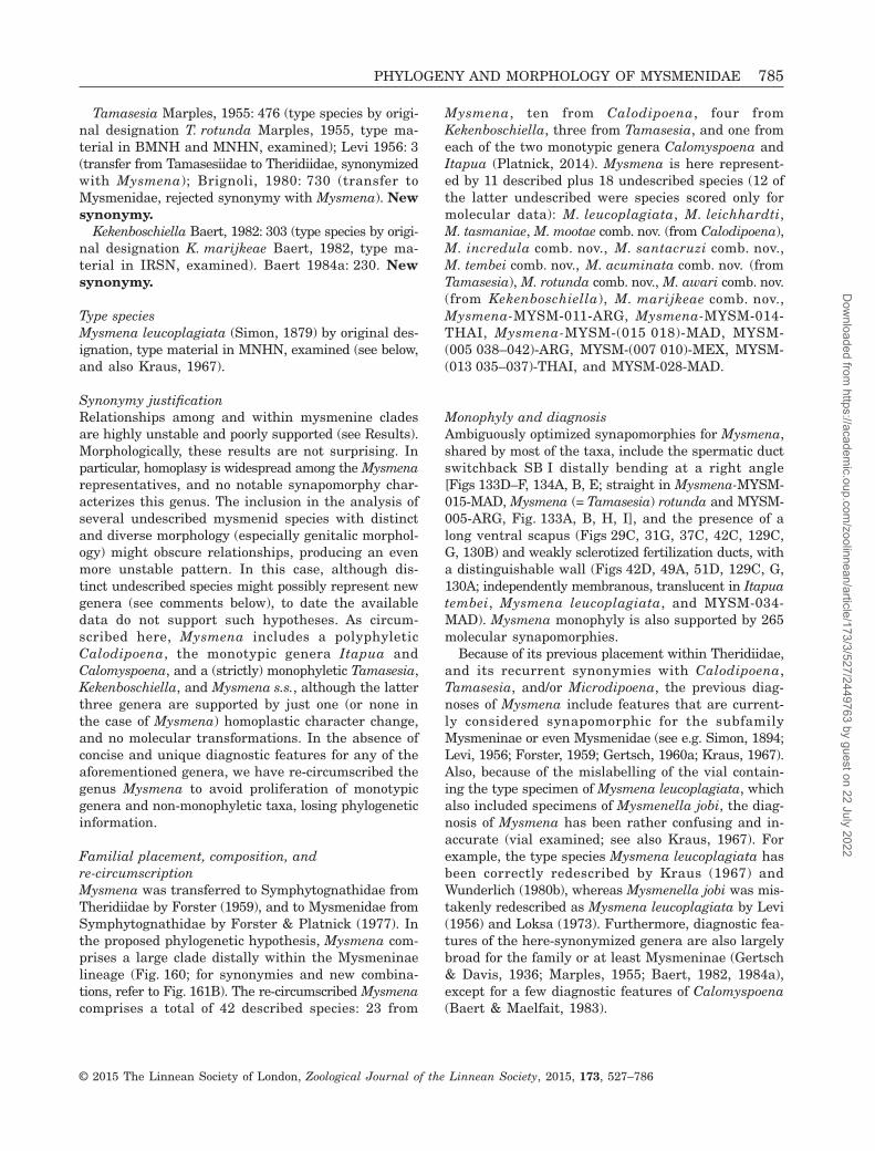
Tamasesia Marples, 1955: 476 (type species by origi-nal designation T. rotunda Marples, 1955, type ma-terial in BMNH and MNHN, examined); Levi 1956: 3(transfer from Tamasesiidae to Theridiidae, synonymizedwith Mysmena); Brignoli, 1980: 730 (transfer toMysmenidae, rejected synonymy with Mysmena). Newsynonymy.
Kekenboschiella Baert, 1982: 303 (type species by origi-nal designation K. marijkeae Baert, 1982, type ma-terial in IRSN, examined). Baert 1984a: 230. Newsynonymy.
Type speciesMysmena leucoplagiata (Simon, 1879) by original des-ignation, type material in MNHN, examined (see below,and also Kraus, 1967).
Synonymy justificationRelationships among and within mysmenine cladesare highly unstable and poorly supported (see Results).Morphologically, these results are not surprising. Inparticular, homoplasy is widespread among the Mysmenarepresentatives, and no notable synapomorphy char-acterizes this genus. The inclusion in the analysis ofseveral undescribed mysmenid species with distinctand diverse morphology (especially genitalic morphol-ogy) might obscure relationships, producing an evenmore unstable pattern. In this case, although dis-tinct undescribed species might possibly represent newgenera (see comments below), to date the availabledata do not support such hypotheses. As circum-scribed here, Mysmena includes a polyphyleticCalodipoena, the monotypic genera Itapua andCalomyspoena, and a (strictly) monophyletic Tamasesia,Kekenboschiella, and Mysmena s.s., although the latterthree genera are supported by just one (or none inthe case of Mysmena) homoplastic character change,and no molecular transformations. In the absence ofconcise and unique diagnostic features for any of theaforementioned genera, we have re-circumscribed thegenus Mysmena to avoid proliferation of monotypicgenera and non-monophyletic taxa, losing phylogeneticinformation.
Familial placement, composition, andre-circumscriptionMysmena was transferred to Symphytognathidae fromTheridiidae by Forster (1959), and to Mysmenidae fromSymphytognathidae by Forster & Platnick (1977). Inthe proposed phylogenetic hypothesis, Mysmena com-prises a large clade distally within the Mysmeninaelineage (Fig. 160; for synonymies and new combina-tions, refer to Fig. 161B). The re-circumscribed Mysmenacomprises a total of 42 described species: 23 from
Mysmena, ten from Calodipoena, four fromKekenboschiella, three from Tamasesia, and one fromeach of the two monotypic genera Calomyspoena andItapua (Platnick, 2014). Mysmena is here represent-ed by 11 described plus 18 undescribed species (12 ofthe latter undescribed were species scored only formolecular data): M. leucoplagiata, M. leichhardti,M. tasmaniae, M. mootae comb. nov. (from Calodipoena),M. incredula comb. nov., M. santacruzi comb. nov.,M. tembei comb. nov., M. acuminata comb. nov. (fromTamasesia), M. rotunda comb. nov., M. awari comb. nov.(from Kekenboschiella), M. marijkeae comb. nov.,Mysmena-MYSM-011-ARG, Mysmena-MYSM-014-THAI, Mysmena-MYSM-(015 018)-MAD, MYSM-(005 038–042)-ARG, MYSM-(007 010)-MEX, MYSM-(013 035–037)-THAI, and MYSM-028-MAD.
Monophyly and diagnosisAmbiguously optimized synapomorphies for Mysmena,shared by most of the taxa, include the spermatic ductswitchback SB I distally bending at a right angle[Figs 133D–F, 134A, B, E; straight in Mysmena-MYSM-015-MAD, Mysmena (= Tamasesia) rotunda and MYSM-005-ARG, Fig. 133A, B, H, I], and the presence of along ventral scapus (Figs 29C, 31G, 37C, 42C, 129C,G, 130B) and weakly sclerotized fertilization ducts, witha distinguishable wall (Figs 42D, 49A, 51D, 129C, G,130A; independently membranous, translucent in Itapuatembei, Mysmena leucoplagiata, and MYSM-034-MAD). Mysmena monophyly is also supported by 265molecular synapomorphies.
Because of its previous placement within Theridiidae,and its recurrent synonymies with Calodipoena,Tamasesia, and/or Microdipoena, the previous diag-noses of Mysmena include features that are current-ly considered synapomorphic for the subfamilyMysmeninae or even Mysmenidae (see e.g. Simon, 1894;Levi, 1956; Forster, 1959; Gertsch, 1960a; Kraus, 1967).Also, because of the mislabelling of the vial contain-ing the type specimen of Mysmena leucoplagiata, whichalso included specimens of Mysmenella jobi, the diag-nosis of Mysmena has been rather confusing and in-accurate (vial examined; see also Kraus, 1967). Forexample, the type species Mysmena leucoplagiata hasbeen correctly redescribed by Kraus (1967) andWunderlich (1980b), whereas Mysmenella jobi was mis-takenly redescribed as Mysmena leucoplagiata by Levi(1956) and Loksa (1973). Furthermore, diagnostic fea-tures of the here-synonymized genera are also largelybroad for the family or at least Mysmeninae (Gertsch& Davis, 1936; Marples, 1955; Baert, 1982, 1984a),except for a few diagnostic features of Calomyspoena(Baert & Maelfait, 1983).
PHYLOGENY AND MORPHOLOGY OF MYSMENIDAE 785
© 2015 The Linnean Society of London, Zoological Journal of the Linnean Society, 2015, 173, 527–786
Dow
nloaded from https://academ
ic.oup.com/zoolinnean/article/173/3/527/2449763 by guest on 22 July 2022

OTHER MYSMENID GROUPS AND TAXA
FIGS 45–50, 129H, 130D, G, 134E–G, H,143D–F, 144A
Molecular clades and taxaNo taxonomic action is taken for the remaining clades/taxa, given that they comprise undescribed species rep-resented, in their majority, exclusively by moleculardata. In this context, no morphological synapomorphiescan be proposed to diagnose any of these clades/taxa.In addition, support for these clades is low. These taxaare regarded here as Mysmeninae incertae sedis (seealso above). Clade C210 comprises only one taxon scoredfor morphology (MYSM-023-MAD), and is supportedby only nine molecular synapomorphies. Clade C163comprises two taxa scored for morphology (MYSM-029-MAD, with only female representatives; MYSM-020-MAD, with only male representatives) and issupported by 83 molecular synapomorphies, a rela-tively larger number. The remaining two taxa notgrouping with other mysmenids in distinct clades(MYSM-006-MAD and MYSM-008-ARG) were not scoredfor morphology, but given their external morphologyand their placement within Mysmeninae in our workinghypothesis, they are regarded as such in the currentstudy. MYSM-019-MAD is sister to the clade compris-ing the relatively well-defined mysmenid generaBrasilionata, Mysmeniola, and Microdipoena. Al-though not remarkably different from other mysmenidgenera, this species possesses some highly homoplas-tic features and could represent a new genus, even ifmorphology alone places it as closely related to otherMysmena species.
Remarkable distinctive morphologyThe following four undescribed mysmenids were scoredfor both morphological and molecular characters;however, morphologically these taxa differ from all otherknown mysmenids (both described and undescribed ex-amined species, see Figs 28–31, 47–50, 129C, 130D,F, 133A–C, 134F–H, 143G–L, 144A). Individually, theymost likely represent three different mysmenid genera,and the phylogenetic hypothesis based on the mor-phological partition alone places these taxa as eitherbasal to major clades (MYSM-005-ARG and MYSM-007-MEX) or as a distinct well-defined, stable, andsupported clade (MYSM-020-MAD and MYSM-023-MAD, see node M118 in Fig. 153). The combined totalevidence analysis does not recover this hypothesis,however, splitting the morphologically robust clade
containing MYSM-020-MAD and MYSM-023-MAD, andplacing the two other taxa within the undefined cladehere referred to as Mysmena. Therefore no taxonom-ic decisions are taken in regard of these taxa, as moredata are needed to more consistently resolve theirphylogenetic placements.
CHINESE MYSMENID GENERA
In a recent study on Chinese symphytognathoids, Milleret al. (2009) described 17 new mysmenid species andfour new genera. Six species were placed in Mysmena,two in Maymena, and nine species were assigned tothe four new genera (Simaoa, Gaoligonga, Mosu, andthe monotypic Chanea). Specimens of these four newgenera were not available for examination when wewere carrying out this project, and therefore they havenot been included in our analyses. These Chinese speciesshare most of the mysmenid diagnostic features, andtherefore their placement within Mysmenidae seemsconclusive; however, as the work was regional in scopeand did not provide an exhaustive generic revision oran explicit phylogenetic justification for the erectionof higher taxa, it is difficult to assess the monophylyof not only the new taxa, but the monophyly of moreinclusive groups that may have been renderedparaphyletic by the erection of new genera. For example,the presence of strong clypeal setae in Chanea, whichhas also been observed and reported for Mysmeniola(Fig. 142M, N; Thaler, 1995; also L. Lopardo, pers.observ.), could suggest a close relationship between thesetwo monotypic genera. Simaoa and Gaoligonga bothbuild the typical mysmenine three-dimensional web,suggesting a close relationship with Microdipoena orMysmena, or at least a placement within Mysmeninae.Therefore, although the new genera include species ofremarkable morphology, not only at the genital level,their monophyly and their exact phylogenetic place-ment within the family has not been tested. Further-more, the generic diagnoses and taxonomic justificationswere based on combinations of features that have cur-rently been found to be highly homoplastic withinMysmenid (e.g. cymbium enveloping the bulb, posteri-or abdominal tubercle, epigynal scapus, etc.). The in-stability of relationships within Mysmeninae, and thelack of unambiguous and consistent diagnostic traitsfor mysmenine genera, suggest that a more explicitlyphylogenetic and monographic approach is needed totest the monophyly and circumscription of the newChinese mysmenid genera.
Cover imageRight: Male palp from Mysmena sp. (Mysmena-MYSM-015-MAD, Mysmeninae, Mysmenidae) from Madagas-car, SEM image. Top left: Male of Microdipoena guttata (Mysmeninae, Mysmenidae) from Comoros, compositeimage, lateral view (photo: Peter Michalik). Bottom left: Typical spherical mysmenid orbweb; Mysmena leichhardtifemale (Mysmeninae, Mysmenidae), from Australia, contrasted image.
786 L. LOPARDO AND G. HORMIGA
© 2015 The Linnean Society of London, Zoological Journal of the Linnean Society, 2015, 173, 527–786
Dow
nloaded from https://academ
ic.oup.com/zoolinnean/article/173/3/527/2449763 by guest on 22 July 2022