«Освободить... от шайтанов и шарлатанов»: дискурсы и...
7
H-NS Mediates the Silencing of Laterally Acquired Genes in Bacteria Sacha Lucchini 1 , Gary Rowley 1 , Martin D. Goldberg 1¤ , Douglas Hurd 2 , Marcus Harrison 2 , Jay C. D. Hinton 1* 1 Molecular Microbiology Group, Institute of Food Research, Colney, Norwich, United Kingdom, 2 Oxford Gene Technology, Yarnton, Oxford, United Kingdom Histone-like nucleoid structuring protein (H-NS) is a modular protein that is associated with the bacterial nucleoid. We used chromatin immunoprecipitation to determine the binding sites of H-NS and RNA polymerase on the Salmonella enterica serovar Typhimurium chromosome. We found that H-NS does not bind to actively transcribed genes and does not co-localize with RNA polymerase. This shows that H-NS principally silences gene expression by restricting the access of RNA polymerase to the DNA. H-NS had previously been shown to preferentially bind to curved DNA in vitro. In fact, at the genomic level we discovered that the level of H-NS binding correlates better with the AT-content of DNA. This is likely to have evolutionary consequences because we show that H-NS binds to many Salmonella genes acquired by lateral gene transfer, and functions as a gene silencer. The removal of H-NS from the cell causes un-controlled expression of several Salmonella pathogenicity islands, and we demonstrate that this has deleterious consequences for bacterial fitness. Our discovery of this novel role for H-NS may have implications for the acquisition of foreign genes by enteric bacteria. Citation: Lucchini S, Rowley G, Goldberg MD, Hurd D, Harrison M, et al. (2006) H-NS mediates the silencing of laterally acquired genes in bacteria. PLoS Pathog 2(8): e81. DOI: 10.1371/journal.ppat.0020081 Introduction Bacteria have an extraordinary capacity to adapt to changes in their external environment. The histone-like nucleoid-structuring protein (H-NS) plays a key role for many Gram-negative bacteria by integrating a complex range of environmental signals such as temperature and osmolarity [1,2]. Consequently, H-NS acts as a pleiotropic regulator of gene expression [3]. Deletion of hns resulted in altered gene expression of almost 5% of Escherichia coli genes, of which more than 80% were up-regulated [4]. H-NS also influences the major DNA transactions, such as replication, trans- position and recombination [5]. The modular nature of the H-NS protein is reflected by the three distinct domains, involved in DNA binding, dimerisation, and the formation of high order oligomers [3,6]. Unlike classical regulatory repressor and activator pro- teins, H-NS does not recognise particular sequences of DNA; however, a number of in vitro studies have shown that the protein has a small preference for curved DNA [7–9]. It has been proposed that after binding to a preferential site, H-NS oligomerizes cooperatively along the DNA to cover a larger region [10–12]. This idea is supported by the observation that mutant H-NS proteins that are unable to oligomerize no longer repress the expression of some promoters [10,13]. Following the binding of H-NS, changes in transcription are likely to be caused by modulations of DNA topology (i.e. compacting and looping DNA, and the constraint of DNA supercoils [14]). As DNA curvature is most pronounced around the tran- scriptional start of genes [15] a simple model involving the preferential binding of H-NS to regulatory regions has been considered [16], with resultant effects upon global gene expression. However, since curved DNA is found near most promoters [17], and the majority of bacterial promoters are not regulated by H-NS (i.e. H-NS-independent), it has never been clear how specificity was achieved. The interaction between H-NS and DNA has been studied in vitro by traditional gel retardation and DNA-footprinting experi- ments, but knowledge of how and where H-NS attaches to DNA in vivo has remained indirect. Using chromatin immunoprecipitation on chip (ChIP-on-chip) [18,19], we have now addressed the question of where H-NS binds to DNA in the macro-molecular context of a living bacterial cell and show how this correlates with the distribution of RNA polymerase on the chromosome. Results/Discussion H-NS and RNA polymerase binding sites on the chromo- some of live Salmonella enterica serovar Typhimurium (S. typhimurium) LT2 cells were identified by ChIP-on-chip, using a tiled oligonucleotide microarray (Figure 1, outer circles). We first identified 495 chromosomal regions that bound RNA polymerase (RNAP) during exponential growth in Luria Broth (LB, Materials and Methods), corresponding to 965 genes (Table S1). As expected, the strongest RNAP binding was observed for genes involved in ribosome, flagella and Editor: Ralph R. Isberg, Tufts University School of Medicine, United States of America Received May 31, 2006; Accepted July 6, 2006; Published August 18, 2006 DOI: 10.1371/journal.ppat.0020081 Copyright: Ó 2006 Lucchini et al. This is an open-access article distributed under the terms of the Creative Commons Attribution License, which permits unrestricted use, distribution, and reproduction in any medium, provided the original author and source are credited. Abbreviations: ChIP-on-chip, chromatin immunoprecipitation on microarray; H- NS, histone-like nucleoid structuring protein; LB, Luria broth; LEE, locus of enterocyte effacement; RNAP, RNA polymerase; SPI2, Salmonella pathogenicity island 2 * To whom correspondence should be addressed. E-mail: [email protected] ¤ Current address: School of Biosciences, University of Birmingham, Edgbaston, Birmingham, United Kingdom PLoS Pathogens | www.plospathogens.org August 2006 | Volume 2 | Issue 8 | e81 0746
Transcript of «Освободить... от шайтанов и шарлатанов»: дискурсы и...

H-NS Mediates the Silencing of LaterallyAcquired Genes in BacteriaSacha Lucchini
1, Gary Rowley
1, Martin D. Goldberg
1¤, Douglas Hurd
2, Marcus Harrison
2, Jay C. D. Hinton
1*
1 Molecular Microbiology Group, Institute of Food Research, Colney, Norwich, United Kingdom, 2 Oxford Gene Technology, Yarnton, Oxford, United Kingdom
Histone-like nucleoid structuring protein (H-NS) is a modular protein that is associated with the bacterial nucleoid. Weused chromatin immunoprecipitation to determine the binding sites of H-NS and RNA polymerase on the Salmonellaenterica serovar Typhimurium chromosome. We found that H-NS does not bind to actively transcribed genes and doesnot co-localize with RNA polymerase. This shows that H-NS principally silences gene expression by restricting theaccess of RNA polymerase to the DNA. H-NS had previously been shown to preferentially bind to curved DNA in vitro.In fact, at the genomic level we discovered that the level of H-NS binding correlates better with the AT-content of DNA.This is likely to have evolutionary consequences because we show that H-NS binds to many Salmonella genes acquiredby lateral gene transfer, and functions as a gene silencer. The removal of H-NS from the cell causes un-controlledexpression of several Salmonella pathogenicity islands, and we demonstrate that this has deleterious consequences forbacterial fitness. Our discovery of this novel role for H-NS may have implications for the acquisition of foreign genes byenteric bacteria.
Citation: Lucchini S, Rowley G, Goldberg MD, Hurd D, Harrison M, et al. (2006) H-NS mediates the silencing of laterally acquired genes in bacteria. PLoS Pathog 2(8): e81. DOI:10.1371/journal.ppat.0020081
Introduction
Bacteria have an extraordinary capacity to adapt tochanges in their external environment. The histone-likenucleoid-structuring protein (H-NS) plays a key role formany Gram-negative bacteria by integrating a complex rangeof environmental signals such as temperature and osmolarity[1,2]. Consequently, H-NS acts as a pleiotropic regulator ofgene expression [3]. Deletion of hns resulted in altered geneexpression of almost 5% of Escherichia coli genes, of whichmore than 80% were up-regulated [4]. H-NS also influencesthe major DNA transactions, such as replication, trans-position and recombination [5]. The modular nature of theH-NS protein is reflected by the three distinct domains,involved in DNA binding, dimerisation, and the formation ofhigh order oligomers [3,6].
Unlike classical regulatory repressor and activator pro-teins, H-NS does not recognise particular sequences of DNA;however, a number of in vitro studies have shown that theprotein has a small preference for curved DNA [7–9]. It hasbeen proposed that after binding to a preferential site, H-NSoligomerizes cooperatively along the DNA to cover a largerregion [10–12]. This idea is supported by the observation thatmutant H-NS proteins that are unable to oligomerize nolonger repress the expression of some promoters [10,13].Following the binding of H-NS, changes in transcription arelikely to be caused by modulations of DNA topology (i.e.compacting and looping DNA, and the constraint of DNAsupercoils [14]).
As DNA curvature is most pronounced around the tran-scriptional start of genes [15] a simple model involving thepreferential binding of H-NS to regulatory regions has beenconsidered [16], with resultant effects upon global geneexpression. However, since curved DNA is found near mostpromoters [17], and the majority of bacterial promoters arenot regulated by H-NS (i.e. H-NS-independent), it has neverbeen clear how specificity was achieved. The interaction
between H-NS and DNA has been studied in vitro bytraditional gel retardation and DNA-footprinting experi-ments, but knowledge of how and where H-NS attaches toDNA in vivo has remained indirect. Using chromatinimmunoprecipitation on chip (ChIP-on-chip) [18,19], we havenow addressed the question of where H-NS binds to DNA inthe macro-molecular context of a living bacterial cell andshow how this correlates with the distribution of RNApolymerase on the chromosome.
Results/Discussion
H-NS and RNA polymerase binding sites on the chromo-some of live Salmonella enterica serovar Typhimurium (S.typhimurium) LT2 cells were identified by ChIP-on-chip, usinga tiled oligonucleotide microarray (Figure 1, outer circles).We first identified 495 chromosomal regions that bound RNApolymerase (RNAP) during exponential growth in LuriaBroth (LB, Materials and Methods), corresponding to 965genes (Table S1). As expected, the strongest RNAP bindingwas observed for genes involved in ribosome, flagella and
Editor: Ralph R. Isberg, Tufts University School of Medicine, United States ofAmerica
Received May 31, 2006; Accepted July 6, 2006; Published August 18, 2006
DOI: 10.1371/journal.ppat.0020081
Copyright: � 2006 Lucchini et al. This is an open-access article distributed underthe terms of the Creative Commons Attribution License, which permits unrestricteduse, distribution, and reproduction in any medium, provided the original authorand source are credited.
Abbreviations: ChIP-on-chip, chromatin immunoprecipitation on microarray; H-NS, histone-like nucleoid structuring protein; LB, Luria broth; LEE, locus ofenterocyte effacement; RNAP, RNA polymerase; SPI2, Salmonella pathogenicityisland 2
* To whom correspondence should be addressed. E-mail: [email protected]
¤ Current address: School of Biosciences, University of Birmingham, Edgbaston,Birmingham, United Kingdom
PLoS Pathogens | www.plospathogens.org August 2006 | Volume 2 | Issue 8 | e810746

ATP synthesis. Consultation of transcriptomic data for LT2grown to exponential phase [2] revealed that RNAP-boundgenes were expressed at high levels, and RNAP binding wasonly associated with genes that were transcribed (Figure 2).
The ChIP-on-chip experiments using anti-H-NS monoclo-
nal antibodies repeatedly displayed high levels of signalacross most of the chromosome (unpublished data). Weconfirmed this observation by repeating the experiment withH200 anti-H-NS polyclonal antibodies, and obtained verysimilar results (unpublished data). In contrast, the RNAPChIP-on-chip showed a much lower background signal. Thesefindings suggest that the low sequence specificity of H-NS andits high abundance [20] allows the protein to bind at differinglevels throughout most of the chromosome. Significantly, thebackground signal was not homogeneously distributed overthe genome; rRNA genes exhibited a low H-NS signalsuggesting that H-NS is specifically excluded from highlytranscribed genes (Figure 3).We identified 422 regions that bound H-NS, corresponding
to 746 genes, which we suggest contain high-affinity bindingsites (Table S1), also known as H-NS nucleation sites [5]. Tovalidate the data, we initially focused on the H-NS bindingsites for published H-NS-dependent genes, such as proV andleuO. ProV is a well characterised H-NS-dependent osmoin-ducible transport protein in Salmonella [21]. H-NS has beenshown to repress proV by binding to a DNA-region, thedownstream regulatory element, which is located within theproV structural gene [7]. H-NS also transcriptionally represses
Figure 1. H-NS Chip and Transcriptomic Data in Context of the Genome Atlas for the S. typhimurium Chromosome
The data from the RNAP and H-NS ChIP-on-chip experiments are shown using coloured concentric circles which are mapped onto the Salmonellachromosome. In addition, we show our previously published transcriptome data for the JH4000 hns-deletion mutant [2] and four DNA structuralmeasures (http://www.cbs.dtu.dk/services/GenomeAtlas [15]). The order of the concentric circles is indicated in the legend. Numbers on the inside ofthe innermost circle are the location relative to position zero measured in millions of base-pairs (Mbp). The origin of replication, predicted terminusregion, location of rRNAs and the SPI1 and SPI2 pathogenicity islands are indicated at the edge of the outermost circle.DOI: 10.1371/journal.ppat.0020081.g001
PLoS Pathogens | www.plospathogens.org August 2006 | Volume 2 | Issue 8 | e810747
Genome-Wide Silencing in Salmonella
Synopsis
In recent decades, gene silencing has been well-characterised inplants and animals, and involves the prevention of transcription byDNA-methylation and histone-modification, or interference withtranslation by small RNA molecules. This issue of PLoS Pathogensreports the discovery that global gene silencing also occurs inbacteria. The novel mechanism is mediated by the highly abundanthistone-like nucleoid structuring protein (H-NS), which blocks theexpression of 254 genes in wild-type Salmonella. Many of thesegenes were acquired through horizontal gene transfer, includingpathogenicity islands, and these are silenced by the binding of H-NSto AT-rich chromosomal regions. The study reveals that H-NSprevents the uncontrolled transcription of genes within pathoge-nicity islands to ensure that bacterial fitness is maintained. It issuggested that H-NS plays a role in bacterial evolution byinfluencing both the acquisition and maintenance of foreign DNA.
leuO, a gene coding for a pleiotropic regulator of geneexpression, by binding to a curved AT-rich DNA region up-stream of the leuO promoter [12]. Our data confirm thereported binding patterns (Figure 3). To add biologicalrelevance to our ChIP-on-chip data we correlated our resultswith the transcriptomic analysis of strain LT2 and itscongenic hns-deletion mutant JH4000 during late-exponen-tial growth [2]. Genes bound by H-NS are generally expressedat lower than average levels in the wild-type LT2 strain(Figure 2A). A large proportion (254 or 47%) of the 535 genesde-repressed in JH4000 was bound by H-NS in the LT2 strain.In contrast, only 20 (6.1%) of the 330 genes down-regulated inJH4000 were bound by H-NS. These findings demonstrate arole for H-NS as a global gene silencer, and this wasconfirmed by the observed negative correlation between H-NS binding and gene expression levels (Figure 2C). Expres-sion of about half of the genes that bound H-NS in LT2 werenot affected by the deletion of hns, probably reflecting theabsence of positive regulatory proteins in our cultureconditions (exponential growth in LB). We hypothesise thatthese 422 binding sites are important for the nucleoidstructuring role of H-NS.
Of the 1,699 genes that were bound by either H-NS orRNAP in wild-type S. typhimurium, only 12 were bound by bothproteins. This suggests that repression by H-NS is generallyachieved by preventing the RNA polymerase from binding totarget promoter sequences, probably via alterations at thelevel of DNA topology. An alternate model of H-NSrepression involves looping and trapping of RNA polymeraseat the promoter [22,23]. This model appears to be contra-dicted by our data, as we do not observe co-localization of H-
NS with the RNA polymerase. However, it remains possiblethat the trapping of RNA polymerase is too transient to bedetected by our experimental approach.No consensus DNA sequence has been identified for H-NS
binding, but the protein was reported to have a slightlyincreased affinity for curved DNA around the regulatoryregion of genes in vitro [7–9]. We therefore correlated H-NSbinding with the base-composition and structural features ofDNA such as curvature, stacking energy, and protein-induceddeformability. For each oligonucleotide on the microarray,we measured the characteristics of the DNA using the dataprovided by the Genome Atlas of the S. typhimuriumchromosome (http://www.cbs.dtu.dk/services/GenomeAtlas/info/background.php). The best observed correlation wasbetween H-NS binding and AT-content (Figure 4). Asexpected, analysis of H-NS binding in relation to stackingenergy and protein deformability gave very similar profiles tothe base composition (unpublished data), as these structuralcharacteristics are largely dependent on AT-content. Thecorrelation between H-NS binding and DNA-curvature wasmuch smaller (Figure 4). Again, we correlated our results withthe transcriptomic analysis of strain LT2 and its congenic hns-deletion mutant JH4000. Base composition was confirmed toplay an important role for H-NS as a repressor of geneexpression, because AT-rich genes were 20 times more likelyto be repressed by H-NS than GC-rich genes. In contrast,genes with a strong curved DNA sequence in their regulatoryregions were only two times more likely to be repressed by H-NS, compared to genes with low or no curvature.Promoter regions are usually associated with both higher
AT-content and curvature of DNA. However, not all bacterial
Figure 2. Correlation between Binding of H-NS, RNAP, and Level of Gene Expression
(A) Frequency distribution of relative gene expression levels for S. typhimurium. The results are shown for all S. typhimurium genes (filled circle) and forgenes found to co-localize with H-NS (filled triangle) or RNAP (open circle) in the ChIP-on-chip experiment. Relative gene expression levels for S.typhimurium strain LT2 were taken from Ono et al [2].(B and C) Scatter plot correlating binding of RNAP or H-NS to gene expression levels. The relative expression level of a gene (y-axis) is plotted against thecorresponding level of binding to RNAP or H-NS (x-axis) as measured in the ChIP-on-chip experiments (see Materials and Methods).DOI: 10.1371/journal.ppat.0020081.g002
PLoS Pathogens | www.plospathogens.org August 2006 | Volume 2 | Issue 8 | e810748
Genome-Wide Silencing in Salmonella
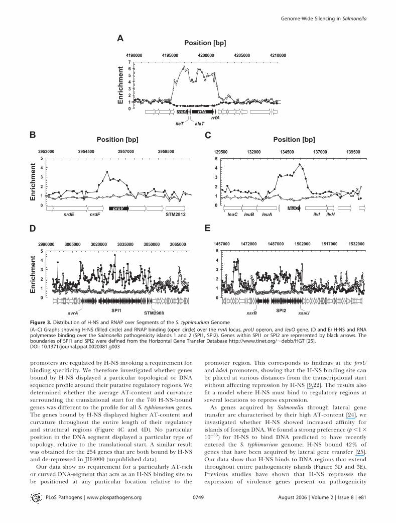
promoters are regulated by H-NS invoking a requirement forbinding specificity. We therefore investigated whether genesbound by H-NS displayed a particular topological or DNAsequence profile around their putative regulatory regions. Wedetermined whether the average AT-content and curvaturesurrounding the translational start for the 746 H-NS-boundgenes was different to the profile for all S. typhimurium genes.The genes bound by H-NS displayed higher AT-content andcurvature throughout the entire length of their regulatoryand structural regions (Figure 4C and 4D). No particularposition in the DNA segment displayed a particular type oftopology, relative to the translational start. A similar resultwas obtained for the 254 genes that are both bound by H-NSand de-repressed in JH4000 (unpublished data).
Our data show no requirement for a particularly AT-richor curved DNA-segment that acts as an H-NS binding site tobe positioned at any particular location relative to the
promoter region. This corresponds to findings at the proUand hdeA promoters, showing that the H-NS binding site canbe placed at various distances from the transcriptional startwithout affecting repression by H-NS [9,22]. The results alsofit a model where H-NS must bind to regulatory regions atseveral locations to repress expression.As genes acquired by Salmonella through lateral gene
transfer are characterised by their high AT-content [24], weinvestigated whether H-NS showed increased affinity forislands of foreign DNA. We found a strong preference (p ,13
10�55) for H-NS to bind DNA predicted to have recentlyentered the S. typhimurium genome; H-NS bound 42% ofgenes that have been acquired by lateral gene transfer [25].Our data show that H-NS binds to DNA regions that extendthroughout entire pathogenicity islands (Figure 3D and 3E).Previous studies have shown that H-NS represses theexpression of virulence genes present on pathogenicity
Figure 3. Distribution of H-NS and RNAP over Segments of the S. typhimurium Genome
(A–C) Graphs showing H-NS (filled circle) and RNAP binding (open circle) over the rrnA locus, proU operon, and leuO gene. (D and E) H-NS and RNApolymerase binding over the Salmonella pathogenicity islands 1 and 2 (SPI1, SPI2). Genes within SPI1 or SPI2 are represented by black arrows. Theboundaries of SPI1 and SPI2 were defined from the Horizontal Gene Transfer Database http://www.tinet.org/;debb/HGT [25].DOI: 10.1371/journal.ppat.0020081.g003
PLoS Pathogens | www.plospathogens.org August 2006 | Volume 2 | Issue 8 | e810749
Genome-Wide Silencing in Salmonella

islands and plasmids in various Gram-negative bacteria,including Salmonella, Shigella, E. coli, and Vibrio [2,3,26,27].Now we have determined that H-NS acts as a selective silencerbased on AT-content. However, as H-NS does not bind to allAT-rich genes, cellular factors such as other nucleoidassociated proteins might modulate H-NS-binding to partic-ular locations on the genome. Interestingly, our data do notshow preferential binding of H-NS to other mobile elementssuch as prophages. This can be explained by the fact that thefour prophages present in the S. typhimurium LT2 genome donot display an AT-content higher than the genome average.We hypothesised that the binding of H-NS to Salmonella
pathogenicity islands would have biological significance if itcaused silencing of large regions of horizontally-acquiredDNA. Therefore, we compared the fitness of a strain unable toexpress the Salmonella pathogenicity island, SPI2 in both anhns� and hnsþ background. We discovered that the absence ofexpression of SPI2 in the strain lacking H-NS substantiallyimproved the growth-rate of Salmonella (Figure 5). The S.typhimurium strain JH4000 displays a severely reduced growth-rate caused by the hns deletion; the growth rate (lmax) of the
Figure 5. H-NS Confers a Growth Advantage for Wild-Type Salmonella by
Silencing Genes Acquired through Lateral Gene Transfer
The growth of the five S. typhimurium strains were followed in LB, asdescribed in Materials and Methods. The data shown are representativeof three experiments, and discussed in the text.DOI: 10.1371/journal.ppat.0020081.g005
Figure 4. Correlation between AT-Content, Curvature and H-NS, and RNAP Binding to DNA
For each oligonucleotide on the microarrays we calculated its average AT-content (A) and curvature (B). To minimize bias that might have beenintroduced by the algorithms used to design the oligonucleotides, flanking sequences (50 bp) of the oligonucleotides were included in the analysis. Theresults of each oligonucleotide were plotted against the level of H-NS binding (grey circles). Window-averaged results (window size: 101) are also shown(solid line).(C and D) AT-content and curvature profiles were calculated from putative promoter sequences aligned by over-laying every S. typhimurium gene at thetranslational start. The profiles have been calculated for all the S. typhimurium genes (black), and for those subsets of genes bound by H-NS (green) orRNAP (red).DOI: 10.1371/journal.ppat.0020081.g004
PLoS Pathogens | www.plospathogens.org August 2006 | Volume 2 | Issue 8 | e810750
Genome-Wide Silencing in Salmonella
LT2 wild-type strain was 1.45, twice as fast as the JH4000 hns-deletion mutant (lmax¼0.72). The fitness defect was partiallycompensated in strain JH4000DssrA, which is unable toexpress genes coded within the SPI2 island; deletion of ssrAfrom JH4000 caused the growth rate to significantly increase(p ,0.05). In contrast, the absence of SPI2 expression in anhnsþ background did not affect growth-rate (strain LT2ssrA).
It has been reported that a S. typhimurium hns mutation wasassociated with compensatory mutations in the phoP and rpoSgenes [28]. It should be noted that wild-type S. typhimuriumstrain LT2 already carries a well-characterised point muta-tion in rpoS which modifies the translation initiation codon,causing decreased translation of RpoS [29,30]. To prove thatthe effect on growth rate was indeed caused by deletion of thessrA gene we ruled out the possibility of secondary mutationsby complementing JH4000ssrA with plasmid pWSK29ssrAB.The plasmid caused a significant decrease in growth rate ofJH4000 DssrA (p ,0.05).
Our data reveal that H-NS offers a fitness advantage towild-type Salmonella by silencing the expression of foreignDNA under appropriate conditions. It appears that somepathogenicity islands have acquired the ability to resist H-NS-mediated silencing; the locus of enterocyte effacement (LEE)pathogenicity island of enteropathogenic E. coli codes for Ler,an H-NS homologue that can displace H-NS from the LEEregulatory regions [31].
In summary, we have discovered that global gene silencingoccurs in bacteria, and is mediated by H-NS, which acts on atleast 254 S. typhimurium genes. Different mechanisms havebeen proposed to explain the silencing of specific promotersby H-NS. For example, at the ilvIH-leuO-leuABCD gene clusterof S. typhimurium, H-NS is initially thought to bind an AT-richcurved DNA sequence which acts as a nucleation site andsubsequently forms an extended nucleoprotein complex [12].We suggest that this type of nucleoprotein complex isresponsible for H-NS-mediated gene silencing, and areinvestigating the co-operative binding of H-NS at otherchromosomal regions in S. typhimurium. Our observation thatH-NS and RNA polymerase rarely co-localize during expo-nential growth indicates that H-NS generally prevents geneexpression by excluding RNA polymerase from binding toDNA, rather than by trapping RNA polymerase at promoters.It is possible that such trapping by H-NS may occur instationary phase bacteria, when a lower number of RNApolymerase molecules is required for active transcription.
After this manuscript was submitted, a related study waspublished by Navarre et al [28]. The authors used ananalogous approach to detect H-NS binding sites in S.typhimurium strain 14028 and also concluded that H-NSpreferentially binds to AT-rich genes. Taken together, thesetwo studies give confidence about the phenomenon of globalgene silencing in bacteria; the H-NS protein was found tobind to AT-rich genes in two different S. typhimurium strains(14028 and LT2) grown in two distinct media (minimalmedium versus LB).
Taken together, our findings prove that H-NS has a strongpreference forAT-richDNAwhich facilitates binding to a largenumber of genes acquired through lateral gene transfer. Weknow that bacterial evolution relies upon the ability of newlyacquired genes to offer a selective advantage to their host [32].However, the expression of genes encoded by novel DNAmustbe controlled or the cell will suffer negative consequences. We
have shown that H-NS is essential for the prevention ofdecreased fitness associated with uncontrolled expression ofpathogenicity island genes. The evolutionary implications ofthis silencing function of a nucleoid–associated protein meritfurther study in many enterobacterial pathogens.
Materials and Methods
Bacterial strains and growth conditions. Salmonella enterica serovarTyphimurium strain LT2 and its hns� derivative JH4000 weredescribed in our recent transcriptomic study [2]. Where necessary,antibiotics were used as appropriate at the following concentrations:ampicillin (100 lg/ml), chloramphenicol (12.5 lg/ml). To measuregrowth rates, overnight cultures were subcultured 100-fold into 25 mlfresh LB-broth [33] and grown in 250-ml flasks in a 37 8C waterbath(Innova 3100, New Brunswick Scientific, Edison, New Jersey, UnitedStates) at 250 rpm.
Strain construction and DNA manipulation. The DssrA deletionderivative of S. typhimurium LT2 (LT2ssrA) was generated using the kRed method [34]. The oligonucleotides used for this constructionwere ssrARedF 59GTGCCAAAGATTTTGCAACAGGCAACT-GGAGGGAAGCATTGTGTAGGCTGGAGCTGCTTC-39 and ssraR-edR 59-ATTCTTTCATTTTGCTGCCCTCGCGAAAATTA AGATAA-TACATATGAATATCCTCCTTAG-39, in conjunction with templateplasmid pKD4. The DssrA deletion mutation was marked with the Kanantibiotic resistance cassette. After mutagenesis, the mutation wasP22-transduced to a clean background using phage P22 HT105/1 int-201. EBU plates were used to select for non-lysogens [35]. Themutation was verified by PCR using primers external to the mutation.The DhnsDssrA double mutant of S. typhimurium LT2 (JH4000ssrA) wasobtained by transducing the DssrA mutation into JH4000.
Design of a S. typhimurium tiled microarray. The microarrays usedin this study were designed and produced by Oxford GeneTechnology (Kidlington, Oxfordshire, United Kingdom). The micro-arrays consisted of 21,340 60-mer oligonucleotides tiled throughoutthe S. typhimurium LT2 chromosome and does not cover the pSLTplasmid. The gap between oligonucleotides averaged 170 bp.
ChIP-on-chip. Cultures of S. typhimurium LT2 were grown in LBbroth with aeration (37 8C, 250 rpm) in an Innova 3100 waterbath(New Brunswick Scientific) until OD600 ¼ 0.6 (early-exponentialphase), at which point formaldehyde was added to a final concen-tration of 1% (v/v). After 20 min incubation at 37 8C, the cross-linkingreaction was quenched by adding glycine to a 0.5 M finalconcentration. Cells were harvested and washed twice in TBS. Thecell pellet was then resuspended in 1 ml of lysis buffer (10 mM Tris[pH 8.0], 20% [w/v] sucrose, 50 mM NaCl, 10 mM EDTA [pH 8.0], 20mg/ml lysozyme, 0.1 mg/ml RNAseA) and incubated for 1 h at 37 8C.Bead beating (3 3 20-s cycles, power 6 [FastPrep FP120, Savant,Farmingdale, New York, United States]) was used to achieve completelysis. The cell lysate was then added to 4 ml of immunoprecipitationbuffer (50 mM HEPES-KOH [pH 7.5], 150 mM NaCl, 1 mM EDTA, 1%[v/v] Triton X-100, 0.1% [w/v] sodium deoxycholate, 0.1% [w/v] SDS, 1mM PMSF). Three aliquots (800 ll) were sonicated (3 3 20-s cycles at10 lm amplitude, (MSE Soniprep 150, Sanyo Scientific, Bensenville,Illinois, United States) to solubilize the chromatin. Chromatinimmuno-precipitation was then performed as described by Graingeret al [18] on two aliquots, using monoclonal H113 anti-H-NSantibodies [36] or anti-RNA polymerase-b subunit monoclonalantibodies (Neoclone, Madison, Wisconsin, United States). As anegative control, chromatin immuno-precipitation was done on thethird aliquot without the addition of any antibodies. Additionalcontrol chromatin immuno-precipitations were performed usingpolyclonal H200 anti-H-NS antibodies [36].
The DNA obtained from chromatin immuno-precipitation waslabelled with Cy5, mixed with a reference (Cy3-labelled S. typhimuriumLT2 genomic DNA) and hybridized against the S. typhimurium LT2tiled oligonucleotide microarray. The protocol used to label bothtypes of DNA samples is described at http://www.ifr.bbsrc.ac.uk/safety/microarrays/protocols.html. The hybridization procedure is de-scribed by Grainger et al [37]. The experiment was performed withthree independent biological replicates.
Data acquisition and analysis. The microarray slides were scannedusing a GenePix 4000A scanner (Axon Instruments, Union City,California, United States). Spot intensities were quantified usingBlueFuse software (BlueGnome, Cambridge, United Kingdom). Dataanalysis and visualization was performed using GeneSpring GX 7.3(Agilent, Palo Alto, California, United States). To compensate forunequal dye incorporation and/or differences in background levels
PLoS Pathogens | www.plospathogens.org August 2006 | Volume 2 | Issue 8 | e810751
Genome-Wide Silencing in Salmonella

caused by the ChIP procedure, data centering was performed bybringing the median Ln(Red/Green) to zero. To determine the level ofbinding of H-NS or RNA polymerase-b subunit (the enrichmentfactor), results were normalized to the ratios obtained for thenegative control (ChIP-on-chip performed without antibodies).
To identify regions of H-NS and RNAP binding, we only consideredoligonucleotides that displayed enrichment factors � 1.5 and 2 forRNAP and H-NS data, respectively, in all three biological replicates.The enrichment factor cut-off of 2 was chosen to reflect the highervariability observed in the H-NS ChIP data. We then retained thosesignals extending to two or more adjacent oligonucleotides on thegenome; the rationale being that since the ChIP procedure generatesDNA fragments 500–1,000 bp in length, we did not expect peaks of H-NS and RNAP binding to correspond to isolated oligonucleotides. Thecomplete dataset is available as Supporting Information and at theArrayExpress database (accession number E-MEXP-806).
Supporting Information
Table S1. H-NS and RNAP ChIP Data on the S. typhimurium LT2Chromosome
Found at DOI: 10.1371/journal.ppat.0020081.st001 (737 KB PDF).
Accession Numbers
The NCBI (http://www.ncbi.nlm.nih.gov/entrez) accession number forthe LT2 genome sequence is NC003197.
Acknowledgments
We enjoyed helpful discussions with Steve Busby, Charles Dorman,David Grainger, John Ladbury, Paul Leonard, Ida Porcelli, andmembers of the Hinton lab. We are grateful to Dave Ussery forassistance in making the Genome Atlas which gave a new perspectiveto our H-NS and RNA polymerase binding data by adding structuralfactors; an interactive version of Figure 1 will be available on ourpublications website (http://www.ifr.ac.uk/safety/molmicro/pubs.html).
Author contributions. SL and JCDH conceived and designed theexperiments. SL, GR, and MDG performed the experiments. SLanalyzed the data. DH and MH contributed reagents/materials/analysis tools. SL and JCDH wrote the paper.
Funding. This work was funded by the BBSRC core strategic grantto JCDH.
Competing interests. The authors have declared that no competinginterests exist.
References1. Tendeng C, Bertin PN (2003) H-NS in Gram-negative bacteria: A family of
multifaceted proteins. Trends Microbiol 11: 511–518.2. Ono S, Goldberg MD, Olsson T, Esposito D, Hinton JCD, et al. (2005) H-NS
is a part of a thermally controlled mechanism for bacterial gene regulation.Biochem J 391: 203–213.
3. Dorman CJ (2004) H-NS: A universal regulator for a dynamic genome. NatRev Microbiol 2: 391–400.
4. Hommais F, Krin E, Laurent-Winter C, Soutourina O, Malpertuy A, et al.(2001) Large-scale monitoring of pleiotropic regulation of gene expressionby the prokaryotic nucleoid-associated protein, H-NS. Mol Microbiol 40:20–36.
5. Rimsky S (2004) Structure of the histone-like protein H-NS and its role inregulation and genome superstructure. Curr Opin Microbiol 7: 109–114.
6. Esposito D, Petrovic A, Harris R, Ono S, Eccleston JF, et al. (2002) H-NSoligomerization domain structure reveals the mechanism for high orderself-association of the intact protein. J Mol Biol 324: 841–850.
7. Owen-Hughes TA, Pavitt GD, Santos DS, Sidebotham JM, Hulton CSJ, et al.(1992) The chromatin-associated protein H-NS interacts with curved DNAto influence DNA topology and gene-expression. Cell 71: 255–265.
8. Yamada H, Yoshida T, Tanaka K, Sasakawa C, Mizuno T (1991) Molecularanalysis of the Escherichia coli H-NS gene encoding a DNA-binding protein,which preferentially recognizes curved DNA sequences. Mol Gen Genet230: 332–336.
9. Jordi B, Fielder AE, Burns CM, Hinton JCD, Dover N, et al. (1997) DNAbinding is not sufficient for H-NS-mediated repression of proU expression.J Biol Chem 272: 12083–12090.
10. Rimsky S, Zuber F, Buckle M, Buc H (2001) A molecular mechanism for therepression of transcription by the H-NS protein. Mol Microbiol 42: 1311–1323.
11. Tupper AE, Owen-Hughes TA, Ussery DW, Santos DS, Ferguson DJP, et al.(1994) The chromatin-associated protein H-NS alters DNA topology invitro. EMBO J 13: 258–268.
12. Chen CC, Chou MY, Huang CH, Majumder A, Wu HY (2005) A cis-spreading nucleoprotein filament is responsible for the gene silencingactivity found in the promoter relay mechanism. J Biol Chem 280: 5101–5112.
13. Badaut C, Williams R, Arluison V, Bouffartigues E, Robert B, et al. (2002)The degree of oligomerization of the H-NS nucleoid structuring protein isrelated to specific binding to DNA. J Biol Chem 277: 41657–41666.
14. Dorman CJ, Deighan P (2003) Regulation of gene expression by histone-likeproteins in bacteria. Curr Opin Genet Dev 13: 179–184.
15. Pedersen AG, Jensen LJ, Brunak S, Staerfeldt HH, Ussery DW (2000) A DNAstructural atlas for Escherichia coli. J Mol Biol 299: 907–930.
16. Jauregui R, Abreu-Goodger C, Moreno-Hagelsieb G, Collado-Vides J,Merino E (2003) Conservation of DNA curvature signals in regulatoryregions of prokaryotic genes. Nucleic Acids Res 31: 6770–6777.
17. Perez-Martin J, Rojo F, De Lorenzo V (1994) Promoters responsive to DNAbending–A common theme in prokaryotic gene expression. Microbiol Rev58: 268–290.
18. Grainger DC, Overton TW, Reppas N, Wade JT, Tamai E, et al. (2004)Genomic studies with Escherichia coli MelR protein: Applications ofchromatin immunoprecipitation and microarrays. J Bacteriol 186: 6938–6943.
19. Ren B, Robert F, Wyrick JJ, Aparicio O, Jennings EG, et al. (2000) Genome-
wide location and function of DNA binding proteins. Science 290: 2306–2309.
20. Azam TA, Iwata A, Nishimura A, Ueda S, Ishihama A (1999) Growth phase-dependent variation in protein composition of the Escherichia coli nucleoid.J Bacteriol 181: 6361–6370.
21. Hinton JCD, Santos DS, Seirafi A, Hulton CSJ, Pavitt GD, et al. (1992)Expression and mutational analysis of the nucleoid-associated protein H-NS of Salmonella Typhimurium. Mol Microbiol 6: 2327–2337.
22. Shin M, Song MY, Rhee JH, Hong YJ, Kim YJ, et al. (2005) DNA looping-mediated repression by histone-like protein H-NS: Specific requirement ofE sigma (70) as a cofactor for looping. Genes Dev 19: 2388–2398.
23. Dame RT, Wyman C, Wurm R, Wagner R, Goosen N (2002) Structural basisfor H-NS-mediated trapping of RNA polymerase in the open initiationcomplex at the rrnB P1. J Biol Chem 277: 2146–2150.
24. Lawrence JG, Ochman H (1997) Amelioration of bacterial genomes: Ratesof change and exchange. J Mol Evolution 44: 383–397.
25. Garcia-Vallve S, Guzman E, Montero MA, Romeu A (2003) HGT-DB: Adatabase of putative horizontally transferred genes in prokaryoticcomplete genomes. Nucleic Acids Res 31: 187–189.
26. Will WR, Lu J, Frost LS (2004) The role of H-NS in silencing F transfer geneexpression during entry into stationary phase. Mol Microbiol 54: 769–782.
27. Coombes BK, Wickham ME, Lowden MJ, Brown NF, Finlay BB (2005)Negative regulation of Salmonella pathogenicity island 2 is required forcontextual control of virulence during typhoid. Proc Natl Acad Sci U S A102: 17460–17465.
28. Navarre WW, Porwollik S, Wang Y, McClelland M, Rosen H, et al. (2006)Selective silencing of foreign DNA with low GC content by the H-NSprotein in Salmonella. Science 14: 236–238.
29. Wilmes-Riesenberg MR, Foster JW, Curtiss R 3rd (1997) An altered rpoSallele contributes to the avirulence of Salmonella typhimurium LT2. InfectImmun 65: 203–210.
30. Lee IS, Lin J, Hall HK, Bearson B, Foster JW (1995) The stationary-phasesigma factor sigma S (RpoS) is required for a sustained acid toleranceresponse in virulent Salmonella typhimurium. Mol Microbiol 17: 155–167.
31. Bustamante VH, Santana FJ, Calva E, Puente JL (2001) Transcriptionalregulation of type III secretion genes in enteropathogenic Escherichia coli:Ler antagonizes H-NS-dependent repression. Mol Microbiol 39: 664–678.
32. Lawrence JG (2005) Common themes in the genome strategies ofpathogens. Curr Opin Genet Dev 15: 584–588.
33. Sambrook J, Russell DW (2001) Molecular Cloning: A Laboratory Manual.Cold Spring Harbor (New York): Cold Spring Harbor Laboratory Press.999 p.
34. Datsenko KA, Wanner BL (2000) One-step inactivation of chromosomalgenes in Escherichia coli K-12 using PCR products. Proc Natl Acad Sci U S A97: 6640–6645.
35. Bochner BR (1984) Curing bacterial cells of lysogenic viruses by using UCBindicator plates. Biotechniques 2: 234–240.
36. Sonnenfield JM, Burns CM, Higgins CF, Hinton JCD (2001) The nucleoid-associated protein StpA binds curved DNA, has a greater DNA-bindingaffinity than H-NS and is present in significant levels in hns mutants.Biochimie 83: 243–249.
37. Grainger DC, Hurd D, Harrison M, Holdstock J, Busby SJW (2005) Studiesof the distribution of Escherichia coli cAMP-receptor protein and RNApolymerase along the E. coli chromosome. Proc Natl Acad Sci U S A 102:17693–17698.
PLoS Pathogens | www.plospathogens.org August 2006 | Volume 2 | Issue 8 | e810752
Genome-Wide Silencing in Salmonella