Original Research Abstract - Oxford Academic
82
Original Research Abstract OR.1. Outcomes of percutaneous coronary intervention in diabetic patients with significant coronary artery disease M.M Oo 1 , K.S. Ng 2 , C.Z. Tan 3 , B.I. Dzafir 1 , Z.A. Imran 1 , W.A. Wan 1 1 Cardiology Unit, Medical Department, University Malaya Medical Center, Malaysia, 2 Medical Department, University Malaya Medical Center, Malaysia, 3 Medical Department, Hospital Queen Elizabeth, Kota Kinabalu, Malaysia Background: Cardiovascular diseases are the major leading cause of death globally out of which 7.4 million were due to coronary artery diseases. In diabetes patients, the prevalence of coronary artery disease is more common compare to general population. Objective: To identify the prevalence of diabetes in patients who underwent for per- cutaneous coronary intervention. To determine immediate and long term outcome of diabetes patients with significant coronary artery disease. Method: Retrospective analysis of national cardiovascular disease data from 2007- 2013. Result: Total 18155 patients had undergone percutaneous coronary intervention. 44% (7990) patients were known to have diabetes with mean age of 58 years. Male patients constitute 76.6% (6121). Hypertension was more commonly seen in diabetic patients (6825 - 85.9%) compared to non-diabetic patients (6281- 62%). Even though heart failure were more commonly associated in diabetic patients ( 5.1% vs 2.8%), lesser percentage of diabetic patients were on optimal heart failure medications. 40.1% (3204) of diabetic patients were presented with acute coronary syndrome out of which 53.5% (1714) were ST elevation myocardial infraction. Ostial lesions were more commonly identified in diabetes patients (7.3% vs 6.6%). Immediate hospital outcome such as death and also cardiac deaths were more common in diabetes (0.7% and 0.4% respectively) compare to non-diabetes ones (0.4% and 0.2% respectively). In survival analysis for long term clinical outcome, hazard ratio is found out to be 1.7 ( 95% CI 1.1-2.6; p value < 0.05), indicating significant higher hazard of death in established coronary artery disease who are having diabetes compare to non-diabe- tes patients. Conclusion: In conclusion, in coronary artery disease patients who are having diabe- tes as preexisting co morbid carries more complex coronary angiogram findings as well as poor immediate in hospital outcome compare to non-diabetic patients. 2 years follow up also identified lower survival rate in diabetes patients. Keywords: Percutaneous coronary interventiondiabetes mellitus • significant coro- nary artery disease OR.2. Significance of QT dispersion as a valuable marker to predict the ischemic burden on SPECT Myocardial Perfusion Imaging Andrico Tobing 1 , Edison Bun 2 , Nizam Akbar 1 , Anggia Lubis 1 , Zulfikri Mukhtar 1 , Harris Hasan 1 1 Department of Cardiology and Vascular Medicine, Faculty of Medicine, University of Sumatera Utara, Haji Adam Malik General Hospital Medan, Indonesia, 2 Department of Nuclear Medicine, Haji Adam Malik General Hospital Medan, Indonesia Background: Increased QT dispersion on the 12-lead electrocardiogram (ECG) has been suggested to be a non-invasive marker of increased ventricular repolarization heterogeneity and linked to increased of mortality of coronary artery disease patients. Ischemic burden is a measurement of ischemic myocardium percentage and can be used to determine the management strategy of Multivessel Coronary Artery Disease (MvCAD). The superiority of revascularization compared with conservative strategy was demonstrated in patients with moderate-large ischemic burden. Objective: We aimed to investigate the value of QT dispersion to predict the ische- mic burden as detected by SPECT Myocardial Perfusion Imaging (MPI). Method: A cross-sectional study of patients with MvCAD who underwent SPECT MPI were included. The QT dispersion, defined as the difference between the maximal and minimal QT interval duration. QT interval was measured as corrected QT interval (QTc) using Bazett Formula. Ischemic burden was measured by SPECT MPI using semi- quantitative scores on 17-segment assessment according to standard nomenclature and interpreted as small and moderate-large ischemic burden. Result: Total of 62 patients (49 males, mean age 55.5 6 8.9 years). There was nega- tive correlation with good strength between QT dispersion and ischemic burden (r ¼ -0.658, P < 0.001). Using ROC analysis, the optimal cut off value of QT dispersion was 80 ms that yielded the highest sensitivity and specificity to discriminate between two groups. Sensitivity, specificity, positive and negative predictive value of QT disper- sion 80 ms to predict moderate-large ischemic burden were 89%, 87%, 86%, and 90%, respectively. Conclusion: QT dispersion is a simple and reliable parameter with good diagnostic value to predict moderate-large ischemic burden as detected by SPECT MPI. This parameter could also be applied to determine the management strategy of Multivessel Coronary Artery Disease (MvCAD) patients in daily practice. Keywords: QT dispersion • SPECT OR.3. Association Between miRNA-26a Platelets, Platelet Reactivity, and TIMI- flow in Patients With Acute ST-segment Elevation Myocardial Infarction Undergoing Primary Percutaneous Coronary Intervention Geis Alazthta, Renan Sukmawan, Adelin Dhivi Kemalasari, Surya Dharma, Erlin Listyaningsih, Nunung Nusyarofah, Soma Wijaya, Elok Ekawati, Anwar Santoso Department of Cardiology and Vacular Medicine, and Harapan Kita National Cardiovascular Center, Jakarta, Indonesia Background: Micro-RNA has been known to play a role in the pathophysiology of vari- ous diseases including cardiovascular disease. Clopidogrel resistance has been known prevalent in Asian population, that may affect mortality and major cardiovascular events. The relationship between the expression of platelet miR-26a and clopidogrel resitance as well as TIMI flow post primary PCI in STEMI among Asian populations, has never been reported. Objective: The aim of this study is to define whether miR-26a platelet expression has a relation with platelet reactivity and myocardial perfusion post Primary PCI. Methods: STEMI patients who underwent primary PCI and has received 600 mg load- ing dose of clopidogrel were recruited for the study. We measured platelet reactivity by VerifyNow P2Y12, high platelet reactivity was defined as > 208 PRU. Realtime PCR by taqman method were performed to asses the expression of miR-26a platelet. Micro-RNA expression and platelet reactivity were correlated with TIMI flow post pri- mary PCI in STEMI Result: There were 100 patients recruited for this study. Among them, 59% of with high expression of miR-26a platelet. Platelet reactivity showed 27% of the patients clopidogrel non-responders. There was a relationship between high miR-26a expres- sion and decreased function of platelet inhibition (OR 4.2, p ¼ 0.006). Platelet reac- tivity index > 208 was associated with TIMI flow < 3 after primary PCI in STEMI (OR 3.3, p ¼ 0.015). There was no direct correlation between miR-26a expression and TIMI flow < 3. Conclusion: Patients with high miR-26a platelet expression had increased risk of being clopidogrel non responders. There is no direct relationship between miR-26a platelet expression and TIMI flow after priary PCI Keywords: miR-26a platelet • VerifyNow • TIMI-flow • myocardial infarction OR.4. A Nationwide Cohort Study of Long-term Outcomes of Valve Repair Versus Replacement in Isolated and Concomitant Tricuspid Surgery Wang-Kin Wong 1 , Shao-Wei Chen 2 1 Department of Medicine, Chang Gung University, Taoyuan City, Taiwan, 2 Division of Thoracic and Cardiovascular Surgery, Department of Surgery, Chang Gung Memorial Hospital, Linkou MedicalCenter, Taoyuan City, Taiwan Background: Surgery for tricuspid valve (TV) diseases is associated with poor progno- sis, but there are few studies describing long-term outcomes by comparing TV repair and replacement in either isolated or concomitant TV surgery. Objectives: The aim of this study is to evaluate the trend of utilization of TV surgery and compare early and late outcomes between TV repair and TV replacement. Methods: Between 2000 and 2013, adult patients who underwent TV repair or replacement were identified from Taiwan National Health Insurance Research Database. Outcomes of interest were all-cause mortality, composite outcome (re-do surgery, heart failure, pacemaker and major bleeding) and readmission due to any cause. Concomitant and isolated TV surgeries were analyzed separately. Inverse probability of treatment weight with stabilized weight was used to reduce confounding. Published on behalf of the European Society of Cardiology. All rights reserved. V C The Author 2019. For permissions please email: [email protected] European Heart Journal Supplements (2019) 21 (Supplement F), F33–F114 The Heart of the Matter doi:10.1093/eurheartj/suz182 Downloaded from https://academic.oup.com/eurheartjsupp/article/21/Supplement_F/F33/5570992 by guest on 07 July 2022
-
Upload
khangminh22 -
Category
Documents
-
view
0 -
download
0
Transcript of Original Research Abstract - Oxford Academic

Original Research Abstract
OR.1. Outcomes of percutaneous coronary intervention in diabetic patients withsignificant coronary artery disease
M.M Oo1, K.S. Ng2, C.Z. Tan3, B.I. Dzafir1, Z.A. Imran1, W.A. Wan11Cardiology Unit, Medical Department, University Malaya Medical Center, Malaysia,2Medical Department, University Malaya Medical Center, Malaysia, 3MedicalDepartment, Hospital Queen Elizabeth, Kota Kinabalu, Malaysia
Background: Cardiovascular diseases are the major leading cause of death globallyout of which 7.4 million were due to coronary artery diseases. In diabetes patients,the prevalence of coronary artery disease is more common compare to generalpopulation.Objective: To identify the prevalence of diabetes in patients who underwent for per-cutaneous coronary intervention. To determine immediate and long term outcome ofdiabetes patients with significant coronary artery disease.Method: Retrospective analysis of national cardiovascular disease data from 2007-2013.Result: Total 18155 patients had undergone percutaneous coronary intervention. 44%(7990) patients were known to have diabetes with mean age of 58 years. Malepatients constitute 76.6% (6121). Hypertension was more commonly seen in diabeticpatients (6825 - 85.9%) compared to non-diabetic patients (6281- 62%). Even thoughheart failure were more commonly associated in diabetic patients ( 5.1% vs 2.8%),lesser percentage of diabetic patients were on optimal heart failure medications.40.1% (3204) of diabetic patients were presented with acute coronary syndrome outof which 53.5% (1714) were ST elevation myocardial infraction. Ostial lesions weremore commonly identified in diabetes patients (7.3% vs 6.6%). Immediate hospitaloutcome such as death and also cardiac deaths were more common in diabetes (0.7%and 0.4% respectively) compare to non-diabetes ones (0.4% and 0.2% respectively).In survival analysis for long term clinical outcome, hazard ratio is found out to be 1.7( 95% CI 1.1-2.6; p value < 0.05), indicating significant higher hazard of death inestablished coronary artery disease who are having diabetes compare to non-diabe-tes patients.Conclusion: In conclusion, in coronary artery disease patients who are having diabe-tes as preexisting co morbid carries more complex coronary angiogram findings aswell as poor immediate in hospital outcome compare to non-diabetic patients. 2years follow up also identified lower survival rate in diabetes patients.Keywords: Percutaneous coronary interventiondiabetes mellitus • significant coro-nary artery disease
OR.2. Significance of QT dispersion as a valuable marker to predict the ischemicburden on SPECT Myocardial Perfusion Imaging
Andrico Tobing1, Edison Bun2, Nizam Akbar1, Anggia Lubis1, Zulfikri Mukhtar1,Harris Hasan11Department of Cardiology and Vascular Medicine, Faculty of Medicine, University ofSumatera Utara, Haji Adam Malik General Hospital Medan, Indonesia, 2Department ofNuclear Medicine, Haji Adam Malik General Hospital Medan, Indonesia
Background: Increased QT dispersion on the 12-lead electrocardiogram (ECG) hasbeen suggested to be a non-invasive marker of increased ventricular repolarizationheterogeneity and linked to increased of mortality of coronary artery diseasepatients. Ischemic burden is a measurement of ischemic myocardium percentage andcan be used to determine the management strategy of Multivessel Coronary ArteryDisease (MvCAD). The superiority of revascularization compared with conservativestrategy was demonstrated in patients with moderate-large ischemic burden.Objective: We aimed to investigate the value of QT dispersion to predict the ische-mic burden as detected by SPECT Myocardial Perfusion Imaging (MPI).Method: A cross-sectional study of patients with MvCAD who underwent SPECT MPIwere included. The QT dispersion, defined as the difference between the maximaland minimal QT interval duration. QT interval was measured as corrected QT interval(QTc) using Bazett Formula. Ischemic burden was measured by SPECT MPI using semi-quantitative scores on 17-segment assessment according to standard nomenclatureand interpreted as small and moderate-large ischemic burden.Result: Total of 62 patients (49 males, mean age 55.5 6 8.9 years). There was nega-tive correlation with good strength between QT dispersion and ischemic burden (r ¼-0.658, P< 0.001). Using ROC analysis, the optimal cut off value of QT dispersion was80ms that yielded the highest sensitivity and specificity to discriminate between two
groups. Sensitivity, specificity, positive and negative predictive value of QT disper-sion � 80ms to predict moderate-large ischemic burden were 89%, 87%, 86%, and90%, respectively.Conclusion: QT dispersion is a simple and reliable parameter with good diagnosticvalue to predict moderate-large ischemic burden as detected by SPECT MPI. Thisparameter could also be applied to determine the management strategy ofMultivessel Coronary Artery Disease (MvCAD) patients in daily practice.Keywords: QT dispersion • SPECT
OR.3. Association Between miRNA-26a Platelets, Platelet Reactivity, and TIMI-flow in Patients With Acute ST-segment Elevation Myocardial InfarctionUndergoing Primary Percutaneous Coronary Intervention
Geis Alazthta, Renan Sukmawan, Adelin Dhivi Kemalasari, Surya Dharma,Erlin Listyaningsih, Nunung Nusyarofah, Soma Wijaya, Elok Ekawati, Anwar SantosoDepartment of Cardiology and Vacular Medicine, and Harapan Kita NationalCardiovascular Center, Jakarta, Indonesia
Background: Micro-RNA has been known to play a role in the pathophysiology of vari-ous diseases including cardiovascular disease. Clopidogrel resistance has been knownprevalent in Asian population, that may affect mortality and major cardiovascularevents. The relationship between the expression of platelet miR-26a and clopidogrelresitance as well as TIMI flow post primary PCI in STEMI among Asian populations, hasnever been reported.Objective: The aim of this study is to define whether miR-26a platelet expressionhas a relation with platelet reactivity and myocardial perfusion post Primary PCI.Methods: STEMI patients who underwent primary PCI and has received 600mg load-ing dose of clopidogrel were recruited for the study. We measured platelet reactivityby VerifyNow P2Y12, high platelet reactivity was defined as> 208 PRU. Realtime PCRby taqman method were performed to asses the expression of miR-26a platelet.Micro-RNA expression and platelet reactivity were correlated with TIMI flow post pri-mary PCI in STEMIResult: There were 100 patients recruited for this study. Among them, 59% of withhigh expression of miR-26a platelet. Platelet reactivity showed 27% of the patientsclopidogrel non-responders. There was a relationship between high miR-26a expres-sion and decreased function of platelet inhibition (OR 4.2, p¼ 0.006). Platelet reac-tivity index > 208 was associated with TIMI flow < 3 after primary PCI in STEMI (OR3.3, p¼ 0.015). There was no direct correlation between miR-26a expression andTIMI flow < 3.Conclusion: Patients with high miR-26a platelet expression had increased risk ofbeing clopidogrel non responders. There is no direct relationship between miR-26aplatelet expression and TIMI flow after priary PCIKeywords: miR-26a platelet • VerifyNow • TIMI-flow • myocardial infarction
OR.4. A Nationwide Cohort Study of Long-term Outcomes of Valve Repair VersusReplacement in Isolated and Concomitant Tricuspid Surgery
Wang-Kin Wong1, Shao-Wei Chen21Department of Medicine, Chang Gung University, Taoyuan City, Taiwan, 2Division ofThoracic and Cardiovascular Surgery, Department of Surgery, Chang Gung MemorialHospital, Linkou Medical Center, Taoyuan City, Taiwan
Background: Surgery for tricuspid valve (TV) diseases is associated with poor progno-sis, but there are few studies describing long-term outcomes by comparing TV repairand replacement in either isolated or concomitant TV surgery.Objectives: The aim of this study is to evaluate the trend of utilization of TV surgeryand compare early and late outcomes between TV repair and TV replacement.Methods: Between 2000 and 2013, adult patients who underwent TV repair orreplacement were identified from Taiwan National Health Insurance ResearchDatabase. Outcomes of interest were all-cause mortality, composite outcome (re-dosurgery, heart failure, pacemaker and major bleeding) and readmission due to anycause. Concomitant and isolated TV surgeries were analyzed separately. Inverseprobability of treatment weight with stabilized weight was used to reduceconfounding.
Published on behalf of the European Society of Cardiology. All rights reserved. VC The Author 2019. For permissions please email: [email protected]
European Heart Journal Supplements (2019) 21 (Supplement F), F33–F114The Heart of the Matterdoi:10.1093/eurheartj/suz182
Dow
nloaded from https://academ
ic.oup.com/eurheartjsupp/article/21/Supplem
ent_F/F33/5570992 by guest on 07 July 2022

Results: Over a 14-year period, a total of 2644 patients underwent TV surgery with amean follow up of 4.9 years. Of them, 12.6% and 87.4% underwent isolated and con-comitant TV surgeries respectively. In-hospital mortality of isolated and concomitantTV surgery was 8.7% and 8.6% respectively. In-hospital mortality rate of TV repair inisolated TV surgery was significantly lower than replacement (5.8% vs. 13.8%; oddsratio 0.39; 95% confidence interval [CI] 0.18-0.85). Proportions of all-cause mortalitywere 41.7% and 36.8% in the isolated and concomitant groups respectively. The TVrepair demonstrated lower risks of all-cause mortality, composite outcome and read-mission in either isolated or concomitant TV surgeries compared to TV replacement.However, a trend was observed that TV repair in isolated TV surgery was associatedwith a lower risk of all-cause mortality, though not significant (hazard ratio 0.66; 95%CI 0.42-1.04; P¼ 0.072).Conclusions: Compared with TV replacement, TV repair is associated with superiorshort-term and long-term outcomes in either isolated or concomitant TV surgeries.Keywords: Tricuspid valve disease • Tricuspid valve surgery • Tricuspid valverepair • Tricuspid valve replacement
OR.5. Malang ACS score as Self Assessment Checklist For Detecting MyocardialInfarction In General Population
Monika Sitio1,2, Muhamad Rizki Fadlan1,2, Diah Ivanasari1,2, Astrid Pramudya1,2,Ardani Galih Prakosa1,2, M.Saifur Rohman1,21Departement of Cardiology and Vascular Medicine, Faculty of Medicine, BrawijayaUniversity-dr.Saiful Anwar General Hospital, Malang East Java, Indonesia, 2BrawijayaCardiovascular Research Center, Brawijaya University
Background: Some studies show that about 25% of patients with ACS wait more than6 h before seeking medical care.This is while treatment of ACS should begin within1 h of symptom onset and every 30min of delay in seeking medical care can increasethe relative risk of 1-year mortality as 7.5% in patients with acute myocardialinfarction.Objective: The aims of this study to examined accuracy of Malang ACS score ForDetecting Myocardial Infarction In General PopulationMethods: Consecutive patients (n¼ 228 subject’s (�20 Yo) with the symptoms con-cerning for ACS admitted in our institution were included in this study. We randomlydivided these eligible patients into derivation (n¼ 160) and validation (n¼ 68), afterbrief information by Resident of cardiology and vascular medicine, All participantswere individually interviewed with a structured questionnaire for collecting baselinecharacteristic, clinical sign. Logistic regression identified statistical predictors formyocardial infarction in a derivation cohort. Statistical coefficients were convertedto whole numbers to create a score. Each participant underwent 2 methods ofscreening: Malang ACS score and 12-lead electrocardiogram with troponin level.Result: In the derivation group, mean age of this subjects were 57,668,35 Yo. Wefound 63,2% patient’s with myocardial infarciton and 68,3% subject’s were male. Amultivariate logistic regression analysis test showed that Known coronary artery dis-ease or� 3 risk factors, Pain radiates to arm and shoulder, Radiating Chest Pain tothe Back, Pain can be associated with pressure, fullness, or tightness in the chest,Duration > 20minutes, Pain occurred or worsened with inspiration, Epigastric chestpain or reproduced by palpation were predictors for myorcardial infarction (OR 2,21,p¼ 0,016, OR :4,05, p¼ 0,004, OR :2,5, p¼ 0,043, OR :2,58, p¼ 0,037, OR :7,49,p¼ 0,000, OR : 0,27 p¼ 0.05, OR :0,78 p¼ 0.015,respectively). MALANG ACS scorehad an area under the receiver operating curve (AUC) of 0.881 (95% CI 0.84 to 0.92)with a sensitivity of 0.85,4 (95% CI 0.82 to 0.92) and a specificity of 0.82 (95% CI 0.79to 0.84) at a cut-off score of 10 on the scale. The predictive performance of thescore was maintained in the validation (AUC 0.84 [95% CI 0.80 to 0.95]).Conclusion: In this study, we suggest that MALANG ACS score has a high sensitivitybut relatively low specificity for detecting myocardial infarction. It is therefore use-ful for ruling out myocardial infarction. It may also be a useful for screening myocar-dial infarction in general population.Keywords: ACS score • Self Checklist • Myocardial infarction
OR.6. Predictors of Good Functional Capacity in Patients with Valvular HeartDisease After Heart Valve Surgery who had Undergone Cardiac Rehabilitation
Rissa Ummy Setiani1,2, Ervan Zuhri1,2, Agil TAgassi1,2, Ade Median Ambari1,2,Bambang Dwiputra1,2, Basuni Radi1,2, Dede Kusmana1,2, Budhi Setianto1,2,Anwar Santoso1,2, Renan Sukmawan1,21National Cardiovascular Center of Harapan Kita, Jakarta, Indonesia, 2Department ofCardiology and Vascular Medicine, Faculty of Medicine, University of Indonesia
Introduction: Patients with valvular heart disease, in contrast to coronary arterybypass graft (CABG) patients, often experience cardiac abnormalities and decreasedfunctional capacity for years before surgery. Functional capacity after surgery is veryimportant because good functional capacity is strongly associated with good qualityof life, morbidity, and mortality in the years to come. Therefore, predicting
functional capacity after valve surgery is essential in determining the prognosis.Currently, there is still few data about functional capacity on valvular heart diseaseafter heart valve surgery.Objective: To determine the predictors of good functional capacity after heart valvesurgery based on pre-operative characteristics.Methods: A retrospective study was performed with multivariate regression analysisof medical record data of patients with heart valve disease undergoing heart valvesurgery and cardiac rehabilitation from September 2009 until June 2018 in HarapanKita National Cardiovascular Center. Factors that predict good functional capacity(METs � 6.00) were assessed based on patient’s pre-operative characteristics, suchas gender, age, body mass index (BMI), left ventricular ejection fraction (LVEF), dia-betes mellitus, hypertension, concomitant coronary artery disease, and electrocar-diogram (ECG) result.Result: The developmental dataset had 418 patients. From 418 patients, 78 (18.7%)patients had aortic valve disease, 269 (64.4%) patients had mitral valve disease, and71 (16.9%) patients had mitral and aortic valve diseases. The type of valve involveddid not significantly affect the functional capacity (p¼ 0.073). The multivariateregression analysis showed five variables that can significantly predict functionalcapacity. Four variables, that were male (OR 0.15, 95%CI 0.08 to 0.27, p< 0.001),hypertension (OR 0.47, 95%CI 0.23 to 0.96, p¼ 0.038), BMI � 25 kg/m2 (OR 0.40,95%CI 0.20 to 0.78, p¼ 0.007), and atrial fibrillation (OR 0.22, 95%CI 0.13 to 0.37,p< 0.001), predicted poor outcome in functional capacity (METs < 6.00). One varia-ble, preserved LVEF (OR 2.08, 95%CI 1.08 to 3.99, p¼ 0.028), predicted good out-come in functional capacity (METs � 6.00).Conclusion: Female gender, no hypertension, no obesity, preserved LVEF (�50%), andsinus rhythm predicted good functional capacity (METs � 6.00) after heart valvesurgery.Keywords: functional capacity • METs • heart valve surgery • cardiac rehabilitation
OR.7. Incremental benefit of bi-directional block as an end point of PulmonaryVein Isolation : One-year outcome of AF recurrence
Dian A. Munawar, Rajiv Mahajan, Thomas A. Agbaedeng, Kashif Khokar,Mehrdad Emami, Anand Thiyagarajah, Kadhim Kadhim, Ricardo Mishima, Dominik Linz,Dennis Lau, Kurt Roberts-Thompson, Prashanthan Sanders, Glenn YoungCenter of Heart Rhythm Disorders, University of Adelaide, Royal Adelaide Hospital
Background: Complete electrical isolation of pulmonary veins (PV) remains the cor-nerstone of ablation therapy for atrial fibrillation (AF). However, various approachesto PV isolation have shown variable efficacy in the outcome of AF recurrence.Objectives: This study sought to compare the efficacy of bi-directional block as com-pared to entrance block only as an end point of PV isolation.Methods: We performed a retrospective analysis of patients undergoing de novo PVisolation between 2009 and 2014 for symptomatic paroxysmal and persistent AF withat least one-year follow-up. Bi-directional block was confirmed by demonstration of:(a) loss of all PV potentials (entrance block), and (b) failure to capture the leftatrium by pacing 10 bipolar pairs of the circumferential catheter placed at PV ostium(exit block). Recurrence of AF was evaluated on clinical visit at 3,6, and 12 monthsafter procedure. AF recurrence within blanking period (3 months) was excluded fromthe analysis.
OR.7. Figure 1 Kaplan Meier estimates of AF recurrence-free survival after PVisolation
F34 Abstracts
Dow
nloaded from https://academ
ic.oup.com/eurheartjsupp/article/21/Supplem
ent_F/F33/5570992 by guest on 07 July 2022

Results: There were 137 consecutive patients undergoing PVI included in this study(mean age 58.2þ9.6 years, female 37.5%). At 12 month of follow-up, recurrence ofAF was present in 14 out of 77 patients (18.2%) in bi-directional block group as com-pared to 23 out of 60 patients (38.3%) in entrance only group, respectively. TheKaplan Meier survival analysis demonstrated a significant reduction in AF recurrencein the bi-directional block group (p value 0.005, log rank test) (see figure). The coxproportional hazards model also demonstrated significant reduction in AF recurrencein the group of patients with bi-directional block after adjusting for age, gender, andtype of AF (HR 0.45; CI 0.22-0.91; p value 0.03).Conclusion: The results of this study suggest that bi-directional block confirmationafter PVI procedure has a significant incremental benefit for prevention of 1-year AFrecurrence.
OR.8. Differences Between Treadmill Diastolic Stress Echocardiography andIsometric Handgrip Diastolic Stress Echocardiography in Left VentricularDiastolic Function in Hypertensive Patients
Nani, Mefri Yanni, Yerizal Karani, Masrul SyafriDepartment of Cardiology and Vascular Medicine, School of Medicine, AndalasUniversity, Padang, West Sumatera
Background: Hypertension is a major risk factor of heart failure with a normal leftventricular ejection fraction (HFpEF). In everyday practice, it is often found com-plaints of tightness during activity but the results of echocardiography show normaldiastolic function or diastolic dysfunction grade I without increasing of left ventricu-lar filling. ASE and EACVI issued guideline for diastolic stress testing. So far the pro-tocol used is supine cycle or treadmill. But there are several limitations of respira-tory and movements artifacts. The isometric handgrip is expected to be a simpleprotocol and reduce all of these limitations.Objective: This research sought to determine the differences between treadmill dia-stolic stress test and isometric handgrip on left ventricular diastolic function inhypertensive patients.Method: This research is an experimental study with a research design pre and posttest only group design. The subjects of the study were outpatient hypertensionpatients at the cardiology department M. Djamil Padang in October-November 2018with exertional dyspnea but normal ejection fraction. Bivariate analysis was per-formed by the independent sample T test.Result: Sixty patients underwent isometric handgrip diastolic stress echocardiogra-phy and treadmill diastolic stress echocardiography. Thirty seven patients (61.7%)were female with average age 58,3368,09 years. Resting diastolic function was atmost normal limits 46 patients (76.7%) and 14 patients (23.3%) were diastolic dys-function grade I. After the isometric handgrip and treadmill diastolic stress echocar-diography performed, the independent sample T test found significant differences inheart rate (HR), but there were no significant differences for other hemodynamicparameters or diastolic functions (DHR: p< 0,001; DSBP:p¼0,31; DDBP:p¼0,55;DMAP: p¼ 0,33; D lateral E/e’: p¼ 0,81; D septal E/e’: p¼ 0,30; average E/e’:p¼ 0,44; TR velocity: p¼ 0,72).Conclusion: There were no significant differences in the treadmill diastolic stressechocardiography and isometric handgrip diastolic stress echocardiography on leftventricular diastolic function in hypertensive patients.Keywords: diastolic dysfunction • HFpEF • diastolic stress echocardiography • iso-metric handgrip
OR.9. Daily Oral Nitroglycerin Improves Vascular Function in Peripheral ArteryDisease Patients: Pre-post Experimental Study
Arditya D. Kusuma, Hariadi Hariawan, Budi Y. Setianto, Nahar TaufiqDepartment of Cardiology and Vascular Medicine, Faculty of Medicine, UniversitasGadjah Mada, Sardjito General Hospital, Yogyakarta, Indonesia
Background: Globally, more than 200 millions people are suffering from peripheralartery disease (PAD) in 2010 with 54.8 millions were in southeast Asia only. Nitricoxide, a signaling molecule involved in endothelial function; is associated with symp-tom worsening in PAD patients. However, little is known on the roles of nitrateadministration in improving the symptoms.Objective: This study aims to observe the effect of nitrate administration on vascu-lar functions which are determined by peak systolic velocity (PSV) ratio and pain-free walking distance in 6-minute walk in PAD patients.Method: Pre-post experimental analytic study was done in this study. Patients withPAD registered in vascular registry at Dr.Sardjito general hospital were included inthe study. Oral nitroglycerin (2.5mg daily) were given to the patients for 7-days. Theprimary endpoints: PSV ratio and pain-free walking distance in 6-minute walk test,were assessed before and 4-hours and 7-days after the treatment. Plasma nitratelevel at baseline, 4-hours and 7-days post-treatment were recorded. The effective-ness and safety of nitroglycerin oral were assessed as secondary endpoints.
Result: Thirty-three patients were enrolled in this study. The oral nitroglycerin treat-ment increased nitrate plasma level by 11.42lM (p:0.209) and by 2.99lM (p:0.865)from the baseline at 4-hours and 7-days post-treatment respectively. PSV ratiosimprovement were observed after 4-hours (reduced by 0.239, p: 0.163) and 7-days oftreatment (reduced by 0.5, p: 0.002). The longer the treatment, the more PSV ratioreduction was observed. Interestingly, free-pain walking distance was improvedby 9.96 meters (p:0.032) and 19.97 meters (p:0.001) after 4-hours and 7-days oftreatment respectively.Conclusion: Nitroglycerin oral treatment improves vascular functions in PADpatients. Further study with a larger sample size might be needed to validate thepotential benefits of nitrate exposures in ameliorating PAD symptoms.Keywords: peripheral artery disease • endothelium • nitric oxide • nitro-glycerin • vascular function
OR.10. Reversine Increase Mesodermal Cardiac Expression In DifferentiationProcess Of Cultured Adiposed-Derived Mesenchymal Stem Cells ToCardiomyocyte
Rendra Mahardhika Putra1, Budi Susetyo Pikir1,2, Budi Baktijasa Dharmadjati11Departement of Cardiology and Vascular Medicine, Faculty of Medicine, AirlanggaUniversity, Surabaya, Indonesia, 2Institute Tropical Disease (ITD), AirlanggaUniversity, Surabaya, Indonesia
Background: The irreversible loss of functional cardiomyocytes is still a critical issue.Successful used of Adipose-derived Mesenchymal Stem Cells (AMSCs) as tissuerenewal to healed scarring or infarct myocard depends on its ability to differentiateto functional cardiomyocytes. Identification of small molecules which have the abil-ity to dedifferentiate or reverse lineage-committed cells to multipotent progenitorcells may overcome many of these obstacles.Objective: To analyze the effect of reversine exposure to cardiomyocyte differentia-tion mesodermal stage Flk-1 and terminal stage of cardiomyocyte cTnT of AMSCsculture.Method: This is an experimental post-test control group study. AMSCs isolated fromhuman adipose tissue, characterization using immunofluorescence cytochemistryrevealed expression of CD90þ, CD105þ, dan CD45-. Reversine were divided intothree dosages of 5nM, 10nM and 20nM for 48 hours then replace to cardiomyocyte dif-ferentiation medium, then analyze the expression of Flk-1 and cTnT at 7th-day usingFITC labelled immunocytochemistry. The density of fluorescens than analyze usingImageJ software and the data obtained analysed using one-way ANOVA and LSD forsignificancyResult: We found a significant increase of Flk-1 expression in early stage of differen-tiation (7th day) at dose 10nM (p 0.005), otherwise cTnT expression have a significantdecrease in all of dosages (p 0.000)Conclusion: Reversine has been shown to induce the process of dedifferentiation ofAdiposed Mesenchymal stem cells to become multipotent progenitors in forming car-diomyocyte cellsKeywords: Stem cells • Adipose • Cardiomyocyte differentiation • Reversine •Epigenetics
OR.11. Assessment of Microvascular Function of Adult Fontan Patients usingTissue Oxygen Saturation Measurements – A Pilot Study
Raksheeth Agarwal1,2, Bill Chaudhry1, Katrijn Jansen3, John J. O’Sullivan1,3,Mark Hudson4, John Allen5,6, Louise Coats1,31Newcastle University Cardiovascular Research Centre, Institute of Genetic Medicine,Newcastle upon Tyne, United Kingdom, 2Faculty of Medicine, Universitas Indonesia,Jakarta, Indonesia, 3Adult Congenital and Paediatric Heart Unit, Freeman Hospital,Newcastle upon Tyne, United Kingdom, 4Liver Transplantation Unit, FreemanHospital, Newcastle upon Tyne, United Kingdom, 5Newcastle UniversityCardiovascular Research Centre, Institute of Cellular Medicine, Newcastle upon Tyne,United Kingdom, 6Microvascular Diagnostics, Northern Medical Physics and ClinicalEngineering, Freeman Hospital, Newcastle upon Tyne, United Kingdom
Background: The Fontan procedure greatly improves survival of univentricular con-genital heart disease patients, but the resulting circulation causes multi-organ dys-function in later life. Long-term outcomes of Fontan patients are variable and diffi-cult to predict, hence non-invasive prognostic markers are needed.Objective: The aim of this pilot study was to assess microvascular function in adultFontan patients by measuring tissue oxygen saturation (StO2) in superficial and deeptissue.Method: Four subject groups were recruited: Fontan patients (N¼ 8), two diseasecontrol groups consisting of repaired tetralogy of Fallot (TOF) patients (N¼ 9) andisolated liver disease patients (N¼ 8), and a healthy control group (N¼ 22).Superficial and deep StO2 was measured on the forearm, thenar eminence, and indexand ring fingertips of both arms using the O2C device (LEA Medizintechnik GmbH).
Abstracts F35
Dow
nloaded from https://academ
ic.oup.com/eurheartjsupp/article/21/Supplem
ent_F/F33/5570992 by guest on 07 July 2022

Result: An overall difference was observed in superficial StO2 across groups(p¼ 0.002). Fontan patients had lower superficial StO2 compared to healthy controls(57.4% vs 67.4%, p¼ 0.002) and TOF patients (57.4% vs 65.5%, p¼ 0.016), but notcompared to isolated liver disease patients (57.4% vs 63.8%, p¼ 0.313). There was nodifference in deep StO2 across groups (p¼ 0.112). No clinically relevant differencesbetween right and left arms were observed. The pattern of variation of StO2 fromthe forearm to the fingertips was similar across groups.Conclusion: StO2 is a feasible and non-invasive measure of microvascularfunction. This pilot study indicates the possibility of microvascular dysfunction inthe Fontan population. However, a prospective cohort study is needed to assess theprognostic value of this measure in a larger Fontan group. We also provide importantnormal range data in the healthy population which can be used to design futurestudies.Keywords: Fontan circulation • single ventricle • microvascular dysfunction • tis-sue oxygen saturation
OR.13. Evaluating Left Ventricular Diastolic Dysfunction using theAtrioventricular Plane Displacement
Denio A. Ridjab, Giovanni Jessica, Kevin WibawaMedical Education Unit School of Medicine and Health Sciences, Atma Jaya CatholicUniversity of Indonesia, Jakarta, Indonesia
Background: Diastolic function plays an important role in ventricular filling. Diastolicassessment by echo needs a diastolic assessment using many 2-dimensional, conven-tional and tissue Doppler variables. Unfortunately, in developing countries, not allechocardiography labs are equipped with a high-end echo machine with tissueDoppler properties. The atrioventricular (AV) valve plane displacement has shown itssimplicity and capability in assessing left ventricular function, especially systolic.During early diastole, the AV plane ascends rapidly toward the atrium away from theapex. Atrial systole also contributes to ventricular filling by further displacingthe AV plane in the same direction. This could be seen in the last part of the diastolicphase of AV plane displacement and is associated with the P wave of theelectrocardiogram.Objective: We examined diastolic function of our patients using a conventional andtissue Doppler variables and added AV plane displacement using M-mode.Method: In this prospective study we examined the diastolic function of 28 patientsin our outpatient clinic. Left ventricular end-diastolic (LVEDV) and end-systolic vol-ume (LVESV), septal thickness, LV ejection fraction (LVEF), LV diastolic function weremeasured. In addition, using M-mode, the AV plane displacement (AVPD), atrial dis-placement (AVPDa) due to atrial systole and the ratio of AVPD:AVPDa were examinedand measured. Diastolic dysfunction is diagnosed according to EAE-ASE recommenda-tions for diastolic function.Result: 60.7% of the patients were male. Mean age 57,8610,6 years. Mean heartrate 75,9613 bpm. Mean septal thickness of 11,161,1mm. Mean LVEDV and LVESVwere 71,9613,6ml and 23,965,7ml, respectively. Mean LVEF 66,964,6%. Mean E/A-ratio 0,8860,26. Mean E/E’-ratio 8,1561,72. Mean AVPD and AVPDa were 1,4960,19and 0,5960,11 cm, respectively. Mean AVPD:AVPDa ratio was 0,3960,08 Of 28patients, 14 have normal diastolic function and 14 have diastolic dysfunction. ASpearman-Rho’s correlation was run to determine the relationship between diastolicdysfunction and AVPD:APDa ratio. There was a strong, positive correlation betweendiastolic dysfunction and AVPD:AVPDa ratio (r¼ 0,625, N¼ 28, p < .001).Conclusion: There is a strong, positive correlation between diastolic dysfunction andAVPD:AVPDa ratio, reflecting an active atrial emptying as a concomitant compensa-tory augmentation in diastolic dysfunction. This study provides a simple echocardio-graphic means of assessing left ventricular diastolic function, especially in conditionwhere tissue Doppler measurement is not available.Keywords: diastolic function • tissue Doppler • M-mode • AV plane dis-placement • atrial displacement.
OR.14. The Screening of Congenital Heart Disease by Cardiac Auscultation andElectrocardiography Examination in First Grade Elementary School Children inProvince of Yogyakarta, Indonesia
Lucia K. Dinarti1, Indah K. Murni2, Dyan W. Anggrahini1, V. Dewanto1, A. Pritazahra1,Muhammad R. Hadwiono1, Anggoro B. Hartopo11Department of Cardiology and Vascular Medicine Faculty of Medicine, Public Health,and Nursing, Universitas Gadjah Mada, Dr Sardjito General Hospital, Yogyakarta,Indonesia, 2Department of Paediatrics Faculty of Medicine, Public Health, andNursing, Universitas Gadjah Mada, Dr Sardjito General Hospital, Yogyakarta,Indonesia
Background: Congenital heart diseases cause declining of functional capacity, cyano-sis, arrhythmia, stroke, and death in adulthood. The screening for congenital heartdisease (CHD) in children has not been established in Indonesia. This results in a rela-tively high prevalence of uncorrected CHD in adulthood, who mostly have developedpulmonary artery hypertension (PAH). The screening program by cardiac auscultationand electrocardiography (ECG) in schoolchildren has been proven to be feasible indeveloped countries, which may also be feasible in Indonesia.Aim: This study was planned to test and assess congenital heart disease screeningmethod based on electrocardiography examination in school children.Methods: We conducted a screening program in elementary school children in theProvince of Yogyakarta, Indonesia. The schools were determined by purposive sam-pling. The trained general practitioners in community health centers were partici-pated in the screening. The physical examinations, which focused on cardiac auscul-tation and ECG examination, were performed. Those who have abnormal findingswill undergo trans-thoracal echocardiography to confirm the CHD.Results: A total of 2,788 first grade students from 45 elementary schools wereincluded in the study. Of 113 (4.05%) students who were suspected to have heartabnormalities, 31 (27%) students were found to have heart murmurs without abnor-mal ECG readings, 80 (71%) students with abnormal ECG readings only, and 2 (2%)students with positive heart murmurs and abnormal ECG readings.Conclusions: The heart screening program in elementary school children is feasible.By cardiac auscultation and ECG examination, the heart abnormalities including CHDcan be detected for further follow-up examination. If this screening program isimplemented, we can prevent complications of the CHD in the adulthood.Keywords: Congenital Heart Disease • Screening • Electrocardiography
OR.15. Correlation Of Left Atrial Volume Index With Plasma Level SolubleSupression Of Tumorgenicity 2 For Stuctural Remodelling Prediction In AcuteHeart Failure With Reduced Ejection Fraction Patients
Irien E. Hermawati, M. Aminuddin, Budi S. Pikir1Department of Cardiology and Vascular Medicine, Airlangga University, Dr.SoetomoGeneral Hospital, Surabaya – Indonesia, 2Department of Clinical Pathology, AirlanggaUniversity, Dr.Soetomo General Hospital, Surabaya-Indonesia
Background: Left Atrial Volume Index (LAVI) is one of the substantial echocardiogra-phy parameters to perceive structural remodeling of the heart in acute heart failurecondition. Not all emergency departments have these facilities. Soluble Suppressionof Tumorgenicity 2 (sST2) was a plasma protein that is markedly induced in mechani-cally overloaded cardiac myocytes due to injury that can succeed echocardiographyroles in diagnostic, prognostic and guiding appropriate management in Acute HeartFailure with Reduced Ejection Fraction (HFrEF).Objective: To prove the relationship between LAVI enhancement as cardiac remodel-ing parameter and sST2 plasma levels in acute HFrEF patients.Method: This is a correlational study. Subjects of this study were acute HFrEFpatients who admitted to the emergency room of Dr. Soetomo Hospital Surabaya.Each study subject was acquired LAVI and plasma sST2 levels examination at thetime of admission. The correlation between LAVI and plasma sST2 levels were ana-lyzed by Pearson Correlation Test.Result: A total of 30 subjects were enrolled in this study. Minimum LAVI value23.26ml/m2, maximum LAVI value 68.57ml/m2, LAVI means value 45.30ml/m2þ12.66. Mild LAVI enlargement was obtained in 4 subjects (13.3%), moderateenlargement in 1 subject (3.3%) and severe enlargement in 21 subjects (70%).Minimum sST2 value 16.01 pg/mL, maximum value 71.26 pg/mL and means value51.61 pg/mLþ18.49. The increased sST2 level was found in 24 subjects (80%). Therewas a significant moderate positive correlation between LAVI and plasma sST2 levelswith r¼ 0.394 and p¼ 0.031.Conclusion: LAVI, the main echocardiographic parameter in acute HFrEF, prove itfunctions as atrial and cardiac structural remodeling in line with sST2 release in myo-cardial stretch conditions.Keywords: soluble ST2 • acute heart failure with reduced ejection fraction • leftatrial volume index.
F36 Abstracts
Dow
nloaded from https://academ
ic.oup.com/eurheartjsupp/article/21/Supplem
ent_F/F33/5570992 by guest on 07 July 2022
OR.16. The Role of a-Mangostin of Garcia Mangostana Pericarp Extract as antioxidant to Inhibit Atherosclerosis Process in High Risk Framingham score Patient
ZI Aris Munandar1, Djanggan Sargowo1, Mohammad Ryan Ramadhan1, AdithaSatria Maulana1, Olivia Handayani1, Puspa Lestari1, Muhamad Rizki Fadlan1,Dion Setiawan1, William Prayogo Susanto21Department of Cardiology and Vascular Medicine, Faculty of Medicine, BrawijayaUniversity/Saiful Anwar General Hospital, Malang, Indonesia, 2Master Program ofBiomedical Science, Faculty of Medicine, Brawijaya University, Malang, Indonesia
Background: Atherosclerosis is the main factor of cardiovascular disease (CVD),which process included oxidative stress dan inflammation. Garcinia has been knownfor anti-oxidant property for years.Objective: This study was done to investigate the role of The Role of a-Mangostin ofGarcia Mangostana Pericarp Extract as anti oxidant in inhibiting atherosclerotic proc-ess in patient with high-risk Framingham score.Methods: A randomized, Singleblind, placebo-controlled clinical trial was conductedin 90 adults with highrisk cardiovascular score which were determined based onFramingham criteria, age 50–70 y. The patients were devided into two group. Onegroup given 2520mg/day Garcinia mangostana Linn extracts (GMLE) in 3 divided dosefor 90 days and the other group given placebo. Parameters were Nitrit Oxide (NO),SOD and MDA, measured at baseline and after 90 day’s of treatment. We use moriskyscore to evaluate treatment’s adherence, and exclude patient with morisky < 6.Independent T-test was performed for normally distributed data and Mann-Whitneytest was performed for abnormally distributed data with significance level ofp� 0.05.Results: A total of 77 subjects were included in the study, 70,5% of whom werefemale.Mean of FRAMIGHAM score of Patients were 33.21 6 19,6. At 90 days, afteradministration of GMLE,we found that there was significantly decreasing ofFramingham score in GMLE compare with placebo (-7,769,4 vs -1,269,1). We foundthat there was a significance decreasing in Nitric Oxide (NO) compared with placebo(�7.91611,8 uM vs. -0,5 6 2,90 uM;respectively P¼ 0.001,). We found that Theplasma,MDA, concentration was significantly decrease compared with placebo(�6,49612,45 vs 3.868,9 pg/ml, respectively;).Interestingly, SOD level slightlyincreased in GMLE patients, but significant against placebo patients (0,1960,79 vs -0,360,7, respectively p¼ 0.007).Conclusion: a-mangostin of Garcinia mangostana pericarp extract has an antioxidanteffect that significantly inhibit atherosclerosis process in high risk Framingham scorepatients.Keywords: a-mangostin • antioxidant • atherosclerosis • Framingham score •Garcinia mangostana
OR.17. Procalcitonin as a Predictor of Major Adverse Cardiovascular Events inPatients with Acute ST Elevation Myocardial Infarction Underwent PrimaryPercutaneous Coronary Intervention
Rika Yandriani, Muhammad Syukri, Didik Hariyanto, Masrul SyafriDepartment of Cardiology and Vascular Medicine Faculty of Medicine AndalasUniversity / General Hospital Dr. M. Djamil Padang
Background: Inflammation response plays an important role in acute myocardialinfarction, in the initiation of atherosclerotic plaque and progression plaque into vul-nerable plaque. Inflammation process will released the cytokine and the inflamma-tion marker which could be used as a predictor of major adverse cardiovascularevents (MACE). Procalcitonin as an inflammation marker could increase in a bacterialand a non-bacterial condition including acute myocardial infarction.Objective: The aim of this study was to reveal the association of procalcitonin levelwith in-hospital and within 30 days MACE in patients with acute ST elevation myocar-dial infarction (STEMI) underwent primary percutaneous coronary intervention (PPCI)Method: This study is conducted using cohort desain (prospective) in STEMI patientsunderwent primary PCI at The Heart Installation Center in RSUP Dr. M. DjamilPadang from July to November 2018. The procalcitonin serum level on the patientwere checked, followed with in-hospital MACE and within 30 days observation.Bivariate analysis on the procalcitonin level and MACE of STEMI patients was doneusing the Independent Sample T-test, followed with diagnostic test to determinedthe cut-off point on the procalcitonin level based on receiver operating curve anal-ysis (ROC).Result: There are 62 subjects of the study assessed for in-hospital and within30 days MACE. The cut-off point of procalcitonin level in the patients who had in-hospital MACE was 3,1945 ng/ml. This value had 76,9% sensitivity and 72,2% specif-icity with Area Under Curve (AUC) 81,1%. The cut-off point of procalcitonin inthe patients who had 30 days MACE was 3,099 ng/ml with 76,4% sensitivity, 71,4%specificity and AUC 77,2%. Level of procalcitonin � 3,099 ng/ml was a predictorfor 30 days MACE with hazard ratio 1,23 (95% CI, p¼ 0,000) and median follow up13 days.Conclusion: Procalcitonin can be a predictor for in-hospital and 30 days MACE inSTEMI patients underwent PPCI.
Keywords: Procalcitonin • STEMI • primary percutaneous coronary intervention •major adverse cardiovascular events
OR.18. Sternal wound infection after open heart surgery
Si Thu Thet Tun, Win Win Kyaw, Aung ThuDepartment of Cardiovascular surgery, University of Medicine (2), Yangon, Myanmar
Background: Infections of the sternal wound are among the most serious complica-tions after open heart surgery. It is associated with increased morbidity and mortalityand decreased long-term life expectancy. Despite the significant clinical and eco-nomic consequences of sternal wound infections, there are currently no specificguidelines for the prevention and treatment of sternal wound infections.Objective: To study the occurrence of sternal wound infection after open heart sur-gery and To find out the risk factors for sternal wound infection after open heartsurgeryMethod: A total 138 patients over 18 years of age who underwent open heart surgerywere studied. Preoperative and intra operative risk factors were recorded. Numbersof sternal wound infected patients were noted during hospital stay and follow-upexamination up to 30 days after surgery. And then, relationship between risk factorsand sternal wound infection was assessed. Sample size was calculated by using theformula (Reference: Daniel, 2013).Result: Among 138 patients, superficial sternal wound infection was found in 59patients (43%), deep sternal wound infection in only 2 patients (1%) and no sternalwound infections in 77 patients (56%). The sternal wound infection was associatedwith preoperative risk factor such as hypertension (p< 0.008) and diabetes mellitus(p< 0.02). There was a significant association between operation time and sternalwound infection (p< 0.001). It demonstrated that there was a significant associationbetween type of surgical procedure and sternal wound infection (p< 0.001).Conclusion: As preoperative risk factors such as hypertension and diabetes mellitusare associated with occurrence of sternal wound infection, it is very important tocontrol blood pressure and blood glucose level for the best optimal outcome of oper-ation. Since the operation time and type of surgical operation are significant associ-ated factors influencing the sternal wound infection, perioperative optimization isneeded to decrease the incidence of sternal wound infection in the modern era ofcardiac surgery.
N ¼ ðZ1�a=2Þ2�p ð1� pÞ
�n o=d2
Keywords: Sternal wound infection • risk factors • guidelines • open heart surgery
OR.19. The Association between Angiotensin II type 1 Receptor A1166C GenePolymorphism and The Risk of Essential Hypertension: Meta-analysis
Melly Susanti1, Budi S. Pikir1, Jonny K. Fajar21Department of Cardiology and Vascular Medicine, Faculty of Medicine, UniversitasAirlangga, Surabaya, Indonesia., 2Medical Research Unit, School of Medicine,Universitas Syiah Kuala, Banda Aceh, Indonesia.
Background: Since first reported having an association with essential hypertension,angiotensin II type 1 receptor (AT1R) A1166C was investigated around the world.However, controversy was found. Furthermore, previous meta-analyses were notstrong enough to clarify the correlation in this context.Objective: To perform a meta-analysis concerning the association between AT1RA1166C single nucleotide polymorphism (SNP) and the risk of essential hypertension.Method: Some information related to sample size of hypertension and controlgroups, and genotype frequencies of hypertension and control groups were extractedfrom each study. Data were analyzed using fixed or random effect model to deter-mine the overall correlation.Result: A total of 41 papers consisting of 10473 cases and 9816 controls wereenrolled for the study. Our overall analysis showed that C allele of AT1R A1166C wasassociated with 1.2-fold increasing the risk of essential hypertension, while thedecreasing risk of essential hypertension was observed in A allele and AA genotype.In subgroup analysis, the association was found only in AA genotype of Europeanpopulation.Conclusion: Our meta-analysis reveals that AT1R A1166C remains a valuable SNPassociated with the risk of essential hypertension.Keywords: angiotensin II type 1 receptor • A1166C • essential hypertension • singlenucleotide polymorphism
Abstracts F37
Dow
nloaded from https://academ
ic.oup.com/eurheartjsupp/article/21/Supplem
ent_F/F33/5570992 by guest on 07 July 2022

OR.20. Associations between Carotid Plaque Score with Severity of CoronaryAtherosclerosis Lesions using Gensini score in Patients with Suspected StableCoronary Heart Disease
Dwi Krisnawati, Irsad A. Arso, Hariadi HariawanDepartment of Cardiology and Vascular Medicine, Faculty of Medicine, Public Health,and Nursing, Universitas Gadjah Mada – Sardjito Hospital, Yogyakarta, Indonesia
Background: The prevalence of coronary heart disease (CHD) and mortality relatedto it in Indonesia keeps increasing. The process of atherosclerosis is a systemic proc-ess involving many blood vessels but with different degrees of progressiveness. Thepresence of carotid plaque be related to 3 times increased risk of incident cardiovas-cular disease. Examination of carotid plaque score (PS) by using the carotid ultraso-nography (USG) to become a useful quantitative parameter of carotid plaque todetect the presence of coronary artery disease.Objective: To examine the prevalence ratio (PR) of carotid PS with severity of coro-nary atherosclerosis lesions using Gensini score in patient with suspected stable CHD.Method: Analytic observational study using cross sectional design was performedbetween July 2017- October 2018. The subjects of this study was patient with sus-pected stable CHD in Dr. Sardjito hospital. Examination of coronary angiography toevaluate Gensini score and carotid USG to evaluate carotid PS performed on thesame day. Chi square test is used to analyze of PR of carotid PS and Gensini score.Multivariate analysis with logistic regression test to find out the confounding factorsthat influence the variables of this study. A p-value <0.05 was considered significant.Result: Of the total 95 study subjects, there are 60 (63.2%) subjects with high caro-tid PS (� 5mm) and 35 (36.8%) subjects with low carotid PS (<5mm). Subjects whohave high Gensini score (�54) in the group with high carotid PS compared to low car-otid PS by as much as 47 (78.3 %) vs 10 (28.6%) (PR: 2.74; CI 95%: 1.59-4.7;p¼ 0.000). Multivariate analysis shows only the carotid PS remained independentlyassociated with the Gensini score (p¼ 0.000).Conclusion: In patients with suspected stable CHD who have high carotid PS has a PR2.74 times for the occurrence of high Gensini score compared with low carotid PS.Keywords: Carotid plaque score • Gensini score • stable coronary heart disease
OR.21. The Role of Echo Calcium Score Index as A Simple Parameter to DetectCoronary Artery Disease Severity
Akhmad Hidayat1, Zainal Safri1, Andika Sitepu1, Nizam Zikri Akbar1, Harris Hasan1,Andre P Ketaren11Departement of Cardiology and Vascular Medicine, Medical Faculty of SumateraUtara University, Adam Malik Hospital, Medan, Indonesia
Background: Coronary angiography is considered as the gold standard to detect andassess the severity of coronary artery disease (CAD), especially for the high riskpatients. Patients with medium risk is best assessed the coronary artery calciumscore (CACS) by CT angiography. However, both of the methods above are not widelyavailable and need highly skillful operator. Calcium score index (CSI) assessed by 2-Dimensional (2D) transthorathoracic echocardiography (TTE) was a cheap and easymethod to detect CAD.Objective: In this study, we evaluated CSI as a new simple parameter for assessmentof CAD severity (Gensini score �20 or Gensini score >20) by using 2-D TTE.Methods: This study included 47 patients who would be chateterized from January2018 to April 2018. Every subjects would be assessed their CSI around 24 hours beforecoronary angiography. Patients with history ACS, valvular stenosis disease, previousPCI or CABG, on routine hemodialysis treatment, and age >65 year old wereexcluded. CSI consisted of four components, that were aortic valve sclerosis, mitralannular calcification, aortic root calcification, and papillary muscle calcification. Theindex then compared with the coronary angiography result determined by Gensiniscore.Results: Of the 47 study subjects, 28 patients with severe atherosclerosis (Gensiniscore >20) and 19 patients with mild athersclerosis (Gensini score �20). From themultivariate analysis, CSI was proven as a atherosclerosis severity predictor that sig-nificant statistically (OR 12.587; CI 1.29-128.44; p¼ 0.029). Aortic valve sclerosis (OR9.056; CI 1.541-53.237; p¼ 0.015) and mitral annular calcification (OR 7.219; CI1.394-37.40; p¼ 0.019) were the main predictor of atherosclerosis severity. Therewas a strong correlation between CSI and Gensini score using Spearman correlation(p< 0.001). Using ROC curve, CSI >2 can predict severe atherosclerosis with 89.3%sensitivity, 94.7% specificity, 96.15% of positive predictive value (PPV), 85.71% ofnegative predtive value (NPV), positive likehood ratio (LRþ) of 16.85, and negativelikehood ratio (LR-) of 0.11. Intraobserver and interobserver variabilities showed byKappa value had a high concordant measurement.Conclusions: The CSI was an easy, cheap, free radiation, quick, and reliable meas-urement to estimate the severity of atherosclerosis.Keywords: CSI • TTE • CAD • atherosclerosis • Gensini score
OR.22. The Differences in Neutrophil to Lymphocyte Ratio (NLR) before andafter Administration of Simvastatin in Rheumatic Mitral Valve Disease
S. B. Utami1, J. Faustin2, S. N. Sofia1, S. A. Wicaksono21Department of Cardiology and Vascular Medicine. Faculty of Medicine, DiponegoroUniversity, Semarang, Indonesia, 2Department of Anesthesiology and IntensiveTherapy. Faculty of Medicine, Diponegoro University, Semarang, Indonesia
Background: Rheumatic heart disease (RHD) is the impairment of the heart valvescaused by a sequelae of abnormal cellular immune response to the previous infectionof group A Streptococcus (GAS), that is commonly effecting the mitral valve.Recognition of bacterial antigen and self antigen depends on antigen presentation byantigen-presenting cells (APCs) that is mediating the molecular mimicry. There areupregulation, infiltration and adhesion of CD4þ and CD8þ T lymphocyte cells in rheu-matic mitral valve disease (RMVD). Neutrophil to lymphocyte ratio (NLR) is the ratiobetween absolute neutrophils to the absolute lymphocytes count. NLR can reflectongoing inflammation. Due to lymphocytes count is high in RMVD, NLR may be low inRMVD. Statins are hypothesized to have pleiotropic effects in anti-inflammatory. Thisstudy was to investigate the differences between NLR before and after simvastatinadministration in RMVD patients.Methods: This study was an experimental study with one group pretest and posttestdesign. Subjects were selected using consecutive sampling from June to September2018 for patients with RMVD by echocardiography. Twenty patients was givenSimvastatin 40mg therapy for 3 months, and NLR was measured before simvastatinadministration and 3 months after simvastatin administration. Statistical analysis willbe calculated by computer program. Normality test was using the Saphiro-Wilk test.The statistical test was using non-parametric Wilcoxon test with significance valuep< 0.05.Results: There were no patients showing complications or adverse events due to sim-vastatin. Male gender were 4 (20.0%) and female gender were 16 (80.0%). Patientswere in age range of 21-30 years old ¼ 4 (20%), 31-40 years old ¼ 4 (20%), 41-50years old ¼ 4 (20%), and 51-60 years old ¼ 8 (40%). Nine (60%) patients was withsinus rhythm and eleven (40%) was with atrial fibrillation. In baseline, leucocytecount was 8.12061.670 /uL, hemoglobin was 13.4861.23 g/dL, thrombocyte countwas 263.860660.320 /uL, uric acid levels was 6.961.68mg/dL, total cholesterol lev-els was 151.93623.35mg/dL, and triglycerides levels was 135.33640.79mg/dL.Patients with left atrial diameter of> 40mm were 16 (80%) and with <40mm were 4(20%). There was no significant difference in NLR between before simvastatin admin-istration and after simvastatin administration (2.3161.09 vs 2.5061.16, p¼0.379).There was a decreased of leucocyte count after simvastatin administration, althoughit was not significant (8.12061.670 vs 7.53061.570, p¼0.068). There were increasedof staff neutrophyl count (2.0660.59 vs 2.3360.61, p¼0.301) and segment neutro-phyl count (59.0668.22 vs 61.066.62, p¼0.215) in after simvastatin administrationin comparation to before simvastatin administration, although they were not signifi-cant. There was a decreased of lymphocyte count after simvastatin administration,but it was not significant (27.468.70 vs 25.6768.58, p¼0.162). There was no differ-ence in monocyte count in pre and post simvastatatin administration (7.8662.13 vs8.062.33, p¼0.698).Conclusion: Simvastatin slightly increased NLR after simvastatin administration inrheumatic mitral valve disease, through increasing of neutrophyl count and decreas-ing of lymphocyte count, although they were not significant.Keywords: Rheumatic Mitral Valve Disease • neutrophil-to-lymphocyte ratio •Simvastatin
OR.23. Diastolic Dysfunction in Regularly Transfused Patients with
Beta Thalassemia MajorJefri, Philipus Andre, Aldi R. Ismail Regional Hospital Prof. Dr. H. Aloei Saboe KotaUtara, Gorontalo, Indonesia
Background: Thalassemia is the most common genetic disease in worldwide. Patientswith thalassemia especially beta major (TM) must receive regular transfusion ther-apy. Regular transfusion while improving patient quality of life, creates a state ofiron overload. The duration of transfusion since TM was diagnosed related to irondeposition in heart. Iron overload leads to myocyte death and one of the complica-tion is diastolic dysfunction.Objective: This research want to find out the relationship between the duration oftransfusion since TM diagnosis and diastolic dysfunction in patient with TMMethod: The study was a cross sectional study done in RSCM, poli thalassemia,Jakarta. Study participants were recruited from regular TM patient visit from Marchto November 2017. Data was collected by using questionnaires. Echocardiographywas used to obtain ejection fraction and E/A ratio. Duration since TM diagnosis wascalculated by subtraction of current age and age of TM diagnosis. Diastolic dysfunc-tion is defined by E/A ratio > 2.0.
F38 Abstracts
Dow
nloaded from https://academ
ic.oup.com/eurheartjsupp/article/21/Supplem
ent_F/F33/5570992 by guest on 07 July 2022

Result: A total of 58 patient with TM was recruited. Majority of the study partici-pants is female. Mean current age of study participants is 23.4 years with mean ageof TM diagnosis of 2.8 years. Mean duration since TM diagnosis is 20.64 years.Diastolic function compared duration since TM diagnosis did not differ significantlybetween duration >20 years and below or equal to � 20 years (p> 0.05). While thelatter not significantly differ, study participants with normal diastolic function haslower mean duration since TM diagnosis than participants with documented restric-tive filling (25 years compared to 30 years).Conclusion: Diastolic dysfunction is not statistically significant to duration of transfu-sion since TM was diagnosed probably caused by the optimal therapy of iron chelat-ing agent in all participants. More data is needed to assess such risk.Keywords: Thalassemia • Iron Overload • Diastolic Dysfunction
OR.24. Early Evaluation of Left Ventricular Remodeling, Systolic Function andDiastolic Parameters During Anthracylines-contained Chemotherapy in BreastCancer Patients
AstriAstuti, Aulia P. Ayu, Mohammad R. Akbar, Erwan MartantoDepartemen of Cardiology and Vascular Medicine Hasan Sadikin General Hospital,Bandung, Indonesia
Background: Anthracyclines are the cornerstone in breast cancer chemotherapy regi-men. Although they improve breast cancer survival, anthracyclines possess potentialcardiotoxic effect. This effect can be prevented by periodic measurement of cardiacfunction.Objective: The aim of this study was to identify the potential cardiotoxic effects inbreast cancer patients undergoing anthracycline-contained chemotherapy duringearly period.Method: This prospective cohort study was a part of Cardiotoxicity CardiomyopathyRegistry in Hasan Sadikin General Hospital, Bandung, Indonesia, from July 2018 –February 2019. Patients who received anthracycline-contained chemotherapy forbreast cancer were enrolled to this study. All patients underwent echocardiographybefore, within 3 weeks, and 12 weeks following first cycle of chemotherapy.Statistical analysis was performed using ANOVA repeated measure, Friedman test,and post-hoc using Bonferroni test.Result: A total of 68 female patients were enrolled to the study, 38 patients wereexcluded due to poor echocardiography window, the remaining 30 patients were thesubject of the present analysis. The patients’ mean age was 47 years old. The leftventricular remodeling parameters were significantly increased, as described byLVESV and LVESd (25 6 9 and 31 6 12, p¼ 0.005; 25.47 6 4.37 and 28.19 6 4.46,p¼ 0.006, respectively). There were also a trend of increasing value in LVEDV andLVEDd (83 6 23 and 92 6 25, p¼ 0.132 and 42.49 6 5.08 and 44.42 6 5.40,p¼ 0.146, respectively) although not significant. The left ventricular ejection frac-tion (LVEF) did not reduce significantly, even though we found the LVEF was tend todecrease at 12th week compared to baseline (66 6 7 and 69 6 5, p¼ 0.271). Therewere no significant differences in diastolic parameters. These changes were occuredin mean cumulative doxorubicin dose 240.38mg/m2.Conclusion: Anthracycline-contained Chemotherapy could precipitate significant LVremodeling at 12 week after first exposure. There were no significant deteriorationon LVEF and differences in diastolic parameters in early 12th week period duringchemotherapyKeywords: Anthracyclines • Breast Cancer • Chemotherapy • Echocardiography
OR.25. Cardiac Profile during 6 Month Follow-up of Breast Cancer Chemotherapyin Dr. Sardjito Hospital: an Early Insight of Chemotherapy-induced Cardiotoxicityfrom Cardio-Oncocare Registry
Hafizha Herman1, Anggoto B. Hartopo1, Vita Y. Anggraeni2, Dyah A. Kusumastuti1,Hasanah Mumpuni1, Mardiah S. Hardianti2, Ibnu Purwanto3, Susanna H. Hutajulu31Department of Cardiology and Vascular Medicine, Faculty of Medicine, Public Healthand Nursing, Universitas Gadjah Mada-Dr. Sardjito Hospital, Yogyakarta, 2Division ofCardiology, Department of Internal Medicine, Faculty of Medicine, Public Health andNursing, Universitas Gadjah Mada-Dr. Sardjito Hospital, Yogyakarta, 3Division ofHematology and Medical Oncology, Department of Internal Medicine, Faculty ofMedicine, Public Health and Nursing, Universitas Gadjah Mada-Dr. Sardjito Hospital,Yogyakarta
Background: Breast cancer is one of the most common diagnosed cancer with thehighest mortality rate amongst Indonesians female. Recent advances in breast cancertreatment with adjuvant chemotherapy may improve overall and disease-free sur-vival, leading to decrease in mortality rates. But certain chemotherapy can also leadto adverse to cardiotoxic manifestation including heart failure, arrhythmia, andthromboembolic disease.
Objective: To elaborate on the cardiac profile of breast cancer patient during aperiod of chemotherapy.Methods: This is a cohort prospective study in patients with breast cancer receivedadjuvant chemotherapy. We use a consecutive sampling from Cardio-OncocareRegistry at Dr Sardjito Hospital from April 2018until 6 months of end chemotherapy.Cardiac profile such as physical examination, standard 12-lead ECG, and echocardiog-raphy were obtained before chemotherapy at baseline, continued with follow up atthe end of first chemotherapy, midterms chemotherapy, and the end ofchemotherapy.Results: We studied 25 Indonesian women with breast cancer. The mean age was5369.62 years old, mean body mass index was 2464.03. Five patients (20%) receivedchemotherapy with Taxane, 18 patients (72%) with Anthracycline, and 1 patients(4%) with Fluoropyrimidine regimen combined with and/or without Alkylating agents.Fourteen patients have completed midterms chemotherapy, 4 patients have com-pleted chemotherapy. Median TAPSE was 23 (min-max: 20-26) mm, there was 1patient with decreased TAPSE, 2 patients (14.3%) showed diastolic dysfunction atmidterms of chemotherapy, and 4 patients (16%) died after first chemotherapy. Meanleft ventricle ejection fraction (LVEF) was 70.5865.4% at of baseline, there was nochanges of LVEF, wall kinetics, and ECG during follow-up to an end of chemotherapy.Conclusion: Breast cancer population has a normal cardiac profile at the beginningof chemotherapy. There was early decreased of TAPSE and diastolic dysfunction dur-ing chemotherapy, but no changes of left ventricle function and ECG.Keywords: Cardiac profile • cardiotoxicity • chemotherapy • breast cancer
OR.27. Elevated Pentraxin-3 Level is Associated With Impaired Post ProceduralMyocardial Perfusion Assessed by Quantitative Blush Evaluator in Patients WithAcute STEMI Undergoing Primary PCI
Andrew Parlautan, Renan Sukmawan, Surya DharmaDepartemen of Cardiology and Vascular Medicine, Faculty of Medicine, UniversitasIndonesia, National Cardiovascular Center Harapan Kita, Jakarta, Indonesia
Background: Long Pentraxin-3 (PTX3) has been known as an emerging cardiac bio-marker and has potential diagnostic and prognostic value in coronary heart disease.Whether plasma PTX3 level is associated with post procedural myocardial perfusionassessed by quantitative blush evaluator (QuBE) in acute ST-segment elevation myo-cardial infarction (STEMI) undergoing primary percutaneous coronary intervention(PCI) is unknown.Objective: This study sought to evaluate the association between plasma PTX3 leveland post procedural myocardial perfusion assessed by QuBE in patients with acuteSTEMI undergoing primary PCI.Method: We enrolled 217 patients with acute STEMI who underwent primary PCI(men¼191, women¼26). Post procedural myocardial perfusion was evaluated usingQuBE. PTX3 level was measured at admission by an ELISA method. We used 0.33 ng/mL for PTX3 level as a cut off point for future worse clinical outcome as shown byprevious study. Impairment of myocardial perfusion was defined as QuBE < 9 arbi-trary unit as also shown by previous studies.Result: Plasma PTX3 level had an inverse correlation with QuBE score (r¼ -0.64,p< 0.001). Patients in elevated PTX3 group (�0.33 ng/mL; N¼ 80) had lower medianQuBE score compared with lower PTX3 group (<0.33ng/mL; N¼ 137), with QuBEscore (8.6 arbitrary unit vs. 15.1 arbitrary unit, P< 0.001). Multivariate logistic anal-ysis showed that plasma PTX3 level �0.33 ng/mL (OR¼ 7.65, p< 0.001) along withDiabetes Mellitus (OR¼ 2.30, p¼ 0.04), and Killip class II-IV (OR¼ 2.57, p¼ 0.04)were independent predictors of impaired myocardial perfusion, as shown by QuBEscore < 9 arbitrary unit.Conclusion: Patients with acute STEMI with high plasma PTX3 level were associatedwith reduced myocardial perfusion after primary PCI shown by low QuBE score.Elevated PTX3 level may be used as a marker for persistent impairment of myocar-dial perfusion after primary PCI in STEMIKeywords: PTX3 • QuBE • STEMI • PPCI • Myocardial Blush
OR.28. The Correlation between Shunt Fraction and Right heart Function Basedon Right Ventricular Myocardial Strain and Fractional Area Change in AtrialSeptal Defect: PPJT RSUD DR. SOETOMO Registry-based study
I Maghfirah1, A Subagjo1
Department of Cardiology and Vascular Medicine, Faculty of Medicine, AirlanggaUniversity - Dr Soetomo Teaching Hospital, Surabaya, Indonesia
Background: Atrial septal defects (ASD) are the most common congenital heart dis-ease encountered in adulthood. It creates volume-overload condition in right heart,which eventually cause pulmonary hypertension and leads RV dysfunction that can be
Abstracts F39
Dow
nloaded from https://academ
ic.oup.com/eurheartjsupp/article/21/Supplem
ent_F/F33/5570992 by guest on 07 July 2022
scored by Right Ventricular Myocardial Strain and Fractional Area Change (FAC). Weinvestigate the relationship between shunt fraction (Qp/Qs) and RV function.Methods: This study was conducted in DR. Soetomo Hospital from January 2018.Shunt fraction (Qp/Qs), FAC and RV strain were calculated based on ASE Guidelineusing echocardiography. RV function parameters include RV strain and FAC. The rela-tionship between RV function and Qp/Qs were analyzed using Pearson correlationtest in SPSS software.Results: The total sample included in this study was 518 subjects with ASD (218 menand 300 women) mean ages 36.3 6 9.62 y.o were included in this study. The subjectswere dominated by Secundum type ASD patients those were 490 people (94.70%).The RV function value ranges from -29.06 up tp -4.40, and the mean was-18.8567.70. Meanwhile FAC score range was 13.63%-70% (the mean was40.19615.29). Furthermore, The Qp/Qs value range was 0.69 – 5.69, and the meanwas 2.63 61.86. The result from Pearson correlation test showed that the relation-ship strength between RV myocardial strain has stronger correlation (p< 0.001; r:-5.35) rather than FAC (p¼ 0.005; r¼ 0.324) toward fraction shunt (Qp/Qs) in ASD.Conclusion: RV myocardial strain and FAC was correlated well with pulmonary hyper-tension parameters in ASD.Keywords: Atrial septal defect • Right ventricle function
OR.29. Global Longitudinal Strain Changes in Correlation to Breast CancerPatient Treated With Chemotherapy during Mid Term Protocol from Dr. SardjitoHospital Cardio-Oncocare Registry
A. Prawasti1, A. B. Hartopo1, V. Y. Anggraeni2, D. A. Kusumastuti1, H. Mumpuni1, M.S. Hardianti2, I. Purwanto3, S. H. Hutajulu31Department of Cardiology and Vascular Medicine, Faculty of Medicine, Public Healthand Nursing, Universitas Gadjah Mada-Dr. Sardjito Hospital, Yogyakarta, 2Division ofCardiology, Department of Internal Medicine, Faculty of Medicine, Public Health andNursing, Universitas Gadjah Mada-Dr. Sardjito Hospital, Yogyakarta, 3Division ofHematology and Medical Oncology, Department of Internal Medicine, Faculty ofMedicine, Public Health and Nursing, Universitas Gadjah Mada-Dr. Sardjito Hospital,Yogyakarta
Background: The high cardiotoxicity morbidity and mortality rates associated with theantineoplastic therapy for breast cancer could be reduced with the early use of cardio-protective drugs. However, the low sensitivity of left ventricular ejection fraction lim-its its use in that preventive strategy. New parameters, such as global longitudinalstrain (GLS), are being used in the early detection of contractile function changes.Objective: To assess the incidence of cardiotoxicity in patients treated for breastcancer.Method: Cohort study enrolling 43 patients with breast cancer that performed chemo-therapy. The inclusion criteria were age < 75 years old. The exclusion criteria werechronic kidney disease, chronic heart failure, history of acute myocardial infarction,valvular heart disease and prior CABG or PCI. Echocardiograpy and global longitudinalstrain was measured before chemotherapy and mid chemotherapy. For analytical pur-pose, baseline GLS was compared with GLS during mid protocol chemotherapy. Wilcoxontest was performed for analysis. A p value < 0.05 was deemed statistically significant.Result: Using Wilcoxon test, the mean GLS pre chemotherapy was -9.1% and GLS postChemotherapy was -9.4%. GLS was decreased in 12 subjects, was increase in 15 sub-jects. GLS has not changed in 16 subject p¼ 0.962. GLS pre and mid term protocolchemotherapy outcomes did not significantly differ.Conclusion: Despite insignificancy changes that we see from GLS, the research stillon progress to see the GLS accuration as cardiotoxicity prediction.Keywords: Cardiac profile • global longitudinal strain • cardiotoxicity • chemo-therapy • breast cancer
OR.30. Outcome Comparison of Primary PCI in Sardjito General Hospital : OfficeHour Versus Out of Office Hour
NI Persada, HP Bagaswoto, N TaufiqDepartment of Cardiology and Vascular Medicine, Gadjah Mada University-SardjitoGeneral Hospital, Yogyakarta, Indonesia
Background: Reperfusion therapy has a key role in the management of ST ElevationMyocardial Infarction (STEMI). Primary PCI is gold standard for reperfusion in STEMI.Generally, general hospitals in Indonesia cannot fulfill the golden period of door towire time due to limited facilities, the primary team has not been 24 hours for 7days, and no agreement to prioritize the primary PCI procedure.Objective: The purpose of this study was to evaluate the management of primaryPCI implementation in Sardjito General Hospital.Method: We retrieved data from Sardjito’s iSTEMI registry, cross sectional registrystudy of patient with STEMI admitted to our cardiology critical care unit. Patientstreated PPCI procedure were included from January 2018 until January 2019. We
recorded adverse event as long as patient are hospitalized. We divided adverse eventinto primary outcome and secondary outcome. We defined primary outcome as adeath due any cause, and secondary outcome as heart failure, bleeding, and strokewhich diagnosed after PPCI procedure. The data were analysed using bivariateanalysis.Result: We registered 158 patients, 74 patients treated by PPCI in office hour and 84patients treated by PPCI in out of office hour . We found that the primary outcome inSTEMI patient group performed by PPCI in office hour and in out of office hour wascomparable (office hour 7.14%, out office hour 9.45%, p¼ 0.597). The same resultwas also obtained in secondary outcome (p¼ 0.128). Beside it, in this study weexamined some variable affected primary outcome including Age (p¼ 0.564),Diabetes Mellitus (p¼ 0.022), KILLIP Class (p< 0.01) and ST Elevation of anterior seg-ment (p¼ 0.532). We also compared this variable to secondary outcome, Age(p¼ 0.105), Diabetes Mellitus (0.189), KILLIP Class (<0.01), ST Elevation of anteriorsegment (p¼ 0.368).Conclusion: Our study suggests there are no significant different for primary out-come and secondary outcome between STEMI patient group performed by PPCI inoffice hour and in out of office hour.Keywords: STEMI patient- PPCI in office hour - Clinical outcome
OR.31. Comparison of Fragmented QRS to Persistent ST Elevation in PredictingPresence of Left Ventricular Aneurysm in Post Myocardial Infarction
Catherine Jillian Hardi1, Ingrid Maria Pardede1,2, Aurea Stella Soetjipto1,Michael Susanto1, Dessytha Nathania Hudjaja1, Queen Sugih Ariyani3, Sunanto Ng1,21Faculty of Medicine, Pelita Harapan University, Tangerang, Indonesia, 2SiloamHospitals, Tangerang, Indonesia, 3Faculty of Medicine, Gadjah Mada University,Yogyakarta, Indonesia
Background: A Left Ventricular Aneurysm (LVA) occurs in 10-30% of cases after myo-cardial infarction (MI) cases. LVA is defined as a discrete, thinned ventricular seg-ment, with akinetic or dyskinetic wall motion of the left ventricle. LVA can be pre-dicted by the presence of persistent ST segment elevation (STE) post MI. It is alsopostulated that fragmented QRS (fQRS) complexes are a sign of myocardial scar, acharacteristic of LVA.Objective: We hypothesized that fragmented QRS can be a useful electrocardio-graphic sign to identify LVAs in comparison to persistent ST elevation.Methods: We consecutively included 30 patients with history of MI (>2 weeks postindex event) and divided them to those with (n¼ 15) and without LVAs (n¼ 15). LVAwas identified by imaging of echocardiography or cardiac magnetic resonance.Fragmented QRS was defined as RSR� pattern or its variant RSr�, rSR�, or rSr�, withoutevidence of bundle branch block (QRS duration < 120ms) in at least 2 contiguousleads in a major coronary artery territory. Persistent ST elevation was defined as STelevation persisting more than 2 weeks post infarct and was measured at the J60point.Results: Presence of fQRS had a higher sensitivity (80.0%) than persistent STE (60.0%)However, both ECG parameters had comparable specificities (60.0% for fQRS and66.7% for persistent STE). Based on the range of prevalence of LVA in post-myocardialinfarction population (10-30%) and on observed sensitivity and specificity, both find-ings also had a similar positive predictive value, with 18.6-46.2% for fQRS and 16.7-43.6% for persistent STE. Fragmented QRS had a higher negative predictive value(87.5-96.4%) than persistent STE (80.0-93.8%).Conclusion: Persistent ST elevation and fragmented QRS are moderately useful signsthat may be used to detect the presence of LVA. Fragmented QRS has a higher sensi-tivity and higher negative predictive value than persistent ST elevation.Keywords: Left Ventricular Aneurysm • Persistent STelevation • fragmented QRS
OR.32. Immediate and long term outcome of patients with cardiacresynchronization therapy devices (pacemaker and defibrillator) from singlecenter experience
M. M. Oo1, K.S Ng2, C. Z. Tan3, Z.A. Imran11Cardiology Unit, Department of Medicine, University Malaya Medical Center,Malaysia., 2Department of Medicine, University Malaya Medical Center, Malaysia,3Queens Elizabeth hospital, Malaysia.
Background: In Asian population, prevalence of heart failure is ranging from 1.26 –6.7% with in hospital mortality varies from 4 to 9% with 6 to 12 month mortality of9-15%. Cardiac resynchronization therapy has been a proven treatment modality forheart failure patients.Objective: To identify the immediate and long term outcome of patients with CRTdevice over 9 years.Method: Retrospective data analysis of heart failure patient who received cardiacresynchronization therapy (CRT-P or CRT-D) device at a single center from 2008 to 2017.
F40 Abstracts
Dow
nloaded from https://academ
ic.oup.com/eurheartjsupp/article/21/Supplem
ent_F/F33/5570992 by guest on 07 July 2022

Result: 57 patients were treated with CRT devices out of which 20 patients withCRT-P and 37 patients were with CRT-D over 9 years. Male patients constitute 80.7%with mean age of (62.27þ/-12.72). Indication for CRT of almost all patients (93%)were poor cardiac function with ejection fraction ranging from 10% to 45%. 17.5% ofpatients were recorded as ischemic dilated cardiomyopathy. Only 2 (3.5%) patients expe-rienced the cardiac arrhythmia event prior to implant. Most of the patients were NYHAclass II and III. Perioperative complication were seen in 4 patients (7%). Device relatedcomplications up to 1 year noted in 2 patients (3.5%) including lead displacement andinfection. Median follow up period of 3 years for most of the patients. Among 44 patientswith follow up outcome data, 12 patients were re hospitalized for decompensated heartfailure. 6 months follow up with repeated echocardiogram showed improved EF ( meanreduction in LVESV >15%) seen in 9 patients with overall improvement in EF recorded in25 patients over 1 year. On long term follow up- up to 3 years, 15 patients (26%) diedand cause of death found out to be cardiac event in 7 patients.Conclusion: Majority of CRT patients diagnosed as non-ischemic cardiomyopathy.Improvement in functional status are noted even though all-cause mortality was 26%over 3 years follow up.Keywords: Cardiac failure • cardiac resynchronization therapy • left ventricularejection fraction
OR.33. The Obesity Paradox: A Phenomenon of Pulse Wave Reflection and SexDifference?
Junli Zuo, Biwen Tang, Audrey Adji, Alberto P. Avolio, Michael F. O’RourkeDepartment of Geriatrics, Ruijin Hospital North/ Jiaotong University School ofMedicine, Shanghai, China, St Vincent’s Clinic/ Faculty of Medicine University of NewSouth Wales/ Victor Chang Cardiac Research Institute, Sydney, Australia, Faculty ofMedicine and Health Sciences, Macquarie University, Sydney, Australia
Background: Obesity is generally considered undesirable on account of its associa-tion with metabolic syndrome and other risk factors for cardiovascular disease.Recent studies, however, showed that with increasing body mass index (BMI), obesesubjects with heart failure paradoxically have better outcomes than their lean coun-terparts. Additionally, Left Ventricular (LV) contractility dysfunction was more appa-rent in women than men.Objective: We sought to explore the effect of obesity through measures of arterialhemodynamics – and how it may affect the function of the large arteries.Method: Data recorded from 414 normal adult individuals attending a health assess-ment clinic for cardiovascular disease screening at Ruijin Hospital North, Shanghai,China, between December 2017 and October 2018. Subjects were divided into 3groups according to their BMI (<¼24 kg/m2 normal, 24-28 overweight,>¼28 obese;for Chinese population). Aortic pressure waves and indices of aortic wave reflectionwere generated from radial tonometry and calibrated to brachial cuff pressures(SphygmoCor, AtCor Medical, Sydney, Australia).Result: Brachial (BSP) and central systolic pressure (CSP), central augmented pres-sure (CAP) and carotid-femoral pulse wave velocity (CFPWV) were highest in over-weight (p< 0.05), while brachial (BDP) and central diastolic pressure (CDP) werehighest in the obese (p< 0.001). Central augmentation index (CAIx), pressureamplification calculated as difference between CSP and BSP and as Brachial (BPP)� Central Pulse Pressure (CPP) were lowest in obese group (p< 0.02). Whenmales and females were studied separately, BSP, CSP, CAP and CFPWV remainedhighest in the overweight group, but only in females (p< 0.05) (table). In males,
CAP and CAIx significantly decreased with increasing BMI, as well as CSP-BSP(p< 0.005).Conclusion: The Obesity Paradox, presently unexplained, is likely to affect arterialstiffness and arterial-LV interaction. This possibility warrants further longitudinalstudies focussed on sex differences, hemodynamics parameters and cardiovascularrisk with obesity.Keywords: Pulsatility • obesity • central pressure
OR.34. Analysis Of Procedural/Angiographic Characteristics Of Indonesian PciRegistry
A.A. Alkatiri1, D. Firman1, N. Haryono1, E. Yonas2, R. Pranata2, I. Fahri3,I.M.J.R. Artha4, V. Pratama5, W.A. Widodo6, N. Taufiq7, A.H. Alkatiri8, S. Ng9,H. Sulastomo10, S. Soerianata11National Cardiovascular Center Harapan Kita, Jakarta, Indonesia, 2ResearchAssistant, Indonesian Percutaneous Coronary Intervention Registry,Jakarta,Indonesia, 3RSUD dr.M Yunus, Bengkulu, Indonesia, 4RSUP Sanglah, Bali, Indonesia,5Gatot Soebroto Central Army Hospital, Jakarta, Indonesia, 6Jakarta Heart Center,Jakarta, Indonesia, 7RSUP dr Sardjito, Yogyakarta, Indonesia, 8RSUP dr Wahidin,Makassar, Indonesia, 9Siloam Hospital Lippo Village, Tangerang, Indonesia, 10RSUPMoewardi, Surakarta, Indonesia
Background: The Indonesia PCI registry is the first multicenter registry involving 9centers across Indonesia. This study represents analysis done on procedural/angio-graphic characteristics based on data from the Indonesian PCI registry.Objective: Our aim is to analyze and compare procedural/angiographic characteris-tics between patients in this registry.Method: This is a retrospective study involving 5420 patients designed to evaluatethe procedural characteristics of patients undergoing PCI in Indonesia. Data collec-tions were performed during cath lab visits and follow up visits. Data were pooledfrom 9 centers acrossIndonesia. Statistical analysis was then performed using IBMSPSS Software.Result: On group assignment, 74.5% of patients received radial access while 25.4% ofpatients received femoral access. Analysis of procedural/angiographic characteristicsrevealed several statistically significant results. More left main stem, LCX and graftlesion treated using femoral approach (8.5 vs 3.8% p< 0.001), (48.8 vs 44.5%p0.006), (1.7 vs 0.4% p< 0.001) respectively. More CTO PCI was done using the radialapproach (57.1 vs 42.9% p< 0.001). More STEMI and NSTEACS patients were treatedusing radial approach (26.7vs17.9% p< 0.001), (6.7vs5.9% p< 0.001) respectively,while a larger proportion of stable angina / elective patients are treated using femo-ral approach (76.2 vs 66.6% p< 0.001). More contrast dye was used in the femoralpatient group (142.9þ 66.5 vs 121.1þ 54ml p< 0.001).Conclusion: Currently, more complex lesion PCI was done using the radial approachin Indonesia. More STEMI patients are also treated using a radial approach. On thebasis of the lesion site, more lesion site are also treated with radial approach inIndonesia. Femoral approach also implements more judicious use of contrast.Keywords: PCI • Registry • Analysis • procedural • characteristics.
OR.33. Table of hemodynamic parameters according to BMI class (mean þ SD).
BMI <24 24<¼ BMI<¼ 28 BMI >28 p-value (BMI)
M F M F M F M F
N 77 88 118 53 54 24Age (years) 55 (14) 51 (13) 52 (11) 59 (13) 47 (12) 57 (13) 0.002 0.001ED (msec) 318 (26) 324 (23) 313 (23) 319 (26) 308 (21) 317 (27) 0.044 NSBSP (mmHg) 135 (18) 130 (22) 138 (18) 141 (21) 135 (15) 140 (21) NS 0.004BDP (mmHg) 78 (12) 73 (12) 80 (12) 79 (13) 81 (11) 82 (11) NS 0.001CSP (mmHg) 124 (19) 119 (21) 126 (19) 131 (20) 122 (14) 129 (22) NS 0.002CDP (mmHg) 81 (12) 75 (12) 82 (12) 81 (13) 83 (11) 83 (11) NS 0.001CAP (mmHg) 13 (9) 13 (8) 11 (7) 16 (8) 8 (6) 13 (8) 0.005 0.045CAIx (%) 26 (11) 28 (10) 24 (10) 31 (11) 20 (11) 27 (10) 0.007 NSCPP � BPP 76 (17) 77 (14) 75 (16) 79 (15) 71 (17) 78 (13) 0.004 NSCPP - BPP 11 (5) 11 (5) 12 (5) 11 (5) 14 (6) 11 (4) 0.004 NSCFPWV (m/s) 8.3 (2.2) 7.6 (1.9) 8.5 (2.0) 9.3 (2.3) 8.4 (1.9) 8.4 (2.0) NS 0.000
M ¼ males, F ¼ females, NS ¼ not significant
Abstracts F41
Dow
nloaded from https://academ
ic.oup.com/eurheartjsupp/article/21/Supplem
ent_F/F33/5570992 by guest on 07 July 2022

OR.35. Analysis Of Periprocedural Adverse Events Between Radial And FemoralAccess Site From The Indonesian Pci Registry
A.A. Alkatiri1, D. Firman1, N. Haryono1, E. Yonas2, R. Pranata2, I. Fahri3,I.M.J.R. Artha4, V. Pratama5, W.A. Widodo6, N. Taufiq7, A.H. Alkatiri8, S. Ng9,H. Sulastomo10, S. Soerianata11National Cardiovascular Center Harapan Kita, Jakarta, Indonesia, 2ResearchAssistant, Indonesian Percutaneous Coronary Intervention Registry,Jakarta,Indonesia, 3RSUD dr.M Yunus, Bengkulu, Indonesia, 4RSUP Sanglah, Bali, Indonesia,5Gatot Soebroto Central Army Hospital, Jakarta, Indonesia, 6Jakarta Heart Center,Jakarta, Indonesia, 7RSUP dr Sardjito, Yogyakarta, Indonesia, 8RSUP dr Wahidin,Makassar, Indonesia, 9Siloam Hospital Lippo Village, Tangerang, Indonesia, 10RSUPMoewardi, Surakarta, Indonesia
Background: The radial approach is the current favored access site for PCI inIndonesia, its use is a breakthrough and beneficial from the perspective patientsambulation, bleeding, and the decreased need for hospitalization. This abstract rep-resents analysis done based on data from the Indonesian PCI registry.Objective: Our aim is to analyze and compare periprocedural adverse eventsbetween patients who underwent either radial or femoral based percutaneous coro-nary intervention.Method: This is a retrospective study involving 5420 patients designed to evaluatethe periprocedural adverse events of patients. Data collections were performed dur-ing cath lab visits and follow up visits. Data were pooled using forms issued by theregistry which are available online and offline. Data were pooled from 9 centersacross Indonesia. Statistical analysis was then performed using IBM SPSS Software.Result: From the total of 5420 patients, 74.5% of patients received radial accesswhile 25.4% of patients received femoral access. Descriptive analysis of periproce-dural adverse events showed several statistically significant variables. In-hospitalmortality was higher on the femoral group (3.3 vs 1.7% p0.001), Cardiogenic shockwas higher on the femoral group (1.8 vs 0.8% p0.005), major arrhythmia were higheron the femoral group (2.8 vs 1.6% p0.001), and lastly Tamponade were higher on thefemoral group( 0.3% vs 0% p0.016)Conclusion: Based on the results from this analysis, fewer adverse events were seenon the radial approach. However, this result might arise from the fact that more clin-ically worse patients were assigned to the femoral approach due to the need forlarger catheter size that rarely used on radial approach.Keywords: PCI • Registry • Analysis • periprocedural • adverse events.
OR.36. Clinical Characteristics and Quality of Life of Adult Uncorrected AtrialSeptal Defect in Sanglah General Hospital Denpasar: Single Center Registry
M.S. Yudha Dewangga1, V.K. Yantie2, E. Gunawijaya2, W. Aryadana11Department of Cardiology and Vascular Medicine, Faculty of Medicine UdayanaUniversity/Sanglah General Hospital Denpasar, 2Division of Pediatric Cardiology,Department of Child Health, Faculty of Medicine Udayana University/SanglahGeneral Hospital Denpasar
Background: Atrial septal defect (ASD) is the most frequent acyanotic congenitalheart disease in children and adulthood. Most problems of ASD in adulthood are latepresentation and development of pulmonary hypertension (PH) that increase morbid-ity, mortality and decrease quality of life (QoL). Most symptoms of adult ASD with PHare similar with heart failure including dyspnea on effort, fatigue and leg swelling.Objectives: The aim of the study were to characterize the clinical characteristicsand quality of life of adult uncorrected ASD patients.Methods: The study design was cross sectional. The subjects were enrolled consecu-tively from outpatient clinics and wards. The demography, clinical, laboratory andimaging data were collected and recorded in case report form. Quality of life meas-urement were obtained with Minnesota Living with Heart Failure Questionnaire(MLHFQ). Descriptive statistics was applied to characterize the subjects.Results: Forty three subjects were enrolled. The majority were women (69.8%) inthe productive and child-bearing ages (48%). The most common symptoms were dyspneaon effort and fatigue. Most subjects had WHO functional class II in 21 patients (48.8%). Themean peripheral oxygen saturation was 94.7%. Based on the echocardiography examina-tion, 31 (72.1%) of subjects had suffered from PH. The mean longest diameter of defectswere 26.9mm. The direction of blood flow was mostly left to right in 30 patients (69.8%).Left and right ventricle function were within normal limit in all patients. Only 11 patients(25.6%) had undergone right heart catheterization and showed mean pulmonary arterypressure of 41.45mmHg. The pulmonary artery resistance index mostly <8 Wood Unit/m(58%) indicating the feasibility to close the defect, whereas 25% patient has irreversiblepulmonary vascular disease (PVD), 16% has reversible PVD and 1 patient were contraindi-cated for closure due to massive PA thrombosis. MLHFQ showed that in most patients stillhave good quality of life (62%).Conclusions: most adult ASD patients in Sanglah general hospital had developed PH,mostly young women in productive ages, admitted due to symptoms of PH, still haveleft to right shunt, mostly had reduced functional capacity and good quality of life.Keywords: Clinical characteristics • Quality of Life • Atrial Septal Defect.
OR.37. Fibrinolytic in North Kalimantan: a Descriptive Study of STEMI Patientsfrom Tarakan Registry for Acute Coronary Syndrome
M.A. Muttaqin, F. Ahmad, D.S. Syamsul, M.H. HasyimTarakan Regional Public Hospital, North Kalimantan
Background: North Kalimantan is the province with the highest prevalence of heartdisease in Indonesia based on Indonesia Basic Health Research 2018 (Riskesdas 2018).ST elevation myocardial infarction (STEMI) is one of the heart diseases that has a rolein increasing its prevalence. Fibrinolytic is one of the strategies for reperfusion ther-apy in patients with STEMI. The Province of North Kalimantan only has fibrinolytic asreperfusion therapy in STEMI patients. Tarakan Regional Public Hospital is the onlyhospital in North Kalimantan that has this therapeutic modality.Objective: This study was aimed to determine the success rate of fibrinolytic therapyin STEMI patients at Tarakan Hospital from January 2017 to December 2018.Method: This was a descriptive observational study with a retrospective approach.Data collected from Tarakan Registry of Acute Coronary Syndrome. Samples wereSTEMI patients at Tarakan Hospital treated with fibrinolytic therapy at TarakanHospital by using consecutive sampling method. Successful fibrinolytic were assessedwith complete resolution from chest pain, electrocardiography changes (>50% STsegment resolution), and appearance of reperfusion arrhythmia.Results: There were 42 patients in this study consisted of 92.9% of males and 7.1% offemales. The average age of patients was 49.6 years (46.8-52.3). The success rate in thisstudy was 83.3% with failure of 16.7%. Reperfusion arrhythmia occurs in 31% of patients.Conclusion: The success rate of STEMI patients who treated by fibrinolytic in NorthKalimantan is quite high at 83.3%. This value can be higher if reperfusion therapywas administered with optimum recommendations and timings.Keywords: Fibrinolytic • STEMI • success rate
OR.38. Correlation of Transmural Dispersion of Repolarization Interval (Tpeak-Tend) with Left Ventricular End Diastolic Pressure in Patients With ST-ElevationMyocardial Infarction That Underwent Primary Percutaneous CoronaryIntervention
A Muthmaina, IW Nugraha, F Hidayati, AB Hartopo, N TaufiqDepartement Cardiology and Vascular Medicine Gadjah Mada University/ SardjitoGeneral Hospital, Yogyakarta, Indonesia
Background: Transmural dispersion of repolarization, that represent in ECG as inter-val of T-peak to T-end (Tpeak-Tend) is associated with echocardiographic markers of
F42 Abstracts
Dow
nloaded from https://academ
ic.oup.com/eurheartjsupp/article/21/Supplem
ent_F/F33/5570992 by guest on 07 July 2022

diastolic dysfunction and elevated Left Ventricular End Diastolic Pressure (LVEDP),that can be measured by combining mitral inflow with mitral annular velocity (E/e’)in echocardiography. E/e’ >15 correspond for elevated LVEDP. Left Ventricular EndDiastolic Pressure can be used as a predictor of heart failure and mortality in patientwith acute myocardial infarction.Objective: The aim of this study is to see the correlation between Tpeak-Tend andLVEDP in patient with (STEMI) that underwent Primary Percutaneous CoronaryIntervention (PCI).Method: A cross sectional study design in STEMI patients who underwent primary PCIand fulfilled the inclusion and exclusion criteria. We correlate the Tpeak-Tend in thefirst electrocardiogram that was taken after primary PCI with E/e’ by echocardiogra-phy that was taken within 6 to 24 hours after intervention using Pearson analysis withP value < 0.05 regarded as a significant result.Result: A total of 40 patients (34 males and 6 females) were included in this studywith mean age was 58611.039. From study population 18 patients were diagnosedwith anterior STEMI and the others were inferior STEMI, 17 patient had reduced Leftventricular ejection fraction (LVEF), 14 patients had mid-range LVEF, and 9 patienthad preserved LVEF. There is a significant positive correlation between Tpeak-Tendand E/e’>15 (p< 0.05)Conclusion: There is a significant positive correlation between Tpeak-Tend and E/e’>15, that may represent diastolic dysfunction and elevated LVEDP in patients withSTEMI that underwent primary PCIKeywords: Transmural dispersion of repolarization • Tpeak-Tend • LVEDP •STEMI • Primary PCI
OR.39. Correlation between B2MG and FGF23 to heart chamber function in postmyocardial infarct (MI) patients with chronic kidney disease (CKD)
M Akbar1, TE Firsty1, AS Khaerani1, E Ginanjar21Faculty of Medicine, Universitas Indonesia, 2Division of Cardiovascular, Departmentof Internal Medicine, Dr. Cipto Mangunkusumo National Central General Hospital,Faculty of Medicine Universitas Indonesia
Background: Beta-2 microglobulin (B2MG) is an immunoregulator molecules andinfection marker in vertebrates secreted by the kidneys. Fibroblast growth factor 23(FGF23) is produced in the bones and increased if the kidney’s failing. These numberswill increase as kidney function decreases, which is known to put more burden intoheart function, even more so after an ischemic event.Objective: To find the correlation between B2MG and FGF23 on MI patients on admis-sion with CKD comorbidity.Method: Data was collected from ACS patient undergoing angiography in RSUPN-CM,and patients assessed with kidney failure are included in the analysis. Bivariate anal-ysis and logistic regression were carried out.Result: Bivariate analysis shows that FGF23 level is not significantly related with sys-tolic function (p¼ 0.611), left ventricle hypertrophy/LVH (p¼ 0.932), type of LVH(p¼ 0.984), left ventricle dilatation (p¼ 0.888), diastolic function (p¼ 0.100), leftatrial dilatation (p¼ 0.253), and right ventricle function (p¼ 0.559). In addition,B2MG level is significantly related with systolic function (p¼ 0.025) and right ven-tricle function (p¼ 0.006). Meanwhile, B2MG level is not related with LVH(p¼ 1.000), type of LVH (p¼ 0.918), LV dilatation (p¼ 0.331), diastolic function(p¼ 0.226), and LA dilatation (p¼ 0.277). On multivariate analysis with logisticregression, it is found that level of FGF23 was associated with right ventricle func-tion (OR 0.248; CI 95% 0.087-0.707; p¼ 0.042) in post-myocardial infarct patientswith chronic kidney failure.Conclusion: b2-M is significantly associated with Ejection Fraction (EF) and Tricuspidannular plane systolic excursion (TAPSE) in MI patients with ckd, however the stageof CKD is found to be confounding factor for TAPSE. This strong association with EFand TAPSE suggests decreasing systolic function and right ventricular function in thispatient group. b2-M and FGF23 are found not to be associated with Left VentricularHypertrophy (LVH), Left Ventricular End-Diastolic Dimension (LVDEd), E/A ratio, norleft atrium dimension.Keywords: B2MG • FGF23 • heart chamber function • myocardial infarct • chronickidney disease
OR.40. Correlation between Tricuspid Annular Plane Systolic Excursion withPulmonary Velocity Acceleration Time and Pulmonary Artery Resistance Index inGrown Up Congenital Heart Disease Patients
H. Arif Setyo, P. Yanuar Surya, Reza M. Munandar, Firman N. Habibi, M. Arif NugrohoDepartment of Cardiology and Vascular Medicine, Faculty of Medicine DiponegoroUniversity - Dr. Kariadi Central General Hospital Semarang, Indonesia
Background: Patients with Grown Up Congenital Heart Disease (GUCHD) usuallyhad Pulmonary Hypertension (PH) characterized by high Pulmonary Resistance index(PARi). Gold standard to measure PARi is by Right Heart Catheterization (RHC).Pulmonary Velocity Acceleration Time (PVAT) has become one of non-invasive param-eters to estimate PH. Right ventricular systolic function will also be aggravatedby PH.
Objective: The aim of this study is to determine if right ventricular systolic functionby Tricuspid Annular Plane Systolic Excursion (TAPSE) has correlation with PVAT andPARi in GUCHD patient.Method: This was a cross sectional study involving patients with GUCHD in Dr. KariadiCentral General Hospital Semarang undergoing surgery from February 2018 toFebruary 2019. PVAT and TAPSE was obtained by Transthoracic Echocardiography(TTE), PARi was obtained by RHC. Data then analyzed with Spearman correlationstudy.Result: A total of 36 patients with mean age 36612 years old, consists of 26 (72.22%)women and 10 (27.78%) man, with diagnosis of ASD 27 patients (75%), VSD 6 patients(16.67%), and PDA 3 patients (8.33%). Mean value of TAPSE was 25.39 6 5.9, PVAT110 6 33.9 and PARi 5.7 6 6.89. Spearman correlation study showed TAPSE has cor-relation coefficient with PVAT 0.378 and significance 0.023, while TAPSE correlationcoefficient with PARi -0.375 and significance 0.024.Conclusion: There is a correlation between PVAT with TAPSE and PARi, even thoughthe correlation is weak. From the study we can concluded that the smaller PVATvalue, the smaller TAPSE and the higher PARi value. Although RHC was the goldstandard to measure PH, measurement of PVAT by non-invasive TTE examinationcould represent PARi, especially in non-catheterization laboratory hospital.Keywords: PVAT • TAPSE • PARi
OR.41. Myocardial Diastolic Dysfunction Index As A ReproducibleEchocardiographic Parameter In Prediction New Onset Atrial Fibrillation AfterCoronary Bypass Graft (CABG) Patients Without Reduced Ejection Fraction
Z. Syahputra, R. Amanda , Tengku WArdini, Andre P Ketaren, N. Akbar, H. HassanDepartment of Cardiology and Vascular Medicine, Adam Malik Hospital, Medan,Indonesia, Faculty of Medicine, Sumatera Utara University, Medan, Indonesia
Introduction: Post Operative Atrial Fibrillation (POAF) is the most complication afterCABG surgery and associated with diastolic dysfunction. The presence of diastolicdysfunction showed the left atrium subtrate of atrial fibrillation and progressiveatrial mechanical remodelling due to increased LA pressure and LV filling pressure.Some conventional diastolic dysfunction parameters with echocardiography had pre-dictive value of new onset atrial fibrillation,but in patients without reduced ejectionfraction remained unclear.Objective: We introduce Myocardial Diastolic Dysfunction Index(MDI) with formula(PA-TDI x LAVI)/S’ as a novel parameter to reflect diastolic dysfunction wheter couldbe a predictor of new onset atrial fibrillation after CABG patients, especially withoutreduced ejection fraction.Method: We analyzed 77 CABG patients in synus rhytm without reduced ejection frac-tion. Patients with history of AF, inadequate echocardiografic images, paced rhytm,congenital heart disease were excluded. The echocardiographic measurements wereperformed according to the recommendations of the American Society ofEchocardiography, working together with European Association of Echocardiography.Results: During the follow-up period ( 30 days; 10 days in hospitals, 20 days after-wards), 19 patients (24,7%) developed AF, the most in day 2 after CABG (7,8%) and 1patients with recurrent AF was found. On Multivariate Cox Analysis showedMyocardial Diastolic Dysfunction Index (MDI) (HR¼ 5,354 95%CI 1,186-24,165,p¼0,029) and PA-TDI (HR¼ 4,325 95%CI 1,204-15,538, p¼ 0,025) were independent pre-dictor of new onset AF. The optimal MDI Cutt-off to predict new onset AF was � 325(85% sensitivity, 60%specificity). New onset AF was higher in patients with MDI �325than in patients with MDI<325 (16(40%) versus 3(8%), p< 0,001) .MDI demonstrated ahighest degree of discriminative power than others diastolic dysfunction parametersand POAF Score with index of discrimination was 0,767 (p< 0,001).Conclusion: Myocardial Diastolic Dysfunction Index(MDI) with formula (PA-TDI xLAVI)/S’ seems to be good parameter in prediction of new onset atrial fibrillationafter CABG patients, especially without reduced ejection fraction.Keywords: Myocardial Diastolic Dysfunction Index • New onset POAF.
OR.42. Association Between Pre-Operative Flow Mediated DilatationAbnormality Degree and Arteriovenous Fistula Maturation in End Stage RenalDisease Patients
Putri R. Dewi1,3, J. NugrohoE. Putranto1,3, Heroe Soebroto2,31Departement of Cardiology and Vascular Medicine Dr. Soetomo Hospital, Surabaya,Indonesia, 2Departement of Cardiothoracic Vascular Surgery Dr. Soetomo Hospital,Surabaya, Indonesia, 3School of Medicine, Airlangga University, Surabaya, Indonesia
Background: Arteriovenous fistula (AVF) is the preferred method of vascular accessfor hemodialysis in End Stage Renal Disease (ESRD) patients. However, 28 – 53% of denovo AVF failed to mature to be used because of endothelial dysfunction. Dopplerultrasound assessment of flow-mediated dilatation (FMD) can be used for endothelialdysfunction screening preoperatively.Objective: To know the association between preoperative FMD abnormality degreeand AVF maturation in ESRD patients.Method: A prospective cohort study involved 27 patients undergoing autologous AVFsurgery. Characteristic of the vessel and FMD of the brachial artery were assessed
Abstracts F43
Dow
nloaded from https://academ
ic.oup.com/eurheartjsupp/article/21/Supplem
ent_F/F33/5570992 by guest on 07 July 2022
pre-operatively then categorized into mild and severe abnormal FMD group. Sixweeks after surgery, fistula maturity were evaluated using Doppler ultrasoundevaluation.Result: Ten of 27 patients had severe abnormal FMD pre-operatively. Immature AVfistula was found in 14 patients. There was no significant difference in AVF maturitybetween mild and severe abnormal FMD (p¼ 1.00). We found higher of systolic bloodpressure (166.92þ25.62mmHg vs 150.00þ23.86mmHg, p¼ 0.025), cephalic vein flow(150.00þ23.86ml/min vs 11.47þ7.21ml/min, p¼ 0.042), and vein remodelling(220.59þ88.32% vs 108.75þ85.96%, p¼ 0.003) in patients with mature fistula than inthe immature group.Conclusion: FMD abnormality degree was not associated with AVF maturation. FMDas a single parameter to assess macrovascular endothelial function may not be suffi-cient to be a prognostic factor of fistula maturity.Keywords: Arteriovenous fistula • hemodialysis • endothelial dysfunction • flowmediated dilatation • Doppler ultrasound
OR.43. Correlation of Posterior to Anterior Mitral Valve Leaflets Ratio with MitralValve Area in Rheumatic Mitral Stenosis
Citra Kiki Krevani, Mefri YanniDepartment of Cardiology and Vascular Disease, DR M Djamil Hospital, Faculty ofMedicine Andalas University
Background: Mitral valve area (MVA) measurement is the gold standard for echocar-diography to determine the severity of rheumatic mitral stenosis. Measurement ofMVA performed by using planimetric method which is an anatomic parameter.Planimetry itself has weaknesses in measurement, especially for poor ecogenicityand existence of valves anatomic distortions, especially mitral valves that havesevere calcification. Posterior to anterior mitral valve leaflets ratio (PMVL / AMVLratio) is a simpler anatomic parameter in evaluating the degree of stenosis severity.Methods: This is a descriptive analytic study with cross-sectional design. Subjectswere all patients with rheumatic mitral stenosis who underwent echocardiographicexamination to measure PMVL / AMVL ratio while the value of mitral valve area(MVA) was measured by the planimetric method. The analysis test was used to assessthe correlation between PMVL / AMVL ratio and value of planimetric MVA usingPearson correlation test.Result: A total of 71 patients with rheumatic mitral stenosis were included. Womanbecame the majority patients with atrial fibrillation. The average ejection fractionfrom echocardiographic examination was 6 55% with LAVI and SPAP escalation basedon the severity of SMR (LAVI; 44 6 1.3 vs 55 6 1.5 vs 74 6 1.7ml / m2 SPAP; 29 6
1.2 vs 46 6 9.0 vs 68 6 1.4mmHg). The mean value of PMVL / AMVL ratio was 0.656 0.15 cm2 while the mean value of MVA planimetry was 1.14 6 0.45 cm2. Therewas a strong correlation between the value of PMVL / AMVL ratio with MVA planime-try, because the correlation was directly proportional (p< 0,0001; r¼ 0,84).Conclusion: The PMVL / AMVL ratio correlates strongly with planimetric MVA valueKeywords: PMVL/AMVL ratio • MVA planimetry • Rheumatic mitral stenosis
OR.44. Effects of N-Acetylcystein on hsCRP Level in Acute Myocardial InfarctionPatients Receiving Fibrinolytic Therapy
Savithri Indriani1, Ahmad Yasa1, Trisulo Wasyanto11Department of Cardiology and Vascular Medicine, Faculty of Medicine, UniversitasSebelas Maret, RS Dr. Moewardi, Surakarta, Indonesia
Background: Worldwide, coronary heart disease (CHD) is a leading cause of death.Inflammation in CHD and acute myocardial infarction (AMI) is a trigger due to the for-mation of atheroma plaques in the coronary arteries. N-Acetylcysteine (NAC) canprevent inflammation, remodeling and left ventricular dysfunction, interstitial fibro-sis, and improve survival.Objective: To determine the effect of NAC on hsCRP levels in patients with acutemyocardial infarction who received fibrinolytic therapy.Methods: This study was an experimental study with pre and post, single blind andrandomization methods on the effect of NAC on hsCRP levels compared to controlscarried out in July - August 2018 on the incidence of ST Elevation MyocardialInfarction (STEMI) came to Dr Moewardi Hospital and get fibrinolytic therapy.Results: A total of 33 patients were included in this study, there were 15 patients(mean age 58.80 6 8.54 years) of the control group and 18 patients (mean age 55.456 9.92 years) the treatment group receiving additional therapy of NAC evervescent600mg three times a day for three days . This study showed that hsCRP levels afterintervention in the control and treatment groups were significantly different withp¼ 0.001. The level of hsCRP in the control group after administration of NAC had amedian of 114.50mg / L (18.60 - 300.00) while in the treatment group had a medianof 18.75mg / L (5.50 - 102.90).Conclusion: The addition of NAC 600mg of therapy three times daily for 3 days canreduce hsCRP levels in patients with STEMI receiving fibrinolytic therapy comparedto patients with acute myocardial infarction with ST segment elevation who did notreceive additional NAC therapy.Keywords: hsCRP • N-Acetylcysteine • acute myocardial infarction
OR.45. Long-Term Effect of Intravenous Vasodilator Nitroglycerin among AcuteHeart Failure Patients: Insight from RAICOM Registry
G. Lilihata, A. Rezeki, I. Firdaus, DA. Juzar, DPLTobing, D. Zamroni, Irmalita,S. Dharma, SS. DannyDepartment of Cardiology and Vascular Medicine, Faculty of Medicine, UniversitasIndonesia - National Cardiovascular Center Harapan Kita, Jakarta, Indonesia
Background: Regardless of the cause, hospitalization for acute heart failure (AHF)indicates acceleration of disease course and worsening of prognosis. However, no sin-gle treatment has been proven superior in improving outcomes among patients hospi-talized for AHF. Vasodilators are routine medication given to provide symptomaticrelief of congestion. However, documented benefit on mortality, especially longterm, is lacking. This study aims to evaluate the long-term efficacy of intravenousvasodilators, principally nitroglycerin, among severe AHF patients requiring care inintensive cardiology care unit (ICCU).Method: We reviewed consecutive AHF patients admitted to ICCU of NationalCardiovascular Center Harapan Kita (NCCHK), Jakarta, as documented on Registry ofAcute and Intensive Cardiovascular Care on Outcome (RAICOM) from 2014-2015.Patients were either treated with intravenous nitroglycerin or not based on the deci-sion of attending physician on top of standard care of treatment. Bivariate analysiswas performed for the effect of nitroglycerin on in-hospital and 6-month mortalityfollowed by multivariate analysis adjusting for other covariates related to outcome.Result: Nitroglycerin group (n¼ 234) compared to control (n¼ 430) were significantlyyounger, have higher systolic blood pressure, have more AHF related to acute coro-nary syndrome, hypertensive AHF or acute pulmonary edema. However, baseline rateof renal dysfunction, anemia, valve disease, use of intravenous diuretic and ino-trope/vasopressor agents were not different among the two groups. Unadjustedanalysis showed lower mortality rate in nitroglycerin group for both in-hospital and6-months [OR 0.595 (95% CI 0.374-0.945, p¼ 0.027 and OR 0.598 (95% CI 0.385-0.928, p¼ 0.021) respectively]. After adjustment for other covariates, nitroglyceringroup showed no difference for in-hospital mortality rate [OR 0.765 (95% CI 0.420-1.393, p¼ 0.381] but strong trend toward lower all-cause mortality after 6 months[OR 0.586 (95% CI 0.340-1.009, p¼ 0.054)].Conclusion: Intravenous vasodilators nitroglycerin showed trend toward beneficiallong-term effect on mortality, irrespective of other treatments received.Keywords: acute heart failure • vasodilator • nitroglycerin • in-hospital mortal-ity • 6-months mortality
OR.46. The Differences in Neutrophil to Lymphocyte Ratio (NLR) before andafter Administration of Simvastatin in Rheumatic Mitral Valve Disease
S. B. Utami1, J. Faustin2, S. N. Sofia1, S. A. Wicaksono21Department of Cardiology and Vascular Medicine. Faculty of Medicine, DiponegoroUniversity, Semarang, Indonesia, 2Department of Anesthesiology and IntensiveTherapy. Faculty of Medicine, Diponegoro University, Semarang, Indonesia
Background: Rheumatic heart disease (RHD) is the impairment of the heart valvescaused by a sequelae of abnormal cellular immune response to the previous infectionof group A Streptococcus (GAS), that is commonly effecting the mitral valve.Recognition of bacterial antigen and self antigen depends on antigen presentation byantigen-presenting cells (APCs) that is mediating the molecular mimicry. There areupregulation, infiltration and adhesion of CD4þ and CD8þ T lymphocyte cells in rheu-matic mitral valve disease (RMVD). Neutrophil to lymphocyte ratio (NLR) is the ratiobetween absolute neutrophils to the absolute lymphocytes count. NLR can reflectongoing inflammation. Due to lymphocytes count is high in RMVD, NLR may be low inRMVD. Statins are hypothesized to have pleiotropic effects in anti-inflammatory. Thisstudy was to investigate the differences between NLR before and after simvastatinadministration in RMVD patients.Methods: This study was an experimental study with one group pretest and posttestdesign. Subjects were selected using consecutive sampling from June to September2018 for patients with RMVD by echocardiography. Twenty patients was givenSimvastatin 40mg therapy for 3 months, and NLR was measured before simvastatinadministration and 3 months after simvastatin administration. Statistical analysis willbe calculated by computer program. Normality test was using the Saphiro-Wilk test.The statistical test was using non-parametric Wilcoxon test with significance valuep< 0.05.Results: There were no patients showing complications or adverse events due to sim-vastatin. Male gender were 4 (20.0%) and female gender were 16 (80.0%). Patientswere in age range of 21-30 years old ¼ 4 (20%), 31-40 years old ¼ 4 (20%), 41-50years old ¼ 4 (20%), and 51-60 years old ¼ 8 (40%). Nine (60%) patients was withsinus rhythm and eleven (40%) was with atrial fibrillation. In baseline, leucocytecount was 8.12061.670 /uL, hemoglobin was 13.4861.23 g/dL, thrombocyte countwas 263.860660.320 /uL, uric acid levels was 6.961.68mg/dL, total cholesterol lev-els was 151.93623.35mg/dL, and triglycerides levels was 135.33640.79mg/dL.Patients with left atrial diameter of> 40 mm were 16 (80%) and with <40mmwere 4 (20%). There was no significant difference in NLR between before simvastatinadministration and after simvastatin administration (2.3161.09 vs 2.5061.16,p¼ 0.379). There was a decreased of leucocyte count after simvastatin
F44 Abstracts
Dow
nloaded from https://academ
ic.oup.com/eurheartjsupp/article/21/Supplem
ent_F/F33/5570992 by guest on 07 July 2022

administration, although it was not significant (8.12061.670 vs 7.53061.570,p¼ 0.068). There were increased of staff neutrophyl count (2.0660.59 vs2.3360.61, p¼ 0.301) and segment neutrophyl count (59.0668.22 vs 61.066.62,p¼ 0.215) in after simvastatin administration in comparation to before simvastatinadministration, although they were not significant. There was a decreased of lym-phocyte count after simvastatin administration, but it was not significant (27.468.70vs 25.6768.58, p¼ 0.162). There was no difference in monocyte count in pre andpost simvastatatin administration(7.8662.13 vs 8.062.33, p¼ 0.698).Conclusion: Simvastatin slightly increased NLR after simvastatin administration inrheumatic mitral valve disease, through increasing of neutrophyl count and decreas-ing of lymphocyte count, although they were not significant.Keywords: Rheumatic Mitral Valve Disease • neutrophil-to-lymphocyte ratio •Simvastatin
OR.47. Smartphone Application Self Checklist For Detecting Atrial Fibrillation InGeneral Population
Muhamad Rizki Fadlan1,2, Ardian Rizal1,2, Monika Sitio1,2, Diah Ivanasari1,2,Astrid Pramudya1,2, Ardani Galih Prakosa1,2, Dea Arie Kurniawan1,21Departement of Cardiology and Vascular Medicine, Faculty of Medicine, BrawijayaUniversity-dr.Saiful Anwar General Hospital, Malang East Java, Indonesia, 2BrawijayaCardiovascular Research Center, Brawijaya University
Background: Atrial fibrillation (AF) is associated with high morbidity and mortality.Accordingly, occult AF may cause stroke before it is clinically diagnosed. Early diag-nosis is likely to improve therapy and prognosis. MENARI (Self Pulses Assesment) isnational program to detect atrial fibrillation, its has low sensitivity and specificityfor detecting atrial fibrillation. We developed clinical scoring for increasing theirsensitivity and specificity.Objective: The aims of this study to examined accuracy of smartphone applicationself checklist for detecting atrial fibrillation in general population.Methods: A total of 226 subject’s (�50 Yo) were collected from high risk patient’s inYayasan Jantung Indonesia Malang raya. We randomly divided these 216 eligiblepatients into derivation (n¼ 160) and validation (n¼ 66), after brief information byResident of cardiology and vascular medicine, All participants were individuallyinterviewed with a structured questionnaire for collecting baseline characteristic,clinical sign, Mini-Mental State Examination (MMSE) score to detect cognitive func-tion and Profile of Mood States-Fatigue (POMS-F) to detect Fatigue. Each participantunderwent 2 methods of screening: a 60-second radial pulse-check; 12-lead electro-cardiogram (AF diagnosed by cardiologist).Result: In the derivation group, mean age of this subjects were 60,268,9 Yo. Wefound 35,2% patient’s with AF and 62,4% subject’s were female. A multivariate logis-tic regression analysis test showed that MENARI, Hypertension,Old more than 60 yo,Palpitation, and Excersise intolerance (fatigue) were related to atrial fibrillation((OR : 4,40, p¼ 0,001, OR :2,38, p¼ 0,038, OR :2,4, p¼ 0,049, OR :2,48, p¼ 0,048,OR :2,43, p¼ 0,048, respectively).MENARI PLUS had an area under the receiver oper-ating curve (AUC) of 0.83 (95% CI 0.84 to 0.92) with a sensitivity of 0.84 (95% CI 0.82to 0.94) and a specificity of 0.80 (95% CI 0.79 to 0.84) at a cut-off score of 7 on thescale. The predictive performance of the score was maintained in the validation(AUC 0.87 [95% CI 0.80 to 0.95]).Conclusion: In this study, we suggest that MENARI PLUS has a high sensitivity but rel-atively low specificity for atrial fibrillation. It is therefore useful for ruling out atrialfibrillation. It may also be a useful screen to apply opportunistically for previouslyundetected atrial fibrillation.Keywords: atrial fibrillation • MENARI • pulse palpation
OR.48. High Lipoprotein(a) Level as a Predictor In-Hospital Major AdverseCardiovascular Events in Acute Myocardial Infarction
I Dewa G. D. Sumajaya1, A. A. Wiradewi Lestari2, K. Badjra Nadha11Department of Cardiology and Vascular Medicine, Sanglah Hospital, Bali, Indonesia,2Department of Clinical Pathology, Sanglah Hospital, Bali, Indonesia
Background: The similarity of lipoprotein(a)/Lp(a) structure with plasminogen(Kringle-IV structure) makes this lipoprotein a unique protein in pathogenesis ofacute myocardial infarction (AMI). Intrinsic thrombolysis activity is disrupted, whichworsens the thrombosis process that occurs in AMI. The Lp(a) component consistingof apoB100 and apoA causes Lp(a) to have a prothrombotic and proinflammatoryeffects. Both have a strong impact on predictors of acute AMI processes.Objective: To evaluate the role of lipoprotein(a) as predictor of major adverse cardi-ovascular events (MACE) in patients with AMI.Methods: During the period June 2018 to August 2018, prospective cohort observatio-nal studies were conducted at Sanglah Hospital, Denpasar. The sample of this studyare 66 AMI patients (STEMI & NSTEMI) who were taken by consecutive sampling.Samples that met the inclusion and exclusion criteria were examined Lp(a) levels atthe time of hospital admission and observed the occurrence of MACE during hospital-ization. Level of Lp(a) was measured with enzyme linked immunosorbent assay(ELISA) technique.
Result: During observation time, MACE occurred in 25 (38%) patients, including: car-diogenic shock in 7 (10.6%) patients, heart failure in 20 (30.3%) patients, cardiovas-cular death in 5 (7.6%) patient, malignant arrhythmias in 5 (7.6%) patients and postinfarction angina in 5 (7.6%) patients. Patients are divided into 2 groups with cut-offvalue high Lp(a) is> 10.25mg/dL based on receiver operating characteristic (ROCcurve) analysis. After the log rank test, there was a significant difference in survival(p¼ 0.001) between groups of high Lp(a) (survival rate of 60.6 hours; 95% CI 43.3 -77.9) and low Lp(a) (average survival of 104.3 hours, 95% CI 91.4 - 117.2). In thebivariate analysis, it was found that the hazard ratio of Lp(a) was high against MACEat 4.63 (p value ¼ 0.002) and become 4.69 in multivariate analysis with cox propor-tional hazards regression test (p¼ 0.003). It is showed that high Lp(a) levels in AMIpatients independently increase risk of MACE 4.69 times higher than low Lp(a)levels.Conclusion: Patients with high Lp(a) levels have poor survival compared to patientswith low Lp(a) levels during hospitalization. The high level of Lp(a) is a risk factorfor the occurrence of MACE during hospitalization in patients with AMI.Keywords: acute myocardial infarction • lipoprotein(a) • major adverse cardiovas-cular events
OR.49. Early Effects of Chemotherapy on Myocardial Function in Breast CancerPatients : a Speckle Tracking Echocardiography Study.
Evan Kurniawan, Astri Astuti, Aang Setiawan, Muhammad R. Akbar, Erwan MartantoDepartemen of Cardiology and Vascular Medicine School of Medicine, PadjadjaranUniversity, Bandung, West Java
Background: Measurement of myocardial function is crucial in screening for chemo-therapy induced cardiotoxicity. Speckle tracking echocardiography offers moredetailed assessment of myocardial contractility than conventional method.Objective: The aim of this study is to examine the early effects of chemotheraphyon myocardial function in asymptomatic breast cancer patients using two-dimen-sional speckle tracking echocardiography.Method: This is a prospective cohort study, part of Cardiotoxicity CardiomyopathyRegistry in Hasan Sadikin General Hospital. The inclusion criteria was all breast can-cer patients who were indicated for fluorouracyl-anthracycline-cyclophosphamide(FAC) regimen of chemotherapy from July 2018- February 2019. All study populationshave never had previous chemotherapy or radiotherapy before and no symptoms orcardiovascular disease prior to chemotheraphy regimen. Left ventricular ejectionfraction (LVEF) by Simpson’s method and global longitudinal strain (GLS) were deter-mined using standard 2D echocardiography and speckle tracking software. Statisticalanalysis was performed using ANOVA repeated measure, Friedman test, and post-hocusing Bonferroni test.Result: A total of 68 patients were enrolled to this study. At final analysis, therewere 38 patients who were eligible. The average age was 47 6 6 years old. The LVEFby Simpson’s method were normal and showed no significant reduction after one andfour cycles of the regimen (69 6 5 vs 67 6 6 vs 66 6 7, p¼ 0.17). The GLS showedno significant difference after one cycle of the regimen, but significant differenceafter four cycles (-19.43 6 1.97 vs -19.37 6 2.08 vs -18.43 6 1.84, p¼ 0.037, areduction of 1 6 0.96). The cumulative dose of anthracycline after four cycles was240.37mg/m2.Conclusion: Myocardial function deterioration were common and began to occur asearly as after four cycles of FAC chemotherapy regimen among breast cancerpatients.Keywords: speckle tracking • breast cancer • anthracycline • myocardialfunction • cardiotoxicity.
OR.50. Relationship of Soluble Interleukin 6 Receptor with Premature CoronaryHeart Disease: A Case Control Study in Indonesia
Jajang Sinardja1, Eryati Darwin2, Eva Decroli3, Djong Hon Tjong41Eka Hospital Pekanbaru, 2Department of Histology, Faculty of Medicine, AndalasUniversity, Padang, Indonesia, 3Department of Internal Medicine, Faculty ofMedicine, Andalas University, Padang, Indonesia, 4Department of Biology, Faculty ofMathematics and Natural Sciences, Andalas University, Padang, Indonesia
Background: Inflammation plays an important role in CHD, and even greater role inpremature CHD. Although it has been recognized as the most important inflammatoryagent for CHD, CRP does not have a causal relationship with the pathogenesis ofCHD. This encourages studies of interleukin 6 (IL-6), which is the upstream of CRP,and has led to the understanding that it is the trans-signal pathway of IL-6 whichplays the important role in chronic inflammation including CHD. Soluble interleukin 6receptor (sIL-6R) and soluble glycoprotein 130 (sgp130) are two important compo-nents of IL-6 trans-signal pathway, in which sIL-6R has pro-inflammatory effect bybinding to IL-6 and exerting the signal cascade of IL-6 activation; on the other handsgp130 has anti-inflammatory effect due to its ability to bind to the IL-6/sIL-6R com-plex and inhibit further reaction of the trans-signal pathway.
Abstracts F45
Dow
nloaded from https://academ
ic.oup.com/eurheartjsupp/article/21/Supplem
ent_F/F33/5570992 by guest on 07 July 2022

Objective: To assess whether there is a relationship between the level of sIL-6R andsgp130 with premature CHD population in Indonesia, to give better understanding ofthe role of IL-6 trans-signal pathway in the pathogenesis of premature CHD.Method: This is an analytic case control study including 30 CHD subjects and 30 non-CHD subjects as control group (male < 45 y.o, female < 55 y.o, age and sex matchedbetween two groups) at Eka Hospital Pekanbaru, Indonesia, from July to November2018. CHD was confirmed by coronary angiography as stenoses of> 70% of at leastone coronary artery, while non-CHD patients were subjects with normal ECG, withouthistory of chest pain and family history of CHD. All participants were checked for sIL-6R and sgp130 serum levels using ELISA assays tests. According to the median level ofsIL-6R and sgp130, participants were then grouped into 2 groups, below or beyondthe 50th percentile level. The results are statistically evaluated using Chi Square.Result: The mean age of CHD group was 45.27 6 5.62 y.o, and 43.97 6 4.22 y.o(p> 0.05). The characteristics of both groups were not significantly different, exceptfor hypertension which showed 53.3% in the CHD group vs 6.7% in the control group(p< 0.05) and diabetes mellitus which showed 30.0% in the CHD group vs 6.7% in thecontrol group (p< 0.05). The incidence of CHD in sIL-6R > 50th percentile group was70% as compared to 30% in sIL-6R < 50th percentile group ( p< 0.05). The incidenceof CHD in sgp130 < 50th percentile group was 53.3% as compared to 46.7% in sgp130> 50th percentile group (p> 0.05).Conclusion: This study showed that sIL-6R has relationship with CHD in which thehigher the level of sIL-6R, the higher the incidence of premature CHD. As for sgp130,this study didn’t show relationship between sgp130 with premature CHD. The rela-tionship between sIL-6R with premature CHD in this study might be an indication ofthe role of IL-6 trans-signal pathway in the pathogenesis of CHD in young age, andneed further study.Keywords: premature coronary heart disease • soluble interleukin 6 receptor • solu-ble glycoprotein 130 • trans-signal interleukin 6 pathway
OR.51. Association between Salt sensitive-related Gene Polymorphism andHypertension in Rural Indonesian Sundanese Population
Nitia Almaida Asbarinsyah, Rony Mario Candrasatria,Bambang Widyantoro, Suko AdiartoDepartment of Cardiology and Vascular Medicine, Faculty of Medicine, UniversitasIndonesia, National Cardiovascular Center Harapan Kita, Jakarta, Indonesia
Background: Salt sensitivity is a trait in which blood pressure “changes parallel tochanges in salt intake.” It is counted as a risk factor for cardiovascular mortality andmorbidity, independent of and as powerful as blood pressure. Previous studies havereported the involvement of G Protein-Coupled Receptor Kinase 4 (GRK4) A486V genepolymorphism with hypertension and salt sensitivity based on ethnic and geographicregion. Risk stratification and therapeutic potential associated to salt sensitivity arecommon background for studies on this gene. As our knowledge, this is the firstreport of GRK4 A486V gene polymorphism on hypertension in Indonesian population.Objective: To investigate the association of GRK4 A486V gene polymorphism andhypertension in rural population of Indonesia.Method: A total of 412 Sundanese adults from Gunung Sari Village in BogorIndonesia, consist of 211 hypertensive subjects and 201 non-hypertensive subjects asa control group were enrolled in this population based case-control study. The geno-types of the GRK4 A486V were determined by a TaqMan assay. Odds ratio with 95 %confidence interval was used to assess the risk of association.Result: Frequency of GRK4 A486V gene polymorphism were 68.2% and 59.2% inhypertensive and non-hypertensive subjects respectively. After adjustment of age,body mass index, waist circumference, and diabetes mellitus, we observed associa-tion between GRK4 A486V gene polymorphism with hypertension (OR 1,7; 95% CI 1,1-2,7)Conclusion: There is an association between GRK4 A486V gene polymorphism andhypertension in rural population of Indonesia.Keywords: hypertension • polymorphism • salt sensitive • genetic study
OR.52. The Roles of Pentraxin-3 in Predicting Major Adverse Cardiac Event inAcute Myocardial Infarction Patients
Adi Bestara, Imelda Christina, Trisulo WasyantoDepartement of Cardiology and Vascular Medicine, Faculty of Medicine Sebelas MaretUniversity/Dr. Moewardi Hospital, Surakarta, Central Java, Indonesia
Background: Pentraxin-3 (PTX3) may be a useful marker for localized vascularinflammation and damage to the cardiovascular system. Recent studies have shownthat plasma PTX3 is elevated in patients with myocardial infarction; however, itsprognostic value still remains unclear.Objective: The aim of this study is to investigate the relationship between PTX3 andin-hospital major adverse cardiac event (MACE) in acute ST elevation and non-ST ele-vation myocardial infarction (STEMI and NSTEMI) patients.Methods: A total of 61 patients were included in the observational study betweenSeptember 1st, 2018 to February 28th, 2019 in Dr. Moewardi Hospital. In-hospitalMACE considered if there is one of acute heart failure, arrhythmia, cardiogenic
shock, cardiopulmonary resuscitation, or death. The admission PTX3 and otherparameter were compared between the patient with and without MACE.Result: Among patient, 52.2% was anterior STEMI, 34.4% was inferior STEMI, and 3.1%was NSTEMI. The MACE occurred in 60,7% patients, while acute heart failure, arrhyth-mia, cardiogenic shock, cardiopulmonary resuscitation, and death event was 44,3%,32,8%, 14,8%, 11,5%, 9,8% of patients. There are different of MACE between the patientwith low (<8.225ng/mL) and high (�8.225ng/mL) PTX3 level (p¼ 0.036). Patient withMACE had higher TIMI and GRACE score, higher Killip status �II, lower estimated glo-merular filtration rate (eGFR), and also lower left ventricular ejection fraction (LVEF).Bivariate analysis shown that PTX3 �8.225 (OR 3.077; p¼ 0.003) as predictor of in-hos-pital MACE, along with TIMI score (OR 0.671; p¼ 0.010), GRACE score (OR 0.965;p¼ 0.003), eGFR (OR 1,02; p¼ 0.036), and LVEF (OR 1.143; p¼ 0.001). The cut offPTX3 level �8.225ng/mL for predicting MACE had a sensitivity of 64.9% and a specific-ity of 66.7% (area under the curve (AUC) 62.9%; 95% CI 0.480-0.779) and had a betterdiagnostic profile with added to LVEF (AUC 84.4%; 95% CI 0.737-0.950).Conclusion: The PTX3 can be used as a biomarker for predicting in-hospital MACE.Keywords: myocardial infarction • pentraxin-3 • major adverse cardiac event
OR.53. High Plasma Malondialdehyde Associated with Diabetes Mellitus notDiastolic Dysfunction in Patients with Heart Failure with Preserved EjectionFraction
M.A. Sobirin1,2, Y. Herry1, N. Maharani2, U. Bahrudin1, P. Ardhianto1, M.A. Nugroho1,S. Herminingsih1, S. Rifqi11Department of Cardiology and Vascular Medicine, Faculty of Medicine DiponegoroUniversity - Dr. Kariadi Central General Hospital Semarang, Indonesia, 2Departmentof Pharmacology and Therapeutics, Faculty of Medicine Diponegoro University -Semarang, Indonesia
Background: Heart failure with preserved ejection fraction (HFpEF) is characterizedby diastolic LV dysfunction leads to HF symptoms. Diabetes mellitus is a commoncomorbid in HFpEF and plays an important role in the development of HFpEF withworse prognosis. Oxidative stress may contribute to diastolic dysfunction by increas-ing relaxation stiffness in cardiomyocytes.Objective: The aim of this study is to determine the association of diabetes mellitusand oxidative stress in patients with HFpEFMethod: This study was a prospective cohort study enrolling subjects with HFpEF inoutpatient clinic at Dr. Kariadi General Hospital, Semarang. Plasma malondialdehyde(MDA), a marker of oxidative stress, and glycated haemoglobin (HbA1c) was measuredin fasting blood during outpatient visit. Based on MDA level, subjects were allocatedto low MDA (�1.375) and high MDA (>1.375). Echocardiographic study for LV diastolicfunction was evaluated by two dimensional and Doppler echocardiography as follows;average E/e0, septal and lateral e0 velocity, and left atrium volume index (LAVI).Result: A total of 30 patients were available for the study. The mean patient age,MDA level, and HbA1c level were 62.1 years, 1.44 mmol/L, and 7.8 % (61.7mmol/mol), respectively. A statistically significant association was observed between highMDA level with diabetes mellitus (p< 0.05) and more severe HbA1c (p< 0.05) inpatients with HFpEF. High MDA level was not associated with age, BMI, blood pres-sure, hypertension, coronary artery disease (CAD), creatinine level dan blood choles-terol. There were no differences in LV diastolic dysfunction between HFpEF patientswith low and high MDA levels in all parameters.Conclusion: High plasma malondialdehyde associated with more severe diabetesmellitus but may not contribute to diastolic dysfunction in patients with heart failurewith preserved ejection fractionKeywords: HFpEF • diabetes mellitus • malondialdehyde • LV diastolic dysfunction
OR.54. Potential Use of CADILLAC Score to Predict Early Discharge following STElevation Myocardial Infarction in Sardjito Hospital
A. Ramadhani, H. Purnasidha, F. SaputraDepartment of Cardiology and Vascular Medicine, Medical Faculty, Gadjah MadaUniversity, Yogyakarta
Background: Patients with STEMI are typically hospitalized to monitor for seriouscomplications such as arrhythmias, heart failure, and reinfarction. Optimal length ofstay is unclear. Whether low risk patients can be safely discharged before 72 hours ofhospitalization is unclear.Objective: To identify number of patients which has low CADILLAC Score and theirfeasibility for early discharge from hospitalization.Methods: This is a cohort retrospective study with 165 subjects taken from STEMIregistry data in Cardiology and Vascular Department RSUP Sardjito, Yogyakarta.Patients with STEMI who underwent successful PCI were retrospectively stratifiedusing CADILLAC risk score to low risk (n¼ 62) and intermediate to high risk (n¼ 107).The primary outcome was composite of adverse clinical events during hospitaliza-tion. Secondary outcome was death during hospitalization.Results: Low risk patients had lower major adverse clinical events compared tointermediate to high risk, but statistically insignificant (25 vs 32%, p¼ 0.192).Despite of insignificancy in major adverse clinical events rate, STEMI patient with
F46 Abstracts
Dow
nloaded from https://academ
ic.oup.com/eurheartjsupp/article/21/Supplem
ent_F/F33/5570992 by guest on 07 July 2022

low CADILLAC risk score less likely to die during hospitalization compared to STEMIpatient with intermediate to high risk score (0 vs 11%, p¼ 0.005).Conclusion: Low risk patients identified using CADILLAC risk score with STEMI treatedsuccessfully with primary PCI have a lower but statistically insignificant adverseevent rate on the hospitalization suggesting that an earlier discharge is remainunclear. Despite of that, CADILLAC risk score is still useful to predict mortality.Keywords: acute myocardial infarction • ST elevation myocardial infarction • eco-nomic • cost-effectivenes • percutaneous coronary intervention
OR.55. The Correlation betweenWire Crossing Time and Tpeak-Tend Interval inST-Elevation Myocardial Infarction Treated with Primary Percutaneous CoronaryIntervention
F Siddiq, IW Nugraha, F Hidayati, AB Hartopo, N TaufiqDepartment of Cardiology and Vascular Medicine, Faculty of Medicine, Public Healthand Nursing, Gadjah Mada University, Yogyakarta, Indonesia
Background: According to recent researches, the Tpeak-Tend (TpTe) interval in sur-face ECG was known as an index of transmural dispersion of repolarization (TDR).Prolongation of TpTe is associated with abnormal relaxation and diastolic dysfunc-tion. Diastolic phase is the first function which impaired following an acute myocar-dial infarction as a results of disturbance of active relaxation and passive filling.Objective: This study is aimed to analyze the correlation between the wire crossingtime and TDR index in patients with ST-elevation myocardial infarction (STEMI)treated with primary percutaneus coronary intervention (PCI).Method: This cross sectional study was designed in STEMI patients who were per-formed primary PCI and fulfilled the inclusion and exclusion criteria. We correlatethe wire crossing time and TpTe interval in ECG that was taken 0-3 hours after pri-mary PCI using pearson analysis with P value < 0.05 represented significant result.Result: A total of 40 patients (34 males and 6 females) were included in this studywith mean age was 58611.04. From study population 18 patients were diagnosedwith anterior STEMI and the others were inferior STEMI. There is a significant positivecorrelation between wire crossing time and TpTe (p< 0.05; R¼ 0.68).Conclusion: There is a significant positive correlation between wire crossing timeand transmural dispersion of repolarization index (TpTe) that may represent abnor-mal relaxation and diastolic dysfunction in patients with STEMI that underwent pri-mary percutaneous coronary intervention.Keywords: Wire crossing time – Transmural dispersion of repolarization – TpTe – STEMI– Percutaneous coronary intervention
OR.56. Outcomes in Patients with Non ST-Elevation Acute Coronary Syndrome(NSTE-ACS) Treated with Invasive versus Conservative Strategy in SardjitoGeneral Hospital Yogyakarta
GK Ahimsa1, HP Bagaswoto2, N Taufiq2, BY Setianto2
Department of Cardiology and Vascular Medicine, Universitas Gadjah Mada - SardjitoGeneral Hospital, Yogyakarta, Indonesia, Department of Cardiology and VascularMedicine, Universitas Gadjah Mada - Sardjito General Hospital, Yogyakarta, Indonesia
Background: There is debate about routine early invasive treatment compared withconservative treatment in patients with non-ST-segment elevation acute coronarysyndromes (NSTE-ACS). Current guidelines recommend an early invasive approach inhigh-risk patients with acute coronary syndromes without ST-segment elevation. Wesought to perform a study to determine whether early invasive strategy improvesclinical outcomes in patients with NSTE-ACS in Sardjito General Hospital.Methods: This cross sectional study included all NSTE-ACS patients who were hospi-talized in Sardjito General Hospital from the registry of SCIENCE (SardjitoCardiovascular Intensive Care) and ISTEMI from January 2018 to February 2019. Wecompare the effect of conservative versus invasive strategies on the clinical outcomeusing chi-square analysis with P value< 0.05 represented significant result. Themajor outcome was an in-hospital mortality from any cause. Additional outcomeswere the incidence of acute stroke, cardiac arrest, bleeding, and Acute heartfailure.Result: A total of 141 patients with NSTE-ACS were included in this study, 86 patientstreated with invasive strategies, while 55 received conservative therapy. In hospitalmortality was higher in patients who treated with invasive strategy (RR 3.12; 95% CI0.59-16.5; p¼ 0.15). Invasive strategy can lower the risk of stroke incidence (RR0.78; 95% CI 0.07-8.41; p¼ 0.07), bleeding incidence (RR 0.52; 95%CI 0.05-4.88;p¼ 0.83), and heart failure (RR 0.78; 95% CI 0.07-8.41; p¼ 0.54), except cardiacarrest incidence during care was higher in invasive strategy group (RR 3.9; 95%CI0.78-19.45; p¼ 0.83)Conclusion: In patients with NSTE-ACS in Sardjito General Hospital, selection ofstrategies between invasive or conservative have no correlation with the clinical out-come of death, stroke, bleeding, acute heart failure, and cardiac arrest. But,
Invasive strategy can reduce the risk of stroke incidence, bleeding and acute heartfailure compare to conservative strategy, though not statistically significant.Keywords: NSTE-ACS – invasive strategy – conservative strategy – clinical outcomes
OR.57. Relationship between Carotid Intima Media Thickness and LeftVentricular Hypertrophy
Yusrina Saragih, Harris Hasan, Zulfikri MukhtarDepartment of Cardiology and Vascular Medicine, University of Sumatera Utara, HajiAdam Malik Hospital, Medan, North Sumatera
Background: Hypertension is a 50% cause of cardiovascular disease and stroke, 40%of cause of death in Diabetics, and is a major risk of kidney failure, pregnancy anddementia. Left ventricular hypertrophy (LVH) is a preclinical manifestation of cardio-vascular disease and a strong predictor of cardiovascular morbidity and mortality.Examination of Carotid Intima-Media Thickness (CIMT) is one method that can beused to Evaluate the occurrence of coronary heart disease and in Several studiesreported that carotid intima-media thickness is also associated with left ventricularfunction and hypertrophyMethods: This cross-sectional study conducted on Hypertensive Patients in the outpa-tient unit in Cardiac Center Haji Adam Malik Hospital since March 2018-August 2018.Examination of Carotid B-Mode ultrasound was conducted to Obtain CIMT values. LVHis assessed by LVMI as measured by M-mode method using the Cube formulas fromechocardiography. Then the analysis is done using the Spearman correlation test tosee the relationship between CIMT and LVHResults: CIMT> 0.5mm have a positive correlation with LVM (r¼ 0.594, p< 0.001),LVMI (r¼ 0.618, p< 0.001), RWT (r¼ 0.364, p< 0.001), and LVH (r¼ 0484, p< 0.001).CIMT> 0.5mm has a sensitivity of 83.6%, specificity 90.4%, PPV NPV 76% and 93.8%.The CIMT value of 0.55mm is Considered to be the optimal value in diagnosing LVH inHypertensive Patients in the RSUP HAM based on the ROC curve with a sensitivity of83.6% and specificity of 90.5%, AUC 0.9.Conclusion: There is a positive correlation between Carotid Intima-Media Thicknessand Left Ventricular Hypertrophy in Hypertensive PatientsKeywords: CIMT • LVH • LVMI • Hypertension
OR.58. A Prevalence of Increased Left Ventricular Mass Index, Left VentricularHypertrophy, Diastolic Dysfunction and Increased Left Atrial Volume Index inDiabetic Individuals with no Signs and Symptoms of Heart Failure and those notKnown to have Coronary Artery Disease
M.M. Oo1, K.L. Tan1, R. Jeyakantha2, Sharmila2, A. Tan2, K.H. Chee11Cardiology unit, University Malaya Medical Center, Malaysia, 2Endocrinology Unit,University Malaya Medical Center, Malaysia
Background: Diabetes is a well-known concomitant risk factor (40%) ofcardiac failure. Echocardiography parameters such as left ventricular mass index.left ventricular hypertrophy, left atrial volume index and diastolic function areuseful tools to identify cardiac remodeling in totally asymptomatic diabeticpatients.Objective: To identify the prevalence of cardiac remodeling in type 2 diabetespatients with no signs and symptoms of heart failure.Method: Type 2 diabetes mellitus patients with no history of coronary artery disease,valvular heart disease, congestive cardiac failure and atrial fibrillation are electivelyrecruited.Result: Three hundred and thirteen patients with mean age of 62.12 (þ/- 10.15)years were involved. Female predominance (64.9% vs 35.1%) with mean BMI of27.84 (þ/- 4.79) are noted. Active smoker in 3.2%, hypertension in 241 patients(77%), dyslipidemia in 281 patients (89.8%), peripheral vascular disease in 6patients (1.9%) and cerebrovascular accident in 17 patients (5.4%). 72.6% wereknown to have diabetes for more than 10 years. Mean left ventricular ejectionfraction was noted as 68.21 þ/- 4.45 percent. Diastolic dysfunction noted in 211patients (67.4%) - Grade I (impaired relaxation) -in 191 patients ( 61%), Grade II(pseudo normal) in 18 patients (5.8%) and Grade III (reversible restricted) in 2patients (0.6%). Increased left ventricular mass index identified in 51.3% ofpatients. Concentric hypertrophy (relative wall thickness >0.42) identified in49.4% of all while eccentric hypertrophy (relative wall thickness �0.42) in 50.6% ofpatients. Left atrial volume index was within normal range on 66.3% and the restbeing abnormal.Conclusion: This single center study proving the significant prevalence of diastolicdysfunction and cardiac remodeling in totally asymptomatic diabetic patients. Widerange association with insulin resistance, variability of HBA1C, other preexisting riskfactors are identified in this study.Keywords: Diabetes • left ventricular mass index • left ventricular hypertrophy •diastolic dysfunction • increased left atrial volume index
Abstracts F47
Dow
nloaded from https://academ
ic.oup.com/eurheartjsupp/article/21/Supplem
ent_F/F33/5570992 by guest on 07 July 2022

OR.59. Correlation between Serum Level Galectin-3 and Early RemodelingIndicator of Left Ventricle in Patient with Acute Myocardial Infarction during Pre-Percutaneous Coronary Intervention
I. N. I. Mataram, W. Aryadana, A.A.W. LestariDepartment of Cardiology and Vascular Medicine, Sanglah General Hospital, School ofMedicine Udayana University, Denpasar, Bali, Department of Clinical Pathology,Sanglah General Hospital, School of Medicine, Udayana University, Denpasar, Bali
Background: Left ventricle remodeling is one of the complication in myocardialinfarction. Early remodeling process (0-72 hours) post-infarction can be assessed bycirculating biomarker (Galectin-3), echocardiography, coronary angiography, andclinically.Objective: Aim of study is to know the correlation between Galectin-3 and earlyremodeling indicator among patients during pre-PCI. The parameters are LVEDV,LVEF, diastolic function, TIMI flow, MBG, and acute heart failure (AHF).Method: Cross sectional study was conducted in Sanglah General Hospital duringMarch-May 2018 with 62 samples were selected consecutively.Result: Spearman correlation shows Galectin-3 correlated with LVEDV (r¼ 0.808;p¼ 0.000), E/e’ average (r¼ 0.297; p¼ 0.019), E/A ratio (r¼ 0.261; p¼ 0.041), andMBG (QuBE) (r¼ 0.647; p¼ 0.000). No correlation was found between Galectin-3 andLVEF Teich (r ¼ -0.213; p¼ 0.097), LVEF Biplane (r ¼ -0.226; p¼ 0.077), and LAVI(r¼ 0.301; p¼ 0.170). Chi square shows no association between Galectin-3 and dia-stolic dysfunction (OR¼ 1.032, p¼ 0.966, 95%CI¼ 0.239-4.462), TIMI flow(OR¼ 1.032, p¼ 0.966, 95%CI¼ 0.239-4.462), MBG score (OR¼ 0.264, p¼ 0.197,95%CI¼ 0.031-2.259), and acute heart failure (OR¼ 0.577, p¼ 0.476, 95%CI¼ 0.127-2.617). Multiple linear regression shows an increase in Galectin-3 has been provenassociated independently with LVEDV, LAVI, E/e’ average, and E/A ratio. Multiplelogistic regression shows Galectin-3 has not been proven independently with diastolicdysfunction, TIMI flow, MBG score, and AHF. LVEDV is the best outcome since its valueinfluenced by constant, BMI, and Galectin-3 (R2 ¼ 0.509).Conclusion: Galectin-3 correlated with LVEDV, average E/e’, E/A ratio, and MBG(QuBE). An independent association was found between Galectin-3 and LVEDV, LAVI,average E/e, and E/A ratio. Anti-remodeling during early phase is strongly recom-mended to prevent worse outcome in short and long term.Keywords: Galectin-3 • early remodeling • myocardial infarction • pre-percutane-ous coronary intervention
OR.60. Garcinia mangostana L. Extract as A Potent Protective VascularEndothelium in Patients with Type 2 Diabetes and High-risk Framingham Score: AProspective Randomized Control Trial
Olivia Handayani, Muhammad Ryan Ramadhan, Aditha Satria Maulana, Aris Munandar,Muhammad Rizki Fadlan, Puspa Lestari, Ardian Rizal, Djanggan SargowoCardiology and Vascular Medicine, Faculty of Medicine Universitas Brawijaya
Background: Endothelial dysfunction is the hallmark of vascular complications in car-diovascular disease with type 2 diabetes as the comorbid. Garcinia mangostana L., ormangosteen, has been known as the queen of fruits from Southeast Asia that ownsanti-inflammatory and anti-oxidant properties for years.Objective: This study was aimed to evaluate the role of G. mangostana extract as apotent protective vascular endothelium in patients with type 2 diabetes and high-risk Framingham score.Method: This is a prospective, randomized, single blind, placebo-controlled trial insubjects with type 2DM and high-risk Framingham score. Subjects were randomlydivided into two groups: one group of G. mangostana extract, administered2,175mg/day in divided dosage for 90 days, and control group receiving placebo.The primary endpoint was the changes in endothelial progenitor cells (EPCs) and cir-culating endothelial cells (CECs) levels; whereas nitric oxide (NO), IL-1, IL-6, TNF-a,fasting blood glucose and HbA1c were also evaluated. Study had been approved bylocal ethics committee. Statistical analysis was done and p< 0.05 consideredsignificant.Results: From 90 subjects with high-risk Framingham score, there were 49 (54.4%)subjects with type 2 diabetes, which randomly divided into two groups. The treat-ment group (n¼ 23) received G. mangostana extract and control group (n¼ 26)received placebo. Statistical analysis showed that increase of EPC and decrease ofCEC levels were significant in treatment group, compared to control (p¼ 0.000 andp¼ 0.002, respectively). The levels of NO increased significantly (p¼ 0.027) and lev-els of IL-1, IL-6, TNF-a decreased significantly in comparison to placebo (p¼ 0.003,p¼ 0.002, p¼ 0.000, respectively). Analysis also showed that HbA1c decreased signif-icantly (p¼ 0.002) compared to placebo; however, it was insignificant for fastingblood glucose reduction (p¼ 0.336)Conclusion: Garcinia mangostana L. extract, as an adjuvant therapy, plays a role inimproving endothelial dysfunction by significant increase in EPCs and decrease inCECs, in patients with type 2 diabetes and high-risk Framingham score.Keywords: Garcinia mangostana • type 2 diabetes • endothelial dysfunction • anti-inflammation • endothelial progenitor cells
OR.61. The Role of b-1,3/1,6-D-Glucan (Polysaccharide Peptide) of MiseliaGanoderma lucidum Extracts To Improve Endothelial and Myocardial Function inPost-Myocardial Infarction Patients : A Double-Blind Randomized Controlled Trial
Liemena Harold Adrian, Muhammad Rizki Fadlan, Djanggan SargowoDepartment of Cardiology and Vascular Medicine, Faculty of Medicine BrawijayaUniversity, Dr. Saiful Anwar General Hospital, Malang, East Java, Indonesia
Background: Bioactive polysaccharide component of Miselia Ganoderma lucidum (b-1,3/1,6-D-Glucan) has shown benefit in coronary artery disease (CAD) prevention.Inflammation and oxidative stress contribute to endothelial dysfunction and athero-sclerosis, leading to reduced myocardial function. CECs and EPCs represented endo-thelial dysfunction in CAD. IL-1, IL-6, hsCRP, SOD, MDA, and NO were markers ofinflammation and oxidation which may found in post myocardial infarction (MI)patients. The effect of b-1,3/1,6-D-Glucan might be associated with amelioration ofendothelial and myocardial function.Method: This is a single-center, prospective, randomized double blind clinical trial inpost MI (STEMI and non-STEMI) patients who had undergone reperfusion or conserva-tive therapy or surviving acute phase. 45 of total 50 patients were selected and div-ided into treated and control groups. The treated group was given daily dose of540mg b-1,3/1,6-D-Glucan polysaccharide peptide (PsP) of Ganoderma lucidumextracts in 3 divided dose for 90 days, and the latter was receiving placebo. AMorisky score was used to evaluate patients’ adherence to treatment, in which score<6 contribute to exclusion. Laboratory and biomolecular examination were assessed.Echocardiography was measured as a standard manner by two standardized physi-cian. All statistical analysis were 2-sided (using SPSS version 24.0). P< 0.05 wasregarded as statistically significant.Result: At 90 days, b-1,3/1,6-D-Glucan PsP of Ganoderma lucidum extracts signifi-cantly increased EPCs levels (P¼ 0.000), but not significantly reduced CECs(P¼ 0.069) compared to placebo. Significant reduction has seen in IL-1 and IL-6 lev-els (P¼ 0.042 and P¼ 0.005; respectively), but not in hsCRP (P¼ 0.064). It alsoexhibited significant elevation of NO (P¼ 0.02) and SOD (P¼ 0.023), and reduction ofMDA levels (P¼ 0.001). Echocardiography showed significant improvement in regionalwall motion abnormality in PsP group compared to placebo (P¼ 0.019).Conclusion: The role of b-1,3/1,6-D-Glucan PsP of Ganoderma lucidum extractsexhibit elevation of EPCs, NO, SOD levels, but reduction of IL-1, IL-6 and MDA levels,ameliorating endothelial and myocardial function in post MI patients through antioxi-dant and anti-inflammatory mechanisms.Keywords: Ganoderma lucidum • b-1,3/1,6-D-Glucan • anti-inflammatory •echocardiography • myocardial infarction
OR.62. The Developmental Role of b-1,3/1,6-D-Glucan (Polysaccharide Peptide)of Miselia Ganoderma lucidum Extracts To TNF-a Levels And Left Ventricular MassAnd Geometry In Post-Myocardial Infarction Patients : A Double-BlindRandomized Controlled Trial
Faris Wahyu Nugroho, Liemena Harold Adrian, Djanggan SargowoDepartment of Cardiology and Vascular Medicine, Faculty of Medicine BrawijayaUniversity, Dr. Saiful Anwar General Hospital, Malang, East Java, Indonesia
Background: Bioactive polysaccharide component of Miselia Ganoderma lucidum (b-1,3/1,6-D-Glucan) has been proven beneficial in the prevention of coronary arterydisease. Inflammation plays an essential role in atherosclerosis, contributing to ven-tricular remodeling process leading to heart failure. Tumor necrosing factor-a (TNF—a), a proinflammatory cytokine, has contributed to myocardial remodeling.Myocardial remodeling might be represented by echocardiographic parameters i.eleft ventricular mass index (LVMI), relative wall thickness (RWT), and LV geometry.The role of b-1,3/1,6-D-Glucan might be associated with improvement of LVMI, RWTand LV geometry through anti-inflammatory mechanisms.Method: This is a single-center, prospective, randomized double blind clinical trial inpost myocardial infarction (STEMI and non-STEMI) patients who had undergone reper-fusion or conservative therapy or surviving acute phase. 50 patients were assessedfor eligibility, whom 45 were selected and divided into treated and control groups.The treated group was given daily dose of 540mg b-1,3/1,6-D-Glucan polysaccharidepeptide (PsP) of Ganoderma lucidum extracts in 3 divided dose for 90 days, and thelatter was given placebo. A Morisky score was used to evaluate patients’ adherenceto treatment, in which score <6 contribute to exclusion. Echocardiography andTNF—a levels were assessed. Echocardiography was measured as a standard mannerby two standardized physician. All statistical analysis were 2-sided (using SPSS ver-sion 24.0). P< 0.05 was regarded as statistically significant.Result: At 90 days, b-1,3/1,6-D-Glucan PsP of Ganoderma lucidum extracts signifi-cantly lowered TNF—a levels (P¼ 0.006) and showed reduction in RWT (P¼ 0.005),LVMI (P¼ 0.04), and left ventricular internal diameter in diastole (LVIDd) (P¼ 0.000)compared to placebo. Pearson correlation showed that decrease of TNF—a levels cor-relate with reduction of LVMI (r¼ 0.69, P¼ 0.046).Conclusion: The role of b-1,3/1,6-D-Glucan PsP of Ganoderma lucidum extractssignificantly reduce TNF—a levels and improve LWMI, RWT and LV geometry in postmyocardial infarction patients.
F48 Abstracts
Dow
nloaded from https://academ
ic.oup.com/eurheartjsupp/article/21/Supplem
ent_F/F33/5570992 by guest on 07 July 2022

Keywords: Ganoderma lucidum • b-1,3/1,6-D-Glucan • TNF—a • echocardiography •myocardial infarction
OR.63. Pulsatile Studies of Arterial and Cardiac Function: Beyond the BrachialCuff
Audrey Adji, Michael F. O’RourkeSt Vincent’s Clinic/ Faculty of Medicine University of New South Wales/ Victor ChangCardiac Research Institute, Sydney, Australia
Background: Impedance is a comprehensive description of Left Ventricular (LV) load,as it includes steady and pulsatile components of load which are related to physio-logical properties of the aorta and large systemic arteries.Objective: Current practice in assessment and management of cardiovascular dis-ease can be improved with non-invasive estimation of vascular impedance.Method: Data in normal population were collected from two research centres; n¼ 50from Paris (E Mousseaux) and n¼ 37 from Sydney (S Grieve). In Paris, subjects (21-70years, 28 males) underwent velocity-encoded Cardiac Magnetic Resonance (CMR) usinga 1.5T system (Signa, USA), followed by carotid tonometric artery waveforms (as surro-gate of central aortic pressure), and calibrated using brachial pressures measured dur-ing CMR examination. In Sydney, subjects (23-79 years, 24 males) had their CMR dataacquired using a 3T system (GE, USA), with simultaneous continuous recording of radialtonometry. Radial waves were converted offline to aortic pressure and calibrated usingbrachial pressures. Impedance was determined by relating in modulus and phase,corresponding frequency components of aortic flow velocity with aortic pressure.Result: Results in humans with non-invasive techniques are comparable with thoseachieved in experimental animals and humans using invasive methods. Results todate confirm the effects of aortic stiffening with age. Aging causes increase in sys-temic vascular resistance (steady component) and aortic characteristic impedance(as an index of arterial stiffening). As arteries stiffens with age, the interactionbetween LV and vascular tree is altered, from rise in aortic systolic pressure, slightfall in aortic diastolic pressure, and impaired LV contractility.Conclusion: Aortic impedance as a measure of LV afterload can be measured accu-rately and non-invasively in humans. The pulsatile cardiac function and the patternof travelling pulse wave can be utilised as a target for management and treatmentof hypertension and heart failure.Keywords: Aortic pressure • Aortic flow velocity • Impedance
OR.64. Garcinia mangostana L Extract Its Constituents to Lower Lipid Contentand Inflammatory Process on Intermediate and High Risk Cardiovascular Patients: Comparison with Statin
M. R. Ramadhan1, Djanggan Sargowo1,3, A. Rizal1, A. S. Maulana1, O. Handayani1,A. Munandar1, M. R. Fadlan1,2, P. Lestari1, D. Setiawan1, W. P. Susanto11Departement of Cardiology and Vascular Medicine, Faculty of Medicine BrawijayaUniversity, Dr.Saiful Anwar General Hospital, Malang East Java, Indonesia, 2BrawijayaCardiovascular Research Center, Brawijaya University, 3Center Study of DegenerativeDisease
Objective: This study aims to prove the effect of Garcinea Mangostana L extract(GMLE) as potent anti-inflammatory and anticholesterol in intermediate and highriskcardiovascular patients.Method: A randomized, singleblind, placebo-controlled clinical trial was conductedin 90 adults with highrisk cardiovascular score which were determined based on
Framingham criteria, age 50–70 y. The patients were devided into two group. Onegroup given 2520mg/day GMLE in 3 divided dose for 90 days and the other groupgiven placebo. Parameters were IL-1,IL-6, TNF-a,HsCRP, HDL, LDL, Trigliserida, TotalCholesterol measured at baseline and after 90 day’s of treatment. In sub group analy-ses,patients were divided into 6 group (A,B,C,D,E,and F). Group A has received2520mg/day GMLE with Moderate intensity Statin, Group B has received 2520mg/day GMLE with high intensity statin, Group C has received high intensity statin withplacebo, Group D has received moderated intensity statin with placebo, Group E hasreceived GMLE and Group F has received placebo. We use morisky score to evaluatetreatment’s adherence, and exclude patient with morisky < 6.Result: We found that The plasma IL-6, IL-1, TNF-a, and HsCRP concentration wassignificantly lower in GMLE group compared with placebo (�3,36 1,8 pg/ml vs. -1,161,4 pg/ml; P¼ 0.000; �2,6 61,6 pg/ml vs. 0.8461.19 pg/ml,; P¼ 0.000;�2,661,75 pg/ml vs. -0,961,09 pg/ml; P¼ 0.000, (�1,626 0,9 pg/ml vs. -0,460,7 pg/ml; P¼ 0.000, respectively). LDL level significantly lowered in GMLEcompared with placebo (�19,3629mg/dl vs -1,3618,11mg/dl; P¼ 0.002, respec-tively). There was no difference in HDL, TG, and total cholesterol. In sub-group anal-ysis, we found that Group B significantly lowered anti-inflammatory marker com-pared with other groups.Conclusion: GMLE has anti-inflammatory effect due to reduce plasma level of IL-1,IL-6, TNF-a and HsCRP. Administration of GMLE was able to reduce LDL significantlyin moderated and highrisk cardiovascular patient.Keywords: Garcinia Mangostana • Anti-Inflammatory • High Risk Cardiovascular •Statin
OR.65. Do Breastfeeding Have an Impact in Left Ventricular FunctionImprovement? An Insight from Single Center Peripartum CardiomyopathyRegistry
Nataya R. Sutrisno, E. Chandra, M. Hasan, Triwedya I. Dewi, Hawani S. PrameswariDepartment of Cardiology and Vascular Medicine, Faculty of Medicine, UniversitasPadjadjaran, Dr. Hasan Sadikin Hospital, Bandung, Indonesia
Background: Peripartum cardiomyopathy (PPCM) is a potentially life-threateningheart disease emerging towards the end of pregnancy or in the first postpartummonths in previously healthy women. Benefit and risk of breastfeeding towards leftventricular (LV) systolic function in PPCM patients are still controversial and there isno consensus stating the recommendation of breastfeeding in PPCM.Objective: To evaluate whether breastfeeding affect the recovery of LV systolic func-tion in PPCM patients.Method: This is a retrospective cohort study from Hasan Sadikin General HospitalPPCM Registry. The recruitment was from 2014 until 2018. The patients were fol-lowed up in the 3rd, 6th and 12th month. The data were divided into two groups basedon LV systolic function recovery in the follow up periods. Association between breast-feeding and recovery of LV systolic function was analyzed bivariate followed by chi-square test. Multivariate analysis was performed by binary logistic regression test toadjust confounding factors.Result: Total patients included were 71. The mean age was 30.35 þ 5.78 years old.The most frequently found risk factors were multiparity (74.6%), preeclampsia(59.2%), and age �30 years old (57.7%). The mean initials LVEF, LVEDD, and LVESDwere 32.89 þ 6.48 %, 55.96 þ 5.24mm, 41.55 þ 9.36mm consecutively. There were49 (69.0%) breastfeeding patients, 34 (69.4%) recovered, whereas 22 (31.0%) who didnot breastfeed, 16 (72.7%) recovered. This wasn’t statistically different (p¼ 0.76).Patients with recovered LV systolic function had current LVEF 58.08 þ 4.26 %.Multivariate analysis showed breastfeeding did not have correlation with LV systolicfunction recovery (p¼ 0.892, CI 95% 0.2 – 3.5). All patients received guideline-directed medical therapy for heart failure.Conclusion: This study showed breastfeeding didn’t affect the recovery of LV systolicfunction in PPCM patients. We encourage PPCM patients to continue breastfeeding.Keywords: Breastfeeding • PPCM • LV systolic function recovery
OR.66. High Coronary Collateral Circulation Increases Left Ventricular ReverseRemodeling Event in Patients with Chronic Ischemic Heart Disease UnderwentCoronary Artery Bypass Surgery
I.F. Yuwono1, U. Bahrudin1, M.H. Cahyadi1, I. Uddin1, Y. Herry1, S. Rifqi1, S. Fatah2,H. Hardian3, M.A. Nugroho1, S. Herminingsih11Department of Cardiology and Vascular Medicine, Faculty of Medicine, DiponegoroUniversity - Dr. Kariadi General Hospital, Semarang, Indonesia., 2Department ofCardiothoracic Surgery, Faculty of Medicine, Diponegoro University - Dr. KariadiGeneral Hospital, Semarang, Indonesia., 3Department of Physiology, Faculty ofMedicine, Diponegoro University, Semarang, Indonesia.
Background: Coronary collateral circulation (CCC) is linked to myocardial remodelingseverity in patients with chronic ischaemic heart disease (IHD). However its effect onleft ventricular reverse remodeling (LVRR) in patients with chronic IHD underwentcoronary artery bypass surgery (CABG) has never been reported.
Abstracts F49
Dow
nloaded from https://academ
ic.oup.com/eurheartjsupp/article/21/Supplem
ent_F/F33/5570992 by guest on 07 July 2022

Objective: Purpose of this study was to investigate the effect of CCC grade on theLVRR event in patients with chronic IHD underwent CABG.Method: This prospective cohort study was performed in patients with chronic heartfailure (CHF) caused by IHD underwent CABG. The CCC was classified using Rentrop col-lateral score, i.e low CCC grade (Rentrop score 0 and 1) and high CCC grade (Rentropscore 2 and 3). LVRR event was defined as a reduction of left ventricular end systolicvolume (LVESV) 10% or more, measured by a 3D full volume echocardiography at 1.5months post CABG compared to the baseline before CABG.Result: A total of 22 patients (81.8% male) with mean of age 58.6 years old wereenrolled. LVRR occurred in 50% patients. LVRR event was significantly higher in thepatients with high CCC grade than the low CCC grade patients (p¼0.009). The highCCC grade increased LVRR event independently (odds ratio¼26.67; relativerisk¼6.93).Conclusion: High coronary collateral circulation may increase left ventricular reverseremodeling event in patients with chronic ischemic heart disease underwent coro-nary artery bypass surgery.Keywords: coronary collateral circulation • left ventricular reverse remodeling •chronic ischaemic heart disease • coronary artery bypass surgery • 3Dechocardiography.
OR.67. Impact of Fibronectin Addition for Adipose Derived Mesenchymal StemCells (AMSCs) Attachment Onto Polytetrafluoroethylene (PTFE) Cardiac Patch
AK Ratri1, IGR Suryawan1, Andrianto1, FA Rantam2, K Sudiana31Cardiology and Vascular Medicine Department, Airlangga University, SoetomoGeneral Hospital, Surabaya, Indonesia, 2Stem Cells Laboratory, Tropical DiseaseCenter, Airlangga University, Surabaya, Indonesia, 3Electron Microscope Laboratory,Pathology Department, Airlangga University, Surabaya, Indonesia
Background: Tissue engineering has become an innovative approach for intendedsurgical replacement of congenital defects. By transplanting multi potential humanAMSCs onto widely used PTFE patch, a new solution for its prosthetic limitations isproposed. The impact of Fibronectin addition on seeded stem cells attachment ontoPTFE patch has not been known.Objective: To prove the impact of Fibronectin addition for AMSCs attachment ontoPTFE cardiac patch and the efficacy of number of days observed (whether it is 5, 7or 10 days).Method: In vitro study using thawed cryopreserved stem cells culture (hAMSCs) derivedfrom adipose tissue. Identified AMSCs with CD90þ, CD105þ, and CD45- with immuno-fluorescence. Minced PTFE patches divided into groups of treatment with Fibronectinaddition to soak. Stem cells were seeded onto each patch. The cell-seeded scaffoldswere placed in 96 medium wells under dynamic cell culture conditions, divided into for5, 7 or 10 days observation groups along with control on each arm. Qualitative and meannumber of cell per field of scanning electron microscope (SEM) view were calculated.Result: Fibronectin has a positive impact on hAMSCs attachment seeded onto PTFEpatch; mean number of cell per field of view was 31.25613.28 versus control 1.1461.13(p 0.000, p< 0.05). However, observation at 5 days was 17.67620.21, at 7 days was12.11610.94, at 10 days was 18.83623.25; longer duration of cell culture did not showsignificant difference on cell-to-patch surface attachment (p 0.802, p< 0.05).Conclusio;: Fibronectin has a positive impact on hAMSCs attachment seeded ontoPTFE patch. However, longer duration of cell culture after Fibronectin addition didnot provide improvement on cell-to-patch surface attachment.Keywords: human-Adipose Mesenchymal Stem cells (hAMSCs) • Fibronectin • PTFEpatch • stem cells seeding • tissue engineering
OR.68. Correlation Between Level of Soluble Suppression of Tumorigenicity-2And Left Ventricle End-Diastolic Pressure In Acute Myocardial Infarction
Dyah A. Kusumastuti1, Nahar Taufiq2, Hasanah Mumpuni3, Anggoro B. Hartopo41Department of Cardiology and Vascular Medicine, Faculty of Medicine, UniversitasGadjah Mada, 2Division of Invasive and Non-Surgical Intervention, Department ofCardiology and Vascular Medicine, Faculty of Medicine, Universitas Gadjah Mada,3Division of Echocardiography, Department of Cardiology and Vascular Medicine,Faculty of Medicine, Universitas Gadjah Mada, 4Division of Prevention andRehabilitation, Department of Cardiology and Vascular Medicine, Faculty ofMedicine, Universitas Gadjah Mada
Background: The acute remodelling process after acute myocardial infarctioninvolves a mechanical mechanism in which there is a change in left ventricular geom-etry as an adaptive response to the incidence of infarction and then results inincreased left ventricular wall stress. Mechanical overload in the myocardium isknown to increase sST2 levels while left ventricular wall stress is directly propor-tional to left ventricular pressure. The relationship between levels of sST2 and leftventricle end-diastolic pressure (LVEDP) in patients with acute myocardial infarctionhas never been studied before. The aim of this study is to examine the correlationbetween levels of sST2 and LVEDP in acute myocardial infarction.Method: This study is an analytic observational study with a cross-sectional designconducted from July to September 2018 at Dr. Sardjito Hospital. Patients diagnosed
with acute myocardial infarction and fulfilling the inclusion and exclusion criteriawere included in the study. We measured sST2 levels using ELISA test. Left ventricleend-diastolic pressure is determined by measuring E/e’ using doppler echocardiogra-phy. Blood sample and echocardiography data was obtained on the first day of admis-sion. Correlation test analysis was conducted to determine the correlation betweensST2 levels and LVEDP.Results: There were 61 subjects, 49 males and 12 females, with mean age 56 yearsold. Mean level of sST2 in this study was 4,243 6 198 pg / mL, and mean E/E’ 11.62.Correlation test using Spearman showed that there was no correlation betweenincreased sST2 levels and E/e’ values in acute myocardial infarction (r¼ 0.168;p¼ 0.196).Conclusion: Increased sST2 levels were not associated with increased LVEDP in acutemyocardial infarction.Keywords: soluble suppression of tumorigenicity 2 • sST2 • left ventricle end-dia-stolic pressure • acute myocardial infarct
OR.69. Association between Polymorphism gain of Function E670G ProproteinConvertase Subtilisin Kexin Type-9 Level And Major CardiocerebrovascularOutcome In Acute ST-segment Elevation Myocardial Infarction UndergoingPrimary Percutaneous Coronary Intervention
Yulianto1,2, Anwar Santoso1,2, Suko Adiarto1,2, Renan Sukawan1,21Departemen of Cardiology and Vascular Medicine Faculty of Medicine, University ofIndonesia, Jakarta – Indonesia, 2National Cardiac Centre, Harapan Kita Hospital,Jakarta – Indonesia
Background: PCSK9 is a molecule that regulates blood LDL cholesterol level.Recent evidences suggest that PCSK9 may also have other mechanisms, such asinflammation, increased Lp(a), triglyceride-rich lipoprotein metabolism, activationof prothrombotic pathways and platelets, and modification of atherosclerotic pla-que, which all may play a role in the pathogenesis of atherosclerotic diseases,including STEMI. Previous advances in the management of STEMI had succeed inincreasing survival. However, some STEMI patients still experienced adverse out-comes eventhough they already received optimal management in accordance withthe guidelines. Polimorphysm gain of function PCSK9 may have a role in the resid-ual risk that those patients have. However, our knowledge regarding this associa-tion between polymorphism gain of function E670G PCSK9 and MACCE in STEMI isstill unknown.Objective: The aim of this study is to evaluate the association between polymor-phism Gain of Function E670G PCSK9 with MACCE in STEMI patients who underwentprimary PCI.Methods: In total, 423 patients with STEMI who were treated with primary PCI hadtheir plasma sample drawn during admission and evaluated for Polymorphism PCSK9.PCSK9 Polymophism was measured with PCR RT. MACCE and other supportive datawere taken from the medical records and telephone follow-up.Results: The prevalence of Poymorphisme E670G PCSK9 in STEMI patient who under-went PPCI is 2,1 %. There were 65 (15,4%) study participants who experiencedMACCE in 180 days. Survival analysis shows a significant association betweenPolymorphsm Gain of Function E670G PCSK9 and MACCE in 180 days. (HR 7,486;IK95% 3.57-15.697; P¼ 0,0000).Conclusion: There was significant association between Polymorphsm gain of functionE670G PCSK9 and 180 days MACCE in STEMI patients treated with primary PCI.Keywords: Polymorphism E670G • PCSK9 • STEMI • PPCI • MACCE
OR.70. Sweet Purple Potato (Ipomoea batatas L.) Extract and Vitamin C IncreaseEndothelial Progenitor Cell Migration in Peripheral Blood of Stable CoronaryDisease Patient
Febryanti Hartono, Yudi H. OktavionoFaculty of Medicine, Airlangga University - Dr. Soetomo General Hospital, Surabaya,Indonesia
Backgroun: Migration function of Endothelial Progenitor Cells (EPC) are reduced incoronary artery disease (CAD) patient. EPC as progenitor of mature endothelial cellhas important role for angiogenesis. Dysfunctional EPC partly because of oxidativestress. Decreasing oxidative stress with antioxidant such as sweet purple potato andvitamin C which is easily found in Indonesia, may improve EPC migration to ischemicorgan in CAD patient.Purpose: To analyze effect of sweet purple potato extract and vitamin C onEndhotelial Progenitor Cell in stable CAD patient.Method: This is experimental post-test control group study. Mononuclear cells (MNC)are isolated from peripheral blood of sample, and cultivated in medium for 3 days,immunofluorescence assay with CD34 as a marker for EPC. EPCs divided into sweetpurple potato extract group (1 and 25mcg/mL), vitamin C group (10 and 250mcg/mL) and control, incubated for 2 days. 5x10⁵ cell taken from each group and place inupper chamber of Transwell system. EPC migration was assessed in lower chamber ofTranswell system after 24 hours using automated cell counters. Statistic testing usingANOVA.
F50 Abstracts
Dow
nloaded from https://academ
ic.oup.com/eurheartjsupp/article/21/Supplem
ent_F/F33/5570992 by guest on 07 July 2022

Results: EPC migration was increased significantly in sweet purple potato extractand vitamin C compared with control (3.03 6 0.01, 2.15 6 0.03 vs control 1.21 6
0.04, p< 0.01). Increased dose of sweet purple potato extract and vitamin C showssignificantly increased of EPC migration (1.81 6 0.02 vs 3.03 6 0.01 and 1.47 6 0.04vs 2.15 6 0.03, p< 0.01). There is significantly differentiation between sweet potatopurple extract and vitamin C (3.03 6 0.01 vs 2.15 6 0.03, p< 0.01).Conclusion: Sweet purple potato extract and vitamin C increased EPC migrationdose-dependently. Sweet purple potato extract induces EPC migration better thanvitamin C.Keywords: EPC migration • Stable CAD • sweet purple potato extract • vitaminC • antioxidant
OR.71. Effect of Pericardial Carbon Dioxide Insufflation on NeurocognitiveOutcome After Open Heart Surgery
S. Po, K. M. Lwin, A. Z. MyoDepartment of Cardiovascular Surgery, University of Medicine (1), Yangon, Myanmar
Background: Neurocognitive dysfunction after open heart surgery continues tothreaten postoperative quality of life, raise mortality, and increase length of hospitalstay. Many centers in the world are using carbon dioxide insufflation method toreduce air embolism following open heart surgery. This prospective comparativestudy was performed to determine the effect of pericardial carbon dioxide insuffla-tion on neurocognitive outcome after open heart surgery.Objective: To describe the various symptoms and signs of neurocognitive dysfunctionin open heart surgery patients with or without CO2 insufflation by the field floodingtechnique and to compare the neurocognitive outcomes of open heart surgerypatients undergoing cardiopulmonary bypass with or without CO2 insufflation by thefield flooding techniqueMethods: In this study, randomly selected patients are differentiated two groups(Group A and Group B). Group A (n¼ 88) was given carbon dioxide and Group B(n¼ 88) was not given carbon dioxide. Mini Mental State Examination was performedone week preoperatively, one week and one month postoperatively.Results: In this study, the means and standard deviations of scores of neurocognitivetests of group A at preoperative, one week and one month after operations were29.1 (6 1.15), 28.6 (6 1.77) and 29.2 (6 1.35), and those of group B were 28.7 (61.50), 28.4 (6 2.03) and 29.0 (6 1.76) respectively. The incidence of the neurocogni-tive dysfunction of the patients in group A (CO2 used) and group B (non-CO2 used)were 0.05, (95% CI: 0.01, 0.11) and 0.06 (95% CI: 0.01, 0.11) respectively. The rela-tive risk between the groups is 0.8 (95% CI: 0.25, 2.55). There was no statisticallysignificant difference between groups (P¼ 0.14).Conclusion: In this study, the use of pericardial carbon dioxide insufflation is ineffec-tive on neurocognitive outcome after open heart surgery.Keywords: open heart surgery • carbon dioxide insufflation • neurocognitive test •neurocognitive dysfunction
OR.72. Left Ventricle End Systolic Volume as an Independent Predictor for MajorAcute Cardiovascular Events on Stable Coronary Artery Disease Patients
Hilfan AP. Lubis, Manoefris Kasim, Elen Sahara, Celly A. AtmadikoesoemahNuclear and Imaging Division on Department of Cardiology and Vascular Medicine,School of Medicine, National Heart Center Harapan Kita, Universitas Indonesia
Background: Left ventricle end systolic volume (LVESV) were known as parameter forassessing left ventricular function besides ejection fraction. Studies considering thecorrelation of LVESV with major acute cardiovascular events (MACE) for post infarc-tion patients have been done but yet for the stable angina patients.Objective: To find the prognostic value and the median LVESV for predicting MACE onstable coronary artery disease (SCAD) patient.Method: A cohort retrospective study were done on 103 SCAD patients without pre-vious history of myocardial infarction or revascularization who undergone SPECT Tc99tetrophosmin in National Heart Centre Harapan Kita from January 2017 to June 2018and were follow for 6 months except the MACE existed.Result: From total 103 subjects, 29 subjects (28,2%) were found to have MACE. ThisMACE group were found having a larger LVESV (190.31þ11.56mL vs105.76þ80.44mL) compared to non MACE patients (p 0.001). Bivariate analysis ofLVESV were correlated significant for MACE. Cox regression analysis were shown thatLVESV >113mL was a significant predictor for MACE (HR 3.753, CI 1.595-8.855, pvalue 0.001)Conclusion: LVESV were found to be one of a significant independent predictor forMACE. LVESV > 113mL were having a prognostic value for MACE.Keywords: Left ventricle end systolic volume (LVESV) • major acute cardiovascularevents (MACE) • stable coronary artery disease (SCAD) • SPECT • nuclear
OR.73. Survivors vs Non Survivors of Post Myocardial Infarction VentricularSeptal Rupture 5 Years Review of Clinical Characteristic and 30-Days Mortality
Oryza Sativa, Ivana P. Dewi, Hendra Gunawan, David C. Hermawan, Yan E. Sembiring,Budi B. DharmadjatiDepartment of Cardiology and Vascular Medicine, Department of Thoracic, Cardiac,and Vascular Surgery, Faculty of Medicine, Airlangga University, Surabaya, Indonesia
Background: Mortality related with post myocardial infarction (MI) ventricular septalrupture (VSR) remain high despite advances in cardiac intervention and surgicalfield.Objective: The objectives of this study is to analyzed clinical characteristics and out-comes in attempt to identify risk factors related to 30-days mortality.Method: A retrospective study was performed on 42 post-MI VSR patients, hospital-ized in Dr. Soetomo General Hospital from February 2014 to February 2019. Patientswere divided into survivor group (n¼14) and non-survivor group (n¼28). The perio-perative conditions, morbidity, and mortality were analyzed. SPSS 20.0 was used forstatistical analysis.Result: Post-MI VSR was more frequent in male (61.9%), with mean age 61.2668.31years. More than half of the patients (57.1%) have no history of previous MI and pre-sented with systolic blood pressure (SBP) �100mmHg during initial presentation(64.3%), where as 28.6% patient have ejection fraction below 40%. Almost half of thepatient (47.6%) died before undergoing catheterization. Double vessel disease arethe most frequent (21.4%0, with apical VSR (64.30%) as the common type. Bivariateanalysis showed that diabetes mellitus (p¼ 0.042), increased creatinine serum(p¼ 0.025), first MI (p¼ 0.047), SBP �100mmHg (p¼ 0.04), VSR pressure gradient(PG) (p¼<0.001), TR PG (p¼ 0.01), ventilator usage (p¼ 0.02), inotropes usage(p¼ 0.004), dynamic changes of VSR size (p¼<0.001), EF� 40% (p¼ 0.036), andEUROSCORE (p¼ 0.04) were associated with increased 30-days mortality risk.Meanwhile from regression analysis only VSR PG, ventilator usage and DM (p¼ 0.005;p¼ 0.03; p¼ 0.042) found as an independent risk factors.Conclusion: The 30-days mortality of post-MI VSR remain high in this study. The pres-ence of DM, dynamic changes of VSR PG, mechanical assisted ventilation, anddecreased EF associated with a higher risk of mortality.Keywords: myocardial infarction • ventricular septal rupture • risk mortality • riskfactors
OR.74. The Effect Of Darapladib Administration To Inflammation Marker In EarlyDevelopment Of Atherosclerosis: In Vivo Study For Dyslipidemia Model
Titin Andri Wihastuti1, Putu Nina Belinda Saka2, Djanggan Sargowo3, Teuku Heriansyah41Department of Biomedicine, Faculty of Medicine, Brawijaya University, Malang,Indonesia, 2Faculty of Medicine, Brawijaya University, Malang, Indonesia,3Department of Cardiology and Vascular Medicine, Faculty of Medicine, BrawijayaUniversity, Malang, Indonesia, 4Department of Cardiology and Vascular Medicine,Faculty of Medicine, University of Syiah Kuala, Banda Aceh, Indonesia
Background: Dyslipidemia is a condition of chronic inflammation which related toLp-PLA2. Lp-PLA2 has anti-inflammatory role as it hydrolyze atherogenesis mediatorssuch as oxLDL to produces lysoPC and oxFA that have pro-inflammatory, proliferativeand pro-atherogenic effect. Atherosclerotic lession is characterized by high expres-sion of Lp-PLA2. Darapladib has been shown to reduce Lp-PLA2 activity in human car-otid plaque. Darapladib becomes a potent inhibitor of atherosclerosis.Methods: This study aimed to discover the expression of inflammation marker ofdyslipidemia in vivo model with darapladib treatment. True experimental labora-tory and only post test with control group design using 30 Spraque Dawley ratswhich is divided into 3 main groups: normal, dyslipidemia, and dyslipidemia withdarapladib administration 20mg/kg body weight per oral/day. The dyslipidemicrats were prepared by feeding them with a high-cholesterol diet. Each group con-sisted of 2 serials treatment time: 8-weeks and 16-weeks. Measurement of lipidprofile is using colometric method by using EnzyChromTMkit produced by BioAssaySystem. ELISA method with Platelet Activating Factor (PAF) ELISA kit (Cat. No.MBS722041) is used for measuring plasma PAF. The expression of Lp-PLA2 is calcu-lated with immunofluorescence method and rhodamin rat as labelling for secondaryantibody (Biosource).Result: The serum levels of total cholesterol (TC), Non-HDL-C, PAF significantlyincreased in dyslipidemia model groups in 8 and 16 week, whereas high-density lipo-protein cholesterol (HDL-C) in decreased (p<0.05). The expression of Lp-PLA2 isincreased in 8 and 16 weeks (p<0.05). Levels of TC, Non-HDL-C, were respectivelyreduced in darapladib groups in 8 and 16 week (p<0.05), as did PAF in 16 week,whereas HDL-C significantly increased in 8 week (p<0.05). Expression of Lp-PLA2were respectively reduced in darapladib groups in 8 and 16 week (p<0.05).Conclusions: This study conclude that darapladib proved to have role to decreasePAF and Lp-PLA2 in vivo model of dyslipidemia.Keywords: Dyslipidemia • inflammation marker • darapladib
Abstracts F51
Dow
nloaded from https://academ
ic.oup.com/eurheartjsupp/article/21/Supplem
ent_F/F33/5570992 by guest on 07 July 2022

OR.75. High Level of Serum Growth Differentiation Factor-15 as IndependentPredictor of Major Adverse Cardiovascular Event in Acute Myocardial InfarctionPatients During Hospitalization Period
M. Sanjiwani1, W. Aryadana1, R. Widiana21Department of Cardiology and Vascular Medicine, Udayana University, SanglahHospital, Denpasar, Indonesia, 2Internal Medicine Department, Udayana University,Sanglah Hospital, Denpasar, Indonesia.
Background: Inflammatory condition and stress following Acute Myocardial Infarction(AMI) can be used to predict avoidable morbidity and mortality. Inflammation andstress may cause acute increase of Growth Differentiation Factor (GDF)-15 secretionfrom cardiomyocytes which plays a role in the pathogenesis of post-AMI majoradverse cardiovascular event (MACE).Objective: GDF-15 is a strong predictor of MACE during the acute phase of IMApatient hospitalization.Methods: This study is an observational analysis with prospective cohort design. Inclusioncriteria includes all IMA patients in emergency unit and all care unit in Sanglah HospitalDenpasar who has given consent to participate. Sample was taken consecutively until atarget of 60 patients was collected from April 2018 to June 2018. Blood GDF-15 level wasmeasured and analyzed in the Clinical Pathology laboratory of Sanglah Hospital. Thisstudy produced a Hazard Ratio (HR) and survival curve from prognostic factors of MACE.Results: Analysis of ROC curve found a cut-off value for GDF-15 of 936.97 pg/mL with81.8% sensitivity and 74.1% specificity. Controlled variables in this study includesage, sex, smoking, hypertension, diabetes mellitus, dyslipidemia, obesity, atrialfibrillation, chronic kidney disease, reperfusion therapy and diagnostic category.
Multivariate analysis with Cox regression shows high serum level of GDF-15 is an inde-pendent factor of MACE with HR of 7.4.Conclusion: High level of serum GDF-15 is proven as an independent predictor ofMACE in AMI patients during the hospitalization.Keywords: GDF-15 • major adverse cardiovascular event.
OR.76. 3D Printed Model of Coronary Arteries: Survey Among Patients and TheirFamilies at Sardjito General Hospital
P.P.R. Gharini1, N. Arfian2, Herianto3, N.C.M. Susilo41Department of Cardiology and Vascular Medicine, Faculty of Medicine, Public Healthand Nursing, Universitas Gadjah Mada-Dr.Sardjito General Hospital, UniversitasGadjah Mada Academic Hospital, Yogyakarta, Indonesia, 2Department of Anatomyand Embryology, Faculty of Medicine, Public Health and Nursing, Universitas GadjahMada, 3Department of Electrical and Information Technology Engineering, Faculty ofEngineering, Universitas Gadjah Mada, Yogyakarta, Indonesia, 4Research Assistance,PKU Muhammadiyah Yogyakarta Hospital
Introduction: Explaining the anatomical structure of the coronary heart to patients ortheir families prior to a certain coronary procedure is challenging. The right media isneeded so that the patient correctly understands, so patients can make better decisions.Objective: To evaluate the use and feasibility of 3d printed model of coronaryarteries for education model for the patients and their familiesDesign: A coronary artery model was reconstructed from cardiac CT dataset usingfused deposition method. Subjects are the patients and their families at RSUP DR.Sardjito Yogyakarta, who are non expert in medical sciences. They were presentedabout their coronary arteries problem from the coronary angiography results.Subjects were randomly assigned to two groups who underwent the presentationswith or without coronary artery models. Participants were then subjected to a 10post- test question of medical questionnaire.Results: Fifty two participants were completed trial; 27 were not using 3d coronarymodel, 25 were using coronary model. Post-test were significantly higher in 3D modelgroups (71.6 vs 58.14, p ¼ 0.001)Conclusion: The finding of this study suggests that use of 3D printing model may con-fer certain benefits to anatomy learning and structure to patients and their families.The 3D model may used as supplements to conventional media from patients coro-nary angiography results.Keywords: coronary artery model – 3D printing – patient education
OR.77. Atrial Septal Defect (ASD) Area Measured by Simple Ellipse Formula HasBetter Correlation with Pulmonary Hypertension, Compared with ASD Diameterin Adult Secundum ASD
Suryo Ardi Hutomo1,2, Rosi Amrilla Fagi1,21Department of Cardiology and Vascular Medicine, Faculty of Medicine, UniversitasAirlangga, 2Dr. Soetomo General Hospital, Surabaya, Indonesia
Background: Pulmonary hypertension in secundum ASD, are developed in response toa chronic volume overload of the pulmonary circulation, caused by a left-to-rightshunt. Size of ASD, usually measured as ASD diameter, is one of echocardiographicfinding that correlates with pulmonary hypertension.Objective: We aimed to investigate whether another sizing method of ASD, ASDarea, measured by simple ellipse formula has better correlation compared with usualASD diameter.Method: The echocardiographic data of 30 subjects with secundum ASD (10 men and20 women), mean ages 35 6 14.1 y.o, were evaluated in this study. All echocardio-graphic assessments were performed according to the criteria of American Society ofEchocardiography (ASE). We examined the size of ASD using ASD area (with simpleellipse formula: p x a x b) and ASD diameter. We also investigate the correlation ofthem with various PH parameters (sPAP, mPAP, LV eccentricity index), right heartdimensions (RA Area, RV basal diameter) and right ventricle function (TAPSE)Result: Most of subject had secundum ASD with left to right shunt (66.6%) and mildPH (50%). ASD Area (measured by simple ellipse formula) significantly correlatedwith sPAP (r: 0.44, p< 0.05). This correlation is higher than ASD diameter-sPAP corre-lation (r: 0.38, p< 0.05). ASD Area also significantly correlated with Right Atrial Area(r: 0.65, p< 0.01). In another parameters, ASD Area is also have better correlationwith mPAP (r: 0.26 vs 0.18), LV eccentricity index (r: 0.33 vs 0.32), RV basal diameter(r: 0.304 vs 0.272) and TAPSE (r: -0.256 vs -0.150), compared with ASD diameter.Conclusion: ASD Area (measured by Simple Ellipse Formula) has better correlationwith severity of pulmonary hypertension, compared with usual ASD diameter. Thismeasurement should be considered in routine examination and prognostication ofadult secundum ASD.Keywords: Secundum ASD • ASD Area • Pulmonary Hypertension • Ellipse Formula
F52 Abstracts
Dow
nloaded from https://academ
ic.oup.com/eurheartjsupp/article/21/Supplem
ent_F/F33/5570992 by guest on 07 July 2022

OR.78. Correlation between Tricuspid Annular Plane Systolic Excursion andSystolic Pulmonary Artery Pressure in Patients Undergoing Valve Surgery
Hari H. Satoto, R. Adrianto, Sulistiyati B. Utami, Mochamad A. NugrohoDepartment of Cardiology and Vascular Medicine, Faculty of Medicine, DiponegoroUniversity – Dr. Kariadi General Hospital, Semarang, Indonesia
Background: Right ventricular dysfunction is the complication of pulmonary hyper-tension. TAPSE is the simplest method for measuring right ventricular systolic func-tion, however there is no data that support TAPSE correlates with increasing pulmo-nary artery pressure. The aim of this study was to investigate the correlationbetween tricuspid annular plane systolic excursion (TAPSE) and the systolic pulmo-nary artery pressure (SPAP) in patients undergoing valve surgery.Objective: The objective of this study was to find correlation between TAPSE andSPAP measured using echocardiography in patients undergoing valve surgery.Method: Patients with mitral and aortic valve disease undergoing valve surgery inKariadi General Hospital during April – December 2018 were enrolled in this study.TAPSE was assessed by using the echocardiographic by placing an M-mode cursorthrough the tricuspid annulus and measuring the amount of longitudinal motion ofthe annulus at peak systole. In the absence of a gradient of across the pulmonicvalve or right ventricular outflow tract (RVOT), SPAP is considered equal to right ven-tricular systolic pressure (RVSP). RVSP was measured by peak tricuspid regurgitation(TR) jet velocity, using the simplified Bernoulli equation and combining this valuewith an estimate of the RA pressure (RAP).Result: There were 49 patients (age 43611 years old, 63% female). The medianTAPSE was 18mm (range, 9-34mm) and the median SPAP was 56mmHg (range, 13-163mmHg). TAPSE had a significant negative moderate correlation with SPAP (r¼-0.57, p< 0.001) in patients undergoing valve surgery. Receiver operating characteris-tic analysis showed that area under curve for TAPSE was 0.79 for the estimated SPAP.Patients with TAPSE < 17mm were likely had SPAP > 50mmHg or pulmonary hyper-tension (OR¼ 9.84, p¼ 0.002).Conclusion: In patients undergoing valve surgery, tricuspid annular plane systolicexcursion was correlated with systolic pulmonary artery pressure.Keywords: TAPSE • SPAP • valve surgery
OR.79. Calcification of Mitral Leaflet is Correlated with Significant MitralRegurgitaion after Percutaneus Baloon Mitral Valvulotomy
F Esa, Novaro A. Tafriend, L Morlim, A Carina, Dina A. Permatasari, B. SulistiyatiDepartment of Cardiology and Vascular Medicine, School of Medicine, DiponegoroUniversity, Kariadi Hospital, Semarang, Indonesia
Background: Significant mitral regurgitation (MR) is one of the complications afterpercutaneous balloon mitral valvulotomy (PBMV) procedure in patients with rheu-matic mitral stenosis (MS). However, there were conflicting data about the correla-tion between Wilkins score and event of significant MR after PBMV procedure.Objective: The aim of this study was to investigate the correlation between Wilkinsscore and the significant MR event after PBMV procedure.Method: Rheumatic MS patients who had Wilkins score �8 and submitted to PBMVprocedure from January 2015 to December 2018 were enrolled into this retrospectivestudy. Two-dimensional and Doppler echocardiography to determine Wilkins scorewere performed in all patients on the day before and one day after PBMV. MRseverity was assessed by jet area and vena contracta. Statistical analysis was per-formed using Spearman test.Result: A total of 29 patients (age 44.6169.4 years old, 68.9% female) wereenrolled. A significant MR following PBMV was found in 8 (27.5%) patients. Mitralvalve area (MVA) before and post PBMV were 0.7860.28 cm2 and 1.4260.5 cm2,respectively. Mean Wilkins score was 6.37þ0.37, valvular thickening was 1.87þ0.1,valvular mobility was 1.12þ0.11, valvular calcification was 2.25þ0.095 and subvalvu-lar thickening was 1.12þ0.08 . There were no correlation between Wilkins score(r¼ 0.1; p¼ 0.57), valvular thickening (r¼ 0.026; p¼ 0.89), valvular mobility (r¼ -0.32; p¼ 0.087), and subvalvular thickening (r¼ -0.07; p¼ 0.69) with significant MRevent after PBMV procedure. However valvular calcification of mitral leaflet was cor-related significantly with the event (r¼ 0.38; p¼ 0.042).Conclusion: Calcification of mitral leaflet, but not Wilkins score, is correlated withsignificant mitral regurgitation after percutaneus baloon mitral valvulotomyKeywords: Percutaneous mitral balloon valvulotomy • Wilkins score • mitral regur-gitation • mitral stenosis
OR.80. Combination of Three Electrocardiogram Criteria From Kaplan ToDiagnose Right Atrial Enlargement In Adults With Uncorrected Secundum AtrialSeptal Defect
Purwati Pole Rio, Hariadi Hariawan, Dyah Wulan Anggrahini, Anggoro Budi Hartopo,Lucia Kris DinartiDepartment of Cardiology and Vascular Medicine, Faculty of Medicine, Public Healthand Nursing Universitas Gadjah Mada and Dr. Sardjito Hospital, Yogyakarta, Indonesia
Background: Enlargement of right atrium (RA) in patients with uncorrected ASD is aconsequence of chronic volume overload. The most famous but insensitive ECG crite-rion for RA enlargement is P pulmonale. However, several studies have shown that Ppulmonale is limited only to obstructive pulmonary disease (emphysema, asthma)and is not found in pulmonary hypertension, interstitial lung disease or RA volumeoverload (such as in patients with ASD).Objective: To compare three electrocardiogram criterias from Kaplan et al. with Ppulmonale to detect RA enlargement in adults with uncorrected secundum ASD.Methods: A cross sectional study with 120 adults with uncorrected secundum ASD. RAenlargement was determined by planimetric measurement of RA volume index bytransthoracic echocardiography using Simpson method. Electrocardiograms wereevaluated for three ECG criteria from Kaplan et al. and were compared with P pul-monale criterion to diagnose RA enlargement. The statistics analysis was performedto compare sensitivity, specificity, accuracy, positive predictive value (PPV) and neg-ative predictive value (NPV).Results: From 120 subjects, increased RAVI was detected in 64.2% subjects. Thethree ECG criterias from Kaplan et al. were: QRS axis >90o, P wave amplitude>1.5mm in V2, and R/S ratio amplitude >1 in V1. They had 77%, 56%, and 71% accu-racy to predict RA enlargement, respectively. The combination of these three crite-rias had 53% accuracy, 35% sensitivity, 86% specificity to predict RA enlargement. Thecombination of only two criterias: QRS axis >90o and R/S ratio amplitude >1 in V1had 73% accuracy, 82% sensitivity, and 56% specificity. On the other hand, P pulmo-nale criteria had 48% accuracy, 19% sensitivity, and 100% specificity to detect RAenlargement.Conclusion: Combination of three ECG criteria from Kaplan et al. was more accurateand had higher PPV than P pulmonale criterion to detect RA enlargement in adultswith uncorrected secundum ASD. The most sensitive, most accurate and highest pre-dictive criterias are QRS axis >90o, R to S ratio >1 in V1, and combination of thesetwo criterias.Keywords: ASD • RA enlargement • RAVI • ECG • P pulmonale
OR.81. Comparison between 12-lead ECG and Transthoracic Echocardiography inDetecting Pulmonary Hypertension in Secundum Atrial Septal Defect
Jimmy O. Santoso, Radityo Prakoso, Ronaldo Simamora, Hary S. Muliawan, BambangB. SiswantoFaculty of Medicine, Universitas Indonesia,National Cardiovascular Center HarapanKita, Departemen of Cardiology and Vascular Medicine Universitas Indonesia
Background: Pulmonary hypertension in congenital heart disease is widely under-diagnosed and untreated while the gold standard (right heart catheterization) is onlyavailable in big city in Indonesia. However, a simple method like echocardiogram andECG is available in remote areas. Since TTE is operator-dependent and not widelyavailable as ECG, this study aim was to compare the ability of ECG and TTE in pre-dicting PH in ASD.Objective: To compare the ability of ECG and TTE in predicting PH in ASDMethods: We performed a cross sectional study on 62 patients (devided into 2 groups: PH and non PH) with secundum ASD and compare the ability of ECG and transthora-cic echocardiogram (TTE) in the detection of PH in secundum ASD. Both methodscompared to gold standard (right heart catheterization).Results: We found that ECG was not much inferior compared to TTE in the detectionof PH in secundum ASD. The area under the curve of RV strain, RVH voltage criteria,and a score derived from this study (RRR score – RAD, RVH voltage criteria, and RVstrain - for the prediction of PH in ASD) is 85,6%, 80,8%, and 87,2% while in echocar-diography, the AUC was 89,8%.Conclusion: ECG is comparable to TTE in detecting PH in secundum ASD so that theearly detection of PH in secundum ASD can be achieved by using ECG.Keywords: Pulmonary hypertension • Secundum ASD • ECG • TTE
Abstracts F53
Dow
nloaded from https://academ
ic.oup.com/eurheartjsupp/article/21/Supplem
ent_F/F33/5570992 by guest on 07 July 2022

OR.82. Regression of Pulmonary Hypertension after Mitral Valve Replacementfor Mitral Stenosis in Early and Late Phase
G. Gerry Tampi, Reza M. Munandar, Firman N. Habibie, M. Fauziar Ahnaf, M. ArifNugrohoDepartment of Cardiology and Vascular Medicine, Faculty of Medicine DiponegoroUniversity – Dr. Kariadi General Hospital Semarang, Indonesia
Backgrounds: Pulmonary hypertension (PH) in mitral stenosis (MS) can be ascribed topulmonary vasoconstriction in respose to increased left atrial pressure. It has beenone determinant of poor outcome in mitral valve replacement (MVR). The reversibil-ity of PH after MVR has been a topic of many investigations. Following cardiac sur-gery such as MVR, transient myocardial dysfunction (stunning) and cellular damagemay develop in relation with cardiopulmonary bypass and cardioplegia.Objective: The purpose of this study is to evaluate regression of PH after MVR for MSpatients in early and late phase by echocardiographic assessment.Methods: The study population included MS patients with PH undergoing MVR with orwithout other cardiac surgery between October 2016 and February 2019 inDepartment of Cardiology and Vascular Medicine, Dr. Kariadi General Hospital. Atotal 23 patient was divided to two group according to duration of echocardiographicassessment after MVR. Early phase for postoperative assessment before 1 month,late phase for assessment after 1 month. Regression of PH was evaluated by estimat-ing pre and postoperative right ventricular systolic pressure (RVSP).Result: Twenty-three patients with MS and RVSP greater than 40mmHg were eval-uated. Early phase assessment was done in 11 patients and late phase assessment in12 patients. All patients showed a decrease in RVSP after MVR. In the group whichevaluated in early phase, the average RVSP decreased from 57 to 42mm Hg (23,9%).While in patients which evaluated in late phase, the average RVSP decreased from72 to 35mm Hg (47,4%).Conclusion: Patients with mitral stenosis may benefit from surgical treatmentregardless of the degree of pulmonary hypertension. Regression of pulmonary hyper-tension can be seen since early phase after mitral valve replacement and greaterimprovement is seen over time.Keywords: pulmonary hypertension • mitral stenosis • mitral valve replacement
OR.83. Developement, Internal Validation and Calibration ofMedan ArrhytmiasPrevention (MAP) Score to predict Malignant Ventricular arrhytmias in Patientswith Myocardial Infarction Presenting Late After Symptoms Onset
Z. Syahputra, T. Delfian, Y Fitrina , A. Sitepu, Anggia C Lubis, Ali N Nasution, H. HassanDepartment of Cardiology and Vascular Medicine, Faculty of Medicine, SumateraUtara University, Medan, Indonesia
Introduction: Patients with delayed presentation of Acute Myocardial Infarction withSTEMI have frequently a poor prognosis and high risk for occurence of life –
threatening arrhythmias, such as sustained ventricular tachycardia or fibrillation.The risk stratification models for prediction of this complication is crucial.Objective: To develope and validate a risk score for the prediction of malignant ven-tricular arrhythmias in patients with late presentation of STEMI.Methods: 480 late stemi presenters were enrolled in this study. Patients were dividedinto developement (n¼ 380) and validation (n¼ 100) cohorts.Multivariate analysiswas applied to determine independent risk factor and developed the MAP Score usinglogistic regression models.Results: In the developement cohort, 11,8% developed malignant ventricular arrhymiasand independent predictors of ventricular arrhythmias analyzed with backward selec-tion logistic regression were KILLIP Class > 2 (OR 2,67, p¼ 0.009 95%CI 1,26-5,37), QRSduration > 0,12seconds (OR 3,60, p¼ 0,004 95%CI 1,52-8,52), 0nset 13-24hours (OR3,36, p¼ 0,002 95%CI 1,56-7,21), Inotropic agents used (OR 3,17,p¼0.002 95%CI 1,55-6,51) and low ejection fraction (OR 3,60 p¼ 0,023 95%CI 1,18-10,91). A scoring systemwas generated from this final model with good calibration by hosner-lemeshow test(p¼ 0,858) .C-indies of ROC for Malignant ventricular arrhytmias were 0,804 and 0,806in the developement and validation groups respectively. Cutt-off points 2, predicting17,5% risk of malignant ventricular arrhythmias (sensitivity 75%, specificity 75%).Conclusion: Medan Arrhytmias Prevention (MAP) Score had been developed and vali-dated to predict malignant ventricular arrhytmias in patients with late presentationof STEMI.Patients who achieve high scores using this predicting system shouldundergo early invasive strategy to avoid this complication.Keywords: Malignant ventricular arrhytmias • Late STEMI presenters • scoringsystem.
OR.84. Ejection Dynamic Parameter Assessment in Aortic Stenosis Severity
Rian Apriza, Bertha G. Napitupulu, Nizam Z. Akbar, Harris HasanDepartment of Cardiology and Vascular Medicine, Faculty of Medicine, UniversitasSumatera Utara, Medan, Indonesia
Background: Assessment of severity of aortic stenosis (AS) is essential for clinicaldecision making based on echocardiographic. Inconsistencies between gradients,velocity and valve area are frequent in evaluation of aortic stenosis from echocardio-graphic. Ejection dynamics parameter are useful in assessing prosthetic valveobstruction, but very limited data are available in the setting of native aorticstenosis.Objective: The aim of this study to evaluate whether Acceleration Time (AT),Ejection Time (ET) and ratio of AT to ET, could be diagnostic parameter in assessingseverity of native aortic stenosis.Method: This cross sectional study involved 28 patients with AS that underwenttransthoracic echocardiography examination on January 2018 in Adam MalikHospital. Ejection Dynamics parameters (AT,ET,AT/ET) and standard severity ASparameters( AVA, velocity, gradient) were analyzed. Ejection dynamic parameterwere analyzed based on stages of AS . A receiver operating characteristic curve wasplotted to determine the best cutoff value to identify severe AS.Result: Twenty eight patients with mean age 52.79 þ 15.09 (60.1 % was male) wereenrolled in this study of whom 13 ( 46.4%) had severe AS, 8 (28.6 %) had moderateAS, 7 (25%) had mild AS. AT was higher in patients with severe AS than non severe AS(131 þ 26 vs 90 þ 19 msec,P < .001). As well as AT/ET ratios ( 0.38 þ 0.05 vs 0.27 þ0.04. P < .001). Using a cutoff of 114 msec, AT had sensitivity of 92% and specificityof 93%, Using cutoff of 0.32 the AT/ET ratio had sensitivity of 92% and specificity of93%.Conclusion: Ejection dynamics parameter particularly AT and AT/ET ratio can help toevaluate AS severity.Keywords: aortic stenosis • ejection parameter • doppler echocardiography
OR.86. Postoperative Fever After Open Heart Surgery
Phyo-Naung Win, Win-Win Kyaw, Aung ThuDepartment of Cardiovascular Surgery, University of Medicine 2, Yangon, Myanmar
Background: Postoperative fever after open heart surgery is common. Inflammatoryresponse and infection are often the major considerations.Objectives: To describe the demographic data and to find out causes of cardiac post-operative fever in Yankin Children Hospital (YKCH) and North Okkalapa General andTeaching Hospital (NOGTH).Methods: This is a prospective study including adult patients (18 years and above)undergoing open heart surgery in YKCH and NOGTH between January 2018 andDecember 2018 (n¼ 86). Fever was defined as temperature above 100.4˚F.Results: Of 86 patients, the mean age (SD) was 54 (10) years, consisting of 51 malepatients (60%). The postoperative fever on days 1, 3 and 7 was 35.2%, 24.4% and4.3% respectively. The main intraoperative factor is Cardiopulmonary Bypass time(>120mins) that was associated with more postoperative fever patients (18.2% versus8.8%; p¼ 0.04). There was one positive culture for blood. However, there were no
OR.81. Table 1 Comparison of ECG and Echocardiography in the Detection of PH inSecundum ASD
F54 Abstracts
Dow
nloaded from https://academ
ic.oup.com/eurheartjsupp/article/21/Supplem
ent_F/F33/5570992 by guest on 07 July 2022

differences in wound infection, urinary tract and other infection, in patients experi-encing postoperative fever.Conclusion: Postoperative fever after open heart surgery is common and resolvesspontaneously by day 7. Postoperative fever after open heart surgery is not associ-ated with infection. Therefore, early postoperative fever after open heart surgeryalone is not sufficient to diagnose postoperative infection.Keywords: Postoperative fever • open heart surgery
OR.87. Survival Analysis of Post-Surgical Ventricular Septal Rupture 5 YearsReview of In-hospital and Long Term Outcome in Dr. Soetomo General Hospital
Oryza Sativa, Ivana P. Dewi, Hendra Gunawan, David H. Christian, Yan E. Sembiring,Budi B. DharmadjatiDepartment of Cardiology and Vascular Medicine; Department of Thoracic, Cardiac,and Vascular Surgery; Faculty of Medicine, Airlangga University, Surabaya, Indonesia
Background: Ventricular septal rupture (VSR) is a rare but well-recognized mechani-cal complication of acute myocardial infarction (MI). The mortality rate remains higheven in the era of reperfusion therapy. Surgery is well known to provide a maximumsurvival benefit.Objective: The objectives of this study are to investigate the risk factors of in-hospi-tal mortality and to do survival analysis in the past five years at our hospital.Method: Data from 15 consecutive patients post-surgical VSR repair hospitalized inDr. Soetomo General Hospital from March 2014 to March 2019 were retrospectivelyanalyzed for demographics, comorbidities, operative and post-operative outcomes.The primary outcome was in-hospital mortality.Result: The mean age of the patient was 58.4767.64 years with 10 males (66.7%)patients. The VSR were mostly apical in 10 (66.7%) patients. Triple vessel diseasewere recorded in 5 (33.3%) patients. More than half of patients (53.3%) were in car-diogenic shock during initial presentation. Mean cardio-pulmonary bypass time was153,47667.21minutes and mean aortic cross clamp time was 130.2644.19minutes.Bivariate analysis showed that VSR site (p¼ 0.019) and low cardiac output syndrome(LCOS) (p¼ 0.02) were associated with increased in-hospital mortality risk.Meanwhile from logistic regression analysis both of VSR site and LCOS (p¼ 0.13;p¼ 0.06) also found to be independent risk factors. The mean survival at two yearswas 10.86 months, with survival rate 46.7%. Almost half of the patients (44%) devel-oped CHF and VA (33.3%) during 2 years follow up periods.Conclusion: Our study demonstrated that post-MI VSR surgical repair had relativelyhigh in-hospital mortality rate. VSR site and LCOS were independent predictors of in-hospital mortality risk.Keywords: myocardial infarction • ventricular septal rupture • risk mortality •ventricular septal repair
OR.88. Functional Area Capacity (FAC) and Right Ventricular Myocardial Strain asIndicators of Pulmonary Hypertension in Secundum Type Atrial Septal Defect :PPJT RSUD Dr. Soetomo Registry
I Maghfirah, A LefiDepartment of Cardiology and Vascular Medicine, Faculty of Medicine, AirlanggaUniversity - Dr Soetomo Teaching Hospital, Surabaya, Indonesia
Background: Atrial septal defects creates intracardiac shunt from left to rightatrium. It is the most common congenital heart disease encountered in adulthood. Itcreates volume-overload condition in right heart, which eventually cause pulmonaryhypertension and leads Right Ventricular (RV) dysfunction that can be scored by RVMyocardial Strain and Fractional Area Change (FAC). We investigate the relationshipbetween pulmonary hypertension and RV function.Methods: This study was conducted in PPJT DR. Soetomo General Hospital fromJanuary 2018 up to March 2019. RV function parameters include RV strain and FAC.Pulmonary Hypertension is scored by Pulmonary Valve Acceleration Time (PV AccT).FAC, RV strain, PV AccT and Est PASP were calculated based on ASE Guideline usingechocardiography. The relationship between RV function and PH parameters wereanalyzed using Pearson correlation test in SPSS software.Results: The total sample included in this study was 518 subjects with ASD (218 menand 300 women) mean ages 36.3 6 9.62 y.o were included in this study. The subjectswere dominated by secundum type ASD patients those were 490 people (94.70%).The RV function value ranges from -29.06 up tp -4.40, and the mean was -18.8567.70. Meanwhile FAC score range was 13.63%-70% (the mean was40.19615.29). Furthermore, The PV AccT value range was 38ms – 125ms, and themean was 88.58626.58. Result from Pearson correlation test showed PV AccT corre-late well with RV myocardial strain (p< 0.001; r: -0.777) as well as FAC (p< 0.001;r¼ 0.662 ) in ASD patients.Conclusion: RV myocardial strain and FAC was correlated well with pulmonary hyper-tension parameters in ASD.Keywords: Atrial septal defect • Right ventricle function
OR.89. The Usefulness of Short-term Heart Rate Variability as Predictors ofDiabetic Cardiomyopathy in Type 2 Diabetic Patients
Johan Senihardja, Liem A. Natalino, Evan J. Gunawan, Darwin C. Indra, Vika Fransiska,Harry Pribadi, Alvin Budiono, Janry A. Pangemanan, Prof. Reggy L. Lefrandt, FrederickG. Langi, Agnes L. PandaDepartment of Cardiology and Vascular Medicine, Faculty of Medicine, Sam RatulangiUniversity, Manado, Indonesia
Background: The non-ischemic diabetic cardiomyopathy has been considered to becaused by imbalance of cardiac autonomic nervous system (ANS). It developed earlierthan diabetic cardiomyopathy. Recently, it was able to be measured by short-termheart rate variability (HRV).Objective: We hypothesized that short-term HRV examination would predict the car-diomyopathy event in newly diagnosed type 2 DM patients.Method: This observational case-control study was analyzed using logistic regression,conducted between December 2017 to April 2018. Totally, there were 69 subjectsinvolved, divided into type 2DM group and control. The blood sampling, ECG exami-nation, echocardiography, and short-term HRV examination were performed in allsubject. The data was analyzed using SPSS.Result: There was significant difference in most short term HRV parameters, timedomain and frequency domain (p value < 0,05), between two groups. The incidenceof diastolic dysfunction between two groups showed significant difference, 45% vs 8%(p¼ 0,003). Area under the curve (AUC) analysis for short term HRV parametersshowed good indicator, between 0,75-0,85. Overall, they tended to be rightly skewedwith lower values dominated the data. As such, the quantities were presented asmedian and IQR. From univariate analysis, all short-term HRV variables demon-strated a significant relationship with the outcome (p value <0.05). For most, alower level of short-term HRV appeared to increase the odds of diastolic dysfunc-tion. The multivariate analysis controlling for serum HbA1c in addition to age, gen-der, and triglyceride level revealed a substantial change of OR level in many short-term HRV variables, but direction of the association was unaltered with p value<0.05. From multivariate analysis, a one percent higher of serum HbA1c on averageincreased the odds of diastolic dysfunction among the patients significantly by afactor from 1.47 to 1.60.Conclusion: There was significant difference of short term HRV value between DMtype II and control group. Lower level of short term HRV parameters increased oddsratio for diastolic dysfunction independently. Increasing level of HbA1c independ-ently increased the odds for diastolic dysfunction.Keywords: Diabetic cardiomyopathy • heart rate variability
OR.90. Modified QRS Selvester Scores and End Systolic Volume as a Predictor ofMyocardial Non-Viability in Ischemic Cardiomyopathy
Mirhansyah Perdana, Cindy Ayuningtias, Edison Bun, Harris Hasan, Ali NafiahDepartment of Cardiology and Vascular Medicine, Faculty of Medicine, UniversitasSumatera Utara / Haji Adam Malik Hospital, Medan, Indonesia
Background: Assessment of viability in ischemic cardiomyopathy is very important toidentify patients who would benefit from revascularization.Objective: This study aims to assess whether modified QRS Selvester scores and rou-tine echocardiography can predict non-viability in ischemic cardiomyopathy.Method: This study consecutively enrolled 60 patients with documented coronaryartery disease, NYHA functional class I-III heart failure symptoms and EF of� 0.35who referred for viability study. Patients with bundle brach block were excluded.All patients underwent rest technetium-99m sestamibi SPECT. The segment viabilitywas evaluated semiquantitatively by using Cedars Quantitative Perfusion SPECT(QPS) software packages. The LV is divided into 17 segments and each segment isgiven a value of 0 - 4. Value of 3 and 4 indicates the radiotracer activity is less than60% and is considered non-viable. The viability defined as those with 11 or moreviable segments. QRS scores were calculated from 12 lead ECGs based on the modi-fied selvester score which consists of 37 criteria and 29 points. Echocardiographywas carried out based on the American Society of Echocardiography (ASE)Guidelines.Result: Patients studied were 54 þ 7 years old and median EF 0.30 (0.25 - 0.34).Most patients (95%) are male with three vessel disease (89%). Viability of myocardiumwas found in 76% of patients. Modified selvester QRS scores, End Systolic Volume(ESV), and End Diastolic Volume (EDV) were associated with non-viable myocardium.Logistic regression analysis showed that QRS score � 10 (OR: 10.8, 95% CI: 2.2 to52.1) and ESV � 157ml (OR: 9.3, 95% CI: 1.9 to 45) were associated significantly withnon-viable myocardium.Conclusion: Modified QRS Selvester score and ESV can predict non-viability in ische-mic cardiomyopathy. Further prospective study to validate is needed.Keywords: Ischemic Cardiomyopathy • Viability • QRS scores • ESV.
Abstracts F55
Dow
nloaded from https://academ
ic.oup.com/eurheartjsupp/article/21/Supplem
ent_F/F33/5570992 by guest on 07 July 2022

OR.91. Accuracy of Malang ACS score as Self Assessment Checklist For DetectingMyocardial Infarction In Diabetes Patients
Diah Ivanasari1,2, Muhamad Rizki Fadlan1,2, Monika Sitio1,2, Astrid Pramudya1,2, ArdaniGalih Prakosa1,2, M.Saifur Rohman1,21Departement of Cardiology and Vascular Medicine, Faculty of Medicine, BrawijayaUniversity-dr.Saiful Anwar General Hospital, Malang East Java, Indonesia, 2BrawijayaCardiovascular Research Center, Brawijaya University
Background: Acute coronary syndrome, some times present in Diabetes patients withatypical symptoms may range from silent myocardial ischemia to a wide spectrum ofnon-chest pain symptoms.Objective: The aims of this study to examined accuracy of Malang ACS score ForDetecting Myocardial Infarction In Diabetes PatientsMethods: Consecutive patients (n¼ 118 subject’s (�20 Yo) with the symptoms con-cerning for ACS with diabetes admitted in our institution were included in this study.after brief information by Resident of cardiology and vascular medicine, All partici-pants were individually interviewed with a structured questionnaire for collectingbaseline characteristic and clinical sign to measure Malang ACS score Each partici-pant underwent 2 methods of screening: Malang ACS score and 12-lead electrocardio-gram with troponin level.We used X2 for determaining Sensitivity and specificity. Thepredictive performance of the score was maintained in ROC curve.Result: We found 68,6% patient’s with myocardial infarciton and 69,2% subject’swere male, mean age of this subjects were 57,7968,35 Yo. Mean of random bloodglucose was not difference between ACS with diabetes and Diabetes with Non ACS(188,6625.6 vs 182,46628.8mg/dl, respectively, p¼ 0,48). A multivariate logisticregression analysis test showed that Known coronary artery disease or� 3 risk fac-tors, Pain radiates to arm and shoulder, Radiating Chest Pain to the Back, Pain canbe associated with pressure, fullness, or tightness in the chest, Duration >
20minutes, Pain occurred or worsened with inspiration, Epigastric chest pain orreproduced by palpation were predictors for myorcardial infarciton. MALANG ACSscore had an area under the receiver operating curve (AUC) of 0.783 (95% CI 0.84 to0.92) with a sensitivity of 0.755 (95% CI 0.73 to 0.82) and a specificity of 0.748 (95%CI 0.72 to 0.81) at a cut-off score of 10 on the scale.Conclusion: In this study, we suggest that MALANG ACS score has a high sensitivitybut relatively low specificity for detecting ACS in diabetes patients. It is thereforeuseful for ruling out myocardial infarction. It may also be a useful for screening myo-cardial infarction in diabetes patients.Keywords: ACS score • Self Checklist • Myocardial infarction
OR.92. Correlation betweenWire Crossing Time with Left Ventricular End-Diastolic Pressure in Patients with ST Segment Elevation Myocardial InfarctionUndergoing Primary Percutaneous Coronary Intervention
Indra W. Nugraha, Nahar Taufiq, Anggoro B. HartopoDepartment of Cardiology and Vascular Medicine, Faculty of Medicine UniversitasGadjah Mada – Dr. Sardjito Hospital Yogyakarta, Indonesia
Background: Mortality and morbidity in acute myocardial infarction depend on theextent of the infarct area. Rapid recovery of coronary artery blood flow with primarypercutaneous coronary intervention (primary PCI) can limit the extent of infarctionand improve left ventricular function. Acute myocardial infarction will reduce sys-tolic and diastolic function. In the early stages of diastolic dysfunction, there is anincrease in left ventricular end-diastolic pressure (LVEDP). The E/e’ ratio is an accu-rate non-invasive predictor of increasing LVEDP and has a strong positive correlation.Objective: To determine the Correlation between Wire Crossing Time with LVEDP inPatients with ST Segment Elevation Myocardial Infarction (STEMI) Undergoing PrimaryPCIMethod: This was a cross-sectional study enrolling 40 consecutive patients withSTEMI who underwent primary PCI at Sardjito Hospital from December 1st, 2018 –January 15th, 2019. The wire crossing time is calculated from the onset of typicalanginal chest pain until the guide wire crossing the infarct-related artery during theprimary PCI procedure. Echocardiography examination is performed within 48 hoursafter the primary PCI. Correlation between the wire crossing time and the E/e’ ratiowas assessed by the Pearson test. The value of p< 0.05 was considered statisticallysignificant.Result: A total of 40 patients were included in this study. The average wire crossingtime was 12.73 6 5.22 hours. The median value of the E/e’ ratio was 8.36 (minimum– maximum range: 4.71 - 22.00). From the correlation analysis, there was a signifi-cant relationship between the wire crossing time and the E/e’ ratio (r¼ 0.572;p< 0.001). Patients with E/e’ ratio >15 had longer wire crossing time than in patientwith E/e’ ratio �15 respectively (20.21 6 2.5 hours vs. 11.41 6 4.39 hours;p< 0.001).Conclusion: There is a significant correlation between the wire crossing time andLVEDP in STEMI patients who underwent primary PCI. Subject groups with an E/e’ratio >15 have a longer wire crossing time.Keywords: STEMI • Primary PCI • wire crossing time • LVEDP • E/e’ ratio
OR.93. SHOuldWe CArry Out an InvaSivE Strategy in Patients with AcuteMyocardial Infarction Without ST-Segment Elevation in Accordance with TheAmerican and European Guidelines? [SHOWCASE-AMI Study]
Hendry P. Bagaswoto1, Stephanie Salim2, Dian Zamroni2, Bambang Widyantoro2, SiskaS. Danny2, Isman Firdaus2, Irmalita2, Daniel P.L. Tobing2, Dafsah A. Juzar21Department of Cardiology and Vascular Medicine, School of Medicine, UniversitasGadjah Mada – Sardjito Hospital, Yogyakarta, Indonesia, 2Department of Cardiologyand Vascular Medicine, School of Medicine, Universitas of Indonesia – NationalCardiovascular Heart Center Hospital, Harapan Kita, Jakarta, Indonesia
Background: The issue of comparing results between invasive and conservative strat-egies in patients with non ST-segment elevation acute coronary syndrome (NSTE-ACS)has been widely discussed. The European and American guidelines recommends inva-sive strategies in patients with moderate to high risk because this provides favorableclinical outcomes. This has never been studied in Harapan Kita National Heart Center(HK-NHC).Objective: To find out how the invasive strategy influences the clinical outcome ofNSTE-ACS patients.Method: This study was conducted from June 2018 to January 2019. All NSTE-ACSpatients with high or very high risk who were hospitalized fulfilled the inclusion cri-teria. The primary outcome was a death due to any cause. Secondary outcomes werethe incidence of stroke, rehospitalization and repeated revascularization. All out-comes was defined as composite end-point. Bivariate analysis was performed to ana-lyze the effect of conservative versus invasive strategies on the clinical outcome ofthe patients and and p< 0.05 showed significant results in this study.Result: Of the 637 NSTE-ACS patients, 338 patients were finally included in this studybecause they met the inclusion criteria. Furthermore, 243 patients underwent con-servative therapy, while the remaining 95 patients underwent invasive strategies. Inour study, invasive strategies are associated with insignificant reduction in risk to pri-mary outcomes (OR 0.90), but on the contrary have the potential to increase theincidence of hospitalization (OR 1.56), repeated revascularization (OR 2.57), stroke(OR 2,59) and composite end-point (OR 1.35) at 1-month of follow-up. The sameresults were obtained when the analysis was conducted in a group of patients withGRACE score >140.Conclusion: Invasive strategies reduce the risk of death compared to the conserva-tive approach, although not statistically significant. This was also found in patientswith GRACE score >140.Keywords: NSTE-ACS patients • invasive strategy • clinical outcomes
OR.94. Clinical Features of Aortic Dissection Patients in National CardiovascularCenter Harapan Kita, Jakarta, Indonesia
Rissa U. Setiani1, Mohamed Amshar2, J.H.T. Kevin Moses2, Edwin A.D. Batubara2,Suko Adiarto2, Taofan2, Suci Indriani21Cardiology and Vascular Department, Faculty of Medicine, Universitas Indonesia,National Cardiovascular Center, Harapan Kita Hospital, Jakarta, Indonesia, 2VascularDivision, Cardiology and Vascular Department, Faculty of Medicine, UniversitasIndonesia, National Cardiovascular Center, Harapan Kita Hospital, Jakarta, Indonesia
Background: Aortic dissection is an uncommon disease that exhibits a variety of clin-ical manifestations, from asymptomatic to a life-threatening condition that requiresurgent treatment. Our aim is to describe the clinical features of aortic dissectionpatients in National Cardiovascular Center Harapan Kita (NCVCHK), Jakarta,Indonesia.Method: This study was a cross-sectional study. Data were collected from medicalrecords. Subjects were patients diagnosed with aortic dissection from 2017-2019.Results: 134 subjects were involved in this study. 50.7% subjects had DeBakey TypeIII aortic dissection (Stanford B), while 40.3% and 9.0% had DeBakey Type I and TypeII aortic dissection (Stanford A) respectively. 61.2% subjects presented with acuteonset. 78.4% subjects were male, and 60.9% subjects were below 60 years old. 59.7%subjects had thoracic aortic aneurysm. 88.8% subjects had hypertension. 75.4% sub-jects experienced abrupt pain (52.2% sharp quality, 17.2% tearing/ripping quality,6.0% other qualities), in which 34.3% subjects had chest pain, 26.9% back pain, and14.2% abdominal pain. D-dimer level was elevated in 93.1% subjects (median value3210 ng/mL) and fibrinogen level was elevated in 35.7% subjects (median value399mg/dL). Complications that occurred include acute renal failure (20.1% sub-jects), pleural effusion (11.9%), stroke (11.2%), heart failure (6.7%), aortic rupture(5.2%), cardiac tamponade (4.5%), limb ischemia (4.5%), mesenteric ischemia (3.7%),acute pulmonary edema (1.5%) and acute coronary syndrome (0.7%). 44.6% subjectswere hospitalized > 10 days (median length of stay 9 days), and intrahospital mortal-ity rate was 13.4%. Endovascular intervention was conducted in 38.8% subjects, sur-gery in 26.9% subjects, hybrid in 6.7% subjects, and conservative treatment in 27.6%subjects.Conclusion: There is a diverse clinical manifestation of aortic dissection patients inNCVCHK. Clinicians need to be aware regarding this matter in order to establish aproper diagnosis and treatment for the patient.Keywords: Aortic dissection • Aorta • National Cardiovascular Center Harapan Kita
F56 Abstracts
Dow
nloaded from https://academ
ic.oup.com/eurheartjsupp/article/21/Supplem
ent_F/F33/5570992 by guest on 07 July 2022

OR.95. Assessment of Myocardial Reperfusion using TIMI Flow Grade afterIntravenous Thrombolytic versus Primary Percutaneous Coronary Intervention(PCI) with Intracoronary Bolus of Thrombolytic in ST-segment ElevationMyocardial Infarction within 12Hours of Symptom Onset
Fildzah Y. Rizal, Harris Hasan, Zulfikri Mukhtar, Ali N. Nasution, Dika AshrindaDepartment of Cardiology and Vascular Medicine, Faculty of Medicine, UniversitasSumatera Utara, Medan, Indonesia
Background: The presence of intracoronary thrombus and impaired Thrombolysis inMyocardial Infarction (TIMI) flow are risk factors associated with adverse cardiovascu-lar events. Reperfusion therapy using thrombolytic agents has been used to mini-mized this thrombus burden.Objective: We investigated myocardial reperfusion in acute ST-segment ElevationMyocardial Infarction (STEMI) patients undergoing reperfusion therapy then com-pared the result between different administration of thrombolytic therapy; intrave-nous and intracoronary during Primary PCI.Methods: This study included 66 patients with acute STEMI within 12 hours of symp-tom onset who underwent reperfusion therapy. First group received 100mg intrave-nous administration of alteplase and the other got low dose (3.5-5mg) intracoronarybolus of alteplase during Primary PCI. Both groups were compared in terms of epicar-dial coronary flow after each reperfusion therapy measured by TIMI flow grade.Results: There was no difference in cardiovascular risk profile or cardiac historybetween two groups. But there was a significant difference of onset to reperfusionbetween groups. The intravenous thrombolytic group tend to received reperfusiontherapy earlier than the primary PCI group (p< 0.01). All patients from both groupsobtained successful reperfusion, with TIMI flow grade II and III. The primary PCI grouphad 28 patients (84.8%) with TIMI Flow III while the thrombolytic group had 19 patients(57.6%). It shows that the primary PCI with intracoronary administration of alteplaserestored epicardial coronary flow better. (OR¼ 4.13; 95% CI 1.23 – 13.37; p< 0.05)Conclusion: Local intracoronary bolus of thrombolytic therapy during primary PCIrestore myocardial perfusion better than systemic intravenous administration.However, conventional intravenous thrombolytic therapy in early onset before trans-ferring patients to the PCI centre, remains a good option for reperfusion due to lotof limitation of mechanical intervention therapy.Keywords: TIMI flow • thrombolytic • intracoronary • primary PCI • STEMI
OR.96. A Simplified Risk Scoring System Provides Good Prediction for in-HospitalMortality among Patients with Acute Heart Failure: A Cohort Study from theMalang Heart Failure Registry
Putri Annisa Kamila, Mohammad Saifur RohmanDepartemen of Cardiology and Vascular Medicine, Faculty of Medicine, UniversitasBrawijaya, Saiful Anwar General Hospital Malang, Malang, Indonesia
Background: Acute heart failure is a leading cause of hospitalization and death.Early identification of risk factors that contribute to in-hospital mortality mayimprove prognosis and aid in decision making.Objective: The objective of this study was to develop a risk score for in-hospitalmortality in patients hospitalized with acute heart failure.Method: This was a cohort study, analyzed 555 patients that were admitted to theIntensive Cardiovascular Critical Care for acute heart failure. Patients were dividedinto two groups, derivation and validation set, with amounts of 155 and 400 patientsrespectively. Independent risk factors for in-hospital mortality were obtained by mul-tiple logistic regression using stepwise variable selection method, and used to estab-lished the risk score. Model performance was assessed using an area under thereceiver operating characteristic curve (AUC) and C-statistic Test.Result: Data from 555 patients with acute heart failure were included in the analy-sis. The final model included 7 independent variables (age, New York HeartAssociation functional class, heart rate, systolic blood pressure, potassium level,sodium level and glomerular filtration rate). The risk scoring system showed good dis-crimination, provides AUC for derivation and validation set, 0.813 (95% CI 0.699-0.868) and 0,811 (95% CI 0.702-0.920) respectively. The specificity and sensitivity ofthe score was 0.807 (95% CI 0.77-0.816) and 0.802 (95% CI 0.785-0.822), respectively.Conclusion: The risk scoring system provide good prediction for in-hospital mortalityin patient with acute heart failure.Keywords: acute heart failure • risk score • mortality
OR.97. Ascosiation Between Time to Wire Crossing with EchocardiographyFinding E/e’ ratio in Acute ST-Elevation Myocardial Infarction (STEMI) in PatientsUnderwent Primary Percutaneus Coronary Intervention (PPCI)
F. Jaya, I. W. Nugraha, H.P. Bagaswoto, N. Taufiq, A.B. HartopoDepartement of Cardiology and Vascular Medicine Faculty of Medicine UniversitasGajah Mada- dr Sardjito Hospital Yogyakarta, Indonesia
Background: The aim to this study is to identify the correlation between time towire crossing with echocardiography finding E/e’ ratio in STEMI patients underwentprimary PCI in Sardjito General Hospital
Objective: Acute myocardial infarction (AMI) is a leading factor associated with heartfailure. The ratio of early transmitral flow velocity (E) to early diastolic septal or lateralmitral annulus velocity (E/e’) has been shown to be the most accurate noninvasive pre-dictor of elevated LV filling pressure. An elevated E/e’ ratio, especially more than 15can predict poorer prognosis following MI. We hypothesized that delayed time to reper-fusion will be associated with worsening E/e’ ratio in patients underwent PPCIMethods: This study enrolled 40 STEMI patients who underwent PPCI in December2018 until January 2019 who received echocardiographyc examinations within48 hours of hospitalization.Results: Median time to wire crossing, defined as the time from symptom onset to guidewire crossing infark related artery, was 12.73 6 5.22hours. Median time echocardiogra-phy examination was 18.04 6 9.48hours. From echocardiographyc finding, median LVIDdwas 45.05 6 6.48mm, median ejection fraction (Simpsons) was 42.5 6 9.26%, medianTAPSE ¼ 17.80 6 5.21mm, median E/A ratio was 0.82 and median E/e’ ratio was 8.36.Patien with E/e’ratio � 15 was 34 (85%) patiens and E/e’ ratio > 15 was 6 (15%)patients. There were a positive correlation between time to wire crossing and E/e’ ratio,which patients with E/e’ ratio >15 had longer time to wire crossing with p< 0.001.Conclusion: prolonged time to wire crossing correlated with elevated E/e’ ratioKeywords: time to crossing wire • E/e’ ratio • primary PCI
OR.98. Renal insufficiency is associated with admission diagnosis and length ofstay in patients with hypertensive heart disease and atrial fibrillation
Faris Jaisyi Umam1, Fadhian Akbar1, Arieska Ann Soenarta2, Bambang Widyantoro21Faculty of Medicine, Universitas Indonesia, 2Department of Cardiology and VascularMedicine, Faculty of Medicine, Universitas Indonesia, National Cardiovascular CenterHarapan Kita Hospital
Background: Hypertension Heart Disease (HHD) is most common form of hyperten-sion mediated organ damage (HMOD) among hypertensive patients. The conditionwhich characterized by left ventricular hypertrophy (LVH) and increased of LV massindex often leads to structural changes resulting in the presence of Atrial Fibrillation(AF). Patients who suffered from HHD and AF concomitantly have been reported toshow worse outcome and higher mortality.Objective: This study aimed to investigate factors associated with in hospital out-come and length of stay in patients admitted with HHD and AF.Method: Data of patient with HHD and AF were collected retrospectively from elec-tronic medical record in National Cardiovascular Centre Harapan Kita. Then, we ana-lyzed contributing factors related to length of stay and diagnosis at admission.Result: There were 160 patients hospitalized with HHD and AF during 2018. They werediagnosed with acute decompensated heart failure (ADHF; 65,2%), AF rapid ventricularresponse (18,6%) and acute coronary syndrome (3,7%) at admission. The most commonco-morbidity was Diabetes Mellitus (36%). Median eGFR was 56ml/min/1,73 m2 (IQR42-76). From multivariate analysis, we observed correlation of renal insufficiency(eGFR < 60) with ADHF and AF RVR in admission (OR 2,525; 95%CI: 1,193 – 5,342 andOR 2,674; 95%CI: 1,261 – 5,673; respectively). There was significant difference oflength of stay among different stages of renal insufficiency based on eGFR (p< 0,05).Conclusion: Among the patients with HHD and AF, renal insufficiency is associatedwith ADHF and AF RVR in admission diagnosis. Stages of renal insufficiency is alsoassociated with difference in length of stay.Keywords: hypertensive heart disease • atrial fibrillation • renal insufficiency •admission diagnosis • length of stay
OR.99. Characteristic of Subjects with High Expression of Galectin-3 Level onMyocardial Infarction: a Perspective Adverse Cardiac Remodelling
I.N.I Mataram1, W. Aryadana2, A.A.W. Lestari31,2Department of Cardiology and Vascular Medicine, Sanglah General Hospital,Faculty of Medicine, Udayana University, Bali, Indonesia, 3Department of ClinicalPathology, Sanglah General Hospital, Faculty of Medicine, Udayana University, Bali,Indonesia
Background: Galectin-3 (Gal-3), a biomarker associated with fibrosis and inflamma-tion has been implicated in heart failure (HF). HF frequently develops after myocar-dial infarction (MI). Several factors has been found to be related with high circulat-ing Gal-3. This study aimed to know characteristic of MI subjects with highexpression of Gal-3.Methods: Descriptive, cross-sectional study was conducted. Serum Gal-3 was meas-ured on the admission time simultaneously with assessing patient’s clinical profileand angiographic parameters.Results: High level Gal-3 was determined by receiver operating characteristic (ROC)curve and yields value of 7.67 ng/ml according to study. Subjects with high expres-sion of Gal-3 (> 7.67 ng/ml) in their serum has mean age 57 6 2 years, dominantlymale on 45 (73%) subjects, normal body mass index (BMI) on 34 (55%) subjects, domi-nantly without existing chronic HF on 45 (73%) subjects, without DM on 37 (60%) sub-jects, without hypertension on 28 (45%) subjects, smoking on 33 (53%) subjects.Subjects have mean symptom onset 8 6 1hours, with further classified as onset of pre-sentation within 12hours on 45 (85%) subjects, presenting with acute heart failure or
Abstracts F57
Dow
nloaded from https://academ
ic.oup.com/eurheartjsupp/article/21/Supplem
ent_F/F33/5570992 by guest on 07 July 2022

advanced killip class (III-IV) on 28 (45%) subjects, and assessment with STEMI on 48(77%) subjects. Subjects dominantly classified into 3VD on 18 (29%) subjects accordingcoronary lesion complexity from angiographic view, IRA in LAD on 41 (66%) subjects,infarcted region dominantly affecting anterior wall on 40 (65%) subjects, MBG pre-pro-cedure 1 on 38 (61%) subjects, and TIMI flow pre-procedure 0 on 21 (34%) subjects.Conclusion: High Gal-3 level in serum was dominantly found on male, normal BMI,and smoking habit. Subjects present within 12 hours of onset, affected anteriorinfarction region, CAD/3VD, IRA on LAD, low MBG, and low TIMI flow pre-procedure.Keywords: galectin-3 • myocardial Infarction • adverse cardiac remodelling •anterior infarction • MBG • TIMI flow
OR.100. Blood Culture-Negative Endocarditis: Risk Factors of In-HospitalMortality
Lira Firiana1, Radityo Prakoso1, Bambang B. Siswanto1
Departemen of Cardiology and Vascular Medicine, Faculty of Medicine UniversitasIndonesia, National Cardiovascular Center Harapan Kita, Jakarta, Indonesia
Background: Infective endocarditis (IE) is a life-threatening disease with high mortalityrates despite recent advances in diagnostics, pharmacotherapy, and surgical interven-tion. Risk stratification in IE patient is very important because this information can helpin deciding better treatment. However, until now, very limited study regarding factorsassociated with mortality in blood culture-negative infective endocarditis (BCNIE),despite difference in patient’s characteristics compared to general IE population.Objective: The aim of this study is to identify risk factors associated with in-hospitalmortality in BCNIE patients.Method: A retrospective cohort study was conducted at National Cardiovascular CenterHarapan Kita, Jakarta in BCNIE patients from 2013-2018. Patient characteristics, clinicalparameters, echocardiographic parameters and clinical complications are categories ofvariables collected through medical records and hospital information systems. The datawere then processed by multivariate analysis using logistic regression method.Result: There were 146 patients with BCNIE, with in-hospital mortality rate 13.5%,higher than patient with positive blood culture (10.6%). Factors related to mortalityare heart failure functional class III and IV (p¼ 0.011), worsening kidney function(p< 0.001) and stroke event (p¼ 0.001).Conclusion: Presence of heart failure with NYHA functional class III and IV, worseningkidney function and stroke during hospitalization are risk factors that associatedwith in-hospital mortality in BCNIE patients.Keywords: risk factors • infective endocarditis • blood culture negative • pre-dictors • mortality.
OR.101. Role of Lp-PLA2 Selective Inhibitor (Darapladib) in Lowering LipidProfile, Blood Glucose and Aortic Tissue Inflammation at Type 2 Diabetes MellitusRat Model
D.Z.P. Andiyani1, D.P. Widiaputro2, T.A. Wihastuti1, T. Heriansyah31Faculty of Medicine, Brawijaya University, Malang, Indonesia, 2Faculty of Medicine,Mataram University, Mataram, Indonesia, 3Faculty of Medicine, Syiah KualaUniversity, Banda Aceh, Indonesia
Background: Type 2 diabetes mellitus (T2DM) is one of the most non communicabledisease involving multiple genes and environmental factors that characterized byimpaired insulin secretion through a dysfunction of pancreatic b-cells and insulinresistance. It is associated with an increased risk of atherosclerotic cardiovasculardisease. Prolonged exposure of hyperglycemia altered vascular homeostasis and asso-ciated with vascular inflammation that has been hypothesized underlying atheroscle-rosis process in T2DM condition. Both free fatty acid and advanced glycation endproduct directly activate nuclear factor-jB (NF-jB) that required for the transcrip-tion of most proinflammatory molecules, adhesion molecules, and cytokine in thepathology of atherosclerosis and diabetic complication. Lipoprotein-associated phos-pholipase A2 (Lp-PLA2) is an enzyme circulated in blood and found in atheroscleroticlesion. This enzyme has a dualism since it has anti inflammatory properties to hydrol-ize pro-inflammatory glycerophospholipid PAF; and has pro inflammatory propertiessince its substrate generates lysophosphatidylcholine and oxidized fatty acid. Lp-PLA2’s pathways that related to inflammation has been hypothesized to have a rolein atherosclerotic process. Darapladib is reversible Lp-PLA2 inhibitor that inhibits Lp-PLA2’s mass and activity both in mRNA and protein level. However, studies show con-troversial results about Lp-PLA2’s role in cardiovascular disease.Objective: This study aimed to measure lipid profile, blood glucose level and aortictissue inflammation such as NF-jB activation with Lp-PLA2 selective inhibitor treat-ment in T2DM sprague dawley rat model.Method: This research used experimental laboratory and serial posttest with controlgroup design. Thirty Sprague-Dawley rats were divided into 3 main groups: normalgroup, T2DM group, and T2DM with darapladib treatment (DMDP) group. Each groupwas divided into 2 serial time treatments, 8 weeks and 16 weeks. Control group wasgiven normal rats food while T2DM induction was done by giving high fat diet (HFD)and low dose streptozotocin (STZ) 30mg/kgbw. Darapladib was given orally 20mg/kgbw once daily according to the serial time group given. Lipid profiles measured
total cholesterol, high density lipoprotein (HDL) cholesterol and low-density lipopro-tein (LDL) cholesterol in rat blood serum. T2DM was diagnosed by measuring bloodglucose level taken from rat’s tail and insulin level in rat’s blood plasma. Activationof NF-jB expression was measured by immunofluorescence staining and quantita-tively analyzed using Olympus FluoViewSoftware v.1.7A. Software statistical productand service solution (SPSS) v.22 with significance level 0.05 (p¼ 0.05) and confidentinterval 95% (a¼ 0.05) is used for data analysis.Result: Darapladib exhibited significant reduction of LDL-cholesterol and reductionof fasting blood glucose as mentioned in table 1 (p< 0.05). Immunofluorescenceanalysis shows darapladib significantly reduced NF-kB activation in T2DM group nearto normal level as shown in figure 1.Conclusion: Darapladib as Lp-PLA2 selective inhibitor can restore evidence of dyslipide-mia and hyperglycemia at in T2DM Sprague dawley rat model. It also reduced aortic tis-sue inflammation though inhibition of NF-kB activation. This study suggested that Lp-PLA2 played a role in pro inflammation process occurring in the metabolic disorder.Keywords: Lp-PLA2 • Darapladib • NF-kB • Atherosclerosis • Type 2 DiabetesMellitus
OR.101. Table 1 LDL cholesterol and fasting blood glucose in each group
Variable Group 8 weekstreatment
16 weeks treatment
LDL cholesterol (mg/dL) N 49,83 þ 5,06 19,54 þ 3,57DM 95,53 þ 8,66 88,24 þ 6,23DMDP 85,92 þ 6,84 61,52 þ 6,03
Fasting blood Glucose (mmol/L) N 5,24 þ 0,23 4,7 þ 0,56DM 8,46 þ 0,91 7,89 þ 0,28DMDP 5,46 þ 0,63 5,44 þ 0,60
OR.102. Prediction Score of Chronic Total Occlusion Percutaneous CoronaryIntervention Success In DR. Kariadi Central General Hospital Semarang
Anggit Pudjiastuti, Sodiqur Rifqi, Sefri N. Sofia, SuhartonoCardiology and Vascular Medicine, Faculty of Medicine Diponegoro University,Dr. Kariadi Central General Hospital Semarang
Background: Percutaneous coronary intervention (PCI) for chronic total occlusion(CTO) is a difficult procedure associated with increased risks of failure and periproce-dural complications. Characteristics of CTO lesion are independent predictors of PCIsuccess. A prediction score consist of lesion characteristics can help a pre-proceduralassessment to improve the procedural success. Various prediction score had beenestablished but none had been validated or established in Indonesian population.Objective: To establish prediction score of CTO PCI succes based on local IndonesianpopulationMethods: We analyzed 200 patients underwent CTO PCI in Dr.Kariadi Hospital during2018. A CTO was defined by angiography as a coronary occlusion without antegradefilling of the distal vessel other than via collateral with estimated duration had to bemore than 3 moths. Success of CTO PCI was defined as successfull stent implantationwith <20% residual stenosis and TIMI flow III. Bivariate and multivariate analysiswere used to identify independent predictors and to establish a scoring model.Results: All of the procedure included in this study used an antegrade CTO PCIapproach. Success rate was 75.5%. Five independent predictors were found and usedfor establishing a prediction score as follows: bending (1 point), calcification (2points), blunt stump (3 points), long lession (1 points), and poor retrograde collateralfilling (2 points). Score value � 3 was categorized as difficult lesion with probabilityof success < 55.3% and had OR 15.4 compared to score value <3. The scoring model
OR.101. Figure 1a mean expression of NF-kB activation and b) immunofluorescencestaining of NF-kB in each group
F58 Abstracts
Dow
nloaded from https://academ
ic.oup.com/eurheartjsupp/article/21/Supplem
ent_F/F33/5570992 by guest on 07 July 2022

had good callibration and discrimination capacity to predict procedural success(Hosmer-lemeshow p¼ 1.000 and ROC¼0.883).Conclusion: Five independent predictors of CTO PCI-success were identified andincluded in the prediction score as follows: bending, calcification, blunt stump, longlession, and poor retrograde collateral. Score value �3 classified as difficult CTOlesion and had higher probability of failure compared to score value <3 (OR 15,4).Keywords: CTO • PCI • success • prediction score
OR.102. Table 1 Bivariate analysis
Variable All subjects Success CTO Failure CTO p(n¼ 200) PCI (n¼ 151) PCI (n¼ 49)
Target Vessel of CTO PCILAD 97 (48,5%) 69(45,7%) 28(57,1%) 0,164LCX 23 (11,5%) 20(13,2%) 3 (6,1%) 0,174RCA 80 (40,0%) 62(41,1%) 18(36,7%) 0,591
Ostial lesion 2 (1,0%) 1 (0,6 %) 1(2,0%) 0,431Side branch 66 (33,0%) 43 (28,5%) 23 (46,9%) 0,017Bending >450 9 (4,5%) 2 (1,3%) 7(14,3%) 0,001Calcification 30 (15,0%) 14 (9,3%) 16 (32,7%) 0,000Blunt stump 55 (27,5%) 21(13,9%) 34(69,4%) 0,000Long lesion 30 (15,0%) 12 (7,9%) 18 (36,7%) 0,000Bridging collateral 160 (80,0%) 126(83,4%) 34(69,4%) 0,033Collateral retrograde <2 14 (7,0%) 1(0,6%) 13(26,5%) 0,000
OR.102. Table 2 Multivariate Analysis
Variable OR 95%CI (min-max) P
Collateral retrograde <2 44,313 4,368-449,650 0,001Blunt stump 11,530 4,620-28,774 0,000Bending 7,002 0,818-59,907 0,076Calcification 6,274 2,144-18,361 0,001Long lesion 3,612 1,123-11,611 0,031
OR.102. Table 3 Scoring calculation
Variable B SE B/SE Score Final score
Collateral retrograde <2 3,791 1,182 3,207 1.805 2Blunt stump 2,445 0,467 5,236 2,946 3Bending 1,946 1,095 1,777 1 1Calcification 1,836 0,548 3,350 1,885 2Long lesion 1,284 0,596 2,154 1.212 1Total Score value 9
OR.103. The Correlation betweenWire Crossing Time and P Dispersion in ST-Elevation Myocardial Infarction Patient Performed Primary PercutaneousCoronary Intervention
A Sakrie, IW Nugraha, F Hidayati, AB Hartopo, N TaufiqCardiology and Vascular Medicine Gadjah Mada University/ Sardjito General Hospital
Background: Clinical evidence shows P-wave dispersion (Pwd) as a simple parameterto predict atrial fibrillation (AF). The prevalence of AF in acute myocardial infarction(AMI) patients was reported as 10-20%. Recent studies indicate a significant reductionof AF in revascularized AMI patient.Objective: This study objective is to determine the association between Pwd andwire crossing time in ST-elevation myocardial infarction (STEMI) managed with pri-mary percutaneous coronary intervention (PCI).Method: We enrolled STEMI patient who has been admitted to Sardjito Hospital sinceDecember 2018 – January 2019 and fulfilled as a subject criteria. The Pwd were measuredusing ImageJ program. Clinical examination and echocardiography result were recorded.Result: Thirty seven patients (31 males and 6 females) were included in this study withmean age was 57.31611.07. Patients had hypertension and diabetes melitus were 26patients (65%) and 14 patients (14%) respectively. There were 18 patients with diagnoseanterior STEMI and the others were inferior STEMI. Mean wire crossing time was 150.026
44.91 (minutes), mean EF was 43.0869.37. We found that there is a non-significantpositive correlation between wire crossing time and P dispersion (P> 0.05; R¼ 0.053).Conclusion: These findings suggest that there is non-significant positive correlationbetween Pwd and wire crossing time in patients with STEMI underwent primary per-cutaneous coronary intervention.Keywords: P-wave dispersion - Wire crossing time – STEMI – Percutaneous coronaryintervention
OR.104. Transcatheter or Surgical Pulmonary Valve Replacement in CongenitalHeart Disease Patients with Ventricular Outflow Tract Pathology: A Meta-Analysisand Systematic Review
Indra Utami, Andi Wiliam, Radityo PrakosoDepartemen of Cardiology and Vascular Medicine Faculty of Medicine, UniversitasIndonesia, National Cardiac Centre Harapan Kita, Indonesia
Background: Transcatheter pulmonary valve replacement (TPVR) has been one ofthe greatest advances in interventional catheterization over the past decade. It isincreasingly utilized to address right ventricular outflow tract pathology in congenitalheart disease patients as it allows for intervention before the development of ven-tricular dysfunction while decreasing the number of sternotomies required over alifetime. While this approach might seem attractive to both patients and physicians,available literature is still limited due to small patient population, and it does notreflect massive procedural practice pattern change over the last decade.Objective: To describe the comparative benefits and pitfalls of TPVR over SPVR forcongenital heart disease patients with right ventricular outflow tract pathologyMethod: A systematic search was conducted using MEDLINE, CENTRAL, and EBSCOdatabases, identifying all relevant studies with comparative data on TPVR versusSPVR published until April 2019. The primary outcomes were 30-day mortality andechocardiographic indicators. We also included secondary outcomes such as peripro-cedural complications, reintervention rate, length of stay, and hospital cost.Qualities of studies were assessed according to The GRADE tool. Analysis of theresults were performed with random effect model (Mantel-Hanszel method) usingRevMan to calculate the mean difference or pooled risk ratio (RR).Result: In 7 retrospective cohorts, 648 patients underwent TPVR and 1494 patientsunderwent SPVR. We found no difference in mortality (pooled RR 0.78, CI 0.19 – 3.16), or pulmonary valve (PV) reintervention rate (pooled RR 0.57 CI 0.17 – 1.89)between 2 groups. Peak pulmonary valve gradient as indicators of PV function was3.7mmHg higher (mean difference 0.38 – 7.13mmHg) in TPVR cohorts. In terms ofperiprocedural outcomes, TPVR group has significantly lower incidence of major hae-morrhage (pooled RR 0.24, CI 0.06 – 0.98)Conclusion: Our study provides favourable updated estimates of clinical outcomes withmarginally higher mean hospital cost after transcatheter pulmonary valve replacementKeywords: Congenital Heart Disease • Transcatheter Pulmonary Valve
OR.105. Effect of Angiotensin Converting Enzyme Inhibitors or AngiotensinReceptor Blockers Consumption and new Onset Lung Cancer: A SystematicReview and Meta-Analysis
Aditya A. Adam1,2, Eliana Susilowati1, Fania D. Aslamy1, Maltadilla R. Hajjrin1, GaluhI. Nirwana1, Nadia Ovianti1,3, Hanani Octaviani1, Cholid T. Tjahjono1,41Faculty of Medicine, Brawijaya University, Malang, East Java, Indonesia, 2Prof. Dr.W.Z. Johannes General Hospital, Kupang, East Nusa Tenggara, Indonesia, 3BaptistHospital, Batu, East Java, Indonesia, 4Departement of Cardiovascular, Saiful AnwarGeneral Hospital, Malang, East Java, Indonesia
Background: Controversies of angiotensin converting enzyme (ACE) inhibitors andangiotensin receptor blockers (ARBs) with cancer was raising in recent years. SomeOR.102. Figure 1 Discriminatory capacity of prediction score (AUC)
Abstracts F59
Dow
nloaded from https://academ
ic.oup.com/eurheartjsupp/article/21/Supplem
ent_F/F33/5570992 by guest on 07 July 2022

meta-analysis showed those drugs could increase cancer risk, but some showed thecontrary.Objective: Current study intended to determine the association between ACE inhibi-tors or ARBs consumption and new onset of lung cancer.Method: A systematic literature search of Pubmed/Medline, Science Direct, andCochrane Library from January 2000 to March 2019 was conducted. All authorsreviewed and selected articles based on predetermined criteria.Result: From 1252 literatures, one randomized clinical trial (RCT) and 9 observatio-nal cohort study were selected for analysis. Current study involved 4,148,928 partici-pants. We found a significant no association between the use of ACE inhibitors orARBs and risk of new onset lung cancer (Relative risk [RR] 0.985 95% confidenceinterval [CI] 0.898-1.080). Two studies showed positive effect of ACE inhibitors andARBs toward lung cancer incidents. The rest of studies that included in this meta-analysis showed RR of no greater than 1, but also did not have sufficient protectiveeffect against lung cancer. Only 1 study correlated ACE inhibitors or ARBs inverselytoward lung cancer incidents. Thus, neither protective effects nor harmful effects ofangiotensin inhibitor toward lung cancer were proved in this study. No publicationbias was detected.Conclusion: Overall, there was no association between the use of ACE inhibitors orARBs and risk of new onset lung cancer. But further studies are still needed to con-firm this finding.Keywords: Angiotensin converting enzyme inhibitors • angiotensin receptor block-ers • lung cancer • meta-analysis
OR.106. Association Between the Severity of CAD and TP-e Interval, Tp-e/QTRatio Parameters Including Fragmented QRS in Patients Undergoing FirstCoronary Angiography
Akhmad Hidayat1, Zunaidi Syahputra1, Zulfikri Mukhtar1, Harris Hasan11Department of Cardiology and Vascular Medicine, Medical Faculty of Sumatera UtaraUniversity, Adam Malik Hospital Medan, Indonesia
Background: Coronary angiography as the gold standard to detect and assess theseverity of CAD especially for the high risk patients and stable symptomatically can-not be used widely in the peripheral area. Tp-e interval, Tp-e/QT ratio, Tp-e/QTcratio, including fragmented QRS (fQRS) can be used to stratify the CAD severity.Objectives: To assess the correlation between ECG parameters on admission with theCAD severity calculated by SYNTAX score in patients undergoing first coronary angiog-raphy and without known vascular diseases and myocardial fibrosis.Methods: This study included 41 angina pectoris patients who will be chateterizedfrom January 2018 to March 2018. The ECG parameters would be evaluated beforecoronary angiography procedure. The coronary angiography results then would bemeasured its SYNTAX score. Patients with history of ACS, previous PCI or CABG,chronic kidney disease, valvular heart disease, age >65 years old, evidence of LVHfrom ECG, incomplete and complete bundle branch block, and reduced EF wereexcluded. The measurements were in the lead V2 and any lead for the fQRS.Results: Of the 41 study subjects, 21 patients had fQRS, 15 among them were withhigh SYNTAX score (�22). Patients with fQRS significantly had higher SYNTAX scoreand more severe stenotic lesions. Tp-e interval was significantly higher in highSYNTAX score compared to low SYNTAX score (p< 0.001). TP-e/QT ratio was also sig-nificantly higher in high SYNTAX score compared to low SYNTAX score (p< 0.027). Inaddition, TP-e/QTc ratio was higher in high SYNTAX score compared to low SYNTAXscore (p< 0.004)Conclusions: The higher frequency of fQRS, TP-e interval, Tp-e/QT ratio, TP-e/QTcratio were significantly associated with the higher SYNTAX score and extent andseverity of coronary lesions.Keywords: fQRS • TP-e interval • TP-e/QT ratio • TP-e/QTc ratio • SYNTAX score
OR.107. The Significance of HEfGRASP as A Simple Scoring SystemWhichComparable to EuroSCORE II for Improving Mortality Prediction After CardiacSurgery on AdamMalik Hospital, an Indonesian Subpopulation, North Sumatra
B. Napitupulu1, A. Nasution1, D. Prabisma2, H. Hasan11Faculty of Medicine, Universitas Sumatera Utara, Department of Cardiology andVascular Medicine of Haji Adam Malik General Hospital, Medan, Indonesia.,2Department of Surgery, Sub Department of Thoracic and Vascular Surgery of HajiAdam Malik General Hospital, Medan Indonesia
Background: Due to over or underestimation of its prediction over populationdefined by such regional studies, the recommendation of EuroSCORE II has already
downgraded even by its previous performance on improving mortality predictionamong the high risk population in Europe. The need for a new scoring system isincreasing for EuroSCORE II has not been validated either over generally Asian popu-lation or Indonesian subpopulation as well.Methods: This is a crossectional study of preprocedural registry data on cardiac sur-gery from January 2017-October 2018 in Cardiac Centre of H. Adam Malik HospitalMedan. EuroSCORE II were tested for its accuracy and discriminating power to pre-dict mortality by ROC analysis. Clinical and echocardiography parameters that arefound significantly corelated with mortality from bivariate analysis are proceed tologistic regression then finalized to a simple scoring system.Results: There are 164 patients with 17.6% mortality rate with EuroSCORE II2.0560.27 are underestimating mortality rate within inhospital mortalitypatients. From some parameters that are found significant until logistic regres-sion, seven factors are statistically arranged into a scoring system abbreviatedinto HEfGRASP. Calibration is adequate for both scores (p> 0.05). HEfGRASPhas a better AUC than EuroSCORE II (0.948 vs 0.656). For score 1 on EF< 50%,age>60 years, RVFrac<40%, PASP >30mmHg and a valve type surgery, then 2on HbA1C>6% and GFR<70mL/min, HEfGRASP equal or more than 4 has a 5.97%mortality rate with 100% sensitivity and 65.7% specificity with mean value3.562.17.Conclusion: Our local scoring system, HEfGRASP is an helpful tools for predictingmortality after cardiac surgery for its better compatibility and suitability over ourpopulation in North Sumatra. HEfGRASP are challenged to be applied on cardiaccentres around ASEAN that have similar population and hospital resources.Keywords: EuroSCORE II • HEfGRASP • cardiac surgery • ASEAN • scoring
OR.108. Analysis of Baseline Patient Characteristics on the Indonesian PCIRegistry
A.A. Alkatiri1, D. Firman1, N. Haryono1, E. Yonas2, R. Pranata2, I. Fahri3,I.M.J.R. Artha4, V. Pratama5, W.A. Widodo6, N. Taufiq7, A.H. Alkatiri8, S. Ng9,H. Sulastomo10, S. Soerianata11National Cardiovascular Center Harapan Kita, Jakarta, Indonesia, 2ResearchAssistant, Indonesian Percutaneous Coronary Intervention Registry,Jakarta,Indonesia, 3RSUD dr.M Yunus, Bengkulu, Indonesia, 4RSUP Sanglah, Bali, Indonesia,5Gatot Soebroto Central Army Hospital, Jakarta, Indonesia, 6Jakarta Heart Center,Jakarta, Indonesia, 7RSUP dr Sardjito, Yogyakarta, Indonesia, 8RSUP dr Wahidin,Makassar, Indonesia, 9Siloam Hospital Lippo Village, Tangerang, Indonesia, 10RSUPMoewardi, Surakarta, Indonesia
Background: The Indonesia PCI registry is the first multicenter registry involving 9centers across Indonesia. This study represents analysis done on baseline patientcharacteristics based on data from the Indonesian PCI registry.Objective: Our aim is to analyze and compare patient baseline characteristicsbetween patients who got assigned between radial and femoral access site for PCI.Method: This is a retrospective study involving 5420 patients designed to evaluatethe clinical profile and outcome of patients. Data collections were performedduring cath lab visits and follow up visits. Data were pooled using forms issued bythe registry which are available online and offline. Data were pooled from 9 cen-ters across Indonesia. Statistical analysis was then performed using IBM SPSSSoftware.Result: From the total of 5420 patients, 74.5% of patients received radial accesswhile 25.4% of patients received femoral access. Analysis of baseline patientcharacteristics revealed several statistically significant results, Age were lower atradial group ( 57.10þ 9.916 vs 58.36þ 9.689 p< 0.001), higher rates of malepatients received radial access ( 84.1% vs 81.4% p0.023), higher HF patients wereassigned to femoral group (25.3% vs 20.3% p< 0.001), higher CKD patientsreceived femoral access (11.9% vs 9.3% p0.007). Higher patients with 2nd&3rd
degree AV block received femoral access (2.5% vs 0.8% p< 0.001), higher patientswith documented significant CAD received femoral access (61.2% vs 46.6%p< 0.001), higher proportion of patients with previous PCI and CABG receivedfemoral access (25.8% vs 37.5% p< 0.001) (4.6%vs 1.1% p< 0.001), and higherpatients with new onset angina received radial approach (21.4% vs 14.9%p< 0.001).Conclusion: Currently, more radial approach PCI was done in Indonesia. Patients withworse clinical state such as heart failure, chronic kidney disease, AV block, signifi-cant CAD, and previous revascularization tends to be assigned for femoral approachPCI.Keywords: PCI • Registry • Analysis • Baseline • patient characteristics.
F60 Abstracts
Dow
nloaded from https://academ
ic.oup.com/eurheartjsupp/article/21/Supplem
ent_F/F33/5570992 by guest on 07 July 2022

OR.109. Impact of Coronary Artery Disease to Outcomes in IntracerebralHemorrhage Patients
Niyata H. Karunawan1, Rizaldy T. Pinzon11Faculty of Medicine, Duta Wacana Christian Univesity, Yogyakarta, Indonesia
Background: The Impact of Coronary Artery Disease (CAD) to outcomes inIntracerebral Hemorrhage (ICH) is unclear. This is the first study to correlate CAD inICH patients with mortality, length of stay and disability by using the modifiedRankin Scale (mRS).Objective: This study aimed to investigate the impact of CAD in the outcome of ICHpatients.Method: This retrospective cohort study of 100 patients were first incidence of acuteICH stroke recorded in the Bethesda Hospital Yogyakarta Stroke Registry(2012-2018).These patients were divided into ICH patients with a history of CAD and ICH patientswithout a history of CAD. CAD included stable angina pectoris, which was confirmedby the cardiac stress test, unstable angina pectoris, ST-elevation myocardial infarc-tion (STEMI), and non-STEMI confirmed by electrocardiography (ECG). The outcomesof the study were in-hospital mortality, disability measured by the modified RankinScale(mRS), and length of stay. The data were analyzed univariate and bivariate fol-lowed by the Chi-square test and Mann-Whitney.Result: Data of 100 patients with 50 ICH patients with CAD consist of thirty male(60.0%), and thirty-three (66.0%) were more than 50 years old. The mortality of ICHpatients with CAD group is seven patients (14.0%), thirty-four patients (68%) havethe poor functional outcome (mRS>2) and have the length of stay 11.64 6 7.97days. Bivariate analysis showed ICH patients with CAD group is significantly associ-ated with worse disability (RR:2.7, 95%CI:1.1–6.1, p< 0.016) and prolonged hospitalstay (11.6467.97 vs 6.9064.30, p< 0.001) than in ICH patients without CAD groupbut the mortality of ICH patients with CAD is not significantly associated with themortality of ICH patients without CAD (RR:1.1, 95% CI:0.3–3.8, p< 0.766).Conclusion: ICH patients with CAD is statistically significantly associated with worsedisability and prolonged hospitalization than in ICH patients without CAD but no sig-nificant difference in the mortality of ICH patients with CAD and without CAD.Keywords: coronary artery disease • intracerebral hemorrhage • outcomes
OR.110. Outcomes of After-hours Versus Regular Working Hours PrimaryPercutaneous Coronary Intervention for ST-elevation Myocardial Infarction inAdam Malik General Hospital
H.D. Putra, H. Hasan, M. Zulfikri, Ali N. NasutionDepartment of Cardiology and Vascular Medicine, School of Medicine, NorthSumatera University, Medan, North Sumatera
Background: The new guideline from European Society of Cardiology stated that pri-mary PCI is recommended if wire crossing for reperfusion with PCI is feasiblein� 120minutes with the performance quality indicator set < 90minutes. An impor-tant factor affecting outcome in primary PCI is delays to treatment, a particular con-cern after regular working hours, when facilities must be activated and staff broughtin from home.Objective: The aim of the study is to compare the outcome after-hours and regularworking hours primary PCI for ST-elevation Myocardial Infarction in Adam MalikGeneral Hospital.Method: Using a registry from cardiac catheterization laboratory of Adam MalikGeneral Hospital, STEMI patients undergoing primary PCI from October 2018 toFebruary 2019 were screened for. Patients were divided into after-hours and work-ing-hours admission. TIMI flow and Myocardial Blush Grade (MBG) as outcomes wereevaluated using Mann-Whitney test.Result: A total of 10 patients (37%) were admitted during regular working-hours and17 patients (67%) during after-working hours for primary PCI (table 1). Median doorto wire time was 138minutes, much longer than guideline recommendation. An inde-pendent sample T-test (figure 1) revealed that door to wire time in after-workinghours group was significantly longer than regular-working hours group (155.76 vs112.80; p< 0.05). Final analysis for outcomes concluded that no statistically differ-ence in TIMI flow post primary PCI and MBG between regular and after-working hoursgroup (p¼ 0.464 & 0.580, table 2).Conclusion: Door to wire time of primary PCI was statistically different between reg-ular-working hours and after-working hours admission. Still, no difference for TIMIflow post primary PCI and MBG in both groups as the outcomes.Keywords: primary PCI • after working hours • door to wire
Abstracts F61
Dow
nloaded from https://academ
ic.oup.com/eurheartjsupp/article/21/Supplem
ent_F/F33/5570992 by guest on 07 July 2022

OR.111. Outcomes of Various Drainage Procedures for Pericardial Effusion
K. H. Wai, K. M. Lwin, A. Z. MyoDepartment of cardiovascular Surgery, Yangon General Hospital
Background: Pericardial effusion is a common findings in clinical practice. The diag-nosis and treatment of massive pericardial effusion have evolved over the years. Theaim of this study was to study the outcomes of various drainage procedures for peri-cardial effusion.Methods: A total of 136 patients underwent various drainage procedure (147 proce-dures) in Department of cardiovascular Surgery, Yangon General Hospital, Myanmarfrom January 1, 2018 to December 31, 2018. To determine the etiology, clinical fea-tures, echocardiographic features and intraoperative findings of pericardial effusion.To describe various drainage procedure and outcomes of pericardial effusion. Allpatients will be reminded to come back to cardiovascular surgical department followup at 1 week, 1 month and 3 months after discharge to assess the outcomes of thepatients.Results: 136 patients ( 52.9% male, 47.01% female) were included. Malignancy andinfection are common aetiological factors for pericardial effusion. Bedside needleaspiration 48 (32.65%), percutaneous pig tail catheter insertion 62 (42.17%), openedpericardial drainage tube insertion 37 (25.17%) were done. Recurrence rate was8.08%. 30-days mortality rate was 3.4%.Conclusion: Surgical drainage is a rapid and effective treatment for pericardial effu-sion. It is also life saving procedure for cardiac tamponadeKeywords: Pericardial effusion • drainage procedures • Recurrence rate • mortal-ity rate
OR.112. Factors Affecting Time to Major Adverse Cardiac Events (MACE) in AcuteCoronary Syndrome (ACS) Patients With Concomitant Chronic Kidney Disease(CKD)
Tamara E. Firsty1, Ayu S. Khaerani1, Muhammad Akbar1, Eka Ginanjar21Faculty of Medicine, Universitas Indonesia, 2Division of Cardiovascular, Departmentof Internal Medicine, Dr. Cipto Mangunkusumo National Central General Hospital,Faculty of Medicine Universitas Indonesia
Background: Major adverse cardiac events (MACE) is a composite end points com-monly used in cardiovascular researches to describe complications and mortalitiesfollowing particular cases, one such case being acute coronary syndrome (ACS).Combined with chronic kidney disease (CKD), this patient group suffers from higherrisk of atherosclerosis and eventual complications. Increased level of kidney regu-lated protein such as B2MG and FGF23 can act as biomarkers in CKD, which in turncan be a risk marker for ACS.Objective: To find factors affecting duration to MACE in aforementioned patients.Method: ACS patient undergoing angiography in RSUPN-CM was checked for kidneyfunction and those with GFR <89ml/min/1,73m2 are included in the data. Bivariateanalysis and logistic regression were then performed.Result: From 117 patients in the study, 81 (69%) are males, 70 (40%) are less than 60years old, and 56 (48%) are of Javanese descent. From bivariate analysis we find sig-nificant association between hemoglobin (p¼ 0.041), FGF23 (p¼ 0.040), and B2MG(p¼ 0.032) level on admission and time to MACE. Yet we find no significant associa-tion between comorbidities specifically diabetes mellitus (p¼ 0.419), dyslipidemia(p¼ 0.254), hypertension (p¼ 0.216), and stage of CKD (p¼ 0.069) and time toMACE. Further multivariate analysis with logistic regression shows significant associa-tion between level of FGF23 (OR 0.454; CI 95% 0.212-0.912; p¼ 0.026) and time toMACE in ACS patients with CKD.
Conclusion: There is significant association between time to MACE and the hemoglo-bin, B2MG, and FGF23 level on admission, with the latter almost cutting the time inhalf, in ACS patient with CKD. Markers of kidney disease may be a better predictorof MACE in ACS patient with concomitant CKD than the disease stage itself.Keywords: factors • time to MACE • major adverse cardiac events • acute coronarysyndrome • chronic kidney disease
OR.113. Modified Shock Index (MSI) as a Bedside Clinical Risk Assessment forAcute Coronary Syndrome Patients Presenting with Acute Heart Failure
Taufik Delfian, Harris Hasan, Ali Nafiah, Zunaidi SyahputraDepartment of Cardiology and Vascular Medicine, School of Medicine, University ofNorth Sumatera, Medan, Indonesia
Background: Identification of acute coronary syndrome (ACS) patients at high risk in-hospital MACE is crucial, especially those presenting with complication such as acuteheart failure. It is needed for determining treatment strategy.Objective: The aim of this study is to evaluate the modified shock index as a simplebedside clinical risk assessment for acute coronary syndrome in patients presentingwith acute heart failure.Methods: We collected data of acute coronary syndrome patients with acute heartfailure admitted to Adam Malik General Hospital from June 2017 to December 2018.Modified Shock Index (MSI) was calculated as the ratio of heart rate over mean arte-rial pressure. Patients presenting with cardiogenic shock were excluded.Results: 153 patients ACS presenting with acute heart failure were enrolled in this study(60.1% NSTEMI, 32% STEMI, 7.9% UAP). In hospital MACE were found in 55 patients. Inmultivariate logistic regression analysis, hospital MACE was a dependent variable, whileage (OR 3.512; 95%CI 1.136-10.862; p0.029), creatinin level (OR 3.512; 95%CI 1.148-8.190; p0.025), and Modified Shock Index (OR 9.372;95%CI 4.221-20.808; p< 0,001) wereindependent variables. Optimal cut-off MSI for predicting MACE was �1.29 (sensitivity71%, specificity 88%). MSI �1.29 was effective with AUC of ROC was 0.808 p< 0,001Conclusion: MSI is a simple bedside clinical index for predicting MACE in ACS patientswith acute heart failure. The patients with MSI �1.29 need intensive and aggressivemanagement.Keywords: ACS • heart failure • MSI
OR.114. Lp-PLA2, ox-LDL and Lipid Profile in Cardiovascular Risk Population
Asri PrameswariDepartment of Internal Medicine, Medical Faculty, Brawijaya University, Malang,Indonesia
Background: Lp-PLA2 (Lipoprotein-associated phospholipase A2) is an phospholipaseA2 enzyme which breaks down oxidized phosphatidylcholine of LDL in the blood ves-sels’s wall so that it can increase the incidence of atherogenesis through induction ofmonocytes and leukocytes with a forming of ox-LDL. Lp-PLA2 is important for inflam-matory biomarkers in blood vessels and is thought to be an early marker of athero-sclerotic risk populations.Objective: This study aimed to analyze the levels of Lp-PLA2, ox-LDL and anothermetabolic characteristic in the cardiovascular risk population after Framingham RiskScore conducted.Method: Research was done on 158 adults, elderly, obesity, and hypertension, on theFramingham Risk Score divided into a group of low risk, moderate risk, and high risk.Then the Lp-PLA2 levels and ox-LDL were measured by the ELISA method and otherlipids profile by spectrophotometry from the subject’s venous blood.Result: Characteristic of respondent based on body mass index shows that obese 1place the highest position in low risk group. Furthermore, low risk group has a sys-tolic pressure around 140-179mmHg and also the highest percentage of total choles-terol and HDL. Surprisingly, mean level of Lp-PLA2 in low risk group is 0.102 ng/mLand became the highest mean compared with moderate and high risk groups. Malesex has a higher mass and activity of Lp-PLA2 compared with female, especially inlow risk group. Spearman analysis shows correlation between Lp-PLA2 and ox-LDL(q¼ 0.465, p¼ 0.000), with a regression equation Y ¼ -3,492 þ 36,263 X.Conclusion: Lp-PLA2 correlates with ox-LDL in cardiovascular risk population.Keywords: Lp-PLA2 • ox-LDL • Lipid Profile • Framingham Risk Score •Cardiovascular Risk
OR.115. Risk Stratification of Womenwith Stable Angina Pectoris Using Perfusionand Functional Parameters assessed by 99m Tc-Sestamibi Single Photon EmissionComputed Tomography (SPECT) Imaging : Can Reveal the Mystery ?
Z. Syahputra, T. Delfian, Hilfan A Lubis, Edison, Ali N. Nasution, H. HassanDepartment of Cardiology and Vascular Medicine, Faculty of Medicine, SumateraUtara University, Medan, Indonesia
Background: Given the lower prevalence of obstuctive ischemic heart disease,greater variety of (atypical) symptoms, and commonly lower functional capacity,detection of heart disesase can be challenging and mystery in women. SPECT is
OR.110. Figure 1 Independent sample T-test analysis for door to wire time betweenregular-working hours and after-working hours admission
F62 Abstracts
Dow
nloaded from https://academ
ic.oup.com/eurheartjsupp/article/21/Supplem
ent_F/F33/5570992 by guest on 07 July 2022

appropriate non invasive testing for risk stratification in women with stable anginapectoris.Objective: The aims of this study to assess the prognostic value of perfusion and fuc-tional parameters in women with stable angina pectoris by using 99m Tc-SestamibiSingle photon emission computed Tomography.Methods: We followed 84 consecutive women patients.Averange follow up was 15months. The endpoints were cardiac death, acute myocardial infarction,heart fail-ure, and late revascularization. Event –free survival curved were obtained. Optimalcutt-off values of perfusion and functional data to predict outcomes were deter-mined by ROC curve analysis. We also analysed the optimal cutt-off were obtained ifadded to highrisk patients in Framingham risk score and Intermediate Clinical Pre-test probabilities (PTP) of 15-85%.Results: : 72 patients with PTP of 15-85%. 17,9% total patients experienced cardiacevents; cardiac death (3patients), myocardial infarction (10 patients) and Heart fail-ure (2 patients). Summed stress score (SSS) (HR 3,482 ;95%CI 1,186-10,220;p¼ 0,023) and LVEF (HR 4,955; 95%CI 1,641-14,958; p¼ 0,005) showed independentincremental prognostic value to predict cardiac events.The survival curved weremaximally separated when using cutt-off values for SSS �14 and LVEF <56%. In Highrisk patients with framingham risk score gave incremental prognostic value if addingSSS�14 or LVEF <56% than only framingharm risk score ( HR 15,744;95%CI 5,448-45,559;p<0,001 by adding SSS�14; HR 8,606; 95% CI 3,003-24,665; p< 0,001 by add-ing LVEF<56%, and OnlyFramingham risk score HR 1,904;95%CI 1,032-6,750;p< 0,001). In intermediate PTP,pateints with PTP 15-65% (intermediate low) andSSS� 14 had incremental prognostic value to predict cardiac events (HR 5,832; 95%CI1,570-21,663; p¼ 0,008); patients with PTP 66-85% (intermediate high) and SSS�14could also predict these events ( HR 6,691 95% CI 2,114-21,183;p¼0,001). By addingLVEF<56% in intermediate PTP patients didn’t show the prognostic value.Conclusion: The perfusion and functional parameter derived fom SPECT canadequately be used for cardiac risk assessment. In women with stable angina pectorisat increased risk of cardiac events, those with an LVEF<56% and or SSS� 14, maybenefit from more agrresive invasive therapy, especially with high risk by framinghamscore and intermediate PTP.Keywords: 99m Tc-Sestamibi SPECT • SSS,LVEF • Women with Stable Angina.
OR.116. Correlation Between Reperfusion Criterias for Successful Fibrinolyticand TIMI flow from Coronary Angiography in STEMI Patients
Indri M. Benazir, Harris Hasan, Zulfikri Mukhtar, Ali NafiahDepartment of Cardiology and Vascular Medicine, School of Medicine, UniversitasSumatera Utara, Medan, Indonesia
Background: STEMI can cause major cardiovascular events, so revascularizationneeds to be performed as soon as possible to restore coronary blood flow which canbe done by fibrinolytic or primary percutaneous coronary intervention. Several crite-rias have been used to determine successful fibrinolytic.Objective: The aim of this study is to assess the correlation between reperfusion cri-terias for successful fibrinolytic and TIMI flow from coronary angiography in STEMIpatients.Method: This study involved STEMI patients who were treated at Adam Malik hospi-tal. All subjects diagnosed with STEMI based on ECG with onset <12 hours andundergo fibrinolytic if there was no contraindications. After the fibrinolytic wasdone, we assessed the succesfullness with criterias: decrease of chest pain, resolu-tion of ST segments >50% and reperfusion arrhythmia. All subjects performed coro-nary angiography when hospitalized. Bad TIMI flow was in 0-1 score and good TIMIflow was in 2-3 score.Result: Of a total of 62 patients, 26 patients experienced successful fibrinolytic. Thefibrinolytic was success if there were 2 from 3 criterias. ST segment resolution >50%had a positive correlation with good TIMI flow (r¼ 1.00; p¼<0.001). There was alsoa significant positive correlation between reperfusion arrhythmia and good TIMI flow(r¼ 0.392; p¼ 0.003). Unfortunately, all subjects had a reduction in chest pain sothey cannot be analyzed.Conclusion: The findings suggest that ST segment resolution >50% and reperfusionarrhythmia criterias have correlation with good TIMI flow from coronary angiographyin STEMI patients.Keywords: fibrinolytic • reperfusion • STEMI • TIMI flow
OR.117. Corrected QT (QTc) Interval as Electrocardiography Parameter forAssessment Right Ventricular Function
Safni Marlina, Harris Hasan, Ali N. NasutionDepartment of Cardiology and Vascular Medicine, Faculty of Medicine, UniversitasSumatera Utara, Medan, Indonesia
Background: Right ventricular function is an important predictor of quality and mor-tality life in patients with left ventricular heart disease, congenital heart diseaseand pulmonary hypertension (PH). QTc in electrocardiogram used to assess electricalproperties of ventricle depolarization and repolarization. In recent study, QTc
prolongation occurs in pulmonary hypertension and decreased RV function. In ratmodels of PH and right ventricular hypertrophy (RVH), QTc interval also showedprolongation.Objective: The aim of this study is to evaluate QTc interval to assess right ventricularfunction.Method: This cross sectional study is conducted in Adam Malik Hospital from Januaryto February 2019. All patients underwent transthoracic echocardiography and elec-trocardiography examination. We used bazett’s formula for calculate the QTc inter-val. RV Fractional Area Change (RV FAC) and other standard echocardiography param-eters were measured. The duration of QTc on ECG was correlated with RV FAC, TAPSEand chamber of right heart.Results: A total of 67 patients, 35 patients (mean age 51 þ 11 years, 78% men) hasimpaired RV function and 32 patients (mean age 54 þ 12 years, 84% men) in controlgroup. QTc has negative correlation with RV FAC (r ¼ -0.62, p< 0.001) and TAPSE(r ¼ -0.53, p< 0.001) and positive correlation with RV basal diameter (r¼ 0.34,p¼ 0.005), RA mayor diameter (r¼ 0.38, p¼ 0.008). QTc interval >466.5ms was82.5% sensitive and 78.8% specific to identify patients with impaired right ventricularfunction.Conclusion: QTc prolongation can be used as a parameter electrocardiography forassessment impaired right ventricular function.Keywords: QTc interval • Right Ventricular Function • RV Fractional Area Change •Echocardiography
OR.118. Correlation Between Average E/e’ Ratio and Plasma NT-proBNP Level inAcute Heart Failure Patients
F. H. Margolang, H. Hasan, A. NafiahDepartment of Cardiology and Vascular Medicine, Faculty of Medicine, UniversitasSumatera Utara, Haji Adam Malik General Hospital, Medan, Indonesia
Background: Acute heart failure (AHF) is a complex clinical syndrome with high inci-dent of mortality and morbidity worldwide include Indonesia. Heart failure can resultfrom any structural or functional cardiac disorder that impairs the ability of the ven-tricle to fill with or eject blood. Systolic and diastolic heart failure are the two clini-cal subsets of the syndrome of heart failure (HF). The severity of HF and its prognosisare closely related to the degree of diastolic filling abnormalities.Objective: Echocardiography parameters such as ratio of early transmitral inflow (E)and early diastolic mitral anulus velocity (e’) and the amino-terminal pro-B-typenatriuretic peptides (NT-proBNP) provide powerful incremental assessment of leftventricle diastolic and systolic function.Method: This is a cross sectional study which conducted 50 acute heart failurepatients in Adam Malik General Hospital. Conventional echocardiography and Tissuedoppler imaging (TDI) and Plasma NT-proBNP level was measured at admission. Thevariables were analyzed with spearmen correlation.Result: From a total 50 acute heart failure patient, men patient (n¼ 41; 46,1%) withmean age 57 (SD6 8) and mean EF 38,7 (SD6 11,9), median NT proBNP level 6088,5and median E/e’ 18. We found a significant correlation between average early trans-mitral inflow to early diastolic mitral anulus velocity ratio (E/e’) and plasma NT-proBNP level in acute heart failure patients (r¼ 0,556; p< 0,001)Conclusion: Average early transmitral inflow to early diastolic mitral anulus velocityratio (E/e’) correlate with NT-proBNP level in acute heart failure patients.Keywords: Acute heart failure • Conventional echocardiography • TDIechocardiography • NT-proBNP
OR.119. Cardiac High-Energy Phosphate Metabolism is not Associated withMeasures of Whole Body Metabolism in Healthy Female Adults
Prisca G. Wibowo, Sarah J. Charman, Nduka C. Okwose, Lazar Velicki, Dejana Popovic,Kieren G. Hollingsworth, Guy A. MacGowan, Djordje G. JakovljevicFaculty of Medicine, University of Indonesia, Jakarta; Newcastle CardiovascularResearch Centre, Newcastle University, Newcastle upon Tyne, Newcastle upon TyneHospitals NHS Foundation Trust, Newcastle upon Tyne, Department of CardiovascularSurgery, Institute of Cardiovascular Diseases Vojvodina, Novi Sad; CardiologyDepartment, Clinical Centre Serbia, Belgrade, MRC Centre for Ageing and Vitality,Newcastle University, Newcastle upon Tyne
Background: Decline in cardiac high-energy phosphate metabolism [phosphocrea-tine-to-ATP (PCr/ATP) ratio] and whole body metabolism increase the risk of heartfailure and metabolic diseases.Objective: The aim of the present study was to assess the relationship between PCr/ATP ratio and measures of body metabolic function.Method: A total of 35 healthy women (56 6 14.0 years of age) underwent cardiac 31Pmagnetic resonance spectroscopy to assess PCr/ATP ratio � an index of cardiac high-energy phosphate metabolism. Fasting and 2-hour glucose levels were assessed usingoral glucose tolerance test. Indirect calorimetry was performed to determine oxygenconsumption and resting metabolic rate.Results: There were no significant relationships between PCr/ATP ratio and restingmetabolic rate (r¼-0.09, p¼ 0.62), oxygen consumption (r¼-0.11, p¼ 0.54), fasting
Abstracts F63
Dow
nloaded from https://academ
ic.oup.com/eurheartjsupp/article/21/Supplem
ent_F/F33/5570992 by guest on 07 July 2022

glucose levels (r¼-0.31, p¼ 0.07), and 2-hour plasma glucose (r¼-0.10, p¼ 0.58).Adjusted analysis for covariates including age, body mass index, fat mass, and physi-cal activity, had no significant influence on the relationship between PCr/ATP ratioand body metabolism.Conclusion: Lack of relationship between cardiac PCr/ATP ratio, glucose controland metabolic rate may suggest that overall metabolic function does not influencecardiac high-energy phosphate metabolism.Keywords: cardiac metabolism • body metabolism • women
OR.120. Minnesota Living With Heart Failure Questionnaire (MLHFQ) IsCorrelated With Left Ventricular Ejection Fraction (LVEF) And New York HeartAssociation (NYHA) Classification In Chronic Heart Failure Patients
K A Shonafi1,2, R R Juwita1, M J Al-Farabi, D N Ghassani1, Andrianto1, R Herdyanto21Department of Cardiology and Vascular Medicine, Faculty of Medicine, AirlanggaUniversity, Jl. Mayjend. Prof. Dr. Moestopo No.6-8, Surabaya, Indonesia, 2Departmentof Cardiology and Vascular Medicine, Sosodoro Djatikoeseomo General Hospital,Bojonegoro, Indonesia
Background: Chronic heart failure may have a significant impact on a patient’s qual-ity of life. However, contradicting result was shown on the association between heartfailure and the quality of life in patients with chronic heart failure.Objective: This study identifies the predictive value of LVEF score and NYHA classifi-cation for the low quality of life patients with chronic heart failure.Methods: This study was conducted on 34 consecutive CHF patients inCardiovascular Departement, Sosodoro Djatikoesoemo General Hospital. LVEF wasobtained from echocardiography, NYHA scores were derived from existing patientdata, and quality of life was assessed using MLHFQ. Low quality of life defined asMLHFQ score � 45.Results: This research showed that the score of LVEF is inversely correlated withMLHFQ Total (r¼-0,727), and NYHA classification is positively correlated with MLHFQTotal (r¼ 0,773). Logistic regression analysis showed that LVEF predicts the risk ofthe low quality of life (OR: 0.858, 95% CI 0.761-0.967, p¼ 0.012) and NYHA classifica-tion (OR: 5.280, 95% CI 1.496-18.631, p¼ 0.010). Cutoff point analysis from ROCcurves shows LVEF � 31.5% can predict the low quality of life with the specificity of79.2% and sensitivity of 80%. While NYHA classification of� 3.5 can predict the lowquality of life with the specificity of 70% and sensitivity of 79.2%.Conclusion: LVEF and NYHA classification are able to predict the low quality of life inchronic heart failure patients.Keywords: Ejection Fraction • Psychological • Quality of Life • ROC Curve
OR.121. Correlation between Visit to Visit Blood Pressure Variability andChanges of Exercise Capacity in Hypertensive Patients Undergoing Phase 2Cardiac Rehabilitation in Hasan Sadikin General Hospital Bandung
Vonna Istananda1, Mega Febrianora1, Badai Bhatara Tiksnadi1, SunaryoB. Sastradimadja21Cardiology and Vascular Medicine Department Hasan Sadikin General Hospital –Universitas Padjadjaran, Bandung, Indonesia, 2Physical Medicine and RehabilitationDepartment Hasan Sadikin General Hospital – Universitas Padjadjaran, Bandung,Indonesia
Background: Visit to Visit BP Variability (VVV) is considered as a novel risk factor forCardiovascular disease. There was no data proving that lower level of VVV will affectexercise capacity in hypertensive patients.Objective: To find correlation between VVV and changes of exercise capacity inhypertensive patients undergoing cardiac rehabilitation (CR)Method: A retrospective study of CR Registry data was presented. The sample wasall hypertensive patients undergoing complete CR program from May untilNovember 2018. A total of 39 patients was recruited. Fourteen patients wereexcluded due to incomplete data. CR consist 12 times of supervised exercise, twicea week. BP was measured every session of CR program before starting exercise.VVV was defined by the oscillation of BP from visit to visit. VVV of SBP and DBPwere calculated as coefficient variation (CV). Maximal stress test was performedusing treadmill or ergocycle. Exercise capacity was measured by using indirect for-mula from duration of maximal stress test as METs . Changes of exercise capacitywas measured by counting difference between the beginning and the end ofprogramResult: From 25 patients, the mean age was 59.36 6 7.36 years old, 23 (92%) male.CV of SBP was 8.08 6 2.37mmHg and 8.46 6 3.15mmHg of DBP. The median of exer-cise capacity parameter changes was 0.59 (-3.36 - 2.52) METs. There was significantcorrelation between CVSBP with changes of exercise capacity (SBP p value 0.036, R:- 0.366; but not with DBP p value 0.163).Conclusion: There is moderate negative correlation between SBP variability. TheLower SBP Variability, the higher improvement of exercise capacity of hypertensivepatients underwent CRKeywords: Visit to Visit Blood Pressure Variability • Cardiac Rehabilitation •Hypertension • cofficient variation • METs
OR.122. Comparison of Left Atrial Remodeling in Heart Failure Reduce EjectionFraction with and without Atrial Fibrilation, A Single Center Study
B.S.I Hutagaol1, Y.P. Santosa2, I. Cahyadi3, J. Henrina31Atma Jaya Teaching and Research Center Hospital, Jakarta, Indonesia, 2Departmentof Internal Medicine-Atma Jaya Teaching and Research Center Hospital,Jakarta,Indonesia, 3Faculty of Medicine, Indonesia Catholic University Atma Jaya,Jakarta, Indonesia
Background: Atrial fibrillation (AF) is an emerging medical epidemic.1 It is found inapproximately 10% of general population and it’s the most common arrhythmia seenby doctors today.2 Remodelling of the left atrial (LA) is the final pathway of a highnumber of cardiac disease. Any condition generating volume or pressure overload ofthe LA could be considered a causal factor for AF development.3
Objective: To understand the impact of left atrial remodeling in heart failure reduceejection fraction as a risk factor for atrial fibrillation.Methods: This observational cohort study included subject with heart failure reduceejection fraction (HFrEF) with AF and HFrEF without AF as the control group.Echocardiography was used to quantify LA diameter (dilated >38mm), and ECG wasused to diagnosed AF condition and Chi Square was used to analyzed bivariate data.Results: Total subjects of this study were 207 subjects, HFrEF without AF 110 sub-jects (53%), HFrEF with AF 97 subjects (47%). In this study, the mean age was 59.05þ 0.75 years with the youngest age 32 years and the oldest 83 years. The mean forleft ventricular ejection fraction (LVEF) is 27.64 þ 0.55 % with the lowest value of 8%and the highest of 41%. There is no significant correlation between age, sex, and leftventricular ejection fraction (LVEF) in HFrEF group with p value 0.499; 0.389; and0.529 (p> 0.05). There was significant correlation with good strength between dila-tation of LA diameter in HFrEF with AF and the p value 0.034 (p< 0.05).Conclusion: LA remodeling which represented by LA dilatation is a risk factor foratrial fibrillation in heart failure reduce ejection fraction condition.Keywords: atrial remodeling • heart failure • atrial fibrillation
OR.123. Admission Plasma Blood Glucose Predicts in Hospital Mortality of ST-Elevation Myocardial Infarction Patients in Intensive Cardiovascular Care Unit
Stephanie Salim, Bambang Widyantoro, Gloria Kartika, Joni Indah Sari, Isman Firdaus,Dafsah Arifa JuzarDepartment of Cardiology and Vascular Medicine, Faculty of Medicine, UniversitasIndonesia- National Cardiovascular Center Harapan Kita
Background: Acute hyperglycemia is a common feature during the early phase afteracute myocardial infarction (AMI). However, hyperglycemic state often associatedwith poor prognosis on the morbidity and mortality of patients presenting with ST-elevation Myocardial Infarction (STEMI).Objectives: This study aimed to describe the correlation between admission plasmablood glucose and in hospital mortality of STEMI patients in Intensive CardiovascularCare Unit (ICVCU).Methods: We used retrospective data from RAICOM study (Registry of Acute andIntensive Cardiovascular on Outcome) conducted at ICVCU Harapan Kita Hospitalfrom 2014 to 2015. Admission plasma BG defined as BG taken before admission toICVCU. There were 918 patients diagnosed with STEMI. Eighty-one patients wereexcluded due to insufficient data.Results: A total 837 patients were included in this study. STEMI patients were pre-dominantly male (85,19%), mean age 56,16610,14 years, predominantly smoking orex-smoker (65,35%) and had hypertension (55,07%). Median admission plasma BG inall patients were 146 (0-767) mg/dL. ROC curve plotting analysis show significantcorrelation between plasma BG and in-hospital mortality (AUC¼ 0.600; p¼ 0.013).BG cut-off obtained was 157.5mg/dL (sensitivity 50% specificity 56,7%). Bivariateanalysis show significant association of BG� 157.5mg/dL to mortality (OR¼ 1.930(1.105-3.371); p¼ 0,019). ICVCU length of stay also prolonged as well inBG� 157.5mg/dL compared to BG< 157.5mg/dL (2 days (0-46) vs 2 days (0-16);p¼ 0,023), and in-hospital length of stay (5 days (2-68) vs 5 days (0-48); p< 0,001)respectively. After adjustment to age, sex, previous history of hypertension, DM, dys-lipidemia, smoker, and family history, multivariate logistic regression analysis showthat admission BG still a significant predictor of in-hospital mortality (OR¼ 1.003(1.000-1.005); p¼ 0,025).Conclusion: Admission plasma blood glucose is an outcome predictor of in hospitalmortality of STEMI patients in Intensive Cardiovascular Care Unit.Keywords: STEMI • blood glucose • mortality
OR.124. Effect Of MDR1 Gene Variants In The Incidence Of In Stent Restenosis InCoronary Heart Disease Patients Who Treated With Clopidogrel
Arini N. Famila1, Heru Sulastomo2, Muhammad T. Nugraha21Faculty of Medicine, Sebelas Maret University, Surakarta, 2Department of Cardiologyand Vascular Medicine, Dr. Moewardi General Hospital, Surakarta
Background: Coronary heart disease is a major cause of death and disability in devel-oped country. The combination of aspirin and P2Y12 inhibitors are recommended for
F64 Abstracts
Dow
nloaded from https://academ
ic.oup.com/eurheartjsupp/article/21/Supplem
ent_F/F33/5570992 by guest on 07 July 2022

acute coronary syndrome patients undergoing PCI (6-12 months). Immediate reperfu-sion therapy, with percutaneous coronary intervention (PCI) is indicated if there isclinical or electrocardiographic (ECG) evidence of ongoing ischemia. Variants in theMulti-Drug Resistance Gene-1 (MDR1) associated with oral clopidogrel bioavailabilityand determine the prognosis of these patients.Objective: To determine the effect of MDR1 gene variant in the incidence of In StentRestenosis (ISR) in coronary heart disease patients who treated with clopidogrel andthe influence of other risk factor.Method: This is a case control study with 44 samples (22 samples from the non-ISRgroup and 22 samples from the ISR group) who underwent PCI and treated with clopi-dogrel at RSUD dr. Moewardi Surakarta, Central Java. Patients who met inclusion cri-teria were carried out DNA isolation, electrophoresis and DNA sequencing. Bivariatetests were performed to find the differences in the proportion of MDR1 gene variantsof non ISR and ISR groups. Multivariate analysis to assess the effect of MDR1 genevariants on ISR. The value of p< 0.05 considered statistically significant.Result: The non-ISR group were patients with MDR 1 C3435 wild type variant, namelyCC as much as 77.3% while in the ISR sample there were more MDR 1 C3435 genes,variants of TT than CC variants, which were 54.5% with chi square values, v2 ¼4.697 with a probability of 0.030 (p< 0.05). Based on multivariate analysis, MDR1gene variants obtained regression coefficient results of b ¼ -1.624 with probability ofp¼ 0.024 and Odd ratio of 0.197. Diabetes mellitus and dyslipidemia variables didnot affect the occurrence of ISR (p> 0.05).Conclusion: MDR1 3435 gene TT variant affects the incidence of ISR in coronary heartdisease patients undergoing PCI treated with clopidogrelKeywords: MDR1 gene variant • In stent restenosis
OR.125. Correlation between Tricuspid Annular Plane Systolic Excursion andRight Atrial Pressure in Mitral Stenosis Patients
Hari H. Satoto, Robert A. Raharjo, Sulistiyati B. Utami, Mochamad A. NugrohoDepartment of Cardiology and Vascular Medicine, Faculty of Medicine, DiponegoroUniversity – Dr. Kariadi General Hospital, Semarang, Indonesia
Background: High right atrial pressure (RAP) provides poor prognostic in patientswith left heart disease such as mitral stenosis. Raising RAP can be caused by rightventricular dysfunction. Tricuspid Annular Plane Systolic Excursion (TAPSE) is the sim-plest method for measuring right ventricular systolic function, however there is nodata that support TAPSE correlates with increasing RAP. The aim of this study was toinvestigate the correlation between TAPSE and RAP in mitral stenosis patients.Objective: The objective of this study was to find correlation between TAPSE andRAP measured using echocardiography in mitral stenosis patients.Method: Patients with mitral stenosis in Kariadi General Hospital during April – June2016 were enrolled in this study. TAPSE was assessed by using the echocardiographicby placing an M-mode cursor through the tricuspid annulus and measuring theamount of longitudinal motion of the annulus at peak systole. RAP was assessed byinferior vena cava diameter and collapsibility during sniffing. If there is minimal IVCcollapse with a sniff (<35%) and secondary signs that indices of elevated RAP arepresent, RAP may be upgraded to 15mmHg.Result: There were 33 patients (age 4269 years old, 61.8% female). The medianTAPSE was 17mm (range, 9-26mm) and the median RAP was 10mmHg (range, 5-20mmHg). TAPSE had a significant negative weak correlation with RAP (r¼-0.34,p¼ 0.046) in mitral stenosis patients.Conclusion: Tricuspid annular plane systolic excursion has a weak correlation withright atrial pressure in mitral stenosis patients.Keywords: TAPSE • RAP • mitral stenosis
OR.126. Relation of QT Interval to Echocardiographic Left VentricularHypertrophy and Geometric Pattern in Hypertensive Patients
Afif Anshari, Mohammad A. Wijayanto, Haikal, Amiliana M. Soesanto,I N. SukamiartadanaFaculty of Medicine, University of Indonesia, Jakarta, Indonesia; Department ofCardiology, Gatot Soebroto Army Center Hospital, Jakarta, Indonesia, Department ofCardiology and Vascular Medicine, Faculty of Medicine, University of Indonesia,Jakarta, Indonesia, Division of Cardiology, Surya Husadha Nusa Dua General Hospital,Bali, Indonesia
Background: In hypertensive patients, left ventricular hypertrophy (LVH) may gener-ate conditions for QT interval prolongation, which is a risk marker for malignant ven-tricular arrhythmias.Objective: We aim to investigate the relation between QT interval and echocardio-graphic LV geometric pattern in hypertensive patients.Method: Corrected QT (QTc) interval (Bazett’s and Framingham’s formula) and LVgeometry (LV Mass Index (LVMI) and Relative Wall Thickness (RWT)) were retrospec-tively assessed in patients underwent echocardiography during year 2016–2017 atSurya Husadha Nusa Dua General Hospital. From 356 data collected, individuals aged18–80 years old with diagnosis of hypertension were included. Patients with irregularheart rhythm, low ejection fraction (<55%), moderate-to-severe valvular heart
disease, history of valve repair or replacement, congenital heart disease, ischemicheart disease, and thyroid disorder were excluded.Result: Among 58 patients (60.3% male, mean age¼57612 years old), ECG findingsshowed that 74.1% subjects were in sinus rhythm with mean QRS rate of 77.7619bpm. Average QTc was 443.9640.1ms and 426.3639ms (Bazett’s and Framingham’sformula, respectively). Average value for RWT was 0.6460.23, LV mass was233.6678.6 g, LVMI was 135.8641.3 g/m2 (60.3% had moderately abnormal LVMI). Incategorical analysis between QTc interval prolongation and LV geometric pattern,QTc interval was particularly prolonged in eccentric hypertrophy type, although notsignificant (p¼ 0.057, Bazett; p¼ 0.076, Framingham). Numerical analysis betweenQTc interval duration and LV geometry also showed no significant association(p¼ 0.511, Bazett; p¼ 0.092, Framingham). However, post-hoc analysis between LVgeometry types showed significant difference between concentric remodeling andeccentric hypertrophy subgroup (p¼ 0.326, Bazett; p¼ 0.015, Framingham).Conclusion: In general, there was no significant association between QT interval andechocardiographic LV geometry. This was most likely due to the markedly low numberof subjects in normal geometry and eccentric hypertrophy subgroup. However, QTcinterval duration showed increasing trend towards more severe LV geometricpattern.Keywords: QT interval • left ventricular hypertrophy • left ventricular geometry •hypertension
OR.127. Comparison of Echocardiographic Parameters in Chronic Kidney DiseasePatients with and without Pulmonary Hypertension at Sardjito General Hospital,Yogyakarta, Indonesia
A. P. Rahman, H. P. BagaswotoCardiology and Vascular Medicine at Gadjah Mada University / Sardjito GeneralHospital, Yogyakarta, Indonesia.
Background: Pulmonary hypertension (PH) is common in patients with ChronicKidney Disease (CKD) and associated with increased mortality. CKD, especially end-stage renal disease, has been proposed to cause pulmonary vascular remodeling andPH. Possible mechanisms that have been suggested include endothelial dysfunctiondue to increased oxidative stress from uremic toxins, chronic inflammation resultingfrom exposure of the blood to dialysis membrane, vascular calcification, andincreased flow from arteriovenous fistula. The purpose of this study was to comparestructural and functional abnormalities of the heart between CKD patients with PHand without PH.Methods: This was a retrospective analytical study of all CKD patients who were onroutine hemodialysis at Sardjito General Hospital. PH in the CKD patients weredefined by echocardiography. The echocardiographic parameter in this study were:left atrial diameter, left ventricular diameter, right atrial diameter, right ventriculardiameter, left ventricular function, right ventricular function, and diastolic dysfunc-tion. Each echocardiography parameter was compared using the chi-square test orit’s alternative test (SPSS ver.23) and a p value of less than 0.05 was considered stat-istically significant.Results: There were 70 samples included in the study, 43 patients with PH and 27others with no PH. The only echocardiography parameter with significant p value wasdiastolic dysfunction.Conclusion: Diastolic dysfunction was the only significant echocardiographic parame-ter between CKD patients with PH and without PH. This study suggests that aware-ness of PH should be higher in patients with diastolic dysfunction during echocardiog-raphy examination.Keywords: Pulmonary Hypertension • Echocardiography • Chronic Kidney Disease
OR.128. Mitral Valve Replacement for Rheumatic Mitral Stenosis: When andWhy?: A Study in Kariadi General Hospital
Tampi, P.A.D. Kuntadi, F.N. Habibie, M.F. Ahnaf, M.A. NugrohoDepartment of Cardiology and Vascular Medicine, Faculty of Medicine DiponegoroUniversity – Dr. Kariadi General Hospital Semarang, Indonesia
Backgrounds: Mitral stenosis (MS) is identically associated with rheumatic heart dis-ease (RHD). In RHD patients, MS often followed by problem such as high tromboem-bolic burden, multivalves involvement, and unfavourable anatomical criteria forPercutaneous Mitral Commissurotomy (PMC), therefore need mitral valve replace-ment (MVR). This study evaluated factors that caused rheumatic MS patients not suit-able for PMC in Kariadi General Hospital by echocardiographic assessment.Methods: The study population included rheumatic MS patients undergoing MVR withor without other cardiac surgery between October 2016 and February 2019 inDepartment of Cardiology and Vascular Medicine, Dr. Kariadi General Hospital.Preoperative echocardiographic data collected include Wilkins Score, left atrialthrombus, and other valves disease requiring surgery.Result: A total of 80 patients age 19-67 year dominated by woman (72.5%) consist of68 severe MS (85%), 8 moderate MS (10%), and 4 mild MS (5%). More than mild mitralregurgitation was present in 32 (40%). There were also 7 patients (8.8%) which haveprior PMC. Patient with high Wilkins score for PMC >8 were only found in 29 patients
Abstracts F65
Dow
nloaded from https://academ
ic.oup.com/eurheartjsupp/article/21/Supplem
ent_F/F33/5570992 by guest on 07 July 2022

(36.3%) with mean score 7.961.7. Fiveteen patients (18.8%) have left atrial throm-bus from echocardiographic finding. A number of 54 patients (67.5%) underwent mul-tiple associated valve surgery included aortic valve replacement (27.5%), tricuspidvalve repair (13.6%), and combination of them (12,5%).Conclusion: There were many factors contribute in decision to perform mitral valvereplacement for rheumatic mitral stenosis. The needs of other associated cardiacsurgery was the most according to multivalvular involvement in RHD.Keywords: mitral stenosis • rheumatic • mitral valve replacement • percutaneousmitral commissurotomy
OR.129. Improvement in Diagnostic Accuracy of Treadmill Stress Test with ST/HRHysteresis in Detection of Significant Coronary Artery Disease
Sakta Suryaguna, Nyoman Wiryawan, Wayan WitaCardiology and Vascular Medicine of Udayana University, Bali, Indonesia
Background: Cardiovascular disease which is the leading cause of death in the worldcosts a lot. In the era of global health insurance and precision medicine, it is neces-sary to improve the quality of examinations carried out at an efficient cost. Thecost-effective treadmills stress test (TST) are more prevalent in hospitals thansophisticated imaging tests for the detection of coronary heart disease, but unfortu-nately it has low sensitivity and specificity. Meanwhile ST/HR hysteresis could beused in detection of coronary artery disease (CAD) in patients undergoing standardexercise testing. Its diagnostic value has never been widely accepted even though ithas the possibility of greater accuracy.Objective: The objective of our study was to determine the capability of ST/HR hys-teresis to detect significant CAD.Method: Retrospective analysis of data was performed on patients referred for evalu-ation of significant CAD. 134 patients underwent a standard TST and coronary angiog-raphy were included. ST/HR hysteresis value was taken from stored TST data. Usingangiographic evidence of significant CAD as a reference, the area under the curve(AUC) of receiver operator characteristic (ROC) of the ST/HR hysteresis and standardST-Depression were calculated and compared. Diagnostic performance of variableswas assessed.Result: Area under ROC curve for ST/HR hysteresis was 84.8% (95% CI 77.7%-91.2%,p< 0.001). Sensitivities of standard ST-Depression and ST/HR hysteresis were 64.4%and 79.7% respectively, specificities were 49.3% and 85.3% respectively. The cutpoints for ST/HR hysteresis was 0.026mV. It has positive likelihood ratio of 5.43, pos-itive predictive value of 81%, accuracy index of 83%, and net reclassificationimprovement of 0.27.Conclusion: In this population, the ST/HR hysteresis could be very useful for diagnos-ing CAD and provide improvement in diagnostic capability of standard diagnostic cri-teria during TSTwithout additional cost.Keywords: ST/HR hysteresis • Diagnostic • CAD • Improvement
OR.130. The Prevalence of Chemotherapy-induced Arrhytmia in Breast CancerPatients Receiving Chemotherapy Regimen
Jagaddhito Probokusumo1, Anggoro Budi Hartopo1, Vita Yanti Anggraeni2, DyahAdhi Kusumastuti1, Hasanah Mumpuni1, Mardiah Suci Hardianti3, Ibnu Purwanto3,Susanna Hilda Hutajulu31Department of Cardiology and Vascular Medicine, Faculty of Medicine, Public Healthand Nursing, Universitas Gadjah Mada-Dr. Sardjito Hospital, Yogyakarta, 2Division ofCardiology, Department of Internal Medicine, Faculty of Medicine, Public Health andNursing, Universitas Gadjah Mada-Dr. Sardjito Hospital, Yogyakarta, 3Division ofHematology and Medical Oncology, Department of Internal Medicine, Faculty ofMedicine, Public Health and Nursing, Universitas Gadjah Mada-Dr. Sardjito Hospital,Yogyakarta
Background: Breast cancer patients receiving chemotherapy regimen have potentialrisk to develop arrhythmia. The prevalence of chemotherapy–induced arrhythmia(CTIA) has not been well described in Indonesian population.Objective: To investigate the prevalence of arrhythmias in breast cancer patientsreceiving chemotherapy regimen.Method: This is a cohort prospective study in patients with breast cancer receivingchemotherapy regimen. We use a consecutive subjects from Cardio-OncologyRegistry at Dr. Sardjito Hospital from August 2018 to March 2019. The baseline profilesuch as physical examination, standard 12-lead ECG, and echocardiography wereobtained before chemotherapy at baseline and continued with 2-month follow up.We defined CTIA as any new arrhythmia diagnosis found on ECG within 2 months afterchemotherapy initiation.Result: All of the subjects are females (n¼ 95) with 37.9% patients are ages < 50years old, 44.2 % at the range of 50-60 years old and 17.9% are > 60 years old. Themost chemotherapy regimen used is a combination of doxorubicin and
cyclophosphamide (25.3%). Others are epirubicin and cyclophosphamide and taxane-based chemotherapy regimens. The 12-lead ECG result at a baseline is 41.5 % withnormal sinus rhythm and 14.6% with ischemic finding. As many as 4.8% of subjectsdeveloped arrhythmias after receiving chemotherapeutic agents. After 2-month fol-low-up, there is 4.9% of subjects developed arrhythmia.Conclusion: The prevalence of CTIA was 4.9% in breast cancer patients. There is noincreased of CTIA prevalence before and 2-month after receiving chemotherapeuticregimen.Keywords: chemotherapy-induced arrhytmia • cchemotherapy • breast cancer
OR.131. Increase of Serum ApoB As Major Adverse Cardiovascular EventsPredictor in Acute Myocardial Infarction During Hospitalization
Sidharta Kunardi, Wayan AryadanaDepartment of Cardiology and Vascular Medicine, School of Medicine, UdayanaUniversity, Denpasar, Bali
Background: -Acute myocardial infarction (AMI) is a major component of cardiovas-cular disease burden, with severe effects which include high mortality and complica-tions as main problem in AMI management. It is recommended that risk stratificationusing clinical signs and biomarkers combination for therapy selection and patientevaluation in short-term or long term. ApoB is the lipoprotein content of LDL whichis the main core in thrombus formation in AMI.Methods: -This is a prospective cohort study with inclusion criteria of all AMI patientstreated in emergency department, with exclusion criteria include chronic heart fail-ure, end stage renal disease, sepsis, malignancy and valvular heart disease. Totalsampel of 68 participants were determined with consecutive sampling from March2018 to June 2018. Serum apoB were measured using ELISA method and analysis ofthe result were done with Hazard Ratio (HR) and estimation of survival curve asmajor adverse cardiovascular event (MACE).Results: -During observation, cardiovascular mortality occurred in 2 patients andMACE experienced by 29 patients which comprised of acute heart failure (15patients, 45.4%), cardiogenic shock (8 patients, 24.3%), arrhythmia (6 patients,18.2%). ROC curve analysis determined apoB level for MACE at cutoff value of92.5mg/dL (sensitivity 71%, specificity 34%). Log rank test showed significant differ-ence on MACE (p¼ 0.003) between high apoB level (average survival rate at 67 hours,95% CI 50.26-85.14) compared to low apoB level (average survival rate at 104 hours,95% CI 93.7-115.52). Bivariate analysis calculated high apoB level HR 3.8 (p¼ 0.026)for MACE with HR of high apoB level of 3.3 (p¼ 0.026) for MACE after multivariateanalysis using Cox proportional hazards regression.Conclusion: -Higher apoB level is a predictor of MACE in AMI patients duringhospitalization.Keywords: acute myocardial infarction • apoB • mortality • major adverse cardio-vascular events
OR.132. Association of Modifiable Risk Factors with the Total Length of CoronaryStent used in patients underwent PCI for Complete Revascularization
Vito A. Damay1,2, Raymond Pranata1, Wendy Wiharja1, Cindy1, Antonia Anna Lukito1,21Faculty of Medicine, Universitas Pelita Harapan, Tangerang, Indonesia, 2Departmentof Cardiovascular Medicine, Siloam Hospital Lippo Village, Tangerang, Indonesia
Background: Despite all advancement in cardiology, prevention is still our best andarguably cheapest effort to reduce the burden of cardiovascular disease. Modifiablerisk factors (MRF) are responsible to development of coronary lesions, which prob-ably will need longer stent in PCI. In the other hand, Total length of coronary stent(TLCS) was independently associated with major adverse cardiac events and financialburden. This study aim is to determine the association between MRF and TLCSneeded for complete revascularization.Method: This is a cross-sectional study involving 308 patients underwent PCI atSiloam Hospitals Lippo Village in the year 2017. Out of 308 patients, 46 patientswere excluded because absence of coronary intervention leaving 262 patients whounderwent coronary intervention. 190 patients who underwent complete revasculari-zation were included.Result and Discussion: In bivariate analysis, longer TLCS was needed in those withDM (40mm vs 36.5mm, p¼ 0.008), family history (43mm vs 38mm, p¼ 0.019), andhypertension (40mm vs 36mm, p¼ 0.043). Gender, Smoking, and dyslipidemia werenot associated with increased TLCS. In multivariate analysis DM (beta-coefficient12.519, p¼ 0.002) and family history (beta-coefficient 12.846, p¼ 0.025).Hypertension lost its significance after adjustment. TLCS at LAD was longer in male(28mm vs 18mm, p¼ 0.017) and smokers (30mm vs 23.5mm, p¼ 0.025).Conclusion: Diabetes and hypertension was associated with total length of coronarystent in patients underwent PCI for complete revascularization. Diabetes was themost significant modifiable risk factor contributing to longer total stent length
F66 Abstracts
Dow
nloaded from https://academ
ic.oup.com/eurheartjsupp/article/21/Supplem
ent_F/F33/5570992 by guest on 07 July 2022

needed, while smoker is associated to longer total stent length needed in LAD PCI.Early and proper prevention perhaps could reduce cardiovascular and also financialburden.Keywords: Stent • PCI • Modifiable • Risk Factors • Coronary
OR.133. Does Intracoronary Alteplase Affect Myocardial Perfusion in Patient withST-segmen Elevation Myocardial Infarction underwent Primary PercutaneousCoronary Intervention ?
Ayub B.N. Raharja, Zulfikir Mukhtar, Harris HasanDepartment of Cardiology and Vascular Medicine, Faculty of Medicine, UniversitasSumatera Utara, Medan, Indonesia
Background: Myocardial blush grade (MBG) is a valuable tool for assessing coronarymicrovasculature and myocardial perfusion in patients undergoing coronary angiogra-phy and angioplasty.Objective: We investigated the effects of intracoronary alteplase in primary PCI withmyocardial perfusion using MBG in patient with ST-segmen Elevation MyocardialInfarction (STEMI) who underwent primary PCI.Methods: This is an observational study of 66 STEMI patients retrospectively whounderwent primary PCI, divided into two groups, 31 patients underwent primary PCIusing intracoronary alteplase and 35 patients underwent primary PCI without intra-coronary alteplase. We then assesed the MBG of each patient. MBG is a qualitativevisual assessment of the amount of contrastmedium filling territory supplied by anepicardial coronary artery. It is graded as MBG 0 to 3. The data was collected fromJanuary 2018 to March 2019. Bivariate analysis was applied.Results: From 31 patients (90.3% man, mean of age of 54.84 6 10.59 years old)underwent primary PCI using intracoronary alteplase, 29 patients (93.5%) had MBG 3,and 2 patients (6.5%) had MBG 2. From 35 patients (91.4% man, mean age 58.836
7.85 years old) who underwent primary PCI without intracoronary alteplase, 26patients (74.3%) had MBG 3 and 9 patients (25.7%) had MBG 2. From the bivariateanalysis, intracoronay alteplase was found to be significant variables for myocardialblush grade. (OR 4.3 with p value < 0.05)Conclusion: Intracoronary use of alteplase during primary PCI showed better MBGscore than PCI only.Keywords: alteplase • myocardial blush grade • primary PCI
OR.135. Diagnostic Value of First Minute Heart Rate Recovery on PredictingCoronary Lession Significancy at Stable Angina Pectoris Patient
Uvitha Y. Suchyar, Muhammad Syukri, Rita Hamdani, Masrul SyafriDepartment of Cardiology and Vascular Medicine, Faculty of Medicine AndalasUniversity / General Hospital Dr. M. Djamil Padang
Background: Treadmill exercise test is an important examination method which isoften being used to evaluate patient with chest pain and becomes the tool for con-sidering more invasive but expensive therapy. An abnormal first minute heart raterecovery (HRR1) after treadmill test has proven role as mortality predictor and coro-nary lession predictor at stable angina pectoris patient. However, on daily clinicalpractice, HRR after treadmill exercise test does not include yet as one of risk scoreor prognostic factor which is considered on decision making for managing stabileangina pectoris patient.Methods: This study used analytical observational approach by using case controlstudy. Data were taken retrospectively at Heart Center Installation at RSUP Dr. M.Djamil Padang from August – October 2018 e.g stable angina pectoris patients withpositive treadmill test results whom undergo coronary angiography. Bivariate analysiswere done at first minute heart rate recovery variables towards coronary lession sig-nificancy with chi-square method, then diagnostic test were done based on receiveroperating curve analysis (ROC).Results: There were 109 subjects of the study which were divided into two groups.The value of cut off point of first minute heart rate recovery was 14 times, so thatfirst minute heart rate recovery was abnormal if the result was < 14 times and firstminute heart rate recovery was normal if the result was � 14 times. This value hadsensitivity 87,7% and specificity 91,7% with AUC ¼ 94,9%. The subjects with signifi-cant lession commonly had abnormal first minute heart rate recovery (p¼ 0.000)Conclusion: first minute heart rate recovery can be a predictor for significant coro-nary lession.Keywords: Heart Rate Recovery • Treadmill Exercise Test • Coronary LessionSignificancy
OR.136. Mortality rate for cardiac surgical patients using STS score
Aung Zayar Oo, Win Win Kyaw, Aung ThuDepartment of Cardiovascular surgery, University of Medicine, Yangon, Myanmar
Background: The Society of Thoracic Surgeons Score (STS score) has been developedfor open heart surgery and coronary artery bypass grafting surgery (CABG). The aim
of our study was to evaluate the performance of STS score in cardiac surgicalpatients in University of Medicine (2), Yangon, Myanmar.Objective: To evaluate the performance of STS Score in cardiac surgical patients inMyanmar and to study the predictive mortality rate for cardiac surgical patientsusing STS ScoreMethod: A total of 119 patients underwent single valve surgery and coronary arterybypass grafting surgery from January 2018 to December 2018 was studied.Preoperative and intra operative risk factors were recorded. And then, predictivemortality rate was calculated by using STS score. 30 days mortality rate wasrecorded. And the performance of STS score in cardiac surgical patients was eval-uated. Sample size was calculated by using the following formula (Reference: Daniel,2013).Result: Data from 119 patients were analyzed. Predictive mortality rate for singlevalve surgery was 3.9% and observed mortality rate was 1.47%. Predictive mortalityrate for CABG was 2.3% and observed mortality rate was 1.96%.Conclusion: The Society of Thoracic Surgeons Score performed well when predictingthe mortality for cardiac surgical patients in Myanmar undergoing single valve sur-gery and coronary artery bypass grafting surgery. This scoring system is suitable forpredicting mortality rate for open heart surgery in Myanmar.
N ¼ ðZ1�a=2Þ2�p ð1� pÞ
�n o=d2
Keywords: The Society of Thoracic Surgeons Score (STS score) • single valve sur-gery • coronary artery bypass grafting surgery • mortality rate
OR.137. Effect of Statins on Mortality in Heart Failure With Preserved EjectionFraction Without Coronary Artery Disease On behalf of Heart Failure Registry –dr. Saiful Anwar General Hospital Malang
Liemena Harold Adrian, Mohammad Saifur RohmanDepartment of Cardiology and Vascular Medicine, Faculty of Medicine BrawijayaUniversity, Dr. Saiful Anwar General Hospital, Malang, East Java, Indonesia
Background: Previous RCTs with ACE inhibitors or ARBs, aldosterone antagonists, andb-blockers showed no benefit regarding to mortality in heart failure with preservedejection fraction (HFpEF). Statin with its pleiotropic effects might be associatedwith improved survival and reduced mortality in HFpEF patients. The effect of statinsin HFpEF without coronary artery disease (CAD), however, remains unclear.Method: An observational, single-center, prospective cohort with 674 patients aged� 20 years requiring hospitalization for heart failure was performed. 108 patientswith LVEF � 50% without CAD were selected for outcome analysis. Based on prescrip-tion of statins at admission, we divided patients into the statin group (n¼ 54) or nostatin group (n¼ 54). Follow-up was performed within 24 months after discharge andassociation between statin use and all-cause mortality was assessed.Result: Male was more predominant in no statin group (11.7% vs. 24.5%, P¼ 0.039).There were no significant difference in diabetes mellitus, atrial fibrillation, smokinghabit, COPD, CKD, eGFR, lipid profile, and HbA1c in both groups. Interestingly, thestatin group showed less incidence of hospitalization for HF and less frequent use ofACE inhibitors or ARBs compared to the latter (29.4% vs. 52.9%, P¼ 0.027; 59% vs.88%, P¼ 0.002, respectively). In this cohort, 2-years mortality was lower in statingroup (HR 0.11; 95%CI: 0.03–0.46; P¼ 0.003). The statin group had a significantlylower incidence of all-cause mortality (log-rank P-value ¼ 0.002). The beneficialeffect of statins on mortality did not have any significant interaction with cholesterollevel, HF severity or prior baseline medications. Diuretic, however, showed signifi-cant correlation with reduced mortality (HR 0.03; 95%CI: 0.01–0.22; P¼ 0.001).Conclusion: Statin might has a beneficial effect on mortality in HFpEF without CAD.These present findings should be tested in an adequately powered randomized clini-cal trial.Keywords: heart failure with preserved ejection fraction • all-cause mortality •statin • coronary artery disease • non-ischemic heart failure
OR.138. Lower Tolerated Beta-Blocker Dosage in Indonesian Population: a PilotStudy in Heart Failure and Coronary Artery Disease Patients
Hans Nuari, Vito A. Damay, Michael Tanaka, Melisa Aziz, Indah SukmawatiDepartment of Cardiology and Vascular Medicine, Siloam Hospitals Lippo Village,Tangerang, Indonesia
Background: It is still being discussed regarding up-titration of beta-blocker as an impor-tant therapy of heart failure with reduced ejection fraction (HFrEF) and coronary arterydisease (CAD). Do we need to achieve reduced heart rate or maximum dose to obtaindesired effect? According to high profile studies, bisoprolol was suggested to be up-titrated to maximum of 10mg daily. However, Asia-Pacific populations tend to use muchlower dose to achieve heart rate reduction target (50-60 b.p.m.). There are only fewstudies in Asia-Pacific and no specific study regarding this in Indonesia.Objective: This preliminary study aims to reveal a real-world heart rate response ofbisoprolol dosage in Tangerang, Indonesia, also discover association between patientcharacteristics and bisoprolol dosage.
Abstracts F67
Dow
nloaded from https://academ
ic.oup.com/eurheartjsupp/article/21/Supplem
ent_F/F33/5570992 by guest on 07 July 2022
Method: Samples were Siloam hospital outpatient subjects. Inclusion criteria wereHFrEF and CAD patients treated at least 2 months with a tolerated bisoprolol dose,20-70 year old. Exclusions were fever/systemic infection, respiratory insufficiency,on digoxin/ivabradine therapy, thyroid disease, and pregnancy.Result: This study enrolled 150 patients. From 94 patients (56 male; 38 female) withheart rate of 50-60 b.p.m. (mean: 56.79 6 2.90 b.p.m.), 77 patients (81.91%) weretreated with less than 5mg bisoprolol. This result was much lower compared toEuropean studies. There is a significant difference between age and bisoprolol doseby T-test (p:0.019). Mean age 60.51 6 9.90 year on bisoprolol <5mg, compared to54.12 6 10.32 year on bisoprolol �5mg.Conclusion: Most patients (81.91%) need lower than daily 5mg bisoprolol to achieveheart rate reduction target of 50-60 b.p.m. Older age is significantly associated withlower bisoprolol dosage to achieve heart rate reduction target. Thus should be partof consideration in real world practices, with respect to dose related benefit ofbeta-blocker.Keywords: heart failure • coronary artery disease • heart rate • beta blocker •bisoprolol
OR.139. The Significance of HEfGRASP as A Simple Scoring SystemWhichComparable to EuroSCORE II for Improving Mortality Prediction After CardiacSurgery on AdamMalik Hospital, an Indonesian Subpopulation, North Sumatra
Bertha G. Napitupulu, Ali N. Nasution, Harris Hasan, Doddy PrabismaDepartment of Cardiology and Vascular Medicine, School of Medicine, Adam MalikHospital, Universitas Sumatera Utara, Medan Indonesia
Background: Due to over or underestimation of its prediction over populationdefined by such regional studies, the recommendation of EuroSCORE II has alreadydowngraded even by its previous performance on improving mortality predictionamong the high risk population in Europe.Objective: The need for a new scoring system is increasing for EuroSCORE II has notbeen validated either over generally Asian population or Indonesian subpopulation aswell.Method: This is a crossectional study of preprocedural registry data on cardiac sur-gery from January 2017-October 2018 in Cardiac Centre of H. Adam Malik HospitalMedan. EuroSCORE II were tested for its accuracy and discriminating power to pre-dict mortality by ROC analysis. Clinical and echocardiography parameters that arefound significantly corelated with mortality from bivariate analysis are proceed tologistic regression then finalized to a simple scoring system.Result: There are 164 patients with 17.6% mortality rate with EuroSCORE II2.0560.27 are underestimating mortality rate within inhospital mortality patients.From some parameters that are found significant until logistic regression, seven fac-tors are statistically arranged into a scoring system abbreviated into HEfGRASP.Calibration is adequate for both scores (p> 0.05). HEfGRASP has a better AUC thanEuroSCORE II (0.948 vs 0.656). For score 1 on EF< 50%, age>60 years, RVFrac<40%,PASP >30mmHg and a valve type surgery, then 2 on HbA1C>6% and GFR<70mL/min,HEfGRASP equal or more than 4 has a 5.97% mortality rate with 100% sensitivity and65.7% specificity with mean value 3.562.17.Conclusion: Our local scoring system, HEfGRASP is an helpful tools for predictingmortality after cardiac surgery for its better compatibility and suitability over ourpopulation in North Sumatra. HEfGRASP are challenged to be applied on cardiaccentres around ASEAN that have similar population and hospital resources.Keywords: EuroSCORE II • HEfGRASP • cardiac surgery • ASEAN • scoring
OR.140. Propotion of Ablation Candidate Based on Left Atrial Diameter andmEHRA Score in Patients with Atrial Fibrillation in Sanglah General Hospital
I D G Surya Mahardika Badung, Putra Swi Antara, Kevin CherlieDepartment of Cardiology and Vascular Medicine, Faculty of Medicine UdayanaUniversity, Sanglah General Hospital, Denpasar, Bali
Background: One established predictor for failure of surgical ablation for atrial fibril-lation is increased left atrial size. Surgeon perception is that surgical ablation inthese patients is ineffective and should not be performed but the procedure stillhave benefits for some patients. In some circles this issue is still being discussed. Onthe other side, there are many patients who should get the benefits and potentialfor ablation but are not properly identified.Objective: The purpose of this study was to determine the candidate patients whowould have a left atrial ablation catheter based on left atrial diameter and mEHRAscore in patients with atrial fibrillation.Method: A cross-sectional study was done on 99 patients with AF (valvular and non-valvular heart disease) in outpatient clinic Sanglah General Hospital. The echocar-diography parameter was LA diameter. Categorized into large group (>5 cm) and nor-mal group (�5 cm) left atrial size groups were compared with mEHRA score. This
study took secondary data and all patients had echocardiography within last 1 yearstart from January until desember 2018.Result: Patients in the normal left atrium group were older (77.5%; N¼ 55), the typeof AF based on duration in normal left atrium group were dominant non-paroksismalAF (76.4%;N¼55) and had higher score the modified of European Rhythm Associationsymptom classification in AF (51.4%;N¼37). The large and normal left atrium groupwere similar to had high risk of TE using CHA2DS2VASc score (84.6; N¼ 22 vs 90.1%;N¼ 64) rhythm at 1 year (86% vs 93%, P 1=4 .04), event of embolic stroke was similar(19.2%;N¼5 vs 12.7%;N¼9) despite the majority of patients not taking anticoagula-tion at 1 year.Conclusion: The prevalence of normal LA diameter was higher in older age, non-paroksismal AF and had higher score of mEHRA and valvular disease were the mostcommon comorbid diseases found in this study. Thromboembolism risk in this studywas relative high. Suggestion the importance of LA catheter ablation should be con-sidering to be candidates. Beside considering the BMV procedure in a number of thepatient. Even though the use of anticoagulant has been optimized.Keywords: Ablation • Atrial Fibrillation • Left Atrial Diameter • mERHA
OR.141. Circadian Variation of ST-elevation Myocardial Infarction Patients in Dr.Iskak General Hospital Patients
N. A. Suyani, F. S. Laitupa, E. Ruspiono, A. Wibisono2, T. Astiawati, A. W. Nugraha,F. Caesario, S. HayonDr. Iskak General hospital, Tulungagung, East Java, Indonesia
Background: The circadian rhythm is known to influence a number of physiologicaland pathological cardiovascular processes, yet little is known about the characteris-tics of circadian rhythm on ST-segment elevation myocardial infarction (STEMI) onsetand its impact on clinical outcomesMethods: We examined circadian rhythm of STEMI in 450 patients over a 1-yearperiod. Patients were categorized by time of symptom onset obtained from patientself-reports in the medical record into three 8-hour intervals: 6AM-2PM, 2PM-10PM,and 10PM-6AM. Patient’s demographic, risk factors, and clinical characteristics ofeach subgroup were analyzed.Results: A circadian variation in the timing of symptom between 6AM- 2PM (40.9%);2PM-10PM (35.1%) and 10PM-6AM (24%) was observed. It peaked at approximately 12AM. There were no age, sex, pre-hospital delay differences between the 3 subgroups.There was also no difference in the prevalence of cardiovascular risk factors andclinical characteristics between the 3 groups except for Killip class. Patients in thetime interval of 10 PM-6 AM have the highest proportion (74.1%) for presenting Killip1 followed by those (67.9%) in the time interval of 6 AM-2 PM (p¼ 0.03).Conclusion: We observed a circadian peak in STEMI onset was at noon. Notably, var-iation in circadian rhythm impact the clinical presentation of Killip class.Keywords: Circadian rhythm • ST-segment Elevation Myocardial Infarction
OR.142. Correlation Between BUN/Creatinine Ratio With Left VentricleDysfunction Degree in Acute Decompensated Heart Failure Patients
Drastis MahardianaMardi Waluyo General Hospital, Blitar, Indonesia, Medicine Faculty of Malang IslamUniversity, Malang, Indonesia
Background: Acute decompensated heart failure is a condition of failing heart tofullfill systemic circulation demand which is represend systolic and diastolic dysfunc-tion of left ventricle followed by heart structural damage. Some evidence existsabout the possible relationship between BUN and creatinine serum in peripheralblood vessel as a sign of acute renal injury (azotemia) with this disease severity.Objective: This study intended to prove the relationship between BUN/creatinine ratiowith left ventricle dysfunction degree in acute decompensated heart failure patients.Method: An explorative study with correlation design was done. 40 patients withacute decompensated heart failure those meet inclusion criteria were consecutivelychoosen. Patients’ peripheral vein blood were took and analized with analyzermachine to get BUN and creatinine data. Degree of left ventricle dysfunctions weremeasured using echocardiogram machine. The correlation among variables wastested with pearson correlation test.Result: There are significant correlations between BUN/creatinine ratio with leftventricle systolic dysfunction degree (EF by Teich r ¼ -0.992; p¼ 0.000 and EF byBiplane r ¼ -0.992; p¼ 0.000) and left ventricle diastolic dysfunction degree (E/A r¼ -0.994; p¼ 0.000, Deceleration Time r¼ 0.986; p¼ 0.000, and E’ r ¼ -0.958;p¼ 0.000). Both analyzed with 95% confidence interval of the difference.Conclusion: The increasing BUN/creatinine ratio as marker of renal injury level havesignificant correlation with decreasing of systolic dan diastolic function of left ven-tricle in patients with acute decompensated heart failure.Keywords: BUN/creatinine ratio • renal injury • left ventricle dysfunction • acutedecompensated heart failure
F68 Abstracts
Dow
nloaded from https://academ
ic.oup.com/eurheartjsupp/article/21/Supplem
ent_F/F33/5570992 by guest on 07 July 2022

OR.143. Blood Urea Nitrogen-to-creatinine Ratio at admission as a Predictor ofRehospitalization in Patients with Acute Decompensated Heart Failure
Vitri Alya, Harris Hasan, Ali N. NasutionDepartemen of Cardiology and Vascular Medicine, Faculty of Medicine, UniversitasSumatra Utara, Medan, Indonesia
Background: Elevated blood urea nitrogen-to-creatinine ratio (BCR) describe highneurohormonal activity in patient with acute decompensated heart failure (ADHF),therefore it’s value has been thought to be a parameter of rehospitalization andmortality in this patient. Although elevated of this marker at hospital admission hasbeen reported to be associated with poor inhospital outcome, its association to reho-spitalization in patients with ADHF remains to be elucidated.Method: Patients were collected from July 2018 until December 2018. A cohort pro-spective study of 110 consecutive ADHF patients admitted to the emergency depart-ment was studied. Variables such as previous comorbidities, medication history, ejec-tion fractions (EF), and laboratory findings were evaluated. The BCR were calculatedfrom the ratio between blood urea nitrogen (BUN) and creatinine on admission. Theywere then further followed up. The patient whom readmitted to any hospital due toADHF within 1 months after discharged was then considered positive.Result: From total 110 patients, there were 38 patients (34.5%) rehospitalization.The utilization of ß-blockers, nitrates, and aspirin as well as BUN, BCR, and randomblood glucose give statistical difference (p< 0.25) between both groups as univariateanalysis. We performed a ROC analysis for BCR to receive a cut-off point of 16.05(AUC 62.7 %) for 71.1% sensitivity and 56.9% specificity in predicting rehospitalization(p¼ 0.029). We then performed logistic regression between them with the results ofstatistically significant of random blood glucose (OR¼ 0.99, 95% CI: 0.986 – 1.000;p¼ 0.041) and BCR (OR¼ 3.38, 95% CI: 1.418 – 8.076; p¼ 0.006).Conclusion: BCR is a independent predictor of rehospitalization in ADHF patients.Further prospective study in validating its predicting value was needed.Keywords: Admission • Blood Urea Nitrogen-to-creatinine Ratio •Rehospitalization • ADHF
OR.144. Association Between Common Carotid Artery Intima-Media ThicknessAnd Microalbuminuria In Type 2 Diabetes Mellitus Patient
Gede Aditya, Wayan Aryadana, Wira GoteraDepartment of Cardiology and Vascular Medicine, School of Medicine, UdayanaUniversity, Bali, Indonesia
Background: Microalbuminuria is associated with cardiovascular mortality in patientwith type 2 diabetes mellitus. It is not known whether microalbuminuria is related toan early stage of atherosclerosis.Objective: The aim of this study was to evaluate the association between microalbu-minuria and Carotid Artery Intima Media Thickness (CIMT) in type 2 Diabetes mellituspatient.Method: This study was an observational study with a cross-sectional design atSanglah Hospital, Bali, Indonesia during January to March 2019. A 60 sample wasdetermined consecutively. The CIMT was measured for all of the patients by oneresearcher. The Albumin Urine Creatinine Ratio (AUCR) was measure for all of thepatient. The patients were divided into normoalbuminuria and microalbuminuriagroup according to urinary albumin excretion rate. All patients underwent routineinvestigations including BMI, blood sugar, lipid profile, ECG, urinary albumin excre-tion rate and glycosylated hemoglobin. CIMT was investigated by B-mode real-timehigh-frequency.Result: A total of 60 patients with type 2 diabetic patient were enrolled with a meandiabetes duration of 4.8 years (26 women and 34 men). Compare to subject with nor-moalbuminuria (n 28), microalbuminuria (n 32) patient dominantly older age (56.768.03 year) vs (51.568.21 year). Bivariate analytic shows significant correlationbetween AUCR and CIMT (r 0.69, r2 0.48, p< 0.001). After adjusted for potentialconfounders, microalbuminuria was associated with CIMT also significantly correlatewith AUCR (p 0.001, 95% CI 0.001-0.002), hypertension (p 0.014, 95% CI 0.016-0.134), and HBA1C (p 0.033, 95% CI 0.002-0.035), r2 0.61, adjusted r2 0.55.Dyslipidemia, duration of diabetes, age don’t correlate significantly with CIMT.Conclusion: This study showed a significant association between CIMT as a marker ofmacrovascular complication of diabetes mellitus and microalbuminuria as an impor-tant index of microvascular complicationKeywords: Carotid intimal medial thickness • Microalbuminuria • Type 2 diabetesmellitus
OR.145. Clinical Profiles and Electrocardiographic Features ofRadiofrequency Catheter Ablation Outcome in Ventricular ArrhythmiasOriginating from The Left Ventricular Summit in National Cardiovascular Centerof Harapan Kita
Dwi Yuda Herdanto, Sunu Budhi Raharjo, Dony Yugo Hermanto, Dicky Armein Hanafy,Yoga YuniadiDepartment of Cardiology and Vascular Medicine, Faculty of Medicine UniversitasIndonesia-National Cardiovascular Center Harapan Kita
Background: Radiofrequency ablation of ventricular arrhythmias (VAs) originatingfrom left ventricular (LV) summit is a challenge. This region is the highest portion ofthe LV epicardium, near the bifurcation of the left main coronary artery (LMCA), andaccounts for up to 14.5% of LV VAs. LV summit VAs have various anatomical limitation,making it difficult and often need multiple mapping and ablation.Objectives: The objectives of this study were to determine clinical profiles and elec-trocardiographic features of LV summit VAs, also to determine features for successfulablation prediction.Methods: We recruited all LV summit VAs cases underwent radiofrequency catheterablation (N¼ 12 patients; male 50%; mean age 53 þ 13 years) between January 2015and July 2018.Results: LV summit VAs ablation success rate in National Cardiovascular Center ofHarapan Kita was 58.3% (Figure). Patients more frequently in preserved LV and RVfunction, with hypertension as traditional risk factor. Only 25% patients underwentrecurrent ablation and 66.7% with 3D-mapping ablation. Only one patient with atrialfibrillation as electrocardiographic (EKG) basic rhythm. All patients with LV summitVAs, show LBBB type morphology with inferior axis and negative in aVL lead. About66.7% patient have earlier VAs precordial lead transition with higher R/S ratio in V2and V3, than their basic rhythm. All patients have Maximum Deflection Index (MDI)>0.55. However, patient with successful ablation of LV summit VAs were older (56 þ15 vs 47 þ 10), hypertensive and using 3D mapping ablation with equivalent proce-dure duration. Earliest ventricular activation in successful ablation was earlier (60 þ15 vs 45 þ 13). Patients with earlier VAs precordial lead transition than basic rhythmshowed higher success rate during ablation. VAs and basic rhythm R/S ratio in V2 andV3 were lower and Betensky score was higher in successful ablation. However, due tosmall population of LV summit VAs underwent radiofrequency ablation, no statisti-cally significant factors found in the study.Conclusion: The LV summit VAs radiofrequency catheter ablation is challenging, withlower success rate rather than ablation of others idiopathic VAs. Patient with suc-cessful ablation of LV summit VAs were older, hypertensive, using 3D mapping, earlierventricular activation, earlier VAs precordial lead transition with lower R/S ratio inV2-3 and higher Betensky score.Keywords: Left Ventricular Summit Ventricular Arrhythmias • RadiofrequencyCatheter Ablation • ECG features
OR.145. Figure 1 LV Summit Ablation Success Rate
Abstracts F69
Dow
nloaded from https://academ
ic.oup.com/eurheartjsupp/article/21/Supplem
ent_F/F33/5570992 by guest on 07 July 2022

OR.145. Table 1 Baseline characteristic of LV summit PVC
Total(N¼ 12)
Success(N¼ 7)
Failed(N¼ 5)
P – value
Age (years) 53 þ 13 56 þ 15 47 þ 10 0,286Male 6 (50%) 4 (57.1%) 2 (40%) 0,558Reduced EF (<40%) 4 (33.3%) 3 (42.9%) 1 (20%) 0.576Reduced TAPSE (<1.7cm) 1 (8.3%) 1 (14.3%) 0 (0%) 0.377Hypertension 7 (58.3%) 5 (71.4%) 2 (40%) 0.558Dyslipidemia 3 (25%) 3 (42.9%) 0 (0%) 0.205DM 3 (25%) 2 (28.6%) 1 (20%) 0.735Weight (kg) 64 þ 15 64 þ 14 64 þ 18 0,947NYHA >I 4 (33.3%) 3 (42.9%) 1 (20%) 0.598History of PVC ablation 3 (25%) 1 (14.3%) 2 (40%) 0.5233D Ablation 8 (66.7%) 6 (85.7%) 2 (40%) 0.222Total Mapping Area 3 (2-4) 4 (2-4) 3 (2-4) 0,681Mapping Coronary Sinus 10 (83.3%) 7 (100%) 3 (60%) 0.152Mapping Coronary Cusp 9 (75%) 5 (71.4%) 4 (80%) 0.735Mapping Below LCC 9 (75%) 5 (71.4%) 4 (80%) 0.735Mapping RVOT 12 (100%) 7 (100%) 5 (100%) -Earliest Activation (ms) 54 þ 16 60 þ 15 45 þ 13 0,088Duration of Procedure (minutes) 230 þ 77 233 þ 58 227 105 0,907
OR.145. Table 2 Baseline electrocardiographic feature of LV summit PVC
Total(N¼ 12)
Success(N¼ 7)
Failed(N¼ 5)
P – value
Basic ECG of Sinus Rhythm 11 (91.7%) 6 (85.7%) 5 (100%) 0.377Basic ECG Axis (..o) 22 þ 37 15 þ 33 33 þ 42 0.433PVC with LBBB type 12 (100%) 7 (100%) 5 (100%) -Inferior lead PVC with positive axis 12 (100%) 7 (100%) 5 (100%) -Lead I PVC with negative axis 7 (58.3%) 5 (71.4%) 2 (40%) 0.558Lead aVL PVC with negative axis 12 (100%) 7 (100%) 5 (100%) -PVC or Basic rhythm
first precordial transition?� Basic rhythm transition first 2 (16.7%) 1 (14.3%) 1 (20%) 0.152� PVC transition first 8 (66.7%) 6 (85.7%) 2 (40%)� Same transition 2 (16.7%) 0 (0%) 2 (40%)PVC R amplitude V1 (mm) 1.4 þ 1.3 1.5 þ 1.3 1.2 þ 1.3 0.747PVC S amplitude V1 (mm) 8 þ 1.6 7.8 þ 1.5 8.3 þ 1.9 0.616PVC R amplitude V2 (mm) 4.4 þ 4.3 4.7 þ 4.2 4 þ 5.1 0.795PVC S amplitude V2 (mm) 12.4 þ 7 12.1 þ 5.1 12.9 þ 9.7 0.850PVC R amplitude V3 (mm) 9.6 þ 6 8.6 þ 4.4 11 þ 8.2 0.530PVC S amplitude V3 (mm) 6.2 þ 3.7 5.9 þ 3.3 6.8 þ 4.6 0.688Basic Rhythm R
amplitude V1 (mm)1.9 þ 3.8 2.6 þ 5 1 þ 0.9 0.502
Basic Rhythm S amplitude V1 (mm) 7.9 þ 3 9 þ 3.2 6.5 þ 2.4 0.185Basic Rhythm R amplitude V2 (mm) 5.4 þ 4.1 4.8 þ 4.9 6.2 þ 3.1 0.583Basic Rhythm S amplitude V2 (mm) 10.2 þ 3.9 10 þ 4.1 10.4 þ 4.2 0.871Basic Rhythm R amplitude V3 (mm) 6.6 þ 5.5 5 þ 5.6 8.8 þ 5.2 0.259Basic Rhythm S amplitude V3 (mm) 6.9 þ 2.6 6.1 þ 1.6 8 þ 3.6 0.250PVC R/S Ratio V1 0.18 þ 0.2 0.2 þ 0.2 0.16 þ 0.21 0.749PVC R/S Ratio V2 0.83 þ 1.44 0.57 þ 0.74 1.2 þ 2.2 0.486PVC R/S Ratio V3 2.3 þ 2 2.1 þ 1.5 2.7 þ 2.8 0.636Basic Rhythm R/S Ratio V1 0.4 þ 0.98 0.56 þ 1.3 0.18 þ 0.15 0.525Basic Rhythm R/S Ratio V2 0.7 þ 0.8 0.67 þ 1 0.77 þ 0.58 0.838Basic Rhythm R/S Ratio V3 1.4 þ 2 1 þ 1.4 2 þ 2.7 0.414PVC TMD (ms) 89 þ 11 88 þ 13 90 þ 10 0.843PVC QRS duration (ms) 147 þ 13 145 þ 14 149 þ 11 0.675Betensky Score 1.09 þ 0.9 1.34 þ 0.9 0.73 þ 0.7 0.250MDI 0.6 þ 0.04 0.6 þ 0.05 0.6 þ 0.04 0.930
OR.146. CHA2DS2-VASc Score was Associated with The Presence Of MultipleVessels Coronary Artery Disease in ST Elevasion Acute Coronary Syndrome
F Esa, Novaro A. Tafriend, L Morlim, A Carina, Dina A. Permatasari, B. SulistiyatiDepartment of Cardiology and Vascular Medicine, School of Medicine, DiponegoroUniversity, Kariadi Hospital, Semarang, Indonesia
Background: CHA2DS2-VASc scores are widely used in clinical practice and includesimilar risk factors for the development of coronary artery disease (CAD). It is knownthat the factors comprising CHA2DS2-VASc score promote atherosclerosis and associ-ated with CAD.
Objective: To investigate association between CHA2DS2-VASc score with the pres-ence of multiple vessels coronary artery disease (MVCAD ) in ST elevation acute coro-nary syndrome (STE-ACS).Method: This was cross-sectional study of of 30 consecutive patients with STE-ACSwithin 12 hours after the onset of chest pain underwent Primary PercutaneusCoronary Intervention ( PPCI ) between Januari and February 2019 were enrolled.The patients were divided into two group CHAD2DS2-Vasc < 2 and CHAD2DS2-Vasc� 2. MVCAD was assessed at the time PPCI and divided into two groups, single vesseland multiple vessle. Statistical analysis was performed using chi-square test.Result: STE-ACS patients underwent PPCI showed baseline characteristic : age 56.936 11.66 years, 26 male (86%), systolic blood pressure 118.36 6 17.7mmHg, diastolikblood pressure 73.9 6 9.7mmHg, 40 % LAD related infarct, 60 % non-LAD relatedinfarct, patients with CHAD2DS2-Vasc < 2 ¼ 13 (43,3%), � 2 ¼ 17 (56,7%), patientswith one vessel ¼ 12 (40%), multiple vessels 18 (60%). There were higher number ofpatients with MVCAD in groups with CHA2DS2-VASc score � 2 (p¼ 0.003)Conclusion: CHA2DS2-VASc score was associated with the presence of MVCAD in STE-ACSKeywords: CHADS2-VASc score • MVCAD • STE-ACS
OR.147. The Relationship between First Minute Heart Rate Recovery andSeverity of Coronary Artery Disease Using the Gensini Score in Patient withStable Angina Pectoris at Heart Center RSUP Dr. M. Djamil Padang
Putri Handayani, Uvitha Y. Suchyar, Citra K. KrevaniDepartment of Cardiology and Vascular Medicine University Andalas Padang; GeneralHospital Dr. M. Djamil Padang
Introduction: Abnormal heart rate recovery (HRR) occurs due to insufficient vagalactivity after exercise and has proven role as mortality predictor. The associationbetween first minute HRR and coronary artery disease (CAD) angiographic severityhas been investigated, but the confounding data about diagnostic ability still exist.Objectives: The aim of this study is to investigate whether first minute HRR caninclude as one of prognostic factor for coronary artery disease severity detection.Methods: This was an observational retrospective study at Heart Center RSUP Dr. M.Djamil Padang from August – October 2018 included 109 stabile angina pectorispatients with positive treadmill test results whom undergo coronary angiography.Abnormal first minute HRR (HRR1) if the result was <14 times at the first minuteafter the recovery phase. The Gensini severity score, a measurement of the severityof coronary stenosis, was also calculated.Results: Significant CAD was detected in 60 (55%) patients, mostly male (n¼ 51; 85%)with mean age 53.066.39 years old. Risk factor for smoking and dyslipidemia statisti-cally significant in CAD group (p< 0.001and p< 0.038, respectively). There were 54(90 %) patients with an abnormal HRR1 in significant CAD group (p< 0.000). Gensiniscores were significantly higher in the CAD group than in the control group[20.8617.08 vs. 1.461.95, p< 0.000]. There was relationship of Gensini scores andabnormal HRR1 (OR¼ 1.9, 95% CI: 1.4 - 2.7, p< 0.000).Conclusion: There is a significant relationship between abnormal HRR at one minuteand the severity of coronary artery diseaseKeywords: Heart Rate Recovery • Coronary Artery Disease • Gensini Score
OR.148. Novel Combination of Digoxin and Ivabradine as Acute Rate ControlTreatment for Rapid-Ventricular-Response Atrial Fibrillation : A Pilot Study
Laksmi Senja Agusta1, Rizal Rahmanda Akbar1, Muhamad Rizki Fadlan2, Ardian Rizal1,21Wava Husada Hospital, Malang, Indonesia, 2Departement of Cardiology and VascularMedicine, Faculty of Medicine, Brawijaya University-dr.Saiful Anwar GeneralHospital, Malang East Java, Indonesia
Background: Recent studies have stimulated interest in ivabradine, a newly emerg-ing If channels inhibitor, to have potential benefit for ventricular rate control in AF.Objective: The aim of this study to compare efficacy between combined therapydigoxin- ivabradine versus digoxin alone in controlling heart rate of rapid AF as wellas investigating the safety issues due to administration.Methods: Single centered, retrospective, observational study was conducted to com-pare patientstreatedby combined therapy of digoxin and Ivabradine and digoxin onlywho were admitted from emergency department with rapid ventricular response AF.Acute rate control, length of stay, and notable safety issues comprise hypotension,severe bradycardia, and use of inotropic agents, were obtained from medical record.Statistical analysis using T-test and chi-square test was employed to assess compari-son between both treatment.Results: We analyzed data from 23 patients who were treated by both digoxin- ivab-radine comparing with 14 patients receiving digoxin. Heart rate was reduced signifi-cantly within four hours after administration in group treated by both drugs com-pared to controls (35,23 % vs 18,46%, p¼ 0.007). In terms of length of stay, meanduration was lower in patients with combined therapy than digoxin only, which were3.26 days and 5.93 days, respectively (p¼ 0.052). In other hand, bradycardia orhypotension occurred in 26.1% of patients treated with ivabradine, whilst the othergroup demonstrated nearly twice higher incidence of 50%.
F70 Abstracts
Dow
nloaded from https://academ
ic.oup.com/eurheartjsupp/article/21/Supplem
ent_F/F33/5570992 by guest on 07 July 2022

Conclusion: This study has demonstrated the efficacy and safety of treatment usingivabradine in concurrent with digoxin for rapid AF. Combination treatment benefitedto significantly reduce heart rate as well as shorten admission duration, withoutremarkable incidence in safety issues. However, further investigation using largersample size should be conducted.Keywords: Ivabradine • digoxin • rate control
OR.149. Trends in Age and Gender and Difference in Mortality of Patients withAcute Coronary Syndrome in Indonesia
Hardya G Hikmahrachim1, Rezza Mahandhika1, Saur M E J Siregar1, Surya Dharma21Faculty of Medicine, Universitas Indonesia, Jakarta, Indonesia, 2Departmen ofCardiology and Vasculara Medicine, Faculty of Medicine, Universitas Indonesia,National Cardiovascular Center Harapan Kita, Jakarta, Indonesia
Background: acute coronary syndrome (ACS) is still a global leading cause of death.As time goes, demographic characteristic in Indonesia was changing since life expect-ancy was higher and healthcare facilities has improved.Objective: to investigate the trends in both age and gender among Indonesian withacute coronary syndrome admitted to hospital and its mortality differences.Method: An analysis was done from Jakarta Acute Coronary Syndrome (JAC) Registrydata in two different period, 2008-2010 (n¼ 2762) and 2015-2017 (n¼ 5406). Thisregistry data is from patients admitted to tertiary academic cardiovascular centersin Jakarta. The comparison was present in descriptive analysis.Result: Compared to 2008-2010 period, female patients admitted for ACS hasreduced from 22.7% to 20.8% (p< 0.05), in contrary to male (77.3% to 79.2%,p< 0,05). Among male patients, the incidence of STEMI was increasing (34.7% to50.5%, p< 0.001), while NSTEMI and UAP was decreasing (28.3% to 22.9%, p< 0.001and 36.9% to 26.5%, p< 0.001, respectively). In female patients, both STEMI andNSTEMI was increasing (19.5% to 33.5%, p< 0.001, and 26.4% to 28.2%, p< 0.001,respectively) while UAP incidence was decreasing (54.2% to 38.2%, p< 0.001).Otherwise, we found a relatively stable trend in age-based analysis between thattwo period. A risk factors analysis showed that dyslipidemia and family history ofACS was significantly reduced in both genders, while hypertension was reduced onlyin male patients. On the other hand, female smoker has significantly increasing (6.1%to 11.2%, p¼ 0.001). A significant increase in mortality was found in 51-65 years oldand 66-75 years old group (46.7% to 51.7%, p< 0.05, and 16.7% to 22.8%, p< 0.05,respectively). There were no difference in mortality among gender and the otherage groups.Conclusion: There were a shifting trends in age and gender of patients with acutecoronary syndrome admitted to tertiary academic hospital in Indonesia. This wouldlead clinician to assign different approach for ACS prevention in male and female. Italso poses a challenge to reduce mortality in specific age groups.Keywords: acute coronary syndrome • age • gender • mortality • trend
OR.150. Polymorphism of CYP2C19 is Associated with Poor Platelet Reactivityand Indirectly Affect TIMI-flow among Asian Patients with STEMI UnderwentPrimary PCI
Adelin Dhivi Kemalasari, Renan Sukmawan, Suko AdiartoDepartment of Cardiology and Vascular Medicine, Faculty of Medicine, UniversitasIndonesia, National Cardiovascular Center Harapan Kita, Jakarta, Indonesia
Background: The platelet response to clopidogrel and prognosis in patients withmyocardial infarction have recently been demonstrated to be significantly worse inpopulation carrying genetic variant of cytochrome P450 (CYP450) which alters bio-transformation of clopidogrel. It is unknown whether platelet reactivity measured byP2Y12 reaction unit (PRU) is affected by CYP2C19 polymorphism or predictive of TIMIflow in Asian populations.Purpose:We sought to define whether polymorphisms on CYP2C19 genes and plateletreactivity may affect the TIMI flow in Asian patients with STEMI heving undergoneprimary percutaneous coronary intervention (PCI).Method: This study recruited 90 patients with ST-elevation myocardial infarction(STEMI) receiving 600mg loading dose of clopidogrel prior to primary percutaneouscoronary intervention (PCI). High-on-treatment platelet reactivity was evaluatedusing the VerifyNow Assay in a subset of patients. Patients presenting with valuesexceeding 208 P2Y12 reactivity unit (PRU) are categorized as non-responders to clo-pidogrel. CYP2C19 genotyping was performed by real-time polymerase chain reaction(PCR). Post-PCI TIMI flow was categorized into TIMI flow 3 and TIMI flow <3.Results: Among all 90 patients (median age ¼ 54.5 (24 – 76) years old; 93.3% male),23.3% of patients with *2 allele, 11.2% of *3 allele carriers, and 1.1% carried bothallele. 23.4% of patients were clopidogrel non-responders. Overall, there was no cor-relation between CYP2C19 polymorphism and TIMI flow <3, but there was a relation-ship between CYP2C19 polymorphism and decreased function of platelet inhibition(OR 4.7, p¼ 0.030). VerifyNow >208 PRU increased the risk of suboptimal reperfu-sion (OR 3.3, p¼ 0.046)Conclusions: In Asian STEMI patients receiving clopidogrel prior to primary PCI, pres-ence of even one-reduced function CYP2C19 allele was associated with a significant
increase in PRU level and the gene indirectly affected post procedural myocardialperfusion. However, populations of clopidogrel non-responder significantly hadincreased risk of getting post-procedural TIMI flow <3.Keywords: CYP2C19 polymorphism • VerifyNow • TIMI flow • myocardial infarction
OR.151. CHA2DS2-VASc-HSF Score: A Novel and Simple Independent Predictorfor Severe Coronary Artery Disease
M. J. Al-Farabi1,2,*, I. G. P. G. Semita1,3, K.A Shonafi1, B. Jovie3, Andrianto11Department of Cardiology and Vascular Medicine, Soetomo General Hospital, Facultyof Medicine, University of Airlangga, Surabaya, Indonesia, 2Postgraduate School,University College London, London, United Kingdom, 3Department of Cardiology andVascular Medicine, Indonesian Navy Hospital of Dr. Ramelan, Surabaya, Indonesia
*Correspondence: [email protected]
Background: A plethora of scores is available to assess the severity of CAD (CoronaryArtery Disease). However, the majority of them requires advanced technologies.CHA2DS2-VASc-HSF is a novel and simple risk scoring which is easily used for primarycare settings in Indonesia.Objective: We hypothesize CHA2DS2-VASc-HSF is predictive to severe CAD as assessedby GS (Gensini Score) and we compare its predictive value with CHA2DS2 andCHA2DS2-VASc score.Method: A total of 210 consecutive patients who underwent coronary angiographywere enrolled in our study. Anthropometric, laboratory, clinical findings, and patienthistory was obtained from medical records and used to calculate CHA2DS2, CHA2DS2-VASc, and CHA2DS2-VASc-HSF score. Severe CAD defined by GS� 20. Statistical analy-ses were done using SPSS 25.0.Result: CHA2DS2, CHA2DS2-VASc and CHA2DS2-VASc-HSF is correlated with GS (r¼ 0.315,p¼ 0.000; r¼ 0.395, p¼ 0.000; and r¼ 0.612, p¼ 0.000). Multiple stepwise linearregression analysis showed that CHA2DS2, CHA2DS2-VASc and CHA2DS2-VASc-HSF scorewas significantly associated with the GS (b¼ 0.351, p¼ 0.000; b¼ 0.298, p¼ 0.001; andb¼ 0.609, p¼ 0.000) and independent from other variables (BMI, Blood Pressure,Complete Blood Count, Renal Function and Blood Haemostasis Parameter). Receiveroperating characteristic (ROC) curve analysis showed the CHA2DS2 score (AUC [AreaUnder the Curve], 0.630; 95% CI, 0.555–0.706; p¼ 0.001), CHA2DS2-VASc score (AUC,0.680; 95% CI, 0.608–0.753; p¼ 0.000), and CHA2DS2-VASc-HSF score (AUC, 0.785; 95%CI, 0.723–0.846; p¼ 0.000) were predictive of severe CAD. Cut-off point analysisshowed that the CHA2DS2-VASc-HSF score � 2.5 (sensitivity ¼ 81.4% and specificity ¼68.1%) provided the highest predictive value for severe CAD.Conclusion: The CHA2DS2-VASc-HSF score provides the highest predictive value forsevere CAD compared to the CHA2DS2 and CHA2DS2-VASc score, suggesting thatCHA2DS2-VASc-HSF score � 2.5 may provide a better prediction of severe CAD in pri-mary care and suggesting referral for coronary angiography.Keywords: Atherosclerosis • Coronary angiography • Gensini Score • ROC curve.
OR.152. Diagnostic Value Of QTc Dispersion And QT Dispersion Ratio Changes InTreadmill Training Test For Detecting Coronary Lesion In Stable Angina PectorisPatients
Putri Yeantesa, Uvitha Suchyar, Hauda El Rasyid, Masrul SyafriDepartment of Cardiology & Vascular Medicine of Andalas University / Dr M DjamilGeneral Hospital Padang
Background: Treadmill test (TMT) is often used in the initial evaluation of patientswith chest pain and can be a filter for more expensive invasive diagnostic. IncreasedQT dispersion (QTD) occurs because of the heterogeneity of ventricular repolariza-tion because transient ischaemia during TMT can be a marker of coronary artery dis-ease (CAD) and can improve the accuracy of exercise tests to diagnose CAD.Objective: Addition of DQTcD and DQTdR parameters in TMT can provide better sen-sitivity for detecting ischemic responses in patients with stable angina pectoris (SAP)than only using conventional criteria that have been determined.Method: This is an analytic observational approach with a cross sectional study. Datawas taken retrospectively at the Heart Center of RSUP Dr. M. Djamil Padang, fromMarch to April 2019, SAP patient with positive TMTwho underwent coronary angiographyas the subject. Bivariate analysis was performed on changes in QTcD (DQTcD) and QTdR(DQTdR) variables on the significance of coronary lesions by the chi-square method,after which a diagnostic test was based on receiver operating curve (ROC) analysis.Result: There were 113 subjects, older age, male and smoking were more commonin groups with significant coronary lesions. Cut off point for DQTcD is� 13ms with asensitivity of 96.7% and specificity 98.0% and AUC 97.8%, while DQTdR �5.5% withsensitivity 95.1% and specificity 96.2% and AUC 96.6% are related to CAD. Obtainedsubjects with CAD generally had a value of DQTcD � 13ms (p< 0.001) and DQTdR �5.5% (p< 0.001).Conclusion: The use of QTD parameters as ECG variables, which are easily obtained inevaluating stress tests, can improve the diagnostic accuracy of exercise tests. In addi-tion, evaluation of QTD variables can provide information about the incidence of CAD.Keywords: DQTcD • DQTdR • Treadmill Exercise Test • Coronary LessionSignificancy
Abstracts F71
Dow
nloaded from https://academ
ic.oup.com/eurheartjsupp/article/21/Supplem
ent_F/F33/5570992 by guest on 07 July 2022
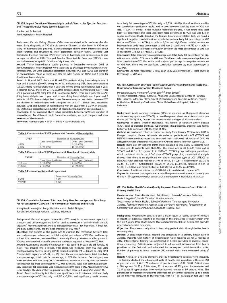
OR.153. Impact Duration of Haemodialysis on Left Ventricular Ejection Fractionand Tricuspid Annulus Plane Systolic Excursion
D.Y. Pertiwi, D. RostiatiBandung Regional Public Hospital
Backround: Chronic Kidney Disease (CKD) have associated with cardiovascular dis-eases. Early diagnostic of CVD (Cardio Vascular Diseases) as risk factor in CKD espe-cially of haemodialysis patients. Echocardiograph shown some information aboutheart function and structure to know association between theirs. Decrease LeftVentricular Ejection Fraction (LVEF) over time in haemodialysis patients may be dueto negative effect of CKD. Tricuspid Annulus Plane Systolic Excursion (TAPSE) is onemethod to measure systolic function of right ventricle.Method: Thirty haemodialysis stable patients in September-November 2018 atBandung Regional Public Hospital were subjected to evaluated by transthoracic echo-cardiographic. We were analyzed association between LVEF and TAPSE and durationof haemodialysis. Value of these are 50% for LVEF, 16mm for TAPSE and 1 year forduration of haemodialysis.Result: In Normal LVEF, there are 18 (60.00%) patients doing haemodialysis over 1year and 6 patients (20.00%) doing less of it. In decreasing LVEF, there are 6 patients(20.00%) doing haemodialysis over 1 year and no one doing haemodialysis less 1 year.In Normal TAPSE, there are 23 (76.67.00%) patients doing haemodialysis over 1 yearand 2 patients (6.67%) doing less of it. In abnormal TAPSE, there are 2 patients (6.67%)doing haemodialysis over 1 year and no one doing haemodialysis less 1 year and 3patients (10.00%) haemodialysis less 1 year. We were analyzed association between LVEFand duration of haemodialysis with chi-square test p 0.171. Beside that, associationbetween TAPSE and duration of haemodialysis with chi-square test p 0.049. In this anali-sis, TAPSE have associated with duration of haemodialysis, but LVEF does not have it.Conclusion: In our analyzed, there is has association between TAPSE and duration ofhaemodialysis. For different result from other analyses, we must compare and knowweakness of the research.Keywords: Haemodialysis • LVEF • TAPSE • Echocardiographic
OR.154. Correlation Between Total Lean Body Mass Percentage, and Total BodyFat Percentage to VO2 Max in The Viewpoint of Medicine and Nursing
Anang Basuki Maharjito1, Bunga Listia Paramita1, Endah Silfiyanti2
Rumah Sakit Olahraga Nasional, Jakarta, Indonesia
Background: Maximal oxygen consumption (VO2 max) is the maximum capacity totransport and utilize oxygen and is often used as a measure of an individual’s aerobiccapacity.4 Previous pertinent studies indicated body mass, fat free mass, % body fat,and body surface area, are the best predictor of VO2 max.2
Objective: The purpose of this paper was to examine the correlation between totallean body mass percentage, and or total body fat percentage to VO2 Max, and how sig-nificant it is. Moreover, we would like to know significancy between total body mass toVO2 Max compared with specific dominant body mass region (i.e. foot) to VO2 Max.Method: Quantitative analysis of 63 person (n¼ 63) aged 18-54 years old (18 female, 45male), was grouped into 2 groups. First group was measured their VO2 Max usingCardiopulmonary Exercise Testing (CPET) Cosmed treadmill (n1 ¼ 46) and or using CPETCosmed static ergocycle (n2¼17, n¼ 63), then the correlation between total lean bodymass percentage, total body fat percentage, to VO2 Max is tested. Second group wasmeasured their VO2 Max using CPET Cosmed static ergocycle (n2¼17), then the correla-tion between leg mass percentage to VO2 Max is tested. Total lean body mass percent-age, total body fat percentage was measured using DXA (dual X-Ray Absorbtiometry) GELunar Prodigy. The data of the two groups were then processed using SPSS version 16.Result: Based on linearity test there was significancy result between total lean bodymass percentage to VO2 Max (sig. ¼ 0,212 (>0,05)), and significancy result between
total body fat percentage to VO2 Max (sig. ¼ 0,734 (>0,05)), therefore there was lin-ear correlation significancy result, and so does between total leg mass to VO2 Max(sig. ¼ 0,947 (> 0,05)). In the multiple regression analysis, it was found that totalbody fat percentage and total lean body mass percentage to VO2 Max was 63% (rsquare coefficient 0,63). Based on the Pearson bivariate correlation test, we found asignificant negative correlation (inversely) between total body fat percentage to VO2Max (r coefficient ¼ - 0,794 (>r table ¼ 0,33)) and siginificant positive correlationbetween lean body mass percentage to VO2 Max (r coefficient ¼ 0,792 (> r table ¼0,33)). We found no significant correlation between leg mass percentage to VO2 Max(r coefficient ¼ 0,225 (< r table ¼ 0,468)).Conclusion: Total lean body mass percentage and total body fat percentage has sig-nificancy correlation 63% towards VO2 Max. Total lean body mass percentage has pos-itive correlation to VO2 Max while total body fat percentage has negative correlationto VO2 Max. there was no significant correlation between leg mass percentage toVO2 Max.Keywords: Leg Mass Percentage • Total Lean Body Mass Percentage • Total Body FatPercentage • VO2 Max
OR.155. Correlation between Type of Acute Coronary Syndrome and TraditionalRisk Factor of Coronary Artery Disease in Papua
Perdana R Kusuma Hermawan1, Ervan Zuhri2,3, Iman Setiadi41Freeport Hospital, Papua, Indonesia, 2National Cardiovascular Center of HarapanKita, Jakarta, Indonesia, 3Department of Cardiology and Vascular Medicine, Facultyof Medicine, University of Indonesia, 4Pasar Rebo General Hospital, Jakarta,Indonesia
Background: Acute coronary syndrome (ACS) can manifest as ST-segment elevationacute coronary syndrome (STEACS) or non-ST-segment elevation acute coronary syn-drome (NSTEACS). But, factors that correlate with the type of ACS are unclear.Objective: To assess whether traditional risk factors of coronary artery disease(CAD), such as diabetes mellitus, hypertension, dyslipidaemia, smoking, and familyhistory of CAD correlate with the type of ACS.Method: We conducted cohort retrospective study from January 2015 to June 2018 inFreeport Hospital, Papua, Indonesia. We collected patients with ACS (STEACS andNSTEACS) from medical record and searched their traditional risk factor of CAD. Weanalysed whether traditional risks factor of CAD had correlation with type of ACS.Result: There are 119 patients (100% men) included in this study, 72 patients withSTEACS and 47 patients with NSTEACS. The mean age is 48 (67.6) years old inSTEACS and 47.3 (68.1) years old in NSTEACS. STEACS group had higher prevalenceof all traditional risk factor of CAD than NSTEACS group. But, the statistical analysisshowed that there is no significant correlation between type of ACS (STEACS orNSTEACS) with diabetes mellitus (13.9% vs 10.6%, p¼ 0.811), hypertension (33.3% vs26.4%, p¼ 0.554), dyslipidaemia (97.2% vs 95.7%, p¼ 0.517), smoking (54.2% vs51.1%, p¼ 0.886), and family history of CAD (12.5% vs 10.6%, p¼ 0.986).Conclusion: Traditional risk factor of CAD did not correlate with type of ACS.Keywords: Acute coronary syndrome • non-ST-segment elevation acute coronary syn-drome • ST-segment elevation acute coronary syndrome • traditional risk factor
OR.156. Better Health Service Quality Improves Blood Pressure Control Visits inPrimary Health Care
Rio Alexsandro1, Rianty Febriandani1, Prily Prianty1, Novendy1, Andrew Parlautan,Maria P. Inggriani2, Sem D. Timothy2, Mustika Mahbubi31Departemen of Public Health, School of Medicine, Tarumanagara University,Jakarta, 2School of Medicine, Gadjah Mada University, Yogyakarta, 3Departemen ofCardiology and Vascular Medicine, Soewondo Hospital, Pati
Background: Hypertension control is still a major issue. A recent survey of Ministryof Health of Indonesia reported an increase in the prevalence of hypertension overthe last 5 years. Prior study showed that communication skills of health care provideraffects hypertensive outcomes.Objective: The present study aims to improving patient visits through better healthservice quality.Method: A quasi-experimental method was conducted in a primary health care inJakarta. Patients with history of hypertension were followed-up for 2 months in2017. Interventional training was performed on health providers to improve educa-tional counseling. Patients were subjected to educational intervention from healthproviders at the first visit and scheduled for subsequent post-intervention visits.Number of patients to blood pressure (BP) control visits were compared using v2
test.Result: A total of 6 health providers and 130 hypertensive patients were included.The training doubled the educational skills of health care providers, with mean6SDof pre-test score of 4661.78 and mean of post-test score of 89610.01. Patient mean(6SD) age was 51.25 (67.98) years, 82.1% women, 67.9% grade I hypertension and32.1% grade II hypertension. Intervention boosted number of BP control visits. Thepercentage of hypertensive patients presented for BP control increased up to 8 timeson the fifth visit compared to the first visit, with 14 (11%) patients on first visit; 104
F72 Abstracts
Dow
nloaded from https://academ
ic.oup.com/eurheartjsupp/article/21/Supplem
ent_F/F33/5570992 by guest on 07 July 2022

(80%) on second visit; 108 (83%) on third visit; 109 (84%) on fourth visit; and 112(86%) on fifth visit. There was a statistically significant increase in number of patientvisits directly after the intervention, which was observable on the second visit(p¼ 0.001), and remained high up to the fifth visit (p¼ 0.001).Conclusion: Educational counseling from health providers improves patients’ visits inhypertension control. Better health service quality may encourage BP control in pri-mary health care.Keywords: hypertension • blood pressure control • patient visits • primary health care
OR.157. Advanced Heart Failure: Demographic Characteristics of Patients withRecurrent Heart Failure Admissions
Niken Anthea S1,2, Nani Hersunarti1,2, Rarsari Soerarso1,2, Renan Sukmawan1,2,Ratna SD Gill1,2, Muhammad Isra Tuasikal1,2, Maruli Wisnu WB1,2, Christian Rendy C1,2,Athikah Khairunnisa1,2, Rienna Diansari1,2, Tommy Alexander1,21Department of Cardiology and Vascular Medicine Faculty of Medicine, UniversitasIndonesia, Jakarta, Indonesia, 2National Cardiovascular Heart Center Hospital,Jakarta, Indonesia
Background: Experts have estimated the prevalence of advanced heart failure (HF) torange from 6% to 25% of the heart failure population. Patients identified with the mostadvanced disease and worst prognosis can be identified using iterative, integrated clini-cal assessment of symptoms, effort intolerance, and cardiac dysfunction. Recognizingthe transition to advanced HF is necessary for its management. However, data regardingcharacteristics of patients with advanced HF is limited. This study was conducted todescribe demographic characteristics of patients with advanced HF.Methods: We analyzed data from inpatient’s registry in National CardiovascularCenter Harapan Kita from January 2017 until December 2018. Patients included inthis study were patients with clinical diagnosis of heart failure with LVEF �30% whoat least rehospitalized >1 in a year. The data were presented in mean (6SD) for nor-mal distribution data and in median (min-max) for abnormal distribution data.Results: There were 87 patients with advanced heart failure hospitalized fromJanuary 2017-December 2018. It counted for 25% of total heart failure cases. Medianage of the patients was 56 (20-92) years old. Mean rehospitalisation in a year was361.5 times (50.6% were hospitalized twice, 20.2% 3 times, 15.7% 4 times, and13.5% 5 or more times). Prevalence was higher for male than female (73.0% vs.27.0%). Mean ejection fraction of subjects was 20.464.2%. The most common etiol-ogy founded was coronary artery disease, followed by hypertensive heart disease andcardiomyopathies (71.9%, 12.4%, 12.4% respectively). Among those with CAD, thetraditional risk factors were hypertension (64%), diabetes mellitus (62.9%), smokinghistory (38.2%), dyslipidemia (12.4%), and family history (3.4%). Patients receivingRAAS blocker were 95.5%, beta blocker 95.5%, MRA 65.2%, and diuretics with optimaldose were 96.6%.Conclusions: In this study, patients with advanced HF count for 25%. The most com-mon etiology was CAD followed by HHD and cardiomyopathies. Patients receivedoptimal GDMT (guideline directed medical therapy) were 61.8%. Each subsequent HFhospitalization is associated with incremental increase in risk of death. Thus, morethan one HF hospitalizations in a previously stable chronic HF patient may herald theonset of advanced HF and should alert the care provider to a potential change in thecourse of the patient’s HF.Keywords: advanced heart failure • coronary artery disease • demographic charac-teristics • hypertensive heart disease
OR.158. Determination of Chronotropic Incompetence Using 24-hours HolterMonitoring : a Lower Cut-off is Needed
Sakta Suryaguna, Putra Antara, Nyoman WiryawanCardiology and Vascular Medicine of Udayana University, Bali, Indonesia
Background: Chronotropic incompetence (CI) is an important thing that is oftenoverlooked. CI is associated with an increased risk of developing atrial fibrillationand mortality, a poor prognosis in heart failure, and is one indication of the implan-tation of a pacemaker. However, there are obstacles in conducting CI studies becausethe lack of consistent methodology for determining CI. CI is usually determined bytreadmill strest test (TST), but in some conditions or patient preference, it cannotbe done and holter is an alternative. 24-hours Holter monitoring report often includechronotropic responses but the statement are subjective, even though the holteritself can provide more objective information.Objective: To analyze the usefulness of 24-hours holter monitoring in detecting chro-notropic incompetenceMethod: Retrospective analysis of data was performed with twenty two patientsunderwent a standard TST and 24-hours holter were included. Maximal HR, Age-pre-dicted Maximal Heart Rate (APMHR), and %APMHR achieved were calculated fromboth examination with additional parameter (circadian index) from holter monitor-ing. The threshold of %APMHR achieved using holter monitoring are evaluated at60%, 65% and 70%. Goodness of fit test of variables was assessed.Result: As CI is defined by inability to achieve 70% of APMHR with TST, the agreementanalysis result with kappa-test evaluation with cut-off of 70%, 65% and 60%APMHRwhile using holter have agreement value of 0.13 (63.6% concordant), 0.19 (72.7%
concordant), 1.0 (100% concordant) respectively. Meanwhile using circadian indexwith cut-off 1.24 and 1.10 have agreement value of 0.06 (45.4% concordant), 0.64(95.4% concordant) respectively.Conclusion: Determination of CI using holter monitoring need a lower cut-off (60%APMHR) for better agreement value. Although it only involves a small number of sam-ples so that its application in clinical settings is limited, this study can be the basisfor conducting larger studies so that the determination and management of CI isimproved.Keywords: Chronotropic • Incompetence • Holter
OR.159. Tolvaptan: Short Term Efficacy of Vasopressin-2-Receptor Antagonist asAcute Heart Failure Symptoms Reliever
Rizky E. P. Yuriza, Indah E. F. Wainsaf, Ahmad Z. AlhamidFaculty of Medicine, University of Indonesia, Jakarta, Indonesia; Faculty of Medicine,University of Papua, Sorong, Indonesia
Background: Congestion and volume overload symptoms are immediate goals treat-ment in acute heart failure (AHF). Tolvaptan as a vasopressin-2-receptor antagonisthas potential benefit to improve the symptoms and support diuretics to preventundesired condition in the acute phase.Objective: This review aimed to critically appraise tolvaptan roles in short termeffect. Ameliorated dyspnea, edema, body weight reduction, increased fluid loss areseveral expected findings at least in the first few days.Method: Relevant RCT and review articles were thoroughly searched from PubMed,EBSCO, ProQuest, and Cochrane Database of Systematic Reviews. Findings were nar-rowed down by using selection criteria. Three selected articles got criticallyappraised by using the Center of Evidenced-based Medicine, University of Oxfordguideline.Result: The analysis was carried out in 2 RCTs and 1 meta-analysis. Two RCTs com-pared Tolvaptan with placebo. On the first RCT, Tolvaptan results in more bodyweight reduction in the first 24 hours [-2.36 6 2.08 kg, p< 0.001] even though ame-liorated dyspnea is not significantly different. Another RCT with greater sampleshows significant difference between Tolvaptan and placebo to ameliorated dyspneain first few days [OR 1.2 (day 1) & 1.22 (day 2), p< 0.05], edema [OR 1.23 (day 1) &1.2 (day 2), p< 0.05], and orthopnea [OR 1.18, 1.28, 1.23, p< 0.05, (the first 3 daysrespectively)]. This result supported by meta-analysis study based on dyspnea [RR0.82; 95% CI 0.71 to 0.95], body weight reduction [WMD 1.35; 95% CI 0.75 to 1.96]and fluid loss [WMD 0.66; 95% CI 0.35 to 0.98]. There were no significant differenceand serious adverse events between Tolvaptan and control.Conclusion: Tolvaptan showed promising efficacy as symptoms reliever in the earlytreatment of acute heart failure. Further studies are needed to explore Tolvaptan assingle or combination therapy in AHF.Keywords: tolvaptan • acute heart failure • efficacy • short term
OR.160. A Meta-analysis of Efficacy and Safety of Extended Thromboprophylaxiswith Direct Oral Anticoagulant in Medically Ill Hospitalized Patients
Eka P. B. Mulia1, J. Nugroho E. Putranto1,2, Irma Maghfirah1, Ardianto Nandiwardhana11Faculty of Medicine, Universitas Airlangga, Surabaya, Indonesia, 2Department ofCardiology and Vascular Medicine, Dr. Soetomo General Hospital, Surabaya, Indonesia
Background: The risk of venous thromboembolism (VTE) is increased significantly inthe hospitalized patient for medical illness, and the risk persists after discharge. Therole of extended-duration pharmacological thromboprophylaxis is effective for theprevention of symptomatic VTE in high-risk surgical patients, but in hospitalizedmedical patients is still a subject of controversy.Objective: This review aimed to evaluate the efficacy and safety of direct oralanticoagulants (DOACs) for extended-duration thromboprophylaxis (ET) in thispopulation.Method: We conducted a systematic literature search from PubMed, Medline andEMBASE until April 2019. We used the Jadad scale to measure the quality of the stud-ies. A meta-analysis was performed to assess the efficacy and safety of ET of DOACs.Efficacy outcomes were defined by the prevention of symptomatic VTE, total VTE,and VTE-related death. Safety outcomes were defined by the occurrence of majorbleeding (MB) and clinically relevant non-major bleeding (CRNMB).Result: Four randomized controlled trials (RCT) with a total of 34.166 patients wereincluded for analysis. The quality of all studies was good. Among medically ill hospi-talized patients, extended DOACs recipients (30-45 days) had a significantly lowerrisk of symptomatic VTE (p¼0.0005, RR:0.59 (95%CI: 0.44-0.79), I2¼0%, absolute riskreduction (ARR)¼0.003, number needed to treat (NNT)¼303) and total VTE(p¼0.0008, RR:0.80 (95%CI:0.70-0.91), I2¼0%, ARR¼0.015, NNT¼68), but no signifi-cant reduction in VTE-related death (p¼0.17, RR:0.81 (95%CI: 0.60-1.10), I2¼0%).Safety outcome showed significantly higher risk of MB (p<0.0001, RR:1.96 (95%CI:1.40-2.75), I2¼37%, absolute risk increase (ARI)¼0.003, number needed to harm(NNH)¼315) and CRNMB (p<0.00001, RR:1.82 (95%CI: 1.54-2.16), I2¼74%, ARI¼0.009,NNH¼114).
Abstracts F73
Dow
nloaded from https://academ
ic.oup.com/eurheartjsupp/article/21/Supplem
ent_F/F33/5570992 by guest on 07 July 2022

Conclusion: ET with DOACs in medically ill hospitalized patients was associated witha significantly lower risk of symptomatic VTE and total VTE but was associated withsignificantly increased risk of MB as well as CRNMB.Keywords: extended thromboprophylaxis • direct oral anticoagulant • medically illhospitalized patients
OR.161. Frequency of Left Atrial Thrombus in Patients of Mitral Stenosis withAtrial Fibrillation at Sardjito General Hospital
Muhammad A Mukti, ErikaMaharani, Hendry P. BagaswotoDepartment of Cardiology and Vascular Medicine, School of Medicine, Gadjah MadaUniversity, Yogyakarta, Indonesia
Background: Left Atrium (LA) thrombus is a common source of thromboemboli and itis usually seen in Atrial Fibrillation (AF). In patients with Mitral Stenosis (MS) and AF,the risk of thrombus formation in the LA is very high. In 90% cases of AF, blood clothsform in the Left Atrial Appendage.Objective: To determine the frequency of left atrial thrombus in patients with mitralstenosis having atrial fibrillationMethod: This Cross Sectional study was conducted at Sardjito General Hospital. Bothmale and female patient between 25 to 65 years with mild to severe mitral stenosisand atrial fibrillation were included while patients with mitral stenosis and normalsinus rhythm were excluded. The severity of mitral stenosis was calculated by MitralValve Area (MVA), which the value less than 1 cm2 classified as severe, 1-1.5 cm2 asmoderate, and more than 1.5 cm2 as mild. Incidence of left atrial thrombus was seenby Trans Esophageal Echocardiography.Result: In this study a total of 152 patients were included with 27% patients weremales, about 18% patients were hypertensive, only 5% were diabetic, and 5% had BMI> 30. Patients who had mild, moderate, and severe mitral stenosis were 5%, 11%,and 84% respectively. Left Atrial thrombus found in 28% patients. There are no corre-lation between left atrial thrombus with age, sex, diabetes, and severity of Mitralstenosis with p-value 0.472; 0.275; 0.419; and 0.064 respectively.Conclusion: There are more incident of left atrial thrombus in the severe mitralstenosis group. Therefore proper screening for thrombus is needed in patient withAF, especially in severe mitral stenosis. But there is still much more predictor thatcan relate to the incident of left atrial thrombus, so we conclude that there is stillroom for improvement in this study.Keywords: Atrial Fibrillation • Mitral Stenosis • Left Atrial Thrombus
OR.162. Table 1
Variable Frequency (n) Percentage (%)
Age• 25-35 years
• 36-45 years
• 46-55 years
• 56-65 years
• 26
• 51
• 49
• 26
• 17,1 %
• 33.6 %
• 32,2 %
• 17,1 %Sex• Male
• Female
• 41
• 111
• 27 %
• 73 %Hypertension• Hypertensive
• Non hypertensive
• 27
• 125
• 17,8 %
• 82,2 %DIabetes• Diabetic
• Non DIabetic
• 7
• 145
• 4,6 %
• 95,4 %BMI• � 30
• > 30
• 145
• 7
• 95,4 %
• 4,6 %Mitral Stenosis• Mild
• Moderate
• Severe
• 7
• 17
• 128
• 4,6 %
• 11,2 %
• 84,2 %Left Atrial Thrombus• Thrombus
• No thrombus
• 42
• 110
• 27,6 %• 72,4 %
OR.162. Postoperative Atrial Fibrillation Following Cardiac Surgery
Aye Nandar Kyaw, Win Win Kyaw, Aung ThuDepartment of Cardiovascular, Yankin Children Hospital, University of Medicine 2,Yangon, Myanmar.
Background: New-onset postoperative atrial fibrillation is the most common form ofrhythm disturbance following cardiac surgery. In this study we are trying to examinethe potential risk factors for post-operative atrial fibrillation in our patients evaluat-ing short- and long-term outcome.Objective:
(1) To determine the Demographic data and clinical characteristics of the studypopulation
(2) To detect the occurrence of AF within postoperative 7 days after cardiacsurgery
(3) To determine the intraoperative and postoperative data of AF within postopera-tive 7 days after cardiac surgery
(4) To determine the complications of POAF within 30 days after cardiac surgery
Methods: A hospital-based cross sectional descriptive study of 120 patients withoutprior history of AF who underwent cardiac surgery in cardiovascular department ofYankin Children Hospital and North Okkalapa General Hospital between January,2018 to December, 2018. Perioperative data including gender, age and postoperativemorbidity and mortality were extracted from medical records.Results: The rate of POAF was 42% and was higher following CABG (34%) than afterDVR (24%) or MVR (22%). In general, patients with POAF were more often male, weremarginally siginificant older, had diabetes and smoking. Patients who developedPOAF had lower ejection fraction, large left atrial volume, prolonged CPB time andACX time, valve surgeries.Conclusion: In our study, male gender, enlarged LA volume, lower ejection fraction,CABG surgeries were found to be the risk of atrial fibrillation after cardiac surgery.Keywords: Postoperative atrial fibrillation • Coronary artery bypass surgery •Double valve replacement • Mitral valve replacement
OR.163. Patency Assessment of Coronary Artery bypass Grafts by Using TransitTime Flow Meter
M.P. Khine, K.M. Lwin, A.Z. MyoDepartment of Cardiovascular Surgery, Yangon General Hospital
Background: A widely accepted technique of graft assessment in coronary arterybypass surgery is transit time flow measurement. This technique is very effective andquick method for intraoperative graft flow assessment in coronary artery bypassgrafting. The aim of this study was to assess the patency of the grafts by usingTransit Time Flow Meter (TTFM ) during CABGMethod: This study was hospital based prospective descriptive study who underwentcoronary artery bypass surgery only from January 2018 to December 2018.Result: In this study, total number of patients was 66. 68.2% of patients had EF> 35%and mean flow >30ml/min, 30.5% had EF> 35% and mean flow <30ml/min, 1.5% hadEF< 35% and mean flow >30ml/min. Among the patients who did not have wallmotion abnormality, 65.2% had mean flow >30ml/min, 22.7% had mean flow <30ml/min. 4.5% of patients who had mean flow >30ml/min and 7.6% who had mean flow<30ml/min had wall motion abnormality .1.5% was EF> 35% and Pulsaltility Index(PI)>5, 97% was EF> 35% and PI< 5. 1.5% was EF< 35% and PI< 5. 1.5% of thepatients who did not have wall motion abnormality had PI> 5and 86.4% had PI< 5and 12.1% had wall motion abnormality and PI< 5.Conclusion: Measurement of coronary blood flow can assess early graft function andpredict early clinical outcomes. This study intends to assess graft patency intra-oper-atively and to measure the flow of the grafts after anastomosis of the patients whoundergoing CABG.Keywords: Transit Time Flow Meter • Pulsaltility Index • Coronary Artery BypassGrafts,Ejection Fraction
OR.162. Table 2
Age p-value
25-35 years 36-45 years 46-55 years 56-65 years Total
Left Atrial Thrombus Thrombus 8 10 16 8 42 0,472No Thrombus 18 41 33 18 110Total 26 51 49 26 152
F74 Abstracts
Dow
nloaded from https://academ
ic.oup.com/eurheartjsupp/article/21/Supplem
ent_F/F33/5570992 by guest on 07 July 2022

OR.164. Invasivepharmacology Strategy Intralesion Single Bolus Administrationof Eptifibatide and Alteplase into Infarct Related Artery during PrimaryPercutaneus Coronary Intervention in Acute STEMI Patients Within 12hoursSymptom Onset
Zulfikri Mukhtar1, Nizam Z Akbar2, Henry Panjaitan3, Nora C Hutajulu3,Sutomo Kasiman2, Harris Hasan41Faculty of Medicine, Universitas Prima Indonesia / Adam Malik General Hospital /Murni Teguh Memorial Hospital, Medan, Indonesia., 2Adam Malik General Hospital /Murni Teguh Memorial Hospital., 3Murni Teguh Memorial Hospital / RS St Elisabet,Medan, Indonesia., 4Cardiology Department Faculty of Medicine, UniversitasSumatera Utara / Adam Malik General Hospital
Background: Acute STEMI patients have complete occlusion of a major epicardialcoronary artery caused by fresh thrombus and accompanied by vasoconstriction atmicrovascular level.Objective: Intralesion (IL) single bolus administration of eptifibatide and alteplasedissolve intracoronary occlusive or residual thrombus and distal microembolizationafter manual thrombectomy during primary PCI in acute STEMI patients (TIMI 0 /1)within 12 hours symptom onset.Method: Prospective nonrandomized controlled study. During the primary PCI (pPCI)procedure, after wiring and recanalizing by manual thrombectomy or balloon infla-tion, we administered local IL bolus of eptifibatide 3.75mg, followed by Alteplase 3.5mg and administered local intracoronary nitroglycerine (NTG) 200 mg after stentdeployment. Echocardiographic assessment Pre and Postprocedure. The controlgroup is colleagues’ patients with standard pPCI. TIMI, MBG flow and corrected TIMIFrame Count (cTFC) were graded by two independent interpreters.Result: There were 165 patients from October 2016 to January 2019, age 26 - 81 y,symptom onset was 1.0-12 hours, 145 males and 20 females, 87 anterior and 78 infe-rior wall infarction. Preprocedural TIMI and MBG 0 were 134 patients, TIMI and MBG 1were 31 patients. Postprocedural TIMI and MBG 3 were 143 patients, TIMI and MBG 2were 22 patients, door to wire crossing time (DWCT) was 26-250minutes, median-cTFC was 17.6, LVEF increased 7.44-31.5%. 2 patients with gum bleeding, 2 patientshaematuria, hospital mortality was 1.8%. There were 3 patients hospitalized and nomortality during 30 days follow-up. A control of 165 colleagues’ patients, age 35 - 87y. Symptom onset was 1.0-12 hours, 130 males and 35 females, 89 anterior and 76inferior wall infarction. Preprocedural TIMI and MBG 0 were 145 patients, TIMI andMBG 1 were 20 patients. Postprocedural TIMI and MBG 3 were 94 patients, TIMI andMBG 2 were 67 patients, TIMI and MBG 1 were 4 patients. DWCTwas 25-309minutes,m-cTFC was 22.0. Statistically, there was significant difference in postproceduralTIMI, MBG flow, cTFC between the 2 groups (p< 0.05).
Conclusion: Intralesion administration of eptifibatide and alteplase showed a suc-cessful dissolved of occlusive thrombus, improved reperfusion microvascular leveland Left Ventricle function in acute STEMI patients.Keywords: STEMI • pPCI • TIMI • MBG • cTFC.
OR.165. Correlation Between P Terminal Force in lead V1 and E/e’ ratio inChronic Kidney Disease Patients on Routine Hemodialysis
Kartika Apshanti, Hasanah MumpuniDepartment of Cardiology and Vascular Medicine, Gadjah Mada University, Jogjakarta
Background: Chronic kidney disease (CKD) patients represent a subset of populationwith special clinical and haemodynamic features that differ from common person.Chronic volume and pressure overload, chronic inflammation, oxidative stress, athe-roslerotic acceleration and non physiological fluid shift in hemodialysis have all putmyocardium to face enormous burden and damage leading to inevitable cardiacremodelling that finally can cause cardiac dysfunction with increased cardiac fillingpressures. In previous studies, P terminal force in lead V1 (PTFV1) has been linkedwith not only left atrial size but also left ventricular filling pressure.Objective: Whether PTFV1 correlates with left ventricular filling pressure measuredby E/e’ ratio in CKD patients is our study interest.Method: This cross sectional study was done in CKD patients on hemodialysis in Dr.Sardjito Hospital Jogjakarta. Electrocardiogram and echocardiography were done afterhemodialysis procedure. PTFV1 was measured by multiplying amplitude and duration ofterminal negative portion of the P wave in lead V1, with value of more than40mm.msec is considered to be abnormal. E velocity measurement was obtained frompulsed-wave Doppler in apical 4 chamber view, sample volume was placed between themitral leaflet tips. Annular pulsed wave Doppler tissue imaging was also derived fromapical 4 chamber view, with e’ is the average of septum and lateral e’ value.Result: This study was done in 71 patients with mean age 50 years old. Forty threesubjects (61%) were men. Sixty four subjects (90%) had hypertension. Sixty twopatients (87%) had normal left ventricular ejection fraction. Forty four subjects(62%) had abnormal PTFV1. Median PTFV1 value was 44mm.msec. By correlationanalysis, it is found that there is significant correlation between PTFV1 value and E/e’ ratio (r¼ 0.265, p¼ 0.027). PTFV1 value also shows significant correlation with LAsize (r¼ 0.4, p¼ 0.001) and LAVI (r¼ 0.255, p¼ 0.032).Conclusion: The present study shows significant correlation between P terminalforce in lead V1 and left ventricular filling pressure measured by E/e’ ratio in CKDpatients on routine hemodialysis.Keywords: P Terminal Force V1 • E/e’ ratio • chronic kidney disease • hemodialysis
OR.162. Table 3
Sex p-value
Male Female Total
Left Atrial Thrombus Thrombus 14 28 42 0,275No Thrombus 27 83 110Total 41 111 152
Hypertension p-valueHypertensive Non-Hypertensive Total
Left Atrial Thrombus Thrombus 5 37 42 0,243No Thrombus 22 88 110Total 27 126 152
Diabetes p-valueDiabetic Non-Diabetic Total
Left Atrial Thrombus Thrombus 1 41 42 0,419No Thrombus 6 104 110Total 7 145 152
BMI p-value� 30 > 30 Total
Left Atrial Thrombus Thrombus 41 1 42 0,419No Thrombus 104 6 110Total 145 7 152
OR.162. Table 4
Mitral Stenosis p-value
Mild Moderate Severe Total
Left Atrial Thrombus Thrombus 1 1 40 42 0.064No Thrombus 6 16 88 110Total 7 17 128 152
Abstracts F75
Dow
nloaded from https://academ
ic.oup.com/eurheartjsupp/article/21/Supplem
ent_F/F33/5570992 by guest on 07 July 2022

OR.166. KARIADI Risk Score: An Attempt to Stratify Intensive Care Needs AfterPrimary Percutaneous Coronary Intervention
An A. Asrial, Yan Herry, Novi Anggriyani, Suhartono, BahrudinDepartment of Cardiology and Vascular Medicine, Faculty of Medicine DiponegoroUniversity, Semarang/ Dr. Kariadi General Hospital, Semarang-Indonesia
Background: Reduction of major cardiovascular events (MACE) in ST-Segmen ElevationMyocardial Infarction (STEMI) patients has been achieved by primary percutaneous coro-nary intervention (PCI) strategy and intensive care management. However, the inten-sive care unit (ICU) bed avaibility and cost remain a problem for those patients, andthus risk stratification using an objective risk score instrument is required.Objective: To develop a risk score of in-hospital MACE for patients with STEMI under-went primary PCI.Methods: A cohort study of 208 patients with STEMI undergoing primary PCI at theDr. Kariadi General Hospital Semarang. Predictor analysis was carried out usingbivariate Chi-Square test and multivariate logistic regression. The quality of the riskscore was tested by the Hosmer and Lemeshow calibration test and AUC ROC analysisfor discrimination test.Results: Seven independent predictors, i.e. Killip class (OR 20.04, p¼ 0.0001), age(OR 3.02, p¼ 0.04), renal insufficiency (OR 9.48, p¼ 0.007), infark related arteryfinal TIMI flow (OR 11.57, p¼ 0.001), admission systolic blood pressure (OR 3.04,p¼ 0.025), duration of total ischaemic time (OR 3.14, p¼ 0.032) and increase ofblood glucose levels (OR 3.04, p¼ 0.029) were fulfilled the criteria for risk scores ofin-hospital MACE. The risk scores had a good quality with the Hosmer and Lemeshowcalibration test> 0.05 and AUC ROC 0.886 (95% CI, 0.827-0.944, p< 0.005).Conclusions: A risk scoring model consisting of 7 independent predictor variables i.e.Killip class, Age, Renal insufficiency, Infark related artery final TIMI flow, Admissionsystolic blood pressure, Duration of total ischaemic time and Increase of blood glu-cose levels (KARIADI) has a good calibration and discrimination in predicting the riskof in-hospital MACE in patients with STEMI underwent primary PCI.Keywords: ST-segment elevation myocardial infarction • primary PCI • risk score.
OR.167. Blood Lactate to Predict Mortality in Patient with Acute MyocardialInfraction
Katherine G. Sugiarto, Winardi E. Setiawan, Fandi Ahmad, Donny S. Syamsul,Muhammad H. HasyimTarakan Regional Public Hospital, North Kalimantan, Indonesia
Background: Acute myocardial infraction (AMI) is a pathological processes cause bythrombosis after rupture of atherosclerotic plaque that lead to ischemic condition.During ischemia, glycolysis process accelerates then produces lactate and hydrogen.The blood lactate can be significantly increase in condition such as sepsis, acute cor-onary syndrome, and correlated to shock cardiogenic.Objective: This study aimed to predict the mortality risk in patient with AMI usingblood lactate.Method: Total sample was 49 patients with AMI at Tarakan Hospital form 2017 to2018. Data was collected form Tarakan registry using consecutive sampling. Lactatedata were taken at the admission and the risk of mortality was assessed using GRACEscore. Data were analysed using Pearson correlation and ROC curve.Result: There was a strong correlation between log lactate levels and GRACE score(r¼ 0,47, p< 0,01). Using cut-off point of blood lactate 2.5mmol/L, there was a sig-nificant mean difference of GRACE score (blood lactate �2.5mmol/L vs< 2.5mmol/L ¼ 149.30 vs 113.17, p< 0.01). The analysis was continued to determine blood lac-tate cut-off of each type of AMI to predict high risk mortality based on GRACE score.Lactate level �2,12mmol/ L in STEMI patients increase the risk of in-hospital mortal-ity rate > 5% (sensitivity 85%, specificity 62%, p¼ 0,01) whereas lactate level inNSTEMI patients was �3.15mmol/L (sensitivity 80%, specificity 90%, p¼ 0,05)increase the risk of in-hospital mortality rate >3%.Conclusion: This study shows blood lactate level can be use predict mortality patientwith AMI.Keywords: Blood lactate • in-hospital mortality • GRACE
OR.168. The Correlation of Albuminuria with Silent Myocardial Ischemia andDelayed Heart Rate Recovery in Type 2 Diabetes Mellitus
Natalia Ch. I. Polii1, Ira Posangi1, Agnes L. Panda1, Edmond L. Jim1, JanryA. Pangemanan1, F. L. Fredrik, G. Langi21Department of Cardiology and Vascular Medicine, Faculty of Medicine, SamRatulangi University, Manado, Indonesia, 2Faculty of Public Health, Sam RatulangiUniversity, Manado, Indonesia
Background: Albuminuria indicates endothelial dysfunction of the renal blood vesselwhich may lead to other diabetes vascular complications, especially cardiac auto-nomic neuropathy and silent myocardial ischemia.Objective: This study aims to prove whether or not albuminuria increases the likeli-hood of SMI and delayed HRR in asymptomatic type 2 diabetic patients.Method: The design of this study was observational with cross sectional approach. Itwas conducted on January - March 2018. All subjects who met the inclusion criteria
went through anamnesis, physical examination, laboratory test, electrocardiographyand echocardiography examination. Subjects with normal ECG and LV function under-went treadmill stress test subsequently. Statistical analysis with logistic regression wasused to determine the correlation between albuminuria with SMI and delayed HRR.Result: From a total of 57 subjects, there were 37 (65%) male and 20 female (35%)with a mean age of 53 years. Logistic regression analysis showed that asymptomatictype 2 diabetes patients with albuminuria had increased probability of SMI (OR 4,5395% CI 1,36 - 15,07; p¼ 0,014), which increased even higher in those with older age,male gender and smoking history (OR 5,56 95% CI 1,42 - 21,77; p¼ 0,014). There wasno statistical difference on delayed HRR between both groups. In ROC curve, theAUC for albuminuria in predicting SMI was 0,67 (sensitivity 88%, specificity 50%, posi-tive predictive value 58% and negative predictive value 84%). This proved that albu-minuria alone was not good enough to predict SMI.Conclusion: Albuminuria increases the probability of SMI four times higher in asymp-tomatic type 2 diabetes mellitus patients. Albuminuria in the presence of older age,male gender, and smoking history increases the probability of SMI and the accuracyon predicting its occurrence.Keywords: albuminuria • silent myocardial ischemia • diabetes
OR.169. High Atherogenic Coeficient Value Correlated with Coronary ArteryStenosis Severity in Stable Coronary Artery Disease
G.A.P. Astara1, I.G.N.P. Gunadhi2, W. Gotera31,2Cardiology Department, Faculty of Medicine, Udayana University/ Sanglah GeneralHospital, Bali, Indonesia, 3Internal Medicine Department, Faculty of Medicine,Udayana University / Sanglah General Hospital,Bali, Indonesia
Background: Dyslipidemia is one of well known classical risk factor of atherosclerosisand coronary artery disease (CAD). Atherogenic coefficient (AC) is a lipid ratio whichreflects the state and amount of atherogenic and non-atherogenic cholesterols. Thisstudy investigated the correlation of AC and coronary artery stenosis severity in sta-ble CAD patients.Method: The study used cross sectional design, which enrolled 66 subjects in SanglahGeneral Hospital Bali whom already undergone coronary artery catheterization, fromDecember 2018 until February 2019. Fasting lipid profile result taken to measure ACusing the formula of : (total cholesterol – HDL cholesterol ) / HDL cholesterol.Result: We divided the study subjects into high AC groups and low AC groups basedon the AC cut off value of 3.83. Degree of coronary artery stenosis was measuredusing SYNTAX score. 32 subjects were in group with low SYNTAX score (<22), and 28subjects (42%) had SYNTAX score >22. Bivariate analysis showed significant correla-tion of high AC score with higher SYNTAX score (PR 8.24, p¼ 0.001, 95% CI 2.6-26.1).Multivariate analysis showed significant correlation of high AC score with severe cor-onary artery stenosis with regression coefficient 1.80 (OR 6.0, p¼ 0.019, 95% CI 1.3-27.5), along with other classical risk factors such as smoking (OR 18.4, p¼ 0.006, 95%CI 2.3-146.4), diabetes (OR 18.9, p¼ 0.001, 95% CI 3.3-108.4), and hypertension (OR31.8, p¼ 0.001, 95% CI 4.4 – 225.6).Conclusion: This study showed significant correlation of high AC score with severecoronary artery stenosis in stable CAD.Keywords: atherosclerosis • coronary artery stenosis severity • SYNTAX score • sta-ble coronary artery disease • atherogenic coefficient
OR.170. Tryptase as a New Horizon Biomarker in Acute Coronary Syndrome: AMeta-Analysis
Ivana P. Dewi1, Louisa F. K. Wardhani1, Oryza Sativa1, Budi B. Dharmadjati21Faculty of Medicine, Airlangga University, Surabaya, Indonesia, 2Departement ofCardiology and Vascular Medicine, Dr. Soetomo General Hospital, Surabaya, Indonesia
Background: Acute coronary syndrome (ACS) was initiated by the rupture or erosionof atherosclerotic plaque. Tryptase has recently been reported to be involved inatherosclerotic plaque destabilization. Release of inflammatory mediators derivedfrom tryptase mast cell degranulation plays a role in process of atherosclerosis thatcausing ACS.Objective: The aim of this meta-analysis was to assess the tryptase level in ACSpatients as a potential biomarker in diagnosis and prognosis of ACS.Method: Major medical databases (PubMed, MEDLINE, Clinical Key, Cochrane Library,EMBASE) were systematically searched using terms “tryptase, mast cell, acute myo-cardial infarction, biomarker”. The search was limited to clinical trials publishedwithin the last ten years, human subjects, written in English, full-text availability.We used the 22-item STROBE checklist to measure the quality of evidence.Result: Six clinical studies one cohort study and five case-control studies met ourinclusion criteria with a total of 736 patients for analysis. The scientific quality of allsix studies were good. Five of the study showed a significant increase of tryptaselevel in ACS group compare to control (MD 1.85; 95%CI 1.54-2.15; p¼ 0.0004;I2¼80%). Two studies assessed the relation between tryptase levels with majoradverse cardiovascular events (MACE). There is significant correlation between tryp-tase levels measured during admission with development of MACE up to 2 years(p< 0.0001). Several limitations are non-random trial assignment, various popula-tion, and small sample size.
F76 Abstracts
Dow
nloaded from https://academ
ic.oup.com/eurheartjsupp/article/21/Supplem
ent_F/F33/5570992 by guest on 07 July 2022

Conclusion: Higher tryptase level among patients with ACS may indicate an impor-tant role in the pathophysiology of ACS. There is also possible benefit of tryptase asa biomarker in long-term prognostic of ACS.Keywords: tryptase • mast cell • acute coronary syndrome • prognosis
OR.171. Heart Rate Recovery: a Simple Marker of Autonomic ModulationImprovement After Cardiac Rehabilitation
S. Cancerita, H. Hasan, A. Nafiah, A.H. RaynaldoDepartment of Cardiology and Vascular Medicine, School of Medicine, UniversitasSumatera Utara, Medan, Indonesia
Background: Exercise-based cardiac rehabilitation has been shown to be beneficialand reduce mortality among cardiac patients. A hypothesis invokes the effect ofexercise on autonomic tone by modifying the sympathovagal control of heart ratetoward an increase in parasympathetic tone. Heart rate recovery (HRR) is a simplenoninvasive assessment of cardiac autonomic activity. This study aimed to evaluatethe effect and correlation between phase II cardiac rehabilitation programs on HRRin patients after Coronary Artery Bypass Graft (CABG).Methods: A retrospective study was conducted from October to December 2018.Patients who completed phase 2 cardiac rehabilitation and had entry and exit exer-cise stress tests (n¼ 30) were included in the study. Then HRR, functional capacityand six minute walking test at baseline and on follow-up were compared. The studyobserved changes in various parameters and analyzed the relationship between car-diac rehabilitation programs and HRR.Results: Following the completion of phase 2 cardiac rehabilitation, the HRR improvedsignificantly from 11.962.8 bpm to 1863.3 bpm (p< 0 .001). Statistically significantimprovement in body mass index (26.0864.2kg/m2 to 25.3863.9kg/m2), six minutewalking distance (192.97674.1 m to 3646128.99 m), and functional capacity(2.6460.42 METs to 6.661.30 METs) were observed with p< 0.001. Furthermore, astrong positive correlation was found between HRR and functional capacity before (r0.811; p< 0.001) and after cardiac rehabilitation (r 0.755; p< 0.001).Conclusion: Exercise training in a cardiac rehabilitation program improves HRR, sixminute walking distance, and functional capacity. HRR may be used in a cardiacrehabilitation facility as a simple parameter for assessing autonomic tone, to identifypatients with higher risk profiles, and can be useful for evaluating patient outcomes.Keywords: heart rate recovery • cardiac rehabilitation
OR.172. The Gensini Score as an Independent Predictor for the Reduced LeftVentricular Ejection Fraction
I Gede P. G. Semita1,2, Makhyan J. Al-Farabi1,3, Radityo B. Wicaksono1, Ruth I. Gunadi1,B. Jovie2, I Gde R. Suryawan11Department of Cardiology and Vascular Medicine, Faculty of Medicine, University ofAirlangga, Surabaya, Indonesia, 2Department of Cardiology and Vascular MedicineIndonesian Navy Hospital of Dr. Ramelan, Surabaya, Indonesia, 3PostgraduateStudent, University College London, United Kingdom
Background: Coronary artery disease (CAD) is the most common cause of HF (heartfailure) and has been reported to account for 23–73% of reduced left ventricularejection fraction (LVEF). The direct association among both remains unconcluded.Objective: We analyze the relationship between the Gensini score as CAD severityassessment with the LVEF.Methods: Gensini score, anthropometric and laboratory findings of the 168 patientswho underwent coronary angiography were consecutively collected. LVEF level wasexamined using Vivid S60 Ultrasound instrument (General Electric) and categorizedinto normal LVEF (LVEF>55%) and reduced LVEF (LVEF�55%)Results: Patient with normal LVEF had significantly lower Gensini score than those withreduced LVEF (26.20 634.86 vs 45.24646.51; p¼ 0.007). Multiple stepwise linearregression analysis showed that the Gensini score was significantly and independentlyassociated with the LVEF level (b¼-0.223, p¼ 0.004). Multivariate logistic regressionanalysis also revealed that the Gensini score >36.5 increased risk of having reducedLVEF (OR¼ 2.612, CI¼ 95%, p¼ 0.004). Receiver operating characteristic (ROC) curveanalysis showed that the Gensini score [area under the curve (AUC), 0.619; 95% confi-dence interval (CI), 0.527–0.710; p¼ 0.011], were predictive of normal LVEF. Cut-offpoint analysis showed that the Gensini score >36.5 (sensitivity ¼ 70.9% and specificity¼ 51.7%) provided the highest predictive value for reduced LVEF.Conclusion: The Gensini score as the coronary atherosclerosis severity assessmentcan be a significant predictor for reduced LVEF while being independent of othermajor risk factors including blood pressure, age, and BMI.Keywords: gensini score • atherosclerosis severity • coronary artery disease • leftventricular ejection fraction.
OR.173. Association of CYP2C19 Polymorphism and Myocardial Reperfusion inPatients with Acute Myocardial Infarction Undergoing Percutaneous CoronaryIntervention
Maya R Amalia, Aldila N Sulma, Robert A Raharjo, Rahageng W Kusuma, Ilham Uddin,Susi HerminingsihDepartment of Cardiology and Vascular Medicine, Diponegoro University, Semarang
Background: Dual antiplatelet (DAPT) is a mandatory in the setting of acute myocar-dial infarction (AMI). Clopidogrel is one of drug of choice of DAPT, given to patientsbased on different underlying conditions such as high risk of bleeding state. Currentguidelines suggest that loading with clopidogrel, along with aspirin, is necessary toAMI patients undergoing percutaneous coronary intervention (PCI), as soon as thefirst medical contact, to prevent further thrombosis. Administrating clopidogrel priorto PCI with stent implantation improves myocardial reperfusion according to severalstudies. Nevertheless, clopidogrel bioavalability could be affected by the reducedhepatic metabolism activity of CYP2C19 genes, furthermore of *2 and *3 alelles.Objective: This study aims to analyze the association of CYP2C19 polimorphism andmyocardial reperfusion in AMI patients undergoing PCI with stent implantation.Method: 23 consecutive patients from February to March 2019 with AMI undergoingPCI with stent implantation were included in this study. Loading with clopidogrel isgiven to all patients prior to PCI. Myocardial perfusion is evaluated and valued asfinal TIMI flow and myocardial blush grade (MBG) after stent implantation. CYP2C19genotipe examination was performed after PCI and analyzed by polymerase chainreaction (PCR) method. This is a cross sectional study using t test and fischer exacttest for univariate analysis, and multiple regression for final multivariate analysis.Result: We found mean age 5967 yo, BMI 21,860,7 (normoweight), STEMI 73,9%,NSTEMI 21,7%, UAP 4,3%. Polimorphism of CYP2C19 fenotipes were found as inter-mediate 26,1% and normal 73,9%. Patient characteristics that correlated with MBGscore were TIMI flow (p¼ 0,001) and trombosit level (p¼ 0,032), also polimorphism(p¼ 0,03). From multivariate analysis TIMI flow was the only variable which affectedMBG (P¼ 0,001, OR 0,63 95% CI 0,34-1,20).Conclusion: CYP2C19 polimorphism is significantly correlated to MBG, but only TIMIflow was found significant to affect MBG.Keywords: clopidogrel • dual antiplatelet • CYP2C19 polimorphism • myocardialreperfusion • acute myocardial infarction
OR.174. Clinical Characteristic Of Patients With Chronic Total Occlusion In Dr.Kariadi General Hospital Semarang
M. Perdana1, A. Pudjiastuti1, A. Sidiek1, LV Bramantyo1, SN Sofia1, S. Rifqi11Department of Cardiology and Vascular Medicine, Faculty of Medicine DiponegoroUniversity, Dr. Kariadi General Hospital, Semarang, Indonesia
Background: Chronic total occlusions (CTO) was found in 7-20 % of patients with cor-onary artery disease (CAD) undergoing coronary angiography. CTO patients had vari-ous clinical presentation from silent ischaemia to overt heart failure. Lack of litera-ture describe the characteristics of CTO patients in Indonesia. This study tried toinvestigate the clinical characteristics of patients with CTO in Dr.Kariadi GeneralHospital Semarang.Methods: We retrospectively reviewed the CTO patients between January 2018 toDecember 2018, in Dr. Kariadi General Hospital Semarang. CTO were confirmed from thecoronary angiographic showed no flow (TIMI flow 0) from antegrade other than from col-lateral with estimated occlusion time more than 3 months. Estimation CTO durationwere analyzed from prior angiography data, clinical data, and collateral development.Results: We had 168 patients included in this study and 92,9% patients were male.Mean age were 57,2þ8,37 years, mean BMI were 24,7þ3,38 kg/m2. The 42,3%patients diagnosed as heart failure and 45,8% patients as stable angina, while 11,9%CTO lessions found as non-culprit lession in acute coronary syndrome. We found52,4% patients had dyslipidemic, 56,5% had hypertensive; 40,5% had Diabetic, 57,7%were all male smoker, and 21,4% had positive family history of CAD. The CTO lesionswere in LAD (53%), followed by RCA(38,1%), and LCX (8,9%). The presence of Qwaves, ST Depression, and T wave inversions in respective CTO teritority wereobserved in 47%, 14,3%, and 38,7% patients. The presence of Q wave in anterior leadfrom LAD CTO were 31% while the Q wave in inferior from RCA CTO were 42%.Conclusion: The incidence of CTO lesion was more common in male than in female.Most clinical presentation was stable angina. The presence of CTO were not alwaysfollowed by the presence of Q wave in electrocardiography of CTO territory.Keywords: Chronic total occlusions • Clinical • Electrocardiography •Characteristics
OR.175. Factors Associated with Successful Pharmacological RevascularizationTherapy among Patients with ST Elevation Myocardial Infarction at CibinongHospital from January 2016 – February 2019
Sonya Aprelladiva, Dian Y. Hasanah, Fahmi I. Shahab, Radityo PrakosoFaculty of Medicine, University of Indonesia, Cibinong Hospital, West Java,Indonesia, Division of Pediatric Cardiology and Congenital Heart Disease, Departmentof Cardiology and Vascular Medicine, Faculty of Medicine, University ofIndonesia, National Cardiovascular Heart Center Harapan Kita Hospital, Jakarta,Indonesia
Background: ST elevation myocardial infarction (STEMI) is a type of acute coronarysyndrome that is recognized as the leading cause of death worldwide. Definitivemanagement of STEMI, particularly those with the onset of< 12 hours, includesrevascularization therapy, either through pharmacological revascularization
Abstracts F77
Dow
nloaded from https://academ
ic.oup.com/eurheartjsupp/article/21/Supplem
ent_F/F33/5570992 by guest on 07 July 2022

(fibrinolysis) or through mechanical intervention (primary percutaneous coronaryintervention); however, majority of healthcare centres in Indonesia do not have anyPCI facilities.Objective: This study aimed to identify factors associated with successful fibrinolysisamong STEMI patients at Cibinong Hospital from January 2016 – February 2019.Method: A cross-sectional study was conducted in a retrospective manner. The datawere collected consecutively. The study samples include STEMI patients whose onsetwas <12 hours who received pharmacological revascularization therapy (fibrinolysis)at Cibinong Hospital from January 2016 – February 2019. Bivariate analyses using Chi-Square test, Fisher exact, Kolmogorov-Smirnov, and independent student T test wereperformed appropriately for assessing the significance of each factor.Result: Of 44 STEMI patients who underwent fibrinolysis at Cibinong Hospital fromJanuary 2016 – February 2019, the treatment was successful in 27 (61%) patients.From the analysis, the onset of chest pain (p¼ 0.037), TIMI score (p¼ 0.018), Killipclass (p¼ 0.018), and smoking (p¼ 0.022) were found to significantly associated withsuccessful fibrinolysis. Regarding the electrocardiographic (ECG) features, the pres-ence of pathological Q wave (p¼ 0.007) was a significant factor associated with suc-cessful fibrinolysis. Conversely, the ST segment amplitude on the lead with the high-est elevation (p¼ 0.216) and QRS duration (p¼ 0.289) were not proven to besignificant.Conclusion: In this cohort, the onset of chest pain, TIMI score, Killip class, smokinghabit, and the presence of pathological Q wave are associated with successful phar-macological revascularization therapy among STEMI patients at Cibinong Hospitalfrom January 2016 – February 2019.Keywords: fibrinolysis • STEMI • early onset
OR.176. Comparison Electrocardiographic Criteria for Left VentricularHypertrophy According to Genre in Hypertension Heart Disease Patient
Muhamad Fakhri, Giky KarwikyHermina Hospital, Sukabumi, Indonesia
Background: Electrocardiogram (ECG) is the most common diagnostic tools for evalu-ating left ventricular hypertrophy in cardiac disease. Several factors are known tointerfere with ECG sensitivity, with the genre being one of the most important ones.Objective: The aim of this study was to compare sensitivity between ECG criteria forleft ventricular hypertrophy (LVH) according to the genre.Method: A retrospective cross-sectional study was carried out in 71 patients(37 females and 34 males) with history and clinical profile of essential hypertensionthat leads to LVH, as evidenced by echocardiography and/or electrocardiography.Data research was collected in Hermina Hospital, Sukabumi. We compare sensitivityand specificity between Sokolow Lyon, Cornell, Romhilt-Estes, and Paguero criteriabetween genre. Sensitivities and specificities were estimated using receiver-operatingcharacteristic (ROC) curve in relation to Left Ventricle Hypertrophy using echocardiog-raphy as standard (LVM/BSA>95g/m2 in female and LVM/BSA>115 g/m2 in males).Results: Sokolow Lyon criteria was able to diagnose LVH in 25 patients with 91.7%specificity and 42.4% sensitivity, whereas Cornell criteria was able to diagnose LVH in14 patients with 100% specificity and 23.7% sensitivity. Paguero Lo Presti criteria wasable to diagnose LVH in 26 patients with 91.7% specificity and 44.1% sensitivity.Romhilt-Estes criteria was able to diagnose LVH in 32 patients with 75% specificityand 54.2% sensitivity. Romhilt-Estes criteria showed better performance than othercriteria in the area under ROC curve analysis (AUC:0.75, p< 0.05). According to thegenre, man is more sensitive than woman for ECG criteria. Romhilt-Estes criteriashow better sensitivity in both man (57.1%) and woman (51.6%).Conclusions: The ECG criteria is more sensitive in man. Romhilt-Estes criteria hadbetter sensitivity in both woman and man than other criteria for diagnosing LVH.Keywords: Romhilt-Estes • Paguero Lo Presti • Sokolow Lyon • ECG criteria • LeftVertricular Hypertrophy
OR.177. Incidence and One-year Risk of Adverse Outcomes in Patients withSevere Rheumatic Mitral Stenosis
Mefri YanniDepartemen of Cardiology and Vascular Medicine, Faculty of Medicine, University ofAndalas / DR.M. Djamil Hospital, Padang, Indonesia
Background: Rheumatic heart disease remains important health problems in devel-oping countries. Mitral stenosis (MS) is the most common form of rheumatic heartdisease. Despite widespread use of echocardiography and continued health concernregarding rheumatic mitral stenosis, data regarding incidence and predictors of long-term adverse outcomes are still lacking.Objective: The aim of this study was to investigate incidence and one-year risk ofadverse outcomes in patients with severe rheumatic mitral stenosis.Method: We retrospectively reviewed medical records of patients diagnosed withsevere isolated rheumatic MS (RMS) at DR.M.Djamil Hospital between 2016 and 2018.Demographic data, echocardiographic data, and one-year risk of adverse outcomes,included all-cause mortality, hospitalization due to heart failure, and/or embolicstroke during follow-up were collected and analyzed statistically.Result: Eighty one patients with severe RMS (aged 44.8612.1 years, 75.3% female)were included during follow-up period. Average mitral valve Wilkins score was
8.561. Only 18 patients (22.2%) underwent mitral valve intervention by percutane-ous balloon mitral valvulotomy (13.6%) or surgery (8.6%) during follow up. Incidenceof one-year adverse outcome was 50.6% of all patients and included mortality in onepatient (1.2%), hospitalization due to heart failure in 38 patients (46.9%) and embolicstroke in 4 patients (4.9%). Clinical parameter associated with one-year adverse out-comes was atrial fibrillation (HR 1.3, 95%CI 1.0-6.6, p< 0.002) while echocardio-graphic parameters associated with one-year adverse outcomes was the appearanceof left atrial thrombus (HR 1.2, 95% CI 1.1-6.1, p value ¼ 0.01).Conclusion: One-year adverse outcomes are common in patients with severe rheu-matic MS. One-year adverse outcomes were found to correlate with atrial fibrillation,the appearance of spontaneous echo contrast and left atrial thrombus in this study.Keywords: mitral stenosis • rheumatic heart disease • one-year adverse outcome
OR.178. Association of Hypomagnesemia and Vascular Calcification amongPatients with Chronic Kidney Disease
Eic John Marayag1, Francis Marie Purino2, Stephanie Andres3, Jenina Jorge4,Mario Milo5, Benjamin Joseph Ong61Primary Investigator and Presenter, Section of Cardiology, University of Santo TomasHospital, 2Section of Cardiology, University of Santo Tomas Hospital, 3,4Section ofNephrology, University of Santo Tomas Hospital, 5,6Department of Radiology,University of Santo Tomas Hospital
Background: Despite aggressive treatment, atherosclerosis is the leading cause ofcardiovascular morbidity and mortality among patients with Chronic Kidney Disease.Coronary artery atherosclerosis correlates with the calcifications in the abdominalaorta as studied by Kaupilla where lateral lumbar radiographs and the calculation ofthe abdominal aortic calcification (AAC) score were done. Since Magnesium is fre-quently deranged in CKD patients and hypomagnesemia is recognized for its athero-sclerotic effect, the AAC score can be a tool for screening for coronary atherosclero-sis in the setting of hypomagnesemia.Objective General Objective: To determine if hypomagnesemia is associated with anincreased Vascular Calcification in patients with Chronic Kidney DiseaseSpecific Objectives: To Determine the baseline characteristics of patients with CKDIV and V To determine the Abdominal Aorta Calcification Score of Patients ChronicKidney Disease in relation to Magnesium levels.Method: CKD subjects were recruited, and lateral lumbar radiographs were obtainedusing standard radiographic equipment and procedures as recommended by Kaupillaet al. Two blinded radiographers interpreted the radiographs utilizing the AAC scoreand interobserver variability was tested. Baseline characteristics and laboratorieswere then reviewed.Result: A total of 31 subjects were recruited in the study. Patients on hemodialysiswere also included. Most of the patients were males and have Diabetes Mellitus.Vascular Calcification (VC) was 63.96% less for every unit of increase in Magnesium Oddratio (OR) 0.3604 (0.12 to 0.99 with P value 0.048 95% CI). Patients with diabetes weresix times more likely to have VC OR 6 (1.05 to 34.21 P value 0.044 95% CI) and forevery year increase in age, the odds of having Vascular calcification also increased by16.53% OR 1.1653 (1.05 to 1.30 P value 0.005 95% CI). A higher creatinine was also cor-related with higher VC OR 1.7715 (1.60 to 1.99 with P value 0.043 95% CI)Conclusion: Higher Mg level decreases the risk of VC which transcribes to a loweratherosclerotic burden among CKD patients. Strong predictors of higher VC wereDiabetes, older age, and a higher creatinine. This method therefore may be utilizedfor screening patients with identified predictors such as hypomagnesemia for ahigher atherosclerotic burden which may warrant more aggressive cardiovascularwork-up and subsequent treatment among CKD patients.Keywords: atherosclerosis • abdominal aorta calcification • hypomagnesemia
OR.179. A Randomized, Double-Blind, Placebo-Controlled Trial on Efficacy andSafety of Blumea balsamifera (Sambong) on Prognostic Parameters and Quality-of-Life of Chronic Heart Failure Patients
Jerome Reymatias, Agnes Custodio, Jan Mikhail Santos, Camilo Oliver AquinoDepartment of Internal Medicine and ICU, Ospital ng Maynila Medical Center, Manila,Philippines
Background: In the Philippines, Blumea balsamifera (Sambong) is one of the herbalmedicines endorsed as an alternative medicine in treating edema and urinary tractstone dissolution. It has diuretic and anti-inflammatory properties but its efficacy inheart failure remains unclear. This study aimed to evaluate the efficacy and safety ofSambong as an adjunct to the standard therapy of heart failure patients withreduced ejection fraction.Method: A total of 39 patients aged 20-75 years with NYHA functional class II or IIIand ejection fraction <40% seen at a tertiary hospital were enrolled and randomizedto placebo (control, n¼ 20) or Sambong capsules (treatment, n¼ 19) in addition totheir standard heart failure regimen for a duration of 2 months. The primary effec-tiveness endpoint includes serum brain natriuretic peptide (BNP) level, six-minutewalk test, and two-dimensional echocardiographic parameters. The secondary effec-tiveness endpoint is quality-of-life using Minnesota Living with Heart FailureQuestionnaire. Safety was assessed by measurement of serum transaminases, creati-nine and electrolytes.
F78 Abstracts
Dow
nloaded from https://academ
ic.oup.com/eurheartjsupp/article/21/Supplem
ent_F/F33/5570992 by guest on 07 July 2022

Result: There was a significant decrease in mean BNP of 501.14 in patients whoreceived Sambong while there was an increase of 118.76 in those who received pla-cebo (Mean difference, 619.90; 95% CI 163.96 – 1075.83; p¼ 0.01). The mean dis-tance walked in six minutes and quality-of-life improved in both groups (p¼ 0.28 andp¼ 0.45, respectively). The differences between the two groups were not statisti-cally significant for all two-dimensional echocardiographic parameters. There was nonoted significant increase in the serum creatinine (p¼ 0.11), alanine aminotransfer-ase (p¼ 0.89) and aspartate amino transferase (p¼ 0.96), nor derangements in theserum sodium(p¼ 0.24) and potasssium (p¼ 0.5).Conclusion: The use of sambong capsules is safe, and causes a decline in serum BNP,which may suggest its potential utility as an adjunct to the standard therapy of heartfailure patients with reduced ejection fraction.Keywords: heart failure • sambong • randomized trial
OR.180. The Association Between Preoperative Pulmonary Function And LengthOf Stay In Patients Undergoing Coronary Artery Bypass Grafting Surgery - ARetrospective Study
Leo D. Pradipta, Maria I.D. Siregar, Indah SukmawatiDepartment of Cardiology and Vascular Medicine, Siloam Hospital Lippo Village,Tangerang, Banten, Indonesia
Background: Heart surgeries still have high morbidity and mortality despite recentadvances in surgical techniques in perioperative cares. Several prognostic scoreshave been developed to help clinicians decide whether cardiac surgery is appropri-ate, for example The European System for Cardiac Operative Risk Evaluation (Euro-SCORE) with Forced Expiratory Volume in one second (FEV1) as one of the parameter,that can be measured by spirometry. Some studies showed that smoking has directeffect to respiratory system which can reduce the FEV1.Objectives: This study is aim to describe the association between spirometry resultand length of stay in patients undergoing coronary artery bypass graft (CABG).Methods: This is retrospective study with medical record review, assessed preopera-tive spirometry test result and length of stay in 33 coronary artery bypass grafting(CABG) patients from October 2018 to December 2018. The Society of ThoracicSurgeons (STS) definition used for determining the respiratory function of thepatient: normal (FEV1 > 75% predicted), mild (FEV1 60 – 75% predicted), moderate(FEV1 50 – 59% predicted), and severe (FEV1 <50% predicted). All data was analyzedusing Spearman correlation test.Results: Spirometry was performed in 33 patients (28 male patients, age mean 61.56 6.73 years and EF 54.54% 6 15.34%, ten patients were smoking. Mean hospital andICCU stay in normal FEV1 patient was 6.22 days and 2.11 days; mild restriction was7.15 days and 2.84 days; moderate restriction was 6.85 days and 2.28 days; andsevere restriction was 5.33 days and 2.00 days (respectively). There is no associationbetween spirometry result and length of stay (hospital and ICCU stay) in patientsundergoing coronary artery bypass grafting (CABG) surgery with p¼ 0.835 andp¼ 0.862, respectively, though there is correlation between smoking and low respira-tory function (p¼ 0.002).Conclusion: There is no association between preoperative pulmonary function andlength of stay in patients undergoing CABG surgery.Keywords: spirometry • coronary artery bypass grafting surgery • length of stay
OR.181. The Correlation Between Preoperative Ejection Fraction And Length OfStay In Patients Undergoing Coronary Artery Bypass Grafting Surgery
Maria I.D. Siregar, Leo D. Pradipta, Indah SukmawatiDepartemen of Cardiology and Vascular Medicine, Siloam Hospital Lippo Village,Tangerang, Banten, Indonesia
Backgrounds: Poor preoperative ejection fraction is commonly found in patient withcoronary heart disease and associated with both clinical and cost outcomes in patientundergoing coronary artery bypass graft surgery (CABG). There are fewer study showthe influence of preoperative ejection fraction on postoperative hospital length of stay.Objectives: The primary objective of this study is to describe the correlationbetween preoperative ejection fraction and length of stay in patients undergoingcoronary artery bypass.Methods: Cross sectional study was conducted in 33 patients underwent CABG sur-gery between October to December 2018 in our Heart center. We divided sample intwo groups (EF< 40% and EF� 40%). Data were collected from medical record andanalysed with pearson test. Length of stay counted from day one post CABG and thelength of ICCU stay. Some studies suggested that EF of 40 % is a critical cut off pointwith EF less than 40% as a significant risk factor for postoperative length of stay.Results: Baseline characteristics of the patient are 28 male patients with mean 61.5 6
6.73 years and mean EF 54.54% 6 15.34%. Hypertension found in 23 patients, 4patients have dyslipidemia, 6 patients have diabetic mellitus, and 10 patients weresmoking. Twenty three patient with EF greater than 40%. There is no significant differ-ences between the length of stay (including ICCU stay) in patient with EF of 40% withmean 6,66 days or less and EF greater than 40% with mean 6,65 days and p¼ 0.87.Conclusion: There is no correlation between preoperative ejection fraction andlength of stay in patient undergoing CABG surgery.
Keywords: ejection fraction • coronary artery bypass grafting surgery • length ofstay
OR.182. Successful Rate of PCI Procedure in District Area with LimitedResources: What a Challenge!
Yos A. Irmansyah1, Y. Pintaningrum2, R. Ermawan31Medical Faculty of Mataram University, 2Cardiology Departement of West NusaTenggara Hospital – Medical Faculty of Mataram University
Background: Cardiovascular disease (CVD) still being number one killer in the worldas non-communicable disease (NCD). In 2012 (WHO) 17.5 million people died becauseof CVD and more than 3=4 happens in developing country including Indonesia thatreaches 12.9% case and still increase. In Indonesia, especially in Mataram, West NusaTenggara, percutaneous coronary intervention (PCI) is the one of strategy todecrease mortality and morbidity. The challenge is, how to manage CAD patients indistrict area, especially Mataram, West Nusa Tenggara Province, which has limitedresources and almost 95% CAD patients are waived by government universal healthinsurance.Objective: This study would present the characteristic and the outcome of patientwho has performing cardiac catheterization in West Nusa Tenggara General Hospitalin 2017.Method: This is an analytic description study based on secondary data that was takenfrom medical record from January to December 2017. All data was collected todescribe the characteristic of patients which performing cardiac catheterization inCath Lab of West Nusa Tenggara General Hospital. All data was measured and ana-lyzed statistically by SPSS and was presented into tables and graphics.Result: From 511 patients in 2017 that has been performing cardiac catheteriza-tion, most of them are male with 352 (68.9%) with mean of ages 58.61 6 9.625following by female in 159 (31.1 %) with mean ages 57.81 6 9.967 years old. Afterperforming cardiac catheterization, most of them are triple vessel disease (29.5%).Chronic Total Occlusion occurred in 32.2% patients. 14.5% are critical stenosis, and9.4% of them are in-stent restenosis. Most of them has double risk factor such ashypertension and smoking (17.0%), following by Hypertension and diabetes mellitustype 2 (13.7%). In our data, from 551 patients, 248 (48.5%) PCI was performed,with 66.3% success through CTO, and overall PCI successful rate procedure was83.4%.Conclusion: This data was the very first data that was collected since cardiac cathe-terization was running in Mataram, West Nusa Tenggara. This study also showed evenin district area with limited resources, equipment, and 95% are covered by govern-ment insurance, but still can have a high chance for succeed.Keywords: Cath lab • PCI • district area • cardiovascular disease • successful rate.
OR.183. Determinant of Invasive Strategy in Patients with Non ST-ElevationAcute Coronary Syndrome (NSTE-ACS) in Sardjito General Hospital Yogyakarta
GK Ahimsa, HP Bagaswoto, N TaufiqDepartment of Cardiology and Vascular Medicine, Gadjah Mada University - SardjitoGeneral Hospital, Yogyakarta, Indonesia
Background: Patients with Non ST-Elevation Acute Coronary Syndrome (NSTE-ACS)are a diverse patient group with clinical variability from low risk to very high riskstratification. Clinically, every patients can fall on any risk stratification during care.Clinical guidelines recommend an early invasive strategy in higher risk NSTE-ACSpatients. In fact, NSTE-ACS patients can experience risk alteration during the careabrubtly. Thus, it is not easy to decide the management of NSTE-ACS, whether inva-sive or conservative.Objective: The aim of this study is to determine factors that lead cardiologist todecide an invasive strategy in NSTE-ACS patients in Sardjito General Hospital.Methods: This retrospective cohort study included all NSTE-ACS patients who werehospitalized in Sardjito General Hospital from the registry of SCIENCE (SardjitoCardiovascular Intensive Care) from June 2018 to March 2019. Patients were com-pared between invasive or conservative management in order to determine predic-tors to undergo invasive management. Multivariate logistic regression models wereperformed to identify independent determinants of invasive management withp< 0.05 represented significant result.Result: A total of 189 patients with NSTE-ACS were included in this study. 80 patients(42.32%) treated with invasive strategies, while 109 patients (57.68%) received con-servative management. The logistic regression analysis identified Age <75 years old(OR¼ 0.09; 95%CI 0.01-0.53), JKN health payment (OR¼ 0.22; 95%CI 0.05-0.92),higher Grace score (OR¼ 1.02; 95%CI 1.00-1.04), lower crusade score (OR¼ 0.95;95%CI 0.92-0.99), and increasing hs-Troponin T level (OR¼ 0.34; 95%CI 0.14-0.83) asindependent determinants to undergo invasive management in NSTE-ACS patients(p< 0.05).Conclusion: In Sardjito General Hospital, The choice to perform invasive manage-ment in NSTE-ACS patients associated more to health payment, younger age, gracescore, crusade score and hs-Troponin T level.Keywords: NSTE-ACS –Determinant- invasive strategy – conservative strategy
Abstracts F79
Dow
nloaded from https://academ
ic.oup.com/eurheartjsupp/article/21/Supplem
ent_F/F33/5570992 by guest on 07 July 2022

OR.184. Cardiovascular Risk Factor In Indonesian Rural Area: An Insight FromBelu Regency
Eka A. Pradipta1, Arvin Pramudita11Marianum Catholic Hospital, Halilulik, East Nusa Tenggara, Indonesia
Background: Cardiovascular disease remains one of the most prominent non-commu-nicable disease and is a growing concern in developing countries. Highly associatedwith urban population and lifestyle, data of cardiovascular risk profile were rarelystudied in rural area and data are scarce.Objective: To provide an insight on cardiovascular risk profile in Indonesian ruralarea.Method: This retrospective descriptive study involves all patient who had laboratoryexamination screening for cardiometabolic risk factor between January 2017-December 2018. All baseline data were then obtained consequently on the day oflaboratory examination. All subjects with incomplete data were excluded. ASCVD 10-year risk score were performed using available instrument provided online.Result: A total of 337 patients out of 498 subjects fulfilled the inclusion criteria inthis retrospective descriptive study. Most patients were women (57%), mean age57.6460.67 and had hypertension (39.2%). Some of the patient had history of CAD(5.9%), CVD (6.8%), CKD (1.5%) and CHF hospitalization (5.3%). ASCVD 10-year riskscore of some study subjects could not be calculated due to extremely old age >79years, low HDL (<20mg/dl) and LDL cholesterol (<70mg/dl).Conclusion: Hypertension and old age remain the main cardiovascular risk factor inrural area. Distinctive lipid profile and extremely old age in this population mitigatethe usefulness of ASCVD 10-year risk score. Further cohort studies are necessary tobetter understand the impact of cardiovascular risk factor in rural area.Keywords: cardiovascular risk factor • Indonesia • rural area • ASCVD risk score
OR.184. Table 1 Baseline characteristic of the study population
Variabel n¼ 337
Age 57.6460.67Male 145(43.0%)EducationCollege 98(29.1%)Senior High School 90(26.7%)Junior High School 46(13.6%)Elementary School 78(23.1%)None 25(7.4%)
OccupationGovernment officer 63(18.7%)Private sector 16(4.7%)Priest/Nun 93(27.6%)Farmer 48(14.2%)Medical officer 11(3.3%)Unemployed/housewife 106(31.5%)
Health insuranceKartu Indonesia Sehat 209(62.0%)Asuransi Kesehatan 74(22.0%)None 54(16.0%)
Fasting blood glucose 111.2363.27Random blood glucose 139.4367.75Systolic blood pressure 140.1861.95Diastolic blood pressure 84.3460.96Total cholesterol 109.0261.77LDL cholesterol 95.3463.62HDL cholesterol 30.9160.8Triglyceride 117.0363.18Diabetes mellitus 43(12.8%)Insulin 4(1.2%)Oral hypoglcemic agent 40(11.9%)
Active Smoker 60(17.8%)Alcohol consumption 42(12.5%)Hypertension 133(39.2%)Coronary artery disease 20(5.9%)Chronic heart failure 18(5.3%)Cerebrovascular disease 23(6.8%)Chronic kidney disease 5(1.5%)Atrial fibrillation 8(2.4%)Prior ACE 85(25.2%)Prior ARB 26(7.7%)Prior BB 19(5.6%)Prior CCB 78(23.1%)Prior Diuretic 18(5.3%)Prior Spironolakton 5(1.5%)
OR.185. Characteristics and Contributing Factors of Acute Myocardial Infarctionwith ST-segment Elevation in Young Adults: Data from Indonesian NationalCardiovascular Centre
Cahyo Baskoro1, Christian Rendy Chandra1, Ronaldo Simamora1, Jessica Putri Natalia1,Nani Hersunarti2, Rarsari Soerarso21Department of Cardiology and Vascular Medicine Faculty of Medicine University ofIndonesia, National Cardiovascular Center Harapan Kita, 2Division of ClinicalCardiology, Department of Cardiology and Vascular Medicine Faculty of MedicineUniversity of Indonesia, National Cardiovascular Center Harapan Kita
Introduction: Acute Myocardial Infarction with ST Segment Elevation (STEMI) is wellknown for causing mortality and morbidity among adult patients and has beenreported in many studies. However, to our knowledge, there has not been such infor-mation especially in Indonesia regarding STEMI in young adults.Aims: To describe characteristic and factors related with STEMI of young adults inIndonesia.Methods: We retrospectively analysed all STEMI patients’ data in NationalCardiovascular Center Harapan Kita (NCCHK), Jakarta, Indonesia between January2014 and December 2018. Data included was young adults under 30 years old hospi-talized with STEMI who undergone Primary Percutaneous Coronary Intervention(PPCI). Analysis included cardiovascular risk factors such as smoking, dyslipidaemia,hypertension, familial history, diabetes mellitus (DM) ,acute heart failure, TIMI,KILIP, ejection fraction (EF), and length of stay (LOS)Results: There were 25 young male patients who had STEMI in the current study.Mean age was 27 years with age ranged from 19 to 30 years old. Smoking (92%) wasthe most influencing risk factor. Furthermore. majority of the patients had anteriorSTEMI ( 68% ) and most of them (80%) had TIMI low risk. The median of first contactwith medical team was 9 (2-72) hours. Single vessel disease (44%) was more preva-lent disease in coronary artery findings. Complications include acute heart failure(20%) and minority of patients (28%) had EF< 40%. Median of length of stays were 4(1-10) days. It was also found that 80% of patients who had first contact with medicalteam � 12 hours had lower TIMI risk ( p< 0.05).Conclusions: STEMI in young adults occurred predominantly in males with smoking asthe major risk factors. In this study, young adults patients mostly had anterior STEMIand single vessel disease. In addition, Patients with early medical contact had a bet-ter outcome.Keywords: STEMI • risk factor • TIMI risk • percutaneous coronary Intervention
OR.187. Predicting the Risk of Left Ventricular Systolic Dysfunction using GlobalRegistry of Acute Coronary Events score at Early Onset of Acute MyocardialInfarction
Winardi E. Setiawan, Fandi Ahmad, Donny S. Syamsul, Muhammad H. HasyimTarakan Regional Public Hospital, North Kalimantan, Indonesia
Background: Heart failure is one of the sequels in acute myocardial infarction (AMI)patients characterized by a decrease in left ventricular ejection fraction (LVEF). Theoccurrence of heart failure in AMI patients may cause mortality in the future.Identification the risk factor of left ventricular systolic dysfunction at the onset ofAMI can optimize patient management.Objective: This study aimed to evaluate the Global Registry of Acute CoronaryEvents (GRACE) score to assess LVEF in patients with AMI.Method: This study was a cohort-retrospective study using consecutive samplingtechnique. Data were obtained from medical records of patients diagnosed with AMIin 2017-2018 based on the Tarakan Registry of Acute Coronary Syndrome (TRACS).The GRACE score was classified as low, moderate and high-risk based on the 6 monthpost-discharge mortality risk. Analisis were done using one-way ANOVA followed bypost-hoc test to compare the mean of LVEF of each group. Relative risk were thendetermined.Result: The study included 72 sample in the analysis. Overall LVEF mean was48.06615.90. The mean ejection fraction in low-risk group was 63.00611.80%, mod-erate-risk was 49.24616.20%, and high-risk was 45.09615.14%. The LVEF mean dif-ference between low-risk and moderate-risk group was 13.7666.47% (p¼0.04), whilelow-risk and high-risk group was 17.9165.77% (p<0.01). Relative risk for low-riskgroup to had LVEF �50% was 2.31 (95%CI 1.54 – 3.46)Conclusion: Left ventricular systolic dysfunction can be predicted at early onset ofmyocardial infarction using GRACE score. Low-risk GRACE score was a protective fac-tor for left ventricular systolic dysfunction in patient with AMI, therefore monitoringof ejection fraction in moderate and high-risk group should be done more frequently.Keywords: GRACE • LVEF • AMI • ACS • outcome
F80 Abstracts
Dow
nloaded from https://academ
ic.oup.com/eurheartjsupp/article/21/Supplem
ent_F/F33/5570992 by guest on 07 July 2022

OR.188. Red Blood Cell Distribution Width as a Novel Biomarker Predictor forClinical Outcomes and Mortality Risk Among Patients with Coronary ArteryDiseases: A Systematic Review and Meta-Analysis
Michael Jonatan, Kevin Luke, Ricardo A. Nugraha, Firas F. Alkaff, Rendra M. PutraFaculty of Medicine, Universitas Airlangga – Dr. Soetomo General Hospital, Surabaya,Indonesia
Background: Red blood cell distribution width (RDW) is a simple and cheap examina-tion that usually overlooked. Recent studies have shown that RDW might reflectpathophysiological changes in human health.Objective: This study aimed to evaluate the association between RDW and clinicaloutcomes among patients with coronary artery diseases (CAD)Method: We reviewed PubMed, Embase, and ScienceDirect literature from 1946 to2018 under Keywords: “RDW”, “Mortality”, and “Clinical Outcomes”, with limitationson English published article only. The eligibility criteria were cohort observationalstudies which reported quantitative clinical outcomes in association with RDW.Statistical analysis was conducted in accordance to PRISMA-MOOSE guideline andquality checklist and was computed using RevMan 5.3.Result: Twenty-one studies (12 prospective and 9 retrospectives) consists of 26,226patients were reviewed. Comparison of low vs high RDW value was conducted withcut-off varied from 13 to 16.3%. Low RDW represents a smaller number of impairedLVEF (EF< 40%) with risk ratio (RR) 0.8 (95% CI, 0.66-0.98; p¼ 0.03). Low RDW exhib-its smaller events of heart failure, re-infarction and major cardiac adverse events(MACE) with pooled RR 0.55 (95% CI, 0.47-0.64; p< 0.0001); RR 0.65 (95% CI, 0.56-0.74; p< 0.0001); and RR 0.49 (95% CI, 0.43-0.57; p< 0.0001), respectively. In termsof mortality, low RDW also indicates a smaller mortality rate than the high RDWgroup. The results for low RDW vs high RDW in in-hospital mortality, short term, andlong term mortality were: pooled RR 0.46 (95% CI, 0.35-0.6; p< 0.0001); RR 0.27(95% CI, 0.2-0.37; p< 0.0001); and RR 0.4 (95% CI, 0.36-0.45; p< 0.0001),respectively.Conclusion: This study indicates that low RDW value is associated with better prog-nostic and better clinical outcomes among patients with CAD. A stratified risk assess-ment and future studies focusing on levels of recommended RDW in conjunctionswith GRACE and TIMI score might provide breakthroughs in the cardiology field.Keywords: Red Blood Cell • Coronary Artery
OR.189. TIMI Flow in Primary Angioplasty With andWithout IntracoronaryAlteplase in Adam Malik General Hospital
Esra J. Bangun, Harris Hasan, Zulfikri Mukhtar, Ali N. NasutionDepartment of Cardiology and Vascular Medicine, School of Medicine, UniversitasSumatera Utara, Medan, Indonesia
Background: Percutaneous coronary intervention (PCI) performed within 12 hoursfrom symptom onset enables complete blood flow restoration in infarct-relatedartery in 90% of patients. A new strategy of low dose intracoronary (IC) fibrinolytictherapy has shown encouraging results in a number of small studies.Objective: The aim of study is to compare the TIMI flow between primary angioplastywith and without IC fibrinolytic (alteplase) in Adam Malik General Hospital and TIMIflow as the outcome.Methods: Using a registry from cardiac catheterization laboratory of Haji Adam MalikGeneral Hospital, STEMI patients undergoing primary PCI within 12 hours fromJanuary 2018 to February 2019 were screened for. Patients were divided into primaryangioplasty with and without IC fibrinolytic (alteplase). TIMI flow as outcome wasevaluated using Chi-Square from SPSS.Results: A total of 66 patients were found which 31 patients (47%) primary angio-plasty with IC fibrinolytic and 35 patients (53%) without IC alteplase group. Overall,we found 11 patients (16.7%) with TIMI flow II and 55 patients (83.3%) with TIMI flowIII post primary angioplasty (table 1). After analysis, we concluded there was statisti-cally different in TIMI flow between IC fibrinolytic group and without IC fibrinolyticgroup (p value <0.05) (table 2).Conclusion: TIMI flow in primary angioplasty was statistically different between ICfibrinolytic (alteplase) group and without fibrinolytic which dominated by TIMI flow 3in IC fibrinolytic group (alteplase).Keywords: TIMI flow • primary angioplasty • alteplase
OR.190. Hypertonic Saline Solution Supplementation During FurosemideAdministration in Acute Decompensated Heart Failure: a Systematic Review andMeta-analysis
Rinaldo Indra Rachman1, Nobian Andre1, Dela Ulfiarakhma1, Bambang Budi Siswanto21Faculty of Medicine, Universitas Indonesia, 2Clinical Cardiology Division, Departmentof Cardiology and Vascular Medicine, Universitas Indonesia
Background: Loop diuretics have long been accepted as the first-line treatment forheart failure patients. However, a lack of response to them—due to a reduction inrenal blood flow in heart failure—is frequently found, resulting in acute events.
Hypertonic saline solution (HSS) mobilizes extravascular fluid into the intravascularspace that can enhance renal perfusion and maintain a therapeutic furosemide con-centration. However, this method of increasing diuresis has not been largely studiednor recommended by international guidelines.Objective: To evaluate the safety and efficacy of the addition of HSS to furosemidein acute decompensated heart failure (ADHF) treatment.Method: Literature searching was done for randomized controlled trials regardingthe administration of HSS as an adjunct to furosemide in ADHF patients and was con-ducted in 4 databases: PubMed, Cochrane, ProQuest, and ScienceDirect.Result: HSS decreased the overall mortality in patients treated with HSS with anRR of 0.36 (0.26-0.51). Patients who were treated with HSS also showed a reducedhospital length of stay (p< 0.0001) and an increased diuresis (p< 0.0001). HSSimproved diuresis as seen from an increase in glomerular filtration rate (59.1 6
31.9 vs 4261; p< 0.0001). Creatinine clearance was significantly higher in thetreatment group (54.2 6 4.5 vs 44.6 6 5.1; p< 0.01). Furthermore, HSSgroup showed a significant reduction in hospitalization time when compared tothe furosemide-only group (p¼ 0.046). In addition, furosemide decreased therelease of cardiac troponin when associated with HSS. B-type natriuretic peptide(BNP) levels were significantly lower in the HSS group (542 6 285 vs 682 6 296;p< 0.0001).Conclusion: Hypertonic saline solution can be a safe therapeutic method in ADHFpatients, improves clinical symptoms, and does not have detrimental effects on themyocardial cells. Future large, multi-centred, randomized controlled trials areneeded to confirm the findings in this report prior to endorsement of HSS in ADHFguidelines. HSS holds a promising solution as an advanced therapy for treating ADHFpatients.Keywords: furosemide • heart failure • hypertonic saline
OR.191. Diagnostic Value of Lung Ultrasound in Differentiating Acute HeartFailure and Chronic Obstructive Pulmonary Disease in Emergency Room
Cindy A. Sitompul, Harris Hasan, Ali NafiahDepartment of Cardiology and Vascular Medicine, Faculty of Medicine, UniversitasSumatera Utara, Haji Adam Malik General Hospital, Medan, Indonesia
Background: Acute dyspnea is the common chief complain in acute heart failure(AHF) and chronic obstructive pulmonary disease (COPD) patient admitted to emer-gency room. The clinical symptoms and signs frequently overlap, so it is challengingto differentiate. Lung ultrasound (LUS) is a reliable, simple and direct diagnostic toolto assess pulmonary extravascular fluid semi-quantitatively through B-line images.Rapid diagnostics are needed to recognize patients with pulmonary edema in acuteheart failure.Objective: The aim of this study is to investigate whether the LUS could differenti-ate AHF and COPD.Method: This cross-sectional study was conducted in Adam Malik General Hospital,Medan. All patients with acute dyspnea who enter the emergency room were exam-ined for LUS and NT-proBNP. Patients diagnosed with AHF were based on the 2016ESC guideline criteria and NT-proBNP results >1000 qg/mL. COPD patients werepatients with NT- proBNP <1000 qg/mL and subsequent FEV1 spirometry value <80%.LUS is performed in 4 zones in each hemithorax. B-Line is a discrete, hyperechoicvertical artifact, shaped like a comet’s tail originating from the pleural line. Thenumber of lines is calculated semi-quantitatively.Result: Two hundred six patients with acute dyspnea entered the emergency room of HajiAdam Malik General Hospital. Fifty patients diagnosed with AHF, and 50 patients diagnosedwith COPD. B-line ROC curve for diagnosis differentiation is 0.96 (95% CI 0.82-1.00).B- line >11 is the best cut-off for assessing pulmonary edema with 90% sensitivity, 98%specificity, PPV 97% and NPV 90%. LUS has a very strong correlation with NT-proBNP(r¼ 0.84; p< 0.001). LUS correlates strongly with E/e’ with a value of r¼ 0.77 withp< 0.001.Conclusion: LUS evaluation is a very good diagnostic tool in differentiating AHF andCOPD.Keywords: lung ultrasound • chronic obstructive pulmonary disease • acute heartfailure
OR.192. High Sensitivity C Reactive Protein as a Predictor of Major AdverseCardiac Events in ST-Elevation Myocardial Infarction
Thomas Rikl1, Anggoro B. Hartopo2, Royhan Rozqie21Faculty of Medicine, Universitas Gadjah Mada, Yogyakarta, Indonesia, 2Departmentof Cardiology and Vascular Medicine, Faculty of Medicine, Universitas Gadjah Mada-Dr. Sardjito Hospital, Yogyakarta, Indonesia.
Background: ST-elevation myocardial infarction (STEMI) is a serious worldwide car-diac problem. Major adverse cardiac events (MACE) can still occur although standardmanagement for STEMI has been given. Studies suggest that the increase of high sen-sitivity C Reactive Protein (hs-CRP) in plasma to be linked with an increased risk ofMACE in STEMI patients.
Abstracts F81
Dow
nloaded from https://academ
ic.oup.com/eurheartjsupp/article/21/Supplem
ent_F/F33/5570992 by guest on 07 July 2022

Objective: This study aimed to investigate the increase of hs-CRP as a predictor ofMACE in STEMI during intensive care.Method: This is a retrospective cohort study analyzing hs-CRP levels measured by theimmunoturbidimetric method from blood samples collected during admission in theEmergency Room of Dr. Sardjito Hospital. MACE was observed during intensive carein the Intensive Cardiac Care Unit (ICCU) Dr. Sardjito Hospital. The patients were div-ided into two groups based on the occurrence of MACE. The proportion of thosegroups were analyzed using the v2 test.Result: A total of 163 STEMI patients were included in this study. The Kolmogorov-Smirnov normality test showed a non-parametric distribution of hs-CRP levels. TheMann-Whitney test showed that there is a significant difference between the hs-CRPlevels in MACE and non-MACE groups (0.475mg/L versus 0.280mg/L, p¼ 0.024). Themedian (0.33mg/L) was used as the cut-off point of the hs-CRP level. The v 2 testresult showed that there is a significant correlation between the increase of hs-CRPlevel in plasma and the increased risk of MACE (p¼ 0.023). STEMI patients who havethe hs-CRP level in plasma >0.33mg/L tend to have 1.95 times higher risk of havingMACE during the intensive care.Conclusion: The increase of hs-CRP level in STEMI patients increases the risk of MACEduring intensive care.Keywords: high sensitivity C Reactive Protein (hs-CRP) • major adverse cardiacevents (MACE) • ST-elevation myocardial infarction (STEMI)
OR.193. Corrected TIMI Frame Count (cTFC) After Primary PCI in Acute STEMIPatients within 12hours of Symptom Onset
Furqan Arief, Harris Hasan, Zulfikri Mukhtar, Ali NafiahDepartment of Cardiology and Vascular Medicine School of Medicine, University ofSumatera Utara/ Adam Malik General Hospital, Medan, Indonesia
Background: ST-segment elevation myocardial infarction (STEMI) is a life threateningmanifestation of coronary artery disease requiring timely reperfusion. The currentESC recommendations is primary percutaneous coronary intervention (PCI) procedurein the management of acute STEMI within 12 hours of symptom onset. Higher postprocedural TIMI flow grade and lower cTFC as successful reperfusion of infarctrelated artery (IRA).Objective: Corrected TIMI Frame Count after Primary PCI in Acute STEMI Patientswithin 12 hours of symptom onset.Methods: This is a prospective and observational study. All acute STEMI patient inEmergency room received oral Clopidogrel 600mg or Ticagrelol 180mg, chewableaspirin 160 or 200mg, atorvastatin 40mg, then patients transferred toCatheterization Laboratory (PCI Center) for performing angioplasty procedure.Patients with coronary angiography showed TIMI flow 0/1 and TIMI thrombus grade4/5 in the IRA vessel, after guide wire crossing, manual thrombus aspiration
OR.191. Figure 1 LV Summit Ablation Success Rate
F82 Abstracts
Dow
nloaded from https://academ
ic.oup.com/eurheartjsupp/article/21/Supplem
ent_F/F33/5570992 by guest on 07 July 2022

catheter-TAC device was to be advanced and suction started (thrombectomy) beforeit crossed the occlusive thrombus lesion, if the TAC device unable to across thelesion, predilatation with a small diameter balloon was to be performed (recanalizedprocedure). Pre and post procedural TIMI flow grade, and cTFC were graded by 2independent interpreters (senior residence and cardiologist intervention).Results: We enrolled 27 patients (case) fulfilled inclusions criteria between October2018 to February 2019 in Adam Malik General Hospital, aged 34-88 y, symptom toprocedural time was 1.0 to 12 h, mean 6.7 hours, 22 males and 5 females, 16 inferiorand 11 anterior wall infarction. Twenty-one patients obtained post procedural TIMIflow grade 3 and mean cTFC was 15. Six patients obtained post procedural TIMI flowgrade 2 and mean cTFC was 24. There was significance difference in mean cTFCbetween 2 groups (p< 0.05, SD 8.9 6 3.5).Conclusion: Primary PCI procedure obtained successful reperfusion of IRA in acuteSTEMI patients within 12 hours of symptom onset.Keywords: STEMI. IRA. Reperfusion. TIMI. cTFC
OR.194. Clinical Characteristics, Length of Stay, and Major Adverse CardiacEvents of ST Elevation Myocardial Infarction Patients at Cardiac Center AdamMalik General Hospital: Early vs Late Presenters
Muhammad D. Pane, Harris Hasan, Zulfikri Mukhtar, H. Ade P. Lubis, Ali N. NasutionDepartment of Cardiology and Vascular Medicine, School of Medicine, UniversitasSumatera Utara, Medan, Indonesia
Background: ST-Elevation Myocardial Infarction (STEMI) is a critically time-depend-ent process prevention of delays in treatment is critical as it influences morbidityand mortality. Based on the latest guideline, early presenters (within 12 hours ofsymptom onset) should be revascularized immediately.Objective: The aim of this study is to compare the clinical characteristics, length ofstay, and MACE of STEMI patients based on onset of symptoms.Methods: Total of 58 patients presenting with STEMI in Adam Malik General HospitalMedan between November 2018 and January 2019 were enrolled and compared in 2equal groups : early and late presenters (�12 hours and >12 hours). Clinical charac-teristics, length of stay, and MACE were analyzed using bivariate analysis (independ-ent T-test and Chi-square).Results: Each group consist of 26 STEMI patients with mean of onset 23.42 hours(14.97-31.88). Demographic and risk factors generally were not differentsignificantly between two arms, except dyslipidemia was more common in latepresenter (25% vs 71.4%; p< 0.05). Although not statistically significant, late pre-senters had more atypical chest pain, higher Killip class, TIMI risk, and GRACE.MACE and mortality were more common in late presenter group but not signifi-cant, contrast with cardiogenic shock which evidence signification (p< 0.05).Length of stay were significantly longer in late presenter group (4.61 vs 9.27 days;p< 0.05). Multivessel disease or left main involvement was found more common inlate presenter group (21.4% vs 47.8%; p< 0.05) and treated more conservatively(p< 0.05).Conclusion: Late presentation STEMI had higher risk score, atypical clinical finding,and multivessel disease. STEMI presenting > 12 hours had higher cardiogenic shockevent, longer duration of hospitalization, and treated more conservatively whichcontributed to higher morbidity and mortality.Keywords: STEMI • onset • clinical characteristics • MACE • length of stay
OR.195. The Severity of Diastolic Dysfunction and The Incidence of Acute HeartFailure in Severe Preeclampsia
Dhira A. Wardana, Rille P. Harjoko, Aruman Y. MochtarDepartment of Cardiology and Vascular Medicine, Diponegoro University Faculty ofMedicine - Dr. Kariadi General Hospital, Semarang, Indonesia
Background: Preeclampsia and heart failure are common causes of maternal morbid-ity and mortality. Preeclampsia may lead to acute heart failure in pregnancy.Diastolic dysfunction may be the cause of acute heart failure in these patients.Objective: The aim of this study is to find out the correlation between the severity ofdiastolic dysfunction with the incidence of acute heart failure in severe preeclampsia.Method: A cross-sectional analytic study using secondary data involving severe pree-clampsia at dr. Kariadi General Hospital Semarang from January 2014 until December2016.Result: A total of 20 patients with severe preeclampsia were observed. The mean agewas 31.2 6 1.53 years old. Diastolic dysfunction was seen in 14 (70%) cases. Amongthese, 7 (50%) patients had grade I diastolic dysfunction and 7 (50%) patients had gradeII diastolic dysfunction. Acute heart failure was seen in 13 (65%) cases, 5 (25%) hadgrade I and 8 (40%) had grade II diastolic dysfunction. There was a strong positive cor-relation and statistically significant between the severity of diastolic dysfunction andthe incidence of acute heart failure in severe preeclampsia (r¼ 0.807, p¼ 0.000).
Conclusion: The severity of diastolic dysfunction is correlated with the incidence ofacute heart failure in severe preeclampsia.Keywords: Severe preeclampsia • diastolic dysfunction • acute heart failure
OR.196. Factors Affecting Hospital Length-of-Stay of STEMI Patients undergoingPPCI in dr. Iskak General Hospital Tulungagung
F. Caesario, A. Wibisono, E. Ruspiono, T. Astiawati, F.S. Laitupa, A.W. Nugraha,S. Hayon, N.A. SuyaniDr. Iskak General hospital, Tulungagung, East Java, Indonesia
Background: To determine the factors affecting hospital length-of-stay (LOS) ofpatients with acute ST segment elevation myocardial infarction after PrimaryPercutenous Coronary InterventionMethod: Data were from a retrospective, single center, hospital-based registry of128 STEMI patients undergoing PPCI. The patients were divided into 2 groups, withthe cut off 4 days length-of-stay. Demographics, Electrocardiographic findings,Angiographic findings and clinical characteristics which influencing Hospital Length-of-Stay collected and analyzed with univariate analysis. Multivariate analysis by step-wise logistic regression models tested variables that were significant at p<0.25 inthe univariate analysis.Result: Longer Hospital Length-of-Stay (>4 days) were more likely among age > 60years (OR: 3.194, p¼0.014), female patients (OR: 2.124, p¼0.246), Anterior MI (OR:0.564, p¼0.175), Posterior MI (OR: 0.577, p¼0.248), and LMCA lesions (OR: 0.295,p¼0.074). A multivariate logistic regression analysis revealed age > 60 years (OR2.798; p¼ 0.036) was an independent factor that influencing longer hospital length-of-stay.Conclusion: Age > 60 years, Female patients, Anterior MI, Posterior MI and LMCAlesions were univariately associated with longer hospital length-of-stay in STEMIpatients undergoing PPCI. However based on logistics stepwise method, only age >
60 years had showed statistically significant and sizeable odds ratio in influencinghospital length-of-stay.Keywords: Hospital Length-of-Stay • STEMI • PPCI
OR.197. TIMI Risk Index as Triage Assessment for In-Hospital Mortality Predictorin Non- Reperfused Stemi Patients
Alberta C. Undarsa1, Emir Yonas2, Irlandi M. Suseno11Department of Cardiovascular Medicine, Regional General Hospital District Subang,West Java, Indonesia, 2Research assistant Cardiology department. Gatot SoebrotoCentral Army Hospital, Jakarta, Indonesia
Background: TIMI Risk index (TRI) is a scoring-based predictors of mortality risk afterpresentation with STEMI. This scoring system provides a very simple tool and acts asa single risk index in predicting mortality. TRI is derived from InTIME II substudy astriage tool for STEMI patients with calculation: (heart rate� [age/10]2/systolic bloodpressure). This risk assessment can be done by any medical practitioner using dataroutinely obtained during first contact with patient.Objective: Our objective is to know the significance of increased TRI score in STEMIsubjects in relation to Mortality and Major Cardiovascular and CerebrovascularAdverse events (MACCE) treated conservatively without reperfusion therapy.Method: This was retrospective cohort study. The subjects were 85 consecutivepatients who were admitted to our hospital whom rejected referral for reperfusiontherapy on March-December 2019. Out of 85 subjects 28 were excluded due to pres-ence of severe clinical deteriorations that might act as confounding factors to in-hos-pital mortality in our study. Analysis were done using T-Test. Normality of data wasdefined using Skewness to Standard Error ratio, all analysis was done using SPSS v25.0 (IBM Corp).Result: Increased mean of TRI was associated with in-hospital mortality in patientswith STEMI (34.78þ15.81 on expired patients vs 23.02þ11.21 on survived groupp¼ 0.017). There was no significant association on MACCE and its relation to TRI(p¼ 0.078). There was also no significant association between TRI and gender(p¼ 0.599), late presentation >12 hours (p¼ 0.295), diabetes (p¼ 0.504), hyperten-sion (p¼ 0.37), history of angina (p¼ 0.87), and smoking (p¼ 0.228). Several limita-tions in our study include the unequal population between groups, absence propen-sity matching between study groups and the abnormal distribution of data defined bySkewness/SE ratio exceeding 2.0.Conclusion: Increased TIMI Risk Index is associated with in-hospital mortality inpatients with STEMIKeywords: TIMI • Risk Index • STEMI • MACCE • In-hospital mortality
Abstracts F83
Dow
nloaded from https://academ
ic.oup.com/eurheartjsupp/article/21/Supplem
ent_F/F33/5570992 by guest on 07 July 2022

OR.198. Association between Pulse Pressure Index and Left VentricularFilling Pressure in Patients with Non ST-Segment Elevation MyocardialInfarction
R. AninditaP. Matondang, Imy Ginting, Bertha G. Napitupulu, Harris Hasan, NizamZ. Akbar, H. Ade P.LubisDepartment of Cardiology and Vascular Medicine, School of Medicine, UniversitasSumatera Utara, Medan, Indonesia
Background: Patients with myocardial infarction often have poor left ventricular dia-stolic dysfunction (LVDD) and poor vascular compliance. The pulse pressure index(PPI) defined as (systolic blood pressure-diastolic blood pressure)/systolic blood pres-sure) determined by both cardiac and vascular factors. Cardiac function and vascularcompliance is reportedly associated with left ventricular diastolic function, whichcan reflects LV filling pressure.Objective: This study aim to investigate whether PPI correlates with estimated leftventricular filling pressure in NSTEMI patients.Method: This study enrolled 69 NSTEMI patients who had been referred for echocar-diographic examination. Blood pressure was manually measured with a sphygmoman-ometer device. The high left ventricular filling pressure (LVFP) was estimated ingrade II or III diastolic dysfunction. Grade II diastolic dysfunction is defined when E/Avalue is 0.8-<2 and two or three diastolic function parameter are met, which are TRvelocity > 2.8 m/s, LAVI > 34ml/m2, and average E/e’ >14. Grade III diastolic dys-function is defined when E/A value is� 2.Result: Among total 69 NSTEMI patients, high LV filling pressure was found in 36patients (52%). Mean PPI in this study was 0.36. PPI was proven to be significantlyassociated and negatively correlated with high LV filling pressure (p 0.010, pearsoncorrelation -0.3).Conclusion: This study showed that PPI was significantly associated with LV fillingpressure, even with weak correlation.Keywords: PPI • LVFP • NSTEMI
OR.199. The Effects of Senolytics on Murine Cardiac Function and MorphologyPost-Ischemia Reperfusion Injury
O, E Yausep1, E Dookun2, AWalaszczyk2, R Redgrave2, S Tual-Chalot2, A Owens2,H Arthur2, J, Passos3, G Richardson21Faculty of Medicine, University of Indonesia, Rumah Sakit Cipto Mangunkusumo,Jakarta, Indonesia, Cardiovascular Research Centre, Institute for GeneticMedicine, Newcastle University, Newcastle upon Tyne, UK, 3Ageing ResearchLaboratories, Institute for Ageing, Newcastle University, Newcastle upon Tyne, UK
Background: Coronary heart disease (CHD) is responsible for over one-third of mor-talities in people >35 years of age globally, amounting to over 7.6 million livesannually. Myocardial infarction (MI) is the most prominent manifestation of CHDand is defined as myocardial cell damage or death due to extended periods ofischemia. Management of CHD involves restoring coronary perfusion by thrombo-lytics or percutaneous coronary intervention. However, this can render the myocar-dium susceptible to further irreversible injury, cardiomyocyte death and longerterm progressive adverse remodelling; which is known as ischemia reperfusioninjury (IRI). Our previous data with mice demonstrate that cardiomyocyte senes-cence occurs following MI with reperfusion and may contribute to myocardialremodeling. We therefore hypothesize that clearance of senescent cells via seno-lytic compounds, a class of drugs that induces apoptosis in senescent cells, willimprove outcome to IRI.Objective: To determine whether clearance of senescent cells via senolytic com-pounds, a class of drugs that induces apoptosis in senescent cells, will improve out-come to IRI in terms of cardiac function, scar tissue size, and cardiomyocyte prolifer-ative capacity.Method: This study used mice as a study population to evaluate the effects of IRI andsubsequent senolytic treatment on the heart. Mice were subjected to a surgicalmodel of induced IRI and 4 days later either provided the senolytic, Navitoclax orlipid vehicle. Magnetic resonance imaging, histological analyses and EdU stainingwere performed to evaluate heart cardiac function, morphology and proliferativecapacity as outcome measures.Result: Our results demonstrate that mice treated with the Navitoclax have areduced number of senescent cardiomyocytes, improved cardiac function (as demon-strated by maintained ejection fraction), significantly smaller scar size, and nochange in proliferative capacity. These findings parallel those of similar studies doneon other animal populations.Conclusion: Hence we suggest that clearance of senescent cells with senolytics maybe a valid therapeutic strategy for IRI.Keywords: Coronary Heart Disease • Ischemia Reperfusion Injury • Senolytics •Mice
OR.200. The Role of Garcinia mangostana L Extract as Anti-inflammatory,Anti-diabetic, and Anti-cholesterol in High-risk Cardiovascular Patient
Olivia Handayani1, Djanggan Sargowo1,3, Ardian Rizal1, M. Ryan Ramadhan1, AdithaSatria Maulana1, Aris Munandar1, Muhamad Rizki Fadlan1,2, Puspa Lestari1,Dion Setiawan1, William Prayogo Susanto11Departement of Cardiology and Vascular Medicine, Faculty of Medicine, UniversitasBrawijaya, dr. Saiful Anwar General Hospital, Malang East Java, Indonesia,2Brawijaya Cardiovascular Research Center, Universitas Brawijaya, 3Center Study ofDegenerative Disease
Background: Oxidative stress and inflammatory process are the major pathology inatherosclerosis, which underlies most cardiovascular diseases. Garcinia mangostanaLinn has been known for years for its health benefits.Objective: This study was aimed to prove the effect of G. Mangostana as a potentanti-inflammatory, anti-diabetic, anti-cholesterol in high-risk cardiovascular patientswith type 2 diabetes.Method: A randomized, Single-blind, placebo-controlled clinical trial was conductedin 90 adults with high-risk cardiovascular score which were determined based onFramingham criteria, age 50–70 y. The patients were divided into two group. Onegroup was given 2520mg/day Garcinia mangostana Linn extracts in 3 divided dosesfor 90 days and the other group given placebo. Parameters were Nitrite Oxide (NO),IL-1, IL-6, TNF a, MDA, HDL, LDL, Total Cholesterol, Fasting Blood Glucose, andHbA1C measured at baseline and after 90 days of treatment.Results: After 90 days of administration of Garcinia mangostana extract, we foundthat there was a significant decrease in NO compared to placebo, p¼ 0.007. Similarto that, plasma IL-6, IL-1, MDA, and hs-CRP concentration was significantly decreasecompared to placebo (p< 0.05). However, there was no difference in TNFa level intreatment and control groups. We observed a significant decrease of cholesteroltotal, LDL, and HbA1c (p< 0.05) between treatment and control groups. There wasno difference in HDL, TG, and Fasting Blood Glucose.Conclusion: Garcinia mangostana Linn extract is a potent adjuvant therapy for high-risk cardiovascular patient with type 2 diabetes that has anti-inflammatory, anti-dia-betic, and anti-cholesterol.Keywords: Garcinia mangostana • anti-Inflammatory • anti-Diabetic • high-riskcardiovascular
OR.201. Mean Platelet Volume and Severity of Coronary Artery Disease
Aldila N. Sulma, Safir SungkarDepartment of Cardiology and Vascular Medicine, School of Medicine, DiponegoroUniversity, Semarang, Indonesia
Background: Platelets play a fundamental role in thrombotic vascular occlusion atthe ruptured coronary atherosclerotic plaque, leading to acute ischemic events.Mean platelet volume (MPV) is a measurement of the average size of platelets andincluded in complete blood routine examination. An increased MPV, as an indica-tor of larger, more reactive platelets resulting from an increased plateletturnover.Objective: This study want to investigate the difference between MPV value andseverity of CAD.Method: We analyzed acute coronary syndrome patient from Januari until December2018 hospitalized with ST Elevation Acute Coronary Syndrome (STEACS) and Non-STElevation Acute Coronary Syndrome (NSTEACS) who undergo Percutaneous CoronaryAngiography (PCA) in RSUP Dr. Kariadi Semarang. MPV value was obtained from labo-ratory record one day before or at the day of PCA procedure. Severity of CAD wasassessed from coronary angiography result and defined as significant lesion if� 70%involves one vessel (1 VD), two vessels (2 VD) and three vessels (3 VD).Result: A total of 163 patients with ACS undergoing PCA, 10 patients were excludedbecause uncomplete data or laboratory tests are conducted more than one daybefore PCA. There were 44 (28.75%) patients in I VD group, 49 (32.02%) patients in 2VD group and 60 (39.21%) patients in 3 VD group. All groups are same in age, gender,platelet count, ureum, creatinine, haemoglobin, leucoyte, HbA1C, total cholesterol,triglycerides, HDL, LDL, uric acid. Even though mean platelet volume tends to behigher in the 3 VD group, but not significantly different between groups (9.8961.09vs 9.8860.87 vs 9.9661.39, p¼ 0.507).Conclusion: Patients with three vessel disease tends to have higher mean MPV valuethan those with one or two vessel diseases, but the difference wasn’t statisticallysignificant between those groups.Keywords: mean platelet volume • ACS
F84 Abstracts
Dow
nloaded from https://academ
ic.oup.com/eurheartjsupp/article/21/Supplem
ent_F/F33/5570992 by guest on 07 July 2022

OR.202. Pulmonary Hypertension Among Patients with Routine Hemodialysis:What Happens with the Structure and Function of the Heart?
A. P. Rahman1,3, H. P. Bagaswoto2,3, F. Saputra2,31Resident of Cardiology and Vascular Medicine at Gadjah Mada University,Yogyakarta, Indonesia., 2Staff of Cardiology and Vascular Medicine at Gadjah MadaUniversity, 3Sardjito General Hospital, Yogyakarta, Indonesia.
Background: Pulmonary hypertension (PH) is common in patients with ChronicKidney Disease (CKD) and associated with increased mortality. CKD, especially end-stage renal disease, has been proposed to cause pulmonary vascular remodeling andPH. Possible mechanisms that have been suggested include endothelial dysfunctiondue to increased oxidative stress from uremic toxins, chronic inflammation resultingfrom exposure of the blood to dialysis membrane, vascular calcification, andincreased flow from arteriovenous fistula.Objective: The purpose of this study was to compare structural and functionalabnormalities of the heart between CKD patients with PH and without PH.Method: This was a retrospective analytical study of all CKD patients who were onroutine hemodialysis at Sardjito General Hospital. PH in the CKD patients weredefined by echocardiography. The echocardiographic parameter in this study were:left atrial diameter, left ventricular diameter, right atrial diameter, right ventriculardiameter, left ventricular function, right ventricular function, and diastolic dysfunc-tion. Each echocardiography parameter was compared using the chi-square test orit’s alternative test (SPSS ver.23) and a p value of less than 0.05 was considered stat-istically significant.Result: There were 70 samples included in the study, 43 patients with PH and 27others with no PH. The only echocardiography parameter with significant p value wasdiastolic dysfunction.Conclusion: Diastolic dysfunction was the only significant echocardiographic parame-ter between CKD patients with PH and without PH. This study suggests that aware-ness of PH should be higher in patients with diastolic dysfunction during echocardiog-raphy examination.Keywords: Pulmonary Hypertension • Echocardiography • CKD
OR.203. Community-Based Education and Exercise Program Results in improvingQuality of life and Reduce Rehospitalization Rate among Stable Coronary ArteryDisease Patients of Malang Cardiovascular Care Community
Dzikrul H. Karimullah, Mohammad S. Rohman, Setyasih Anjarwani, Tonny Adriyanto,Muhammad R. Fadlan, Erdo P. SidartaDepartment of Cardiology and Vascular Medicine, Faculty of Medicine, UniversitasBrawijaya-Dr. Saiful Anwar General Hospital, Malang East Java, Indonesia, BrawijayaCardiovascular Research Center, Universitas Brawijaya
Background: Previous studies suggested that community-based cardiac rehabilitationprogram improve Health Related Quality of Life (HRQOL) among CAD patients.However, whether this program more effective performed in a certain cardiac-inter-ested group rather than in wide general population remained uncertain.Objective: This study compared the efficacy of community-based rehabilitation (edu-cation and exercise program) between member or not member of MalangCardiovascular Care Community (MC3) in improving HRQOL, rehospitalization rateamong CAD patients undergoing Primary Coronary Intervention (PCI) or CoronaryArtery Bypass Graft (CABG) with optimal medical therapyMethods: This cohort study conducted to stable CAD patients with optimal medicaltreatment and outpatient-based education. They also have undergone either PCI orCABG as indicated by ESC guideline. The CAD patients were randomly assigned toControl Group, not member of Malang Cardiovascular Care Community (CG¼ 78) orIntervention Group, member of Malang Cardiovascular Care Community (IG¼ 70). IGreceived exercise program three times a week (396 METs/min/week) for 6 months.We used population-wide intervention focused on group discussion, posters, leafletsand handouts for IG. Patients with poor medical compliance and inadequate treat-ment were excluded. All participants were individually interviewed with a structuredquestionnaire for collecting baseline characteristics. Quality of life (sf 36 and SAQ),Diet (DASH diet), Physical activity (IPAQ), and Outcome (MACE and POCE) wereassessed by the individual level at the baseline and 6-month post-intervention. Bodymass index (BMI), waist to hip, and echocardiography were measured according tostandard protocol.Results: A significant better quality of life observed in CAD patient who are themember of Malang Cardiovascular Care Community (IG) in term of HRQOL (SAQ score94.8066.98 vs 84.54614,8, p< 0.000), physical functioning (SF 36 score 90.2269.79vs 82.11617,49, p< 0.001). The salt diet also more strictly taken by IG as shown byDASH score (32.5862.85 vs 29.9861.90, p< 0.000, respectively). However, bothgroups consumed higher than a standard total salt diet should be taken. The physicalactivity level (IPAQ score) also showed higher IG than CG (44.3% vs 23.1%, p< 0.000)suggesting the CAD patients of IG more active than those of CG. The rehospitaliza-tion rate was lower in IG compare to CG (2.9% vs 20.5%, p< 0.000).Conclusion: This study has shown that improving quality of life and reducing rehospi-talization more effectively achieved when education and routinely exercise given ina certain cardiac interested-group such as Malang cardiovascular care community
Keywords: Malang cardiovascular care community • education • quality of life •rehospitalization
OR.204. Chronotropic Index as A Predictor of Significant Coronary Artery Lesionin Patient Whose Underwent Treadmill Test
Deri Arara, Rita Hamdani, Putri Yeantesa, Uvitha Yulistin Suchyar, Muhammad Fadil,Masrul Syafri, Muhammad SyukriDepartment of Cardiology & Vascular Medicine of Andalas University / RSUP Dr MDjamil Padang
Background: Treadmill test is one of the exercise stress testing that offer a non-inva-sive, less expensive way of risk stratification prior to coronary angiography. As thebody works harder during the test, it requires more oxygen so the heart must pumpmore blood to the circulation. Significant CAD lesion will lead to several changesthat can be found in some parameters that already known, with Chronotropic Index(CI) as a one of them. Optimizing of non-invasive stress test-based CAD diagnosis ismandatory to reduce the unnecessary coronary angiography procedure.Objective: This study aimed to determine whether CI could be used as a predictor ofsignificant coronary artery lesion in patients whose underwent treadmill test.Method: In this retrospective study, data were gathered from patients who under-went treadmill test with Bruce protocol in M. Djamil General Hospital betweenAugust-October 2018 with the most common diagnosis is the atypical chest pain.Patient with a positive result of treadmill test then been planned to underwent coro-nary angiography. The pattern of lesion was defined as a significant based on syntaxdefinition. Chronotropic index was calculated by comparing between the differencesof peak and resting heart rate with heart rate recovery.Result: There were 109 subjects with positive ischemic response based on the tread-mill test. Among the patients, 60 patients (55%) had a significant coronary lesionthat need an intervention. Smoking and dyslipidaemia (P< 0.05) were the major riskfactor in patient with a significant coronary lesion. Cardiovascular drugs prescriptionwas commonly found in patient with significant coronary artery lesion. The optimalcut off value of Chronotropic Index to predict that the patient had a significantlesion is 0.61 with AUC is 70%.Conclusion: The result of this study show that CI may support the clinicians to pre-dict if the patient has a significant coronary artery lesion.Keywords: chronotropic index • coronary artery lesion • coronary artery disease •treadmill test
OR.205. Adherence to The European Society of Cardiology Guidelines for TheTreatment of Chronic Heart Failure in North Sumatera
A. Sitepu1,2,3,4, J. Felix1,3, K. Hamdani11Faculty of Medicine, University of Sumatera Utara, Medan, Indonesia, 2Departmentof Cardiology and Vascular Medicine, Haji Adam Malik General Hospital, Medan,Indonesia, 3Sembiring General Hospital, Deli Serdang, Indonesia, 4Grand Med GeneralHospital, Deli Serdang, Indonesia
Background: In order to improve the outcome for the management of heart failure,the treatment should be applied appropriately based on the practical and evidence-based guidelines.Objective: This study aims to evaluate the adherence to European Society ofCardiology (ESC) guidelines for the treatment of chronic heart failure and to deter-mine associated factors with guideline adherence.Method: This study is an observational study comprising 736 patients with chronic heartfailure with reduced ejection fraction from three hospitals in North Sumatera Province,Indonesia. This study is a continuation of our previous study conducted in Haji AdamMalik General Hospital Medan. The guideline adherence was assessed by the use ofguideline adherence indicator (GAI), which consider GAI-3 or GAI-5, by calculating theproportion of recommended drugs prescribed divided by a number of drugs indicatedaccording to the ESC guidelines, in the absence of contraindications. The demographicand clinical data of the patients were evaluated from hospital medical records.Result: The use of each indicated drugs were angiotensin-converting enzyme inhibi-tors or angiotensin receptor blockers (82.5%), beta-blockers (60.9%), mineralocorti-coid receptor antagonists (82.7%), diuretics (96.7%), and digitalis (24.6%). The pre-dominant categories of GAI-3 and GAI-5 were moderate and high, respectively.Furthermore, the associated factors affecting GAI-3 were blood pressure, congenitalheart disease, hypertension and acute kidney injury/chronic kidney disease(p< 0.05); whereas, blood pressure, heart rate, ejection fraction, cardiomyopathy,type 2 diabetes mellitus, acute kidney injury/chronic kidney disease, dyspnea, rales,and palpitation were associated factors affecting GAI-5 (p< 0.05).Conclusion: We found a predominance of moderate adherence for GAI-3 and highadherence for GAI-5. This study shows improvement of guideline adherence com-pared to our previous single center study. Also, these data suggest various associatedfactors that influence the implementation of ESC guidelines and may become a hintin managing heart failure treatment in clinical practice.Keywords: Chronic heart failure • adherence to treatment guidelines • ESCguidelines
Abstracts F85
Dow
nloaded from https://academ
ic.oup.com/eurheartjsupp/article/21/Supplem
ent_F/F33/5570992 by guest on 07 July 2022

OR.206. The Effect Of Darapladib Administration To Inflammation Marker In EarlyDevelopment Of Atherosclerosis: In Vivo Study For Dyslipidemia Model
Titin Andri Wihastuti, PhD1, Putu Nina Belinda Saka, MD2, Djanggan Sargowo, MD3,Teuku Heriansyah, MD41Department of Biomedicine, Faculty of Medicine, Brawijaya University, Malang,Indonesia, 2Faculty of Medicine, Brawijaya University, Malang, Indonesia,3Department of Cardiology and Vascular Medicine, Faculty of Medicine, BrawijayaUniversity, Malang, Indonesia, 4Department of Cardiology and Vascular Medicine,Faculty of Medicine, University of Syiah Kuala, Banda Aceh, Indonesia
Background: Dyslipidemia is a condition of chronic inflammation which related toLp-PLA2. Lp-PLA2 has anti-inflammatory role as it hydrolyze atherogenesis mediatorssuch as oxLDL to produces lysoPC and oxFA that have pro-inflammatory, proliferativeand pro-atherogenic effect. Atherosclerotic lession is characterized by high expres-sion of Lp-PLA2. Darapladib has been shown to reduce Lp-PLA2 activity in human car-otid plaque. Darapladib becomes a potent inhibitor of atherosclerosis.Methods: This study aimed to discover the expression of inflammation marker of dys-lipidemia in vivo model with darapladib treatment. True experimental laboratoryand only post test with control group design using 30 Spraque Dawley rats which isdivided into 3 main groups: normal, dyslipidemia, and dyslipidemia with darapladibadministration 20mg/kg body weight per oral/day. The dyslipidemic rats were pre-pared by feeding them with a high-cholesterol diet. Each group consisted of 2 serialstreatment time: 8-weeks and 16-weeks. Measurement of lipid profile is using colo-metric method by using EnzyChromTMkit produced by BioAssay System. ELISA methodwith Platelet Activating Factor (PAF) ELISA kit (Cat. No. MBS722041) is used for meas-uring plasma PAF. The expression of Lp-PLA2 is calculated with immunofluorescencemethod and rhodamin rat as labelling for secondary antibody (Biosource).Result: The serum levels of total cholesterol (TC), Non-HDL-C, PAF significantlyincreased in dyslipidemia model groups in 8 and 16 week, whereas high-density lipo-protein cholesterol (HDL-C) in decreased (p<0.05). The expression of Lp-PLA2 isincreased in 8 and 16 weeks (p<0.05). Levels of TC, Non-HDL-C, were respectivelyreduced in darapladib groups in 8 and 16 week (p<0.05), as did PAF in 16 week,whereas HDL-C significantly increased in 8 week (p<0.05). Expression of Lp-PLA2were respectively reduced in darapladib groups in 8 and 16 week (p<0.05).Conclusions: This study conclude that darapladib proved to have role to decreasePAF and Lp-PLA2 in vivo model of dyslipidemia.Keywords: Dyslipidemia • inflammation marker • darapladib
OR.207. Correlation Between Lactate Levels and Myocardial Depression InElderly Patients with Sepsis
F. Wantania, L. Suoth, J. Pangemanan, P.N. HarijantoDepartment of Internal Medicine, Medical Faculty, Sam Ratulangi University, Manado,Indonesia
Background: The prevalence of complicated sepsis such as septic myocardial depres-sion, are increasing in elderly patients. Unfortunately, this condition sometimes wasunderdiagnosed. Cardiac Troponin I as a marker of sepsis severity has a limitation incost. Lactat levels which are increasing in sepsis may correlates with this condition.Aim of this study was to determine the correlation between lactate acid levels andmyocardial depression in elderly patients with sepsis.Methods: This was a preliminary study that conducted at RSUP Prof. R. D. KandouManado since January to March 2019. Subjects including the elderly patients withsepsis in C Ward Room which fullfil inclusion criteria. All of the subjects were eval-uated their lactate acid levels and myocardial depression. The lactate acid levelswas examined using by Accutrend Plus Roche. We use Cardiac Troponin I level as amarker of myocardial depression.Results: There were 16 elderly subjects, consisted of 8 males (50%) and 8 females(50%). Mean age was 70.88 6 8.04 years old. The lactate acid and Toponin I levelswas 3.32 6 1.56mmol/L and 240.12 6 519.18 pg/dl. This study found that the lac-tate acid levels had significant correlation with troponin I levels as a marker of myo-cardial depression (r¼0.788, p¼0.000).Conclusion: Lactate acid levels was correlated with the myocardial depression inelderly patients with sepsis. It is important for clinician to check the cardiovascularfunction in sepsis patients, especially elderly.Keywords: Lactate • Myocardial Depression • Geriatric • Sepsis
OR.208. Percutaneous Coronary Intervention Beyond 12Hours of The OnsetReduced Mortality and Rehospitalization As Compare to Conservative Therapy inPatients With ST-Segment–Elevation Myocardial Infarction
Hendrawati1, M. S. Rohman1,2, C. T. Tjahjono1, S. Widito1, M. T. Hapsito1,A.R. Pratama1, O.S. Pamuna1, Y.W. Ashari1
1Department of Cardiology, Faculty of Medicine, Universitas Brawijaya/dr. SaifulAnwar General Hospital, Malang, Indonesia., 2Brawijaya Cardiovascular ResearchCenter.
Background: Timely reperfusion with percutaneous coronary intervention (PCI)within 12 hours of symptom onset is currently the treatment of choice in ST-segment–elevation myocardial infarction (STEMI). However, there are still a high proportion ofpatients with STEMI missing out early reperfusion even in the primary PCI era. Thelate open artery study hypothesized that coronary revascularization may yield clini-cally relevant benefit even when performed more than 12 hours from onset (late pre-sentation) in a time-independent fashion through a variety of mechanisms, but stillremains debatable due to lack of data.Objective: The aim of this study was to compare rehospitalization and mortality inSTEMI patients underwent PCI beyond 12 hours onset and conservative therapy.Method: This retrospective cohort study recruited 213 consecutive patients admittedto Saiful Anwar General Hospital with STEMI from December 2016 to March 2018. 64patients were excluded due to incomplete data or loss of follow up. We comparedoutcomes among STEMI patients with late presentation PCI and conservative strat-egies during follow-up period of 12 months. STEMI patient refused for PCI included toa conservative group. Cardiac cause mortality and rehospitalization due to recurrentmyocardial infarction (MI) or Heart Failure (HF) were assessed.Results: STEMI patients who undergone late PCI showed significant reduction in mor-tality compared to patients who taken conservative treatment (P¼ 0.024).Statistical analysis showed that late PCI as compare to medical treatment only,higher TIMI flow, and less number of occluded vessel were significantly improve car-diac-cause mortality (P¼ 0.001, P¼ 0.036, P¼ 0.002, respectively). Patients withhigher TIMI flow even in late presentation of PCI showed significant lower incidenceof rehospitalization due to recurrent MI or HF (P¼ 0.037).Conclusion: Reperfusion with PCI beyond 12-hours of symptoms duration, higher TIMIflow and less number of occluded vessel significantly reduced cardiac-cause mortalityand rehospitalizationKeywords: ST-elevation myocardial infarction • late presentation PCI • cardiac-cause mortality • rehospitalization
OR.209. Significance of Atrial Fibrillation in Anterolateral Wall Ischemia orInfarction
Faisal Adam, Indah P. Siregar, Zulfikri MukhtarBambu Dua Heart Clinic, Medan, North Sumatera, Indonesia
Background: Atrial fibrillation (AF) is the most common cardiac arrhythmia in clinicalpractice, accounting for 1–1.5% among the general population1. In clinical settings,AF is a well-known complication after ACS, with a reported incidence of between 2.3and 37%2. The incidence of new-onset AF was greater in patients with more severeACS; 3.8% in patients with non-ST-segment ACS (NSTE-ACS) and 6.5% in patients withSTEMI3. Previous study conducted by Tomasz,et.al (2017) showed that AF wasstrongly related with location of ischemia/infarction4. The exact pathophysiologicalmechanisms responsible for AF after ACS are not understood completely. However,Sinno, et al demonstrated that atrial ischemia induced by the occlusion of atrialartery led to local conduction slowing, with re-entry contributing to increaed AF1.Since atrial ischemia plays role in developing AF, this study aims to assess the corre-lation between atrial fibrillation and location of ischemia or infarction.Objective: The objective of this study is to assess the correlation between atrialfibrillation and location of ischemia or infarction.Method: We successfully obtained 232 outpatients who admitted to Bambu DuaHeart Clinic from February untilApril 2019. We excluded 7 patients due to insufficientdata. Subjects were included into Group AF and group non-AF. Subsequently, bothgroups were divided into patients with anterolateral wall and non-anterolateral wallischemia or infarction group based on Electrocardiographic examination. We ana-lyzed the correlation between categorical variables using Chi-Square Test.Result: Of 225 eligible patients, subjects with AF was recognized in 48 patients(21.3%). Anterolateral wall ischemia/infarction were significantly higher among AFsubjects than the other location of ischemia or infarction (p< 0.05).Conclusion: In conclusion, the anterolateral ischemia or infarction was stronglyrelated with AF.Keywords: Arrythmia • Atrial Fibrillation • Anterolateral wall
OR.210. Association between Metabolic Syndrome and Cerebro-cardiovascularDiseases in Kapongan Subdistrict, Situbondo, East Java
Ika Christine1, Vindyanita Simanjuntak2, Della Oriyanti Tomasila2, SeraficaA. Anggraini2, Merian W. Gabriella2, Angela S. Nariswari3, Nita Kurniawati4,Mulya Dinata5, Steven Wijono61Department of Internal Medicine, Medical Faculty of Widya Mandala CatholicUniversity, Surabaya, Indonesia, 2Medical Faculty of Widya Mandala Catholic
F86 Abstracts
Dow
nloaded from https://academ
ic.oup.com/eurheartjsupp/article/21/Supplem
ent_F/F33/5570992 by guest on 07 July 2022

University, Surabaya, Indonesia, 3Department of Physiology, Medical Faculty of WidyaMandala Catholic University, Surabaya, Indonesia, 4Department of Neurology, MedicalFaculty of Widya Mandala Catholic University, Surabaya, Indonesia, 5Department ofClinical Pathology, Medical Faculty of Widya Mandala Catholic University, Surabaya,Indonesia, 6Department of Public Health, Medical Faculty of Widya Mandala CatholicUniversity, Surabaya, Indonesia.
Background: Metabolic syndrome has been a major global epidemic, with increasingprevalence both in developed and developing countries. Instant food and high calo-ries intake have been part of current lifestyle that trigger obesity and cerebro-cardi-ovascular diseases. Kapongan Sub District is a sub district in East Java with low socio-economic community status. The incidence of hypertension and type 2 diabetes mel-litus are increasing in this area. However, no data available regarding metabolicsyndrome.Objective: To investigate the association between metabolic syndrome and cerebro-cardiovascular diseases in area covered by Kapongan Primary Health Care,Situbondo.Method: This is an analytic observational study with cross-sectional design. Data col-lected by anamnesis, measurements of body height, weight, blood pressure, waistcircumference, and ECG. Triglyceride, HDL, and fasting blood glucose through capil-lary blood sampling were measured using Lipid ProVR and Accu Check PerformaVR . TheIDF 2009 criteria was used to confirm the metabolic syndrome. Cerebro-cardiovascu-lar diseases identified were heart failure, coronary arterial disease, and history ofstroke.Result: Eighty-five subjects were included in this study, consisted of 64 (75.3%)female, mean of age was 57 6 13.0 years, 47 (55.3%) subjects had metabolic syn-drome, 42 (49.4%) had heart failure, 24 (28.2%) had coronary arterial disease, and 18(21.2%) had history of stroke. Chi square test showed that metabolic syndrome wasnot a risk factor for heart failure (p¼ 0.099; PR¼ 1.455; 95% CI¼ 0.915–2.315), coro-nary arterial disease (p¼ 0.071; PR¼ 1.964; 95% CI¼ 0.910–4.238), nor history ofstroke (p¼ 0.576; PR¼ 1.271; 95% CI¼ 0.545–2.960) in this population. After adjust-ment, the logistic regression showed a significant association between metabolic syn-drome and cerebro-cardiovascular diseases (p¼ 0.000; Adj R2 ¼ 0.949).Conclusion: There is a significant association between metabolic syndrome and cere-bro-cardiovascular diseases. Screening for metabolic syndrome is necessary in orderto prevent cerebro-cardiovascular diseases.Keywords: Metabolic syndrome • cerebro-cardiovascular diseases.
OR.211. Correlation between High Sensitivity C Reactive Protein withHyperglycemia in Acute Myocardial Infarction Patients
Queen S. Ariyani1, Anggoro B. Hartopo1,2, Royhan Rozqie1,21Faculty of Medicine Universitas Gadjah Mada, Yogyakarta, Indonesia, 2Departmentof Cardiology RSUP Dr. Sardjito, Yogyakarta, Indonesia
Background: As one of cardiovascular diseases, acute myocardial infarction giveshigh prevalence of death. Measurement of inflammation marker such as hs-CRP dem-onstrates strong prediction of future acute myocardial infarction. Hyperglycemicstate may elevate inflammation and becomes comorbid of acute myocardial infarc-tion. The aim of this study is to investigate the correlation between high sensitivityC reactive protein and hyperglycemia in patients with acute myocardial infarction.Method: This cross-sectional study included 195 blood samples from acute myocar-dial infarction patients who met the inclusion criteria in ICCU RSUP Dr. Sardjito.From the samples, the level of high sensitivity C reactive protein and admissionplasma glucose were measured. The method used in measuring high sensitivity creactive protein was immunoturbidimetric. The result of these measurements wereanalysed using Spearman Correlation test.Result: The correlation test showed the correlation between high sensitivity C reac-tive protein and admission plasma glucose is r¼ 0.01, p¼ 0.87. There are no differ-ence between the mean of hs-CRP in hyperglycemia and non-hyperglycemia group(p¼ 0.57).Conclusion: There is no significant correlation between high sensitivity C reactiveprotein and admission plasma glucose in acute myocardial infarction in this research.Keywords: Acute Myocardial Infarction • High Sensitivity C Reactive Protein •Hyperglycemia • Admission Plasma Glucose.
OR.212. Attitude of General Practitioners Towards ST-Elevation MyocardialInfarction Referral System in Banten, Indonesia
Michael Susanto1, Queen S. Ariyani2, Ng Sunanto31Faculty of Medicine, Universitas Pelita Harapan, Tangerang, Indonesia, 2Faculty ofMedicine, Universitas Gadjah Mada, Yogyakarta, Indonesia, 3Siloam Hospital LippoKarawaci - Faculty of Medicine Universitas Pelita Harapan, Tangerang, Indonesia
Background: ST-elevation myocardial infarction (STEMI) requires immediate reperfu-sion therapy which can be done through fibrinolysis or primary percutaneous
coronary intervention (PPCI). Currently, there does not exist a system of referral forSTEMI patients in Banten, Indonesia which may lead to treatment delays for patientscoming to medical centers unequipped with such facilities.Objective: The objective of this study is to find out the attitudes of general practi-tioners (GPs) in Banten regarding STEMI in their workplace and their attitude towardshaving a referral system in dealing with such patients.Method: A survey aimed at GPs regarding the issue was done and the data receivedanalyzed.Result: A total of 104 GPs from 52 different healthcare institutions in Banten tookpart in the survey. 59% worked in the emergency department of 16 different hospi-tals, while 41% worked at various clinics unequipped with reperfusion therapy. Theamount of STEMI cases the GPs received varied (27% >20 cases per month, 21.2% 11-20 cases, 23.1% 6-10 cases, 23% 1-5 cases, 8.7% <1 case). 75.2% of the GPs wereinterested in taking part in a STEMI referral system. Among those interested, 31.6%worked in a place with PCI capabilities, 34.2% only fibrinolytic capabilities, and34.2% with neither. Similarly, among those uninterested, 32.0% worked in a placewith PCI capabilities, 32.0% only fibrinolytic capabilities, and 36.0% with neither.Conclusion: The majority of GPs in Banten were interested in taking part in a STEMIreferral system. The availability of reperfusion therapy in the workplace were similarbetween the GPs interested and uninterested in taking part in such a system. A goodSTEMI referral system is necessary to decrease unnecessary delays in reperfusion ther-apy. These positive responses warrant further follow up in the creation of such a system.Keywords: STEMI • Fibrinolytic • Primary PCI • General Practitioner • STEMIReferral System
OR.213. Basic Demographic Characteristics OF Chronic Heart Failure PatientsBased on Left Ventriculat Ejection Fraction in Dr. M. Djamil Hospital Padang,Sumatera Barat
Wiza Erlanda, Hauda El Rasyid, Masrul SyafriDivision of Cardiology and Vascular Medicine, Faculty of Medicine Andalas University/General Hospital of Dr. M. Djamil Padang
Background: On the European Society of Cardiology (ESC) guidelines for heart failure2016, heart failure based on the left ventricular ejection fraction (LVEF) is dividedinto 3 classification, namely preserved EF (HFpEF �50%), mid range EF (HFmrEF 40-49%), and reduced EF (HFrEF <40%). The purpose of this new classification is toassess the prognosis and determine the right therapy for the patient.Objective: Seeing the differences in the basic characteristics of chronic heart failure(CHF) patients based on LVEFMethod: This is an observational approach with a cross sectional study design. Thedata was taken retrospectively on the medical records of patients with chronic heartfailure who went to the cardiac department at Dr. M. Djamil Padang Hospital inJanuary-August 2018. Bivariate analysis was performed each variable with left ven-tricular ejection fraction classification by chi-square (categoric-categoric) and one-way anova test (categoric-numeric).Result: There were 283 CHF patients divided into 3 categories, HfpEF (n¼ 97),HFmrEF (n¼ 88), and HfrEF (n¼ 98). Significant differences were found in genderwhich HFrEF and HFmrEF were commonly found in men, while HFpEF in women (p0,018). Smoking risk factor was more common in HFmrEF and HfrEF (p 0,029),Coronary artery disease (CAD) commonly found in HFrEF followed by HFmREF(p< 0,001), while Hypertension heart disease (HHD) much on HfpEF followed by HfmEF(p< 0,001). The use of antiplatelet and diuretics is common in HFrEF followed byHFmrEF (p< 0,001), while CCB on HfpEF followed by HFmrEF (p 0,001). From echocar-diography concentric LVH and LV concentric remodeling were commonly found inHFpEF (p 0,002 and p 0,023 respectively), while eccentric LVH on HfrEF (p< 0,001), LAdimension increases with decreasing EF and TAPSE decreases with decreasing EF(p< 0.001 ). No significant differences were found in age with an average patients ageof 58.33 6 10.28 years, presence of hypertension, dyslipidemia, diabetes mellitus,atrial fibrillation, and use of ACEI /ARB and betablocker in each group (p> 0.05) .Conclusion: HFrEF and HFmrEF are more commonly found in men with CAD as themain cause, whereas HFpEF is more in women with the main cause of HHD. therewas no difference in the age of each group. there were no significant differences inrisk factors except smokingKeywords: chronic heart failure • left ventricular ejection fraction • basiccharacteristic
OR.214. Clinical Presentation and In-Hospital Mortality of Acute CoronarySyndrome Patients in Young Adults
Annisa Tridamayanti, Trisulo WasyantoDepartment of Cardiology and Vascular Medicine, Faculty of Medicine, UniversitasSebelas Maret, Moewardi General Hospital, Surakarta, Indonesia.
Background: Incidence of cardiovascular disease is positively correlate with age.However, recent studies have reported that there is rising incidence of AcuteCoronary Syndrome (ACS) in the young. The cardiovascular risk profile of these
Abstracts F87
Dow
nloaded from https://academ
ic.oup.com/eurheartjsupp/article/21/Supplem
ent_F/F33/5570992 by guest on 07 July 2022

patient appears to be different. Hence, it is important to identify the major risk fac-tors, clinical profile and prognosis of ACS in young adults so that future preventivecan be taken.Objective: To compare the clinical presentation of Acute Coronary Syndrome inyoung and old patients.Methods: Data were analyzed from 477 patients whom diagnosed with ACS fromJanuary 2017 until July 2018 in Moewardi General Hospital, Surakarta. ACS patients<45 years old were compared with patients �45 years old.Results: A total of 477 patients were analyzed. The mean age of patients was 60.08611.04 year, which 72.9 % were men. From all sample, 8.32% of patients was <45years old (group 1), and 91.68% was �45 years old (group 2). NSTEMI and UA incidenceare significantly higher in young group (p¼ 0.022). While significantly higher incidencerate of STEMI ACS was observed in old age (p< 0.001). Compared to old group, theprevalence in young group was higher for smoking (75.9% vs 33.1%, p< 0001), diabetes(14.6% vs 5.1%, p¼ 0.001), and obesity (30.0% vs 20.3%, p¼ 0.029). Both systolic anddiastolic pressures were not significantly different in the participants. High-density lip-oproteins, fasting blood sugar, low-density lipoproteins cholesterol is slightly higher inyoung group, however, it is not statistically significant. Killip >II presentation and mor-tality was significant higher in old group (p¼ 0.031 and p¼ 0.02).Conclusion: The main cardiovascular risk factor of ACS patients in young adult issmoking, and obesity. This encourage the need for early primary prevention measure.The young group has less prevalent of heart failure and better prognosis.Keywords: Acute Coronary Syndrome • Cardiovascular Risk Factor
OR.215. Outcomes of Bisoprolol Treatment in Heart Failure Patients with AtrialFibrillation in Comparison to Sinus Rhythm: A Result of Malang Heart FailureRegistry
Olivia Handayani1, Mohammad Saifur Rohman2,On behalf of the Heart Failure Registry Team1Cardiology and Vascular Medicine Resident, Faculty of Medicine UniversitasBrawijaya, 2Cardiology and Vascular Medicine Department, Faculty of MedicineUniversitas Brawijaya, Faculty of Medicine Universitas Brawijaya/ Saiful AnwarGeneral Hospital Malang, Indonesia
Background: Beta-blockers are indicated in patients with heart failure (HF) due toits importance to achieve targeted resting heart rate that leads to better clinicaloutcomes. Whether they are as useful in atrial fibrillation (AF) as in sinus rhythm(SR) is uncertain and lack of supporting data in Indonesia.Objective: This study was aimed to investigate the outcomes of bisoprolol in HFpatients with AF as compared to SR.Method: Retrospective observational analysis study of database was performed atinpatient unit in Saiful Anwar General Hospital, Malang, Indonesia. Subjects includedwere patients aged 18 years old and above, who were admitted within 3 years andfulfilled the inclusion and exclusion criteria. Statistical analysis was done andp< 0.05 considered significant.Results: Overall there were 385 subjects; consisted of 233 males (60.5%) and 152females (39.5%). There were 56 subjects with AF (14.5%) and 329 with SR (85.5%). Inthis study, all subjects were treated with bisoprolol. Mean length of stay of HF sub-jects with AF was 4.8165.2 days, whilst with SR was 6.1763.8 days. Analysis indi-cated significant length of stay difference between AF and SR subjects (p¼ 0.007).Mortality rate of AF subjects was 10.7%, whereas in SR subjects were 24.6%. Clinicalimprovements could be seen in 89.3% in AF subjects, similarly in SR subjects thatwas 83.2%. Furthermore, bisoprolol treatment significantly reduced the mortalityrate in HF subjects with AF (p¼ 0.02).Conclusion: The use of bisoprolol in HF significantly shortens length of stay as wellas reduces mortality rate in AF subjects compared to SR subjects.Keywords: Heart failure • atrial fibrillation • sinus rhythm • beta-blocker • lengthof stay
OR.216. Pre-hospital Delay in Patients with ST-segment Elevation MyocardialInfarction: An Observational Study in Dr. Iskak General Hospital
N. A. Suyani, E. Ruspiono, A. Wibisono, T. Astiawati, F. S. Laitupa, A. W. Nugraha,F. Caesario, S. HayonDr. Iskak General hospital, Tulungagung, East Java, Indonesia
Background: The delivery of definitive treatment for acute coronary syndrome (ACS)should begin as soon as possible after symptom onset to decrease morbidity and mor-tality. Unfortunately, the time between the onset of symptoms and admission to hos-pital or primary health care is unacceptably long.Methods: Data were from a retrospective, single center, hospital-based registry ofpatients with acute myocardial infarction. We evaluated the predictive value ofdemographics and medical history of 309 patients with STEMI which influencing pre-hospital delay. Pre-hospital delay was defined as the time from the onset of symp-toms suggestive of MI to hospital or primary health care admission. Determinantsassociated with pre-hospital delay� 6 h were identified by univariate analysis.Multivariate analysis by stepwise logistic regression models tested variables thatwere significant at p<0.05 in the univariate analysis.
Results: The mean age of presentation for male and female was 59 6 0.72 and 63.36 1.26 years, respectively. The median pre-hospital delay was 229min and 74.4%patients reached the hospital within 6 hours after symptom onset. Out of 309patients who received reperfusion therapy, thrombolysis was done in 128 (41,4%)patients while percutaneous coronary intervention was performed in 181 (58,6%)patients. Longer pre-hospital delay were more likely among female (OR 1.951;p¼0.027), lower socioeconomic status (OR 1.890; p¼0.038), those having healthinsurance (OR 1.890; p¼0.025), and patients with diabetes mellitus (OR 2.023;p¼0.013). A multivariate logistic regression analysis revealed diabetes mellitus (OR1.849; p¼ 0.038) was an independent factor that influencing longer pre-hospitaldelay.Conclusion: Female, having health insurance, lower socioeconomic status and diabe-tes mellitus were univariately associated with longer pre-hospital delay. Howeverbased on logistics stepwise method, only diabetes mellitus had showed statisticallysignificant and sizeable odds ratio in influencing pre-hospital delay.Keywords: Pre-hospital delay • ST-segment Elevation Myocardial Infarction
OR.217. Predictor of High Sensitive Troponin I (hs-Troponin I) Level in ST-Elevated Myocardial Infarction in Dr. Iskak General Hospital
S. Hayon, T. Astiawati, E. Ruspiono, A. Wibisono, F. S. Laitupa, A. W. Nugraha,F. Caesario, N. A. SuyaniDr. Iskak General hospital, Tulungagung, East Java, Indonesia
Background: High sensitive troponin I (hs-Troponin I) is important component of myo-cardial cells that have not been reported to occur following to injury to non-cardiactissues. This study explored predictor that might influence hs-Troponin level in STEMIpatients.Method: Data were collected from Dr. Iskak General Hospital ACS registry retrospec-tively from January 2018 – December 2018 to include patients with ST-elevated myo-cardial infarction. Patients were divided into 2 group based on hs-Troponin I levelwith cutoff point 100 ng/L. Each group were analyzed to evaluate baseline character-istics and associated factor that influence hs-Troponin I level with bivariate analysis.Variables that significantly associated (p< 0.05) will be adjusted with multivariateanalysis, logistic regression to determine independent predictor.Result: From total 368 STEMI patients, mean age was 60.56 6 11.724 years. Out of368 patients, 286 (77%) patients had higher hs-Troponin I level. Variables that wereassociated with higher level of hs-Troponin I were smoker (p¼ 0.045), heart rate(p< 0.001), symptom onset (p¼ 0.013), anterior infarct (OR¼ 2.17, p¼ 0.005), infe-rior infarct (OR¼ 0.391, p< 0.001), posterior infarct, (OR¼ 0.226, p< 0.001) andright ventricle involvement (OR¼ 0.523, p¼ 0.027). In-hospital mortality was alsohigher for patients with higher level of hs-Troponin I (OR¼ 2.88, p¼ 0.015).Multivariate analysis were also significant for heart rate (OR¼ 1.017, p¼ 0.01) andposterior infarct (OR¼ 3.204, p¼ 0.001) as independent predictor.Conclusion: Higher level of hs-Troponin I was associated with smoking status, infarctlocation, symptom onset, heart rate, and in-hospital mortality. Whereas, the inde-pendent predictor were heart rate and posterior infarct based on electrocardiogram.Keywords: STEMI • hs-Troponin I • Predictor
OR.218. Clinical Characteristic Of Patients With Chronic Total Occlusion In Dr.Kariadi General Hospital Semarang
A. Perdana, A. Pudjiastuti, A. Sidiek, LV Bramantyo, SN Sofia, S. RifqiDepartment of Cardiology and Vascular Medicine, Faculty of Medicine DiponegoroUniversity, Dr. Kariadi General Hospital, Semarang, Indonesia
Background: Chronic total occlusions (CTO) was found in 7-20 % of patients with cor-onary artery disease (CAD) undergoing coronary angiography. CTO patients had vari-ous clinical presentation from silent ischaemia to overt heart failure. Lack of litera-ture describe the characteristics of CTO patients in Indonesia.Objective: This study tried to investigate the clinical characteristics of patients withCTO in Dr.Kariadi General Hospital Semarang.Method: We retrospectively reviewed the CTO patients between January 2018 toDecember 2018, in Dr. Kariadi General Hospital Semarang. CTO were confirmed fromthe coronary angiographic showed no flow (TIMI flow 0) from antegrade other thanfrom collateral with estimated occlusion time more than 3 months. Estimation CTOduration were analyzed from prior angiography data, clinical data, and collateraldevelopment.Result: We had 168 patients included in this study and 92,9% patients were male.Mean age were 57,2þ8,37 years, mean BMI were 24,7þ3,38 kg/m2. The 42,3%patients diagnosed as heart failure and 45,8% patients as stable angina, while 11,9%CTO lessions found as non-culprit lession in acute coronary syndrome. We found52,4% patients had dyslipidemic, 56,5% had hypertensive; 40,5% had Diabetic, 57,7%were all male smoker, and 21,4% had positive family history of CAD. The CTO lesionswere in LAD (53%), followed by RCA(38,1%), and LCX (8,9%). The presence of Qwaves, ST Depression, and T wave inversions in respective CTO teritority wereobserved in 47%, 14,3%, and 38,7% patients. The presence of Q wave in anterior leadfrom LAD CTO were 31% while the Q wave in inferior from RCA CTO were 42%.
F88 Abstracts
Dow
nloaded from https://academ
ic.oup.com/eurheartjsupp/article/21/Supplem
ent_F/F33/5570992 by guest on 07 July 2022

Conclusion: The incidence of CTO lesion was more common in male than in female.Most clinical presentation was stable angina. The presence of CTO were not alwaysfollowed by the presence of Q wave in electrocardiography of CTO territory.Keywords: chronic total occlusions • clinical • electrocardiography •characteristics
OR.219. Association between Mean Arterial Pressure and Estimated LeftVentricular Filling Pressure in Patients with Non ST-Segment ElevationMyocardial Infarction
Imy Ginting, R. Anindita, P. Matondang, Bertha G. Napitupulu, Harris Hasan,H. AdeP. Lubis, Nizam Z. AkbarDepartment of Cardiology and Vascular Medicine, School of Medicine, UniversitasSumatera Utara, Medan, Indonesia
Background: Patients with myocardial infarction often have poor left ventricularrelaxation. This results in impairment in left ventricular diastolic dysfunction (LVDD).Increase in left ventricular filling pressure (LVFP) was demonstrated in high gradeLVDD. Previous study showed that mean arterial pressure (MAP) was associated withLV relaxation. It was a weak correlation but significant association.Objective: This study aims to investigate the association between mean arterialpressure and estimated left ventricular filling pessure in NSTEMI patients.Method: Patients enrolled in this study were 69 NSTEMI patients who had beenreferred for echocardiographic examination. Blood pressure was manually measuredwith a sphygmomanometer device. Mean arterial pressure (MAP) was acquired duringroutine vital sign examination. High left ventricular filling pressure (LVFP) was esti-mated in grade II or III diastolic dysfunction. Grade II diastolic dysfunction is definedwhen E/A value is 0.8-<2 and two or three diastolic function parameter are met,which are TR velocity > 2.8 m/s, LAVI > 34ml/m2, and average E/e’ >14. Grade IIIdiastolic dysfunction is defined when E/A value is� 2.Result: Among total 69 NSTEMI patients enrolled, high LV filling pressure was foundin 36 patients (52%). Mean MAP was 93.5. MAP exhibited a weak but proven signifi-cant association with LV filling pressure (p 0.027, pearson correlation 0.26).Conclusion: This study showed that MAP was weakly correlated but significantly asso-ciated with LV filling pressure.Keywords: MAP • LVFP • NSTEMI.
OR.220. Left Ventricular Features and Echocardiographic Data in HemodialysisPatients
Poundra Adhisatya Pratama, Dista Yuristia Pertiwi, Dini RostiatiBandung Regional Public Hospital, Bandung, Indonesia
Background: Cardiovascular complications are the most common cause of morbidityand mortality in hemodialysis patients, where arterial hypertension is one of the mostdominant risk factors affecting it. Left ventricular hypertrophy (LVH) is a characteristicfinding that present in 50-75% of initial hemodialysis treatment, and therefore has animportant role as a predictor of cardiovascular complications in hemodialysis patients.Objective: The aim of the study was to assess the prevalence of left ventricular andother echocardiographic abnormalities in patients under hemodialysis.Method: This descriptive study was conducted between September-November 2018in patients treated with hemodialysis at Bandung Regional Public Hospital. The stablepatients were evaluated by transthoracic echocardiography. Left ventricular featuresand other echocardiographic profiles were presented based on the examinations.Result: There are 11 male patients (36.67%) and 19 female patients (69.33%) aged 35-72 years old (55.10 6 10.819). The patients were treated with hemodialysis in averagelength of 26 months, with the longest duration was 48 months. The mean of Body MassIndex (BMI) was 22.9 (6 3.8). Transthoracic echocardiography examination showed thatLeft Ventricular Hypertrophy (LVH) occurred in 21 patients (70%) and reduced ejectionfraction (EF) occurred in 5 patients (16.67%). Other echocardiographic abnormalitieswere mitral regurgitation (8 patients, 26.67%), diastolic dysfunction (16 patients,53.33%) and reduced right ventricular contractility (4 patients, 13.33%).Conclusion: LVH and diastolic dysfunction were the most common abnormalities inpatients under the maintenance of hemodialysis. There were also valvular and con-tractility abnormalities. Detection of echocardiographic abnormalities was importantfor further prevention of cardiovascular complication in hemodialysis patients.Keywords: Echocardiography • hemodialysis • LVH.
OR.221. The Consequences of Hyperoxia in Acute vs Chronic Settings towardsPro-Oxidative and Anti-Oxidative Markers in Heart Tissue and Blood Plasma
Rio Alexsandro1, David Limanan1, Eny Yulianti1, Renny Benettan1, Frans Ferdinal11Departemen of Biochemistry and Molecular Biology, School of Medicine,Tarumanagara University, Jakarta
Background: Heart is highly dependent on supply of oxygen to maintain its integrity.In many cases, high level ith over-exposure to high-level oxygen, state of hyperoxia
is achieved. Hyperoxia increase the production of free radicals which will induce theinflammatory cell, angiogenesis, and necrosis. Leading to irreversible heart damage.Antioxidant can be very useful to counteract the oxidative-stress in the heart, how-ever the activity remains unknown.Objective: The purpose of this study is to observe the changes oxidative stress-related biomarkers concentration of hyperoxia induced in plasma and heartMethod: Rats were divided into 5 groups (n¼ 6/group). Control group (normoxia) andHyperoxia (75% O2, N2¼25%) for 1, 3, 7, and 14 days. At the end of this we did anevaluation. Blood and heart samples were uof oxygen will be given to the patientregarding of the heart disease either acute or chronic. We use for blood gas analysisand hematology test, also for specific oxidative stress and antioxidant.Result: All parameter in blood gas analysis is in line with the Intervention. SpecificActivity of pro-oxidant shows the MDA level in plasma and heart tissue increased signifi-cantly from the first day. Plasma and heart tissue’s GSH which work as anti-oxidantincreased in day 1 to day 7 and shows decrease in day 14. The result of MDA level of thecardiac already significant since 1-day (p¼ 0.000) showing the hyperoxia works well.There are some of endogen antioxidant to counteract like GSH began to increase signifi-cantly in the 3- days group (p¼ 0.005), catalase occur in the 7-days group (p¼ 0.004),However, the antioxidant shows a delay to achieve their peak level on Acute Settings.Conclusion: Hyperoxia caused oxidative stress and may harm the heart tissue espe-cially on acute settingsKeywords: Heart tissue • Glutathione • Hyperoxia • Malondialdehyde
OR.222. The Relations Of Modifiable Cardiovascular Risk Factors In ChronicKidney Disease Patients Undergoing Chronic Hemodialysis; A MultivariateApproach
Shandy, D. Nindita, R. M. Santoso, W.K. AnggoroAwal Bros Hospital Tangerang Indonesia
Background: Chronic kidney disease can bring significant morbidity and mortality ifuncontrolled. Some traditional modifiable cardiovascular (CV) risk factors that canlead to chronic kidney disease (CKD) and its progressivity are smoking, obesity,hypertension, dyslipidemia, and diabetes mellitus (DM).Objective: This Study aimed to investigation the most correlated between modifiablecardiovascular (CV) risk factors that can lead to CKD among Indonesian people.Methods: A Cross sectional study was conducted from Januari 2018 until Desember2018, sample subjects were 261 patient at the Renal Unit of Awal Bros HospitalTangerang Indonesia, The inclusion criterion was the CKD patients undergoing routineHD. The exclusion criteria were patients undergoing acute HD or suffering from acutediseases (infection, obstructive uropathy, etc).Results: The result showed that Hypertension and DM was the most correlated withincreased CKD (p¼ 0.095, p¼ 0.081 respectively) N¼ 261. High Total Cholesterolwith BMI >25 kg/m2 increased risk to have high MAP (�105) correlated withincreased CKD risk, compared to normal total cholesterol with BMI under � 25 kg/m2
(p¼ 0.064), but smoking was weak significant correlated with CKD risk (p¼ 0.021).The analysis also used multiple logistic regression test and showed that Pearson test(p¼ 0.998) and Nagelkerke R2 test (p¼ 0.895) had a strong correlation.It’s mean that hypertension, diabetes mellitus (DM), obesity, dyslipidemia, and smok-ing, had a correlated with increasing CKD and the test showed that the most corre-lated factor was Hypertension.Conclusion: The hypertension, DM and Obesity are the three most common risk fac-tors in this study, so maintenance the CVD risk is important to reduce CKD risk.Keywords: cardiovascular risk factors • chronic kidney disease • hypertension •diabetes mellitus
OR.223. Correlation between Increased Rate Pressure Product and 6-MinuteWalking Distance in Chronic Heart Failure Patients
P. Nugiaswari1, N. Wiryawan1, W. Aryadana1, K. Badjra1, W. Wita1, R. Widiana2,I. Sri Iswari3, IGK Susrama41Departement of Cardiology, Udayana University, Bali, Indonesia, 2Departement ofInternal Medicine, Udayana University, Bali, Indonesia, 3Clinical Microbiology,Udayana University, Bali Indonesia, 4Agroecotechnology, Agricultural Faculty,Udayana University, Bali Indonesia
Background: Heart failure is the highest prevalence of rehospitalization despite opti-mal medical therapy. Chronic heart failure differs from acute heart failure. Chronicheart failure means stable heart failure from routine control with no worsening ofsymptoms. Functional capacity for chronic heart failure patients divided into subjec-tive and objective measurements. New York Heart Association (NYHA) is commonlyused as subjective measurement. However, functional capacity needs to be measuredobjectively. One of the simple and rapid examination is six-minute walk test. Six-minute walk test is a submaximal test for chronic heart failure patients that is pro-ven to be safe. Rate Pressure Product (RPP) is the result of systolic blood pressuretimes heart rate. RPP is a parameter for oxygen supply to tissues while exercising.Objective: The aim for this study is to know the correlation between increased RPPand 6-minute walking distance in chronic heart failure patients.
Abstracts F89
Dow
nloaded from https://academ
ic.oup.com/eurheartjsupp/article/21/Supplem
ent_F/F33/5570992 by guest on 07 July 2022

Method: There were 60 subjects of chronic heart failure in this study. Subjects wereexamined through interview, physic diagnostic, and electrocardiography in polyclinic.Then subjects went through serial blood pressure and heart rate examination beforeand after six-minute walk test. Finally, the result of walking distance was measuredusing the ruler on the floor of the tract in third floor of heart service building inSanglah Hospital.Result: Results showed strongly negative correlation between increased RPP and 6-minute walking distance in chronic heart failure subjects (r¼ -0.786; p¼ 0.000). Thesafe range for chronic heart failure patients is 191 until 255 meters and 1.4 until 2.5increased in RPP in six-minute walk test.Conclusion: It is concluded that increasing RPP correlated with decreased 6-minutewalking distance in chronic heart failure subjects.Keywords: Rate Pressure Product • Walking Distance • Six-Minute Walk Test
OR.224. The Antiinflammatory Effect OF b-1,3/1,6-D-Glucan (PolysaccharidePeptide) Of Misela Ganoderma Lucidum Extract Administration To ImprovementOf LV Ejection Fraction And LVMI In Post Myocardial Infarction Patients : ADouble-Blind Randomized Controlled Trial
Aditya Reza Pratama, Djanggan SargowoDepartement of Cardiology and Vascular Medicine – Faculty of Medicine BrawijayaUniversity – Dr. Saiful Anwar General Hospital Malang East Java Indonesia
Background: Coronary artery disease is one of the leading causes of morbidity in theworld with high medical cost and rates of disability. Plaque atherosclerosis is a majorrole in this condition. The current study evaluated anti-inflammatory of polysacchar-ide peptide (ganoderma lucidum) has proven beneficial in the prevention of high riskpatient with coronary artery disease (CAD). Inflammatory has major role in endothe-lial dysfunction and atherosclerotic heart disease, further contributes to ventricularremodeling process leading to heart failure. hsCRP, IL-1, IL-6 play a pathogenic rolein the left ventricular ejection fraction in patients post myocardial infaction. Theeffect of b-1,3/1,6-D-Glucan may contribute to endothelial function, LVEF and LVMIthrough anti-inflammatory mechanisms.Methods: This is single center, A randomized, double blind, placebo-controlled clini-cal trial was conducted in 50 adults with history of STEMI and Non-STEMI patientswho undergone reperfusion therapy less than 24 hours or surviving acute phase, age50–70 y. The patients were devided into two group. One group given 250mg/dayGanoderma lucidum for 90 days and the other group given placebo. Parameters wereIL-1,IL-6, TNF a,HsCRP, HDL, LDL, Total Cholesterol, echocardography measured atbaseline and after 90 day’s of treatment. Echocardiography was measured usingstandard menner. We use morisky score to evaluate treatment’s adherence, andexclude patient with morisky < 6. Independent T-test was performed for normallydistributed data and Mann-Whitney test was performed for abnormally distributeddata with significance level of p� 0.05. All statistical was performed using SPSS ver-sion 24.0.Result: At 90 days, b-1,3/1,6-D-Glucan polysaccharide peptide (PsP) of GanodermaLucidum extracts showed reduction in IL-1 and IL-6 levels (-15.7 6 27.1 vs. 3.42 6
33.1, P¼ 0.042; and -29.6 6 104 vs. 75.7 6 130.8, P¼ 0.005;respectively) comparedto placebo. Echocardiographic showed significant improvement in LVEF in PsP groupcompared to placebo (2.3 6 9.77 vs. 02.08 6 3.35, P¼ 0.042) and LVMI -4.55 6
14.46 vs 11.48 6 23.94, P¼ 0.012.Conclusion: These result suggest that consumption of of b-1,3/1,6-D-Glucan polysac-charide peptide (PsP) of Ganoderma lucidum extracts significantly increase IL-1 andIL-6 levels, also LVEF and LVMI in post myocardial infarction patients through anti-inflammatory mechanism and improvement of endothelial function.Keywords: Ganoderma lucidum • b-1,3/1,6-D-Glucan • IL-1 and IL-6 • anti-inflam-matory • echocardiography • myocardial infarction
OR.225. Reciprocal Effect of Early Pulse Wave Reflection on Central AorticPressure and Flow Velocity: Clinical Application of Pulse Wave Analysis
Audrey Adji, Alberto P. Avolio, Michael F. O’RourkeSt Vincent’s Clinic/ Faculty of Medicine University of New South Wales/ Victor ChangCardiac Research Institute, Sydney, Australia, Faculty of Medicine and HealthSciences, Macquarie University, Sydney, Australia
Background: Favourable elastic properties of the aorta is to cushion, rectify andsmooth pulsatile flow from the left ventricle with each beat. Previous studies haveshown the reciprocal effects of pulse wave reflection on aortic pressure and flowwaves. Invasive and non-invasive studies of aortic blood flow have shown characteris-tic aging changes as reduction in late systolic (ejection) flow, which corresponds tothe higher systolic (peak) of aortic pressure.Objective: This study aimed to better understand the mechanisms of central aorticpressure and flow changes due to early wave reflection and stiffening of arteries.Method: Radial artery pressure waveforms were collected non-invasively from 161males (age 21-80 years) undergoing cardiac catheterisation for suspected coronarydisease. Ascending Aortic (AA) pressure was derived from radial artery pressure usingvalidated SphygmoCor system. AA flow velocity waves were generated from AA
pressure and age-related AA impedance using mathematical relations governing flowand pressure in the frequency domain and solved using discrete Fourier transformanalysis (Matlab v2014).Result: Pressure (PAIx) and Flow Augmentation Index (FAIx), as manifestations of AAimpedance and wave reflection, were compared. FAIx shows significant decreasewith age, whereas PAIx increases with age (table). Findings are consistent with thedisparate effect of early wave reflection with aging; aging augments aortic pressurein late systole while simultaneously decreasing AA late systolic flow velocity. Thisinverse relationship, when combined, summates to a straight line which approxi-mates zero.Conclusion: Findings confirm the reciprocal effect of early wave reflection whichadds to AA pressure and subtracts from AA flow velocity. No such relationship wasseen with conventional Doppler blood flow measurement. This gives fresh insight intoleft ventricular failure with preserved or reduced ejection fraction. ... “Might wavereflection be the cause of both?”Keywords: Aortic pressure • Aortic flow velocity • Augmentation index
Average (SD) p-value
Young (�50) Old (>50)
No of subjects 34 127Height (cm) 179 (6) 173 (8) <0.01Weight (kg) 89 (15) 88 (14) NSPressure AIx (%) 17 (11) 27 (9) <0.01Flow AIx (%) -17 (7) -22 (7) <0.01Pressure þ Flow AIx 0.4 (15.4) 4.9 (13.7) NS
OR.226. Role of Omega-3 Poly Unsaturated Fatty Acid in Primary and SecondaryPrevention of Heart Failure: A Systematic Review and Meta-analysis
E. Susilowati1, Ade M. Ambari21Research Assistant Division of Preventive and Rehabilitative, National CardiovascularCentre Harapan Kita, Indonesia, 2Department of Cardiology and Vascular Medicine,Faculty of Medicine University of Indonesia, National Cardiovascular Center HarapanKita, Indonesia
Background: Omega-3 Poly Unsaturated Fatty Acid (PUFA) has widely recognised forits beneficial role in cariovascular system. It also has been noted to be protectiveagainst chronic heart failure and improves clinical outcome. Some studies showed itsrole in supressing lipid levels, inflammation, cardiac fibrosis, and improve left ven-tricle performance, but some of them demonstrated inconsistent results.Objective: This study aims to review and meta analysed the role of Omega-3 PUFA inprimary and secondary prevention of Heart Failure.Method: A systematic litrature search of Pubmed, Cochrane, Google Scholar, andProquest was conducted, by restricting language to English, 10 years range of recentpublications, and only randomised clincal trial (RCT) with Jadad score �3 wereincluded in this meta analysis. A total of 3569 subjects from 17 RCT wasanalysed.The I2 statistic was used to assess heterogeneity. The pooled mean differ-ence and associated 95% confidence intervals were calculated, and a fixed or ran-dom-effects model was used for the meta-analysis.Result: Heart Failure patients who received Omega-3 PUFA showed signifficantimprovement in ejection fraction (SMD¼0.629, p¼ 0.008, I2¼96,38% ) compared to pla-cebo groups. Pooled RR for cardiovascular mortality was decreased in interventiongroups, but not statistically signifficant (pooled RR¼ 0.801, p¼ 0.418). Majority ofinflammation and fibrotic biomarkers was decreased in treatment groups. CRP wasdecreased with SMD of -2,22, p¼ 0.005. TNF-a and IL-6 were also decreased (randomeffect’s SMD¼-2.04, p¼ 0.02 and SMD¼-1,23, p value¼0.001), but IL-6 parametershowed study’s bias. BNP was decreased signifficantly (SMD¼-5.32, p¼ 0.001). Twostudies also demontrated decrease of ST2 and pentraxin as fibrosis biomarker in heartfailure. There was signifficant result’s differences of functional capacity, assessed usingpeak VO2 between both groups (total fixed effects of SMD¼0.559, p value¼0.001).Conclusion: Supplementation of Omega-3 PUFA may be beneficial for primary andsecondary prevention of Heart Failure.Keywords: omega-3 • prevention • heart failure
OR.227. Taurine Supplementation as Anti-Atherogenic: A Systematic Review
I. Ivan1, S. Kallista1, G. Tandecxi1, I. P. Wendi2, L. Namretta2, A. Bambang2,A. Sumarpo21Faculty of Medicine, Atma Jaya Catholic University of Indonesia, Jakarta, Indonesia,2Department of Chemistry and Biochemistry, Atma Jaya Catholic University ofIndonesia, Jakarta, Indonesia
Background: The prevention and treatment of atherosclerotic cardiovascular dis-eases are mediated by modulation of plasma low-density lipoprotein (LDL)
F90 Abstracts
Dow
nloaded from https://academ
ic.oup.com/eurheartjsupp/article/21/Supplem
ent_F/F33/5570992 by guest on 07 July 2022

cholesterol level. Meanwhile, taurine supplementation has been known for its effectin improving atherosclerosis. The aim of this systematic review is to investigate theeffect of taurine supplementation as anti-atherogenic in human.Methods: Our methodology followed the Preferred Reporting Items for SystematicReviews and Meta-Analyses (PRISMA) guidelines using MeSH terms. PubMed databasewas searched up to March 2019. The articles were included if: english-language;human study; randomized controlled trials, and controlled clinical trial. Two authorsindependently assessed the quality of each included study using Cochrane Risk ofBias Tool 2.0 which cover 6 domains with scores of “low”, “high”, or “unclear”.Results: We retrieved 50 studies, which later 4 articles were selected for comprehen-sive review. All studies are randomized controlled trial including around 80 participantswith heart failure administered taurine orally with 1.5-6 gram/day for 2-7 weeks.Taurine supplementation can significantly lower triacylglycerol level, atherogenic index,and Castelli’s Risk Index-I/II (p< 0.05). In addition, it can attenuate the increase ofserum cholestrol after high calorie diet. One study administered taurine intravenouslywith 1.5 g/L/day for 1 week revealed to lower LDL by 2.83þ30.9mg/dL.Conclusion: We concluded that taurine may have the potential as anti-atherogenic.Future studies with more participants and longer period are encouraged to estab-lished the pooled effect quantitatively.Keywords: Taurine • lipid • cholesterol • atherogenesis • atherosclerosis
OR.228. Relationship Between Left Ventricular Hyperthropy FromElectrocardiogram by Sokolow-Lyon Criteria with Left Ventricular DiastolicDysfunction
Marolop C. Hutapea, Muhammad D. Pane, Harris Hasan, Nizam Akbar, Ali N. NasutionDepartment of Cardiology and Vascular Medicine, School of Medicine, UniversitasSumatera Utara, Medan, Indonesia
Background: Electrocardiogram (ECG) is generally used to predict left ventriclehyperthropy (LVH) by using the Sokolow-Lyon criteria. LVH anatomically diagnosed byechocardiography is the gold standard. Based on echocardiography guideline, leftventricle remodelling is always consistent with diastolic dysfunction.Objective: This study aims to evaluate correlation of Sokolow-lyon criteria from ECGwith diastolic dysfunction as a predictor.Method: Total of 31 patients presenting with Hypertensive Heart Disease with normalejection fraction in Adam Malik General Hospital between November 2018 andJanuary 2019 were enrolled and compared in two groups: with and without LVH (bySokolow-Lyon criteria). Meanwhile E/e’, relative wall thickness (RWT), and left ven-tricle (LV) mass were measured by echocardiography. The correlation were analyzedusing bivariate analysis (independent sample T-test).Result: The two groups (LVH vs no LVH by ECG) consist of 15 and 16 patients withmean of age was 56.8 vs 64.8 years (p< 0.05), and showed no significant differencefor body mass index for both groups. Statistical analysis showed significant differenceof E/e’ by echocardiography as a marker of diastolic dysfunction between two groups(9.83 vs 7.39; p< 0.05). There were no significant difference for either RWT and LVmass for both study groups.Conclusion: LVH diagnosed by ECG using the Sokolow-Lyon criteria was successfullyshowing positive relationship with left ventricular diastolic dysfunction byechocardiography.Keywords: LVH • Sokolow-Lyon • diastolic dysfunction • HHD
OR.229. Association Between Metabolic Syndrome and Heart Failure amongMiddle Aged in Rural Area. (Research in Kapongan Subdistrict, Situbondo, EastJava)
Della Oriyanti Tomasila1, Ika Christine2, Angela S. Nariswari31Medical Faculty of Widya Mandala Catholic University, Surabaya, Indonesia,2Department of Internal Medicine, Medical Faculty of Widya Mandala CatholicUniversity, Surabaya, Indonesia, 3Department of Physiology, Medical Faculty of WidyaMandala Catholic University, Surabaya, Indonesia.
Background: Metabolic syndrome has been a major problem in developed and devel-oping countries. Epidemiology data showed that every metabolic syndrome compo-nent relates to increased risk of type 2 diabetes mellitus and heart failure, whichhave been among the main causes of morbidity and mortality in the world.Objective: To investigate the association between metabolic syndrome and heartfailure among rural middle aged people in area covered by Kapongan Primary HealthCare, Situbondo District, East Java.Method: This study is an analytic observational with cross sectional design.Measurement of body height, body weight, heart rate, blood pressure, and waist cir-cumference were collected. Capillary blood sampling used the Lipid ProVR and AccuCheck PerformaVR to measure triglyceride, HDL, and fasting blood glucose. Criteriaused for defining metabolic syndrome was the IDF 2009, while heart failure diagnosiswas confirmed by history taking and physical examination. Hypothesis test used wasChi-square test, continued with logistic regression.Result: Fifty-one subjects, aged 25-60 years old, were included in this study.Subjects consisted of 38 (74.51%) female with mean of age 47.71 6 8.307 years.
There were 26 (51.0%) subjects with metabolic syndrome and 15 (29.4%) with heartfailure. Chi Square test showed non-significant association between the metabolicsyndrome and heart failure (p¼ 0.406; p> 0.05; PR¼l.271; 95%¼0.742 – 2.177).However, logistic regression indicated that diastolic dysfunction was a significant co-variable that affect both the metabolic syndrome and heart failure (p¼ 0.011;p< 0.05).Conclusion: There is a non-significant association between metabolic syndrome andheart failure among middle aged that affected by diastolic dysfunction. Elevation ofdiastolic pressure must be taken into account in preventing metabolic syndrome andheart failure. Metabolic syndrome tends to be a risk factor for heart failure amongmiddle aged people in this population.Keywords: Metabolic syndrome • heart failure • middle aged.
OR.230. Association Between Metabolic Syndrome and Heart Failure amongElderly in Rural Area (Study in Kapongan Subdistrict, Situbondo, East Java)
V Simanjunta1, I Christine2, S Wijono31Faculty of Medicine, Widya Mandala Catholic University, Surabaya, Indonesia,2Department of Internal Medicine, Medical Faculty of Widya Mandala CatholicUniversity, Surabaya, Indonesia, 3Department of Public Health, Medical Faculty ofWidya Mandala Catholic University, Surabaya, Indonesia
Background: Metabolic syndrome is a group of metabolic disorder that also a riskfactor for heart failure. Heart failure is a non-communicable disease that has beenthe major cause of death in the world. Degenerative process increases vulnerabilityof the elderly to metabolic syndrome which consequently increases the risk of heartfailure.Objective: To analyze the association between metabolic syndrome and heart failureamong elderly in area covered by Kapongan Primary Health Care, Situbondo District,East Java.Method: This study is an analytic observational with cross-sectional design. Data col-lected by measuring body height, weight, heart rate, blood pressure, and waist cir-cumference. Meanwhile, capillary blood sampling collected using Lipid ProVR and AccuCheck PerformaVR to measure triglyceride, HDL, and fasting blood glucose. Metabolicsyndrome criteria used was the IDF 2009, while heart failure diagnosis was confirmedby anamnesis and physical examination. Hypothesis test used was Chi-square testcontinued with logistic regression.Result: Forty-two subjects, aged 60-80 years old were included in this study.Subjects consisted of female 33 (78.57%) with mean of age 67.2 6 4.29 years old.There were 24 (83.3%) subjects diagnosed as metabolic syndrome and heart failure.There was a non-significant association between metabolic syndrome and heart fail-ure in the elderly (p¼ 0.385; p> 0.05; PR¼ 1.154; 95% CI¼ 0.823–1.617). However,logistic regression showed that diastolic dysfunction was a significant confoundingfactor between metabolic syndrome and heart failure (p¼ 0.022; p< 0.05).Conclusion: There is a non-significant association between metabolic syndrome andheart failure in the elderly that affected by diastolic dysfunction. Increase of dia-stolic dysfunction should be considered in order to prevent the metabolic syndromeand heart failure. Metabolic syndrome in this study tends to be a risk factor for heartfailure among elderly.Keywords: Metabolic syndrome • heart failure • elderly.
OR.231. Highly Sensitive Troponin as Possible Predictive Marker forCardiovascular Disease in Mild Chronic Kidney Disease Patients
A.B. Md Radzi1, K.S Ibrahim1, N.Y.C. Chua1, H.A. Zainal Abidin1, R. Najme Khir1,C.W. Lim1, S.S Kasim1,2
1Department of Medicine, Faculty of Medicine, Universiti Teknologi MARA, SungaiBuloh Campus, Selangor, Malaysia, 2Institute for Pathology, Laboratory and ForensicMedicine (I-PPerFoRM), Universiti Teknologi MARA, Sungai Buloh Campus, Selangor,Malaysia.
Background and aim: There is increasing evidence that elevations in serum troponinlevels in stable asymptomatic patients with advanced chronic kidney disease patientsare predictive of worse long-term cardiovascular outcomes and survival. The associa-tion between highly sensitive troponin T (hs-TnP) and early CKD is not well estab-lished. We aimed to study the presence elevated hs-TnP in patients with stage 2 CKDpatients.Methods: Patients below the age of 55 years with stage 2 CKD and normal renal func-tion were recruited. Demographic details, co-morbidity, risk factors, medications aswell as blood investigations including hs-TnP were collected. Arterial stiffness wasdetermined using carotid-femoral (aortic) PWV. Results were analysed using SPSS ver-sion 22.0.Results: 39 patients with stage 2 CKD and 39 control patients were recruited. Themean age of CKD patients was 46 years þ 5.7. Stage 2 CKD patients had a significanthigher mean hs-TnP (0.026 ng/L þ 0.4) compared to controls (0.010 ng/L þ 0.02)(p<0.001, 95% CI -2.47,-1.26). Patients with stage 2 CKD had a significant highermean PWV (7.5 m/s þ 1.5) compared to controls (5.7 m/s þ 1.1) (p<0.001, 95% CI
Abstracts F91
Dow
nloaded from https://academ
ic.oup.com/eurheartjsupp/article/21/Supplem
ent_F/F33/5570992 by guest on 07 July 2022
-2.45,-1.21). Multiple linear regression analysis revealed pulse pressure as the inde-pendent predictor of abnormal hs-TnP (r2¼0.568, p¼ 0.006).Conclusions: Our findings demonstrate an association between hs-TnP, a marker ofsubclinical cardiac ischaemia in mild CKD patients. Further research is required toestablish the predictive value of hs-TnP in early renal impairment.Keywords: highly sensitive troponin • predictive marker • cardiovascular disease •mild chronic kidney disease
OR.232. Assessing the Impact of Switching to the Tobacco Heating System onCardiovascular Events: Translating Basic Science Into Clinical Benefit
C. Pater, C. Haziza, A, Elamin, S. Pouly, G. de La BourdonnayeC.T. Tran, N. Blanc,A. van der Plas, A. HeremansPMI R&D, Philip Morris Products S.A., Quai Jeanrenaud 5, CH-2000 Neuchatel,Switzerland
Background: Cigarette smoke (CS) is causally linked to the development of cardio-vascular disease (CVD). Tobacco harm reduction, by substituting cigarettes with lessharmful products, is a complementary approach to current strategies for smokerswho would otherwise continue to smoke. The Tobacco Heating System (THS) 2.2 is anovel tobacco product that heats tobacco instead of burning it, never allowing thetemperature to exceed 350 �C, thereby preventing the combustion process from tak-ing place and producing substantially lower levels of toxicants compared with CS.Objective: Philip Morris International’s (PMI) assessment program aims to demon-strate that switching to THS has the potential to reduce the risk of smoking-relateddiseases versus continued smoking.Method: The program includes in vitro/in vivo toxicology, a systems toxicologyapproach, and clinical studies. In order to elucidate the effects of switching to THSon the risk of cardiovascular events, PMI also collects safety data at post-market lev-els based on principles of Good Pharmacovigilance Practice.Result: The results of the THS assessment program demonstrated positive cardiovas-cular effects in both in vitro, in vivo, as well as in clinical studies. Since the start ofTHS commercialization in November 2014 and cumulatively up to the end of 2018, 11cases of myocardial infarction (MI) and 5 cases of ischemic stroke were reported byusers. In most of these cases, no information was provided about the smoking historyor the time of switching to THS, which makes it difficult to assess the causalrelationship.Conclusion: The evidence available to date indicates that switching to THS has thepotential to reduce the risk of smoking-related diseases, such as CVD. As a next step,PMI will complement its THS assessment program with cardiovascular outcome stud-ies intended to further support the clinical benefits (e.g., reduction in the risk ofcardiovascular death, MI, and stroke) of switching to THS as compared with continu-ous smoking.Keywords: smoking • cardiovascular risk • tobacco harm reduction • cardiovascu-lar prevention
OR.233. Which Anthropometric Measurements is the Most Correlate to BloodPressure?
A. F. Lubis1, R.V.P. Aruan21Cengkareng District Hospital, Jakarta, Indonesia, 2Tegal Alur 2 Primary Health Care,Jakarta, Indonesia
Background: Hypertension is already a highly prevalent cardiovascular risk factorworldwide because of increasing longevity and prevalence of contributing factorssuch as obesity. Anthropometry provides the single most portable, universally appli-cable, inexpensive and non-invasive technique for assessing the size, proportions andcomposition of the human body.Objective: The aim of this study is to investigate which anthropometric measure-ments will correlate the most to elevated blood pressure.Method: This is a cross-sectional study. The research was conducted at primary healthcare facility throughout February. The data was collected through monthly screeningprogram by visiting their house door to door. Then they had their blood pressure, bodyweight, waist circumference, and their height measured using standard guideline.Results: 122 patients participated in this study. 52 patients was diagnosed withhypertension. The average systolic blood pressure (SBP) and diastolic blood pressure(DBP) are 146.06 6 15.30 and 89.33 6 11.11 compared to normotensive group111.83 6 9.0 and 74.39, respectively. The average body mass index (BMI), waist cir-cumference (WC), waist-to-height ratio (WHtR) are 28.51 6 5.70, 94.59 6 12.2, 0.66 0.93 compared to normotensive group 26.64 6 5.2, 89.48 6 11.8, 0.56 6 0.95,p> 0.05, respectively. There is a significant correlation between SBP and anthropo-metric measurement (BMI, WC, and WHtR) (Pearson correlation¼0.27, 0.33, 0.33,p< 0.00, respectively). DBP also showing significant correlation to anthropometricmeasurement (BMI, WC, and WHtR) (Pearson correlation¼0.35, 0.40, 0.36, p< 0.00,respectively).Conclusion: Based on the coefficient correlation, waist circumference and weight-to-height ratio showing the strongest correlation to blood pressure either systolic ordiastolic blood pressure.
Keywords: Anthropometric • Blood pressure • Correlate
OR.234. Correlation Between Neutrophyl-Lymphocyte Ratio and Global Registryof Acute Coronary Event Mortality Risk Score on Admission in Non-ST SegmentElevation Myocardial Infarction in North Kalimantan
Christopher A. Yandoyo, Winardi E. Setiawan, Fandi Ahmad, Donny S. Syamsul,Muhammad H. HasyimTarakan Regional Public Hospital, North Kalimantan, Indonesia
Background: Neutrophil-lymphocyte ratio (NLR) in peripheral blood is established tocorrelate with the morbidity and mortality of heart disease patients. Studies evaluat-ing neutrophil-lymphocyte ratio on admission and its relationship with grace mortal-ity risk score were relatively exiguous. This study evaluated the relation betweenNLR on admission and Global Registryof Acute Coronary Event (GRACE) mortality riskscore among Non-ST Elevation Myocardial Infarction (NSTEMI) patients.Objective: This study aimed to determine the relationship between NLR and GRACEscores for predicting the risk of in-hospital mortality in NSTEMI patients.Methods: NSTEMI patients undergoing treatment in Tarakan Regional Public Hospitalfrom January 2017 to December 2018 were retrospectively analyzed. Demographicprofile, NLR, and GRACE score was measured in the emergency department prior tofurther treatment in hospital ward.Results: The mean age of the patient was 59612 years old and most of them weremale (66%). The mean (6SE) NLR on admission in the data was 5.01(61.15) and themean of GRACE score on admission was 107.52 6 33.75. NLR had a positive moderatecorrelation with GRACE in hospital mortality score (Sig. 2-tailed p¼ 0.02, r¼ 0.416)In ROC analysis, a cutoff level of NLR �3.22 had 90% sensitivity and 63.4% specificityin predicting High risk category of In-hospital mortality by using GRACE score (ROCarea under curve: 0.768, 95% CI: 0.616-0.921, P< 0.01)Conclusion: Patients with higher neutrophil-lymphocyte ratio on admission had a sig-nificant higher GRACE score than patients with lower neutrophil to lymphocyte inNSTEMI patients. NLR shows a promising inexpensive, fast, and easily accessible prog-nostic parameter in patients with NSTEMI; it is an important inflammatory markerthat can take a role in the prognosis of NSTEMI if supported by further large-scaleprospective studies.Keywords: Neutrophil-Lymphocyte Ratio • GRACE • NSTEMI
OR.235. Relation between In-hospital Outcome and Mode of Transport of ST-Segment Elevation Myocardial Infarction: From Dr Iskak General Hospital ACSRegistry
S. Hayon, A. W. Nugraha, E. Ruspiono, A. Wibisono, T. Astiawati, F. S. Laitupa,F. Caesario, N. A. Suyani1Dr. Iskak General hospital, Tulungagung, East Java, Indonesia
Background: Patient awareness and early recognition in first medical contact ofSTEMI is key factor for a successful reperfusion and better outcome. This study inves-tigate the risk factor between self transport and inter-facility transferal related to inhospital mortality and morbidity.Method: Data was observed retrospectively from January 2018-December 2018 fromDr. Iskak general hospital ACS registry. STEMI patients who met criteria for reperfu-sion were included and divided into self-transport and inter-facility transferredrelated to in-hospital mortality. Baseline characteristic for each group were analyzedusing bivariate analysis with significance level, p< 0.05.Result: Out of 303 STEMI patients, onset time of self transported patient is shorterthan transferred patient (median¼ 152.5minutes vs 251minutes, p< 0.001).Duration between first medical contact to reperfusion time was also significantly dif-ferent between each group (median ¼240minutes vs 105.5minutes, p< 0.001). Inmortality outcome, mode of transport was associated with diabetes (p< 0.001),smoking status (p¼ 0.022), and Killip class 2-4 (<0.001). There was no associationbetween in-hospital outcome and mode of transportation.Conclusion: Compared to transferred patient, self transport patient has significantlyshorter onset time and duration between first medical contact to reperfusion.Although there is no association between in-hospital outcome and mode of transport,in-hospital mortality in each group was associated with diabetes, smoking status,and Killip Class.Keywords: STEMI • Self transport • Inter-facility transferral • in-hospital outcome
OR.236. Pork Meat Consumption and Incidence of Acute Coronary Syndrome inWisma Prashanti General Hospital Tabanan Bali
Mikhael Asaf, I. W. SutarmawanWisma Prashanti General Hospital
Background: Acute Coronary Syndrome (ACS) is life-threatening condition thatranges from an unstable pattern of angina pectoris to the development of a large
F92 Abstracts
Dow
nloaded from https://academ
ic.oup.com/eurheartjsupp/article/21/Supplem
ent_F/F33/5570992 by guest on 07 July 2022

acute myocardial infarction. It is a major cause of death and disability worldwide.However, some of the common risk factors of ACS are amendable to modification andcan help with the prevention of ACS.Objective: To examine the relationship of pork meat consumption and incidence ofACS as a common modifiable risk factor.Method: An observational case control study involving a total of 50 patients thatcame to the hospital emergency room from March 2018 to August 2018 was con-ducted. The patients were divided equally into the case group and the control group.The case group consist of patients diagnosed with ACS while the control group consistof patients without ACS or any history of it. Each patient from both groups wasassessed retrospectively through a food frequency questionnaire for history of porkmeat consumption and classified further by the frequency into < 1x/week, 1-2x/week and >2x/week.Result: Hypothesis test using chi square with continuity correction analysis showedthat statistically there is a significant relationship between pork meat consumptionand incidence of ACS with p¼ 0.034. Relationship strength parameter used is oddsratio with OR¼ 4.400 and 95% CI 1.283 – 15.091 which means that patients with his-tory of pork meat consumption > 2x/week in this study have higher chance (4.400times) to experience ACS compared to patients with history of pork meat consump-tion � 2x/week.Conclusion: Pork meat consumption is related with the incidence of ACS. In addition,it increases the chance of a person to present with ACS by 4.4 times if consumedmore than twice a week. The result of this study can help modify behavior regardingpork meat consumption in order to reduce the risk of ACS.Keywords: pork meat • acute coronary syndrome • risk factors
OR.237. Echocardiographic Characteristics in Patients with Chronic TotalOcclusion of Left Anterior Descending Artery
L.V. Bramantyo1, A. Pudjiastuti1, A. Perdana1, A. Sidiek1, S.N. Sofia1, S. Rifqi11Department of Cardiology and Vascular Medicine, Medical Faculty of DiponegoroUniversity – Dr. Kariadi Central General Hospital, Semarang, Indonesia
Background: Chronic total occlusion defined as the presence of total occlusion withno antegrade flow other than from collateral in respective coronary artery for morethan 3 months. CTO in LAD were found in 41 – 46% patients with CTO. LAD territoritywere known as fundamental part of LV pump function.Objective: This study tried to investigate the echocardiographic characteristics inpatients with CTO of LAD in Dr. Kariadi General Hospital Semarang.Methods: We retrospectively investigated the echocardiographic characteristics inpatients with CTO of LAD who had CTO-PCIs experience between January 2018 toDecember 2018, in a multioperator single centre Dr. Kariadi Central General HospitalSemarang.Results: A total of 32 patients with CTO of LAD who had CTO-PCIs were included, 11patients (34%) had lessions in proximal, 19 patients (59%) in mid, 1 patient (3%) indistal, and 1 patient (3%) in ostial. Sex distribution in male 29 (90.6%), female 3(9.4%), with mean of age was 57 6 8 years old. The mean left ventricular end-dia-stolic dimension was 5.4 6 1 cm, the mean left ventricular end-systolic dimensionwas 3.9 6 1.1 cm, the mean ventricular septal thickness was 1.2 6 0.3 cm, the meanleft ventricular posterior wall thickness was 1.1 6 0.2 cm, the mean left ventricularmass index was 175 6 76 gr/m2, the mean left ventricular ejection fraction was 48 6
13%, the mean tricuspid annular plane systolic excursion was 1.9 6 0.3 cm, the meanaortic root diameter at the level of the sinuses was 2.8 6 0.3 cm, the mean leftatrial dimension was 3.9 6 0.5 cm. 18% had normal LV diastolic function and 66% hadgrade I diastolic dysfunction. 47% had mild mitral regurgitaion and 6% had severemitral regurgitation due to dilatation of LV cavity.Conclusion: Echocardiographic characteristics in patients with CTO of LAD had anydifferences than normal parameters. They had more hypertrophied left ventricle,lower ejection fraction, more abnormal left ventricular diastolic function, and moreprevalent significant mitral regurgitation.
Keywords: Chronic total occlusion • Left anterior descending • Echocardiographiccharacteristics
OR.238. Characteristic of Arrhythmias in Acute Coronary Syndrome: An Analysisfrom Tarakan Registry of Acute Coronary Syndrome
Karina Patricia, Fandi Ahmad, Donny S. Syamsul, M. Hasbi HasyimTarakan Regional General Hospital, North Kalimantan, Indonesia
Background: It is known that Acute Coronary Syndrome (ACS) leads to metabolic andelectrophysiological changes that induce silent or symptomatic, even life-threateningcardiac arrhythmias.Objective: The prevalence of cardiac arrhythmias in Tarakan has not been previouslyreported. The study results will serve as the local database for future studies.Method: Data was collected from 271 patients of Tarakan Registry of ACS (TRACS)from January 2017 to December 2018 by using a retrospective-descriptive studythrough the medical records. Patients were first confirmed to have ACS, including STelevation myocardial infarction (STEMI), non-ST elevation myocardial infarction(NSTEMI), and unstable angina (UA).Result: The prevalence of cardiac arrhythmia complications in TRACS was 17.7 % or48 patients, significantly more in men (71.42 %) compared to women (28.58 %).Among them, 47.90 % had premature ventricular contraction (PVC), 31.25 % hadatrial fibrillation (AF), 6.25 % had atrioventricular (AV) block, 6.25 % had AV nodalreentry tachycardia (AVNRT), and 6.25 % were having sustained ventricular tachycar-dia (VT)/ventricular fibrillation (VF). More than half of the patients (52.38 %) withcardiac arrhythmias had STEMI, 38.09 % had NSTEMI, and 9.52 % had UA. Maximumarrhythmias were observed in the age group ranging from 51 to 60 years old (57.14%). Almost half of the patients had shown anterior wall infarction. About 23.8 % ofpatients were smokers. Hypertension and diabetes mellitus were found in more thanhalf of the patients.Conclusion: The most common arrhythmia in this study was PVC. Early diagnosis andprompt treatment had an important role in the management of ACS patients witharrhythmias. Since the preferred management of early ACS with arrhythmia is gener-ally interventional, a cardiac catheterization laboratory should be developed in orderto reduce mortality and morbidity of the patients.Keywords: Acute Coronary Syndrome • Cardiac arrhythmia • Prevalence • NorthKalimantan.
OR.239. Uric Acid and Acute Coronary Syndrome- Is It an Independent RiskFactor?
Kent Ter Lau, Kee Tat LeeDepartment of Internal Medicine, Miri General Hospital, Sarawak, Malaysia
Background: Smoking, diabetic mellitus (DM), hypertension and dyslipidemia are thecommonest and well established risk factors for developing acute coronary syndrome(ACS). Other factor such as hyperuricemia still remain a controversial independentrisk factor leading to coronary artery disease.Objective: Identifying hyperuricemia as one of the independent risk factor leadingto coronary artery disease. Reference value for uric acid in this study is 387lmol/L.Method: We review the uric acid level of patient admitted to Miri General hospitalbetween year 2016 and 2017 with the diagnosis of Non-ST elevation MI (NSTEMI) andST elevation MI (STEMI). Demographic data, gout history and other comorbidities ofthe patients are collected.Result: Total of 68 patients was included in this study, 59 were men (mean age 54.1þ/- 11.6 years) and 9 were female (mean age 65.1 þ/- 14.4 years). Of the studypatients, 58.8% of them are smokers, 31% of them had diabetes mellitus, 44.1% hadhypertension and 16.2% had dyslipidemia. Only 6 patients had history of gout andsurprisingly none of them were on treatment. There were 33 cases of NSTEMI and 35
OR.237. Table1 Bivariate Analysis Result of Pork Meat Consumption and ACS
Acute Coronary Syndrome P Value OR 95% CI
Yes No Total Min Max
Frequency of Pork Meat Consumption > 2x/week Count 12 6 18 0.034 4.400 1.283 15.091% within ACS 54.5 21.4 36.0
� 2x/week Count 10 22 32% within ACS 45.5 78.6 64.0
Total Count 22 28 50% within ACS 100.0 100.0 100.0
Abstracts F93
Dow
nloaded from https://academ
ic.oup.com/eurheartjsupp/article/21/Supplem
ent_F/F33/5570992 by guest on 07 July 2022

cases of STEMI. 66.2% of them had uric acid level above 387lmol/L with mean uricacid level of 457lmol/L. 8 patients develop ACS despite not having any risk factorsand their mean uric acid level is 491lmol/L.Conclusion: Most of the patients have at least one modifiable cardiovascular risk fac-tor. We are not able to conclude that high uric acid level as the independent risk fac-tor leading to ACS. However, most of the studied patients have high uric acid levelwith mean uric acid level that is significantly higher than the reference level.Notably, mean uric acid level for the group without any risk factor is high. Thus,hyperuricemia does associated with ACS event. Larger cohort study with isolatedhyperuricemia patients can be carried out in future to identify uric acid as the inde-pendent risk factor for developing ACS.Keywords: - Acute Coronary Syndrome • Hyperuricemia • Risk Factor
OR.240. Grades of Body Mass Index is Correlated with Left Ventricle Mass andFunction in Patients with Hypertensive Heart Disease
M. S. Bhisma1, M. J. Al-Farabi1,2, B. S. Pikir11Department of Cardiology and Vascular Medicine, Soetomo General Hospital, Facultyof Medicine, University of Airlangga, Jl. Mayjend. Prof. Dr. Moestopo No.6-8,Surabaya, Indonesia, 2Postgraduate School, University College London, Gower St,Bloomsbury, London WC1E 6BT, United Kingdom
Background: HHD (hypertensive heart disease) is caused by long-standing hyperten-sion and characterized by changes in the ventricle structure and function. It isunclear whether different grades of BMI (body mass index) will also affect the pheno-type of HHD. Hence, we evaluate the echocardiographic findings of HHD patientsgrouped by the grades of BMI.Material and Method: This cross-sectional study was conducted on 50 consecutiveHHD patients in Cardiovascular Departement, Soetomo General Hospital.Antropromoteric measurement was carried out by experienced research assistantsand stratified by BMI grades (normal/underweight: <25 kg/m2, overweight: 25 � BMI< 30 kg/m2, obese: �30 kg/m2). Echocardiographic examination was carried outwith Vivid S60 Ultrasound instrument (General Electric).Results: The research showed that the grades of BMI were positively correlated withLA major (r¼ 0.335), RA Major (r¼ 0.371), LVD Mass (r¼ 0.341), LVS Mass (r¼ 0.303),LVPWD (r¼ 0.369), LVPWS (r¼ 0.391), and inversely correlated with LVSI Dopp (r¼-0.376). Obese patients have significantly higher LVD Mass (194.93658.05 vs140.21636.58; p¼ 0.001) and higher LVPWS (1.7160.36 vs 1.3960.15; p¼ 0.001)compared to normoweight patients. Multiple stepwise linear regression analysisshowed that the grades of BMI were significantly independently associated with theLVDMass (b¼ 0.356, p¼ 0.010) and LVPWS (b¼ 0.399, p¼ 0.004).Conclusion: The grades of BMI in HHD patients was correlated and can be the signifi-cant predictor for the left ventricle mass and function, characterized by LVDMassand LVPWS echocardiographic findings.Keywords: Cardiomegaly • Echocardiography • Obesity • Ventricle Function.
OR.241. Correlation between Troponin I Level and Left Ventricular EjectionFraction in Patients with Acute Myocardial Infarction
Dwinta R. Sidabutar, Robby M. Simangunsong, Fandi Ahmad, Donny S. Syamsul,Muhammad H. HasyimTarakan Regional Public Hospital, North Kalimantan, Indonesia
Background: After acute myocardial infarction, the patient’s prognosis is closelyrelated to the degree of irreversible myocardial damage. In clinical practice theextent of infarction can be known through a non-invasive approach such as echocar-diography and to determine damage of the myocardium, cardiac enzymes can beexamined. The heart enzyme in this case, Troponin I has a high sensitivity and specif-icity as an indicator of myocardial necrosis. The release of Troponin I is closelyrelated to the size of the infarction. The presence of myocardial necrosis results inthe failure of the heart muscle contraction so that it is related to the ability of theheart pump which eventually decreases left ventricular ejection fraction. We evalu-ate the calculation of Troponin I after acute myocardial infarction and its relation-ship to decreasing ejection fraction of left ventricular in Tarakan Regional PublicHospital.Patients and Methods: Data were obtained from the Tarakan Registry of AcuteCoronary Syndrome (TRACS) in 2017-2018. This study was a cross-sectional studyusing samples of STEMI and NSTEMI patients with consecutive sampling techniquewith inclusion criteria was patients who have complete medical record data includetroponin when entering the emergency room and data of echocardiography. Datawere analyzed using pearson correlation.Results: The mean of troponin I level in this population was 15.72 6 5.6, while themeans of ejection fraction was 53.82 6 2.74. We found a negative correlation of log-Troponin I level and ejection fraction (r¼ -0,601; p¼0.002).
Conclusion: There was a significant relationship between an increasing of Troponin Ilevel and the reduction of left ventricular ejection fraction.Keywords: Troponin I • ejection fraction • acute myocardial infarction
OR.242. Profile of Cardiovascular Emergencies in the Emergency Room ofWangaya Regional General Hospital on 2017
Hendrikus G. S. A. Putra, Fredric Zulkifly, I P. Parwata JayaRegional General Hospital Wangaya, Denpasar, Bali
Background: Based on National Basic Health Research 2018, non-communicable dis-eases was increased significantly in Indonesia. Cardiovascular diseases (CVD) becomeone of the diseases that get the most attention from health workers. Data fromNational Basic Health Research 2013 shows the prevalence of coronary heart diseaseis 0.4% and heart failure is 0.1% in Bali.Objective: The aim of this study is to find the proportion of cardiology cases beingtreated at the emergency room (ER) of Wangaya Regional General Hospital on 2017.The characteristic of patients with CVD will provide current prevalence and informa-tion of CVD in Wangaya Regional General Hospital on 2017.Method: This study was a descriptive study towards patients attended at the ER ofWangaya Regional General Hospital. Samples are collected using total populationsampling to show a complete picture of patients’ cases being treated at the ER ofWangaya Regional General Hospital.Result: Total number of patients visiting ER in 2017 was 33,879 patients. The studyshows that there are 492 cases of cardiovascular emergencies being treated atWangaya Regional General Hospital on 2017. This 492 cases stand for 1.45% from thetotal cases treated at ER on 2017. 420 (85.4%) cases from those 492 cases are consid-ered false emergencies such as hypertension (40.2%), chronic heart failure (34%),coronary artery disease (16.4%), and arrhythmia (7.2%); while the rest are consideredas true emergencies. Out of true emergencies, acute coronary syndrome (ACS) hasthe biggest prevalence of 47.2% followed by emergency hypertension (29.2%), acuteheart failure (18.1%). These true cardiovascular emergencies stands for 14.6% oftotal cardiology emergencies treated at the ER.Conclusion: In conclusion, true cardiovascular emergencies stands for 14.6% fromthe total of cardiovascular cases treated at ER Wangaya Regional General Hospital on2017. The biggest proportion of cardiovascular emergencies was acute coronary syn-drome (47.2%).Keywords: cardiovascular • emergencies • profile
OR.243. Calabash as a Novel Cardioprotective Agent towards CardiovascularHypoxia due to Catalase Activity
Rio Alexsandro1, Rizal Helmi1, Michele Arviani1, Daniel F. Poso1, Grace Madeleine1,David Limanan1, Eny Yulianti1, Frans Ferdinal11Departemen of Biochemistry and Molecular Biology, School of Medicine,Tarumanagara University, Jakarta
Introduction: Cardiovascular disease is a major health problem in some countrieswith high mortality and cost burden. Hypoxia plays role in the pathogenesis of manyheart disease. Thus, the best model to achieve this is by animal experiment model.With the high availability of Herbal in Indonesia, It will be a very promising strategyway to counter the burden of heart disease. Calabash (Crescentia cujete) is one ofherbs known for its various potency and antioxidant capacities. However, their effi-cacy as cardioprotective agents remain unknown.Objective: The purpose of this study is to observe cardioprotective potency inCalabashMethod: . Calabash leaves were made into simplicia and macerated to obtain theextract. Control group (normoxia) and Hypoxia (8% O2) for 3, 7, and 14 days. Ratswere divided into 2 group (control and fed by the herbal extracts). At the end of thiswe did an evaluation. Blood and heart samples were used for blood gas analysis andhematology test, also for specific oxidative stress and antioxidant. Capacity ofExtract also measured.Result: All parameter in blood gas analysis is in line with the Intervention. SpecificActivity of pro-oxidant shows the Pro-oxidant level in plasma and heart tissueincreased significantly. With Calabash leaf extracts showed antioxidant capacitieslower than that of ascorbic acid 158.46 lg/mL. Normoksia (0,00898 6 0,0008 vs0,00781 6 0,0006), Hypoxia day-3 (0,0155 6 0,0005 vs 0,0122 6 0,001), Hypoxiaday-7 (0,00536 6 0,0007 vs 0,00365 6 0,0005), Hypoxia day-14 (0,0051 6 0,0004 vs0,00307 6 0,0003). The pro-oxidant is lower, and the antioxidant is higher after themice eat the extract of calabash. Statistical Analysis show the significant differentby the use of calabash extract (Mann - Whitney, p< 0,05)Conclusion: Calabash is a potential cardioprotective agent.Keywords: Calabash • Hypoxia • Anti-oxidant • Pro-oxidant
F94 Abstracts
Dow
nloaded from https://academ
ic.oup.com/eurheartjsupp/article/21/Supplem
ent_F/F33/5570992 by guest on 07 July 2022

OR.244. Incidence of Nephrogenic Systemic Fibrosis in Stage III Renal FailurePatients After Cardiac Magnetic Resonance Imaging With Gadolinium-basedContrast Agents: A Single-center Study
Novita G. Liman, Elen, Celly A. Atmadikoesoemah, Manoefris KasimDepartment of Cardiology and Vascular Medicine – Universitas Indonesia, NationalCardiovascular Center Harapan Kita, Indonesian Heart Association
Background: Nephrogenic systemic fibrosis (NSF) has been related to the use ofgadolinium-based contrast agents (GBCAs) in patients with renal insufficiency. Thereis no consensus as to whether or not gadolinium-based contrast agents should beadministered to patients with an eGFR of 30 to 60mL/min/1.73 m2.Objective: This study aimed at determining the incidence of NSF in patients withstage III renal failure.Method: Adult patients with eGFR 30-59ml/min/1.73m2 receiving a cardiac magneticresonance imaging (MRI) prescribed with GBCA were included. Subjects were dividedinto group 1 (eGFR 45-59ml/min/1.73m2) and group 2 (eGFR 30-44ml/min/1.73m2).Each patient was followed up to detect any dermatological event that occured 4weeks after the examination.Result: A total of 189 patients were included, with 138 patients (73%) in group 1 and51 patients (26.9%) in group 2. Almost half of group 1 and two-third of group 2received a linear GBCA. All in all, no patients reveal any evidence of NSF.Conclusion: The incidence of NSF after a single dose of GBCA is just lower than 2% instage IIIA renal failure and 7% in stage IIIB renal failure. When contrast-enhanced MRIcan be essential or even decisive to the diagnosis, these results are important andreassuring if physicians need to perform contrast-enhanced MRI in stage III renal fail-ure patients.Keywords: nephrogenic systemic fibrosis • stage III renal disease • magnetic reso-nance imaging • gadolinium-based contrast agents
OR.245. Association Between Single Nucleotide Polymorphism Gen SLCO1B1 andSimvastatin Pleiotropic Effect Measured by Endothelial Function Parameter FlowMediated Dilation
Mia Puspitasari, Meity Ardiana, AndriantoDepartment of Cardiology and Vascular Medicine, School of Medicine, AirlanggaUniversity, Surabaya, Indonesia
Background: Atherosclerosis is chronic inflammation of medium to large arteries.Atherosclerosis is known to be the leading cause of coronary artery disease, stroke,and peripheral artery disease. Endothelial dysfunction is the key of atheroscleroticprocess itself. Simvastatin so far known as an agent in term of primary prevention,not only as cholesterol lowering agent but also famous of its pleiotropic effects atendothelial cell. Recently, interest in genetic polymorphism involvement at simvasta-tin resistance issue has already increased. It has been suspected that this issue alsorelated to simvastatin pleiotropic effect, especially in recovering endothelialfunction.Objective: To determine association between single nucleotide polymorphismc.521T>C and simvastatin’s pleiotropic effect measured by endothelial functionparameter, flow mediated dilation.Method: This research was a multicentre cross sectional study including 71 hypercho-lesterolemia subjects, which already taken simvastatin for at least 3 months. Singlenucleotide polymorphism c.521T>C was identified by real-time polymerase chainreaction. Flow mediated dilation was measured by right brachial arteryultrasonography.Result: The prevalence of SNP c.521T>C among 71 hypercholesterolemia subjectswas 9.9%. There was no significant association between SNP c.521T>C (TC genotype)and FMD with a p value 0.973 on chi-square analysis. Simvastatin treatment durationhad increased odds (AdjOR: 2.424; CI 1.117-5.260) with a p value 0.025 and alsodecreased in systolic blood pressure (Adj.OR: 0,92; CI: 0,025-0,333) with a p value <0.001 on logistic regression analysis.Conclusion: There was no association between SNP c.521T>C (TC genotype) andFMD. Simvastatin treatment duration and systolic blood pressure were significantlycontributing towards FMD.Keywords: Simvastatin • SNP • pleiotropic • FMD
OR.246. Mortality and Re-hospitalization Among Patients with HypertensiveHeart Disease and Atrial Fibrillation: A 5-year Retrospective Study
Faris Jaisyi Umam1, Fadhian Akbar1, Arieska Ann Soenarta2, Bambang Widyantoro21Faculty of Medicine, Universitas Indonesia, 2Department of Cardiology and VascularMedicine, Faculty of Medicine, Universitas Indonesia, National Cardiovascular CenterHarapan Kita Hospital
Background: Hypertension may cause structural changes of myocardium and lead tohypertensive heart disease (HHD). Atrial fibrillation (AF) is the most common arrhyth-mia among HHD patients. Further, the presence of HHD and AF is associated with theincreased mortality.
Objective: The aim of this study is to investigate factors associated with mortalityoutcome and re-hospitalization in patients hospitalized with HHD and AF.Method: We extracted 5-year retrospective data of patients with HHD and AF admit-ted to emergency department (ED) from e-medical record in National CardiovascularCenter Harapan Kita during 2014-2018. Contribution of demographic characteristic,admission diagnosis, co-morbidities, length of stay, mortality outcome, and re-hospi-talization were analyzed with bivariate and multivariate analysis.Result: A total of 892 patients with HHD and AF were included, 62,2% were male and35% were above 70 years old. The most common admission diagnosis was acutedecompensated heart failure (ADHF; 55,3%) and AF RVR (16,8%). In-hospital mortal-ity, re-hospitalization within 1 year, and median length of stay were 3,6%, 19,2%, and4 days (IQR 1-8), respectively. Admission diagnosis was associated with re-hospitaliza-tion, mortality, and length of stay (p< 0,05). Multivariate analysis shows diabetesmellitus (OR 0,68; 95% CI: 0,50-0,91), coronary artery disease (CAD) (OR 1,72; 95%CI: 1,30 – 2,27), valvular heart disease (VHD) (OR 0,44; 95% CI: 0,31 – 0,63), and his-tory of heart valve surgery (OR 5,96; 95% CI:1,28 – 27,6) were associated with ADHF.While, DM (OR 0,55; 95% CI: 0,35-0,85), CAD (OR 0,22; 95% CI: 0,13-0,35), VHD (OR0,33; 95% CI: 0,18-0,58), and Pneumonia (OR 0,32; 95% CI: 0,13-0,75) were associ-ated with AF RVR in admission.Conclusion: This study revealed that admission diagnosis was associated with re-hos-pitalization, mortality, and length of stay among patients with HHD and AF. DM, CAD,VHD, and Heart Valve surgery were associated with ADHF, while DM, CAD, VHD, andPneumonia were associated with AF RVR in admission.Keywords: hypertensive heart disease • atrial fibrillation • re-hospitalization •acute decompensated heart failure
OR.247. Major Bleeding Incidences in Dual Anti Platelet Therapy with andwithout Clopidogrel in Patients with STEMI after Percutaneous CoronaryIntervention: A Meta-analysis
Y. Purnamasari1, B.A. Mahdi2, J.K. Fajar31Faculty of Medicine, Brawijaya University, Malang, Indonesia, 2EmergencyDepartement of Aisyiyah Islamic Hospital, Malang, Indonesia, 3Faculty of Medicine,Syiah Kuala University, Banda Aceh, Indonesia
Background: Dual anti platelet therapy (DAPT) with or without clopidogrel has beenwidely used to treat patients with ST elevation myocardial infarction (STEMI) afterpercutaneous coronary intervention (PCI). However, evidences are shown to be con-flicting. The aim of this study was to compare major bleeding between DAPT withclopidogrel and without clopidogrel to treat patients with STEMI after PCI.Methods: A Meta-analysis was performed during October 2018 to March 2019.Some information related to author’s name and year, sample size, andfrequencies of patients receiving DAPT with and without clopidogrel were extractedfrom each study. Overall correlation was determined using fixed or random effectmodel.Result: A total of 10 studies consisting of 75395 patients were evaluated to comparethe incidences of major bleeding between STEMI patients after PCI who treated withDAPT with and without clopidogrel. Our overall analysis found that the use of DAPTwith or without clopidogrel for treating STEMI patients after PCI had no significantdifference (OR 0.92, 95% CI 0.76-1.13, p> 0.05). Furthermore, in sub-group analysis,we also failed to confirm the difference between DAPT with clopidogrel and ticagre-lor, clopidogrel and ticlopidine, and clopidogrel and prasugerol (OR 0.81, 95% CI0.58-1.12), (OR 1.15, 95% CI 0.68-1.96), and (OR 0.98, 95% CI 0.58-1.66] to incidenceof major bleeding, respectively (p> 0.05).Conclusion: By evaluating the major bleeding incidences, this meta-analysis revealsthat DAPT with and without clopidogrel has the same outcome. However, furtherstudies are required to investigate other complications.Keywords: Major Bleeding • Percutaneous Coronary Intervention • Clopidogrel •Dual Anti Platelet Therapy
OR.248. Effect of N-3 Polyunsaturated Fatty Acids Supplementation on Mortalityand Rehospitalisation in Heart Failure Patients with Reduced Ejection Fraction:A Meta-Analysis
Della P. Sari, Lima PeniRSUD Banyumas, Central Java, Indonesia
Background: European Society of Cardiology suggest the use of n-3 polyunsaturatedfatty acids (n-3 PUFAs) as adjunctive therapy in patients with symptomatic heart fail-ure with reduced ejection fraction (HFrEF) especially to reduce mortality and reho-spitalisation. However, some evidence shown inconsistent results.Objective: A meta-analysis of randomised controlled trials (RCTs) was performed todetermine the benefit of N-3 PUFA on mortality and rehospitalisation in HFrEFpatients.Method: Literature search was conducted using PubMed, Cochrane Library, andScience Direct. RCTs comparing N-3 PUFAs supplementation (� 1 g/ daily) with pla-cebo in HFrEF patients were included, with mortality and rehospitalisation as themain outcome. Cochrane Risk of Bias Assessment Tool was used to assess the quality
Abstracts F95
Dow
nloaded from https://academ
ic.oup.com/eurheartjsupp/article/21/Supplem
ent_F/F33/5570992 by guest on 07 July 2022

of the study. Heterogeneity, 95% confidence interval and fixed effect model wereused for the meta-analysis.Result: A total of 6 RCTs including 37.506 patients were eligible for inclusion.Compared with patients taking placebo, patients in N-3 PUFA groups experiencedslightly lower but not significant cardiovascular death (low heterogeneity I2¼0%,p¼ 0.80; RR: 0.97, 95% CI: 0.92-1.03, p¼ 0.32) and rehospitalisation due to cardio-vascular events (moderate heterogeneity I2¼61%, p¼ 0.02; RR: 0.98, 95% CI:0,96-1.01, p¼ 0,13). Subgroup analysis from 5 RCTs including 25.001 patients showed simi-lar rehospitalisation due to heart failure risk compared to placebo group (moderateheterogeneity I2¼64%, p¼ 0.03; RR: 1.00; 95% CI: 0.94 – 1.06; p¼ 0.88).Conclusion: Daily supplementation with N-3 PUFA supplementation did not reducedcardiovascular mortality and rehospitalisation in heart failure patients with reducedejection fraction compared to placebo. Therefore routine N-3 PUFA supplementationshould be reconsidered due to its high cost and not significant benefit.Keywords: N-3 PUFA • omega-3 • heart failure • mortality • rehospitalisation
OR.249. Taurine Has the Potential to Increase Ejection Fraction in Heart Failure:A Systematic Review and Meta Analysis
Ignatius Ivan1, Stella Kallista1, Indra P. Wendi2, Lisca Namretta2, Alfredo Bambang2,Anton Sumarpo21Faculty of Medicine and Health Science, Atma Jaya Catholic University of Indonesia,Jakarta, Indonesia, 2Department of Chemistry and Biochemistry, Atma Jaya CatholicUniversity of Indonesia, Jakarta, Indonesia
Background: Change in ejection fraction (EF) in heart failure is closely related topatient survivability. On the other hand, taurine supplementation has been acknowl-edged for its protective effect on cardiac hemodynamic particularly in EF. Therefore,the aim of this systematic review is to investigate the effect of taurine supplementa-tion on EF in human.Methods: Our methodology followed the Preferred Reporting Items for SystematicReviews and Meta-Analyses (PRISMA) guidelines. PubMed database was searched upto March 2019. The articles were included if: english-language; human study;randomized controlled trials, and controlled clinical trial while excluded if: taurinemixed with other substances; and population of infant and children. Two authorsindependently assessed the quality of each included study using Cochrane Risk ofBias Tool 2.0 which cover 6 domains with scores of “low”, “high”, or “unclear”.Results: We retrieved 33 studies. After articles removal, we selected 2 articles forreview. The studies are randomized, double blind, controlled trial including a totalof 22 participants with heart failure administered orally with 0.5-3 gram/day for 8weeks-3 months. The risk of bias was judged as low in both studies. Meta analysisrevealed a significant increase of EF (6.47 (2.98 – 9.95), p¼ 0.0003) with moderateheterogeneity (I2 57%, p¼ 0.13) after taurine supplementation for 8 weeks – 3month.Conclusion: Despite limited studies available, we concluded that taurine may havethe potential to improve EF. Regading its limited evidence, therefore future studieswith more participants and longer period are encouraged.Keywords: Taurine • heart failure • ejection fraction • hemodynamics • ventricu-lar function
OR.250. Relationship between Cardiac Torsion, Diastolic Function, and CardiacResponse to Exercise : The Impact of Age
Leroy D. Vincent1, Guy A. Macgowan2,3, Djordje G. Jakovljevic2,31Faculty of Medicine, Universitas Indonesia, Jakarta, Indonesia, 2CardiovascularResearch Centre, Institute of Cellular and Genetic Medicine, Newcastle University,Newcastle Upon Tyne, United Kingdom, 3Newcastle Upon Tyne Hospital NHSFoundation Trust, Newcastle Upon Tyne, United Kingdom
Background: With ageing, the heart undergoes structural and functional changesthat lower threshold for clinical sign and symptoms. Cardiac torsion, the wringingmotion of the left ventricle, is a variable of interest for its role in cardiac functionand ageing.Objectives: The aim of the present study was twofold, i) to define the effect ofageing on cardiac function and structure, and ii) to assess the relationship betweencardiac torsion, diastolic function, and cardiac response to exercise in ageing.Methods: Thirty-five healthy women grouped according to age (young � 50 yearsold, n¼ 20; old � 60 years old, n¼ 15) were selected for the study and
underwent cardiac magnetic resonance imaging to assess cardiac structure andfunction. All study participants underwent maximal graded cardiopulmonaryexercise testing with non-invasive gas-exchange and haemodynamicmeasurements.Results: Cardiac torsion was significantly higher (8.162.0 vs. 6.261.7 deg, p¼ 0.01)while diastolic function (i.e. early-to-late filling ratio, E/A) was significantly lower(1.360.5 vs. 2.760.7, p< 0.01) in older age group. Peak cardiac power output wasalso significantly lower in older age group (3.360.7 vs. 4.160.8, p¼ 0.01) aswas peak oxygen consumption (1382.96255.0 vs. 1940.36434.4mL/min, p< 0.01).There was a significant negative relationship between cardiac torsion and E/A ratio(r ¼ - 0.45, p¼ 0.01) in combined data, while no significant relationship was foundbetween cardiac torsion and peak cardiac power output (r ¼ - 0.19, p¼ 0.27) andrelative oxygen consumption (r¼ 0.12, p¼ 0.49).Conclusion: Cardiac function declines with age. Increased cardiac torsion correlateswith diminished diastolic function in ageing, but does not influence ability of theheart to respond to exercise.Keywords: cardiac torsion • cardiac function • cardiac response to exercise •healthy women • ageing
OR.251. Filling the Gaps on Indonesian Heart Failure Real-world Data: What CanWe Learn from CORE-HF Surakarta Registry?
Irnizarifka1, H. Arifianto1, A.K. Martiana1, S.P. Mahaley2, F. Azzumar21Departement of Cardiology and Vascular Medicine, Faculty of Medicine, UniversitasSebelas Maret, Sebelas Maret Hospital, Surakarta, Indonesia, 2General Practitioner,Research Assistant of CORE-HF
Background: Heart Failure (HF) has been defined as global pandemic, since it affectsaround 26 million people worldwide and still counting. The prevalence, incidence,mortality and morbidity rates reported show geographic variations, depending on thedifferent etiologies and clinical characteristics observed among them. There waslack of HF incidence and hospitalization data in Indonesia.Objective: We tried to report our registry data to fulfill those shortages ofinformation.Method: This is a COmprehensive Registry and rEsearch on HF (CORE-HF) done in UNSHospital, Surakarta, Indonesia, which was part of Indonesian Working Group on HF(InaHF) national data. All HF patients were included from 2017 and followed untilnow.Result: In 2018, 654 samples were enlisted and HFpEF became the most frequentcases (58.3%), while HFrEF and HFmrEF comprises 31.7% and 10.1% consecutively.HFpEF group was more prominent in proportion of initial class 1 functional class andhypertension which need CCB more than other groups (30.3% compared with 10.3% inHFrEF). In the other hand, HFrEF more prominent in term of echo-based PH probabil-ity and usage of diuretic as symptom reliever. Etiology of CAD was proven in 26.8%,35.4%, and 31.8% among HFpEF, HFmrEF, and HFrEF consecutively, and each one-fourth of cases undergone complete revascularization. Rehospitalization within 12months were evidenced more in HFmrEF (21.2%) and HFrEF (20.8%). Those resultsare in line with 12-months mortality in which 1.4% happened in HFrEF compared to0.5% in HFpEF.Conclusion: We evidenced that HFpEF has better initial functional class andneed more antihypertensive agent due to its HT origin. In the other hand,HFrEF showed more extensive case in term of PH occurrence, use of diuretic,cardiac rehospitalization, and also death. Further real-world data needed to getbetter data that can be used as reference for therapy in the region, as it maydiffer.Keywords: heart failure • registry • real-world data • CORE-HF • surakarta
F96 Abstracts
Dow
nloaded from https://academ
ic.oup.com/eurheartjsupp/article/21/Supplem
ent_F/F33/5570992 by guest on 07 July 2022

OR.252. A Descriptive Statistics of Rheumatic Fever and Rheumatic HeartDisease at Tangerang General Hospital in 2014-2019 : A New Local Data
Akhmad I. Nurudinulloh, Putri R. Indrisia, Dezar A. Putra, Siti E. Nauli, Hardja Priatna,Pudjo RahastoTangerang General Hospital
Background: Rheumatic Fever (RF) and Rheumatic Heart Disease (RHD) remains adisease of international importance, despite its decline in incidence in developedcountries, they are still major public health problems among children and youngadults in developing countries. However, there are few local data in Indonesia thatdescribe RF/RHD both descriptively and inferentially study.Objective: To give an overview of the local data on the incidence of RF/RHD inIndonesia, especially in Tangerang–Banten.Method: We analyzed 49 patients on ages under 18 y.o. in the time period 2014 –2019, who were diagnosed with RF/RHD based on the Jones Criteria which wasrevised by WHO in 2003 and recently by AHA in 2015, and have received GuidelineDirected Medical Therapy both primary and secondary prophylaxis.Result: Patients consist of 44.9% of men and 55.1% of woman. The average age ofpatients is 11.12 y.o. with the lowest age diagnosed with this disease is 1 y.o. (a rareRHD case). We have found that 32.7% of patients had relapsed (50% of men, 50% ofwomen), 21.7% of patients had Ejection Fraction under 40%, 40.8% of patients hadcardiac sequelae (40% of men, 60% of women) consist of Mitral Stenosis moderate-severe (5%), Mitral Regurgitation moderate-severe (100%), and Aortic Regurgitationmoderate-severe (25%), and 8.69% of patients had Pulmonary Hypertension. Therewere 6.3% of patients had severe RHD and had surgical treatment.Conclusion: These data indicate that RF/RHD events are still found, the rate ofrelapse is still high, and the problem with cardiac sequel in patients is still found.The prognosis of young Indonesian people diagnosed with RF/RHD is poor; based onthis data, early detection and prompt treatment should be done in order to reducethe morbidity.Keywords: rheumatic fever • rheumatic heart disease
OR.253. Young Adults with ST Elevation Acute Myocardial Infarction Admitted atDr. Kariadi Hospital Semarang : Risk Factors, Angiographic Profile, and Outcome
P.S Putri, A.R. Andini, M.A Sobirin, S. RifqiDepartment of Cardiology and Vascular Medicine, Faculty of Medicine DiponegoroUniversity - Dr. Kariadi Central General Hospital Semarang, Indonesia
Background: In recent decades, the incidence of myocardial infarction is increasingin younger population, which may caused by increasing prevalence of coronary arterydisease (CAD) risk factors in young adults.Objective: The purpose of this study was to observe the characteristics of angio-graphic, risk factors, and outcome in young adults admitted to Dr. Kariadi Hospitalwith acute ST segment elevation myocardial infarction (STEMI).Method: This retrospective study was conducted in Dr. Kariadi Hospital, Semarang,Central Java. Inclusion criteria including patients less than 45 years old, admittedwith acute STEMI that underwent primary PCI from January 2016 to December 2018.We observed their clinical characteristics including CAD risk factors, coronary angio-graphic findings and outcome.Result: Data were available for 29 patients, 89.7% males and 10.3% females. Themean age was 39.2 6 4.9 years. In-hospital mortality was documented in 4 patients(13.7%). The areas of MI were anterior in 16 (55.2%) patients and inferior in 13(44.8%) patients. Coronary angiographic findings showed that most patients (69%)had single vessel disease, and left anterior descending (LAD) artery was the predomi-nantly common infarct-related artery (58.6%). Most of patients had dyslipidemia(88.9%) and smoker (78.6%), whereas other CAD risk factors were diabetes mellitus(44.4%), family history of CAD (20.7%) and hypertension (20.7%).Conclusion: Young adults with STEMI mostly occurred in man and dyslipidemia wasthe most common risk factors. Single vessel disease and LAD as the culprit lesionwere predominantly found in coronary angiographic findings.Keywords: young adult • acute myocardial infarction
OR.251. Table 1 Baseline Characteristic
Variables HFrEF (207; 31.7%) N (%) HFmrEF (66; 10.1%) N (%) HFpEF (381; 58.3%) N (%)
Gender Male 203 (53.5) 51 (77.3) 130 (62.8)Female 178 (46.7) 15 (22.7) 77 (37.2)
Glycaemic Status Normal 242 (72.7) 39 (66.1) 122 (73.1)IFG 20 (6) 1 (1.7) 9 (5.4)IGT 7 (2.1) 1 (1.7) 0DM 64 (19.2) 18 (30.5) 36 (21.6)
History of HT 319 (83.7) 53 (80.3) 140 (68)History of Smoking 155 (40.7) 42 (63.6) 96 (46.6)Initial Functional Class Fc. I 61 (25.1) 9 (20) 24 (19.5)
Fc. II 144 (59.3) 30 (66.7) 67 (54.5)Fc. III 33 (13.6) 4 (8.9) 26 (21.1)Fc. IV 5 (1.3) 2 (4.4) 6 (4.9)
PH Probable by Echo 109 (28.8) 22 (33.3) 80 (38.8)AFb 37 (9.8) 8 (12.1) 16 (7.8)Initial TreatmentFurosemide 81 (21.3) 21 (32.3) 149 (72)ACEi 313 (82.4) 56 (86.2) 182 (87.9)ARB 67 (17.6) 9 (13.8) 25 (12.1)Beta Blocker 343 (90.5) 62 (95.4) 202 (97.6)Spironolactone 27 (7.1) 6 (9.2) 62 (30)Ivabradine 8 (2.1) 3 (4.6) 4 (1.9)Digoxin 2 (0.5) 0 6 (2.9)Nitrate 32 (8.4) 8 (12.3) 17 (8.2)CCB 115 (30.3) 9 (13.8) 21 (10.3)CAD Proven by MSCT/
Coroangiography100 (26.8) 23 (35.4) 63 (31.8)
CompleteRevascularization
91 (23.9) 16 (24.2) 53 (25.6)
Rehospitalization12-mo Cardiac 51 (13.4) 14 (21.2) 43 (20.8)24-mo Cardiac 1 (0.3) N/A 3 (1.4)Non Cardiac 38 (10) 4 (6.1) 16 (7.7)MortalityWithin 12mo 2 (0.5) N/A 3 (1.4)Within 24mo 1 (0.3) N/A N/A
Abstracts F97
Dow
nloaded from https://academ
ic.oup.com/eurheartjsupp/article/21/Supplem
ent_F/F33/5570992 by guest on 07 July 2022

OR.254. Mean Platelet Volume was not Associated with CHAD2DS2-Vasc score inPatients with ST-Segment Elevation Acute Coronary Syndrome
L Morlim., F Esa., Novaro A. Tafriend, Dina A. Permatasari, Sulistiyati B. UtamiDepartement of Cardiology and Vascular Medicine, School of Medicine, DiponegoroUniversity, dr.Kariadi Hospital, Semarang, Indonesia
Backround: Mean platelet volume (MPV) is the most common measurement of plate-let size. It is a potential marker of platelet reactivity. Elevated MPV is associatedwith other markers of platelet activity, including increased platelet aggregation,thromboxane synthesis and b-thromboglobulin release, and expression of adhesionmolecules. MPV is related to high thrombus burden in patients with ST segment ele-vation acute coronary syndrome (STE-ACS). CHAD2DS2-Vasc score is a simple tool topredict thrombus burden in STE-ACS patients. The aim of this study was to evaluatethe association between MPV and CHAD2DS2-Vasc score in STE-ACS undergoing pri-mary percutaneous coronary intervention (PPCI).Objective: The aim of this study was to evaluate the association between MPV andCHAD2DS2-Vasc score in STE-ACS undergoing primary percutaneous coronary interven-tion (PPCI).Methods: This was cross-sectional study of 30 consecutive STE-ACS patients under-went primary PCI between Januari and February 2019. MPV cut off for high thrombusburden was � 9,24 fl. CHAD2DS2-VASc score was calculated at the first admision.Statistical analysis was performed using Pearson correlation testResults: Baseline characteristic : age 56.93 6 11.66 years, LAD related infarct ¼ 12(40%) vs non-LAD related infarct ¼ 18(60%). Use thrombosuction ¼ 12(26.7 %) vswithout use thrombosuction 18 (73.3%). Final TIMI flow < 3 ¼ 8 (26,7 %) vs final TIMIflow 3 ¼ 22 (73,3%). Mean CHAD2DS2-Vasc was score 1.46 6 1.69. There was no asso-ciation between MPV and CHAD2DS2-VASc score (p¼ 0.8)Conclusion: There were no association between MPV and CHAD2DS2-VASc score inSTE-ACS patients who underwent primary PCIKeywords: Mean platelet volume • STE-ACS • CHAD2DS2-VASc score.
OR.255. Association Between Sedentary Time andWHtR as Universal InitialMarker for Cardiovascular Disease Predictor
Devina A. Halim1, A. Stella, Vito A. Damay2
Faculty of Medicine, Universitas Pelita Harapan, Tangerang, Indonesia, 2Departmentof Cardiovascular Medicine, Universitas Pelita Harapan, Tangerang, Indonesia
Background: Obesity is now considered a world epidemic. Waist-to-height ratio(WHtR) is a simple and universal obesity parameter which is not limited by ethnic,gender nor age. Based on meta analysis, WHtR is more superior predicting the preva-lence of cardiovascular disease (CVD). Recently, meta-regression analysis study foundthe link between high sedentary duration and all-cause mortality in adults. Manystudies stated increasing sedentary behaviour leads to obesity, as a conventional riskfor CVD. However, there is limited data in Indonesia analyzing WHtR and sedentaryduration, especially in young adults.Objective: The aim of the research is to find the association between the sedentarytime and WHtR as the universal initial marker for CVD predictor.Method: This was a cross sectional study conducted in our institution at Tangerang.Data collection started from January-March 2019. Sedentary Behaviour Questionnaire(SBQ) was used to assess sedentary time during weekday and weekend to get averagetime per day. The cut-off for sedentary duration is> 7 hours. Healthy young adultswere measured for WHtR, defining obesity with cut off higher than 0.5. Results wasanalyzed using SPSS.Result: There were 136 subjects in this study, consists of 46(33,8%) males and90(66,2%) females, with mean age 19,8261,2. Among them, 64 subjects defined asobese. From the measurements, WHtR in sedentary (n¼ 77,56,62%) and non-seden-tary (n¼ 59,43,38%) population were found 0,5260,08 and 0,4960,07 (p¼ 0.015),consecutively. Between 9 mentioned sedentary activities in SBQ, there was a correla-tion between WHtR and playing video games or sitting in front of the computer withr¼ 0,268 and p¼ 0,002.Conclusion: WHtR were significantly increased in sedentary people thus increasingthe risk of cardiovascular metabolic risk and mortality. Playing video games or sittingin front of the computer is the only significant cause correlated with increasingWHtR in our study based on SBQ.Keywords: waist-to-height ratio • sedentary behaviour • obesity
OR.256. CHA2DS2 VASc Score Was Associated With The Use Of Thrombosuction InPatient With STE-ACS Who Underwent Primary PCI
Novaro A. Tafriend, F Esa, L Morlim, A Carina, Dina A. Permatasari, Sulistiayati B. UtamiDepartment of Cardiology and Vascular Medicine, Scholl of Medicine, DiponegoroUniversity, Kariadi Hospital, Semarang, Indonesia
Background: Routine thrombus aspiration in patients undergoing primary percutane-ous coronary intervention (PPCI) for ST-segment elevation Acute Coronary Syndrome(STE-ACS) does not improve clinical outcomes. The 2017 ESC guideline still catego-rized routine thrombosuction for patient with STE-ACS as class III recomendation.
CHA2DS2-VASc score is known closely related to thrombosis event. We aimed to inves-tigate the association between CHA2DS2-VASc score and the used of thrombosuctionin patients with STEMI who underwent PPCI.Method: In the study; a total of 30 consecutive STE-ACS patient underwent PPCI indr.Kariadi General Hospital between Desember-March 2019 were included. Patientswere divided into 2 groups, with use of thrombosuction during PPCI and without useof thrombosuction during PPCI. CHA2DS2-VASc scores were calculated for all patients.CHA2DS2 �2 categories as high Thrombosis state. Statistic analysis using chi-square.Result: STE-ACS patients underwent primary PCI showed baseline characteristic : age56.93 6 11.66 (male 86%), 40 % infarct related LAD, 60% non-LAD, patients withCHAD2DS2-Vasc < 2 ¼ 13 (43.3%), � 2 ¼ 17 (56.7%), use trombosuction ¼12 (26,7 %)vs 18 (733%), there were moderate correlation between CHA2DS2 VASc score with useof thrombosuction (p< 0.036)Conclusion: High CHA2DS2 VASc Score was associated with the use of thrombosuctionin patient with STEMI who underwent Primary PCI.Keywords: CHA2DS2 VASc • STE-ACS • Thrombosuction.
OR.257. Neutrophil to Lymphocyte Ratio (NLR) and Platelet to Lymphocyte Ratio(PLR) as Inflammatory Markers for Outcomes Prediction of Fibrinolytic Therapyin STEMI Patients
Ayu A.D. Adityawati1, Nizamuddin Ubaidillah2, Setyasih Anjarwani1, Indra Prasetya1
Department of Cardiology and Vascular Medicine, 1School of Medicine, BrawijayaUniversity, Malang, Indonesia, 2Dustira Military Hospital, Cimahi, Indonesia
Background: ST elevation myocardial infarction (STEMI) is the most common acutecoronary syndrome in adults. The platelet activation plays a central role in the initia-tion and progression of atherosclerosis. Fibrinolysis as reperfusion therapy will be animmediate action for restoration the blood flow. The inflammatory markers such asneutrophil to lymphocyte ratio (NLR) and platelet to lymphocyte ratio (PLR) havebeen shown to be prognostic parameter outcomes among survivors of STEMI.Objective: To evaluate the association between inflammatory marker and outcomesin fibrinolysis.Method: A retrospective cohort study was done to collect samples according to theinclusion criteria. Subject was classified into two groups (success and failed fibrino-lytic). The NLR, PLR, and also platelet value has been collected and t-test analysiswere done to evaluate an association in fibrinolysis outcomes.Result: A total 84 STEMI patients received fibrinolytic therapy. There was significantmean difference of leucocyte level between success and failed fibrinolytic(1157965787 vs 1420064041/mL respectively, p¼ 0.018). A NLR mean showed thesignificant associated in successful fibrinolytic (4.689863.81736 vs 7.302463.41675respectively, p¼ 0.001), inversely to PLR and Platelet mean value. The success fibri-nolytic commonly found in anterior infarct area compare with inferior (58.9% vs32.1%, respectively, p¼ 0.037). Interestingly, there was no differences in age, genderand killip classification within two groups.Conclusion: The NLR as an inflammatory marker, leukocyte value, and infarct areawere significantly association with success fibrinolytic therapyKeywords: STEMI • Fibrinolytic • Leukocyte • NLR • PLR
OR.258. Correlation betweenWhite Blood Cells Counts and TIMI Risk Score in ST-elevation Myocardial Infarction
Hari H. Satoto, Robert A. Raharjo, H. Haryadi, Sulistiyati B. UtamiDepartment of Cardiology and Vascular Medicine, Faculty of Medicine, DiponegoroUniversity – Dr. Kariadi General Hospital, Semarang, Indonesia
Background: An elevated white blood cells count was an indicator of systemicinflammation. White blood cells count is commonly elevate in acute myocardialinfarction. Both white blood cells count and TIMI risk score were known as mortalitypredictors in ST-elevation myocardial infarction (STEMI). However, there is lack ofdata available that correlates between white blood cells count and TIMI risk score inSTEMI.Objective: The objective of this study was to find correlation between white bloodcells counts and TIMI Risk Score in patients with ST-elevation myocardial infarction.Method: Patients with STEMI undergoing primary percutaneous coronary interventionin Kariadi General Hospital between January 2016 – May 2017 were enrolled in thisstudy. Both white blood cells count and TIMI risk score were obtained duringpatients’ admission in the emergency room. White blood cells count was obtainedusing vein blood samples. TIMI risk score in STEMI was a summation of patient’s age,history of diabetes, hypertension, and angina, systolic blood pressure, heart rate,Killip class, bodyweight, infarct location, and time to treatment.Result: There were 40 patients (age 56611 years old, 85% male). The mean whiteblood cells count was 13,556þ4,932/microliter and the median TIMI risk score was 4(range, 1 – 7). White blood cells count had a significant positive weak correlationwith TIMI risk score (r¼ 0.37, p¼ 0.018) in patients STEMI.Conclusion: White blood cells count has a weak correlation with TIMI risk score inSTEMI patients.Keywords: white blood cells count • TIMI risk score • STEMI
F98 Abstracts
Dow
nloaded from https://academ
ic.oup.com/eurheartjsupp/article/21/Supplem
ent_F/F33/5570992 by guest on 07 July 2022

OR.259. Coagulation Test Profile in Non ST-Elevation Acute Coronary SyndromeTreated by Enoxaparin Versus Fondaparinux in Adam Malik General Hospital
Nazif Fuadi Noer, Harris Hasan, Ali NafiahNasutionDepartment of Cardiology and Vascular Medicine, School of Medicine, University ofNorth Sumatera, Medan, Indonesia
Background: In recent years, low-molecular weight heparin (LMWH) has graduallybeen replacing unfractioned heparin (UFH) for the prevention and treatment ofvenous and arterial thrombosis. The effect of LMWH on the activated clotting time(ACT) has been less well studied, but there is some evidence that LMWH also has lessof an effect on the ACT than UFH. Fondaparinux is a new indirect anti-factor Xa anti-coagulant of the heparin family, a few cases of thrombocytopenia associated withthe use of fondaparinux have been reported.Objective: Aim of this study is to compare the effect between enoxaparin and fonda-parinux on coagulation test profile of NSTEACS patient in Adam Malik General Hospital.Methods: Total 41 patients NSTEACS whom hospitalized in cardiovascular intensivecare unit (CVCU) of Adam Malik General Hospital from August 2017 to November2018 were enrolled and compared into 2 groups: enoxaparin anticoagulation and fon-daparinux anticoagulation. Thrombocyte count (TC), aPTT, and INR as target evalua-tion of anticoagulant therapy were analysed using independent sample T-test.Result: From baseline characteristic, 21 patients (51.2%) got enoxaparin anticoagula-tion and 20 patients (48.8%) got fondaparinux anticoagulation which average of anti-coagulation duration was 5 days. Mean of age was 54.56 years and majority patientswere presented as NSTEMI (75.6%). From coagulation test analysis, we found mean ofTC, aPTT post anticoagulation, aPTT ratio, and INR were 307.585, 37.59, 1.19, and1.2 respectively. Final analysis revealed that no statistically difference of TC changebetween 2 groups (p¼ 0.59). Still aPTT post anticoagulation, aPTT ratio, and INRwere higher in fondaparinux group (p¼ 0.36; 0.22; 0.21).Conclusion: There is no difference of TC change in anticoagulant administration ofenoxaparin and fondaparinux. Meanwhile, coagulation test value of aPTT post antico-agulation, aPTT ratio, and INR are higher in fondaparinux administration.Keywords: anticoagulation test • NSTEACS • thrombocyte • enoxaparin •fondaparinux
OR.260. The Role of Platelet-Lymphocyte Ratio and Neutrophil-LymphocyteRatio Level in Acute Myocardial Infarction Events in North Kalimantan Population
Y.C. Nuraini, F. Ahmad, D.S. Syamsul, M.H. HasyimTarakan Regional Public Hospital, North Kalimantan
Background: Neutrophilia, thrombocytosis, and lymphopenia are related to systemicinflammation severity that has become one of the main pathogenesis for acute coronarysyndrome event. Inflammation occurs from early stages of atheroma formation to plaquerupture and thrombosis. Haematology laboratory, especially platelet-lymphocyte ratio(PLR) and neutrophil-lymphocyte ratio (NLR) have been widely known for its role in diag-nostic and prognostic of acute coronary syndrome patients. This study evaluates the roleof platelet, neutrophyl, and lymphocyte as associate marker for ST-elevation myocardialinfarction (STEMI) and non ST-elevation myocardial infarction (NSTEMI) events in NorthKalimantan population, sampled from Tarakan Registry of Acute Coronary Syndrome.Methodology: In this study, we used consecutive sampling where 94 patients regis-tered with acute myocardial infarction in RSUD Tarakan, North Kalimantan for period2017 until 2018. It consisted of 47 patients with STEMI and 47 patients with NSTEMI.Haematology laboratory test was taken when the patients is on admission. This datawas analyzed using comparative t-test.Results: The mean of PLR in NSTEMI patient was 147.1þ16.8 and 217.02þ58.6 inpatients with STEMI. This difference has a significant meaning with p value is 0.043.Meanwhile, the mean of NLR in STEMI (6.4þ1.44) and NSTEMI (5.9þ1.32) patientswere not significantly different (p : 0.21). This PLR value is associated with thrombusformation and extensive heart damage where higher PLR value leading to moreextensive myocardiac injury.Conclusion: Our study demonstrated that there is a significant difference of PLR inpatients with STEMI and NSTEMI. STEMI patients have higher PLR level than NSTEMIbecause its inflammatory response is more severe, the injury of myocardium is moreextensive, and the thrombus formation is larger leading to much less blood supply.Higher PLR value has more tendency to the occurence of STEMI than NSTEMI. NLR isnot significantly different for STEMI and NSTEMI events.Keywords: Acute Myocardial Infarction • Neutrophil-Lymphocyte Ratio • Non-STElevation Myocaridal Infarcition • Platelet Lymphocyte Ratio • ST ElevationMyocardial Infarction
OR.261. Admission Blood Glucose as an Independent Mortality Predictor inPatients with Acute Coronary Syndrome
D. Rifanda, W. E. Setiawan, F. Ahmad, D. S. Syamsul, M. H. HasyimTarakan Regional General Hospital, North Kalimantan
Background: Abnormal glucose metabolism carries detrimental impact to a worseoutcome in acute coronary syndrome patients. Nonetheless, the parameter is often
being overlooked and does not included in any risk predictor. We believe that bloodglucose at admission is not merely ordinary glucometabolic state, but also a markerof stress that contains prognostic value, regardless of diabetic status in all spectrumof Acute Coronary Syndrome patients.Objective: We inquire to evaluate whether or not the blood glucose value at admis-sion alone carries impacts to in hospital mortality.Method: Assessment of predictive value of death by admission blood glucose weredone by comparing patients with or without hyperglycemia and the incidence of inhospital mortality, the comparison were conducted using chi square test.Hyperglycemia is defined as elevated blood glucose at admission (>200mg/dl) . Thesignificance of the results was determined in 95.0% confidence interval and a valueof p< 0.05 was considered to be statistically significant.Result: The risk of incidence in hospital mortality was more significantly associatedin patients with elevated blood glucose at admission, they have 4.5 times higher riskcompared to patients with blood glucose below set points, (p¼ 0,02; RR¼ 4.5, 95%CI 1,203 – 16,83).Conclusion: Elevated glucose level on admission in ACS patients remains to be anindependent significant predictor of in hospital mortalityKeywords: Admission Blood Glucose • In Hospital Mortality • North Kalimantan
OR.262. Myocardial Infarction in Use of Antipsychotic Drug: A Meta-Analysis
W.A. PrastaraBali Psychiatric Hospital, Bali, Indonesia
Background: The use of antipsychotic medication has greatly increased over the lastfifteen years. Recent evidence suggests that psychiatric patients may be at increasedrisk for cardiovascular events, there is an emerging concern that antipsychotic drugmay be associated with an increased risk of myocardial infarction.Objective: The aim of this study is to analyze published data from studies aboutmyocardial infarction in use of antipsychotic drug.Methods: We made an electronic database search in Pubmed from last five years forrandomized, controlled trials of myocardial infarction in the use of antipsychoticdrug. Odds ratio (OR) and 95% confidence intervals (CI) were measured using theRevMan 5.3 computer program.Result: A total of 96430 patients from four major trials were included. The odds ratioin use of antipsychotic drug associated with increased risk of myocardial infarctionwas 1.65, with 95% CI was 1.12-2.43 (P¼ 0.00001). Data were derived from the ran-dom effects model due to the heterogeneous result between studies (I2 ¼97%).Conclusion: The result of this meta-analysis supports an antipsychotic drug use maybe associated with an increase in risk for myocardial infarction. Further research isrequired to clarify underlying biological mechanisms.Keywords: myocardial infarction • antipsychotic drug • increased risk • meta-analysis.
OR.263. Impact Duration of Haemodialysis on Left Ventricular Ejection Fractionand Tricuspid Annulus Plane Systolic Excursion: Meta-Analysis
Dista Y. Pertiwi, Dini RostiatiDepartment of Cardiology and Vascular medicine, Bandung Regional Public Hospital,Indonesia
Backround: Chronic Kidney Disease (CKD) have associated with cardiovascular dis-eases. Early diagnostic of CVD (Cardio Vascular Diseases) as risk factor in CKD espe-cially of haemodialysis patients. Echocardiograph shown some information aboutheart function and structure to know association between theirs. Decrease LeftVentricular Ejection Fraction (LVEF) over time in haemodialysis patients may be dueto negative effect of CKD. Tricuspid Annulus Plane Systolic Excursion (TAPSE) is onemethod to measure systolic function of right ventricle.
Abstracts F99
Dow
nloaded from https://academ
ic.oup.com/eurheartjsupp/article/21/Supplem
ent_F/F33/5570992 by guest on 07 July 2022

Objective: To analysed Impact Duration of Haemodialysis on Left VentricularEjection Fraction and Tricuspid Annulus Plane Systolic ExcursionMethod: Thirty haemodialysis stable patients in September-November 2018 atBandung Regional Public Hospital were subjected to evaluated by transthoracic echo-cardiographic. We were analyzed association between LVEF and TAPSE and durationof haemodialysis. Value of these are 50% for LVEF, 16mm for TAPSE and 1 year forduration of haemodialysis.Result: In Normal LVEF, there are 18 (60.00%) patients doing haemodialysis over 1 yearand 6 patients (20.00%) doing less of it. In decreasing LVEF, there are 6 patients(20.00%) doing haemodialysis over 1 year and no one doing haemodialysis less 1 year. InNormal TAPSE, there are 23 (76.67.00%) patients doing haemodialysis over 1 year and 2patients (6.67%) doing less of it. In abnormal TAPSE, there are 2 patients (6.67%) doinghaemodialysis over 1 year and no one doing haemodialysis less 1 year and 3 patients(10.00%) haemodialysis less 1 year. We were analyzed association between LVEF andduration of haemodialysis with chi-square test p 0.171. Beside that, associationbetween TAPSE and duration of haemodialysis with chi-square test p 0.049. In this ana-lisis, TAPSE have associated with duration of haemodialysis, but LVEF does not have it.Conclusion: In our analyzed, there is has association between TAPSE and duration ofhaemodialysis. For different result from other analyses, we must compare and knowweakness of the research.Keywords: Haemodialysis • LVEF • Echocardiographic
OR.264. Clinical Outcomes of Tricuspid Regurgitation in Mitral Valve SurgeryPatients
Myat Hsu Aung, Khin Mg Lwin, Aung Zaw MyoDepartment of Cardiovascular surgery, University of Medicine(1), Yangon, Myanmar
Background;: Functional tricuspid regurgitation (TR) is frequently associated withrheumatic mitral valve (MV) disease. The aim of this study is to evaluate outcomesof TR in patients undergoing MV surgery.Objective;: To study functional status,preoperative and postoperative echocardio-graphic parameters of TRMethod;: This prospective descriptive study started from January 2018 to December2018. Based on 83patients admitted for MV surgery, had moderate to severe func-tional TR,Under replacement method of 66patients,48 underwent tricuspid valveannuloplsty(TVA) by using flexible ring and another18patients using glutaraldehydetreated autologous pericardium method. Under MVrepair method,17 received TVAusing flexible ring.The procedure were conducted with the results of each patientmonitored under each group of replacement and repair.Result;: The mean age was 42.7 years.By clinical and echocardiographic follow-upbased on observations and findings after 1 week of surgery in 48patients usingring,14 with no TR(29.2%),32 with mild TR(66.7%),2 with moderateTR(4.1%).After3months,28 were without TR(58%)and 20 with mildTR(41.7%).The other group of18patients using pericardium method with similar clinical and echocardiographicfindings based on after 1week ,15 with mildTR(83%),2 with moderateTR(11%),1 withsevereTR(5.6%). After 3months,8 with noTR(44%),9 with mildTR(50%),1 withmoderateTR(5.6%).Another group of MVrepair 17 patients based on findings after1week,7 with no TR(41.2%),10 with mildTR(58%).After 3months,11 with noTR(64.7%),6 with mildTR(35.3%). Early mortality was 2.4%in ring group.One patientusing pericardium came back with pericardial effusion.Right ventricular reverseremodeling was marked in both groups. No patients were re-operated. All patientshad few or no symptoms(NYHA IorII) after surgery.Conclusion;: Patients undergoing MV surgery concomitant TVA show reduced rate ofTR progression, improved right ventricular reverse remodeling and better functionaloutcomes.Keywords: • Rheumatic mitral valve disease • MV surgery • Tricuspid regur-gitation • Tricuspid valve annuloplasty • Outcomes
OR.265. Preeclampsia is Associated with Left Ventricle Concentric Hypertrophyand Diastolic Dysfunction
A. Chairunnisa, P.S. Putri, U. Bahrudin, S. Sungkar, M.A. NugrohoDepartment of Cardiology and Vascular Medicine, Faculty of Medicine DiponegoroUniversity - Dr. Kariadi Central General Hospital Semarang, Indonesia
Background: Pregnancy induces cardiovascular changes in order to meet the increas-ing metabolic needs. Adaptive change of left ventricle (LV) might be modified inpregnancy complicated by preeclampsia.Objective: Aim of this study was to observe left ventricular function profile inpatients with preeclampsia.Method: This was a retrospective study involved patients with preeclampsia at The Dr.Kariadi Central General Hospital Semarang, from June 2016 to June 2018. Transthoracicechocardiography was done and LV mass index (LVMI), relative wall thickness (RWT) anddiastolic function were analyzed by E/A ratio, E/e’, and E Deceleration time.Result: A total of 23 patients age 30.566.3 years old with gestation week 3464weeks were enrolled. The systolic and diastolic blood pressure 149618mmHg and95614mmHg. Data showed 10 (40%) patients had left ventricular concentric
hypertrophy, 6 (24%) patients had eccentric hypertrophy, 2 (8%) patients had concen-tric remodeling, 5 (20%) patients within normal limits. There were 7 (30%) patientswith reduce systolic left ventricular function (LVEF <56%). Diastolic function showed15 patients (60%) had grade II diastolic dysfunction, 3 (12%) patients had grade I dia-stolic dysfunction, 1 (4%) patients had grade III diastolic dysfunction, and 4 patientscan’t be evaluated.Conclusion: Preeclampsia is associated with left ventricle concentric hypertrophyand diastolic dysfunction. Echocardiography is a valuable tool to stratify risk andguide management and counseling in preeclampsia.Keywords: preeclampsia • echocardiography • left ventricular function
OR.266. Echocardiography and Catheterization Data Characteristic ofPercutaneous Balloon Mitral Valvuloplasty During 2016-2019 in Doctor KariadiHospital
M.G. Wahyuni, D.E. Wismiyarso, L. Effendi, M.A. Nugroho, S. RifqiDepartment of Cardiology and Vascular Medicine, Faculty of Medicine DiponegoroUniversity, Dr. Kariadi Central General Hospital Semarang, Indonesia
Background: A mitral valve gradient (MVG) that decreases to 50% post-PercutaneousBalloon Mitral Valvuloplasty (PBMV) and a mitral valve area (MVA) that roughlyincreases by 100% or> 1.5 cm2 without significant mitral regurgitation (MR) arehemodynamic criteria used to gauge success. Aim of this study was to describe thecharacteristics of echocardioraphy and catheterization findings in patients post PBMVenrolled in Doctor Kariadi Hospital, Semarang. This study compares about MVG andMVA which examined pre and post PBMV.Methods: This was a retrospective study involved patients with PBMV enrolled inDoctor Kariadi Hospital, Semarang between January 2016 and February 2019. Pre andpost- PBMV data echocardiography about Wilkins score, MVA post procedure, leftventricular ejection fraction, MR, and complication post procedure was noted. Thisstudy also compares MVG pre and post procedure by catheterization.Results: Overall, number of PBMV had been done in Doctor Kariadi Hospital,Semarang were sixteen in 2016, twenty-one in 2017, twelve in 2018, and six inJanuary-February 2019 – totally 55 procedures had been done. From pre PBMV echo-cardiography, three patients had Wilkins score more than 8 (0.05%), 24 patients hadmild MR (0.43%). Post PBMV, the iatrogenic atrial septal defect found in eightpatients (14%) with estimation 1-6mm, MVA increases >1.5 cm in 33 patients (60%).From catheterization data, MVG decreases > 50% post procedure in 45 patients(81%).Conclusions: PBMV improve MVA and decrease MVG examined by echocardiographyand catheterization which analyzed pre and post procedure. Trends of procedurewere increase by years with successful criteria about 81%. The rest unsuccessful pro-cedures need to futher investigated.Keywords: percutaneous balloon mitral valvuloplasty • mitral valve gradient •mitral valve area
OR.267. Platelet-to-Lymphocyte Ratio as Simple Haematological Biomarker forPredicting Risk of Mortality in Patients with Non-ST Segment ElevationMyocardial Infarction
Winardi E. Setiawan, Fandi Ahmad, Donny S. Syamsul, Muhammad H. HasyimTarakan Regional Public Hospital, North Kalimantan, Indonesia
Background: Despite of decreasing in past 20 years, mortality rate of patient withNon-ST segment elevation myocardial infarction (NSTEMI) was still high. GlobalRegistry of Acute Coronary Events (GRACE) score is a useful tool to predict in-hospitaland 6-month post discharge mortality rate in NSTEMI patients. A hematological bio-marker platelet-to-lymphocyte ratio (PLR) was considered simple and stable parame-ter compared to others hematological parameters for predicting clinical outcomes inacute coronary syndromes.Objective: This study aimed to determine the relationship between PLR and GRACEscores for predicting the risk of in-hospital mortality and 6-month post-dischargemortality in NSTEMI patients.Method: Data were obtained from the Tarakan Registry of Acute Coronary Syndrome(TRACS) in 2017-2018. This study was a cross-sectional study with NSTEMI patients assubject. Sample of subject was taken using consecutive sampling technique. Datawere analyzed using Pearson’s correlation and the cut-off points for high risk in-hos-pital and 6-month post-discharge mortality was determined using ROC curve.Result: A total of 54 medical records were analyzed. The mean of PLR in the popula-tion was 135.75 6 80.17, while the mean of GRACE score was 108.13 6 31.89. Thecorrelation of log-PLR and GRACE score was moderate (r¼ 0.42, p<0.01). PLR cut-offpoint for high risk in-hospital mortality prediction (>3%) was 124,96 (sensitivity 80%,specificity 69%, p<0.01), while PLR cut off for high risk 6-month post-discharge mor-tality (>8%) was 108,81 (sensitivity 77%; specificity 60%; p¼0,01).Conclusion: There was a moderate correlation between PLR and GRACE score. PLRvalue can be used to predict the risk of in-hospital and 6-month post-dischargemortality.Keywords: PLR • GRACE • NSTEMI • mortality • correlation
F100 Abstracts
Dow
nloaded from https://academ
ic.oup.com/eurheartjsupp/article/21/Supplem
ent_F/F33/5570992 by guest on 07 July 2022

OR.268. Association of Waist-to-Height-Ratio and Resting Heart Rate in YoungAdult as a Surrogate Marker of Cardiovascular Risk Factor and Mortality – APreliminary Study
Stella Angelina1, Devina A. Halim1, Vito A. Damay21Faculty of Medicine, Universitas Pelita Harapan, Tangerang, Indonesia, 2Departmentof Cardiovascular Medicine, Universitas Pelita Harapan, Tangerang, Indonesia
Background: Increased resting heart rate (RHR) has been associated with variousmetabolic-cardiovascular diseases, as well as cardiovascular mortality. One of thecontributing factors to the increase in RHR is obesity. Obesity can be assessed usingthe latest universal indicator, the waist-to-height ratio (WHtR). Compared to othermore well-known parameters, namely BMI and waist circumference, WHtR is superiorin assessing cardiovascular risk. It is because WHtR accounts for distribution of fat inabdomen and also the individual’s height, as highlighted in the latest EuropeanSociety of Cardiology (ESC) Congress 2018. Meanwhile, the young adult population isselected considering the strong relationship between young adult obesity andadverse cardiovascular event later in life.Objective: The aim of this study is to analyze the association between WHtR andresting heart rate in young adults.Method: A cross sectional study was performed to 56 young adults aged between 18-25 years old with no known chronic disease. The measurement of height, waist cir-cumference, and heart rate of 1minute was done in Universitas Pelita Harapan inMarch 2019. The definition of obesity using WHtR by Ashwell indicates the valueof� 0.5 as obese.Result: The mean age of the participants are 19.8360.7 in males (n¼ 23) and20.0961.1 females (n¼ 33). There were 22 subjects classified as non-obese and 34subjects classified as obese with mean WHtR 0.45260.024 and 0.55860.061 consecu-tively. The mean HR for non-obese dan obese group were 77.8267.268 and85.0967.459 respectively. Based on pearson correlation test, pearson correlationcoefficient ¼ 0.432; p¼ 0.001.Conclusion: A positive moderate correlation was significantly found between WHtRand RHR. Thus, WHtR might be a good predictor of resting heart rate, which has alinear relation with risk of cardiovascular morbidity and mortality.Keywords: waist-to-height ratio • resting heart rate • young adult
OR.269. High Atherogenic Index Plasma Correlates with Severe Coronary ArteryStenosis in Patient with Stable Coronary Artery Disease at Sanglah Hospital
D. G. Widyawati1, I G. N. P. Gunadhi1, W. Gotera21Cardiovascular Departement, Faculty of Medicine, Udayana University, Denpasar,Indonesia, 2Internal Medicine Departement, Faculty of Medicine, Udayana University,Denpasar, Indonesia
Background: Dyslipidemia identified as the most important risk factor that correlatewith coronary heart disease. Framingham study showed role of lipid profile in CADprogression and LDL-C as a primary target therapy. However, LDL-C levels within tar-get goal is not enough to identify patient with CAD because approximately 50% cardi-ovascular events happened in subjects with normal LDL-C levels. Hypothetically,residual risk factors, such as atherogenic dyslipidemia, with characteristic low HDL-Clevels, high TG levels, and high sdLDL levels, contribute to this phenomen.Atherogenic index plasma (AIP), calculated as log TG/HDL, has reverse correlationwith LDL particle diameter and reflect sdLDL. AIP considered as CAD indicator thatmore economic and can be trusted. In this research, we studied correlation betweenAIP and degree of coronary artery stenosis in stable CAD patients.Methods: A hospital based cross sectional study including 66 stable CAD patients wasconducted. AIP value and SYNTAX score were calculated based on medical recorddata.Results: The result of Pearson correlation analysis indicated that AIP was positivelyand significantly associated with coronary artery stenosis (RP 7.43, p¼ 0.001, 95% CI1.83-30.18). In the multivariate logistic regression analysis, AIP was strongly associ-ated with coronary artery stenosis and independent risk factor for severe coronaryartery stenosis (RP 11.51, p¼ 0.001).Conclusion: High AIP value associated with severe coronary artery stenosis in stableCAD patients at Sanglah general hospital.Keywords: stable coronary artery disease • atherogenic index plasma • coronaryartery stenosis
OR.270. Neutrophil to Lymphocyte Ratio as a Prognostic Factor of ST ElevationMyocardial Infarction After Hospitalization at Tarakan Regional Public HospitalNorth Kalimantan
Steffiany, Winardi E Setiawan, Fandi Ahmad, Donny S. Syamsul, Muhammad H. HasyimTarakan Regional Public Hospital, North Kalimantan
Background: Myocardial infarction is one of the leading causes morbidity and mortal-ity in the world. In 2018, North Kalimantan has the most patients with cardiac insuf-ficiency in Indonesia. STEMI has a high early and late morbidity and mortality ratedependent upon initial clinical presentation. Atherosclerosis has been shown as one
of pathogenic mechanism leads to occlusion in ST elevated myocardial infarcts(STEMI). Atherosclerosis comes from inflammation of the vessel wall involving initia-tion, progression and destabilitation of plaque. In early phase of plaque, lymphocytesand monocytes are found, whereas neutrophils play a major role in disruption andthrombotic occlusion.Objective: This study aim to correlate the neutrophil to lymphocytes ratio (NLR) onadmission with the outcome after hospitalization.Methods: This study derived from Tarakan registry of acute coronary syndrome(TRACS) in Tarakan regional public hospital. This study involved hospitalized 60STEMI cases from January 2017 till December 2018. STEMI patients, are admittedeither from emergency room or clinic. The blood sample was taken in 6 hours afteradmission. Outcome was divided in good and poor outcome group. Poor outcomeinclude death, cardiogenic shock, arrhythmia, failed of fibrinolysis, heart failure,valve insufficient and recurrent MI. All data was analyzed with logistic regression topredict the ability of NLR value to outcome.Result: There are 60 patient included in this study, consists of 51 men and 9 women.Age mean of this study is 54.92612; and the mean of Neutrophil to lymphocytes ratio(NLR) was 4.88 6 4.05. In this study, there’s no significant relation between NLR andpatients outcome. The P value of NLR to patients outcome was 0.820 with Odds ratio(OR) 0.984.Conclusions: NLR value on admission cannot be used for patients prognostic factorafter hospitalization in Tarakan regional public hospital, North Kalimantan.Keywords: NLR • STEMI • Atherosclerosis • Prognosis • Hospitalization
OR.271. A Pilot Study of Computed Tomography Myocardial Perfusion Imaging atRest as A Prognostic Marker for Left Ventricular Ejection Fraction Improvementin Heart Failure Reduced Ejection Fraction Patients with Guideline DirectedMedical Therapy
Sofina Kusnadi1, Annisa Tridamayanti2, Shigma P. Mahaley3, Habibie Arifianto4,Irnizarifka51,2Department of Cardiology and Vascular Medicine, Faculty of Medicine, SebelasMaret University, Surakarta, Central Java, Indonesia, 3,4,5Department of Cardiologyand Vascular Medicine, Universitas Sebelas Maret (UNS) Hospital, Sukoharjo, CentralJava, Indonesia
Background: Heart failure (HF) has become a global pandemic and burden. Multislicecomputed tomography (MSCT) is a widely available imaging modality with dual prop-erties. Beside its ability to rule out coronary problem in HF, with a more novel tech-nique, it can also asses myocardial perfusion. CT myocardial perfusion imaging (MPI)is able to detect myocardial ischemia at rest. However, only limited data is availableworldwide.Objective: This study is aimed to prove the ability of CT MPI at rest in predictingfunctional improvement of heart failure reduced ejection fraction (HFREF) in guide-line directed medical therapy (GDMT).Method: Forty-seven patients with HFREF were consecutively assessed at HF Clinic inUNS Hospital from January to June 2018. Baseline left ventricular ejection fraction(LVEF) was measured by echocardiography with biplane Simpson method. Then, con-trast coronary CTwas performed to rule out coronary artery disease (CAD) as etiologyof HF and to assess myocardial perfusion at rest. Patients were followed up for LVEFafter given approximately 6 months of GDMT.Result: Data from 47 patients were assessed and divided into two groups of patientswith �2 and >2 perfusion defect segments. The group with �2 perfusion defect seg-ments showed more significant improvement of LVEF compared to the other groupwith mean improvement 27.9 611.85 % vs. 11.86 9.79 % (p< 0.001). A receiveroperating characteristic (ROC) curve analysis revealed perfusion defect segments �2has an area under curve (AUC) 0.789 with 72.2% sensitivity and 81.1% specificity forpredicting improvement of LVEF.Conclusion: This study showed that MSCT can be used as a one stop solution to evalu-ate anatomy and function in HFREF patients. It also offers the ability as a prognosticmarker of LVEF improvement. Moreover, it is widely available, less invasive, moreconvenient, and cost-effective.Keywords: Heart failure • myocardial perfusion • rest perfusion CT • LVEF
OR.272. Comparison of Antihypertensive Effect between Valsartan 160mg andCandesartan 16mg in Reducing Systolic and Diastolic Blood Pressure in HealthyAdults
Alfianto Martin1, Johan2, Marcella E Rumawas31Internal Medicine Department, School of Medicine, Tarumanagara University,Jakarta, Indonesia, 2Pharmacology Department, School of Medicine, TarumanagaraUniversity, Jakarta, Indonesia, 3Public Health Department, School of Medicine,Tarumanagara University, Jakarta, Indonesia
Background: By 2018, It is estimated that 34.1% of Indonesian population has hyper-tension. Angiotensin II Receptor Blockers (ARB) are used as first-line therapy forhypertension and valsartan is one of the most commonly used. In 2018, the Indonesia
Abstracts F101
Dow
nloaded from https://academ
ic.oup.com/eurheartjsupp/article/21/Supplem
ent_F/F33/5570992 by guest on 07 July 2022

National Agency of Drug and Food Control (BPOM) released valsartan withdrawal dueto “impurities” that pose a potential cancer risk.Objective: The aim of this study was to assess the substitution possibilities fromValsartan 160mg to Candesartan 16mg by comparing their first twelve-hour antihy-pertensive effect.Method: We compared the antihypertensive effects of 160mg valsartan to 16mg can-desartan on 50 healthy male subjects aged 18-55. Preliminary studies were con-ducted to ensure subjects were eligible for the study. The 50 subjects then dividedinto 2 groups, 25 subjects each. Subjects were given 1 dose candesartan or valsartanblindly and the baseline systolic blood pressure (SBP) and diastolic blood pressure(SDP) were taken before drug administration. The next SBP and SDP were monitoredat 1; 2; 3; 4; 7; 9 and 12 hours after dosing. To evaluate the repeated measurements,we used the Generalized Estimating Equation (GEE) statistical procedure. Statisticalsignificant was determined at p< 0.05.Result: Valsartan significantly reduced SBP (p¼ 0.03) and DBP (p¼ 0.02) lower thanCandesartan. Within the subject in each treatment group, mean differences of SBPand DBP significantly lowered over time (p< 0.001 for each SBP and DBP, respec-tively). Both treatment groups showed a parallel trend of SBP and DBP reduction,indicated no correlation between the type of drugs and the duration of use(p> 0.05).Conclusion: The first 12-hour antihypertensive effect of valsartan 160mg with can-desartan 16mg is significantly different with valsartan poses a greater effect thancandesartan, thus implying that candesartan 16mg cannot substitute valsartan160mg.Keywords: antihypertensive • valsartan • candesartan
OR.273. Residual Ischemia Events in Myocardial Infarction Patients Treated withFibrinolysis According to Chest Pain Onset in Tidar District General Hospital
Daniel Sukmadja, Nurhayadi A. Sulistyo, Bintang K. PrabowoDepartment of Cardiology and Vascular Medicine, Tidar District General Hospital,Magelang, Central Java
Background: Fibrinolysis is the main choices of reperfusion strategy besides primarypercutaneous coronary intervention (PCI) in early phase of ST-Elevation MyocardialInfarction (STEMI), particularly in non-PCI centers. Fibrinolysis is recommended if PCIcannot be performed in 120min from STEMI diagnosis in 3-12hour infarction. ResidualIschemia is one of major indicators of bad prognosis after an acute myocardial infarc-tion evaluated in risk stratification, and can be detected with positive treadmillstress testing result.Objective: The purpose of this study is to compare treadmill stress testing results inSTEMI patients in Tidar District General Hospital treated with fibrinolysis accordingto chest pain onset.Method: We analyzed medical records of 30 patients diagnosed with STEMI, duringthe period of January 2017 to December 2018 in Tidar District General Hospital whowere treated with fibrinolysis and whose residual ischemia events were evaluatedwith treadmill stress testing using Bruce protocol. The results of treadmill stresstesting and patient’s chest pain onset were obtained and studied with Chi-Squareand Fisher Exact analysis.Result: From 30 samples diagnosed with STEMI, treated with fibrinolysis and eval-uated with treadmill stress testing, we obtained that positive treadmill stress testingresult was found in 6 cases of< 3hour onset infarction and 6 cases of 3-12hour onsetinfarction. Negative treadmill stress testing results was found in 15 cases of< 3houronset infarction and 3 cases of 3-12hour onset infarction. Fisher Exact test showed pvalue 0.062.Conclusion: We found 3 cases of negative treadmill stress testing results in 3-12houronset infarction cases. The result difference between <3hour onset infarction and 3-12hour infarction was not significant.Keywords: treadmill stress testing • chest pain onset • STEMI • fibrinolysis
OR.274. Baseline Characteristics of Cardio-Oncology Registry Patients: BreastCancer Patients with Chemotherapy Regimen of Fluorouracyl-Anthracycline-Cyclophosphamide in RSUP Dr. Hasan Sadikin Bandung
Aang Setiawan, Astri Astuti, Mohammad R. Akbar, Erwan MartantoDepartment of Cardiology and Vascular Medicine, Faculty of Medicine, PadjadjaranUniversity, Bandung, Indonesia
Backgroun: Breast cancer is most common cancer among women around the world.In Indonesia, breast cancer was second most common cancer among women aftercarcinoma cervix (Riskesdas 2013). The improvement of screening and therapy makethe survival rate is increasing. Anthracycline class, fluorouracil, cyclophosphamide,and carboplatin are most common chemotherapy drugs used for breast cancer.Unfortunately, it was reported that one of these chemotherapy drugs that wasanthracycline class, could cause complication that was cardiomyopathy-cardiotoxicity.
Objective: A prospective cohort study was performed to determine the incidence ofFluorouracyl-Anthracycline-Cyclophosphamide (FAC) Continuous Intravenous (CIV)cardiomyopathy-cardiotoxicity in Breast Cancer Patients.Method: The inclusion criteria were all breast cancer patients with age more thaneighteen years old and given six cycles FAC regimen for chemotherapy as both adju-vant and neoadjuvan from July 2018 – March 2019. All patients who were given six-cycle-dose 5FU, doxorubicin/epirubicin, and cyclophosphamide-CIV were monitoredfor cardiac function by clinical examination, echocardiography, and laboratory testof high sensitivity troponin I.Result: From thirty patients who were enrolled to this study, the mean age of thepatients were 47.73 67.01 years old. The mean BMI of the patients were 25.94 6
4.52. Nine patients were normal (30%), six patients were overweight (20%), and fif-teen patients were obese (50%). Most of them were diagnosed with right breast can-cer (twenty patients, 66.66%). Based on anatomical pathology, sixteen patients diag-nosed with Invasive Ductal Carcinoma (IDC) Grade III (53.33%). The rest of patientsrespectively were diagnosed with Invasive Ductal Carcinoma (IDC) Grade II (elevenpatients, 36.66%), Medullary Carcinoma (two patients, 6.66%) and Invasive LobullarCarcinoma (one patient, 3.33%). Almost of the patients were given 5FU, doxorubicin,and cyclophosphamide regimen (twenty nine patients, 96.66%). Twenty four patientsreceived this treatment as adjuvant (80%). Seven patients had hypertension(23.33%). Five patients had diabetes mellitus type II (16.67%) and two patients hadhistory of dyslipidemia (6.66%).Conclusion: Thirty patients were enrolled to this study based on inclusion criteria.Most of them were forties years old, obese and diagnosed with right breast cancer.Invasive Ductal Carcinoma (IDC) Grade III became the most dominant result fromanatomy pathological examination. Some of the patients had others disease such ashypertension, diabetes mellitus type II, and dyslipidemia.Keywords: cardiomyopathy-cardiotoxicity • breast cancer • chemotherapy • FAC
OR.275. Left ventricular ejection fraction and gender correlate to the 6MinuteWalking Test after Phase II Cardiac Rehabilitation Programme in Post CABGSurgery Patients.
A.H. Raynaldo, R. Septiarini, Khairunnisaq, C.A. AndraDepartment of Cardiology and Vascular Medicine, Haji Adam Malik Hospital, Medan,Indonesia, School of Medicine, University of Sumatera Utara, Medan, Indonesia.
Background: 6minute Walking has been widely using in patients after had cardiacsurgery. It is a simple tool and does not require expensive equipment to asses thephysical performance of the patients.Objective: We study which factor could affect the result of 6MWT in Post CABG sur-gery patients after complete Phase II Cardiac Rehabilitation Programme.Method: The data were prospectively collected from ninety patients concecutivelyadmitted to Phase II Cardiac Rehabilitation Programme ( 80% male, mean age5368.3) from November 2018-January 2019. 6MWT were perform at baseline andrepeating after complete the Phase II Cardiac Rehabilitation.Result: All the participants were tollerate to 6MWT. The mean distance walked6MWT at baseline and after phase II cardiac rehabilitation programme was 192 6 90m and 4216 102 m. The after rehabilitation programme 6MWT distance was higher inthe Male group ( 439 6 96 m vs 364 6 107 m, p¼ 0.01) and in the normal EF group(435 6 105 m vs 362 6 57 m, p¼ 0.001). There was an improvement of the 6MWTdistance after the rehabilitation programme according to gender, age, left ventricleejection fraction and history of diabetes. There was positive correlation between6MWT with Left Ventricular ejection fraction (p¼ 0.002) and negative correlationbetween 6MWTwith gender (p¼ 0.02)Conclusion: The patients were well tolerated to 6MWT even shortly right after CABGsurgery. The improvement of 6MWT distance were found in all participant. Phase IIcardiac rehabilitation give a good impact of the physical performance, eventhoughsome factors might affect the result.Keywords: 6MWT,CABG • Rehabilition • Cardiac Surgery
OR.276. Profile and Outcome of Ventricular Septal Rupture After MyocardialInfarction : A Study Population in Dr. Kariadi Hospital, Semarang
A. Chairunnisa, P.S Putri, S. Sungkar, U. Bahrudin, I. UddinDepartment of Cardiology and Vascular Medicine, Faculty of Medicine DiponegoroUniversity - Dr. Kariadi Central General Hospital Semarang, Indonesia
Background: Ventricular Septal Rupture (VSR) is an uncommon but well-recognizedas mechanical complication of acute myocardial infarction (MI) with high mortalityrate.Objective: Aim of this study was to analyze profile and outcome in patients with VSRafter MI in our hospital.Method: This was a retrospective study patients with ventricular septal rupture aftermyocardial infarction at Dr. Kariadi Central General Hospital Semarang, from January2014 to January 2019. We collect data of baseline characteristics, risk factors, man-agement and outcome.
F102 Abstracts
Dow
nloaded from https://academ
ic.oup.com/eurheartjsupp/article/21/Supplem
ent_F/F33/5570992 by guest on 07 July 2022

Result: A total of 18 patients with VSR after MI age 60.866.9 years old, 78% malesand 22% females were enrolled. 13 (72%) patients had Diabetes Mellitus, 12 (67%)patients had dyslipidemia, 10(56%) patients were active smoker, 6 (33%) patients hadhypertension, and 2 (11%) patients had family history of CAD. Coronary angiographywas done in 10 patients, only 1 patient had single vessel disease, 7 patients had CAD2VD, 2 had CAD 3VD. The outcome was described as, in hospital mortality rate was61% (11 patients). Total patients who survived was 8 patients, with 4 patients (22%)had VSR closure and CABG in more than 5 months after diagnosed and 4 patients dis-charge with stable hemodynamic and planned for VSR closure.Conclusion: VSR is a serious and rare complication of myocardial infarction with highmortality. Optimal management and timing of surgery may provide better outcome.Keywords: ventricular septal rupture • myocardial infarction
OR.277. OR.277. Angina Pectoris Without Plaque Deposition : A ShiftingParadigm
B Presidiana1, A Ratri2, R. Chandra11Ibnu Sina Hospital, Gresik, Indonesia, 2Airlangga University, Surabaya, Indonesia
Background: Myocardial Bridging (MB) is another terminology causing chest pain.Due to compression underneath coronary artery insufficient blood supply held,instead of coexisting stenotic lesionObjective: To evaluate the characteristics of MBMethod: Cross sectional study in 389 enrolled patients (mean age 55 years, 50,39%men) with typical chest pain who underwent 128-slice computerized-tomography(CT)Results: Obstructive coronary artery were found in 37,27% patients. Normal and non-obstructive coronary artery were found in 52% and 31,62%. MB was discovered in10,8% which mostly occured at left coronary artery (LAD), only one case discoveredon right coronary artery (RCA). 24,93% morphology alteration occurred as atheros-chlerotic plaque deposition along with MB. Superficial type discovered in 95,12%.Most prevalence type was mix calsified (43,21%), followed by calsified plaque 31,43%and soft plaque 25,36%Conclusion: CT scan was a useful modality to differentiate the etiology behind typi-cal chest pain and able to detect MBKeywords: myocardial bridging • chest pain
OR.278. Ejection Fraction was not Related with Chronic Total OcclusionPercutaneous Coronary Intervention Procedural Success
A. Sidiek, A. Pujiastuti, A. Perdana, LV Bramantyo, SN Sofia, S. RifqiDepartment of Cardiology and Vascular Medicine, Faculty of Medicine DiponegoroUniversity Dr. Kariadi Central General Hospital Semarang
Background: Left ventricular ejection fraction (LVEF) known as one of the strongestpredictors of cardiovascular events in patients with coronary artery disease.Presence of chronic total occlusion (CTO) in ischaemic heart disease associated withworse long-term outcome. Percutaneous coronary intervention was remain be thelast option to manage CTO patients present with reduced LVEF. We aimed to assessthe impact of LVEF in CTO PCI procedural success rates.Objective: This study tried to investigate the relation between systolicdysfunction and CTO PCI procedural success in Dr. Kariadi Central General HospitalSemarang.Method: We analyzed 67 patients underwent CTO PCI in Kariadi general hospitalSemarang during 2018. We defined CTO as total occlusion in major coronaryvessel (TIMI flow 0) with estimated occlusion duration of> 3 months, without ante-grade flow to distal of occlusion other than from collateral. CTO was confirmedwith previous diagnostic coronary angiography data. CTO PCI procedural successdefined as successful stent implantation with residual stenosis < 20% and TIMIflow 3.Result: We analyzed 42 CTO patients with systolic dysfunction (reduced LVEF) and 25CTO patients without systolic dysfunction (preserved LVEF). Patients with systolicdysfunction commonly presented with heart failure symptoms (p¼ 0.004) while thosewithout systolic dysfunction commonly presented with angina pectoris (p¼ 0.005).Patients in systolic dysfunction group were younger and had more multivessel coro-nary disease although not statistically significant (57.11 þ 7.83 vs 58.24 þ 8.98;p¼ 0.594 and 71% vs 52%; p¼ 0.109). No difference in LAD CTO presence (p¼ 0.298)and classical risk factors between groups (p¼ 0.874 for hypertension; p¼ 0.826 forsmoking; p¼ 0.774 for diabetes; p¼ 0.507 for dyslipidemia; and p¼ 0.463 for familyhistory). Patients without systolic dysfunction had higher CTO PCI procedural successbut not statistically significant (72% vs 67%; p¼ 0.649).Conclusion: Systolic dysfunction was not related to CTO PCI procedural success.There were no differences between clinical and coronary artery classical risk factorsin CTO patients with and without systolic dysfunction.Keywords: CTO • PCI • success • systolic dysfunction
OR.279. The Correlation between Random Plasma Glucose Level with Troponin IEnzyme in the Non Diabetic Acute Myocardial Infarction Patient
Syahdi N. Kadafi1, Ilham Uddin2, Sefri N. Sofia21Faculty of Medicine Diponegoro University, Semarang, Indonesia, 2CardiologyDepartment of Faculty of Medicine Diponegoro University, Semarang, Indonesia
Background: Acute myocardial infarction (AMI) diagnosis can be confirmed throughECG examination and/or level of Troponin I enzyme where not all health providerhave it. Random plasma glucose (RPG) level suspected of having a relationship withthe level of Troponin I enzyme.Objective: To find out the relationship between RPG levels with the level of TroponinI enzyme in non-diabetic AMI patients.Method: Observational analytic method was conducted and used secondary datafrom medical records. 51 patients as sample whose met certain criteria. Pearson’scorrelation coefficient (r) was used to find the correlation between RPG level andTroponin I enzyme.Results: Non-diabetic AMI patients were 41 men and 10 women. The average of RPGlevels were obtained 135.88mg/dl and the average levels of Troponin I enzyme wereobtained 5.89 ug/l. The Pearson correlation test showed that RPG levels had a weakpositive correlation (r¼ 0.224) which is not significant (p¼ 0.057) with the Troponin Ienzyme levels.Conclusion: RPG levels had a weak positive correlation which is not significant withthe Troponin I enzyme levels in the non-diabetic AMI patients.Keywords: random plasma glucose level • troponin I enzyme • acute myocardialinfarction
OR.280. WhyWe Don’t Get Fibrinolytic?: Descriptive Study From TarakanRegistry Of Acute Coronary Syndrome (TRACS)
Muhammad A. Muttaqin, Fandi Ahmad, Donny S. Syamsul, Muhammad H. HasyimTarakan Regional Public Hospital, North Kalimantan
Background: Fibrinolytic is one of the strategies for reperfusion therapy in patientswith acute myocardial infarction with ST segment elevation (STEMI). TarakanHospital (RSUD Tarakan) is a tertiary referral hospital (and the only one) in NorthKalimantan Province that has fibrinolytic modalities as reperfusion therapy in STEMIpatients. Not all patients with STEMI get fibrinolytic therapy, there are medical andnon-medical reasons related to this.Objective: To find out how many STEMI patients have fibrinolytic therapy or not, thereasons for not doing fibrinolytics, and related backgrounds.Method: This study involved 81 STEMI patients between January 2017 and September2018. Data were collected from the Tarakan Registry of Acute Coronary Syndrome(TRACS) Tarakan Hospital, North Kalimantan. Patients were grouped starting fromhow the patient entered (referral or not), did fibrinolytic or not, and the reasons fordoing fibrinolytic or not.Results: Of the 81 STEMI patients who came to Tarakan Hospital, whose there weredirectly to the emergency department (65 patients, 80%), public health center refer-rals (4 patients (4%), and hospital referrals (12 patients, 15%), fibrinolytics were per-formed in 37 patients (45.7%) and 44 patients (54.3%) did not get fibrinolytic. 37patients who did not get fibrinolytic came with late onset STEMI, 4 patients did notget fibrinolytic because there were absolute contraindications, and 3 patients didnot get fibrinolytic due to personal rejection. 33 STEMI patients (42.9%) covered byinsurance (BPJS) received fibrinolytic therapy, while 44 patients with STEMI (57.1%)covered by insurance (BPJS) did not receive fibrinolytic therapy. All STEMI patientswho had no insurance (4 people, 4.9%) received fibrinolytic therapy.Conclusion: From this study, there were still many STEMI patients who did not getfibrinolytic in Tarakan Hospital, the most common cause of patients not getting fibri-nolytic was patients who arrived with late onset STEMI.Keywords: Fibrinolytic • STEMI
OR.281. Blood Urea Nitrogen in Comparison to Serum Creatinine for MortalityRisk Assessment in Patients with ST-Elevation Myocardial Infarction
Stefanus, Fandi Ahmad, Donny S. Syamsul, Muhammad H. HasyimTarakan Regional Public Hospital, North Kalimantan, Indonesia
Background: Eventhough blood urea nitrogen (BUN) and serum creatinine are notthe primary laboratory examination for acute myocardial infarction, they may have arole in predicting poor prognosis or mortality risk in patient with ST-elevation myo-cardial infarction (STEMI).Objective: This study aimed to compare the prognostic significance of BUN versuscreatinine levels for estimating the risk of death in patients with STEMI.Method: The subjects of this study were patients with STEMI from Tarakan Registryof Acute Coronary Syndrome from Januari 2017 to December 2018. BUN and theserum creatinine were taken at the moment when the patient was admitted to the
Abstracts F103
Dow
nloaded from https://academ
ic.oup.com/eurheartjsupp/article/21/Supplem
ent_F/F33/5570992 by guest on 07 July 2022
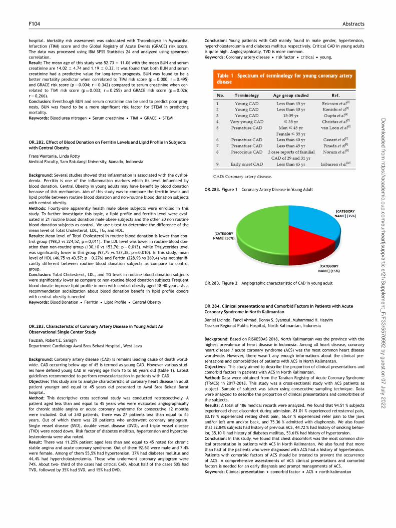
hospital. Mortality risk assessment was calculated with Thrombolysis in MyocardialInfarction (TIMI) score and the Global Registry of Acute Events (GRACE) risk score.The data was processed using IBM SPSS Statistics 24 and analyzed using spearmancorrelation.Result: The mean age of this study was 52.73 6 11.06 with the mean BUN and serumcreatinine are 14.02 6 4.74 and 1.19 6 0.33. It was found that both BUN and serumcreatinine had a predictive value for long-term prognosis. BUN was found to be abetter mortality predictor when correlated to TIMI risk score (p¼ 0.000; r¼ 0.495)and GRACE risk score (p¼ 0.004; r¼ 0.342) compared to serum creatinine when cor-related to TIMI risk score (p¼ 0.033; r¼ 0.255) and GRACE risk score (p¼ 0.026;r¼ 0,266).Conclusion: Eventhough BUN and serum creatinine can be used to predict poor prog-nosis, BUN was found to be a more significant risk factor for STEMI in predictingmortality.Keywords: Blood urea nitrogen • Serum creatinine • TIMI • GRACE • STEMI
OR.282. Effect of Blood Donation on Ferritin Levels and Lipid Profile in Subjectswith Central Obesity
Frans Wantania, Linda RottyMedical Faculty, Sam Ratulangi University, Manado, Indonesia
Background: Several studies showed that inflammation is associated with the dyslipi-demia. Ferritin is one of the inflammation markers which its level influenced byblood donation. Central Obesity in young adults may have benefit by blood donationbecause of this mechanism. Aim of this study was to compare the ferritin levels andlipid profile between routine blood donation and non-routine blood donation subjectswith central obesity.Methods: Fourty-one apparently health male obese subjects were enrolled in thisstudy. To further investigate this topic, a lipid profile and ferritin level were eval-uated in 21 routine blood donation male obese subjects and the other 20 non routineblood donation subjects as control. We use t-test to determine the difference of themean level of Total Cholesterol, LDL, TG, and HDL.Results: Mean level of Total Cholesterol in routine blood donation is lower than con-trol group (198,2 vs 224,52; p¼ 0,011). The LDL level was lower in routine blood don-ation than non-routine group (130,10 vs 153,76; p¼ 0,013), while Triglycerides levelwas significantly lower in this group (97,75 vs 137,38, p¼ 0,010). In this study, meanlevel of HDL (46,75 vs 43,57; p¼ 0,276) and Feritin (228,93 vs 269,4) was not signifi-cantly different between routine blood donation subjects as compare to controlgroup.Conclusion: Total Cholesterol, LDL, and TG level in routine blood donation subjectswere significantly lower as compare to non-routine blood donation subjects Frequentblood donate improve lipid profile in men with central obesity aged 18-40 years. As arecommendation socialization about blood donation benefit in lipid profile donorswith central obesity is neededKeywords: Blood Donation • Ferritin • Lipid Profile • Central Obesity
OR.283. Characteristic of Coronary Artery Disease in Young Adult AnObservational Single Center Study
Fauziah, Robert E. SaragihDepartment Cardiology Awal Bros Bekasi Hospital, West Java
Background: Coronary artery disease (CAD) is remains leading cause of death world-wide. CAD occurring below age of 45 is termed as young CAD. However various stud-ies have defined young CAD in varying age from 15 to 60 years old (table 1). Latestguidelines recommended to perform revascularization in patients with CAD.Objective: This study aim to analyze characteristic of coronary heart disease in adultpatient younger and equal to 45 years old presented to Awal Bros Bekasi Barathospital.Method: This descriptive cross sectional study was conducted retrospectively. Apatient aged less than and equal to 45 years who were evaluated angiographicallyfor chronic stable angina or acute coronary syndrome for consecutive 12 monthswere included. Out of 240 patients, there was 27 patients less than equal to 45years. Out of which there was 20 patients who underwent coronary angiogram.Single vessel disease (SVD), double vessel disease (DVD), and triple vessel disease(TVD) were noted down. Risk factor of diabetes mellitus, hypertension and hypercho-lesterolemia were also noted.Result: There was 11.25% patient aged less than and equal to 45 noted for chronicstable angina and acute coronary syndrome. Out of them 92.6% were male and 7.4%were female. Among of them 55,5% had hypertension, 37% had diabetes mellitus and44,4% had hypercholesterolemia. Those who underwent coronary angiogram were74%. About two- third of the cases had critical CAD. About half of the cases 50% hadTVD, followed by 35% had SVD, and 15% had DVD.
Conclusion: Young patients with CAD mainly found in male gender, hypertension,hypercholesterolemia and diabetes mellitus respectively. Critical CAD in young adultsis quite high. Angiographically, TVD is more common.Keywords: Coronary artery disease • risk factor • critical • young.
OR.284. Clinical presentations and Comorbid Factors in Patients with AcuteCoronary Syndrome in North Kalimantan
Daniel Licindo, Fandi Ahmad, Donny S. Syamsul, Muhammad H. HasyimTarakan Regional Public Hospital, North Kalimantan, Indonesia
Background: Based on RISKESDAS 2018, North Kalimantan was the province with thehighest prevalence of heart disease in Indonesia. Among all heart disease, coronaryheart disease / acute coronary syndrome (ACS) was the most common heart diseaseworldwide. However, there wasn’t any enough informations about the clinical pre-sentations and comorbidities of patients with ACS in North Kalimantan.Objectives: This study aimed to describe the proportion of clinical presentations andcomorbid factors in patients with ACS in North Kalimantan.Method: Data were obtained from the Tarakan Registry of Acute Coronary Syndrome(TRACS) in 2017-2018. This study was a cross-sectional study with ACS patients assubject. Sample of subject was taken using consecutive sampling technique. Datawere analyzed to describe the proportion of clinical presentations and comorbities ofthe subjects.Results: A total of 186 medical records were analyzed. We found that 94.51 % subjectsexperienced chest discomfort during admission, 81.01 % experienced retrosternal pain,83.19 % experienced resting chest pain, 66.67 % experienced refer pain to the jawsand/or left arm and/or back, and 75.36 % admitted with diaphoresis. We also foundthat 32.84% subjects had history of previous ACS, 44.72 % had history of smoking behav-ior, 35.10 % had history of diabetes mellitus, 53.61% had history of hypertension.Conclusion: In this study, we found that chest discomfort was the most common clin-ical presentation in patients with ACS in North Kalimantan. We also found that morethan half of the patients who were diagnosed with ACS had a history of hypertension.Patients with comorbid factors of ACS should be treated to prevent the occurrenceof ACS. A comprehensive assesstments of ACS clinical presentations and comorbidfactors is needed for an early diagnosis and prompt managements of ACS.Keywords: Clinical presentation • comorbid factor • ACS • north kalimantan
OR.283. Figure 2 Angiographic characteristic of CAD in young adult
OR.283. Figure 1 Coronary Artery Disease in Young Adult
F104 Abstracts
Dow
nloaded from https://academ
ic.oup.com/eurheartjsupp/article/21/Supplem
ent_F/F33/5570992 by guest on 07 July 2022

OR.285. High Platelet Distribution Width (PDW) Values As Predictors of MajorCardiovascular Events in Patients with Acute Myocardial Infarction
Sang AyuN. Yuli Sutarmini, D. GdeA. Diprabawa, Raka Widiana, Wayan WitaDepartemen of Cardiology and Vascular Medicine, School of Medicine, UdayanaUniversity, Denpasar, Bali
Background: Coronary heart disease (CHD) is the highest cause of death, although ithas been declining since the last few years because management has progressed.Platelets have an important role in pathogenesis of acute myocardial infarction, wherethe occurrence of plaque rupture is followed by platelet activation and thrombus forma-tion which causes occlusion of coronary arteries. Platelet distribution width (PDW) is amarker of platelet reactivity which can be used as a simple and inexpensive marker intorisk stratification and predict the prognosis of patients with myocardial infarctionObjective: Our objective was to examine the association high platelet distributionwidth (PDW) with major adverse cardiovascular events in pastients with acute myo-cardial infarction during hospitalizationMethod: This prospective study included 60 consecutive patients admitted withacute myocardial infarction to RSUP Sanglah between April 1st and June 30th 2018.All patients were examined by PDW level that analyzed using abbott cell-dyn ruby.The study population was divided into two groups consisting of high PDW groups and lowPDW using the Receiver Operating Curve (ROC) curve. To describe the characteristics ofthe research subjects based on high and low PDW groups, univariate analysis was carriedout followed by bivariate analysis which was displayed in the graph of Kaplan-meier sur-vival estimates with log rank statistical tests. Therefore, a multivariate analysis usingthe Cox regression statistical test was conducted to determine whether a high PDWvalue was an independent predictor of major cardiovascular events.Result: Based on the ROC curve analysis the optimal cut-off value is 20.55, there are28 patients with high PDW (> 20.55) and 32 patients with low PDW(� 20.55). From60 patients there are 24 (40%) who experienced major cardiovascular events.Bivariate analysis showed that patients has Diabetes Melitus (DM) (HR 3.195 CI 95%1.08-9.44 p¼ 0.036) and High PDW (HR 4.94 CI 95% 1.67 p¼ 0.004) correlate withmajor cardiovascular events in patients with acute myocardial infarction.Multivariate analysis with cox regression showed that high PDW has been shown to berelated to major cardiovascular events (HR 3.878CI 95% 1.078-13.952 p¼ 0.038)Conclusion: High PDW values as an independent predictor of major cardiovascularevents in patients with acute myocardial infarction during hospitalization.Keywords: Platelet distribution width • Acute myocardial infarction • Major adversecardiovascular events
OR.286. Six Minute Walking Test Upon Discharge May Predict 30Day Readmissionin Acute Decompensated Heart Failure Patients
Minsy T. Sari, Lia Valentina AstariDepartment of Cardiology and Vascular MedicineArifin Achmad General Hospital,Pekanbaru-Riau
Background: Readmission is a common and costly problem in heart failure patients.Aproximately, 23%of patients are readmitted within 30 days and up to 75% of these earlyreadmission may be preventable. Six minute walking test (6MWT) is a simple and inex-pensive methodeof indirectly assesing excersice tolerance. Limited studies showed thatthe walking distance during a 6MWT is an independent predictor of hospital readmission.Objective: The goal of this study was to examine the 6MWT of the hospitalized heartfailure patients at their discharge from Arifin Achmad General Hospital and theirreadmission within 30 days.Method: This is a descriptive observational study. During March 2019, there weretwelve patients eligible to studied. Six minute walking test was performed prior totheir discharge and the patients were followed up for their 30 day readmission.Result: Median 6MWT were 301,2 (161,4-427,5) meters. Three patients (25%) werereadmitted and had 6MWT <400 meters. Only 1 patient had 6MWT > 400 meters.Median heart rate at discharge was 76 (66-94) beat per minute. Median Ureum was25 (11-49) mg/dl. Only 58% patients received Angiotensin Converting EnzymeInhibitor or Angiotensin II Receptor Blocker and 8,3% patients received beta blockerduring hospitalization.Conclusion: 6MWT may predict early hospital readmission in patient with heart fail-ure. Low 6MWT was found in readmitted and non readmitted patients.Optimalization of heart failure therapy and cardiac rehabilitation program may helpincrease 6MWT in heart failure patients and reduce readmission rate.Keywords: Six minute walking test • heart failure • readmission • 30-day readmission
OR.287. Medical Adherence of Hypertension Management in Primary HealthCare Settings
S. A. Yuwono1, N. G. Arviana1, S. Adiwena1, S. Ng1,21Faculty of Medicine, University of Pelita Harapan, Banten, Indonesia, 2SiloamHospital Lippo Village, Banten, Indonesia.
Background: Hypertension is an important risk factor in developing cardiovasculardisease and mortality. Compliance to treatment plays an important role for better
outcome. The objective of this study was to evaluate medication adherence charac-teristic of hypertensive patients in primary health care settings.Methods: We performed a cross-sectional study at a primary health care center inTangerang. Two hundred ninety-seven out of 433 home-care participants diagnosedwith hypertension were included. Patient’s socio-demographic status, medical status,present of co-morbidities, smoking habits were assessed by interview and question-naire. Adherence to medication was assessed using the Morisky MedicationAdherence Scale (MMAS-8).Results: The mean age of participants was 58 þ 9 years and 254 (89%) were female.Two hundred and sixteen participants (75%) had low antihypertensive medicationadherence. Among those, 26% is not having adequate medication due to them wait-ing for monthly visit from the health care workers, followed by forgetful and laziness(22% and 21%, respectively). Due to limited drug availability, the facility only gave 10pills per patients. The majority, 167 (58%) never received any formal education.Even in patients with high medical adherence, the systolic blood pressure remainsabove guideline recommendations (157 þ 23). The low medical adherence was notsignificantly corelated with education level, hypertension in family, smoking habits,history of heart disease, diabetes mellitus, and physical activity.Conclusion: The study demonstrates a poor blood pressure control in primary healthcare settings, even in patients with high medical adherence. Adherence towardhypertension medication remains very low.Keywords: Hypertension • Medication adherence • MMAS-8 • Primary healthcare • Risk factors
OR.288. Clinical Characteristics and Mortality Following Primary PercutaneousCoronary Intervention for Acute ST-Segment Elevation Myocardial Infarction inAdam Malik General Hospital
M. Fahrial, H. Hasan, Z. Mukhtar, A. Nafiah, H.A. PutraDepartment of Cardiology and Vascular Medicine, School of Medicine, NorthSumatera University, Medan, North Sumatera
Background: ST-segment elevation myocardial infarction (STEMI) is a mayor healthproblem In Indonesia, including Medan. It is important to evaluate clinical character-istics of STEMI patients and mortality after reperfusion therapy for STEMI patients,especially primary PCI.Objective: This study aims to investigate the clinical features and mortality follow-ing primary percutaneous coronary intervention for ST-segment elevation myocardialinfarction (STEMI) patients in Adam Malik General Hospital.Method: We investigated the clinical characteristic and mortality on acute ST seg-ment elevation myocardial infarction registry in cardiac catheterization fromOctober 2018 to February 2019. The registry included consecutive patients of allages with acute ST-segment elevation myocardial infarction (�12 hours from onset)who underwent primary PCI in Adam Malik General Hospital. All data was analyzedby using SPSS.Result: From clinical characteristic analysis, we found that male was higher thanfemale (81.5% vs 18.5%) for acute STEMI event, which dominated by STEMI Inferior(77%). Risk factors analysis revealed that smoking had the highest prevalence. Majorcardiac event findings as an outcome in this study were death (7.4%), pulmonaryedema (7.4%), cardiogenic shock (3.7%), malignant arrhythmia (3.7%). Average lengthof stay duration in this study was 5 days (3 – 11 days).Conclusion: Mortality of STEMI patients is still relatively high (7.4%) in STEMI patientunderwent primary PCI in Adam Malik General Hospital. In the future, education toearly recognition of ACS symptoms and improved cardiac care in PCI capable hospitalare needed to reduce mortality and length of stay in hospital.Keywords: clinical characteristic of primary PCI • mortality in primary PCI
OR.289. Clinical Profile and Medical Adherence of Hypertension Patients inSikumana Primary Health Center, Kupang, East Nusa Tenggara
Queen S. Ariyani1, Michael Susanto2, Leonora J. Tiluata31Sikumana Primary Health Center, Kupang, Indonesia, 2Faculty of MedicineUniversitas Pelita Harapan, Tangerang, Indonesia, 3Department of Cardiology, ProfWZ Johannes General Hospital, Kupang, Indonesia
Background: Hypertension is a highly prevalent chronic disease in Indonesia thatmay cause significant morbidity and mortality. Medical adherence plays an importantrole in achieving optimal medical therapy and good clinical outcomes in patientswith hypertension. However, there exists a challenge to achieving high medicaladherence among patients with chronic diseases such as hypertension. This researchaims to determine the clinical profile and medical adherence of hypertensionpatients in Sikumana Primary Health Center, Kupang, East Nusa Tenggara.Method: We conducted a survey to determine the clinical profile and medical adher-ence of hypertension patients in Sikumana Primary Health Center, Kupang, East NusaTenggara. The Morisky Medical Adherence Score-8 (MMAS-8) was used to assess thelevel of medical adherence.Result: A total of 69 subjects took part in this study. 62,5% of the subjects werefemale and 50.7% aged between 60-69 years old. Flores (24,6%), Rote (24,6%) and
Abstracts F105
Dow
nloaded from https://academ
ic.oup.com/eurheartjsupp/article/21/Supplem
ent_F/F33/5570992 by guest on 07 July 2022

Timor (23.2%) tribes dominate the research population. 14,5% of the subjects hadexperienced a stroke. Comorbidities include diabetes mellitus (31,9%), dyslipidemia(30,4%), and heart problems (7,2%). Risk factors include family history of hyperten-sion (40,6%) and smoking (11,6%). 73,6% claimed to only have had hypertension for �5 years and were mostly diagnosed in a primary health center (65.2%). According tothe MMAS-8, 49.3% have high medical adherence, 27.5% have medium adherence,23.3% have low adherence.Conclusion: Half of the subjects in this study have high medical adherence.However, more educational intervention and health promotion must be made toimprove medical adherence among patients with hypertension especially in the pri-mary health care setting. In this situation, healthcare professionals play a key role inensuring patients to take their medications regularly in order to achieve a betterclinical outcome.Keywords: Hypertension • Clinical profile • Medical Adherence • Primary HealthCenter • East Nusa Tenggara
OR.290. Does Diabetes Truly Obscure Typical Presentation of Chest Pain AmongAcute Coronary Syndrome?Analysis from Banda Aceh
M. Hustiar Hakim, Muhammad Ridwan, Teuku M. Haykal, YusniDepartment of Cardiology and Vascular Medicine, School of Medicine, Syiah KualaUniversity, Banda Aceh, Indonesia
Background: As an important major risk factor of cardiovascular disease, diabetes iscommon among Acute Coronary Syndrome (ACS). Identifying ACS based on clinicalpresentation alone is difficult in patients with diabetes. Diabetes patients usuallydevelop atypical characteristic of chest pain. We sought to investigate whetherpatients with diabetes truly have atypical characteristic of chest pain rather thanpatients without diabetes. Other descriptions of chest pain among diabetic patientswith ACS were also analyzed.Methods: This is a cross-sectional study in patients presented with ACS in ZainoelAbidin hospital, Banda Aceh, between November 2018 to January 2019. All patientswere interviewed by a physician during hospitalization. The data obtained frominterview included detailed descriptions of chest pain and detailed risk factors char-acteristic. Medical data regarding diagnostic examination and therapeutic interven-tion was obtained from medical record. Statistical analysis using bivariate analysiswas performed to identify association between variables.Results: Fifty patients presented with ACS were enrolled prospectively for the study.Diabetes was identified in 34% patients. The mean age was 55.8 6 9.6 years old and80% of the subjects were male. Diagnosis of myocardial infarct were made in 28patients (56%) while others were admitted as Unstable Angina Pectoris (UAP). Typicalpresentation of chest pain as described as heavy or tight sensation was identified in86% cases. No association was detected between atypical presentation of chest painand diabetes condition (p¼ 0.23). Radiating sensation of chest pain was also notassociated with diabetes (p¼ 0.30) as well as the presence of diaphoresis (p¼ 0.49).In contrast, nausea was significantly common among ACS patients without diabetes(p¼ 0.04).Conclusion: In ACS population, no association was identified between diabetes andtypical presentation of chest pain with its detailed descriptions. The propositionstating that diabetic patients usually presented with atypical characteristic of chestpain need to be reinvestigated.Keywords: Angina Pectoris • Diabetes Mellitus • Acute Coronary Syndrome
OR.291. Random Blood Glucose Level and Hospital Readmissions in Heart FailurePatients: A Study From RSBA Heart Registry
Vidyawati, R.R. Menaldi, R.A. Fanani, M.B.R. Jati, R. RasminDepartemen of Cardiology and Vascular Medicine, Budhi Asih General Hospital,Jakarta, Indonesia
Background: Hospital readmission remains a continued challenge in the care of heartfailure (HF) patients. The Framingham Heart Study (FHS) first demonstrated anincreased risk of congestive heart failure (CHF) in patients with diabetes. But therelationship between random blood glucose (RBG) at admission and hospital readmis-sions in HF patients remain unknown.Objective: We aim to analyze the relationship between RBG level and hospital read-missions in HF patients with mid-range and reduced ejection fraction (EF).Method: BA-HF (Budhi Asih Heart Failure) Registry is a single center registry whichprospectively enrolls all hospitalized HF patients at Budhi Asih Regional Hospitalsince 2017. We searched the inpatient medical records of 270 individuals with mid-range and reduced EF (<50%) between 2017-2018. Patients with sub-optimal HFtreatment were excluded.Result: A total of 126 patients with mid-range and reduced EF were included in thestudy, represented 46% of all HF populations in BA-HF Registry. Mean age of58.45611.8 years, 65.9% were male, mean EF of 31.69610.56, 25.4% were re-admit-ted between 2017-2018. All patients were treated with ACE-inhibitor and/or ARB,and beta blocker. Based on statistical analysis using SPSS, no association was foundbetween RBG and hospital readmission (v2(2)>¼3.740, p¼ 0.053).
Conclusion: Several studies showed significant association between diabetes and HF,but our findings suggested the association between RBG and HF was not significant.We acknowledge this is a relatively small study, thus limits its statistical power. Moresamples are needed to better determine the association between RBG and HF.Keywords: random blood glucose • heart failure • diabetes • readmission
OR.292. Echocardiography Profile of Indonesian Athletes
Irma Kartikasari, Davin E. FrizzyDepartment of Cardiology and Vascular Medicine, Dr. Soegiri Public Hospital,Lamongan, East Java, Indonesia
Background: Sudden death arising from rigorous physical sports is on the rise inmany countries. Intense training affects the athlete’s body, such as the physiologicalremodeling of the heart due to the hemodynamics changes. In such cases, echocar-diography is widely used, as it is non-invasive and highly reliable. Existing studiesshow the echocardiography values in each races are varied. Besides, some concen-trated on the heart morphologies of athletes and non-athletes have primarily focusedon Caucasian subjects in North America and Europe, with very limited studies onAsian athletes.Objective: This study aims to provide the anthropometry and echocardiographyresults that can be used as a reference values for Asian athletes.Method: A total of 24 asymptomatic Indonesian athletes from Lamongan, who didnot have any history of past illnesses, were screened before the football season usingechocardiogram to measure the heart structure and function. The results were thencompared to the reference values of Asian non-athletes.Result: In both athletes and non-athletes, the diameter of left ventricle and thethickness of intraventricular septum were considerably similar, although the left ven-tricle’s posterior wall was thinner and the ejection fraction was slightly increased forthe athletes. In addition, the volume of the left ventricle in systolic and diastolicphases was markedly enlarged in athletes, as was its mass, with the respective meanvalues consecutively 61.68611.97ml/m2; 21.6566.86ml/m2 and 86.12616.12 g/m2.However, other than the left ventricle, no significant difference was evident in bothgroups.Conclusion: Intense training in athletes generates adjustments of the heart becauseof the remodeling due to hemodynamic changes. As observed in this study, therewere significant changes of the left ventricle in athletes compared to non-athletes.Hence, further studies are essential to obtain reference values of Asian athletes.Keywords: Athlete’s Heart • Echocardiography • Cardiac Volume
OR.293. Formula to Predict Functional Capacity in Patients with MultivesselCoronary Artery Disease After Coronary Artery Bypass Grafting Surgery
Ervan Zuhri, Ade M. Ambari, Bambang Dwiputra, Basuni Radi, Dede Kusmana,Budhi Setianto, Anwar SantosoNational Cardiovascular Center of Harapan Kita, Jakarta, Indonesia, Department ofCardiology and Vascular Medicine, Faculty of Medicine, University of Indonesia
Background: Pre-operative surgical risk assessment is crucial for weighing the riskand benefit of cardiac surgery. The European System for Cardiac Operative RiskEvaluation (EuroSCORE) II and the Society of Thoracic Surgeons (STS) score risk pre-diction model is widely employed to evaluate the risk of perioperative mortality andmorbidity in patients undergoing cardiac surgery. But, these scoring system do notinclude prediction of the functional capacity after cardiac surgery, especially coro-nary artery bypass grafting (CABG) surgery. Functional capacity after CABG surgery isvery essential because high functional capacity is strongly related to good outcomein quality of life, morbidity, and mortality after years later. Prediction of good func-tional capacity after CABG surgery is very useful for weighing prognosis after CABGsurgery.Objective: To construct formula to predict good functional capacity after CABG sur-gery based on pre-operative patient’s characteristic data.Method: We make retrospective study with multivariate regression analysis frommedical record data of patients with multivessel coronary artery disease (CAD)undergoing isolated CABG surgery who had undergone cardiac rehabilitation fromSeptember 2009 until June 2017 in National Cardiovascular Center of Harapan Kita(NCCHK). We make prediction whether the patient can get good functional capacity(METs > 6.00) or not, based on pre-operative patient’s characteristic data.Result: The developmental dataset had 839 patients. The multivariate regressionanalysis showed six variables that can significantly predict METs score > 6.00. Thevariable was age � 71 years old (OR 0.05, 95%CI 0.01 to 0.55, p¼ 0.013), gendermale (OR 11.25, 95%CI 6.86 to 18.44, p< 0.001), BMI � 25 kg/m2 (OR 0.58, 95%CI0.37 to 0.91, p¼ 0.019), moderate LVEF 40-49% (OR 0.52, 95%CI 0.35 to 0.78,p¼ 0.002), reduced LVEF < 40% (OR 0.31, 95%CI 0.17 to 0.59, p< 0.001), and atrialfibrillation (OR 0.25, 95%CI 0.07 to 0.87, p¼ 0.029). The regression formula wasy¼ 0.851 – (2.925 x age � 71 years old) – (0.544 x IMT � 25 kg/m2)þ (2.42 x Male) –(0.652 x LVEF 40-49%) – (1.16 x LVEF < 40%) – (1.371 x AF). In the formula, valued 1 ifage � 71 years old, IMT � 25 kg/m2, male, LVEF 40-49%, LVEF < 40%, or AF waspresent and valued 0 if the variable was absent. The patient’s probability got METs
F106 Abstracts
Dow
nloaded from https://academ
ic.oup.com/eurheartjsupp/article/21/Supplem
ent_F/F33/5570992 by guest on 07 July 2022

score > 6.00 was P¼ 1/ {1þexponent (-y)}. Calibration by Hosmer Lemeshow ChiSquare was (8) ¼ 9.192 (p¼ 0.327) and the area under the ROC curve was 0.779(95%CI 0.742 to 0.817, p< 0.001).Conclusion: Formula to predict good functional capacity after CABG surgery hadbeen constructed with good quality.Keywords: Functional capacity • coronary artery bypass grafting surgery
OR.294. Formula to Predict Functional Capacity in Patients with MultivesselCoronary Artery Disease After Coronary Artery Bypass Grafting Surgery
Ervan Zuhri, Ade M. Ambari, Bambang Dwiputra, Basuni Radi, Dede Kusmana,Budhi Setianto, Anwar SantosoNational Cardiovascular Center of Harapan Kita, Jakarta, Indonesia, Department ofCardiology and Vascular Medicine, Faculty of Medicine, University of Indonesia
Background: Leucocyte count especially measured using neutrophil-lymphocyte ratio(NLR) is considered a more specific determiner than the neutrophil or leucocytecount for cardiovascular events and mortality in acute coronary syndromes (ACS).Left ventricular systolic dysfunction (LVSD) is an important cause both of mortalityand morbidity in patients with ACS and develops more frequently in patients with ST-elevation myocardial infarction (STEMI) who have a high NLR on admission to the hos-pital. This study aimed to investigate the the predictive value of NLR for LVSD inpatients both with STEMI and non-ST elevation myocardial infarction (STEMI).Objective: This study aimed to determine a predictive value of LVSD in patient withAcute Myocardial Infarction.Methods: STEMI and NSTEMI patients undergoing treatment in Tarakan RegionalPublic Hospital from January 2017 to December 2018 were retrospectively analyzed.Demographic profile, NLR was measured in the emergency department prior to fur-ther treatment in hospital ward while EF was measured during treatment period inhospital (EF for STEMI patient was focused for anterior infarction involvement).Results: The mean age of the patient was 56612 years old and most of them weremale (75%). The mean (6SE) NLR on admission in the data was 4.66(64.06) and themean of EF was 46,98 6 16.5. In this study, NLR had no significant negative correla-tion with EF for STEMI patient (Sig. 2-tailed p¼ 0.200, r¼-0.213) as well as forNSTEMI patient (Sig. 2-tailed p¼ 0.555, r¼-0.123).Conclusion: Higher neutrophil-lymphocyte ratio on admission may had a lower EF inSTEMI and NSTEMI patients. While this study wasn’t concluding a strong negative cor-relation statistically, there was a tendency towards it. It is a clinically importantinflammatory marker that can take a role in the management and the prognosis ofpatient with Acute Coronary Syndrome. Further larger study is needed to observethe relation between NLR and left ventricular systolic dysfunction.Keywords: Neutrophil-Lymphocyte Ratio • EF • STEMI • NSTEMI
OR.295. Neutrophil-Lymphocyte Ratio on Admission as A Predictive Value of LeftVentricular Systolic Dysfunction in Patients with Acute Myocardial Infarction inNorth Kalimantan
Christopher A. Yandoyo, Winardi E. Setiawan, Fandi Ahmad, Donny S. Syamsul,Muhammad H. HasyimTarakan Regional Public Hospital, North Kalimantan, Indonesia
Background: Children with heart disease are serious conditions that have significantimpact on morbidity, mortality, and healthcare costs. There were no reported datain Sampang Hospital that measure epidemiological profile of heart disease in childrenin this region.Objective: This study was aimed specifically to determine the most lesion and typeof heart disease in children in one populated area in Sampang, Madura.Method: The retrospective observational study was conducted during January 2016to Agustus 2018. Inclusion criteria are children of age ranged from 0 days to 17 yearsold, first time diagnosed with their clinical and echocardiographic. The data of inand out pediatric patients with heart disease in children taken from medical record.Children with heart disease with more than single lesion was simplified as one lesionwhich had most major impact on hemodynamic circulation. Data were entered in MsExcel and analyzed by SPSS.Result: We had total 85 patients with heart disease in children registered toSampang Hospital during January 2016 to Agustus 2018. Gender based percentageshowed 39% for female and 61% male. Ventricle septal defect (VSD) was the mostcommon lesion of congenital heart diseases (CHD) among children (17 patients, 20%)followed by atrial septal defect (ASD) 11 patients (13%) and persistent ductus arte-riosus (PDA) 10 patients (12%). The highest incidence of cyanotic heart diseases wasTetralogy of Fallot (TOF) 3 patients (4%). Rheumatoid Heart Disease were found in 21patients (25%) and 13 patients (15%) with others heart diseases. The highest ageranged of CHD are 0 days to 2 years old and 10 to 17 years old for rheumatoid heartdisease.Conclusion: The most common lesion of acyanotic heart disease in children was VSDand the highest incidence of cyanotic heart disease was TOF. Early diagnosis is
important and follow up early in the course of the disease in order to improve out-come and reduces mortality rates.Keywords: Incidence • heart disease • children.
OR.296. Incidence and Pattern of Children with Heart Disease: ATwo Year Studyin Moh Zyn Regional Public Hospital of Sampang, Madura, East Java, Indonesia
I.A Rakhmawati, A.I Sadiati, F. DiastyariniMoh Zyn Regional Public Hospital of Sampang, Madura
Background: Acute coronary syndrome (ACS) is one of the causes of morbidity andmortality in human with 12.9% percentage of all cases. Octogenarian are individualsaged 80 years or more and are called elderly. Data showed coronary heart diseaseincidence in elderly in Indonesia by 33% and will keep increasing in line with increas-ing population of elderly. Data stated that the population of elderly in Indonesia in2000 reached 7.23% and is predicted to keep increasing up to 11.34% in 2020.According to the prediction of Bureau of Census USA in 1993, by 2045, the number ofelderlies in Indonesia will increase to 414%. Even though data showed an increase ofsurvivability in acute coronary syndrome patients, this does not apply to elderly pop-ulation. Anatomical and physiological changes become some of the causes of acutecoronary syndrome. Endothelial dysfunction that triggers arteriosclerosis in the ves-sels becomes the base of anatomical changes that cause acute coronary syndrome.Anatomical changes, comorbid factors (hypertension, diabetes mellitus, dyslipide-mia) become the physiological cause of acute coronary syndrome in elderly. Along2018, there was 62 octogenarian patients diagnosed with acute coronary syndromeadmitted in the ER and received treatment in ICCU of Buleleng General Hospital.Objectives: This study was to determine the prevalence and comorbid in octogenar-ian patients with acute coronary syndrome during 2018 in Buleleng General Hospital.Methods: This study was a descriptive retrospective, conducted in octogenarianpatients admitted to ER and and receive treatment in ICCU of Buleleng GeneralHospital during 2018. This clinical study involved gender, the type of ACS experi-enced, comorbid factors (Hypertension, diabetes mellitus, dyslipidemia) according tothe data obtained through the patients medical records.Results: During 2018, there were 62 octogenarian patients admitted with acute coro-nary syndrome in Buleleng General Hospital. The average age of patients was 84.5years. Males consisted of 27 people (43.5%) and female were 35 people (56.5%).From above data, 16 patients (19.7%) were diagnosed with STEMI and 46 patients(80.3%) were diagnosed with NSTEMI. Hypertension was found in 31 patients (50%).Diabetes mellitus was found in 9 patients (14.5%). Dyslipidemia was found in 31patients (50%).Conclusion: Out of all octogenarian patients admitted to Buleleng General Hospital,there were more female patients compared to males. The average age of octogenar-ian with ACS was 84.5 years old. The number of NSTEMI type ACS patients were moredominant compared to STEMI type. The highest comorbid factors in octogenarianwith ACS were hypertension and dyslipidemia.Keywords: Acute Coronary Syndrome • Octogenarian • Hypertension •Dyslipidemia.
OR.297. The Distribution of Coronary Artery Lession in Patient withHypertention and Diabetes Mellitus History in Jakarta Heart Center Hospital
Todung Silalahi1, Andreas M Siborutorop1, Simon Salim2
1Division of Cardiovascular, Department of Internal Medicine, Jakarta Heart CenterHospital, Jakarta, and Faculty of Medicine, Universitas Kristen Krida Wacana,Indonesia., 2Division of Cardiology, Department of Internal Medicine, University ofIndonesia, Jakarta, Indonesia
Background: WHO stated that around 7.4 million people died from Coronary ArteryDisease (CAD) in 2012. Based on data released by The Center of Information and Dataof Health Secretary Indonesian Republic known, prevalence of CAD in Indonesia on2013 is 1,5% or around 2.650.340 cases. Risk factors that contribute to CAD are :non-modifiable (age, gender) and modifiable (smoking, dyslipidemia, hypertension,obesity, and diabetes).Objective: We sought to see the pattern of CAD based on the underlying modifiablerisk factors such as Diabetes and Hypertension to describe distribution of CAD corre-lated with location and amount of coronary artery lessions.Methods: The design was a descriptive cross sectional study, that conducted by col-lecting data from medical records of patients with CAD, who has CAD with history ofhypertention and diabetes mellitus and been catheterized in Jakarta Heart CenterHospital (JHC),Jakarta ,from April 2015 to March 2016. The number of samples whohave fulfil the criteria of inclusion and exclusion is 706 patients.Result: From706 patients who enrolled in this study, patients with hypertention his-tory who has CAD1VD is 162 (31.57%), CAD2VD is 130 (25.34%), CAD3VD is 160(31.18%), CAD3VDþLMD is 61 (11.89%). Patients with Diabetes Mellitus history whohas CAD1VD is 12 (31.57%), CAD2VD is 10 (26.31%), CAD3VD is 11 (28.94%),CAD3VDþLMD is 5 (13.15%). Patients with both hypertention and diabetes mellitus
Abstracts F107
Dow
nloaded from https://academ
ic.oup.com/eurheartjsupp/article/21/Supplem
ent_F/F33/5570992 by guest on 07 July 2022

history who has CAD1VD is 38 (24.51%), CAD2VD is 17 (10.96%), CAD3VD is 67(43.22%), CAD3VDþLMD is 33 (21.29%).Conclusion: Majority of Hypertension only patients, and DM only patients have 1 VD,and majority of HT þDM have 3VD. CAD 3VD was found in 31.18% of patients withhypertension only, 28.94% of patients with DM only, and 43.22% of patients with DMþ HT combined.Keywords: Distribution CAD • Hypertension • Diabetes Mellitus
OR.298. Association of Serum Sodium, Potassium and Chloride Value onAdmission With ST Elevation Myocardial Infarcts Patient’s Global Registry ofAcute Coronary Event at Tarakan Regional Public Hospital, North Kalimantan
Steffiany, Winardi E Setiawan, Fandi Ahmad, Donny S. Syamsul, Muhammad H. HasyimTarakan Regional Public Hospital, North Kalimantan
Background: Myocardial infarction is one of the leading causes morbidity and mortal-ity in the world. In 2018, North Kalimantan placed in the first rank with in patientswith cardiac insufficiency in Indonesia. In ST elevated myocardial infarcts (STEMI),there is an occlusion that lead to infarction of cell and necrosis of cell. Serum elec-trolytes imbalance after an episode of myocardial infarct are common. Serumsodium, potassium are considered associated with electrophysiological property ofmyocardial membrane. While, global registry of acute coronary event (GRACE) scorewas a parameter to assess patient’s mortality prognosis in 6 months after event.Objective: This study aim to correlate the serum electrolyte STEMI patients onadmission with patient GRACE score.Methods: This study derived from Tarakan regional public hospital medical record.Study involved hospitalized 77 STEMI cases from January 2017 till December 2018.STEMI patients are admitted either from emergency room or clinic. The serum elec-trolyte was taken in 6 hours after admission. All data was analyzed with independentT test in SPSS statistic 20.Result: There are 56 patient included in this study. Age mean of this study is 54.48 6
13.56; sodium mean ¼ 137.262.89 mEq/L; potassium mean ¼ 3.8260.49 mEq/L;and chloride mean ¼105.4963.79Mmol/L. There is a significant result betweenpatients GRACE score mean in hyponatremia patients and normonatremia patients(180.33642.43 vs 138.47629.48, p < 0.01; p¼ 0.001 ). Meanwhile, there is no signif-icant result between potassium (p ¼ 0,79) and chloride value ( p ¼ 0.172) to GRACEscore in STEMI patients.Conclusions: There is a significant result between sodium value to GRACE score inSTEMI patients at Tarakan regional public hospital, North Kalimantan. The lessersodium value would lead to better prognosis rather than higher sodium value patienton admission.Keywords: Myocardial infarction • STEMI • dyselectrolytemia • GRACE.
OR.299. Correlation between Anemia And Global Registry of Acute CoronaryEvents Score in Patient With Non-ST Elevation Myocardial Infarction : An Analysisfrom Tarakan Registry of Acute Coronary Syndrome
Juliana, Ahmad Fandi, Donny S. Syamsul, Muhammad H. HasyimTarakan Regional Public Hospital, North Kalimantan
Background: Cardiovascular disease are the number 1 cause of death globally. Anestimated 17.9 million people died from cardiovascular disease in 2016, representing31% of all global death. National epidemiology data 2018 shows that the highestprevalence of heart disease is in the North Kalimantan province. Anemia canadversely influence prognosis in patients by various mechanisms, such as decreasingthe oxygen content of the blood supply. This study evaluated the relationshipbetween anemia and Global Registry of Acute Coronary Events (GRACE) risk scorewith in-hospital death in patients with Non-ST Elevation Myocardial Infarction(NSTEMI).Methodology: The retrospective study included 59 patients admitted for NSTEMIfrom January 2017 until December 2018. Patients with anemia were identified(hemoglobin <12g/dl for women and <13g/dl for men) using convenient sex-specificthresholds by World Health Organization. GRACE risk score was calculated upon firstarrival to the hospital.Result: The study population included 59 patients with a mean age of 58.3 61.7years, 64% male. Anemia was present in 20 (33.9%) patients, the mean of hemoglobinon admission was 13.09 62.09. The mean of GRACE risk score was 101.75639.8.Anemic patients were seen in older and more often in male, higher proportion of dia-betics and hypertensive. There was a correlation between lower hemoglobin withhigher GRACE in hospital mortality score (Sig 2-tailed p¼ 0.001, r¼-0.439).
Conclusions: Our study results demonstrated that patient with lower hemoglobin onadmission had a significant higher GRACE score and it was associated with in-hospitalcardiovascular mortality in NSTEMI.Keywords: NSTEMI • GRACE • anemia • hemoglobin
OR.300. Does Diabetes Truly Obscure Typical Presentation of Chest Pain AmongAcute Coronary Syndrome?Analysis from Banda Aceh
M. Hustiar Hakim, Muhammad Ridwan, Teuku M. Haykal, YusniDepartment of Cardiology and Vascular Medicine, School of Medicine, Syiah KualaUniversity, Banda Aceh, Indonesia
Background: As an important major risk factor of cardiovascular disease, diabetes iscommon among Acute Coronary Syndrome (ACS). Identifying ACS based on clinicalpresentation alone is difficult in patients with diabetes. Diabetes patients usuallydevelop atypical characteristic of chest pain. We sought to investigate whetherpatients with diabetes truly have atypical characteristic of chest pain rather thanpatients without diabetes. Other descriptions of chest pain among diabetic patientswith ACS were also analyzed.Methods: This is a cross-sectional study in patients presented with ACS in ZainoelAbidin hospital, Banda Aceh, between November 2018 to January 2019. All patientswere interviewed by a physician during hospitalization. The data obtained frominterview included detailed descriptions of chest pain and detailed risk factors char-acteristic. Medical data regarding diagnostic examination and therapeutic interven-tion was obtained from medical record. Statistical analysis using bivariate analysiswas performed to identify association between variables.Results: Fifty patients presented with ACS were enrolled prospectively for the study.Diabetes was identified in 34% patients. The mean age was 55.8 6 9.6 years old and80% of the subjects were male. Diagnosis of myocardial infarct were made in 28patients (56%) while others were admitted as Unstable Angina Pectoris (UAP). Typicalpresentation of chest pain as described as heavy or tight sensation was identified in86% cases. No association was detected between atypical presentation of chest painand diabetes condition (p¼ 0.23). Radiating sensation of chest pain was also notassociated with diabetes (p¼ 0.30) as well as the presence of diaphoresis (p¼ 0.49).In contrast, nausea was significantly common among ACS patients without diabetes(p¼ 0.04).Conclusion: In ACS population, no association was identified between diabetes andtypical presentation of chest pain with its detailed descriptions. The propositionstating that diabetic patients usually presented with atypical characteristic of chestpain need to be reinvestigated.Keywords: Angina Pectoris • Diabetes Mellitus • Acute Coronary Syndrome
OR.301. Correlation Between Body Mass Index and Cardiorespiratory FitnessAmong Youth in Jakarta, Indonesia
Stephanie Widodo Subagio, Yudistira Panji SantosaDepartment of Internal Medicine, School of Medicine and Health Sciences Atma JayaCatholic University of Indonesia, Jakarta, Indonesia
Background: Cardiovascular disease is the number one cause of death every yearglobally and one of the most common cause of death in Indonesia, besides stroke anddiabetes. The predictive factor of cardiovascular disease can be seen since the ageof youth (15-24 years). Parameters that can be used in predicting cardiovascular dis-ease are cardiorespiratory fitness (VO2max) and nutritional status (BMI). Severalstudies shown that low cardiorespiratory fitness accompanied with high body massindex has association with cardiovascular disease.Objective: To determine the correlation between body mass index and cardiorespira-tory fitness (VO2max) among youth in Jakarta, Indonesia.Method: A cross-sectional study was conducted to 80 youth in Jakarta, Indonesia.Weight and height were measured using standard methods to assess the body massindex. VO2max measurements were collected with the Queen’s College Step Testmethods to assess the cardiorespiratory fitness. The obtained data was analyzed withPearson’s correlation.Result: The majority of respondents who are overweight and obese have poorVO2max (91.1%) similar results were obtained among the respondents with normalbody mass index (91.4%). Statistically, there was no significant correlation betweenbody mass index and cardiorespiratory fitness (r¼ 0.05; p¼ 0.64) because the cardi-orespiratory fitness among youth tends to be the same and it’s not influenced byweight.Conclusion: There was no significant correlation between body mass index and cardi-orespiratory fitness (VO2max) among youth in Jakarta, Indonesia.Keywords: cardiorespiratory fitness • body mass index • Queen’s college step test
F108 Abstracts
Dow
nloaded from https://academ
ic.oup.com/eurheartjsupp/article/21/Supplem
ent_F/F33/5570992 by guest on 07 July 2022

OR.302. Clinical Factors Correlation to Patient Medication Adherence inHypertensive Patients
Dylan Hadi1, Filemon M. W. Sondakh1, Ng Sunanto1,21Faculty of Medicine, Universitas Pelita Harapan , 2Siloam General Hospital,Tangerang, Indonesia
Background: Blood Pressure (BP) Control is important in controlling CardiovascularDiseases (CVD) and it has been shown that non-adherence to therapy are one of thereasons most patients don’t achieve optimal BP control. There are several studies inthe correlation between various clinical factors to patient adherence but no similarresearch has been done in Tangerang which has a different demographic characteris-tic thus there may be a need to examine these factors on these patients.Objective: Examining the Correlation between various clinical factors to patientadherence in Hypertensive Patients.Method: The study was designed as a cross-sectional observational study with con-secutive sampling done from March to April 2019. Sixty two patients that were diag-nosed with Primary Hypertension were included, and the following informations wereobtained; Name, Age, Sex, Duration of HT, Patient adherence and knowledge in HT,Patient Motivation, and Family as well as Medical Staff Support. Patient adherencewas determined using the MMAS-8 (Morisky Medication Adherence Scale)questionnare.Result: The results were obtained on 62 samples and presented as follow [Median,[min-max)] : There were 43 females (69.4%), 52 patients (83.9%) were recently diag-nosed with Hypertension, Patient’s age was varied [54, (23-74)]. From 62 patients,30 Patients (48.4%) were adherent, 31 (50%) Patients has good knowledge about HT,37 patients (47%) had good motivation, 49 Patients (79%) has good family support,and 37 (59.7%) Patients received good medical staff support. Of those 30 AdherentPatients, 20 were females, 22 had good knowledge about HT, 26 had good motiva-tion, 27 had good family support and 26 had good medical staff support. The correla-tions between age and sex was insignificant with adherence with P values of 0.071and 0.657 respectively, However the correlation between patient knowledge, moti-vation and both family and medical staff support were significant with odds ratio of7.0 ,12.4, 4.09, 0.2 and P values of 0.001, 0.001, 0.040 and 0.016 respectively.Conclusion: There were significant correlations between patient knowledge, motiva-tion and both family and medical staff support to adherence.Keywords: Hypertension • Medication Adherence • Knowledge in Hypertension.
OR.303. Neutrophil-Lymphocyte Ratio as a Useful Hematologic Biomarker inPredicting Mortality in Non-ST Elevation Myocardial Infarction
Stefanus, Winardi E. Setiawan, Fandi Ahmad, Donny S. Syamsul, Muhammad H. HasyimTarakan Regional Public Hospital, North Kalimantan, Indonesia
Background: Previous studies have shown that the neutrophil-lymphocyte ratio (NLR)and the thrombolysis in myocardial infarction (TIMI) risk score have been describedas a predictor of mortality in patients with acute coronary syndrome.Objective: This study aimed to determine the correlation between NLR and TIMI riskscore in predicting mortality in patients with acute coronary syndrome especiallynon-ST elevation myocardial infarction (NSTEMI).Method: The subjects of this study were patients with NSTEMI from Tarakan Registryof Acute Coronary Syndrome. The NLR was taken from the laboratory result whenthe patient was admitted to the hospital and the mortality assessment was calcu-lated using TIMI risk score. The data was processed using IBM SPSS Statistics 24 andanalyzed using spearman correlation.Result: The subject’s mean age was 60.08 6 9.82 years with the mean TIMI score ofthe studied patients was 2.61 6 1.89 and the mean of the neutrophil-lymphocyteratio was 4.49 6 8.51. It was found that there was a correlation between neutorphil-lymphocyte ratio and TIMI risk score (p< 0,05) with its correlation coefficient was0,29.Conclusion: Based on the findings of the present study, the neutrophil-lymphocyteratio was found useful in predicting risk at 14 days of all-cause mortality, new orrecurrent myocardial infarction or severe recurrent ischemia requiring urgentrevascularization.Keywords: Neutrophil-lymphocyte ratio • TIMI • NSTEMI
OR.304. Correlation of Elevated Blood Pressure on Cardiorespiratory FitnessAmong Youth in Jakarta, Indonesia
Stephanie Widodo Subagio, Yudistira Panji SantosaDepartment of Internal Medicine, School of Medicine and Health Sciences Atma JayaCatholic University of Indonesia, Jakarta, Indonesia
Background: Cardiovascular disease is a major cause of morbidity and mortalityworldwide and one of the most common cause of death in Indonesia, besides strokeand diabetes. Many factors can contribute to this disease, including hypertension,obesity, dyslipidemia, etc. These risk factors are often shown in youth, so that pre-ventive measure should be initiated since youth age.
Objective: To determine the correlation between elevated blood pressure and cardi-orespiratory fitness (VO2max) among youth in Jakarta, Indonesia.Method: A cross-sectional study was conducted to 52 youths in Jakarta, Indonesia.Weights and heights were measured using standard methods to assess the body massindex. Blood pressures were measured by standardized sphygmomanometer andVO2max measurements were collected with the Queen’s College Step Test methodsto assess the cardiorespiratory fitness. The obtained data was analyzed withPearson’s correlation.Result: Similar results were obtained among respondents who have overweight andnormal body mass index. The majority of respondents have an elevated systolic anddiastolic blood pressure and a decreased VO2max. There was no significant differencebetween respondents with overweight and normal body mass index. Statistically,there was no significant correlation between elevated blood pressure and cardiores-piratory fitness (p> 0.05) because the cardiorespiratory fitness among youth tends tobe the same and it’s not influenced by blood pressure.Conclusion: There was no significant correlation between elevated blood pressureand cardiorespiratory fitness (VO2max) among youth in Jakarta, Indonesia.Keywords: blood pressure • cardiorespiratory fitness • body mass index • Queen’scollege step test.
OR.305. Effect of Guideline Directed Medical Therapy (GDMT) in patients HeartFailure with Reduce Ejection Fraction (HFrEF) in the Left Ventricular EjectionFraction (LVEF) Improvement in Cibinong General Hospital Indonesia : a SingleCentre Experience
Dian Yaniarti Hasanah, Zul Effendi, Rarsari Soerarso, Nani HersunartiDepartment of Cardiology and Vascular Medicine, School of Medicine, IndonesiaUniversity, Jakarta
Background: Improvement in functional status, long term survival, and quality of lifehas always been the goal of therapy in patients heart failure with reduced ejectionfraction. Neurohormonal modulating medications help patients achieve these goalsand, in a subgroup of patients, can promote reverse remodeling resulting in therecovery of left ventricular systolic function. Optimal medical therapy appears to bea key component of achieving myocardial recovery, with recovery more likely inpatients with a shorter duration of heart failure and a non-ischemic etiology. Weevaluate 94 patients HFrEF due to ischemic (all not revascularized ) and non ischemiccause such as myocarditis that got optimal GDMT and comorbid treatment. LVEF wascompared from the first admission an after 12 months GDMT.Objective: To assess the effect of optimal GDMT for improvement of LVEF in HFrEFpatients in Cibinong General Hospital.Method: This study was conducted in Cibinong General Hospital, Indonesia, fromNovember 2017 to 2018. Ninety four patients were included in this prospective cohortstudy with all patients had standard heart failure therapy based on guideline (Loop diu-retic, MRA, Ace inhibitor/ARB/ uperio, beta blocker). The primary end point of thisstudy was LV EF recovery that was follow up to 12 months. NYHA, heart rate, and bloodpressure were compared from first admission and after 12 months therapy. We alsoevaluate the dose of optimal Ace inhibitor/ARB and beta blocker that can be achievedin this study compared to definition of optimal therapy based on the guideline.Result: EF and heart rate were significantly different between baseline (30.2 6 8.9)and follow up (43.8 6 11.9 with p< 0.001, as well as NYHA Functional Class. Optimaldrug dose for Ace-i/ARB and beta blocker were not associated with EF recovery, withp value were 1.000 for all drug type, with definition of optimal therapy based onguideline. The average dose for Ace I in this studi is 7.3mg, the average dose forARB is 18.3mg, and the average dose for beta blocker is 6.4mg. This result showedthat optimal therapy is very individual for each patient. In this study we cannotmeet the optimal dose in the guideline due to heart rate and blood pressure consid-eration. In the CAD group, the percentage of EF recovery is 95.8%, while in the nonCAD group, the percentage of EF recovery is 91.3%.Conclusion: Optimal drug dose for Ace-I, ARB, and BB were not associated with EFimprovement although the dose of Ace-i/ARB and beta blocker in this study is quitehigh and EF improvement is achieved in this study.Keywords: heart failure • guideline directed medical therapy • left ventricularejection fraction
OR.306. Clinical Characteristics of Adult Uncorrected Acyanotic CongenitalHeart Disease in Sanglah General Hospital
Hendrikus G. S. Adhi Putra1, M. S. Yudha Dewangga1,2, E. Gunawijaya31Sanglah General Hospital, Denpasar, Indonesia, 2Department of Cardiology andVascular Medicine, Faculty of Medicine Udayana University, Indonesia, 3Division ofPediatric Cardiology, Department of Child Health, Faculty of Medicine UdayanaUniversity/Sanglah General Hospital Denpasar, Indonesia
Background: Congenital heart disease (CHD) in adults is a problem for the healthcare system because the number continues to increase every year both globally andIndonesia in particular. More than 90% of children who experience CHD manage tosurvive into adulthood.
Abstracts F109
Dow
nloaded from https://academ
ic.oup.com/eurheartjsupp/article/21/Supplem
ent_F/F33/5570992 by guest on 07 July 2022

Objective: The aim of the study is to describe the clinical characteristics of adultuncorrected acyanotic CHD.Method: The study design was cross-sectional. The subjects were enrolled consecu-tively from outpatient clinics and inpatient wards. The data were collected andrecorded in case report form. Descriptive statistics was applied to describe the char-acteristics of subjects.Result: Seventy eight subjects were enrolled in this study. The majority were women53 (67.9%) subjects and productive age of 18-60 years old 72 (92.3%) subjects. Themost common symptom were dyspnea, easily fatigue, and palpitation. The meanperipheral oxygen saturation was 96%. Based on electrocardiography, 70 (89.7%) sub-jects had sinus rhythm and 8 (10.3%) subjects had atrial fibrillation. Meanwhile, 13(16.7%) subjects had tachycardia and 6 (7.7%) had bradycardia. Based on echocar-diography examination, 43 (55.1%) of subjects had suffered from pulmonary hyper-tension. The mean diameter of defects were 26.6mm in ASD subjects, 13,6mm inVSD subjects, and 11,3mm for ampulla diameter in PDA subjects. The direction ofblood flow was mostly left to right 46 (58.8%) subjects and bidirectional shunt 14(18.4%) subjects. The mean of left ventricular ejection fraction was 64.7%. Twentyeight (35.9%) subjects have undergone catheterization, the mean of mean pulmonaryarterial pressure was 37.2mmHg with PARi<5 17 (60,7%) subjects and >5 11 (39.3%)subjects. Twelve (85.7%) subjects had reaction to oxygen test and 2 (14.3%) subjectshad not reaction to oxygen test.Conclusion: In conclusion, most adult uncorrected acyanotic congenital heart diseasehad developed pulmonary hypertension, mostly young women and productive ages,mainly visited to hospital due to dyspnea.Keywords: clinical characteristics • congenital heart disease • uncorrected
OR.307. Correlation between Blood Glucose Levels in Admission with Length ofStay in Patients with Acute Myocardial Infarction
Robby M. Simangunsong, Dwinta R. Sidabutar, Fandi Ahmad, Donny S. Syamsul,Muhammad H. HasyimTarakan Regional Public Hospital, North Kalimantan, Indonesia
Background: Hyperglycemia has been considered to be one of the risk factors foracute coronary syndrome. Short and long-term mortality rates of patients with acutecoronary syndrome with hyperglycemia are higher than those who do not have hyper-glycemia. We evaluated the relationship between admission random blood glucoseand length of stay of patients with myocardial infarction in Tarakan Regional PublicHospital.Patients and Methods: Data were analyzed from 130 patients with the final diagnosisof ACS during January 2017 to December 2018 obtained from the Tarakan Registry ofAcute Coronary Syndrome (TRACS). This was a cross-sectional study with consecutivesampling study. Inclusion criteria in this study was all patients with acute myocardialinfarction (STEMI and NSTEMI) who have complete medical record data includingblood glucose level data on the emergency room and length of stay data. Data wereanalyzed using Pearson Correlation.Result: The mean blood glucose level in admission in this population was 147,65 6
68.07, while the means of length of stay was 5,91 6 1,635. The correlation of log-blood glucose level in admission and length of stay was strong (r¼ 0,504;p¼<0,001).Conclusion: Admission hyperglycemia has strong correlation with length of stay inpatients admitted with acute myocardial infarction.Keywords: Acute myocardial infarction • blood glucose level in admission • lengthof stay
OR.308. Characteristics, Treatment and In-hospital Outcomes of Patients withAcute Coronary Syndrome in North Kalimantan : an Initial Report of TarakanRegistry of Acute Coronary Syndrome
Haris Jauhari, Fandi Ahmad, Donny S. Syamsul, Muhammad H. HasyimTarakan Regional Public Hospital, North Kalimantan, Indonesia
Background: Acute coronary syndrome (ACS) comprises a group of entities includingacute ST-segment elevation myocardial infarction (STEMI), non-ST segment elevationmyocardial infarction (NSTEMI) and unstable angina (UA). These manifestations arecommon causes of medical consultations and admissions at emergency departments,as well as morbidity and mortality worldwide. North Kalimantan is a province inIndonesia with the highest prevalence of heart disease based on physician diagnosis,adopted from RISKESDAS 2018.Objective: To identify the characteristics, treatment and in-hospital outcomes ofpatients diagnosed with the acute coronary syndrome in Tarakan Regional PublicHospital, North Kalimantan, Indonesia.Method: Tarakan Registry of Acute Coronary Syndrome (TRACS) is a single-center,two years retrospective registry from January 2017 to December 2018. Web-baseddata entry was used and the data were centrally managed and analyzed.
Result: The mean age was 54.5 6 12.06 years, and 75.1% were male. More thanhalf (52.7%) had STEMI, 40.6% had NSTEMI, and 6% had UA. The smoking rate was31.5%, and diabetes mellitus was found in 21.2%. Most patients (80.6%) camedirectly to the emergency department. For patients with STEMI, the median ofsymptom to hospital time was 129minutes (IQR 50-1440), and the median of door-to-needle time was 141minutes (IQR 50-210). Thrombolysis was found in less thanhalf of total patients of STEMI (48,2%). During hospitalisation, only a minority ofthe patients did not receive beta-blockers and ACE inhibitors or angiotensin recep-tor blockers therapy.Conclusion: STEMI is the most common type of acute coronary syndrome. The num-ber of thrombolysis is still low. Interventions of patient management and revasculari-zation with invasive strategy is needed to improve the system.Keywords: acute coronary syndrome registry • STEMI • NSTEMI • UA
OR.309. Clinical Spectrum of Endocarditis in Brunei Darussalam
NAS Nor Amin1, FI Idris2, N Luqman3, S Johar3
PAPRSB Institute of Health Sciences Universiti Brunei Darussalam, Senior Lecturerand Clinical Academic Universiti Brunei Darussalam, Consultant CardiologistDepartment of Cardiology Raja Isteri Pengiran Anak Saleha Hospital
Introduction: This study investigated the demographic and clinical profile of patientsdiagnosed with endocarditis in Brunei Darussalam. This study also determined theuse of Duke Criteria in establishing the diagnosis of endocarditis as well as patients’outcomes 2 months after diagnosis. In addition, this study explored whether there isany association of endocarditis with patients’ medical conditions, underlying heartdisease, as well as association of patients’ outcomes with their medical history,results of blood culture and echocardiogram.Materials and Methods: A cross sectional study of 38 patients diagnosed with endo-carditis from January 2015 to December 2018 from two hospitals in BruneiDarussalam was conducted. Data collected included patients’ demographics, medicalhistory, presence of underlying heart disease, patient outcomes 2 months after diag-nosis, clinical profile and criteria of patient’s diagnosis using Duke’s criteria.Results: The mean age of 38 patients diagnosed with endocarditis was 54.5 6 16.0years old. Majority of patients suffered from hypertension (78.9%, n¼ 30) and renalinsufficiency (60.5%, n¼ 60.5%). Fever was the most common clinical manifestation(76.3%, n¼ 29) and Staphylococcus aureus was the main causative microorganism(52.6%, n¼ 20). Vegetations were found upon echocardiography in 89.5% (n¼ 34) ofpatients with affected mitral valve in 57.9% (n¼ 22) of patients. 86.8% (n¼ 33) ofpatients were diagnosed with definite endocarditis according to Duke Criteria.Majority of the patients’ outcomes were unremarkable (76.3%, n¼ 29) while nine(23.7%) patients died. There was no association of endocarditis with medical condi-tions, risk factor of intravenous drug use and underlying heart disease as well aspatients’ outcomes with their medical history, results of blood culture andechocardiogram.Conclusion: Our study findings potentially provided new knowledge and greaterunderstanding of the clinical and demographic profile of endocarditis patients inBrunei Darussalam. Identifying the underlying heart diseases and risk factors com-monly associated with endocarditis could also help in raising preventive measuresand early diagnosis of the disease.
OR.310. Percutaneous Transvenous Mitral commissurotomy in University MalayaMedical Centre Malaysia
MF Mohd Al-Baqlish1,2, ZA Imran1; WAWan Azman11University Malaya Medical Centre, Kuala Lumpur, Malaysia, 2Kulliyyah of Medicine,International Islamic University Malaysia, Kuantan, Malaysia.
Introduction: PTMC is a procedure of choice for the treatment of severe mitralstenosis. This is a descriptive study of all patient that undergone PTMC procedure inour centre from 2012 to 2018.Method: Patient that underwent PTMC procedure was trace through hospital onlinedatabase . The patient detail echocardiogram parameter pre procedure, post proce-dure and outcome was documented.Results: Eleven patient was underwent PTMC in our centre from 2012 to 2018. Onepatient was failed PTMC and underwent Mitral valve replacement . One patient thathad successful PTMC but subsequently underwent mitral valve replacement. All theother patient who underwent PTMC had a good improvement in the echocardio-graphic parameter post PTMC. 5 patients were complicated with significant mitralregurgitation.Conclusion: Despite small number of patient underwent PTMC in our centre, we hada good success rate and outcome.Keywords: PTMC • Mitral Stenosis
F110 Abstracts
Dow
nloaded from https://academ
ic.oup.com/eurheartjsupp/article/21/Supplem
ent_F/F33/5570992 by guest on 07 July 2022

OR.311. Characteristics of Chronic Heart Failure Patients in Abdul MoeloekGeneral Hospital
BA Pratama1, N Imansyah1, R Handayani1, DCD Valentina2, RA Prakasa2,NR Rachmawani2, R Abdiana21Abdul Moeloek General Hospital, Bandar Lampung, Indonesia, 2Medical, FacultyLampung University, Bandar Lampung, Indonesia
Background: Chronic Heart Failure (CHF) is one of health issues in cardiovascular dis-ease. Instead of its prevalence that increases every year, there is no data about char-acteristics of CHF in Abdul Moeloek General Hospital (AMGH). This study wasobtained to determine the characteristics of CHF patients in AMGH.Methods: We conducted a retrospective descriptive study with analysis from medicalrecord between January–June 2018 at AMGH. Patients diagnosed with CHF based onFramingham criteria then classified into HFrER, HFmrEF, HfpEF. Data was analyzedusing SPSS24 to asses characteristics of CHF patients.Results and Discussion: There we re 117 data obtained with 67 patients (57.3%)HFrER, 8(6.8%) HFmrEF, 42(35.9%) HfpEF. The mean age was 57616 years old withthe youngest of 19 and the eldest of 97 years old. There were 60(51.3%) females and57(48.7%) males and 82(70.1%) living in rural areas. Fifty four patients had multiplerisk factors with the major of hypertension with 66(56.41%) followed by diabeteswith 27(23.08%), 15(12.8%) had ACS and 17(14.53%) were smokers. Of 117, 83(70.9%)included into NYHA FC III, followed by 23(19.7%) and 11(9.4%) were NYHA FC IV and IIrespectively. Thorax x-rays showed 100% patients had cardiomegaly. The mean lengthof stay was 6.6562.98 days. There were 109(93.2%) survived and remaining 8(6.8%)were deceased. Based on study, CHF was the most case admitted in AMGH. Mostpatients suffered from HFrEF. Females were dominant than male. Hypertension anddiabetes were the main risk factors of CHF.Conclusion: CHF remains a public concern in our healthcare. This study showed thatCHF was related to hypertension and diabetes, thus, early control may reduce itsprevalence.Keywords: CHF • HFrER • HFmrEF • HFpEF
OR.312. Clinical Characteristics of Adult Uncorrected Acyanotic CongenitalHeart Disease in Sanglah General Hospital
Hendrikus G. S. Adhi Putra1, M. S. Yudha Dewangga1,2, E. Gunawijaya1,31Sanglah General Hospital, Denpasar, Indonesia, 2Department of Cardiology andVascular Medicine, Faculty of Medicine Udayana University, 3Division of PediatricCardiology, Department of Child Health, Faculty of Medicine Udayana University
Background: Congenital heart disease (CHD) in adults is a problem for the healthcare system because the number continues to increase every year both globally andIndonesia in particular. More than 90% of children who experience CHD manage tosurvive into adulthood.Objective: The aim of the study is to describe the clinical characteristics of adultuncorrected acyanotic CHD.Method: The study design was cross-sectional. The subjects were enrolled consecu-tively from outpatient clinics and inpatient wards. The data were collected andrecorded in case report form. Descriptive statistics was applied to describe the char-acteristics of subjects.Result: Seventy eight subjects were enrolled in this study. The majority were women 53(67.9%) subjects and productive age of 18-60 years old 72 (92.3%) subjects. The mostcommon symptom were dyspnea, easily fatigue, and palpitation. The mean peripheraloxygen saturation was 96%. Based on electrocardiography, 70 (89.7%) subjects had sinusrhythm and 8 (10.3%) subjects had atrial fibrillation. Meanwhile, 13 (16.7%) subjects hadtachycardia and 6 (7.7%) had bradycardia. Based on echocardiography examination, 43(55.1%) of subjects had suffered from pulmonary hypertension. The mean diameter ofdefects were 26.6mm in ASD subjects, 13,6mm in VSD subjects, and 11,3mm forampulla diameter in PDA subjects. The direction of blood flow was mostly left to right46 (58.8%) subjects and bidirectional shunt 14 (18.4%) subjects. The mean of left ven-tricular ejection fraction was 64.7%. Twenty eight (35.9%) subjects have undergonecatheterization, the mean of mean pulmonary arterial pressure was 37.2mmHg withPARi<5 17 (60,7%) subjects and >5 11 (39.3%) subjects. Twelve (85.7%) subjects hadreaction to oxygen test and 2 (14.3%) subjects had not reaction to oxygen test.Conclusion: In conclusion, most adult uncorrected acyanotic congenital heart diseasehad developed pulmonary hypertension, mostly young women and productive ages,mainly visited to hospital due to dyspnea.Keywords: clinical characteristics • congenital heart disease • uncorrected
OR.313. Factor Associated with Hypertension Control in Hypertensive Patients inTangerang Primary Health Care
Filemon M. W. Sondakh1, H Dylan1, Sunanto Ng1,21Faculty of Medicine, Universitas Pelita Harapan, 2Siloam General Hospital,Tangerang, Indonesia
Background: Hypertension is known to be the major risk factor for globalCardiovascular dieases (CVD) morbidity and mortality. Controlling hypertension (HT)
can leads to significant reductions in the prevalence and incidence of target organdamage, and mortality from CVD. Hypertension is also one of the most poorly con-trolled risk factors. Limited studies have been conducted on prevalence of uncon-trolled hypertension and associated factors among adult hypertensive patients.Objective: The aim of the study was to assess factors associated with hypertensioncontrol among patients attending primary health care in Tangerang, Indonesia.Method: A cross-sectional study was conducted among sixty two hypertensivepatients in Tangerang Primary Health Care between March-April 2019. Blood pressurewas measured with Omron automatic sphygmomanometer based on AHA guideline.Several variables were measured such as patient adherence using the MoriskyMedication Adherence Scale (MMAS-8), knowledge in HT, patients’ motivation, familysupport and healthcare support. Uncontrolled hypertension was defined as systolicblood pressure � 140mmHg and diastolic blood pressure � 90mmHg with current useof antihypertensive medication. Data analyses were done using SPSS 23.Result: A total of sixty two patients, 69.4% of subjects were female, median age 54(23-74), 83.9% had < 5 years of hypertension duration, 50% use single drug amlodi-pine therapy, 35% didn’t have any formal education, 75.8% had uncontrolled HT, 48%with good adherent, and 50% good knowledge about HT. In this study we found thatpatient adherence (p¼ 0.001, OR 11.4) and patient knowledge (p¼ 0.0001, OR 3.52)significantly related to adequately control of hypertension. While gender, duration ofhypertension, formal education, patient motivation, family support and healthcaresupport did not significantly related to adequately control of hypertension.Conclusion: These data strongly suggest that improving patient’s adherence andknowledge are important factors in the control of hypertension in the primary healthcare setting.Keywords: hypertension • knowledge • adherence
OR.314. Health-Related Quality of Life in Hemodialysis Patients: a Comparisonwith Hypertensive Heart Disease
Jesslyn N. Hariyanto, Sunanto Ng, Ingrid M. Pardede, Indah SukmawatiDepartment of Cardiology and Vascular Medicine, Faculty of Medicine, PelitaHarapan University, Tangerang, Indonesia
Background: Quality of life research endpoints have emerged as valuable researchtools in assessing the outcome of therapeutic intervention in chronic diseases. InIndonesia, previous studies involved comparing the quality of life of hemodialysispatients to that of healthy patients without chronic disease. The effects of cardiaccompensation associated with end-stage renal disease and hypertensive heart dis-ease plays a major role in determining physical health, which ultimately determinesthe quality of other functional domains such as role limitations, bodily pain and gen-eral health perceptions.Objective: This study aimed to compare the health-related quality of life of hemo-dialysis patients with patients of hypertensive heart disease.Method: This cross-sectional study was carried out in the hemodialysis centre andoutpatient department of an academic hospital. Demographic information wasobtained from all patients. 33 patients undergoing hemodialysis for a minimum of 3years and 22 outpatients with hypertensive heart disease were enrolled. Assessmentof quality of life for both populations was conducted with a validated Indonesian ver-sion of the SF-36.Result: There is no difference between hemodialysis and hypertensive heart diseasepatients in domains of physical functioning (72% 6 20 vs 72% 6 23), role limitationsdue to physical health (58% 6 41 vs 63% 6 41), role limitations due to emotionalproblems (70% 6 41 vs 80% 6 29), vitality (76% 6 24 vs 73% 6 14), emotional well-being (85% 6 20 vs 73% 6 14), social functioning (94% 6 18 vs 94% 6 11), generalhealth (60% 6 16 vs 63% 6 15) and health changes (71% 6 31 vs 58% 6 31).Conclusion: In comparison to the hypertensive heart disease patients, there is noevidence to support a significant difference of quality of life in hemodialysis patientsin any of the 8 domains assessed by the SF-36.Keywords: quality of life • hemodialysis • hypertensive heart disease
OR.315. The Role of Hypertension in Cognitive Impairment in Elderly
Giovanni Jessica, Denio A. Ridjab, Kevin Wibawa, Irvan Cahyadi, Joshua HenrinaMedical Education Unit, School of Medicine and Health Sciences, Atma Jaya CatholicUniversity of Indonesia, Jakarta, Indonesia
Background: Cognitive impairment is highly prevalent in elderly and impacts qualityof life. Although several studies showed that hypertension increases risk of cognitiveimpairment, it is still unclear whether hypertension is a modifiable risk factor forcognitive impairment.Objective: The purpose of this study was to investigate the relationship betweenhypertension and cognitive function in elderly.Method: This is a cross sectional study of elderly in nursing home in Jakarta andTangerang during February - March 2019. Hypertension defined as blood pressure �140/90mmHg measured by digital sphygmomanometer or has history of hypertensionwith ongoing antihypertensive therapy. Cognitive function was assessed usingMontreal Cognitive Assessment Indonesia (MoCA-Ina). Score � 26 is classified as
Abstracts F111
Dow
nloaded from https://academ
ic.oup.com/eurheartjsupp/article/21/Supplem
ent_F/F33/5570992 by guest on 07 July 2022

normal and <26 suggests cognitive impairment. Subjects with history of stroke wereexcluded. Data were analyzed with Fischer’s exact test using IBM SPSS 25th.Result: We examined 33 elderly, 26 (78.8%) females and 7 (21.2%) males. Mean agewas 74.8 6 1.7 (range 48 to 92). Average systolic and diastolic blood pressure was126.4 6 2.4 (range 100 to 162) mmHg and 78.5 6 1.7 (range 60 to 100) mmHg, and17 (51.5%) cohorts had hypertension. Average MoCA-Ina scores was 17.3 6 0.9 (range8 to 28). Thirty one (93.9%) from 33 cohorts has cognitive impairment. Data was ana-lyzed with Fischer exact test and showed no significant relation between hyperten-sion and cognitive impairment in elderly (p¼ 0.485, p< 0.005).Conclusion: Although hypertension is hypothesized to be an important, modifiablerisk factor for cognitive impairment, we found no significant relation betweenhypertension and cognitive impairment in our elderly cohort. This could be due tothe complexity of blood pressure and brain function, which is dependent on varietiesof factors such as age, duration of hypertensive blood pressure and antihypertensivetreatment.Keywords: hypertension • cognitive impairment • MoCA-Ina • elderly
OR.316. Overexpression of Toll-Like Receptors 2 and 4 May Predict Incidence ofAtrial Fibrillation: A Systematic Review
Ignatius Ivan1, Indra P. Wendi2, Anton Sumarpo21Undergraduate Medical Program, 2Department of Chemistry and Biochemistry,1,2School of Medicine and Health Sciences, Atma Jaya Catholic University ofIndonesia, Jakarta, Indonesia
Background: Atrial fibrillation (AF) is the most common sustained arrhythmia.Growing evidences suggest a possible association between innate immune responseand AF particularly in relation to TLR2/4.Objective: This systematic review aims to evaluate association of TLR2/4 on AF andits potentials as novel immune-related biomarker.Method: This systematic review was in accordance with the Preferred ReportingItems for Systematic Reviews and Meta-Analyses (PRISMA) guidelines. We searchedPubMed for scientific articles published in English from 1985 to 2015. Titles ofarticles extracted were reviewed for their relevance. Two authors independentlyassessed the quality of each included study using Modified Newcastle-Ottawa QualityAssessment Scale for the studies to determine the quality of selection, comparabilityand outcome. Study qualities were categorized into three categories: high (>7);moderate (5-7); low (<5).Result: Seven relevant full-text articles met our inclusion criteria with high qualityassessment score (9.0). Most of the studies showed that an overexpression of TLR2/4level on AF is evident. One study reported no significant difference in TLR4 expres-sion level between sinus rhythm and AF group. One study showed that TLR2 andTLR4 overexpression might predict new-onset AF after myocardial infarction(OR: 1.08 and 1.02, respectively; P< 0.005) while another revealed the potential topredict recurrent AF (HR: 1.090 and 1.075, respectively; p< 0.001)Conclusion: We concluded that overexpression of TLR2/4 may be potential for pre-dicting AF incidence. More studies are necessary to elucidate the causal link betweenTLR2/4 on AF.Keywords: Toll-like receptor • innate immune response • innate immunity • atrialfibrillation • arrhytmia
OR.317. The Effect of Resveratrol on Arterial Stiffness Measured by Flow-Mediated Dilation: A Systematic Review of Randomized Controlled Trial
I. Ivan1, I. P. Wendi2, L. Namretta2, A. Sumarpo21Faculty of Medicine, Atma Jaya Catholic University of Indonesia, Jakarta, Indonesia,2Department of Chemistry and Biochemistry, Atma Jaya Catholic University ofIndonesia, Jakarta, Indonesia
Background: Cardiovascular disease (CVD) has been associated with endothelial dys-function, an early feature of vascular disease. Improvement in endothelial functionassessed with flow mediated dilation (FMD) above 7.1% is associated with lower riskof the coronary events. Resveratrol has been acknowledged for its potential inimproving FMD.Objective: This systematic review aimed to assess and present the current evidenceof the effect of resveratrol toward arterial stiffness measured with FMD as a poten-tial agent for lowering the risk of cardiovascular events.Methods: Our systematic review followed Preferred Reporting Items for SystematicReviews and Meta-Analyses (PRISMA) guidelines. We performed literature search inPubmed from 2005 to February 2019 using MeSH terms: "resveratrol", "endothelium",“atherosclerosis”, “arterial stiffness”, “vascular stiffness”, “blood pressure”,“hypertension”, “vasodilation”, “blood flow velocity”. Titles of articles extractedwere reviewed for their relevance. Two authors independently assessed the qualityof each included study using Cochrane Risk of Bias Tool 2.0 which cover 6 domainswith scores of “low”, “high”, or “unclear”.Results: Initially, we identified 82 relevant articles, which later we selected 2articles that both randomized, cross-over, double-blind, placebo-controlled trial,with a total of 51 participants. Supplementation of resveratrol was associated with
significantly higher FMD compared with placebo controlled group (p< 0.05) afteracute treatment with a single dose of resveratrol 75mg (5.48 þ 0.69 vs. 7.42 þ 0.62,p¼ 0.002) or 300mg (4.3 þ 0.6 vs. 6.1 þ 0.8, p¼ 0.026). Chronic supplementationwith 75mg/day for 6 weeks also suggest significant improvement on FMD (7.21 þ0.51 vs. 5.83 þ 0.68, p¼ 0.021)Conclusion: Despite limited studies available, a significant results from both low-biasstudies indicating higher FMD may encourage more future studies with more partici-pants and longer period.Keywords: resveratrol • flow-mediated dilation • endothelium • arterial stiffness
• blood pressure • atherosclerosis
OR.318. Sacubitril/Valsartan Superiority over Angiotensin Receptor Blockers inHypertension: A Mini Systematic Review of Randomized Controlled Trials
Rizky E. P. Yuriza, Muhammad I. R. Hasibuan, Afif AnshariFaculty of Medicine, University of Indonesia, Jakarta, Indonesia; RantauprapatGeneral Hospital, Rantauprapat, Indonesia; Kanujoso Djatiwibowo Regional GeneralHospital, Balikpapan, Indonesia
Background: Limited data is available on the efficacy and safety Sacubitril/Valsartancompared to angiotensin-converting enzyme (ACE) inhibitors or angiotensin receptorblockers (ARBs) as an antihypertensive agent.Objective: We decided to perform a mini systematic review of previous randomizedcontrolled trials (RCTs) to determine the efficacy and safety of Sacubitril/Valsartancompared to ARBs for clinical treatment of hypertension.Method: PubMed, EBSCO, JSTOR, ProQuest, ScienceDirect, and Cochrane Librarywere thoroughly searched for relevant RCTs. The review incorporated exclusivelyRCTs in which Sacubitril/Valsartan was compared with ARBs. Full-texts of studiesthat met the inclusion criteria were further evaluated by two independent investiga-tors. The primary outcome was mean systolic and diastolic blood pressure (BP)reduction.Result: Six RCTs involving a total of 4297 patients were included for analysis. Fivestudies compared Sacubitril/Valsartan with Olmesartan, 1 study comparedSacubitril/Valsartan with Valsartan alone. Three studies were conducted in a periodof 8 weeks, 1 study was conducted in 14 weeks period, and 2 studies in 52 weeksperiod. Sacubitril/Valsartan at the dose of 200mg was better than 20mg Olmesartanand Valsartan in reducing mean office systolic BP for both short-term (8-14 weeksperiod) and long term (52 weeks) treatment. Sacubitril/Valsartan at the dose of200mg was also superior to 20mg Olmesartan and Valsartan in reducing mean officediastolic BP in 8-14 weeks period. However, there was no difference in mean officediastolic BP reduction for 52 weeks period of treatment between Sacubitril/Valsartanand Olmesartan. All studies showed no significant difference in terms of adverseevents between Sacubitril/Valsartan and ARBs treatment.Conclusion: Sacubitril/Valsartan was proven to be superior to ARBs in blood pressurereduction and is generally safe and well-tolerated in hypertensive patients.Sacubitril/Valsartan showed promising efficacy and safety for the treatment ofhypertension.Keywords: Sacubitril/Valsartan • LCZ696 • ARNI • ARB • hypertension
OR.319. Prognostic Value of 6-Minutes Walk Test for Predicting Mortality in HeartFailure Patients: A Literature Review
Rizki Radhitya, Vidyawati, R RasminAsih General Hospital, Jakarta Timur
Background: Cardiopulmonary exercise testing (CPET) is broadly used for assessingfunctional limitation in patients with heart failure (HF), but hard to perform in cen-ters with limited facilities. 6-minutes walk test (6MWT) is simple, easy to conduct,cheap and considered to closely approximate the capacity to perform activities ofdaily living.Objective: We aim to analyze the prognostic value of 6MWT and CPET for predictingmortality in HF patients in increasingly used 6MWT.Method: Structured literature search was performed on several databases such asCochrane, Wiley Online Library, PubMed, and Elsevier, analyzing prognostic value of6MWT and CPET. 53 studies were retained for full-text examination, but only 4 cohortstudies fulfilled the inclusion criterias.Result: 6MWT and CPET (bicycle ergometer in 3 studies, treadmill in 1 study) wereconducted in all studies with 860 heart failure patients as the subject. Out of 860patients from all studies 79.4% was male, mean age of all study was 56 6 11.2 yearsold, and mean left ventricular ejection fraction (LVEF) was 29.9 6 11.7 %. Durationbetween 6MWT and CPET were varied. A study by Opasich et al. showed 6MWT has0.6 sensitivity and 0.52 specificity in predicting all-cause death in HF patients.Meanwhile, pVO2 was proven to be significant in 2 out of 3 studies. One study byRubim et al. did not analyze the value of pVO2. When compared to CPET in univari-ate and multivariate analysis, 6MWT distance lost its significance in all studies.Conclusion: 6MWT is a simple, cheap, and reliable test, but our findings suggested6MWT, as an alternative test, was proven to not have a better prognostic value
F112 Abstracts
Dow
nloaded from https://academ
ic.oup.com/eurheartjsupp/article/21/Supplem
ent_F/F33/5570992 by guest on 07 July 2022

compared to CPET. However, larger studies with more samples are needed to betterdetermine the prognostic value of 6MWT.Keywords: heart failure • mortality • 6-minute walking test
References:
Zugck C, Kruger C, Durr S, Gerber SH, Haunstetter A, Hornig K, et al. Is the 6-minutewalk test a reliablie substitute for peak oxygen uptake in patients with dilatedcardiomyopathy?Eur Heart J (2000); 21: 540-9
Opasich C, Pinna GD, Mazza A, Febo O, Riccardi R, Riccardi PG, et al. Six-minutewalking performance in patients with moderate-to-severe heart failure. EurHeart J (2001); 22: 488–96
Guazzi M, Dickstein K, Vicenzi M, Arena R. Six-minute walk test and cardiopulmonaryexcercise testing in patients with chronic heart failure: A comparative analysison clinical and prognosis insights. Circh Heart Fail (2009); 2: 549–55.
Rubim VSM, Neto CD, Romeo JLM, Montera MW. Prognostic value of the six-minutewalk test in heart failure. Arquivos Brasileiros de Cardiologia (2006); 86.
OR.320. Right Ventricular Out Flow Tract Stenting VS Modified Blalock-TaussigShunt as an Initial Procedure for Symptomatic Tetrealogy of Fallot: A SystemicReview
Jimmy O. Santoso, Ferry Liwang, Matthew Billy, Radityo PrakosoFaculty of Medicine, Universitas Indonesia, National Cardiovascular Center HarapanKita, Department of Cardiology and Vascular Medicine Universitas Indonesia
Introduction: Management of the Tetralogy of Fallot (TOF) remains a challenge inwhich some cases require initial management before total repair. BT shunt hasremained the primary initial management since the 1940s. However, because of itshigh level of risk, several studies have investigated right ventricular outflow tract(RVOT) stenting as an alternative to modified Blalock-Taussig (BT) shunt.Objective: This study provides an overview and evaluation of the effects of RVOTstenting compared to modified BT shunt as the initial procedure on TOF.Methods: A systematic search was performed in PubMed, SpringerLink, LILACS, andProquest databases from 2012 to 2017 for studying about RVOT stenting, BT shunt, andTOF. Outcomes studied were PICU admission, length of stay, and time to total repair.Risk of bias was assessed regarding randomization, allocation sequence concealment,blinding, incomplete outcome data, selective outcome reporting, and other biases.Results: Three articles showed lower PICU admission, shorter length of stay, andshorter time-to-complete repair in the RVOT stenting group. No difference found of30-day mortality and mortality until total repair between the two groups.Subclinically, RVOT stenting provides better pulmonary arterial growth and oxygensaturation.Conclusion: RVOT had a better clinical and subclinical outcome as initial therapy inTOF compared to modified BT shunts. However, the three articles had a disadvantageof retrospective cohort as study design.Keywords: RVOTstenting • TOF • mBTshunt
OR.321. Percutaneous Closure of Atrial Septal Defects Safety and Outcome inInfants Weighing Less Than 10 kilogram : A Systematic Review
Prissilia Prasetyo1, Radityo Prakoso21Faculty of Medicine, Universitas Indonesia, 2Division of Pediatric Cardiology andCongenital Heart Disease, Departement of Cardiology and Vascular Medicine, Facultyof Medicine Universitas Indonesia, National Cardiovascular Center of Harapan Kita.
Background: Percutaneous closure of secundum atrial septal defects (ASDs) is cur-rently considered as the treatment of choice in adult and older children. However,this procedure in small infants remains challenging due to procedure-related vascularcomplications. Reports on the outcome of transcatheter ASD closure in adults andolder children have shown an excellent efficacy with lower complication rate.Unfortunately, few data exists on feasibility, efficacy, and outcome of ASD closure ininfants weighing less than 10 kg.Objective: The aim of this study was to analyze feasibility, safety, and clinical out-come of percutaneous ASD device closure in infants with a bodyweight less than10 kg.Method: Journal searching in PubMed, MEDLINE and COCHRANE databases from ear-liest available to 2018 was conducted. After screening the titles and abstracts byinclusion and exclusion criteria, four studies were critically appraised. The resultsfrom 4 studies comprising 168 patients were included in this review. These studiesusing the Amplatzer septal occluder (ASO) and the Gore Helex septal occluder(GHSO).Result: All of studies in this review showed a consistent result. The mean age was1.08 (range 0.2-2.85 year). The mean weight was 6.34 kg (range 2.3-10 kg). Thepooled successful rate of percutaneous ASD closure in infants weighing less than10 kg were 97.5%. One patient experienced acute occluder embolization to the rightatrium. Minor complications observed in the patient including transient arrhythmias(0.04%) and small residual shunt (0.1%). However, no other major procedure-related
complications such as death, pericardial effusion or tamponade, vascular injury, andimpingement to adjacent structure were found in these studies.Conclusion: Transcatheter ASD closure in infants weighing less than 10 kg is techni-cally feasible with high successful rate and relatively safe. However, device selectionand technique of implantation are critical to minimize the risk of vascular complica-tion. Furthermore, a large multicenter study is needed to evaluate clinical safetyand efficacy, in order to support this practice widely.Keywords: atrial septal defect • transcatheter closure • small infants
OR.322. Role of The Third Generation of Beta Blocker in Increasing EjectionFraction for Patients with Heart Failure
Swastya D. Putra, Risca Rini Rini, Hananto AndriantoroNational Cardiovascular Center Hospital Harapan Kita, Jakarta, Indonesia
Background: Heart failure is still a problem with its increasing number of morbidityand mortality worldwide. This condition is due to structural and functional myocar-dial problems, thus resulting in decreased cardiac output and increased intracardialpressure. Beta blocker as the first line treatment for heart failure works as a betaadrenergic antagonist. Nebivolol is the third generation of beta blocker and has aneffect of nitric oxide which can vasodilate and fix endothelial myocardium functions.Objective: To evaluate whether the third generation of beta blocker can increaseejection fraction in patients with heart failure.Method: Articles were searched through online database such as Pubmed, ClinicalKey, EBSCO and Cochrane. Keywords and MeSh terminology were applied. Screeningof titles and abstracts was performed by two investigators to review selectedarticles. Relevant articles were included by following inclusion and exclusion crite-ria. Data were extracted and critically appraised afterwards.Result: There were four articles that fulfilled criteria to be critically appraised. Totalsubjects were 2128 patients from all studies included. All articles were RandomizedControl Trials (RCTs) with Nebivolol as the third generation of beta blocker as theintervention. Those studies found that Nebivolol can increase ejection fraction (RR2.09 from Ghia S et al. study) in patients with heart failure.Conclusion: The third generation of Beta belocker has beneficial effect in increasingejection fraction for heart failure patients.Keywords: Third generation of beta blocker • nebivolol • heart failure • ejectionfraction.
OR.323. S1Q3T3 Pattern As a Predictor of Mortality in Acute PulmonaryEmbolism
M. Rizky Felani1, E.D. Nabila1, S. Budhi2
National Cardiovascular Heart Center Hospital, Jakarta, Indonesia
Background: Pulmonary embolism is a cardiovascular emergency that often occurswith a variety of clinical manifestations from asymptomatic to life-threatening con-ditions. One of its ECG findings is S1Q3T3, which can be attributed to one of theprognostic factors of mortality.Objective: To investigate S1Q3T3 pattern in ECG as a predictive factor for overallmortality in patients with acute pulmonary embolism.Method: Eligible articles were searched through online database namely Pubmed,Cochrane, and EBSCO using MeSh keywords and terminology. Selected studies wereincluded based on inclusion and exclusion criteria. Data were then extracted andcritically appraised following appropriate study design used.Result: There were three articles, which consist of two meta-analysis and one retro-spective cohort, be included and critically appraised in this study. Total subjects ofthree studies were 9.653 people. All studies evaluated S1Q3T3 pattern as a predictorfor mortality in pulmonary embolism patients. The two meta-analysis studies showedthat S1Q3T3 pattern can predict mortality in patients with acute pulmonary embo-lism, OR 2.06 for study by Shopp et al. and OR 3.38 for study by Qaddoura et al.However, one retrospective cohort showed otherwise (OR 0.8).Conclusion: S1Q3T3 is one of ECG patterns that can be found in patients with acutepulmonary embolism. This finding may predict mortality during hospitalization andup to 30 days after the diagnosis of acute pulmonary embolism.Keywords: Pulmonary Embolism • S1Q3T3 • Electrocardiography • Mortality
OR.324. Atrioventricular Block Event in Ticagrelor Therapy: A SystematicLiterature Review
Sheilla E.S. Pambayun1, Dyonisa N. Pakha, Annisa N. Hafika31Nirmala Suri Hospital, Sukoharjo, Indonesia, 3Sebelas Maret Hospital, Sukoharjo,Indonesia
Background: Ticagrelor was found to be more potent than clopidogrel as it producedfaster and stronger inhibition of platelet aggregation. However, ticagrelor has beendemonstrated to increase adenosine concentrations which is correlated to cardiacconduction abnormalities, such as atrioventricular block.
Abstracts F113
Dow
nloaded from https://academ
ic.oup.com/eurheartjsupp/article/21/Supplem
ent_F/F33/5570992 by guest on 07 July 2022

Objective: A systematic review was conducted to understand the effect of ticagreloron atrioventricular block event.Method: A literature search was conducted using Pubmed, Cochrane, Springer Link,DOAJ, and Europe PMC with the collection of data from the last five years. Ticagrelorand atrioventricular block were used as the search terms. A systematic review ofpublished studies was performed with PRISMA. Studies which had randomized con-trolled trial as design study were included. Furthermore, we analyzed the effect ofticagrelor with the incidence of atrioventricular block.Result: Recent studies (n¼ 9) showed the incidence of atrioventricular block whichwas induced by Ticagrelor. This drug inhibits cellular uptake of adenosine, resultingan increase of adenosine concentration plasma. Thus, it will affect the autonomicinnervation in the heart by inhibition of the A1 receptors, which leads a negativechronotropic and dromotropic effect in the conduction system. On contrary, the RCTshowed that ticagrelor didn’t contribute in bradyarrhytmia (including atrioventricu-lar block) compared to clopidogrel which had similar role in acute coronary syndrome(p¼ 0.75). However, this study cannot be generalized in all patients, especiallypatients who have advanced conduction disease (such as second and third degreeatrioventricular block) since such patients were excluded.Conclusion: Bradyarrhythmia (including atrioventricular block) event did not havecorrelation with an increase risk in Ticagrelor used, according to the RCT.Furthermore, it is recommended to have further studies with larger population andvarious characteristic samples, for instance advance bradyarrhythmia.Keywords: ticagrelor • atrioventricular block • bradyarrhytmia • adenosine
OR.325. Myocardial Infarction with Nonobstructive Coronary Arteries (MINOCA):The Underestimated Diagnosis.
D. AdrianPanti Secanti Hospital Gisting, Tanggamus, Indonesia
Background: Myocardial infarction with no obstructive coronary arteries (MINOCA) isa syndrome with different causes. The pathophysiology is poorly understood whichmakes diagnostic of MINOCA is challenging. One in ten myocardial infarction fulfilledMINOCA diagnostic criteria. To diagnose this condition, it is important to evaluatethe pathophysiology that may be responsible, thus require different treatments.Objective: The objective of this paper was to systematically review the available lit-erature on outcomes of patient with MINOCA versus myocardial infarction due to cor-onary artery disease (MI-CAD).Methods: Quantitative assessment using a systematic review approach evaluated theunderlying mechanisms of MINOCA and prognosis of MINOCA compared to MI-CAD.Results: Mechanisms that cause MINOCA based on Cardiac Magnetic Resonance (CMR)are myocarditis in 33% of patients, myocardial infarct in 24% of patients, Takotsubocardiomyopathy in 18% of patients and no abnormality found in 26% of MINOCApatients. Thrombophilia disorders were detected in 14% of patients and coronaryartery spasm was founded in 27% of MINOCA patients. Mortality rate of MINOCA less
than MI-CAD in hospital (0.97% vs 3.1%) and in 12 months follow-up (4.3% vs 6.7%).Mortality rate of MINOCA in 4.5 years follow-up is 13.6%.Conclusions: Physicians should consider to evaluate a patient with myocardial infarctfurther to differentiate if it is a MINOCA or a MI-CAD. MINOCA should be consideredas a working diagnosis that require evaluation so that directed therapies mayimprove patient’s outcome. Diagnosis of MINOCA should be a red flag for a need offurther cardiac evaluation and minimized a “false-positive” acute myocardial infarct(AMI) patients whom discharged without appropriate treatment.Keywords: MINOCA • MI-CAD • myocardial infarction • mortality • prognosis.
OR.326. Neutrophil Extracellular Traps Promote the Risk of VenousThromboembolism: A Future Potential Biomarker
Eka P. B. Mulia1, Yoga A. Noor1, Ismail Damanik1, Osca Imatsu1, Rendra M. Putra1,Mohammad Budiarto1,21Faculty of Medicine, Universitas Airlangga, Surabaya, Indonesia, 2Department ofCardiology and Vascular Medicine, Dr. Soetomo General Hospital, Surabaya, Indonesia
Background: Neutrophil extracellular traps (NETs) are bundles of chromatin fibresreleased from activated neutrophils that enmesh microorganisms. NETs also promotethrombus formation by serving as a scaffold that activates platelets and coagulation.The thrombogenic involvement of NETs has been described in various settings ofthrombosis in animals. However, the evidence of NETs in the development of venousthromboembolism (VTE) in human is still limited.Objective: This literature review aimed to determine whether NETs is associatedwith the risk of VTE.Method: Major medical databases (PubMed, MEDLINE, Clinical Key, Cochrane Library,EMBASE) were systematically searched until March 2019. The search was limited toclinical trials published within the last ten years, human subjects, written in English,full-text availability. We used Newcastle Ottawa Quality Assessment Scale (NOS) tomeasure the quality of evidence.Result: Four clinical studies: one cohort and three case controls study met our inclu-sion criteria with a total of 1,430 patients for analysis. The scientific quality of allfour studies were good. One cancer population study showed that NETs increase therisk of VTE, two studies showed NETs was associated with deep vein thrombosis(DVT), and one study showed increased NETs in residual vein obstruction and ele-vated D-dimer. A significant association between NETs and the occurrence of VTE wasfound in all four of the studies (p¼ 0.003; p< 0.001; p< 0.01; p¼ 0.018). Severallimitations include non-random trial assignment, various population, small samplesize, and variations in NETs measurement (biomarker) and study outcome.Conclusion: This literature review shows evidence that NETs is associated with therisk of VTE. However, further research is needed to establish the role of NETs as adiagnostic tool or therapeutic target in VTE.Keywords: neutrophil extracellular traps • nets • venous thromboembolism • riskAppendix
F114 Abstracts
Dow
nloaded from https://academ
ic.oup.com/eurheartjsupp/article/21/Supplem
ent_F/F33/5570992 by guest on 07 July 2022