
MicroRNA199b-5p Impairs Cancer Stem Cells through Negative Regulation of HES1 in Medulloblastoma
Upload
khangminh22Category
view
2download
0
Int J Clin Exp Med 2016;9(6):9828-9842www.ijcem.com /ISSN:1940-5901/IJCEM0022404
Original ArticleDiagnostic significance of circulating let-7c-5p and its potential target gene network in non-small cell lung cancers
Dongning Huang, Li Qin, Zhiyong Zheng, Jiang Huang, Haixin Huang
Department of Medical Oncology, Fourth Affiliated Hospital of Guangxi Medical University/Liuzhou Worker Hospi-tal, 1 Liushi Road, Liuzhou 545005, Guangxi Zhuang Autonomous Region, China
Received December 21, 2015; Accepted April 3, 2016; Epub June 15, 2016; Published June 30, 2016
Abstract: Background: Variations of the microRNA (miRNA) expression pattern have progressively been related to pathophysiology of cancer cells, which facilitates the interest of miRNAs becoming one of the most explored mol-ecules in the field of cancer research, especially non-small cell lung cancers (NSCLCs). Circulating miRNAs indicate an extraordinary influence as diagnostic biomarkers, since they play an essential role in many cellular processes contributing to carcinogenesis. However, the clinical diagnostic significance of circulating let-7c-5p in NSCLC pa-tients remains unclarified. Hence, the present meta-analysis was performed to evaluate the probability of circulat-ing let-7c-5p as a diagnostic biomarker with microarray datasets for NSCLCs, as well as its potential molecular mechanism. Material and methods: NSCLC-related circulating miRNA microarray datasets were accumulated from two individual public databases: the NCBI Gene Expression Omnibus (GEO) and EBI Array Express, till 30th November 2015. The quality control of the data output was assessed with Limma package and ExiMiR package in R. Standard-ized mean difference (SMD) and 95% confidence intervals (CIs) of let-7c-5p level was analyzed with STATA 12.0. Cochran’s Q test, as well as the I2 statistic was used to value the probable heterogeneity. Significant heterogeneity was considered when a p value was < 0.05 or I2 was > 50%. The stability of the pooled results was estimated with sensitivity analysis. In addition, the predicted target genes of let-7c-5p by bioinformatics approaches were evaluated with Gene Ontology (GO) and KEGG pathway. Results: Expression data of circulating let-7c-5p from NSCLC patients (n = 250) and healthy controls (n = 242) was extracted from 6 miRNA datasets (GSE61741, GSE46729, GSE40738, GSE24709, GSE17681 and GSE27486). A remarkable downregulation of circulating let-7c-5p level was achieved in NSCLC cases as compared to that in the control groups (SMD = -0.425; 95% CI, -0.793 to -0.057; P = 0.023), as assessed with random-effects model, since significant heterogeneity existed (P = 0.005, I2 = 70.3%). Furthermore, 154 validated targeting genes were recruited by literatures screening and in silico prediction via miRWalk. The bioin-formatics analysis indicated that let-7c-5p may play an important role in the development of NSCLC through Chronic myeloid leukemia pathway, Adherens junction pathway, ErbB signaling pathway, Pathways in cancer, MAPK signaling pathway and other pathways. Conclusions: Let-7c-5p expression level is significantly reduced in peripheral blood and whole blood cells of NSCLC patients. Let-7c-5p may play a vital role in the tumorigenesis of NSCLC via targeting different genes and influencing variant pathways. However, the diagnostic value of let-7c-5p is still required to be confirmed with larger cohorts. More well-designed experiments are also desired to explore the molecular mecha-nism of let-7c-5p in NSCLC.
Keywords: Biomarker, non-small cell lung cancer, meta-analysis, microarray datasets, let-7c-5p, bioinformatics
Introduction
MicroRNAs (miRNAs) are small molecules (about 20 nucleotides) with essential power on the function of cells, which regulate the expres-sion level of their target genes generally on the post-transcriptional level via binding to comple-mentary sequences on corresponding mRNAs
and lead to gene silencing [1, 2]. Hitherto, the miRNA Database MiRBase (http://www.mir-base.org) presents 1881 precursors and 2588 mature Homo sapiens miRNAs [3, 4]. Some of the miRNAs have been investigated to play important roles in the carcinogenesis and development of different malignancies [5, 6]. Cancer-related tissue miRNAs, as well as circu-

Circulating let-7c-5p and target genes in NSCLCs
9829 Int J Clin Exp Med 2016;9(6):9828-9842
lating miRNAs, have also been proved to pro-vide an encouraged prospect of detecting neo-plasia at their early stages [5, 7-9].
Non-small cell lung cancer (NSCLC) is the prom-inent cause of cancer related deaths world-wide. The 5-year overall survival rate is strongly correlated with the appropriate timing of diag-nosis [8, 10-16]. As a non-invasive tool for screening of NSCLCs, blood based analysis approach offers a compatible choice for clinic. Some circulating miRNAs have been validated as constant and prospective biomarkers in early diagnosis of NSCLCs [17-20], such as miR-21, miR-25, miR-126, miR-141, miR-155, etc. However, there is no appropriate biomarker established for NSCLC in a screening setting at early stages as of now. Hence, there is an undisputable requirement for molecular inves-tigations to assist in the early diagnosis of NSCLCs.
Let-7c-5p has been studied in several cancers. Zhao et al. [21] found that the expression of let-7c-5p in NSCLC tissues was significantly lower than that in normal lung tissues. Similar to the downregulating pattern in cancer tissues, Dou et al. [22] reported that the expression of let-7c-5p in plasma was consistently downregulated in the patients with NSCLC and the ROC curve for let-7c-5p showed a solid diagnostic value with AUC of 0.714. However, only the aforemen-tioned two studies have explored the signifi-cance of let-7c-5p on NSCLC. The diagnostic effect of circulating let-7c-5p on NSCLC remains still unclear.
Since a single miRNA can regulate lots of target genes, it is supposed that the majority of the human genes (20,000-25,000) can be regulat-ed by particular miRNAs [23]. Silencing of the target genes is evidently the central mecha-nism of regulation, including via translational repression and mRNA degradation. To abun-dantly benefit from the huge number of miRNAs and their potential targets, computational algo-rithms like miRWalk (http://www.umm.uni-hei-delberg.de/apps/zmf/mirwalk/) has been dev- eloped to detect adequate miRNA targets [24, 25], which hosts the predicted and the experi-mentally validated miRNA-target interaction pairs [24, 25], thus miRWalk can provides a publicly available comprehensive and conve-nient bioinformatics resource.
Thus, the current study included two parts. The first part was a meta-analysis carried out to assess the possibility of circulating let-7c-5p as a biomarker with microarray datasets for early detection of NSCLCs, followed by the second part of a systematic gene signature of validated targets of let-7c-5p with bioinfor-matics methods.
Materials and methods
Data acquisition
The 2 well-established databases of microarray were searched for the current meta-analysis up to 30th November 2015: the National Center of Biotechnology Information (NCBI) Gene Expre- ssion Omnibus (GEO; http://www.ncbi.nlm.nih.gov/geo/) and the European Bioinformatics Institute (EBI) Array Express (http://www.ebi.ac.uk/arrayexpress/). The following keywords were: bronchi OR lung OR bronchioles OR alve-oli OR pulmonary OR respiratory, carcinoma OR tumor OR cancer OR adenocarcinoma OR neop-las* OR malignan*, OR blood ser* OR plasma OR circulating, miRNA OR microRNA OR non-coding RNA.
Inclusion criteria
The inclusion criteria were: (i) NSCLC patients were considered as case group and non-can-cerous healthy persons were as controls, and each study should have at least 3 samples; (ii) the original expression profiling data of miRNAs should be offered or could be calculated.
Quality control and data extraction
Two authors (Dongning Huang and Li Qin) extracted the data from all qualified datasets in line with inclusion criteria independently. Discrepancies were resolved via careful consid-eration with the third author (Haixin Huang). Quality control was performed with Limma package and ExiMiR package in R, including background correction and normalization pro-cessing [26, 27]. Expression values of let-7c-5p and sample size were collected. Means and standard deviations (SD) of these expression values were calculated.
Statistical analysis
The meta package in R was employed to carry out the present meta-analysis [26, 28]. Continuous outcomes were estimated as stan-

Circulating let-7c-5p and target genes in NSCLCs
9830 Int J Clin Exp Med 2016;9(6):9828-9842
dard mean difference (SMD) with 95% confi-dence interval (CI), and effect sizes were pooled with random- or fixed-effects model according to different conditions. Heterogeneity across studies was assessed with the chi-square test of Q and the I2 statistic [26, 29, 30]. A P value < 0.05 or I2 > 50% was considered as heteroge-neous and the random-effects model (Der- Simonian-Laird method) would be chosen to calculate the pooled SMD. Otherwise, the fixed-effects model (Mantel-Haenszel method) would be selected for the pooling process [31].
To further reveal whether the pooled result was due to one single study with a divergent result, sensitivity analysis was performed to omit one study at a time. Additionally, the potential publi-cation bias was evaluated with Begg’s and Egger’s tests. When p was less than 0.05, the results would be considered as containing pub-lication bias.
Experimentally validated target genes of let-7c-5p
Firstly, we performed a full-scale literature screening to collect all experimentally validated target genes with the following keywords: let-7c-5p OR let-7c*, lung OR pulmonary OR respi-
ratory OR bronchi OR bronchioles OR alveoli OR pneumocytes, cancer OR carcinoma OR tumor OR neoplas* OR malignan* OR adenocarcino-ma. The searching databases included Pub- Med, Wiley Online Library, Web of Science, Science Direct, Cochrane Central Register of Controlled Trials, Google Scholar, EMBASE, Ovid, LILACS, Chinese CNKI, Chong Qing VIP, Wan Fang, China Biology Medicine disc (up to November 30, 2015). Next, we collected the validated genes provided by miRWalk (http://www.umm.uni-heidelberg.de/apps/zmf/mir-walk/ [24, 25]). Two groups of gene clusters were eventually merged together for further analysis.
GO analysis
GO analysis was conducted in DAVID Bio- informatics Resources 6.7 (https://david.ncif-crf.gov/), and visualized with the Bingo plugin of Cytoscape v3.2.1 software. Genes were sorted into three major groups, including biological process (GO-BP), cellular component (GO-CC) and molecular function (GO-MF).
Pathway analysis
The KEGG Pathway data obtained from DAVID Bioinformatics Resources 6.7 (https://david.
Table 1. Characteristics of hsa-let-7c-5p expression profiling datasets included in the current meta-analysis
Series Tissue Platform Lung can-cer types Country Sample of Lung
Cancer PatientsSample of
Healthy Controls Citation (ref.) Year
GSE61741 Peripheral blood GPL9040 NSCLC Germany 73 109 Keller A. 2014GSE46729 Serum GPL8786 NSCLC USA 24 24 Godrey A, et al. 2014GSE40738 Whole blood GPL16016 NSCLC USA 86 59 Patnaik SK, et al. 2012GSE27486 Whole blood GPL11432 NSCLC* USA 22 12 Patnaik SK, et al. 2011GSE24709 Peripheral blood GPL9040 NSCLC Germany 28 19 Keller A, et al. 2011GSE17681 Peripheral blood GPL9040 NSCLC Germany 17 19 Keller A, et al. 2009NSCLC: non-small cell lung carcinoma. *Only adenocarcinomas were involved.
Table 2. Forest plot of studies evaluating standard mean difference (SMD) of hsa-let-7c-5p expres-sion between NSCLC and control group (a random-effects model)
Experimental ControlStudy Mean SD Total Mean SD Total Weight SMD. Random [95% CI]GSE61741 8.466539463 1.116836837 73 8.210447531 1.758626047 109 21.88% 0.167 [-0.130, 0.464]
GSE46729 4.884751292 0.663548196 24 5.199114083 0.743794157 24 15.76% -0.446 [-1.019, 0.127]
GSE40738 -1.10066039 0.505776181 86 -0.934552213 0.425154876 59 21.08% -0.350 [-0.684, -0.016]
GSE24709 8.817995811 1.021785898 28 9.396355936 1.259252917 19 15.36% -0.515 [-1.107, 0.077]
GSE17681 12.20957075 0.636246072 17 12.94234859 0.61790384 19 13.06% -1.169 [-1.880, -0.459]
GSE27486 -1.200640093 0.547758744 22 -0.876657818 0.333798332 12 12.86% -0.668 [-1.390, 0.054]
Total [95% CI] 250 242 100.00% -0.425 [-0.793, -0.057]Heterogeneity: Chi2 = 16.85; df = 5 (P = 0.005); I2 = 70.3%. Test for overall effect: Z = 2.27 (P = 0.023).

Circulating let-7c-5p and target genes in NSCLCs
9831 Int J Clin Exp Med 2016;9(6):9828-9842
ncifcrf.gov/) was used in genes interactions. Differentially modified genes were mapped to KEGG pathway database, and the enrichment P-value was performed for each pathway.
Results
Features of the included datasets for meta-analysis
Table 1 showed the details of included 6 datas-ets, including GSE61741 (Germany), GSE46729
-1.169 to 0.167 (Figure 1). The heterogeneity test showed that significant heterogeneity was found among these individual datasets (P = 0.005, I2 = 70.3%, Figure 2). Thus, the random-effects model was used to calculate the pooled SMD and 95% CI and the result showed that there was markedly difference between NSCLC and non-cancerous control group (SMD = -0.425; 95% CI, -0.793 to -0.057, P = 0.023) and lower circulating let-7c-5p expression was more likely to exist in NSCLC patients as com-
Figure 1. Forest plot of meta-analysis of the diagnostic value of let-7c-5p expression for patients with NSCLCs with six datasets involved. Random effects model was applied when combining standardized mean difference (SMD).
Figure 2. Sensitivity analysis of the value of let-7c-5p on the early diagnosis of NSCLC with six datasets involved.
(USA), GSE40738 (USA), GS- E27486 (USA), GSE24709 (Germany), and GSE17681 (Germany). GSE61741, GSE- 24709, and GSE17681 we- re from peripheral blood cells, while GSE46729 was from serum, and GSE40738 and GSE27486 were from whole blood. In sum, 250 NSCLC cases and 242 con-trols were finally enrolled.
Potential diagnostic signifi-cance of circulating let-7c-5p for lung cancer
The pooled means and SD was showed in Table 2 and the SMD ranged from

Circulating let-7c-5p and target genes in NSCLCs
9832 Int J Clin Exp Med 2016;9(6):9828-9842
pared to non-cancer cont- rols.
Sensitivity analysis and publication bias assess-ment
According to sensitivity an- alysis, results indicated th- at the study of GSE61741 could have the most nega-tive influence on the pooled SMD (Figures 1 and 2). Th- us, we re-calculated the pooled SMD by removing the data of GSE61741. Sin- ce no heterogeneity existed among the left 5 databases (P = 0.352, I2 = 9.5%), fixed-
Figure 3. Forest plot of meta-analysis of the diagnostic value of let-7c-5p expression for patients with NSCLCs after removing GSE61741. Fixed effects model was applied when combining standardized mean difference (SMD).
Table 3. Forest plot of studies evaluating standard mean difference (SMD) of hsa-let-7c-5p expres-sion between NSCLC and control group (a fixed-effects model) after removal of GSE61741
Experimental ControlStudy Mean SD Total Mean SD Total Weight SMD. Random [95% CI]GSE46729 4.884751292 0.663548196 24 5.199114083 0.743794157 24 17.31% -0.446 [-1.019, 0.127]
GSE40738 -1.10066039 0.505776181 86 -0.934552213 0.425154876 59 43.50% -0.350 [-0.684, -0.016]
GSE24709 8.817995811 1.021785898 28 9.396355936 1.259252917 19 16.30% -0.515 [-1.107, 0.077]
GSE17681 12.20957075 0.636246072 17 12.94234859 0.61790384 19 11.61% -1.169 [-1.880, -0.459]
GSE27486 -1.200640093 0.547758744 22 -0.876657818 0.333798332 12 11.27% -0.668 [-1.390, 0.054]
Total [95% CI] 177 133 100.00% -0.510 [-0.740, -0.279]Heterogeneity: Chi2 = 4.42; df = 4 (P = 0.352); I2 = 9.5%. Test for overall effect: Z = 4.33 (P < 0.001).
Figure 4. Sensitivity analysis of the value of let-7c-5p on the early diagnosis of NSCLC after removing GSE61741.

Circulating let-7c-5p and target genes in NSCLCs
9833 Int J Clin Exp Med 2016;9(6):9828-9842
effects model could be performed for the pool SMD, which showed similar, but more evident trend compared to that before GES61741 being omitted (SMD = -0.510; 95% CI, -0.74 to -0.279, P < 0.001, Figure 3; Table 3). This founding indicated that the study of GSE61741 was the source of heterogeneity (Figure 4). Consistently, symmetry distribution was present by the Begg’s funnel plot and no noticeable publica-tion bias was found after the removal of GSE61741 (Figure 5, P = 0.086). In addition, constant no indication of potential publication
CACNG8, RYR2, ITGA10; P = 0.001166809), ribosome pathway (RPL18A, RPL3, RPS4Y1, RPS13, UBA52, RPS24; P = 0.002136602), and adherens junction pathway (ACTB, ACVR1B, CSNK2A1, ACTN4, TGFBR1; P = 0.008671443).
Discussion
The different expression pattern of miRNAs in human lung cancers has improved the under-standings of the pathomechanisms of this life-threatening disease, and has also offered new
Figure 5. Begg’s funnel plot for the assessment of potential publication bias. No potential publication bias was found.
Figure 6. Egger’s funnel plot for the assessment of potential publication bias. No potential publication bias was observed.
bias was revealed with Egger’s test (Figure 6, P = 0.125).
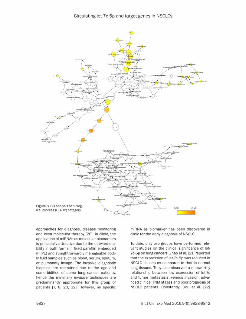
Validated targets, GO analysis and pathway analysis
After proving the downregu-lation of let-7c-5p in NSCLC, we then further investigat-ed its potential role and tar-gets in NSCLC develop-ment. Altogether, 154 tar- get genes were finally gai- ned from literatures search-ing and miRWalk extrac-tion. In order to investigate the potential mechanism of let-7c-5p, the integrative results of let-7c-5p targets in GO were categorized ac- cording to three parts: bio-logical process (BP), cellu-lar component (CC) and molecular function (MF) (Table 4; Figures 7-10). Su- bsequent to the pathway analysis, there were 10 pathways available with P < 0.05, out of which four sig-naling pathways were re- markably significant (P < 0.01, Table 5): chronic my- eloid leukemia pathway (NRAS, ACVR1B, TGFBR1, CBL, BCL2L1, MYC; P = 0.001098827), arrhythmo-genic right ventricular car-diomyopathy (ARVC) path-way (ACTB, ATP2A2, ACTN4,

Circulating let-7c-5p and target genes in NSCLCs
9834 Int J Clin Exp Med 2016;9(6):9828-9842
Table 4. GO analysis of the predicted target genesGO Category Term P Value TargetsBiological Process GO:0006325: chromatin organization 0.000000069 18Biological Process GO:0016568: chromatin modification 0.000000231 15Biological Process GO:0051276: chromosome organization 0.000002260 18Biological Process GO:0006396: RNA processing 0.000156000 16Biological Process GO:0006338: chromatin remodeling 0.000163000 6Biological Process GO:0008380: RNA splicing 0.000301000 11Biological Process GO:0045884: regulation of survival gene product expression 0.000907000 4Biological Process GO:0006414: translational elongation 0.002430000 6Biological Process GO:0046456: icosanoid biosynthetic process 0.002870000 4Biological Process GO:0006397: mRNA processing 0.002930000 10Biological Process GO:0000375: RNA splicing, via transesterification reactions 0.002950000 7Biological Process GO:0000377: RNA splicing, via transesterification reactions with bulged adenosine as nucleophile 0.002950000 7Biological Process GO:0000398: nuclear mRNA splicing, via spliceosome 0.002950000 7Biological Process GO:0006636: unsaturated fatty acid biosynthetic process 0.003740000 4Biological Process GO:0001516: prostaglandin biosynthetic process 0.004410000 3Biological Process GO:0046457: prostanoid biosynthetic process 0.004410000 3Biological Process GO:0016570: histone modification 0.005450000 6Biological Process GO:0006633: fatty acid biosynthetic process 0.006120000 5Biological Process GO:0016569: covalent chromatin modification 0.006240000 6Biological Process GO:0022613: ribonucleoprotein complex biogenesis 0.006500000 7Biological Process GO:0040007: growth 0.007030000 7Biological Process GO:0030728: ovulation 0.007170000 3Biological Process GO:0016071: mRNA metabolic process 0.007350000 10Biological Process GO:0006690: icosanoid metabolic process 0.009290000 4Cellular Component GO:0031981: nuclear lumen 0.000000091 34Cellular Component GO:0031974: membrane-enclosed lumen 0.000000109 39Cellular Component GO:0070013: intracellular organelle lumen 0.000000116 38Cellular Component GO:0043233: organelle lumen 0.000000209 38Cellular Component GO:0008180: signalosome 0.000071200 4Cellular Component GO:0030529: ribonucleoprotein complex 0.000131000 15Cellular Component GO:0043232: intracellular non-membrane-bounded organelle 0.000153000 40Cellular Component GO:0043228: non-membrane-bounded organelle 0.000153000 40Cellular Component GO:0005654: nucleoplasm 0.000173000 20

Circulating let-7c-5p and target genes in NSCLCs
9835 Int J Clin Exp Med 2016;9(6):9828-9842
Cellular Component GO:0005730: nucleolus 0.000300000 17Cellular Component GO:0022626: cytosolic ribosome 0.000657000 6Cellular Component GO:0005829: cytosol 0.000819000 24Cellular Component GO:0044451: nucleoplasm part 0.000945000 14Cellular Component GO:0022627: cytosolic small ribosomal subunit 0.004860000 4Cellular Component GO:0033279: ribosomal subunit 0.004930000 6Cellular Component GO:0033276: transcription factor TFTC complex 0.006240000 3Cellular Component GO:0000123: histone acetyltransferase complex 0.009060000 4Molecular Function GO:0003723: RNA binding 0.000168000 18Molecular Function GO:0000166: nucleotide binding 0.001370000 34Molecular Function GO:0030528: transcription regulator activity 0.005740000 24Molecular Function GO:0032555: purine ribonucleotide binding 0.008260000 27Molecular Function GO:0032553: ribonucleotide binding 0.008260000 27Molecular Function GO:0005524: ATP binding 0.008840000 23Notes: GO analysis was categorized according to three parts: biological process, cellular component and molecular function. Only those P value was less than 0.01 were presented in the table.

Circulating let-7c-5p and target genes in NSCLCs
9836 Int J Clin Exp Med 2016;9(6):9828-9842
Figure 7. GO analysis of validated target genes of let-7c-5p.
Circulating let-7c-5p and target genes in NSCLCs
9837 Int J Clin Exp Med 2016;9(6):9828-9842
approaches for diagnose, disease monitoring and even molecular therapy [20]. In clinic, the application of miRNAs as molecular biomarkers is principally attractive due to the outward sta-bility in both formalin fixed paraffin embedded (FFPE) and straightforwardly manageable bodi-ly fluid samples such as blood, serum, sputum, or pulmonary lavage. The invasive diagnostic biopsies are restrained due to the age and comorbidities of some lung cancer patients, hence the minimally invasive techniques are predominantly appropriate for this group of patients [7, 8, 20, 32]. However, no specific
miRNA as biomarker has been discovered in clinic for the early diagnosis of NSCLC.
To date, only two groups have performed rele-vant studies on the clinical significance of let-7c-5p on lung cancers. Zhao et al. [21] reported that the expression of let-7c-5p was reduced in NSCLC tissues as compared to that in normal lung tissues. They also observed a noteworthy relationship between low expression of let-7c and tumor metastasis, venous invasion, adva- nced clinical TNM stages and poor prognosis of NSCLC patients. Constantly, Dou et al. [22]
Figure 8. GO analysis of biolog-ical process (GO-BP) category.

Circulating let-7c-5p and target genes in NSCLCs
9838 Int J Clin Exp Med 2016;9(6):9828-9842
found that the plasma let-7c-5p level was also downregulated in NSCLCs. Furthermore, Dou et al. [22] drew the ROC curve for let-7c-5p and achieved a moderate diagnostic value (AUC = 0.714). However, the precise diagnostic effect of circulating let-7c-5p on NSCLC remains still unclear due to the only one available publica-tion. Thus, we performed the current meta-analysis based on GEO microarray datasets, to further confirm if let-7c-5p could be a potential biomarker for the diagnosis of lung cancer or not. Herein, six miRNA profiling datasets with 250 NSCLC samples and 242 healthy controls were finally included (GSE61741, GSE46729, GSE40738, GSE24709, GSE17681 and GSE- 27486) for analysis. The summary SMD showed that let-7c-5p expression in patients’ whole blood, peripheral blood cells and serum was significantly reduced in NSCLC patients than that of the healthy controls (SMD = -0.425; 95% CI, -0.793 to -0.057; P = 0.023), as evaluated
with random-effects model, since significant heterogeneity existed (P = 0.005, I2 = 70.3%). To exclude the influence of heterogeneity, we then re-calculated the pooled SMD by removing the data of GSE61741, which may be the source to produce heterogeneity. The pooled SMD became -0.510 (P < 0.001, 95% CI, -0.74 to -0.279) with fixed-effects model (P = 0.352, I2 = 9.5%), which presented the similar but more powerful value as that with GSE61741 remain-ing within the statistics. Thus, the result from current meta-analysis displayed the concor-dant trend with the report of Dou et al. [22] that markedly lower expression of circulating let-7c-5p occurred in NSCLC patients, as compared to non-cancerous controls.
The biological function and molecular mecha-nisms of let-7c-5p on NSCLC cells have been investigated by several groups. Zhan et al. [33] performed a series of in vitro and in vivo experi-
Figure 9. GO analysis of cellular component (GO-CC) category.

Circulating let-7c-5p and target genes in NSCLCs
9839 Int J Clin Exp Med 2016;9(6):9828-9842
ments, including MTS, colony formation, flow cytometry assays and mouse model, which demonstrated that forced overexpression of let-7c could inhibit NSCLC cell proliferation by inducing G1 arrest and suppress tumorigene-ses. This cell growth inhibitory function was achieved via targeting different genes, such as HOXA1 [33], ITGB3, MAP4K3 [21], ABCC2, Bcl-XL [34] and TRIB2 [35]. Since one single miRNA can target hundreds of genes, the exploration of a single target gene cannot reveal a wide-spread molecular mechanism of let-7c-5p. Hence, we collected 154 validated targeting genes via literatures screening and in silico pre-diction by miRWalk for a comprehensive target gene network analysis. The GO and KEGG path-way analysis indicated that let-7c-5p may play an essential role in the development of NSCLC through different pathways, such as Chronic myeloid leukemia pathway, Adherens junction pathway, ErbB signaling pathway, Pathways in cancer and MAPK signaling pathway. This could
lead to novel direction for the future investiga-tion of the molecular mechanisms of let-7c-5p on NSCLC.
Besides the clinical role and bio-function, let-7c-5p also gains the potential in the therapeu-tic intervention strategy in NSCLC, especially in chemotherapy [36] and molecular therapy [37]. Let-7c-5p could increase the sensitivity of gefi-tinib, an EGFR-targeting drug, via inhibiting RAS and deactivating the phosphoinositide 3-kinase (PI3K)/AKT and mitogen-activated extracellular signal-regulated kinase (MEK)/extracellular signal-regulated kinase (ERK) signaling path-ways. And let-7c-5p plays an essential role in fulvestrant-induced upregulation of gefitinib sensitivity in lung cancer H1975 cells with T790M resistant mutation [37].
To the best of our knowledge, this is the first meta-analysis so far to evaluate the suitability of let-7c-5p as a blood-based marker for early
Figure 10. GO analysis of molec-ular function (GO-MF) category.

Circulating let-7c-5p and target genes in NSCLCs
9840 Int J Clin Exp Med 2016;9(6):9828-9842
Table 5. KEGG pathway of validated target genes of let-7c-5p KEGG Pathway Count P Value Geneshsa05220: Chronic myeloid leukemia 6 0.001098827 NRAS, ACVR1B, TGFBR1, CBL, BCL2L1, MYChsa05412: Arrhythmogenic right ventricular cardiomyopathy (ARVC) 6 0.001166809 ACTB, ATP2A2, ACTN4, CACNG8, RYR2, ITGA10hsa03010: Ribosome 6 0.002136602 RPL18A, RPL3, RPS4Y1, RPS13, UBA52, RPS24hsa04520: Adherens junction 5 0.008671443 ACTB, ACVR1B, CSNK2A1, ACTN4, TGFBR1hsa05410: Hypertrophic cardiomyopathy (HCM) 5 0.012191074 ACTB, ATP2A2, CACNG8, RYR2, ITGA10hsa04012: ErbB signaling pathway 5 0.013195175 NRAS, PTK2, PAK4, CBL, MYChsa05414: Dilated cardiomyopathy 5 0.015932867 ACTB, ATP2A2, CACNG8, RYR2, ITGA10hsa00590: Arachidonic acid metabolism 4 0.021116706 PTGES2, PTGS2, PTGS1, LTA4Hhsa05200: Pathways in cancer 9 0.021160803 FZD8, NRAS, ACVR1B, PTK2, PTGS2, TGFBR1, CBL, BCL2L1, MYChsa04010: MAPK signaling pathway 8 0.021701553 MEF2C, NRAS, ACVR1B, CACNG8, TGFBR1, MYC, FLNB, DUSP7Ten pathways were available, among which, four signaling pathways were significant (P < 0.01).

Circulating let-7c-5p and target genes in NSCLCs
9841 Int J Clin Exp Med 2016;9(6):9828-9842
diagnosis of NSCLCs based on the microarray datasets. However, several limitations should be mentioned in the current study. Firstly, the small sample size of six datasets restricted the robustness of the current meta-analysis. Further studies with more samples included should be deliberated to study the role of circu-lating let-7c-5p in NSCLCs. Secondly, the let-7c-5p expression was only based on microarray assays. Future confirmation with more precise techniques like real time RT-qPCR should be performed. Thirdly, extra source of a relevant bias might be due to the only two regions involved in the meta-analysis (USA and Germany). Fourthly, only validated target genes were collected to analyze the potential gene-network of let-7c-5p. Other in silico predictions from different software could be gathered to have a more expansive insight of the molecular mechanism of let-7c-5p in NSCLC. Therefore, the current result needs to be interpreted cautiously.
Conclusion
Collectively, our data show that circulating let-7c-5p is reduced in blood of lung cancer patients as compared to that of non-cancerous controls. Let-7c-5p can be used to detect human lung cancers from blood cells as a promising biomarker candidate. Further stud-ies of high quality and large cohort in the future are preferred to support the predictive power of circulating let-7c-5p with validated detection techniques. Furthermore, our prediction bioin-formatics data may assist researchers to explore the molecular mechanisms of let-7c-5p in the tumorigenesis and progression of lung cancer comprehensively.
Disclosure of conflict of interest
None.
Address correspondence to: Haixin Huang, Depart- ment of Medical Oncology, Fourth Affiliated Hospital of Guangxi Medical University/Liuzhou Worker Hospital, 1 Liushi Road, Liuzhou 545005, Guangxi Zhuang Autonomous Region, China. Tel: 0086-772-3815405; Fax: 0086-772-3815405; E-mail: [email protected]
References
[1] Zheng RL, Jiang YJ, Wang X. Role of microRNAs on therapy resistance in Non-Hodgkin’s lym-
phoma. Int J Clin Exp Med 2014; 7: 3818-3832.
[2] Aghanoori MR, Mirzaei B, Tavallaei M. MiRNA molecular profiles in human medical condi-tions: connecting lung cancer and lung devel-opment phenomena. Asian Pacif J Cancer Pre-vent 2014; 15: 9557-9565.
[3] Van Peer G, Lefever S, Anckaert J, Beckers A, Rihani A, Van Goethem A, Volders PJ, Zeka F, Ongenaert M, Mestdagh P, Vandesompele J. miRBase Tracker: keeping track of microRNA annotation changes. Database (Oxford) 2014; 2014.
[4] Kozomara A, Griffiths-Jones S. miRBase: anno-tating high confidence microRNAs using deep sequencing data. Nucleic Acids Res 2014; 42: D68-73.
[5] Jackson BL, Grabowska A, Ratan HL. MicroR-NA in prostate cancer: functional importance and potential as circulating biomarkers. BMC Cancer 2014; 14: 930.
[6] Oom AL, Humphries BA, Yang C. MicroRNAs: novel players in cancer diagnosis and thera-pies. Biomed Res Int 2014; 2014: 959461.
[7] Fujita Y, Kuwano K, Ochiya T, Takeshita F. The impact of extracellular vesicle-encapsulated circulating microRNAs in lung cancer research. Biomed Res Int 2014; 2014: 486413.
[8] Ramshankar V, Krishnamurthy A. Lung cancer detection by screening-presenting circulating miRNAs as a promising next generation bio-marker breakthrough. Asian Pacif J Cancer Prevent 2013; 14: 2167-2172.
[9] Cheng HH, Mitchell PS, Kroh EM, Dowell AE, Chery L, Siddiqui J, Nelson PS, Vessella RL, Knudsen BS, Chinnaiyan AM, Pienta KJ, Mor-rissey C, Tewari M. Circulating microRNA profil-ing identifies a subset of metastatic prostate cancer patients with evidence of cancer-asso-ciated hypoxia. PLoS One 2013; 8: e69239.
[10] Wang Z. Selection of chemotherapy for non-small cell lung cancer is facilitated by new therapeutic strategies. Int J Clin Exp Med 2014; 7: 3833-3842.
[11] Wairagu PM, Park KH, Kim J, Choi JW, Kim HW, Yeh BI, Jung SH, Yong SJ, Jeong Y. Combined therapeutic potential of nuclear receptors with receptor tyrosine kinase inhibitors in lung can-cer. Biochem Biophys Res Commun 2014; 447: 490-495.
[12] Tejero R, Navarro A, Campayo M, Vinolas N, Marrades RM, Cordeiro A, Ruiz-Martinez M, Santasusagna S, Molins L, Ramirez J, Monzó M. miR-141 and mir-200c as markers of over-all survival in early stage non-small cell lung cancer adenocarcinoma. PLoS One 2014; 9: e101899.
[13] Othman N, Nagoor NH. The role of microRNAs in the regulation of apoptosis in lung cancer

Circulating let-7c-5p and target genes in NSCLCs
9842 Int J Clin Exp Med 2016;9(6):9828-9842
and its application in cancer treatment. Biomed Res Int 2014; 2014: 318030.
[14] Vosa U, Vooder T, Kolde R, Vilo J, Metspalu A, Annilo T. Meta-analysis of microRNA expres-sion in lung cancer. Int J Cancer 2013; 132: 2884-2893.
[15] She J, Yang P, Hong Q, Bai C. Lung cancer in China: challenges and interventions. Chest 2013; 143: 1117-1126.
[16] Roy M, Luo YH, Ye M, Liu J. Nonsmall cell lung cancer therapy: insight into multitargeted small-molecule growth factor receptor inhibi-tors. Biomed Res Int 2013; 2013: 964743.
[17] Qin X, Xu H, Gong W, Deng W. The tumor cyto-sol miRNAs, fluid miRNAs, and exosome miR-NAs in lung cancer. Front Oncol 2014; 4: 357.
[18] Jeong HC. Clinical aspect of MicroRNA in lung cancer. Tuberculos Respirat Dis 2014; 77: 60-64.
[19] Del Vescovo V, Grasso M, Barbareschi M, Denti MA. MicroRNAs as lung cancer biomarkers. World J Clin Oncol 2014; 5: 604-620.
[20] Keller A, Leidinger P, Borries A, Wendschlag A, Wucherpfennig F, Scheffler M, Huwer H, Len-hof HP, Meese E. miRNAs in lung cancer-study-ing complex fingerprints in patient’s blood cells by microarray experiments. BMC Cancer 2009; 9: 353.
[21] Zhao B, Han H, Chen J, Zhang Z, Li S, Fang F, Zheng Q, Ma Y, Zhang J, Wu N, Yang Y. MicroR-NA let-7c inhibits migration and invasion of hu-man non-small cell lung cancer by targeting ITGB3 and MAP4K3. Cancer Lett 2014; 342: 43-51.
[22] Dou H, Wang Y, Su G, Zhao S. Decreased plas-ma let-7c and miR-152 as noninvasive bio-marker for non-small-cell lung cancer. Int J Clin Exp Med 2015; 8: 9291-9298.
[23] van Kouwenhove M, Kedde M, Agami R. Mi-croRNA regulation by RNA-binding proteins and its implications for cancer. Nat Rev Cancer 2011; 11: 644-656.
[24] Dweep H, Gretz N, Sticht C. miRWalk database for miRNA-target interactions. Methods Mol Biol 2014; 1182: 289-305.
[25] Dweep H, Sticht C, Pandey P, Gretz N. miRWalk--database: prediction of possible miRNA bind-ing sites by “walking” the genes of three ge-nomes. J Biomed Informat 2011; 44: 839-847.
[26] Meng X, Xiao C, Zhao Y, Jia L, Tang Y, Li D. Me-ta-analysis of microarrays: diagnostic value of microRNA-21 as a biomarker for lung cancer. Int J Biol Markers 2015; 30: e282-285.
[27] Sewer A, Gubian S, Kogel U, Veljkovic E, Han W, Hengstermann A, Peitsch MC, Hoeng J. Assess-ment of a novel multi-array normalization method based on spike-in control probes suit-able for microRNA datasets with global de-creases in expression. BMC Res Notes 2014; 7: 302.
[28] Cheung SF, Chan DK. Meta-analyzing depen-dent correlations: an SPSS macro and an R script. Behav Res Methods 2014; 46: 331-345.
[29] Lau J, Ioannidis JP, Schmid CH. Quantitative synthesis in systematic reviews. Ann Intern Med 1997; 127: 820-826.
[30] Higgins JP, Thompson SG, Deeks JJ, Altman DG. Measuring inconsistency in meta-analy-ses. BMJ 2003; 327: 557-560.
[31] Zamora J, Abraira V, Muriel A, Khan K, Cooma-rasamy A. Meta-DiSc: a software for meta-anal-ysis of test accuracy data. BMC Med Res Meth-odol 2006; 6: 31.
[32] Huang Y, Hu Q, Deng Z, Hang Y, Wang J, Wang K. MicroRNAs in body fluids as biomarkers for non-small cell lung cancer: a systematic re-view. Technol Cancer Res Treat 2014; 13: 277-287.
[33] Zhan M, Qu Q, Wang G, Liu YZ, Tan SL, Lou XY, Yu J, Zhou HH. Let-7c inhibits NSCLC cell prolif-eration by targeting HOXA1. Asian Pac J Cancer Prev 2013; 14: 387-392.
[34] Zhan M, Qu Q, Wang G, Zhou H. Let-7c sensi-tizes acquired cisplatin-resistant A549 cells by targeting ABCC2 and Bcl-XL. Pharmazie 2013; 68: 955-961.
[35] Wang PY, Sun YX, Zhang S, Pang M, Zhang HH, Gao SY, Zhang C, Lv CJ, Xie SY. Let-7c inhibits A549 cell proliferation through oncogenic TRIB2 related factors. FEBS Lett 2013; 587: 2675-2681.
[36] Cui SY, Huang JY, Chen YT, Song HZ, Feng B, Huang GC, Wang R, Chen LB, De W. Let-7c gov-erns the acquisition of chemo- or radioresis-tance and epithelial-to-mesenchymal transi-tion phenotypes in docetaxel-resistant lung adenocarcinoma. Mol Cancer Res 2013; 11: 699-713.
[37] Shen H, Liu J, Wang R, Qian X, Xu R, Xu T, Li Q, Wang L, Shi Z, Zheng J, Chen Q, Shu Y. Fulves-trant increases gefitinib sensitivity in non-small cell lung cancer cells by upregulating let-7c ex-pression. Biomed Pharmacother 2014; 68: 307-313.
Copyright © 2022 FDOKUMEN