Organometallic derivatizing agents in bioanalysis
12
REVIEW Organometallic derivatizing agents in bioanalysis Susanne Bomke & Michael Sperling & Uwe Karst Received: 2 January 2010 / Revised: 22 February 2010 / Accepted: 22 February 2010 / Published online: 21 March 2010 # Springer-Verlag 2010 Abstract Over the last few decades, the development of several innovative hyphenated analytical techniques and their routine use in laboratories has led to new possibilities for the quantitative analysis of biomolecules. Today, the identification and quantification of biomolecules such as peptides and proteins are essential to answer important medical, pharmaceutical, and biological questions. To allow efficient detection and structure elucidation of biomolecules, several approaches including derivatization strategies were investigated and applied during recent years. This article summarizes the current approaches for labeling and presents the different types of organometallic derivatizing agents used as labels. Furthermore, their analytical potential with respect to quantification and structure elucidation for different ap- plications in the field of bioanalysis is discussed. Keywords Mass spectrometry . ICP-MS . Organometals . Bioanalytical methods Introduction The systematic acquisition of information relevant to ge- nomes, gene transcripts, and proteins including the identifi- cation of structure and function is a fundamental aspect in current bioanalytical chemistry. Most of the recent achieve- ments in this field benefit from the developments of mass spectrometric detection techniques such as electrospray (ESI) [1], matrix-assisted laser desorption and ionization (MALDI) [2, 3], and inductively coupled plasma–mass spectrometry (ICP–MS) [4]. Moreover, the routine use of hyphenated techniques combining a separation module such as liquid chromatography (LC) coupled with mass spectrometric methods such as ESI–MS and ICP–MS offers new possibil- ities for the identification and quantification of biomolecules. The potential of these techniques for protein analysis has been discussed in several reviews focusing on MS [5, 6] and ICP–MS [7–9]. It rapidly became evident that apart from identification and relative quantification, absolute quantitative data are required for further characterization of biological samples and that the whole field of proteomics “had to turn quan- titative” [10]. Thus, dynamic biological systems should be quantitatively examined and the search for biomarkers in clinical proteomics must be expanded with new technolo- gies aimed at absolute quantification. In particular, the ability of ICP–MS for simultaneous isotope abundance measurements leads the way to innovative metabolic studies and the quantification of biomolecules [11]. Since the ICP is a plasma open to the surrounding atmosphere, the detection capabilities for C, N, and O (i.e., the main constituents of atmospheric air) are rather limited, and therefore quantifica- tion is frequently based on other elements. Fortunately, some peptides and proteins contain natural heteroelements apart from H, C, N, and O. The role of such natural element tags has been reviewed recently by Prange and Pröfrock [12]. The analysis of naturally covalently incorporated heteroelements such as 32 S[13–15], 80 Se [16–19], 127 I[20, 21], or 31 P[22– 25] by ICP–MS is enjoying increased interest in recent years. However, isobaric interferences, the high first ionization S. Bomke : M. Sperling : U. Karst (*) Institut für Anorganische und Analytische Chemie, Westfälische Wilhelms-Universität Münster, Corrensstr. 30, 48149 Münster, Germany e-mail: [email protected] M. Sperling European Virtual Institute for Speciation Analysis, Mendelstr. 11, 48149 Münster, Germany Anal Bioanal Chem (2010) 397:3483–3494 DOI 10.1007/s00216-010-3611-1
-
Upload
uni-muenster -
Category
Documents
-
view
4 -
download
0
Transcript of Organometallic derivatizing agents in bioanalysis

REVIEW
Organometallic derivatizing agents in bioanalysis
Susanne Bomke & Michael Sperling & Uwe Karst
Received: 2 January 2010 /Revised: 22 February 2010 /Accepted: 22 February 2010 /Published online: 21 March 2010# Springer-Verlag 2010
Abstract Over the last few decades, the development ofseveral innovative hyphenated analytical techniques andtheir routine use in laboratories has led to new possibilitiesfor the quantitative analysis of biomolecules. Today, theidentification and quantification of biomolecules such aspeptides and proteins are essential to answer importantmedical, pharmaceutical, and biological questions. To allowefficient detection and structure elucidation of biomolecules,several approaches including derivatization strategies wereinvestigated and applied during recent years. This articlesummarizes the current approaches for labeling and presentsthe different types of organometallic derivatizing agents usedas labels. Furthermore, their analytical potential with respectto quantification and structure elucidation for different ap-plications in the field of bioanalysis is discussed.
Keywords Mass spectrometry . ICP-MS . Organometals .
Bioanalytical methods
Introduction
The systematic acquisition of information relevant to ge-nomes, gene transcripts, and proteins including the identifi-cation of structure and function is a fundamental aspect in
current bioanalytical chemistry. Most of the recent achieve-ments in this field benefit from the developments of massspectrometric detection techniques such as electrospray (ESI)[1], matrix-assisted laser desorption and ionization (MALDI)[2, 3], and inductively coupled plasma–mass spectrometry(ICP–MS) [4]. Moreover, the routine use of hyphenatedtechniques combining a separation module such as liquidchromatography (LC) coupled with mass spectrometricmethods such as ESI–MS and ICP–MS offers new possibil-ities for the identification and quantification of biomolecules.The potential of these techniques for protein analysis hasbeen discussed in several reviews focusing on MS [5, 6] andICP–MS [7–9].
It rapidly became evident that apart from identificationand relative quantification, absolute quantitative data arerequired for further characterization of biological samplesand that the whole field of proteomics “had to turn quan-titative” [10]. Thus, dynamic biological systems should bequantitatively examined and the search for biomarkers inclinical proteomics must be expanded with new technolo-gies aimed at absolute quantification. In particular, theability of ICP–MS for simultaneous isotope abundancemeasurements leads the way to innovative metabolic studiesand the quantification of biomolecules [11]. Since the ICP isa plasma open to the surrounding atmosphere, the detectioncapabilities for C, N, and O (i.e., the main constituents ofatmospheric air) are rather limited, and therefore quantifica-tion is frequently based on other elements. Fortunately, somepeptides and proteins contain natural heteroelements apartfrom H, C, N, and O. The role of such natural element tagshas been reviewed recently by Prange and Pröfrock [12]. Theanalysis of naturally covalently incorporated heteroelementssuch as 32S [13–15], 80Se [16–19], 127I [20, 21], or 31P [22–25] by ICP–MS is enjoying increased interest in recent years.However, isobaric interferences, the high first ionization
S. Bomke :M. Sperling :U. Karst (*)Institut für Anorganische und Analytische Chemie,Westfälische Wilhelms-Universität Münster,Corrensstr. 30,48149 Münster, Germanye-mail: [email protected]
M. SperlingEuropean Virtual Institute for Speciation Analysis,Mendelstr. 11,48149 Münster, Germany
Anal Bioanal Chem (2010) 397:3483–3494DOI 10.1007/s00216-010-3611-1

energies of these heteroelements, and the resulting highlimits of detection often lead to unsatisfactory results. Betteranalytical results can be expected by introducing elementaltags via an additional derivatization step. Additionally, sincestandards for most biomolecules of natural origin areunavailable, such tagging using different derivatizationapproaches is a valuable alternative for quantification [26].
In analytical chemistry, derivatization reactions are oftencarried out due to missing chromophoric, fluorophoric, orelectroactive groups in the analyte of interest. A suitablederivatization reaction must convert the analyte into a stableproduct, and the conversion should be quantitative andenable or improve the use of more sensitive detection tech-niques. In order to be practicable, the derivatization, whichalways is an extra reaction step during sampling prepara-tion, should be simple and straightforward.
To date, several derivatization approaches have beendeveloped mainly based on chemical, metabolic, and enzy-matic labeling. The structure and properties of the differentderivatizing agents are intended to match the analyticaldemands with respect to target analyte, detection technique,and application field. With respect to their structures, thederivatization reagents can be divided into different com-pound classes.
This review focuses on current labeling approaches, andthe main focus is directed to the presentation of the variouscompound classes of labeling reagents (Table 1). Moreover,the analytical possibilities resulting from formed deriva-tives, and the advantages and disadvantages of thederivatizing agents will be further discussed.
Current labeling strategies
Isotope labeling
Stable isotope labeling has emerged as a powerful tool toidentify and quantify peptides and proteins in complex mix-tures. This labeling strategy can be applied for chemical,metabolic, and enzymatic labeling. The first of these involveschemical reactions for the labeling of biomolecules prior totheir analysis. For example, functional groups of proteins canbe selectively labeled with stable-isotope-containing affinityreagents, allowing fast analysis of complex protein mixtures.The prototype of these chemical labeling approaches wasintroduced by Gygi et al. as isotope-coded affinity tags(ICAT) in 1999 [27]. This method is based on the differentialisotope labeling of the rare cysteine residues with tagscontaining a thiol-selective reactive group, an affinity tag(biotin), and an isotopically marked linker group. Generally,ICAT is used to determine the relative expression of peptidesin diseased versus healthy states (Fig. 1). Because ICAT isrestricted to only cysteine-containing proteins, attempts to
broaden the applicability of this approach were made. Forexample, isotope-coded protein labels (ICPL) were devel-oped, which selectively label all free amino groups inproteins [28]. However, the lack of robustness, differentialelution of identical peptides labeled with the hydrogen/deuterium isotope pairs on reversed phases, and complicatedinterpretation of the resulting tandem mass spectra [29]severely restricted the application of ICAT despite manyrefinements [30–32]. A ‘bottom-up’ approach involving thedigestion of proteins into peptide fragments that can bedetected and sequenced with liquid chromatography coupledwith tandem mass spectrometry (LC/MS/MS) uses isobarictags of the proteolytic peptides for relative and absolutequantification (iTRAQ). This method is based on the in-corporation of up to four mass tags that label the N-terminusand lysine residues of peptides resulting from a tryptic digestof the proteins [33, 34]. Furthermore, the introduction ofstable isotopes can be performed in vivo by using metaboliclabeling such as SILAC (stable isotope labeling of aminoacids in cell culture) [35]. Metabolic labeling has beenwidely used for the relative quantification of protein ex-pression in differently treated cell cultures. The enzymaticlabeling approach makes use of the incorporation of isotopesby using H2
18O during a tryptic digest [36, 37]. A seriousdrawback of this approach is the pH-mediated back ex-change of 16O and 18O [36]. Also, the variation resultingfrom the exchange of one or two carboxyl oxygen atomsmakes the interpretation of data very complex. Furthermore,missing separations at the protein level seriously restrict theanalytical value of this in vivo labeling strategy [38].
Chelating compounds
Historically, the most common tool for the analysis ofproteins makes use of chelating compounds in combinationwith radioactive tracers. As a result of developments in massspectrometry in recent years, chelating compounds withdifferent incorporated metals came into use for the detectionof biomolecules. The term chelate implies the involvementof a complex, in which a metal ion is bound to two or moreatoms of the chelating agent, whereas the bonds may be anycombination of coordination or ionic bond [39]. Accordingto the strict definition of the term “organometallic com-pounds”, metal chelates are not included, as the presence of ametal–carbon bond is essential for this classification.Bifunctional chelating agents have various medical applica-tions, e.g., they are frequently used in combination withradiopharmaceuticals and contrast agents [40] as well as inbioanalytical assays [41]. Macrocyclic metal chelates such as1,4,7,10-tetraazacyclododecane-1,4,7,10-tetraacetic acid(DOTA) loaded with different lanthanide (Me3+) ions formvery stable metal complexes with high stability constants(Fig. 2) that can be applied to the metal-coded affinity tag
3484 S. Bomke et al.

(MeCAT©) technique that allows absolute quantitativedetermination of peptides and proteins [42–44] by ICP–MSwith the help of an external standard.
Bifunctional chelating agents like diethylenetriamine-pentaacetate (DTPA) have been used to label conjugatedfunctional groups on peptides and proteins [45] and che-lated with 111In or other radioactive isotopes to produceradiopharmaceuticals. Other DTPA derivatives containingmaleimide, bromoacetamide, and pyridyldithio linkers were
used as luminescent probes in combination with Eu3+ andTb3+ as chromophores [46]. Recently, DTPA was subse-quently labeled with naturally and isotopically enrichedEu3+ and was used as a labeling tag for relative quantifi-cation [47]. Liu et al. demonstrated the labeling of peptides byusing yttrium and terbium–DTPA complexes [48]. Furtherlanthanide ions such as Eu, Tb, and Ho were implemented ina 1,4,7,10-tetraazacyclododecane-1,4,7,10-tetraacetic acidsuccinimide ester (SCN–DOTA) complex to label bovine
Table 1 Overview of the most important classes of labeling reagents
S
NHNH
O
O
NH
OD
DD
DO
O NH
D
DD
DO
I
Chemical structure types Reference cited
[27]
Reagent
Isotope labeling
ICAT
iTRAQ [33, 34]
Mercury tags
Chelatingcompounds
e.g. MeCAT
CO2R
NN
RhCl1 R = CH32 R = C6F5
L(CO)4W COR1
R2Carbenecomplexes
[76, 77]
[78-81]
CO
O N
O
OMLn
OC Me
1 M = Mo, L = CO, n = 22 M = Fe, L = CO, n = 13 M = W, L = CO, n = 3
Carbonylcomplexes
N N
N N
O
OO O
O
OHO
HN
O
N
O
OLa3+
O
CH3HgCl
CH3CH2HgCl
[127-129][135, 136]
[130-134]
[42-44]
[59-71]
HOOC Hg OH
Metallocene-based
reagents
Fe
RR = (CO)H, (CO)Cl, CH2COOH, B(OH)2
R = N
O
O
N
O
O
O O
C
O
O N
O
OCo+
[96-117]
[120-126]
ON
O
O
ON
N
1 L = CO, R1 = R2 = Me2 L = CO, R1 = Et, R2 = Ph3 L = PBu3, R = Et, R2 = PH4 L = CO, R2 = Li, R2 = Ph
1
Organometallic derivatizing agents in bioanalysis 3485

serum albumin and hen egg white lysozyme [48]. Theresulting derivatives were separated by sodium dodecylsulfate polyacrylamide gel electrophoresis (SDS–PAGE).Until recently, only a few groups have made use of chelatingcompounds in combination with atomic spectrometry analysistechniques such as ICP–MS. For example, peptide quan-tification was performed by using In–DOTA complexes incombination with complementary hyphenation of LC/ESI–TOF–MS and LC/ICP–MS [49]. Commercially availablefluorescent probes (DELFIA™) containing the lanthanidesEu, Tb, and Sm were employed for DOTA labeling ofantibodies and detection by ICP–MS [50] even in combina-tion with multiplex analysis [51]. Additionally, the firstICP–MS-based multiplex profiling of glycoproteins waspublished recently, in which lectins conjugated to lanthanide-chelating compounds were used [52].
Despite the excellent detectability of lanthanide ionsby ICP–MS and the high stability of those reagentsavoiding metal loss or metal exchange, the highly polarcomplexes and their derivatives are often not suitablefor separations on reversed-phase (RP) columns. Unfortu-nately, without a satisfactory separation of the derivatized
biomolecules, an absolute quantification cannot be ex-pected. Baseline separation of the derivatives is necessaryto avoid suppression effects when using LC/ESI–MS.However, the use of chelating compounds as derivatizingagents is mainly combined with LC/ICP–MS as the
N N
N N
HOOC
COOH
HOOC
N
O
O
La3+
COOH
R
chelate complex (e.g. DOTA)
lanthanide ion
R = biotin
cysteine selectivemaleimide group
N N
N N
HOOC
COOH
HOOC
N
O
O
La3+
COOH
R
chelate complex (e.g. DOTA)
lanthanide ion
R = biotin
cysteine selectivemaleimide group
Fig. 2 Structure of the DOTA-based metal-coded affinity tag(MeCAT©) reagent. The substituted maleimide reacts selectively withcysteine residues. The biotin group provides the possibility of avidin-biotin chromatography for purification. According to the low limits ofdetection in ICP–MS, the lanthanide ion allows absolute quantificationof the analytes
„Normal“ mouse „Giant“ mouse
a b
S
NHNH
O
O
NH
OH
HH
HO
O NH
H
HH
HO
I
S
NHNH
O
O
NH
OD
DD
DO
O NH
D
DD
DO
I
Extraction of proteins from blood
Addition of
heavy (D) label
Mixture of both & enzymatic digest
light (1H) label
„Normal“ mouse „Giant“ mouse
S
NHNH
O
O
NH
OH
HH
HO
O NH
H
HH
HO
I
S
NHNH
O
O
NH
OD
DD
DO
O NH
D
DD
DO
I
Extraction of proteins from blood
Addition of
heavy (D) label
Mixture of both & enzymatic digest
light (1H) label
Fig. 1 Labeling procedureusing isotope-coded affinity tags(ICATs) for the determination ofindividual proteins originatingfrom two different samples(here: “normal” and “giant”mice). The ICAT reagentconsists of on isotope-codedlinker group (containing eight1H or eight D atoms) and athiol-reactive iodoacetamidegroup for the selectivealkylation of cysteine residues.Additionally, a biotin moietyallows the enrichment of thecysteine-containing labeledproteins via biotin-avidinaffinity chromatography
3486 S. Bomke et al.
hyphenated technique. Through its compound- and matrix-independent ionization, large dynamic range of over6 decades, and excellent limit of detection in the nanogramper liter range the ICP–MS technique has become oneof the most versatile and sensitive tools in bioanalyticalchemistry today.
Metallocarbonyl complexes
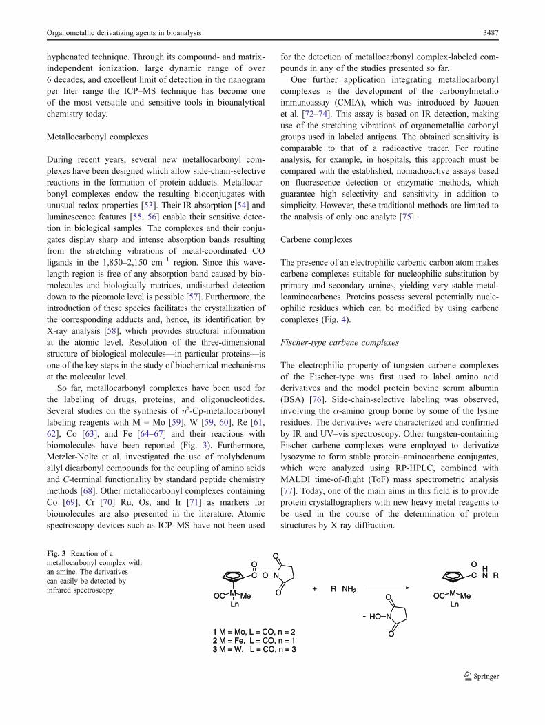
During recent years, several new metallocarbonyl com-plexes have been designed which allow side-chain-selectivereactions in the formation of protein adducts. Metallocar-bonyl complexes endow the resulting bioconjugates withunusual redox properties [53]. Their IR absorption [54] andluminescence features [55, 56] enable their sensitive detec-tion in biological samples. The complexes and their conju-gates display sharp and intense absorption bands resultingfrom the stretching vibrations of metal-coordinated COligands in the 1,850–2,150 cm−1 region. Since this wave-length region is free of any absorption band caused by bio-molecules and biologically matrices, undisturbed detectiondown to the picomole level is possible [57]. Furthermore, theintroduction of these species facilitates the crystallization ofthe corresponding adducts and, hence, its identification byX-ray analysis [58], which provides structural informationat the atomic level. Resolution of the three-dimensionalstructure of biological molecules—in particular proteins—isone of the key steps in the study of biochemical mechanismsat the molecular level.
So far, metallocarbonyl complexes have been used forthe labeling of drugs, proteins, and oligonucleotides.Several studies on the synthesis of η5-Cp-metallocarbonyllabeling reagents with M = Mo [59], W [59, 60], Re [61,62], Co [63], and Fe [64–67] and their reactions withbiomolecules have been reported (Fig. 3). Furthermore,Metzler-Nolte et al. investigated the use of molybdenumallyl dicarbonyl compounds for the coupling of amino acidsand C-terminal functionality by standard peptide chemistrymethods [68]. Other metallocarbonyl complexes containingCo [69], Cr [70] Ru, Os, and Ir [71] as markers forbiomolecules are also presented in the literature. Atomicspectroscopy devices such as ICP–MS have not been used
for the detection of metallocarbonyl complex-labeled com-pounds in any of the studies presented so far.
One further application integrating metallocarbonylcomplexes is the development of the carbonylmetalloimmunoassay (CMIA), which was introduced by Jaouenet al. [72–74]. This assay is based on IR detection, makinguse of the stretching vibrations of organometallic carbonylgroups used in labeled antigens. The obtained sensitivity iscomparable to that of a radioactive tracer. For routineanalysis, for example, in hospitals, this approach must becompared with the established, nonradioactive assays basedon fluorescence detection or enzymatic methods, whichguarantee high selectivity and sensitivity in addition tosimplicity. However, these traditional methods are limited tothe analysis of only one analyte [75].
Carbene complexes
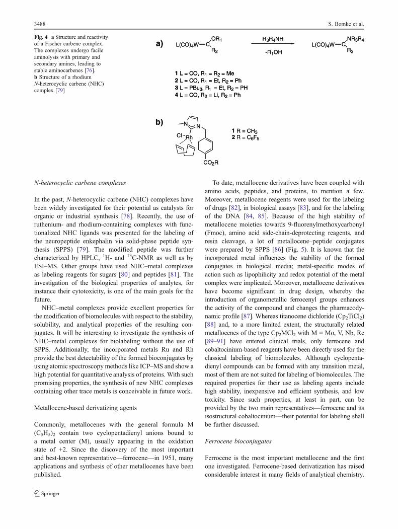
The presence of an electrophilic carbenic carbon atom makescarbene complexes suitable for nucleophilic substitution byprimary and secondary amines, yielding very stable metal-loaminocarbenes. Proteins possess several potentially nucle-ophilic residues which can be modified by using carbenecomplexes (Fig. 4).
Fischer-type carbene complexes
The electrophilic property of tungsten carbene complexesof the Fischer-type was first used to label amino acidderivatives and the model protein bovine serum albumin(BSA) [76]. Side-chain-selective labeling was observed,involving the α-amino group borne by some of the lysineresidues. The derivatives were characterized and confirmedby IR and UV–vis spectroscopy. Other tungsten-containingFischer carbene complexes were employed to derivatizelysozyme to form stable protein–aminocarbene conjugates,which were analyzed using RP-HPLC, combined withMALDI time-of-flight (ToF) mass spectrometric analysis[77]. Today, one of the main aims in this field is to provideprotein crystallographers with new heavy metal reagents tobe used in the course of the determination of proteinstructures by X-ray diffraction.
CO
O N
O
OMLn
OC Me
1 M = Mo, L = CO, n = 22 M = Fe, L = CO, n = 13 M = W, L = CO, n = 3
+ R NH2
CO
HN
MLn
OC Me
R
HO N
O
O
-
CO
O N
O
OMLn
OC Me
1 M = Mo, L = CO, n = 22 M = Fe, L = CO, n = 13 M = W, L = CO, n = 3
+ R NH2
CO
HN
MLn
OC Me
R
HO N
O
O
-
Fig. 3 Reaction of ametallocarbonyl complex withan amine. The derivativescan easily be detected byinfrared spectroscopy
Organometallic derivatizing agents in bioanalysis 3487
N-heterocyclic carbene complexes
In the past, N-heterocyclic carbene (NHC) complexes havebeen widely investigated for their potential as catalysts fororganic or industrial synthesis [78]. Recently, the use ofruthenium- and rhodium-containing complexes with func-tionalized NHC ligands was presented for the labeling ofthe neuropeptide enkephalin via solid-phase peptide syn-thesis (SPPS) [79]. The modified peptide was furthercharacterized by HPLC, 1H- and 13C-NMR as well as byESI–MS. Other groups have used NHC–metal complexesas labeling reagents for sugars [80] and peptides [81]. Theinvestigation of the biological properties of analytes, forinstance their cytotoxicity, is one of the main goals for thefuture.
NHC–metal complexes provide excellent properties forthe modification of biomolecules with respect to the stability,solubility, and analytical properties of the resulting con-jugates. It will be interesting to investigate the synthesis ofNHC–metal complexes for biolabeling without the use ofSPPS. Additionally, the incorporated metals Ru and Rhprovide the best detectability of the formed bioconjugates byusing atomic spectroscopymethods like ICP–MS and show ahigh potential for quantitative analysis of proteins. With suchpromising properties, the synthesis of new NHC complexescontaining other trace metals is conceivable in future work.
Metallocene-based derivatizing agents
Commonly, metallocenes with the general formula M(C5H5)2 contain two cyclopentadienyl anions bound toa metal center (M), usually appearing in the oxidationstate of +2. Since the discovery of the most importantand best-known representative—ferrocene—in 1951, manyapplications and synthesis of other metallocenes have beenpublished.
To date, metallocene derivatives have been coupled withamino acids, peptides, and proteins, to mention a few.Moreover, metallocene reagents were used for the labelingof drugs [82], in biological assays [83], and for the labelingof the DNA [84, 85]. Because of the high stability ofmetallocene moieties towards 9-fluorenylmethoxycarbonyl(Fmoc), amino acid side-chain-deprotecting reagents, andresin cleavage, a lot of metallocene–peptide conjugateswere prepared by SPPS [86] (Fig. 5). It is known that theincorporated metal influences the stability of the formedconjugates in biological media; metal-specific modes ofaction such as lipophilicity and redox potential of the metalcomplex were implicated. Moreover, metallocene derivativeshave become significant in drug design, whereby theintroduction of organometallic ferrocenyl groups enhancesthe activity of the compound and changes the pharmacody-namic profile [87]. Whereas titanocene dichloride (Cp2TiCl2)[88] and, to a more limited extent, the structurally relatedmetallocenes of the type Cp2MCl2 with M = Mo, V, Nb, Re[89–91] have entered clinical trials, only ferrocene andcobaltocinium-based reagents have been directly used for theclassical labeling of biomolecules. Although cyclopenta-dienyl compounds can be formed with any transition metal,most of them are not suited for labeling of biomolecules. Therequired properties for their use as labeling agents includehigh stability, inexpensive and efficient synthesis, and lowtoxicity. Since such properties, at least in part, can beprovided by the two main representatives—ferrocene and itsisostructural cobaltocinium—their potential for labeling shallbe further discussed.
Ferrocene bioconjugates
Ferrocene is the most important metallocene and the firstone investigated. Ferrocene-based derivatization has raisedconsiderable interest in many fields of analytical chemistry.
CO2R
NN
RhCl1 R = CH32 R = C6F5
L(CO)4W COR1
R2
a)
b)
R3R4NH
-R1OHL(CO)4W C
NR3R4
R2
1 L = CO, R1 = R2 = Me2 L = CO, R1 = Et, R2 = Ph3 L = PBu3, R = Et, R2 = PH4 L = CO, R2 = Li, R2 = Ph
1
CO2R
NN
RhCl1 R = CH32 R = C6F5
L(CO)4W COR1
R2
a)
b)
R3R4NH
-R1OHL(CO)4W C
NR3R4
R2
1 L = CO, R1 = R2 = Me2 L = CO, R1 = Et, R2 = Ph3 L = PBu3, R = Et, R2 = PH4 L = CO, R2 = Li, R2 = Ph
1
1 L = CO, R1 = R2 = Me2 L = CO, R1 = Et, R2 = Ph3 L = PBu3, R = Et, R2 = PH4 L = CO, R2 = Li, R2 = Ph
1
Fig. 4 a Structure and reactivityof a Fischer carbene complex.The complexes undergo facileaminolysis with primary andsecondary amines, leading tostable aminocarbenes [76].b Structure of a rhodiumN-heterocyclic carbene (NHC)complex [79]
3488 S. Bomke et al.

Two reviews nicely summarize the formation of ferroceneconjugates with amino acids, peptides, and proteins and theiranalytical and biological applications [92, 93]. Ferrocene canundergo electrophilic aromatic substitution reactions on thecyclopentadienyl ring(s). Other metallocenes frequentlydecompose under such reaction conditions [94]. Over thepast few decades, a large number of ferrocene-based reagentswere developed for several functional groups, whereby thelargest number of reagents is dedicated to the analysis ofamino functionalities. This is due to the fact that the aminoacid lysine with its aliphatic ε-amine group is present tosome extent in peptides and proteins and is often quiteabundant in biomolecules [95]. Apart from substitutedamine-reactive groups, reagents which react selectivelytowards alcohols [96–98], aldehydes [99], carboxyl groups[100, 101], dienes [102, 103], imino groups [104], isocya-nates [105, 106], and thiols [107] are now well known andestablished.
The non-polar ferrocene-based derivatizing agents turnhighly polar analytes into less polar reaction products andthus make them suitable for separation on reversed-phase
columns. Ferrocenes (Fe(II)) can be easily oxidized to thecorresponding ferrocinium cation (Fe(III)). As a result of thisone-electron redox behavior, ferrocenes enable the detectionof originally non-electroactive analytes by electrochemicalmethods like cyclic voltammetry [108]. For the detection ofthe derivatized biomolecules, mass spectrometric approacheslike ICP–MS [109, 110], ESI–MS [111, 112], and MALDI–MS [104] were applied (Fig. 6). Additionally, applicationsusing atomic spectroscopic detection such as atomic absorp-tion spectroscopy (AAS) [113, 114], atomic emissiondetection (AES) [115, 116], and electrochemiluminescence[117] have been published.
Cobaltocinium bioconjugates
The cobaltocinium ion is isoelectronic with ferrocene,thus providing even higher stability against strong oxi-dation reagents such as fuming nitric acid, potassiumpermanganate, and ozone [118, 119]. The presence of thepositive charge of the cobaltocinium unit may increase thehydrophilic properties of the analytes [120], making
a)
b)
Fmoc1) Fmoc deprotection
H2) Coupling to amino acid
Phe Fmoc
3) Repeating steps 1 and 2
Phe Arg Lys Fmoc
4) Fmoc-Deprotection & reaction with Metallocene-COOH
Phe Arg Lys
O
NH Fe
5) Cleavage & side chain deprotection
Phe Arg Lys
O
NH FeH2N
O
O N
O
OCo+ + R-NH2
O
NHCo+
N
O
O
O
O
NH
O
+ R-NH2Fe Fe
R
PF6
R
N
O
O + R-SHFe
O
NH
N
O
OFe
O
NH
SR
PF6
[107]
[109]
[125]
Fig. 5 a Different ferrocene-and cobaltocinium-basedderivatizing agents for thioland amino functionalities.b Derivatization of anamino group by a ferrocenederivative using solid-phasepeptide synthesis (with =polymer and Fmoc =fluorenylmethoxycarbonyl)
Organometallic derivatizing agents in bioanalysis 3489

cobaltocinium salts very attractive as haptens and tracersin biological systems [121, 122]. Metzler-Nolte et al.described the successful synthesis of a conjugate of acobaltocinium ion with an antigen nuclear localizationsignal (NLS) by SPPS, which specifically delivers theorganometallic species into the nucleus of a cell [123].The much higher redox potential and chemical stability ofthe cobaltocinium ion over the ferrocene moiety were usedfor enhancing cellular uptakes of bioconjugates. Moredetailed information on the synthesis of cobaltocinium–peptide bioconjugates prepared by solid-phase peptidesynthesis is published elsewhere [86, 124]. The potentialof these bioconjugates for biological assays is discussed,and metallohaptens of the activated cobaltocinium esterwith psychostimulant drugs such as amphetamine anddesipramine [125] and antiepileptic drugs such as pheno-barbital and phenytoin [126] were synthesized.
To date, the stable cobaltocinium conjugates werecharacterized by using 1H- and 13C-NMR, IR spectroscopy,X-ray crystallography, and ESI–MS, but no attempt hasbeen made so far to use separation techniques such asHPLC and capillary electrophoresis in combination withatomic spectroscopy detection possibilities like ICP–MS.
Unfortunately, as cobalt is a monoisotopic element, theuse of isotope dilution analysis for the quantification ofmodified biomolecules is not possible; however, evenexternal calibration and detection by ICP–MS offer limitsof detection in the lower parts per billion range, making thisapproach an attractive alternative.
Other metal-containing derivatizing agents
Mercury tags
Based on the strong mercury–sulfur affinity, mercurialreagents are known to be highly selective for thiols even inthe presence of all other types of reactive groups typicallypresent in proteins. Thus, the dissociation constants for themercaptide formed between the inorganic mercuric ion (Hg2+)and cysteine or the organic methylmercury (CH3Hg
+) andcysteine are 10−15.8 and 10−20.3, respectively [127]. Whereasdivalent mercuric ions (Hg2+) are capable of reacting in away to connect two thiol groups and to undergo interactionswith disulfide bridges [128], monofunctional organomercu-rial compounds of the type RHgX have the advantage ofonly reacting with one thiol group [129].
2 4 6 8 10 120
1.0
2.0
x106
Inte
nsi
ty [
cps]
Time [min]
LC/ESI-MS tR = 7.13
Time [min]
0.5
1.0
1.5
x104
0 2 4 6 8 10 120 2 4 6 8 10 120 2 4 6 8 10 120 2 4 6 8 10 12
123
4
x103
Time [min]
Inte
nsity
[cps
]
02 4 6 8 10 12
56Fe57Fe
LC/ICP-MStR = 5.37 57Fe
1200 1400 16000
0.5
1.5
x105
+13
+12
+10
+11
1231.0
1599.9
1454.4
1333.4
Inte
nsi
ty [
cps]
m/z15000 16000
0
2.0
4.0
15987.0x105
Deconvolution
16227.0
m/z
B
C
N
O
O
O
O
NH
O
+ R-NH2
pH 9Fe Fe
R
N
O
O
HO-
A
2 4 6 8 10 120
1.0
2.0
x106
Inte
nsi
ty [
cps]
Time [min]
LC/ESI-MS tR = 7.13
2 4 6 8 10 120
1.0
2.0
x106
Inte
nsi
ty [
cps]
Time [min]
LC/ESI-MS tR = 7.13
Time [min]
0.5
1.0
1.5
x104
0 2 4 6 8 10 120 2 4 6 8 10 120 2 4 6 8 10 120 2 4 6 8 10 12
123
4
x103
Time [min]
Inte
nsity
[cps
]
02 4 6 8 10 12
56Fe57Fe
LC/ICP-MStR = 5.37 57Fe
Time [min]
0.5
1.0
1.5
x104
0 2 4 6 8 10 120 2 4 6 8 10 120 2 4 6 8 10 120 2 4 6 8 10 12
123
4
x103
Time [min]
Inte
nsity
[cps
]
02 4 6 8 10 12
56Fe57Fe
LC/ICP-MStR = 5.37 57Fe
0.5
1.0
1.5
x104
0 2 4 6 8 10 120 2 4 6 8 10 120 2 4 6 8 10 120 2 4 6 8 10 12
123
4
x103
Time [min]
Inte
nsity
[cps
]
02 4 6 8 10 12
56Fe57Fe
LC/ICP-MStR = 5.37 57Fe
1200 1400 16000
0.5
1.5
x105
+13
+12
+10
+11
1231.0
1599.9
1454.4
1333.4
Inte
nsi
ty [
cps]
m/z15000 16000
0
2.0
4.0
15987.0x105
Deconvolution
16227.0
m/z1200 1400 16000
0.5
1.5
x105
+13
+12
+10
+11
1231.0
1599.9
1454.4
1333.4
Inte
nsi
ty [
cps]
m/z15000 16000
0
2.0
4.0
15987.0x105
Deconvolution
16227.0
m/z
B
C
N
O
O
O
O
NH
O
+ R-NH2
pH 9Fe Fe
R
N
O
O
HO-
AFig. 6 a Reaction scheme forthe derivatization of an aminewith an amine-selectiveferrocene-based reagent to thecorresponding amide. b LC/ESI–MS and LC/ICP–MSmeasurements of the derivatizedprotein lysozyme. Variations inretention time are due to differ-ent void volumes in the usedHPLC instrumentation. c Massspectrum and deconvolution re-sult of the derivatized proteinlysoyzme; all six lysine residuesand the N-terminus of the pro-tein were fully derivatizedwith the ferrocene tag
3490 S. Bomke et al.
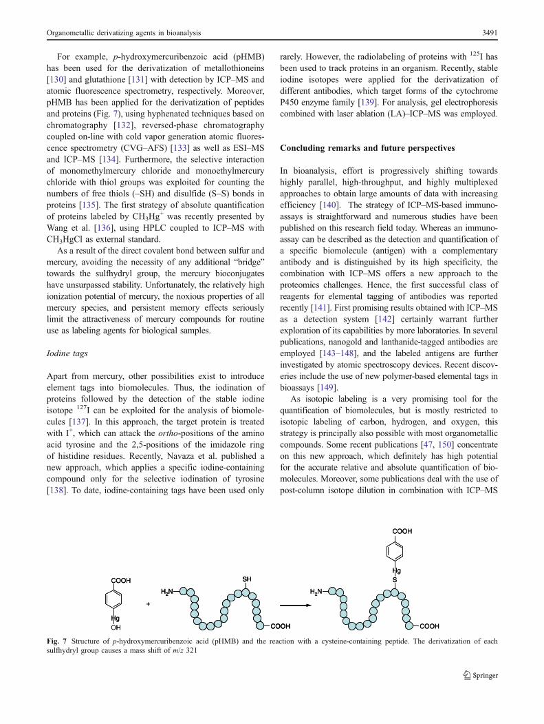
For example, p-hydroxymercuribenzoic acid (pHMB)has been used for the derivatization of metallothioneins[130] and glutathione [131] with detection by ICP–MS andatomic fluorescence spectrometry, respectively. Moreover,pHMB has been applied for the derivatization of peptidesand proteins (Fig. 7), using hyphenated techniques based onchromatography [132], reversed-phase chromatographycoupled on-line with cold vapor generation atomic fluores-cence spectrometry (CVG–AFS) [133] as well as ESI–MSand ICP–MS [134]. Furthermore, the selective interactionof monomethylmercury chloride and monoethylmercurychloride with thiol groups was exploited for counting thenumbers of free thiols (–SH) and disulfide (S–S) bonds inproteins [135]. The first strategy of absolute quantificationof proteins labeled by CH3Hg
+ was recently presented byWang et al. [136], using HPLC coupled to ICP–MS withCH3HgCl as external standard.
As a result of the direct covalent bond between sulfur andmercury, avoiding the necessity of any additional “bridge”towards the sulfhydryl group, the mercury bioconjugateshave unsurpassed stability. Unfortunately, the relatively highionization potential of mercury, the noxious properties of allmercury species, and persistent memory effects seriouslylimit the attractiveness of mercury compounds for routineuse as labeling agents for biological samples.
Iodine tags
Apart from mercury, other possibilities exist to introduceelement tags into biomolecules. Thus, the iodination ofproteins followed by the detection of the stable iodineisotope 127I can be exploited for the analysis of biomole-cules [137]. In this approach, the target protein is treatedwith I+, which can attack the ortho-positions of the aminoacid tyrosine and the 2,5-positions of the imidazole ringof histidine residues. Recently, Navaza et al. published anew approach, which applies a specific iodine-containingcompound only for the selective iodination of tyrosine[138]. To date, iodine-containing tags have been used only
rarely. However, the radiolabeling of proteins with 125I hasbeen used to track proteins in an organism. Recently, stableiodine isotopes were applied for the derivatization ofdifferent antibodies, which target forms of the cytochromeP450 enzyme family [139]. For analysis, gel electrophoresiscombined with laser ablation (LA)–ICP–MS was employed.
Concluding remarks and future perspectives
In bioanalysis, effort is progressively shifting towardshighly parallel, high-throughput, and highly multiplexedapproaches to obtain large amounts of data with increasingefficiency [140]. The strategy of ICP–MS-based immuno-assays is straightforward and numerous studies have beenpublished on this research field today. Whereas an immuno-assay can be described as the detection and quantification ofa specific biomolecule (antigen) with a complementaryantibody and is distinguished by its high specificity, thecombination with ICP–MS offers a new approach to theproteomics challenges. Hence, the first successful class ofreagents for elemental tagging of antibodies was reportedrecently [141]. First promising results obtained with ICP–MSas a detection system [142] certainly warrant furtherexploration of its capabilities by more laboratories. In severalpublications, nanogold and lanthanide-tagged antibodies areemployed [143–148], and the labeled antigens are furtherinvestigated by atomic spectroscopy devices. Recent discov-eries include the use of new polymer-based elemental tags inbioassays [149].
As isotopic labeling is a very promising tool for thequantification of biomolecules, but is mostly restricted toisotopic labeling of carbon, hydrogen, and oxygen, thisstrategy is principally also possible with most organometalliccompounds. Some recent publications [47, 150] concentrateon this new approach, which definitely has high potentialfor the accurate relative and absolute quantification of bio-molecules. Moreover, some publications deal with the use ofpost-column isotope dilution in combination with ICP–MS
SH
H2N
COOH
COOH
HgH
+
COOH
HgS
H2N
COOH
SH
H2N
COOH
SH
H2N
COOH
COOH
HgOH
+
COOH
HgS
H2N
COOH
Fig. 7 Structure of p-hydroxymercuribenzoic acid (pHMB) and the reaction with a cysteine-containing peptide. The derivatization of eachsulfhydryl group causes a mass shift of m/z 321
Organometallic derivatizing agents in bioanalysis 3491

as a detector for bioanalytical purposes and showed verypromising results recently [15, 151]. Especially in this field,progress can be expected in future work.
Furthermore, new approaches including the utilization ofnanoparticles as derivatization reagents for antibodies [152]and oligonucleotides [153] and their subsequent analysis byICP–MS have been presented. All of these approaches com-bine the use of immunoreactions coupled with ICP–MS-baseddetection. This general strategymay offer new possibilities forbiological assays and clinical diagnoses. Recently, a novelsandwich assay for human α-thrombin was reported whichtakes advantage of gold nanoparticles for signal amplificationin combination with ICP–MS detection [154].
New technological developments and their implementa-tion, for instance LA–ICP–MS, are promising tools in thefield of bioanalysis. The excellent separation of proteins bypolyacrylamide gel electrophoresis is well known, but theinvestigation of heteroatom-tagged proteins with LA–ICP–MS is a new, powerful tool for quantification. Monoclonal[139] and polyclonal [155] antibodies have been labeledwith metal-containing chelate complexes or even goldclusters [156]. After the immunoreaction of the labeledantibodies with the antigen, the element label was detectedby LA–ICP–MS.
Other groups separated the modified biomolecules bysodium dodecyl sulfate polyacrylamide gel electrophoresis(SDS–PAGE) followed by LA–ICP–MS detection. In thisarea, challenges have to be overcome in the future. Forexample, a drawback of the labeling procedure using chelate-based derivatizing agents is the change of electrophoreticmobility during gel electrophoresis due to the relatively highmolecular weight of the chelate added to the weight of thebiomolecule. However, the large variety of chemical andtechnological innovations clearly indicates the excellentfuture perspectives of organometallic derivatizing agents inbioanalysis.
References
1. Fenn JB, Mann M, Meng CK, Wong SF, Whitehouse CM (1989)Science 246:64–71
2. Finney LA, O’Halloran TV (2003) Science 300:931–9363. Karas M, Hillenkamp F (1988) Anal Chem 60:2299–23014. Houk RS, Fassel VA, Flesch GD, Svec HJ, Gray AL, Taylor CE
(1980) Anal Chem 52:2283–22895. Leitner A, Lindner W (2004) J Chromatogr B 813:1–266. Iliuk A, Galan J, Tao WA (2009) Anal Bioanal Chem 393:503–
5137. Wang M, Feng WY, Zhao YL, Chai ZF (2009) Mass Spectrom
Rev 29:326–3488. Ferrarello CN, Fernández de la Campa MR, Sanz-Medel A
(2002) Anal Bioanal Chem 373:412–4219. Bettmer J, Jakubowski N, Prange A (2006) Anal Bioanal Chem
386:7–1110. Ong SE, Mann M (2005) Nat Chem Biol 1:252–262
11. Sanz-Medel A (2008) Anal Bioanal Chem 391:885–89412. Prange A, Pröfrock D (2008) J Anal At Spectrom 23:432–45913. Svantesson E, Pettersson J, Markides KE (2002) J Anal At
Spectrom 17(5):491–49614. Wind M, Wegener A, Eisenmenger A, Kellner R, Lehmann WD
(2003) Angew Chem Int Ed 42:3425–342715. Rappel C, Schaumlöffel D (2008) Anal Bioanal Chem 390:605–
61516. Xu M, Yang L, Wang Q (2008) J Anal At Spectrom 23:1545–
154917. Jitaru P, Cozzi G, Gambaro P, Cescon P, Barbante C (2008) Anal
Bioanal Chem 391:661–66918. Wróbel K, Wróbel K, Caruso JA (2005) Anal Bioanal Chem
381:317–33119. Goenaga-Infante H, Hearn R, Catterick T (2005) Anal Bioanal
Chem 382:957–96720. Takatera K, Watanabe T (1993) Anal Chem 65:759–76221. Shah M, Wuilloud RG, Kannamkumarath SS, Caruso JA (2005)
J Anal At Spectrom 20:176–18222. Bandura DR, Baranov VI, Tanner SD (2002) Anal Chem
74:1497–150223. Wind M, Edler M, Jakubowski N, Linscheid M, Wesch H,
Lehmann WD (2001) Anal Chem 73:29–3524. Navaza AP, Encinar JR, Carrascal M, Abián J, Sanz-Medel A
(2008) Anal Chem 80:1777–178725. Bandura DR, Ornatsky O, Liao L (2004) J Anal At Spectrom
19:96–10026. Szpunar J (2005) Analyst 130:442–46527. Gygi SP, Rist B, Gerber SA, Turecek F, Gelb MH, Aebersold R
(1999) Nat Biotechnol 17:994–99928. Schmidt A, Kellermann J, Lottspeich F (2005) Proteomics 5:4–
1529. Wiese S, Reidegeld KA, Meyer HE, Warscheid B (2007)
Proteomics 7:340–35030. Yu Y, Cui J, Wang X, Liu Y, Yang P (2004) Proteomics 4:3112–
312031. Zhou H, Boyle R, Aebersold R (2004) Methods Mol Biol
261:511–51832. Zhou H, Ranish JA, Watts JD, Aebersold R (2002) Nat
Biotechnol 20:512–51533. Ross PL, Huang YN, Marchese JN, Williamson B, Parker K,
Hattan S, Khainovski N, Pillai S, Dey S, Daniels S, Purkayastha S,Juhasz P, Martin S, Bertlet-Jones M, He F, Jacobson A, Pappin DJ(2004) Mol Cell Proteomics 3:1154–1169
34. Wu WW, Wang G, Baek SJ, Shen RF (2006) J Proteome Res5:651–658
35. Wu CC, Maccoss MJ, Howell KE, Matthews KE, Yates JR(2004) Anal Chem 76:4951–4959
36. Mirgorodskaya OA, Kozmin YP, Titov MI, Korner R, Sonksen CP,Roepstorff P (2000) Rapid CommunMass Spectrom 14:1226–1232
37. Brown KJ, Fenselau CJ (2004) J Proteome Res 3:455–46238. Waanders LF, Hanke S, Mann M (2007) J Am Soc Mass
Spectrom 18:2058–206439. Jakubowski N, Waentig L, Hayen H, Venkatachalam V, von
Bohlen A, Roos PH, Manz A (2008) J Anal At Spectrom23:1497–1507
40. Dawson P, Cosgrove DO, Grainger RG (1999) Textbook ofcontrast media. ISIS Media, Oxford
41. Hempen C, Karst U (2006) Anal Bioanal Chem 384:572–58342. Ahrends R, Pieper S, Kühn A, Weisshoff H, Hamester M,
Lindemann T, Scheler C, Lehmann K, Taubner K, Linscheid MW(2007) Mol Cell Proteomics 6:1907–1916
43. Whetstone PA, Butlin NG, Corneillie TM, Meares CF (2004)Bioconjug Chem 15:3–6
44. Ahrends R, Pieper S, Neumann B, Scheler C, Linscheid MW(2009) Anal Chem 81:2176–2184
3492 S. Bomke et al.

45. Fincha J, Janecka A (2003) Bioconjug Chem 14:3–746. Kural C, Ge PH, Selvin PR (2003) Biophys J 84:288A47. Patel P, Jones P, Handy R, Harrington C, Marshall P, Evans EH
(2008) Anal Bioanal Chem 390:61–6548. Liu H, Zhang Y, Wang J, Wang D, Zhou C, Cai Y, Qian X
(2006) Anal Chem 78:6614–662149. Koellensperger G, Groeger M, Zinkl D, Petzelbauer P, Hann S
(2009) J Anal At Spectrom 24:97–10250. Baranov VI, Quinn Z, Bandura DR, Tanner SD (2002) J Anal At
Spectrom 17:1148–115251. Quinn ZA, Baranov VI, Bandura DR, Tanner SD, Wrana JL
(2002) J Anal At Spectrom 17:892–89652. Leipold MD, Herrera I, Ornatsky O, Baranov V, Nitz M (2009)
J Proteome Res 8:443–44953. van Staveren DR, Metzler-Nolte N (2004) Chem Rev 104:5931–
598554. Salmain M, Jaouen G (2003) C R Chimie 6:249–25855. Lo KK, Hui WK, Ng DC, Cheung KK (2002) Inorg Chem
41:40–4656. Dattelbaum JD, Abugo OO, Lakowicz JR (2000) Bioconjug
Chem 11:533–53657. Jaouen G, Vessiéres A, Butler IS (1993) Acc Chem Res 26:361–
36958. Beck W, Krämer R (1991) Angew Chem Int Ed Engl 30:1467–
146859. Rudolf B, Palusiak M, Zakrzewski J, Salmain M, Jaouen G
(2005) Bioconjug Chem 16:1218–122460. Gorfti A, Salmain M, Jaouen G, Mc Glichey MJ, Bennouna A,
Mousser A (1996) Organometallics 15:142–15161. Salmain M, Gorfti A, Jaouen G (1998) Eur J Biochem 258:192–
19962. Salmain M, Gunn M, Gorfti A, Top S, Jaouen G (1993)
Bioconjug Chem 4:425–43363. El Amouri H, Besace Y, Vaissermann J, Jaouen G (1996)
J Organomet Chem 515:103–10764. Mouatassim EB, Elamouri H, Vaissermann J, Jaouen A (1995)
Organometallics 14:3296–330265. Rudolf B, Zakrzewski J (1994) Tetrahedron Lett 35:9611–961266. Rudolf B, Zakrzewski J, Salmain M, Jaouen G (1998) New
J Chem 22:813–81867. Rudolf B, Zakrzewski J, Salmain M, Jaouen G (1998)
Tetrahedron Lett 39:4281–428268. van Staveren DR, Weyhermüller T, Metzler-Nolte N (2000)
Organometallics 19:3730–373569. Varenne A, Salmain M, Brisson C, Jaouen G (1992) Bioconjug
Chem 3:471–47670. Egan D, Salmain M, Mc Ardle P, Jaouen G, Caro B (2002)
Spectrochim Acta A 58:941–95171. Osella D, Pollone P, Ravera M, Salmain M, Jaouen G (1999)
Bioconjug Chem 10:607–61272. Jaouen G, Vessiéres A, Butler IS (1993) Acc ChemRes 26:361–36973. Salmain M, Vessiéres A, Brossier P, Butler IS, Jaouen G (1992)
J Immunol Methods 148:65–7574. Vessiéres A, Salmain M, Brossier O, Jaouen G (1999) J Pharm
Biomed Anal 21:625–63375. Metzler-Nolte N (2001) Angew Chem 113:1072–107676. Salmain M, Licandro E, Baldoli E, Maiorana C, Tran-Huy H,
Jaouen G (2001) J Organomet Chem 617–618:376–38277. Salmain M, Blais JC, Tran-Huy H, Compain C, Jaouen G (2001)
Eur J Biochem 269:5479–548778. Enders D, Niemeier O, Henseler A (2007) Chem Rev 107:5606–
565579. Lemke J, Metzler-Nolte N (2008) Eur J Inorg Chem 3359–336680. Nishioka T, Shibata T, Kinoshita I (2007) Organometallics
26:1126–112881. Xu G, Gilbertson SR (2005) Org Lett 7:4605–4608
82. Lavastre I, Bescancon J, Brossiert P, Moise C (1991) ApplOrganomet Chem 5:143–149
83. Lavastre I, Bescancon J, Brossiert P, Moise C (1990) ApplOrganomet Chem 4:9–17
84. Beilstein AE, Grinstaff MW (2001) J Organomet Chem637:398–406
85. Long YT, Li TC, Sutherland TC, Chahama M, Lee JS, Kraatz HB(2003) J Am Chem Soc 125:8724–8725
86. Chantson JT, Falzacappa MVV, Crovella S, Metzler-Nolte N(2006) ChemMedChem 1:1268–1274
87. Biot C, Taramelli D, Forfar-Bares I, Maciejewski LA, Boyce M,Nowogrocki G, Brocard JS, Basilico N, Olliaro O, Egan TJ(2005) Mol Pharm 2:185–193
88. Bowman DC (2006) Chem Commun 83:735–74089. Köpf-Maier P, Köpf H (1986) Drugs Future 11:297–32090. Harding MM, Mokdsi G (2000) Curr Med Chem 7:1289–130391. Köpf-Maier P, Klapötke T (1992) Cancer Chemother Pharmacol
29:361–36692. Seiwert B, Karst U (2008) Anal Bioanal Chem 390:181–20093. Kraatz HB (2005) J Inorg Organomet Polymer Mater 15:83–10694. Cotton FA, Wilkinson G, Gaus PL (1995) Basic inorganic
chemistry. Wiley, New York, p 68995. Brinkley M (1992) Bioconjug Chem 3:2–1396. Van Berkel GJ, Quirke JME, Tigani RA, Dilley AS, Covey TR
(1998) Anal Chem 70:1544–155497. Kubab N, Farinotti R, Montes C, Chalom J, Mahuzier G (1998)
Analysis 17:559–56498. Higashi T, Shimada K (2004) Anal Bioanal Chem 378:875–88299. Shimada K, Sakayori C, Nambara T (1987) J Liq Chromatogr
10:2177–2187100. Shimada K, Nagashima E, Orii S, Nambara T (1987) J Pharm
Biomed Anal 5:361–368101. Murao N, Ishigai M, Sekiguchi N, Takahashi T, Aso Y (2005)
Anal Biochem 346:158–166102. Ishigai M, Murao N, Sekiguchi N, Takahashi T (2004) Patent
No. WO2004002996103. Mukumoto K, Nojima T, Sato S, Waki M, Takenaka S (2007)
Anal Sci 23:115–119104. Mukumoto K, Nojima T, Tekenaka S (2005) Tetrahedron
61:11705–1105. Seiwert B, Henneken H, Karst U (2004) J Am Soc Mass
Spectrom 15:1727–1736106. Lo KKW, Lau JSY, Ng DCM, Zhu N (2002) J Chem Soc Dalton
Trans 1753–1756107. Seiwert B, Karst U (2007) Anal Chem 79:7131–7138108. Heller A (1990) Acc Chem Res 23:128–134109. Bomke S, Pfeifer T, Meermann B, Buscher W, Karst U (2009)
Anal Bioanal Chem. doi:10.1007/s00216-009-3123-z110. Deng AP, Liu HAT, Jiang SJ, Huang HJ, Ong CW (2002) Anal
Chim Acta 472:55–61111. Seiwert B, Karst U (2007) Anal Bioanal Chem 388:1633–1643112. Bomke S, Dudek L, Seiwert B, Karst U (2009) Anal Bioanal
Chem 393:247–256113. Kunugi S, Murakami Y, Ikeda K, Itoh N (1992) Int J Biol
Macromol 14:210–214114. Cheret P, Brossier P (1986) Res Commun Chem Pathol
Pharmacol 54:237–253115. Kolbe N, Andersson JT (2006) J Agric Food Chem 54:5736–
5741116. Rolfes J, Andersson JT (2001) Anal Chem 73:3073–3082117. Cao W, Ferrance JP, Demas J, Landers JP (2006) J Am Chem
Soc 128:7572–7578118. Sheats JE, Rausch M (1969) J Org Chem 35:3245–3249119. Fischer EO, Herberich GE (1961) Z Naturforsch 1517–1523.120. Bordes AL, Schöllhorn B, Limoges B, Degrand C (1998) Appl
Organom Chem 12:59–65
Organometallic derivatizing agents in bioanalysis 3493
121. Gill TJ, Mann LT (1966) J Immun 5:906–912122. Maurer A, Kraatz H-B, Metzler-Nolte N (2005) Eur J Inorg
Chem 16:3207–3210123. Noor F, Wüstholz A, Kinscherf R, Metzler-Nolte N (2005)
Angew Chem Int Ed 44:2429–2432124. Chantson JT, Falzacappa MVV, Crovella S, Metzler-Nolte N
(2006) ChemMedChem 1:1268–1274125. Lavastre I, Bescancon J, Brossiert P, Moise C (1991) Appl
Organomet Chem 5:143–149126. Bordes AL, Schöllhorn B, Limoges B, Degrand C (1998) Appl
Organomet Chem 12:59–65127. Simpson RB (1961) J Am Chem Soc 83:4711–4717128. Jovin TM, England PT, Komberg A (1969) J Biol Chem
244:3009–3018129. Zaluzec EJ, Gage DA, Watson JT (1994) J Am Soc Mass
Spectrom 5:359–366130. Takatera K, Watanabe T (1992) Anal Sci 8:469–474131. Bramanti E, Vecoli C, Neglia D, Pellegrini MP, Raspi G,
Barsacchi R (2005) Clin Chem 51:1007–1013132. Bramanti E, Lucchesini S, D’Ulivo A, Lampugnani L, Zamboni R,
Spinetti MC, Raspi G (2001) J Anal At Spectrom 16:166–171133. Bramanti E, Lomonte C, Galli A, Onor M, Zamboni R, Raspi G,
D’Ulivo A (2004) J Chromatogr A 1054:285–291134. Kutscher DJ, del Castillo Busto ME, Zinn N, Sanz-Medel A,
Bettmer J (2008) J Anal At Spectrom 23:1359–1364135. Guo Y, Chen L, Yang L, Wang Q (2008) J Am Mass Spectrom
19:1108–1113136. Guo Y, Xu M, Yang L, Wang Q (2009) J Anal At Spectrom
24:1184–1187137. Jakubowski N,Messerschmidt J, AňorbeMG,Waentig L, Hayen H,
Roos PH (2008) J Anal At Spectrom 23:1487–1496138. Navaza AP, Encinar JR, Ballesteros A, Gonz Iez JM, Sanz-
Medel A (2009) Anal Chem 81:5390–5399139. Roos PH, Venkatachalam A, Manz A, Waentig L, Koehler CU,
Jakubowski N (2008) Anal Bioanal Chem 392:1135–1147
140. Ornatsky OI, Kinach R, Bandura DR, Lou X, Tanner SD,Baranov VI, Nitz M, Winnik MA (2008) J Anal At Spectrom23:463–469
141. Lou X, Zhang GM, Herrera I, Kinach R, Ornatsky O, Baranov VI,Nitz M, Winnik MA (2007) Angew Chem Int Ed 46:6111–6114
142. Tanner SD, Ornatsky O, Bandura DR, Baranov VI (2007)Spectrochim Acta B 62:188–195
143. Zhang S, Zhang C, Xing Z, Zhang X (2004) Clin Chem 7:1214–1221
144. Zhang C, Wu F, Zhang Y, Wang X, Zhang X (2001) J Anal AtSpectrom 16:1393–1396
145. Baranov VI, Quinn Z, Bandura DR, Tanner SD (2002) AnalChem 74:1629–1636
146. Careri M, Elviri L, Mangia A, Mucchino C (2007) Anal BioanalChem 387:1851–1854
147. Razumienko E, Ornatsky O, Kinach R, Milyavsky M, Lechman E,Baranov V, Winnik MA, Tanner SD (2008) J Immunol Methods336:56–63
148. Lathia US, Ornatsky O, Baranov V, Nitz M (2010) AnalBiochem 398:93–98
149. Lou X, Zhang G, Herrera I, Kinach R, Ornatsky O, Baranov V,Nitz M, Winnik MA (2007) Angew Chem Int Ed 46:6111–6114
150. Kutscher DJ, Bettmer J (2009) Anal Chem 81:9172–9177151. Yan X, Xu M, Yang L, Wang Q (2010) Anal Chem 82:1261–
1269152. Zhang C, Zhang Z, Yu B, Shi J, Zhang X (2002) Anal Chem
74:96–99153. Kerr SL, Sharp B (2007) Chem Commun 43:4537–4539154. Zhao Q, Lu X, Yuan CH, Li XF, Le XC (2009) Anal Chem
81:7484–7489155. Waentig L, Roos HR, Jakubowski N (2009) J Anal At Spectrom
24:924–933156. Müller SD, Diaz-Bone RA, Felix J, Goedecke W (2005) J Anal
At Spectrom 20:907–911
3494 S. Bomke et al.