Optimizing kernel size in generalized auto-calibrating partially parallel acquisition in parallel...
6
Optimizing kernel size in generalized auto-calibrating partially parallel acquisition in parallel magnetic resonance imaging Haitham M. Ahmed 1 , Refaat E. Gabr 1, 2 , Abou-Bakr M. Youssef 1 , Yasser M. Kadah 1 Departnment of Biomedical Engineering, Cairo University, Giza, Egypt 2 Division of MR Research, Department of Radiology, Johns Hopkins University, MD, USA ABSTRACT Parallel magnetic resonance imaging achieves reduction in scan time by collecting a partial set of signals using an array of receiving coils each with a local sensitivity pattern. An image is then reconstructed from the partial dataset using the additional information of coil sensitivity. GRAPPA (generalized auto calibrating partially parallel acquisitions) is one of the most successful reconstruction techniques in which the missing k-space lines are interpolated from the acquired data in the whole coil array using a convolution kernel estimated from a fully sampled data patch in the center of k-space. The interpolation kernel is usually small but fixed in size for all coils. Here, we show that a variable kernel with a size dependent on the coil sensitivity can lead to better image quality. The kernel size is estimated from the ratio of the coil sensitivities obtained from a reference scan or from the same dataset. Conventional GRAPPA kernel estimation and image reconstruction is modified to employ the variable-size kernel for improved reconstruction. The new technique shows improved image quality compared to GRAPPA. Keywords: magnetic resonance imaging, parallel imaging, GRAPPA, kernel size 1. INTRODUCTION Parallel magnetic resonance imaging (MRI) is a family of acquisition and reconstruction techniques that increase image acquisition speed by taking advantage of the localized sensitivity of multiple surface radio-frequency (RF) coils [1-8]. In MRI, the image is reconstructed from a set of samples collected in the spatial frequency domain, also known as k-space. In conventional (un-accelerated) MRI, the full data in k-space are collected corresponding to a certain field of view (FOV) and resolution in the image, whereas in parallel imaging the k-space is subsampled by a factor R. Consequently, individual aliased images are obtained for every coil. These images are either unfolded in the image domain, or the missed k-space lines are estimated from the acquired data and prior information about the coil sensitivities. Many parallel imaging reconstruction techniques have been proposed like sensitivity encoding (SENSE), simultaneous acquisition of spatial harmonics (SMASH), generalized auto-calibrating partially parallel acquisition (GRAPPA) and their derivatives [1-8]. These methods can be generally divided into k-space methods and image domain methods. The k- space methods when used with the additional acquired auto-calibration data are very powerful in cases where determination of the coil sensitivity is difficult or is time varying. In GRAPPA, reconstruction of data in coil j at a line (ky-mΔky) offset from the acquired data is given by: ∑∑ = − = Δ − = Δ − N l N b y y l y y j b k bR k S m l b j n k m k S 1 1 0 ) ( ) , , , ( ) ( (1) where S j (k y ) is the signal in coil j at line k y . In this case, N b lines which are separated by RΔk y are combined using the weights n(j,b,l,m) to form each line, corresponding to an under-sampling (or reduction) factor R. The coefficients n(j,b,l,m) represent the weights used in this linear combination. The index l counts through the individual coils, while the index b counts through the individual reconstruction blocks. This process (shown in Fig. 1) is repeated for each coil in the array, resulting in L uncombined coil images which can then be combined using a conventional sum of squares reconstruction or any other optimum array combination [9]. The interpolation kernel is usually small but fixed in size for all coils. In this work, a general variable-size kernel approach is introduced that derives from the theory introduced by Bao and Maudsley [10]. The variable kernel used in 1 Medical Imaging 2010: Physics of Medical Imaging, edited by Ehsan Samei, Norbert J. Pelc, Proc. of SPIE Vol. 7622, 762258 · © 2010 SPIE · CCC code: 1605-7422/10/$18 · doi: 10.1117/12.844597 Proc. of SPIE Vol. 7622 762258-1
-
Upload
independent -
Category
Documents
-
view
0 -
download
0
Transcript of Optimizing kernel size in generalized auto-calibrating partially parallel acquisition in parallel...
Optimizing kernel size in generalized auto-calibrating partially parallel acquisition in parallel magnetic resonance imaging
Haitham M. Ahmed1, Refaat E. Gabr1, 2, Abou-Bakr M. Youssef1 , Yasser M. Kadah
1Departnment of Biomedical Engineering, Cairo University, Giza, Egypt 2Division of MR Research, Department of Radiology, Johns Hopkins University, MD, USA
ABSTRACT Parallel magnetic resonance imaging achieves reduction in scan time by collecting a partial set of signals using an array of receiving coils each with a local sensitivity pattern. An image is then reconstructed from the partial dataset using the additional information of coil sensitivity. GRAPPA (generalized auto calibrating partially parallel acquisitions) is one of the most successful reconstruction techniques in which the missing k-space lines are interpolated from the acquired data in the whole coil array using a convolution kernel estimated from a fully sampled data patch in the center of k-space. The interpolation kernel is usually small but fixed in size for all coils. Here, we show that a variable kernel with a size dependent on the coil sensitivity can lead to better image quality. The kernel size is estimated from the ratio of the coil sensitivities obtained from a reference scan or from the same dataset. Conventional GRAPPA kernel estimation and image reconstruction is modified to employ the variable-size kernel for improved reconstruction. The new technique shows improved image quality compared to GRAPPA. Keywords: magnetic resonance imaging, parallel imaging, GRAPPA, kernel size
1. INTRODUCTION Parallel magnetic resonance imaging (MRI) is a family of acquisition and reconstruction techniques that increase image acquisition speed by taking advantage of the localized sensitivity of multiple surface radio-frequency (RF) coils [1-8]. In MRI, the image is reconstructed from a set of samples collected in the spatial frequency domain, also known as k-space. In conventional (un-accelerated) MRI, the full data in k-space are collected corresponding to a certain field of view (FOV) and resolution in the image, whereas in parallel imaging the k-space is subsampled by a factor R. Consequently, individual aliased images are obtained for every coil. These images are either unfolded in the image domain, or the missed k-space lines are estimated from the acquired data and prior information about the coil sensitivities. Many parallel imaging reconstruction techniques have been proposed like sensitivity encoding (SENSE), simultaneous acquisition of spatial harmonics (SMASH), generalized auto-calibrating partially parallel acquisition (GRAPPA) and their derivatives [1-8]. These methods can be generally divided into k-space methods and image domain methods. The k-space methods when used with the additional acquired auto-calibration data are very powerful in cases where determination of the coil sensitivity is difficult or is time varying. In GRAPPA, reconstruction of data in coil j at a line (ky-mΔky) offset from the acquired data is given by:
∑∑=
−
=
Δ−=Δ−N
l
N
byylyyj
b
kbRkSmlbjnkmkS1
1
0)(),,,()( (1)
where Sj(ky) is the signal in coil j at line ky. In this case, Nb lines which are separated by RΔky are combined using the weights n(j,b,l,m) to form each line, corresponding to an under-sampling (or reduction) factor R. The coefficients n(j,b,l,m) represent the weights used in this linear combination. The index l counts through the individual coils, while the index b counts through the individual reconstruction blocks. This process (shown in Fig. 1) is repeated for each coil in the array, resulting in L uncombined coil images which can then be combined using a conventional sum of squares reconstruction or any other optimum array combination [9]. The interpolation kernel is usually small but fixed in size for all coils. In this work, a general variable-size kernel approach is introduced that derives from the theory introduced by Bao and Maudsley [10]. The variable kernel used in
1
Medical Imaging 2010: Physics of Medical Imaging, edited by Ehsan Samei, Norbert J. Pelc,Proc. of SPIE Vol. 7622, 762258 · © 2010 SPIE · CCC code: 1605-7422/10/$18 · doi: 10.1117/12.844597
Proc. of SPIE Vol. 7622 762258-1

this work is two-dimensional (2D) with coil-dependent size. The kernel size is estimated from the coil sensitivities obtained from a reference scan or from the same dataset. Conventional GRAPPA kernel estimation and image reconstruction is modified to employ the variable-size kernel for improving the reconstruction.
2. METHODS The MR signal generated in the lth coil is given by
∑−
=∑−
=+−=
1
0
1
0))(2exp(),(),(),(
Nx
x
Ny
yyykxxkjyxlCyxrykxkld π (2)
where r is the weighted spin density of the imaged object and lC is the coil sensitivity of coil l. Following the work in [10], Eq. (2) can be re-written as
∑−
=∑−
== +−
1
0
1
0)exp(
),('
),(),(),( )(2)),('
Nx
x
Ny
y yxlC
yxlCyxrykxkld yxl ykxkjyxC π (3)
where ),(' yxCl is the sensitivity map of coil 'l . Following the convolution theorem
∑ ∑ −−′=
x yykyxxkllCyxldykxkld
ω ωωωωω ),(',
~),(),( (4)
where
⎪⎭
⎪⎬⎫
⎪⎩
⎪⎨⎧
=),('
),(),(',
~yxlC
yxlCFTykxkllC (5)
and FT{.} denotes the Fourier transformation. Eq. (5) shows that the relation between the signals in any two coils is a convolution operation wherein the convolution kernel is the Fourier transform of the ratio between the sensitivity maps of these two coils. The optimal convolution kernel is thus specific to each coil pair and will be different in size and shape depending on the relative coil sensitivities and the geometry of the coil array. The estimation of the missing lines of each of the coils in conventional GRAPPA is given by,
∑=
∑−
=Δ−′≈Δ−′ ⎥
⎦
⎤⎢⎣
⎡L
l
bN
bxykbRykldmlblnxykmykld
1
1
0),(),,,(),(
(6)
Although the theoretical kernel n is of infinite extent, its energy is concentrated at the origin. This is because the individual coil sensitivity profiles are inherently smooth and the Fourier transform of this smooth sensitivity ratio is concentrated in the center of k-space as shown in Fig. 3. This provides a justification for the small size of the convolution kernel used in GRAPPA reconstruction. Here, we extend the GRAPPA method to include the concept of coil-specific convolution kernel by allowing the size of the kernel n to vary as a function of the two coils l and l’. Figure 2 shows the size of the function ),(~
', yxll kkC when truncated at 90% of its total energy (white rectangles). The conventional GRAPPA method is modified to use this truncation window size as the kernel size for each pair of coils. The proposed technique is validated using simulated data of the Shepp-Logan (SL) phantom [11] with a matrix size 128x128. The phase encoding direction is left-right. Eight loop coils are used in the simulation with coil sensitivity derived from the Biot-Savart law for circular loop coils. Complex Gaussian noise is added to the simulated data of all coils with zero mean and standard deviation that is 0.001 times the root-mean square value of all signals in the eight coils. Different reduction factor (R=2,3,4) were tested as well as different number of ACS lines (8,12,16 and 20 lines).
Proc. of SPIE Vol. 7622 762258-2
To determine the kernel size for each coil pair ),(~', yxll kkC was truncated at 60% of its total energy. The size of the
truncation window in the direction of phase encoding is divided by the reduction factor R to get the number of blocks or the kernel size to be used in GRAPPA reconstruction. The kernel size in the frequency encoding direction is the same as the size of the truncation window in that direction. The kernel coefficient estimation is performed by stacking the ACS data from all coils and then using regularized inversion of the resulting linear system to get the different kernels. These kernels are then used in the reconstruction as in GRAPPA except that each kernel corresponding to a coil-pair has its own size. The reconstruction quality is quantified by the SNR measured in the large grey ellipse to assess the noise amplification. The SNR also serves as a measure of the intensity of reconstruction artifacts.
3. RESULTS AND DISCUSSION The images reconstructed using the proposed variable-size kernel GRAPPA are shown in Fig. 4, along with the conventional GRAPPA reconstruction using a kernel size of four. Less artifacts and better SNR is noticed when the variable-size kernel is used than with the conventional fixed-size kernel when the number of ACS lines is sufficiently high. The better performance of the proposed method is evident for different acceleration factors and for ACS lines greater than eight. The performance with the variable-size kernel approach is less successful than GRAPPA when the number of ACS line is small. This is due to the ill-conditioning of the system of equations used to estimate the kernel when the number of ACS lines is relatively small. However, for moderate or large number of ACS lines the performance of variable-size kernel is remarkable in suppressing noise that is otherwise amplified with the fixed-size kernel. The proposed variable-size convolution kernel is a rationale extension of the fixed-size kernel initially employed in GRAPPA. The variable-size kernel follows directly from the mutual coil sensitivity of each coil pair. The kernel is defined though the ratio of the sensitivities of each pair of coils which depends on the inherent sensitivity of each coil and the geometry of the coil array. The kernel is a 2D function and is concentrated in a direction that depends on the location of the two coils under consideration relative to each other. In this work the kernel size was directly computed from a full-resolution coil sensitivity map, but it can be directly estimated from the auto-calibrating signal acquired in the same dataset. This approach is preferred to acquiring a full scan reference acquisition since it can avoid registration problems when patient motion occurs. Estimation of a low-resolution coil sensitivity ratio can then obtained in the same manner as in auto-calibrating SENSE methods. The proposed approach of using variable-size kernel can improve our understanding of how GRAPPA works and can help in optimizing the reconstruction as well as the selection of various scan parameters like the number and locations of the ACS lines.
4. CONCLUSIONS
GRAPPA reconstruction with the proposed variable-size kernel provides better image quality with a reduction in the power of artifacts and enhanced SNR. The variable-size reconstruction does not require the acquisition of additional data and can be implemented with little modification to the existing GRAPPA technique.
ACKNOWLEDGEMENTS
We thank Dr Jim Li from Texas A&M University for providing the MATLAB open source code for GRAPPA reconstruction.
Proc. of SPIE Vol. 7622 762258-3
REFERENCES [1] K. P. Pruessmann, M.Weiger, M. B. Scheidegger, and P. Boesiger, "SENSE: sensitivity encoding for fast MRI,"
Magn Reson Med, vol. 42, pp. 952-62,1999. [2] K. Heberlein and X. Hu, "kSENSE: k-Space Sensitivity Encording Reconstruction," presented at Proc. 9th Ann.
Meeting Intl. Soc.Mag. Reson.Med., 2001. [3] D. K. Sodickson and W. J. Manning, "Simultaneous acquisition of spatial harmonics (SMASH): fast imaging with
radiofrequency coil arrays," Magn Reson Med, vol.38, pp. 591-603, 1997. [4] M. Blaimer, F. Breuer, M. Mueller, R. M. Heidemann, M. A. Griswold, and P. M. Jakob, "SMASH, SENSE, PILS,
GRAPPA: how to choose the optimal method," Top Magn Reson Imaging, vol. 15, pp. 223-36, 2004. [5] P. M. Jakob, M. A. Griswold, R. R. Edelman, and D. K.Sodickson, "AUTO-SMASH: a self-calibrating technique
for SMASH imaging. SiMultaneous Acquisition of Spatial Harmonics," Magma, vol. 7, pp.42-54, 1998. [6] R. M. Heidemann, M. A. Griswold, A. Haase, and P. M. Jakob, "VD-AUTO-SMASH imaging," Magn Reson Med,
vol. 45, pp. 1066-74, 2001. [7] M. Bydder, D. J. Larkman, and J. V. Hajnal, "Generalized SMASH imaging," Magn Reson Med, vol.47, pp. 160-
70,2002. [8] M. A. Griswold, P. M. Jakob, R. M. Heidemann, M. Nittka, V. Jellus, J. Wang, B. Kiefer, and A. Haase,
"Generalized autocalibrating partially parallel acquisitions (GRAPPA)," Magn Reson Med, vol. 47, pp. 1202-10,2002.
[9] P. B. Roemer, W. A. Edelstein, C. E. Hayes, S. P. Souza, and O. M. Mueller, "The NMR phased array," Magn. Reson. Med., vol. 16, pp. 192-225,1990.
[10] Yufang Bao; Maudsley, A.A. “Image reconstruction in the GRAPPA algorithm formalism”. 4th IEEE International Symposium on Biomedical Imaging,Vol , 12-15 pp.113 – 116,2007.
[11] L.A. Shepp and B.F. Logan, “Reconstructing interior head tissue from X-ray transmission,” IEEE Trans. Nucl. Sci. NS-21, pp. 228-236, 1974.
Proc. of SPIE Vol. 7622 762258-4

Figure 2. The magnitudes of the Fourier transform of the ratio between the sensitivity of coils pairs in a four-
coil array using the Shepp-Logan phantom. Brightness is in logarithmic scale for better visualization. The rectangular boxes overlaid correspond to truncation windows with 90% of the total energy.
Figure 1. Schematic description of GRAPPA with an acceleration factor R = 2 and a single auto-calibrating line.
Proc. of SPIE Vol. 7622 762258-5
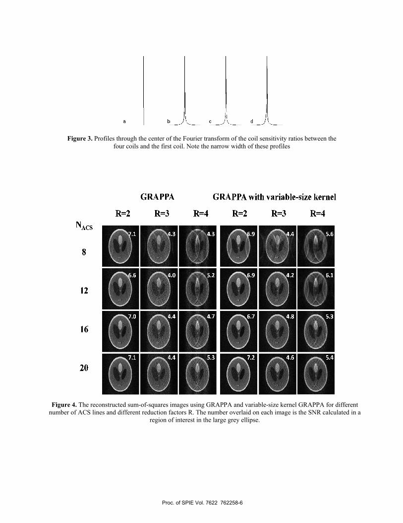
Figure 4. The reconstructed sum-of-squares images using GRAPPA and variable-size kernel GRAPPA for different
number of ACS lines and different reduction factors R. The number overlaid on each image is the SNR calculated in a region of interest in the large grey ellipse.
Figure 3. Profiles through the center of the Fourier transform of the coil sensitivity ratios between the
four coils and the first coil. Note the narrow width of these profiles
Proc. of SPIE Vol. 7622 762258-6