Optimized conditions for primary culture of pituitary cells from the Atlantic cod (Gadus morhua)....
11
(This is a sample cover image for this issue. The actual cover is not yet available at this time.) This article appeared in a journal published by Elsevier. The attached copy is furnished to the author for internal non-commercial research and education use, including for instruction at the authors institution and sharing with colleagues. Other uses, including reproduction and distribution, or selling or licensing copies, or posting to personal, institutional or third party websites are prohibited. In most cases authors are permitted to post their version of the article (e.g. in Word or Tex form) to their personal website or institutional repository. Authors requiring further information regarding Elsevier’s archiving and manuscript policies are encouraged to visit: http://www.elsevier.com/copyright
-
Upload
independent -
Category
Documents
-
view
0 -
download
0
Transcript of Optimized conditions for primary culture of pituitary cells from the Atlantic cod (Gadus morhua)....

(This is a sample cover image for this issue. The actual cover is not yet available at this time.)
This article appeared in a journal published by Elsevier. The attachedcopy is furnished to the author for internal non-commercial researchand education use, including for instruction at the authors institution
and sharing with colleagues.
Other uses, including reproduction and distribution, or selling orlicensing copies, or posting to personal, institutional or third party
websites are prohibited.
In most cases authors are permitted to post their version of thearticle (e.g. in Word or Tex form) to their personal website orinstitutional repository. Authors requiring further information
regarding Elsevier’s archiving and manuscript policies areencouraged to visit:
http://www.elsevier.com/copyright
Author's personal copy
Optimized conditions for primary culture of pituitary cells from the Atlanticcod (Gadus morhua). The importance of osmolality, pCO2, and pH
Kjetil Hodne a, Kristine von Krogh a, Finn-Arne Weltzien a,b, Olav Sand b, Trude M. Haug b,⇑a Norwegian School of Veterinary Science, Department of Basic Sciences and Aquatic Medicine, P.O. Box 8146 Dep, N-0033 Oslo, Norwayb University of Oslo, Department of Molecular Biosciences, P.O. Box 1041 Blindern, N-0316 Oslo, Norway
a r t i c l e i n f o
Article history:Received 19 March 2012Revised 1 June 2012Accepted 3 June 2012Available online 15 June 2012
Keywords:TeleostCulture protocolElectrophysiologyMembrane potentialqPCRViability
a b s t r a c t
Protocols for primary cultures of teleost cells are commonly only moderately adjusted from similar pro-tocols for mammalian cells, the main adjustment often being of temperature. Because aquatic habitatsare in general colder than mammalian body temperatures and teleosts have gills in direct contact withwater, pH and buffer capacity of blood and extracellular fluid are different in fish and mammals. Plasmaosmolality is generally higher in marine teleosts than in mammals. Using Atlantic cod (Gadus morhua) asa model, we have optimized these physiological parameters to maintain primary pituitary cells in culturefor an extended period without loosing key properties. L-15 medium with adjusted osmolality, adaptedto low pCO2 (3.8 mm Hg) and temperature (12 �C), and with pH 7.85, maintained the cells in a physiolog-ically sounder state than traditional culture medium, significantly improving cell viability compared tothe initial protocol. In the optimized culture medium, resting membrane potential and response to releas-ing hormone were stable for at least two weeks, and the proportion of cells firing action potentials duringspawning season was about seven times higher than in the original culture medium. The cells were mod-erately more viable when the modified medium was supplemented with newborn calf serum or artificialserum substitute. Compared to serum-free L-15 medium, expression of key genes (lhb, fshb, and gnrhr2a)was better maintained in medium containing SSR, whereas NCS tended to decrease the expression level.Although serum-free medium is adequate for many applications, serum supplement may be preferablefor experiments dependent on membrane integrity.
� 2012 Elsevier Inc. All rights reserved.
1. Introduction
In many in vitro studies of animal cells in primary culture, it istacitly assumed that cultured, viable cells have properties similarto those of intact cells in vivo. However, important functional prop-erties of the cells may be lost during dispersion of the cells andtheir subsequent maintenance in culture, although the cells maystay viable for an extended period. For example, the contractilityof dissociated smooth muscle cells [7] and cardiomyocytes[23,43] kept under standard culture conditions may decay withina couple of days, which may be only a fraction of the time the cellsstay ‘‘healthy’’ according to common viability tests. However, mod-erate adjustments of standard culture procedures and media areoften sufficient to greatly improve the culture conditions for a spe-cific cell type. Accordingly, a vast literature describes customizedculture protocols for a large number of mammalian cell types(for reviews and protocols, see [2,15]). In comparison, specialized
methods for culturing cells from exothermic vertebrates, i.e., fish,amphibians and reptiles, are rather few.
When we recently commenced an investigation of the func-tional properties of pituitary cells from the teleost Atlantic cod(Gadus morhua), we realized that the most commonly used proce-dures for establishing primary cultures of teleost cells were moder-ately adjusted protocols for mammalian cells. The main differencebetween most culture procedures for teleosts and mammals is thetemperature, whereas important parameters like osmolality, pCO2,pH, and serum supplement are usually similar. Still, scientistsemploying such methods for studying teleost cells in primary cul-ture seem to achieve reasonable results. Therefore, in our initialwork on cod pituitary cells [16], we used a slightly adjusted versionof an established protocol for dissociation and incubation of pitui-tary cells from the European eel (Anguilla anguilla) [24]. The mainmodification of the culture procedure used for Atlantic cod, whichis usually adapted to an ambient temperature below about 12 �C,was a reduction in temperature both during dissociation and incu-bation of the cells.
However, using these primary cultures in electrophysiologicalexperiments, we realized that the culture conditions were subopti-mal, and did thus start systematic trials in an attempt to optimize
0016-6480/$ - see front matter � 2012 Elsevier Inc. All rights reserved.http://dx.doi.org/10.1016/j.ygcen.2012.06.005
⇑ Corresponding author. Fax: +47 22857655.E-mail addresses: [email protected] (K. Hodne), [email protected]
(K. von Krogh), [email protected] (F.-A. Weltzien), [email protected](O. Sand), [email protected] (T.M. Haug).
General and Comparative Endocrinology 178 (2012) 206–215
Contents lists available at SciVerse ScienceDirect
General and Comparative Endocrinology
journal homepage: www.elsevier .com/locate /ygcen
Author's personal copy
the different elements of the culture procedure. We aimed atdesigning a primary cell culture protocol that fulfilled the follow-ing criteria:
� For at least two weeks, the cells should be viable and in additionshow all the properties listed below.� The resting cell membrane potential should stay within the
range for pituitary cells reported in other teleost species, forthe whole time period.� At least a subgroup of the cells should be able to generate action
potentials, either spontaneously or triggered by currentinjections.� The cells should continue to express key genes essential for the
endocrine function of the pituitary.
It was an obvious criterion that the cells should stay viable forthe required period. Furthermore, a sound negative resting mem-brane potential and action potential firing require high membraneintegrity and consume energy, and do thus indicate healthy cells.Action potentials are important for regulation of secretion in mam-malian pituitary cells, for review see [37,38]. Less is known aboutthe role of action potentials in teleost pituitary cells [16,19,42,48],but voltage-activated Ca2+ channels are shown to be involved inregulation of secretion in several species, for reviews see[10,11,20,47]. Finally, genes linked to the functional propertiesunder investigation must of course be expressed under the selectedculture conditions. We are currently studying the regulation ofsexual maturation and spawning in the Atlantic cod, and are focus-ing on the gonadotropin releasing hormone (GnRH)-induced pro-duction and secretion of gonadotropins, i.e., follicle-stimulatinghormone (FSH) and luteinizing hormone (LH), in the pituitarygonadotrope cells. Consequently, the last criterion was that cul-tured cells should continue to express genes for the FSH ß-subunit(fshb), the LH ß-subunit (lhb), and GnRH receptor 2a (gnrhr2a), andrespond to GnRH by a transient elevation of cytosolic free Ca2+ sim-ilar to in mammals [38] and goldfish [10].
In our effort to optimize the culture procedures, we focused onthe following parameters, which are likely to be rather different ina cod pituitary in vivo compared to the standard mammalian cul-ture conditions: temperature, osmolality, CO2 partial pressure(pCO2) and pH. By optimizing these parameters, we have designeda primary cell culture protocol yielding cod pituitary cells thatallow studies of essential, functional properties for at least twoweeks.
2. Materials and methods
2.1. Animals
Atlantic cod (1–3 kg) were captured in the Oslo fjord and kept inthe aquarium facilities at the University of Oslo for a maximum of2 weeks before being sacrificed. They were fed shrimps while incaptivity. The aquaria were continuously perfused with seawaterwith salinity of 28% and a temperature of 7–8 �C. The light cyclewas adjusted to fit the normal night/day cycle in Oslo. Both maleand female fish were used in the study.
2.2. Initial procedure for primary culture of pituitary cells from theAtlantic cod
The initial procedure for preparing primary pituitary culturesfrom the cod [16] was based on established methods for mamma-lian cells, and similar to a procedure used successfully for the Euro-pean eel [24]. Pituitaries were removed immediately afterdecapitation and placed in M199 medium (Invitrogen, Carlsbad,
USA) on ice. The pituitaries were then washed with ice-cold phos-phate-buffered saline (PBS; Invitrogen), chopped in approximately1 mm3 pieces with a scalpel and washed again. The tissue frag-ments were treated with trypsin (type II-S, 1 mg/ml PBS) for45 min in a shaker water bath at 18 �C. The trypsin solution wasthen replaced with PBS containing trypsin inhibitor (type I-S,1 mg/ml) supplemented with approximately 1 lg/ml DNaseI, be-fore the tissue was incubated for another 20 min in the water bath.Subsequently, the tissue fragments were mechanically dissociatedin ice-cold PBS by using a plastic pipette. The cell suspension wasfiltered through a nylon mesh and centrifuged for 10 min at 100g(4 �C). Cells were then resuspended in M199 medium supple-mented with newborn calf serum (NCS; 5%) and seeded at a den-sity of 1.5 � 105 cells/cm2 in 35 mm plastic dishes or 24/96-wellplates (Corning, Amsterdam, Netherland) coated with poly-L-ly-sine. The cells were kept at 12 �C in a humidified atmosphere of5% CO2 in air (pCO2; 38 mm Hg) until start of experiments. The cul-ture medium M199 contains 26.2 mM HCO3
�, which provides a pHof approximately 7.4 in an atmosphere with 5% CO2, and the pH ofthe PBS used in the first steps of the procedure, was also adjustedto 7.4. The osmolality of the PBS and the M199 medium was 270–280 mOsm. Apart from the culture medium and PBS, all chemicalswere purchased from Sigma (St. Louis, MO, USA).
2.3. Modifications of the protocol for primary culture of pituitary cellsfrom the Atlantic cod
In our attempt to develop an improved procedure for makingprimary culture of pituitary cells from the Atlantic cod, we testedthe effects of the following adjustments of the initial protocol:
The culture medium M199, which contains 26.2 mM HCO3�,
was replaced with the medium L-15 (Life Technologies (Invitro-gen), Paisley, UK) added 1.8 mM D(+)-glucose. This medium doesnot include a pH buffer, and was supplemented with 10 mM NaH-CO3. When using the L-15 medium modified in this manner, theCO2 content of the incubator atmosphere was lowered to 0.5%, cor-responding to a pCO2 of 3.8 mm Hg, which resulted in a pH of 7.85in the medium at 12 �C.
The different L-15-based media tested in the present studywere further supplemented with either NCS (5%), the artificial ser-um substitute SSR (5%) [4], or the medium was completely serum-free. Subsequent to these additions, the osmolality was adjusted to320 mOsm with NaCl. Finally, the medium was Millipore-filtered(0.2 lm) before added Pen-Strep (20 U/ml, Lonza, Verviers, Bel-gium). The pH of the PBS was adjusted to 7.85 at 12 �C with NaOH,and the osmolality was increased to 320 mOsm with NaCl. The sal-ine was then Millipore-filtered (0.2 lm) before added Pen-Strep(40 U/ml).
2.4. Solutions
The electrophysiological recordings were performed in the fol-lowing standard extracellular solution (mM): 150 NaCl, 5 KCl, 2.4CaCl2, 1.3 MgCl2, 1.8 glucose, 10 HEPES/NaOH, pH 7.85, 320 mOsm.The patch electrodes were filled with the following standard intra-cellular solution (mM): 120 CH3SO3K, 20 KCl, 10 HEPES/NaOH, 20sucrose, pH 7.2, 290 mOsm.
2.5. Electrophysiology
The patch-clamp recordings were carried out at 12 �C, using apeltier element for temperature control (TC-10, Dagan Corporation,Minneapolis, MN, USA), as described by Haug et al. [16] and Hodneet al. [17]. All experiments were performed with the perforatedpatch configuration, which prevents loss of organic molecules fromthe cytosol. Perforation was achieved using b-escin, following the
K. Hodne et al. / General and Comparative Endocrinology 178 (2012) 206–215 207

Author's personal copy
procedure described by Sarantopoulos et al. [33]. In brief, b-escinfrom a stock solution (25 mM) was added to the pipette solution(50 lM), which was then vortexed for 1 min. The stock solutionwas kept at�20 �C for up to two weeks, whereas the pipette solutionwas prepared daily. Both the stock solution and the final pipettesolution were protected from direct light by aluminum foil. Thepatch electrodes were made from borosilicate glass with filament,and the electrode resistance was 2–5 MX. The electrodes were con-nected to an EPC-9 patch-clamp amplifier controlled by the softwarePULSE (HEKA Elektronik, Lambrecht, Germany). The recorded sig-nals were digitized at 4–10 kHz, filtered at 1/3 of the sampling rate,and stored on a computer. Data analysis was performed using PULS-FIT (HEKA) and Origin (OriginLab, Massachusetts, USA).
2.6. Number of surviving cells and cell viability
The cells were seeded at a density of 1.5 � 105 cells/cm2 in 96-well plates, corresponding to approximately 50,000 cells per well,and incubated in either serum-free L-15 medium, L-15 mediumsupplemented with 5% NCS, or L-15 medium with 5% SSR. Theabsolute number of remaining cells per well at a given time was as-sessed by counting the cells in a haemocytometer. For each well,the cell suspension for counting was prepared by removing themedium and washing the well with 1 ml PBS, before replacingthe PBS with 200 ll 0.25% Trypsin- EDTA (Gibco, Invitrogen). Theenzyme solution was then removed after 40 s, followed by 3 minincubation to allow the remaining trypsin to complete detachmentof the cells. Finally, 1 ml PBS was added and the cell suspensionwas collected after stirring with a pipette.
To gain information about the condition of the cells, viabilitytests using two fluorescent indicator dyes, i.e., AlamarBlue (AB)(Invitrogen) and 5-carboxyfluorescein diacetate-actetoxymethylester (CFDA-AM) (Invitrogen), were performed. These tests indicatemetabolic activity and membrane integrity, respectively [6]. The ABassay measures the conversion of the non-fluorescent dye resazurininto the fluorescent resorufin by the action of mitochondrial andother enzymes. In living cells able to maintain a cytoplasmic milieuthat allows esterase function, non-fluorescent CFDA-AM is con-verted by nonspecific esterases to the fluorescent carboxyfluores-cein (CF). The test procedures were based on the original reportby Scheer et al. [35] and in accordance with the modificationsdescribed by Tollefsen et al. [41]. Cells were seeded in 96-wellplates and incubated as described above. At day 7, the culture med-ium in the wells was replaced with 100 ll Tris buffer (50 mM, pH7.5) containing 5% AB and 4 lM CFDA-AM (from 4 mM stock inDMSO). The plates were then incubated in the dark on a shaker(85 rpm) for 30 min at room temperature, before the concentrationof the fluorescence products was measured simultaneously for bothprobes with a Bio-Tek FLX 800 fluorescence plate reader (Bio-TekInstruments Inc., USA). The software Gen5 was used for data collec-tion (Gen5 Data Analysis Software, Bio-Tek Instruments Inc., USA).The employed excitation–emission wavelength pair was 530–590 nm for AB and 485–530 nm for CFDA. The fluorescence signalfrom each well was corrected for background fluorescence by sub-tracting the signal from wells without cells, and the adjusted valueswere expressed as percentage of the values for serum-free medium.As a positive control for cell toxicity, cells were incubated in serum-free medium with 2.6 mM CuSO4 for 24 h from day 6 in culture. Atthis concentration, Cu2+ is lethal to the cells [41]. All experimentalgroups were in replicates of five wells and the whole experimentwas repeated three times.
2.7. RNA extraction and qPCR
Cod pituitary cells seeded in 24 well plates at a density ofapproximately 300,000 cells per well were lysed in the wells by
removing the culture medium, washing the wells with 300 llice-cold PBS, before adding 300 ll of Trizol (Invitrogen). After stir-ring with a pipette, the cell lysate was instantly frozen on liquid N2
and stored at �80 �C. In order to extract total RNA, 700 ll Trizolwas added to the lysate from each well, and total RNA was ex-tracted following standard procedures and resuspended in 10 llwater (Ambion, Life Technologies, Carlsbad, USA). To remove geno-mic DNA, the RNA was DNase-treated using TURBO DNase-free(Ambion) according to the manufacturer’s protocol. The quantityof the extracted RNA was determined spectrophotometrically(NanoDrop, Thermo Scientific, Wilmington, DE, USA), and the qual-ity was assessed by electrophoretic validation (Bioanalyzer, AgilentTechnologies, USA) of the RNA Integrity Number (RIN). Only RNAsamples with RIN number above eight were analyzed further.
qPCR analyses were carried out using the LightCycler 480 plat-form (Roche, Switzerland), as previously described [17,45]. Firststrand cDNA synthesis was performed on 1 lg total RNA using ran-dom hexamers, dNTP mix, and Super Script III reverse transcriptase(all from Invitrogen). The cDNA was stored at �20 �C until qPCR.Each PCR reaction mixture contained Taq DNA polymerase, SYBRgreen I detection dye, buffer, gene specific primers, and diluted(1:10) cDNA. The qPCR primers were designed using the Primer3software (http://frodo.wi.mit.edu/primer3/input.htm). Potentialprimers were further analyzed using Vector NTI (Invitrogen) to testfor possible self-annealing and primer dimer formations (seeTable 1 for sequence details). In each pair, one primer was targetedto an exon-exon border to avoid amplification of genomic DNA. Theprimers were synthesized by MWG-Biotech AG (Ebersberg,Germany), diluted to 1 mM with nuclease-free water (Ambion)upon arrival, and stored at�20 �C. From the stock solution, workingdilutions of 5 lM were prepared. All samples were run in duplicate,and in every round two non-template negative control (NTC) reac-tions were conducted for each primer pair by substituting the cDNAtemplate with nuclease-free water (Ambion). In addition, toaccount for plate-to-plate variation, a standard positive calibratorcontrol in duplicate was included on every plate. The qPCR reac-tions were carried out using an initial step for 10 min at 95 �C toactivate Taq polymerase, followed by 45 cycles consisting of 10 sat 95 �C, 10 s at 60 �C, and elongation at 72 �C for 6 s. The fluores-cence was measured at the end of each cycle (after each elongation)and used for determining the quantification cycle values (Cq). Amelting curve analysis was performed directly following the PCRby continuously reading the fluorescence while slowly heatingthe reaction mixture from 65 to 98 �C.
The relation between increasingly diluted cDNA starting mate-rial and the corresponding Cq was used for making cDNA dilutioncurves. After running the qPCR, the Cq was plotted against the log-arithm of the relative concentration of the cDNA starting material.The efficiency (E) of the qPCR assay is described by the slope of theregression line E = 10�1/slope. If the slope of the dilution curve is�3.32, the efficiency equals two, meaning that for each PCR cyclethere is a doubling of product. The LC480 software calculates theefficiency directly, which was employed together with the dilutioncurve and melting curve analyses for optimizing the conditions forthe various qPCR assays with regards to primers, elongation time,and annealing temperature.
In the present study, we investigated the expression of threedifferent genes specifically related to pituitary function, i.e., fshb(GenBank ID: DQ402373), lhb (GenBank ID: DQ402374), andgnrhr2a (GenBank ID: GU332298.1). To allow accurate normaliza-tion of the qPCR, we also tested the stability of three referencegenes, bactin, arp2/3, and ubiquitin using Bestkeeper Software[29]. The sequences of these genes were obtained by search inthe recently sequenced Atlantic cod genome (http://www.ensem-bl.org/Gadus_morhua/Info/Index). All reference genes were stablyexpressed with a low standard deviation SD. However, ubiquitin
208 K. Hodne et al. / General and Comparative Endocrinology 178 (2012) 206–215

Author's personal copy
proved most stable (SD = 0.35), while the SD of bactin and arp2/3were 0.53 and 0.42, respectively. Thus, ubiquitin was used fornormalizing the qPCR data. The relative expression levels weredetermined using an efficiency-corrected method [28]:
Relative expression ¼ EDCqðcalibrator�sampleÞtarget � EDCqðsample�calibratorÞ
references ð1Þ
2.8. Ca2+ imaging
For Ca2+ imaging, the cells were plated in dishes fitted with acentral glass bottom and coated with poly-L-lysine. For improvedattachment of the cells on the glass bottom, 5% NCS was addedto the culture medium. Prior to experiments, the cells were incu-bated with 5 lM fura-2 AM (Life Technologies (Invitrogen,Carlsbad, CA, USA)) in standard extracellular solution for 60 minat 12 �C, followed by washout of the fura-2 ester, and further30 min incubation. The dish was then mounted on an OlympusIX71 inverted microscope with objectives of high UV light trans-mittance (Olympus, Tokyo, Japan) for imaging. A Lambda 10-2 fil-ter wheel (Sutter, CA, USA) switched the excitation light from aLambda LS Xenon Arc Lamp (Sutter). Exposure times at the wave-lengths were between 250 and 350 ms for 340 nm, and between 50and 150 ms for 380 nm, according to the degree of fura-uptake inthe cells. The ratio between emissions at these excitation wave-lengths (F340/F380) reflects the cytosolic Ca2+ concentration([Ca2+]i). Emission of fluorescence at 510 nm was recorded usinga Hamamatsu ORCA ER camera (Hamamatsu Photonics, Hamama-tsu, Japan). The software Imaging Workbench 6 (INDEC BioSys-tems, Santa Clara, CA, USA) was used for recording and analysis.The digitized data were background-subtracted. In the presentstudy, the relative increase in [Ca2+]i is used as a measure ofresponse to the GnRH. Therefore, calibration in order to determinethe absolute Ca2+ concentrations was not performed. Responseswere calculated from baseline to peak of the response, and pre-sented as the ratio peak/baseline. In order to stimulate the cells,a mix of 10�7 M GnRH 1, 2 and 3 (Bachem, Bubendorf, Switzerland)in extracellular solution was prepared from three individual stocksolutions of 10�3 M in DMSO. The GnRH solution was applied via apressure ejection pipette (about 1 kPa) positioned about three celldiameters from the cell. No ejection artifacts were observed fromthis distance.
2.9. Statistics
Numerical data are presented as mean ± SEM if not otherwisestated. The significance of the observed effects was assessed usinglinear correlation analysis, t-test and one- or two-way ANOVA. ATukey‘s post test was conducted subsequent to one-way ANOVAtests while a Bonferroni multiple comparisons test used followingtwo-way ANOVA. Analysis of the cell count data was performed
using Tukey’s test, and the number of remaining cells were testedboth as a continuous and a discrete variable with time. The ob-served values were tested for normality by applying the Shapiro–Wilk test. The differences between the mean fluorescence valuesobtained in media supplemented with either NCS or SSR and themean value for serum-free medium were analyzed using theStudent’s t-test. In all cases, p-values <0.05 were regarded as statis-tically significant. All statistics except for cell viability tests wereperformed using GraphPad Prism version 5.0d for Mac (GraphPadSoftware, San Diego, CA, USA). Cell viability data were analyzedusing JMP 7 software (SAS Institute Inc, Cary, NC, USA).
3. Results
3.1. Condition of cells cultured in traditional medium
The initial protocol for making primary pituitary cell culturefrom the Atlantic cod was based on the M199 medium and wasquite similar to standard procedures for dispersing and maintain-ing mammalian cells, except for the lower temperature (12 �C)during incubation. As an estimate of the general health of the cells,the resting membrane potential was recorded after 2 and 5 days inculture. The average membrane potential was �37.4 ± 2.2 mV(n = 7) after 2 days and most of the cells were firmly attached tothe bottom of the dish. However, after 5 days in culture, the celldensity was markedly reduced and the remaining cells were poorlyattached to the bottom. The membrane potential of the few cellsstill possible to record from was �22.4 ± 1.0 mV (n = 5). After7 days in culture, almost all cells were detached, and inaccessiblefor electrophysiological experiments.
The osmolality of the M199 medium is approximately280 mOsm, whereas the osmolality of the cod plasma is closer to320 mOsm. The quality of the cultures was greatly improved byincreasing the osmolality of the incubation medium and all workingsolutions to 320 mOsm. In the osmolality-adjusted M199 medium,the resting membrane potential of the cells was recorded at day 1,2, 4, 5, 7, and 8 in culture (Fig. 1). Compared to the values in theunadjusted medium, the average resting membrane potentials onday 2 and 5 were significantly increased to �43.4 ± 3.1 mV(n = 21) and �41.0 ± 5.1 mV (n = 5), respectively. Furthermore,more cells remained after 5 days in culture. The slope of the regres-sion line showing resting membrane potential as a function of timewas significantly different from zero, indicating that resting mem-brane potential (Vm) still declined with time (X) in culture(F15.75,4, Vm=�51.76 + 3.38X, P = 0.0165). In fact, 80% of the variationin resting membrane potential can be explained by the time in cul-ture. These recordings were performed in April, i.e., during thespawning season, and 12% of the recorded cells were able to gener-ate action potentials, either spontaneously or triggered by currentinjections.
Table 1qPCR primers used in the present study.
Target Reference Primer sequence Amplicon size (nt) Efficiency
lhb Hodne et al. (2010) 50-GTGGAGAAGAAGGGCTGTCC-30
50-GGACGGGTCCATGGTG-3‘81 1.97
fshb Hodne et al. (2010) 50-GAACCGAGTCCATCAACACC-30
50-GGTCCATCGGGTCCTCCT-3063 2.02
gnrhr2a Hildahl et al. (2011) 50-TTCACCTTCTGCTGCCTCTT-30
50-TCCGTGGAGGAAAGATTGTC-30113 1.90
bactin Genome browser 50-TTCTACAACGAGCTGAGAGTGG-30
50-CATGATCTGGGTCATCTTCTCC-30102 2.06
arp2/3 Genome browser 50-GGAGGTTAGAAGTAGCAAGGAGC-30
50-TGCTGACTCTCACGGAGTTG-30107 2.11
ubiquitin Genome browser 50-TGTCAAAGCCAAGATTCAGG-30
50-TGGATGTTGTAATCCGAGAGG-30111 1.80
K. Hodne et al. / General and Comparative Endocrinology 178 (2012) 206–215 209

Author's personal copy
3.2. Condition of cells cultured in modified L15 media
Because the CO2 and the HCO3� concentration in blood plasma
are very different in mammals and fish, the next obvious step inthe attempt to optimize the culture medium was to adjust theseparameters to appropriate values for the Atlantic cod. Therefore,the M199 medium, which contains 26.2 mM HCO3
�, was replacedwith the HCO3
�-free medium L-15. Based on the physiological val-ues measured in cod, the L-15 medium was adjusted by adding10 mM HCO3
� and the pCO2 in the incubator was reduced to 0.5%(3.80 mm Hg), resulting in a pH of 7.85 in the medium at 12 �C. Fi-nally, 1.8 mM glucose was added and the osmolality adjusted to320 mOsm. The condition of cells cultured in this serum-free,adjusted L-15 medium was then compared to the properties of cellsincubated in the adjusted L-15 medium supplemented with either5% newborn calf serum (NCS) or 5% artificial serum (SSR).
3.2.1. Membrane potentialAfter two days incubation in the serum-free L-15 medium, the
average resting membrane potential of the cells was �49.1 ±1.7 mV (n = 35). The corresponding values for cells incubated inL-15 medium supplemented with NCS or SSR were �44.5 ±2.4 mV (n = 23) and �48.3 ± 1.9 mV (n = 25), respectively (Fig. 2).The differences between theses values are not significant, and noneof the values are significantly different from the resting membranepotential of cells incubated for 2 days in the osmolarity-adjustedM199 medium. However, whereas the linear correlation analysisshows that the slope of the regression line is significantly differentfrom zero in the M199 medium (Fig. 1), the resting membranepotential was stable for at least two weeks when the cells wereincubated in any of the three versions of the L-15 medium(Fig. 2). After 12–14 days in culture, the resting potential was�49.4 ± 1.8 mV (n = 33), �44.1 ± 1.8 mV (n = 41), and �48.7 ±1.3 mV (n = 47) for cells incubated in serum-free L-15, L-15 supple-mented with NCS, and L-15 supplemented with SSR, respectively.Furthermore, whereas only 12% of the cells cultured in osmolal-ity-adjusted M199 medium during the spawning season in Aprilwere excitable, action potentials (either spontaneous or triggered)were recorded from 82% of the cells incubated in the L-15 mediaduring the same season. No obvious difference in excitability wasobserved between cells incubated in the three versions of theL-15 medium. During the post-spawning season (May), none ofthe cells cultured in M199 medium were able to generate action
potentials, whereas 32% of the cells incubated in the L-15 mediawere electrically excitable. The observation that pituitary cells aremore excitable during the spawning period than after cessation ofspawning has interesting implications for our ongoing studies ofthe signaling pathways involved in GnRH-induced gonadotropinrelease and production, and further indicates that the improvedculture conditions are more physiologically relevant than the initialprotocols.
An indirect measure of the condition of a primary culture is howlong the cells remain sufficiently attached to the bottom of the dishto allow electrophysiological experiments. Under the initial cultureconditions, electrophysiological experiments were possible for3.8 ± 2.3 days (n = 5). After adjustment of the osmolality of all solu-tions, the number of experimental days was 5.4 ± 2.0 (n = 8), whichis an insignificant increase. On the other hand, after changing toL-15 medium and adjusting the HCO3
� concentration, pCO2, andpH, the average number of experimental days increased signifi-cantly to 14.0 ± 4.2 (n = 9) (p < 0.001).
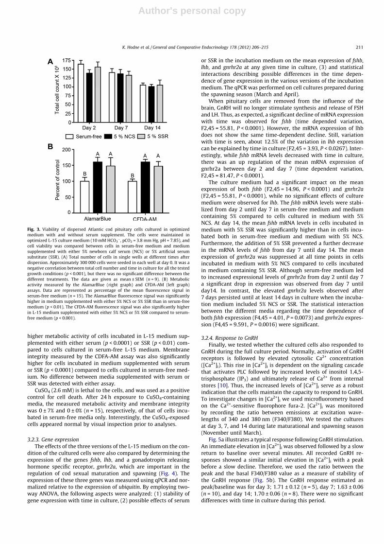
3.2.2. Total cell count and cell viabilityThe total number of cells remaining after 2, 7, and 14 days in
culture was used as a coarse estimate of the stability of culturesincubated in the three versions of the L-15 medium (serum-free,5% NCS, 5% SSR) (Fig. 3A). Double blind counting of the cells wasperformed to eliminate bias. At day 2 in culture, approximately50% of the seeded cells were lost, independently of the version ofincubation medium. From day 2 until the end of experiment, thetotal cell number was negatively correlated with time for all media(Fig. 3A; p < 0.001), and at day 14, only around 30% of the cellswere remaining. Thus, serum or SSR supplement alone do not re-duce the decline of the number of remaining cells with incubationtime.
To obtain more information about the condition of the culturedcells, assays based on the fluorescent indicators AB and CFDA-AM(Fig. 3B) were performed on cells after 7 days in culture. Thesefluorophores give an indication of metabolic activity and mem-brane integrity, respectively. The AB assay revealed a significantly
Fig. 1. Resting membrane potential of dispersed Atlantic cod pituitary cells in aculture medium developed for mammalian cells. The membrane potential wasmeasured using a perforated whole cell patch-clamp configuration. The cells weremaintained in the M199 culture medium, which contains 26.2 mM HCO3
�, at apCO2 of 38 mm Hg and a pH of 7.4. The osmolality was adjusted to 320 mOsm andthe temperature was kept at 12 �C. The data are presented as mean ± SEM (day 1;n = 10, day 2; n = 21, day 4; n = 3 day 5; n = 5, day 7; n = 2, day 8; n = 3). Linearcorrelation analysis shows that the slope of the regression line is significantlydifferent from zero (F15.75,4, P = 0.0165).
Fig. 2. Resting membrane potential of dispersed Atlantic cod pituitary cells inculture medium optimized for this species. The membrane potential was measuredusing a perforated whole cell patch-clamp configuration. The cells were maintainedin L-15 culture medium with 10 mM HCO3
�, at a pCO2 of 3.8 mm Hg and a pH of7.85. The osmolality was adjusted to 320 mOsm and the temperature was kept at12 �C. The membrane potential at day 2 and day 12–14 were compared betweenserum-free optimized L-15 medium and optimized L-15 medium supplementedwith either 5% newborn calf serum (NCS) or 5% artificial serum substitute (SSR). Themembrane potential of cells in serum free environment was �49.1 ± 1.7 mV at day2 (n = 35) and �49.4 ± 1.8 at day 12–14 (n = 33). Cells in L-15 medium supple-mented with 5% NCS had a membrane potential of �44.5 ± 2.4 mV (n = 23) at day 2and �44.1 ± 1.8 at day 12–14 n = 41). Cells in L-15 medium supplemented with 5%SSR had a membrane potential of �48.3 ± 1.9 mV (n = 15) at day 2 and �48.7 ± 1.3(n = 47) at day 12–14. The data are presented as mean ± SEM. There were nostatistical differences in membrane potential between days or treatments.
210 K. Hodne et al. / General and Comparative Endocrinology 178 (2012) 206–215
Author's personal copy
higher metabolic activity of cells incubated in L-15 medium sup-plemented with either serum (p < 0.0001) or SSR (p < 0.01) com-pared to cells cultured in serum-free L-15 medium. Membraneintegrity measured by the CDFA-AM assay was also significantlyhigher for cells incubated in medium supplemented with serumor SSR (p < 0.0001) compared to cells cultured in serum-free med-ium. No difference between media supplemented with serum orSSR was detected with either assay.
CuSO4 (2.6 mM) is lethal to the cells, and was used as a positivecontrol for cell death. After 24 h exposure to CuSO4-containingmedia, the measured metabolic activity and membrane integritywas 0 ± 7% and 0 ± 0% (n = 15), respectively, of that of cells incu-bated in serum-free media only. Interestingly, the CuSO4-exposedcells appeared normal by visual inspection prior to analyses.
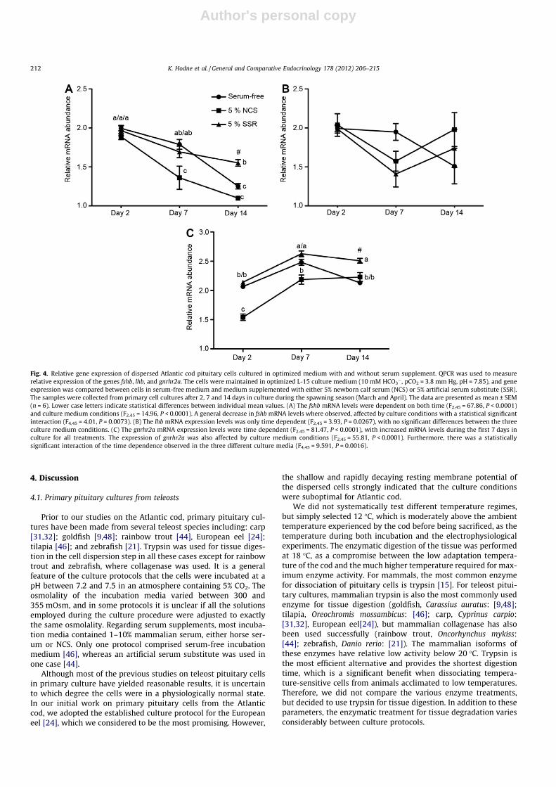
3.2.3. Gene expressionThe effects of the three versions of the L-15 medium on the con-
dition of the cultured cells were also compared by determining theexpression of the genes fshb, lhb, and a gonadotropin releasinghormone specific receptor, gnrhr2a, which are important in theregulation of cod sexual maturation and spawning (Fig. 4). Theexpression of these three genes was measured using qPCR and nor-malized relative to the expression of ubiquitin. By employing two-way ANOVA, the following aspects were analyzed: (1) stability ofgene expression with time in culture, (2) possible effects of serum
or SSR in the incubation medium on the mean expression of fshb,lhb, and gnrhr2a at any given time in culture, (3) and statisticalinteractions describing possible differences in the time depen-dence of gene expression in the various versions of the incubationmedium. The qPCR was performed on cell cultures prepared duringthe spawning season (March and April).
When pituitary cells are removed from the influence of thebrain, GnRH will no longer stimulate synthesis and release of FSHand LH. Thus, as expected, a significant decline of mRNA expressionwith time was observed for fshb (time depended variation,F2,45 = 55.81, P < 0.0001). However, the mRNA expression of lhbdoes not show the same time-dependent decline. Still, variationwith time is seen, about 12.5% of the variation in lhb expressioncan be explained by time in culture (F2,45 = 3.93, P < 0.0267). Inter-estingly, while fshb mRNA levels decreased with time in culture,there was an up regulation of the mean mRNA expression ofgnrhr2a between day 2 and day 7 (time dependent variation,F2,45 = 81.47, P < 0.0001).
The culture medium had a significant impact on the meanexpression of both fshb (F2,45 = 14.96, P < 0.0001) and gnrhr2a(F2,45 = 55.81, P < 0.0001), while no significant effects of culturemedium were observed for lhb. The fshb mRNA levels were stabi-lized from day 2 until day 7 in serum-free medium and mediumcontaining 5% compared to cells cultured in medium with 5%NCS. At day 14, the mean fshb mRNA levels in cells incubated inmedium with 5% SSR was significantly higher than in cells incu-bated both in serum-free medium and medium with 5% NCS.Furthermore, the addition of 5% SSR prevented a further decreasein the mRNA levels of fshb from day 7 until day 14. The meanexpression of gnrhr2a was suppressed at all time points in cellsincubated in medium with 5% NCS compared to cells incubatedin medium containing 5% SSR. Although serum-free medium ledto increased expressional levels of gnrhr2a from day 2 until day 7a significant drop in expression was observed from day 7 untilday14. In contrast, the elevated gnrhr2a levels observed after7 days persisted until at least 14 days in culture when the incuba-tion medium included 5% NCS or SSR. The statistical interactionbetween the different media regarding the time dependence ofboth fshb expression (F4,45 = 4.01, P = 0.0073) and gnrhr2a expres-sion (F4,45 = 9.591, P = 0.0016) were significant.
3.2.4. Response to GnRHFinally, we tested whether the cultured cells also responded to
GnRH during the full culture period. Normally, activation of GnRHreceptors is followed by elevated cytosolic Ca2+ concentration([Ca2+]i). This rise in [Ca2+]i is dependent on the signaling cascadethat activates PLC followed by increased levels of inositol 1,4,5-trisphosphate (IP3) and ultimately release of Ca2+ from internalstores [10]. Thus, the increased levels of [Ca2+]i serve as a robustindication that the cells maintain the capacity to respond to GnRH.To investigate changes in [Ca2+]i we used microfluorometry basedon the Ca2+-sensitive fluorophore fura-2. [Ca2+]i was monitoredby recording the ratio between emissions at excitation wave-lengths of 340 and 380 nm (F340/F380). We tested the culturesat day 3, 7, and 14 during late maturational and spawning season(November until March).
Fig. 5a illustrates a typical response following GnRH stimulation.An immediate elevation in [Ca2+]i was observed followed by a slowreturn to baseline over several minutes. All recorded GnRH re-sponses showed a similar initial elevation in [Ca2+]i with a peakbefore a slow decline. Therefore, we used the ratio between thepeak and the basal F340/F380 value as a measure of stability ofthe GnRH response (Fig. 5b). The GnRH response estimated aspeak/baseline was for day 3; 1.71 ± 0.12 (n = 5), day 7; 1.63 ± 0.06(n = 10), and day 14; 1.70 ± 0.06 (n = 8). There were no significantdifferences with time in culture during this period.
Fig. 3. Viability of dispersed Atlantic cod pituitary cells cultured in optimizedmedium with and without serum supplement. The cells were maintained inoptimized L-15 culture medium (10 mM HCO3
�, pCO2 = 3.8 mm Hg, pH = 7.85), andcell viability was compared between cells in serum-free medium and mediumsupplemented with either 5% newborn calf serum (NCS) or 5% artificial serumsubstitute (SSR). (A) Total number of cells in single wells at different times afterdispersion. Approximately 300 000 cells were seeded in each well at day 0. It was anegative correlation between total cell number and time in culture for all the testedgrowth conditions (p < 0.001), but there was no significant difference between thedifferent treatments. The data are given as mean ± SEM (n = 9). (B) Metabolicactivity measured by the AlamarBlue (right graph) and CFDA-AM (left graph)assays. Data are represented as percentage of the mean fluorescence signal inserum-free medium (n = 15). The AlamarBlue fluorescence signal was significantlyhigher in medium supplemented with either 5% NCS or 5% SSR than in serum-freemedium (p < 0.01). The CFDA-AM fluorescence signal was also significantly higherin L-15 medium supplemented with either 5% NCS or 5% SSR compared to serum-free medium (p < 0.001).
K. Hodne et al. / General and Comparative Endocrinology 178 (2012) 206–215 211
Author's personal copy
4. Discussion
4.1. Primary pituitary cultures from teleosts
Prior to our studies on the Atlantic cod, primary pituitary cul-tures have been made from several teleost species including: carp[31,32]; goldfish [9,48]; rainbow trout [44], European eel [24];tilapia [46]; and zebrafish [21]. Trypsin was used for tissue diges-tion in the cell dispersion step in all these cases except for rainbowtrout and zebrafish, where collagenase was used. It is a generalfeature of the culture protocols that the cells were incubated at apH between 7.2 and 7.5 in an atmosphere containing 5% CO2. Theosmolality of the incubation media varied between 300 and355 mOsm, and in some protocols it is unclear if all the solutionsemployed during the culture procedure were adjusted to exactlythe same osmolality. Regarding serum supplements, most incuba-tion media contained 1–10% mammalian serum, either horse ser-um or NCS. Only one protocol comprised serum-free incubationmedium [46], whereas an artificial serum substitute was used inone case [44].
Although most of the previous studies on teleost pituitary cellsin primary culture have yielded reasonable results, it is uncertainto which degree the cells were in a physiologically normal state.In our initial work on primary pituitary cells from the Atlanticcod, we adopted the established culture protocol for the Europeaneel [24], which we considered to be the most promising. However,
the shallow and rapidly decaying resting membrane potential ofthe dispersed cells strongly indicated that the culture conditionswere suboptimal for Atlantic cod.
We did not systematically test different temperature regimes,but simply selected 12 �C, which is moderately above the ambienttemperature experienced by the cod before being sacrificed, as thetemperature during both incubation and the electrophysiologicalexperiments. The enzymatic digestion of the tissue was performedat 18 �C, as a compromise between the low adaptation tempera-ture of the cod and the much higher temperature required for max-imum enzyme activity. For mammals, the most common enzymefor dissociation of pituitary cells is trypsin [15]. For teleost pitui-tary cultures, mammalian trypsin is also the most commonly usedenzyme for tissue digestion (goldfish, Carassius auratus: [9,48];tilapia, Oreochromis mossambicus: [46]; carp, Cyprinus carpio:[31,32], European eel[24]), but mammalian collagenase has alsobeen used successfully (rainbow trout, Oncorhynchus mykiss:[44]; zebrafish, Danio rerio: [21]). The mammalian isoforms ofthese enzymes have relative low activity below 20 �C. Trypsin isthe most efficient alternative and provides the shortest digestiontime, which is a significant benefit when dissociating tempera-ture-sensitive cells from animals acclimated to low temperatures.Therefore, we did not compare the various enzyme treatments,but decided to use trypsin for tissue digestion. In addition to theseparameters, the enzymatic treatment for tissue degradation variesconsiderably between culture protocols.
Fig. 4. Relative gene expression of dispersed Atlantic cod pituitary cells cultured in optimized medium with and without serum supplement. QPCR was used to measurerelative expression of the genes fshb, lhb, and gnrhr2a. The cells were maintained in optimized L-15 culture medium (10 mM HCO3
�, pCO2 = 3.8 mm Hg, pH = 7.85), and geneexpression was compared between cells in serum-free medium and medium supplemented with either 5% newborn calf serum (NCS) or 5% artificial serum substitute (SSR).The samples were collected from primary cell cultures after 2, 7 and 14 days in culture during the spawning season (March and April). The data are presented as mean ± SEM(n = 6). Lower case letters indicate statistical differences between individual mean values. (A) The fshb mRNA levels were dependent on both time (F2,45 = 67.86, P < 0.0001)and culture medium conditions (F2,45 = 14.96, P < 0.0001). A general decrease in fshb mRNA levels where observed, affected by culture conditions with a statistical significantinteraction (F4,45 = 4.01, P = 0.0073). (B) The lhb mRNA expression levels was only time dependent (F2,45 = 3.93, P = 0.0267), with no significant differences between the threeculture medium conditions. (C) The gnrhr2a mRNA expression levels were time dependent (F2,45 = 81.47, P < 0.0001), with increased mRNA levels during the first 7 days inculture for all treatments. The expression of gnrhr2a was also affected by culture medium conditions (F2,45 = 55.81, P < 0.0001). Furthermore, there was a statisticallysignificant interaction of the time dependence observed in the three different culture media (F4,45 = 9.591, P = 0.0016).
212 K. Hodne et al. / General and Comparative Endocrinology 178 (2012) 206–215

Author's personal copy
Although some of our tests of the culture quality are targetinggonadotropes in particular, i.e. gene transcription levels and GnRHresponse, most results are based on all cells in the culture dish anddo not distinguish between the different pituitary cell types pres-ent. Still, we have no reason to believe that the culture conditionspresented should not be equally optimized for all the differentendocrine pituitary cell types, as they are closely related andexposed to the same microenvironment within the pituitaryin vivo. Thus, we consider it likely that the results presented in thisstudy apply to all pituitary cell types.
4.2. Improved procedures for establishing primary cultures of codpituitary cells
4.2.1. The importance of osmolalityMost commercial culture media and salt solutions for mamma-
lian cell cultures have an osmolality far below the plasma osmolal-ity of the Atlantic cod at the temperature (7–8 �C) and salinity(28%) of the seawater tanks at our animal facility [22,26]. It is areasonable assumption that osmotic stress during the culture pro-cedures might be deleterious for the cells. The condition of cellsincubated in the initial M199 medium was markedly improvedby adjusting the osmolality of all solutions to 320 mOsm, whichis close to the normal osmolality of cod plasma [26]. The restingcell membrane potential increased significantly and reached valuesin the range reported for both goldfish and mammalian pituitarycells [30,39,42]. The resting membrane potential was also morestable after adjustment of the osmolality, although it still decayedwith time in culture. It is possible that the improved condition ofthe cells is less dependent on the absolute value of osmolality thanon keeping the osmolality constant during the whole process ofcell dissociation, incubation, and electrophysiological recordings.
A rapid change in osmolality is stressful to cells, in particular whenbeing particularly fragile due to the dissociation procedure. It isthus important to measure and adjust the osmolality of all solu-tions used for establishing and maintaining the cell cultures.
4.2.2. The importance of pCO2 and pHBecause CO2 is easily exchanged over the gills, the pCO2 in fish is
only a small fraction of the pCO2 in mammalian blood (40–46 mmHg or 5.3–6.1 kPa, arterial-venous pCO2) [34]. In teleosts, the arte-rial pCO2 range from 1.7 to 3.4 mm Hg (0.23–0.45 kPa), while thevenous pCO2 range from 3.2 to 5.7 mm Hg (0.42–0.79 kPa), depend-ing on the species [1]. Due to the low pCO2 and the correspondinglylow HCO3
� concentration, the buffer capacity of the blood andextracellular fluids is lower in fish than in mammals and culturemedia adapted for mammalian cells. Mammalian culture mediausually include buffer systems that result in pH 7.4 when the med-ium is equilibrated to the standard atmosphere (5% CO2 in humid-ified air) and temperature (37 �C) in the incubator. Consequently,changing any of these parameters (temperature, pCO2, [HCO3
�])in an attempt to adjust to the internal conditions of a teleost will al-ter the pH of the medium. Furthermore, the value for neutral pHincreases with decreasing temperature [8]. Consequently, in cold-water fish, the blood plasma may have a pH about 0.5 units abovenormal values in mammals, which represents approximately thesame alkalinity of the plasma in these animal groups. In teleosts,plasma pH ranges from 7.7 to 7.9, depending on temperature[12]. In the Atlantic cod, the plasma pH is �7.9 at 12 �C [18]. Thus,it is to be expected that neither the pCO2 nor the pH recommendedfor incubation of mammalian cells are optimal for teleost cells.Therefore, culture media customized for teleost cells should containmodified buffer systems compared to those used in most mediadeveloped for mammalian cells. By adjusting not only the osmolal-ity, but also the interrelated parameters pCO2, [HCO3
�], and pH ofthe incubation medium to physiological values for teleosts[12,34], the resting cell membrane potential became stable for atleast two weeks in culture. In order to achieve this, the M199medium, which contains 26.2 mM HCO3
�, was replaced with theHCO3
� free medium L-15 supplemented with 10 mM HCO3�. The
CO2 content of the incubation atmosphere was set to 0.5%(3.80 mm Hg), resulting in a physiological pH of 7.85 in the mediumat 12 �C.
In addition to a deeper and more stable membrane potential,the adjustments of pCO2, [HCO3
�], and pH significantly increasedthe electrical excitability of the cells. The cells also stayed attachedto the bottom of the dish for about two weeks in the fully adjustedincubation medium, whereas most cells were detached after lessthan one week when the culture protocol only included osmolalityadjustments.
Thus, by simple adjustments of the osmolality of all appliedsolutions and the pCO2, [HCO3
�], and pH of the incubation med-ium, the physiological condition of the cultured cod pituitary cellswas dramatically improved compared to the result following atraditional culture protocol adapted for mammalian cells.
4.2.3. The importance of serum supplementAddition of bovine serum to the culture medium is standard
procedure in most protocols for mammalian cell culture. The argu-ments for adding serum to the culture media range from improvedcell adherence to the bottom of the culture vessel, to stronger plas-ma membranes and a generally improved condition of the cells[15]. However, except for blood cells and the endothelial cells lin-ing the blood vessels, cells are not in direct contact with serumin vivo. Although serum supplement is usually beneficial for cellsin culture, serum may also be detrimental for at least some celltypes. Several reports have demonstrated that even mammalianprimary cell cultures may benefit from serum-free media. For
Fig. 5. Cytosolic Ca2+ response to GnRH1–3 in dispersed Atlantic cod pituitary cellsduring the culture period. Microfluorometric recordings using fura-2 loaded cellswere used to monitor changes in cytosolic Ca2+ levels [Ca2+]i following gonadotro-pin releasing hormone (GnRH) stimulation. The ratio between emissions at theexcitation wavelengths 340 and 380 nm (F340/F380) reflects [Ca2+]i. (A) Representsa typical response to a mixture (10�7 M) of the three different GnRH forms for 10 s(black line under trace). An immediate elevation of [Ca2+]i was followed by steadydecline lasting for one to several minutes before returning to resting levels. (B) Theratio between the peak and basal F340/F380 levels was compared for GnRHresponses in cells grown for different time in culture. Average peak/baseline ratiowas for day 3: 1.71 ± 0.12 (n = 5), day 7: 1.63 ± 0.06 (n = 10), and day 14: 1.70 ± 0.06(n = 8). The data are presented as mean ± SEM. No statistical differences wereobserved between the time points.
K. Hodne et al. / General and Comparative Endocrinology 178 (2012) 206–215 213

Author's personal copy
example, Bowers and Dahm [7] showed that amniotic smoothmuscle cells in culture maintained their contractility if serumwas omitted from the culture medium. By growing pig liver sinu-soidal endothelial cells in a serum-free medium supplementedwith an artificial serum substitute, Elvevold et al. [14] managedto maintain functional cells for 20 days, compared to maximum4 days in traditional serum-containing medium. The serum substi-tute contained no proteins or peptides that might interfere withthe cell function, but did include transferrin, surfactant, adherencepromoters, low molecular weight lipids, and replacements for me-tal ion buffers [4]. In order to keep total control of the experimentalconditions, artificial serum supplement has previously also beenused in the culture of head kidney macrophages from the Atlanticcod [36]. Thus, it is evident that serum-free media and mediasupplemented with serum or artificial serum substitutes shouldbe compared when the aim is to develop optimized culture condi-tions for a specific cell type.
The improvements of the culture conditions discussed in thetwo previous sections were achieved using serum-free L-15 med-ium. Therefore, addition of serum (NCS) to the incubation mediumis not required in order to maintain seemingly healthy cod pitui-tary cells in primary culture for at least two weeks. Nevertheless,in order to investigate if supplementing the incubation mediumwith NCS or the artificial serum substitute SSR is beneficial ornot, electrophysiological properties, viability, and gene expressionwere compared in cells cultivated in serum-free medium and med-ium supplemented with either NCS or SSR.
Addition of neither NCS nor SSR to the incubation medium hadany significant effect on the resting cell membrane potential,although it tended to be somewhat shallower when NCS or SSRwere present in the medium. The number of surviving cells withtime in culture was also unaffected by these supplements. In con-trast to this lack of evident effects on electrophysiological proper-ties and survival, both the metabolic activity and the membraneintegrity of cells incubated in L-15 medium supplemented withNCS or SSR were significantly higher than in cells cultured in ser-um-free L-15 medium. The effects of NCS and SSR on these param-eters were similar.
The physiological relevance of NCS and SSR supplements wasfurther examined by measuring the expression of selected genes.The expected decrease in the expression of fshb with time in cul-ture, due to absence of GnRH influence, was observed in all med-ium versions. At day 14 in culture, the expression of fshb wassignificantly higher in medium supplemented with SSR than inthe other media. We did not observe a similar decrease in lhbexpression. The differential GnRH-regulation of fshb and lhb willbe the topic of further studies.
Transcription of the GnRH receptor gene gnrhr2a is essentialregarding GnRH responses in pituitary cells. It is common thatreduced ligand concentration induces upregulation of the relatedreceptors. Because the tested media were lacking GnRH, it is notsurprising that the expression of gnrhr2a was higher at day 7 thanat day 2 in culture for all medium versions. Also for this receptorgene, the expression level was lowest in medium supplementedwith serum, and highest in medium containing SSR. Furthermore,both SSR and NCS supplements caused the elevated gnrhr2aexpression to remain also at day 14. Thus, for primary culture ofcod pituitary cells, the gene expression data indicate that SSR is apreferable medium supplement compared to NCS if the aim is tostudy the GnRH response.
It is well documented that NCS promotes viability and substrateattachment in primary cultures of many types of teleost cells [5].Also, we experienced that using NCS was necessary for properattachment when cells where plated on glass bottom. Fish serum,on the other hand, may be beneficial or toxic, depending on celltype (reviewed in [27]). Barlian et al. [3] showed that it might be
favorable to replace the poorly defined serum with calf serumalbumin (BSA). However, the success was dependent on the typeof BSA. Other similar proteins, like ovalbumin, did not have thesame positive effect.
An important concern regarding total removal of serum fromincubation media customized for pituitary cells is the fact thatgonadotropin releasing hormone (GnRH) receptors, like many Gprotein-coupled receptors, are dependent on lipid rafts in orderto be localized close to their cognate signaling molecules. Navratilet al. [25] showed that the GnRH receptors are localized to the lowdensity membrane fraction together with c-raf kinase, and thatdepletion of cholesterol disrupts the association between thereceptors and the lipid rafts. Cholesterol depletion also reducedthe GnRH activation of ERK (extracellular signal-related kinase)and c-fos-regulated gene transcription. There is increasing evi-dence that also many ion channels are localized to lipid rafts,which can affect channel function [13]. For instance, the channelgating kinetics may be regulated by interactions between channelproteins and both lipids and other associated proteins in the raft.
Tocher et al. [40] showed that the lipid composition of theplasma membrane of fish cells in long-term cultures was affectedby the NCS added to the medium, reflecting the lipid componentsof the serum more than those of the original fish tissue. Therefore,an optimal serum substitute should contain the essential factorsfor keeping the structure and the constituents of the plasma mem-brane intact, while omitting unknown growth factors and othersignaling molecules that can interfere with the physiology of thecell.
5. Conclusion
By adjusting the osmolality of all solutions included in theculture protocol, and the temperature, pCO2, [HCO3
�], and pH ofthe incubation medium, to physiological values for the Atlanticcod, it was possible to maintain viable cod pituitary cells in pri-mary culture for at least two weeks. During this period, the restingmembrane potential was completely stable, and the expressionpattern of key genes was physiologically reasonable. In addition,the cells responded similarly to GnRH during the whole cultureperiod. In contrast, cells dispersed and maintained following a tra-ditional culture procedure for teleosts displayed rapidly decayingresting membrane potentials and succumbed within one week.The cells were moderately more viable in medium supplementedwith either NCS or an artificial serum substitute (SSR). Comparedto serum-free medium, the gene expression was better maintainedin medium containing SSR, whereas NCS tended to depress theexpression level. Although serum-free medium is adequate formany applications, serum supplement may be preferable forexperiments dependent on maximizing the membrane integrityand receptor function. Using a well-defined, artificial serum substi-tute, thus avoiding introduction of undesired signaling molecules,may be the ultimate solution in many experimental situations.
Acknowledgments
We thank Göran Nilsson and Sylvie Dufour for valuable discus-sions. We are grateful to Kjetil Elvevold for providing us with SSR,to Stine Berg Vaule for performing all the qPCR experiments andRønnaug A.U. Strandabø for helping with the Ca2+ imaging. Thisstudy was supported by the Research Council of Norway, GrantsNo. 184851 (to F-A.W.) and 191825 (to T.M.H.).
References
[1] Fish respiration, Academic Press, San Diego, USA, 1998.[2] Basic Cell Culture Protocols, third ed., Humana Press 2004.
214 K. Hodne et al. / General and Comparative Endocrinology 178 (2012) 206–215
Author's personal copy
[3] A. Barlian, R.C. Ganassin, D. Tom, N.C. Bols, A comparison of bovine serum-albumin and chicken ovalbumin as supplements for the serum-free growth ofchinook salmon embryo cells, chse-214, Cell Biol. Int. 17 (1993) 677–684.
[4] K. Bertheussen, Growth of cells in a new defined protein-free medium,Cytotechnology 11 (1993) 219–231.
[5] N.C. Bols, L.E.J. Lee, Cell lines availability propagation and isolation, in: H.A.Mommsen (Ed.), Biochemistry and Molecular Biology of Fishes, Elsevier, 1994,pp. 145–159.
[6] S.K. Bopp, T. Lettieri, Comparison of four different colorimetric andfluorometric cytotoxicity assays in a zebrafish liver cell line, BMC Pharmacol.8 (2008) 8.
[7] C.W. Bowers, L.M. Dahm, Maintenance of contractility in dissociated smoothmuscle: low-density cultures in a defined medium, Am. J. Physiol. 264 (1993)C229–236.
[8] J.N. Cameron, Acid-base homeostasis - past and present perspectives, Physiol.Zool. 62 (1989) 845–865.
[9] J.P. Chang, H. Cook, G.L. Freedman, A.J. Wiggs, G.M. Somoza, R. de Leeuw, et al.,Use of a pituitary cell dispersion method and primary culture system for thestudies of gonadotropin-releasing hormone action in the goldfish, Carassiusauratus. I. Initial morphological, static, and cell column perifusion studies, Gen.Comp. Endocrinol. 77 (1990) 256–273.
[10] J.P. Chang, J.D. Johnson, G.R. Sawisky, C.L. Grey, G. Mitchell, M. Booth, et al.,Signal transduction in multifactorial neuroendocrine control of gonadotropinsecretion and synthesis in teleosts-studies on the goldfish model, Gen. Comp.Endocrinol. 161 (2009) 42–52.
[11] J.P. Chang, J.D. Johnson, F. Van Goor, C.J. Wong, W.K. Yunker, A.D. Uretsky,et al., Signal transduction mechanisms mediating secretion in goldfishgonadotropes and somatotropes, Biochem. Cell Biol. 78 (2000) 139–153.
[12] J.B. Claiborne, S.L. Edwards, A.I. Morrison-Shetlar, Acid-base regulation infishes: cellular and molecular mechanisms, J. Exp. Zool. 293 (2002) 302–319.
[13] C. Dart, Lipid microdomains and the regulation of ion channel function, J.Physiol. 588 (2010) 3169–3178.
[14] K. Elvevold, G.I. Nedredal, A. Revhaug, K. Bertheussen, B. Smedsrod, Long-termpreservation of high endocytic activity in primary cultures of pig liversinusoidal endothelial cells, Eur. J. Cell Biol. 84 (2005) 749–764.
[15] R.I. Freshney, Culture of animal cells, John Wiley & son Inc., Hoboken, NewJersey, USA, 2010.
[16] T.M. Haug, K. Hodne, F.A. Weltzien, O. Sand, Electrophysiological properties ofpituitary cells in primary culture from Atlantic cod (Gadus morhua),Neuroendocrinology 86 (2007) 38–47.
[17] K. Hodne, T.M. Haug, F.A. Weltzien, Single-cell qPCR on dispersed primarypituitary cells -an optimized protocol, BMC Mol. Biol. 11 (2010) 82.
[18] B.K. Larsen, H.O. Portner, F.B. Jensen, Extra- and intracellular acid-base balanceand ionic regulation in cod (Gadus morhua) during combined and isolatedexposures to hypercapnia and copper, Mar. Biol. 128 (1997) 337–346.
[19] B. Levavi-Sivan, C.L. Bloch, M.J. Gutnick, I.A. Fleidervish, Electrotonic couplingin the anterior pituitary of a teleost fish, Endocrinology 146 (2005) 1048–1052.
[20] B. Levavi-Sivan, J. Bogerd, E.L. Mananos, A. Gomez, J.J. Lareyre, Perspectives onfish gonadotropins and their receptors, Gen. Comp. Endocrinol. 165 (2010)412–437.
[21] S.W. Lin, W. Ge, Differential regulation of gonadotropins (FSH and LH) andgrowth hormone (GH) by neuroendocrine, endocrine, and paracrine factors inthe zebrafish-An in vitro approach, Gen. Comp. Endocrinol. 160 (2009) 183–193.
[22] S.H. Magill, M.D.J. Sayer, The effect of reduced temperature and salinity on theblood physiology of juvenile Atlantic cod, J. Fish Biol. 64 (2004) 1193–1205.
[23] J.S. Mitcheson, J.C. Hancox, A.J. Levi, Cultured adult cardiac myocytes: futureapplications, culture methods, morphological and electrophysiologicalproperties, Cardiovasc. Res. 39 (1998) 280–300.
[24] M. Montero, N. Le Belle, B. Vidal, S. Dufour, Primary cultures of dispersedpituitary cells from estradiol-pretreated female silver eels (Anguilla anguillaL.): immunocytochemical characterization of gonadotropic cells andstimulation of gonadotropin release, Gen. Comp. Endocrinol. 104 (1996)103–115.
[25] A.M. Navratil, S.P. Bliss, K.A. Berghorn, J.M. Haughian, T.A. Farmerie, J.K.Graham, et al., Constitutive localization of the gonadotropin-releasinghormone (GnRH) receptor to low density membrane microdomains isnecessary for GnRH signaling to ERK, J. Biol. Chem. 278 (2003) 31593–31602.
[26] R.E. Olsen, K. Sundell, E. Ringo, R. Myklebust, G.I. Hemre, T. Hansen, et al., Theacute stress response in fed and food deprived Atlantic cod Gadus morhua L,Aquaculture 280 (2008) 232–241.
[27] P. Párt, E. Bergström, 8 Primary cultures of teleost branchial epithelial cells, in:M.W. Chris, J.S. Trevor (Eds.), Fish Physiology, Academic Press, 1995, pp. 207–227.
[28] M.W. Pfaffl, A new mathematical model for relative quantification in real-timeRT-PCR, Nucleic Acids Res. 29 (2001) e45.
[29] M.W. Pfaffl, A. Tichopad, C. Prgomet, T.P. Neuvians, Determination of stablehousekeeping genes, differentially regulated target genes and sampleintegrity: bestkeeper–excel-based tool using pair-wise correlations,Biotechnol. Lett. 26 (2004) 509–515.
[30] C.J. Price, J.I. Goldberg, J.P. Chang, Voltage-activated ionic currents in goldfishpituitary cells, Gen. Comp. Endocrinol. 92 (1993) 16–30.
[31] L. Ribeiro, W. Ahne, Fish cell-culture - initiation of a line of pituitary-cells fromcarp (Cyprinus Carpio) to study the release of gonadotropin Invitro, In Vitro CellDev. B. 18 (1982) 419–420.
[32] L.P. Ribeiro, W. Ahne, V. Lichtenberg, Primary culture of normal pituitary cellsof carp (Cyprinus carpio) for the study of gonadotropin release, In Vitro 19(1983) 41–45.
[33] C. Sarantopoulos, J.B. McCallum, W.M. Kwok, Q. Hogan, Beta-escin diminishesvoltage-gated calcium current rundown in perforated patch-clamp recordingsfrom rat primary afferent neurons, J. Neurosci. Methods 139 (2004) 61–68.
[34] K. Schmidt-Nielsen, Animal Physiology, 5 ed., Cambridge University Press,New York, 1997.
[35] A. Schreer, C. Tinson, J.P. Sherry, K. Schirmer, Application of Alamar blue/5-carboxyfluorescein diacetate acetoxymethyl ester as a noninvasive cellviability assay in primary hepatocytes from rainbow trout, Anal. Biochem.344 (2005) 76–85.
[36] K.K. Sorensen, B. Sveinbjornsson, R.A. Dalmo, B. Smedsrod, K. Bertheussen,Isolation, cultivation and characterization of head kidney macrophages fromAtlantic cod, Gadus morhua L. J. Fish Dis. 20 (1997) 93–107.
[37] S.S. Stojilkovic, Pituitary cell type-specific electrical activity, calcium signalingand secretion, Biol. Res. 39 (2006) 403–423.
[38] S.S. Stojilkovic, Molecular mechanisms of pituitary endocrine cell calciumhandling, Cell Calcium (2012) 212–221.
[39] S.S. Stojilkovic, H. Zemkova, F. Van Goor, Biophysical basis of pituitary celltype-specific Ca2+ signaling-secretion coupling, Trends Endocrinol. Metab. 16(2005) 152–159.
[40] D.R. Tocher, J.R. Sargent, G.N. Frerichs, The fatty-acid compositions ofestablished fish cell-lines after long-term culture in mammalian sera, FishPhysiol. Biochem. 5 (1988) 219–227.
[41] K.E. Tollefsen, C. Blikstad, S. Eikvar, E.F. Finne, I.K. Gregersen, Cytotoxicity ofalkylphenols and alkylated non-phenolics in a primary culture of rainbowtrout (Onchorhynchus mykiss) hepatocytes, Ecotoxicol. Environ. Saf. 69 (2008)64–73.
[42] F. Van Goor, J.I. Goldberg, J.P. Chang, Electrical membrane properties and ioniccurrents in cultured goldfish gonadotrophs, Can. J. Physiol. Pharmacol. 74(1996) 729–743.
[43] C. Viero, U. Kraushaar, S. Ruppenthal, L. Kaestner, P. Lipp, A primary culturesystem for sustained expression of a calcium sensor in preserved adult ratventricular myocytes, Cell Calcium 43 (2008) 59–71.
[44] C. Weil, P. Hansen, D. Hyam, F. Legac, B. Breton, L.W. Crim, Use of pituitary-cells in primary culture to study the regulation of gonadotropin hormone (Gth)secretion in rainbow-trout - setting up and validating the system as assessedby Its responsiveness to mammalian and salmon gonadotropin-releasing-hormone, Gen. Comp. Endocrinol. 62 (1986) 202–209.
[45] F.A. Weltzien, C. Pasqualini, P. Vernier, S. Dufour, A quantitative real-time RT-PCR assay for European eel tyrosine hydroxylase, Gen. Comp. Endocrinol. 142(2005) 134–142.
[46] S.H. Xu, I.M. Cooke, Voltage-gated currents of tilapia prolactin cells, Gen.Comp. Endocrinol. 150 (2007) 219–232.
[47] Z. Yaron, G. Gur, P. Melamed, H. Rosenfeld, A. Elizur, B. Levavi-Sivan,Regulation of fish gonadotropins, Int. Rev. Cytol. 225 (2003) 131–185.
[48] Y. Yu, D.W. Ali, J.P. Chang, Characterization of ionic currents andelectrophysiological properties of goldfish somatotropes in primary culture,Gen. Comp. Endocrinol. 169 (2010) 231–243.
K. Hodne et al. / General and Comparative Endocrinology 178 (2012) 206–215 215