Optimization of MR protocols: A statistical decision analysis approach
21
Optimization of MR Protocols: A Statistical Decision Analysis Approach Elliot R. McVeigh, Michael J. Bronskill, and R. Mark Henkelman Department of Medical Biophysics, University of Toronto, and Physics Division, Ontario Cancer Institute, 500 Sherbourne Street, Toronto, Ontario M4X 1K9, Canada Abstract A new method of optimizing MRI data acquisition protocols is presented. Tissues are modeled with probability density functions (PDFs) of tissue parameter values (such as T 1 , T 2 ). The imaging data acquisition process is modeled as a mapping from a tissue parameter space to a signal strength space. Tissue parameter PDFs are mapped to signal strength PDFs for each tissue in a clinical problem. The efficacy of an MRI protocol is evaluated using the methods of statistical decision analysis applied to the signal strength PDFs, including the propagation of noise. This procedure evaluates the ability to discriminate different tissues based on the signal strengths produced with the protocol. The model can incorporate an arbitrary number of tissues, parameters, and pulse sequences in the protocol. The multivariate nature of MRI and the observed broad distribution of tissue parameter values makes this model more appropriate for optimizing data acquisition protocols than methods which maximize the signal-difference-to-noise ratio between discrete values of the tissue parameters. It is shown that these two methods may calculate different optimal protocols. The method can be used to optimize data acquisition for quantitative computer-based tissue classification, as well as imaging. Data acquisition and image processing philosophies are discussed in light of the method. 1. Introduction The application of NMR in medicine, as originally proposed, was a simple diagnostic test based on measured relaxation times T 1 and T 2 (1). It was recognized that this test would be most useful when performed in vivo on an isolated region of tissue. Magnetic resonance imaging (MRI) techniques provided the means for this isolation and today yield tomograms of impressive visual quality. However, with the movement toward a rich radiological technique, MRI has suffered the loss of its development as a quantitative diagnostic test. The majority of the MRI research to date has been geared toward producing aesthetically pleasing images with higher signal-to-noise ratio (SNR), finer resolution, and faster scan times. While these are laudable goals in the context of imaging, it is not clear how to transform improvements in these attributes into improvements in diagnostic performance. Although many diagnoses can be made from observation of abnormal morphology, MRI offers a unique opportunity to derive a multivariate diagnostic test based on tissue parameters. In order to exploit this opportunity, we should think of the MRI experiment not as something that generates contrast between tissues, but as a process that discriminates between different tissues. This difference may seem subtle but, as this paper will show, these two philosophies lead to different methods of optimization. Early methods for deriving optimal MRI pulse sequences applied differential calculus to the signal strength equations (2-7) and used the maximum signal-difference-to-noise ratio (SDNR) or maximum signal gradient as the quantitative figure of merit. These methods address the one- dimensional problem of producing a gray-scale representation of an object with the maximum “sensitivity” at a specific value, or maximum SDNR between two specific values of the tissue parameters; as such, they are elegant and the mathematics is straightforward. However, these methods do not directly address the problem of maximizing the sensitivity and specificity of MRI as a diagnostic test applied to a patient population. To do this, one must incorporate into NIH Public Access Author Manuscript Magn Reson Med. Author manuscript; available in PMC 2008 May 26. Published in final edited form as: Magn Reson Med. 1988 March ; 6(3): 314–333. NIH-PA Author Manuscript NIH-PA Author Manuscript NIH-PA Author Manuscript
-
Upload
independent -
Category
Documents
-
view
4 -
download
0
Transcript of Optimization of MR protocols: A statistical decision analysis approach

Optimization of MR Protocols:A Statistical Decision Analysis Approach
Elliot R. McVeigh, Michael J. Bronskill, and R. Mark HenkelmanDepartment of Medical Biophysics, University of Toronto, and Physics Division, Ontario CancerInstitute, 500 Sherbourne Street, Toronto, Ontario M4X 1K9, Canada
AbstractA new method of optimizing MRI data acquisition protocols is presented. Tissues are modeled withprobability density functions (PDFs) of tissue parameter values (such as T1, T2). The imaging dataacquisition process is modeled as a mapping from a tissue parameter space to a signal strength space.Tissue parameter PDFs are mapped to signal strength PDFs for each tissue in a clinical problem. Theefficacy of an MRI protocol is evaluated using the methods of statistical decision analysis appliedto the signal strength PDFs, including the propagation of noise. This procedure evaluates the abilityto discriminate different tissues based on the signal strengths produced with the protocol. The modelcan incorporate an arbitrary number of tissues, parameters, and pulse sequences in the protocol. Themultivariate nature of MRI and the observed broad distribution of tissue parameter values makes thismodel more appropriate for optimizing data acquisition protocols than methods which maximize thesignal-difference-to-noise ratio between discrete values of the tissue parameters. It is shown thatthese two methods may calculate different optimal protocols. The method can be used to optimizedata acquisition for quantitative computer-based tissue classification, as well as imaging. Dataacquisition and image processing philosophies are discussed in light of the method.
1. IntroductionThe application of NMR in medicine, as originally proposed, was a simple diagnostic test basedon measured relaxation times T1 and T2 (1). It was recognized that this test would be mostuseful when performed in vivo on an isolated region of tissue. Magnetic resonance imaging(MRI) techniques provided the means for this isolation and today yield tomograms ofimpressive visual quality. However, with the movement toward a rich radiological technique,MRI has suffered the loss of its development as a quantitative diagnostic test. The majority ofthe MRI research to date has been geared toward producing aesthetically pleasing images withhigher signal-to-noise ratio (SNR), finer resolution, and faster scan times. While these arelaudable goals in the context of imaging, it is not clear how to transform improvements in theseattributes into improvements in diagnostic performance. Although many diagnoses can bemade from observation of abnormal morphology, MRI offers a unique opportunity to derive amultivariate diagnostic test based on tissue parameters. In order to exploit this opportunity, weshould think of the MRI experiment not as something that generates contrast between tissues,but as a process that discriminates between different tissues. This difference may seem subtlebut, as this paper will show, these two philosophies lead to different methods of optimization.
Early methods for deriving optimal MRI pulse sequences applied differential calculus to thesignal strength equations (2-7) and used the maximum signal-difference-to-noise ratio (SDNR)or maximum signal gradient as the quantitative figure of merit. These methods address the one-dimensional problem of producing a gray-scale representation of an object with the maximum“sensitivity” at a specific value, or maximum SDNR between two specific values of the tissueparameters; as such, they are elegant and the mathematics is straightforward. However, thesemethods do not directly address the problem of maximizing the sensitivity and specificity ofMRI as a diagnostic test applied to a patient population. To do this, one must incorporate into
NIH Public AccessAuthor ManuscriptMagn Reson Med. Author manuscript; available in PMC 2008 May 26.
Published in final edited form as:Magn Reson Med. 1988 March ; 6(3): 314–333.
NIH
-PA Author Manuscript
NIH
-PA Author Manuscript
NIH
-PA Author Manuscript

the figure of merit the ideas associated with evaluation of a diagnostic test: the reduction ofmultiparametric collected data into a single test value, and the distribution of expected testvalues from the patient population (8-10).
The need for a universal optimization method becomes more apparent with the developmentof each new MRI technique. Innovations in MRI tend to exploit additional tissue parameters,and bring with them new pulse sequence parameters. For example, fast imaging with gradientechoes is sensitive to chemical shift (δ) and magnetic susceptibility (χ0), and the signal strengthis highly dependent upon the interaction of relaxation rates and the rf flip angles (α). Thus, itintroduces two new tissue parameters (δ, χ0), and a new pulse sequence parameter (α). Whilenew innovations are assumed to increase the power of MRI as a diagnostic test, the increasedcomplexity necessitates a coherent method of choosing the subset of techniques to use in thelimited examination time. What is needed is a framework upon which all of these techniquescan be placed and evaluated in terms of their power as diagnostic tests, individually and whenused in concert.
2. A Model of MRI as a Diagnostic TestImages with an acceptable SNR are now available at a wide range of magnetic field strengths.They usually have a SNR far in excess of the minimum required to demonstrate the pathologybeing investigated. This implies that we can now turn our attention to the more subtle problemof consistently distinguishing tissue types by means of their MRI signal characteristics. Anincrease in specificity may require a decrease in overall SNR; that is, imaging time may bemore wisely spent increasing the specificity of the technique with the addition of different pulsesequences, rather than increasing the SNR by averaging the same pulse sequence. Any modelof MRI should allow one to calculate the tradeoffs between overall SNR and specificity.
For the purpose of optimizing the application of MRI in the clinical environment we model itas an in vivo diagnostic test. When assessing the value of any diagnostic test, one must observethe distribution of test results obtained from the normal (those without the disease) andabnormal (those with the disease) patient populations. The performance of the test can becalculated by determining the overlap of these distributions.
2.1. Model of the Patient PopulationIt became evident quite early in the development of NMR in medicine that there was a largesample-to-sample variability in the measured values of T1 and T2 in the same tissue; thisvariability was much greater than the predicted error in the measurements (10-13). This doesnot come as a surprise; the medical community is quite familiar with the concept of the normalvariant. Determination of optimal data acquisition protocols for differential diagnoses mustincorporate this observed variability of tissue parameters (14). A single value for a tissueparameter does not adequately characterize a tissue; thus, statements such as “The T1 of whitematter is 700 ms at 1.5 T” are of limited worth.
The appropriate mathematical construct for modeling the variability of tissue parameter valuesfound in a given tissue type is a probability density function (PDF). For MRI, a tissue specificprobability density function (TPDF) describes the patient-to-patient variability of tissueparameters, and the variability within a single tissue in a single patient. A specific examplewill assist with the explanation of the TPDF and the underlying assumptions in its construction.Suppose that in each of 100 patients 10 separate volumes of interest in normal fibrous breasttissue are isolated with MRI, and two tissue parameters are measured in each volume. Theresulting 1000 points are pairs of tissue parameter values, and as such may be plotted in a two-dimensional space. In this two-dimensional space the points will cluster together with a densitydescribed by the TPDF for normal fibrous breast. Such a two-dimensional distribution, based
McVeigh et al. Page 2
Magn Reson Med. Author manuscript; available in PMC 2008 May 26.
NIH
-PA Author Manuscript
NIH
-PA Author Manuscript
NIH
-PA Author Manuscript

on the tissue parameters T1 and T2, is shown in Fig. 1, accompanied by TPDFs for fat andinfiltrating duct carcinoma (15). The extension to higher dimensions (more tissue parameters)is mathematically and conceptually straightforward.
There are a large number of tissue parameters that are now important in MRI: the relaxationtimes (before, after, and during the administration of contrast agents) T1, T2, T1ρ, the protondensity N(H), the ratio of chemical species, the diffusion coefficient D, the magneticsusceptibility χ0, and the velocity v, to name only the main ones. When collecting data toconstruct TPDFs, the number of tissue parameters measured in each voxel determines thedimension of the TPDF. Many of the tissue parameters are not mathematically independent,which increases the complexity of the TPDFs. This is discussed further in Section 4. In thefollowing analysis it is assumed that the TPDFs are independent. If the tissue parameter valuesfound in the lesion are correlated with those in the normal tissues, then the TPDFs as describedhere are inappropriate.
2.2. Model of the Imaging Data AcquisitionThe relative signal strength values for most MRI pulse sequences and the effects of sliceselection are predicted accurately by equations found in the literature (16-25). For theoptimization procedure proposed here, we deliberately separate the tissue parameters, and thepulse sequence parameters. There is a fundamental difference between these two classes ofvariables; the tissue parameters are assumed to follow stationary TPDFs (they are the given inthe diagnostic problem), while the pulse sequence parameters are the variables to bedetermined. The data collection process consists of transforming the entire tissue parameterspace encompassed by the TPDFs into signal strengths. With this holistic model, we caninvestigate the interaction of the imager with a whole domain of possible tissue parametervalues.
Mathematically, MRI data acquisition from the patient population is modeled by mapping theTPDFs from a tissue parameter space (T-space) to signal strength space (S-space). Thetransformed distributions can now be called signal probability density functions (SPDFs). Themapping comprises one or more signal strength equations, with the pulse sequence parametersfixed and the tissue parameters as independent variables; thus, each set of pulse sequenceparameters produces an individual mapping with its domain in T-space and its range in S-space.Figure 2 shows how two Gaussian TPDFs (shown as 2σ perimeters) are mapped from a two-dimensional T-space into two SPDFs in a one-dimensional S-space.
In general, a mapping is given formally by
[1]
where ψi is the signal strength function for each of the Ns pulse sequences used, and Xj are thetissue parameters, which are the independent variables in the expression. We see that Np-dimensional points in the T-space (tissue) (X1,X2, . . . , XNp) are mapped to Ns-dimensionalpoints in the S-space (signal) (S1,S2, . . . , SNs). In this manner, the TPDFs that give the expecteddistribution of tissue parameters, now give, as SPDFs, the distribution of expected signalvalues, which are the quantities that we measure during the MRI examination. Of course, thephysical result of a mapping is a set of Ns images of a slice of the patient; these images comprisepoints in the S-space. The data acquisition optimization is simply the search for the bestmapping, once a method of evaluating the efficacy has been established.
Modeling MRI data collection as shown in Fig. 2 makes one concept intuitively obvious. Nomapping will improve the separation of the distributions; the SPDFs can only overlap as muchor more than the TPDFs. Thus the separation of the TPDFs sets the upper bound on MRI as a
McVeigh et al. Page 3
Magn Reson Med. Author manuscript; available in PMC 2008 May 26.
NIH
-PA Author Manuscript
NIH
-PA Author Manuscript
NIH
-PA Author Manuscript

discriminator of tissues based on signal strength characteristics, regardless of the pulsesequences available. In Section 2.3 the quantitative evaluation of this separation is described.
An alternative method of visualizing how well a mapping will discriminate tissues is to lookat isosignal contours for the individual pulse sequences in the T-space (26). If the T-space isof dimension Np, the isosignal contours for a single pulse sequence, or an algebraic combinationof pulse sequences, will be loci of dimension Np - 1. Figure 3a shows an example of isosignallines in the T-space for a single IR pulse sequence. In this case, the line s = s0 acts as a gooddecision boundary between tissue A and tissue B. The representation of data collection shownin Fig. 3a demonstrates the cardinal idea underpinning this model of MRI; that is, the signalstrength equations provide us with discriminant functions in the T-space. If one were modelinga diagnostic test, this concept would be taken for granted, but somehow in the development ofMRI it seems to have been lost. This isosignal picture in T-space is useful for demonstratinghow MRI will distinguish tissues, but when noise is included in the model the examination ofthe SPDFs in S-space becomes the simpler of the two pictures.
An essential part of any model of data acquisition is the behavior of the noise. Thecharacteristics of the noise in spin-warp MR images are simple; the noise is spatially invariant,it has a flat power spectrum, and it follows a Gaussian PDF (27). Edelstein et al. (28) outlineda good SNR calibration procedure for MR imagers, which gives the SNR per milliliter timesroot hertz. Thus, given the minimum volume required for the diagnostic task, the bandwidthcontaining that volume, and the maximum signal strength obtainable from the imager, theexpected root mean square deviation (RMSD) of the signal in the volume can be calculated.Thus, to incorporate noise into the model, each point in S-space is broadened by convolutionwith a Gaussian distribution whose breadth is the RMSD for the volume. After this convolutionthe overlap of the SPDFs will increase with the amount of the increase depending upon theirproximity to one another. Extension to higher dimensions is straightforward. The changes inthe noise level as a function of sampling and filtering have been described elsewhere (27,29,30).
In this optimization procedure a number of imaging parameters are treated as constraints onthe problem. These constraints are specified by the diagnostician and are task dependent. Theminimum required resolution must be specified, that is, the dimensions of the smallest volumeupon which the diagnostician expects to employ the diagnostic test. The total volume to becovered in the examination, the maximum imaging time, and the importance of artifactrejection must be specified as well. Assuming the constraints are not inconsistent, there willbe a domain of possible pulse sequences and pulse sequence parameters from which the optimalmapping may be extracted.
2.3. Model of Performance EvaluationWhen deriving a figure of merit, one must keep the diagnostic task in mind—to discriminateabnormal from normal tissues. The figure of merit should be an estimate of how well thetechnique performs this task. Performance estimates may be expressed in different forms, suchas ROC curves (31), contingency tables (8), or risk functions calculated from Bayes’ decisionrule (32,33). The derivation of this type of figure of merit for the model described in Sections2.1 and 2.2 is illustrated with an example in the following paragraph.
First, we deal with the calculation of the upper bound of performance. Suppose a clinicalproblem of interest has two tissues involved, normal tissue and a lesion. Suppose also that thetwo TPDFs for the tissues are known exactly for a two-dimensional T-space, say (T1 × T2). Inthis example, the desire is to estimate how well we can distinguish each tissue from the other,without any assumption as to the cost of false identification. This means that we do not imposeany external bias upon a decision rule; we let the TPDFs dictate how to classify a given point
McVeigh et al. Page 4
Magn Reson Med. Author manuscript; available in PMC 2008 May 26.
NIH
-PA Author Manuscript
NIH
-PA Author Manuscript
NIH
-PA Author Manuscript
in the T1 × T2 plane. Because we make the assumption that the TPDFs are known exactly, thisis a problem in statistical decision theory (32-34).
For the figure of merit, we require an estimate of how well separated the two tissues are withrespect to their NMR characteristics. In order to evaluate the degree of separation, a decisionrule must be constructed by which each point of the T-space is classified to be one of the twotissues. The rate of correct classification quantifies the degree of separation of the TPDFs. Thedecision rule for this example is simple; each point in the T-space will be classified as the tissuetype whose TPDF has the greatest value at that point. In this way, two regions of the T-spaceare defined, one for each tissue. The decision boundary between these regions is the line onwhich the two TPDFs have equal value. This is illustrated in Fig. 4 for the TPDFs describinginfiltrating duct carcinoma and fibrous breast tissue. This method of deriving a decisionboundary can be applied to any set of TPDFs of known functional form.
The construction of the contingency table is simple for this example. The integral of the lesionTPDF on its side of the decision boundary will give the fraction of correct classifications oflesion. In this same region, the integral of the normal TPDF will give the probability ofclassifying the normal tissue as lesion. By repeating this process for the normal tissue one cancalculate a 2 × 2 contingency table describing how accurately the MRI parameters discriminatethese tissues and where the major confusions will arise. Extensions to more tissues is notdifficult. This calculation will give the upper bound of the performance for any imagingtechnique that depends on the two tissue parameters chosen; therefore, one can hope to matchbut cannot exceed this performance with MRI data acquisition. This implies that, givenunlimited examination time, one obtains the most accurate and specific examination bycalculating the tissue parameters. However, this may not be the most efficient way ofperforming the diagnostic test, because we may be able to differentiate the tissues adequatelywith less information than is required to calculate the tissue parameters accurately.
In the example above, two important features have been omitted. These are the a prioriprobability that a disease will be present and the relative cost of the classification errors. First,let us deal with the a priori probabilities. The probability that a patient is bearing a lesion willdepend on the population from which that patient is drawn. This a priori probability for theexistence of a tissue is incorporated into the analysis by normalizing the TPDFs to reflect theprevalence of the tissue in the patient population. This normalization will change the decisionboundaries between tissues. This aspect of the model is therefore important. The values usedfor the a priori probabilities will depend upon the clinical environment in which the MR scanneris being used.
The other factor that can influence the position of the decision boundaries is the relative costof erroneous classifications. The radiologist’s decision rule often varies with the situation. Forexample, if grave circumstances follow from a missed lesion (false-negative call), theradiologist will be more aggressive in calling cases positive. In contrast to this, if a positivecall implies a morbid treatment regime with limited chance of cure, a more conservative callis appropriate. This variability in the threshold values for positive and negative calls can bebuilt into the figure of merit through the use of a risk function (32).
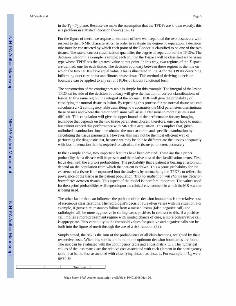
Simply stated, the risk is the sum of the probabilities of all classifications, weighted by theirrespective costs. When this sum is a minimum, the optimum decision boundaries are found.The risk can be evaluated with the contingency table and a loss matrix, Lci. The numericalvalues of the loss matrix are the relative cost associated with each element in the contingencytable, that is, the loss associated with classifying tissue i as tissue c. For example, if Lci weregiven as
True tissue
McVeigh et al. Page 5
Magn Reson Med. Author manuscript; available in PMC 2008 May 26.
NIH
-PA Author Manuscript
NIH
-PA Author Manuscript
NIH
-PA Author Manuscript

i = 1 i = 2 i = 3
Classified tissuec = 1 -2 2 2c = 2 1 -1 0c = 3 1 0 -1
this would imply a high reward for correctly identifying tissue 1 (negative cost), a high penaltyfor misclassifying tissues 2 or 3 as tissue 1, a moderate penalty for misclassifying tissue 1 astissue 2 or 3, no penalty for confusing tissues 2 and 3 with each other, and moderate rewardfor correctly identifying tissues 2 or 3. If tissue 1 is considered the target, this loss matrix willweigh heavily against false-positive calls and more moderately against false-negative calls.The total risk for a given set of decision boundaries is calculated by multiplying each elementof Lci with its corresponding element in the contingency matrix, and then taking the sum ofthese products. The reader is referred elsewhere for a more detailed account of risk functions(32,33).
To estimate the actual performance obtained with MRI, the analysis is best done in the S-space.First, the TPDFs are mapped to the S-space. Second, the resulting SPDFs are convolved withthe Gaussian noise function to give the final SPDFs that we expect to measure with the imager.Third, the same analysis that was applied to the TPDFs can be used to derive the contingencytable and risk function representing the performance of the mapping. The optimizationprocedure is a search for the mapping that produces the risk that comes closest to that derivedfrom the TPDFs.
3. An ExampleSuppose we are presented with the simple problem of detecting a specific lesion in abackground tissue. To derive the optimal MRI protocol for this task the following steps shouldbe followed: (1) Characterize the lesion and the normal tissues found in the patient populationas thoroughly as possible with TPDFs in a tissue parameter space. (2) Set the imagingconstraints, such as maximum scan time, minimum resolution, minimum total volume scanned.(3) Decide upon an appropriate figure of merit for the task. (4) Search for the mapping (i.e.,pulse sequence protocol) that produces the maximum figure of merit.
This process is illustrated for a hypothetical example in Fig. 5. In Fig. 5a two Gaussiandistributions are plotted as 2σ contours in a two-dimensional T-space, T1 × T2. They have thefollowing parameters: tissue A, μT1 = 400, σT1 = 100, μT2 = 40, σT2 = 10, and a correlationcoefficient ρ = 0; tissue B, μT1 = 600, σT1 = 100, μT2 = 60, σT2 = 10, and ρ = 0. In this examplethe a priori probabilities are the same and both distributions are normalized to unity; that is,the integral under the bivariate normal TPDF is 1.0. In Fig. 5b the two TPDFs are mapped intoa one-dimensional S-space by an inversion recovery (IR) sequence with TR = 1100, TE = 35,and TI = 165. (This will be shown to be the optimal IR sequence.) The resulting SPDFs areconvolved with a Gaussian model of image noise to produce the final SPDFs. The method ofderiving this Gaussian was given in Section 2.3. For this example the imaging constraints werea total time of 5 min, a required resolution of 2 × 2 × 5 mm, 20 slices. The SNR at this resolutionwas assumed to be 100.
The figure of merit chosen is the minimum risk of the technique. Assuming equal cost for false-positive and false-negative calls, we define the risk as the sum of the false-positive rate andthe false-negative rate. For each mapping a threshold signal s0 is chosen in the S-space as adecision boundary. This divides the space into two domains: signals above s0 will be calledtissue A, signals below s0 will be called tissue B. For a given value of the threshold signal s0,a 2 × 2 contingency table is calculated and the risk evaluated. The set of all possible mappingsand decision boundaries is searched for the minimum risk. The optimal mapping and decisionboundary is shown in Fig. 5c.
McVeigh et al. Page 6
Magn Reson Med. Author manuscript; available in PMC 2008 May 26.
NIH
-PA Author Manuscript
NIH
-PA Author Manuscript
NIH
-PA Author Manuscript

The IR pulse sequence that produces the maximum SDNR between the points (600, 60) and(400, 40) in the T1 × T2 plane, for the same imaging constraints, is TR = 1100, TE = 15, TI =340. This is significantly different from the optimal IR pulse sequence derived from the riskcalculation. In order to examine why this discrepancy occurs, it is instructive to look at plotsof isosignal contours for each sequence in the T1 × T2 plane. In Fig. 6a we show the SPDFsfor the optimal sequence with the decision boundary marked on the axis. Corresponding tothis, the two TPDFs are shown in the T1 × T2 plane with the isosignal contours of the optimalsequence overlayed. The dashed isosignal contour serves as a good decision boundary for theTPDFs representing tissue A and tissue B. Figure 6b shows this same relationship between theSPDFs and the isosignal contours of the suboptimal sequence that produces the maximumSDNR between the points (600, 60) and (400, 40). Note that the overlap of the SPDFs is greaterfor this sequence than that for the optimal sequence of Fig. 6a. The reason for this isdemonstrated well in the plot of isosignal contours for the suboptimal sequence shown in Fig.6b. While the points (600, 60) and (400, 40) have the maximum SDNR in the suboptimalsequence of Fig. 6b, there does not exist an isosignal contour that acts as a good decisionboundary. This demonstrates why it is important to model the tissues as TPDFs rather thandiscrete points.
One further point can be made with this simple example: in some instances, one can obtainclose to maximum discrimination with far fewer data than are required to calculate the tissueparameters. Recall that the minimum risk one can obtain from any method that depends onT1 and T2 is calculated from the TPDFs in the T-space. For this example, the minimum riskcalculated from the TPDFs is 0.1. This is close to that obtained with the single optimal sequence(in Fig. 6a 0.10 + 0.07 = 0.17).
4. Characteristics of the ModelIn this section some conclusions regarding MRI data acquisition are drawn based on amathematical analysis of this optimization procedure.
4.1. DimensionalityFor each clinical problem there are three fundamental quantities: the number of tissues involved(Nt), the number of parameters used to model these tissues (Np), and the number of pulsesequences used in the protocol (Ns). Naturally, part of the optimization problem is to determinethe most appropriate values for Nt, Np, and Ns. The three dimensions are dependent upon oneanother. This section is devoted to investigating this dependence and how it affects the dataacquisition philosophies.
The appropriate order of operations to determine the dimensions of the optimization problemis as follows.
1. Determine the number of tissues involved in the problem. For example, when lookingfor a lesion in the brain, one would include white matter, gray matter, CSF, and thelesion. This sets Nt = 4.
2. Decide whether one tissue will be treated as the target, or more than one tissue mustbe completely differentiated, and set the loss matrix accordingly. This may affect thenumber of both tissue parameters and pulse sequences needed.
3. Determine which tissue parameters offer the best potential in discriminating the targettissue from the background, or all tissues from each other. This sets the value of Np.
4. Choose the pulse sequences used in the protocol as the mapping from T-space to S-space. This sets Ns.
McVeigh et al. Page 7
Magn Reson Med. Author manuscript; available in PMC 2008 May 26.
NIH
-PA Author Manuscript
NIH
-PA Author Manuscript
NIH
-PA Author Manuscript

4.1.1. Number of tissues, Nt—For a given radiological task, it is usually possible to namethe tissues that will be involved. Attempting to find a lesion against a background of knownnormal tissues is a common case. If the number of tissues in the problem is Nt, including thelesion, there are Nt - 1 discrimination problems to consider. In the best case, one pulse sequencewill supply all of the decision boundaries for the Nt - 1 discrimination problems. The otherextreme situation has Nt tissues, all of which must be discriminated from each other, givingNt(Nt - 1)/2 discrimination problems. For either of these situations, in order to be able to derivethe best protocol, it is necessary to include all Nt tissues in the optimization simultaneously.Breaking the problem up into separate two-tissue problems may simplify the computationalcomplexity, but the optimal solution may then not be obtained.
Most situations will lie between the two extreme cases of a single target tissue and a full Nttissue discrimination problem. This character of the problem is modeled with the loss matrix,Lci as described in Section 2.3.
4.1.2. Number of parameters, Np—After deciding what tissues to include in theoptimization, and their relative importance, one must choose which parameters should be usedto model these tissues. This is an important step because the upper bound of the performanceis set by the amount of overlap of the parameter values described by the TPDFs.
The amount of data needed to determine the TPDF function to within a specified accuracyincreases exponentially with the dimension of the TPDF (32). This has come to be known asthe curse of dimensionality (35). However, only a subset of the complete list of availableparameters is usually needed to attack a given problem. In order to minimize expense, some apriori knowledge should be used when deciding to collect multivariate data for the purpose ofcharacterizing tissues with TPDFs. The parameters chosen should not be highly correlated. Iftwo parameters are highly correlated, one of them can usually be dropped without reducingthe effective dimension of the T-space.
For each tissue parameter in the T-space, there must exist at least one pulse sequence whichexploits differences in that parameter. This pulse sequence should be robust under the day-to-day variations in the operating conditions of the imager and be relatively simple to implement.Unless such a pulse sequence is devised, it may not be worth the effort to add the correspondingdimension to the TPDFs.
Addressing the problem of estimating PDF parameters or constructing parameter free PDFsfrom the collected data is beyond the scope of this paper. The theory for these processes isfound in standard textbooks on pattern recognition and statistical decision theory (32-34). Thebulk of published tissue parameter data cannot be used for fitting multiparametric PDFs.However, suitable data are starting to emerge (10-12,15). For the remainder of this paper it isassumed that the functional form of the TPDFs is known exactly. Optimization with incompleteknowledge of the functional form of the TPDFs will be treated in a subsequent paper.
4.1.3. Number of sequences, Ns—Data acquisition protocols can be divided into twoclasses of mappings: those that have the potential to be a one-to-one mapping and those thatdo not. In the first case a necessary condition is that the number of pulse sequences is greaterthan or equal to the number of tissue parameters in the pulse sequence equations, that is, Ns ≥Np. In the second case, Ns < Np. We deal with these cases separately.
In the context of this model of MRI data acquisition, Ns < Np implies that the mapping is many-to-one; i.e., it contracts the Np-dimensional T-space to an Ns-dimensional S-space. Figure 3shows a two-dimensional T-space (Np = 2) being mapped to a one-dimensional S-space (Ns =1) by an IR pulse sequence. Note that each point in the one-dimensional S-space corresponds
McVeigh et al. Page 8
Magn Reson Med. Author manuscript; available in PMC 2008 May 26.
NIH
-PA Author Manuscript
NIH
-PA Author Manuscript
NIH
-PA Author Manuscript

to a curve in the T-space. Therefore, the SPDF value at a point s0 in the S-space correspondsto the line integral of the TPDF along the contour s = s0 in the T-space. In Fig. 7 we show anexample with Np = 3, Ns = 2. In this case the isosignal contours for each sequence in the T-space are two-dimensional surfaces. The line of intersection of two surfaces, S1 = k1, S2 = k2,corresponds to a point in the S-space. Again, the value of the SPDF at the point (k1, k2) is theline integral of the TPDF in the T-space along the line of intersection of the two surfaces S1 =k1, S2 = k2.
In general, the contour surfaces of individual pulse sequences are (Np - 1)-dimensional loci inthe T-space. The intersections of the contour surfaces for the Ns sequences are loci of dimensionNp - Ns, and the SPDF value at a point in the S-space is the integral of the TPDF on this locus.In the special case of a one-sequence protocol (Ns = 1), one must pay special heed to the decisionboundaries supplied by the pulse sequence isosignal contours in the T-space, for these are theonly ones available. When Ns is greater than one, decision boundaries may be synthesized byalgebraic combination of the images from each sequence.
In the alternative case when Ns ≥ Np, there exists the potential to construct a one-to-one mappingfrom the T-space to the S-space. This is equivalent to a coordinate transformation in the domainof the T-space for which the mapping is one-to-one. An example of this is given in Fig. 8 inwhich the “T1-weighted, T2-weighted” imaging protocol is shown mapping a domain of theT1 × T2 plane (Np = 2) to a range in the S-space; each point in the domain is uniquely definedby a pair of signal values. We can now construct any decision boundary we choose in thisdomain; therefore, the optimum decision boundary derived from the risk function and theTPDFs is available. This optimum decision boundary is, of course, mapped from the T-spaceto the S-space where it may be used directly; however, after the inclusion of noise it may nolonger give optimal performance. When this happens, a new optimal decision boundary mustbe calculated from the SPDFs.
When the number of sequences is greater than the number of tissue parameters, the SPDFs arecontained within an Np-dimensional manifold in the Ns-dimensional S-space. In Fig. 9, forexample, we assume that we have two pulse sequences (Ns = 2) which are dependent upononly one parameter, say T2, and the T-space is one-dimensional. The mapping of the SPDFsin this case is from the one-dimensional T2 axis to a curve in the two-dimensional signal strengthplane. This situation is similar to that of a multiecho sequence. The pixels of an eight-echosequence of images can be considered to be points in an eight-dimensional S-space. However,because the signal strength from echo to echo is only modulated by T2, most of this eight-dimensional space is empty. In fact, if one normalizes out the T1 and N(H) dependence withthe first image of the sequence (assuming TE ≪ T2), the pixels of the remaining sevennormalized images lie near a single one-dimensional curve; excursions from this curve are dueto image noise only. The relative position of the pixel on the curve is determined by the T2value of the voxel it represents.
The locus upon which the image data are contained in the S-space has a simple analyticalexpression. It is an Np-dimensional manifold in the Ns-dimensional space, parameterized bythe Np tissue parameters. This relation is given by Eq. [1]. Thus, it is not surprising that whenprincipal component analysis is performed on any number of pulse sequences which aredependent only on T1 and T2 (N(H) and T1 being highly correlated), nearly all of the informationis contained in the first two components (36). In this case, the T-space is effectively two-dimensional and, therefore, the image information is mostly contained in a two-dimensionalmanifold contained in any S-space of dimension greater than two. (The first two PCA imagesare the basis of a linear manifold; therefore there will be some residual power in higherdimensions, but this is found to be small for the sequences used thus far.) One way of usingthe redundant images to advantage is to decrease the noise in the final displayed image. When
McVeigh et al. Page 9
Magn Reson Med. Author manuscript; available in PMC 2008 May 26.
NIH
-PA Author Manuscript
NIH
-PA Author Manuscript
NIH
-PA Author Manuscript

an S-space is contracted by combining images (for example, weighted addition of the multiechoimages), the random noise level can be reduced with respect to the level of the coherent signal.The maximum gain in SNR available from this kind of contraction is where N is the numberof images used in the contraction.
5. DiscussionThis paper has presented a new approach to optimizing MRI data acquisition protocols fordifferential diagnosis. The main innovation of the method is the movement away from thetraditional methods of optimizing the imaging technology and toward the methods used foroptimizing the diagnostic test. Two sources of previous work are useful in this study, those ofstatistical pattern recognition (32-34) and clinical decision analysis (9). The existingdevelopment of these theories is sufficiently general to apply them to MRI with fewmodifications.
One of the major drawbacks of this method of optimization is its dependence upon a data baseto determine the TPDFs. There are two problems associated with acquiring these data:obtaining truth and the cost. In order to collect tissue parameter data from a specified tissue,one needs to unambiguously classify the tissue from which the data point is collected. Thisrequires an independent method of classifying the tissue that has a very low probability oferror. In MRI, the classification of the normal tissues can be done using anatomical location.For lesions the problem is more complicated, unless there are results from a biopsy or autopsyavailable. These difficulties magnify the importance of publishing tissue parameter data ascorrelated values from the same sample, not as independent means.
The imaging data that are collected using the optimal protocol offer the maximum separationbetween tissues in a mathematical sense. In order to translate this mathematical separation intoa diagnostic improvement, a set of “display” images must be synthesized. The collected imagescan be considered a basis set from which display images are derived; the algorithm thatsynthesizes the display images may be changed according to the needs of the diagnostician.For example, it may be suitable to synthesize an image for each discrimination problem (i.e.,lesion vs white matter, lesion vs gray matter).
The optimization algorithm described in this paper has mathematically separated tissue-dependent signal characteristics in the entire patient population; therefore, any imageprocessing that is done automatically on all of the images should be based on the SPDFs.However, once the data for an individual have been taken, some image processing can be donebased solely on the acquired data. Such processing includes principal component analysis(36,37), the calculation of eigenimages (38), and SDNR maximization between specific regionsof interest (ROIs) with matched filters (39). For example, the radiologist may spot a region ofsuspicion in a background of normal tissue. By defining two ROIs, one for the region ofsuspicion, the other for the known normal tissue, the data in the S-space can be contracted bytaking the sum of the collected images of the slice with weighting coefficients that producemaximum SDNR between the region of suspicion and the background. This summation isbased on the data for this patient only and therefore may differ from that derived from thewhole population. But, it should be noted that the region of suspicion must be found first; it isthe job of the optimized protocol to give the radiologist the best chance at finding this regionof suspicion. Once the region of suspcicion is found, many sophisticated image processing anddata analysis techniques can be used.
McVeigh et al. Page 10
Magn Reson Med. Author manuscript; available in PMC 2008 May 26.
NIH
-PA Author Manuscript
NIH
-PA Author Manuscript
NIH
-PA Author Manuscript

6. ConclusionsA number of conclusions have been drawn throughout this analysis and are summarized forclarity in point form.
1. In order to derive the optimal data acquisition protocol, the natural variability of thetissue parameters must be taken into account.
2. The figure of merit needs to reflect the clinical task, not machine performance. Forthis reason the separability of tissues is a more pertinent figure of merit than SNR orSDNR.
3. The upper bound of separability of tissues with MRI signal values is set by the overlapof the PDFs describing the distribution of tissue parameter values in the patientpopulation. The first stage of any optimization should be the calculation of the PDFsdescribing the tissue parameters.
4. All of the tissues relevant to the clinical problem must be included in the algorithmin order to derive the optimal protocol.
5. The prevalence of disease in the patient population can be accounted for throughnormalization of the TPDFs.
6. The relative importance of differentiating the tissues in the problem can be modeledwith a loss matrix.
7. The effective dimension of the signal strength space is given by the number of tissueparameters upon which the pulse sequences are dependent. That is, all signal strengthvalues will lie upon a manifold in a signal strength space whose dimension isdetermined by the number of tissue parameters (assuming the number of sequencesis greater than or equal to the number of tissue parameters). Additional pulsesequences added to a protocol may stretch this manifold, but they will not increaseits dimension unless a new, independent tissue parameter is introduced.
8. If the TPDFs are known exactly, the image extraction process used to derive thedisplay images from the collected data can be established in advance. Further adaptiveimage processing may be done with human assistance based on what the radiologistfinds in the display image set.
Acknowledgments
The authors thank Art Burgess, Adrian Crawley, and Patrick Goebel for their useful comments. This research wassupported by the Medical Research Council, the National Cancer Institute of Canada, the Ontario Cancer Treatmentand Research Foundation, the Ontario Ministry of Health, and Canadian General Electric Medical Systems. E. R.McVeigh was supported by the Ontario Ministry of Colleges and Universities.
References1. Damadian R. Science 1971;171:1151. [PubMed: 5544870]2. Edelstein WA, Bottomley PA, Hart HR, Smith LS. J. Comput. Assist. Tomogr 1983;7:391. [PubMed:
6841698]3. Wehrli FW, MacFall JR, Glover GH, Grigsby N. Magn. Reson. Imaging 1984;2:3. [PubMed: 6530915]4. Hendrick RE, Nelson TR, Hendee WR. Magn. Reson. Imaging 1984;2:193. [PubMed: 6530926]5. Ortendahl DA, Hylton N, Kaufman L, Watts JC, Crooks LE, Mills CM, Stark DD. Radiology
1984;153:479. [PubMed: 6091173]6. Young IR, Burl M, Bydder GM. J. Comput. Assist. Tomogr 1986;10:271. [PubMed: 3950156]7. Buxton RB, Edelman RR, Rosen BR, Wismer GL, Brady TJ. J. Comput. Assist. Tomogr 1987;11:7.
[PubMed: 3805431]
McVeigh et al. Page 11
Magn Reson Med. Author manuscript; available in PMC 2008 May 26.
NIH
-PA Author Manuscript
NIH
-PA Author Manuscript
NIH
-PA Author Manuscript

8. Weinstein, MC.; Fineberg, IV. Clinical Decision Analysis. Saunders; Philadelphia: 1980.9. Droege RT, Wiener SN, Rzeszotarski MS. Radiology 1984;153:425. [PubMed: 6484175]10. Henkelman RM, Hardy PA, Poon PY, Bronskill MJ. Radiology 1986;161:727. [PubMed: 3786723]11. Herfkens R, Davis P, Crooks L, Kaufman L, et al. Radiology 1981;141:211. [PubMed: 7197379]12. Davis P, Sheldon P, Kaufman L, Crooks L, Margulis AR, Miller T, Watts J, Arakawa J, Hoenninger
J. Cancer 1983;51:433. [PubMed: 6821826]13. Gordon R, Coumans J. Med. Phys 1983;11:79. [PubMed: 6700559]14. Herbert DE. Magn. Reson. Imaging 1986;4:215. [PubMed: 3669933]15. Bronskill, MJ.; Brown, DW.; Yaffe, MJ.; Johns, PC.; Foster, FS.; D’Astous, FT. Magnetic Resonance
in Cancer. Pergamon; Toronto: 1986.16. Ernst RR, Anderson WA. Rev. Sci. Instrum 1966;37:93.17. Bakker CJG, De Graaf CN, van Dijk P. Phys. Med. Biol 1984;29:1511. [PubMed: 6514785]18. van Uijen CMJ, den Boef JH. Magn. Reson. Imaging 1984;1:502.19. Young IR, Bryant DJ, Payne JA. Magn. Reson. Med 1985;2:355. [PubMed: 4094552]20. Hardy PA, Bronskill MJ, Henkelman RM. Med. Phys 1985;12:581. [PubMed: 2995779]21. Sperber GE, Ericsson A, Hemmingsson A, Jung B, Thuomas KA. Magn. Reson. Imaging 1986;3:685.22. Rosen BR, Pykett IL, Brady TJ. J. Comput. Assist. Tomogr 1984;8:195. [PubMed: 6323554]23. Majumdar S, Orphanoudakis SC, Gmitro A, O’Donnell M, Gore J. Magn. Reson. Med 1986;3:397.
[PubMed: 3724419]24. Majumdar S, Orphanoudakis SC, Gmitro A, O’Donnell M, Gore J. Magn. Reson. Med 1986;3:562.
[PubMed: 3747818]25. Crawley AP, Henkelman RM. Magn. Reson. Med 1987;4:34. [PubMed: 3821477]26. Kurtz D, Dwyer A. J. Comput. Assist. Tomogr 1984;8:819. [PubMed: 6470247]27. McVeigh ER, Bronskill MJ, Henkelman RM. Med. Phys 1985;12:586. [PubMed: 4046992]28. Edelstein WA, Bottomley PA, Pfeifer LM. Med. Phys 1984;11:180. [PubMed: 6727793]29. Edelstein WA, Glover GH, Hardy CJ, Redington RW. Magn. Reson. Med 1986;3:604. [PubMed:
3747821]30. Oppelt, A.; Stetter, E.; Loeffler, W. Soc. Magn. Reson. Med. First Annual Meeting; 1981; p. 121
[Abstract book]31. Green, DM.; Swets, JA. Signal Detection Theory and Psychophysics. Krieger; Huntington, NY: 1973.32. Meisel, WS. Computer Oriented Approaches to Pattern Recognition. Academic Press; New York:
1972.33. Fukunaga, K. Introduction to Statistical Pattern Recognition. Academic Press; New York: 1972.34. Duda, RO.; Hart, PE. Pattern Classification and Scene Analysis. Wiley; New York: 1973.35. Bellman, RE. Adaptive Control Processes. Princeton Univ. Press; Princeton: 1961.36. Smith, GD.; Mitchell, MR.; Pickens, DR.; Price, RR.; James, AE, Jr. Soc. Magn. Reson. Med. 5th
Annual Meeting; 1986; p. 227[Abstract book]37. Schmiedl U, Ortendahl DA, Mark AS, Berry I, Kaufman L. Magn. Reson. Med 1987;4:471. [PubMed:
3600253]38. Miller JWV, Windham JP, Kwatra SC. IEEE Trans. Med. Imaging MI 1984;3:116.39. Riederer SJ, Hall AL, Maier JK, Pelc NJ, Enzmann DR. Med. Phys 1983;10(2):209. [PubMed:
6346033]
McVeigh et al. Page 12
Magn Reson Med. Author manuscript; available in PMC 2008 May 26.
NIH
-PA Author Manuscript
NIH
-PA Author Manuscript
NIH
-PA Author Manuscript

Fig. 1.Three bivariate normal TPDFs in the T1 × T2 plane. These distributions were derived from datameasured in vitro for fat, fibrous, and infiltrating duct carcinoma tissues.
McVeigh et al. Page 13
Magn Reson Med. Author manuscript; available in PMC 2008 May 26.
NIH
-PA Author Manuscript
NIH
-PA Author Manuscript
NIH
-PA Author Manuscript

Fig. 2.Two bivariate normal TPDFs with 2σ perimeters shown as dashed lines are mapped from atwo-dimensional tissue parameter space (T-space) to a one-dimensional signal strength space(S-space) with an IR pulse sequence. The signal strengths are in units of the root mean squaredeviation (RMSD) of the image noise.
McVeigh et al. Page 14
Magn Reson Med. Author manuscript; available in PMC 2008 May 26.
NIH
-PA Author Manuscript
NIH
-PA Author Manuscript
NIH
-PA Author Manuscript

Fig. 3.(a) Two bivariate normal distributions in T-space (T1 × T2), with three isosignal contours froman IR pulse sequence overlayed. (b) Each contour maps to a single signal strength value asshown in the plot of the S-space. It is demonstrated in both the T-space and the S-space thatthe signal value s = s0 acts as a good decision boundary between tissue A and tissue B.
McVeigh et al. Page 15
Magn Reson Med. Author manuscript; available in PMC 2008 May 26.
NIH
-PA Author Manuscript
NIH
-PA Author Manuscript
NIH
-PA Author Manuscript

Fig. 4.TPDFs for fibrous breast tissue (bottom) and infiltrating duct carcinoma (top) are shown asisoprobability density contours. The dashed line is the decision boundary derived from theseTPDFs when the costs of both types of error (false-positive and false-negative) are identical.
McVeigh et al. Page 16
Magn Reson Med. Author manuscript; available in PMC 2008 May 26.
NIH
-PA Author Manuscript
NIH
-PA Author Manuscript
NIH
-PA Author Manuscript

Fig. 5.The method of optimization shown in three steps. (a) The model of the tissues. (b) The mappingto an S-space and the incorporation of image noise. (Note: the Gaussian is scaled down fordisplay purposes.) (c) Selection of the decision boundary and risk calculation.
McVeigh et al. Page 17
Magn Reson Med. Author manuscript; available in PMC 2008 May 26.
NIH
-PA Author Manuscript
NIH
-PA Author Manuscript
NIH
-PA Author Manuscript

Fig. 6.The SPDFs and associated TPDFs for two sequences. The TPDFs have isosignal contours ofthe sequence overlayed. The sequence shown in (a) has the minimum risk, the sequence in (b)has the maximum SDNR between the means of the TPDFs. Note the poor decision boundariesprovided by sequence (b).
McVeigh et al. Page 18
Magn Reson Med. Author manuscript; available in PMC 2008 May 26.
NIH
-PA Author Manuscript
NIH
-PA Author Manuscript
NIH
-PA Author Manuscript

Fig. 7.A schematic of a mapping for which Ns < Np. In this case the mapping is from a three-dimensional T-space to a two-dimensional S-space. The TPDF is shown as an ellipsoid withisosignal surfaces intersecting it. One isosignal surface from each pulse sequence is shown.The integral of the TPDF along the line of intersection of the two surfaces gives the value ofthe SPDF at a point in the two-dimensional S-space. The SPDF is shown as isoprobabilitydensity contours in the S-space.
McVeigh et al. Page 19
Magn Reson Med. Author manuscript; available in PMC 2008 May 26.
NIH
-PA Author Manuscript
NIH
-PA Author Manuscript
NIH
-PA Author Manuscript

Fig. 8.A one-to-one mapping from a two-dimensional T-space (T1 × T2) to a two-dimensional S-space(spin-echo, T1-weighted × T2-weighted). (a) The isosignal contours draw a grid over a regionin the T-space. (b) The same region of T-space as that in (a) mapped into the S-space. The gridshown in the S-space is equivalent to a set of curvilinear coordinates.
McVeigh et al. Page 20
Magn Reson Med. Author manuscript; available in PMC 2008 May 26.
NIH
-PA Author Manuscript
NIH
-PA Author Manuscript
NIH
-PA Author Manuscript

Fig. 9.A mapping for which Ns > Np. In this case the mapping is from a one-dimensional T-space(T2) to a two-dimensional S-space (spin-echo, TE = k, TE = 3k). Note that all of the signalstrenghts will be confined to the curve .
McVeigh et al. Page 21
Magn Reson Med. Author manuscript; available in PMC 2008 May 26.
NIH
-PA Author Manuscript
NIH
-PA Author Manuscript
NIH
-PA Author Manuscript