ICU Protocols
613
123 A Step-wise Approach, Vol I Rajesh Chawla Subhash Todi Editors Second Edition ICU Protocols
-
Upload
khangminh22 -
Category
Documents
-
view
1 -
download
0
Transcript of ICU Protocols
123
A Step-wise Approach, Vol I
Rajesh Chawla Subhash TodiEditors
Second Edition
ICU Protocols

ICU Protocols
An endeavour of Indian College of Critical Care Medicine under the auspices of Indian Society of Critical Care Medicine (ISCCM).
Rajesh Chawla • Subhash TodiEditors
ICU ProtocolsA Step-wise Approach, Vol I
Second Edition
EditorsRajesh ChawlaDepartment of Respiratory, Critical Care and Sleep MedicineIndraprastha Apollo HospitalsNew DelhiIndia
Subhash TodiA.M.R.I. Hospital Critical Care DepartmentKolkataWest BengalIndia
ISBN 978-981-15-0897-4 ISBN 978-981-15-0898-1 (eBook)https://doi.org/10.1007/978-981-15-0898-1
© Springer Nature Singapore Pte Ltd. 2020This work is subject to copyright. All rights are reserved by the Publisher, whether the whole or part of the material is concerned, specifically the rights of translation, reprinting, reuse of illustrations, recita-tion, broadcasting, reproduction on microfilms or in any other physical way, and transmission or infor-mation storage and retrieval, electronic adaptation, computer software, or by similar or dissimilar methodology now known or hereafter developed.The use of general descriptive names, registered names, trademarks, service marks, etc. in this publica-tion does not imply, even in the absence of a specific statement, that such names are exempt from the relevant protective laws and regulations and therefore free for general use.The publisher, the authors, and the editors are safe to assume that the advice and information in this book are believed to be true and accurate at the date of publication. Neither the publisher nor the authors or the editors give a warranty, expressed or implied, with respect to the material contained herein or for any errors or omissions that may have been made. The publisher remains neutral with regard to jurisdictional claims in published maps and institutional affiliations.
This Springer imprint is published by the registered company Springer Nature Singapore Pte Ltd.The registered company address is: 152 Beach Road, #21-01/04 Gateway East, Singapore 189721, Singapore

To my parents, wife Renu, and children Ankit, Aakanksha and Aakriti for their unconditional love and support. Special thanks to Dr. Sudha Kansal, Dr. Roseleen Bali, my students, residents, fellows, and colleagues who inspire and educate me.
—Rajesh Chawla
To my mother, my wife Shailja, and daughter Suchira for their understanding, tolerance, and patience shown during the gestational period of this manual.
—Subhash Todi
vii
Preface
It gives us immense pleasure to present to you the second edition of ICU Protocols: A Stepwise Approach, an official publication of the Indian Society of Critical Care Medicine (ISCCM).
The first edition was published in 2012 under the same editorship. Concepts and evidence-based bedside practices in critical care medicine have further evolved since the first edition, and it was thought that modification and updating of the book was needed. The basic tenet of the ICU protocol book remains the same, i.e. to pro-vide residents, fellows, critical care practitioners and allied health care profession-als with a current and comprehensive stepwise approach for bedside diagnosis and management of the most frequently encountered problems in the intensive care unit (ICU). To prevent the manual from becoming voluminous, we have not gone into the details of the epidemiology and pathophysiology of each condition and restricted ourselves to practice points helpful to clinicians. The format of the book consisted of introductory case scenario, bullet points, stepwise approach, flow charts, ample number of tables and figures for easy readability in each chapter. We have received positive feedback on the content and presentation of the chapters from our readers over the last 5 years. We have also collected feedback and suggestions from them on modifications, which are incorporated in the present edition.
There are some major changes that have been incorporated in the second edition. This edition is published in two volumes for increasing portability and allowing space for new chapters. Five new chapters have been added, namely High Flow Nasal Cannula, Antibiotic Pearls, CRBSI, Posttracheostomy Care in ICU and Research Methodology. Addition of these chapters became necessary due to the advancement of medical technology and increasing importance of research knowl-edge need for clinicians. All the chapters from the first edition have been incorpo-rated with an updated version. For this multiauthor book, authors were carefully chosen for their expertise in the subject matter. In keeping with the multidisciplinary nature of our speciality, authorship also included non-intensivists like infectious disease specialists and gastroenterologists. Each chapter has been thoroughly dis-cussed by both the editors, especially those referring to newer practices from Uptodate reference manual. Specifically, “Suggested Reading” sections with anno-tations have been updated with new references over the past 5 years, and current websites on the subject are added. Chapters on “Procedures” have also been revised to ensure correctness. The “Appendix” section has been thoroughly revised with
viii
special emphasis on “Dosing” section in which doses of newer drugs like antibiotics have been added. A new appendix on “Glossary of Statistical Terms” has been added to make the reader familiar with ever increasing and sometimes confusing statistical terms. Formulae and equations have been updated wherever needed.
We sincerely hope that the second edition will give a new flavour to the much appreciated first edition and will serve its objective of improving bedside patient care by updating critical care practitioners. At the same time, we also realise that the field of critical care, like everything else, is not static but changes constantly and further modifications of this book will be needed in future.
ISCCM has been in the forefront of critical care education in India. This is an important educational venture of ISCCM, and we hope the book will be read not only in India but also regionally and internationally. Last but not least, we sincerely hope that this manual will be used by residents, wherever they are, for better bedside care of critically ill patients.
New Delhi, Delhi, India Rajesh Chawla Kolkata, West Bengal, India Subhash Todi
Preface
ix
Acknowledgements
We would like to sincerely thank all the chapter authors who contributed their name and expertise in this endeavour. It would not have been possible to produce this manual without the hard work and support received from all of them. A special thanks to them for allowing editors the liberty to edit the chapters freely in order to maintain uniformity throughout the book. We would like to thank the Executive Committee of ISCCM for their unconditional support and patience during the long gestational period of this book. We would like to thank the experts who took the time and trouble to review the chapters and provide us with their inputs. We would like to acknowledge the hard work of all the fellows at Indraprastha Apollo Hospitals (Delhi) and AMRI Hospitals (Kolkata), who read all chapters and gave their inputs from the end users’ perspective.
Special thanks to editorial team of Springer who have supported this medical project. We particularly thank Dr. Naren Aggarwal for multiple helpful suggestions and support throughout the process of finalisation of this book.
Also, we would like to give our special thanks to Mr. Vijay Prakash, Mr. Tapas Kayal, Mr. Bhagwan Dass, and Ms. Rajni for their assistance in the office work and completing the manuscript.

xi
Contents
Part I Respiratory System
1 Airway Management . . . . . . . . . . . . . . . . . . . . . . . . . . . . . . . . . . . . . . . . 3Sheila Nainan Myatra, Nirmalyo Lodh, and Jigeeshu V. Divatia
2 Acute Respiratory Failure . . . . . . . . . . . . . . . . . . . . . . . . . . . . . . . . . . . . 19Randeep Guleria, Jaya Kumar, and Rajesh Chawla
3 Noninvasive Positive-Pressure Ventilation . . . . . . . . . . . . . . . . . . . . . . . 25Rajesh Chawla and Subhash Todi
4 High Flow Nasal Canula (HFNC) . . . . . . . . . . . . . . . . . . . . . . . . . . . . . . 35Rajesh Chawla and Subhash Todi
5 Basic Mechanical Ventilation . . . . . . . . . . . . . . . . . . . . . . . . . . . . . . . . . . 45Gopi Chand Khilnani and Vijay Hadda
6 Mechanical Ventilation in Acute Respiratory Distress Syndrome . . . . 55Farhad N. Kapadia and Umakant Bhutada
7 Management of Refractory Hypoxemia in Acute Respiratory Distress Syndrome . . . . . . . . . . . . . . . . . . . . . . . . . . . . . . . . 63Samir Jog, Divyesh Patel, and Massimo Antonelli
8 Mechanical Ventilation in Obstructive Airway Diseases . . . . . . . . . . . . 73Raj Kumar Mani
9 Weaning . . . . . . . . . . . . . . . . . . . . . . . . . . . . . . . . . . . . . . . . . . . . . . . . . . . 79Rajesh Chawla, Sudha Kansal, Roseleen Kaur Bali, and Aakanksha Chawla Jain
10 Massive Hemoptysis . . . . . . . . . . . . . . . . . . . . . . . . . . . . . . . . . . . . . . . . . 91Avdhesh Bansal and Ravi Shekhar
11 Pulmonary Thromboembolism . . . . . . . . . . . . . . . . . . . . . . . . . . . . . . . . 99Rajesh Chawla and Subhash Todi
12 Severe Community-Acquired Pneumonia . . . . . . . . . . . . . . . . . . . . . . . 111Khalid Khatib, Subhal Dixit, Rajesh Chawla, and Subhash Todi

xii
13 Ventilator-Associated Pneumonia . . . . . . . . . . . . . . . . . . . . . . . . . . . . . . 119Rajesh Pande and Vikas Maurya
14 Pleural Diseases . . . . . . . . . . . . . . . . . . . . . . . . . . . . . . . . . . . . . . . . . . . . . 129Sudha Kansal and Rajesh Chawla
15 Sleep-Disordered Breathing . . . . . . . . . . . . . . . . . . . . . . . . . . . . . . . . . . . 139Jagdish Chander Suri and Tejas M. Suri
16 Oxygen Therapy . . . . . . . . . . . . . . . . . . . . . . . . . . . . . . . . . . . . . . . . . . . . 147Pradip Kumar Bhattacharya and Lata Bhattacharya
17 Pulse Oximetry and Capnography . . . . . . . . . . . . . . . . . . . . . . . . . . . . . 161Deepak Govil, Sachin Gupta, and Ashish Srivastava
Part II Cardiovascular System
18 Hemodynamic Monitoring . . . . . . . . . . . . . . . . . . . . . . . . . . . . . . . . . . . . 173Sheila Nainan Myatra and Jigeeshu V. Divatia
19 Echocardiography . . . . . . . . . . . . . . . . . . . . . . . . . . . . . . . . . . . . . . . . . . . 187Rahul Pandit
20 Fluid Therapy, Vasopressors, and Inotropes . . . . . . . . . . . . . . . . . . . . . 197Jigeeshu V. Divatia and Sheila Nainan Myatra
21 Cardiorespiratory Arrest . . . . . . . . . . . . . . . . . . . . . . . . . . . . . . . . . . . . . 207Sheila Nainan Myatra and Amol T. Kothekar
22 Cardiogenic Shock . . . . . . . . . . . . . . . . . . . . . . . . . . . . . . . . . . . . . . . . . . 217Ashit V. Hegde, Khusrav Bajan, and Subhash Todi
23 Acute Heart Failure . . . . . . . . . . . . . . . . . . . . . . . . . . . . . . . . . . . . . . . . . 223Suresh Ramasubban and Rajeeve Kumar Rajput
24 Cardiac Arrhythmias . . . . . . . . . . . . . . . . . . . . . . . . . . . . . . . . . . . . . . . . 233Supradip Ghosh
25 Acute Coronary Syndromes . . . . . . . . . . . . . . . . . . . . . . . . . . . . . . . . . . . 247Rajesh Rajani, Farhad N. Kapadia, and Nagarajan Ramakrishnan
26 Hypertensive Urgencies and Emergencies . . . . . . . . . . . . . . . . . . . . . . . 259Vinayak Agrawal and Yatin Mehta
Part III Nervous System
27 Acute Ischemic Stroke . . . . . . . . . . . . . . . . . . . . . . . . . . . . . . . . . . . . . . . 271Vinit Suri and Kunal Suri
28 Spontaneous Intracerebral Hemorrhage . . . . . . . . . . . . . . . . . . . . . . . . 285V. Dedeepiya Devaprasad and Nagarajan Ramakrishnan
Contents

xiii
29 Subarachnoid Hemorrhage . . . . . . . . . . . . . . . . . . . . . . . . . . . . . . . . . . . 291Shiva Kumar Iyer, Jignesh Shah, and Kiran Vadapalli
30 Status Epilepticus . . . . . . . . . . . . . . . . . . . . . . . . . . . . . . . . . . . . . . . . . . . 301Rajesh Chawla and Chirag Madan
31 Acute Flaccid Paralysis . . . . . . . . . . . . . . . . . . . . . . . . . . . . . . . . . . . . . . . 309Charu Gauba, Mukul Varma, and Pooja Chopra
32 Coma . . . . . . . . . . . . . . . . . . . . . . . . . . . . . . . . . . . . . . . . . . . . . . . . . . . . . 319Kayanoosh Kadapatti and Shiva Kumar Iyer
33 Intracranial Pressure Monitoring and Management . . . . . . . . . . . . . . 327Rajesh Chawla, Rajagopal Senthilkumar, and Nagarajan Ramakrishnan
34 Acute Febrile Encephalopathy . . . . . . . . . . . . . . . . . . . . . . . . . . . . . . . . . 339Jignesh Shah and Shiva Kumar Iyer
35 Sedation and Analgesia . . . . . . . . . . . . . . . . . . . . . . . . . . . . . . . . . . . . . . . 347Sheila Nainan Myatra and Nishanth Baliga
36 Brain Death . . . . . . . . . . . . . . . . . . . . . . . . . . . . . . . . . . . . . . . . . . . . . . . . 361Babu K. Abraham and Nagarajan Ramakrishnan
Part IV Gastrointestinal system
37 Upper Gastrointestinal Bleeding . . . . . . . . . . . . . . . . . . . . . . . . . . . . . . . 375Rupa Banerjee and Duvvur Nageshwar Reddy
38 Lower Gastrointestinal Bleeding . . . . . . . . . . . . . . . . . . . . . . . . . . . . . . . 383Surinder S. Rana and Deepak Kumar Bhasin
39 Acute Diarrhea . . . . . . . . . . . . . . . . . . . . . . . . . . . . . . . . . . . . . . . . . . . . . 393Mahesh Kumar Goenka and Shivaraj Afzalpurkar
40 Acute Abdominal Distension . . . . . . . . . . . . . . . . . . . . . . . . . . . . . . . . . . 401Neha Berry, Mohd. Talha Noor, and Rakesh Kochhar
41 Intra-abdominal Hypertension . . . . . . . . . . . . . . . . . . . . . . . . . . . . . . . . 407Rajesh Chawla, Sananta K. Dash, and Aakanksha Chawla Jain
42 Acute Pancreatitis . . . . . . . . . . . . . . . . . . . . . . . . . . . . . . . . . . . . . . . . . . . 417Ajay Kumar and Akshat Kumar
43 Acute Liver Failure . . . . . . . . . . . . . . . . . . . . . . . . . . . . . . . . . . . . . . . . . . 427Shalimar and Subrat Kumar Acharya
44 Acute Decompensation in Chronic Liver Failure. . . . . . . . . . . . . . . . . . 435Ajay Kumar and Deepak Amarapurkar
45 Nutrition Support . . . . . . . . . . . . . . . . . . . . . . . . . . . . . . . . . . . . . . . . . . . 443Pravin Amin
Contents
xiv
Part V Renal System
46 Acute Kidney Injury . . . . . . . . . . . . . . . . . . . . . . . . . . . . . . . . . . . . . . . . . 457Subhash Todi and Arghya Majumdar
47 Renal Replacement Therapy . . . . . . . . . . . . . . . . . . . . . . . . . . . . . . . . . . 469Sunil Prakash, Arghya Majumdar, and Bhanu Mishra
48 Managing a Patient on Dialysis . . . . . . . . . . . . . . . . . . . . . . . . . . . . . . . . 477Arghya Majumdar and Raj Kumar Mani
49 Drug Dosing in Renal Failure in ICU . . . . . . . . . . . . . . . . . . . . . . . . . . . 485Sanjiv Jasuja, Pratik Das, Arghya Majumdar, and Gaurav Sagar
Part VI Infection Diseases
50 General Measures of Infection Control . . . . . . . . . . . . . . . . . . . . . . . . . 493Sheila Nainan Myatra and Jacob George Pulinilkunnathil
51 Antibiotic Stewardship . . . . . . . . . . . . . . . . . . . . . . . . . . . . . . . . . . . . . . . 505Subhash Todi and Rajesh Chawla
52 Septic Shock . . . . . . . . . . . . . . . . . . . . . . . . . . . . . . . . . . . . . . . . . . . . . . . . 511Suresh Ramasubban
53 Severe Tropical Infections . . . . . . . . . . . . . . . . . . . . . . . . . . . . . . . . . . . . 519Dhruva Chaudhry, Diksha Tyagi, and Sushmitha Jakka
54 New-Onset Fever . . . . . . . . . . . . . . . . . . . . . . . . . . . . . . . . . . . . . . . . . . . . 533Subhash Todi and Rajesh Chawla
55 Fungal Infections . . . . . . . . . . . . . . . . . . . . . . . . . . . . . . . . . . . . . . . . . . . . 539Subhash Todi and Rajesh Chawla
56 Infections in the Immunocompromised Host . . . . . . . . . . . . . . . . . . . . . 547Subhash Todi
57 Central Line Related Blood Stream Infections (CRBSI) . . . . . . . . . . . 553Srinivas Samavedam, Ramakrishna Reddy, and Rajesh Pande
58 Antibiotic Pearls: A Case Based Discussion . . . . . . . . . . . . . . . . . . . . . . 563Subhash Todi, Rajesh Chawla, and Subramanian Swaminathan
Appendix A . . . . . . . . . . . . . . . . . . . . . . . . . . . . . . . . . . . . . . . . . . . . . . . . . . . . 571
Appendix B . . . . . . . . . . . . . . . . . . . . . . . . . . . . . . . . . . . . . . . . . . . . . . . . . . . . 595
Appendix C . . . . . . . . . . . . . . . . . . . . . . . . . . . . . . . . . . . . . . . . . . . . . . . . . . . . 605
Appendix D: Syllabus for ICU Training . . . . . . . . . . . . . . . . . . . . . . . . . . . . . 611
Glossary of Statistical Terms . . . . . . . . . . . . . . . . . . . . . . . . . . . . . . . . . . . . . . 617
Index . . . . . . . . . . . . . . . . . . . . . . . . . . . . . . . . . . . . . . . . . . . . . . . . . . . . . . . . . . 623
Contents
xv
Babu K. Abraham Department of Critical Care Medicine, Apollo Hospitals, Chennai, India
Subrat Kumar Acharya Department of Gastroenterology, All India Institute of Medical Sciences, New Delhi, India
Shivaraj Afzalpurkar Institute of Gastrosciences, Apollo Gleneagles Hospitals, Kolkata, India
Vinayak Agrawal Department of Cardiology, Medanta, The Medicity, Gurugram, India
Deepak Amarapurkar (Deceased) Department of Gastroenterology, Bombay Hospital & Medical Research Centre, Breach Candy Medical Research Centre, Mumbai, India
Pravin Amin Department of Internal Medicine and Critical Care, Bombay Hospital Institute of Medical Sciences, Mumbai, India
Massimo Antonelli Department of Critical Care, Fondazione Policlinico Universitario A. Gemelli—Università Cattolica del Sacro Cuore Largo Agostino Gemelli, Rome, Italy
Khusrav Bajan Emergency Department, P.D. Hinduja Hospital and Medical Research Centre, Mumbai, India
Roseleen Kaur Bali Department of Respiratory, Critical Care & Sleep Medicine, Indraprastha Apollo Hospitals, New Delhi, India
Nishanth Baliga Department of Anaesthesia, Critical Care and Pain, Tata Memorial Hospital, Homi Bhabha National Institute, Mumbai, India
Rupa Banerjee Department of Gastroenterology, Asian Institute of Gastroenterology, Hyderabad, India
Avdhesh Bansal Department of Respiratory, Critical Care & Sleep Medicine, Indraprastha Apollo Hospitals, New Delhi, India
Neha Berry Institute of Digestive and Liver Diseases, BLK Hospital, New Delhi, India
Contributors
xvi
Deepak Kumar Bhasin Department of Gastroenterology, Postgraduate Institute of Medical Education & Research, Chandigarh, India
Pradip Kumar Bhattacharya Department of Trauma & Emergency Care Services, Rajendra Institute of Medical Sciences (RIMS), Ranchi, Jharkhand, India
Lata Bhattacharya Department of Anaesthesia, Jawaharlal Nehru Cancer Hospital, Bhopal, Madhya Pradesh, India
Umakant Bhutada Department of Medicine & Intensive Care, P.D. Hinduja National Hospital and Medical Research Centre, Mumbai, India
Dhruva Chaudhry Department of Pulmonary and Critical Care Medicine, Pt. B. D. Sharma Post Graduate Institute of Medical Sciences, Rohtak, Haryana, India
Rajesh Chawla Department of Respiratory, Critical Care & Sleep Medicine, Indraprastha Apollo Hospitals, New Delhi, India
Pooja Chopra Department of Critical Care Medicine, Fortis Memorial Research Institute, Gurugram, Haryana, India
Pratik Das Department of Nephrology, Rabindranath Tagore Hospital, Kolkata, West Bengal, India
Sananta K. Dash Department of Critical Care Medicine, Royal Hobart Hospital, Hobart, TAS, Australia
V. Dedeepiya Devaprasad Department of Critical Care Medicine, Apollo Hospitals, Chennai, India
Jigeeshu V. Divatia Department of Anaesthesia, Critical Care and Pain, Tata Memorial Hospital, Mumbai, India
Subhal Dixit Department of Critical Care, Sanjeevan Hospital, Pune, India
MJM Hospital, Pune, India
Charu Gauba Department of Neurology, Indraprastha Apollo Hospitals, New Delhi, India
Supradip Ghosh Department of Critical Care Medicine, Fortis-Escorts Hospital, Faridabad, India
Mahesh Kumar Goenka Institute of Gastrosciences, Apollo Gleneagles Hospitals, Kolkata, India
Deepak Govil Medanta Institute of Critical Care, Medanta—The Medicity Hospital, Gurgaon, India
Randeep Guleria Department of Pulmonary Medicine, All India Institute of Medical Sciences, New Delhi, India
Sachin Gupta Narayana Superspeciality Hospital, Gurgaon, India
Contributors
xvii
Vijay Hadda Department of Pulmonary Medicine, All India Institute of Medical Sciences, Delhi, India
Ashit V. Hegde Department of Medicine and Critical Care, P.D. Hinduja National Hospital & Medical Research Centre, Mumbai, India
Shiva Kumar Iyer Department of Intensive Care and Critical Care Medicine, Bharati Vidyapeeth (DTU) Medical College, Pune, India
Aakanksha Chawla Jain Department of Respiratory, Critical Care & Sleep Medicine, Indraprastha Apollo Hospitals, New Delhi, India
Sushmitha Jakka Department of Pulmonary and Critical Care Medicine, Pt. B. D. Sharma Post Graduate Institute of Medical Sciences, Rohtak, Haryana, India
Sanjiv Jasuja Department of Nephrology, Indraprastha Apollo Hospital, New Delhi, India
Samir Jog Department of Critical Care, Deenanath Mangeshkar Hospital and Research Center, Pune, India
Kayanoosh Kadapatti Department of Critical Care, Jehangir Hospital, Pune, India
Sudha Kansal Department of Respiratory Medicine and Critical Care, Indraprastha Apollo Hospitals, New Delhi, India
Farhad N. Kapadia Department of Medicine and Intensive Care, P.D. Hinduja National Hospital and Medical Research Centre, Mumbai, India
Khalid Khatib Department of Medicine, Smt. Kashibai Navale Medical College, Pune, India
Gopi Chand Khilnani PSRI Institute of Pulmonary, Critical Care and Sleep Medicine, PSRI Hospital, New Delhi, India
Rakesh Kochhar Department of Gastroenterology, Postgraduate Institute of Medical Education and Research, Chandigarh, India
Amol T. Kothekar Department of Anaesthesia, Critical Care and Pain, Tata Memorial Hospital, Homi Bhabha National Institute, Mumbai, India
Jaya Kumar Department of Pulmonary Medicine, All India Institute of Medical Sciences, New Delhi, India
Ajay Kumar Department of Gastroenterology & Hepatology, BLK Hospital, New Delhi, India
Akshat Kumar Saint Peters University Hospital/Rutgers University, New Brunswick, NJ, USA
Nirmalyo Lodh Department of Anaesthesia, Critical Care and Pain, Tata Memorial Hospital, Mumbai, India
Contributors
xviii
Chirag Madan Department of Respiratory, Critical Care & Sleep Medicine, Indraprastha Apollo Hospitals, New Delhi, India
Arghya Majumdar Department of Nephrology, A.M.R.I. Hospitals, Kolkata, West Bengal, India
Raj Kumar Mani Department of Pulmonology and Critical Care, Batra Hospital and Medical Research Centre, New Delhi, India
Vikas Maurya Department of Respiratory Medicine & Intervention Pulmonology, Fortis Hospital, Shalimarbagh, New Delhi, India
Yatin Mehta Department of Anaesthesia and Critical Care, Medanta, The Medicity, Gurugram, India
Bhanu Mishra Department of Nephrology and Renal Transplant, B L Kapur Superspeciality Institute, Delhi, India
Sheila Nainan Myatra Department of Anaesthesia, Critical Care and Pain, Tata Memorial Hospital, Homi Bhabha National Institute, Mumbai, India
Mohd. Talha Noor Department of Gastroenterology, Postgraduate Institute of Medical Education and Research, Chandigarh, India
Rajesh Pande Department of Critical Care, BLK Superspeciality Hospital, New Delhi, India
Rahul Pandit Department of Intensive Care, Fortis Hospital, Mumbai, India
Divyesh Patel Department of Critical Care, Tricolour Hospital, Vadodara, India
Sunil Prakash Department of Nephrology and Renal Transplant, B L Kapur Superspeciality Institute, Delhi, India
Jacob George Pulinilkunnathil Department of Anaesthesia, Critical Care and Pain, Tata Memorial Hospital, Mumbai, India
Rajesh Rajani P.D. Hinduja Hospital and Medical Research Centre, Mumbai, India
Rajeeve Kumar Rajput Department of Cardiology, Indraprastha Apollo Hospitals, New Delhi, India
Nagarajan Ramakrishnan Department of Critical Care Medicine, Apollo Hospitals, Chennai, India
Suresh Ramasubban Department of Respiratory, Critical Care and Sleep Medicine, Apollo Gleneagles Hospital, Kolkata, India
Surinder S. Rana Department of Gastroenterology, Postgraduate Institute of Medical Education & Research, Chandigarh, India
Duvvur Nageshwar Reddy Department of Gastroenterology, Asian Institute of Gastroenterology, Hyderabad, India
Ramakrishna Reddy Department of Critical Care, Virinchi Hospitals, Hyderabad, India
Contributors
xix
Gaurav Sagar Department of Nephrology, Indraprastha Apollo Hospital, New Delhi, India
Srinivas Samavedam Department of Critical Care, Virinchi Hospitals, Hyderabad, India
Rajagopal Senthilkumar Department of Critical Care Medicine, Apollo Hospitals, Chennai, India
Jignesh Shah Department of Intensive Care and Critical Care Medicine, Bharati Vidyapeeth (DTU) Medical College, Pune, India
Shalimar Department of Gastroenterology, All India Institute of Medical Sciences, New Delhi, India
Ravi Shekhar Department of Pulmonology and Sleep Medicine, Fortis Hospital, Faridabad, Haryana, India
Ashish Srivastava Medanta Institute of Critical Care, Medanta—The Medicity Hospital, Gurgaon, India
Jagdish Chander Suri Department of Pulmonary, Critical Care and Sleep Medicine, Medeor JCS Institute, New Delhi, India
Tejas M. Suri Department of Pulmonary, Critical Care and Sleep Medicine, AIIMS, New Delhi, India
Vinit Suri Department of Neurosciences, Indraprastha Apollo Hospitals, New Delhi, India
Kunal Suri Department of Medicine, Gitanjali Medical College, Udaipur, India
Subramanian Swaminathan Department of Infectious Diseases, Global Hospital, Chennai, India
Subhash Todi Department of Critical Care & Emergency Medicines, A.M.R.I. Hospital, Kolkata, India
Diksha Tyagi Department of Pulmonary and Critical Care Medicine, Pt. B. D. Sharma Post Graduate Institute of Medical Sciences, Rohtak, Haryana, India
Kiran Vadapalli Department of Critical Care Medicine, Bharati Vidyapeeth Medical College, Pune, India
Mukul Varma Department of Neurology, Indraprastha Apollo Hospitals, New Delhi, India
Contributors
Part I
Respiratory System

3© Springer Nature Singapore Pte Ltd. 2020R. Chawla, S. Todi (eds.), ICU Protocols, https://doi.org/10.1007/978-981-15-0898-1_1
S. N. Myatra (*) · N. Lodh · J. V. Divatia Department of Anaesthesia, Critical Care and Pain, Tata Memorial Hospital, Mumbai, India
1Airway Management
Sheila Nainan Myatra, Nirmalyo Lodh, and Jigeeshu V. Divatia
Tracheal intubation is one of the most commonly performed procedures in the ICU. In the ICU, unlike in the operating room with controlled conditions, a signifi-cant proportion of these procedures can be associated with life-threatening compli-cations. This chapter gives a stepwise approach to airway management in the ICU, along with a detailed description of the preparation, assessment, procedure, precau-tions, maintenance, and complications associated with tracheal intubation.
Step 1: Be Prepared for Airway Management Before Patient Arrival
• History from the treating team will tell you about the condition of the patient and give you some idea of the equipment and expertise needed for airway manage-ment (e.g., mental state, respiratory and hemodynamic status, time of last meal, comorbidities that might complicate airway management etc.)
• Check oxygen source, availability of a properly working suction, airway tray/cart, monitors, drugs, and personal protection equipment are kept ready.
A 60-year-old morbidly obese diabetic male patient with a left lobar pneumonia was shifted from the ward to the intensive care unit (ICU). He had history of progressive breathlessness and altered mental status for 6 h. He was drowsy but arousable, and had a respiratory rate of 33 breaths/min. SpO2 was 92% with facemask using 6 L of oxygen per min. Heart rate was 110/min and blood pres-sure was 90/60 mmHg.
4
Step 2: History and Initial Stabilisation
• The incidence of complications during intubation in critically ill patients in the ICU ranges from 22% to 54%, significantly higher than the operating room, making tracheal intubation in ICU a high risk procedure.
• Provide oxygen therapy on arrival to ICU while evaluating the patient. Attach the cardiac monitor, noninvasive blood pressure, and pulse oximeter and secure an intravenous line.
• A quick history and assessment of the airway, breathing and circulation is required. History should include that related to the present illness, presence of co-morbidities, fasting status, contraindications to use succinylcholine/other drugs and previous history of a difficult intubation.
• Critically ill patients, requiring airway support often present with hypoten-sion and may be hypovolemic. The induction of general anaesthesia for intubation increases the intra-thoracic pressure during positive pressure ventilation, which may further worsen the hemodynamics especially in a hypovolemic patient, leading to precipitous fall in blood pressure, arrhyth-mias and sometimes cardiac arrest. Keeping this in mind, it is important to provide adequate volume support and keep vasopressor agents ready for use prior to tracheal intubation.
Step 3: Assess the Need for Tracheal Intubation
• Look for clinical signs of acute respiratory failure: anxiousness, sweating, rest-lessness, cyanosis, shortness of breath, rapid breathing, air hunger, use of acces-sory muscles of ventilation, paradoxical abdominal breathing, exhaustion, confused state or drowsiness. The respiratory system examination findings are important
• Lung ultrasound facilitates fast and accurate bedside examinations of most of the acute respiratory disorders.
• The oxygen saturation by pulse-oximetry, an arterial blood gas analysis and a chest X-ray/CT scan if performed, can help assess the disease severity. However, this should not replace clinical evaluation or delay an airway intervention.
• Common indications for endotracheal intubation are as follows: – Facilitation of invasive mechanical ventilation (inadequate oxygen-
ation/ventilation, shock, cardiac arrest, avoidance of hypercarbia, con-trolled hyperventilation, need for neuromuscular paralysis, postoperative elective ventilation)
– Protection of the respiratory tract from aspiration of gastric contents – Tracheobronchial toilet – Relief of upper airway obstruction
S. N. Myatra et al.
5
Step 4: Assessment for a Difficult Intubation
• Several methods and tests are available; however, they are often impractical to use and also difficult to assess in the ICU unlike in the operating room, especially during emergency airway management.
• Generally accepted, independent predictors of difficult airway in controlled set-ting which can be quickly and easily assessed are as follows: – Length of upper incisor—relatively long – Interincisor distance—less than two fingers (3 cm) – Overbite—maxillary incisors override mandibular incisors – Temporomandibular joint translation—cannot place mandibular incisors
anterior to maxillary incisors – Mandibular space compliance—small, stiff, indurated, or occupied by mass – Thyromental distance—less than three fingers (6 cm) – Mallampati class—III and IV (Table 1.1 and Fig. 1.1) – Neck—short, thick – Limited neck mobility—cannot touch chin to chest or cannot extend neck
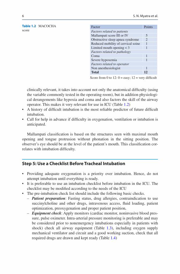
• MACOCHA Score: This is a simple score developed for ICU patients which has been shown to differentiate between a difficult from a non difficult airway in ICU patients. The MACOCHA score has seven easily identifiable variables which are
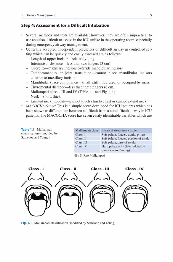
Table 1.1 Mallampati classificationa (modified by Samsoon and Young)
Mallampati class Intraoral structures visibleClass I Soft palate, fauces, uvula, pillarsClass II Soft palate, fauces, portion of uvulaClass III Soft palate, base of uvulaClass IV Hard palate only (later added by
Samsoon and Young)aBy S. Rao Mallampati
Fig. 1.1 Mallampati classification (modified by Samsoon and Young)
1 Airway Management
6
clinically relevant, it takes into account not only the anatomical difficulty (using the variable commonly tested in the operating room), but in addition physiologi-cal derangements like hypoxia and coma and also factors the skill of the airway operator. This makes it very relevant for use in ICU (Table 1.2)
• A history of difficult intubation is the most reliable predictor of future difficult intubation.
• Call for help in advance if difficulty in oxygenation, ventilation or intubation is anticipated.
Mallampati classification is based on the structures seen with maximal mouth opening and tongue protrusion without phonation in the sitting position. The observer’s eye should be at the level of the patient’s mouth. This classification cor-relates with intubation difficulty.
Step 5: Use a Checklist Before Tracheal Intubation
• Providing adequate oxygenation is a priority over intubation. Hence, do not attempt intubation until everything is ready.
• It is preferable to use an intubation checklist before intubation in the ICU. The checklist may be modified according to the needs of the ICU
• The pre-intubation check list should include the following basic checks. – Patient preparation: Fasting status, drug allergies, contraindication to use
succinylcholine and other drugs, intravenous access, fluid loading, patient optimization, preoxygenation and proper patient position,
– Equipment check: Apply monitors (cardiac monitor, noninvasive blood pres-sure, pulse oximeter. Intra-arterial pressure monitoring is preferable and may be considered prior to nonemergency intubations especially in patients with shock) check all airway equipment (Table 1.3), including oxygen supply mechanical ventilator and circuit and a good working suction, check that all required drugs are drawn and kept ready (Table 1.4)
Table 1.2 MACOCHA score
Factor PointsFactors related to patientsMallampati score III or IV 5Obstructive sleep apnea syndrome 2Reduced mobility of cervical soine 1Limited mouth opening < 3 1Factors related to pathologyComa 1Severe hypoxemia 1Factors related to operatorNon anesthesiologist 1Total 12
Score from 0 to 12: 0 = easy; 12 = very difficult
S. N. Myatra et al.
7
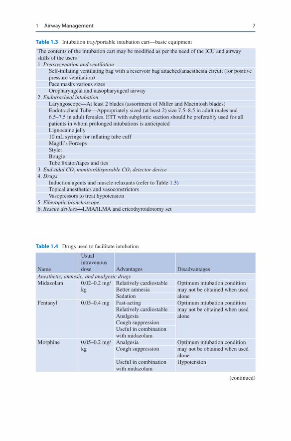
Table 1.3 Intubation tray/portable intubation cart—basic equipment
The contents of the intubation cart may be modified as per the need of the ICU and airway skills of the users1. Preoxygenation and ventilation Self-inflating ventilating bag with a reservoir bag attached/anaesthesia circuit (for positive
pressure ventilation) Face masks various sizes Oropharyngeal and nasopharyngeal airway2. Endotracheal intubation Laryngoscope—At least 2 blades (assortment of Miller and Macintosh blades) Endotracheal Tube—Appropriately sized (at least 2) size 7.5–8.5 in adult males and
6.5–7.5 in adult females. ETT with subglottic suction should be preferably used for all patients in whom prolonged intubations is anticipated
Lignocaine jelly 10 mL syringe for inflating tube cuff Magill’s Forceps Stylet Bougie Tube fixator/tapes and ties3. End-tidal CO2 monitor/disposable CO2 detector device4. Drugs Induction agents and muscle relaxants (refer to Table 1.3) Topical anesthetics and vasoconstrictors Vasopressors to treat hypotension5. Fiberoptic bronchoscope6. Rescue devices—LMA/ILMA and cricothyroidotomy set
Table 1.4 Drugs used to facilitate intubation
Name
Usual intravenous dose Advantages Disadvantages
Anesthetic, amnesic, and analgesic drugsMidazolam 0.02–0.2 mg/
kgRelatively cardiostable Optimum intubation condition
may not be obtained when used alone
Better amnesiaSedation
Fentanyl 0.05–0.4 mg Fast-acting Optimum intubation condition may not be obtained when used alone
Relatively cardiostableAnalgesiaCough suppressionUseful in combination with midazolam
Morphine 0.05–0.2 mg/kg
Analgesia Optimum intubation condition may not be obtained when used alone
Cough suppression
Useful in combination with midazolam
Hypotension
(continued)
1 Airway Management
8
– Team preparation: Allocate roles and responsibilities to the team members. A plan for who to call for help and who will note the time should be made, Check that personal protective equipment is being used
– Preparation for airway difficulty: Discuss the airway plan, further plan in case of failed intubation and address any concerns the team members may have before proceeding for tracheal intubation
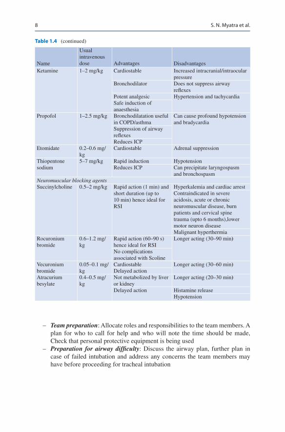
Table 1.4 (continued)
Name
Usual intravenous dose Advantages Disadvantages
Ketamine 1–2 mg/kg Cardiostable Increased intracranial/intraocular pressure
Bronchodilator Does not suppress airway reflexes
Potent analgesic Hypertension and tachycardiaSafe induction of anaesthesia
Propofol 1–2.5 mg/kg Bronchodilatation useful in COPD/asthma
Can cause profound hypotension and bradycardia
Suppression of airway reflexesReduces ICP
Etomidate 0.2–0.6 mg/kg
Cardiostable Adrenal suppression
Thiopentone sodium
5–7 mg/kg Rapid induction HypotensionReduces ICP Can precipitate laryngospasm
and bronchospasmNeuromuscular blocking agentsSuccinylcholine 0.5–2 mg/kg Rapid action (1 min) and
short duration (up to 10 min) hence ideal for RSI
Hyperkalemia and cardiac arrestContraindicated in severe acidosis, acute or chronic neuromuscular disease, burn patients and cervical spine trauma (upto 6 months),lower motor neuron diseaseMalignant hyperthermia
Rocuronium bromide
0.6–1.2 mg/kg
Rapid action (60–90 s) hence ideal for RSI
Longer acting (30–90 min)
No complications associated with Scoline
Vecuronium bromide
0.05–0.1 mg/kg
Cardiostable Longer acting (30–60 min)Delayed action
Atracurium besylate
0.4–0.5 mg/kg
Not metabolized by liver or kidney
Longer acting (20–30 min)
Delayed action Histamine releaseHypotension
S. N. Myatra et al.

9
Step 6: Patient Positioning for Tracheal Intubation
• The Ramp position (alignment of the external auditory meatus and sternum Fig. 1.2) or the head elevated laryngoscopy position (HELP), a 25-degree head- elevation or back-up position is recommended over the classic sniffing position for tracheal intubation in ICU. This position is more comfortable for a patient to breathe. In the supine position, the posterior portions of the lung become more prone to collapse and atelectasis, thus reducing the oxygen reserves and shorten-ing the safe apnoea time.
• In patients with suspected cervical spine injury, maintain the head in neutral position and give manual in-line cervical stabilization. Use the cervical collar at all times during airway manipulations.
Step 7: Preoxygenation and Apnoic Oxygenation
• Remember, failure to intubate will not harm a patient but failure to oxygenate will. Ensure adequate oxygenation at all times.
• Optimal pre-oxygenation increases the non-hypoxic apnoea time and provides a safety margin before desaturation occurs. Thus preoxygenation for at least 3 min is recommended to prolong the safe apnoea time before intubation.
• Critically ill patients may have oxygen transport limitation and time-consuming airway management. During apnea, the time for oxyhemoglobin desaturation below 85% is much faster in these patients.
• Though preoxygenation may not be feasible at all times, especially during an emergency tracheal intubation, every attempt should be made to preoxygenate the patient if time permits. In addition, it is important to remember that the response to preoxygenation may not always be good in critically ill patients, especially in those with respiratory conditions as compared to patients with nor-mal lung function.
• Pre-oxygenation with 100% oxygen prior to tracheal intubation can be done by one of the following methods: – Bag valve mask device with a reservoir bag (minimum 12–15 L/min of
oxygen)
Fig. 1.2 Ramp position: external auditory meatus and sternum should be in same alignment
1 Airway Management
10
– High flow nasal cannula oxygen (HFNCO) – Non invasive ventilation (NIV)
• Use of NIV for preoxygenation has been presently shown to be superior in case of acute respiratory failure, compared to other methods of preoxygenation in ICU. The NIV settings recommended for preoxygenation are: Pressure support ventilation with FiO2 of 1.0, inspiratory pressure support from 5 to 15 cm H2O, to obtain an expiratory tidal volume between 6 and 8 mL/kg and positive end- expiratory pressure of 5 cm H2O.
• Apnoic oxygenation (oxygen delivery during the apnoea time). Providing nasal oxygen during intubation can help further prolong the safe apnoea time. Hence whenever feasible apnoic oxygenation should be provided. This can be provided using a nasal cannula with high flow oxygen. Two methods are available – Using High Flow Nasal Canula Oxygen (HFNCO). This is a special device
which provides up to 60 L/min of heated humidified oxygen (100%). This can be used for preoxygenation first and then continued during apnoea until tra-cheal intubation. This requires dedicated equipment with oxygen humidifica-tion unit, nasal cannula and tubing connecting standard oxygen regulator to the transnasal oxygen cannula. This technique provides CPAP during pre- oxygenation and apnoeic oxygenation with gas exchange by flow-dependent flushing of the dead space. This method significantly prolongs the safe apnoea time, thus allowing securing a definitive airway during a difficult intubation or failed intubation to be done in an unhurried manner. HFNCO should be used when available, especially when a difficult airway is anticipated.
– Alternatively, a simple nasal cannula may be used to deliver 15 L/min of oxy-gen during apnea when attempts at tracheal intubation are performed. This technique is called NO DESAT (Nasal Oxygen During Efforts at Securing A Tube). It is not as effective as HFNCO in ICU.
Step 8: Proceed with Tracheal Intubation
• Check that personal protection gear used is adequate (gloves, mask, and eye protection) and expert help is available in case of an anticipated difficult airway.
• Ensure that there is adequate monitoring with the cardiac monitor, noninvasive blood pressure, and pulse oximeter
• Secure an intravenous line and give volume support• If the patient is to be ventilated, set up the ventilator and prepare drugs for long-
term sedation.• Awake or asleep tracheal intubation can be performed. Awake intubation is the
gold standard for an anticipated difficult intubation using a flexible broncho-scope (FOB) or video laryngoscope (VL). However, this may not be feasible in most ICU patients as they may be uncooperative due to their critical illness and administration of local anesthetic agents to facilitate the procedure may also be difficult. Asleep intubation can be performed after administering general
S. N. Myatra et al.
11
anaesthesia using a direct laryngoscope (DL) or a vidolaryngoscope (VL). The use of VL improves glottic visualization as compared to DL, making it an impor-tant tool for difficult airway management in ICU, especially in expert hands.
• Drug therapy (preintubation): The choice of agents (Table 1.4) will depend on the hemodynamic status of the patient and the anticipated nature of difficulty in intubation. – Patients may be given intravenous fentanyl, morphine, or midazolam.
Physicians with appropriate experience may choose to use anesthetic induc-tion agents such as ketamine, thiopentone sodium, propofol, or etomidate. These drugs should be given slowly to effect with or without muscle relaxants.
– Intravenous ketamine, unless contraindicated, is the preferred induction agent, especially in hemodynamically unstable patients.
– Etomidate is a cardiostable agent, but there are concerns of adrenal insuffi-ciency following even a single dose.
– Propofol can cause profound hypotension and myocardial depression and should be used with extreme caution.
– Rapidly acting muscle relaxants such as succinylcholine/rocuronium may be used for rapid sequence intubation. Succinylcholine is used only in absence of hyperkalemia, severe acidosis, acute or chronic neuromuscular disease, exten-sive burn, and cervical trauma.
– Longer-acting muscle relaxants (e.g., atracurium and vecuronium) should be given only after confirming that ventilation is possible.
– Note that in sick, fatigued patients, very small drug doses may be sufficient. Inject drugs very slowly and until effect (do not give calculated/standard induction doses).
• Clear upper airway obstruction if present: – Snoring, gurgling sound, paradoxical movement of the chest wall (inward
movement during inspiration) and abdomen and inadequate/absent chest rise during ventilation may suggest upper airway obstruction.
– Perform an oral or nasal (with soft malleable catheter) suctioning for no more than 10 s at a time and resume oxygenation soon after.
– Use an oropharyngeal or nasopharyngeal airway if obstruction is not cleared by suctioning. The airway should have a length equivalent to distance from the tip of the nose/angle of the mouth to the tragus. Nasopharyngeal airway diameter should be less than the patient’s nostril. It should be avoided if the patient has risk of nasal trauma/bleeding or cerebrospinal fluid rhinorrhea.
• Rapid sequence intubation (RSI): Most ICU patients are at a risk of aspiration. Critically ill patients may be not be fasted or have a slower gastric emptying (gastroparesis of critical illness, other medical conditions like diabetes etc.). Thus, conventionally, a rapid sequence induction (RSI), i.e. administration of rapid onset agents and avoidance of ventilation between induction and intuba-tion, to limit gastric insufflation and aspiration is indicated. However, hypoxemia during this period is a concern these patients. The recent landmark PREVENT study showed that patients receiving gentle ventilation during RSI experienced
1 Airway Management

12
lesser desaturation compared to controls, without suffering from an increased rate of pulmonary aspiration. Thus a modified RSI using gentle ventilation can be used to limit hypoxia during RSI. – After giving adequate preoxygenation and proper position, cricoid pressure
(Sellick maneuver) is given just before the beginning of induction. As soon as the patient is asleep, increase the pressure.
– Use only rapidly acting muscle relaxants (suxamethonium or rocuronium) while maintaining cricoid pressure.
– Data from a large dataset suggests that the use of muscle relaxants in associ-ated with fewer complications, including in patients with difficult airways.
– Give gentle positive pressure ventilation (modified RSI) to maintain the oxy-gen saturation, especially in hypoxic patients.
– Ensure adequate chest rise during mask ventilation. Hold the mask with both hands if ventilation is difficult.
– Mask ventilation may be difficult in the following cases: BONES (B = beard, O = obese N = no teeth E = elderly (>55 years) or cathectic (sunken cheeks or edentulous) S = snores.
– Apnoeic oxygenation using nasal oxygen (using oxygen flow of 15 L/min or HFNC) should be continued during attempts at intubation.
– Perform laryngoscopy and intubation. Hold the laryngoscope handle in the left hand. Open the mouth of the patient with the thumb and the index finger of the right hand. Insert the laryngoscope blade gently into the mouth from the right-side angle of the mouth and move it to the left side taking the tongue along with the blade as it is inserted further inside the mouth. When the epi-glottis is visualized, insert the curved blade into the vallecula and pull the laryngoscope forward and upward to expose the glottis (Fig. 1.3). Now, insert the ETT using the right hand between the vocal cords under direct vision.
– For nasal intubation, use prior nasal mucosal vasoconstrictors and lubrication; Magill’s forceps may be used to guide the tube into the trachea.
– Optimal external laryngeal manipulation (OELM) with the right hand or by an assistant by quickly pressing in both cephalad and posterior direction over the thyroid, cricoids or hyoid cartilage may be used to further optimize laryn-goscopic view.
Laryngoscope blade
Epiglottis
Glottic Opening
Vocal Cords
Fig. 1.3 Glottic view during laryngoscopy
S. N. Myatra et al.
13
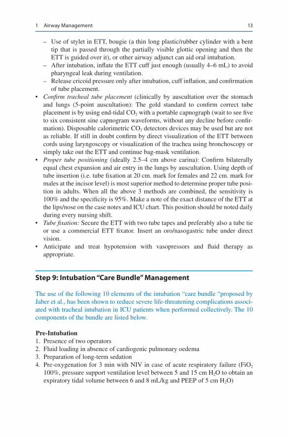
– Use of stylet in ETT, bougie (a thin long plastic/rubber cylinder with a bent tip that is passed through the partially visible glottic opening and then the ETT is guided over it), or other airway adjunct can aid oral intubation.
– After intubation, inflate the ETT cuff just enough (usually 4–6 mL) to avoid pharyngeal leak during ventilation.
– Release cricoid pressure only after intubation, cuff inflation, and confirmation of tube placement.
• Confirm tracheal tube placement (clinically by auscultation over the stomach and lungs (5-point auscultation): The gold standard to confirm correct tube placement is by using end-tidal CO2 with a portable capnograph (wait to see five to six consistent sine capnogram waveforms, without any decline before confir-mation). Disposable calorimetric CO2 detectors devices may be used but are not as reliable. If still in doubt confirm by direct visualization of the ETT between cords using laryngoscopy or visualization of the trachea using bronchoscopy or simply take out the ETT and continue bag-mask ventilation.
• Proper tube positioning (ideally 2.5–4 cm above carina): Confirm bilaterally equal chest expansion and air entry in the lungs by auscultation. Using depth of tube insertion (i.e. tube fixation at 20 cm. mark for females and 22 cm. mark for males at the incisor level) is most superior method to determine proper tube posi-tion in adults. When all the above 3 methods are combined, the sensitivity is 100% and the specificity is 95%. Make a note of the exact distance of the ETT at the lips/nose on the case notes and ICU chart. This position should be noted daily during every nursing shift.
• Tube fixation: Secure the ETT with two tube tapes and preferably also a tube tie or use a commercial ETT fixator. Insert an oro/nasogastric tube under direct vision.
• Anticipate and treat hypotension with vasopressors and fluid therapy as appropriate.
Step 9: Intubation “Care Bundle” Management
The use of the following 10 elements of the intubation “care bundle “proposed by Jaber et al., has been shown to reduce severe life-threatening complications associ-ated with tracheal intubation in ICU patients when performed collectively. The 10 components of the bundle are listed below.
Pre-Intubation 1. Presence of two operators 2. Fluid loading in absence of cardiogenic pulmonary oedema 3. Preparation of long-term sedation 4. Pre-oxygenation for 3 min with NIV in case of acute respiratory failure (FiO2
100%, pressure support ventilation level between 5 and 15 cm H2O to obtain an expiratory tidal volume between 6 and 8 mL/kg and PEEP of 5 cm H2O)
1 Airway Management
14
During Intubation 5. Rapid sequence induction: Etomidate 0.2–0.3 mg/kg or ketamine 1.5–3 mg/kg
combined with succinylcholine 1–1.5 mg/kg in absence of allergy, hyperkalae-mia, severe acidosis, acute or chronic neuromuscular disease, burn patient for more than 48 h and spinal cord trauma
6. Sellick maneuver
Post-Intubation 7. Immediate confirmation of tube placement by capnography 8. Norepinephrine if diastolic blood pressure remains low 9. Initiate long-term sedation
Initial “protective ventilation”: Tidal volume 6–8 mL/kg of ideal body weight, PEEP 5 cm H2O and respiratory rate between 10 and 20/min, FiO2 100%, plateau pressure <30 cm H2O.
Step 10: Use of Guidelines for Tracheal Intubation in ICU
• Recognizing the high risk of airway management in ICU, guidelines specific to tracheal intubation in ICU have been recently formulated by various interna-tional societies. These guidelines have subtle differences, however the broad principles are the similar with a focus on strategies to enhance safety during tracheal intubation.
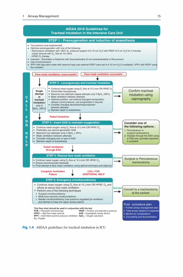
• The first guidelines on tracheal intubation in ICU was published by the All India Difficult Airway Association (AIDAA) in 2016 (Fig. 1.4). This guideline gives a stepwise approach to tracheal intubation in ICU using evidence based recom-mendations. These include the presence of two operators during intubation, hemodynamic optimization, preoxygenation, apnoiec oxygenation, use of a modified rapid sequence intubation with gentle ventilation to prevent hypoxia during intubation and limiting attempts at intubation to avoid life threatening complications.
• These guidelines also provide a stepwise approach to the management of failed intubation being, continuation of mask ventilation, insertion of a supra-glottic airway, one last attempt at mask ventilation if this fails and finally performing an emergency cricothyroidotomy to maintain the oxygenation while a tracheostomy is performed to establish a definite airway management. Unlike in the operating room, waking up the patient and postponing the sur-gery is not an option.
S. N. Myatra et al.
15
AIDAA 2016 Guidelines forTracheal Intubation in the Intensive Care Unit
STEP 1 : Preoxygenation and induction of anaesthesiaTwo persons (one experienced)Optimise preoxygenation with one of the following :- Noninvasive ventilation with 100% O
2, pressure support of 5-15 cm H
2O with PEEP of 5 cm H
2O for 3 minutes
(nasal cannula with O2, flow at 15 L/min)
- HFNC O2 therapy
Induction - Etomidate or Ketamine with Succinylcholine (if not contraindicated) or RocuroniumUse cricoid pressureIPPV with bag-valve mask with reservoir bag (use external PEEP valve set to 5-10 cm H
2O if available) / IPPV with PEEP using
the ventilator
Face mask ventilation unsuccessful Face mask ventilation successful
Singleattempt
attracheal
intubationonly if
SpO2 ≥95%)
••
•••
•••••
•
•
••••••
•••
•
•
STEP 2 : Laryngoscopy and tracheal intubation
Failed Intubation
Failed ventilationthrough SAD
Complete VentilationFailure
CALL FORADDITIONAL HELP
STEP 3 : Insert SAD to maintain oxygenation
Succeed
Succeed
Succeed
Succeed
CALL
FOR
HELP
Confirm trachealintubation using
capnography
Consider one ofthe following options :1. Percutaneous or surgical tracheostomy2. Intubate through the SAD using a FOB only, provided expertise is available
Surgical or Percutaneoustracheostomy
Convert to a tracheostomyat the earliest
Post - procedure plan1. Further airway management plan2. Treat airway oedema if suspected3. Monitor for complications4. Counselling and documentation
This flow chart should be used in conjunction with the textFOB = Fibreoptic bronchoscope PEEP = Positive end-expiratory pressureHFNC = High flow nasal cannula SAD = Supraglottic airway deviceIPPV = lntermittent positive pressure ventilation SpO
2 = Oxygen saturation
O2= Oxygen
Continue nasal oxygen using O2 flow at 15 L/min OR HFNC O2 andefforts at rescue face mask ventilationPerform one of the following techniques Surgical cricothyroidotomy Wide bore cannula cricothyroidotomy Needle cricothyroidotomy (use pressure regulated jet ventilation and attempt to keep the upper airway patent)
STEP 4: Rescue face mask ventilation
Continue nasal oxygen using O2 flow at 15 L/min OR HFNC O2,Ensure neuromuscular blockadeFinal attempt at face mask ventilation using optimal technique and adjuncts
Continue nasal oxygen using O2 flow at 15 L/min OR HFNC O2Preferably use second generation SADMaximum two attempts (only if SpO2 ≥ 95%)Mask ventilation between attemptsConsider changing size or type of SADMaintain depth of anaesthesia
Continue nasal oxygen using O2 flow at 15 L/min OR HFNC O2Direct/video laryngoscopyMaximum two attempts (repeat attempts only if SpO2 ≥95%)Mask ventilation between attemptsOptimise position, use external laryngeal manipulation,release cricoid pressure, use bougie/stylet if requiredConsider changing device/technique/operatorbetween attemptsMaintain depth of anaesthesia
STEP 5: Emergency cricothyroidotomy
Fig. 1.4 AIDAA guidelines for tracheal intubation in ICU
1 Airway Management

16
Step 11: Steps After Tracheal Intubation
• Initiate mechanical ventilation if required.• Give analgesia and sedation as required.• Obtain chest radiograph to confirm tube position, bilateral lung expansion, and
oro/nasogastric tube position.• Do not start feeding the patient until the position of oro/nasogastric tube is con-
firmed on chest radiograph.• Check the ETT cuff pressure using the cuff pressure machine and maintain it
below 20 mmHg at all times.
Step 12: Watch for and Treat Immediate Complications of endotracheal intubation
• Immediate complications – Esophageal intubation/endobronchial intubations/accidental ETT disconnec-
tions—atelectasis formation/collapse in the unventilated lung and hyperinfla-tion and barotrauma with development of pneumothorax of the intubated lung (in endobronchial intubations) can cause profound hypoxemia manifesting as bradycardia and even progressing to cardiac arrest
– Hypertension, tachycardia, raised intracranial pressure, and myocardial isch-emia due to stimulation from laryngoscopy and intubation
– Hypotension due to loss of sympathetic tone from drugs for intubation or dynamic hyperinflation due to hyperventilation or relative dehydration
– Aspiration of gastric contents – Airway trauma, bleeding – Negative pressure pulmonary edema after sudden relief of severe airway
obstruction – Cardiac arrest
• Following an unanticipated difficult tracheal intubation, post procedure monitor-ing for complications is required. Watch for airway odema. Documentation of airway difficulty along with counseling of the patient/family is essential.
Step 13: Follow a Protocol for Airway Maintenance
• Proper maintenance of the airway will reduce the incidence of accidental extuba-tions, disconnections, tube blockage, and nosocomial pneumonia.
• Keep the head elevated at 30–45°.• All ETT and tracheostomy tubes (TT) should be checked for position at incisor
teeth/alae nasi, adequate fixation, patency, tracheal cuff pressure (<20 mmHg), and pharyngeal leak during each shift and should be documented.
• In case of oral ETTs, secure firmly at the angle of the mouth and change position preferably every 24 h to avoid sores/ulcers.
S. N. Myatra et al.

17
• Oral ETTs (without subglottic suction) should be cut 2–3 cm from the angle of the mouth.
• The universal connector should be pushed right down to its shoulder to avoid accidental disconnections.
• Confirm correct positioning of ETTs above the carina on the X-ray and docu-ment in the case notes.
• All ventilated patients should receive humidification (with HME (Heat and Moisture Exchanger) filter or using a heated humidifier circuit).
• ETT/TT suction should be done only when required and preferably using a closed suction system.
• Sedate patients well when they need to remain intubated. Do not allow them to get restless.
• Start weaning the patient off sedation, only in the daytime when ICU staff is in adequate strength.
• Do not leave the patient unattended when sedation has been turned off and the patient is just about waking up. Reassure patients as they wake up from sedation.
• Apply boxer gloves/restraints to those patients who appear agitated. Refrain from tying patient’s limbs.
• Report any airway accident as a “critical incident.”
Step 14: Extubation of the Airway
• Perform a good oral and endotracheal suction prior to extubation.• Keep all equipment ready for reintubation/noninvasive ventilation if required.• Do a cuff-leak test (especially after prolonged intubations)—deflate the ETT cuff
and check for air leak around the cuff or tidal volume loss on the ventilator. If absent, suspect laryngeal edema. Consider the use of steroids and plan extuba-tion at a later date over a tube exchanger.
• Intravenous methylprednisolone started 12 h before a planned extubation has been shown to substantially reduce the incidence of postextubation laryngeal edema and reintubation in patients intubated for more than 36 h and having absent cuff leak.
• In a patient with a difficult airway, ensure that expert airway help is available prior to extubation and extubate preferably over a tube exchanger FOB. Oxygenate the patient through the exchanger and remove it only when you are sure that the airway is not compromised/obstructed. If in doubt, pass the ETT back inside over the tube exchanger or FOB and secure in place.
Step 15: Continue to Watch for and Treat Complications of Tracheal Intubation (Days to Months After Extubation)
• Sore throat• Airway edema
1 Airway Management
18
• Airway infections/pneumonia• Laryngeal damage/granuloma• Tracheal stenosis, tracheomalacia, trachea-esophageal fistula
Suggested Reading
Casey JD, Janz DR, Russell DW, et al. Bag-mask ventilation during tracheal intubation of criti-cally ill adults. N Engl J Med. 2019;380:811–21. https://doi.org/10.1056/NEJMoa1812405. Critically ill adult patients receiving bag-mask ventilation had higher oxygen saturations and a lower incidence of severe hypoxemia, than those receiving no ventilation during tracheal intubation an increased risk of pulmonary aspiration
François B, Bellissant E, Gissot V, Desachy A, Normand S, Boulain T, Brenet O, Preux PM, Vignon P. Association des Réanimateurs du Centre-Ouest (ARCO). 12-h pretreatment with methylprednisolone versus placebo for prevention of postextubation laryngeal oedema: a ran-domised double-blind trial. Lancet. 2007;369(9567):1083–9. Methylprednisolone started 12 h before a planned extubation substantially reduced the incidence of postextubation laryngeal oedema and reintubation. Such pretreatment should be considered in adult patients before a planned extubation that follows a tracheal intubation of more than 36 h
Frat J-P, Ricard J-D, Quenot J-P, et al. Non-invasive ventilation versus high-flow nasal cannula oxygen therapy with apnoeic oxygenation for preoxygenation before intubation of patients with acute hypoxaemic respiratory failure: a randomised, multicentre, open-label trial. Lancet Respir Med. 2019;7:303–12. https://doi.org/10.1016/S2213-2600(19)30048-7. Use of non- invasive ventilation or high-flow nasal cannula oxygen therapy with apnoeic oxygenation for preoxygenation before intubation. Severe hypoxaemia occurred less frequently after preoxy-genation with non-invasive ventilation than with high-flow oxygen
Higgs A, McGrath BA, Goddard C, et al. Guidelines for the management of tracheal intubation in critically ill adults. Br J Anaesth. 2018;120:323–52. https://doi.org/10.1016/j.bja.2017.10.021. Guidelines for the management of tracheal intubation in critically ill adults
Jaber S, Jung B, Corne P, Sebbane M, Muller L, Chanques G, Verzilli D, Jonquet O, Eledjam JJ, Lefrant JY. An intervention to decrease complications related to endotracheal intubation in the intensive care unit: a prospective, multiple-center study. Intensive Care Med. 2010;36(2):248–55. The implementation of an intubation management protocol can reduce immediate severe life-threatening complications associated with intubation of ICU patients
Kaufman D. Etomidate versus ketamine for sedation in acutely ill patients. Lancet. 2009;374(9697):1240–1. Ketamine is a safe and valuable alternative to etomidate for endotra-cheal intubation in critically ill patients and should be considered in those with sepsis
Myatra SN, Ahmed SM, Kundra P, et al. Republication: All India Difficult Airway Association 2016 Guidelines for Tracheal Intubation in the Intensive Care Unit. Indian J Crit Care Med. 2017;21:146–53. https://doi.org/10.4103/ijccm.IJCCM_57_17. Indian Guidelines for Tracheal Intubation in the Intensive Care Unit
Sitzwohl C, Langheinrich A, Schober A, Krafft P, Sessler DI, Herkner H, Gonano C, Weinstabl C, Kettner SC. Endobronchial intubation detected by insertion depth of endotracheal tube, bilat-eral auscultation, or observation of chest movements: randomised trial. BMJ. 2010;341:c5943. The highest sensitivity and specificity for ruling out endobronchial intubation, however, is achieved by combining tube depth, auscultation of the lungs, and observation of symmetrical chest movements
S. N. Myatra et al.

19© Springer Nature Singapore Pte Ltd. 2020R. Chawla, S. Todi (eds.), ICU Protocols, https://doi.org/10.1007/978-981-15-0898-1_2
R. Guleria (*) · J. Kumar Department of Pulmonary Medicine, All India Institute of Medical Sciences, New Delhi, India
R. Chawla Department of Respiratory, Critical Care & Sleep Medicine, Indraprastha Apollo Hospitals, New Delhi, India
2Acute Respiratory Failure
Randeep Guleria, Jaya Kumar, and Rajesh Chawla
Case scenario 1
A 30-year-old male patient presented with acute onset of breathlessness, dry cough, fever, myalgia, and malaise for 4 days. On examination, he was found to be febrile and restless, with respiratory rate of 46/min and pulse rate of 124/min. His oxygen saturation was 80% on room air, and chest skiagram showed bilat-eral parenchymal infiltrate.
Case scenario 2
A 60-year-old male patient with chronic obstructive airway disease presented with increasing shortness of breath, cough, and expectoration for 5 days and drowsiness with confusion for 1 day. On examination, he was found to be drowsy, cyanosed with respiratory rate of 30/min, tachycardia, and flapping tremors. His oxygen saturation was 80% on initial evaluation, and a chest radiograph showed hyperinflated lung fields and right lower zone infiltrates.
Case scenario 3
A 30-year-old female patient with anxiety disorder presented to the emergency department in a comatosed condition with history of ingestion of some unknown tablets. On examination, she was found to be E2M4V1, with pulse rate of 64/min, respiratory rate of 14/min, and blood pressure of 90/60 mmHg.
20
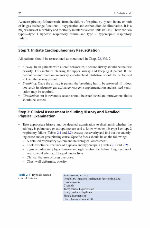
Acute respiratory failure results from the failure of respiratory system in one or both of its gas exchange functions—oxygenation and carbon dioxide elimination. It is a major cause of morbidity and mortality in intensive care units (ICUs). There are two types—type 1 hypoxic respiratory failure and type 2 hypercapnic respiratory failure.
Step 1: Initiate Cardiopulmonary Resuscitation
All patients should be resuscitated as mentioned in Chap. 23, Vol. 2.
• Airway: In all patients with altered sensorium, a secure airway should be the first priority. This includes clearing the upper airway and keeping it patent. If the patient cannot maintain an airway, endotracheal intubation should be performed to keep the airway patent.
• Breathing: Once the airway is patent, the breathing has to be assessed. If it does not result in adequate gas exchange, oxygen supplementation and assisted venti-lation may be required.
• Circulation: An intravenous access should be established and intravenous fluids should be started.
Step 2: Clinical Assessment Including History and Detailed Physical Examination
• Take appropriate history and do detailed examination to distinguish whether the etiology is pulmonary or extrapulmonary and to know whether it is type 1 or type 2 respiratory failure (Tables 2.1 and 2.2). Assess the severity and find out the underly-ing cause and/or precipitating cause. Specific focus should be on the following: – A detailed respiratory system and neurological assessment. – Look for clinical features of hypoxia and hypercapnia (Tables 2.1 and 2.2). – Signs of pulmonary hypertension and right ventricular failure: Engorged neck
veins, Pedal edema, Enlarged tender liver. – Clinical features of drug overdose. – Chest wall deformity, obesity.
Table 2.1 Hypoxia-related clinical features
Restlessness, anxietyIrritability, impaired intellectual functioning, and consciousnessCyanosisTachycardia, hypertensionBradycardia, arrhythmiaShock, hypotensionConvulsions, coma, death
R. Guleria et al.

21
Step 3: Check Pulse Oximetry and Do Arterial Blood Gas Analysis
Pulse oximetry and arterial blood gases are the mainstay of diagnosis and essential to decide on the therapeutic intervention.
• Oximetry is a rapid technique to know if there is significant hypoxia, but it gives no clue about the presence or absence of hypercapnia. In patients on supplemen-tal oxygen, deteriorating pulmonary function is difficult to ascertain by pulse oxymetry as oxygen saturation in the flat part of oxyhemoglobin dissociation curve may not decrease appreciably with substantial decrease in PaO2.
• Arterial blood gas analysis is essential for both diagnostic and therapeutic decisions. – Type 1 respiratory failure is recognized by hypoxemia (PaO2 < 60 mmHg).
With or without widening of alveolar-arterial oxygen gradient, PaCO2 is either low or normal.
– Type 2 respiratory failure is diagnosed when a PaO2 of less than 60 mmHg is associated with a PaCO2 of more than 45 mmHg and respiratory acidosis.
• This needs to be followed by an assessment of the pH and HCO3 to decide whether the type 2 respiratory failure is acute, acute on chronic, or chronic.
• Type II acute respiratory failure presents with low pH, high PaCO2, and normal HCO3; acute on chronic presents with low pH, high PaCO2, and high HCO3; while chronic respiratory failure presents with normal pH along with raised PaCO2 and HCO3.
• This should be followed by an assessment of alveolar-arterial oxygen gradient, which helps to narrow down the cause of respiratory failure (see Appendix B).
• A-a gradient = PAO2 − PaO2
• A-a gradient = [FiO2 × (Patm − PH2O) − (PaCO2/0.8)] − PaO2
Step 4: Differentiate Between Type 1 and Type 2 Respiratory Failures
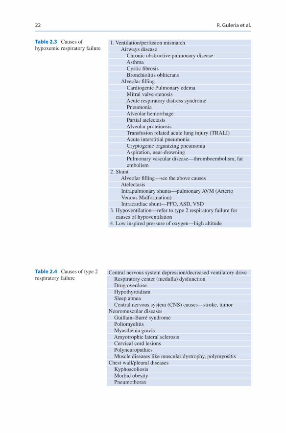
Type 1 respiratory failure occurs when the gas exchange is inadequate at rest or dur-ing exercise, leading to hypoxemia, and PaO2 is less than 60 mmHg (Table 2.3).
Type 2 respiratory failure occurs as a result of alveolar hypoventilation, which can be due to a pulmonary or extrapulmonary cause. Chronic obstructive pulmonary disease is the commonest cause of type 2 respiratory failure, but various other con-ditions listed below can also lead to hypercapnia and respiratory failure (Table 2.4).
Table 2.2 Hypercapnia- related clinical features
HeadacheDrowsiness, confusionWarm extremities, flushing, sweatingBounding pulse, tachycardiaTremors, myoclonic jerks, asterixis, seizuresPapilledema, coma
2 Acute Respiratory Failure
22
Table 2.3 Causes of hypoxemic respiratory failure
1. Ventilation/perfusion mismatch Airways disease Chronic obstructive pulmonary disease Asthma Cystic fibrosis Bronchiolitis obliterans Alveolar filling Cardiogenic Pulmonary edema Mitral valve stenosis Acute respiratory distress syndrome Pneumonia Alveolar hemorrhage Partial atelectasis Alveolar proteinosis Transfusion related acute lung injury (TRALI) Acute interstitial pneumonia Cryptogenic organizing pneumonia Aspiration, near-drowning Pulmonary vascular disease—thromboembolism, fat
embolism2. Shunt Alveolar filling—see the above causes Atelectasis Intrapulmonary shunts—pulmonary AVM (Arterio
Venous Malformation) Intracardiac shunt—PFO, ASD, VSD3. Hypoventilation—refer to type 2 respiratory failure for
causes of hypoventilation4. Low inspired pressure of oxygen—high altitude
Table 2.4 Causes of type 2 respiratory failure
Central nervous system depression/decreased ventilatory drive Respiratory center (medulla) dysfunction Drug overdose Hypothyroidism Sleep apnea Central nervous system (CNS) causes—stroke, tumorNeuromuscular diseases Guillain–Barré syndrome Poliomyelitis Myasthenia gravis Amyotrophic lateral sclerosis Cervical cord lesions Polyneuropathies Muscle diseases like muscular dystrophy, polymyositisChest wall/pleural diseases Kyphoscoliosis Morbid obesity Pneumothorax
R. Guleria et al.

23
An approach to a patient with acute hypoxemic respiratory failure is summarized in Fig. 2.1.
Step 5: Send Investigations
• Complete hemogram and biochemistry• Lung function tests (if possible) that helps to differentiate between obstructive,
restrictive, and mixed ventilatory defects• Chest radiograph that may help to identify hyperinflation, pulmonary edema,
pneumonia, pneumothorax, neoplasm and others to give a clue to the underlying etiology
• Electrocardiogram to identify cardiac disorders• Computed tomography (CT) or magnetic resonance imaging (MRI) if indicated
for interstitial lung disease, neoplasm, stroke, and other neurological disorders• Two-dimensional echocardiography for identification of cor pulmonale, intracar-
diac shunt, patent ductus arteriosus, and pulmonary embolism
Step 6: Initiate Specific Treatment
The primary aim is to maintain oxygenation and adequate alveolar ventilation and treatment for the underlying etiology. The key principles in the management of respiratory failure are as follows:
• Optimized oxygen therapy.• Identification of the underlying cause and adequate treatment for the same.
Hypoxemicrespiratory failure
Normal A-agradient
High PaCo2
YesYes NoNo
Hypoventilation Low inspired O2V/Q mismatch Shunt
improvement withsupplemental oxygen
Increased A-agradient
Fig. 2.1 An approach to hypoxemic respiratory failure to know the etiology
2 Acute Respiratory Failure

24
• Clinical assessment and arterial blood gases to help decide the severity.• Treatment for any precipitating cause.• Appropriate pharmacological treatment e.g. Bronchodialators.• Ventilatory support—noninvasive and invasive.• Oxygen therapy (see Chap. 14, Vol. 1).• The primary goal is to correct the hypoxemia to maintain adequate tissue
oxygenation.• Oxygen has to be given cautiously with monitoring as uncontrolled high-flow
oxygen can lead to respiratory depression and worsening hypercapnia in type 2 respiratory failure. Oxygen saturation around 90% should be maintained.
• Supplemental oxygen can be provided through nasal prongs at a flow rate of 1–3 L/min or through a Venturi mask to deliver 24–28% oxygen in hypercapnic failure.
• Nasal prongs are better tolerated but provide less predictable oxygen concentra-tion in comparison to the Venturi mask.
• The aim is to maintain oxygen saturation above 90%, PaO2 more than 60 mmHg, and pH more than 7.35.
• Assisted ventilation, either noninvasive or invasive, is indicated if there is clini-cal deterioration or if respiratory acidosis persists despite optimum oxygen and medical therapy. Refer to specific Chaps. 3 and 4, Vol. 1 for further details.
Step 7: Further Management
• Optimum treatment for the underlying etiology must be undertaken simultaneously.
Suggested Reading
Baudouin S, Blumenthal S, Cooper B, et al. Role of non-invasive ventilation in management of acute respiratory failure in emergency department. Thorax. 2002;57:192–211. BTS standards of care committee—noninvasive ventilation in acute respiratory failure
Epstein SK. Acute respiratory failure. In: Bope ET, Kellerman R, Rakel RE, editors. Conn’s cur-rent therapy 2011, , Section 4. 1st ed. Philadelphia: Elsevier Saunders; 2011. p. 233–8. This chapter discusses causes and treatment strategies for acute respiratory failure.
Goldman L. Goldman’s Cecil medicine, Chapter 104. 24th ed. Philadelphia: Elsevier Saunders; 2011. p. 629–38. This chapter discusses the physiology and algorithmic approach for acute respiratory failure
Lellouche F. Non-invasive ventilation in patients with hypoxemic acute respiratory failure. Curr Opin Crit Care. 2007;13(1):12–9. This article discusses the role of noninvasive ventilation in management of acute respiratory failure
Yeow ME, Santanilla JI. Non-invasive positive pressure ventilation in the emergency department. Emerg Med Clin North Am. 2008;26:835–47.
R. Guleria et al.

25© Springer Nature Singapore Pte Ltd. 2020R. Chawla, S. Todi (eds.), ICU Protocols, https://doi.org/10.1007/978-981-15-0898-1_3
R. Chawla (*) Department of Respiratory, Critical Care & Sleep Medicine, Indraprastha Apollo Hospitals, New Delhi, India
S. Todi Department of Critical Care & Emergency Medicines, A.M.R.I. Hospital, Kolkata, India
3Noninvasive Positive-Pressure Ventilation
Rajesh Chawla and Subhash Todi
Noninvasive positive-pressure ventilation (NIV) augments spontaneous ventilation using the tight-fitting nasal or oronasal mask without endotracheal intubation. This can be used in a large number of conditions if there is no contraindication. The application of NIV should not delay clinically indicated endotracheal intubation.
Step 1: Initial Resuscitation
• The patient should be resuscitated as mentioned in Chap. 23, Vol. 2.• The first step after resuscitation would be to quickly examine the patient in detail.• Look for hemodynamic instability, sensorium, and oxygenation by pulse oximetry.• If SpO2 is low, give oxygen—not more than 1–2 L/min. Titrate oxygen to mini-
mum flow to keep SpO2 at 88–92%.• Check arterial blood gas (ABG) and initiate other investigations as mentioned below:
– Hemogram, blood urea, serum creatinine, and serum electrolytes – Blood and sputum culture if infection is suspected
A 56-year-old male patient, a known case of chronic obstructive pulmonary dis-ease (COPD), presented with acute breathlessness, cough with increase in expectoration, and low-grade fever. On examination, he was found to be in acute respiratory distress, with respiratory rate (RR) of 28/min. He was using his accessory muscles, was slightly drowsy, and breath sounds were diminished on both sides.
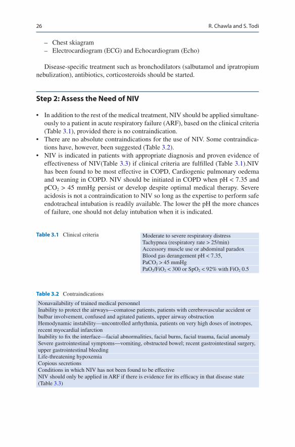
26
– Chest skiagram – Electrocardiogram (ECG) and Echocardiogram (Echo)
Disease-specific treatment such as bronchodilators (salbutamol and ipratropium nebulization), antibiotics, corticosteroids should be started.
Step 2: Assess the Need of NIV
• In addition to the rest of the medical treatment, NIV should be applied simultane-ously to a patient in acute respiratory failure (ARF), based on the clinical criteria (Table 3.1), provided there is no contraindication.
• There are no absolute contraindications for the use of NIV. Some contraindica-tions have, however, been suggested (Table 3.2).
• NIV is indicated in patients with appropriate diagnosis and proven evidence of effectiveness of NIV(Table 3.3) if clinical criteria are fulfilled (Table 3.1).NIV has been found to be most effective in COPD, Cardiogenic pulmonary oedema and weaning in COPD. NIV should be initiated in COPD when pH < 7.35 and pCO2 > 45 mmHg persist or develop despite optimal medical therapy. Severe acidosis is not a contraindication to NIV so long as the expertise to perform safe endotracheal intubation is readily available. The lower the pH the more chances of failure, one should not delay intubation when it is indicated.
Table 3.1 Clinical criteria Moderate to severe respiratory distressTachypnea (respiratory rate > 25/min)Accessory muscle use or abdominal paradoxBlood gas derangement pH < 7.35, PaCO2 > 45 mmHgPaO2/FiO2 < 300 or SpO2 < 92% with FiO2 0.5
Table 3.2 Contraindications
Nonavailability of trained medical personnelInability to protect the airways—comatose patients, patients with cerebrovascular accident or bulbar involvement, confused and agitated patients, upper airway obstructionHemodynamic instability—uncontrolled arrhythmia, patients on very high doses of inotropes, recent myocardial infarctionInability to fix the interface—facial abnormalities, facial burns, facial trauma, facial anomalySevere gastrointestinal symptoms—vomiting, obstructed bowel; recent gastrointestinal surgery, upper gastrointestinal bleedingLife-threatening hypoxemiaCopious secretionsConditions in which NIV has not been found to be effectiveNIV should only be applied in ARF if there is evidence for its efficacy in that disease state (Table 3.3)
R. Chawla and S. Todi
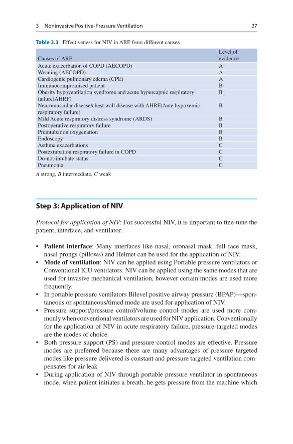
27
Step 3: Application of NIV
Protocol for application of NIV: For successful NIV, it is important to fine-tune the patient, interface, and ventilator.
• Patient interface: Many interfaces like nasal, oronasal mask, full face mask, nasal prongs (pillows) and Helmet can be used for the application of NIV.
• Mode of ventilation: NIV can be applied using Portable pressure ventilators or Conventional ICU ventilators. NIV can be applied using the same modes that are used for invasive mechanical ventilation, however certain modes are used more frequently.
• In portable pressure ventilators Bilevel positive airway pressure (BPAP)—spon-taneous or spontaneous/timed mode are used for application of NIV.
• Pressure support/pressure control/volume control modes are used more com-monly when conventional ventilators are used for NIV application. Conventionally for the application of NIV in acute respiratory failure, pressure-targeted modes are the modes of choice.
• Both pressure support (PS) and pressure control modes are effective. Pressure modes are preferred because there are many advantages of pressure targeted modes like pressure delivered is constant and pressure targeted ventilation com-pensates for air leak
• During application of NIV through portable pressure ventilator in spontaneous mode, when patient initiates a breath, he gets pressure from the machine which
Table 3.3 Effectiveness for NIV in ARF from different causes
Causes of ARFLevel of evidence
Acute exacerbation of COPD (AECOPD) AWeaning (AECOPD) ACardiogenic pulmonary edema (CPE) AImmunocompromised patient BObesity hypoventilation syndrome and acute hypercapnic respiratory failure(AHRF)
B
Neuromuscular disease/chest wall disease with AHRF(Aute hypoxemic respiratory failure)
B
Mild Acute respiratory distress syndrome (ARDS) BPostoperative respiratory failure BPreintubation oxygenation BEndoscopy BAsthma exacerbations CPostextubation respiratory failure in COPD CDo-not-intubate status CPneumonia C
A strong, B intermediate, C weak
3 Noninvasive Positive-Pressure Ventilation
28
is called Inspiratory Positive Airway Pressure (IPAP) which is applied all through inspiration. When the flow falls to a predetermined value set on the ventilator, patient gets a pressure which is applied all through expiration which is called Expiratory Positive Airway Pressure(EPAP). IPAP Improves ventilation and increases tidal volume. and helps in CO2 removal. EPAP improves oxygenation, opens up the Upper airway and neutralize auto PEEP. Pressure Support applied is the difference between IPAP and EPAP(PS = IPAP-EPAP).
Application of NIV Using Portable Pressure Ventilators
Explain the therapy and its benefit to the patient in detail. Also, discuss the possibil-ity of intubation.
• Choose the correct size interface. There are many types of interfaces like nasal mask, oronasal mask, total face mask and Helmet The oronasal mask is preferred in ARF.
• Set the NIV portable pressure ventilator in spontaneous or spontaneous/timed mode.
• Try to set machine parameters which are comfortable for the patient.• Always set the back up respiratory rate 2 to 3 less than patient respiratory rate.• Set I:E ratio 1:2 in COPD and 1:1 in OHS, NMD, CWD.• Rise Time is the time taken from EPAP to IPAP. In obstructive patients set Rise
time 100 ms to 400 ms(1 to 4) and in restrictive disease patient, set rise time 300 ms to 600 ms (from 3 to 6).
• Set max Inspiratory time 0.8 to 1.2 s (COPD) and 1.2 to 1.4 s in OHS,NMD,CWD.• Now Start titration with very low settings, low inspiratory positive airway pres-
sure (IPAP) of 6–8 cm H2O with 2–4 cm H2O of expiratory positive airway pres-sure (EPAP). The difference between IPAP and EPAP should be at least 4 cm H2O at all times.
• To start with administer oxygen at 2 L/min.• Hold the mask with the hand over the face. Do not fix it.• It is important to select proper size mask. Use the smallest possible mask.
Increase EPAP by 1–2 cm increments until the patient’s inspiratory efforts are able to trigger the ventilator.
• If the patient is making inspiratory effort and the ventilator does not respond, it indicates that the patient has not generated enough respiratory effort to counter auto-PEEP and trigger the ventilator (in COPD patients). Increase EPAP further until this happens. Most of the patients require EPAP of about 4–6 cm H2O. Patients who are obese or have obstructive sleep apnea require higher EPAP to trigger the ventilator.
• When the patient’s effort is triggering the ventilator, leave EPAP at that level.• Now, start increasing IPAP in increments of 1–2 cm up to a maximum pressure,
which the patient can tolerate without discomfort and there is no major mouth or air leak.
R. Chawla and S. Todi
29
• Now, secure interface with head straps. Avoid excessive tightness. If the patient has a nasogastric tube, put a seal connector in the dome of the mask to minimize air leakage.
• After titrating the pressure, increase oxygen to bring oxygen saturation to around 88 to 92% in hypercapnic respiratory failure. Oxygen should be entrained as close to the patient as possible. Adjust regularly oxygen flow required to main-tain Spo2 between 88 to 92%.
• As the settings may be different in wakefulness and sleep, readjust them accordingly.
• When NIV is being initiated for ARF, close monitoring and the capability to initiate endotracheal intubation and other resuscitation measures should be avail-able in the same setup. Start NIV preferably in the intensive care unit or in the emergency room in ARF.
• Humidification is not routinely required. Heated humidification can be used in cases of mucosal dryness or if respiratory secretions are thick and tenacious
• If the patient is dependent on NIV, bronchodilator drugs can be given via a nebu-liser inserted into the ventilator tubing.
Application of NIV Using a Critical Care Ventilator
There are several advantages of using NIV through standard ICU ventilator like one can give precise and also high concentration of oxygen. Separate inspiratory and expiratory tubing decrease the rebreathing of carbon dioxide. Large mask leaks and/or patient disconnections are appreciated quickly and it has better alarms and moni-toring features.
• The first step is to select a ventilator, which is capable of fulfilling the needs of the patient.
• Explain the therapy to the patient.• Choose the NIV mode. Pressure support or pressure control modes are preferred.
Standard critical care ventilators using flow-by system (noninvasive mode option) allow the patient to breathe without expending effort to open valves. In selected patients, such as those suffering from neuromuscular diseases, volume assist or volume control mode may be used.
• Choose an appropriate interface.• Silent ventilator alarms.• Keep FiO2 at 0.5.
Using pressure support approach
• Start with low settings such as inspiratory pressure support at 5–6 cm H2O and PEEP at 4 cm H2O.
• Initiate NIV while holding the mask in place and confirm optimum fit. If it is big or small or loose, change it.
3 Noninvasive Positive-Pressure Ventilation
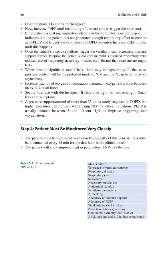
30
• Hold the mask. Do not fix the headgear.• Now, increase PEEP until inspiratory efforts are able to trigger the ventilator.• If the patient is making inspiratory effort and the ventilator does not respond, it
indicates that the patient has not generated enough respiratory effort to counter auto-PEEP and trigger the ventilator (in COPD patients). Increase PEEP further until this happens.
• Once the patient’s inspiratory efforts trigger the ventilator, start increasing pressure support further, keeping the patient’s comfort in mind. (Reduced respiratory rate, reduced use of respiratory accessory muscle, etc.) Ensure that there are no major leaks.
• When there is significant mouth leak, there may be asynchrony. In that case, pressure control will be the preferred mode of NIV and the Ti can be set to avoid asynchrony.
• Increase fraction of oxygen concentration to maintain oxygen saturation between 88 to 92% at all times.
• Secure interface with the headgear. It should be tight, but not overtight. Small leaks are acceptable.
• A pressure support/control of more than 25 cm is rarely required in COPD, but higher pressures can be used when using NIV for other indications. PEEP is usually titrated between 5 and 10 cm H2O to improve triggering and oxygenation.
Step 4: Patient Must Be Monitored Very Closely
• The patient must be monitored very closely clinically (Table 3.4). All this must be documented every 15 min for the first hour in the clinical notes.
• The patient will show improvement in parameters if NIV is effective.
Table 3.4 Monitoring of NIV in ARF
Mask comfortTolerance of ventilator settingsRespiratory distressRespiratory rateSensoriumAccessory muscle useAbdominal paradoxVentilator parametersAir leakingAdequacy of pressure supportAdequacy of PEEPTidal volume (5–7 mL/kg)Patient–ventilator synchronyContinuous oximetry (until stable)ABG, baseline and 1–2 h, then as indicated
R. Chawla and S. Todi

31
• ABG sample should be sent after 30 min to 1 h after the application of noninva-sive ventilation.
• To control pH and pCO2-manipulate the minute ventilation, the respiratory rate and tidal volume. So Increase IPAP to increase tidal volume or If using Pressure Control ventilation, increase Peak inspiratory pressure.
• To control pO2-adjust the FiO2 and the mean airway pressure (PEEP and PIP)• Increasing the PEEP or EPAP is the most efficient way of increasing the MAP• One can also increase the I-time to increase the MAP (PC).• In ventilator setting, look for air leaks, triggering and patient–ventilator
interaction.• Time on NIV should be maximised in the first 24 h depending on patient toler-
ance and/or complications.• NIV can be discontinued when there has been normalisation of pH and pCO2 and
a general improvement in the patient’s condition
Step 5: Continuously Look for Complications and Manage them Table 3.5
• Monitor carefully for the worsening respiratory distress, sensorium, tachypnea, and deteriorating blood gases, and intervene early because delay in intubation is a common major complication of NIV.
• Most complications are minor that can be managed easily, and so every attempt should be made to continue NIV (Table 3.5).
Table 3.5 Complications of NIVand corrective action
Complications Corrective action• Mask discomfort• Excessive leaks around mask• Pressure sores
• Check mask for correct size & fit• Minimize headgear tension• Use spacers or change to a different mask• Use wound care dressing over nasal bridge
• Nasal or oral dryness or nasal congestion
• Add or increase humidification• Irrigate nasal passage with saline• Apply topical decongestants
• Aerophagia/gastric distension • Use lowest effective pressure for adequate tidal volume• Use simethicone agents
• Aspiration • Make sure patients are able to protect the airway• Mucus plugging • Ensure adequate hydration
• Ensure adequate humidification• Avoid excessive O2 flow rates (>20 L/Min)• Allow short breaks from NIV to permit directed
coughing techniques• Hypotension • Avoid excessively high peak pressure (≥20 cm H2O)
3 Noninvasive Positive-Pressure Ventilation

32
• It is extremely important for the air seal to be tight. Ulceration and pressure necrosis related to local skin effects commonly occur at the bridge of the nose. Protective synthetic coverings may help prevent skin breakdown and ulceration on the bridge of the nose.
• Eye irritation and pain or congestion of the nasal sinuses may occur. Put some decongestant nasal drops.
• Distension of the stomach due to aerophagia and aspiration can occur secondary to vomiting. A nasogastric tube can be used to relieve the distension while still allowing the mask to seal.
• Adverse hemodynamic effects from NIV are unusual, although preload reduc-tion and hypotension may occur. Give intravenous fluids.
Step 6: Discontinuation of NIV
It is very important to know when to discontinue NIV and intubate and ventilate the patient.
• NIV failure. – Worsening mental status – Deterioration of pH and PaCO2 after 1–3 h of therapy – Refractory hypoxemia—when even a brief discontinuation of NIV leads to
significant fall in oxygen saturation• Intolerance to NIV.• Hemodynamic instability.• Inability to clear secretions.
Step 7: Weaning
• Initially, give NIV continuously as long as possible.• Once the patient is tolerating periods off NIV, start discontinuing during daytime
and give during nighttime. In 2–3 days, the patient can be weaned off the NIV.• A brief outline of the application of NIV is shown in Fig. 3.1.
R. Chawla and S. Todi

33
Check for underlyingDisease
Level of evidence A, B for NIVRef. to table 3.3
Need for ventilatory requirement
Supportivemedical therapy
Good candidatefor NIV
Check for contraindicationsTable 3.2
Start NIV with use of appropriatefacemask and oxygenUse pressure support or CPAP mode
1-2 hour check pointImprove gas exchange Patient tolerance
Start intermittentdiscontinuation of NIV
- Patient remains stable- No respiratory distress
DiscontinueNIV
Level of evidence C with for NIVRef. to table 3.3
Assess for use of NIV
Poor candidate for NIV
Intubate
Consider NIVfor extubation
in selectedcases
ContinueNIV
Yes
No
YesNo
No Yes
Acute respiratory distressTachypnea RR >25/min
Respiratory failurePaCO2>45, and/or pH <7.35
Inprovement in respiratory failureand primary disease, O2 saturation
>90% on 6 lit / min O2
••
Fig. 3.1 Application of NIV
3 Noninvasive Positive-Pressure Ventilation

34
Suggested Reading
Ambrosino N, Vagheggini G. Noninvasive positive pressure ventilation in the acute care setting: where are we? Eur Respir J. 2008;31(4):874–86. A comprehensive review focusing on recent developments for the application of NIV in the treatment of acute and acute-on-chronic respira-tory failure
Confalonieri M, Garuti G, Cattaruzza M-S, et al. A chart of failure risk for noninvasive ventilation in patients with COPD exacerbation. Eur Respir J. 2005;25(2):348–55.j.
Davidson AC, et al. BTS/ICS guideline for the ventilatory management of acute hypercapnic respi-ratory failure in adults. Thorax. 2016;71:ii1–ii35. A comprehensive guidelines for the ventila-tory management in acute hypercapnic respiratory failure
Majid A, Hill NS. Noninvasive ventilation for acute respiratory failure. Curr Opin Crit Care. 2005;11(1):77–81. This review critically examines the recent literature related to applications of noninvasive ventilation in the acute setting
R. Chawla and S. Todi

35© Springer Nature Singapore Pte Ltd. 2020R. Chawla, S. Todi (eds.), ICU Protocols, https://doi.org/10.1007/978-981-15-0898-1_4
R. Chawla (*) Department of Respiratory, Critical Care & Sleep Medicine, Indraprastha Apollo Hospitals, New Delhi, India
S. Todi Department of Critical Care & Emergency Medicines, A.M.R.I. Hospital, Kolkata, India
4High Flow Nasal Canula (HFNC)
Rajesh Chawla and Subhash Todi
In patient of hypoxemic respiratory failure, oxygen is applied via variable low- flow systems (e.g., nasal cannulae or face masks) or high-flow systems (e.g., Venturi masks, nonrebreathing reservoir mask) (Fig. 4.1). These conventional systems have lot of disadvantages. They deliver unreliable fraction of inspired oxygen and pro-vide inadequate warming and humidification of inspired gas. In a patients with acute respiratory failure, peak inspiratory flow rate is high and often exceeds the oxygen flow delivered by these traditional oxygen devices, which results in flow starvation. High-flow nasal cannula (HFNC) oxygen therapy overcomes these limi-tations. HFNC is a technique of Oxygen therapy which delivers heated and humidi-fied oxygen through a nasal cannula at high flow rates (~60 L/min)at higher oxygen concentration (FiO2 0.21–1). This has been found noninferior to NIV in mild hypox-emic respiratory failure and is recommended in some other clinical conditions. Moreover complications associated with NIV like lung injury and patient dysyn-chrony is increasingly being recognised.
A 42 years old female, non smoker presented with fever, nasal discharge, cough and shortness of breath for the last five days. On examination, patient was con-scious, RR 38/min, SPO2 80% on nasal prongs with 5 L/min, BP 130/70. His TLC was 5.2. Chest X-ray revealed bilateral infiltrates. Her throat swab came out as + H1N1. She was started on nonrebreathing mask with oxygen flow at 15 L/m. His SpO2 was still 86%. Patient was started on Heated Humidified high flow nasal canula at 60 L/m. She became comfortable and RR came down to 23/m after 10 min.
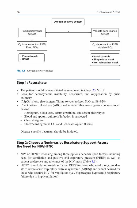
36
Step 1: Resuscitate
• The patient should be resuscitated as mentioned in Chap. 23, Vol. 2.• Look for hemodynamic instability, sensorium, and oxygenation by pulse
oximetry.• If SpO2 is low, give oxygen. Titrate oxygen to keep SpO2 at 88–92%.• Check arterial blood gas (ABG) and initiate other investigations as mentioned
below: – Hemogram, blood urea, serum creatinine, and serum electrolytes – Blood and sputum culture if infection is suspected – Chest skiagram – Electrocardiogram (ECG) and Echocardiogram (Echo)
Disease-specific treatment should be initiated.
Step 2: Choose a Noninvasive Respiratory Support- Assess the Need for NIV/HFNC
• NIV or HFNC: Choosing among these options depends upon factors including need for ventilation and positive end expiratory pressure (PEEP) as well as patient preference and tolerance of the NIV mask (Table 4.1).
• HFNC is unlikely to provide sufficient PEEP for those who need it (e.g., moder-ate to severe acute respiratory distress syndrome [ARDS]) and cannot be used for those who require NIV for ventilation (i.e., hypercapnic hypoxemic respiratory failure due to hypoventilation).
Oxygen delivery system
Variable performancedevices
Fixed performancedevices
O2 Independent on PIFRFixed FiO2
• Venturi mask• HFNC
• Nasal cannula• Simple face mask• Non rebreather mask
O2 dependent on PIFRVariable FiO2
Fig. 4.1 Oxygen delivery devices
R. Chawla and S. Todi

37
• It is also unlikely to reduce the work of breathing as effectively as NIV.• There are two group of patients who are not given HFNC are those who improve
on simple nasal canula and those who are intubated.• Although so far there are no defined clearcut indications of HFNC but it is being
used for a variety of clinical conditions.• HFNC has lot of physiological advantages as mentioned below.
– HFNC provides very high flow rates which can be adjusted to meet or exceed the patient’s inspiratory flow rate demands. This leads to less entrain-ment of room air, so it provides reliably delivered FiO2 and maintains a constant FiO2 despite a high patient minute ventilation. FiO2 may be titrated from) 0.21 to 1.0 by air/oxygen blender. High flow rates result in an improved breathing pattern by decreasing respiratory rate and increasing tidal volume (Fig. 4.2).
– HFNC provides an anatomical oxygen reservoir in the nasopharynx and oro-pharynx due to a CO2 washout effect as a result of the high oxygen flow. This reduces dead space and the work of breathing in turn. This also helps in decreased rebreathing of CO2 in hypercapnic patient.
– The continuous delivery of high flow, which impedes expiratory flow, gener-ates a degree of PEEP. This PEEP effect results in increased end-expiratory lung volume, and thus alveolar recruitment. Flow rates of around 35 to 60 L/min generate mean expiratory pressures of 2 to 3 cm H2O with the mouth open and 5 to 7 cm H2O with the mouth closed.
– The splinting of the upper airway also has the effect of reducing airflow resis-tance in the nasopharynx, thereby reducing the work of breathing.
– The metabolic load associated with warming and increasing the humidity of inspired gas is averted as the gas is optimally conditioned before delivery in HFNC.
– Conditioning of gas prevents airway desiccation, improves mucociliary func-tion, facilitates clearance of secretions, and is associated with less atelectasis.
– Conventional oxygen devices and NIV delivering dry and unwarmed gas are associated with mask discomfort, claustrophobia, oronasal dryness, eye irrita-tion, nasal and eye trauma, gastric distension, and aspiration.
Table 4.1 Factors that determine which system to use
1. Increased work of breathing: NIV preferable
2. Need of high FiO2: HFNC preferable3. Need of humidification: HFNC preferable4. Hypercapnia: NIV preferable5. Efficient & economical use of oxygen: NIV
preferable6. Comfort and acceptance: HFNC preferable
4 High Flow Nasal Canula (HFNC)

38
Indications (Table 4.2)
Acute Mild Hypoxemic Respiratory Failure
• The main use of HFNC is to provide a relatively high FiO2 to patients with acute hypoxemic respiratory failure (AHRF) with PaO2/FiO2 ratio 150–300.
• HFNC has successfully been used in AHRF due to severe influenza, ARDS, con-gestive heart failure, hematologic malignancies and organ transplant.
Physiologicalbenefits
FIO2 valuesHigher &Stable
Anatomicaldead space is
reduced
ReducesWOB
Gasdelivered isHumidified
ProvidesPositiveairway
pressure
ReducesAirway
resistance
Fig. 4.2 Physiological benefits of HFNC
Table 4.2 Indications of HFNC
Clinical condition Evidence LevelAcute mild hypoxemic RF Meta-analysis and RCT ∗∗∗Immunocompromised patients RCT ∗∗Post extubation RF RCT ∗∗Post cardiac surgery hypoxemia RCT ∗∗Pre-intubation oxygenation RCT ∗During bronchoscopy RCT ∗COPD RCT ∗Palliative
R. Chawla and S. Todi
39
• The results of HFNC use failed to consistently demonstrate an improvement in mortality, intubation rates, length of stay in nonhypercapnic hypoxemic respira-tory failure.
• A meta-analysis of 13 randomized trials failed to demonstrate a decrease in the rate of intubation or mortality.
• Most of trials in ARDS have consistently shown that HFNC decreases the work of breathing, decrease respiratory rate and increases patient comfort.
Cardiogenic Pulmonary Edema
• HFNC has been shown to improve oxygenation, comfort, respiratory rate and heart rate in patients with AHRF.
• However, HFNC has not shown a consistent and convincing benefit in intubation rates and survival in Cardiogenic pulmonary edema.
Preintubaton Oxygenation
• HFNC is an acceptable way of providing oxygen to patients both before (preoxy-genation) and during (to prevent desaturation) endotracheal intubation.
• NIV for pre-oxygenation must be stopped during laryngoscopy, and thus does not enable oxygenation during intubation. In contrast, HFNC therapy can be continued uninterrupted throughout thereby provided prolonged non hypoxemic apnea time.
• Post extubation respiratory compromise: Elective use of HFNC post extubation has been found to reduce reintubation rates in post cardiothoracic surgery, abdominal surgery or general ICU patients who are both at high or low risk for reintubation. HFNC performed equally well compared to NIV in these circum-stances and was more acceptable to patients.
• HFNC improves physiological parameters like respiratory rate, and increase end-expiratory lung volume.
• It decreases the requirement for CPAP and re-intubation rates but does not improve P/F ratio and atelectasis.
• Post extubation HFNC can be used successfully in prevention and treatment of postoperative respiratory failure.
During Invasive Procedures
• Invasive procedures that are associated with hypoxemia.• Hypoxemic patients undergoing fibreoptic bronchoscopy can successfully be
oxygenated with HFNC.
4 High Flow Nasal Canula (HFNC)
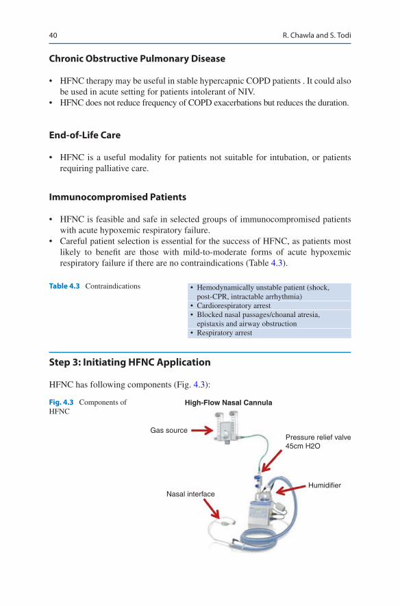
40
Chronic Obstructive Pulmonary Disease
• HFNC therapy may be useful in stable hypercapnic COPD patients . It could also be used in acute setting for patients intolerant of NIV.
• HFNC does not reduce frequency of COPD exacerbations but reduces the duration.
End-of-Life Care
• HFNC is a useful modality for patients not suitable for intubation, or patients requiring palliative care.
Immunocompromised Patients
• HFNC is feasible and safe in selected groups of immunocompromised patients with acute hypoxemic respiratory failure.
• Careful patient selection is essential for the success of HFNC, as patients most likely to benefit are those with mild-to-moderate forms of acute hypoxemic respiratory failure if there are no contraindications (Table 4.3).
Step 3: Initiating HFNC Application
HFNC has following components (Fig. 4.3):
Table 4.3 Contraindications • Hemodynamically unstable patient (shock, post-CPR, intractable arrhythmia)
• Cardiorespiratory arrest• Blocked nasal passages/choanal atresia,
epistaxis and airway obstruction• Respiratory arrest
Fig. 4.3 Components of HFNC
High-Flow Nasal Cannula
Gas source
Nasal interfaceHumidifier
Pressure relief valve45cm H2O
R. Chawla and S. Todi
41
• Gas source, blender: An air/oxygen blender, allows the inspiratory fraction of oxygen (FiO2) from 0.21 to 1.0 in a flow of up to 60–70 L/min. The flow rate (ranging from 5 to 60 L/min) and FiO2 (0.21 to 1) are set at the air/oxygen blender.
• Humidifier: Humidity can be provided by a disposable vapour transfer cartridge, a heated plate humidifier or a bubble humidifier. The gas is heated and humidi-fied through an active heated humidifier and delivered via a single-limb heated inspiratory circuit. The patient breathes the heated and humidified gas through nasal wide bore cannulas.
• Nasal interface: Nasal cannulas are available in different sizes. Most interfaces have wide-bore, soft contoured nasal prongs designed to reduce gas jetting, while some use the traditional narrow-bore nasal cannula.
The gas is heated to 37 C and humidified 100% with the active humidifier and is deliv-ered through the heated circuit through a nasal canula at high flow at least 60 L/min.
HFNC should be applied if there is no contradictions.
Connecting the Patient
• Explain the therapy to the patient.• Check the proper sized nasal prongs that sits well into the nares. Nasal cannulae
should fit snugly and it should be ensured that nasal cannulae occlude no more than 50% of the nostril.
• HFNC can be started with flow rates of around 30 L/min. Flow rates should be increased in 5 to 10 L/min increments, aiming to reduce respiratory rate, or until further increases are not tolerated. Patient discomfort is usually due to the veloc-ity of gas rather than the flow itself, and can be mitigated by using a large-bore cannula. – In case of persistent discomfort, the flow can be decreased down to a mini-
mum of 30 L/min• Flows should be titrated to a maximum flow of 60 L/min depending on patient
comfort.• Select the FiO2 to obtain the desired arterial oxygen saturation. Within 2 h it
should be possible to reduce the FiO2 and clinical stabilization should be observed.
• Set humidifier on the desired temperature. Temperature may be reduced if the patient complains that the gas is too warm.
Step 5: Monitoring of a Patient on HFNC
Monitoring should be done as in any other patient on any respiratory support ICU (Table 4.4).
4 High Flow Nasal Canula (HFNC)
42
Table 4.4 Monitoring during HFNC application
ComfortToleranceRespiratory distressRespiratory rateSensoriumAccessory muscle useAbdominal paradoxContinuous oximetry (until stable)ABG, baseline and 1–2 h, then as indicated
• HFNC therapy should be provided in controlled setting with close monitoring for signs of respiratory failure that necessitate intubation and mechanical ventilation.
• Look for failure to adequately improve oxygenation within an hour of HFNC initiation, and increase of respiratory rate and presence of thoracoabdominal asynchrony. Discontinue if the patient is not improving or deteriorating. Delaying intubation increases mortality like in a patient on NIV.
• In most cases, nebulized medication is given directly by mouthpiece and not delivered through HFNC equipment. However, aerosol delivery may not be guar-anteed at high flows.
• HFNC can be delivered in a less controlled environment once the patient is improving.
Step 6: Look for Complications and Treat them
• HFNC is usually well tolerated and complications are rare in adults.• Blocked cannulae due to secretions.• Local trauma, discomfort and pressure areas, epistaxis, abdominal distension,
aspiration, and, rarely, barotrauma.• Failure of HFNC may result in delayed intubation and worse clinical outcomes
in patients with respiratory failure.
Step 7: Discontinuation of HFNC
• This should begin with FiO2, while maintaining a steady flow rate.• Once FiO2 is <0.4 to 0.5, than reduce flow rates.• Convert to conventional low-flow oxygen once the flow rate reaches ≤20 L/min
and FiO2 ≤ 0.4.
R. Chawla and S. Todi

43
Step 8: Maintain HFNC Device
• HFNC devices should be cleaned and disinfected before use in a new patient.• Consumables need to be replaced for every new patient.• For prolonged treatment, the breathing tube and humidifying chamber kit should
be replaced every 14 days, and the patient interface every 7 days.
Conclusions: Much of the proven benefit of HFNC is subjective and physio-logic. HFNC is an alternative to other high-flow systems and NIV. The choice between these systems depends on patient and clinician preference, need for venti-lation and PEEP, severity of hypoxemia and institutional availability.
Suggested Reading
Guitton C, Ehrmann S, Volteau C, Colin G, Maamar A, Jean-Michel V, Mahe P, Landais M, Brule N, Bretonnière C, Zambon O, Vourc’h M. Nasal high-flow preoxygenation for endo-tracheal intubation in the critically ill patient: a randomized clinical trial. Intensive Care Med. 2019;45(4):447–5. Compared with standard bag mask oxygenation, preoxygenation with HFNC in the ICU did not improve the lowest SpO2 during intubation in the non-severely hypox-emic patients but led to a reduction in intubation-related adverse events
Hare A. High-flow nasal cannula therapy in adults. Clin Pulm Med. 2017;24(3):95–104. A review article
Helviz Y, Einav S. A systematic review of the high-flow nasal cannula for adult patients. Crit Care. 2018;22(1):71. A systematic review of the high-flow nasal cannula for adult patients
Hill NS. High flow nasal cannula, is there a role in COPD? Tanaffos. 2017;16(Suppl 1):S12. A reviw article
Mauri T, Turrini C, Eronia N, Grasselli G, Volta CA, Bellani G, et al. Physiologic effects of high-flow nasal cannula in acute hypoxemic respiratory failure. Am J Respir Crit Care Med. 2017;195:1207–15. HFNC exerts multiple physiologic effects including less inspiratory effort and improved lung volume and compliance in acute hypoxemic respiratory failure. These ben-efits might underlie the clinical efficacy of HFNC
Nishimura M. High-flow nasal cannula oxygen therapy in adults. J Intensive Care. 2015;3:15–23. A review article
Ou X, Hua Y, Liu J, Gong C, Zhao W. Effect of high-flow nasal cannula oxygen therapy in adults with acute hypoxemic respiratory failure: a meta-analysis of randomized controlled trials. Can Med Assoc J. 2017;189(7):E260–7. The intubation rate with HFNC oxygen therapy was lower than the rate with conventional oxygen therapy and similar to the rate with noninvasive ventila-tion among patients with acute hypoxemic respiratory failure
4 High Flow Nasal Canula (HFNC)
45© Springer Nature Singapore Pte Ltd. 2020R. Chawla, S. Todi (eds.), ICU Protocols, https://doi.org/10.1007/978-981-15-0898-1_5
G. C. Khilnani (*) PSRI Institute of Pulmonary, Critical Care and Sleep Medicine, PSRI Hospital, New Delhi, India
V. Hadda Department of Pulmonary Medicine, All India Institute of Medical Sciences, Delhi, India
5Basic Mechanical Ventilation
Gopi Chand Khilnani and Vijay Hadda
Mechanical ventilation is indicated in patients with acute respiratory failure when they are unable to maintain adequate oxygenation and/or remove carbon dioxide. None of the ventilatory mode can cure the disease process. However, it supports ventilation till you address the reversible primary problem. Mechanical ventilation should not be started without thoughtful consideration as tracheal intubation and ventilation are associated with significant complications.
Step 1: Initial Resuscitation (Refer to Chap. 23, Vol. 2)
• Any patient coming to the emergency department should be examined quickly to assess oxygenation and hemodynamic status. Resuscitation should be started without delay.
• All patients with respiratory distress require immediate attention to airway.• They should be put on supplemental oxygen via nasal cannula or face mask
devices (set at flow rate > 4 L or a high-flow oxygen device (Flow rate 40–60 L/min) (HFNC) wherever indicated.
A 30-year-old male patient came to the emergency department with history of fever, shortness of breath for 3 days, and alteration in sensorium since morning. He was in respiratory distress with respiratory rate of 34/min. He was drowsy (Glasgow coma score 8) and febrile (102 °F). His pulse was 110/min, blood pressure was 116/78 mmHg, and JVP was normal. He was immediately intu-bated and put on assisted ventilation.
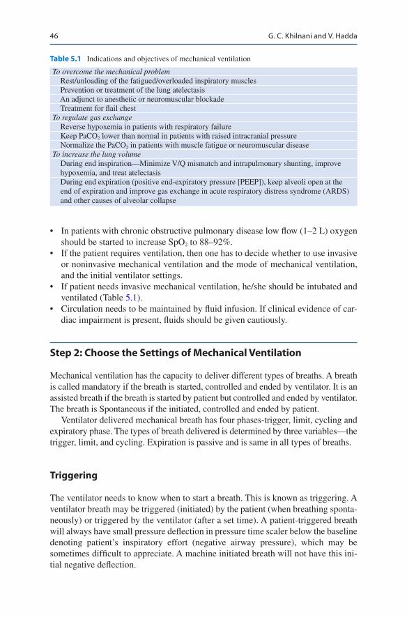
46
• In patients with chronic obstructive pulmonary disease low flow (1–2 L) oxygen should be started to increase SpO2 to 88–92%.
• If the patient requires ventilation, then one has to decide whether to use invasive or noninvasive mechanical ventilation and the mode of mechanical ventilation, and the initial ventilator settings.
• If patient needs invasive mechanical ventilation, he/she should be intubated and ventilated (Table 5.1).
• Circulation needs to be maintained by fluid infusion. If clinical evidence of car-diac impairment is present, fluids should be given cautiously.
Step 2: Choose the Settings of Mechanical Ventilation
Mechanical ventilation has the capacity to deliver different types of breaths. A breath is called mandatory if the breath is started, controlled and ended by ventilator. It is an assisted breath if the breath is started by patient but controlled and ended by ventilator. The breath is Spontaneous if the initiated, controlled and ended by patient.
Ventilator delivered mechanical breath has four phases-trigger, limit, cycling and expiratory phase. The types of breath delivered is determined by three variables—the trigger, limit, and cycling. Expiration is passive and is same in all types of breaths.
Triggering
The ventilator needs to know when to start a breath. This is known as triggering. A ventilator breath may be triggered (initiated) by the patient (when breathing sponta-neously) or triggered by the ventilator (after a set time). A patient- triggered breath will always have small pressure deflection in pressure time scaler below the baseline denoting patient’s inspiratory effort (negative airway pressure), which may be sometimes difficult to appreciate. A machine initiated breath will not have this ini-tial negative deflection.
Table 5.1 Indications and objectives of mechanical ventilation
To overcome the mechanical problem Rest/unloading of the fatigued/overloaded inspiratory muscles Prevention or treatment of the lung atelectasis An adjunct to anesthetic or neuromuscular blockade Treatment for flail chestTo regulate gas exchange Reverse hypoxemia in patients with respiratory failure Keep PaCO2 lower than normal in patients with raised intracranial pressure Normalize the PaCO2 in patients with muscle fatigue or neuromuscular diseaseTo increase the lung volume During end inspiration—Minimize V/Q mismatch and intrapulmonary shunting, improve
hypoxemia, and treat atelectasis During end expiration (positive end-expiratory pressure [PEEP]), keep alveoli open at the
end of expiration and improve gas exchange in acute respiratory distress syndrome (ARDS) and other causes of alveolar collapse
G. C. Khilnani and V. Hadda

47
Ventilators use signals from various sites from within the ventilator circuit. The trigger signal can be sensed at the proximal endotracheal tube, in the inspiratory limb, and in the expiratory limb of the circuit. The trigger signal can be pressure, flow, time and neural signal.
Pressure triggering: This requires the patient to generate a small negative inspira-tory pressure (generally negative 1–3 cm H2O). This negative pressure is sensed by the ventilator, causing the ventilator to start inspiration and deliver the next breath.
Flow triggering: In this a minimum of flow around 10 L/m is always present in the ventilator circuit (Bias Flow). Flow triggering occurs when a flow transducer in the patient/ventilator systems senses a change in flow i.e. flow moves in to airway opening. Usually flow trigger is kept at 2 L/m. This is the preferable triggering mode in spontaneously breathing patient.
Time triggering: A breath is time triggered when the patient does not initiate a breath and ventilator delivers a breath after a set time (depends on the set respiratory rate).This is the default setting in patient who do not have spontaneous breathing effort (e.g. on neuromuscular blocker).
Neural triggering: It is currently developed to minimise the delay interval between the generation of the signal to breathe in the brain (sensed by diaphrag-matic muscle signals) and the actual delivery of flow from the ventilator.
Limit or Inspiratory Phase
This is the phase of the ventilator delivered breath that begins with the initiation of the breath, and ends when the ventilator stops inspiratory flow. Inspiratory valve is open and the expiratory valve is closed. During the inspiratory phase, air flow is determined by variables called limit variables which could be either pressure or flow. The limit variable does not terminate the inspiration; it allows inspiration to continue till the cycling criterion is reached.
Cycling: Changeover from Inspiration to Expiration
This is known as ‘cycling.’ Cycling defines how the ventilator recognizes that the inspiratory phase is over, and expiration starts with opening of the expiratory valve. Ventilators may cycle (changeover to expiration) when a certain tidal volume (set inspiratory tidal volume, volume cycled), inspiratory time (time cycled), flow rate (flow cycled). Pressure controlled ventilation is time cycled, volume controlled ven-tilation without pause is volume cycled and Pressure support mode is flow cycled.
In the expiratory phase, the inspiratory valve is closed and the expiratory valve is open.
• Once it has been decided that patient requires mechanical ventilation, the next step is to choose the appropriate mode of mechanical ventilation. Mode specifies the manner in which mechanical breath is delivered.
5 Basic Mechanical Ventilation

48
• The characteristics of commonly used modes of mechanical ventilation are sum-marized in Table 5.2. Assist-control mechanical ventilation (ACMV) is the most widely used method.
• Assist-control mode can be volume-controlled (more commonly used) or pressure- controlled. The differences between two are summarized in Table 5.3.
• The ventilatory support can be controlled mandatory ventilation (CMV) or assisted controlled mandatory ventilation (ACMV) depending on the use of patients’ effort for triggering the ventilator.
• The patient’s ventilation is totally controlled by the ventilator and the patient cannot trigger the ventilator during CMV, while ACMV delivers controlled breaths as well as assists patient-triggered breath.
Table 5.2 Characteristics of commonly used modes of mechanical ventilation
Mode Type of breathTriggering mechanism
Cycling mechanism Comments
CMV Each breath delivers a preset mechanical tidal volume or pressure
Time Inspiration is terminated by delivery of preset tidal volume or by time in pressure- controlled
Used in patients who have no respiratory effort
ACMV Each breath, assist or control, delivers a preset mechanical tidal volume or pressure
Either patient- triggered (assist) or time-triggered (controlled)
Inspiration is terminated either by delivery of preset tidal volume (volume controlled) or by time in pressure- controlled
Alveolar hyperventilation may be a potential hazard
SIMV Mechanical tidal breath at preset rate and the patient may breathe spontaneously between mandatory breaths
Mandatory breath may be time-triggered or patient- triggered
Mandatory breaths are volume- or time-cycled in; pressure- controlled patient controls spontaneous rate and volume
It provides an interval (synchronization window) just prior to triggering during which the ventilator is responsive to patient’s effort and supports spontaneous breath
PSV Pressure-support breaths are considered as spontaneous
Patient- triggered
The breaths are flow cycled by a set flow threshold, which is generated by the patient
This a commonly used mode for weaning
CMV controlled mandatory ventilation, ACMV assist-control mechanical ventilation, IMV inter-mittent mandatory ventilation, SIMV synchronized intermittent mandatory ventilation, PSV pressure- support ventilation
G. C. Khilnani and V. Hadda
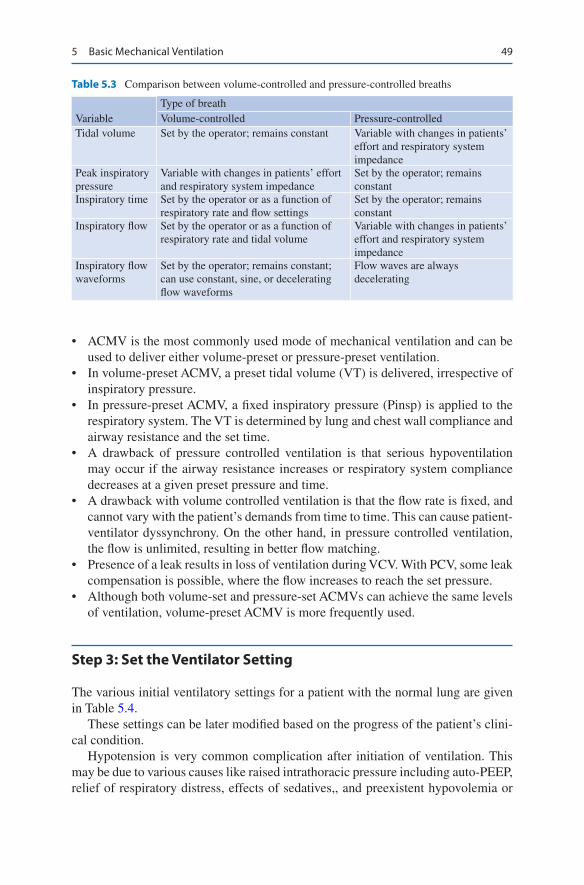
49
• ACMV is the most commonly used mode of mechanical ventilation and can be used to deliver either volume-preset or pressure-preset ventilation.
• In volume-preset ACMV, a preset tidal volume (VT) is delivered, irrespective of inspiratory pressure.
• In pressure-preset ACMV, a fixed inspiratory pressure (Pinsp) is applied to the respiratory system. The VT is determined by lung and chest wall compliance and airway resistance and the set time.
• A drawback of pressure controlled ventilation is that serious hypoventilation may occur if the airway resistance increases or respiratory system compliance decreases at a given preset pressure and time.
• A drawback with volume controlled ventilation is that the flow rate is fixed, and cannot vary with the patient’s demands from time to time. This can cause patient- ventilator dyssynchrony. On the other hand, in pressure controlled ventilation, the flow is unlimited, resulting in better flow matching.
• Presence of a leak results in loss of ventilation during VCV. With PCV, some leak compensation is possible, where the flow increases to reach the set pressure.
• Although both volume-set and pressure-set ACMVs can achieve the same levels of ventilation, volume-preset ACMV is more frequently used.
Step 3: Set the Ventilator Setting
The various initial ventilatory settings for a patient with the normal lung are given in Table 5.4.
These settings can be later modified based on the progress of the patient’s clini-cal condition.
Hypotension is very common complication after initiation of ventilation. This may be due to various causes like raised intrathoracic pressure including auto-PEEP, relief of respiratory distress, effects of sedatives,, and preexistent hypovolemia or
Table 5.3 Comparison between volume-controlled and pressure-controlled breaths
VariableType of breathVolume-controlled Pressure-controlled
Tidal volume Set by the operator; remains constant Variable with changes in patients’ effort and respiratory system impedance
Peak inspiratory pressure
Variable with changes in patients’ effort and respiratory system impedance
Set by the operator; remains constant
Inspiratory time Set by the operator or as a function of respiratory rate and flow settings
Set by the operator; remains constant
Inspiratory flow Set by the operator or as a function of respiratory rate and tidal volume
Variable with changes in patients’ effort and respiratory system impedance
Inspiratory flow waveforms
Set by the operator; remains constant; can use constant, sine, or decelerating flow waveforms
Flow waves are always decelerating
5 Basic Mechanical Ventilation

50
other cardiac compromise. This should be kept in mind and should be prevented or treated initially with fluids. FiO2 may be initially set at 1.0 as one may not be aware of the oxygen requirement of patient and later decrease to minimum in order to maintain target oxygenation.
• During mechanical ventilation the oxygenation is determined by the FiO2, PEEP and mean airway pressure and PaCO2 is determined by minute ventilation.
Step 4: Set Alarms
The following alarms need to be set:
• Peak pressure—high/low—10–15 cm above or below the peak inspiratory pressure
• Minute ventilation—high/low—50% above or below the set volume• Low exhaled tidal volume—50% of the delivered tidal volume• High respiratory rate• Set apnea ventilation parameters
Step 5: Connect the Ventilator to the Patient
• Connect the patient to the ventilator.
Table 5.4 Initial ventilatory setting
Mode—assist/control (volume or pressure)Tidal volume 6–8 mL/Kg ideal body weight (see formula in Appendix B)Inspiratory timeInspiratory flow
0.7–1.2 sFour times minute ventilation (approx)
Rate 12–20 breaths/minPEEP 4–5 cm H2OFiO2 1.0Plateau pressure <30 cm H2OOnce the patient is stabilizedFiO2 To maintain PaO2 more than 60 mmHg or SpO2 more than 93–94% in
normal lung and 88–92% in hypercapnic respiratory failurePEEP Set according to FiO2 requirements (predetermined according to the
degree of hypoxemia)Plateau pressureDriving pressure (plateau-PEEP)
Recheck in an attempt to keep plateau pressure below 30 cm H2OKeep below 13 cm H2O
G. C. Khilnani and V. Hadda
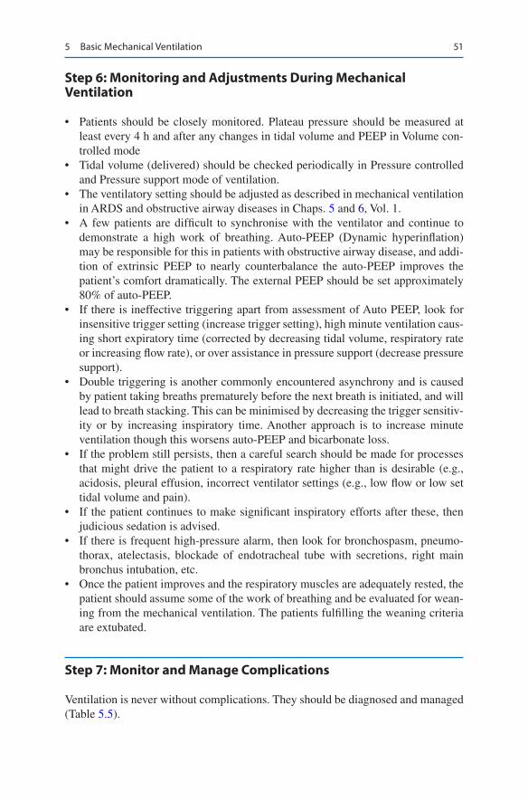
51
Step 6: Monitoring and Adjustments During Mechanical Ventilation
• Patients should be closely monitored. Plateau pressure should be measured at least every 4 h and after any changes in tidal volume and PEEP in Volume con-trolled mode
• Tidal volume (delivered) should be checked periodically in Pressure controlled and Pressure support mode of ventilation.
• The ventilatory setting should be adjusted as described in mechanical ventilation in ARDS and obstructive airway diseases in Chaps. 5 and 6, Vol. 1.
• A few patients are difficult to synchronise with the ventilator and continue to demonstrate a high work of breathing. Auto-PEEP (Dynamic hyperinflation) may be responsible for this in patients with obstructive airway disease, and addi-tion of extrinsic PEEP to nearly counterbalance the auto-PEEP improves the patient’s comfort dramatically. The external PEEP should be set approximately 80% of auto-PEEP.
• If there is ineffective triggering apart from assessment of Auto PEEP, look for insensitive trigger setting (increase trigger setting), high minute ventilation caus-ing short expiratory time (corrected by decreasing tidal volume, respiratory rate or increasing flow rate), or over assistance in pressure support (decrease pressure support).
• Double triggering is another commonly encountered asynchrony and is caused by patient taking breaths prematurely before the next breath is initiated, and will lead to breath stacking. This can be minimised by decreasing the trigger sensitiv-ity or by increasing inspiratory time. Another approach is to increase minute ventilation though this worsens auto-PEEP and bicarbonate loss.
• If the problem still persists, then a careful search should be made for processes that might drive the patient to a respiratory rate higher than is desirable (e.g., acidosis, pleural effusion, incorrect ventilator settings (e.g., low flow or low set tidal volume and pain).
• If the patient continues to make significant inspiratory efforts after these, then judicious sedation is advised.
• If there is frequent high-pressure alarm, then look for bronchospasm, pneumo-thorax, atelectasis, blockade of endotracheal tube with secretions, right main bronchus intubation, etc.
• Once the patient improves and the respiratory muscles are adequately rested, the patient should assume some of the work of breathing and be evaluated for wean-ing from the mechanical ventilation. The patients fulfilling the weaning criteria are extubated.
Step 7: Monitor and Manage Complications
Ventilation is never without complications. They should be diagnosed and managed (Table 5.5).
5 Basic Mechanical Ventilation

52
Step 8: Weaning from the Ventilator (Chap. 9, Vol. 1)
• Patients who have recovered considerably from the underlying diseases should be assessed daily for readiness of weaning (Table 5.6), and those satisfying cri-teria should be given a spontaneous breathing trial (SBT).
• Patients may be given SBT after they fulfill the weaning assessment criteria and monitored for 5 min for signs of SBT failure (Table 5.7). Those who successfully tolerate this the SBT trial should be prolonged for 30–120 min and if successful, can be extubated. If SBT trial fails patient should be reconnected to the ventilator.
Table 5.5 Complications of intubation and mechanical ventilation
Equipment Malfunction or disconnection Incorrect settingsPulmonary Airway intubation (e.g., damage to teeth, vocal cords, and trachea) Ventilator-associated pneumonia Ventilator-associated lung injury (e.g., diffuse lung injury due to regional overdistension or
tidal recruitment of alveoli) Overt barotraumas (e.g., pneumothorax) O2 toxicity Patient–ventilator asynchronyCirculation ↓ Right ventricular preload → ↓ cardiac output ↑ Right ventricular afterload (if the lung is overdistended) ↓ Splanchnic blood flow with high levels of PEEP or mean Paw↑ Intracranial pressure with high levels of PEEP or mean Paw Fluid retention due to ↓ cardiac output → ↓ renal blood flowOther Gut distension (air swallowing, hypomotility) Mucosal ulceration and bleeding Peripheral and respiratory muscle weakness Sleep disturbance, agitation, and fear (which may be prolonged after recovery) Neuropsychiatric complications
Table 5.6 Criteria for assessing readiness of patients for weaning
Some evidence of reversal of the underlying cause of respiratory failureAdequate oxygenation (e.g., PaO2/FiO2 > 200, requiring PEEP < 5–8 cm H2O, FiO2 < 0.4–0.5, and pH > 7.25)Hemodynamic stability, no active myocardial ischemia, no clinically significant hypotension or use of vasopressors (low dose vasopressor is acceptable)The capability to initiate an inspiratory effort
G. C. Khilnani and V. Hadda
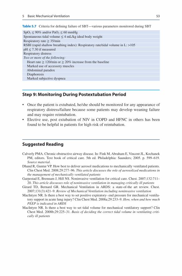
53
Step 9: Monitoring During Postextubation Period
• Once the patient is extubated, he/she should be monitored for any appearance of respiratory distress/failure because some patients may develop weaning failure and may require reintubation.
• Elective use, post extubation of NIV in COPD and HFNC in others has been found to be helpful in patients for high risk of reintubation.
Suggested Reading
Calverly PMA. Chronic obstructive airway disease. In: Fink M, Abraham E, Vincent JL, Kochanek PM, editors. Text book of critical care. 5th ed. Philadelphia: Saunders; 2005. p. 599–619. Source material.
Dhand R, Guntur VP. How best to deliver aerosol medications to mechanically ventilated patients. Clin Chest Med. 2008;29:277–96. This article discusses the role of aerosolized medications in the management of mechanically ventilated patients
Garpestad E, Brennam J, Hill NS. Noninvasive ventilation for critical care. Chest. 2007;132:711–20. This article discusses role of noninvasive ventilation in managing critically ill patients
Girard TD, Bernard GR. Mechanical Ventilation in ARDS: a state-of-the art review. Chest. 2007;131(3):421–9. Review of Mechanical Ventilation including noninvasive ventilation
MacIntyre NR. Is there a best way to set positive expiratory- end pressure for mechanical ventila-tory support in acute lung injury? Clin Chest Med. 2008a;29:233–9. How, when and how much PEEP is indicated in ARDS
MacIntyre NR. Is there a best way to set tidal volume for mechanical ventilatory support? Clin Chest Med. 2008b;29:225–31. Basis of deciding the correct tidal volume in ventilating criti-cally ill patients
Table 5.7 Criteria for defining failure of SBT—various parameters monitored during SBT
SpO2 ≤ 90% and/or PaO2 ≤ 60 mmHgSpontaneous tidal volume ≤ 4 mL/kg ideal body weightRespiratory rate ≥ 35/minRSBI (rapid shallow breathing index): Respiratory rate/tidal volume in L: >105pH ≤ 7.30 if measuredRespiratory distressTwo or more of the following: Heart rate ≥ 120/min or ≥ 20% increase from the baseline Marked use of accessory muscles Abdominal paradox Diaphoresis Marked subjective dyspnea
5 Basic Mechanical Ventilation
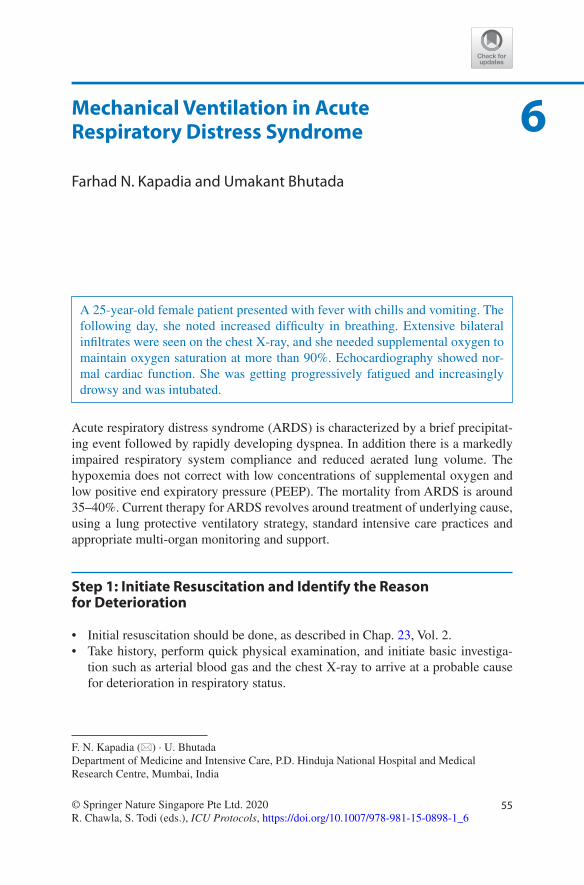
55© Springer Nature Singapore Pte Ltd. 2020R. Chawla, S. Todi (eds.), ICU Protocols, https://doi.org/10.1007/978-981-15-0898-1_6
F. N. Kapadia (*) · U. Bhutada Department of Medicine and Intensive Care, P.D. Hinduja National Hospital and Medical Research Centre, Mumbai, India
6Mechanical Ventilation in Acute Respiratory Distress Syndrome
Farhad N. Kapadia and Umakant Bhutada
Acute respiratory distress syndrome (ARDS) is characterized by a brief precipitat-ing event followed by rapidly developing dyspnea. In addition there is a markedly impaired respiratory system compliance and reduced aerated lung volume. The hypoxemia does not correct with low concentrations of supplemental oxygen and low positive end expiratory pressure (PEEP). The mortality from ARDS is around 35–40%. Current therapy for ARDS revolves around treatment of underlying cause, using a lung protective ventilatory strategy, standard intensive care practices and appropriate multi-organ monitoring and support.
Step 1: Initiate Resuscitation and Identify the Reason for Deterioration
• Initial resuscitation should be done, as described in Chap. 23, Vol. 2.• Take history, perform quick physical examination, and initiate basic investiga-
tion such as arterial blood gas and the chest X-ray to arrive at a probable cause for deterioration in respiratory status.
A 25-year-old female patient presented with fever with chills and vomiting. The following day, she noted increased difficulty in breathing. Extensive bilateral infiltrates were seen on the chest X-ray, and she needed supplemental oxygen to maintain oxygen saturation at more than 90%. Echocardiography showed nor-mal cardiac function. She was getting progressively fatigued and increasingly drowsy and was intubated.
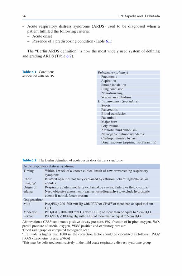
56
• Acute respiratory distress syndrome (ARDS) used to be diagnosed when a patient fulfilled the following criteria: – Acute onset – Presence of a predisposing condition (Table 6.1)
The “Berlin ARDS definition” is now the most widely used system of defining and grading ARDS (Table 6.2).
Table 6.1 Conditions associated with ARDS
Pulmonary (primary) Pneumonia Aspiration Smoke inhalation Lung contusion Near-drowning Venous air embolismExtrapulmonary (secondary) Sepsis Pancreatitis Blood transfusion Fat emboli Major burn Poly trauma Amniotic fluid embolism Neurogenic pulmonary edema Cardiopulmonary bypass Drug reactions (aspirin, nitrofurantoin)
Table 6.2 The Berlin definition of acute respiratory distress syndrome
Acute respiratory distress syndromeTiming Within 1 week of a known clinical insult of new or worsening respiratory
symptomsChest imaginga
Bilateral opacities not fully explained by effusion, lobar/lung/collapse, or nodules
Origin of edema
Respiratory failure not fully explained by cardiac failure or fluid overloadNeed objective assessment (e.g., echocardiography) to exclude hydrostatic edema if no risk factor present
Oxygenationb
Mild Pao2/FiO2: 200–300 mm Hg with PEEP or CPAPC of more than or equal to 5 cm H2O
Moderate PaO2/FiO2 100–200 mm Hg with PEEP, of more than or equal to 5 cm H2OSevere PaO2/FiO2 < 100 mg Hg with PEEP of more than or equal to 5 cm H2O
Abbrevations: CPAP continuous positive airway pressure, FiO2 fraction of inspired oxygen, PaO2 partial pressure of arterial oxygen, PEEP positive end-expiratory pressureaChest radiograph or computed tomograph scanbIf altitude is higher than 1000 m, the correction factor should be calculated as follows: [PaO2/FiO2X (barometric pressure/760)]cThis may be delivered noninvasively in the mild acute respiratory distress syndrome group
F. N. Kapadia and U. Bhutada

57
Step 2: Assess the Need for Continuous Positive Airway Pressure (CPAP) or Noninvasive Ventilation (NIV)/High Flow Nasal Canula (HFNC)
• NIV/High flow nasal canula may have a potentially useful role in a patient devel-oping mild ARDS, .They should not be used if PaO2/FiO2 is less than 150 mm Hg. HFNC is emerging as a more tolerable modality in hypoxemic respiratory failure as compared to NIV. Both NIV and HFNC can delay intubation and if not properly monitored may lead to adverse outcome and increase mortality.
• They may only be used in selected less severely ill patients, with close monitor-ing to help improve oxygenation and decrease the work of breathing.
• In patients who are deteriorating or show no response on NIV/HFNC, it is safer not to persist and instead proceed to tracheal intubation early, before a major deterioration occurs.
Step 3: Assess the Need for Mechanical Ventilation
• In the following situations of ARDS, mechanical ventilation should be initiated electively. To avoid complications of emergent intubation, it is improper to wait till the patient deteriorates further. – Worsening respiratory fatigue due to increased work of breathing – Persistent hypoxemia (SpO2 < 90%) on non-rebreathing facemask oxygen or
NIV/HFNC – Worsening hypercarbia
Step 4: Understand Principles of Ventilation in ARDS
• The main aim is to keep the patient stable and cause minimal iatrogenic damage till such time the underlying disease resolves.
• In ARDS, mechanical ventilation is primarily used to reverse hypoxemia and decrease the work of breathing.
• Positive-pressure ventilation is unphysiological, and adverse effects of this must be prevented or rapidly reversed.
• Initially, there may be a significant hemodynamic deterioration, which mandates adequate monitoring and reversal with fluids and appropriate inotrope and vaso-pressor agents.
• High volumes, high airway pressures, and repeated opening and closing of col-lapsed alveoli may further damage the lung, worsen the ARDS, and contribute to systemic inflammation.
• These patients are prone to ventilator-associated pneumonia due to prolonged ventilation and occasionally due to the use of corticosteroid.
• The mechanical ventilation protocol for a patient with ARDS is based on the concept that the lung is largely consolidated, and may be viewed as a “baby
6 Mechanical Ventilation in Acute Respiratory Distress Syndrome

58
lung” with only about a third of the alveoli remaining open. This “consolidation” primarily results from the alveolar wall becoming stiff and shutting down, rather than the alveoli actually being fluid-filled.
• The concept of a “sponge lung” implies the gravitational effect of lung injury i.e. the dependent portion of the lung is more atelectatic and the appropriate ventila-tory strategy can open up or recruit these shut alveoli. This is demonstrated in imaging studies that show that changes of position of the patient cause changes in the patterns of aeration of the lung, with the lowest or most dependent areas shutting down due to the weight of the lung above, the alveoli in the highest part remaining mostly open. On the basis of these concepts, one of the strategies of mechanical ventilation is to “open up the lung and keep it open.”
Step 5: Decide on the Initial Settings on the Ventilator
• A protocol should be followed for initiating ventilator setting, which may be customized according to the patient’s need.
• Initially the following setting needs to be decided: – Mode—volume control ventilation (ARDS network protocol) or pressure
control (targeting a preset volume) as the starting mode• Tidal volume: (usually 300–400 mL).
– Calculate ideal body weight (IBW): Male IBW = 50 + 2.3 [height (inches) − 60]; female IBW = 45.5 + 2.3 [height (inches) − 60]
– Set initial tidal volume (TV) to 8 mL/kg IBW – Reduce TV by 1 mL/kg intervals every 2 h until TV = 6 mL/kg IBW
• Inspiratory pressure (pressure control). – Inspiratory airway pressure should be limited to less than 30 cm H2O
• FiO2 and positive end-expiratory pressure (PEEP). – Initial FiO2 should be kept high and PEEP 5–10 cm H2O to keep oxygen satu-
ration more than 90%. (See FIO2/PEEP chart below) – In severe & diffuse ARDS, start with a higher PEEP of 10–12 – FiO2 should be titrated down subsequently if oxygen saturation is more than
90%. Titrate PEEP as per ARDSnet Table 6.3• Minute ventilation.
– Adjust respiratory rate (maximum up to 35/min) to achieve a minute ventila-tion commensurate with patients’ demand
• Inspiratory flow or inspiratory time or I:E ratio (depending on the ventilator type). – Set the inspiratory flow rate to achieve goal of inspiratory–expiratory ratio of
1:1.0–1.3
Step 6: Try to Achieve Goals of Ventilation
• After initial ventilator setup, the patient should be monitored for safety and effi-cacy of ventilator settings and an attempt should be made to ventilate within certain goals. – Oxygenation goal: PaO2 = 55–80 mmHg or SpO2 = 88–95%
F. N. Kapadia and U. Bhutada

59
• Table 6.3 incremental FiO2–PEEP combinations to achieve oxygenation goal.
Plateau pressure (Pplat) goal: ≤ 30 cm H2O
• Keep inspiratory pressure in pressure control below 30 cm H2O.• In volume assist control, check Pplat (use 0.5-s inspiratory pause), Driving pres-
sure (Plateau-PEEP), SpO2, Minute ventilation, Patient respiratory rate, TV, and arterial blood gases.
• If Pplat is more than 30 cm H2O, decrease TV by 1 mL/kg steps (minimum 4 mL/kg IBW).
• If Pplat is less than 30 cm H2O and breath stacking occurs, one may increase TV in 1 mL/kg IBW increments (to a maximum of 8 mL/kg) as long as Pplat is less than 30 cm H2O.
• In patients with obesity and stiff chest wall or high Intraabdominal pressure (IAP) a higher plateau pressure may be tolerated. – pH goal: 7.30–7.45
Acidosis management: pH less than 7.30
• If pH is 7.15–7.30, increase RR until pH is more than 7.30 or PaCO2 is less than 25 mmHg (maximum RR = 35); if RR is 35 and PaCO2 is less than 25 mmHg, NaHCO3 may be given.
• If pH is still less than 7.15 and NaHCO3 is considered or infused, TV may be increased by 1 mL/kg steps until pH is more than 7.15 (Pplat goal may be exceeded) Alkalosis management: if pH is more than7.45, decrease RR if possible.
• All patients should be administered with neuromuscular agents for the first 48 h.• Restricted fluid strategy is recommended once patient is stabilized.• Avoid routine use of glucocorticoids. It has not been consistently shown to
increase survival. They may be administered early (before 14 days) in moderate to severe ARDS on case to case basis (1 mg/Kg methylprednisolone for 21 days).
Step 7: If Patient Has Severe ARDS (Berlin Definition) and Needs High FiO2 (More than 0.6) to Maintain SpO2
• Patient should be put in prone positioning when FiO2 requirement is high (>0.6) – Contraindications: Life-threatening shock (mean arterial pressure < 65 mmHg
with or without vasopressors) – Raised intracranial pressure more than 30 mmHg, or cerebral perfusion pres-
sure less than 60 mmHg Contraindications:
Table 6.3 Incremental FiO2–PEEP combinations to achieve oxygenation goal
FiO2 0.3 0.4 0.4 0.5 0.5 0.6 0.7 0.7PEEP 5 5 8 8 10 10 10 12FiO2 0.7 0.8 0.9 0.9 0.9 1.0 1.0 1.0PEEP 14 14 14 16 18 20 22 24
6 Mechanical Ventilation in Acute Respiratory Distress Syndrome

60
– Spinal instability or any unstable fracture – Recent thoracoabdominal surgery – Open wound or burns on ventral body surface – Massive hemoptysis – Arrhythmias
• Steps to proning: – Proning needs trained staff; it requires preferably 4–6 persons – Have a central and arterial line in place – Arrange for cushions – Fix tube and lines well – Empty the stomach by aspirating the nasogastric tube – Sedate well and paralyze if required – Cover eyes – Place electrocardiograph (ECG) electrodes on the back – One person stands at the head end and holds the ETT with one hand and the
head with the other – Disconnect monitoring – Bring the patient on the edge of the bed – Turn the patient with the help of three people and place supporting cushions
under chest and lower pelvis – Immediately reconnect monitors and take BP, SpO2 – Auscultate chest – Make sure abdomen is free (for respiration). One should be able to pass hands
between abdomen and mattress – Extra cushion pads for genitalia, axilla, ears, breasts, knees, foot – Check ABG within 30 min – Turn the head alternately to right and left every 2 h
• Duration: > 16–18 h every day.• Complications:
– Pressure ulcers – Displacement of endotracheal tubes, thoracotomy tubes, and vascular
catheters• Similar steps when positioning back from prone to supine
Step 8: Management Strategy for Life-Threatening Hypoxemia-Refer to Chap. 7, Vol. 1
Step 9: Decide on Need for Tracheostomy
• Tracheostomy should be performed once the patient is off high FiO2 and PEEP support but still needs continuing ventilator support due to high minute ventilation.
F. N. Kapadia and U. Bhutada

61
• There are no specifics cut off values for FiO2 & PEEP when deciding timing of traceostomy
• The optimal timing for tracheostomy is not known, but there is evidence that early tracheostomy is not beneficial
Step 10: Initiate Aggressive Mobilization Regimen
• In order to prevent long-term neuromuscular disability, early aggressive physio-therapy and mobilization regimen should be started from the initial days.
Step 11: Consider Weaning
• Weaning attempts should be started once FiO2 and PEEP support decrease and minute ventilation requirement comes down. (Refer to chap. 7, Vol. 1)
Suggested Reading and Important Trials
Briel M, Meade M, Mercat A, et al. Higher versus lower positive end-expiratory pressure in patients with acute lung injury and acute respiratory distress syndrome: systematic review and meta-analysis. JAMA. 2010;303:865–73. Treatment with higher vs lower levels of PEEP was not associated with improved hospital survival. However, higher levels were associated with improved survival among the subgroup of patients with severe ARDS
Brower RG, Lanken PN, MacIntyre N, et al. Higher versus lower positive end-expiratory pressures in patients with the acute respiratory distress syndrome. N Engl J Med. 2004;351:327–36. ARDS who receive mechanical ventilation with tidal-volume goal of 6 ml per kilogram of predicted body weight and an end-inspiratory plateau-pressure limit of 30 cm of water a, clini-cal outcomes are similar with lower or higher PEEP levels
Guérin C, Reignier J, Richard JC, et al. for the PROSEVA Study Group. Prone positioning in severe acute respiratory distress syndrome. N Engl J Med. 2013;368:2159–68. In severe ARDS, early application of prone-positioning for 1 to 20 hrs significantly decreased 28-day and 90-day mortality
Meade MO, Cook DJ, Guyatt GH, et al. Lung Open Ventilation Study Investigators. Ventilation strategy using low tidal volumes, recruitment maneuvers, and high positive end-expiratory pressure for acute lung injury and acute respiratory distress syndrome: a randomized con-trolled trial. JAMA. 2008;299:637–45. For patients with ARDS, a multifaceted protocolized ventilation strategy designed to recruit and open the lung resulted in no significant difference in all-cause hospital mortality or barotrauma However it improved secondary end points related to hypoxemia and use of rescue therapies
Mercat A, Richard JC, Vielle B, et al. Positive end-expiratory pressure setting in adults with acute lung injury and acute respiratory distress syndrome: a randomized controlled trial. Expiratory Pressure (Express) Study Group. JAMA. 2008;299:646–55. A strategy for setting PEEP aimed at increasing alveolar recruitment while limiting hyperinflation did not significantly reduce mortality. However, it did improve lung function and reduced the duration of mechanical ven-tilation and the duration of organ failure
National Heart, Lung, and Blood Institute Acute Respiratory Distress Syndrome (ARDS) Clinical Trials Network, Wheeler AP, Bernard GR, et al. Pulmonary-artery versus central venous
6 Mechanical Ventilation in Acute Respiratory Distress Syndrome

62
catheter to guide treatment of acute lung injury. N Engl J Med. 2006;354:2213–24. PAC-guided therapy did not improve survival or organ function but was associated with more complications than CVC-guided therapyin ARDS patients
Patroniti N, Isgrò S, Zanella A. Clinical of severely hypoxemic patients. Critical Care. 2011;17:50–6. A review article on therapies for refractory hypoxemia
Pipeling MR, Fan E. Therapies for refractory hypoxemia in acute respiratory distress syndrome. JAMA. 2010;304:2521–7.
The Acute Respiratory Distress Syndrome Network. Ventilation with lower tidal volumes as com-pared with traditional tidal volumes for acute lung injury and the acute respiratory distress syndrome. N Engl J Med. 2000;342:1301–8. In ARDS patient, mechanical ventilation with a lower tidal volume than is traditionally used results in decreased mortality and increases the number of days off ventilation
The ARDS Definition Task Force. Acute respiratory distress syndrome: the Berlin definition. JAMA. 2012;307(23):2526-2533. https://doi.org/10.1001/jama.2012.5669. An article on new Berlin definition
The National Heart, Lung, and Blood Institute Acute Respiratory Distress Syndrome (ARDS) Clinical Trials Network∗. Comparison of two fluid-management strategies in acute lung injury. N Engl J Med. 2006;354:2564–75. The conservative strategy of fluid management improved lung function and shortened the duration of mechanical ventilation and intensive care without increasing nonpulmonary-organ failures
Websiteshttp://www.ardsnet.org. The official website of the ARDS Network, initiated by National Heart,
Lung, and Blood Institute and National Institutes of Health to carry out multicenter clinical trials of ARDS treatments
F. N. Kapadia and U. Bhutada
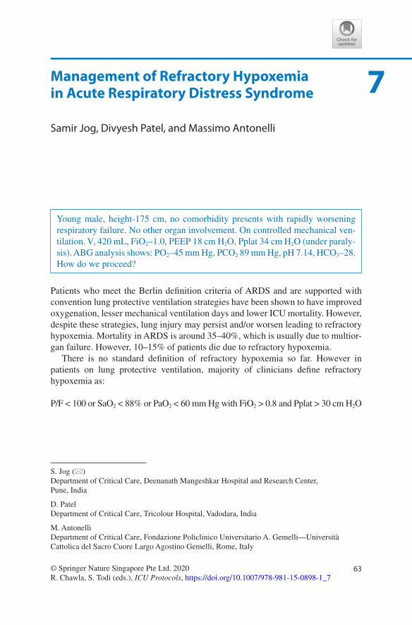
63© Springer Nature Singapore Pte Ltd. 2020R. Chawla, S. Todi (eds.), ICU Protocols, https://doi.org/10.1007/978-981-15-0898-1_7
S. Jog (*) Department of Critical Care, Deenanath Mangeshkar Hospital and Research Center, Pune, India
D. Patel Department of Critical Care, Tricolour Hospital, Vadodara, India
M. Antonelli Department of Critical Care, Fondazione Policlinico Universitario A. Gemelli—Università Cattolica del Sacro Cuore Largo Agostino Gemelli, Rome, Italy
7Management of Refractory Hypoxemia in Acute Respiratory Distress Syndrome
Samir Jog, Divyesh Patel, and Massimo Antonelli
Patients who meet the Berlin definition criteria of ARDS and are supported with convention lung protective ventilation strategies have been shown to have improved oxygenation, lesser mechanical ventilation days and lower ICU mortality. However, despite these strategies, lung injury may persist and/or worsen leading to refractory hypoxemia. Mortality in ARDS is around 35–40%, which is usually due to multior-gan failure. However, 10–15% of patients die due to refractory hypoxemia.
There is no standard definition of refractory hypoxemia so far. However in patients on lung protective ventilation, majority of clinicians define refractory hypoxemia as:
P/F < 100 or SaO2 < 88% or PaO2 < 60 mm Hg with FiO2 > 0.8 and Pplat > 30 cm H2O
Young male, height-175 cm, no comorbidity presents with rapidly worsening respiratory failure. No other organ involvement. On controlled mechanical ven-tilation. Vt 420 mL, FiO2–1.0, PEEP 18 cm H2O, Pplat 34 cm H2O (under paraly-sis). ABG analysis shows: PO2–45 mm Hg, PCO2 89 mm Hg, pH 7.14, HCO3–28. How do we proceed?
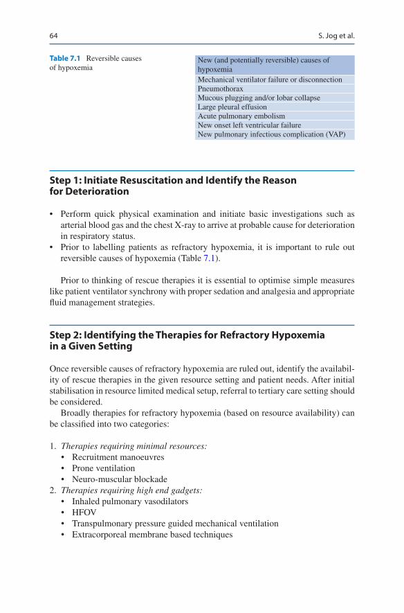
64
Step 1: Initiate Resuscitation and Identify the Reason for Deterioration
• Perform quick physical examination and initiate basic investigations such as arterial blood gas and the chest X-ray to arrive at probable cause for deterioration in respiratory status.
• Prior to labelling patients as refractory hypoxemia, it is important to rule out reversible causes of hypoxemia (Table 7.1).
Prior to thinking of rescue therapies it is essential to optimise simple measures like patient ventilator synchrony with proper sedation and analgesia and appropriate fluid management strategies.
Step 2: Identifying the Therapies for Refractory Hypoxemia in a Given Setting
Once reversible causes of refractory hypoxemia are ruled out, identify the availabil-ity of rescue therapies in the given resource setting and patient needs. After initial stabilisation in resource limited medical setup, referral to tertiary care setting should be considered.
Broadly therapies for refractory hypoxemia (based on resource availability) can be classified into two categories:
1. Therapies requiring minimal resources:• Recruitment manoeuvres• Prone ventilation• Neuro-muscular blockade
2. Therapies requiring high end gadgets:• Inhaled pulmonary vasodilators• HFOV• Transpulmonary pressure guided mechanical ventilation• Extracorporeal membrane based techniques
Table 7.1 Reversible causes of hypoxemia
New (and potentially reversible) causes of hypoxemiaMechanical ventilator failure or disconnectionPneumothoraxMucous plugging and/or lobar collapseLarge pleural effusionAcute pulmonary embolismNew onset left ventricular failureNew pulmonary infectious complication (VAP)
S. Jog et al.
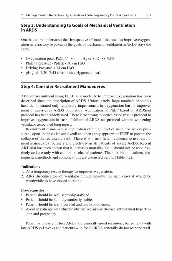
65
Step 3: Understanding to Goals of Mechanical Ventilation in ARDS
One has to be understand that irrespective of modalities used to improve oxygen-ation in refractory hypoxemia the goals of mechanical ventilation in ARDS stays the same.
• Oxygenation goal: PaO2 55–80 mm Hg or SaO2 88–95%.• Plateau pressure (Pplat): <30 cm H2O.• Driving Pressure < 14 cm H2O.• pH goal: 7.20–7.45 (Permissive Hypercapnoea).
Step 4: Consider Recruitment Manoeuvres
Alveolar recruitment using PEEP as a modality to improve oxygenation has been described since the description of ARDS. Unfortunately, large numbers of studies have demonstrated only temporary improvement in oxygenation but no improve-ment of survival in ARDS population. Application of PEEP based on ARDSnet protocol has been widely used. There is no strong evidence based rescue protocol to improve oxygenation in case of failure of ARDS net protocol without worsening ventilator associated lung injury.
Recruitment manoeuvre is application of a high level of sustained airway pres-sure to open up the collapsed alveoli and then apply appropriate PEEP to prevent the collapse of the recruited alveoli. There is still insufficient evidence to use recruit-ment manoeuvres routinely and electively in all patients of severe ARDS. Recent ART trial has even shown that it increases mortality. So it should not be used rou-tinely and use only with caution in selected patients. The possible indications, pre- requisites, methods and complications are discussed below. (Table 7.2).
Indications 1. As a temporary rescue therapy to improve oxygenation. 2. After disconnection of ventilator circuit (however in such cases it would be
worthwhile to have closed suction).
Pre-requisites• Patient should be well sedated/paralysed.• Patient should be hemodynamically stable.• Patient should be well hydrated and not hypovolemic.• Avoid in patients with chronic obstructive airway disease, intracranial hyperten-
sion and pregnancy.
Patient with early diffuse ARDS are generally good recruiters, but patients with late ARDS (>1 week) and patients with focal ARDS generally do not respond well.
7 Management of Refractory Hypoxemia in Acute Respiratory Distress Syndrome
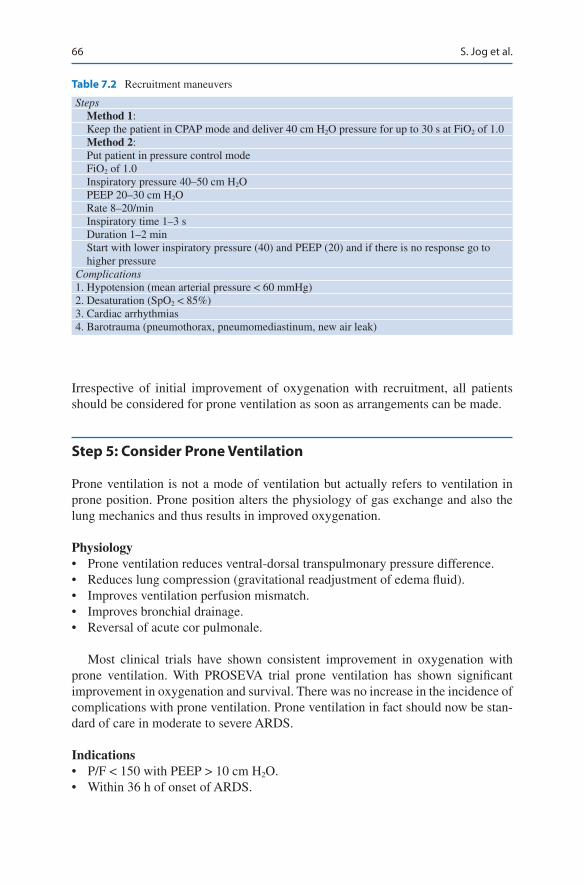
66
Irrespective of initial improvement of oxygenation with recruitment, all patients should be considered for prone ventilation as soon as arrangements can be made.
Step 5: Consider Prone Ventilation
Prone ventilation is not a mode of ventilation but actually refers to ventilation in prone position. Prone position alters the physiology of gas exchange and also the lung mechanics and thus results in improved oxygenation.
Physiology• Prone ventilation reduces ventral-dorsal transpulmonary pressure difference.• Reduces lung compression (gravitational readjustment of edema fluid).• Improves ventilation perfusion mismatch.• Improves bronchial drainage.• Reversal of acute cor pulmonale.
Most clinical trials have shown consistent improvement in oxygenation with prone ventilation. With PROSEVA trial prone ventilation has shown significant improvement in oxygenation and survival. There was no increase in the incidence of complications with prone ventilation. Prone ventilation in fact should now be stan-dard of care in moderate to severe ARDS.
Indications• P/F < 150 with PEEP > 10 cm H2O.• Within 36 h of onset of ARDS.
Table 7.2 Recruitment maneuvers
Steps Method 1: Keep the patient in CPAP mode and deliver 40 cm H2O pressure for up to 30 s at FiO2 of 1.0 Method 2: Put patient in pressure control mode FiO2 of 1.0 Inspiratory pressure 40–50 cm H2O PEEP 20–30 cm H2O Rate 8–20/min Inspiratory time 1–3 s Duration 1–2 min Start with lower inspiratory pressure (40) and PEEP (20) and if there is no response go to
higher pressureComplications1. Hypotension (mean arterial pressure < 60 mmHg)2. Desaturation (SpO2 < 85%)3. Cardiac arrhythmias4. Barotrauma (pneumothorax, pneumomediastinum, new air leak)
S. Jog et al.
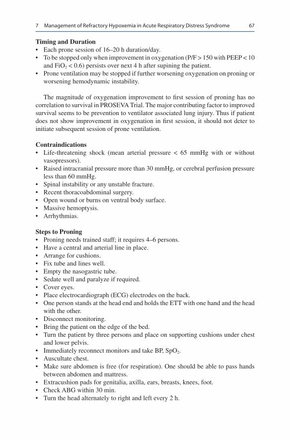
67
Timing and Duration• Each prone session of 16–20 h duration/day.• To be stopped only when improvement in oxygenation (P/F > 150 with PEEP < 10
and FiO2 < 0.6) persists over next 4 h after supining the patient.• Prone ventilation may be stopped if further worsening oxygenation on proning or
worsening hemodynamic instability.
The magnitude of oxygenation improvement to first session of proning has no correlation to survival in PROSEVA Trial. The major contributing factor to improved survival seems to be prevention to ventilator associated lung injury. Thus if patient does not show improvement in oxygenation in first session, it should not deter to initiate subsequent session of prone ventilation.
Contraindications• Life-threatening shock (mean arterial pressure < 65 mmHg with or without
vasopressors).• Raised intracranial pressure more than 30 mmHg, or cerebral perfusion pressure
less than 60 mmHg.• Spinal instability or any unstable fracture.• Recent thoracoabdominal surgery.• Open wound or burns on ventral body surface.• Massive hemoptysis.• Arrhythmias.
Steps to Proning• Proning needs trained staff; it requires 4–6 persons.• Have a central and arterial line in place.• Arrange for cushions.• Fix tube and lines well.• Empty the nasogastric tube.• Sedate well and paralyze if required.• Cover eyes.• Place electrocardiograph (ECG) electrodes on the back.• One person stands at the head end and holds the ETT with one hand and the head
with the other.• Disconnect monitoring.• Bring the patient on the edge of the bed.• Turn the patient by three persons and place on supporting cushions under chest
and lower pelvis.• Immediately reconnect monitors and take BP, SpO2.• Auscultate chest.• Make sure abdomen is free (for respiration). One should be able to pass hands
between abdomen and mattress.• Extracushion pads for genitalia, axilla, ears, breasts, knees, foot.• Check ABG within 30 min.• Turn the head alternately to right and left every 2 h.
7 Management of Refractory Hypoxemia in Acute Respiratory Distress Syndrome

68
Complications• Pressure ulcers on face abdomen.• Facial edema.• Inadvertent extubation.• Dislodgement of catheters or lines.
Step 6: Consider Adjunctive Neuromuscular Blockade
Neuromuscular blockade has been postulated to facilitate lung protective low vol-ume ventilation by improving patient ventilator synchrony. It limits the risk of asyn-chrony related alveolar collapse and regional alveolar pressure increase with overdistension of alveoli. It is also postulated to cause decrease in lung inflammation.
Benefit with neuromuscular blockade have been documented in early ARDS with P/F < 150 (infusion for 48 h). This benefit has been shown with cisatracurium. It may also be used as an adjunct in patient having severe patient ventilator asyn-chrony despite heavy sedation. Neuromuscular blockade should be used judiciously considering that, its use is associated with critical illness neuromyopathy and is a confounder to neurological assessment.
In patient with refractory hypoxemia with ARDS not improving despite adequate sedation, paralysis, recruitment manoeuvres, prone ventilation and ongoing primary disease treatment efforts, patient referral to higher centre for further advanced res-cue therapies should be considered.
Further choice of rescue therapy for refractory hypoxemia in severe ARDS is predominantly directed by availability of resources, expertise to use these therapies and patients medical condition.
Step 7: Transpulmonary Pressure Guided Mechanical Ventilation
ARDSnet protocol recommends targeting Pplat < 30 cm H2O. Unfortunately pla-teau pressure does not tell us about lung compliance, but is a composite measure of lung and chest wall compliance. Without partitioning the respiratory system compo-nents, it is not possible to identify the factors that contribute to low pulmonary compliance as evident by high Pplat. In patients with obesity, thick chest wall, abdominal compartment syndrome, ascites, burns etc. it may be difficult to exactly quantify pulmonary compliance from Pplat measurements. Airway pressures in these conditions are not reflective of distending pressures of lung.
Transpulmonary pressure can be calculated as difference between airway pres-sure and pleural pressure. Oesophageal pressure can be used as a surrogate of pleu-ral pressure and can be easily measured with specially designed oesophageal catheters.
S. Jog et al.

69
PEEP can be titrated to target end expiratory transpulmonary pressure between 0–10 cm H2O to prevent cyclic alveolar collapse. Tidal volume can be titrated to maintain end inspiratory transpulmonary pressure < 25 cm H2O to prevent cyclic alveolar overdistension. This strategy can thus decrease VALI by preventing cyclic alveolar collapse and overdistension.
Esophageal pressure guided strategy has been shown to improve oxygenation but still there are no trials demonstrating mortality benefit. However in certain cases of ARDS where there are concerns that airway pressures are not actually representa-tive of actual lung compliance, it can be used to optimise mechanical ventilation.
Step 8: Consider Extracorporeal Membrane Oxygenation (ECMO)
ECMO is a technique where blood is removed from patient and passed through artificial biomembrane which functions to oxygenate blood and remove CO2 from blood and then return it back to patient. Thus essentially it is decoupling of mechanical ventilation and gas exchange. ECMO can be considered in severe ARDS especially in cases of refractory hypoxemia and in cases where adherence to lung protective ventilation leads to severe hypercapnic respiratory acidosis. ECMO thus provides an way to manage gas exchange well while managing mechanical ventilation with acceptable tidal volumes and plateau pressures. This decreases chances of VALI.
• Data on use of ECMO in severe adult ARDS is evolving. The largest trial for ECMO in severe ARDS (CESAR trial) reported a survival of 63% for patients of ECMO. Recent EOLIA trial has shown survival was higher in those who received early compared with late. ECMO patients with severe ARDS who fail to respond to optimal treatment should be managed with ECMO promptly rather than later as a rescue treatment.
ECMO is an important tool in management of life threatening hypoxemia in severe ARDS. In case of isolated respiratory failure, it can be accomplished with venovenous ECMO and continued till recovery of lung functions. In case of associ-ated circulatory failure, it can be accomplished with venoarterial ECMO.
Indications• Potentially reversible cause of hypoxemic respiratory failure with <7 days from
onset of disease with – Murray score 3–4 despite optimal care for 6 h or more – P/F < 100 with fiO2 > 0.9 despite optimal care for 6 h or more
• Severe air leak syndromes• Oxygenation index equal or above 25–30 indicated need for ECMO• Severe refractory respiratory acidosis arising from low tidal volume ventilation
7 Management of Refractory Hypoxemia in Acute Respiratory Distress Syndrome
70
Contra-Indications• Contra-indication to anticoagulation.• Irreversible lung diseases (e.g. lung metastases, advanced pulmonary fibrosis in
absence of transplant possibilities).• Underlying moderate to severe chronic lung disease.
Complications• Cannulation related-vessel perforation, limb ischaemia and arterial dissection.• Bleeding.• Thromboembolism.• Heparin induced thrombocytopenia.
Extracorporeal CO2 removal (ECCO2R) is a type of extracorporeal support spe-cifically designed to remove CO2 from the blood using low blood flows (< 1.0 L/min). ECCO2R can also be attained by arterio-venous route where patient’s native cardiac output serves as pump.
In this either of the techniques, CO2 removal is extremely efficient but oxygen-ation of the blood is minimally benefitted. It can be used specially in conditions where despite conventional lung protective ventilation strategy there is significant hypercarbic respiratory acidosis. Thus ECCO2R with low tidal volume ventilation may help in protective ventilation (prevent VALI) and prevent severe respiratory acidosis. Clinical trials still have not shown any outcome benefit.
Other Rescue Therapies
HFOV
HFOV is a type of ventilation which combines high respiratory rate (>180 breaths/min) with tidal volumes as low as anatomical dead space. The oscillator delivers very low tidal volume by oscillating around a mean airway pressure. This prevents alveolar collapse and at the same time avoids high airway pressures. Risks with HOFV include hypotension and barotraumas. Early trials of HFOV in refractory hypoxemia in ARDS demonstrated improved oxygenation and mortality benefit. But recent large RCTs (OSCAR and OSCILLATE) failed to demonstrate any mor-tality benefit.
As a result of these studies HFOV at present is not recommended for use in adult ARDS.
Inhaled Pulmonary Vasodilators
Inhaled Nitric oxide and epoprostenol are commonly used inhaled pulmonary vaso-dilators. They promote pulmonary vasodilatation and improved blood flow to venti-lated areas of lungs and divert blood away from poorly ventilated areas of lung. This
S. Jog et al.
71
improves ventilation perfusion mismatch and thus improves oxygenation. Many tri-als have shown improved oxygenation with these therapies. However there is no mortality benefit and inhaled nitric oxide may be associated with side effects like methemoglobinemia, cyanide toxicity and renal insufficiency. Epoprostenol may be associated with hypotension.
Thus with no mortality benefit and significant side effect profile, inhaled vasodi-lators are not routine used in refractory hypoxemia with severe ARDS. However they can be used as transient measure to improve oxygenation in patient with refrac-tory hypoxemia especially in patients with severe pulmonary hypertension devel-oped secondary to hypoxemia.
Suggested Reading
Bein T, Grasso S, Moerer O, et al. The standard of care of patients with ARDS: ventilatory settings and rescue therapies for refractory hypoxemia. Intensive Care Med. 2016;42:699. A specific standard of care is required for the management of severe ARDS with refractory hypoxemia
Cavalcanti AB, Suzumura ÉA, et al. Effect of lung recruitment and titrated positive end- expiratory pressure (PEEP) vs low PEEP on mortality in patients with acute respiratory distress syn-drome: a randomized clinical trial. JAMA. 2017;318(14):1335–45. In moderate to severe ARDS, a strategy with lung recruitment and titrated PEEP compared with low PEEP increased 28-day all-cause mortality. The routine use of lung recruitment maneuver and PEEP titration in these patients is not recommended
Combes A, Hajage D, Capellier G, et al. Extracorporeal membrane oxygenation for severe acute respiratory distress syndrome. N Engl J Med. 2018;378(21):1965–71. Patients with very severe ARDS, 60-day mortality was not significantly lower with ECMO than with a strategy of con-ventional mechanical ventilation that included ECMO as rescue therapy
Guérin C, Reignier J, Richard JC, et al. PROSEVA Study Group. Prone positioning in severe acute respiratory distress syndrome. N Engl J Med. 2013;368(23):2159. In severe ARDS, early application of prolonged prone-positioning sessions significantly decreased 28-day and 90-day mortality
Peek GJ, Mugford M, Tiruvoipati R, et al. CESAR trial collaboration. Efficacy and economic assessment of conventional ventilatory support versus extracorporeal membrane oxygenation for severe adult respiratory failure (CESAR): a multicentre randomised controlled trial. Lancet. 2009;374(9698):1351.
Villar J, Kacmarek RM. Rescue strategies for refractory hypoxemia: a critical appraisal. Med Rep. 2009;1:91–4. A review article on the management of ARDS
7 Management of Refractory Hypoxemia in Acute Respiratory Distress Syndrome

73© Springer Nature Singapore Pte Ltd. 2020R. Chawla, S. Todi (eds.), ICU Protocols, https://doi.org/10.1007/978-981-15-0898-1_8
R. K. Mani (*) Department of Pulmonology and Critical Care, Batra Hospital and Medical Research Centre, New Delhi, India
8Mechanical Ventilation in Obstructive Airway Diseases
Raj Kumar Mani
Obstructive pulmonary diseases are a major cause of mortality and morbidity. Acute respiratory failure in chronic obstructive pulmonary disease (COPD) is one of the common reasons for admission to the intensive care unit (ICU). Use of noninvasive ventilation has revolutionized the treatment and outcome of COPD patients.
Step 1: Initiate Resuscitation
• The patient should be resuscitated as mentioned in Chap. 23, Vol. 2.• All the patients admitted with respiratory distress require immediate attention to
the airway. This assessment is done mainly by clinical means.
They should be put on supplemental oxygen to increase SpO2 to more than 90%. For chronic obstructive pulmonary disease (COPD) patient, use controlled inhaled oxygen through the venturi mask to keep SpO2 88–90%. Patients who have increased work of breathing and seem to be getting exhausted may require assisted ventilation.
A 23-year-old female patient, known to be asthmatic since childhood and on regular inhalers, developed breathlessness at work. Several puffs of salbutamol failed to relieve the symptoms, and she rapidly went on to have wheezing and restlessness followed by air hunger. She was brought to the emergency department.

74
Step 2: Assess Severity
It is done based on the following:
• Able to speak full sentences.• Restlessness.• Respiratory rate and pattern of respiration.• Use of accessory muscles.• Pulse rate and pulsus paradoxus (inspiratory decrease in systolic blood pressure
by >10 mmHg).• Sensorium, fatigue.• Auscultation: Wheezes and crackles; silent chest signifies very severe airflow
obstruction.• Peak expiratory flow rate is an objective measure of airflow obstruction: less than
30% of baseline/predicted would indicate likelihood of respiratory failure. Initially, check every 30 min to assess response to the therapy. In a dyspneic patient this might be a difficult manouever to perform.
• SpO2: Hypoxia is usually correctable with supplemental oxygen. Refractory hypoxia should trigger search for pneumothorax, atelectasis, pneumonia, or occult sepsis.
• Arterial blood gases: In asthma, normal or elevated PaCO2 signifies respiratory failure due to respiratory muscle fatigue. pH of less than 7.28 would indicate the need for ventilatory support. Hyperlactatemia may occur due to muscle fatigue or adrenergic agents.
Step 3: Start Immediate Medical Management
• Nebulized salbutamol 2.5 mg (0.5 mL of 5% solution in 2.5 mL saline) or levo-salbutamol should be repeated every 20 min for three doses and then less fre-quently, dictated by the patient’s clinical response. More frequent and even continuous nebulization of salbutamol at a dosage of 10–15 mg can be used within limits of toxic effects such as tachycardia and tremors.
• Nebulized ipratropium (0.5 mg every 20 min) should be included in initial treat-ment concomitantly with salbutamol for better bronchodilatation.
• If the nebulizer is not available, use four puffs of salbutamol meter dose inhalers (MDI) through a spacer device.
• Corticosteroids should be initiated at the earliest to prevent respiratory failure. The usual doses are as follows: Hydrocortisone injection 100 mg every 6 h or methylprednisolone 60–125 mg every 6–8 h. Oral prednisolone 60 mg is equally effective especially in COPD.
• Oxygen supplementation is continued to keep SpO2 more than 90%.• Methylxanthines: Aminophylline may be used as a second-line agent, although
its role is much debated. A loading dose of 5–6 mg/kg is followed by a continuous
R. K. Mani

75
infusion of 0.6 mg/kg/h. Avoid loading dose in case the patient has been on oral theophyllines earlier.
• Magnesium sulfate (2 g infusion) over 20 min can also be tried in refractory cases of asthma, although its role is unproven.
• Antibiotics are not required routinely in bronchial asthma exacerbation and should be given only if there is evidence of infection.
• Quinolones or macrolides may be used for COPD exacerbation and should be given only if there is evidence of infection, although most of these are viral in origin.
Step 4: Assess Need for Respiratory Support
• Noninvasive ventilation (NIV) : In COPD with respiratory distress despite medi-cal treatment, NIV may be tried. Inspiratory positive airway pressure reduces work of breathing and expiratory positive airway pressure overcomes auto- positive end-expiratory pressure (auto-PEEP). In patients with CO2 retention and respiratory acidosis, bilevel support is initiated. To avoid hyperinflation, a single level of pressure, continuous positive airway pressure (CPAP) of 5–8 cm H2O may also be applied. Extended trials of NIV may be warranted if the senso-rium and patient comfort are improving (see Chap. 3, Vol. 1).
• Noninvasive positive pressure ventilation is well established for patients with COPD; there are limited data on its use in acute severe asthma.
• NIV may not useful in patients without respiratory acidosis.• Continuously monitor the heart rate, respiratory rate, SpO2, blood pressure, and
sensorium. Reassess every 30 min until the patient is stable and comfortable. Nursing attendance should be continuous.
Step 5: Assess the Need for Intubation and Mechanical Ventilation (MV)
• Impending respiratory arrest.• Circulatory failure.• Altered sensorium: progressive drowsiness, agitation, or severe restlessness.• In a conscious patient, no improvement or deterioration after 3–4 h of optimal
medical therapy and NIV support.• In COPD, severe hypercarbia and acidosis are well tolerated. However, the gen-
eral appearance and the degree of distress and fatigue of the patient are more important than the absolute values. and the decision to initiate mechanical venti-lation is based on clinical judgement.
• NIV is initially used in these patients. MV is used only if there are contraindica-tions or failure of NIV.
8 Mechanical Ventilation in Obstructive Airway Diseases

76
• In patients with end stage COPD a detailed discussion with family regarding a possibility of prolonged weaning and tracheostomy and patient preference should be done before initiating mechanical ventilation.
Step 6: Initiate MV
Principles: Because of severe airway obstruction, dynamic hyperinflation or air trapping takes place. Progressive hyperinflation leads to equilibrium of inflow and outflow of air in the lungs to take place at a high total lung volume. MV aimed at normalizing blood gas values would further overdistend the lungs with possible barotrauma and circulatory consequences.
• Orotracheal intubation: Follow the steps of rapid sequence intubation. Preoxygenation with NIV prior to induction should be done. High flow nasal cannula (HFNC) may be employed to ensure adequate apneic oxygenation dur-ing intubation. As far as possible, a tube size of 8 or more is used, and therefore orotracheal route is preferred.
• In asthma exacerbation, intubation should proceed cautiously as manipulation of airway may lead to exaggerated airflow obstruction and respiratory arrest.
• Sedation and paralysis: At the time of intubation, short-acting sedatives (mid-azolam) and short-acting neuromuscular blocking agents (succinylcholine) are used. For maintenance of sedation to assist MV, midazolam/propofol/dexmeto-medine infusion can be used. Neuromuscular blocking agents should be avoided as infusion to prevent critical illness neuropathy.
• Avoid delivering high rate and tidal volume with bag ventilation.• Initial ventilator settings: Volume Controlled mechanical ventilation (CMV)
mode; tidal volume 7–9 mL/kg or less; respiratory rate 10–12 breaths/min; min-ute ventilation 6–8 L/min or less; peak flow rate 60–80 L/min; FiO2 of 1.0, I:E ratio at least 1:3; PEEP should be set to ≤5 cm H2O to avoid overinflation in control ventilation (Table 8.1).
• After stabilisation the patient is switched from assist control mode to spontane-ous mode of ventilation.
Table 8.1 Initial ventilator settings in status asthmaticus and COPD
Setting RecommendationMode Volume controlRespiratory rate 10–12 breaths/minTidal volume 7–9 mL/kgMinute ventilation 6–8 L/minPEEP ≤5 cm H2OInspiratory flow 60–70 L/minWaveform SquarePlateau airway pressure (Pplat) < 30 cm H2OI:E ratio ≥1:3FiO2 SPO2 > 90%
R. K. Mani
77
• The intrinsic PEEP is equal to the airway pressure measured during a breath hold period at end expiration (total PEEP), minus the amount of external applied PEEP.
• Increasing the amount of extrinsic PEEP to 80% of intrinsic PEEP which may be titrated further to counteract auto-PEEP for easier triggering in COPD and reduce the inspiratory work effort.
• The accurate measurement of intrinsic PEEP is essential to avoid adding excess extrinsic PEEP.
Aerosolized bronchodilator therapy should be used properly during MV as mentioned below
• Always do proper suctioning before starting nebulization.• Heat and moisture exchangers, if used, should be removed.• Water in circuit reduces delivery of aerolized bronchodilators, and therefore
remove this before starting bronchodilators.• Change alarm limits and other settings on the ventilator to suit the use of nebulizers,
and do not forget to reset them back to the original settings after nebulization is over.• Nebulizers and pressurized meter dose inhalers (PMDIs) are equally effective.• Higher dose of bronchodilators is required in MV than in ambulatory patients.• PMDIs should be used with adaptors and synchronized with inspiration of the
ventilatory cycle.• The nebulizer should be attached in inspiratory line of the ventilator 30cm from
the endotracheal tube.• The vibrating mesh nebulizer can also be used to deliver nebulizer therapy.
Monitor• Pplat (plateau pressure) reflects intrinsic PEEP (PEEPi) or dynamic hyperinfla-
tion and should be kept at less than 30 cm H2O.• Peak airway pressure (Ppk) reflects only proximal airway pressure, and not alve-
olar distending pressure and is generally high, often >60 cm H2O, and increases with higher inspiratory flow rate.
• Avoid dynamic hyperinflation (DHI). This can be achieved by keeping inspiratory time as short as possible and allowing prolonged expiration. Ventilatory manouevers that can help to achieve this goal is to avoid inspiratory pause, increase inspiratory flow rate, square wave form of flow delivery, decrease tidal volume and decrease minute ventilation, which may lead to permissive hypercapnia.
• The risk of barotrauma generally correlates with end-expiratory lung volume, not with Ppk. Hypotension is usual after MV due to dynamic hyperinflation, intrinsic PEEP, dehydration, and use of sedatives. It should be managed by giv-ing fluid challenge.
• Hypotension due to dynamic hyperinflation may be managed by temporary dis-connecting the ventilator circuit from the endotracheal tube.
• Rarely airflow obstruction is so severe that sufficient ventilation cannot be achieved despite maximal standard ventilatory strategy. Extracorporeal removal of CO2 has been successfully used which permits standard ventilatory settings to maintain normocarbia.
8 Mechanical Ventilation in Obstructive Airway Diseases
78
Step 7: Liberation from MV (See Chap. 9, Vol. 1)
Once the airway resistance decreases as reflected by improvement in Pplat and hypercarbia, larger minute ventilation becomes possible without increase in DHI.
• Spontaneous breathing is then allowed by discontinuing paralysis and deep sedation.
• The patient is given spontaneous breathing trials with a T-piece or low continu-ous positive airway pressure (≤8 cm H2O).
• After 30–120 min, if the trial is successful, the ventilator is discontinued and the patient is extubated. In the event of failure of the trial, the patient is placed back on assist-control or pressure support modes.
• While on spontaneous breathing, a PEEP of 5–8 cm H2O may be applied to reduce inspiratory threshold load imposed by PEEPi.
• Additional attempts at liberation are carried out after 24 h to allow for the return of diaphragmatic function.
Step 8: Supportive Therapy
Adequate deep vein thrombosis prophylaxis and stress ulcer prophylaxis are man-datory in these patients. In COPD patients, adequate nutrition support with less carbohydrate proportion to decrease CO2 production is desirable.
Suggested Reading
Hill NS, Brennan J. Noninvasive ventilation in acute respiratory failure. Crit Care Med. 2007;35(10):2402–7. A comprehensive review for application of NIV in acute settings
Leatherman JW. Mechanical ventilation fir severe Asthma. In: Tobin MJ, editor. Principles and practice of mechanical ventilation. 3rd ed. New York: The McGraw Hill Co.; 2013. p. 727–39. An authoritative complete account of the subject.
Leatherman JW. Mechanical ventilation for severe Asthma. Chest. 2015;147(6):1671–80. A com-prehensive update on the principles underlying ventilator management of asthma
Levy BD, Kitch B, Fanta CH. Medical and ventilatory management of status asthmaticus. Intensive Care Med. 1998;24:105–17. The treatment of choice in status asthmaticus includes high doses of inhaled beta-2 agonists, systemic corticosteroids, and supplemental oxygen. The roles of the-ophylline and anticholinergics remain controversial, although in general these agents appear to add little to the bronchodilator effect of inhaled beta-agonists in most patients
McCrory DC, Brown C. Management of acute exacerbations of COPD: a summary and appraisal of published evidence. Chest. 2001;119(4):1190–209.
Tuxen DV. Detrimental effects of positive end expiratory pressure during of positive end- expiratory controlled mechanical ventilation of patients with severe airflow obstruction. Am Rev Respir Dis. 1989;140(1):5–9.
Websiteswww.healthcentral.com/mechanical-ventilationwww.respiratoryguidelines.ca
R. K. Mani
79© Springer Nature Singapore Pte Ltd. 2020R. Chawla, S. Todi (eds.), ICU Protocols, https://doi.org/10.1007/978-981-15-0898-1_9
R. Chawla (*) · R. K. Bali · A. C. Jain Department of Respiratory, Critical Care & Sleep Medicine, Indraprastha Apollo Hospitals, New Delhi, India
S. Kansal Department of Respiratory Medicine and Critical Care, Indraprastha Apollo Hospitals, New Delhi, India
9Weaning
Rajesh Chawla, Sudha Kansal, Roseleen Kaur Bali, and Aakanksha Chawla Jain
Weaning from mechanical ventilation means the transition from total ventilatory support to spontaneous breathing. It is usually a rapid and smooth process in most of the patients however this can become a progressive and prolonged process in 20–25% of the cases. These patients require a systematic approach for successful liberation from the ventilator. It should be started early once the patients fulfils the criteria for weaning trial. This weaning has two components: liberation from the ventilator and extubation. Liberation process itself has multiple components like, assessment for readiness for weaning (once daily in the morning), assessment for predictors of a successful weaning (2–5 min) followed by a proper weaning trial (30–120 min) The sooner the patient is liberated from the ventilator, the lesser are the chances of ventilator-associated pneumonias, ventilator-induced lung injury, decreased ICU length of stay, and overall reduced mortality.
A 50-year-old smoker, a known case of chronic obstructive pulmonary disease (COPD) with dilated cardiomyopathy, was admitted to the intensive care unit (ICU) with altered sensorium and acute respiratory distress. His arterial blood gases showed severe hypoxemia and acute respiratory acidosis. He was intu-bated and put on controlled mechanical ventilation (CMV). He improved in 2 days. Spontaneous breathing trial (SBT) was tried and the patient was extu-bated after successful SBT. After 5 h, the patient again developed severe respira-tory distress and had to be reintubated. He failed several trials of SBT. Tracheostomy was done on the ninth day of ventilation.
80
Step 1: Identify Readiness for Weaning
1. Weaning should never be hurried as it can be successful only when the patient is ready both physically and mentally. At the same time, it should also not be delayed as it is associated with complications. During readiness testing, objec-tive clinical criteria are preferred to determine whether a patient is ready to begin weaning. It is important to assess patients daily who are mechanically ventilated for more than 24 h. Any patient on MV should be considered for weaning if he/she fulfils the readiness criteria.
2. Prerequisites “readiness criteria”.Screen patient daily for readiness to wean either by clinical criteria or auto-
mated readiness testing• The underlying reason for MV has been stabilized and the patient is
improving.• The patient is hemodynamically stable on minimal-to-no pressors.• Oxygenation is adequate (e.g., PaO2/FiO2 > 150 PEEP < 5–8 cm H2O,
FiO2 < 0.5) or SpO2 > 90% on FiO2 < 40%.• The patient is able to initiate spontaneous inspiratory efforts.• Besides these criteria, the patient should be afebrile (temperature < 38 °C),
have stable metabolic status (pH ≥ 7.25), adequate hemoglobin (e.g., Hb > 8–10 g/dL), and adequate mentation (e.g., arousable, Glasgow coma scale >13) and optimum fluid balance.
3. Understand the predictors of successful weaning.• If the patient fulfils readiness for weaning criteria an assessment for predic-
tion of successful weaning should be performed before starting a proper weaning trial.
• The predictors of successful weaning have been designed from physiologic parameters to help the decision-making process. Weaning predictors can be classified as measurements of oxygenation and gas exchange, measurements of respiratory system load, measurements of respiratory muscle capacity, and integrative indices.. Numerous weaning predictors have been studied, how-ever, none of the tests alone are particularly powerful, and clinical judgment is of paramount importance.
• The Rapid Shallow Breathing Index (RSBI) is the most widely studied and popular weaning predictor. Rapid shallow breathing index) is assessed by put-ting patient on T-piece for 2 min. – F (frequency)/Vt (tidal volume in litres) less than 100 is predictor of suc-
cessful weaning. – The threshold of 100 is not binding and can be relaxed by 10–20% in
patients with endotracheal tube size less than 7 and in women. – RSBI >100 breaths/min/L is better at identifying patients who will fail
weaning than a RSBI<100 breaths/min/L is at identifying patients who can be successfully weaned.
• Minute ventilation less than 10 L/min.• Respiratory ate (RR) less than 35 breaths/min.
R. Chawla et al.
81
• Maximum inspiratory pressure more negative than −30 cm H2O.• Ultrasound of the diaphragm that measures diaphragmatic excursion and dia-
phragmatic thickening fraction is a promising predictor to predict weaning outcome.
A comprehensive list of weaning predictors can be seen in Table 9.1.
• Protocols have been used by many ICUs to encourage the early identification of patients that are ready to wean.
• Automated weaning systems use closed-loop control to interpret clinical and ventilator data and then change ventilator settings and potentially enable earlier weaning that is less labor-intensive and decreases ICU length of stay.
Traditional methods of weaning include spontaneous breathing trials (SBTs), progressive decreases in the level of pressure support during pressure support ven-tilation (PSV), and progressive decreases in the number of ventilator-assisted breaths during intermittent mandatory ventilation (IMV).
Step 2: Prepare for Weaning
Stop continuous infusion of sedation daily to awaken the patient to do spontaneous awakening trial (SAT) . In patients whose sedation has been titrated for an appropri-ate sedation goal may not need daily SAT.
• Communicate with patient, explain the procedure, and calm them.• Record baseline parameters and keep flow sheet at the patient’s bedside.• Keep a calm peaceful environment and have the nurse or physician remain at the
bedside to offer encouragement and support.• If patient fails SAT (Table 9.2), restart sedation on half of the previous dose.• If patient passes the SAT after stopping sedation, assess the patient for spontane-
ous breathing trial (SBT) based on the prerequisites criteria mentioned in step 1.
Step 3: Do Spontaneous Breathing Trial (SBT)
An SBT refers to a patient breathing through the endotracheal tube either without any ventilator support (e.g., through a T-piece) or with minimal ventilator support (e.g., a low level of pressure support, automatic tube compensation [ATC], or con-tinuous positive airway pressure (CPAP). Once it has been determined that a patient is ready to be weaned, many prefer weaning via once-daily SBTs, rather than PSV or IMV.
• Whenever possible, position the patient upright in bed.• Thoroughly suction the endotracheal tube and ensure patency.
9 Weaning
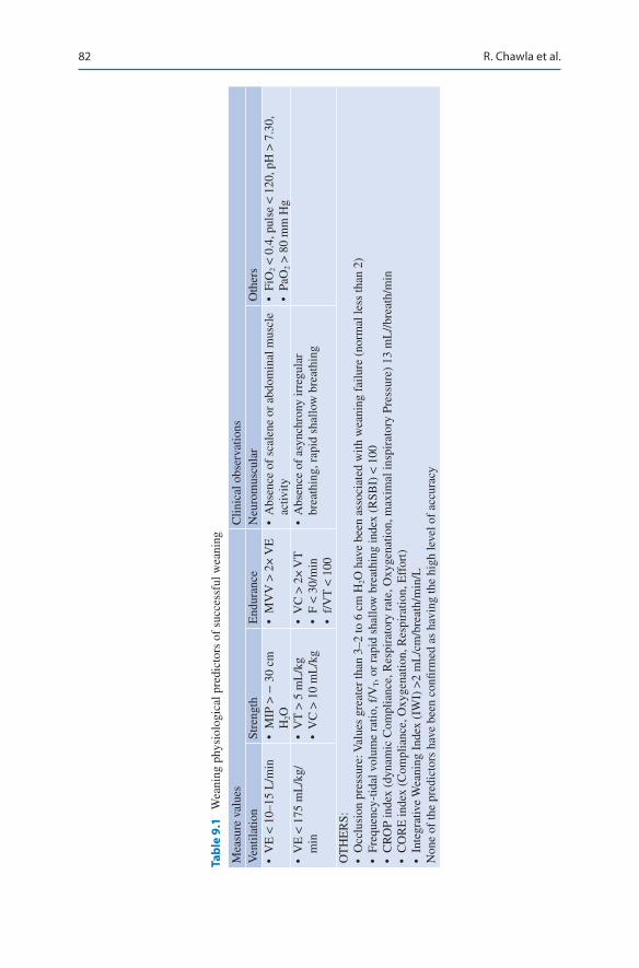
82
Tabl
e 9.
1 W
eani
ng p
hysi
olog
ical
pre
dict
ors
of s
ucce
ssfu
l wea
ning
Mea
sure
val
ues
Clin
ical
obs
erva
tions
Ven
tilat
ion
Stre
ngth
End
uran
ceN
euro
mus
cula
rO
ther
s•
VE
< 1
0–15
L/m
in•
MIP
> −
30
cm
H2O
• M
VV
> 2
× V
E•
Abs
ence
of
scal
ene
or a
bdom
inal
mus
cle
activ
ity•
FiO
2 < 0
.4, p
ulse
< 1
20, p
H >
7.3
0,
• Pa
O2 >
80
mm
Hg
• V
E <
175
mL
/kg/
min
• V
T >
5 m
L/k
g•
VC
> 1
0 m
L/k
g•
VC
> 2
× V
T•
F <
30/
min
• f/
VT
< 1
00
• A
bsen
ce o
f as
ynch
rony
irre
gula
r br
eath
ing,
rap
id s
hallo
w b
reat
hing
OT
HE
RS:
• O
cclu
sion
pre
ssur
e: V
alue
s gr
eate
r th
an 3
–2 to
6 c
m H
2O h
ave
been
ass
ocia
ted
with
wea
ning
fai
lure
(no
rmal
less
than
2)
• Fr
eque
ncy-
tidal
vol
ume
ratio
, f/V
T, o
r ra
pid
shal
low
bre
athi
ng in
dex
(RSB
I) <
100
• C
RO
P in
dex
(dyn
amic
Com
plia
nce,
Res
pira
tory
rat
e, O
xyge
natio
n, m
axim
al in
spir
ator
y Pr
essu
re)
13 m
L//b
reat
h/m
in•
CO
RE
inde
x (C
ompl
ianc
e, O
xyge
natio
n, R
espi
ratio
n, E
ffor
t)•
Inte
grat
ive
Wea
ning
Ind
ex (
IWI)
>2
mL
/cm
/bre
ath/
min
/LN
one
of th
e pr
edic
tors
hav
e be
en c
onfir
med
as
havi
ng th
e hi
gh le
vel o
f ac
cura
cy
R. Chawla et al.
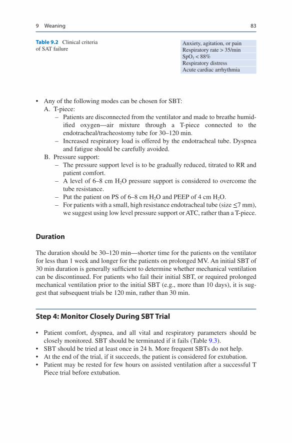
83
• Any of the following modes can be chosen for SBT: A. T-piece:
– Patients are disconnected from the ventilator and made to breathe humid-ified oxygen—air mixture through a T-piece connected to the endotracheal/tracheostomy tube for 30–120 min.
– Increased respiratory load is offered by the endotracheal tube. Dyspnea and fatigue should be carefully avoided.
B. Pressure support: – The pressure support level is to be gradually reduced, titrated to RR and
patient comfort. – A level of 6–8 cm H2O pressure support is considered to overcome the
tube resistance. – Put the patient on PS of 6–8 cm H2O and PEEP of 4 cm H2O. – For patients with a small, high resistance endotracheal tube (size ≤7 mm),
we suggest using low level pressure support or ATC, rather than a T-piece.
Duration
The duration should be 30–120 min—shorter time for the patients on the ventilator for less than 1 week and longer for the patients on prolonged MV. An initial SBT of 30 min duration is generally sufficient to determine whether mechanical ventilation can be discontinued. For patients who fail their initial SBT, or required prolonged mechanical ventilation prior to the initial SBT (e.g., more than 10 days), it is sug-gest that subsequent trials be 120 min, rather than 30 min.
Step 4: Monitor Closely During SBT Trial
• Patient comfort, dyspnea, and all vital and respiratory parameters should be closely monitored. SBT should be terminated if it fails (Table 9.3).
• SBT should be tried at least once in 24 h. More frequent SBTs do not help.• At the end of the trial, if it succeeds, the patient is considered for extubation.• Patient may be rested for few hours on assisted ventilation after a successful T
Piece trial before extubation.
Table 9.2 Clinical criteria of SAT failure
Anxiety, agitation, or painRespiratory rate > 35/minSpO2 < 88%Respiratory distressAcute cardiac arrhythmia
9 Weaning
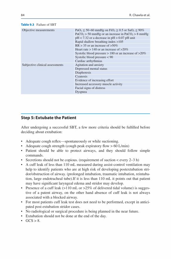
84
Step 5: Extubate the Patient
After undergoing a successful SBT, a few more criteria should be fulfilled before deciding about extubation:
• Adequate cough reflex—spontaneously or while suctioning.• Adequate cough strength (cough peak expiratory flow > 60 L/min)• Patient should be able to protect airways, and they should follow simple
commands.• Secretions should not be copious. (requirement of suction < every 2–3 h)• A cuff leak of less than 110 mL measured during assist-control ventilation may
help to identify patients who are at high risk of developing postextubation stri-dor/obstruction of airway. (prolonged intubation, traumatic intubation, reintuba-tion, large endotracheal tube).If it is less than 110 mL it points out that patient may have significant laryngeal edema and stridor may develop.
• Presence of a cuff leak (>110 mL or >25% of delivered tidal volume) is sugges-tive of a patent airway, on the other hand absence of cuff leak is not always associated with a blocked airway.
• For most patients cuff leak test does not need to be performed, except in antici-pated post extubation stridor cases.
• No radiological or surgical procedure is being planned in the near future.• Extubation should not be done at the end of the day.• GCS > 8.
Table 9.3 Failure of SBT
Objective measurements PaO2 ≤ 50–60 mmHg on FiO2 ≥ 0.5 or SaO2 ≤ 90%PaCO2 > 50 mmHg or an increase in PaCO2 > 8 mmHgpH < 7.32 or a decrease in pH > 0.07 pH unitRapid shallow breathing index >105RR > 35 or an increase of >50%Heart rate > 140 or an increase of >20%Systolic blood pressure > 180 or an increase of >20%Systolic blood pressure < 90Cardiac arrhythmias
Subjective clinical assessments Agitation and anxietyDepressed mental statusDiaphoresisCyanosisEvidence of increasing effortIncreased accessory muscle activityFacial signs of distressDyspnea
R. Chawla et al.
85
Step 6: Monitor for Extubation Failure
After extubation, the patient should be observed closely for signs of extubation failure as mentioned below:
• RR more than 25/min for 2 hrs• Heart rate more than 140 beats/min or sustained increase or decrease of more
than 20%• Clinical signs of respiratory muscle fatigue or increased work of breathing• SaO2 less than 90%; PaO2 less than 80 mmHg, on FiO2 more than 0.50• Hypercapnia (PaCO2 > 45 mmHg or > 20% from preextubation), pH < 7.33
Step 7: Try Noninvasive Ventilation (NIV)
• If the signs of extubation failure are present, the physician should try NIV par-ticularly in conditions where its role is proved; for example, in COPD, postop-erative failure after lung resection surgery, or decompensated obstructive sleep apnea.
• NIV has the advantage of reduced complications and better patient interactions. However, it is important to keep in mind that it should not delay reintubation (if required), and every hour that a patient spends on NIV when intubation is clearly required increases mortality and delays recovery.
• NIV is used in three clinical settings: – As an alternative weaning technique for the patients who failed SBT: Extubate
and put on NIV—well-documented role in COPD patients without significant comorbidities and in centers with expertise in NIV use.
– As a prophylactic measure for the patients with a high risk of reintubation: Studied in postoperative patients. Start NIV electively after successful SBT and extubation.
– As the treatment of respiratory insufficiency after extubation (postextubation failure): Useful in COPD.
In conditions where role of NIV is not proved, the patients should be reintubated.
High Flow nasal oxygen (HFNC) may be used in non COPD patients at risk of extubation failure.
Reintubation carries higher mortality either because of underlying medical con-dition or because of possible complications such as aspiration or ventilator- associated pneumonia.
Intravenous corticosteroid (Methylprednisolone 20 mg) four hours before extu-bation, and every four hourly for four doses, or a single dose of 40 mg four hours before extubation may be tried in patients with high risk for post extubation stridor
9 Weaning
86
(prolonged ventilation, difficult intubation, reintubation case) specially in the absence of cuff leak.
• Assess safety of starting feed after extubation, preferably after a swallow assess-ment, to avoid aspiration
Step 8: Identify Difficult Weaning
Weaning success is defined as extubation and the absence of ventilator support 48 h following extubation.
• Weaning failure is defined as one of the following: – Failed SBT – Reintubation and/or resumption of ventilator support following successful
extubation – Death within 48 h following extubation
• The term weaning in progress is used for the patients who are extubated, but remain supported by NIV.
• Difficult weaning—Patients who fail initial weaning and require up to three SBT or as long as 7 days from the first SBT to achieve successful weaning.
• Prolonged weaning—Patients who fail more than three weaning attempts or require more than 7 days of weaning after the first SBT.
Step 9: Ascertain the Cause of Weaning Difficulty
• Carry out a detailed examination of the patient, and look for the cause of difficult weaning.
• Difficult or prolonged weaning is usually due to incomplete resolution of initial indication for ventilation.
• Make a checklist based on pathophysiologic mechanisms: I. Inadequate respiratory drive
– Nutritional deficiencies – Excess of sedatives – Central nervous system abnormality – Sleep deprivation
II. Inability of the lungs to carry out gas exchange effectively – Unresolving pneumonia – Unresolved pulmonary edema/fluid overload – Undiagnosed pulmonary embolism – The splinting effect of obesity, abdominal distension, or ascites – Respiratory muscle fatigue/weakness – Nutritional and metabolic deficiencies – Critical illness polyneuropathy/myopathy
R. Chawla et al.

87
– Hypokalemia – Hypomagnesemia – Hypocalcemia – Hypophosphatemia – Hypoadrenalism – Hypothyroidism – Corticosteroids: myopathy, hyperglycemia – Chronic renal failure – Systemic disease sepsis: impaired diaphragmatic force generation – Refractory hypoxemia and hypercapnia – Persistently increased work of breathing – Ineffective triggering, auto-PEEP – Increased resistance due to ventilator tubings or humidification devices – Poor cardiac performance – Neuromuscular dysfunction/disease – Drugs
III. AnxietyIt is difficult to distinguish anxiety from ventilatory failure. If in doubt,
always presume it to be ventilatory failure. IV. Psychological dependency in difficult weaning
Step 10: Treat All the Reversible Causes Identified
• Provide good nutrition, but avoid overfeeding.• Have good glycemic control (110–140 mg/dL).• Correct metabolic factors (especially metabolic alkalosis).• Maintain hemoglobin above 7–8 g/dL.• Maintain adequate cardiac output and tissue perfusion.• Treat arrhythmia.• Treat hypothyroidism and steroid deficiency or excess.• Control the patient’s underlying illness.• Abolish ventilator dyssynchrony with appropriate inspiratory flow and trigger
settings.• Change of the mode of ventilation may help improve patient–ventilator
interactions.• Reverse bronchospasm as much as possible and reduce dynamic hyperinflation.• Drain out significant pleural effusions and ascites.• Treat intraabdominal hypertension.• Treat pulmonary edema aggressively.• Discontinue the use of steroids, aminoglycosides, colistin, and statins, if
possible.• Avoid fluid overload in renal failure and cardiac failure—do dialysis if
indicated.• Make the patient comfortable.
9 Weaning
88
• Aggressive physiotherapy and mobilization.• Reverse oversedation.• Treat anxiety: Improve patient communication, use relaxation techniques, and
give low-dose benzodiazepines.• Diagnose and treat narcotic/benzodiazepine withdrawal.• Treat delirium and depression.• Ensuring nighttime sleep may be helpful. Zolpidem may be added
Step 11: Plan the Weaning Process in Difficult Weaning
1. Select the mode of ventilationThe mode of ventilation used should provide adequate respiratory support
and prevent diaphragmatic atrophy.• Pressure support ventilation: It is most commonly used, and has been shown
to be better than SIMV for weaning.• Continuous positive airway pressure (CPAP): Besides the usual benefits of
improved oxygenation and improved left ventricular function, it has benefi-cial role in selected patients with hypoxemic respiratory failure.
• Automatic tube compensation: It may be helpful in narrow endotracheal tubes to overcome tube resistance.
• Proportional assist ventilation: It has been studied with CPAP and shown to improve respiratory mechanics.
• Adaptive support ventilation: It has been shown to be better than SIMV in postcardiac surgery patients.
• Control Mechanical Ventilation (CMV): It has logical use in patients showing respiratory fatigue on spontaneous mode. So it is recommended to use this mode in cases of difficult weaning at night to give rest to the muscles.
2. Plan tracheostomy.• Percutaneous tracheostomy has been shown to have fewer complications than
surgical tracheostomy and to be more cost-effective• Potential benefits of using tracheostomy in difficult-to-wean patients are as
follows: – Decreased work of breathing – Reduced requirement of sedation and improved patient comfort and
cooperation – Earlier reinstitution of oral feeding – Less chances of accidental extubation
• In spite of the above-mentioned benefits, tracheostomy has not been consis-tently shown to decrease mortality. It has resulted in a number of dependent survivors. It facilitates easier bedside management of such patients.
3. Do aggressive physiotherapy and mobilization• Physiotherapy and mobilization are prerequisites for successful weaning.
Early institution of physiotherapy in a protocol-driven approach and daily assessment to achieve maximum mobility is now an integral part of ICU management.
R. Chawla et al.
89
4. Select proper place for weaning• Cost-effective care has been shown to be provided in respiratory intermediate
care units and specialized regional weaning centers. It requires team effort and expertise.
Step 12: Choose a Weaning Protocol
Protocol-driven weaning has more chances of success, reduced costs, and probably reduced mortality. Two basic weaning protocols are used for a prolonged weaning patient:
• Progressive reduction of ventilator support• Progressively longer periods of SBTs
No significant difference in weaning success and mortality rate, duration of ven-tilatory assistance, or total hospital length of stay is reported between these two weaning techniques in the difficult-to-wean patients. A combination of both the protocols can also be used.
Step 13: Decide About Home Ventilation or Transfer to a Long Term Care Facility
Indications
• An inability to be completely weaned from ventilatory support including NIV• A progression of disease etiology that requires increasing ventilatory support
Patients should have stable physiology and proper resources, personnel, and motivation.
Suggested Reading
AARC, Respiratory Home Care Focus Group. AARC clinical practice guideline. Long- term invasive mechanical ventilation in the home—2007 revision & update. Respir Care. 2007;52(8):1056–62. This article discusses multiorganizational effort to clear the controver-sies concerning the best method for conducting weaning, liberation from mechanical ventila-tion, and extubation.
Ambrosino N, Gabbrielli L. The difficult-to-wean patient. Expert Rev Respir Med. 2010;4(5):685–92. The study describes the epidemiology, causes, and management strategies for difficult-to- wean patients.
Blackwood B, Alderdice F, Burns K, Cardwell C, Lavery G, O’Halloran P. Use of weaning pro-tocols for reducing duration of mechanical ventilation in critically ill adult patients: Cochrane systematic review and meta-analysis. Br Med J. 2011;342:c7237. This review included 11 trials that included 1971 patients. They found that compared with usual care, the mean duration of
9 Weaning

90
MV in the weaning protocol group was reduced by 25%, the duration of weaning was reduced by 78%, and the ICU length of stay by 10%.
Boles JM, Bion J, Connors A, Herridge M, Marsh B, Melot C, et al. Weaning from mechanical ventilation. Statement of the sixth international consensus conference on intensive care medi-cine. Eur Respir J. 2007;29(5):1033–56. Home MV is now increasingly being used to rehabili-tate long-term ventilator-dependent patients.
Jubran A, Lawm G, Kelly J, Duffner LA, Gungor G, Collins EG, et al. Depressive disorders dur-ing weaning from prolonged mechanical ventilation. Intensive Care Med. 2010;36:828–35. Depressive disorders were diagnosed in 42% of the patients who were weaned from prolonged ventilation. Patients with depressive disorders were more likely to experience weaning failure and death.
MacIntyre NR, Epstein SK, Carson S, Scheinhorn D, Christopher K, Muldoon S. Management of patients requiring prolonged mechanical ventilation: report of a NAMDRC consensus confer-ence. Chest. 2005;128:3937–54. The goal of this conference was not only to review existing practices but also to develop recommendations on a variety of assessment, management, and reimbursement issues.
R. Chawla et al.
91© Springer Nature Singapore Pte Ltd. 2020R. Chawla, S. Todi (eds.), ICU Protocols, https://doi.org/10.1007/978-981-15-0898-1_10
A. Bansal (*) Department of Respiratory, Critical Care & Sleep Medicine, Indraprastha Apollo Hospitals, New Delhi, India
R. Shekhar Department of Pulmonology and Sleep Medicine, Fortis Hospital, Faridabad, Haryana, India
10Massive Hemoptysis
Avdhesh Bansal and Ravi Shekhar
Hemoptysis is defined as expectoration of blood from the trachea-bronchial tree. Massive hemoptysis is defined as expectoration of 300–600 mL of blood in 24 h or >100 mL/h. It is estimated that 400 mL of blood in the alveolar space is sufficient to inhibit gaseous exchange significantly and the cause of death is usually asphyxi-ation rather than exsanguination. Any amount of hemoptysis that causes respiratory compromise and/or hemodynamic instability is life threatening and constitutes a medical emergency. The risk to life is determined by the amount and speed of bleed-ing, patient’s ability to expectorate and underlying cardiopulmonary reserve. Mortality rate can be in excess of 50%.
Step 1: Initiate Resuscitation
Take history of the patient and find out the approximate amount of hemoptysis. After a quick physical examination of the patient, initiate resuscitation.
Case Scenario
A 65 year old male, recently diagnosed as a case of pulmonary tuberculosis, on antitubercular treatment for the past 2 months, presented to emergency depart-ment with history of coughing out blood. For the past 2 weeks, he has a history of sputum streaking of blood, but today morning, he expectorated about a cup of bright red blood. While giving history of his symptoms, he expectorated large amount of blood, about 300 mL.
92
Airway
• Maintaining an open airway should be the first priority in the management. The main objective is to prevent asphyxiation.
• Lateral decubitus position with the affected side towards floor, in order to avoid aspiration of blood into the unaffected lung. Therefore, the hemithorax in which the bleeding is originating must first be determined. Clinical signs and symptoms and Chest X-ray findings can be used to narrow down the site of the lesion.
• Intubate with single-lumen endotracheal tube in cases of severe and diffuse endobronchial bleeding. It may achieve immediate control of the airways to allow adequate suctioning and diagnostic and therapeutic fiber-optic bronchos-copy. It may also be helpful in cases of nonavailability of the double-lumen tube or expertise.
• Double-lumen endotracheal tube can be helpful to isolate and ventilate the lungs separately if the bleeding is lateralized. The double-lumen tubes are, however, easily obstructed by clots and do not permit passage of bronchoscopes of ade-quate size to allow bronchial toilet under unobstructed vision. It may still, how-ever, be possible to confirm the tube position by the fiberbronchoscope. Be careful as it can result in tube displacement.
• Unilateral lung ventilation (healthier side) may also be achieved by single lumen endotracheal tube advanced to the unaffected side.
Breathing
• Large amount of blood in the tracheobronchial tree may be a major impediment to gas exchange. Maintain oxygen saturation above 94% by administering oxygen.
Circulation:• All patients should be admitted to the intensive care unit. Vital signs should be
monitored continuously.• The intra-arterial line should be inserted for continuous blood pressure
monitoring.• Two large-bore intravenous lines or a central venous catheter should be inserted
and intravenous fluids should be started.• If the condition is rapidly deteriorating, O-positive blood (O-negative in case of
women of childbearing age) should be transfused while waiting for blood group-ing and cross matching. Meanwhile, determine the blood type, cross-match, and request for red cell units, depending on the requirement. Assess the severity of volume loss.
• Monitoring of vital signs and quantification of the hemoptysis.
A. Bansal and R. Shekhar
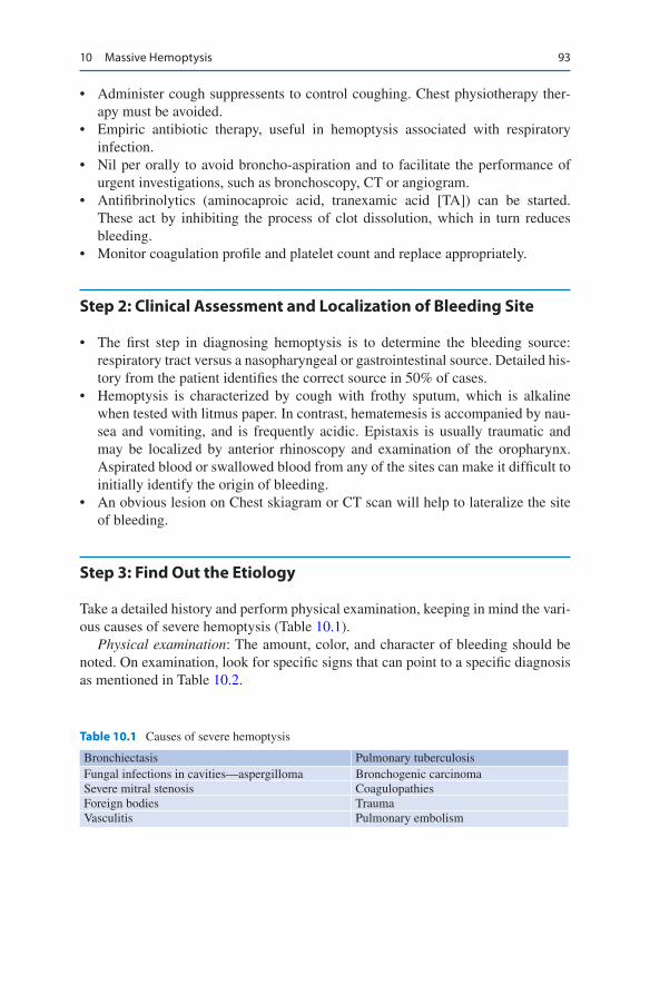
93
• Administer cough suppressents to control coughing. Chest physiotherapy ther-apy must be avoided.
• Empiric antibiotic therapy, useful in hemoptysis associated with respiratory infection.
• Nil per orally to avoid broncho-aspiration and to facilitate the performance of urgent investigations, such as bronchoscopy, CT or angiogram.
• Antifibrinolytics (aminocaproic acid, tranexamic acid [TA]) can be started. These act by inhibiting the process of clot dissolution, which in turn reduces bleeding.
• Monitor coagulation profile and platelet count and replace appropriately.
Step 2: Clinical Assessment and Localization of Bleeding Site
• The first step in diagnosing hemoptysis is to determine the bleeding source: respiratory tract versus a nasopharyngeal or gastrointestinal source. Detailed his-tory from the patient identifies the correct source in 50% of cases.
• Hemoptysis is characterized by cough with frothy sputum, which is alkaline when tested with litmus paper. In contrast, hematemesis is accompanied by nau-sea and vomiting, and is frequently acidic. Epistaxis is usually traumatic and may be localized by anterior rhinoscopy and examination of the oropharynx. Aspirated blood or swallowed blood from any of the sites can make it difficult to initially identify the origin of bleeding.
• An obvious lesion on Chest skiagram or CT scan will help to lateralize the site of bleeding.
Step 3: Find Out the Etiology
Take a detailed history and perform physical examination, keeping in mind the vari-ous causes of severe hemoptysis (Table 10.1).
Physical examination: The amount, color, and character of bleeding should be noted. On examination, look for specific signs that can point to a specific diagnosis as mentioned in Table 10.2.
Table 10.1 Causes of severe hemoptysis
Bronchiectasis Pulmonary tuberculosisFungal infections in cavities—aspergilloma Bronchogenic carcinomaSevere mitral stenosis CoagulopathiesForeign bodies TraumaVasculitis Pulmonary embolism
10 Massive Hemoptysis

94
Step 4: Plan Investigations
The following investigations should be ordered in all patients:
Blood Investigations
• Hemoglobin, hematocrit, platelets count, coagulation studies (prothrombin time including international normalized ratio, partial thromboplastin time).
• Renal function tests (CKD patients have poor platelet function and tend to bleed more).
• Liver function tests (CLD patients tend to have poor coagulation profile).• Arterial blood gas analysis.• D-dimer (For Venous thromboembolism).• urinalysis. (For vasculitis and Good pastures syndrome).
Chest Skiagram
Chest radiography helps in localizing the bleeding source in 60% of cases and iden-tifying possible etiology such as a lung mass, cavitary lesion, or alveolar hemorrhage.
Further Investigations Will Depend on the Possible Diagnosis
• Sputum/bronchoalveolar lavage should be tested for bacteria including myco-bacteria, fungi, and malignant cells.
• Multidetector computed tomography (MDCT/HRCT) has proved to be of consid-erable diagnostic value in localizing the site of bleeding. Contrast-enhanced CT produces high-resolution angiographic studies with a combination of multiplanar reformatted images. Therefore, MDCT angiography is able to identify the source of bleeding and underlying pathology with high sensitivity. This is of particular importance to the interventional radiologist planning for arterial embolization.
Table 10.2 Focused physical examination
Digital clubbing—bronchiectasis or lung carcinomaStridor—tracheal tumors or a foreign bodyOral and aphthous ulcers, genital ulcers, and uveitis—Behcet’s disease in which pulmonary arteriovenous malformations (AVM) are responsible for hemoptysisCutaneous purpura or ecchymosis—coagulation disordersDiastolic murmur—mitral stenosisSaddle nose with rhinitis and septal perforation—Wegener’s granulomatosis
A. Bansal and R. Shekhar

95
• If MDCT is not available, contrast-enhanced single-detector spiral CT can be performed to detect bronchial and nonbronchial systemic arterial vascular lesions such as thoracic aneurysm and AV malformation. Except for life-threatening situations, thoracic CT scans should ideally be performed prior to bronchoscopy.
Step 5: Bronchoscopy
• Diagnostic bronchoscopy is the primary method for diagnosis and localization of hemoptysis. Rigid bronchoscopy is ideally recommended in cases of massive hemoptysis because of its ability to maintain airway patency.
• Flexible bronchoscopy (FOB) is used more widely, considering the ease of per-formance at the patient’s bedside without the use of general anesthetic and oper-ating theatre suite. Vasoactive drugs can be instilled directly into the bleeding source. The overall diagnostic accuracy of bronchoscopy in localizing the site of bleeding is 50%, but it is less useful in identifying the underlying cause. Further disadvantages include nonvisualization during active hemoptysis, and ineffec-tiveness of endobronchial therapies in most cases. The ideal time for bronchos-copy is controversial, but the consensus is to perform urgent bronchoscopy in patients with massive hemoptysis.
• In case plug of blood clot is seen in a segment, it is recommended not to remove it as it may restart massive hemoptysis.
• Administer local therapy: The following therapeutic measures can be tried through the fiber bronchoscope to control bleeding. – Iced saline bronchial lavage in the involved lung – Iced saline lavage of up to 1000 mL in 50 mL aliquots at the bleeding site – Administration of topical hemostatic agents, such as epinephrine (1:20,000)
or thrombin-fibrinogen – Tamponade therapy, using oxidized regenerated cellulose mesh
• Another alternative site-specific therapy that can be tried using the flexible bron-choscope includes endobronchial tamponade using a balloon tamponade catheter to prevent aspiration to the unaffected contralateral lung and preserve gas exchange.
• A bronchus-blocking balloon catheter (Freitag Catheter) has been designed to be used through the working channel of the flexible bronchoscope which has a sec-ond channel that is used to instill vasoactive hemostatic agents, such as iced saline, epinephrine, vasopressin, or thrombin-fibrinogen, to control bleeding.
Step 6: Start Pharmacotherapy
• Antibiotics and steroids, depending on the underlying condition, are necessary to control the precipitating cause.
• Intravenous vasopressin has not been shown to improve the outcome, and thera-pies such tranexamic acid are usually prescribed.
10 Massive Hemoptysis
96
• Correct coagulation abnormalities with appropriate platelet, fresh frozen plasma, and cryoprecipitate.
• Prothrombin Complex concentrate (PCC) /Recombinant activated factor VII (rFVIIa) can be tried as it has been reported as an effective temporizing measure in unstable patients with hemoptysis, when conventional treatment is not imme-diately available.
Step 7: Bronchial Artery Embolization (BAE)
• Bronchial artery angiography is highly effective in localizing the source of bleed-ing and identifying a vessel for embolization. The bronchial circulation (a high- pressure circuit with systolic pressure of 120 mmHg) is the most common source of massive hemoptysis, accounting for 95% of all cases. Less than 5% of bleed-ing arises from the pulmonary circulation (a low-pressure circuit with a systolic pressure of 15–20 mmHg).
• Once the bleeding bronchial artery is located, particles (polyvinyl alcohol foam, absorbable gelatin, pledgets of Gianturco steel coils) are infused into the artery. BAE achieves immediate control of the bleeding in 75–90% of cases. Post embo-lization recurrence of hemoptysis has been observed in 20% of cases.
• Remember that this is a temporizing procedure while every effort is being made to make a clear diagnosis and treat accordingly, except in AV malformations in which this could be the definitive treatment.
• If bronchial artery angiography does not reveal a bleeding vessel, a pulmonary angiogram is required to investigate the pulmonary circulation.
• After Failed Bronchial artery embolisation a relook Bronchoscopy preferably with rigid bronchoscopy should be tried for localising and controlling bleed prior to evaluating feasibility for surgical resection.
Step 8: Surgical Resection (Segmentectomy, Lobectomy, and Pneumonectomy)
Surgery is indicated in the following conditions:
• BAE is unavailable or technically unfeasible, or bleeding or aspiration of blood continues despite embolization.
• Surgical resection of the bleeding site is possible if the lesion can be localized and the patient is fit for surgery.
• Surgery is also preferable when the acuity (rate and amount of bleeding) of hemoptysis precludes safe BAE.
• More specific indications for surgery in massive hemoptysis include persistent bleeding from a mycetoma resistant to medical management, bronchial adenoma, iatrogenic pulmonary artery rupture, leaking aortic aneurysm, hydatid cysts, and selected AV malformations.
A. Bansal and R. Shekhar
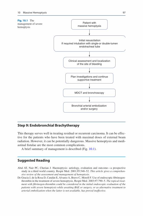
97
Step 9: Endobronchial Brachytherapy
This therapy serves well in treating residual or recurrent carcinoma. It can be effec-tive for the patients who have been treated with maximal doses of external beam radiation. However, it can be potentially dangerous. Massive hemoptysis and medi-astinal fistulae are the most common complications.
A brief summary of management is described (Fig. 10.1).
Suggested Reading
Abal AT, Nair PC, Cherian J. Haemoptysis: aetiology, evaluation and outcome—a prospective study in a third world country. Respir Med. 2001;95:548–52. This article gives a comprehen-sive review of the assessment and management of hemoptysis.
De Gracia J, de la Rosa D, Catalán E, Alvarez A, Bravo C, Morell F. Use of endoscopic fibrinogen- thrombin in the treatment of severe hemoptysis. Respir Med. 2003;97:790–5. The topical treat-ment with fibrinogen-thrombin could be considered in the initial endoscopic evaluation of the patients with severe hemoptysis while awaiting BAE or surgery, or as alternative treatment to arterial embolization when the latter is not available, has proved ineffective.
Patient withmassive hemoptysis
Initial resuscitationIf required intubation with single-or double-lumen
endotracheal tube
Clinical assessment and localizationof the site of bleeding
Plan investigations and continuesupportive treatment
MDCT and bronchoscopy
Bronchial arterial embolizationand/or surgery
Fig. 10.1 The management of severe hemoptysis
10 Massive Hemoptysis
98
Dupree HJ, Lewejohann JC, Gleiss J, Muhl E, Bruch HP. Fiberoptic bronchoscopy of intubated patients with life-threatening hemoptysis. World J Surg. 2001;25:104–7. Widespread use of flexible bronchoscopy makes this procedure immediately applicable in critical situations. Intubated and mechanically ventilated patients with life-threatening hemoptysis especially benefit from this rapidly feasible procedure.
Jean-Baptiste E. Clinical assessment and management of massive hemoptysis. Crit Care Med. 2000;28:1642–7. This article gives comprehensive review of assessment and management of hemoptysis.
Larici AR, et al. Diagnosis and management of hemoptysis. Diagn Interv Radiol. 2014;20(4):299–309.
MacDonald JA, Fraser JF, Foot CL, Tran K. Successful use of recombinant factor VII in mas-sive hemoptysis due to community-acquired pneumonia. Chest. 2006;130:577–9. Recombinant factor VII may be a useful temporizing measure in the unstable patient with pulmonary hem-orrhage without coagulopathic bleeding when conventional treatment is not immediately available.
Ong TH, Eng P. Massive hemoptysis requiring intensive care. Intensive Care Med. 2003;29:317–20. Embolization is a suitable first-line treatment for massive hemoptysis not responding to conservative treatment, reserving emergency surgery for cases in which the above measures are insufficient to control bleeding.
Razazi K, Parrot A, Khalil A, et al. Severe haemoptysis in patients with nonsmall cell lung carci-noma. Eur Respir J. 2015;45:756.
Yoon W, Kim JK, Kim YH, Chung TW, Kang HK. Bronchial and nonbronchial systemic artery embolization for life-threatening hemoptysis: a comprehensive review. Radiographics. 2002;22:1395–409. BAE is a safe and effective nonsurgical treatment for the patients with massive hemoptysis. However, nonbronchial systemic arteries can be a significant source of massive hemoptysis and a cause of recurrence after successful BAE.
A. Bansal and R. Shekhar
99© Springer Nature Singapore Pte Ltd. 2020R. Chawla, S. Todi (eds.), ICU Protocols, https://doi.org/10.1007/978-981-15-0898-1_11
R. Chawla (*) Department of Respiratory, Critical Care & Sleep Medicine, Indraprastha Apollo Hospitals, New Delhi, India
S. Todi Department of Critical Care & Emergency Medicines, A.M.R.I. Hospital, Kolkata, India
11Pulmonary Thromboembolism
Rajesh Chawla and Subhash Todi
Pulmonary thromboembolism (PTE) is the most common preventable cause of hos-pital death. PE should be suspected in all patients who present with new or worsen-ing dyspnea, chest pain, or sustained hypotension without any other obvious cause. Pulmonary Embolism (PE) can be caused by air, fat or tumor apart from thrombus. PTE can be classified based on the temporal pattern of presentation (acute, sub-acute, or chronic) or the presence or absence of hemodynamic stability (hemody-namically unstable or stable) or the anatomical location (saddle, lobar, segmental, subsegmental).
Patients are considered to be hemodynamically unstable if they are in shock or have a systolic blood pressure of less than 90 mmHg or a drop in systolic pressure of more than 40 mmHg from baseline for more than 15 min requiring vasopressor or clear evidence of shock. Hemodynamically stable PTE are those who do not meet the definition of hemodynamically unstable PTE.
High index of suspicion and prompt management can improve survival in these patients. Mortality of acute PE is approximately 30%, which can be reduced to 2–10% by appropriate management.
A 67-year-old male patient was admitted to hospital with severe community- acquired pneumonia and acute respiratory failure. He was treated with antibiot-ics and mechanical ventilation. He improved with the treatment and was extubated on day 4. On day 7, he suddenly developed acute severe breathlessness and chest pain.
100
Step 1: Initiate Resuscitation
• Provide oxygen to maintain saturation at more than 90% in suspected case of PE, and resuscitate as mentioned in Chap. 23, Vol. 2.
• If the patient is hypotensive, administer 500–1000 mL isotonic crystalloid. Any more volume resuscitation should be given with caution as it may increase right ventricle (RV) wall tension and cause ischemia and worsening of shock. Vasopressor therapy should be added if no response to IV fluids.
• Mechanically ventilate if there is respiratory collapse Due consideration should be given during intubation to prevent hypotension with minimal use of sedation and least positive pressure needed for adequate ventilation.
• Record a detailed medical history and perform physical examination.
Step 2: Assess the Risk Factors and Differentiate from Other Causes of Acute Dyspnea and Chest Pain
Assess the risk factors for PE and deep venous thrombosis (DVT) from medical history as mentioned below:
• Prior venous thromboembolism• Immobility for more than 48 h—congestive heart failure, septic shock, surgery
with general anaesthesia, (specially orthopedic surgery) mechanical ventilation• Abdominal or lower extremity surgery or trauma• Hypercoagulable states-Common inherited hypercoagulable states-factor V
Leiden mutation and the prothrombin gene mutation, Protein S deficiency, Protein C deficiency, Antithrombin III deficiency, Antiphospholipids antibodies
• Malignancy• Hematological disorders: Polycythaemia, Essential thrombocythaemia• Trauma, Spinal cord injury• Nephrotic syndrome• Heparin-induced thrombocytopenia• Pregnancy or use of oral contraceptives• Indwelling central venous catheters• Obesity• Drugs-tamoxifen, Thalidomide• Activated factor VII, Prothrombin complex concentrate administration• Intravenous drug use• Prolonged travel• Other causes of acute dyspnea and chest pain: Myocardial infarction, aortic dis-
section, pericarditis, pneumothorax, pneumonia, heart failure
R. Chawla and S. Todi
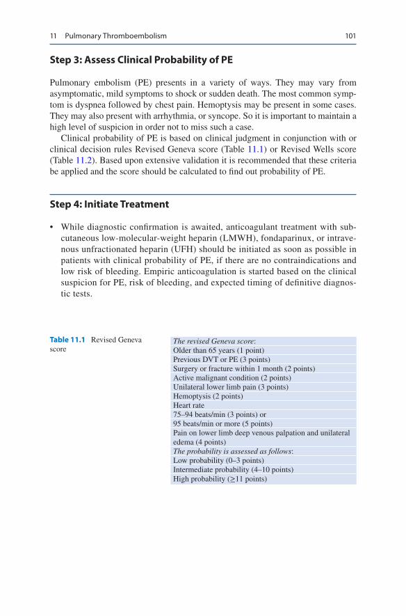
101
Step 3: Assess Clinical Probability of PE
Pulmonary embolism (PE) presents in a variety of ways. They may vary from asymptomatic, mild symptoms to shock or sudden death. The most common symp-tom is dyspnea followed by chest pain. Hemoptysis may be present in some cases. They may also present with arrhythmia, or syncope. So it is important to maintain a high level of suspicion in order not to miss such a case.
Clinical probability of PE is based on clinical judgment in conjunction with or clinical decision rules Revised Geneva score (Table 11.1) or Revised Wells score (Table 11.2). Based upon extensive validation it is recommended that these criteria be applied and the score should be calculated to find out probability of PE.
Step 4: Initiate Treatment
• While diagnostic confirmation is awaited, anticoagulant treatment with sub-cutaneous low-molecular-weight heparin (LMWH), fondaparinux, or intrave-nous unfractionated heparin (UFH) should be initiated as soon as possible in patients with clinical probability of PE, if there are no contraindications and low risk of bleeding. Empiric anticoagulation is started based on the clinical suspicion for PE, risk of bleeding, and expected timing of definitive diagnos-tic tests.
Table 11.1 Revised Geneva score
The revised Geneva score:Older than 65 years (1 point)Previous DVT or PE (3 points)Surgery or fracture within 1 month (2 points)Active malignant condition (2 points)Unilateral lower limb pain (3 points)Hemoptysis (2 points)Heart rate75–94 beats/min (3 points) or95 beats/min or more (5 points)Pain on lower limb deep venous palpation and unilateral edema (4 points)The probability is assessed as follows:Low probability (0–3 points)Intermediate probability (4–10 points)High probability (≥11 points)
11 Pulmonary Thromboembolism
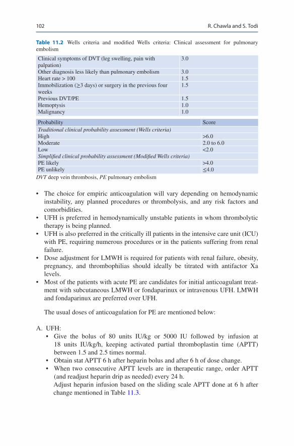
102
• The choice for empiric anticoagulation will vary depending on hemodynamic instability, any planned procedures or thrombolysis, and any risk factors and comorbidities.
• UFH is preferred in hemodynamically unstable patients in whom thrombolytic therapy is being planned.
• UFH is also preferred in the critically ill patients in the intensive care unit (ICU) with PE, requiring numerous procedures or in the patients suffering from renal failure.
• Dose adjustment for LMWH is required for patients with renal failure, obesity, pregnancy, and thrombophilias should ideally be titrated with antifactor Xa levels.
• Most of the patients with acute PE are candidates for initial anticoagulant treat-ment with subcutaneous LMWH or fondaparinux or intravenous UFH. LMWH and fondaparinux are preferred over UFH.
The usual doses of anticoagulation for PE are mentioned below:
A. UFH:• Give the bolus of 80 units IU/kg or 5000 IU followed by infusion at
18 units IU/kg/h, keeping activated partial thromboplastin time (APTT) between 1.5 and 2.5 times normal.
• Obtain stat APTT 6 h after heparin bolus and after 6 h of dose change.• When two consecutive APTT levels are in therapeutic range, order APTT
(and readjust heparin drip as needed) every 24 h.Adjust heparin infusion based on the sliding scale APTT done at 6 h after change mentioned in Table 11.3.
Table 11.2 Wells criteria and modified Wells criteria: Clinical assessment for pulmonary embolism
Clinical symptoms of DVT (leg swelling, pain with palpation)
3.0
Other diagnosis less likely than pulmonary embolism 3.0Heart rate > 100 1.5Immobilization (≥3 days) or surgery in the previous four weeks
1.5
Previous DVT/PE 1.5Hemoptysis 1.0Malignancy 1.0
Probability ScoreTraditional clinical probability assessment (Wells criteria)High >6.0Moderate 2.0 to 6.0Low <2.0Simplified clinical probability assessment (Modified Wells criteria)PE likely >4.0PE unlikely ≤4.0
DVT deep vein thrombosis, PE pulmonary embolism
R. Chawla and S. Todi
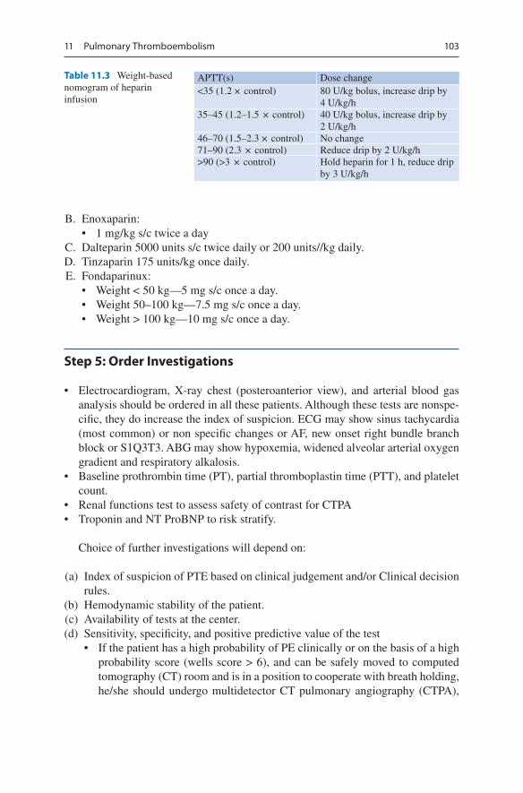
103
B. Enoxaparin:• 1 mg/kg s/c twice a day
C. Dalteparin 5000 units s/c twice daily or 200 units//kg daily. D. Tinzaparin 175 units/kg once daily. E. Fondaparinux:
• Weight < 50 kg—5 mg s/c once a day.• Weight 50–100 kg—7.5 mg s/c once a day.• Weight > 100 kg—10 mg s/c once a day.
Step 5: Order Investigations
• Electrocardiogram, X-ray chest (posteroanterior view), and arterial blood gas analysis should be ordered in all these patients. Although these tests are nonspe-cific, they do increase the index of suspicion. ECG may show sinus tachycardia (most common) or non specific changes or AF, new onset right bundle branch block or S1Q3T3. ABG may show hypoxemia, widened alveolar arterial oxygen gradient and respiratory alkalosis.
• Baseline prothrombin time (PT), partial thromboplastin time (PTT), and platelet count.
• Renal functions test to assess safety of contrast for CTPA• Troponin and NT ProBNP to risk stratify.
Choice of further investigations will depend on:
(a) Index of suspicion of PTE based on clinical judgement and/or Clinical decision rules.
(b) Hemodynamic stability of the patient. (c) Availability of tests at the center. (d) Sensitivity, specificity, and positive predictive value of the test
• If the patient has a high probability of PE clinically or on the basis of a high probability score (wells score > 6), and can be safely moved to computed tomography (CT) room and is in a position to cooperate with breath holding, he/she should undergo multidetector CT pulmonary angiography (CTPA),
Table 11.3 Weight-based nomogram of heparin infusion
APTT(s) Dose change<35 (1.2 × control) 80 U/kg bolus, increase drip by
4 U/kg/h35–45 (1.2–1.5 × control) 40 U/kg bolus, increase drip by
2 U/kg/h46–70 (1.5–2.3 × control) No change71–90 (2.3 × control) Reduce drip by 2 U/kg/h>90 (>3 × control) Hold heparin for 1 h, reduce drip
by 3 U/kg/h
11 Pulmonary Thromboembolism
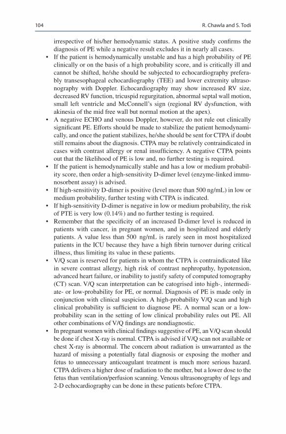
104
irrespective of his/her hemodynamic status. A positive study confirms the diagnosis of PE while a negative result excludes it in nearly all cases.
• If the patient is hemodynamically unstable and has a high probability of PE clinically or on the basis of a high probability score, and is critically ill and cannot be shifted, he/she should be subjected to echocardiography prefera-bly transesophageal echocardiography (TEE) and lower extremity ultraso-nography with Doppler. Echocardiography may show increased RV size, decreased RV function, tricuspid regurgitation, abnormal septal wall motion, small left ventricle and McConnell’s sign (regional RV dysfunction, with akinesia of the mid free wall but normal motion at the apex).
• A negative ECHO and venous Doppler, however, do not rule out clinically significant PE. Efforts should be made to stabilize the patient hemodynami-cally, and once the patient stabilizes, he/she should be sent for CTPA if doubt still remains about the diagnosis. CTPA may be relatively contraindicated in cases with contrast allergy or renal insufficiency. A negative CTPA points out that the likelihood of PE is low and, no further testing is required.
• If the patient is hemodynamically stable and has a low or medium probabil-ity score, then order a high-sensitivity D-dimer level (enzyme-linked immu-nosorbent assay) is advised.
• If high-sensitivity D-dimer is positive (level more than 500 ng/mL) in low or medium probability, further testing with CTPA is indicated.
• If high-sensitivity D-dimer is negative in low or medium probability, the risk of PTE is very low (0.14%) and no further testing is required.
• Remember that the specificity of an increased D-dimer level is reduced in patients with cancer, in pregnant women, and in hospitalized and elderly patients. A value less than 500 ng/mL is rarely seen in most hospitalized patients in the ICU because they have a high fibrin turnover during critical illness, thus limiting its value in these patients.
• V/Q scan is reserved for patients in whom the CTPA is contraindicated like in severe contrast allergy, high risk of contrast nephropathy, hypotension, advanced heart failure, or inability to justify safety of computed tomography (CT) scan. V/Q scan interpretation can be catogrised into high-, intermedi-ate- or low-probability for PE, or normal. Diagnosis of PE is made only in conjunction with clinical suspicion. A high-probability V/Q scan and high clinical probability is sufficient to diagnose PE. A normal scan or a low- probability scan in the setting of low clinical probability rules out PE. All other combinations of V/Q findings are nondiagnostic.
• In pregnant women with clinical findings suggestive of PE, an V/Q scan should be done if chest X-ray is normal. CTPA is advised if V/Q scan not available or chest X-ray is abnormal. The concern about radiation is unwarranted as the hazard of missing a potentially fatal diagnosis or exposing the mother and fetus to unnecessary anticoagulant treatment is much more serious hazard. CTPA delivers a higher dose of radiation to the mother, but a lower dose to the fetus than ventilation/perfusion scanning. Venous ultrasonography of legs and 2-D echocardiography can be done in these patients before CTPA.
R. Chawla and S. Todi

105
• When both CTPA and V/Q scan both are contraindicated, Magnetic Resonance Pulmonary Angiography (MRPA) may be an imaging option for diagnosis of PE. In MRPA no ionizing radiation is involved and the exami-nation can be coupled with MR venography. It needs expertise to avoid tech-nically inadequate images and interpretation.
Step 6: Identify the Risk of Adverse Outcome for Triage
• Risk stratification should be done promptly because fatal PE generally occurs early after hospital admission (Table 11.4).
• It is based on clinical features and markers of myocardial dysfunction or injury.• If the patient is hemodynamically stable, then order TEE, troponin, and brain
natriuretic peptide (BNP) levels.• Myocardial dysfunction (right ventricular dilatation, hypokinesia, and ventricu-
lar septal bowing) based on echo and injury markers (elevated troponin and BNP) is useful and helps decide about thrombolysis, as mentioned below (Table 11.4).
Step 7: Consider Thrombolysis
(a) If the patient is hemodynamically unstable• Admit to the ICU.• Start anticoagulation, preferably intravenous UFH. Keep APTT time 1.5–2.5
to normal.• Administer thrombolytic therapy (Tables 11.5 and 11.6) if there are no con-
traindications (Table 11.7). Discontinue heparin during thrombolysis.• Give other supportive measures to stabilize the patient.
(b) Hemodynamically stable patients maybe considered for thromolytic therapy on case to case basis in the following situations:
Table 11.4 Risk stratification of patients with PE also has potential clinical implications for triage
Absence of right ventricular dysfunction and normal troponin level—admit in wardHemodynamically stable with right ventricular dysfunction or injury—admit the patient in High Dependency Unit (HDU)Hemodynamically unstable and right ventricular dysfunction and injury—admit to the ICU
The risk of adverse outcome is more in the following situations:Shock (systolic blood pressure < 90 mmHg) and/or BP drop ≥40 mmHg for >15 min and sustained hypotension Immobilization due to neurological disease Age 75 years or more Cardiac, renal, or respiratory disease or cancer
11 Pulmonary Thromboembolism
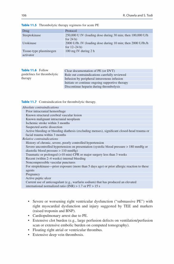
106
Table 11.5 Thrombolytic therapy regimens for acute PE
Drug ProtocolStreptokinase 250,000 U IV (loading dose during 30 min; then 100,000 U/h
for 24 h)Urokinase 2000 U/lb. IV (loading dose during 10 min; then 2000 U/lb./h
for 12–24 h)Tissue-type plasminogen activator
100 mg IV during 2 h
Table 11.6 Follow guidelines for thrombolytic therapy
Clear documentation of PE (or DVT)Rule out contraindications carefully reviewedInfusion by peripheral intravenous infusionInitiate or continue ongoing supportive therapyDiscontinue heparin during thrombolysis
Table 11.7 Contraindication for thrombolytic therapy.
Absolute contraindications Prior intracranial hemorrhage Known structural cerebral vascular lesion Known malignant intracranial neoplasm Ischemic stroke within 3 months Suspected aortic dissection Active bleeding or bleeding diathesis (excluding menses), significant closed-head trauma or
facial trauma within 3 monthsRelative contraindications History of chronic, severe, poorly controlled hypertension Severe uncontrolled hypertension on presentation (systolic blood pressure > 180 mmHg or
diastolic blood pressure > 110 mmHg) Traumatic or prolonged (>10 min) CPR or major surgery less than 3 weeks Recent (within 2–4 weeks) internal bleeding Noncompressible vascular punctures For streptokinase—prior exposure (more than 5 days ago) or prior allergic reaction to these
agents Pregnancy Active peptic ulcer Current use of anticoagulant (e.g., warfarin sodium) that has produced an elevated
international normalized ratio (INR) > 1.7 or PT > 15 s
• Severe or worsening right ventricular dysfunction (“submassive PE”) with right myocardial dysfunction and injury suggested by TEE and markers (raised troponin and BNP).
• Cardiopulmonary arrest due to PE.• Extensive clot burden (e.g., large perfusion defects on ventilation/perfusion
scan or extensive embolic burden on computed tomography).• Floating right atrial or ventricular thrombus.• Extensive deep vein thrombosis.
R. Chawla and S. Todi

107
Step 8: Hemodynamically Stable Patients with PE without Myocardial Dysfunction or Injury
• Admit to the ward.• Anticoagulate with LMWH or fondaparinux or UFH.• Closely watch for vitals and respiratory distress.• Consider early mobilization as it does not increase the incidence of recurrent
embolism.
Step 9: Consider Invasive Treatment
Embolectomy should be considered in patients in whom thrombolytic therapy is contraindicated or whose hemodynamic status does not improve with thrombolysis. The following should be considered:
• Catheter-directed techniques- Catheter directed thrombolysis or Percutaneous mechanical thrombectomy (thrombus fragmentation and suction)
• This can also be used in unstable patients with high risk of bleeding or in renal failure
• Surgical embolectomy
Step 10: Initiate Long Term Oral Anticoagulation
• The choice of anticoagulant could be factor Xa inhibitors, direct thrombin inhibi-tors, or warfarin) or parenteral subcutaneous anticoagulants (low molecular weight [LMW] heparin or fondaparinux).
• For most non-pregnant patients who do not have severe renal insufficiency (i.e., creatinine clearance >30 mL/min) or active cancer, a directly acting oral antico-agulant (DOAC) (i.e., apixaban, edoxaban, rivaroxaban, or dabigatran) rather than other agents are recommended as drug of choice in most cases. They are of two types. Direct thrombin inhibitors and Direct factor Xa inhibitors. The intro-duction of newer anticoagulant have replaced the use of Vit K antagonists.The main advantage of DOAC is similar efficacy, does not require monitoring and without any increase in the risk of bleeding.
• Only Rivaroxaban and apixaban may be used as the sole initial anticoagulant in a patient presenting with venous thromboembolism in a hemodynamically stable patient without initial heparin. However short course of heparin is acceptable. No overlap is required with LMWH while initiating DOAC.
• The dose of rivaroxaban is 15 mg twice daily with food (for the first three weeks) and than 20 mg once daily with food and apixaban 10 mg twice daily (for first seven days) and than 5 mg twice daily.
• Direct thrombin inhibitors like Dabigatran should be given after an initial five to ten days of parenteral anticoagulation. Dose of Dabigatran is 150 mg twice daily.
11 Pulmonary Thromboembolism
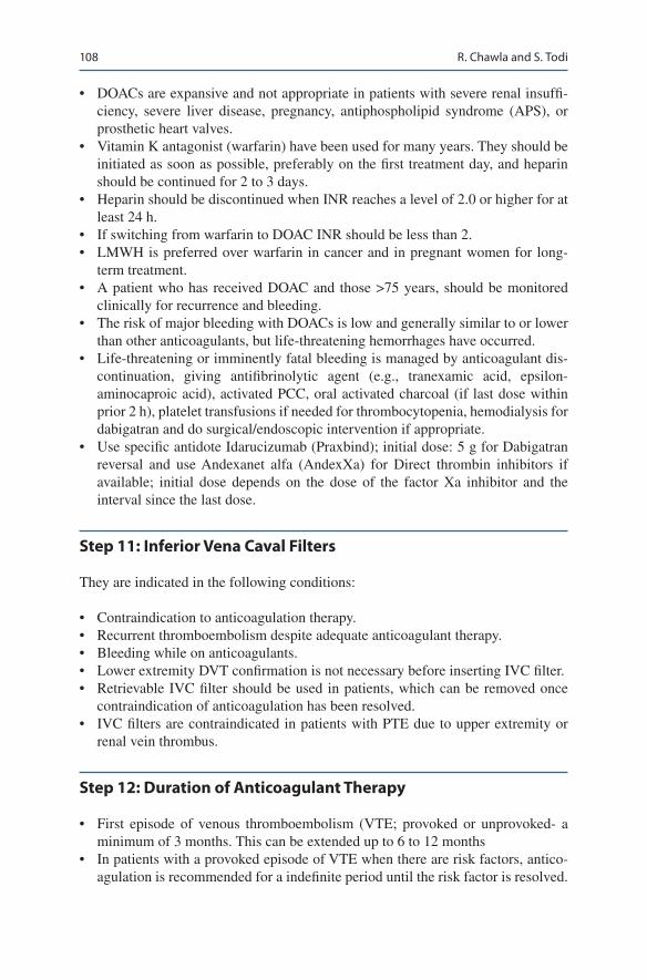
108
• DOACs are expansive and not appropriate in patients with severe renal insuffi-ciency, severe liver disease, pregnancy, antiphospholipid syndrome (APS), or prosthetic heart valves.
• Vitamin K antagonist (warfarin) have been used for many years. They should be initiated as soon as possible, preferably on the first treatment day, and heparin should be continued for 2 to 3 days.
• Heparin should be discontinued when INR reaches a level of 2.0 or higher for at least 24 h.
• If switching from warfarin to DOAC INR should be less than 2.• LMWH is preferred over warfarin in cancer and in pregnant women for long-
term treatment.• A patient who has received DOAC and those >75 years, should be monitored
clinically for recurrence and bleeding.• The risk of major bleeding with DOACs is low and generally similar to or lower
than other anticoagulants, but life-threatening hemorrhages have occurred.• Life-threatening or imminently fatal bleeding is managed by anticoagulant dis-
continuation, giving antifibrinolytic agent (e.g., tranexamic acid, epsilon- aminocaproic acid), activated PCC, oral activated charcoal (if last dose within prior 2 h), platelet transfusions if needed for thrombocytopenia, hemodialysis for dabigatran and do surgical/endoscopic intervention if appropriate.
• Use specific antidote Idarucizumab (Praxbind); initial dose: 5 g for Dabigatran reversal and use Andexanet alfa (AndexXa) for Direct thrombin inhibitors if available; initial dose depends on the dose of the factor Xa inhibitor and the interval since the last dose.
Step 11: Inferior Vena Caval Filters
They are indicated in the following conditions:
• Contraindication to anticoagulation therapy.• Recurrent thromboembolism despite adequate anticoagulant therapy.• Bleeding while on anticoagulants.• Lower extremity DVT confirmation is not necessary before inserting IVC filter.• Retrievable IVC filter should be used in patients, which can be removed once
contraindication of anticoagulation has been resolved.• IVC filters are contraindicated in patients with PTE due to upper extremity or
renal vein thrombus.
Step 12: Duration of Anticoagulant Therapy
• First episode of venous thromboembolism (VTE; provoked or unprovoked- a minimum of 3 months. This can be extended up to 6 to 12 months
• In patients with a provoked episode of VTE when there are risk factors, antico-agulation is recommended for a indefinite period until the risk factor is resolved.
R. Chawla and S. Todi
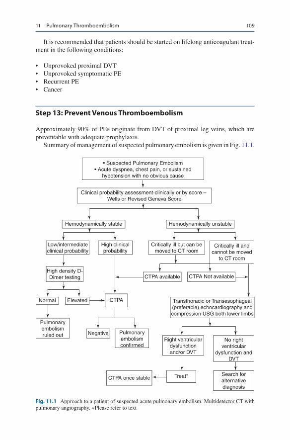
109
It is recommended that patients should be started on lifelong anticoagulant treat-ment in the following conditions:
• Unprovoked proximal DVT• Unprovoked symptomatic PE• Recurrent PE• Cancer
Step 13: Prevent Venous Thromboembolism
Approximately 90% of PEs originate from DVT of proximal leg veins, which are preventable with adequate prophylaxis.
Summary of management of suspected pulmonary embolism is given in Fig. 11.1.
• Suspected Pulmonary Embolism• Acute dyspnea, chest pain, or sustained
hypotension with no obvious cause
Clinical probability assessment‐clinically or by score – Wells or Revised Geneva Score
Hemodynamically stable Hemodynamically unstable
Low/intermediateclinical probability
High clinicalprobability
Critically ill but can bemoved to CT room
Critically ill andcannot be moved
to CT room
High density D-Dimer testing CTPA available CTPA Not available
Normal Elevated CTPA Transthoracic or Transesophageal(preferable) echocardiography andcompression USG both lower limbs
Pulmonaryembolismruled out Negative Pulmonary
embolismconfirmed
Right ventriculardysfunctionand/or DVT
No rightventricular
dysfunction andDVT
CTPA once stable Treat* Search foralternativediagnosis
Fig. 11.1 Approach to a patient of suspected acute pulmonary embolism. Multidetector CT with pulmonary angiography. ∗Please refer to text
11 Pulmonary Thromboembolism

110
Suggested Reading
Agnelli G, Becattini C. Acute pulmonary embolism. N Engl J Med. 2010;363:266–74.Gould MK, Dembitzer AD, Doyle RL, Hastie TJ, Garber AM. Low-molecular-weight heparins
compared with unfractionated heparin for treatment of acute deep venous thrombosis. A meta- analysis of randomized, controlled trials. Ann Intern Med. 130(10):800–9. This meta-analysis shows that LMWH treatment reduces mortality rates after acute DVT. These drugs seem to be as safe as UFH with respect to major bleeding complications and appear to be as effective in preventing thromboembolic recurrences.
Jaff MR, McMurtry MS, Archer SL, Cushman M, Goldenberg N, Goldhaber SZ. Management of massive and submassive pulmonary embolism, iliofemoral deep vein thrombosis, and chronic thromboembolic pulmonary hypertension: a scientific statement from the American Heart Association. Circulation. 2011;123(16):1788–830. Most up-to-date guidelines for the diagno-sis and management of PE.
Kearon C, Kahn SR, Agnelli G, Goldhaber S, Raskob GE, Comerota AJ. American College of Chest Physicians. Antithrombotic therapy for venous thromboembolic disease. Chest. 2008;133:454–545. This study about treatment for venous thromboembolic disease is part of the American College of Chest Physicians Evidence-Based Clinical Practice Guidelines.
Kuderer NM, Ortel TL, Francis CW. Impact of venous thromboembolism and anticoagulation on cancer and cancer survival. J Clin Oncol. 2009;27(29):4902–11. This meta-analyses performed for the American Society of Clinical Oncology and the Cochrane Collaboration suggest overall favorable effects of anticoagulation on survival of patients with cancer, mainly with LMWH.
Stein PD, Chenevert TL, Fowler SE, Goodman LR, Gottschalk A, Hales CA, et al. PIOPED III Investigators. Gadolinium-enhanced magnetic resonance angiography for pulmonary embo-lism: a multicenter prospective study (PIOPED III). Ann Intern Med. 2010;152(7):434–43, W1 42–43. This study shows that the technically adequate magnetic resonance angiography has a sensitivity of 78% and a specificity of 99%. Technically adequate magnetic resonance angiography and venography have a sensitivity of 92% and a specificity of 96%, but 52% of patients have technically inadequate results.
Stein PD, Fowler SE, Goodman LR, Gottschalk A, Hales CA, Hull RD, et al. PIOPED II Investigators. Multidetector computed tomography for acute pulmonary embolism. N Engl J Med. 2006;354(22):2317–27. In this study, the sensitivity of CT angiography (CTA) is 83% and the specificity is 96%. In patients with suspected PE, multidetector CTA-CTV has a higher diagnostic sensitivity than does CTA alone, with similar specificity.
Todd JL, Tapson VF. Thrombolytic therapy for acute pulmonary embolism: a critical appraisal. Chest. 2009;135(5):1321–9. This article reviews historical aspects and current evidence for thrombolytic therapy in acute PE with specific attention to bleeding risk, and data regarding hemodynamic parameters and mortality.
R. Chawla and S. Todi

111© Springer Nature Singapore Pte Ltd. 2020R. Chawla, S. Todi (eds.), ICU Protocols, https://doi.org/10.1007/978-981-15-0898-1_12
K. Khatib (*)Department of Medicine, Smt. Kashibai Navale Medical College, Pune, India
S. DixitDepartment of Critical Care, Sanjeevan Hospital, Pune, India
MJM Hospital, Pune, India
R. Chawla Department of Respiratory, Critical Care & Sleep Medicine, Indraprastha Apollo Hospitals, New Delhi, India
S. Todi Department of Critical Care & Emergency Medicines, A.M.R.I. Hospital, Kolkata, India
12Severe Community-Acquired Pneumonia
Khalid Khatib, Subhal Dixit, Rajesh Chawla, and Subhash Todi
Community Acquired Pneumonia (CAP) is one of the leading causes of hospitaliza-tion and morbidity and mortality. Of all patients presenting with CAP, only about 18% require hospitalization, of which 2–24% require intensive care unit (ICU) care. Mortality in hospitalized patients is 17–49%.
Step 1: Initiate Resuscitation
1. Airway patency is ensured. 2. If SpO2 < 90% or PaO2 < 60 mmHg, Oxygen therapy or HFNC is started. If
hypoxemia persists despite maximal O2 therapy, hypercapnea progresses, or if there is severe acidosis, the patient is transferred immediately to the ICU.
A 58-years-old male, smoker (8 pack-years) and having Diabetes mellitus and Hypertension for 8 years, presented to the ED with fever and acute dyspnoea for the last 48 h. On examination, respiratory rate was 34 breaths/min, blood pres-sure-150/96 mm Hg, heart rate-112/min, regular, and Oxygen saturation of 86% on 4L of oxygen by mask. He was conscious and oriented. His chest X-ray showed left lower zone consolidation.
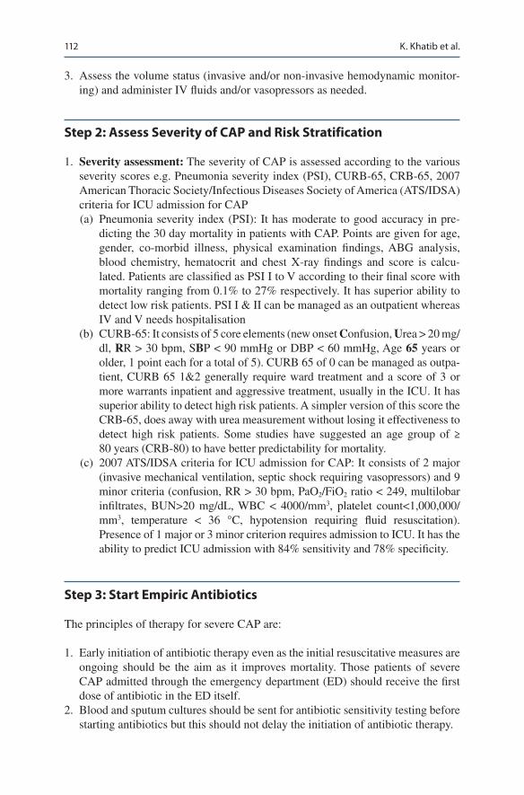
112
3. Assess the volume status (invasive and/or non-invasive hemodynamic monitor-ing) and administer IV fluids and/or vasopressors as needed.
Step 2: Assess Severity of CAP and Risk Stratification
1. Severity assessment: The severity of CAP is assessed according to the various severity scores e.g. Pneumonia severity index (PSI), CURB-65, CRB-65, 2007 American Thoracic Society/Infectious Diseases Society of America (ATS/IDSA) criteria for ICU admission for CAP (a) Pneumonia severity index (PSI): It has moderate to good accuracy in pre-
dicting the 30 day mortality in patients with CAP. Points are given for age, gender, co-morbid illness, physical examination findings, ABG analysis, blood chemistry, hematocrit and chest X-ray findings and score is calcu-lated. Patients are classified as PSI I to V according to their final score with mortality ranging from 0.1% to 27% respectively. It has superior ability to detect low risk patients. PSI I & II can be managed as an outpatient whereas IV and V needs hospitalisation
(b) CURB-65: It consists of 5 core elements (new onset Confusion, Urea > 20 mg/dl, RR > 30 bpm, SBP < 90 mmHg or DBP < 60 mmHg, Age 65 years or older, 1 point each for a total of 5). CURB 65 of 0 can be managed as outpa-tient, CURB 65 1&2 generally require ward treatment and a score of 3 or more warrants inpatient and aggressive treatment, usually in the ICU. It has superior ability to detect high risk patients. A simpler version of this score the CRB-65, does away with urea measurement without losing it effectiveness to detect high risk patients. Some studies have suggested an age group of ≥ 80 years (CRB-80) to have better predictability for mortality.
(c) 2007 ATS/IDSA criteria for ICU admission for CAP: It consists of 2 major (invasive mechanical ventilation, septic shock requiring vasopressors) and 9 minor criteria (confusion, RR > 30 bpm, PaO2/FiO2 ratio < 249, multilobar infiltrates, BUN>20 mg/dL, WBC < 4000/mm3, platelet count<1,000,000/mm3, temperature < 36 °C, hypotension requiring fluid resuscitation). Presence of 1 major or 3 minor criterion requires admission to ICU. It has the ability to predict ICU admission with 84% sensitivity and 78% specificity.
Step 3: Start Empiric Antibiotics
The principles of therapy for severe CAP are:
1. Early initiation of antibiotic therapy even as the initial resuscitative measures are ongoing should be the aim as it improves mortality. Those patients of severe CAP admitted through the emergency department (ED) should receive the first dose of antibiotic in the ED itself.
2. Blood and sputum cultures should be sent for antibiotic sensitivity testing before starting antibiotics but this should not delay the initiation of antibiotic therapy.
K. Khatib et al.

113
Table 12.1 Etiology of severe community acquired pneumonia
BacteriaStreptococcus pneumonia,Legionella pneumophilus,Staphylococcus aureus (methicillin-sensitive S. aureus),Gram-negative bacilli (ESBL or non-ESBL),Haemophilus influenzae, Moraxella catarrhalisPseudomonas spp.,Community-acquired methicillin-resistant S. aureus (MRSA),Others-Mycoplasma pneumoniae, Coxiella burnetti, Chlamydia spp.VirusesInfluenza A (including H1N1) and B viruses,Rhinovirus,Parainfluenza virus,Human metapneumovirus,Respiratory syncytial virus,Middle East Respiratory Syndrome corona virus (MERS CoV)Unknown
Table 12.2 Factors present in patients at risk for infection with drug resistant organisms
Hospitalization for >48 h in previous 9 monthsAntibiotics use in last 3 monthsResident of Nursing Home or long term treatment facilityFamily member residing with patient diagnosed with multi-drug resistant pathogenHome wound care or recent domiciliary infusion therapyAcute or chronic comorbiditiesHepatic failure,Renal failure or chronic dialysis,Chronic Obstructive Pulmonary Disease (Class C-D GOLD),Cardiac failure,Diabetes mellitus,AspleniaUse of steroids and/or immunosuppressive drugs
3. The initial choice of antibiotic will depend on a detailed and in-depth history with particular emphasis on identifying etiology of CAP (see Table 12.1) and whether the patient is at risk for infection with drug resistant organisms (see Table 12.2).
4. The initial choices of antibiotics in patient with no risk factors are: (a) A Beta-lactam/Beta lactamase inhibitor (Amoxicillin-clavulanate,
Ampicillin- sulbactum, OR a cephalosporin (Cefuroxime, Cefotaxime, Ceftriaxone) plus a Macrolide (Azithromycin, Clarithromycin)/Doxycycline/respiratory Fluoroquinolone (Levofloxacin, Moxifloxacin).OR
(b) A Beta-lactam (Amoxicillin-clavulanate, Ampicillin-sulbactum, Cefu-roxime, Cefotaxime, Ceftriaxone) plus Aztreonam (for patients with penicil-lin allergy).
12 Severe Community-Acquired Pneumonia
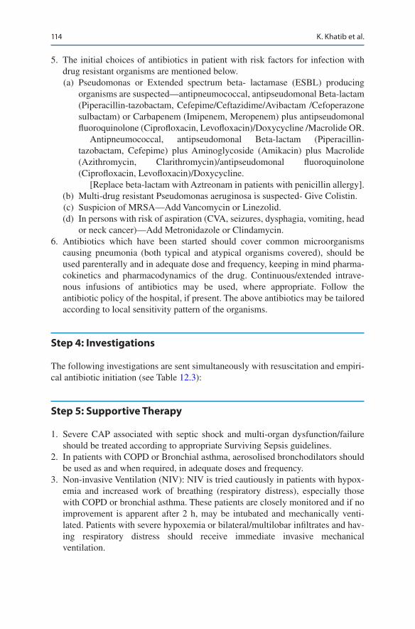
114
5. The initial choices of antibiotics in patient with risk factors for infection with drug resistant organisms are mentioned below. (a) Pseudomonas or Extended spectrum beta- lactamase (ESBL) producing
organisms are suspected—antipneumococcal, antipseudomonal Beta-lactam (Piperacillin-tazobactam, Cefepime/Ceftazidime/Avibactam /Cefoperazone sulbactam) or Carbapenem (Imipenem, Meropenem) plus antipseudomonal fluoroquinolone (Ciprofloxacin, Levofloxacin)/Doxycycline /Macrolide OR.
Antipneumococcal, antipseudomonal Beta-lactam (Piperacillin- tazobactam, Cefepime) plus Aminoglycoside (Amikacin) plus Macrolide (Azithromycin, Clarithromycin)/antipseudomonal fluoroquinolone (Ciprofloxacin, Levofloxacin)/Doxycycline.
[Replace beta-lactam with Aztreonam in patients with penicillin allergy]. (b) Multi-drug resistant Pseudomonas aeruginosa is suspected- Give Colistin. (c) Suspicion of MRSA—Add Vancomycin or Linezolid. (d) In persons with risk of aspiration (CVA, seizures, dysphagia, vomiting, head
or neck cancer)—Add Metronidazole or Clindamycin. 6. Antibiotics which have been started should cover common microorganisms
causing pneumonia (both typical and atypical organisms covered), should be used parenterally and in adequate dose and frequency, keeping in mind pharma-cokinetics and pharmacodynamics of the drug. Continuous/extended intrave-nous infusions of antibiotics may be used, where appropriate. Follow the antibiotic policy of the hospital, if present. The above antibiotics may be tailored according to local sensitivity pattern of the organisms.
Step 4: Investigations
The following investigations are sent simultaneously with resuscitation and empiri-cal antibiotic initiation (see Table 12.3):
Step 5: Supportive Therapy
1. Severe CAP associated with septic shock and multi-organ dysfunction/failure should be treated according to appropriate Surviving Sepsis guidelines.
2. In patients with COPD or Bronchial asthma, aerosolised bronchodilators should be used as and when required, in adequate doses and frequency.
3. Non-invasive Ventilation (NIV): NIV is tried cautiously in patients with hypox-emia and increased work of breathing (respiratory distress), especially those with COPD or bronchial asthma. These patients are closely monitored and if no improvement is apparent after 2 h, may be intubated and mechanically venti-lated. Patients with severe hypoxemia or bilateral/multilobar infiltrates and hav-ing respiratory distress should receive immediate invasive mechanical ventilation.
K. Khatib et al.
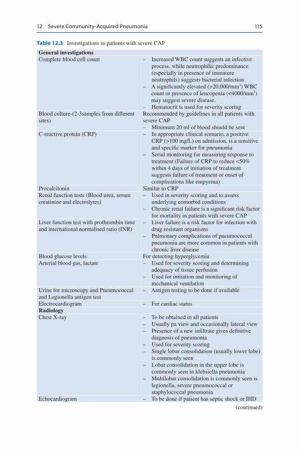
115
Table 12.3 Investigations in patients with severe CAP
General investigationsComplete blood cell count – Increased WBC count suggests an infective
process, while neutrophilic predominance (especially in presence of immature neutrophils) suggests bacterial infection
– A significantly elevated (>20,000/mm3) WBC count or presence of leucopenia (<4000/mm3) may suggest severe disease.
– Hematocrit is used for severity scoringBlood culture-(2-3samples from different sites)
Recommended by guidelines in all patients with severe CAP– Minimum 20 ml of blood should be sent
C-reactive protein (CRP) – In appropriate clinical scenario, a positive CRP (>100 mg/L) on admission, is a sensitive and specific marker for pneumonia
– Serial monitoring for measuring response to treatment (Failure of CRP to reduce <50% within 4 days of initiation of treatment suggests failure of treatment or onset of complications like empyema)
Procalcitonin Similar to CRPRenal function tests (Blood urea, serum creatinine and electrolytes)
– Used in severity scoring and to assess underlying comorbid conditions
– Chronic renal failure is a significant risk factor for mortality in patients with severe CAP
Liver function test with prothrombin time and international normalised ratio (INR)
– Liver failure is a risk factor for infection with drug resistant organisms
– Pulmonary complications of pneumococcal pneumonia are more common in patients with chronic liver disease
Blood glucose levels For detecting hyperglycemiaArterial blood gas, lactate – Used for severity scoring and determining
adequacy of tissue perfusion– Used for initiation and monitoring of
mechanical ventilationUrine for microscopy and Pneumococcal and Legionella antigen test
– Antigen testing to be done if available
Electrocardiogram – For cardiac statusRadiologyChest X-ray – To be obtained in all patients
– Usually pa view and occasionally lateral view– Presence of a new infiltrate gives definitive
diagnosis of pneumonia– Used for severity scoring– Single lobar consolidation (usually lower lobe)
is commonly seen– Lobar consolidation in the upper lobe is
commonly seen in klebsiella pneumonia– Multilobar consolidation is commonly seen is
legionella, severe pneumococcal or staphylococcal pneumonia
Echocardiogram – To be done if patient has septic shock or IHD
(continued)
12 Severe Community-Acquired Pneumonia
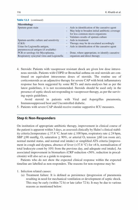
116
4. Steroids: Patients with vasopressor resistant shock are given low dose intrave-nous steroids. Patients with COPD or Bronchial asthma on oral steroids are con-tinued on equivalent intravenous doses of steroids. The routine use of corticosteroids as an adjunctive therapy for severe CAP with brisk inflammatory response has been suggested by some RCTs and meta-analyses but as per the latest guidelines, it is not recommended. Steroids should be used only in the presence of septic shock not responding to vasopressor therapy, as per the surviv-ing sepsis guidelines.
Avoid steroid in patients with Viral and Aspergillus pneumonia, Immunosuppressed host and Uncontrolled diabetic.
5. Patients with severe CAP should receive routine supportive ICU measures.
Step 6: Non-Responders
On institution of appropriate antibiotic therapy, improvement in clinical course of the patient is apparent within 3 days, as assessed clinically by Halm’s clinical stabil-ity criteria [temperature ≤ 37.8 °C, heart rate ≤ 100 bpm, respiratory rate ≤ 24 bpm, SBP ≥90 mmHg, O2 saturation ≥ 90%, or arterial O2 tension ≥60 (on room air), normal mental status, and normal oral intake) or simplified ATS criteria [improve-ment in cough and dyspnea, absence of fever (>37.8 °C) for >8 h, normalisation of total leukocyte count by 10% from the previous day, and adequate oral intake]. An associated improvement in biomarkers (CRP reduction <50%, reduction in procal-citonin) will also act as a guide to response.
Patients who do not show the expected clinical response within the expected timeline are labelled as non-responders. The reasons for non-response may be:
1. Infection related causes: (a) Treatment failure: It is defined as persistence /progression of pneumonia
resulting in need for mechanical ventilation or development of septic shock. This may be early (within 72 h) or late (after 72 h). It may be due to various reasons as mentioned below:
Table 12.3 (continued)
MicrobiologySputum-gram stain – Aids in identification of the causative agent
– May help to broaden initial antibiotic coverage for less common micro-organisms
– Validates results of sputum cultureSputum-aerobic culture and sensitivity (C&S)
– Aids in treatment– Therapy may be de-escalated accordingly
Urine for Legionella antigen, pneumococcal antigen (if available)
– Aids in identification of the causative agent
PCR or serology for Mycoplasma, Respiratory syncytial virus and Legionella
– Done, where appropriate, to identify causative organism and direct therapy
K. Khatib et al.
117
• Infection by pathogens not covered by initial empiric therapy,• Infection with atypical pathogens (tuberculosis, strongyloidosis, influ-
enza H1N1 virus),• Nosocomial secondary infections, iv) infectious complications (parap-
neumonic effusions, empyema, lung abscess, bronchial obstruction). (b) Slow responders: These patients are improving with therapy, but at a rate
slower than expected. Old Age, presence of co-morbidities, severe infections (with organisms like Gram negative bacilli, Legionella, Staphylococcus aureus) are predictors of slow response. Eight or nine days of treatment may be needed before clinical improvement is noted .
2. Non infectious causes: (a) Malignancy (lung cancer or metastatic), (b) Interstitial lung disease [Cryptogenic organising pneumonia, Diffuse alveo-
lar damage, Hypersensitivity pneumonia, Eosinophilic pneumonia, Alveolar hemorrhage, Drug fever, Vasculitis (Churg-Strauss, Wegener’s)],
(c) Foreign body, (d) Pulmonary embolism Pulmonary infarct, (e) Pulmonary edema, (f) Lipoid pneumonia.
Step 7: Further Workup for Non-Responders
1. Fibreoptic Bronchoscopy with analysis of BAL. 2. Endotracheal aspirate culture (preferably quantitative) and sensitivity. 3. Serology for HIV, Influenza A virus (H1N1), Anti-nuclear antibody, anti-
neutrophil cytoplasmic antibody. 4. BNP and pro-BNP levels. 5. D-dimer levels, Venous Doppler of lower limbs. 6. Ultrasonography of the Chest/Computed tomography of the Chest-demonstrates
presence of effusion, empyema or abscess and their localisation.
Step 8: Duration of Antibiotics
1. The duration of therapy depends on the clinical response, organism involved, co-morbidities present, biomarker response and presence of complications.
2. Patients with severe CAP should be treated for a minimum of 5 days and gener-ally for 7–10 days. Patient should be afebrile for 48–72 h, and should be clini-cally stable before discontinuation of therapy.
3. The duration of antibiotics may be prolonged (upto 14 days) in slow responders, infections with Pseudomonas, Gram negative bacilli or Staphylococcus, pres-ence of complications like lung abscess or empyema and presence of extra- pul-monary and metastatic infections (meningitis, endocarditis) secondary to the severe CAP.
12 Severe Community-Acquired Pneumonia
118
Step 9: Key Preventive Measures
Smoking cessation, Influenza and Pneumococcal Vaccination.
Suggested Reading
Aliberti S, et al. Criteria for clinical stability in hospitalised patients with community-acquired pneumonia. Eur Respir J. 2013;42(3):742–9. ATS 2001 and ATS/IDSA 2007 criteria for clini-cal stability in hospitalised patients with CAP are clinically equivalent and both can be used in clinical practice as well as in clinical research.
Blasi F, et al. Early versus later response to treatment in patients with community-acquired pneu-monia: analysis of the REACH study. Respir Res. 2014:15–6. The achievement of early clinical stabilization in CAP (≤4 days) is associated with improved outcomes, lower requirement for initial treatment modification or readmission.
Finch S, Chalmers JD. Brief clinical review: Non-responding pneumonia(NRP). EMJ Respir. 2014;2:104–11. NRP may vary from a benign delay in recovery to life-threatening progressive pneumonia. A systematic approach to investigation and management is needed with consider-ation of both infectious and non-infectious causes.
Metlay JP, Waterer GW, Long AC, Anzueto A, Brozek J, Crothers K, et al. Diagnosis and treatment of adults with community-acquired pneumonia. An official clinical practice guideline of the american thoracic society and infectious diseases society of america. Am J Respir Crit Care Med. 2019;200(7):e45–e67.
Phua J, Dean NC, Guo Q, Kuan WS, Lim HF, Lim TK, et al. Severe community-acquired pneumo-nia: timely management measures in the first 24 hours. Crit Care. 2016;20:237. Early and close collaboration between emergency medicine and respiratory and critical care medicine teams is required to successfully decrease mortality for severe CAP
Prina E, Ceccato A, Torres A. New aspects in the management of pneumonia. Crit Care. 2016;20:267. The use of corticosteroids in patients with severe CAP can reduce the time to clinical stability and the risk of treatment failure, and the administration of intravenous immu-noglobulins can reinforce the immune response to infection.
Torres A, Sibila O, Ferrer M, Polverino E, Menendez R, Mensa J, Gabarrús A, et al. Effect of corticosteroids on treatment failure among hospitalized patients with severe community- acquired pneumonia and high inflammatory response a randomized clinical trial. JAMA. 2015;313(7):677–86. Acute use of methylprednisolone compared with placebo decreased treat-ment failure in severe community-acquired pneumonia and high initial inflammatory response.
Websites
www.brit-thoracic.org.ukwww.fda.govwww.idsociety.orgwww.japi.org
K. Khatib et al.

119© Springer Nature Singapore Pte Ltd. 2020R. Chawla, S. Todi (eds.), ICU Protocols, https://doi.org/10.1007/978-981-15-0898-1_13
R. Pande (*) Department of Critical Care, BLK Superspeciality Hospital, New Delhi, India
V. Maurya Department of Respiratory Medicine & Intervention Pulmonology, Fortis Hospital, Shalimarbagh, New Delhi, India
13Ventilator-Associated Pneumonia
Rajesh Pande and Vikas Maurya
When a patient on ventilatory support develops a new or progressive infiltrate in the chest X-ray along with fever and leukocytosis after 48 h of intubation, it is suggestive of ventilator-associated pneumonia (VAP). This occurs in 9–27% of intubated patients and the risk increase with the duration of mechanical ventilation. Despite a proto-colized antibiotic use, the clinical cure rates are about 60% with a high recurrence rate. This can be attributed partly to the prevalence of infection with MDR pathogens like Enterobacter, Staphylococcus aureus, Klebseilla pneumoniae, Acinetobacter bau-mannii, Pseudomonas aeruginosa and Enterobacteriaceae (ESKAPE bugs).
Step 1: Initiate Resuscitation If Patient Is in Shock
The patient should be resuscitated, as mentioned in Chap. 23, Vol. 2.
A 50-year-old diabetic male patient was admitted to the hospital with ischemic stroke (GCS = E1V1M3). He was put on invasive positive-pressure ventilation support. On the fourth day of ICU stay, he developed fever (38.6 °C), a rise in total leukocyte count (156,000, N 93%), and heterogeneous, ill-defined shadows in the right lower zone in the chest X-ray. Chest auscultation revealed bronchial breathing in the right infra-axillary area, and the nurse reported an increase in amount and purulence of secretions requiring frequent suctioning. The patient needed 5 mcg/min noradrenaline to maintain systolic blood pressure of more than 100 mmHg.

120
Step 2: Rule Out Noninfectious Cause of Chest Infiltrate
Various non infectious conditions can result in pulmonary shadows similar to venti-lator associated pneumonia, and should be ruled out:
• Atelectasis: It is common in postoperative period following upper abdominal surgery due to hypoventilation. Left lower lobe atelectasis is common following coronary artery bypass grafting. Radiological signs include displaced fissures; crowded bronchovascular markings and shifts in the positions of the hila, dia-phragm, and mediastinum; and increased density or radiopacity of the lung tis-sue. Fever and leukocytosis may not always be present.
• Aspiration: It occurs in patients with impaired consciousness, regurgitation of enteral feed and microaspiration around endotracheal tube cuff. Right lower lobe is commonly involved. Infiltrates may take up to 12 h to appear in the chest X-ray after the event. History of vomiting or bile-colored endotracheal secretions on suctioning is a clue to the diagnosis. It may be a purely chemical pneumonitis initially, caused by acidic gastric contents.
• Pulmonary embolism with infarction: It is generally acute onset in the back-ground of risk factors for deep venous thrombosis and pulmonary embolism (PE). Diagnosis can be confirmed by CT Pulmonary Angiography.
• Pulmonary hemorrhage may resemble dense alveolar consolidation. Generally, endotracheal (ET) secretions are bloodstained. Coagulation profiles or other immunological markers may be deranged.
• Cardiogenic pulmonary edema.• Acute respiratory distress syndrome (ARDS).• Fluid overload.• Hypersensitivity pneumonitis.• Drug reactions: Cyclophosphamide, Methotrexate.• Cryptogenic organizing pneumonia (history of recent viral infection).• Radiation pneumonitis.• Lung contusion.
Step 3: Make a Clinical Diagnosis of VAP
• The diagnosis of VAP is difficult, therefore scoring systems such as Clinical pulmonary infection score (CPIS) (Table 13.1) and biomarkers like C reactive protein (CRP) & procalcitonin are often used in establishing the diagnosis albeit with low predictive values.
• The newly proposed ventilator-associated events (VAE) surveillance definitions (Table 13.2), endorsed by the Centre for Disease Control and Prevention should be used for epidemiological purposes only and not for diagnosing VAP.
• The new 2016 IDSA-ATS-VAP management guidelines recommend that only clinical criteria should be used for the diagnosis of VAP and scores like CPIS or markers like CRP, procalcitonin should not be used in conjunction with clinical criteria.
R. Pande and V. Maurya
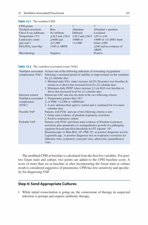
121
The modified CPIS at baseline is calculated from the first five variables. For posi-tive Gram stain and culture, two points are added to the CPIS baseline score. A score of more than six at baseline or after incorporating the Gram stain or culture result is considered suggestive of pneumonia. CPIS has low sensitivity and specific-ity for diagnosing VAP.
Step 4: Send Appropriate Cultures
• While initial resuscitation is going on, the cornerstone of therapy in suspected infection is prompt and empiric antibiotic therapy.
Table 13.1 The modified CPIS
CPIS points 0 1 2Tracheal secretions Rare Abundant Abundant + purulentChest X-ray infiltrates No infiltrate Diffused LocalizedTemperature (°C) ≥36.5 and ≤38.4 ≥38.5 and ≤38.9 ≥39 or ≤36Leukocytes count (per mm3)
≥4000 and ≤11,000
<4000 or >11,000
<4000 or >11,000+ band forms ≥500
PaO2/FiO2 (mm Hg) >240 or ARDS ≤240 and no evidence of ARDS
Microbiology Negative Positive
Table 13.2 The ventilator associated events (VAE)
Ventilator associated complication (VAC)
At least one of the following indicators of worsening oxygenation following a sustained period of stability or improvement on the ventilator for ≥2 calendar days 1. Minimal daily FiO2 values increase ≥0.20 (20 points) over baseline &
remain at or above that increased level for ≥2 calendar days 2. Minimum daily PEEP values increase ≥3 cm H2O over baseline or
above that increased level for ≥2 calendar daysInfection related Ventilator associated complication (IVAC)
Patient has VAC and also fits both of the two following criteria: 1. Temperature greater than 38 C 2. or WBC >12,000 or <4000/mm3. 3. A new antimicrobial agent is started and is continued for 4 or more
calendar days.Possible VAP Patients with IVAC and one of the following criteria is met:
1. Gram stain evidence of purulent respiratory secretions 2. Positive respiratory culture
Probable VAP Patients with IVAC and Gram stain evidence of Purulent respiratory secretions plus quantitative or semiqualitative growth of a pathogenic organism beyond specified thresholds for ET aspirate: 106, Bronchoscopic or Mini BAL 105, PSB 103, or positive diagnostic test for Legionella spp., or positive diagnostic test on respiratory secretions for influenza virus, respiratory syncytial virus, adenovirus, parainfluenza virus
13 Ventilator-Associated Pneumonia

122
• Ideally, one should send blood and endotracheal aspirate or do fiberoptic bron-choscopy with bronchoalveolar lavage for Gram stain and quantitative aerobic culture prior to starting or changing antibiotics.
• Both non-invasive (Endotracheal aspiration) sampling with Gram Stain and quantitative or semi-quantitative (Light, moderate or heavy growth) culture and invasive (mini BAL, bronchoscopic BAL or protected specimen brush) sampling with Gram stain and quantitative culture has been proposed by various guidelines for the diagnosis of VAP.
• However, if the patient is on empiric antibiotics for VAP, and invasive quantita-tive cultures have been done prior to starting or changing antibiotic but the results are below diagnostic threshold, then the antibiotics should be stopped.
• If there is delay in obtaining samples for logistic reasons beyond an hour, antibi-otics should be given without delay.
Step 5: Start Empirical Antibiotics
1. The initial choice of antibiotics is of utmost importance. An inappropriate initial choice increases mortality. (a) Selection of empiric antibiotic therapy should be based on the following:
• Patient risk factors for infection with MDR pathogensThe classical distinction and treatment of ventilator associated pneumo-nia based on early and late onset VAP concept seems inappropriate as it may result in low intensity treatment for multi-drug resistant pathogens that are commonly seen in early onset VAP as well as aggressive high intensity treatment in late onset VAP on the assumption that it is always caused by resistant pathogens. No significant difference in pathogens have been found between early & late onset VAP. New guidelines suggest that the antibiotic therapy should focus on presence of risk factors for MDR pathogens rather than early or late VAP. It is suggested that hospital admission rather than intubation should be taken as the starting point.
(b) Recent exposure to specific antibiotic classes (c) Local epidemiology of infection and antibiogram- the common organisms to
cause VAP include Gram negative ESBL enterobacteriacae, Non fermenters like pseudomonas and acinetobacter and gram positives like MRSA
(d) Previous antibiotic therapy 2. Detailed history should be taken to identify patients who are at high risk of drug-
resistant infection as mentioned below: (a) Intravenous Antimicrobial therapy in preceding 90 days (b) Current hospitalization of 5 days or more (c) Septic shock at the time of VAP (d) ARDS preceding VAP (e) Need of renal replacement therapy before VAP
3. Risk factor for MDR gram negative Bacilli and Pseudomonas aeuginosa (a) Treatment in an ICU where more than 10% of gram negative isolates are
resistant to an agent considered for monotherapy (b) Treatment in an ICU in which local antimicrobial susceptibility rates are not
known
R. Pande and V. Maurya

123
4. Risk factors for MRSA (a) Treatment in a unit in which >10 to 20% of staphylococcal aureus isolates
are methicillin resistant (b) Treatment in a unit in which prevalence of MRSA is not known
Step 6: Principles of Choosing an Antibiotic: (Fig. 13.1)
1. The 2016 IDSA/ATS guidelines recommend that the choice of empiric antibiot-ics should be based on local distribution of pathogens and the local antibiograms.
Suspicion of VAP is low
Suspicion of VAP is high
• Fever or hypothermia• Leukocytosis or leukopenia• ↑ sputum, purulence & change in colour• ↑ O2/ ventilatory requirement
• Microscopy is negative• Do not start antibiotics• Consider other causes
• Paired blood culture• Non‐invasive endotracheal aspirate (Double catheter blind technique or• Invasive LRTI sample (BAL, PSB)• Other relevant cultures• Biomarkers: CRP, Serum procalcitonin• Look for risk factors for MDR
Send the following:
Start empiric antibiotic therapy that covers for S. aureus, Pseudomonas aeruginosa andother relevant GNB’s (Avoid colistin, if other effective agents are available)
• Therapy should cover for MRSA with Linezolid or vancomycin, if MRSA risk is high• If no MRSA risk is present, therapy should cover for MSSA• If initial choice is piperacillin‐tazobactam, cefepime, levofloxacin, imipenem, meropenem, do not add specific MSSA agents like oxacillin, nafcillin or cefazolin
After the culture reports are available• If bug is sensitive only to Polymyxins or aminoglycosides ® Use IV + inhaled therapy• If bug is resistant to carbapenems, use IV Polymyxins + inhaled colistin• De‐escalate to specific antibiotics if pathogen identified• Stop antibiotics if culture is sterile & patient is clinically improved• Antibiotic therapy should be stopped after 7 days• A longer therapy may be considered, if clinical, radiological signs are still unresolved and laboratory parameters are not improved
Specific antibiotic therapyAcinetobacter
• If isolate is susceptible, use carbapenem, ampi‐sulbactam, pip‐tazo.• If sensitive only to Polymyxin, use IV Polymyxin and inhaled colistin• Do not use rifampicin or tigecycline
Pseudomonas• Avoid aminoglycosides• Use one antibiotic, if no risk factors are present for antimicrobial resistance• Use two antibiotics from different class, If risk factors for antimicrobial resistance are present
Fig. 13.1 Management of ventilator associated pneumonia
13 Ventilator-Associated Pneumonia

124
2. Antibiotic should cover resistant gram negative organisms like ESBL in patients at risk.
3. MRSA cover should be included only if risk factors for MRSA are present, otherwise MSSA cover should be provided.
4. Avoid the antibiotic class to which the patient has been recently exposed. 5. Use parenteral antibiotics. 6. Use antibiotics in adequate dose and frequency. It is suggested to use PK/PD
data for antibiotic dosing. 7. Ventilator associated tracheobronchitis should preferably not be treated with
antibiotic therapy. 8. Follow the antibiotic policy of your unit/hospital. 9. For starting antibiotic therapy in suspected VAP, clinical criteria alone should
be used rather than clinical criteria + serum procalcitonin or CRP or CPIS. 10. The duration of antibiotic therapy should be seven days and not more. 11. Monitoring serial procalcitonin level can help guide the decision to discontinue
antibiotic 12. Specific recommendations based on new guidelines:
(a) Patients with risk factors for infection with MDR VAP• PiperacillinTazobactam/Imipenem/Meropenem/Ceftazidime/Cefepime/
AztreonamPlus
• Amikacin/Gentamicin/Tobramycin/Levofloxacin/Ciprofloxacin/Aztreonam (if not used earlier)Plus
• Vancomycin/Linezolid/Teicoplanin (b) Patient with No risk factor for MDR pathogen and No Risk factors for
MDR gram negative pathogen and no Risk factor for MRSA• Piperacillin/Tazobactam/Cefepime/Levofloxacin
(c) Patients with Risk factors for MRSA:• Vancomycin/Linezolid/Teicoplanin
(d) Other recommendations• If Piperacillin-tazobactam, cefepime, levofloxacin, imipenem or
meropenem are being used for empiric cover, it is not necessary to add specific MSSA cover like oxacillin, nafcillin or cefazolin.
• Piperacillin/Tazobactam used with Vancomycin has been associated with acute kidney injury.
• Aminoglycosides, Polymyxins should be avoided in suspected VAP, if other gram negative antibiotics are available and provide adequate cover.
• In cases of VAP due to gram negative bacilli susceptible only to amino-glycosides or Polymyxins, it is suggested to use both inhaled and intra-venous (systemic) antibiotics rather than systemic antibiotics alone.
• In cases of VAP due to Acinetobacter species, use a carbapenem or a BL-BLI (betalactam—beta lactam inhibitor) combination like ampicil-lin/sulbactam, piperacillin-tazobactam (if the isolate is susceptible to these antibiotics).
R. Pande and V. Maurya

125
• In cases of VAP due to Acinetobacter species sensitive only to Polymyxins, it is recommended to use intravenous Polymyxins (colistin or Polymyxin B) along with inhaled colistin. Do not use rifampicin in such cases.
• Do not use tigecycline in VAP caused by Acinetobacter species.• Use of Inhaled antibiotics: For treating VAP, the lung concentration of
the antibiotic must exceed the MIC of infecting pathogen Penetration of some systemic antibiotics like Colistin, Beta lactams, aminoglycosides and glycopeptides into lung is not as effective as flouroquinolones. Targeting lungs with aerosolized antibiotics in ventilated patients in addition to intravenous antibiotics is a good strategy and may facilitate shorter durations of therapy for multi-drug-resistant pathogens. The par-ticle size should be 1–5 mm for the antibiotic to be delivered to the lower airways and the lung parenchyma.
• However, lack of specific formulation, need of special aerosolization devices and suboptimal aerosol delivery may impose limitation on the usefulness. Currently vibrating mesh nebulizers can be used for delivery of nebulized antibiotics through the ventilator circuit. Currently approved inhaled antibiotics include colistin, and tobramycin.
• Aerosolized antibiotic therapy should be synchronized to be delivered with the inspiratory flow rather than continuous nebulization. The venti-lator frequency should be low to give sufficient inspiratory time for drug delivery. Humidification may be important for the patient but its use may greatly reduce the nebulized drug delivery.
Step 7: Send Further Investigations
While initial resuscitation and empirical antibiotics are being given, basic diagnos-tic workup should be sent. These should include the following:
• Complete blood count• Procalcitonin, C reactive protein• urea, creatinine• Liver function test• Prothrombin time, Na, K• Blood culture—two sets if not sent earlier• Endotracheal aspirates or fiberbronchoscopy with protected specimen brush
(PSB) or bronchoalveolar lavage (BAL) (in selected cases like immunosuppressed)
• Arterial blood gas, lactate• Urine for microscopy• Chest X-ray• ECG or echocardiogram (optional) if the patient is in septic shock
13 Ventilator-Associated Pneumonia
126
Step 8: De-escalate Antibiotics
• Clinical improvement usually takes 2–3 days, and therapy should not be changed during this time unless the condition deteriorates. The guidelines rec-ommend De-escalation of antibiotic therapy. Clinical criteria along with serum procalcitonin should be used for De-escalation rather than clinical criteria alone.
• If protected specimen brush (PSB) or bronchoalveolar lavage (BAL) culture results are negative in a patient who is clinically improving and hemodynami-cally stable, antibiotic therapy can be discontinued.
• If the culture is negative for MRSA, linezolid or vancomycin can be safely stopped, and if the culture is positive for MRSA, other antibiotics can be stopped.
• If no organism is found, one should try to look for other causes of lung shadows, that is, atelectasis, collapse, aspiration, pulmonary embolism, and hemorrhage. It decreases unnecessary exposure to antibiotics and helps to reduce resistance to antibiotics.
• Change to oral therapy once the patient accepts orally and is hemodynamically stable.
• The initial antibiotics should be given intravenously with changeover to oral therapy in responsive patients with an intact gastrointestinal function. The organ-ism should be sensitive to oral antibiotics.
• Fluoroquinolones and linezolid are equally bioavailable in either intravenous or oral preparations.
• All patients with VAP should receive antibiotics at least for 7 days. The clinical, radiological and laboratory improvement can suggest a longer need of antibiotic therapy. Nonfermenters like acinetobacter and pseudomonas should also be treated for 14 days.
• Antibiotic dosing should be adjusted in patients with impaired renal or hepatic function.
Step 9: Prevention of VAP in the ICU
Evidence-based guidelines have recommended a number of measures that may affect the development of VAP. VAP bundles is a group of proven preventive strate-gies that can significantly reduce VAP rates. (Table 13.3).
R. Pande and V. Maurya
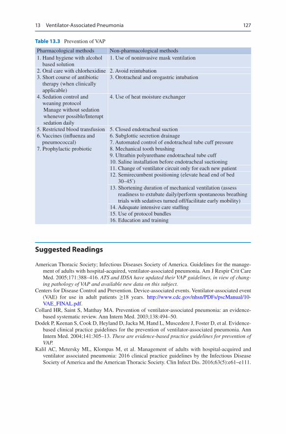
127
Suggested Readings
American Thoracic Society; Infectious Diseases Society of America. Guidelines for the manage-ment of adults with hospital-acquired, ventilator-associated pneumonia. Am J Respir Crit Care Med. 2005;171:388–416. ATS and IDSA have updated their VAP guidelines, in view of chang-ing pathology of VAP and available new data on this subject.
Centers for Disease Control and Prevention. Device-associated events. Ventilator-associated event (VAE) for use in adult patients ≥18 years. http://www.cdc.gov/nhsn/PDFs/pscManual/10-VAE_FINAL.pdf.
Collard HR, Saint S, Matthay MA. Prevention of ventilator-associated pneumonia: an evidence- based systematic review. Ann Intern Med. 2003;138:494–50.
Dodek P, Keenan S, Cook D, Heyland D, Jacka M, Hand L, Muscedere J, Foster D, et al. Evidence- based clinical practice guidelines for the prevention of ventilator-associated pneumonia. Ann Intern Med. 2004;141:305–13. These are evidence-based practice guidelines for prevention of VAP.
Kalil AC, Metersky ML, Klompas M, et al. Management of adults with hospital-acquired and ventilator associated pneumonia: 2016 clinical practice guidelines by the Infectious Disease Society of America and the American Thoracic Society. Clin Infect Dis. 2016;63(5):e61–e111.
Table 13.3 Prevention of VAP
Pharmacological methods Non-pharmacological methods1. Hand hygiene with alcohol
based solution1. Use of noninvasive mask ventilation
2. Oral care with chlorhexidine 2. Avoid reintubation3. Short course of antibiotic
therapy (when clinically applicable)
3. Orotracheal and orogastric intubation
4. Sedation control and weaning protocolManage without sedation whenever possible/Interupt sedation daily
4. Use of heat moisture exchanger
5. Restricted blood transfusion 5. Closed endotracheal suction6. Vaccines (influenza and
pneumococcal)7. Prophylactic probiotic
6. Subglottic secretion drainage7. Automated control of endotracheal tube cuff pressure8. Mechanical tooth brushing9. Ultrathin polyurethane endotracheal tube cuff10. Saline installation before endotracheal suctioning11. Change of ventilator circuit only for each new patient12. Semirecumbent positioning (elevate head end of bed
30–45°)13. Shortening duration of mechanical ventilation (assess
readiness to extubate daily/perform spontaneous breathing trials with sedatives turned off/facilitate early mobility)
14. Adequate intensive care staffing15. Use of protocol bundles16. Education and training
13 Ventilator-Associated Pneumonia

128
Kollef MH, Morrow LE, Niederman MS, Leeper KV, Anzueto A, Benz-Scott L, Rodino FJ. Clinical characteristics and treatment patterns among patients with ventilator associated pneumonia. Chest. 2006;129:1210–8. The overall mortality rate from VAP remains unacceptably high. The de-escalation of therapy in VAP patients appears to be associated with a reduction in mortality, which is an association that warrants further clinical study.
Morrow LE, Kollef MH. Recognition and prevention of nosocomial pneumonia in the intensive care unit and infection control in mechanical ventilation. Crit Care Med. 2010;38:352–62. This article provides a brief overview of the current approaches for the diagnosis of nosocomial pneumonia and focus on strategies for prevention.
Valencia M, Torres A. Ventilator associated pneumonia. Curr Opin Crit Care. 2009;15:30–4.
R. Pande and V. Maurya
129© Springer Nature Singapore Pte Ltd. 2020R. Chawla, S. Todi (eds.), ICU Protocols, https://doi.org/10.1007/978-981-15-0898-1_14
S. Kansal (*) Department of Respiratory Medicine and Critical Care, Indraprastha Apollo Hospitals, New Delhi, India
R. Chawla Department of Respiratory, Critical Care & Sleep Medicine, Indraprastha Apollo Hospitals, New Delhi, India
14Pleural Diseases
Sudha Kansal and Rajesh Chawla
Pleural effusion is a relatively uncommon cause for admission to intensive care unit; however, it occurs during stay in the ICU due to complications of diseases, fluid overload and procedures performed in these patients. It may be difficult to detect pleural effusion and pneumothorax in critically ill patients in supine chest X-ray.
Step 1: Initiate Resuscitation and Take History
• After initial resuscitation, take a detailed history of chest pain, palpitation, fever, cough with expectoration, hemoptysis, decrease in urine output, edematous feet, distension of abdomen, right hypochondriac pain, and weight loss.
• Also, inquire about medication and other relevant history, keeping in mind the common causes of pleural effusion in the ICU (Table 14.1).
A 65-year-old chronic male smoker, with a known case of coronary artery dis-ease with history of congestive heart failure, presented with increasing shortness of breath. He had right-sided pleuritic chest pain. He was afebrile, tachycardic, tachypneic, and hypoxemic on room air. Chest skiagram done in triage showed bilateral pleural effusion, with more pleural fluid on the right side than on the left side. The patient was shifted to the ICU.

130
Step 2: Perform Examination
• Perform a thorough examination to establish the diagnosis. Check vital signs, JVP, cyanosis, jaundice, SpO2, pallor, edematous feet, lymphadenopathy, and any evidence of deep venous thrombosis (DVT).
• Systemic examination should be carried out for S3, asymmetric breath sounds, crepitations, bronchial breathing, hepatomegaly, right hepatic tenderness, and ascites.
Step 3: Plan Investigations
• Hemogram.• Renal function tests.• Liver functions tests, prothrombin time/partial thromboplastin time (PT/PTT).• ECG.• 2D echo.• Cardiac enzymes—NTproBNP.• Relevant cultures—Depending on the suspected etiology.• Chest X-ray—Classically, Chest skiagram shows obliteration of the costophrenic
angle. However portable X-ray can not identify small effusion (<500 mL). Moreover, supine portable chest X-ray may not show classical features of pleural effusion. Subtle features such as haziness over entire hemithorax and loss of diaphragm outline may only be noted. The yield can be increased by taking X-ray in propped up position. If there is a high clinical suspicion of pleural effu-sion, bed side Ultrasound should be done.
• Ultrasonography (USG) of the chest—It is a very useful bedside tool. It has bet-ter sensitivity to diagnose pleural effusion. USG of the chest also helps in evalu-ation and quantification of fluid. It helps to know whether fluid is free or loculated. USG may also help to know the character of fluid depending on the echogenicity. It also helps in identifying underlying lung pathology and cardiac status.
Table 14.1 Common causes of pleural effusion in the ICU
Causes Types of fluidCongestive heart failure (36%) TransudatePneumonia (22%) ExudateMalignancy (14%) ExudatePulmonary embolism (11%) BothViral disease ExudatePostcoronary artery bypass graft ExudateCirrhosis with ascites TransudateFluid overload/renal failure TransudateAcute respiratory distress syndrome TransudateSevere hypoalbuminemia TransudateTuberculosis Exudate
S. Kansal and R. Chawla
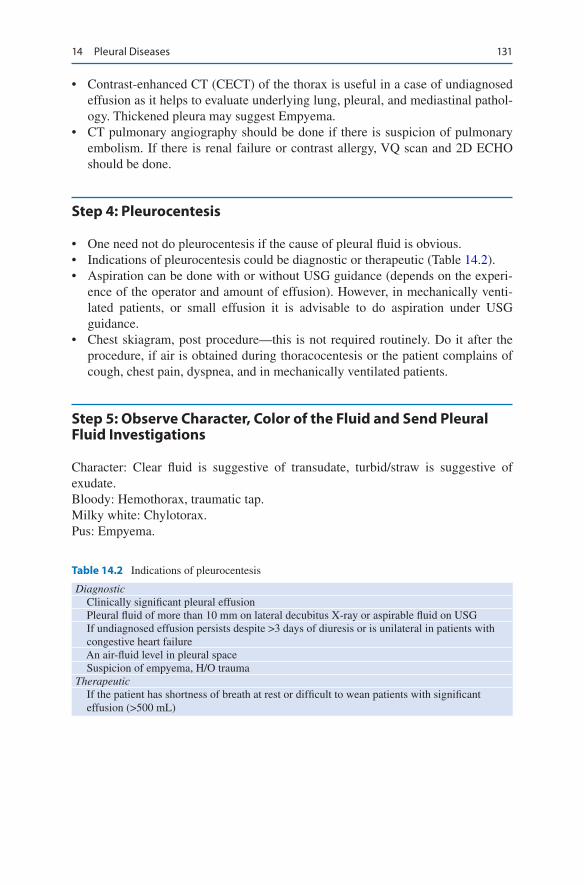
131
• Contrast-enhanced CT (CECT) of the thorax is useful in a case of undiagnosed effusion as it helps to evaluate underlying lung, pleural, and mediastinal pathol-ogy. Thickened pleura may suggest Empyema.
• CT pulmonary angiography should be done if there is suspicion of pulmonary embolism. If there is renal failure or contrast allergy, VQ scan and 2D ECHO should be done.
Step 4: Pleurocentesis
• One need not do pleurocentesis if the cause of pleural fluid is obvious. • Indications of pleurocentesis could be diagnostic or therapeutic (Table 14.2).• Aspiration can be done with or without USG guidance (depends on the experi-
ence of the operator and amount of effusion). However, in mechanically venti-lated patients, or small effusion it is advisable to do aspiration under USG guidance.
• Chest skiagram, post procedure—this is not required routinely. Do it after the procedure, if air is obtained during thoracocentesis or the patient complains of cough, chest pain, dyspnea, and in mechanically ventilated patients.
Step 5: Observe Character, Color of the Fluid and Send Pleural Fluid Investigations
Character: Clear fluid is suggestive of transudate, turbid/straw is suggestive of exudate.Bloody: Hemothorax, traumatic tap.Milky white: Chylotorax.Pus: Empyema.
Table 14.2 Indications of pleurocentesis
Diagnostic Clinically significant pleural effusion Pleural fluid of more than 10 mm on lateral decubitus X-ray or aspirable fluid on USG If undiagnosed effusion persists despite >3 days of diuresis or is unilateral in patients with
congestive heart failure An air-fluid level in pleural space Suspicion of empyema, H/O traumaTherapeutic If the patient has shortness of breath at rest or difficult to wean patients with significant
effusion (>500 mL)
14 Pleural Diseases
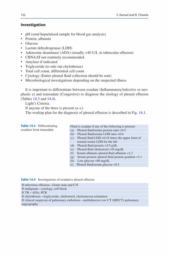
132
Investigation
• pH (send heparinised sample for blood gas analysis)• Protein, albumin• Glucose• Lactate dehydrogenase (LDH)• Adenosine deaminase (ADA) (usually >40 U/L in tubercular effusion)• CBNAAT-not routinely recommended• Amylase if indicated• Triglyceride (to rule out chylothorax)• Total cell count, differential cell count• Cytology (Entire pleural fluid collection should be sent)• Microbiological investigations depending on the suspected illness
It is important to differentiate between exudate (Inflammatory/infective or neo-plastic e) and transudate (Congestive) to diagnose the etiology of pleural effusion (Tables 14.3 and 14.4).
Light’s Criteria.If anyone of the three is present (a–c).The workup plan for the diagnosis of pleural effusion is described in Fig. 14.1.
Table 14.3 Differentiating exudates from transudate
Fluid is exudate if any of the following is present:(a) Pleural fluid/serum protein ratio >0.5(b) Pleural fluid/serum LDH ratio >0.6(c) Pleural fluid LDH >0.45 times the upper limit of
normal serum LDH for the lab(d) Pleural fluid protein >2.9 g/dL(e) Pleural fluid cholesterol >45 mg/dL(f) Serum albumin–pleural fluid albumin <1.2(g) Serum protein–pleural fluid protein gradient <3.1(h) Low glucose <60 mg/dL(i) Pleural fluid/serum glucose <0.5
Table 14.4 Investigations of exudative pleural effusion
If infectious effusion—Gram stain and C/SIf malignant—cytology, cell blockIf TB—ADA, PCRIf chylothorax—triglyceride, cholesterol, chylomicron estimationIf clinical suspicion of pulmonary embolism—multidetector row CT (MDCT) pulmonary angiography
S. Kansal and R. Chawla

133
Step 6: Disease-specific Management
The management of pleural effusion in special situations is described as follows:
A. Parapneumonic effusionWhen the patient develops parapneumonic effusion, the main treatment consists of antibiotics. A parapneumonic effusion is aspirated only if it fulfills the criteria mentioned above for indication of pleurocentesis. It is important to differentiate between complicated and uncomplicated effusions.
(a) Place the ICD tube in parapneumonic effusion only if it is complicated as mentioned below.• If it is loculated effusion or fills more than half of hemithorax, or an air-
fluid level is seen.• Pus on aspiration, Gram stain, or culture positive.• pH less than 7.2, glucose less than 60 mg%.
Thoracocentesis
Purulent Fluid appearance Bloody
(Hematocrit)If >50% ofperipheral
blood
Hemothorax
ICD tube
ExudateTransudate
ICD tube
Empyema
Otherchecklist
criteria (LDH, protein)
Fig. 14.1 The workup plan for the diagnosis of pleural effusion
14 Pleural Diseases
134
(b) Remove the tube when:• The patient has improved or drain is less than 50 mL/day.
(c) If parapneumonic effusion does not improve:• Consider fibrinolysis with streptokinase, thoracoscopy, or thoracotomy.
B. Malignant effusion (a) Often large and symptomatic. (b) Common in lung cancer, breast cancer and lymphoma, gastrointestinal tract
malignancy, and unknown primary. (c) If recollects in less than 3 weeks and the patient is symptomatic—do tube
thoracostomy and pleurodesis. C. Pleural effusion associated with pulmonary embolism
If there is high clinical suspicion in appropriate setting, investigate and treat them (see Chap. 11).
D. Post cardiac surgery Pleural effusionPost op pleural effusion are common in patients undergoing cardiac surgery. It can occur with postcardiotomy syndrome or as a part of CHF or Pulmonary Embolism.
Late pleural effusion (> 30 days) are common in patient undergoing CABG or Valve surgery. Cause can be due to underlying pericarditis, auto immune mediated or pleural injury. It is usually unilateral, left sided. The effusions are exudative and hemorrhagic with eosinophil and neutrophils preponderance in early and lymphocytic preponderance in late effusion. Usually they resolve on their own.
E. Undiagnosed pleural effusion (a) In 20% effusion, despite extensive investigation, cause may not be found. (b) If clinically stable, continue conservative treatment. (c) If deterioration in condition, plan thoracoscopy and pleural biopsy.
Pneumothorax
• Air in pleural space can be a medical emergency in ICU patients and requires immediate attention.
• It is very important to suspect a pneumothorax in a ventilated patients with wors-ening oxygenation or rising airway pressures
• Pneumothorax can be spontaneous or traumatic. Spontaneous pneumothorax can be primary when no cause is identified or secondary if there is underlying pulmo-nary disease.
• Traumatic pneumothorax also includes iatrogenic pneumothorax (central line, barotrauma, Post surgery, post CPCR) (Fig. 14.2).
A brief outline of management of pneumothorax is described in Fig. 14.3.
• Needle aspiration is appropriate for a simple primary pneumothorax with only 15–30% collapse.
S. Kansal and R. Chawla
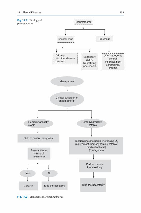
135
Pneumothorax
Spontaneous Traumatic
PrimaryNo other diseasepresent
SecondaryCOPD
Necrotizingpneumonia
Often iatrogeniccentral
line placementBarotrauma,
Trauma
Fig. 14.2 Etiology of pneumothorax
Management
Clinical suspicion ofpneumothorax
HemodynamicallyUnstable
Tension pneumothorax (increasing O2requirement, hemodynamic unstable,
mediastinal shift)(Emergency)
Perform needlethoracostomy
Tube thoracosstomy
Hemodynamicallystable
CXR to confirm diagnosis
Pneumothorax<15% of
hemithorax
Yes No
Observe Tube thoracostomy
Fig. 14.3 Management of pneumothorax
14 Pleural Diseases

136
• However, if the patient is on mechanical ventilation, any degree of pneumotho-rax should be drained by tube thoracostomy.
• It is very important to diagnose pneumothorax early as the patient on mechani-cally ventilator can have a sudden collapse. USG chest is a very useful point of care tool to diagnose Pneumothorax.
• In a normal USG, sliding of lung over pleura is seen along with Vertical comet tails which runs from pleura to lung. These are called “B lines.
• If there is air in the pleural space, due to inability of air to transmit the ultrasound waves, the sliding and these lines are no longer seen, called as “loss of lung slid-ing”. If you see even a single B line pneumothorax is ruled out.
• “Lung point” denotes the junction between sliding and non sliding lung. Presence of these two findings confirm presence of pneumothorax. However in case of large pneumothorax lung point may be absent. On M mode of ECHO the normal image is seen as Sea shore sign (Sandy pattern) whereas in presence of pneumothorax it gives appearance of Stratosphere (only horizon-tal lines)
Step 7: Remove ICD
• Pneumothorax resolved.• No air leak for 24 h and lung remains expanded after clamping chest tube for
6–12 h.• Lung fully expanded for 24 h.• However, if lungs are extensively affected in patients on mechanical ventilation
who require high positive end-expiratory pressure (PEEP), then it is better to remove ICD only when the patient improves and off positive-pressure ventilation.
Step 8: Persistent Air Leak
If air leak persists beyond 5–7 days, VAT (Video Assisted Thoracoscopy) or thora-cotomy is considered. If patient is not fit for surgery, Chemical Pleurodesis (with Asbestos free talc, Betadine or Bleomycin) can be attempted after the lung is fully expanded.
Bronchopleural Fistula
BPF is a direct communication between the airway and the pleural cavity. It should be suspected if air leak persists beyond 24 h of ICD placement.
It can present acutely as a pneumothorax or subacutely or chronically as empyema.
S. Kansal and R. Chawla

137
Causes
Post surgical: Pneumonectomy and lobectomy are the most common causes.Infection: Necrotizing Pneumonia, ARDS.Trauma: Chest injury, Mechanical ventilation induced Lung injury.Malignacy and post chemo and Radio therapy.Iatrogenic: Post Central line or post ICD placement.
Diagnosis
1. Presence of persistent air leak in a patient with ICD tube. 2. The leak may be only during Inspiration or during both phases of respiration or
along with bubbling in both phases there is significant difference in inspiratory and expiratory volume when on ventilator.
3. The last one is associated with significant symptoms and higher mortality.
Treatment
• Place large bore ICD tube. Sometimes if the leak is high, may require to place more than one tube.
• In case of post operative fistula, surgical review for stump management should be done.
• In a case of BPF due to spontaneous or secondary Pneumothorax, If leak persists beyond 7 days, surgical intervention should be done by VAT or open thoracot-omy. If leak is detected to be small (< 3 mm) on bronchoscopy, bronchoscopic closure can be attempted if patient is stable and expertise is available.
• If on Ventilator- use low Tidal volume, lower PEEP, low respiratory rate, shorten-ing the inspiratory time thereby reducing Pleatue pressure and auto PEEP. Permissive hypercapnea should be accepted.
• There are case reports of Independent lung ventilation, High frequency ventila-tion if leak persists despite above measures.
• ECMO support can be considered for cases of complicated necrotizing pneumo-nia, or necrotizing pneumonia with refractory hypoxia, by itself, or with planned surgical intervention at a later date.
Suggested Reading
Azoulay E. Pleural effusions in the intensive care unit. Curr Opin Pulm Med. 2003;9(4):291–7. The study discusses safety of thoracocentesis in patients receiving invasive mechanical ventila-tion, distinguishing exudates from transudates, and diagnosing and managing infected pleural effusions in critically ill patients.
Ball CG, Wyrzykowski AD, Kirkpatrick AW. Thoracic needle decompression for tension pneu-mothorax: clinical correlation with catheter length. Can J Surg. 2010;53(3):184–8. Tension
14 Pleural Diseases

138
pneumothorax decompression using a 3.2-cm catheter was unsuccessful in up to 65% of cases. When a larger 4.5-cm catheter was used, fewer procedures (4%) failed. Thoracic ultrasonog-raphy can be used to confirm placement.
Bouhemad B, Zhang M, Lu Q. Clinical review: bedside lung ultrasound in critical care prac-tice. Crit Care. 2007;11(1):205. This reviews the performance of bedside lung ultrasound for diagnosing pleural effusion, pneumothorax, alveolar interstitial syndrome, lung consolidation, pulmonary abscess, and lung recruitment/derecruitment in critically ill patients with acute lung injury.
Cheatham ML, Promes JT. Independent lung ventilation in management of traumatic brnchopleu-ral fistula. Am Surg J. 2006;72:530. In a case of traumatic pneumothorax Intensive ILV resulted in recruitment of the atelectatic right lung, resolution of the bronchopleural fistula, and signifi-cant improvement in oxygenation and pulmonary compliance.
Christie NA. Management of pleural space: effusions and empyema. Surg Clin North Am. 2010;90(5):919–34. This article discusses therapeutic options for the two most common causes of pleural effusions encountered by the surgeon: pleural sepsis and malignant pleural effusions.
Galvin I, Krishnamoorthy R, Saad RS. Management of advanced ARDS complicated by bilat-eral pneumothoraces with high frequency oscillatory ventilation in an adult. Br J Anaesth. 2004;93:454. A Case report.
Light RW, Rogers JT, et al. Large pleural effusion occurring after Coronary Artery Bypass Surgery, Cardiovascular surgery associates. PC Ann intern Med. 1999;130:891.
Mattison LE, Coppage L, Alderman DF. Pleural effusions in the medical ICU: prevalence, causes, and clinical implications. Chest. 1997;111(4):1018–23. When clinical suspicion for infection is low, observation of effusions in the ICU is warranted initially because most are caused by noninfectious processes that should improve with treatment of the underlying disease.
Svigals PZ, Chopra A. The accuracy of pleural ultrasonography in diagnosing complicated parap-neumonic pleural effusions. Thorax. 2017;72(1):94. Pleural ultrasound appears to be a supe-rior modality to rule in a CPPE when compared with chest CT and CXR.
Websites
www.winfocus.org. A website on lung ultrasound by a dedicated group.www.criticalecho.com. A comprehensive site for ICU sonography.
S. Kansal and R. Chawla
139© Springer Nature Singapore Pte Ltd. 2020R. Chawla, S. Todi (eds.), ICU Protocols, https://doi.org/10.1007/978-981-15-0898-1_15
J. C. Suri (*) Department of Pulmonary, Critical Care and Sleep Medicine, Medeor JCS Institute, New Delhi, India
T. M. Suri Department of Pulmonary, Critical Care and Sleep Medicine, AIIMS, New Delhi, India
15Sleep-Disordered Breathing
Jagdish Chander Suri and Tejas M. Suri
Sleep-disordered breathing (SDB) has been increasingly recognized as an indepen-dent cause or an important factor contributing to the development of acute respira-tory failure in the ICU. The two most commonly encountered conditions in ICUs are overlap of OSA and COPD, and obesity hypoventilation syndrome. Appropriate and timely treatment can change the outcomes in these patients.
Step 1: Initiate Resuscitation
• Initiate resuscitation as described in Chap. 23, Vol. 2.
A 50 years old morbidly obese female patient, a known case of diabetes, hyper-tension, hypothyroidism and bronchial asthma was admitted to the ICU with a history of sudden worsening of breathlessness, cough with expectoration, wheez-ing and palpitation. On examination she was visibly breathless, mildly drowsy with tachycardia and tachypnea. Her oxygen saturation by pulse oximetry was 80% and blood pressure was 180/100 mmHg in the right arm in supine position. Her arterial blood gases were suggestive of acute on chronic respiratory aca-demia with marked hypoxemia. On examination of respiratory system there was bilateral wheeze and basal end inspiratory crackles. She was diagnosed as a case of acute asthma with obesity hypoventilation syndrome with hypercapnic respi-ratory failure. She was treated in the ICU with steroids, bronchodilators, oxygen and non-invasive ventilation.
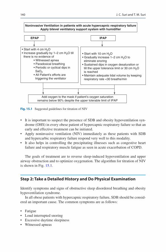
140
• It is important to suspect the presence of SDB and obesity hypoventilation syn-drome (OHS) in every obese patient of hypercapnic respiratory failure so that an early and effective treatment can be initiated.
• Apply noninvasive ventilation (NIV) immediately as these patients with SDB and hypercarbic respiratory failure respond very well to this modality.
• It also helps in controlling the precipitating illnesses such as congestive heart failure and respiratory muscle fatigue as seen in acute exacerbation of COPD.
The goals of treatment are to reverse sleep-induced hypoventilation and upper airway obstruction and to optimize oxygenation. The algorithm for titration of NIV is shown in Fig. 15.1.
Step 2: Take a Detailed History and Do Physical Examination
Identify symptoms and signs of obstructive sleep disordered breathing and obesity hypoventilation syndrome.
In all obese patients with hypercapnic respiratory failure, SDB should be consid-ered an important cause. The common symptoms are as follows:
• Fatigue• Loud interrupted snoring• Excessive daytime sleepiness• Witnessed apneas
Noninvasive Ventilation in patients with acute hypercapnic respiratory failureApply bilevel ventilatory support system with humidifier
EPAP IPAP
• Start with 4 cm H2O• Increase gradually by 1–2 cm H2O till there is no evidence of • Witnessed apnea • Paradoxical breathing • Periodic or cyclical dips in SaO2 • All Patient's efforts are triggering the ventilator
• Start with 10 cm H2O• Gradually increase 1–2 cm H2O to eliminate snoring• Sustained dips in oxygen desaturation or till the upper tolerance limit or 30 cm H2O is reached• Maintain adequate tidal volume by keeping respiratory rate <30 breaths/min
Add oxygen to the mask if patient’s oxygen saturationremains below 90% despite the upper tolerable limit of IPAP
Fig. 15.1 Suggested guidelines for titration of NIV
J. C. Suri and T. M. Suri

141
• Nocturnal awakening, snorting, or gasping• Unrefreshing sleep• Mood disorders• Morning headaches• Large neck circumference• Poorly controlled hypertension• Craniofacial abnormalities (micrognathia, retrognathia, macroglossia)• Breathlessness on minimal exertion
Step 3: Admit to the ICU
The patient should be admitted to the ICU if any of the following criteria are met:
• Acute acidemia—pH less than 7.30• Decreased level of consciousness or coma• Hemodynamic instability• Refractory hypoxemia• Intolerance to continuous positive airway pressure (CPAP) therapy
Step 4: Understand Respiratory Failure in SDB
• SDB constitutes a spectrum of disorders of various severities with intermittent snoring as the mildest form at one end and OHS as the most severe form at the other end of the spectrum. Heavy snoring and upper airway resistance syndrome and mild, moderate, and severe sleep apnea lie in between these two extremes.
• The patients commonly encountered in the ICUs generally suffer from severe obstructive sleep apnea syndrome (OSAS) and/or OHS or those with overlap syndrome, that is, when OSAS occurs simultaneously with chronic obstructive pulmonary disease (COPD).
Respiratory failure in sleep occurs because of the following reasons:
• Increased airflow resistance due to partial or complete obstruction of the upper airway.
• Decreased ventilatory response to hypoxic and hypercapnic stimuli.• Marked hypotonia of accessory muscles of respiration, particularly during rapid
eye movement (REM) sleep leading to severe hypoventilation.• Altered lung mechanics due to obesity result in decreased functional residual
capacity (FRC), expiratory reserve volume (ERV), vital capacity (VC), and forced expiratory volume in 1 s (FEV1).
• The consequences of untreated SDB include hypertension, stroke, cardiac fail-ure, and excessive daytime sleepiness.
15 Sleep-Disordered Breathing
142
Respiratory failure is usually precipitated by complicating respiratory illnesses such as infections, acute exacerbation of asthma and COPD, and congestive cardiac failure.
Step 5: Perform Relevant Laboratory Investigations
• Complete blood counts• Blood glucose (fasting and postprandial)• HbA1C• Lipid profile• Thyroid function test• Serum electrolytes• Arterial blood gases• ECG• Chest X-ray• Echocardiography• Spirometry
Step 6: Monitor Closely During NIV
The following parameters should be monitored during treatment:
• The level of consciousness• Vital signs• Respiratory rate• Use of accessory muscles• SaO2, end-tidal CO2
• Triggering• Patient–ventilator synchrony• Esophageal pressure monitoring (selected cases)• Arterial blood gas frequently
Precautions
• In patients with overlap syndrome, expiratory positive airway pressure (EPAP) higher than auto-positive end-expiratory pressure (auto-PEEP) may worsen the hyperinflation, leading to increase in respiratory rate and work of breathing.
• There may be worsening of blood gases in the first few days due to intense hypoventilation caused by rebound increase in delta and REM sleep.
J. C. Suri and T. M. Suri

143
Step 7: Intubate If Indicated
Indication of Intubation and Mechanical Ventilation
• NIV failure. – Worsening mental status – Deterioration of pH and PaCO2 after 1–3 h of therapy – Refractory hypoxemia – Intolerance to NIV
• Hemodynamic instability.• Inability to clear secretions.• Intubation of the patient with severe OSAS or OHS is associated with significant
difficulties and complications due to limited mouth opening and neck mobility.• The pharynx is anatomically small with large tongue. The ability to withstand
apnea or hypopnea is poor due to low oxygen reserves associated with decreased FRC.
• The intubation should be done by an experienced intensivist.
Indications of Tracheostomy• It was the main treatment before the development of NIV. Now, it is used occa-
sionally in patients who cannot tolerate NIV or have poor compliance to NIV or who cannot be successfully extubated after a period of mechanical ventilation.
Step 8: Manage Comorbid Medical Conditions
• Most patients of SDB and OHS have concomitant respiratory, cardiac, and meta-bolic comorbidities such as COPD, asthma, congestive heart failure, and diabetes.
• In addition to NIV and oxygen, the appropriate treatment of these conditions should also be instituted.
Step 9: Plan a Sleep Study (Polysomnography) Before Discharge
• Although some patients may already have the diagnosis, majority of the patients presenting to the ICU with acute respiratory failure had no prior diagnosis.
• OSA are at risk of Motor Vehicle accident (MVA) while driving and this history should be ellicited in all MVA patients at risk for OSA.
• If the diagnosis of OSAS or OHS is suspected, a bedside sleep study may be performed for both diagnostic and titration purposes. However, if the bedside sleep laboratory is not available, the patient can be treated empirically with NIV with the help of a pulse oximeter, as shown in Fig. 15.1.
15 Sleep-Disordered Breathing

144
Diagnostic Criteria for SDB
• The third edition of the International Classification of Sleep Disorders defines sleep disordered breathing as a significant disorder when a patient has a respira-tory distress index (RDI) i.e., (apneas + hypopneas + respiratory effort-related arousals) of more than or equal to five per hour of sleep along with clinical pre-sentation such as excessive daytime sleepiness, unrefreshing sleep, fatigue, insomnia, mood disorders, or other neurocognitive disturbances.
• The severity of SDB is assessed by the number of abnormal breathing events per hour of sleep, the degree of sleepiness, and the degree of oxygen desaturation during sleep.
Mild RDI 5–15/h of total sleep timeModerate RDI 16–30/h of total sleep timeSevere RDI >30/h of total sleep time
RDI respiratory disturbance index
Diagnostic Criteria for OHS
• BMI more than 30 kg/m2
• Daytime alveolar hypoventilation (awake arterial PaCO2 > 45 mmHg)• Severe OSA (AHI > 30/h) or severe oxygen desaturation• Absence of other causes of hypoventilation
Suggested Readings
American Academy of Sleep Medicine Task Force. Sleep-related breathing disorders in adults: recommendations for syndrome definitions and measurement techniques in clinical research. Sleep. 1999;22:667–89. Obese patients with sleep hypoventilation have an increased risk of acute hypercapnic respiratory failure. Early diagnosis and implementation of noninvasive positive- pressure ventilation is recommended for these patients.
BaHammam A. Acute ventilatory failure complicating obesity hypoventilation: update on a “criti-cal care syndrome.”. Curr Opin Pulm Med. 2010;16:543–51. An update on acute ventilatory failure in obesity hypoventilation syndrome.
Buckle P, Pouliot Z, Millar T, et al. Polysomnography in acutely ill intensive care unit patients. Chest. 1992;102(1):288–9.
Fletcher EC, Shah A. “Near miss” death in obstructive sleep apnea: a critical care syndrome. Crit Care Med. 1991;19(9):1158–64. The objective of this study was to alert critical care physicians to the syndrome of obstructive sleep apnea with respiratory failure (“near miss” death) and to elucidate characteristics that might allow earlier recognition and treatment of such patients.
Lee WY, Mokhlesi B. Diagnosis and management of obesity hypoventilation syndrome in the ICU. Crit Care Clin. 2008;24(3):533–49. A comprehensive review on morbidity, mortality, and OHS management.
J. C. Suri and T. M. Suri

145
Malhotra A, Hillman D. Obesity and the lung: 3. Obesity, respiration and intensive care. Thorax. 2008;63(10):925–31. The important physiological concepts are illustrated by focusing on obstructive sleep apnea, obesity hypoventilation syndrome, abdominal compartment syndrome, and ventilatory management of the obese patient with acute respiratory distress syndrome.
Websites
www.sleepapnea.org/resources/pubs/mayo.pdf. Postoperative complications in patients with obstructive sleep apnea.
http://Chestjournal.chestpubs.org/content/118/3/591.full. Cardiac rhythm disturbances in the obstructive sleep apnea syndrome.
15 Sleep-Disordered Breathing

147© Springer Nature Singapore Pte Ltd. 2020R. Chawla, S. Todi (eds.), ICU Protocols, https://doi.org/10.1007/978-981-15-0898-1_16
P. K. Bhattacharya (*) Department of Trauma & Emergency Care Services, Rajendra Institute of Medical Sciences (RIMS), Ranchi, Jharkhand, India
L. Bhattacharya Department of Anaesthesia, Jawaharlal Nehru Cancer Hospital, Bhopal, Madhya Pradesh, India
16Oxygen Therapy
Pradip Kumar Bhattacharya and Lata Bhattacharya
Correct administration of oxygen is lifesaving, but many a times it is given without careful evaluation of its potential benefits and side effects. Oxygen is the common-est drug used in patients admitted in Intensive Care Unit. Like other drugs oxygen also carries a clear indication for treatment and appropriate methods for its delivery. Inappropriate dose and failure to monitor can have serious consequences. Vigilant monitoring is essential to detect and correct adverse effects.
Step 1: Assess the Need for Oxygen Therapy
• Provide oxygen for measured hypoxemia and not only for breathlessness.• Measure oxygen saturation and if it is below 90%, provide supplemental
oxygen.• If pulse oximetry signal not adequate, check arterial PaO2.• Record approximate FiO2 given and oxygen saturation or PaO2/FiO2 ratio in
patient’s chart and monitor it at interval as per hospital protocol.
The Intensivist was called to evaluate a female patient in emergency department who was known to have advanced emphysema. She was getting oxygen through a nasal cannula at 6 L/min. The nurse says that she has become drowsy and less responsive since the oxygen was given to her an hour ago. Her ABG results on the oxygen show the following: PaO2, 84 mmHg, PaCO2, 65 mmHg and pH, 7.32.
148
• Set a target of oxygen saturation for each patient, low normal (88–92%) for COPD, target for not at risk of hypercapnic respiratory failure is 94–98%.
• In emergency situations (e.g., cardiorespiratory arrest, acute cardiogenic pulmo-nary edema, or stroke), oxygen administration may be initiated empirically, pending detailed clinical and laboratory evaluation.
• Investigate and manage underlying cause of hypoxemia simultaneously.• Supplemental oxygen improve oxygenation, but does not treat the underlying
causes of hypoxaemia which must be diagnosed and treated urgently.• Oxygen delivery devices and flow rates should be adjusted to keep the oxygen
saturation in the target range.• Prompt clinical assessment is needed if oxygen therapy needs to be initiated or
increased due to a decreasing saturation level.
Step 2: Identify Type of Hypoxia
Hypoxemia is lack of oxygen in the blood whereas hypoxia is lack of oxygen in the tissue level.
Hypoxia is of four types: Hypoxemic hypoxia, Anemic hypoxia, Stagnant hypoxia and Histotoxic hypoxia.
1. Hypoxemic hypoxia may be because of- (a) Low FiO2, hypoventilation (b) Ventilation perfusion mismatch, (c) Diffusion defect, or (d) Shunt effect
2. Anaemic hypoxia may be because of- (a) A decreased hemoglobin level (b) CO poisoning (c) Excessive blood loss (d) Methemoglobinemia (e) Iron deficiency
3. Stagnant hypoxia may be because of- (a) Capillary perfusion is diminished
• Decreased heart rate• Decreased cardiac output• Shock• Embolism• Exposure to cold weather
4. Histotoxic hypoxia may be because of- (a) The oxidative enzyme mechanism of the cell is impaired as a result of:
• Cyanide poisoning• Alcohol poisoning
(b) Rarely accompanied by hypoxemia but is accompanied by increased venous PO2 levels.
P. K. Bhattacharya and L. Bhattacharya
149
Step 3: Initiate Oxygen Administration
• Before giving oxygen, one needs to ensure patency of the airways. This might require endotracheal intubation or tracheostomy.
• It is generally customary to start with a high FiO2—100% for cardiorespiratory arrest and 50–100% for acute hypoxemic respiratory failure.
• The FiO2 can be increased or decreased after the assessment of clinical and labo-ratory response to the initial administration.
• Relatively lower concentrations are used in patients with hypercapnic respiratory failure (such as COPD) with the preexisting chronic hypoventilation.
• High concentration of oxygen may worsen CO2 retention and cause CO2 narcosis by abolishing the hypoxic respiratory stimulation. However, optimum FiO2 must be ensured since hypoxia is always more deleterious than hypercapnia. Various devices can be used for applying oxygen.
Source: Most well-equipped ICUs have continuous pressurized oxygen and air supply available at each bed. In this fashion, both oxygen and air can be simultane-ously fed into an oxygen blender to control the output FiO2. Oxygen cylinders and concentrators are required as a backup source in case of failure of central supply.
• Select optimum oxygen delivery device
Oxygen Delivery Devices
1. Low flow oxygen systems: Low flow oxygen systems or delivery devices does not meet the patients inspiratory flow demands. Therefore room air must make up the remainder of the patient’s tidal volume.
The percentage of oxygen delivered by low flow devices is variable depend-ing on patient’s tidal volume, respiratory rate, inspiratory time and ventilatory pattern (a) Nasal Cannula (delivers 24–40% of oxygen at a flow rate of 1–6 L/min)
(Fig. 16.1) (b) Nasal Reservoir Cannula (Reservoir stores upto 20 mL of oxygen, delivers
22–35% of oxygen at a flow rate of 1–4 L/min) (Fig. 16.2) (c) Pendant Reservoir Cannula (delivers 22–35% of oxygen at a flow rate of
1–4 L/min) (Fig. 16.3) (d) Simple Oxygen Mask (delivers 35–50% of oxygen at a flow rate of 5–10 L/
min) (Fig. 16.4) (e) Partial Re-breathing Mask (delivers 40–70% of oxygen at a flow rate of
10–15 L/min) (Fig. 16.5) (f) Non Re-breathing Mask (delivers 60–80% of oxygen at a flow rate of
10–15 L/min) (Fig. 16.5) (g) Transtracheal Oxygen Catheter (delivers 22–35% oxygen at a flow rate of
1–4 L/min) (Fig. 16.6)
16 Oxygen Therapy
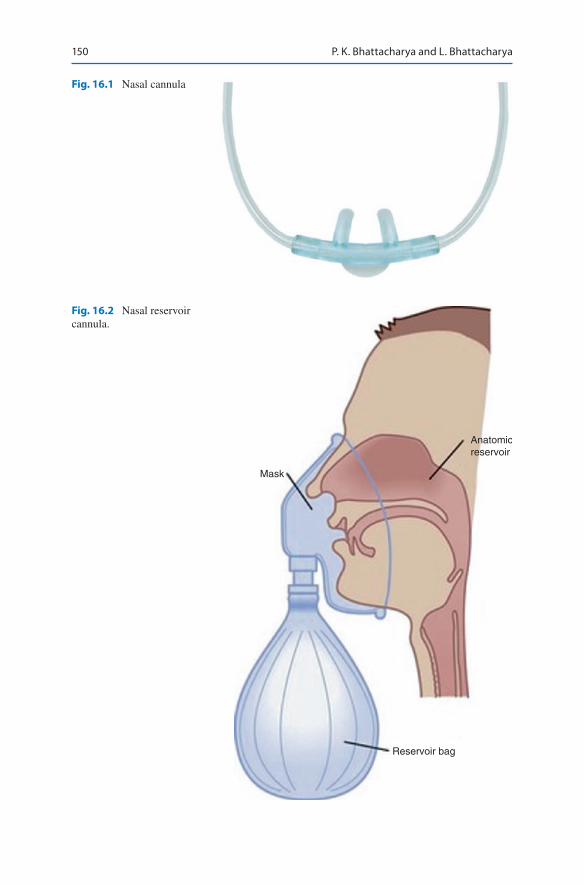
150
Fig. 16.1 Nasal cannula
Mask
Anatomicreservoir
Reservoir bag
Fig. 16.2 Nasal reservoir cannula.
P. K. Bhattacharya and L. Bhattacharya
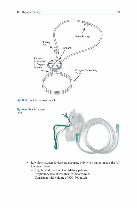
151
Nasal Prongs
Pendant
Oxygen ConnectingTube
TubingClip
FemaleConnectorto OxygenSource
Fig. 16.3 Pendant reservoir cannula
Fig. 16.4 Simple oxygen mask
• Low flow oxygen devices are adequate only when patient meets the fol-lowing criteria- – Regular and consistent ventilatory pattern. – Respiratory rate of less than 25 breaths/min. – Consistent tidal volume of 300–700 mL/h
16 Oxygen Therapy
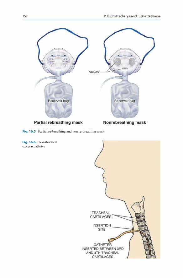
152
Partial rebreathing mask Nonrebreathing mask
Reservoir bagReservoir bag
Valves
Fig. 16.5 Partial re-breathing and non re-breathing mask.
TRACHEALCARTILAGES
INSERTIONSITE
CATHETERINSERTED BETWEEN 3RD
AND 4TH TRACHEALCARTILAGES
Fig. 16.6 Transtracheal oxygen catheter
P. K. Bhattacharya and L. Bhattacharya

153
High Flow Oxygen Delivery Devices
(a) Air Entrainment Mask (Provides 24–50% of oxygen) (Fig. 16.7)The jet size of the entrainment port determines the FiO2. The larger the jet size the less air entrained and higher the FiO2. Smaller the jet size more air entrained and lower the FiO2.
The larger the entrainment port more air entrained and lower the FiO2. Similarly, smaller the entrainment port less air entrained and higher the FiO2.
O2 percentage Air/O2 Entrainment ratio24 25:128 10:130 8:135 5:140 3:145 2:150 1.7:160 1:1
Airentrainment
port
Narrowedorifice
Entrainedroom air
100% O2
Removable adapter(jet diluter)
Exhaled gasExhalation port
Mask strap
Flex tube (6 in long)
Inhaled mixture of100% O2 and room air
Fig. 16.7 Air entrainment mask
16 Oxygen Therapy
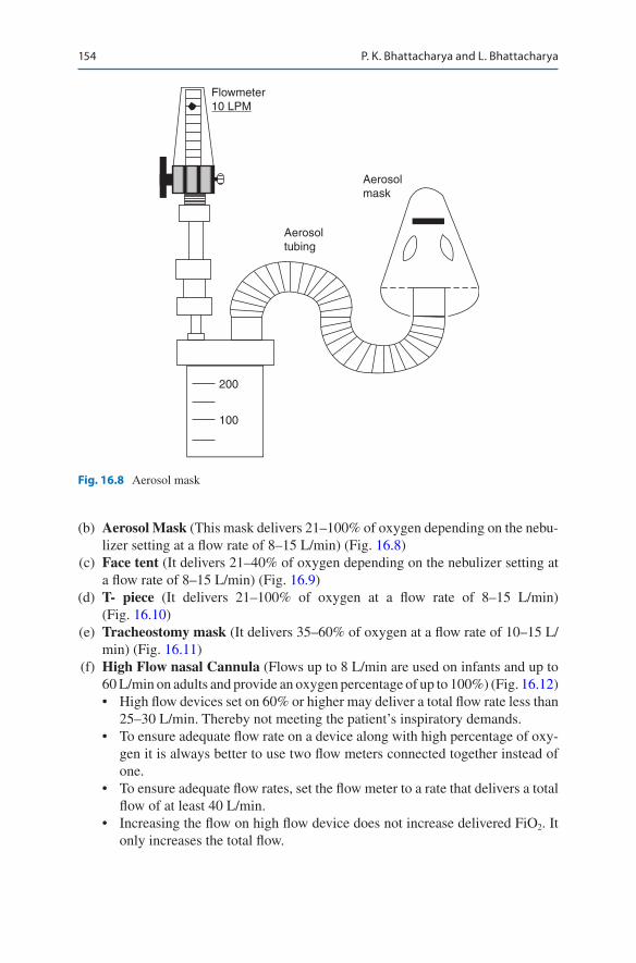
154
(b) Aerosol Mask (This mask delivers 21–100% of oxygen depending on the nebu-lizer setting at a flow rate of 8–15 L/min) (Fig. 16.8)
(c) Face tent (It delivers 21–40% of oxygen depending on the nebulizer setting at a flow rate of 8–15 L/min) (Fig. 16.9)
(d) T- piece (It delivers 21–100% of oxygen at a flow rate of 8–15 L/min) (Fig. 16.10)
(e) Tracheostomy mask (It delivers 35–60% of oxygen at a flow rate of 10–15 L/min) (Fig. 16.11)
(f) High Flow nasal Cannula (Flows up to 8 L/min are used on infants and up to 60 L/min on adults and provide an oxygen percentage of up to 100%) (Fig. 16.12)• High flow devices set on 60% or higher may deliver a total flow rate less than
25–30 L/min. Thereby not meeting the patient’s inspiratory demands.• To ensure adequate flow rate on a device along with high percentage of oxy-
gen it is always better to use two flow meters connected together instead of one.
• To ensure adequate flow rates, set the flow meter to a rate that delivers a total flow of at least 40 L/min.
• Increasing the flow on high flow device does not increase delivered FiO2. It only increases the total flow.
Flowmeter10 LPM
Aerosolmask
Aerosoltubing
200
100
Fig. 16.8 Aerosol mask
P. K. Bhattacharya and L. Bhattacharya

155
Fig. 16.9 Face tent
Fig. 16.10 T-piece
Fig. 16.11 Tracheostomy mask
16 Oxygen Therapy
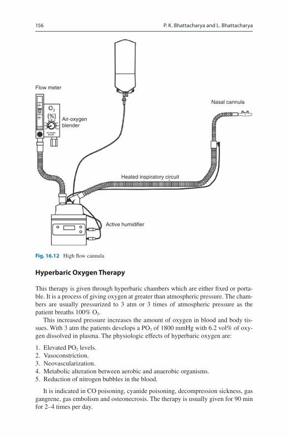
156
Flow meter
Air-oxygenblender
Heated inspiratory circuit
Active humidifier
Nasal cannula
Fig. 16.12 High flow cannula
Hyperbaric Oxygen Therapy
This therapy is given through hyperbaric chambers which are either fixed or porta-ble. It is a process of giving oxygen at greater than atmospheric pressure. The cham-bers are usually pressurized to 3 atm or 3 times of atmospheric pressure as the patient breaths 100% O2.
This increased pressure increases the amount of oxygen in blood and body tis-sues. With 3 atm the patients develops a PO2 of 1800 mmHg with 6.2 vol% of oxy-gen dissolved in plasma. The physiologic effects of hyperbaric oxygen are:
1. Elevated PO2 levels. 2. Vasoconstriction. 3. Neovascularization. 4. Metabolic alteration between aerobic and anaerobic organisms. 5. Reduction of nitrogen bubbles in the blood.
It is indicated in CO poisoning, cyanide poisoning, decompression sickness, gas gangrene, gas embolism and osteonecrosis. The therapy is usually given for 90 min for 2–4 times per day.
P. K. Bhattacharya and L. Bhattacharya
157
A. O2 supplementation during noninvasive positive-pressure ventilation (NIV)• NIV in ICUs is administered either through conventional mechanical ventila-
tion or through a portable system.• Supplemental oxygen is delivered by simply adding it to the mask or the circuit.• Oxygen should be added into the circuit distal to the exhalation port and as
close to the patient as possible.• The highest concentration is achieved with O2 added to the mask, with the
leak port in the circuit, and with the lowest setting of inspiratory and expira-tory pressures. Unfortunately, the delivered FiO2 with NIV portable systems remains unpredictable.
• In case there is a need for precise oxygen delivery, it is better to use a conven-tional ventilator with an integrated oxygen blender.
B. O2 supplementation through conventional mechanical ventilation• Currently available mechanical ventilators are capable of delivering tightly
controlled and high levels of FiO2 to intubated patients.• The integrated microprocessor-controlled gas blenders allow blending of
compressed oxygen and air and precise delivery of a preset FiO2 for contin-ued and long periods.
• It is also possible to deliver high FiO2 and even 100% oxygen through such systems.• This is the reason that mechanical ventilation remains the cornerstone of
management of severe hypoxia to improve oxygenation to a satisfactory level in most severe forms of respiratory failure.
Step 4: Monitoring Oxygen Therapy
• Arterial Blood Gas• Perform arterial blood gas analysis before oxygen therapy is started and measure
again within 2 h of starting oxygen therapy and FiO2 should be adjusted accordingly.• An adequate response to oxygen therapy is defined as PaO2 of more than
60 mmHg and SpO2 of more than 90%.• Frequency of arterial blood gas monitoring depends on the severity of respiratory
failure and hypoxemia.• Arterial blood gas also monitors the total oxygen content of the blood which
helps in calculation of oxygen delivery and consumption.• Pulse oximetry measures spO2 and is easily available.• Co-oximetry: This is used to measure hemoglobin, oxyhemoglobin and carboxy-
hemoglobin separately.• Monitoring oxygenation at tissue level: Mixed venous oxygen saturation
(SvO2) with the help of pulmonary artery catheter, can be measured by taking blood sample from the proximal pulmonary artery. This reflects the amount of oxygen “leftover” in the venous blood after body tissues have removed (used) whatever oxygen they needed.
• ScvO2, the central venous oxygen saturation measured by the placement of a catheter in the superior vena cava, can be taken as a surrogate for SvO2.A low SvO2/ScvO2 suggests that the cardiac output and the level of oxygenation are insufficient to meet the metabolic demands of body tissues.
16 Oxygen Therapy

158
Step 5: Complications of Oxygen Therapy
Assess Oxygen Toxicity
• Oxygen is a drug when given in excess it causes toxicity.• It is the partial pressure of oxygen and not the FiO2 which is responsible for the
toxic effect of oxygen along with partial pressure of oxygen.• The duration of exposure is also an important criteria for development of oxygen
toxicity.• The primary effects of oxygen toxicity is on the central nervous system which
includes tremors, twitching and convulsions.• High partial pressure of oxygen also damages the capillary endothelium in the
lungs. Later on patient may develop pulmonary arterial hypertension.• The goal should be to use the lowest possible FiO2 to achieve adequate tissue
oxygenation.• Depression of Ventilation—This is a common observation in patients with COPD
and chronic hypercapnia. – It has got a direct relationship with accompanying elevation in arterial partial
pressure of carbon dioxide (PaCO2) along with suppression of the hypoxic drive. – In these patients normal response to high PaCO2 is blunted. – Management of hypoxemia with supplemental oxygen should never be
avoided in these patients.• Hypoxemia is primarily sensed by peripheral chemoreceptors. Increase in blood
oxygen level in these patients suppresses the peripheral chemoreceptors which depresses the ventilatory drive and elevates the PaCO2.
Identify Retinopathy of Pre Maturity
• It occurs in pre matured low birth infants who receive supplemental oxygen.• Excess oxygen causes retinal vasoconstriction which leads to necrosis of blood
vessels. As a compensatory mechanism neovascularization occurs. Hemorrhage of these newly formed vessels causes scarring behind the retina.
• Other factors which give rise to this condition are hypercapnia, intra ventricular hemorrhage, infection, lactic acidosis, anemia, hypocalcemia and hypothermia.
• The recommended preventive measure is to keep the partial pressure of oxygen (PaO2) less than 80 mm Hg.
Absorption Atelectasis
• FiO2 of greater than 0.5 presents a risk of absorption atelectasis, it happens because of denitrogenation. Breathing high level of oxygen quickly depletes the nitrogen level in alveoli and blood. With rapid denitrogenation the total gas pres-sure in the alveolus progressively decreases.
P. K. Bhattacharya and L. Bhattacharya
159
• In the absence of any repletion this ultimately leads to alveolar collapse. Absorption atelectasis also increases physiologic shunt and worsen blood oxygenation.
• Conditions like sedation, surgical pain and CNS dysfunction give rise to more rapid collapse as compared to normal lung.
Fire Hazard: The fire hazard is mostly associated with procedures like tracheos-tomy, laryngoscopy, use of electronic scalpels etc.• Hyperbaric oxygen therapy also carries an increased risk of fire hazard.• Simple strategies which can prevent these fire hazards are use of lowest effective
FiO2 for a clinical situation, use of scavenger systems, educating clinicians, patients, care givers and follow up of fire prevention protocol strictly.
Suggested Reading
Barnhart SL. Oxygen administration. In: Walsh BK, Czervinske MP, DiBlasi RM, editors. Perinatal and pediatric respiratory care. 3rd ed. St Louis: Saunders; 2010.
Huang YC. Monitoring oxygen delivery in the critically ill. Chest. 2005;128(5 Suppl. 2):554S–60S. This article reviews the basic principles of DO(2) and the abnormal oxygen supply–demand relationship seen in patients with shock. It also discusses approaches for monitoring DO(2), including clinical symptoms/signs, acid–base status, and gas exchange, which pro-vide global assessment, as well as gastric tonometry, which may reflect regional DO(2).
Leach RM, Treacher DF. The pulmonary physician in critical care. 2. Oxygen delivery and con-sumption in the critically ill. Thorax. 2002;57:170–7. Early detection and correction of tissue hypoxia is essential if progressive organ dysfunction and death are to be avoided. However, hypoxia in individual tissues or organs caused by disordered regional distribution of supple-mental oxygen may be lifesaving in some situations but cannot correct inadequate oxygen delivery caused by a low cardiac output or impaired ventilation.
O’Driscoll BR, Howard LS, et al. BTS Guideline for oxygen use in adults in healthcare and emer-gency settings. Thorax. 2017;72(Suppl 1):pi1–i89. A comprehensive guidelines on oxygen therapy.
American Association for Respiratory Care: clinical practice guideline: oxygen therapy for adults in the acute care facility: 2002 revision and update. Respir Care. 2002;47:717. Guidelines on oxygen therapy.
Schwartz AR, Kacmarek RM, Hess DR. Factors affecting oxygen delivery with bi-level positive airway pressure. Respir Care. 2004;49:270–5. Delivered oxygen concentration during BiPAP is a complex interaction between the leak port type, the site of oxygen injection, the ventilator settings, and the oxygen flow.
16 Oxygen Therapy

161© Springer Nature Singapore Pte Ltd. 2020R. Chawla, S. Todi (eds.), ICU Protocols, https://doi.org/10.1007/978-981-15-0898-1_17
D. Govil (*) · A. Srivastava Medanta Institute of Critical Care, Medanta—The Medicity Hospital, Gurgaon, India
S. Gupta Narayana Superspeciality Hospital, Gurgaon, India
17Pulse Oximetry and Capnography
Deepak Govil, Sachin Gupta, and Ashish Srivastava
Pulse oximetry and capnography are essential components of respiratory monitor-ing in the intensive care unit.
Step 1: Examine the Patient in Detail and Check Pulse Oximetry
• Presence of nonspecific symptoms and signs, which are suggestive of hypoxia, should be evaluated with objective measurements of oxygenation.
• Standard blood gas analyzer calculates oxygen saturation by oxy-hemoglobin dissociation curve by plotting partial pressure of oxygen against arterial oxygen saturation at varying pH. Blood gas analyzer fitted with co-oximeter on the other hand directly measures oxygen saturation in the arterial blood sample (SaO2).
• The pulse oximeter is a medical device that non invasively monitors the arterial oxygen saturation by spectrophotometry method and by sensing pulsatile changes of arterial blood volume producing a photoplethysmograph and is con-sidered the “fifth vital sign”
A 50-year-old male patient, a known case of chronic obstructive pulmonary dis-ease (COPD), was admitted to the hospital with acute exacerbation. He was drowsy and cyanotic. His respiratory rate was 26/min. He was admitted for the management of COPD.
162
Step 2: Understand the Principle of Pulse Oximetry
• The fundamental physical property that allows the pulse oximeter to measure the oxygen saturation of hemoglobin is based on Beer–Lambert law, which relates to the concentration of a solute in a solution to the absorption of light of a given wavelength passing through a non absorbing solvent containing the absorbent solute.
• The pulse oximetry is based on two physical principles: – The light absorbance of the oxygenated hemoglobin is different from that of
the reduced hemoglobin, at the oximeter’s two wavelengths, which include red (deoxyhemoglobin) and near-infrared (oxyhemoglobin) light.
– The absorbance of both wavelengths has a pulsatile component, which is due to the fluctuations in the volume of arterial blood between the source and the detector.
• Typically, a pulse oximeter has a pair of small light-emitting diodes (LEDs) fac-ing a light detector (sensor) through a translucent part of the patient’s body, usu-ally a fingertip or an earlobe.
• One LED is red, with the wavelength of 660 nm, and the other is infrared, with the wavelength of 905, 910, or 940 nm. Absorption at these wavelengths differs significantly between oxyhemoglobin and its deoxygenated form. The ratio of light absorbance between oxyhemoglobin and the sum of oxyhemoglobin plus deoxyhemoglobin is calculated and compared with an in built algorithm of direct measurement of arterial oxygen to estimate peripheral oxygen saturation.
• The arterial signal is pulsatile and it can be distinguished from nonpulsatile sig-nals by the microprocessor by selecting out the absorbance of the pulsatile frac-tion of the blood, that is, due to arterial blood (AC), from the constant absorbance by nonpulsatile venous or capillary blood and other tissue pigments (DC), thus eliminating the effect of tissue absorbance to measure the oxygen saturation of arterial blood.
Step 3: Understand Pitfalls and Limitations
1. Dyshemoglobinopathies• Pulse oximetry is considered to be accurate when oxygen saturation is
between 70% and 100% provided that oxygenated hemoglobin and reduced hemoglobin are the only measured species of hemoglobin. But if methemo-globin and carboxyhemoglobin (COHb) increase in concentration, then the reliability of pulse oximetry becomes doubtful.
• Meth-Hb absorbs equal amount of red and infrared light, and the ratio is equal to 1 at 85% saturation. So even if the patient is hypoxic, the pulse oximeter will read 85%. Vice versa, if the true oxygen saturation is 100%, then also the pulse oximeter will read 85%.
• COHb absorbs very little light at infrared range of 940 nm, while at 660 nm, it absorbs light very similar to oxyhemoglobin. Thus, the presence of
D. Govil et al.
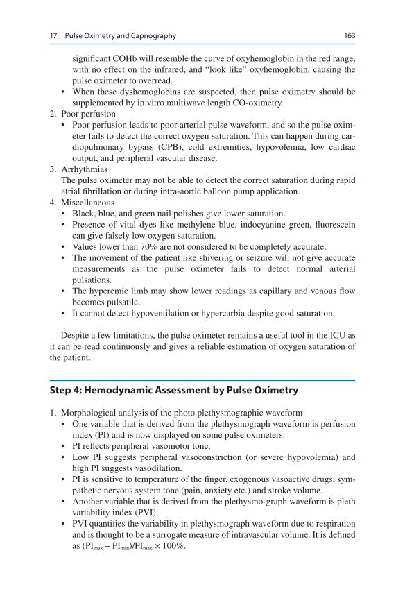
163
significant COHb will resemble the curve of oxyhemoglobin in the red range, with no effect on the infrared, and “look like” oxyhemoglobin, causing the pulse oximeter to overread.
• When these dyshemoglobins are suspected, then pulse oximetry should be supplemented by in vitro multiwave length CO-oximetry.
2. Poor perfusion• Poor perfusion leads to poor arterial pulse waveform, and so the pulse oxim-
eter fails to detect the correct oxygen saturation. This can happen during car-diopulmonary bypass (CPB), cold extremities, hypovolemia, low cardiac output, and peripheral vascular disease.
3. ArrhythmiasThe pulse oximeter may not be able to detect the correct saturation during rapid atrial fibrillation or during intra-aortic balloon pump application.
4. Miscellaneous• Black, blue, and green nail polishes give lower saturation.• Presence of vital dyes like methylene blue, indocyanine green, fluorescein
can give falsely low oxygen saturation.• Values lower than 70% are not considered to be completely accurate.• The movement of the patient like shivering or seizure will not give accurate
measurements as the pulse oximeter fails to detect normal arterial pulsations.
• The hyperemic limb may show lower readings as capillary and venous flow becomes pulsatile.
• It cannot detect hypoventilation or hypercarbia despite good saturation.
Despite a few limitations, the pulse oximeter remains a useful tool in the ICU as it can be read continuously and gives a reliable estimation of oxygen saturation of the patient.
Step 4: Hemodynamic Assessment by Pulse Oximetry
1. Morphological analysis of the photo plethysmographic waveform• One variable that is derived from the plethysmograph waveform is perfusion
index (PI) and is now displayed on some pulse oximeters.• PI reflects peripheral vasomotor tone.• Low PI suggests peripheral vasoconstriction (or severe hypovolemia) and
high PI suggests vasodilation.• PI is sensitive to temperature of the finger, exogenous vasoactive drugs, sym-
pathetic nervous system tone (pain, anxiety etc.) and stroke volume.• Another variable that is derived from the plethysmo-graph waveform is pleth
variability index (PVI).• PVI quantifies the variability in plethysmograph waveform due to respiration
and is thought to be a surrogate measure of intravascular volume. It is defined as (PImax – PImin)/PImax × 100%.
17 Pulse Oximetry and Capnography
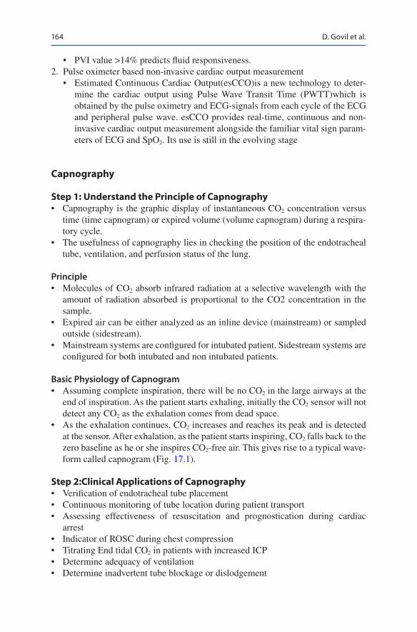
164
• PVI value >14% predicts fluid responsiveness. 2. Pulse oximeter based non-invasive cardiac output measurement
• Estimated Continuous Cardiac Output(esCCO)is a new technology to deter-mine the cardiac output using Pulse Wave Transit Time (PWTT)which is obtained by the pulse oximetry and ECG-signals from each cycle of the ECG and peripheral pulse wave. esCCO provides real-time, continuous and non- invasive cardiac output measurement alongside the familiar vital sign param-eters of ECG and SpO2. Its use is still in the evolving stage
Capnography
Step 1: Understand the Principle of Capnography• Capnography is the graphic display of instantaneous CO2 concentration versus
time (time capnogram) or expired volume (volume capnogram) during a respira-tory cycle.
• The usefulness of capnography lies in checking the position of the endotracheal tube, ventilation, and perfusion status of the lung.
Principle• Molecules of CO2 absorb infrared radiation at a selective wavelength with the
amount of radiation absorbed is proportional to the CO2 concentration in the sample.
• Expired air can be either analyzed as an inline device (mainstream) or sampled outside (sidestream).
• Mainstream systems are configured for intubated patient. Sidestream systems are configured for both intubated and non intubated patients.
Basic Physiology of Capnogram• Assuming complete inspiration, there will be no CO2 in the large airways at the
end of inspiration. As the patient starts exhaling, initially the CO2 sensor will not detect any CO2 as the exhalation comes from dead space.
• As the exhalation continues, CO2 increases and reaches its peak and is detected at the sensor. After exhalation, as the patient starts inspiring, CO2 falls back to the zero baseline as he or she inspires CO2-free air. This gives rise to a typical wave-form called capnogram (Fig. 17.1).
Step 2:Clinical Applications of Capnography• Verification of endotracheal tube placement• Continuous monitoring of tube location during patient transport• Assessing effectiveness of resuscitation and prognostication during cardiac
arrest• Indicator of ROSC during chest compression• Titrating End tidal CO2 in patients with increased ICP• Determine adequacy of ventilation• Determine inadvertent tube blockage or dislodgement
D. Govil et al.

165
Step 3: Identify Representative Capnographic Waveform• Slanting and prolongation of expiratory phase is indicative of obstruction in the
respiratory pathway, that is, either obstruction in the endotracheal tube or obstructive lung disease (Fig. 17.2).
• The elevation of the baseline is indicative of rebreathing, insufficient gas flows (Fig. 17.3).
• Contamination of the expired sample by fresh gas flows or sampling site too near to fresh gas (Fig. 17.4).
• Low EtCO2 indicates hyperventilation (Fig. 17.5).• High EtCO2 indicates hypoventilation (Fig. 17.6).• Sudden fall in EtCO2 can be due to asystole, hypotension, or massive pulmonary
embolism (Fig. 17.7).• This type of capnogram is observed when sudden CO2 is released after unclamp-
ing a major blood vessel or release of tourniquet (Fig. 17.8).• Sudden fall and rise of EtCO2 is due to small air embolus (Fig. 17.9).• Cardiac oscillations are due to contraction and relaxation of the heart (Fig. 17.10).• This shows a sedated and inadequately paralyzed patient having spontaneous
respiratory efforts (Fig. 17.11).
40
0
CO2
Expiration Inspiration
PHASEI
PHASEII
PHASEIII
PHASEIVu
a b
Fig. 17.1 Normal Capnogram. Phase I: At the start of exhalation, anatomical and physiological dead space is expired, so no CO2. Phase II: Exhalation continues, so CO2 rises. Phase III: CO2 plateau (End tidal CO2). Phase IV: Inspiration
40
CO2
0Time
Fig. 17.2 Capnogram showing obstruction in the respiratory pathway
17 Pulse Oximetry and Capnography
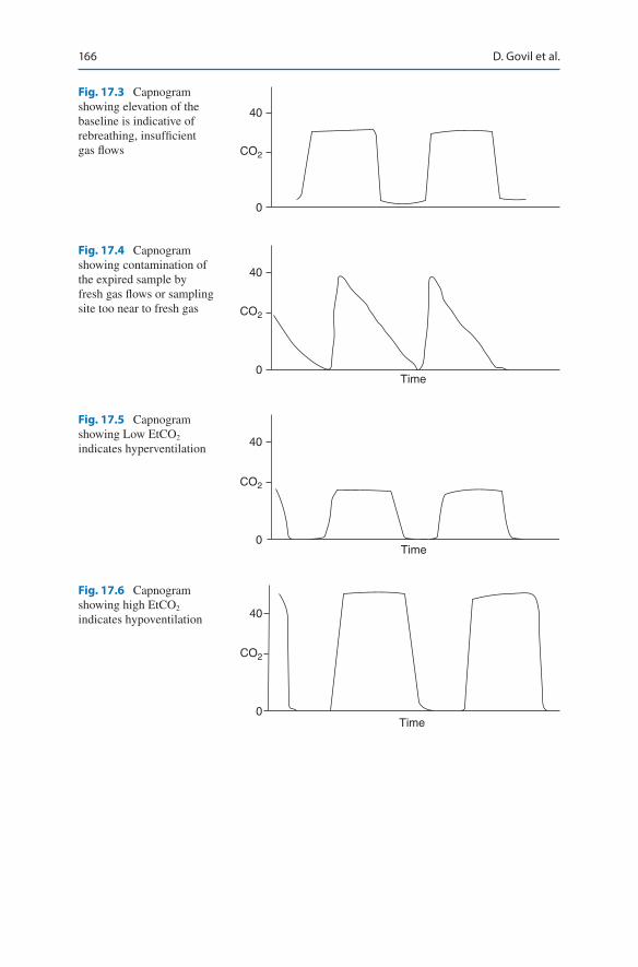
166
40
CO2
0
Fig. 17.3 Capnogram showing elevation of the baseline is indicative of rebreathing, insufficient gas flows
40
0Time
CO2
Fig. 17.4 Capnogram showing contamination of the expired sample by fresh gas flows or sampling site too near to fresh gas
40
0Time
CO2
Fig. 17.5 Capnogram showing Low EtCO2 indicates hyperventilation
40
0Time
CO2
Fig. 17.6 Capnogram showing high EtCO2 indicates hypoventilation
D. Govil et al.

167
40
0Time
CO2
Fig. 17.7 Capnogram showing sudden fall in EtCO2 can be due to asystole, hypotension, or massive pulmonary embolism
40
0Time
CO2
Fig. 17.8 Capnogram showing sudden rise in CO2 after unclamping a major blood vessel or release of tourniquet
40
0Time
CO2
Fig. 17.9 Capnogram showing sudden fall and rise of EtCO2 is due to small air embolus
40
0
CO2
Time
Fig. 17.10 Capnogram showing cardiac oscillations due to contraction and relaxation of the heart
17 Pulse Oximetry and Capnography
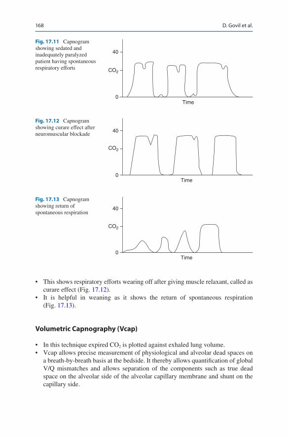
168
• This shows respiratory efforts wearing off after giving muscle relaxant, called as curare effect (Fig. 17.12).
• It is helpful in weaning as it shows the return of spontaneous respiration (Fig. 17.13).
Volumetric Capnography (Vcap)
• In this technique expired CO2 is plotted against exhaled lung volume.• Vcap allows precise measurement of physiological and alveolar dead spaces on
a breath-by-breath basis at the bedside. It thereby allows quantification of global V/Q mismatches and allows separation of the components such as true dead space on the alveolar side of the alveolar capillary membrane and shunt on the capillary side.
40
Time0
CO2
Fig. 17.11 Capnogram showing sedated and inadequately paralyzed patient having spontaneous respiratory efforts
40
0Time
CO2
Fig. 17.12 Capnogram showing curare effect after neuromuscular blockade
40
0Time
CO2
Fig. 17.13 Capnogram showing return of spontaneous respiration
D. Govil et al.
169
• Measurement of dead space has diagnostic, prognostic and therapeutic applications.
• In the intensive care unit (ICU) dead space measurement can be used to guide therapy for patients with acute respiratory distress syndrome (ARDS); in the emergency department it can guide thrombolytic therapy for pulmonary embo-lism; in peri-operative patients it can indicate the success of recruitment maneuvers
Conclusion
Capnogram is a very useful tool for ventilated patients, and it gives us a real-time idea of various lung interactions that can have the implications on the outcome of the patient.
Suggested Reading
Biais M, Berthezène R, Petit L, Cottenceau V, Sztark F. Ability of esCCO to track changes in cardiac output. Br J Anaesth. 2015;115(3):403–10. They investigated whether cardiac output measured with pulse wave transit time (esCCO, Nihon Kohden) was able to track changes in cardiac output induced by an increase in preload (volume expansion/passive leg-raising) or by changes in vasomotor tone (variation in norepinephrine dosage) in critically ill patients.
Cheifetz IM, Myers TR. Respiratory therapies in the critical care setting. Should every mechani-cally ventilated patient be monitored with capnography from intubation to extubation? Respir Care. 2007;52(4):423–38.
Desebbe O, Cannesson M. Using ventilation induced plethysmo-graphic variations to optimize patient fluid status. Curr Opin Anaesthesiol. 2008;21:772–8. Automatic detection of respiratory variations in pulse oximetry plethysmographic waveform amplitude can predict fluid respon-siveness in the operating room in patients under mechanical ventilation and has potential for fluid optimization in this setting
Feiner JR, Severinghaus JW. Effects of skin pigmentation on pulse oximeter accuracy at low satu-ration. Anesthesiology. 2005;102(4):715–9. Pulse oximeters overestimate arterial oxygen satu-ration during hypoxia in dark-skinned individuals.
Hampson NB. Pulse oximetry in severe carbon monoxide poisoning. Chest. 1998;114(4):1036–41. Presently available pulse oximeters overestimate arterial oxygenation in patients with severe CO poisoning. Accurate assessment of arterial oxygen content in patients with CO poisoning can currently be performed only by analysis of arterial blood with a laboratory CO-oximetry
Kamat V. Pulse oximetry. Indian J Anaesth. 2002;46(4):261–8. Despite problems and limitations, pulse oximetry remains the standard of care in all clinical situations, and its use for all patients under anesthesia must be mandated
Nagler J, Krauss B. Capnography: a valuable tool for airway management. Emerg Med Clin North Am. 2008;26(4):881–97. Clinical applications of capnography with regard to ventilation and airway management are discussed.
Van de Louw A, Cracco C. Accuracy of pulse oximetry in the intensive care unit. Intensive Care Med. 2001;27(10):1606–13. Large SpO2 to SaO2 differences may occur in critically ill patients with poor reproducibility of SpO2. A SpO2 above 94% appears necessary to ensure an SaO2 of 90%.
Verscheure, et al. Volumetric capnography: lessons from the past and current clinical applications. Crit Care. 2016;20:184. This is a comprehensive review of volumetric capnography and they
17 Pulse Oximetry and Capnography

170
have discussed the components of dead space, explained important differences between the Bohr and Enghoff approaches, discussed the clinical significance of arterial to end-tidal CO2 gradient and finally summarized potential clinical indications for Vcap measurements in the emergency room, operating room and ICU.
Walsh BK, Crotwell DN, Restrepo RD. Capnography/Capnometry during mechanical ventilation. Respir Care. 2011;56(4):503–9. This updated clinical practice guideline is based on 234 clini-cal studies and systematic reviews, 19 review articles investigating the use of capnography/capnometry during mechanical ventilation.
Wilson BJ, Cowan HJ, Lord JA. The accuracy of pulse oximetry in emergency department patients with severe sepsis and septic shock: a retrospective cohort study. BMC Emerg Med. 2010;10:9. Pulse oximetry overestimates ABG-determined SaO2 by a mean of 2.75% in emergency department patients with severe sepsis and septic shock. This overestimation is exacerbated by the presence of hypoxemia. When SaO2 needs to be determined with a high degree of accuracy, arterial blood gases are recommended.
Websites
www.pulseox.info. Basic information on pulse oximetry.www.pulseoximeteronline.com. Industry website to review different types of pulse oximeters.www.capnography.com. Website produced by Bhavani Shankar Kodali. An atlas of capnograms.www.linxdown.com. Downloadable atlas of capnograms.
D. Govil et al.

Part II
Cardiovascular System
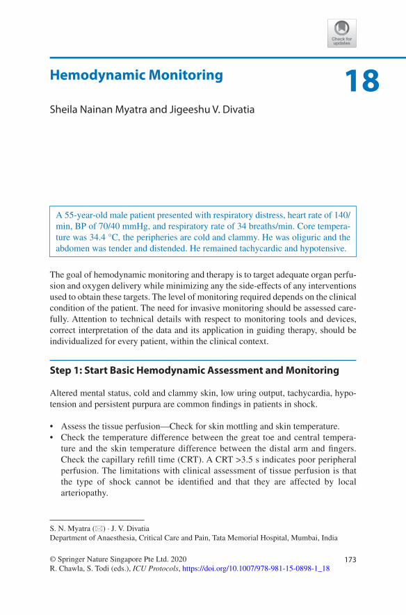
173© Springer Nature Singapore Pte Ltd. 2020R. Chawla, S. Todi (eds.), ICU Protocols, https://doi.org/10.1007/978-981-15-0898-1_18
S. N. Myatra (*) · J. V. Divatia Department of Anaesthesia, Critical Care and Pain, Tata Memorial Hospital, Mumbai, India
18Hemodynamic Monitoring
Sheila Nainan Myatra and Jigeeshu V. Divatia
The goal of hemodynamic monitoring and therapy is to target adequate organ perfu-sion and oxygen delivery while minimizing any the side-effects of any interventions used to obtain these targets. The level of monitoring required depends on the clinical condition of the patient. The need for invasive monitoring should be assessed care-fully. Attention to technical details with respect to monitoring tools and devices, correct interpretation of the data and its application in guiding therapy, should be individualized for every patient, within the clinical context.
Step 1: Start Basic Hemodynamic Assessment and Monitoring
Altered mental status, cold and clammy skin, low uring output, tachycardia, hypo-tension and persistent purpura are common findings in patients in shock.
• Assess the tissue perfusion—Check for skin mottling and skin temperature.• Check the temperature difference between the great toe and central tempera-
ture and the skin temperature difference between the distal arm and fingers. Check the capillary refill time (CRT). A CRT >3.5 s indicates poor peripheral perfusion. The limitations with clinical assessment of tissue perfusion is that the type of shock cannot be identified and that they are affected by local arteriopathy.
A 55-year-old male patient presented with respiratory distress, heart rate of 140/min, BP of 70/40 mmHg, and respiratory rate of 34 breaths/min. Core tempera-ture was 34.4 °C, the peripheries are cold and clammy. He was oliguric and the abdomen was tender and distended. He remained tachycardic and hypotensive.

174
• Do cardiovascular and respiratory system examination.• Noninvasive monitoring—Electrocardiography, noninvasive blood pressure
(NIBP), pulse oximetry, and plethysmographic signals can be monitored continuously.
• Shock index—This is the ratio of the heart rate (HR) divided by the SBP (HR/SBP). The normal range is 0.5–0.7 in healthy adults. The shock index has a linear inverse relationship to cardiac output (CO) and stroke volume (SV).
• Insert arterial line for intra-arterial blood pressure monitoring. NIBP is not reli-able in low flow states, thus invasive arterial line should be inserted to guide therapy using blood pressure targets in patients in shock.
• Do arterial Blood Gas analysis (ABG).• Do screening echocardiography and lung ultrasound.• Do central venous pressure (CVP) monitoring.• Find out biologic indices of tissue perfusion—central venous oxygen saturation
(ScvO2), mixed venous oxygen saturation, serum lactate level and the gradient between venous and arterial PCO2 (PvaCO2).
Step 2: Start Advanced Hemodynamic Monitoring in Select Patients
• These should be initiated in patients following the initial resuscitation if the patient continues to be hemodynamically unstable. Especially in those patients on high vasopressors, high ventilatory support, compromised cardiac and renal function, and where the risk of giving empirical fluid therapy is high.
• Options for advanced hemodynamic monitoring techniques include monitoring cardiac output, preload responsiveness parameters’, and parameters to assess the risk of fluid therapy. Advanced hemodynamic monitors are as follows: – Minimally invasive devices for cardiac output monitoring using pulse wave
analysis and transpulmonary and lithium thermodiluton, esophageal doppler monitoring, echocardiography and bioimpedance/bioreactance.
– Pulmonary arterial catheter monitoring. – Non invasive devices.
Step 3: Set Up the Pressure Transducing System
This consists of a pressure transducing assembly with a flushing system.
• The accuracy of invasive pressure measurement will depend on the proper setup and function of the pressure transducing system. – The pressure transducing assembly consists of a coupling system, pressure
transducer, amplifier and signal conditioner, analog to digital converter, and microprocessor which converts the signal received from the vein or the artery into a waveform on a bedside monitor.
S. N. Myatra and J. V. Divatia

175
– The flushing system is set up using a 500-mL sterile saline bag encased in a pressurized system to 300 mmHg. At this pressure, the catheter will be flushed with 3 mL saline per hour and help keep the catheter patent. The flushing device helps flush the assembly as required. Before connecting, flush the pressure transducing system with saline using the flushing device, remove all air bubbles, and keep it ready to connect to the catheter. Heparinized saline is no longer routinely used in view of concerns about heparin-induced thrombocytopenia. Continuous heparin flush solution has been shown to affect coagulation studies if the sample is drawn via the indwelling line.
Step 4: Zero the Transducer (Static Calibration) (Fig. 18.1)
• To obtain accurate pressure measurements, the air fluid interface must be aligned with the chamber or the vessel being measured.
• The reference point is usually at the level of the heart. Use the phlebostatic axis (junction of the fourth intercostal space and the midpoint between the anterior and posterior chest walls).
• A spirit level should be used to level this point with the stopcock of the pressure transducing system which is used for zeroing.
• The stopcock is opened to air, and the recorded pressure (atmospheric pressure) is used by convention as the reference value of 0 mmHg.
4 ICS
MidpointA-P chest wall
Fig. 18.1 Phlebostatic axis
18 Hemodynamic Monitoring
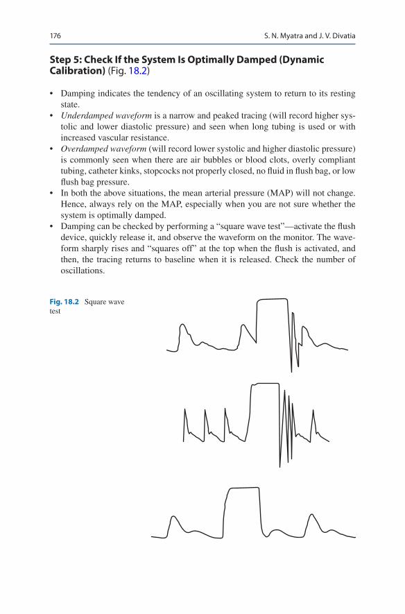
176
Step 5: Check If the System Is Optimally Damped (Dynamic Calibration) (Fig. 18.2)
• Damping indicates the tendency of an oscillating system to return to its resting state.
• Underdamped waveform is a narrow and peaked tracing (will record higher sys-tolic and lower diastolic pressure) and seen when long tubing is used or with increased vascular resistance.
• Overdamped waveform (will record lower systolic and higher diastolic pressure) is commonly seen when there are air bubbles or blood clots, overly compliant tubing, catheter kinks, stopcocks not properly closed, no fluid in flush bag, or low flush bag pressure.
• In both the above situations, the mean arterial pressure (MAP) will not change. Hence, always rely on the MAP, especially when you are not sure whether the system is optimally damped.
• Damping can be checked by performing a “square wave test”—activate the flush device, quickly release it, and observe the waveform on the monitor. The wave-form sharply rises and “squares off” at the top when the flush is activated, and then, the tracing returns to baseline when it is released. Check the number of oscillations.
Fig. 18.2 Square wave test
S. N. Myatra and J. V. Divatia
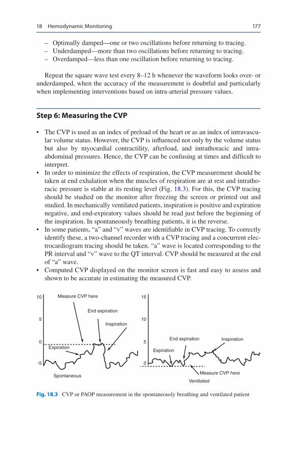
177
– Optimally damped—one or two oscillations before returning to tracing. – Underdamped—more than two oscillations before returning to tracing. – Overdamped—less than one oscillation before returning to tracing.
Repeat the square wave test every 8–12 h whenever the waveform looks over- or underdamped, when the accuracy of the measurement is doubtful and particularly when implementing interventions based on intra-arterial pressure values.
Step 6: Measuring the CVP
• The CVP is used as an index of preload of the heart or as an index of intravascu-lar volume status. However, the CVP is influenced not only by the volume status but also by myocardial contractility, afterload, and intrathoracic and intra- abdominal pressures. Hence, the CVP can be confusing at times and difficult to interpret.
• In order to minimize the effects of respiration, the CVP measurement should be taken at end exhalation when the muscles of respiration are at rest and intratho-racic pressure is stable at its resting level (Fig. 18.3). For this, the CVP tracing should be studied on the monitor after freezing the screen or printed out and studied. In mechanically ventilated patients, inspiration is positive and expiration negative, and end-expiratory values should be read just before the beginning of the inspiration. In spontaneously breathing patients, it is the reverse.
• In some patients, “a” and “v” waves are identifiable in CVP tracing. To correctly identify these, a two-channel recorder with a CVP tracing and a concurrent elec-trocardiogram tracing should be taken. “a” wave is located corresponding to the PR interval and “v” wave to the QT interval. CVP should be measured at the end of “a” wave.
• Computed CVP displayed on the monitor screen is fast and easy to assess and shown to be accurate in estimating the measured CVP.
15
10
10
5
0
-5
5
0
ExpirationExpiration
End expiration
End expiration
Inspiration
Inspiration
Measure CVP here
Measure CVP here
VentilatedSpontaneous
Fig. 18.3 CVP or PAOP measurement in the spontaneously breathing and ventilated patient
18 Hemodynamic Monitoring

178
• CVP is a simple, inexpensive, meaningful physiologic variable to measure. When used in conjunction with other hemodynamic variables, it may be useful to assess the volume status. Nevertheless, CVP does not predict fluid responsive-ness and should not be used to guide fluid administration.
Step 7: Interpret Intra-arterial Pressure Waveform (Fig. 18.4)
• The arterial pressure waveform differs at different sites. As the arterial pressure is recorded more distally, the trace gets progressively more peaked and the dicrotic notch migrates away from the peak. The MAP, however, does not vary widely as one measures more distally. Besides more accurate and real-time recording of arterial pressure, other hemodynamic interpretations can be made from the arterial waveform: – Systolic blood pressure variations (swing in the waveform) can be seen during
hypovolemia. – Steep slope of upstroke means good contractility and vice versa. – Area under the curve represents the stroke volume. – Position of the dicrotic notch—low (low systemic vascular resistance [SVR])
and high (high afterload). – Slope of the descent—steep (low SVR).
Step 8: Interpreting Pulmonary Artery Occlusion Pressure (PAOP)
The four most important measurements obtained from the pulmonary artery cathe-ter (PAC) are the following:
1. PAOP (Pulmonary artery Occlusion pressure) 2. Pulmonary artery systolic (PASP) and diastolic pressure (PADP) 3. Thermodilution cardiac output 4. Mixed venous oxygen saturation
4
1
2
3
Fig. 18.4 Components of the arterial waveform. (1) Peak systolic pressure, (2) dicrotic notch, (3) diastolic pressure, and (4) anacrotic notch
S. N. Myatra and J. V. Divatia
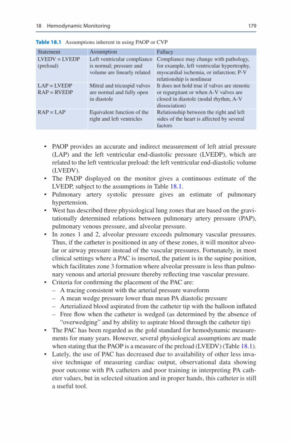
179
• PAOP provides an accurate and indirect measurement of left atrial pressure (LAP) and the left ventricular end-diastolic pressure (LVEDP), which are related to the left ventricular preload: the left ventricular end-diastolic volume (LVEDV).
• The PADP displayed on the monitor gives a continuous estimate of the LVEDP, subject to the assumptions in Table 18.1.
• Pulmonary artery systolic pressure gives an estimate of pulmonary hypertension.
• West has described three physiological lung zones that are based on the gravi-tationally determined relations between pulmonary artery pressure (PAP), pulmonary venous pressure, and alveolar pressure.
• In zones 1 and 2, alveolar pressure exceeds pulmonary vascular pressures. Thus, if the catheter is positioned in any of these zones, it will monitor alveo-lar or airway pressure instead of the vascular pressures. Fortunately, in most clinical settings where a PAC is inserted, the patient is in the supine position, which facilitates zone 3 formation where alveolar pressure is less than pulmo-nary venous and arterial pressure thereby reflecting true vascular pressure.
• Criteria for confirming the placement of the PAC are: – A tracing consistent with the arterial pressure waveform – A mean wedge pressure lower than mean PA diastolic pressure – Arterialized blood aspirated from the catheter tip with the balloon inflated – Free flow when the catheter is wedged (as determined by the absence of
“overwedging” and by ability to aspirate blood through the catheter tip)• The PAC has been regarded as the gold standard for hemodynamic measure-
ments for many years. However, several physiological assumptions are made when stating that the PAOP is a measure of the preload (LVEDV) (Table 18.1).
• Lately, the use of PAC has decreased due to availability of other less inva-sive technique of measuring cardiac output, observational data showing poor outcome with PA catheters and poor training in interpreting PA cath-eter values, but in selected situation and in proper hands, this catheter is still a useful tool.
Table 18.1 Assumptions inherent in using PAOP or CVP
Statement Assumption FallacyLVEDV = LVEDP (preload)
Left ventricular compliance is normal; pressure and volume are linearly related
Compliance may change with pathology, for example, left ventricular hypertrophy, myocardial ischemia, or infarction; P-V relationship is nonlinear
LAP = LVEDPRAP = RVEDP
Mitral and tricuspid valves are normal and fully open in diastole
It does not hold true if valves are stenotic or regurgitant or when A-V valves are closed in diastole (nodal rhythm, A-V dissociation)
RAP = LAP Equivalent function of the right and left ventricles
Relationship between the right and left sides of the heart is affected by several factors
18 Hemodynamic Monitoring
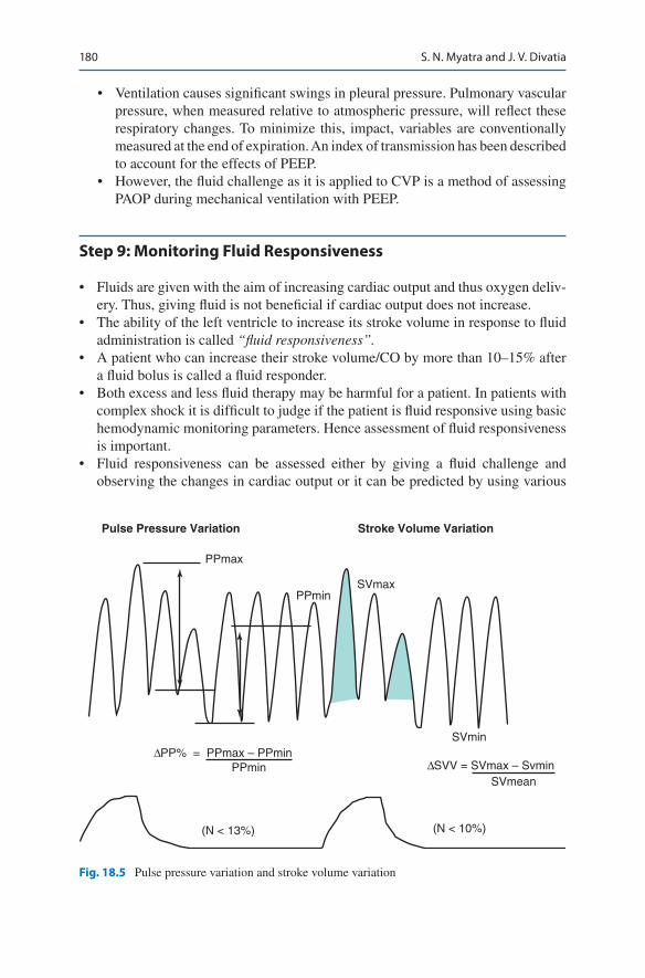
180
• Ventilation causes significant swings in pleural pressure. Pulmonary vascular pressure, when measured relative to atmospheric pressure, will reflect these respiratory changes. To minimize this, impact, variables are conventionally measured at the end of expiration. An index of transmission has been described to account for the effects of PEEP.
• However, the fluid challenge as it is applied to CVP is a method of assessing PAOP during mechanical ventilation with PEEP.
Step 9: Monitoring Fluid Responsiveness
• Fluids are given with the aim of increasing cardiac output and thus oxygen deliv-ery. Thus, giving fluid is not beneficial if cardiac output does not increase.
• The ability of the left ventricle to increase its stroke volume in response to fluid administration is called “fluid responsiveness”.
• A patient who can increase their stroke volume/CO by more than 10–15% after a fluid bolus is called a fluid responder.
• Both excess and less fluid therapy may be harmful for a patient. In patients with complex shock it is difficult to judge if the patient is fluid responsive using basic hemodynamic monitoring parameters. Hence assessment of fluid responsiveness is important.
• Fluid responsiveness can be assessed either by giving a fluid challenge and observing the changes in cardiac output or it can be predicted by using various
Pulse Pressure Variation Stroke Volume Variation
PPmax
SVmax
SVmin
(N < 13%)
∆PP% = PPmax – PPmin∆SVV = SVmax – SvminPPmin
SVmean
(N < 10%)
PPmin
Fig. 18.5 Pulse pressure variation and stroke volume variation
S. N. Myatra and J. V. Divatia
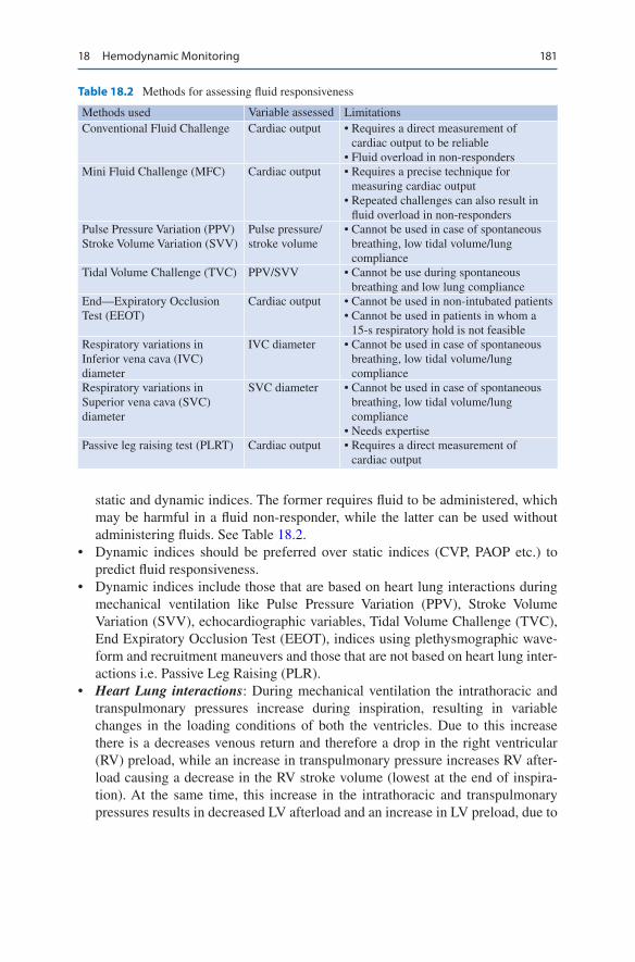
181
static and dynamic indices. The former requires fluid to be administered, which may be harmful in a fluid non-responder, while the latter can be used without administering fluids. See Table 18.2.
• Dynamic indices should be preferred over static indices (CVP, PAOP etc.) to predict fluid responsiveness.
• Dynamic indices include those that are based on heart lung interactions during mechanical ventilation like Pulse Pressure Variation (PPV), Stroke Volume Variation (SVV), echocardiographic variables, Tidal Volume Challenge (TVC), End Expiratory Occlusion Test (EEOT), indices using plethysmographic wave-form and recruitment maneuvers and those that are not based on heart lung inter-actions i.e. Passive Leg Raising (PLR).
• Heart Lung interactions: During mechanical ventilation the intrathoracic and transpulmonary pressures increase during inspiration, resulting in variable changes in the loading conditions of both the ventricles. Due to this increase there is a decreases venous return and therefore a drop in the right ventricular (RV) preload, while an increase in transpulmonary pressure increases RV after-load causing a decrease in the RV stroke volume (lowest at the end of inspira-tion). At the same time, this increase in the intrathoracic and transpulmonary pressures results in decreased LV afterload and an increase in LV preload, due to
Table 18.2 Methods for assessing fluid responsiveness
Methods used Variable assessed LimitationsConventional Fluid Challenge Cardiac output • Requires a direct measurement of
cardiac output to be reliable • Fluid overload in non-responders
Mini Fluid Challenge (MFC) Cardiac output • Requires a precise technique for measuring cardiac output
• Repeated challenges can also result in fluid overload in non-responders
Pulse Pressure Variation (PPV)Stroke Volume Variation (SVV)
Pulse pressure/stroke volume
• Cannot be used in case of spontaneous breathing, low tidal volume/lung compliance
Tidal Volume Challenge (TVC) PPV/SVV • Cannot be use during spontaneous breathing and low lung compliance
End—Expiratory Occlusion Test (EEOT)
Cardiac output • Cannot be used in non-intubated patients• Cannot be used in patients in whom a
15-s respiratory hold is not feasibleRespiratory variations in Inferior vena cava (IVC) diameter
IVC diameter • Cannot be used in case of spontaneous breathing, low tidal volume/lung compliance
Respiratory variations in Superior vena cava (SVC) diameter
SVC diameter • Cannot be used in case of spontaneous breathing, low tidal volume/lung compliance
• Needs expertisePassive leg raising test (PLRT) Cardiac output • Requires a direct measurement of
cardiac output
18 Hemodynamic Monitoring
182
the blood that is squeezed out of alveolar. This leads to an increase in LV stroke volume, which will be maximum at the end of inspiration. The reduction in RV stroke volume during inspiration leads to decreased LV which causes decreased LV stroke volume (lowest during expiration). Thus, mechanical ventilation pro-duces cyclic changes in LV stroke volume (maximum during inspiration and lowest during expiration). The magnitude of this change in LV stroke volume, or its surrogates, like pulse pressure, is magnified when the patient is preload- dependent. This principles is used in the dynamic indices that assess preload responsiveness.
• SVV and PPV (Fig. 18.5): These indices are derived from the arterial wave form analysis. The PPV is the difference between the maximum and minimum pulse pressure value over one ventilatory cycle, divided by the average value of the two values. Several hemodynamic monitors can automatically calculate and display the PPV values. The SVV is derived from the analysis of the arterial pressure waveform. This is automatically calculated by calibrated and uncalibrated pulse contour analysis cardiac output monitors.
• Tidal Volume Challenge: The dynamic indices PPV and SVV, are not reliable in predicting fluid responsiveness during low tidal volume (Vt) ventilation (Vt less than or equal to 6 mL/kg PBW).. It has been shown that PPV is a reliable predic-tor of fluid responsiveness only when the tidal volume was at least 8 mL/kg PBW. A low Vt might not be sufficient to produce significant change in the intra-thoracic pressure, making these indices indicate a nonresponsive state even in a responder. This may limit the use of PPV and SVV during low Vt ventilation. To overcome this limitation, the ‘tidal volume challenge’ has been proposed. This is a novel test to improve the reliability of PPV and SVV during low Vt ventilation. The test involves transiently increasing tidal volume from 6 mL/kg PBW to 8 mL/kg PBW for 1 min and observing the change in PPV (ΔPPV6–8) and SVV (ΔSVV6–8) from baseline to that at 8 mL/kg PBW. A change in PPV of 3.5% or SVV of 2.5%, after performing the tidal volume challenge, reliably predicted fluid responsiveness.
• Echocardiographical variables: The changes in the diameter of the superior or inferior vena cava (IVC) with respiration, or changes in aortic flow velocity can be measured. During controlled mechanical ventilation the IVC diameter is max-imum during inspiration. The respiratory variations in the diameter of the IVC are used to calculate the IVC distensibility index (calculates as maximum minus minimum diameter, divided by the minimum diameter). A cut off value of ≥18% has been shown to reliably predicts fluid responsiveness.
• Transesophageal echocardiography can be used to measure changes in the diam-eter of the SVC. A change in SV diameter more than 36% during controlled ventilation, has been shown to reliability predictive of fluid responsiveness. These echocardiographic variable have the same limitations as PPV and SVV to predict fluid responsiveness, however they are reliable in patients with arrhythmias.
• Indices Using Plethysmographic Waveform The pulse oximetry plethysmo-graphic (POP) waveform can be captured non-invasively. This depends on the
S. N. Myatra and J. V. Divatia
183
arterial pulsatility and the stroke volume. Currently commercial monitors auto-matically calculate the respiratory variation of the POP and this is called “pleth variability index” (PVI). The PVI, reliably predicts fluid responsiveness in peri-operative settings. The reliability of this index depends on good signal capture, which may not be always possible in patients who are hypothermic. In addition, it has been shown to be unreliable in patients on vasopressor Thus the applicabil-ity of these indices may be limited in the ICU patients and this variable may be more useful for hemodynamic monitoring in the operating room.
• End Expiratory Occlusion Test (EEOT). This test is performed by giving a 15 s hold at the end of expiration in a mechanically ventilated patient. During this inspiration is prevented when the hold is given, thereby preventing an increase in the intra-thoracic pressure. In addition, the increase in transpulmonary pressure during inspiration will also not occur. The resultant effect is an increase in the venous return increases, increasing ventricular preload, which results in an increased right ventricular stroke volume in a fluid responsive patient. An increase in CO of more than 5% after an EEOT reliably predicts fluid responsive-ness. This test is like giving a mini auto fluid challenge. This test has been shown to be more reliable when used with a continuous cardiac output monitor.
• Passive Leg Raising (PLR) Test These tests described above, use the heart lung interaction during mechanical ventilation to assess fluid responsiveness. However, these tests are not without limitations, their applicability in certain situations like spontaneous breathing activity, arrhythmias, low tidal volume ventilation, low lung compliance, open thorax etc. is limited. The PLR can reli-ably predict fluid responsiveness in these situations, since it does not depend on heart lung interactions. This test involves transiently raising the legs to 45° with the horizontal. This results in an auto fluid challenge, mimicking the effects of volume expansion and with the advantage of being completely reversible. A PLRT-induced changes in CO >10% reliably predicts fluid responsiveness in patients with acute circulatory failure. However, this test requires continuous cardiac output monitoring and cannot be used in patients with neurotrauma or those requiring immobilization.
• Assessing preload responsiveness before giving fluid is essential, especially fol-lowing the initial resuscitation phase. However, It is important to remember that even if a patient is preload responsive, fluid should only be given only if these is the presence of acuter circulatory shock and if there is no risk of giving fluids.
Step 10: Interpret ScvO2/SvO2
• Shock is defined as the disruption of the balance between oxygen demand (VO2) and supply (DO2) to the tissues.
• A low DO2 can be because of anemia, hypoxia, low cardiac output, or maldistri-bution of blood flow in the microcirculation.
• VO2 can be increased in sepsis and systemic inflammation.• We thus need to assess the balance between oxygen supply and demand.
18 Hemodynamic Monitoring

184
• SvO2 estimates all components of DO2. It reflects cardiac output (CO) if VO2 and Hb are constant, and most importantly, it reflects the balance between oxygen supply and demand.
• An SvO2 below 65% implies low oxygen delivery, while a value below 60% indicates that there is a serious risk of tissue hypoxia if corrective measures are not taken.
• A low SvO2 (<40%) implies critical oxygen supply–demand imbalance.• If SvO2 is high (>80%), then either the demand has declined, the O2 supply has
increased, or the cells are unable to utilize the oxygen.• Thus, a falling or low SvO2 is an important indicator that the oxygen delivery is
compromised and is deficient relative to the needs of the tissues.• The measurement of mixed venous oxygen saturation (SvO2) from the pulmo-
nary artery is an indirect index of tissue oxygenation.• However, sampling of mixed venous blood requires insertion of a PAC, which is
an invasive procedure with risks, and is not universally used. An alternative is to measure ScvO2. Central venous catheterization is a simpler and safer procedure and is commonly used.
• In this case, a catheter is positioned in the superior vena cava or the upper right atrium.
• In circulatory shock, the ScvO2 is generally greater than SvO2 with a difference of 5–18%.
• Following the results of recent large, randomized controlled trials, achieving an ScvO2 more than 70% during resuscitation within 6 h of septic shock, is no lon-ger recommended as a goal of resuscitation. However the ScvO2 can still be used to determine whether resuscitation is adequate if the ScvO2 has been low initially.
Step 11: Interpret Lactate Levels
• Lactate levels often reflect anaerobic metabolism due to tissue hypoxia.• High and rising levels (>2 mmol/L) has adverse prognosis, while falling lactate
levels indicate an adequate response to resuscitation of the shocked patient.• Lactate levels may increase due to increased production due to global or local
tissue hypoxia but also due to stimulation of glycolysis and metabolic pathways that accelerate lactate formation in sepsis. Adrenaline infusions can increase lac-tate levels by accelerating glycolysis.
• High lactate levels may also represent decreased clearance due to reduced liver blood flow or hepatic dysfunction.
• Thus, interpretation of lactate levels may be complicated.• Also, arterial lactate levels are a global measure, and regional hypoperfusion of
some vascular beds may exist even in the presence of normal lactate levels.• In patients with sepsis, a lactate level > 4 mmol/L may be considered as a marker
of tissue hypoperfusion and is a trigger for the rapid resuscitation including com-mencement of administration of 30 mL/kg of fluid.
S. N. Myatra and J. V. Divatia
185
The current Surviving Sepsis Campaign guidelines suggest that if initial lactate is elevated (> 2 mmol/L), it should be remeasured within 2–4 h to guide resuscita-tion and to normalise lactate in patients with elevated lactate levels.
Step 12: Integrate Findings
The goal of hemodynamic monitoring and therapy is to target adequate organ perfu-sion and oxygen delivery while minimizing any the side-effects of any interventions used to obtain these targets. No monitoring tool or device is perfect. Hence a com-bination of hemodynamic monitoring devices, integration of the haemodynamic variables and correlation with the clinical condition of the patient is essential.
Suggested Reading
De Backer D, Bakker J, Cecconi M, Hajjar L, Liu DW, Lobo S, Monnet X, Morelli A, Myatra SN, Perel A, Pinsky MR, Saugel B, Teboul JL, Vieillard-Baron A, Vincent JL. Alternatives to the Swan-Ganz catheter. Intensive Care Med. 2018;44(6):730–41. It is useful to combine different techniques instead of relying on a single one and to adapt the "package" of interventions to the condition of the patient.
De Backer D, Cecconi M, Lipman J, Machado F, Myatra SN, Ostermann M, Perner A, Teboul JL, Vincent JL, Walley KR. Challenges in the management of septic shock: a narrative review. Intensive Care Med. 2019;45(4):420–33. A review article
Jozwiak M, Monnet X, Teboul JL. Less or more hemodynamic monitoring in critically ill patients. Curr Opin Crit Care. 2018;24(4):309–15. This article reviews the commercially available minimally invasive CO monitoring devices, their technical characteristics and discusses the limitations
Myatra SN, Prabu NR, Divatia JV, Monnet X, Kulkarni AP, Teboul JL. The Changes in pulse pres-sure Variation or stroke volume variation after a “tidal volume challenge” reliably predict fluid responsiveness during low tidal volume ventilation. Crit Care Med. 2017;45(3):415–21. The changes in pulse pressure variation or stroke volume variation by transiently increasing tidal volume (tidal volume challenge from 6 to 8 ml) are superior to pulse pressure variation and stroke volume variation in predicting fluid responsiveness during lung protective ventilation using low tidal volume
Rhodes A, Evans LE, Alhazzani W, Levy MM, Antonelli M, Ferrer R, et al. Surviving Sepsis Campaign: International Guidelines for Management of Sepsis and Septic Shock: 2018. Intensive Care Med. 2017;43(3):304–77. A review article on the management of sepsis and septic shock
18 Hemodynamic Monitoring

187© Springer Nature Singapore Pte Ltd. 2020R. Chawla, S. Todi (eds.), ICU Protocols, https://doi.org/10.1007/978-981-15-0898-1_19
R. Pandit (*) Department of Intensive Care, Fortis Hospital, Mumbai, India
19Echocardiography
Rahul Pandit
Noninvasive, echocardiographic point-of-care monitoring has increasingly been used in ICUs. This has advantage of easy and quick availability, repeatability, and avoiding transportation of an unstable patient.
Intensivists should be familiar with basic echocardiography, and use this as a basic screening tool for rapid assessment of the circulation in hemodynamically unstable patients as an extension of clinical examination. The rest of the examination should be undertaken by intensivists or clinician who have had advanced training in echocar-diography. Caution should however be exercised during interpretation of data pro-vided and patient management should be individualized within the clinical context.
Step 1: Urgent Assessment and Resuscitation
• While resuscitation efforts are underway, a quick assessment with bedside echo-cardiogram can guide the clinician in rationalizing the use of volume resuscita-tion, inotropes, and vasopressors. This can be repeated to assess the response to therapy.
• Using the bedside echocardiogram, one can assess the following: – The left ventricular (LV) function – Right ventricular (RV) function – Presence of pericardial effusion and tamponade
A 55 years old male patient is admitted to intensive care with a history of short-ness of breath since last 1 day, and temp 39.7° C on admission. His pulse rate is 104/min, Blood Pressure- 92/45 mmHg, X-ray chest is suggestive of bilateral costo-phrenic angle obliteration and increased broncho vascular markings. ECG is showing T wave inversion in lead II, III and aVF. Echocardiography is ordered.

188
– Preload assessment and fluid responsiveness – Valve lesions
Step 2: Understand the Limitation of Bedside Echo in the ICU
• ICU echos are often different: – Patients are often supine, ventilated, and unconscious. – Other equipment can affect examination. – The situation may be dynamic with concurrent resuscitation underway. – Repeated studies may be required to determine the efficacy of treatment.
Step 3: Familiarize with Practical Aspects of Bedside Echo (Fig. 19.1a–d)
Echocardiography has four basic views.
1. Parasternal long axis 2. Parasternal short axis (papillary muscle level, mitral level, aortic level, and apex) 3. Apical view—four-chamber, five-chamber, and two-chamber view 4. Subcostal view
a b
c d
Fig. 19.1 Four basic views: (a) left parasternal long axis, (b) left parasternal short axis, (c) apical four chambers, and (d) subcostal
R. Pandit

189
Step 4: Assess Left Ventricle (Table 19.1)
• This is best viewed in Parasternal long axis, short axis, and apical views.• Focused left-sided heart examination
– Size—small, normal, or dilated – Contractility—normal, decreased, hyperdynamic – Left ventricular wall thickness – Aortic valve appearance, mitral valve appearance – Segmental wall motion abnormality
• Size and wall thickness – M-mode performed in the parasternal long axis measuring perpendicular to
LV through the mitral valve leaflet will give a reasonable estimation of LV systolic and diastolic diameter and interventricular septum and LV posterior wall thickness.
• Left atrium and aortic root – It is measured by M-mode at the level of the aortic valve. This will give an
estimation of left atrium (LA) size and aortic root.• Left ventricular wall thickness:
– Presence of significant Left Ventricular Hypertrophy (LVH) points to likely diastolic dysfunction.
– Systolic wall thickening is a guide to segmental wall motion defects.• Contractility
Table 19.1 Normal echo values
Size and wall thicknessM-mode performed in the parasternal long axis measuring perpendicular to LV through the mitral valve leaflet will give a reasonable estimation of LV systolic and diastolic diameter and interventricular septum and LV posterior wall thickness.Dimensions Normal rangeEnd-diastolic LVID 40–56 mmEnd-systolic LVID 20–38 mmEnd-diastolic IVS and PW thickness <11 mmLeft atrium and aortic root It can be measured by M-mode at the level of aortic valve and will give an estimation of LA size and aortic root.LA normal range: <40 mmAortic root normal range: ≤25–30 mmEjection fraction (EF)Normal contractility—EF > 50%Hyperdynamic—EF > 70%Low—EF < 50%
19 Echocardiography
190
The left ventricle not only contracts in radial direction, but also has a shortening of its length and a circumferential motion as well. The inward motion of endocar-dium and thickening of myocardium during LV systole is most often used to deter-mine the wall motion.
Eyeballing
The most common way of assessment is eyeballing. It has been shown in numerous studies that an experienced ehocardiographer will reasonably assess the LV function with eyeballing and give a reasonable estimate of its wall motion and ejection fraction.
Following helps in assessment during eyeballing.
• Inward motion of the endocardium• Thickening of the myocardium• Longitudinal motion of the mitral annulus• Geometry of the ventricle• Regional wall motion abnormality (RWMA) means motions of a particular
region of the heart is found to be abnormal on echocardiography. Different types of regional wall motion abnormalities are, – Hypokinesia: Reduced contraction of a region of the heart muscle. – Akinesia: A region of the heart muscle is not contracting at all. – Hyperkinesia: A region of the heart muscle is contracting more vigorously
than normal. – Dyskinesia: A region of the heart muscle bulges out when the rest of the heart
is contracting
The different regions of the heart are divided as per the arterial territories (Fig. 19.2). Parasternal short axis gives a quick guide on RWMA for all three territories.
LV Function Assessment
• Usually the assessment is done and reported as Hyperdynamic, Good LV Function, Moderate LV Dysfunction or Poor or severe LV dysfunction.
• Most echocardiographs are able to assess and give a number on basis of eyeball-ing as well. However a more objective way is to assess the LV systolic function with various techniques available like Fractional Shortening, Simpsons method, three dimensional echocardiography and dp/dt method. The most common used is Simpsons method.
R. Pandit
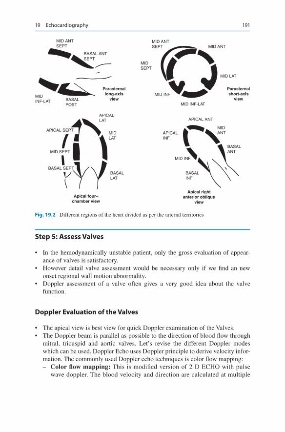
191
Step 5: Assess Valves
• In the hemodynamically unstable patient, only the gross evaluation of appear-ance of valves is satisfactory.
• However detail valve assessment would be necessary only if we find an new onset regional wall motion abnormality.
• Doppler assessment of a valve often gives a very good idea about the valve function.
Doppler Evaluation of the Valves
• The apical view is best view for quick Doppler examination of the Valves.• The Doppler beam is parallel as possible to the direction of blood flow through
mitral, tricuspid and aortic valves. Let’s revise the different Doppler modes which can be used. Doppler Echo uses Doppler principle to derive velocity infor-mation. The commonly used Doppler echo techniques is color flow mapping:
– Color flow mapping: This is modified version of 2 D ECHO with pulse wave doppler. The blood velocity and direction are calculated at multiple
MID ANTSEPT
BASAL ANTSEPT
MIDINF-LAT BASAL
POST
Parasternallong-axis
view
APICALLAT APICAL ANT
MIDLAT
MID INF
MIDANTAPICAL
INF
MID INF
MID INF-LAT
MID LAT
Parasternalshort-axis
view
Apical rightanterior oblique
viewApical four–
chamber view
MID ANTSEPT MID ANT
MIDSEPT
BASALLAT
BASALANT
BASALINF
APICAL SEPT
MID SEPT
BASAL SEPT
Fig. 19.2 Different regions of the heart divided as per the arterial territories
19 Echocardiography
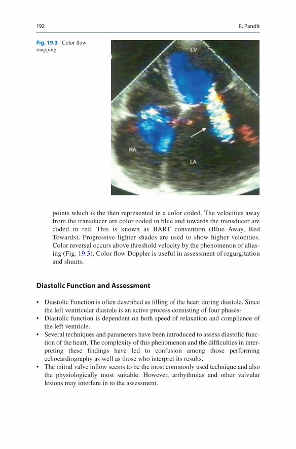
192
points which is the then represented in a color coded. The velocities away from the transducer are color coded in blue and towards the transducer are coded in red. This is known as BART convention (Blue Away, Red Towards). Progressive lighter shades are used to show higher velocities. Color reversal occurs above threshold velocity by the phenomenon of alias-ing (Fig. 19.3). Color flow Doppler is useful in assessment of regurgitation and shunts.
Diastolic Function and Assessment
• Diastolic Function is often described as filling of the heart during diastole. Since the left ventricular diastole is an active process consisting of four phases-
• Diastolic function is dependent on both speed of relaxation and compliance of the left ventricle.
• Several techniques and parameters have been introduced to assess diastolic func-tion of the heart. The complexity of this phenomenon and the difficulties in inter-preting these findings have led to confusion among those performing echocardiography as well as those who interpret its results.
• The mitral valve inflow seems to be the most commonly used technique and also the physiologically most suitable. However, arrhythmias and other valvular lesions may interfere in to the assessment.
Fig. 19.3 Color flow mapping
R. Pandit
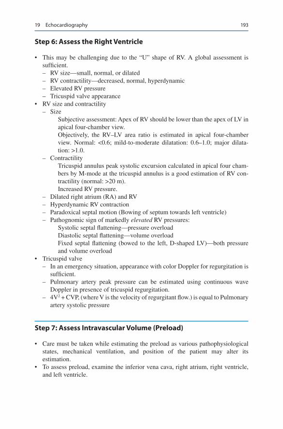
193
Step 6: Assess the Right Ventricle
• This may be challenging due to the “U” shape of RV. A global assessment is sufficient. – RV size—small, normal, or dilated – RV contractility—decreased, normal, hyperdynamic – Elevated RV pressure – Tricuspid valve appearance
• RV size and contractility – Size
Subjective assessment: Apex of RV should be lower than the apex of LV in apical four-chamber view.Objectively, the RV–LV area ratio is estimated in apical four-chamber view. Normal: <0.6; mild-to-moderate dilatation: 0.6–1.0; major dilata-tion: >1.0.
– ContractilityTricuspid annulus peak systolic excursion calculated in apical four cham-bers by M-mode at the tricuspid annulus is a good estimation of RV con-tractility (normal: >20 m).Increased RV pressure.
– Dilated right atrium (RA) and RV – Hyperdynamic RV contraction – Paradoxical septal motion (Bowing of septum towards left ventricle) – Pathognomic sign of markedly elevated RV pressures:
Systolic septal flattening—pressure overloadDiastolic septal flattening—volume overloadFixed septal flattening (bowed to the left, D-shaped LV)—both pressure and volume overload
• Tricuspid valve – In an emergency situation, appearance with color Doppler for regurgitation is
sufficient. – Pulmonary artery peak pressure can be estimated using continuous wave
Doppler in presence of tricuspid regurgitation. – 4V2 + CVP, (where V is the velocity of regurgitant flow.) is equal to Pulmonary
artery systolic pressure
Step 7: Assess Intravascular Volume (Preload)
• Care must be taken while estimating the preload as various pathophysiological states, mechanical ventilation, and position of the patient may alter its estimation.
• To assess preload, examine the inferior vena cava, right atrium, right ventricle, and left ventricle.
19 Echocardiography
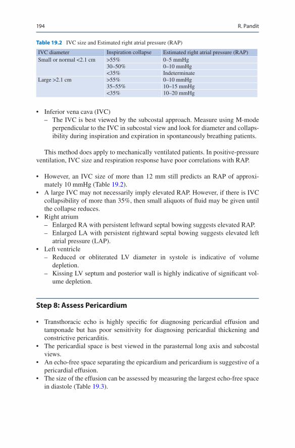
194
• Inferior vena cava (IVC) – The IVC is best viewed by the subcostal approach. Measure using M-mode
perpendicular to the IVC in subcostal view and look for diameter and collaps-ibility during inspiration and expiration in spontaneously breathing patients.
This method does apply to mechanically ventilated patients. In positive-pressure ventilation, IVC size and respiration response have poor correlations with RAP.
• However, an IVC size of more than 12 mm still predicts an RAP of approxi-mately 10 mmHg (Table 19.2).
• A large IVC may not necessarily imply elevated RAP. However, if there is IVC collapsibility of more than 35%, then small aliquots of fluid may be given until the collapse reduces.
• Right atrium – Enlarged RA with persistent leftward septal bowing suggests elevated RAP. – Enlarged LA with persistent rightward septal bowing suggests elevated left
atrial pressure (LAP).• Left ventricle
– Reduced or obliterated LV diameter in systole is indicative of volume depletion.
– Kissing LV septum and posterior wall is highly indicative of significant vol-ume depletion.
Step 8: Assess Pericardium
• Transthoracic echo is highly specific for diagnosing pericardial effusion and tamponade but has poor sensitivity for diagnosing pericardial thickening and constrictive pericarditis.
• The pericardial space is best viewed in the parasternal long axis and subcostal views.
• An echo-free space separating the epicardium and pericardium is suggestive of a pericardial effusion.
• The size of the effusion can be assessed by measuring the largest echo-free space in diastole (Table 19.3).
Table 19.2 IVC size and Estimated right atrial pressure (RAP)
IVC diameter Inspiration collapse Estimated right atrial pressure (RAP)Small or normal <2.1 cm >55% 0–5 mmHg
30–50% 0–10 mmHg<35% Indeterminate
Large >2.1 cm >55% 0–10 mmHg35–55% 10–15 mmHg<35% 10–20 mmHg
R. Pandit

195
• Signs of tamponade – Right atrium—early systolic collapse. – Right ventricle—early diastolic collapse. – IVC diameter and collapsibility—dilated and/or fixed.
Lung Ultrasound
• No assessment is complete unless a lung ultrasound is completed.• Coupled with IVC assessment, lung ultrasound gives an very comprehensive
assessment of the haemodynamic status.
Technique of Lung Ultrasound
• A range of frequencies (4 to 12 MHz) can be used to visualize the lungs. High frequencies are useful to look at the periphery of the lung with a high resolution as in looking for ‘lung sliding’ and other signs of pneumothorax, as well as studying lung comets.
• Lower frequencies help with the imaging of deep lung tissues as in looking at consolidation and pleural effusion. Hence a vascular probe is used for the assess-ment of pneumothorax, while the ultrasound probe is used for consolidation and pleural effusion.
• In the supine position, the anterior and lateral lung areas can be easily scanned, but the patient may have to be turned to a lateral decubitus position for scanning posteriorly.
• Six regions, delineated by the anterior and posterior axillary lines should be sys-tematically examined: upper and lower parts of the anterior, lateral and posterior chest wall.
• When an ultrasound transducer is laid on a normal chest wall, the following is observed: (Fig. 19.4).
• A lines: Horizontal, regularly spaced hyperechogenic lines representing rever-berations of the pleural line. These are motionless and are artifacts of repetition. In two-thirds of normal lungs, this is the only artifact pattern that can be seen.
• B lines: Vertical narrow based lines arising from the pleural line to the edge of the ultrasound screen. The “comet-tail image” (Ultrasound Lung Comets, ULC) is a sonographic image detectable at the bedside with ultrasound probe positioned
Table 19.3 Severity of the pericardial effusion and the echo-free space in diastole
Severity CharacteristicsPhysiological End-systolic separation of epi- and pericardium posteriorlySmall Echo-free space of ≤10 mmModerate Echo-free space of 10–20 mm surrounding the entire heartLarge Echo-free space of >20 mm around the entire heart
19 Echocardiography

196
over the chest. It is defined as a hyper echogenic, coherent bundle with a narrow base spreading from the transducer to the further border of the screen. It extends to the edge of the screen (short comet-tail artifacts may exist in other regions), and arises only from the pleural line. These are also called by the descriptive term “comet tail artifacts”. When several B lines are visible, the term used is “lung rockets”.
• B Lines are often pathological and represent acute intestitial syndrome.• A new B line appearing in a patient who is been resuscitated is often a good sign
to stop giving any further fluids.
Suggested Reading
Beaulieu Y, Marik PE. Bed side ultrasonography in ICU part-II. Chest. 2005;128:1766–81. A com-prehensive review on the subject
Brennan JM, Blair JE, Goonewardena S, Ronan A, Shah D, Vasaiwala S, Kirkpatrick JN, Spencer KT. Reappraisal of the use of IVC for estimating RAP. JASE. 2007;20(7):857–61. An article on the reappraisal of IVC
Jones R, Blaivas M. The handbook of ultrasound in trauma and critical illness. Ohio: ACEP; 2003.Wang XF, Deng YB, Nanda NC, et al. Live three-dimensional echocardiography: imaging princi-
ples and clinical application. Echocardiography. 2003;20:593. Live three-dimensional echocar-diography (L3DE) is an great breakthrough. It will provide a great potential tool for diagnosis and treatment of clinical conditions
Websites
www.asecho.org. Homepage for American Society of Echocardiographywww.niccer.org.au. Website of Nepean hospital ICUwww.criticalecho.com/content/icu-echo-tutorialswww.sonoguide.com/cardiac.html. Excellent virtual illustration of bedside echo windows
Fig. 19.4 Lung ultrasound A line and B lines
R. Pandit
197© Springer Nature Singapore Pte Ltd. 2020R. Chawla, S. Todi (eds.), ICU Protocols, https://doi.org/10.1007/978-981-15-0898-1_20
J. V. Divatia (*) · S. N. Myatra Department of Anaesthesiology, Critical Care and Pain, Tata Memorial Hospital, Mumbai, India
20Fluid Therapy, Vasopressors, and Inotropes
Jigeeshu V. Divatia and Sheila Nainan Myatra
Fluids (crystalloid or colloid), vasopressors, and inotropes are common interven-tions in managing hemodynamically unstable patients in the ICU. These interven-tions should be used judiciously to maintain perfusion to vital organs while definitive therapy is being instituted. On the other hand, if they are used injudiciously, these interventions may be potentially harmful. Since only about 50% patients with sepsis are fluid responsive (i.e., increase the stroke volume in response to a fluid bolus), fluid should be given only to hemodynamically unstable patients who are fluid responsive, and in whom there is little risk of giving fluid.
Fluid therapy can be considered to take place in four phases, termed the SOSD phases: Salvage or resuscitation, Optimisation, Stabilisation and De-escalation (Fig. 20.1).
A 55-year-old male patient presented with acute respiratory distress. His heart rate was 140/min, BP was 70/40 mmHg, and respiratory rate was 34 breaths/min. Core temperature was 34.4 °C. He was oliguric, and the abdomen was ten-der, firm, and distended. Fluid resuscitation was commenced and 1.5 L normal saline was infused within 1 h. The PaO2/FiO2 ratio was 140. He was intubated and mechanically ventilated with a tidal volume 400 mL, respiratory rate 16/min, FiO2 0.7 and PEEP 15. An arterial line and a central venous catheter were inserted, and the central venous pressure (CVP) was 16 mmHg. He remained tachycardic and hypotensive.

198
Step 1: Phase 1—Salvage
• In patients with new-onset hypotension, tachycardia, unexplained oliguria, or other evidence of hypoperfusion such as decreased capillary refill, increased lactate or base deficit, commence fluid resuscitation, if this seems to be of low risk clinically, for example, patients with no evidence of overt heart failure.
• Administer 500–1000 mL crystalloid over 20–30 min.• In patients with sepsis, balanced crystalloids are the preferred initial resuscita-
tion fluid. Upto 30 mL/kg fluid may be given over the first 3 h.• If hypotension is severe, vasopressors may be started along with fluid
resuscitation.• Clinical monitoring of heart rate, blood pressure and tissue perfusion may be all
that is required in this phase.
Selecting Fluid for Resuscitation
• Balanced crystalloids are preferred as the initial fluid for resuscitation preferable to normal saline for large volume resuscitation.
• Infusion of large volumes of normal saline is associated with hyperchloremic acidosis and worsening renal function.
• If a colloid is used as part of initial resuscitation, 4% albumin or 5% albumin should be preferred.
• Hydroxyethyl starch (HES) solutions (Including the newer low molecular weight starch preparations) should not be used as their use has been shown to result in worst renal outcomes as well as mortality in patients with sepsis and septic shock. There is no data on the safety of gelatins in this group of patients.
Step 2: Phase 2—Optimisation
• Following initial fluid resuscitation, additional fluids be guided by frequent reas-sessment of hemodynamic status.
• Dynamic variables (pulse pressure variation, stroke volume variation, inferior vena cava distensibility in mechanically ventilated patients) should be preferred over static variables to predict fluid responsiveness, where available
Salvage Optimisation Stabilisation
Hemodynamic Monitoring
Phases of Fluid Resuscitation
De-escalation
Fig. 20.1 Phases of fluid resuscitation
J. V. Divatia and S. N. Myatra

199
• Fluid challenge in the ICU should be protocolized with clear direction about the various components of fluid challenge—type of fluid, rate of administration, clinical and pressure end points, and safety limits (see Chap. 18, Vol. 1).
• These parameters need to be individualized depending on patients’ clinical sta-tus, comorbidity, and underlying pathology.
• Careful monitoring of patients, clinically and hemodynamically, is mandatory during fluid challenge to assess fluid response as well as evidence of fluid overload.
Assess Response
• Response to a fluid challenge should be assessed clinically by features of increased perfusion like improved sensorium, increased sense of well-being, and increased urine output.
• This should also be assessed by improvement of hemodynamic parameters such as decreased tachycardia, improved blood pressure, and improved CVP or wedge pressure.
• Static measures of preload such as CVP or pulmonary artery occlusion pressure (PAOP) do not adequately reflect need for fluid challenges. There are two princi-pal reasons for this realization: – As per Frank–Starling law, preload (CVP or PAOP) is related to stroke vol-
ume or cardiac output in a curvilinear manner; that is, rise in preload will result in increasing stroke volume in the steep part of the curve till the flat part is reached, after which increasing preload will not lead to further increase in stroke volume. In a given patient, it is difficult to predict the position of a preload measure on this curve by static values as it is dependent on ventricular compliance which is variable.(Fig. 20.2)
– Left ventricular compliance varies among patients and may vary at different times in the same patient, so the pressure–volume curves are variable; thus, a stiff ventricle (hypertrophy) will lead to a decrease in ventricular compliance with shift of pressure–volume curves of the ventricle to the left. It means high pressure values (CVP or PAOP) for the same or low ventricular volume, whereas a more compliant (dilated) ventricle will shift this curve to the right; thus, a low pressure (CVP or PAOP) reading may indicate a high ventricular volume. Thus, it is difficult to predict ventricular volume by a given pressure index (CVP or PAOP).
• Dynamic indices such as pulse pressure variation (PPV), systolic pressure varia-tion (SPV) or stroke volume variation (SVV), using a Tidal Volume Challenge in patients receiving tidal volumes ≤6 mL/kg, echocardiographic vena cava diam-eter, or esophageal Doppler aortic blood flow changes during controlled positive- pressure ventilation or during passive leg raising in spontaneously breathing patients are more representative for predicting fluid responsiveness (see Chap. 16, Vol. 1).
20 Fluid Therapy, Vasopressors, and Inotropes

200
• When the risk of fluid challenge is not trivial, for example, in patients with com-promised lung or cardiac function, consider using a dynamic predictor to guide fluid boluses.
• Since the hemodynamic state changes rapidly, reassessment of hemodynamics should be done frequently.
Step 3: Phase 3—Stabilisation
• Continue to monitor and titrate fluids and vasoactive therapy till the patient’s hemodynamics are stabilized, the phase of Stabilisation
Step 4: Select Inotrope or Vasopressors
• Despite adequate volume replacement, if the patient is hypotensive and perfusion of vital organs is jeopardized, vasoactive agents may be administered to improve cardiac output and blood pressure.
• It is useful to understand the receptors through which adrenergic agents exert their effect.
Preload
SV
Increase in SV
No increase in SV
A B
C D
Fig. 20.2 Frank-Starling Curve of the heart. When the preload is increased from point A to point B on the steep part of the curve, stroke volume increases. With a similar increase in preload on the flat part of the curve, from point c to point D, there is a negligibler increase in stroke volume. SV-stroke volume
J. V. Divatia and S. N. Myatra
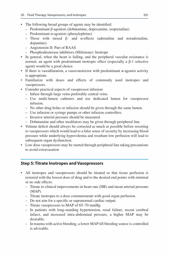
201
• The following broad groups of agents may be identified: – Predominant β-agonists (dobutamine, dopexamine, isoprenaline) – Predominant α-agonists (phenylephrine) – Those with mixed β- and α-effects (adrenaline and noradrenaline,
dopamine). – Angiotensin II: Part of RAAS – Phosphodiesterase inhibitors (Milrinone): Inotrope
• In general, when the heart is failing, and the peripheral vascular resistance is normal, an agent with predominant inotropic effect (especially a β-1 selective agent) would be a good choice.
• If there is vasodilatation, a vasoconstrictor with predominant α-agonist activity is appropriate.
• Familiarize with doses and effects of commonly used inotropes and vasopressors.
• Consider practical aspects of vasopressor infusion: – Infuse through large veins preferably central veins. – Use multi-lumen catheters and use dedicated lumen for vasopressor
infusion. – No other drug bolus or infusion should be given through the same lumen. – Use infusion or syringe pumps or other infusion controllers. – Invasive arterial pressure should be measured. – Dobutamine and other inodilators may be given through peripheral line.
• Volume deficit should always be corrected as much as possible before resorting to vasopressors which would lead to a false sense of security by increasing blood pressure while underlying hypovolemia and resultant low perfusion will lead to subsequent organ dysfunction.
• Low dose vasopressors may be started through peripheral line taking precautions to avoid extravasation
Step 5: Titrate Inotropes and Vasopressors
• All inotropes and vasopressors should be titrated so that tissue perfusion is restored with the lowest dose of drug and to the desired end points with minimal or no side effects: – Titrate to clinical improvements in heart rate (HR) and mean arterial pressure
(MAP). – Titrate inotropes to a dose commensurate with good organ perfusion. – Do not aim for a specific or supranormal cardiac output. – Titrate vasopressors to MAP of 65–70 mmHg. – In patients with long-standing hypertension, renal failure, recent cerebral
infarct, and increased intra-abdominal pressure, a higher MAP may be desirable.
– In trauma with active bleeding, a lower MAP till bleeding source is controlled is advisable.
20 Fluid Therapy, Vasopressors, and Inotropes
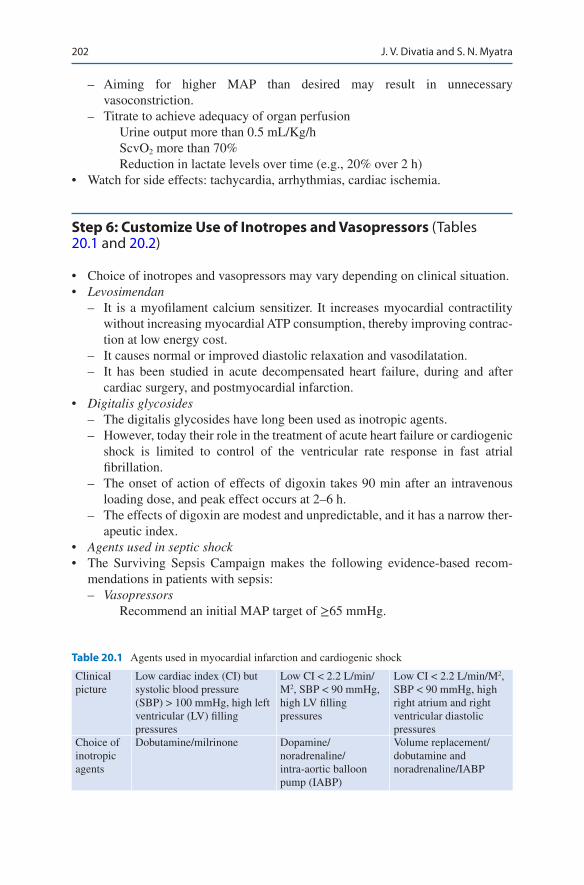
202
– Aiming for higher MAP than desired may result in unnecessary vasoconstriction.
– Titrate to achieve adequacy of organ perfusionUrine output more than 0.5 mL/Kg/hScvO2 more than 70%Reduction in lactate levels over time (e.g., 20% over 2 h)
• Watch for side effects: tachycardia, arrhythmias, cardiac ischemia.
Step 6: Customize Use of Inotropes and Vasopressors (Tables 20.1 and 20.2)
• Choice of inotropes and vasopressors may vary depending on clinical situation.• Levosimendan
– It is a myofilament calcium sensitizer. It increases myocardial contractility without increasing myocardial ATP consumption, thereby improving contrac-tion at low energy cost.
– It causes normal or improved diastolic relaxation and vasodilatation. – It has been studied in acute decompensated heart failure, during and after
cardiac surgery, and postmyocardial infarction.• Digitalis glycosides
– The digitalis glycosides have long been used as inotropic agents. – However, today their role in the treatment of acute heart failure or cardiogenic
shock is limited to control of the ventricular rate response in fast atrial fibrillation.
– The onset of action of effects of digoxin takes 90 min after an intravenous loading dose, and peak effect occurs at 2–6 h.
– The effects of digoxin are modest and unpredictable, and it has a narrow ther-apeutic index.
• Agents used in septic shock• The Surviving Sepsis Campaign makes the following evidence-based recom-
mendations in patients with sepsis: – Vasopressors
Recommend an initial MAP target of ≥65 mmHg.
Table 20.1 Agents used in myocardial infarction and cardiogenic shock
Clinical picture
Low cardiac index (CI) but systolic blood pressure (SBP) > 100 mmHg, high left ventricular (LV) filling pressures
Low CI < 2.2 L/min/M2, SBP < 90 mmHg, high LV filling pressures
Low CI < 2.2 L/min/M2, SBP < 90 mmHg, high right atrium and right ventricular diastolic pressures
Choice of inotropic agents
Dobutamine/milrinone Dopamine/noradrenaline/intra-aortic balloon pump (IABP)
Volume replacement/dobutamine and noradrenaline/IABP
J. V. Divatia and S. N. Myatra
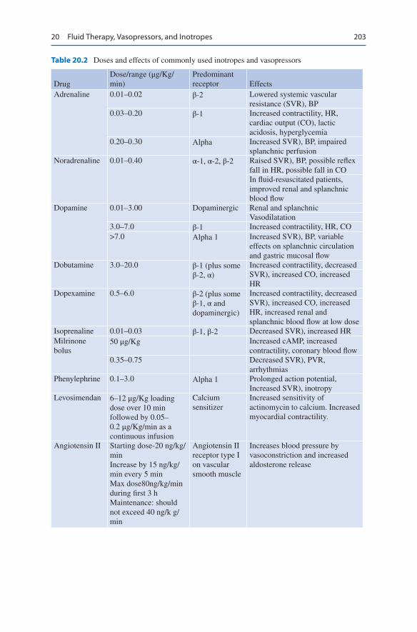
203
Table 20.2 Doses and effects of commonly used inotropes and vasopressors
DrugDose/range (μg/Kg/min)
Predominant receptor Effects
Adrenaline 0.01–0.02 β-2 Lowered systemic vascular resistance (SVR), BP
0.03–0.20 β-1 Increased contractility, HR, cardiac output (CO), lactic acidosis, hyperglycemia
0.20–0.30 Αlpha Increased SVR), BP, impaired splanchnic perfusion
Noradrenaline 0.01–0.40 α-1, α-2, β-2 Raised SVR), BP, possible reflex fall in HR, possible fall in COIn fluid-resuscitated patients, improved renal and splanchnic blood flow
Dopamine 0.01–3.00 Dopaminergic Renal and splanchnicVasodilatation
3.0–7.0 β-1 Increased contractility, HR, CO>7.0 Αlpha 1 Increased SVR), BP, variable
effects on splanchnic circulation and gastric mucosal flow
Dobutamine 3.0–20.0 β-1 (plus some β-2, α)
Increased contractility, decreasedSVR), increased CO, increased HR
Dopexamine 0.5–6.0 β-2 (plus some β-1, α and dopaminergic)
Increased contractility, decreasedSVR), increased CO, increased HR, increased renal and splanchnic blood flow at low dose
Isoprenaline 0.01–0.03 β-1, β-2 Decreased SVR), increased HRMilrinone bolus
50 μg/Kg Increased cAMP, increased contractility, coronary blood flow
0.35–0.75 Decreased SVR), PVR, arrhythmias
Phenylephrine 0.1–3.0 Αlpha 1 Prolonged action potential, Increased SVR), inotropy
Levosimendan 6–12 μg/Kg loading dose over 10 min followed by 0.05–0.2 μg/Kg/min as a continuous infusion
Calcium sensitizer
Increased sensitivity of actinomycin to calcium. Increased myocardial contractility.
Angiotensin II Starting dose-20 ng/kg/minIncrease by 15 ng/kg/min every 5 minMax dose80ng/kg/min during first 3 hMaintenance: should not exceed 40 ng/k g/min
Angiotensin II receptor type I on vascular smooth muscle
Increases blood pressure by vasoconstriction and increased aldosterone release
20 Fluid Therapy, Vasopressors, and Inotropes
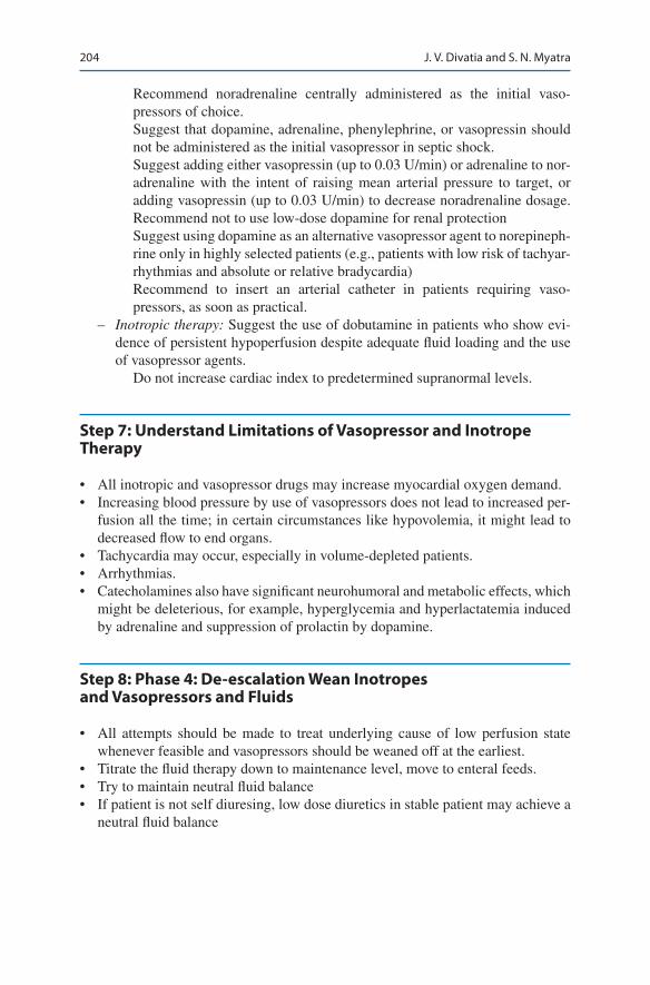
204
Recommend noradrenaline centrally administered as the initial vaso- pressors of choice.Suggest that dopamine, adrenaline, phenylephrine, or vasopressin should not be administered as the initial vasopressor in septic shock.Suggest adding either vasopressin (up to 0.03 U/min) or adrenaline to nor-adrenaline with the intent of raising mean arterial pressure to target, or adding vasopressin (up to 0.03 U/min) to decrease noradrenaline dosage. Recommend not to use low-dose dopamine for renal protectionSuggest using dopamine as an alternative vasopressor agent to norepineph-rine only in highly selected patients (e.g., patients with low risk of tachyar-rhythmias and absolute or relative bradycardia)Recommend to insert an arterial catheter in patients requiring vaso- pressors, as soon as practical.
– Inotropic therapy: Suggest the use of dobutamine in patients who show evi-dence of persistent hypoperfusion despite adequate fluid loading and the use of vasopressor agents.
Do not increase cardiac index to predetermined supranormal levels.
Step 7: Understand Limitations of Vasopressor and Inotrope Therapy
• All inotropic and vasopressor drugs may increase myocardial oxygen demand.• Increasing blood pressure by use of vasopressors does not lead to increased per-
fusion all the time; in certain circumstances like hypovolemia, it might lead to decreased flow to end organs.
• Tachycardia may occur, especially in volume-depleted patients.• Arrhythmias.• Catecholamines also have significant neurohumoral and metabolic effects, which
might be deleterious, for example, hyperglycemia and hyperlactatemia induced by adrenaline and suppression of prolactin by dopamine.
Step 8: Phase 4: De-escalation Wean Inotropes and Vasopressors and Fluids
• All attempts should be made to treat underlying cause of low perfusion state whenever feasible and vasopressors should be weaned off at the earliest.
• Titrate the fluid therapy down to maintenance level, move to enteral feeds.• Try to maintain neutral fluid balance• If patient is not self diuresing, low dose diuretics in stable patient may achieve a
neutral fluid balance
J. V. Divatia and S. N. Myatra
205
Suggested Reading
Beale RJ, Hollenberg SM, Vincent JL, Parrillo JE. Vasopressor and inotropic support in septic shock: an evidence-based review. Crit Care Med. 2004;32(Suppl):S455–65. A comprehensive literature review on the subject
Cecconi M, Hernandez G, Dunser M, et al. Fluid administration for acute circulatory dysfunction using basic monitoring: narrative review and expert panel recommendations from an ESICM task force. Intensive Care Med. 2019;45:21–32. The panel identified significant gaps in fluid administration outside the ICU and made some recommendations
De Backer D, Biston P, Devriendt J, et al. Comparison of dopamine and norepinephrine in the treatment of shock. N Engl J Med. 2010;362:779–89. The use of dopamine was associated with a greater number of adverse events. There were more arrhythmic events among the patients treated with dopamine than among those treated with norepinephrine. A subgroup analysis showed that dopamine, as compared with norepinephrine, was associated with an increased rate of death at 28 days among the 280 patients with cardiogenic shock
Hoste EA, Maitland K, Brudney CS, et al. Four phases of intravenous fluid therapy: a concep-tual model. Br J Anaesth. 2014;113(5):740–7. Excellent overview of the four phases of fluid resuscitation
Levy MM, Evans LE, Rhodes A. The Surviving Sepsis Campaign Bundle: 2018 Update. Crit Care Med. 2018;46:997–1000. Surviving sepsis campaign has revised and suggested sepsis bundle to be completed in one hour
Rhodes A, Evans LE, Alhazzani W, et al. Surviving Sepsis Campaign: International Guidelines for Management of Sepsis and Septic Shock:2016. Crit Care Med. 2017;45:486–552. Widely practiced guideline, a must read for all intensivists
Websites
www.survivingsepsis.org. Homepage of surviving sepsis campaign.www.slideshare.net. Powerpoint slide collection on the topic.
20 Fluid Therapy, Vasopressors, and Inotropes
207© Springer Nature Singapore Pte Ltd. 2020R. Chawla, S. Todi (eds.), ICU Protocols, https://doi.org/10.1007/978-981-15-0898-1_21
S. N. Myatra (*) · A. T. Kothekar Department of Anaesthesia, Critical Care and Pain, Tata Memorial Hospital, Homi Bhabha National Institute, Mumbai, India
21Cardiorespiratory Arrest
Sheila Nainan Myatra and Amol T. Kothekar
Basic Life Support (Fig. 21.1)
Step 0: Verify Scene Safety
Safe environment on the scene of an emergency should be ensured for the person (provider) delivering cardio-pulmonary resuscitation (CPR). A quick look at the patient’s location and surroundings helps to rule out imminent physical threats such as toxic or electrical hazards. CPR should never be provided in situations unsafe for providers.
Step 1: Early Recognition of Sudden Cardiac Arrest—Check Responsiveness
Check response: Gently tap the patient on his/her shoulders and check for a response.
A 60-year-old female diabetic patient with ischemic heart disease was operated for a cholecystectomy. On the first postoperative day in the ICU, she complained of sudden chest discomfort. While eliciting the history from the patient, she sud-denly stopped speaking and fell back in the bed.

208
Step 2: Activate the Emergency System If Patient Is Unresponsive
Activate the emergency system if present in your hospital or just shout for help. Get the defibrillator or send someone to get it.
BLS Healthcare Provider Adult Cardiac Arrest Algorithm-2015 Update
verify scene safety.
Victim is unresponsive.Shout for nearby help.
Activate emergency response system viamobile device (if appropriate).
Get AED and emergency equipment (or send someone to do so).
Look for no breathing or onlygasping and check pulse(simultaneously).Is pulse
definitely felt within 10 seconds?
CPRBeing cycles of
30 compressions and 2 breaths. Use AED as soon as it is available.
AED arrives.
Check rhythm. Shockable rhythm?
By this time in all scenarios,emergency response system or backupis activated, and AED and emergencyequipment are retrieved or someone isretrieving them.
Give 1 shock, Resume CPR immediatelyfor about 2 minutes (until prompted by
AED to allow rhythm check).Continue until ALS providers take over
or victim starts to move.
Yes,shockable
No,nonshockable
No breathing oronly gasping,
no pulse
No normalbreathing,has pulse
Normalbreathing,has pulse
Resume CPR immediately forabout 2 minutes (until prompted by
AED to allow rhythm check).Continue until ALS providers take
over or victim starts to move.
Monitor untilemergency
responders arrive.
Provide rescuebreathing:1 breath every 5-6seconds, or about 10-12breaths/min.• Activate emergency response system (if not already done) after 2 minutes.• Continue rescue breathing; check pulse about every 2 minutes. If no pulse, being CPR (go to "CPR" box).• If possible opioid overdose, administer naloxone if available per protocol.
Fig. 21.1 AHA BLS Health Provider Adult Cardiac Arrest Algorithm-2015 update
S. N. Myatra and A. T. Kothekar

209
Step 3: Simultaneously Check the Breathing and Pulse
• Check breathing—no breathing or no normal breathing (i.e., only gasping). Remember that short period of seizure-like activity or agonal gasps may occur in victims of cardiac arrest and often confuse the rescuer.
• Check pulse—Healthcare Providers should check the pulse simultaneously while checking for breathing, to minimize delay in detection of cardiac arrest and ini-tiation of CPR. Check the pulse using a central pulse (carotid or femoral) for no more than 10 s. If pulse is not felt in 10 s or there is any doubt, start chest com-pression. Lay rescuers need not check for a pulse.
Step 4: Start Cardiopulmonary Resuscitation (CPR): Initiate Chest Compressions Before Giving Rescue Breaths (Airway, Breathing, and Circulation [ABC] Is Now Circulation, Airway, and Breathing [CAB])
Positioning• The victim should lie supine on a hard surface.• The rescuer should kneel beside the victim’s thorax (either side).• Keep arms straight, elbows locked, and the shoulder and the hands of the pro-
vider should be in vertical line.• Hand placement: Place the heel of the hand on the lower half of the victim’s
sternum in the center (middle) of the chest, between the nipples, and then place the heel of the second hand on top of the first so that the hands are overlapped and parallel. Interlock fingers to avoid compression on the ribs.
TechniqueDuring CPR, remember to “push hard and push fast” (all you need is two hands):
• Compressions rate—100–120/min.• Depth of sternal compression—At least 2 inches i.e. 5 cm (at least one-third
anteroposterior diameter in children and infants). Avoid excessive chest com-pression depths (greater than 6 cm). Avoid leaning on the chest between com-pressions to allow full chest wall recoil
• Compression ventilation ratio—the adult patient 30:2 (with one or more rescu-ers) and in the child or infant 30:2 (with one rescuer) and 15:2 (with more than one rescuer).
• Compression relaxation ratio—1:1 (allow complete recoil of the chest).• Perform five cycles (approximately 2 min) of compression and ventilation (ratio
30:2).• In adult victim of cardiac arrest with an unprotected airway, perform CPR with
the goal of a chest compression fraction as high as possible, with a target of at least 60%
21 Cardiorespiratory Arrest
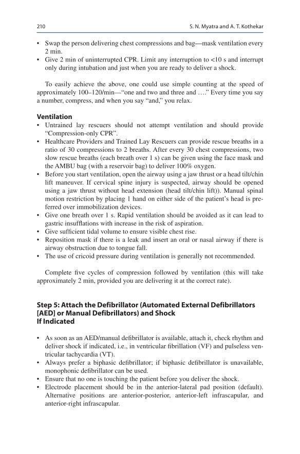
210
• Swap the person delivering chest compressions and bag—mask ventilation every 2 min.
• Give 2 min of uninterrupted CPR. Limit any interruption to <10 s and interrupt only during intubation and just when you are ready to deliver a shock.
To easily achieve the above, one could use simple counting at the speed of approximately 100–120/min—“one and two and three and ….” Every time you say a number, compress, and when you say “and,” you relax.
Ventilation• Untrained lay rescuers should not attempt ventilation and should provide
“Compression- only CPR”.• Healthcare Providers and Trained Lay Rescuers can provide rescue breaths in a
ratio of 30 compressions to 2 breaths. After every 30 chest compressions, two slow rescue breaths (each breath over 1 s) can be given using the face mask and the AMBU bag (with a reservoir bag) to deliver 100% oxygen.
• Before you start ventilation, open the airway using a jaw thrust or a head tilt/chin lift maneuver. If cervical spine injury is suspected, airway should be opened using a jaw thrust without head extension (head tilt/chin lift)). Manual spinal motion restriction by placing 1 hand on either side of the patient’s head is pre-ferred over immobilization devices.
• Give one breath over 1 s. Rapid ventilation should be avoided as it can lead to gastric insufflations with increase in the risk of aspiration.
• Give sufficient tidal volume to ensure visible chest rise.• Reposition mask if there is a leak and insert an oral or nasal airway if there is
airway obstruction due to tongue fall.• The use of cricoid pressure during ventilation is generally not recommended.
Complete five cycles of compression followed by ventilation (this will take approximately 2 min, provided you are delivering it at the correct rate).
Step 5: Attach the Defibrillator (Automated External Defibrillators [AED] or Manual Defibrillators) and Shock If Indicated
• As soon as an AED/manual defibrillator is available, attach it, check rhythm and deliver shock if indicated, i.e., in ventricular fibrillation (VF) and pulseless ven-tricular tachycardia (VT).
• Always prefer a biphasic defibrillator; if biphasic defibrillator is unavailable, monophonic defibrillator can be used.
• Ensure that no one is touching the patient before you deliver the shock.• Electrode placement should be in the anterior-lateral pad position (default).
Alternative positions are anterior-posterior, anterior-left infrascapular, and anterior- right infrascapular.
S. N. Myatra and A. T. Kothekar

211
• Shock energy—(a) Biphasic: Use the manufacturer’s recommendation (120–200 J); if unknown, use maximum available. Second and subsequent doses should be equivalent, and higher doses may be considered if available. (b) Monophasic: 360 J (in children and infants, use 2–4 J/Kg first and 4 J/Kg for subsequent shocks; higher energy may be considered but not to exceed 10 J/Kg).
• No pulse check is recommended after defibrillation; resume CPR immediately.• Reattach the defibrillator after every 2 min of CPR.• Reduce time between the last compression and shock delivery and the time
between shock delivery and resumption of compressions. CPR should be per-formed while the defibrillator is readied.
• There is no upper limit to the number of shocks you give. Remember that the shockable rhythms are the ones with the better prognosis, so never give up on a VF or pulseless VT.
• AEDs can now be used even in infants with a pediatric dose attenuator if the manual defibrillator is not available. If neither is available, the AED without the pediatric dose attenuator can be used. (All AEDs are biphasic.)
• The precordial thump may be considered in witnessed, monitored, and unstable ventricular tachyarrhythmias only when a defibrillator is not available.
• Electric pacing is not recommended for routine use in cardiac arrest.• If it is not a shockable rhythm, resume CPR for 2 min and then re attach the
defibrillator
Extracorporal CPR can be considered in select patients who have not responded to initial conventional CPR, if it can be rapidly implemented.
Advanced Cardiac Life Support
Step 6: Drug Therapy
• Use intravenous (IV) or intraosseous (IO) route for bolus delivery of drugs. For IV use, give the bolus drug followed by a 20-mL saline push and raise the extremity.
• If both IV and IO are unavailable, then tracheal route may be used. Epinephrine, naloxone and lidocaine may be administered through this route. (Use 2–2½ times the dose diluted in 5–10 mL of distilled water or saline for the tracheal route)
• Give a vasopressor soon after giving the shock. Epinephrine IV/IO dose should be 1 mg every 3–5 min.
• Amiodarone should be given when VF/VT is unresponsive to CPR, defibrillation, and vasopressor therapy. IV/IO first dose should be 300 mg bolus, and the second dose should be 150 mg (after 3–5 min if VF/VT recurs or persists). This may be followed by a 24-h infusion. (Use lidocaine only if amiodarone is unavailable.)
• Atropine should not be used during pulseless electrical activity or asystole as it is unlikely to have a therapeutic benefit.
• Other drugs are not routinely used and should be considered only in specific situations:
21 Cardiorespiratory Arrest

212
– Magnesium sulphate (1–2 g) for torsades de pointes associated with a long QT interval or if there is hypomagnesemia.
– Sodium bicarbonate (initial dose is 1 mEq/Kg) should be used only if there is hyperkalemia, bicarbonate-responsive acidosis, or tricyclic antidepressant overdose. It is harmful in hypercarbic acidosis.
– Calcium—Hyperkalemia, hypocalcemia or calcium channel blocker toxicity – Naloxone—In opioid overdose, administer naloxone if available – Steroids—There may be some benefit with the use during in hospital cardiac
arrest, but the evidence is weak
Step 7: Advanced Airway
• Weigh the need for minimally interrupted compressions against the need for insertion of an advanced airway, i.e., the endotracheal tube or the supraglottic airway (laryngeal mask airway, esophageal tracheal tube—Combitube, or laryn-geal tube).
• Continue bag mask ventilation if advanced airway is not placed.• Confirm the placement of advanced airway by the clinical method (chest expansion
and breath sound), and in addition, use the Continuous waveform capnography to confirm and monitor the correct placement. Record the depth and secure the tube.
• Once advanced airway is in place, give 10 breaths per minute and deliver uninter-rupted chest compressions at 100–120/min (No need for synchrony between chest compression and ventilation).
Step 8: Treat Reversible Causes
During each 2-min period of CPR, review the most frequent causes—five H’s and five T’s—to identify factors that may have caused the arrest or may be complicating the resuscitation.
Five H’s Five T’sHypovolemia Tension pneumothoraxHypoxia Tamponade, cardiacHydrogen ion (acidosis) ToxinsHypo-/hyperkalemia Thrombosis, pulmonaryHypothermia Thrombosis, coronary
Step 9: Monitor the CPR Quality Throughout Resuscitation
• Give emphasis on delivering high-quality CPR. This means giving compressions of adequate rate and depth, allowing complete chest recoil between compres-sions, minimizing interruptions in compressions, avoiding excessive ventilation, and rotating the compressor every 2 min.
S. N. Myatra and A. T. Kothekar

213
• Use quantitative waveform capnography to monitor end tidal CO2 if available (expressed as a partial pressure in mmHg—PetCO2) in intubated patients. If PetCO2 is less than 10 mmHg, attempt to improve CPR quality.
• If intra-arterial pressure monitoring is already present and the diastolic pressure is less than 20 mmHg, attempt should be made to improve quality of CPR.
Step 10: Return of Spontaneous Circulation ROSC
• Return of spontaneous circulation (ROSC) can be confirmed by return of pulse or blood pressure or abrupt sustained increase in PetCO2 (typically ≥40 mmHg) or spontaneous arterial pressure waves with intra-arterial monitoring.
Step 11: Postcardiac Arrest Care After ROSC
• The goal should be to optimize cardiopulmonary function and vital organ perfusion.
• Transfer the patient to an appropriate hospital or ICU with facility to deliver post-cardiac arrest care.
• Optimize ventilation to minimize the lung injury. Do chest X-ray to confirm airway position and to diagnose pneumonia/pulmonary edema. Use lung- protective ventilation if there is pulmonary dysfunction; adjust settings using blood gas values. Avoid excessive ventilation and hyperoxia.
• Once ROSC is achieved, the fraction of inspired oxygen (FiO2) should be adjusted to the minimum concentration needed to achieve arterial oxyhemoglo-bin saturation of ≥94%, with the goal of avoiding hyperoxia while ensuring adequate oxygen delivery. PaCO2 can be maintained within a normal physiologi-cal range, taking into account any temperature correction.
• Treat hypotension (systolic blood pressure [SBP] < 90) with fluid and vasopres-sors and treat other reversible causes.
• Consider targeted temperature management (TTM) in adult patients with persis-tent coma after ROSC. It is recommended to select and maintain a constant tem-perature between 32 and 36 °C and to actively prevent fever. TTM should be maintained for at least 24 h after achieving target temperature.
• The wide range of TTM between 32 °C and 36 °C practically removes contrain-dications of hypothermia. All patients in whom intensive care is continued are eligible for TTM. The patients in whom, lower temperature conveys some risk like bleeding, can be offered higher temperature up to 36 °C. Lower temperatures might be preferred when patients have clinical conditions like seizures, cerebral edema that can be worsened at higher temperatures. Patients who present at the lower end of the TTM range can be maintained at that lower temperature without warming them to a higher target.
• Cooling can be done using cold IV fluid bolus of 30 mL/Kg, surface cooling (ice packs, mattresses), endovascular cooling, etc. Sedation/muscle relaxants may be used to control shivering, agitation, or ventilator dyssynchrony as needed. After 24 h, start slow rewarming at 0.25 °C/h. Prevent hyperpyrexia (>37.7 °C).
21 Cardiorespiratory Arrest
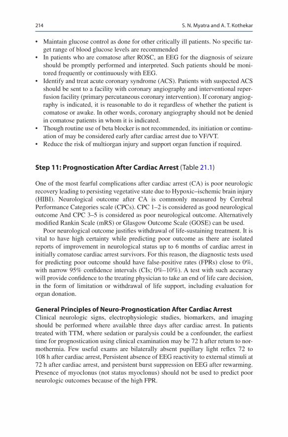
214
• Maintain glucose control as done for other critically ill patients. No specific tar-get range of blood glucose levels are recommended
• In patients who are comatose after ROSC, an EEG for the diagnosis of seizure should be promptly performed and interpreted. Such patients should be moni-tored frequently or continuously with EEG.
• Identify and treat acute coronary syndrome (ACS). Patients with suspected ACS should be sent to a facility with coronary angiography and interventional reper-fusion facility (primary percutaneous coronary intervention). If coronary angiog-raphy is indicated, it is reasonable to do it regardless of whether the patient is comatose or awake. In other words, coronary angiography should not be denied in comatose patients in whom it is indicated.
• Though routine use of beta blocker is not recommended, its initiation or continu-ation of may be considered early after cardiac arrest due to VF/VT.
• Reduce the risk of multiorgan injury and support organ function if required.
Step 11: Prognostication After Cardiac Arrest (Table 21.1)
One of the most fearful complications after cardiac arrest (CA) is poor neurologic recovery leading to persisting vegetative state due to Hypoxic–ischemic brain injury (HIBI). Neurological outcome after CA is commonly measured by Cerebral Performance Categories scale (CPCs). CPC 1–2 is considered as good neurological outcome And CPC 3–5 is considered as poor neurological outcome. Alternatively modified Rankin Scale (mRS) or Glasgow Outcome Scale (GOSE) can be used.
Poor neurological outcome justifies withdrawal of life-sustaining treatment. It is vital to have high certainty while predicting poor outcome as there are isolated reports of improvement in neurological status up to 6 months of cardiac arrest in initially comatose cardiac arrest survivors. For this reason, the diagnostic tests used for predicting poor outcome should have false-positive rates (FPRs) close to 0%, with narrow 95% confidence intervals (CIs; 0%–10%). A test with such accuracy will provide confidence to the treating physician to take an end of life care decision, in the form of limitation or withdrawal of life support, including evaluation for organ donation.
General Principles of Neuro-Prognostication After Cardiac ArrestClinical neurologic signs, electrophysiologic studies, biomarkers, and imaging should be performed where available three days after cardiac arrest. In patients treated with TTM, where sedation or paralysis could be a confounder, the earliest time for prognostication using clinical examination may be 72 h after return to nor-mothermia. Few useful exams are bilaterally absent pupillary light reflex 72 to 108 h after cardiac arrest, Persistent absence of EEG reactivity to external stimuli at 72 h after cardiac arrest, and persistent burst suppression on EEG after rewarming. Presence of myoclonus (not status myoclonus) should not be used to predict poor neurologic outcomes because of the high FPR.
S. N. Myatra and A. T. Kothekar
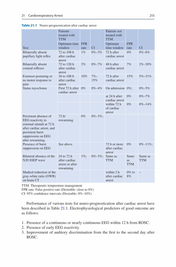
215
Performance of various tests for neuro-prognostication after cardiac arrest have been described in Table 21.1. Electrophysiological predictors of good outcome are as follows:
1. Presence of a continuous or nearly continuous EEG within 12 h from ROSC. 2. Presence of early EEG reactivity. 3. Improvement of auditory discrimination from the first to the second day after
ROSC.
Table 21.1 Neuro-prognostication after cardiac arrest
Test
Patients treated with TTM
FPR rate CI
Patients not treated with TTM
FPR rate CI
Optimum time window
Optimum time window
Bilaterally absent pupillary light reflex
72 to 108 h after cardiac arrest
1% 0%–3% 72 h after cardiac arrest
0% 0%–8%
Bilaterally absent corneal reflexes
72 to 120 h after cardiac arrest
2% 0%–7% 48 h after cardiac arrest
7% 2%–20%
Extensor posturing or no motor response to pain
36 to 108 h after cardiac arrest
10% 7%–15%
72 h after cardiac arrest
15% 5%–31%
Status myoclonus First 72 h after cardiac arrest
0% 0%–4% On admission 0%; 0%–5%
at 24 h after cardiac arrest
0% 0%–7%
within 72 h of cardiac arrest
0% 0%–14%
Persistent absence of EEG reactivity to external stimuli at 72 h after cardiac arrest, and persistent burst suppression on EEG after rewarming,
72 h/rewarming
0% 0%–3%;
Presence of burst suppression on EEG
See above 72 h or more after cardiac arrest
0% 0%–11%;
Bilateral absence of the N20 SSEP wave
24 to 72 h after cardiac arrest or after rewarming
1%; 0%–3%; Same as TTM
Same as TTM
Same as TTM
Marked reduction of the gray-white ratio (GWR) on brain CT
within 2 h after cardiac arrest
0% to 8%
–
TTM: Therapeutic temperature managementFPR rate: False positive rate (Desirable: close to 0%)CI: 95% confidence intervals (Desirable: 0%–10%)
21 Cardiorespiratory Arrest

216
Step 12: Assist Survivors from Cardiac Arrest Who Require Rehabilitation Services
The ultimate goal of resuscitation should be restoring the pre-arrest health-related quality of life (HRQOL). HRQOL should be assessed at a minimum of 3 months. Patients who get successfully discharged alive from the hospital after cardiac arrest often report cognitive impairment, restricted mobility, depression, and restricted societal participation. Further studies in this area are warranted.
If patients resuscitated after cardiac arrest, subsequently die or have brain death, do not achieve ROSC or have resuscitation terminated, they should be evaluated as potential organ donors.
Suggested Reading
Callaway CW, Donnino MW, Fink EL, et al. Part 8: Post-Cardiac Arrest Care: 2015 American Heart Association Guidelines Update for Cardiopulmonary Resuscitation and Emergency Cardiovascular Care. Circulation. 2015;132(18 Suppl 2):S465–82. An American Heart Association update on the guidelines for Post cardiac arrest care
Kilgannon JH, Jones AE, Shapiro NI, et al. Association between arterial hyperoxia following resus-citation from cardiac arrest and in-hospital mortality. JAMA. 2010;303:2165–71. Patients who are admitted to the ICU following resuscitation from cardiac arrest, arterial hyperoxia was independently associated with increased in-hospital mortality compared with either hypoxia or normoxia
Kleinman ME, Brennan EE, Goldberger ZD, Swor RA, Terry M, Bobrow BJ, Gazmuri RJ, Travers AH, Rea T. Part 5: adult basic life support and cardiopulmonary resuscitation quality: 2015 American Heart Association Guidelines Update for Cardiopulmonary Resuscitation and Emergency Cardiovascular Care. Circulation. 2015;132(suppl 2):S414–35. An update on the guidelines for cardiopulmonary resuscitation and emergency cardiovascular care quality
Nolan JP, Soar J, Cariou A, Cronberg T, Moulaert VR, Deakin CD, Bottiger BW, Friberg H, Sunde K, Sandroni C. European Resuscitation Council and European Society of Intensive Care Medicine Guidelines for Postresuscitation Care 2015: Section 5 of the European Resuscitation Council Guidelines for Resuscitation 2015. Resuscitation. 2015;95:202–22. A comprehensive review on guidelines for Postresuscitation Care
Sandroni C, D’Arrigo S, Nolan JP. Prognostication after cardiac arrest. Crit Care. 2018;22:150. A multimodal approach combining multiple prognostication tests is recommended so that the risk of a falsely pessimistic prediction is minimised
Websites
https://eccguidelines.heart.org/wp-content/uploads/2015/10/2015-AHA-Guidelines-Highlights-English.pdf
S. N. Myatra and A. T. Kothekar
217© Springer Nature Singapore Pte Ltd. 2020R. Chawla, S. Todi (eds.), ICU Protocols, https://doi.org/10.1007/978-981-15-0898-1_22
A. V. Hegde Department of Medicine and Critical Care, P.D. Hinduja National Hospital & Medical Research Centre, Mumbai, India
K. Bajan Emergency Department, P.D. Hinduja Hospital and Medical Research Centre, Mumbai, India
S. Todi (*) Department of Critical Care & Emergency Medicines, A.M.R.I. Hospital, Kolkata, India
22Cardiogenic Shock
Ashit V. Hegde, Khusrav Bajan, and Subhash Todi
Cardiogenic shock most commonly occurs in the setting of acute myocardial infarc-tion leading to severe reduction in cardiac output (Cardiac index <2 L/min/m2), elevated filling pressure of left, right or both ventricle, and severe hypotension (Systolic less than 90 mmHg). The management of acute coronary syndrome and its complications has increasingly been protocolized. Timely implementation of these protocols especially in patients with shock is the essence as “time is muscle.” Cardiogenic shock carries a high 30-day mortality within the range of 30–40%.
Step 1: Urgently Resuscitate
• The patient with prolonged cardiogenic shock needs to be ventilated in spite of normal oxygenation parameters to decrease oxygen consumption by the respira-tory muscles and utilization of low cardiac output by vital organs.
• Use sedatives that are less likely to worsen hypotension during intubation, namely, etomidate, ketamine, and fentanyl.
• In patients who are not clinically in heart failure, cautious fluid resuscitation with proper hemodynamic monitoring should be initiated.
A 55-year-old male patient was admitted to the hospital with history of chest pain for about 3 h. He was drowsy, extremities were cold, and his blood pressure was 84/60 mmHg. His electrocardiogram showed extensive anterior ST eleva-tion myocardial infarction.
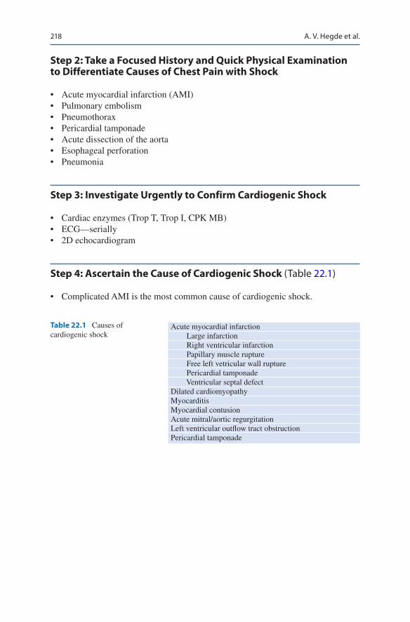
218
Step 2: Take a Focused History and Quick Physical Examination to Differentiate Causes of Chest Pain with Shock
• Acute myocardial infarction (AMI)• Pulmonary embolism• Pneumothorax• Pericardial tamponade• Acute dissection of the aorta• Esophageal perforation• Pneumonia
Step 3: Investigate Urgently to Confirm Cardiogenic Shock
• Cardiac enzymes (Trop T, Trop I, CPK MB)• ECG—serially• 2D echocardiogram
Step 4: Ascertain the Cause of Cardiogenic Shock (Table 22.1)
• Complicated AMI is the most common cause of cardiogenic shock.
Table 22.1 Causes of cardiogenic shock
Acute myocardial infarction Large infarction Right ventricular infarction Papillary muscle rupture Free left vetricular wall rupture Pericardial tamponade Ventricular septal defectDilated cardiomyopathyMyocarditisMyocardial contusionAcute mitral/aortic regurgitationLeft ventricular outflow tract obstructionPericardial tamponade
A. V. Hegde et al.
219
Step 5: Initiate Medical Management
• Aspirin: 160–325 mg of soluble or chewable aspirin should be administered, but the decision to administer clopidogrel should be made only after angiography (in case the patient needs urgent coronary artery bypass graft [CABG]).
• Beta blockers and Calcium Channel Blockers should be avoided.• Thrombolysis: In the presence of hypotension, thrombolytic drugs may not reach
the coronary vessel. Thrombolytic therapy is therefore not very effective in established cardiogenic shock. Consider thrombolysis only if primary percutane-ous intervention (PCI) is not possible urgently. Thrombolytic drugs are more effective if administered after the BP has been normalised (preferably after the use of an intra-aortic balloon pump [IABP]).
Step 6: Initiate Hemodynamic Management (See Chap. 18, Vol. 1)
• A central venous line preferably under ultrasound guidance to avoid arterial punctures and an intra-arterial line (preferably radial) should be urgently inserted.
• Urine output should be monitored hourly.• Urgent 2D echo is mandatory to rule out mechanical causes of shock (papillary
muscle rupture, acute ventricular septal defect, free wall rupture, and pericardial tamponade). 2D echo also gives an idea of left ventricular ejection fraction (LVEF) and left ventricular (LV) filling pressures.
• PA catheter may be used occasionally, specially in Right ventricular infarct.• Fluid boluses may be cautiously administered to most patients with cardio-
genic shock. Even patients with pulmonary edema may have intravascular volume depletion because there is redistribution of fluid from the intravascu-lar compartment into the alveolus. These fluid boluses should be guided care-fully by frequent physical examination and intravascular pressure monitoring.
• Most patients will also need a vasopressor for systolic blood pressure <80 mmHg and an inotrope. The least dose of these medications required to maintain ade-quate perfusion to the tissues should be used. There is no definite evidence of superiority of one vasopressor over another. Norepinephrine is the preferred vasopressor than dopamine because of lower rates of tachyarrthmias with the former. Dobutamine (2.5–10 mcg/Kg/min) is the inotrope of choice in patients with a BP of more than 80 mmHg. Levosimendan, a calcium sensitizer inotrope, has also been increasingly used in cardiogenic shock as it has a relatively less effect on increase in oxygen consumption by the myocardium. There is increas-ing evidence that many patients with cardiogenic shock are inappropriately vaso-dilated because of an inflammatory response.
22 Cardiogenic Shock

220
Step 7: Consider Inserting an IABP in Selected Patients (See Chap. 50, Vol. 2)
• IABP is not recommended in most patients in whom cardiac revascularisation procedure like primary PTCA is planned or in whom fibrinolysis is administered
• It may act as a bridge to surgical intervention for mechanical complications like mitral regurgitation or ventricular septal effect, insertion of other mechanical assist devices (LVAD, RVAD, Impella), or cardiac transplant.
• In cases of myocardial stunning, it buys time while other therapeutic measures take effect.
Step 8: Consider Coronary Revascularization
• Urgent left heart catheterization and revascularization, if coronary anatomy is suitable, should be undertaken.
• Timely primary PCI of the infarct related artery (within 120 min of hospital pre-sentation) is the preferred mode of reperfusion in patients with cardiogenic shock complicating AMI.
• Urgent CABG is indicated in patients with coronary anatomy not favorable for PCI (e.g. triple vessel disease or left main disease). However in patient not fit for CABG
• Primary PCI may be performed in the infract artery urgently if expertise is avail-able, followed later by CABG
• Proper hydration and N-acetylcysteine (600 mg b.i.d for 3 days) should be given to prevent contrast-induced nephropathy as these patients are at risk of acute kidney injury (AKI).
• The shock study demonstrated a 13% decrease in mortality in patients with car-diogenic shock assigned to early revascularization (primary PCI or CABG).
• Although revascularization should be performed as early as possible, there is a survival benefit for up to 48 h after MI and within 18 h of the onset of shock.
• For those who can not undergo timely angiography, fibrinolytic therapy is indi-cated rather than no reperfusion.
Step 9: Manage Specific Situations
Mechanical complications
Mechanical complications of MI, including rupture of the ventricular septum, free wall, or papillary muscles, need urgent surgical correction (after temporary stabilization with an IABP).
Right ventricular (RV) infarction
Patients with RV dysfunction and shock need adequate right-sided filling pressures to maintain cardiac output. However, overzealous fluid therapy may do more harm by over distending the RV and compromising LV filling. RV end-diastolic pressure of 10–15 mmHg is optimum. If the patient remains hypotensive in spite of reasonable fluid therapy, inotropes and IABP are indicated.
A. V. Hegde et al.

221
Pericardial tamponade
It is an uncommon but rapidly reversible cause of cardiogenic shock. Its existence should be actively sought in all cases of shock by a bedside echo looking for evidence of diastolic compression of the right side of the heart. Immediate pericardiocentesis is lifesaving. Fluid boluses and vasopressors may be used as a temporizing method.
Step 10: Consider Rescue Therapy in Refractory Shock
• The left or right, ventricular assist device (LVAD, RVAD, Impella) placed surgi-cally or percutaneously should be considered in patients’ refractory to medical therapy and IABP
• It should be instituted early before irreversible organ damage occurs to work as a “bridge” before definitive therapy like cardiac transplantation is available.
• Extracorporeal assist devices have been increasingly used in the ICU as a bridge to definitive therapy
Suggested Reading
Hochman JS, Sleeper LA. Early revascularization in acute myocardial infarction complicated by cardiogenic shock. SHOCK investigators. N Engl J Med. 1999;341(9):625–34. In patients with cardiogenic shock, emergency revascularization did not significantly reduce overall mortality at 30 days. However, after 6 months, there was a significant survival benefit.
Hollenberg SM. Cardiogenic shock. Crit Care Clin. 2001;17(2):391–410. A comprehensive review article on the subject.
Scholz KH, Maier SKG, Maier LS, et al. Impact of treatment delay on mortality in ST-segment elevation myocardial infarction (STEMI) patients presenting with and without haemodynamic instability: results from the German prospective, multicentre FITT-STEMI trial. Eur Heart J. 2018;39:1065. The time between the first medical contact to primary PCI is a strong predictor of an adverse outcome. This patient group benefitted most from immediate PCI treatment, so special efforts to shorten contact-to-balloon time should be practiced in particular to these high-risk STEMI patient.
Thiele H, Zeymer U, Thelemann N, et al. Intraaortic balloon pump in cardiogenic shock compli-cating acute myocardial infarction. Circulation. 2019;139:395. IABP has no effect on all-cause mortality at 6-year long-term follow-up. Mortality remains high, with two thirds of patients with cardiogenic shock dying despite contemporary treatment with revascularization therapy.
22 Cardiogenic Shock
223© Springer Nature Singapore Pte Ltd. 2020R. Chawla, S. Todi (eds.), ICU Protocols, https://doi.org/10.1007/978-981-15-0898-1_23
S. Ramasubban (*) Department of Respiratory, Critical Care and Sleep Medicine, Apollo Gleneagles Hospitals, Kolkata, India
R. K. Rajput Department of Cardiology, Indraprastha Apollo Hospitals, New Delhi, India
23Acute Heart Failure
Suresh Ramasubban and Rajeeve Kumar Rajput
Acute Heart failure (AHF) is defined as an acute clinical syndrome of new or wors-ening signs and symptoms of heart failure (HF). It as a clinical syndrome character-ised by a constellation of symptoms (dyspnea, orthopnea, lower limb swelling) and signs (elevated jugular venous pressure, gallop sounds (S4 and S3 and pulmonary crepitations) often caused by a structural and/or functional cardiac abnormality resulting in reduced cardiac output and/or elevated intracardiac pressures.
Almost 75% of AHF cases are due to worsening chronic heart failure (CHF), referred to as Acute decompensated heart failure (ADHF) while the remaining are new onset (De novo) AHF. The severity of symptoms of AHF vary and depends on the severity of the underlying cardiac disorder and the rapidity of progress of the symptoms. The common major causes for new onset AHF are acute coronary syn-drome (ACS), acute valve syndromes (Aortic regurgitation and Mitral regurgita-tion), progressive valve disease (Aortic stenosis, Mitral stenosis), cardiomyopathy and uncontrolled hypertension. AHF may develop in patients with chronic stable heart failure commonly due to precipitating factors such as ischemia, arrhythmia, infection and drugs.
A 60-year-old male patient, with history of coronary artery disease (CAD) and type 2 diabetes, presented with history of acute onset of breathlessness and orthopnea for 2 days. On examination, he was found to have tachycardia, tachy-pnea, and bilateral basal crepitations, and SpO2 was 87% on 2Lof oxygen by nasal cannula.

224
Step 1: Recognise Acute Heart Failure (AHF)
• Symptoms/signs of left sided failure/congestion include orthopnea, paroxysmal nocturnal dyspnea, bilateral pulmonary crackles, S3 on cardiac auscultation and bilateral peripheral oedema.
• Symptoms/signs of right sided failure/congestion include jugular venous disten-tion, bilateral peripheral oedema, hepatomegaly, hepatojugular reflex, ascites and symptoms of gut congestion.
• Symptoms of hypoperfusion clinically include cold extremities, oliguria, mental confusion, dizziness and narrow pulse pressure. Laboratory measures include metabolic acidosis, lactic acidosis and increase in creatinine. Hypoperfusion is not the same as hypotension, but most of the time hypoperfusion is present in a patient with hypotension.
• Clinically AHF can manifest as congestive symptoms/signs or with symptoms of hypoperfusion or a combination, giving rise to four clinical profiles of patient’s with AHF. A warm and wet (well perfused but congested), cold and wet (hypoperfused and congested), cold and dry (hypoperfused only) and warm and dry (Compensated). The most common presentation is that of warm and wet.
• Usually acute heart failure is predominantly left sided failure with reduced ejec-tion fraction and low cardiac output (Heart failure with reduced ejection fraction, EF < 40%) Other types of heart failure are Heart failure with preserved ejection fraction i.e. EF > 50%) and heart failure with mid range ejection fraction, EF40% to 50%, Acute heart failure may be predominantly right sided heart failure due to conditions such as pulmonary embolism or high output cardiac failure such as thyrotoxicosis
Step 2: Initial Urgent Management
• AHF is a life threatening medical emergency.• Confirmation of diagnosis, identification of coexisting life threatening condi-
tions and/or life threatening precipitants of AHF are parallel process and have to be tackled simultaneously, not necessarily in a step wise manner.
• Coexistent life threatening condition refers to cardiogenic shock and respiratory failure.
• Life threatening precipitating causes include acute coronary syndrome (ACS), hypertensive emergency, rhythm disorders either tachycardia or severe bradycar-dia/conduction abnormalities, mechanical causes and acute pulmonary embolism.
• Diagnosis of AHF is based on clinical suspicion as outlined in step 1, confirmed with additional investigations such as ECG, biomarkers, chest X-ray and echocardiography.
S. Ramasubban and R. K. Rajput
225
• However a step wise approach starting with urgent management of life threaten-ing conditions like respiratory failure and cardiogenic shock is the priority.
Step 2a: Manage Respiratory Failure in Suspected AHF
• Pulmonary congestion leads to an intrapulmonary shunt physiology resulting in hypoxemia. Oxygen therapy should be initiated in hypoxemic patients while monitoring oxygen saturation with aim to keep saturation 92–94%.
• In suspected AHF, oxygen is no longer recommended to be used routinely in patients without hypoxemia as it causes vasoconstriction and a reduction in car-diac output Oxygen therapy is initiated if PaO2 < 60 mmHg or SpO2 < 90%.
• An arterial blood gas to measure pH and PCO2 and lactates should be done in all patients with congestive symptoms or a background of COPD
• A trial of NIV is indicated if there is no urgent need of intubation and no contra-indication of NIV.
• Non invasive Ventilation (NIV) reduces work of breathing and respiratory dis-tress. NIV also decreases preload and left ventricular afterload quickly leading to increased cardiac output and decreased pulmonary congestion. The provision of adequate positive end expiratory pressure (PEEP) with NIV also increases oxy-genation by increasing the mean airway pressure. Inspiratory positive pressure support by NIV also improves minute ventilation and is especially useful in cases of hypercapnia and concomitant chronic obstructive pulmonary disease (COPD).
• NIV includes both Continuous positive airway pressure (CPAP) and bilevel posi-tive pressure ventilation (BPAP). CPAP is feasible even in the Emergency Department, while NIV support requires more expertise.
• Invasive ventilation and intubation is recommended in AHF when respiratory failure cannot be managed noninvasively (see contraindications and predictors of failure of NIV) (Chap. 3, Vol. 1).
Step 2b: Manage Cardiogenic Shock in AHF
• Cardiogenic shock is defined as hypotension and signs of tissue hypoperfusion despite an adequate filling pressures.
• Signs of hypoperfusion are as mentioned in Step 1.• In case of cardiogenic shock, an immediate ECG and echocardiography are
needed to diagnose the common pathogenetic conditions like STEMI, arryth-mias and advanced chronic heart failure with decompensation.
• In case of ACS as cause of AHF with cardiogenic shock, it is recommended that an immediate coronary angiography be done with an intent to revascularization.
• All patients with AHF and cardiogenic shock should ideally have an arterial line in situ for continuous monitoring of arterial blood pressure.
23 Acute Heart Failure
226
• Aim of pharmacologic therapy in AHF with cardiogenic shock is to increase cardiac output and improve organ perfusion.
• Fluid challenge with 250-300 mL crystalloid over 15–20 min using the principle of fluid challenges should be done with caution in subset of patients with hypo-perfusion but no congetion.
• After the fluid challenge pharmacologic management with vasopressors and ino-tropes is initiated.
• Intravenous inotropic agents like dopamine or dobutamine should be used to increase cardiac output. Dose titration should be done with continuous monitor-ing of perfusion of organs.
• Norepinephrine is the preferred vasopressor medication when arterial blood pressure needs support.
• There are a limited number of studies which have shown benefit with a combina-tion of Levosimendan with vasopressors.
• Adding levosimendan to a combination of dobutamine and vasopressors has also been looked into with some benefit in hemodynamics.
• For patients with preserved ejection fraction presenting with cardiogenic shock, treat for possible left ventricular outflow tract obstruction with IV fluids (pro-vided patient is not in pulmonary oedema). Use norepinephrine or phenylephrine as vasopressors.
• Mechanical circulatory support for a short term can also be attempted in cardio-genic shock not responding to inotropes and vasopressors
Step 2c: Confirm Diagnosis of AHF
• The initial diagnosis of AHF is clinical and requires, a thorough history enquir-ing about symptoms, prior cardiac history and potential precipitants, as well a physical examination to assess for signs of congestion and hypoperfusion with appropriate basic investigations like ECG, chest X-ray, laboratory investigations including biomarkers and echocardiography.
• The symptoms and signs are as outlined in step 1.• The initial step in confirming diagnosis is to rule out alternate causes for the
patients symptoms. The main differential diagnosis include pulmonary embo-lism, acute asthma, pneumonia, noncardiogenic pulmonary oedema (ARDS) and pericardial tamponade.
• Chest X-ray though normal in almost 20% of cases is still a very useful test for the diagnosis of AHF. Pulmonary venous congestion, interstitial or alveo-lar infiltrates, pleural effusion and cardiomegaly is useful for diagnosis of AHF.
• ECG is very useful in diagnosis of AHF, a normal ECG has a very high negative predictive value and most importantly it is essential to diagnose ACS as a precipitant.
• Echocardiography is required in most of the patients with AHF. An urgent echo-cardiography is required in cases of patients with AHF and hemodynamic instability.
S. Ramasubban and R. K. Rajput
227
• Lung ultrasonography with multiple B-Lines may also be helpful in diagnosing pulmonary congestion.
• Natriuretic peptides (BNP, NT-proBNP) should be a part of the initial evaluation of AHF. The natriuretic peptides help in distinguishing dyspnea of cardiac origin from non cardiac causes. The natriuretic peptides have a very high sensitivity and therefore a normal levels of NP makes the diagnosis of AHF unlikely. The cut off values for BNP is <100 pg/mL and NT pro-BNP <300 pg/mL. Most heart failure patients will have a BNP >300 pg/mL. However an elevated levels of NP’s are not diagnostic of AHF as a wide variety of conditions can lead to an elevated levels of natriuretic peptides. NP are elevated in non cardiac condition like advanced age, ischemic stroke, renal failure, liver cirrhosis with ascites, chronic obstructive pulmonary disease, sepsis and severe metabolic abnormalities like thyrotoxicosis.
• Hemodynamic monitoring is generally not routinely recommended for diagnosis of AHF, one may consider a monitoring tool in hemodynamically unstable patients with an unclear mechanism of deterioration.
• Laboratory evaluation – All patients with AHF should have the following laboratory assessment at
admission. Blood Urea, creatinine, electrolytes, glucose, liver function tests and a complete blood count.
– Cardiac troponins should also be routinely ordered in AHF. Troponins are elevated in ACS and Pulmonary embolism in addition to AHF. In AHF they reflect myocyte injury or necrosis and hence levels are elevated.
– D-Dimer if pulmonary embolism is suspected. – Procalcitonin measurement to be considered in AHF with suspected infection,
especially for differential diagnosis of pneumonia and to guide antibiotic therapy.
– TSH should be ordered in all patients with newly diagnosed AHF as both hypothyroidism and hyperthyroidism can precipitate AHF.
Step 2d: Recognise Precipitants of AHF Which Require Immediate Management
• It is important to recognize the precipitants quickly and treat them simultane-ously to avoid further deterioration.
• Acute Coronary Syndrome (ACS): ACS should be looked for and managed on urgent basis. AHF due to ACS is a subset of patients who are at high risk of adverse outcomes. ST elevation MI should be treated by urgent revascularization either by primary angioplasty or by thrombolysis by fibrin specific fibrinolytic agents. NON ST elevation MI in patients with recurrent ischemia, ST changes, arrhythmia should also be managed by early angiography and intervention, i.e., Invasive strategy with intention to revacularize within 2 h of presentation.
• Hypertensive emergency: Hypertensive emergency can precipitate AHF and manifests predominantly as pulmonary oedema. Vasodilators and diuretics to
23 Acute Heart Failure
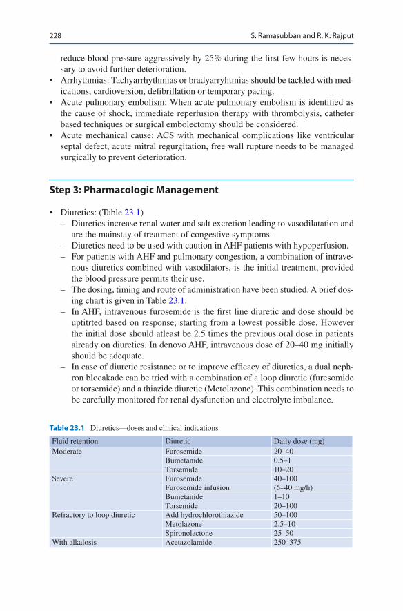
228
reduce blood pressure aggressively by 25% during the first few hours is neces-sary to avoid further deterioration.
• Arrhythmias: Tachyarrhythmias or bradyarryhtmias should be tackled with med-ications, cardioversion, defibrillation or temporary pacing.
• Acute pulmonary embolism: When acute pulmonary embolism is identified as the cause of shock, immediate reperfusion therapy with thrombolysis, catheter based techniques or surgical embolectomy should be considered.
• Acute mechanical cause: ACS with mechanical complications like ventricular septal defect, acute mitral regurgitation, free wall rupture needs to be managed surgically to prevent deterioration.
Step 3: Pharmacologic Management
• Diuretics: (Table 23.1) – Diuretics increase renal water and salt excretion leading to vasodilatation and
are the mainstay of treatment of congestive symptoms. – Diuretics need to be used with caution in AHF patients with hypoperfusion. – For patients with AHF and pulmonary congestion, a combination of intrave-
nous diuretics combined with vasodilators, is the initial treatment, provided the blood pressure permits their use.
– The dosing, timing and route of administration have been studied. A brief dos-ing chart is given in Table 23.1.
– In AHF, intravenous furosemide is the first line diuretic and dose should be uptitrted based on response, starting from a lowest possible dose. However the initial dose should atleast be 2.5 times the previous oral dose in patients already on diuretics. In denovo AHF, intravenous dose of 20–40 mg initially should be adequate.
– In case of diuretic resistance or to improve efficacy of diuretics, a dual neph-ron blocakade can be tried with a combination of a loop diuretic (furesomide or torsemide) and a thiazide diuretic (Metolazone). This combination needs to be carefully monitored for renal dysfunction and electrolyte imbalance.
Table 23.1 Diuretics—doses and clinical indications
Fluid retention Diuretic Daily dose (mg)Moderate Furosemide 20–40
Bumetanide 0.5–1Torsemide 10–20
Severe Furosemide 40–100Furosemide infusion (5–40 mg/h)Bumetanide 1–10Torsemide 20–100
Refractory to loop diuretic Add hydrochlorothiazide 50–100Metolazone 2.5–10Spironolactone 25–50
With alkalosis Acetazolamide 250–375
S. Ramasubban and R. K. Rajput
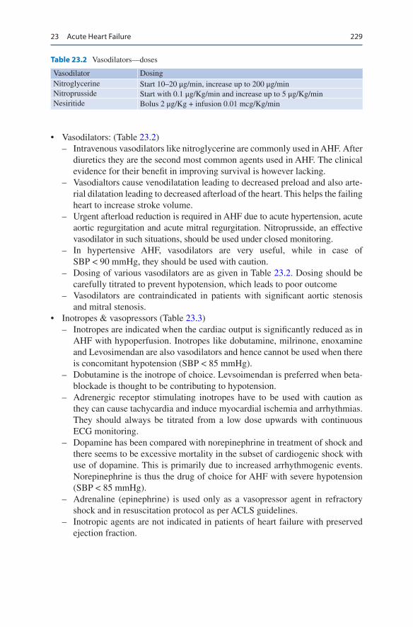
229
• Vasodilators: (Table 23.2) – Intravenous vasodilators like nitroglycerine are commonly used in AHF. After
diuretics they are the second most common agents used in AHF. The clinical evidence for their benefit in improving survival is however lacking.
– Vasodialtors cause venodilatation leading to decreased preload and also arte-rial dilatation leading to decreased afterload of the heart. This helps the failing heart to increase stroke volume.
– Urgent afterload reduction is required in AHF due to acute hypertension, acute aortic regurgitation and acute mitral regurgitation. Nitroprusside, an effective vasodilator in such situations, should be used under closed monitoring.
– In hypertensive AHF, vasodilators are very useful, while in case of SBP < 90 mmHg, they should be used with caution.
– Dosing of various vasodilators are as given in Table 23.2. Dosing should be carefully titrated to prevent hypotension, which leads to poor outcome
– Vasodilators are contraindicated in patients with significant aortic stenosis and mitral stenosis.
• Inotropes & vasopressors (Table 23.3) – Inotropes are indicated when the cardiac output is significantly reduced as in
AHF with hypoperfusion. Inotropes like dobutamine, milrinone, enoxamine and Levosimendan are also vasodilators and hence cannot be used when there is concomitant hypotension (SBP < 85 mmHg).
– Dobutamine is the inotrope of choice. Levsoimendan is preferred when beta-blockade is thought to be contributing to hypotension.
– Adrenergic receptor stimulating inotropes have to be used with caution as they can cause tachycardia and induce myocardial ischemia and arrhythmias. They should always be titrated from a low dose upwards with continuous ECG monitoring.
– Dopamine has been compared with norepinephrine in treatment of shock and there seems to be excessive mortality in the subset of cardiogenic shock with use of dopamine. This is primarily due to increased arrhythmogenic events. Norepinephrine is thus the drug of choice for AHF with severe hypotension (SBP < 85 mmHg).
– Adrenaline (epinephrine) is used only as a vasopressor agent in refractory shock and in resuscitation protocol as per ACLS guidelines.
– Inotropic agents are not indicated in patients of heart failure with preserved ejection fraction.
Table 23.2 Vasodilators—doses
Vasodilator DosingNitroglycerine Start 10–20 μg/min, increase up to 200 μg/minNitroprusside Start with 0.1 μg/Kg/min and increase up to 5 μg/Kg/minNesiritide Bolus 2 μg/Kg + infusion 0.01 mcg/Kg/min
23 Acute Heart Failure

230
Step 4: Pharmacological Management-Miscellaneous Drugs
• Digoxin: – Digoxin may be used in AHF with Atrial fibrillation with a rapid ventricular
response (> 110/min) and is used as boluses of 0.25 mg to 0.5 mg, especially if patient not exposed to digoxin in the past. Renal adjustment of the dosing also needs to be done.
– Maintenance dose depends on a multitude of factors, such as age, sex and renal function.
– Some medications like ACEI and betablockers should be avoided during the initial stages of decompensated heart failure.
• Vasopressin antagonists: – Drugs like tolvaptan are V2 receptor anatgonists at the renal tubules and pro-
mote water excretion (aquaresis). – Tolvaptan is indicated in AHF with volume overload and hyponatremia.
• Opiates: – Opiates were once considered as first line agents for treatment of AHF for
their ability to relieve dyspnea and anxiety. However due to the side effects of nausea, hypotension, bradycardia and respiratopry depression, there is a potential for increased need for invasive ventilation. There are studies which have pointed to an elevated mortality risk in patients receiving morphine.
– Opiates are not routinely recommended for management of patients with AHF. They should be used with caution in patients with severe dyspnea due to pulmonary oedema.
• Anxiolytics and sedatives: – AHF patients may have agitation and delirium, this needs to be cautiously
treated with sedatives and anxiolytics – Benzodiazepines (Lorazepam or diazepam) can be used cautiously for man-
agement of agitation and delirium
Table 23.3 Inotropes and vasopressors—mode of administration and doses
Bolus Infusion rateDobutamine No 2–20 μg/Kg/minDopamine No 3–5 μg/Kg/min: inotropic
>5 μg/Kg/min: vasopressorMilrinone 25–75 μg/Kg over 10–20 min 0.375–0.75 μg/Kg/minEnoximone 90 mcg/Kg/min over 10–30 mins 5–20 mcg/Kg/minLevosimendan 12 μg/Kg over 10 min (optional) 0.1 μg/Kg/min which can be
decreased to 0.05 or increased to 0.2 μg/Kg/min
Norepinephrine No 0.2–1.0 μg/Kg/minEpinephrine Bolus: 1 mg can be given IV during
resuscitation, repeated every 3–5 min0.05–0.5 μg/Kg/min
S. Ramasubban and R. K. Rajput

231
Step 5: Renal Replacement Therapy
• The most common modality of renal replacement therapy in AHF with conges-tion is generally ultrafiltration. Movement of water across a semipermeable membrane due to a transmembrane pressure is called as ultrafiltration.
• Ultrafiltration is indicated for diuretic refractory pulmonary congestion. It is not the first line therapy for patients with AHF and congestion.
• Indications for dialysis in AHF are the standard indications of dialysis in critical illness. They include but are not limited to hyperkalemia, acidosis and volume overload.
Step 6: Mechanical Assist Devices
• Intra-aortic balloon pump (IABP): IABP can be used to support the circulation, as a bridge to surgery, in case of mechanical complications like ventricular septal rup-ture and acute mitral refurgitation. The recent IABP-SHOCK II trial has not shown any outcome benefit of IABP in patients with cardiogenic shock following AMI.
• Extracorporeal membrane oxygenation (ECMO): Patients with AHF and cardio-genic shock may be supported with ECMO as temporary measure until the heart and other organs have recovered their function.
Step 7
• Once stabilised patient should be initiated on ARB or ACEI or Angiotensin receptor-neprilysin inhibitor (ARNI) beta blockers and mineralocorticoid recetor antagonists if there are no contraindication.
Suggested Reading
Lindenfeld J, Albert NM, et al. Heart Failure Society of America, HFSA 2010 comprehensive heart failure practice guideline. J Card Fail. 2010;16(6):e1–2. A comprehensive guidelines on the management of heart failure.
McCullough PA, Nowak RM, McCord J, et al. B-type natriuretic peptide and clinical judg-ment in emergency diagnosis of heart failure: analysis from Breathing Not Properly (BNP) multinational. Circulation. 2002;106:416–22. The evaluation of acute dyspnea would be improved with the addition of BNP testing to clinical judgment in the emergency department.
Voors PA, et al. 2016 ESC Guidelines for the diagnosis and treatment of acute and chronic heart failure: The Task Force for the diagnosis and treatment of acute and chronic heart failure of the European Society of Cardiology (ESC). Eur Heart J. 2016;37:2129–200.
Yancy CW, et al. 2017 ACC/AHA/HFSA Focused Update of the 2013 ACCF/AHA Guideline for the Management of Heart Failure: A Report of the American College of Cardiology/American Heart Association Task Force on Clinical Practice Guidelines and the Heart Failure Society of America. Circulation. 2017;136:137–61. A comprehensive review of guidelines on te manage-ment of acute heart failure.
23 Acute Heart Failure
233© Springer Nature Singapore Pte Ltd. 2020R. Chawla, S. Todi (eds.), ICU Protocols, https://doi.org/10.1007/978-981-15-0898-1_24
S. Ghosh (*) Department of Critical Care Medicine, Fortis-Escorts Hospital, Faridabad, India
24Cardiac Arrhythmias
Supradip Ghosh
With frequent exposure to metabolic, ischemic and neurohumoral stressors and not so infrequent presence of underlying structural heart disease, critically ill patients are particularly at higher risk of cardiac arrhythmias. Arrhythmias often result in significant hemodynamic compromise in an already unstable critically ill patient. Prompt recognition and treatment of arrhythmias can substantially reduce morbid-ity and mortality and meticulous attention to correction of contributing factors can prevent recurrence of arrhythmias.
Assessment sequence of arrhythmias is presented in this chapter in a longitudinal stepwise manner for the sake of understanding. However, in an actual clinical situ-ation, many of these steps need to be performed in parallel or simultaneously.
Step 1: Resuscitate
The first step in the evaluation of an arrhythmia is to assess the hemodynamic status of the patient including assessment of peripheral perfusion (cold extremities, delayed capillary refill, low volume pulse), blood pressure, evidence of myocardial
A 64 years old male with history of hypertension and chronic obstructive pulmo-nary disease was admitted to the intensive care unit with acute infective exacerba-tion of his pulmonary condition and type 2 respiratory failure. He was doing reasonably well with standard medical treatment and non-invasive ventilator sup-port, until he suddenly became hypotensive on his third hospitalization day. Monitor showed an irregularly irregular narrow QRS complex tachycardia with a ventricular rate of 156/min and blood pressure of 76/40 mmHg, pulse oximetry reading of 80% on fractional inspiratory oxygen 0.4.
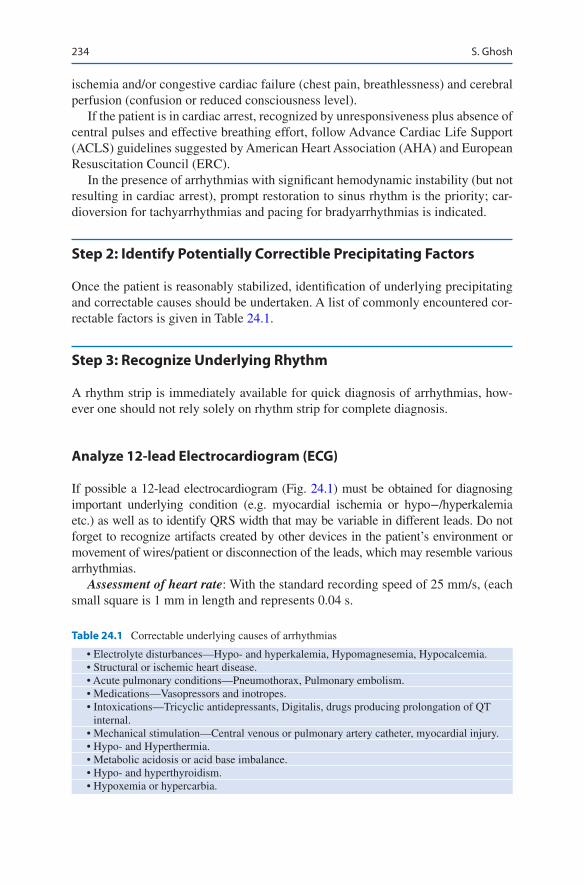
234
ischemia and/or congestive cardiac failure (chest pain, breathlessness) and cerebral perfusion (confusion or reduced consciousness level).
If the patient is in cardiac arrest, recognized by unresponsiveness plus absence of central pulses and effective breathing effort, follow Advance Cardiac Life Support (ACLS) guidelines suggested by American Heart Association (AHA) and European Resuscitation Council (ERC).
In the presence of arrhythmias with significant hemodynamic instability (but not resulting in cardiac arrest), prompt restoration to sinus rhythm is the priority; car-dioversion for tachyarrhythmias and pacing for bradyarrhythmias is indicated.
Step 2: Identify Potentially Correctible Precipitating Factors
Once the patient is reasonably stabilized, identification of underlying precipitating and correctable causes should be undertaken. A list of commonly encountered cor-rectable factors is given in Table 24.1.
Step 3: Recognize Underlying Rhythm
A rhythm strip is immediately available for quick diagnosis of arrhythmias, how-ever one should not rely solely on rhythm strip for complete diagnosis.
Analyze 12-lead Electrocardiogram (ECG)
If possible a 12-lead electrocardiogram (Fig. 24.1) must be obtained for diagnosing important underlying condition (e.g. myocardial ischemia or hypo−/hyperkalemia etc.) as well as to identify QRS width that may be variable in different leads. Do not forget to recognize artifacts created by other devices in the patient’s environment or movement of wires/patient or disconnection of the leads, which may resemble various arrhythmias.
Assessment of heart rate: With the standard recording speed of 25 mm/s, (each small square is 1 mm in length and represents 0.04 s.
Table 24.1 Correctable underlying causes of arrhythmias
• Electrolyte disturbances—Hypo- and hyperkalemia, Hypomagnesemia, Hypocalcemia. • Structural or ischemic heart disease. • Acute pulmonary conditions—Pneumothorax, Pulmonary embolism. • Medications—Vasopressors and inotropes. • Intoxications—Tricyclic antidepressants, Digitalis, drugs producing prolongation of QT
internal. • Mechanical stimulation—Central venous or pulmonary artery catheter, myocardial injury. • Hypo- and Hyperthermia. • Metabolic acidosis or acid base imbalance. • Hypo- and hyperthyroidism. • Hypoxemia or hypercarbia.
S. Ghosh
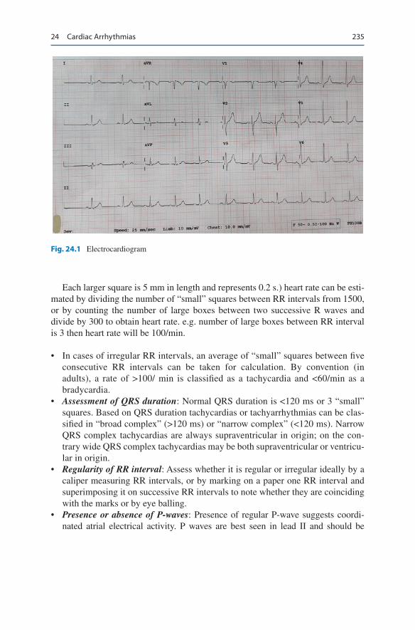
235
Each larger square is 5 mm in length and represents 0.2 s.) heart rate can be esti-mated by dividing the number of “small” squares between RR intervals from 1500, or by counting the number of large boxes between two successive R waves and divide by 300 to obtain heart rate. e.g. number of large boxes between RR interval is 3 then heart rate will be 100/min.
• In cases of irregular RR intervals, an average of “small” squares between five consecutive RR intervals can be taken for calculation. By convention (in adults), a rate of >100/ min is classified as a tachycardia and <60/min as a bradycardia.
• Assessment of QRS duration: Normal QRS duration is <120 ms or 3 “small” squares. Based on QRS duration tachycardias or tachyarrhythmias can be clas-sified in “broad complex” (>120 ms) or “narrow complex” (<120 ms). Narrow QRS complex tachycardias are always supraventricular in origin; on the con-trary wide QRS complex tachycardias may be both supraventricular or ventricu-lar in origin.
• Regularity of RR interval: Assess whether it is regular or irregular ideally by a caliper measuring RR intervals, or by marking on a paper one RR interval and superimposing it on successive RR intervals to note whether they are coinciding with the marks or by eye balling.
• Presence or absence of P-waves: Presence of regular P-wave suggests coordi-nated atrial electrical activity. P waves are best seen in lead II and should be
Fig. 24.1 Electrocardiogram
24 Cardiac Arrhythmias
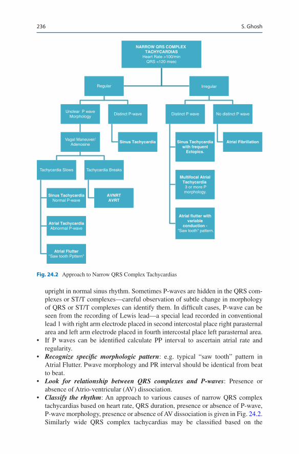
236
upright in normal sinus rhythm. Sometimes P-waves are hidden in the QRS com-plexes or ST/T complexes—careful observation of subtle change in morphology of QRS or ST/T complexes can identify them. In difficult cases, P-wave can be seen from the recording of Lewis lead—a special lead recorded in conventional lead 1 with right arm electrode placed in second intercostal place right parasternal area and left arm electrode placed in fourth intercostal place left parasternal area.
• If P waves can be identified calculate PP interval to ascertain atrial rate and regularity.
• Recognize specific morphologic pattern: e.g. typical “saw tooth” pattern in Atrial Flutter. Pwave morphology and PR interval should be identical from beat to beat.
• Look for relationship between QRS complexes and P-waves: Presence or absence of Atrio-ventricular (AV) dissociation.
• Classify the rhythm: An approach to various causes of narrow QRS complex tachycardias based on heart rate, QRS duration, presence or absence of P-wave, P-wave morphology, presence or absence of AV dissociation is given in Fig. 24.2. Similarly wide QRS complex tachycardias may be classified based on the
NARROW QRS COMPLEX TACHYCARDIAS
Heart Rate >100/minQRS <120 msec
Regular
Unclear P waveMorphology
Vagal Maneuver/Adenosine
Tachycardia Slows
Sinus TachycardiaNormal P-wave
Atrial TachycardiaAbnormal P-wave
Atrial Flutter"Saw tooth Ppttern"
AVNRTAVRT
Tachycardia Breaks
Sinus Tachycardia Sinus Tachycardiawith frequent
Ectopics.
Multifocal AtrialTachycardia 3 or more Pmorphology.
Atrial flutter withvariable
conduction - "Saw tooth" pattern.
Atrial Fibrillation
Distinct P-wave Distinct P wave No distinct P wave
Irregular
Fig. 24.2 Approach to Narrow QRS Complex Tachycardias
S. Ghosh
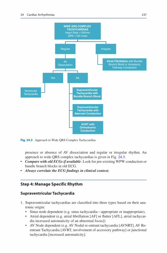
237
presence or absence of AV dissociation and regular or irregular rhythm. An approach to wide QRS complex tachycardias is given in Fig. 24.3.
• Compare with old ECGs if available: Look for pre-existing WPW conduction or bundle branch blocks in old ECG.
• Always correlate the ECG findings in clinical context.
Step 4: Manage Specific Rhythm
Supraventricular Tachycardia
1. Supraventricular tachycardias are classified into three types based on their ana-tomic origin:• Sinus node dependent (e.g. sinus tachycardia—appropriate or inappropriate),• Atrial dependent (e.g. atrial fibrillation [AF] or flutter [AFL], atrial tachycar-
dia increased automaticity of an abnormal focus])• AV Node dependent (e.g. AV Nodal re-entrant tachycardia [AVNRT], AV Re-
entrant Tachycardia [AVRT, involvement of accessory pathway] or junctional tachycardia [increased automaticity].
WIDE QRS COMPLEX TACHYCARDIAS
Heart Rate >100/minQRS >120 msec
Regular
AVDissociation
Yes
VentricularTachycardia
SupraventricularTachycardia with
Bundle Branch Block
SupraventricularTachycardia with
Aberrant Conduction
AVRT withOrthodromicConduction
No
Irregular
Atrial Fibrillation with BundleBranch Block or Accessory
Pathway Conduction
Fig. 24.3 Approach to Wide QRS Complex Tachycardias
24 Cardiac Arrhythmias
238
2. Supraventricular tachycardias are narrow QRS complex regular tachycardias (except AF, atrial flutter with variable conduction and multifocal atrial tachycardia).
3. They may be further differentiated by the location and morphology of P-wave and the response of tachycardia to AV nodal blocking agents.
4. In case of Atrial tachycardia, P-wave is located in the usual position (i.e. preced-ing QRS complex) with almost normal PR interval.
5. In AVNRT or Junctional tachycardia, QRS complexes almost always obscure P-wave as atria and ventricle is almost simultaneously activated; P-wave is often seen at the terminal portion of QRS complex especially in lead V1 and lead III, AVF.
6. P-wave is seen usually within the ST segment in case of AVRT, as atria is mostly activated retrogradely via accessory pathway.
7. Vagal maneuvers and AV nodal blocking agents can terminate AV dependent rhythms but can only temporarily reduce the ventricular rate in automatic rhythms like Atrial or Junctional tachycardias.
Manage Narrow QRS Complex Regular Tachycardias• In all hemodynamically unstable patients, arrhythmia should be rapidly termi-
nated with synchronized DC Cardioversion. Cardioversion is frequently ineffec-tive in converting junctional tachycardia; may even be harmful if the increased automaticity is because of Digitalis toxicity.
• Inciting agents like Digitalis and excessive catecholamine should be withdrawn if feasible.
• In hemodynamically stable patient, vagal maneuver like carotid massage may be attempted to terminate AVNRT/AVRT and slower ventricular rate in Atrial or Junctional tachycardias.
• One should be careful in performing carotid massage in elderly patients and avoid the same in the presence of carotid bruit. Do not perform carotid massage on both side at the same time.
• Intravenous adenosine is effective in terminating, AVNRT and AVRT, but not Atrial or Junctional tachycardias. It is administered as a bolus of 3–6 mg in a large vein followed by a rapid bolus of saline flush. If ineffective another bolus of 6–12 mg may be repeated after 2–5 min. Dyspnea, facial flushing and chest tightness are common but short lived adverse effect. Certain conditions in ICU are known to interfere with the use of Adenosine.
• Methylxanthines used to relieve refractory bronchospasm have adenosine recep-tor antagonist activity and may render the patient less sensitive to adenosine. As such adenosine should be avoided in patients with reactive airway disease, as it can provoke bronchospasm. Dipyridamole, an antiplatelet drug, can block ade-nosine transport back into the cell and can enhance the response to adenosine.
• In cases of contraindication to Adenosine, non-dihydropyridine calcium channel blockers (Verapamil, Diltiazem) and beta-blockers can be tried.
• Verapamil (especially when administered intravenously) can produce rapid ven-tricular response in the presence of accessory pathways in patients with AF.
S. Ghosh

239
Atrial Fibrillation
• AF is the commonest arrhythmia (other than Sinus Tachycardia) encountered in ICU setting.
• AF is common in post-operative period, particularly after cardiac surgery with an incidence as high as 25–40% with a peak onset at day two.
• In AF, there is a loss of atrial contribution to ventricular preload (normally con-tributing 25% of ventricular preload), which becomes significant in a hemody-namically unstable patient, patients with diastolic dysfunction or in the presence of tachycardia (shortened diastolic time).
• Loss of atrial activity in AF also contributes to atrial clot formation and throm-boembolic episodes.
Diagnosis: Hallmark of the diagnosis is presence of irregularly irregular RR intervals and loss of distinct P-wave (Fig. 24.4). Some chaotic atrial activity result-ing in undulating baseline is seen—known as “Fibrillation wave”.
Management: Acute management of AF has two dimensions:
• Control of rhythm (electrical or pharmacological cardioversion)• Control of ventricular rate and therapeutic anticoagulation (to prevent thrombo-
embolic episodes).
Rhythm Control: Electrical Cardioversion• Preferred in hemodynamically unstable patients with hypotension, decompen-
sated heart failure or active myocardial ischemia.• Premedication with sedation and opioids if feasible. Always synchronized with
QRS complex.• Should not be attempted in cases of Digoxin toxicity or severe hypokalemia.
Fig. 24.4 Atrial fibrillation with fast ventricular rate and narrow QRS morphology
24 Cardiac Arrhythmias
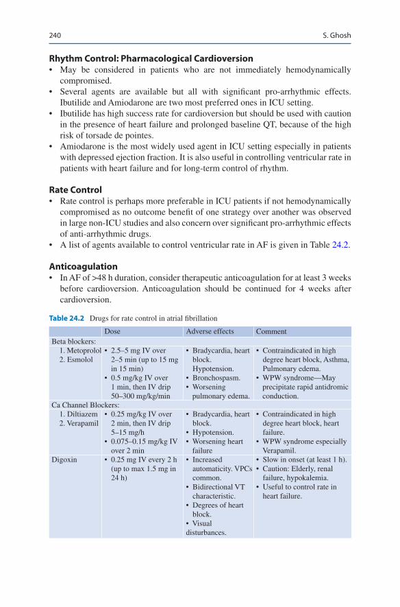
240
Rhythm Control: Pharmacological Cardioversion• May be considered in patients who are not immediately hemodynamically
compromised.• Several agents are available but all with significant pro-arrhythmic effects.
Ibutilide and Amiodarone are two most preferred ones in ICU setting.• Ibutilide has high success rate for cardioversion but should be used with caution
in the presence of heart failure and prolonged baseline QT, because of the high risk of torsade de pointes.
• Amiodarone is the most widely used agent in ICU setting especially in patients with depressed ejection fraction. It is also useful in controlling ventricular rate in patients with heart failure and for long-term control of rhythm.
Rate Control• Rate control is perhaps more preferable in ICU patients if not hemodynamically
compromised as no outcome benefit of one strategy over another was observed in large non-ICU studies and also concern over significant pro-arrhythmic effects of anti-arrhythmic drugs.
• A list of agents available to control ventricular rate in AF is given in Table 24.2.
Anticoagulation• In AF of >48 h duration, consider therapeutic anticoagulation for at least 3 weeks
before cardioversion. Anticoagulation should be continued for 4 weeks after cardioversion.
Table 24.2 Drugs for rate control in atrial fibrillation
Dose Adverse effects CommentBeta blockers: 1. Metoprolol 2. Esmolol
• 2.5–5 mg IV over 2–5 min (up to 15 mg in 15 min)
• 0.5 mg/kg IV over 1 min, then IV drip 50–300 mg/kg/min
• Bradycardia, heart block. Hypotension.
• Bronchospasm. • Worsening
pulmonary edema.
• Contraindicated in high degree heart block, Asthma, Pulmonary edema.
• WPW syndrome—May precipitate rapid antidromic conduction.
Ca Channel Blockers: 1. Diltiazem 2. Verapamil
• 0.25 mg/kg IV over 2 min, then IV drip 5–15 mg/h
• 0.075–0.15 mg/kg IV over 2 min
• Bradycardia, heart block.
• Hypotension.• Worsening heart
failure
• Contraindicated in high degree heart block, heart failure.
• WPW syndrome especially Verapamil.
Digoxin • 0.25 mg IV every 2 h (up to max 1.5 mg in 24 h)
• Increased automaticity. VPCs common.
• Bidirectional VT characteristic.
• Degrees of heart block.
• Visual disturbances.
• Slow in onset (at least 1 h). • Caution: Elderly, renal
failure, hypokalemia. • Useful to control rate in
heart failure.
S. Ghosh

241
• In situation where 3 weeks anticoagulation is not possible, Transesophageal Echocardiography may be done to exclude atrial clots, before attempting cardioversion.
• In urgent situation, heparin must be administered before attempting cardiover-sion, if AF is of >48 h duration. Anticoagulation should be continued for at least 4 weeks if CHA2DS2VASc score is 2 or higher.
• Decision to continue therapeutic anticoagulation must be weighed against the risk of bleeding; HAS-Bled score of 3 or higher is considered to be at high risk of bleeding.
Atrial Flutter
• Atrial flutter is a macroreentrant arrhythmia characterized by presence of flutter wave with typical “saw tooth” pattern (Fig. 24.5. Flutter wave is best seen in the inferior leads with an usual rate of 250-350/ min.
• QRS response is usually regular with fixed 2:1 or 3:1 conduction of flutter wave. But variable block may produce irregular narrow QRS complex tachycardia resembling AF.
• Management of AFL in similar to AF except that AFL needs lower energy to cardiovert and is more resistant to chemical cardioversionn.
Multifocal Atrial Tachycardia (MAT)
• MAT is an irregular narrow QRS complex tachycardia characterized by the pres-ence of three or more P-waves with different morphologies and variable PR intervals (Fig. 24.6).
• Usually associated with hypoxia in the setting of pulmonary disease or use of theophylline, metabolic derangements and end-stage cardiomyopathy.
• Treatment consists of correcting underlying condition. Intravenous Magnesium or AV nodal blockers are sometimes useful in decreasing ventricular response in the interim.
Fig. 24.5 Atrial flutter with characteristic “saw tooth” pattern with 2:1 conduction
24 Cardiac Arrhythmias

242
Ventricular Tachycardia
• Ventricular tachycardia is defined as three or more consecutive premature complexes at a rate of >100/min; when continued for >30 s, it is called sus-tained VT.
• VT is classified into Monomorphic VT with uniform QRS morphology (Fig. 24.7) and Polymorphic VT with beat-to-beat variation in QRS morphology along with varying QRS axis (Fig. 24.8).
Monomorphic Ventricular Tachycardia
1. Monomorphic VT is usually associated with structural heart disease or electro-lyte disturbances, old myocardial scar post infarction or cardiac surgery.
Diagnosis: Monomorphic VT needs to be differentiated from Supraventricular tachycardia with aberrant conduction. Following features favor the diagnosis of VT over SVT.
Fig. 24.6 Multifocal Atrial Tachycardia
Fig. 24.7 Monomorphic Ventricular Tachycardia with RBBB morphology very wide QRS com-plex, AV dissociation (variable QRS morphology) and positive Brugada’s sign
S. Ghosh
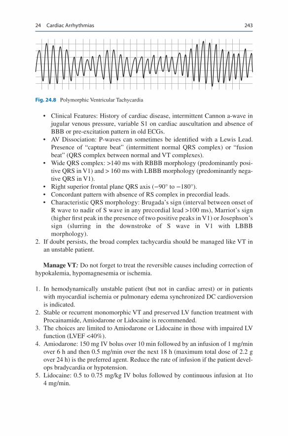
243
• Clinical Features: History of cardiac disease, intermittent Cannon a-wave in jugular venous pressure, variable S1 on cardiac auscultation and absence of BBB or pre-excitation pattern in old ECGs.
• AV Dissociation: P-waves can sometimes be identified with a Lewis Lead. Presence of “capture beat” (intermittent normal QRS complex) or “fusion beat” (QRS complex between normal and VT complexes).
• Wide QRS complex: >140 ms with RBBB morphology (predominantly posi-tive QRS in V1) and > 160 ms with LBBB morphology (predominantly nega-tive QRS in V1).
• Right superior frontal plane QRS axis (−90° to −180°).• Concordant pattern with absence of RS complex in precordial leads.• Characteristic QRS morphology: Brugada’s sign (interval between onset of
R wave to nadir of S wave in any precordial lead >100 ms), Marriot’s sign (higher first peak in the presence of two positive peaks in V1) or Josephson’s sign (slurring in the downstroke of S wave in V1 with LBBB morphology).
2. If doubt persists, the broad complex tachycardia should be managed like VT in an unstable patient.
Manage VT: Do not forget to treat the reversible causes including correction of hypokalemia, hypomagnesemia or ischemia.
1. In hemodynamically unstable patient (but not in cardiac arrest) or in patients with myocardial ischemia or pulmonary edema synchronized DC cardioversion is indicated.
2. Stable or recurrent monomorphic VT and preserved LV function treatment with Procainamide, Amiodarone or Lidocaine is recommended.
3. The choices are limited to Amiodarone or Lidocaine in those with impaired LV function (LVEF <40%).
4. Amiodarone: 150 mg IV bolus over 10 min followed by an infusion of 1 mg/min over 6 h and then 0.5 mg/min over the next 18 h (maximum total dose of 2.2 g over 24 h) is the preferred agent. Reduce the rate of infusion if the patient devel-ops bradycardia or hypotension.
5. Lidocaine: 0.5 to 0.75 mg/kg IV bolus followed by continuous infusion at 1to 4 mg/min.
Fig. 24.8 Polymorphic Ventricular Tachycardia
24 Cardiac Arrhythmias

244
6. Procainamide: 17 mg/kg IV loading dose (at 20 mg/min) followed by an infusion at 1 to 4 mg/min. The infusion should be stopped if the patient becomes hypoten-sive or the QRS widens by 50% above baseline.
7. Overdrive ventricular pacing is an option in refractory cases with a pacing rate 10 to 20 beats /min faster than the rate of VT.
Polymorphic Ventricular Tachycardia
• Polymorphic VT can be associated with either a normal QTc interval (usually ischemic in origin unless proved otherwise) or with prolonged QTc interval (>460 ms; when it is known as Torsades de pointes).
• Patients are usually hemodynamically unstable and needs urgent non- synchronized DC cardioversion.
• Myocardial ischemia should be treated expeditiously in patients with Polymorphic VT and normal QTc and medications that might predispose to ischemia, such as inotropes or vasopressors, should be stopped or tapered, if possible.
• Amiodarone and Beta-blockers are effective for the suppression of arrhythmia.• In cases of Torsades de pointes, the focus should be to identify and correct under-
lying cause of prolonged QTc if possible. Common causes of prolonged QTc are given in Table 24.3.
• Consider IV Magnesium 1 to 2 g over 20 to 30 min in all cases unless contrain-dicated. Other treatments options are overdrive pacing or isoproterenol infusion to increase underlying sinus rate and thus shorten QTc.
Bradycardias
1. In the ICU setting, bradycardias are common but often transient related to air-way manipulation or hypoxemia and rarely life-threatening; notable exception is in the setting of acute MI.
Table 24.3 Common causes of Prolonged QTc
1. Congenital: Congenital Long QT syndromes.2. Medications: • Anti-arrhythmics: Class Ia (Quinidine, Disopyramide, Procainamide), Class Ic
(Flecainide, Ibutilide) and Class III (Amiodarone, Sotalol) agents. • Antipsychotics: Phenothiazines, Butyrophenones, atypical anti-psychotics (Olanzapine,
Quetiapine). • Tricyclic Anti-depressants. • Antimicrobial agents: Quinolones, Macrolides, Pentamidine, Ketoconazole, Quinine. • Anti-histaminics: Terfenadine, Astemizole.3. Electrolyte Disturbances: Hypokalemia, hypomagnesemia, hypocalcemia.4. Neurological conditions: Subarachnoid Hemorrhage, large stroke, encephalopathy.
S. Ghosh
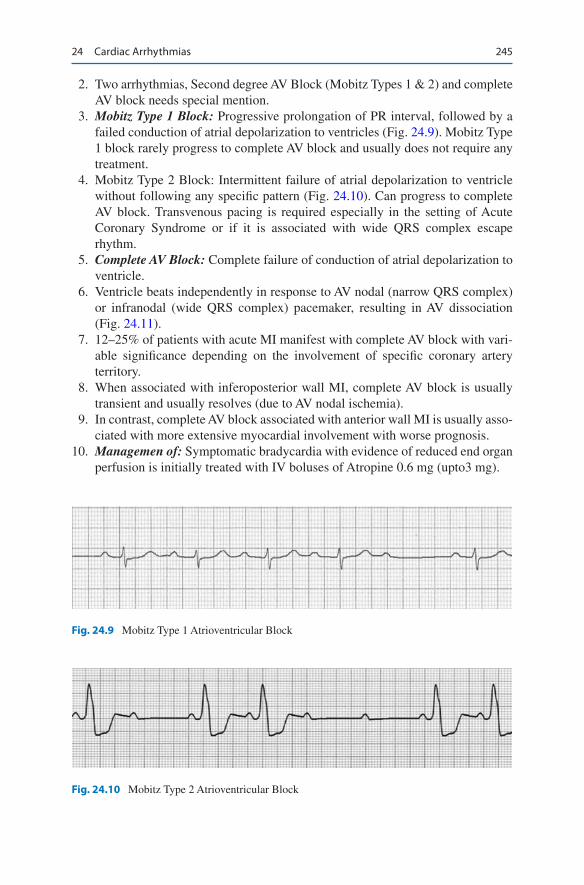
245
2. Two arrhythmias, Second degree AV Block (Mobitz Types 1 & 2) and complete AV block needs special mention.
3. Mobitz Type 1 Block: Progressive prolongation of PR interval, followed by a failed conduction of atrial depolarization to ventricles (Fig. 24.9). Mobitz Type 1 block rarely progress to complete AV block and usually does not require any treatment.
4. Mobitz Type 2 Block: Intermittent failure of atrial depolarization to ventricle without following any specific pattern (Fig. 24.10). Can progress to complete AV block. Transvenous pacing is required especially in the setting of Acute Coronary Syndrome or if it is associated with wide QRS complex escape rhythm.
5. Complete AV Block: Complete failure of conduction of atrial depolarization to ventricle.
6. Ventricle beats independently in response to AV nodal (narrow QRS complex) or infranodal (wide QRS complex) pacemaker, resulting in AV dissociation (Fig. 24.11).
7. 12–25% of patients with acute MI manifest with complete AV block with vari-able significance depending on the involvement of specific coronary artery territory.
8. When associated with inferoposterior wall MI, complete AV block is usually transient and usually resolves (due to AV nodal ischemia).
9. In contrast, complete AV block associated with anterior wall MI is usually asso-ciated with more extensive myocardial involvement with worse prognosis.
10. Managemen of: Symptomatic bradycardia with evidence of reduced end organ perfusion is initially treated with IV boluses of Atropine 0.6 mg (upto3 mg).
Fig. 24.9 Mobitz Type 1 Atrioventricular Block
Fig. 24.10 Mobitz Type 2 Atrioventricular Block
24 Cardiac Arrhythmias
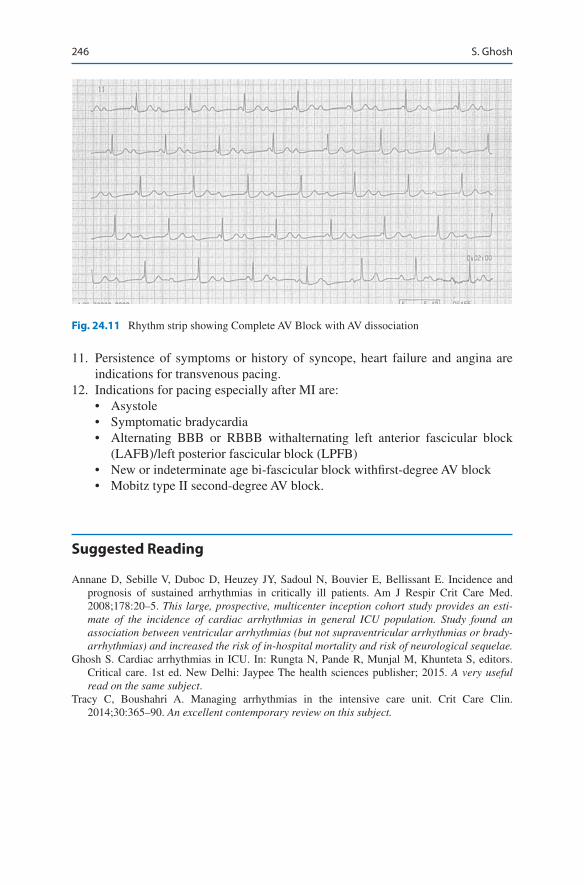
246
11. Persistence of symptoms or history of syncope, heart failure and angina are indications for transvenous pacing.
12. Indications for pacing especially after MI are:• Asystole• Symptomatic bradycardia• Alternating BBB or RBBB withalternating left anterior fascicular block
(LAFB)/left posterior fascicular block (LPFB)• New or indeterminate age bi-fascicular block withfirst-degree AV block• Mobitz type II second-degree AV block.
Suggested Reading
Annane D, Sebille V, Duboc D, Heuzey JY, Sadoul N, Bouvier E, Bellissant E. Incidence and prognosis of sustained arrhythmias in critically ill patients. Am J Respir Crit Care Med. 2008;178:20–5. This large, prospective, multicenter inception cohort study provides an esti-mate of the incidence of cardiac arrhythmias in general ICU population. Study found an association between ventricular arrhythmias (but not supraventricular arrhythmias or brady-arrhythmias) and increased the risk of in-hospital mortality and risk of neurological sequelae.
Ghosh S. Cardiac arrhythmias in ICU. In: Rungta N, Pande R, Munjal M, Khunteta S, editors. Critical care. 1st ed. New Delhi: Jaypee The health sciences publisher; 2015. A very useful read on the same subject.
Tracy C, Boushahri A. Managing arrhythmias in the intensive care unit. Crit Care Clin. 2014;30:365–90. An excellent contemporary review on this subject.
Fig. 24.11 Rhythm strip showing Complete AV Block with AV dissociation
S. Ghosh

247© Springer Nature Singapore Pte Ltd. 2020R. Chawla, S. Todi (eds.), ICU Protocols, https://doi.org/10.1007/978-981-15-0898-1_25
R. Rajani P.D. Hinduja Hospital and Medical Research Centre, Mumbai, India
F. N. Kapadia Department of Medicine and Critical Care, P.D. Hinduja National Hospital and Medical Research Centre, Mumbai, India
N. Ramakrishnan (*) Department of Critical Care Medicine, Apollo Hospitals, Chennai, India
25Acute Coronary Syndromes
Rajesh Rajani, Farhad N. Kapadia, and Nagarajan Ramakrishnan
Acute coronary syndrome (ACS) is a common diagnosis in patients presenting to the emergency department with acute onset chest pain. Timely and appropriate management in a protocolized manner is mandatory to salvage patients from this life-threatening syndrome.
Step 1: Initiate Resuscitation
• Patients with acute chest pain should have immediate intravenous access as they may deteriorate suddenly.
• A continuous ECG monitor and pulse oximeter should be placed.• Give oxygen and assist breathing if needed initially. Supplemental oxygen in
patients who do not have hypoxemia (oxygen saturation > 90% on room air) is not recommended.
A previously healthy smoker presented with severe chest pain to the emergency department. On arrival he was clinically stable. The ECG showed ST-segment elevation in the anterior leads and blood test showed elevated troponin T and CK-MB. The echocardiograph showed hypokinesia in the anteroseptal region. In the emergency department, his symptoms progressively worsened with ongoing intense chest pain, and he became increasingly restless and breathless.

248
• In the case of cardiac arrest, follow ACLS (advanced cardiac life support) proto-col (see Chap. 21, Vol. 1).
• Cardiovert immediately if ventricular or atrial tachyarrhythmia is detected and patient is hemodynamically unstable (shock, pulmonary edema or ongoing chest pain).
Urgent transcutaneous or temporary intravenous pacing is required if severe symptomatic bradycardia or heart block occurs.
Step 2: Take Focused History and Perform Physical Examination
• Elaborate the symptom of chest pain—site, type, severity, relation to exertion.• Sweating.• Cardiorespiratory symptoms.• Symptoms of low output state such as fatigue, dizziness, and syncope.• Risk factors for ischemic heart disease—Obesity, hypertension, diabetes, hyper-
lipidemia, smoking, obstructive sleep apnea, positive family history.• History of bleeding diathesis or recent blood loss.• BP in both arms.• Palpate all peripheral and central pulses.• Auscultate the heart for gallop or murmurs.• Auscultate the chest for basal crepitations.• Baseline Neuroexam specially if thrombolysis is a consideration.
Step 3: Perform Basic Investigation
1. 12-lead ECG (a) Look for features of new ST elevation acute myocardial infarction (STEMI)
• New ST elevation in two or more contiguous leads (inferior or anterior)Threshold values for new ST-segment elevation consistent with STEMI are:
– J-point elevation 0.2 mV (2 mm) in leads V2 and V3 and 0.1 mV (1 mm) in all other leads (men more than 40 years old).
– J-point elevation 0.25 mV (2.5 mm) in leads V2 and V3 and 0.1 mV (1 mm) in all other leads (men less than 40 years old).
– J-point elevation 0.15 mV (1.5 mm) in leads V2 and V3 and 0.1 mV (1 mm) in all other leads (women).
• New LBBB (left bundle branch block).• ST elevation in right precordial leads V4r for right ventricular myocar-
dial infarction (MI).• ST depression in precordial leads (V1–V3) for posterior MI.• Pathological “Q” waves.• NSTEMI or Unstable angina: ST segment depression or deep T wave
inversion without Q waves or no ECG changes.
R. Rajani et al.

249
(b) Echocardiogram• Regional wall motion abnormality.• Reduced systolic function (ejection fraction).• Mechanical complication–papillary muscle rupture, VSD.
(c) Chest X-ray• Cardiomegaly.• Pulmonary congestion.
(d) Cardiac enzymes• Raised troponins, CPK-MB.• Serial measurement of cardiac troponin is the preferred biomarker.• High sensitivity assays of Troponin are now available and has increase
diagnostic sensitivity to rule out myocardial infarction within a few hours. However, such testing has decreased clinical specificity for myo-cardial infarction, as they detect presence of troponin in other pathologi-cal states.
(e) A complete blood count• Leukocytosis may be seen in MI.
(f) Platelet count, prothrombin time, activated partial thromboplastin time (APTT). (g) Liver and renal function test. (h) Electrolytes
• Look for hypokalemia, hypomagnesemia.
Step 4: Confirm Diagnosis of ACS
• Typical chest pain in patients at risk of myocardial ischemia with classical ECG changes and raised cardiac enzymes establishes diagnosis in many cases.
• Consider other causes of chest pain (Table 25.1).• Consider other causes of raised troponin (Table 25.2).• Occasionally, patients may have typical chest pain without ECG abnormalities or
atypical chest pain (diabetics, female, elderly) and nonspecific changes on ECG. In these situations, perform serial monitoring of ECG and cardiac enzymes. An early echo is useful to look for regional wall motion abnormalities.
Table 25.1 Causes of acute chest pain
Life-threatening causes ACS (unstable angina, NSTEMI, STEMI) Pulmonary embolus (may be associated with ↑ troponin) Aortic dissection (may be overlooked due to associated
MI)“Benign” causes Pericarditis (both ↑ troponin and ST elevation may
occur) Gastrointestinal (esophageal reflux, spasm) Musculoskeletal (e.g., costochondritis) Neurologic (cervical radiculopathy, herpes zoster)
25 Acute Coronary Syndromes
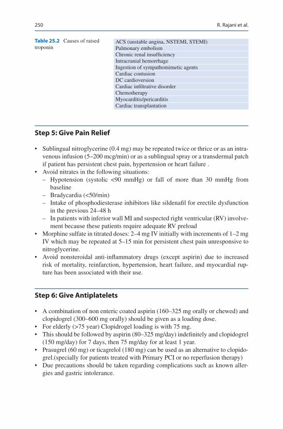
250
Step 5: Give Pain Relief
• Sublingual nitroglycerine (0.4 mg) may be repeated twice or thrice or as an intra-venous infusion (5–200 mcg/min) or as a sublingual spray or a transdermal patch if patient has persistent chest pain, hypertension or heart failure .
• Avoid nitrates in the following situations: – Hypotension (systolic <90 mmHg) or fall of more than 30 mmHg from
baseline – Bradycardia (<50/min) – Intake of phosphodiesterase inhibitors like sildenafil for erectile dysfunction
in the previous 24–48 h – In patients with inferior wall MI and suspected right ventricular (RV) involve-
ment because these patients require adequate RV preload• Morphine sulfate in titrated doses: 2–4 mg IV initially with increments of 1–2 mg
IV which may be repeated at 5–15 min for persistent chest pain unresponsive to nitroglycerine.
• Avoid nonsteroidal anti-inflammatory drugs (except aspirin) due to increased risk of mortality, reinfarction, hypertension, heart failure, and myocardial rup-ture has been associated with their use.
Step 6: Give Antiplatelets
• A combination of non enteric coated aspirin (160–325 mg orally or chewed) and clopidogrel (300–600 mg orally) should be given as a loading dose.
• For elderly (>75 year) Clopidrogel loading is with 75 mg.• This should be followed by aspirin (80–325 mg/day) indefinitely and clopidogrel
(150 mg/day) for 7 days, then 75 mg/day for at least 1 year.• Prasugrel (60 mg) or ticagrelol (180 mg) can be used as an alternative to clopido-
grel.(specially for patients treated with Primary PCI or no reperfusion therapy)• Due precautions should be taken regarding complications such as known aller-
gies and gastric intolerance.
Table 25.2 Causes of raised troponin
ACS (unstable angina, NSTEMI, STEMI)Pulmonary embolismChronic renal insufficiencyIntracranial hemorrhageIngestion of sympathomimetic agentsCardiac contusionDC cardioversionCardiac infiltrative disorderChemotherapyMyocarditis/pericarditisCardiac transplantation
R. Rajani et al.
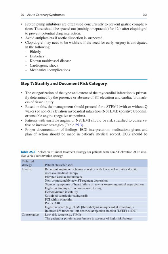
251
• Proton pump inhibitors are often used concurrently to prevent gastric complica-tions. These should be spaced out (mainly omeprazole) for 12 h after clopidogrel to prevent potential drug interaction.
• Avoid antiplatelets if aortic dissection is suspected• Clopidogrel may need to be withheld if the need for early surgery is anticipated
in the following: – Elderly – Diabetics – Known multivessel disease – Cardiogenic shock – Mechanical complications
Step 7: Stratify and Document Risk Category
• The categorization of the type and extent of the myocardial infarction is primar-ily determined by the presence or absence of ST elevation and cardiac biomark-ers of tissue injury.
• Based on this, the management should proceed for a STEMI (with or without Q waves) or non-ST elevation myocardial infarction (NSTEMI) (positive troponin) or unstable angina (negative troponins).
• Patients with unstable angina or NSTEMI should be risk stratified to conserva-tive or invasive strategies (Table 25.3).
• Proper documentation of findings, ECG interpretation, medications given, and plan of action should be made in patient’s medical record. ECG should be
Table 25.3 Selection of initial treatment strategy for patients with non-ST elevation ACS: inva-sive versus conservative strategy
Preferred strategy Patient characteristicsInvasive Recurrent angina or ischemia at rest or with low-level activities despite
intensive medical therapyElevated cardiac biomarkersNew or presumably new ST-segment depressionSigns or symptoms of heart failure or new or worsening mitral regurgitationHigh-risk findings from noninvasive testingHemodynamic instabilitySustained ventricular tachycardiaPCI within 6 monthsPrior CABGHigh-risk score (e.g., TIMI [thrombolysis in myocardial infarction])Reduced LV function (left ventricular ejection fraction [LVEF] < 40%)
Conservative Low-risk score (e.g., TIMI)The patient or physician preference in absence of high-risk features
25 Acute Coronary Syndromes

252
obtained within 10 min of presentation and should be repeated every 10 to 15 min if initial clinical suspicion is high and initial ECG is non diagnostic (Table 25.4).
• Proper coordination among all disciplines concerned with patient care should be organized in a timely fashion.
Step 8: Consider Reperfusion Therapy (Table 25.5)
• This can be accomplished by pharmacological (fibrinolysis) or catheter-based (primary percutaneous coronary intervention [PCI]) approaches or by emergency coronary artery bypass grafting (CABG).
• Implementation of these strategies varies on the basis of capabilities at the treat-ing facility and transport time to advanced facility.
Step 9: Consider Primary Angioplasty as the Preferred Mode of Treatment for All Suitable STEMI Patients
• Coronary angioplasty with or without stent placement for the culprit vessel is the treatment of choice for the management of STEMI when it is performed effec-tively with a door-to-balloon time (<90 min) by a skilled provider, at a skilled PCI facility. Radial artery access preferred and associated with reduced rate of clinical adverse events.
Table 25.4 ACS: drug orders (for the initial 24 h)
Time of the onset of chest pain: ECG taken at:ECG diagnosis: ACS-ST elevation/non-ST elevationIs the patient a candidate for primary angioplasty (PAMI)? Y/NDrug class Drug Prescribed?
(Y/N)Dose Time
givenGiven by
Antiplatelets AspirinClopidogrel
Thrombolytic therapyContraindications∗ to thrombolytic therapy? (Y/N)∗ Hemorrhagic stroke at any time, ischemic stroke within 1 year, intracranial neoplasm, active internal bleeding, suspected aortic dissection, major surgery (<3 weeks), major trauma (2–4 weeks), uncontrolled hypertension >180/110 mmHgHeparin/low-molecular-weight heparinNitratesβ-blockerACEIAnalgesiaStatinLaxativeSedation
R. Rajani et al.

253
• PCI to manage stenoses in non-culprit coronary arteries remains controversial. Current evidence recommends considering PCI of non-culprit lesions either at the time of primary PCI in hemodynamically stable patients or as a staged procedure.
• Documented outcome benefits of PAMI (primary angioplasty in myocardial infarction) included a trend for a reduction of in-hospital mortality, reduction in combined end point of death or reinfarction, and intracranial hemorrhage.
• Treatment with primary angioplasty appears to reduce infarct rupture and has been associated with a significant reduction in acute mitral regurgitation, ven-tricular septal rupture, and a lower risk of free wall rupture.
• PAMI was also beneficial when comparing surrogate markers such as ST- segment resolution and the tissue myocardial perfusion.
• The degree of benefit depends on the severity of the disease, age, and delay in instituting therapy. “Door-to-balloon” time should be kept as short as possible and should be regularly audited.
Table 25.5 Initial documentation, choice of reperfusion strategy, and medication: assessment of reperfusion options for STEMI patients
Step 1: Assess time and risk Time since the onset of symptoms Risk of STEMI Risk of fibrinolysis Time required for transport to a skilled PCI laboratoryStep 2: Determine if fibrinolysis or invasive strategy is preferred If presentation is <3 h and there is no delay to an invasive strategy, there is no preference for
either strategyStep 3: Fibrinolysis is generally preferred if Early presentation (≤3 h from symptom onset and delay to invasive strategy) Invasive strategy is not an option Catheterization laboratory occupied or not available: Vascular access difficulties Lack of access to a skilled PCI laboratory Prolonged transport Medical contact-to-balloon or door-to-balloon more than 90 minStep 4: An invasive strategy is generally preferred if Skilled cardiac catheterization laboratory is available with surgical backup Medical contact-to-balloon or door-to-balloon less than 90 min High-risk score (TIMI) Cardiogenic shock Killip class ≥3 Contraindications to fibrinolysis including increased risk of bleeding and intracranial
hypertension Late presentation Symptom onset was more than 3 h ago Diagnosis of STEMI is in doubt
25 Acute Coronary Syndromes
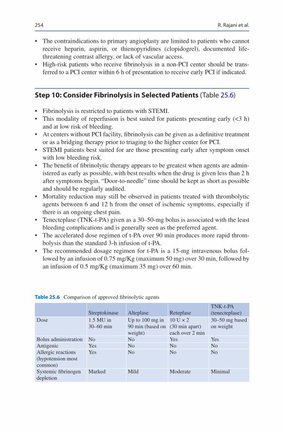
254
• The contraindications to primary angioplasty are limited to patients who cannot receive heparin, aspirin, or thienopyridines (clopidogrel), documented life- threatening contrast allergy, or lack of vascular access.
• High-risk patients who receive fibrinolysis in a non-PCI center should be trans-ferred to a PCI center within 6 h of presentation to receive early PCI if indicated.
Step 10: Consider Fibrinolysis in Selected Patients (Table 25.6)
• Fibrinolysis is restricted to patients with STEMI.• This modality of reperfusion is best suited for patients presenting early (<3 h)
and at low risk of bleeding.• At centers without PCI facility, fibrinolysis can be given as a definitive treatment
or as a bridging therapy prior to triaging to the higher center for PCI.• STEMI patients best suited for are those presenting early after symptom onset
with low bleeding risk.• The benefit of fibrinolytic therapy appears to be greatest when agents are admin-
istered as early as possible, with best results when the drug is given less than 2 h after symptoms begin. “Door-to-needle” time should be kept as short as possible and should be regularly audited.
• Mortality reduction may still be observed in patients treated with thrombolytic agents between 6 and 12 h from the onset of ischemic symptoms, especially if there is an ongoing chest pain.
• Tenecteplase (TNK-t-PA) given as a 30–50-mg bolus is associated with the least bleeding complications and is generally seen as the preferred agent.
• The accelerated dose regimen of t-PA over 90 min produces more rapid throm-bolysis than the standard 3-h infusion of t-PA.
• The recommended dosage regimen for t-PA is a 15-mg intravenous bolus fol-lowed by an infusion of 0.75 mg/Kg (maximum 50 mg) over 30 min, followed by an infusion of 0.5 mg/Kg (maximum 35 mg) over 60 min.
Table 25.6 Comparison of approved fibrinolytic agents
Streptokinase Alteplase ReteplaseTNK-t-PA (tenecteplase)
Dose 1.5 MU in 30–60 min
Up to 100 mg in 90 min (based on weight)
10 U × 2 (30 min apart) each over 2 min
30–50 mg based on weight
Bolus administration No No Yes YesAntigenic Yes No No NoAllergic reactions (hypotension most common)
Yes No No No
Systemic fibrinogen depletion
Marked Mild Moderate Minimal
R. Rajani et al.

255
• It is associated with lower mortality and slight increase in incidence of intracra-nial hemorrhage but with a better overall composite outcome of death and dis-abling stroke.
• Contraindications for thrombolysis should be considered prior to using lytic agents (see Chap. 11, Vol. 1).
• Intracranial hemorrhage may be fatal in half to two-thirds of patients and remains a devastating peril of thrombolytic therapy.
• In a comparative analysis, the risk of intracranial hemorrhage was found to be 1% with thrombolysis and 0.05% with PCI.
• Bleeding can also occur in sites like the gut or other sites including those of recent trauma or surgery.
• Other complications are reperfusion arrhythmias and allergic reactions.
Step 11: Consider Adjunctive Therapies
• Nitroglycerine – Nitrates have a limited role in ACS. – The indication in the setting of an ACS is limited to ongoing chest pain or
ischemia, hypertension, or as a vasodilator in the management of pulmonary edema secondary to left ventricular (LV) failure.
– Route and dose of nitrate should be individualized. – Due precaution should be taken while using nitrates (see Step 5).
• β-Blockers – Oral β-blockers should be used within the first 24 h in those without evidence
of heart failure, low cardiac output, shock, bradyarrhythmias, or conduction blocks and other conventional contraindication like asthma.
– Cardioselective agents without intrinsic sympathomimetic activity are pre-ferred. Metoprolol (50–200 mg/day) is the most commonly used agent.
– Intravenous β-blocker is used (metoprolol 5 mg IV thrice a day) titrated to patient response, heart rate (avoid if <50/min), and blood pressure (avoid if less than 100 mmHg systolic).
– Its use is best limited to situations with ongoing chest pain or uncontrolled tachycardia or hypertension.
– Calcium channel blocking agents (diltiazem) may be used as alternative ther-apy in patients with contraindication to β-blockers but should be avoided in patients with poor left ventricular function and pulmonary congestion.
• Anticoagulants: Indications – Low-molecular-weight heparins (enoxaparin 1 mg/Kg 12-hourly SC or dalte-
parin 120 U/Kg 12-hourly SC), or fondaparinux (in patient at high risk of bleeding), are commonly used agents specially in patients who have received fibrinolytic therapy .
– Bivalirudin (0.1 mg/Kg bolus followed by 0.25 mg/Kg/h infusion) may be considered as an alternative agent specially in patients who have received Clopidrogel as an antiplatelet .
25 Acute Coronary Syndromes
256
– Appropriate dosing adjustments need to be done for renal dysfunction. – As with antiplatelets, active bleeding and other contraindications need to be
excluded, and the regime should be timed in consultation with cardiologist for patients requiring intervention.
– Conventional unfractionated heparin is preferable if rapid reversal prior to surgery (CABG) is anticipated and in patients treated with primary PCI and ticagreolol or prasugrel.
– It should be given as a bolus of 60 U/Kg (maximum 4000 units), then 14 U/Kg/h (maximum 1000 U/h)—titrate to APTT 2.5 times control (see in Chap. 9, Vol. 1).
– A weight-based nomogram may be followed for heparin titration. – If intervention is not done, the duration for continuing anticoagulation is usu-
ally 5–10 days.• GP IIb/IIIa inhibitors
– Abciximab 0.25 mg/Kg IV bolus and then 0.125 mcg/Kg/min (maximum 10 mcg/min) may be given “upstream” prior to primary PCI in the catheteriza-tion laboratory.
– Eptifibatide 180 mcg/Kg bolus, then 2 mcg/Kg/min infusion or tirofiban 0.4 mcg/Kg/min for 30 min, and then 0.1 mcg/Kg/min infusion should be started as a part of early intervention strategy in high-risk patients with non-STEMI.
• Angiotensin-converting enzyme (ACE) inhibitors and angiotensin receptor blockers – These drugs should be used within the first 24 h of ACS in patients without
hypotension. – They are most beneficial in patients with pulmonary congestion and depressed
LV systolic function.• Statins
– Start 80 mg of atorvastatin or equivalent statin as early as possible and prefer-ably before PCI in patients not on statin.
– The dose can later be decreased to achieve the target lipids (LDL < 70–100 mg/dL).
– Patients who are already on statin should be continued on it.
Step 12: Manage Arrhythmic Complication
• Primary ventricular fibrillation (VF) accounts for the majority of early deaths during AMI. The incidence of primary VF is highest during the first 4 h after the onset of symptoms but remains an important contributor to mortality during the first 24 h.
• Secondary VF occurring in the setting of congestive heart failure or cardiogenic shock can also contribute to death from AMI.
• Prompt defibrillation and appropriate pharmacotherapy should be instituted (see Chap. 19, Vol. 1).
R. Rajani et al.
257
• Maintain serum potassium greater than 4 mEq/L and magnesium level more than 2 mEq/L. Cautious replacement to be done in patients with significant impair-ment of renal function
• Prophylactic antiarrhythmics are not recommended.
Step 13: Manage Mechanical Complications
• Cardiogenic shock, LV failure, and congestive heart failure should be managed with appropriate pharmacological therapy and rarely with mechanical devices
• Ventricular, septal, or papillary muscle rupture need emergent surgery• RV infarction or ischemia may occur in up to 50% of patients with inferior wall
MI.• Suspect RV infarction in patients with inferior wall infarction, hypotension, and
clear lung fields.• In patients with inferior wall infarction, obtain an ECG with right-sided leads.
Look for ST-segment elevation (>1 mm) in lead V4r.• Nitrates, diuretics, and other vasodilators (ACE inhibitors) should be avoided
because severe hypotension may result.• Hypotension in patients with suspected RV involvement is initially treated with
an IV fluid bolus.
Step 14: Consider Other Supportive Therapy
• Sedatives and laxatives are useful in the initial stage.• Hyperglycemia, either stress-induced or due to preexisting diabetes, should be
controlled preferably at near-normal levels.• Routine medication taken by the patient prior to the ACS should be reintroduced
as appropriate.
Suggested Reading
Amsterdam EA, Wenger NK, Brindis RG, et al. AHA/ACC guideline for the management of patients with non-ST-elevation acute coronary syndromes: executive summary: a report of the American College of Cardiology/American Heart Association Task Force on Practice Guidelines. Circulation. 2014;130:2354. A comprehensive guidelines for the management of non ST-elevation coronary syndromes.
Anderson JL, Morrow DA. Acute myocardial infarction. N Engl J Med. 2017;376:2053–64. This article describes the initial presentation and in-hospital management of acute myocar-dial infarction, including selection of a management strategy and options for antithrombotic therapy.
Antman EM, Cohen M. The TIMI risk score for unstable angina/non-ST elevation MI: a method for prognostication and therapeutic decision making. JAMA. 2000;284(7):835–42. The 7 TIMI risk score predictor variables were 65 years or older, at least three risk factors for coronary
25 Acute Coronary Syndromes

258
artery disease, prior coronary stenosis of 50% or more, ST-segment deviation on electrocardio-gram at presentation, at least two anginal events in prior 24 h, use of aspirin in prior 7 days, and elevated serum cardiac marker.
Siemieniuk RAC, Chu DK, Kim LH, et al. Oxygen therapy for acutely ill medical patients: a clini-cal practice guideline. BMJ. 2018;k4169:363. BMJ guidelines for oxygen therapy for a variety of conditions including acute coronary syndrome.
Website
www.acc.org. A comprehensive website of American College of Cardiology.
R. Rajani et al.
259© Springer Nature Singapore Pte Ltd. 2020R. Chawla, S. Todi (eds.), ICU Protocols, https://doi.org/10.1007/978-981-15-0898-1_26
V. Agrawal (*) Department of Cardiology, Medanta, The Medicity, Gurugram, India
Y. Mehta Department of Anaesthesia and Critical Care, Medanta, The Medicity, Gurugram, India
26Hypertensive Urgencies and Emergencies
Vinayak Agrawal and Yatin Mehta
Hypertensive crises represents 3% of all medical urgencies, with a prevalence of 24% and 76%, for hypertensive emergencies and urgencies, respectively, Hypertensive emergencies occur in up to 2% of hypertensive patients with a pro-gressive decrease in mortality rate over the past 4 decades. Risk factors include female sex, obesity, high BMI, hypertensive or coronary heart disease, higher num-ber of antihypertensive drugs and, most importantly, nonadherence to medication. Prompt recognition, evaluation and appropriate treatment of these crises are critical to prevent high morbidity and mortality.
Step 1: Identify Hypertensive Emergency
• Assess severity of hypertension and urgency of treatment• Hypertensive emergency is an acute increase in BP associated with severe,
potentially life-threatening target end-organ damage mostly requiring immediate hospitalization in an intensive care unit (ICU) for prompt BP control within min-utes or a few hours by intravenous administration of antihypertensive drugs.
A 65-year-old long standing diabetic and hypertensive lady presented to the emergency department with acute onset orthopnea. Blood pressure recorded was 230/130 mmHg, pulse 120/min regular, oxygen saturation at 86% at room air, orthopneic and very distressed with intact higher mental functions. Clinical examination revealed a left ventricular S3 gallop, with bibasal soft crepitations. Biochemical tests revealed deranged renal function test and proteinuria.
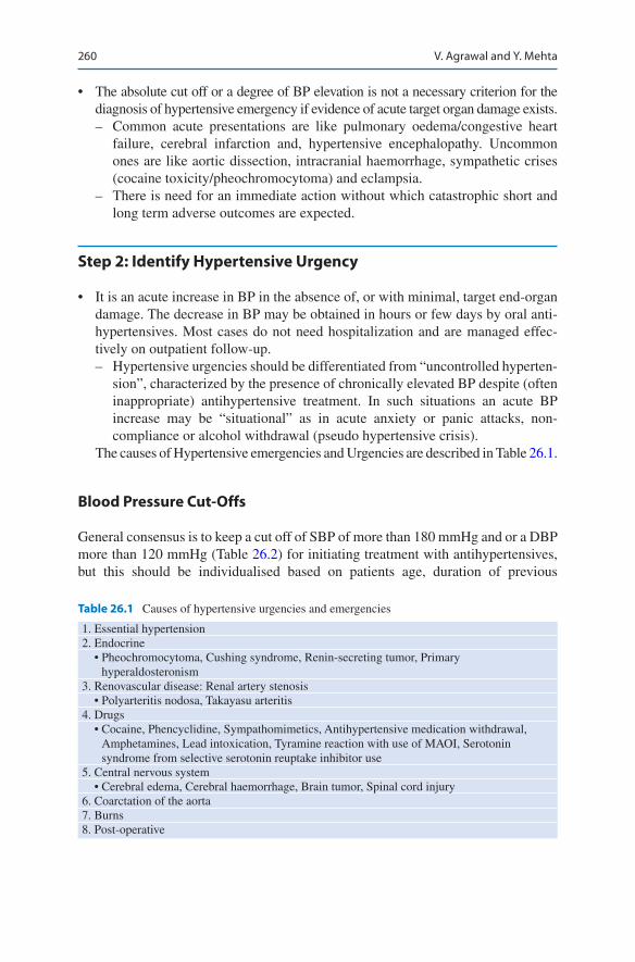
260
• The absolute cut off or a degree of BP elevation is not a necessary criterion for the diagnosis of hypertensive emergency if evidence of acute target organ damage exists. – Common acute presentations are like pulmonary oedema/congestive heart
failure, cerebral infarction and, hypertensive encephalopathy. Uncommon ones are like aortic dissection, intracranial haemorrhage, sympathetic crises (cocaine toxicity/pheochromocytoma) and eclampsia.
– There is need for an immediate action without which catastrophic short and long term adverse outcomes are expected.
Step 2: Identify Hypertensive Urgency
• It is an acute increase in BP in the absence of, or with minimal, target end-organ damage. The decrease in BP may be obtained in hours or few days by oral anti-hypertensives. Most cases do not need hospitalization and are managed effec-tively on outpatient follow-up. – Hypertensive urgencies should be differentiated from “uncontrolled hyperten-
sion”, characterized by the presence of chronically elevated BP despite (often inappropriate) antihypertensive treatment. In such situations an acute BP increase may be “situational” as in acute anxiety or panic attacks, non- compliance or alcohol withdrawal (pseudo hypertensive crisis).
The causes of Hypertensive emergencies and Urgencies are described in Table 26.1.
Blood Pressure Cut-Offs
General consensus is to keep a cut off of SBP of more than 180 mmHg and or a DBP more than 120 mmHg (Table 26.2) for initiating treatment with antihypertensives, but this should be individualised based on patients age, duration of previous
Table 26.1 Causes of hypertensive urgencies and emergencies
1. Essential hypertension 2. Endocrine • Pheochromocytoma, Cushing syndrome, Renin-secreting tumor, Primary
hyperaldosteronism 3. Renovascular disease: Renal artery stenosis • Polyarteritis nodosa, Takayasu arteritis 4. Drugs • Cocaine, Phencyclidine, Sympathomimetics, Antihypertensive medication withdrawal,
Amphetamines, Lead intoxication, Tyramine reaction with use of MAOI, Serotonin syndrome from selective serotonin reuptake inhibitor use
5. Central nervous system • Cerebral edema, Cerebral haemorrhage, Brain tumor, Spinal cord injury 6. Coarctation of the aorta7. Burns8. Post-operative
V. Agrawal and Y. Mehta

261
hypertension and specific target organ damage due to hypertension and underlying disease process.
Pathophysiology of Hypertensive Emergencies & Urgencies
• Abrupt unexpected rise in systemic vascular resistance result in acute mechani-cal stress on endothelium leading to dysfunction, enhanced vascular permeabil-ity, platelet activation, trigger of coagulation cascade and fibrin deposition.
• Increased humoral vasoconstrictors and inflammatory cytokines eventually lead to acute onset arterial vasoconstriction and thrombosis, vascular damage, hypo-perfusion, end-organ ischemia and TOD.
Step 3: Evaluate for Target Organ Involvement/TOD (Table 26.3)
1. Pertinent/focussed History with leading questions and relevant clinical examination: (a) Symptoms: e.g. excessive and recent onset chest pain/angina, dyspnea,
orthopnea, decreased urine output, effort intolerance, weakness/fatigue, pal-pitations or sensory loss, severe headache, altered sensorium, imbalance or weakness).
(b) Current complete medication list: names, doses, any recent changes in their administration, and sudden discontinuations from previously taken prescrip-tions (non-compliance), use of any sympatholytic medications such as cloni-dine or beta-blockers, sudden stoppage of which can cause severe rebound hypertension, recent use of oral contraceptive pills, monoamine oxidase
Table 26.2 Hypertensive emergency vs hypertensive urgency
Hypertensive emergency Hypertensive urgencySystolic BP >180 mmHg and/or >180 mmHg and/orDiastolic BP ≥120 mm Hg ≥120 mm HgSymptoms Yes No/minimalAcute target organ damage (TOD)
Yes No
Time recommended for BP reduction
Within minutes/hours.Admit patient to an intensive care unit for continuous BP monitoring and parenteral antihypertensive administration
Hours to few daysReinstitute or intensify antihypertensive drug therapy and treat anxiety as applicable
Evaluation for secondary HT
Needed Needed
Remarks Many are noncompliant with antihypertensive therapy
26 Hypertensive Urgencies and Emergencies
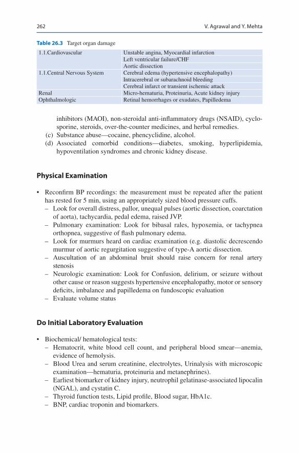
262
inhibitors (MAOI), non-steroidal anti-inflammatory drugs (NSAID), cyclo-sporine, steroids, over-the-counter medicines, and herbal remedies.
(c) Substance abuse—cocaine, phencyclidine, alcohol. (d) Associated comorbid conditions—diabetes, smoking, hyperlipidemia,
hypoventilation syndromes and chronic kidney disease.
Physical Examination
• Reconfirm BP recordings: the measurement must be repeated after the patient has rested for 5 min, using an appropriately sized blood pressure cuffs. – Look for overall distress, pallor, unequal pulses (aortic dissection, coarctation
of aorta), tachycardia, pedal edema, raised JVP. – Pulmonary examination: Look for bibasal rales, hypoxemia, or tachypnea
orthopnea, suggestive of flash pulmonary edema. – Look for murmurs heard on cardiac examination (e.g. diastolic decrescendo
murmur of aortic regurgitation suggestive of type-A aortic dissection. – Auscultation of an abdominal bruit should raise concern for renal artery
stenosis – Neurologic examination: Look for Confusion, delirium, or seizure without
other cause or reason suggests hypertensive encephalopathy, motor or sensory deficits, imbalance and papilledema on fundoscopic evaluation
– Evaluate volume status
Do Initial Laboratory Evaluation
• Biochemical/ hematological tests: – Hematocrit, white blood cell count, and peripheral blood smear—anemia,
evidence of hemolysis. – Blood Urea and serum creatinine, electrolytes, Urinalysis with microscopic
examination—hematuria, proteinuria and metanephrines). – Earliest biomarker of kidney injury, neutrophil gelatinase-associated lipocalin
(NGAL), and cystatin C. – Thyroid function tests, Lipid profile, Blood sugar, HbA1c. – BNP, cardiac troponin and biomarkers.
Table 26.3 Target organ damage
1.1.Cardiovascular Unstable angina, Myocardial infarctionLeft ventricular failure/CHFAortic dissection
1.1.Central Nervous System Cerebral edema (hypertensive encephalopathy)Intracerebral or subarachnoid bleedingCerebral infarct or transient ischemic attack
Renal Micro-hematuria, Proteinuria, Acute kidney injuryOphthalmologic Retinal hemorrhages or exudates, Papilledema
V. Agrawal and Y. Mehta

263
• If secondary hypertension suspected – Serum Renin and aldosterone ratio, Serum catecholamines. – Plasma cortisol and dexamethasone suppression test—Cushing’s syndrome.
• Electrocardiogram—ST-T changes, evidence of left ventricular hypertrophy (LVH).• Chest radiograph—cardiomegaly and pulmonary congestion, aortic pathology.• Echocardiogram (LVH, left ventricular dysfunction, wall motion abnormalities,
valve abnormalities, aortic dissection, diastolic dysfunction). 2D speckled track-ing for strain measurements which may be depressed in HT crisis.
• USG whole abdomen to assess kidneys, adrenals.• Non-contrast plain CT-head (if neurologic findings).• CT aortogram to rule out aortic dissection if clinically relevant
– Fundoscopy, or Optic nerve sheath diameter by bedside Ultrasonography can detect early papilledema.
Step 4: Understand Management Principles and Achievable Goals
Hypertensive Emergency
To maintain adequate and stable blood flow to the vital organs like brain, heart, and kidneys, autoregulation of vasculature is of prime importance. The threshold of hypoperfusion is approximately 20–25% lower than the existing blood pressure and this physiologic rationale is behind the recommendation to limit the initial blood pressure reduction to 20–25% of pretreatment values (Table 26.4).
• Immediate goal—lower BP preferably in an intensive care unit with intra-arterial line for clinical surveillance and continuous BP monitoring.
• Prompt intravenous administration of short-acting and titratable drugs is pre-ferred in the first few minutes of treatment.
Hypertensive Urgency
(a) Immediate goal—lower blood pressure within 24–72 h. (b) Treatment setting—clinical discretion is required. (c) Medications—oral medications with rapid onset of action; occasionally
intravenously.
Table 26.4 Goals of decreasing BP
Time % decrease from baseline BP as goalWithin 2 h By 15–25%Within 12 h By 25%Within 48 h By 30%
26 Hypertensive Urgencies and Emergencies

264
Step 5: Get Familiar with Drugs Used in Hypertensive Urgencies and Emergencies (Tables 26.5, 26.6 and 26.7)
Table 26.5 Drugs for hypertensive urgencies
Agent DoseOnset of action Comment
Captopril 12.5–25 mg PO 15–60 min Can precipitate acute renal failure in patients with bilateral renal artery stenosis
Nifedipine (extended release)
10–20 mg PO 20 min Avoid short-acting or sublingual nifedipine due to risk of sudden hypotension, stroke, cardiac event
Labetalol 200–400 mg PO 20–120 min Heart failure, bradycardia, bronchospasmClonidine 0.1–0.2 mg PO 30–60 min Rebound hypertension due to abrupt
withdrawalPrazosin 1–2 mg PO 2–4 h First-dose hypotension, syncope, tachycardiaAmlodipine 5–10 mg PO 30–50 min Headache, tachycardia, flushing
Table 26.6 Parenteral medications used for treatment of hypertensive crisis
Drug Dosing Onset of action Preload AfterloadCardiac output
Sodium nitroprusside
0.25–10 μg/kg/min IV infusion
Within seconds to minutes
Decreased Decreased No effect
Nitroglycerin 5–100 μg/min IV infusion
1–5 min Decreased Decreased No effect
Labetalol 20–80 mg bolus every 10 minor 0.5–2 mg/min IV infusion
5–10 min No effect Decreased Decreased
Esmolol 500 mcg/kg or 30 mg bolus over 30 s then50–150 mcg/kg/min infusion
1–2 min No effect Decreased Decreased
Hydralazine 10–20 mg IV bolus
10–20 min No effect Decreased Increased
Phentolamine 5–15 mg IV bolus 1–2 min No effect Decreased Increased
Table 26.7 Special indications and warnings for parenteral medications
Drug Special indications WarningsSodium nitroprusside
Most hypertensive emergencies
Caution with renal insufficiency; can develop cyanide toxicity, acidosis, methemoglobinemia, increased intracranial pressure, nausea, vomiting, muscle twitching, theoretical “coronary steal” (shunting of blood from diseased vessels to well-perfused vessels may produce coronary ischemia)
Nitroglycerin Most hypertensive emergencies, coronary ischemia
Headache; can develop tolerance, tachycardia, vomiting, methemoglobinemia, flushing
V. Agrawal and Y. Mehta
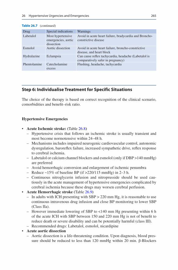
265
Step 6: Individualise Treatment for Specific Situations
The choice of the therapy is based on correct recognition of the clinical scenario, comorbidities and benefit–risk ratio.
Hypertensive Emergencies
• Acute Ischemic stroke: (Table 26.8) – Hypertensive crisis that follows an ischemic stroke is usually transient and
most become normotensive within 24–48 h. – Mechanisms includes impaired neurogenic cardiovascular control, autonomic
dysregulation, baroreflex failure, increased sympathetic drive, reflex response to cerebral ischemia.
– Labetalol or calcium channel blockers and esmolol (only if DBP >140 mmHg) are preferred
– Avoid hemorrhagic conversion and enlargement of ischemic penumbra – Reduce ~15% of baseline BP (if >220/115 mmHg) in 2–3 h. – Continuous nitroglycerin infusion and nitroprusside should be used cau-
tiously in the acute management of hypertensive emergencies complicated by cerebral ischemia because these drugs may worsen cerebral perfusion.
• Acute Hemorrhagic stroke (Table 26.9) – In adults with ICH presenting with SBP > 220 mm Hg, it is reasonable to use
continuous intravenous drug infusion and close BP monitoring to lower SBP (Class IIa).
– However immediate lowering of SBP to <140 mm Hg presenting within 6 h of the acute ICH with SBP between 150 and 220 mm Hg is not of benefit to reduce death or severe disability and can be potentially harmful (class III).
– Recommended drugs: Labetalol, esmolol, nicardipine• Acute aortic dissection
– Aortic dissection is a life-threatening condition. Upon diagnosis, blood pres-sure should be reduced to less than 120 mmHg within 20 min. β-Blockers
Drug Special indications WarningsLabetalol Most hypertensive
emergencies, aortic dissection
Avoid in acute heart failure, bradycardia and Broncho- constrictive disease
Esmolol Aortic dissection Avoid in acute heart failure, broncho-constrictive disease, and heart block
Hydralazine Eclampsia Can cause reflex tachycardia, headache (Labetalol is comparatively safer in pregnancy)
Phentolamine Catecholamine excess
Flushing, headache, tachycardia
Table 26.7 (continued)
26 Hypertensive Urgencies and Emergencies
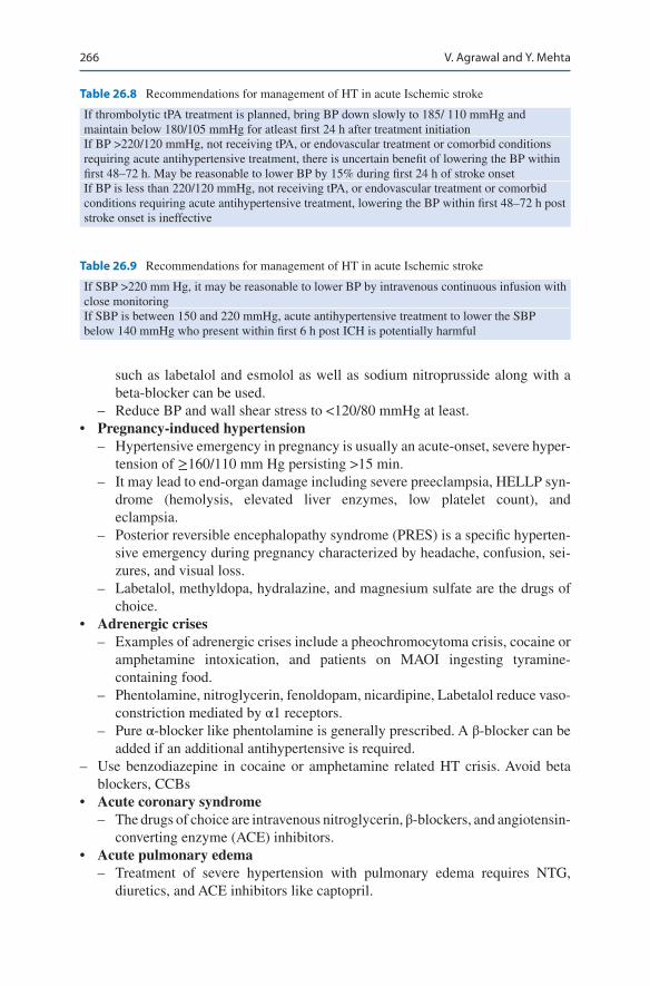
266
such as labetalol and esmolol as well as sodium nitroprusside along with a beta-blocker can be used.
– Reduce BP and wall shear stress to <120/80 mmHg at least.• Pregnancy-induced hypertension
– Hypertensive emergency in pregnancy is usually an acute-onset, severe hyper-tension of ≥160/110 mm Hg persisting >15 min.
– It may lead to end-organ damage including severe preeclampsia, HELLP syn-drome (hemolysis, elevated liver enzymes, low platelet count), and eclampsia.
– Posterior reversible encephalopathy syndrome (PRES) is a specific hyperten-sive emergency during pregnancy characterized by headache, confusion, sei-zures, and visual loss.
– Labetalol, methyldopa, hydralazine, and magnesium sulfate are the drugs of choice.
• Adrenergic crises – Examples of adrenergic crises include a pheochromocytoma crisis, cocaine or
amphetamine intoxication, and patients on MAOI ingesting tyramine- containing food.
– Phentolamine, nitroglycerin, fenoldopam, nicardipine, Labetalol reduce vaso-constriction mediated by α1 receptors.
– Pure α-blocker like phentolamine is generally prescribed. A β-blocker can be added if an additional antihypertensive is required.
– Use benzodiazepine in cocaine or amphetamine related HT crisis. Avoid beta blockers, CCBs
• Acute coronary syndrome – The drugs of choice are intravenous nitroglycerin, β-blockers, and angiotensin-
converting enzyme (ACE) inhibitors.• Acute pulmonary edema
– Treatment of severe hypertension with pulmonary edema requires NTG, diuretics, and ACE inhibitors like captopril.
Table 26.8 Recommendations for management of HT in acute Ischemic stroke
If thrombolytic tPA treatment is planned, bring BP down slowly to 185/ 110 mmHg and maintain below 180/105 mmHg for atleast first 24 h after treatment initiationIf BP >220/120 mmHg, not receiving tPA, or endovascular treatment or comorbid conditions requiring acute antihypertensive treatment, there is uncertain benefit of lowering the BP within first 48–72 h. May be reasonable to lower BP by 15% during first 24 h of stroke onsetIf BP is less than 220/120 mmHg, not receiving tPA, or endovascular treatment or comorbid conditions requiring acute antihypertensive treatment, lowering the BP within first 48–72 h post stroke onset is ineffective
Table 26.9 Recommendations for management of HT in acute Ischemic stroke
If SBP >220 mm Hg, it may be reasonable to lower BP by intravenous continuous infusion with close monitoringIf SBP is between 150 and 220 mmHg, acute antihypertensive treatment to lower the SBP below 140 mmHg who present within first 6 h post ICH is potentially harmful
V. Agrawal and Y. Mehta
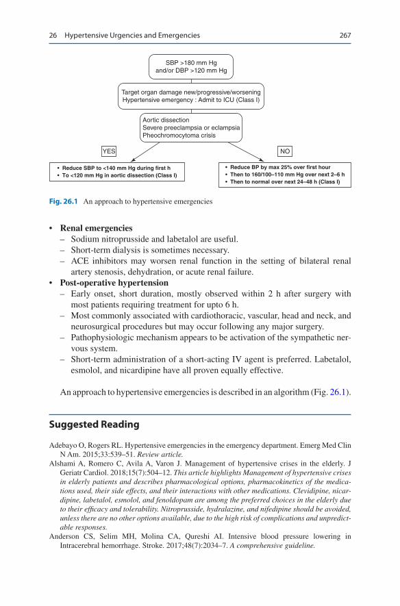
267
• Renal emergencies – Sodium nitroprusside and labetalol are useful. – Short-term dialysis is sometimes necessary. – ACE inhibitors may worsen renal function in the setting of bilateral renal
artery stenosis, dehydration, or acute renal failure.• Post-operative hypertension
– Early onset, short duration, mostly observed within 2 h after surgery with most patients requiring treatment for upto 6 h.
– Most commonly associated with cardiothoracic, vascular, head and neck, and neurosurgical procedures but may occur following any major surgery.
– Pathophysiologic mechanism appears to be activation of the sympathetic ner-vous system.
– Short-term administration of a short-acting IV agent is preferred. Labetalol, esmolol, and nicardipine have all proven equally effective.
An approach to hypertensive emergencies is described in an algorithm (Fig. 26.1).
Suggested Reading
Adebayo O, Rogers RL. Hypertensive emergencies in the emergency department. Emerg Med Clin N Am. 2015;33:539–51. Review article.
Alshami A, Romero C, Avila A, Varon J. Management of hypertensive crises in the elderly. J Geriatr Cardiol. 2018;15(7):504–12. This article highlights Management of hypertensive crises in elderly patients and describes pharmacological options, pharmacokinetics of the medica-tions used, their side effects, and their interactions with other medications. Clevidipine, nicar-dipine, labetalol, esmolol, and fenoldopam are among the preferred choices in the elderly due to their efficacy and tolerability. Nitroprusside, hydralazine, and nifedipine should be avoided, unless there are no other options available, due to the high risk of complications and unpredict-able responses.
Anderson CS, Selim MH, Molina CA, Qureshi AI. Intensive blood pressure lowering in Intracerebral hemorrhage. Stroke. 2017;48(7):2034–7. A comprehensive guideline.
SBP >180 mm Hgand/or DBP >120 mm Hg
Target organ damage new/progressive/worseningHypertensive emergency : Admit to ICU (Class I)
Aortic dissectionSevere preeclampsia or eclampsiaPheochromocytoma crisis
YES NO
• Reduce SBP to <140 mm Hg during first h• To <120 mm Hg in aortic dissection (Class I)
• Reduce BP by max 25% over first hour• Then to 160/100–110 mm Hg over next 2–6 h• Then to normal over next 24–48 h (Class I)
Fig. 26.1 An approach to hypertensive emergencies
26 Hypertensive Urgencies and Emergencies

268
Jauch EC, Saver JL, Adams HP Jr. Guidelines for the early management of patients with acute ischemic stroke: a guideline for healthcare professionals from the American Heart Association/American Stroke Association. American Heart Association Stroke Council; Council on Cardiovascular Nursing; Council on Peripheral Vascular Disease; Council on Clinical Cardiology. Stroke. 2013;44(3):870–947. A authoritative guideline on the subject.
Marik PE, Rivera R. Hypertensive emergencies: an update. Curr Opin Crit Care. 2011;17:569–80. Review article.
Padilla Ramos A, Varon J. Current and newer agents for hypertensive emergencies. Curr Hypertens Rep. 2014;16(7):450. This article reviews commonly used antihypertensive medications as well as evidence-based recommendations for state-of-the-art treatment for hypertensive emergencies.
Whelton PK, Carey RM, et al. ACC/AHA/AAPA/ABC/ACPM/AGS/APhA/ASH/ASPC/NMA/PCNA guideline for the prevention, detection, evaluation, and management of high blood pres-sure in adults: executive summary: a report of the American College of Cardiology/American Heart Association Task Force on Clinical Practice Guidelines. J Am Coll Cardiol. 2017; https://doi.org/10.1016/j.jacc.2017.11.005. A consensus guideline.
V. Agrawal and Y. Mehta

Part III
Nervous System

271© Springer Nature Singapore Pte Ltd. 2020R. Chawla, S. Todi (eds.), ICU Protocols, https://doi.org/10.1007/978-981-15-0898-1_27
V. Suri (*) Department of Neurosciences, Indraprastha Apollo Hospitals, New Delhi, India
K. Suri Department of Medicine, Gitanjali Medical College, Udaipur, India
27Acute Ischemic Stroke (AIS)
Vinit Suri and Kunal Suri
Acute ischemic stroke is amongst the most frequent causes of admission to an ICU. It is essential to treat them as a medical/neurological emergency as early as possible to salvage the brain.
Step 1: Identify Signs of a Possible Stroke—BEFAST
It is important to identify early signs of stroke. BEFAST:
• B: Sudden Balance Impairment• E: Sudden vision loss in one Eye or one field• F: Facial droop (have patient show teeth or smile)• A: Arm drift (patient closes eyes and extends both arms straight out, with palm
up [for 10 s).• S: Abnormal Speech.• T: Time to call emergency
Step 2: Prehospital Management and Exclude Common Mimics Such as Hyoglycemia
• Assess the airway, breathing and circulation.• Monitor patient with pulse oximetry. Begin oxygen therapy on any patient with
Sp02 < 94%. Routine oxygen therapy is not recommended for all stroke patients.
64 years old male patient develops abrupt onset weakness of right upper and lower limb with inability to speak noticed 30 min ago.

272
• Check the patient’s blood sugar. Hypoglycemia can cause the patient to present with stroke symptoms. Correct hypoglycemia (Blood Sugar <60 mg/dL) which maybe the cause of the deficit.
• Do quick neurological examination.• Exclude stroke mimics by history and physical exam like seizure with post ictal paral-
ysis, functional deficit (conversion reaction), hypertensive encephalopathy and syncope.
• Determine the time of Onset of symptoms. This information is critical for the patient who present with stroke symptoms. Establish time when the patient was last known to be neurologically normal. If the patient was sleeping and wakes up with symptoms, time last known well (LKW) is the last time the patient was seen to be normal before sleeping.
Establish a baseline 12 lead ECG to determine any arrhythmias.
• Establish an IV route preferably on the non-paralysed arm.• Avoid dextrose containing fluids. Normal saline can be administered for
euvolemic patients at the rate of 50 mL/h.• Check Blood Pressure. Do not lower BP unless it is >220/120 mmHg. Avoid
rapid lowering of blood pressure by medications like sublingual nifedipine or intravenous agents.
Prenotification
• Update the receiving Emergency Department so that their staff can prepare for the patient’s arrival and begin therapeutic efforts without delay.
• Transport the patient as early as possible.• Do not delay transport. Speed is vital for the most optimal patient outcome.• Assess neurological status while the patient is being transported.
Step 3: Triage Management
• Sequence of events in the emergency department (faster treatment-better clinical outcomes).
• NINDS (National Institute of Neurological Disorders and Stroke and ACLS Recommended Stroke Evaluation Time Benchmarks for Potential Thrombolysis Candidate (Table 27.1).
• NINDS (National Institute of Neurological Disorders and Stroke) time goal.• Remember patients are eligible for IV rtPA up to 4.5 h from onset.
Table 27.1 Stroke evaluation time benchmarks for potential thrombolysis
Time interval Time targetDoor to doctor 10 minAccess to neurologic expertise 15 minDoor to CT scan completion 25 minDoor to CT scan interpretation 45 minDoor to treatment 60 minAdmission to stroke unit or ICU 3 h
V. Suri and K. Suri
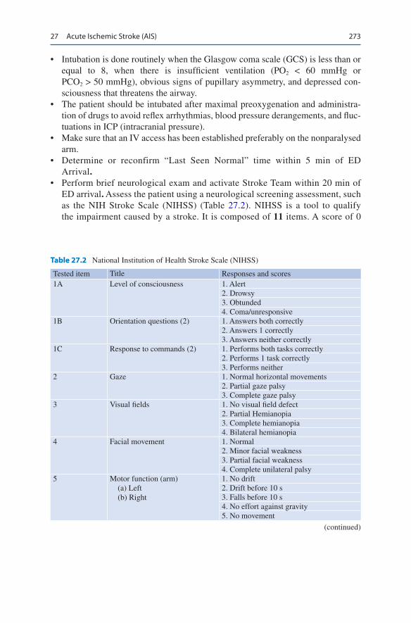
273
• Intubation is done routinely when the Glasgow coma scale (GCS) is less than or equal to 8, when there is insufficient ventilation (PO2 < 60 mmHg or PCO2 > 50 mmHg), obvious signs of pupillary asymmetry, and depressed con-sciousness that threatens the airway.
• The patient should be intubated after maximal preoxygenation and administra-tion of drugs to avoid reflex arrhythmias, blood pressure derangements, and fluc-tuations in ICP (intracranial pressure).
• Make sure that an IV access has been established preferably on the nonparalysed arm.
• Determine or reconfirm “Last Seen Normal” time within 5 min of ED Arrival.
• Perform brief neurological exam and activate Stroke Team within 20 min of ED arrival. Assess the patient using a neurological screening assessment, such as the NIH Stroke Scale (NIHSS) (Table 27.2). NIHSS is a tool to qualify the impairment caused by a stroke. It is composed of 11 items. A score of 0
Table 27.2 National Institution of Health Stroke Scale (NIHSS)
Tested item Title Responses and scores1A Level of consciousness 1. Alert
2. Drowsy3. Obtunded4. Coma/unresponsive
1B Orientation questions (2) 1. Answers both correctly2. Answers 1 correctly3. Answers neither correctly
1C Response to commands (2) 1. Performs both tasks correctly2. Performs 1 task correctly3. Performs neither
2 Gaze 1. Normal horizontal movements2. Partial gaze palsy3. Complete gaze palsy
3 Visual fields 1. No visual field defect2. Partial Hemianopia3. Complete hemianopia4. Bilateral hemianopia
4 Facial movement 1. Normal2. Minor facial weakness3. Partial facial weakness4. Complete unilateral palsy
5 Motor function (arm) (a) Left (b) Right
1. No drift2. Drift before 10 s3. Falls before 10 s4. No effort against gravity5. No movement
(continued)
27 Acute Ischemic Stroke (AIS)

274
Table 27.2 (continued)
Tested item Title Responses and scores6 Motor function (leg) 1. No drift
2. Drift before 5 s3. Falls before 5 s4. No effort against gravity5. No movement
7 Limbs ataxia 1. No ataxia2. Ataxia in 1 limbs3. Ataxia in 2 limbs
8 Sensory 1. No sensory loss2. Mild sensory loss3. Severe sensory loss
9 Language 1. Normal2. Mild aphasia3. Severe aphasia4. Mute or global aphasia
10 Articulation 1. Normal2. Mild dysarthria3. Severe dysarthria
11 Extinction or inattention 1. Absent2. Mild loss (1 sensory modality lost)3. Severe loss (2 modalities lost)
NIHSS score Stroke severity0 No deficit1–4 Minor stroke5–15 Moderate stroke16–25 Moderate to senior stroke>25 Secure stroke
indicates normal function and the higher score indicates impairment with maximum score of 42.
• Perform non-contrast CT scan rapidly to rule out intracranial hemorrhage. Rule out stroke mimics like tumor and to estimate tissue at risk of infarc-tion within 25 min of ED arrival and it should be interpreted with in 45 min. Five Early Signs of Ischaemic Stroke on CT head are described in Table 27.3.
V. Suri and K. Suri
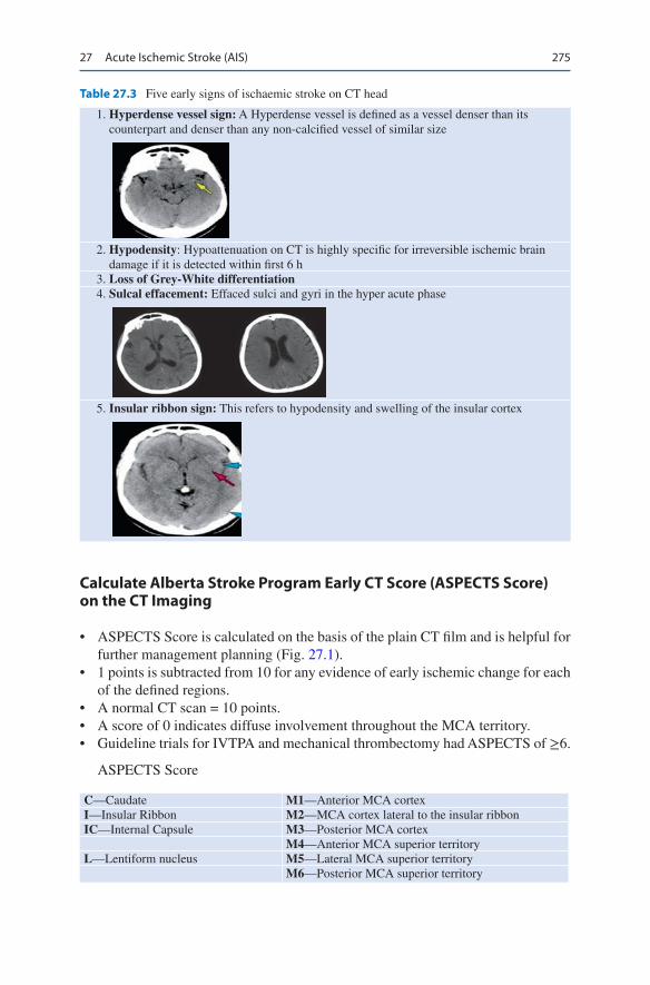
275
Table 27.3 Five early signs of ischaemic stroke on CT head
1. Hyperdense vessel sign: A Hyperdense vessel is defined as a vessel denser than its counterpart and denser than any non-calcified vessel of similar size
2. Hypodensity: Hypoattenuation on CT is highly specific for irreversible ischemic brain damage if it is detected within first 6 h
3. Loss of Grey-White differentiation 4. Sulcal effacement: Effaced sulci and gyri in the hyper acute phase
5. Insular ribbon sign: This refers to hypodensity and swelling of the insular cortex
Calculate Alberta Stroke Program Early CT Score (ASPECTS Score) on the CT Imaging
• ASPECTS Score is calculated on the basis of the plain CT film and is helpful for further management planning (Fig. 27.1).
• 1 points is subtracted from 10 for any evidence of early ischemic change for each of the defined regions.
• A normal CT scan = 10 points.• A score of 0 indicates diffuse involvement throughout the MCA territory.• Guideline trials for IVTPA and mechanical thrombectomy had ASPECTS of ≥6.
ASPECTS Score
C—Caudate M1—Anterior MCA cortexI—Insular Ribbon M2—MCA cortex lateral to the insular ribbonIC—Internal Capsule M3—Posterior MCA cortex
M4—Anterior MCA superior territoryL—Lentiform nucleus M5—Lateral MCA superior territory
M6—Posterior MCA superior territory
27 Acute Ischemic Stroke (AIS)
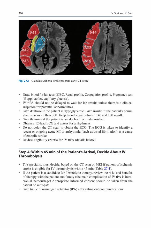
276
• Draw blood for lab tests (CBC, Renal profile, Coagulation profile, Pregnancy test (if applicable), capillary glucose).
• IV rtPA should not be delayed to wait for lab results unless there is a clinical suspicion for potential abnormalities.
• Give dextrose if the patient is hypoglycemic. Give insulin if the patient’s serum glucose is more than 300. Keep blood sugar between 140 and 180 mg/dL.
• Give thiamine if the patient is an alcoholic or malnourished.• Obtain a 12-lead ECG and assess for arrhythmias.• Do not delay the CT scan to obtain the ECG. The ECG is taken to identify a
recent or ongoing acute MI or arrhythmia (such as atrial fibrillation) as a cause of embolic stroke.
• Review eligibility criteria for IV rtPA (details below).
Step 4: Within 45 min of the Patient’s Arrival, Decide About IV Thrombolysis
• The specialist must decide, based on the CT scan or MRI if patient of ischemic stroke is eligible for IV thrombolysis within 45 min (Table 27.4).
• If the patient is a candidate for fibrinolytic therapy, review the risks and benefits of therapy with the patient and family (the main complication of IV tPA is intra-cranial hemorrhage) Appropriate informed consent should be taken from the patient or surrogate.
• Give tissue plasminogen activator (tPA) after ruling out contraindications
Fig. 27.1 Calculate Alberta stroke program early CT score
V. Suri and K. Suri

277
If the patient is rapidly improving and moving to normal, fibrinolytics may still be necessary if symptoms are minor but disabling.
Treatment
Recombinant tissue plasminogen activator (tPA) is given at a dose of 0.9 mg/Kg up to a maximum of 90 mg (10% of dose given as a bolus over 1 min and the remainder infused over 1 h) (Table 27.5)
• Anticoagulant and antithrombotic agents, such as warfarin, heparin, or antiplatelet drugs, should not be administered for at least 24 h after the tPA infusion is com-pleted and a follow-up CT scan at 24 h does not show intracranial hemorrhage
Table 27.4 Decision for IV thrombolysis: Indications and Contraindications
IndicationsWithin 4.5 h ((if exact time of stroke onset is unclear, it is defined as the time when the patient was last noted to be normal) • 18 years or more • With measurable persistent neurological deficit consistent with ischemic stroke • BP controlled to <185/110 • Glucose >50 mg/dLExclusion criteria: • ICH/SAH • Aortic arch dissection • Severe head trauma or ischemic stroke <3 month • Intracranial/spinal surgery <3 month • History of ICH • Intracranial neoplasm • Acute Bleeding diathesisRelative contraindications (special precautions) • Minor or rapidly improving stroke symptoms • Major surgery or serious trauma within the past 14 days • Recent gastrointestinal or urinary tract hemorrhage within the past 3 weeks • Post-myocardial infarction pericarditis • Recent acute myocardial infarction within the past 3 months • Abnormal blood sugar level < 50 mg/dL or > 400 mg/dL • Platelet count <100,000/mm3
• Heparin received within 48 h prior to onset to stroke, with elevated activated partial thromboplastic time (aPTT)
• Current use of anticoagulant (e.g., warfarin) with an elevated international normalized ratio (INR) > 1.7
• Infective endocarditisAdditional exclusion criteria for 3 to 4.5 h • Age > 80y • History of diabetes mellitus and prior stroke • Any OAC use with any PT • NIHSS Score > 25
27 Acute Ischemic Stroke (AIS)

278
• Invasive procedures, such as vascular puncture, urinary catheter placement, and nasogastric tube insertion, should be avoided for at least 24 h.
• Hemodynamic and close neurologic monitoring should be charted.• Consider intracerebral hemorrhage (ICH) to be the likely cause of neurological
worsening after the use of a thrombolytic drug until a brain scan confirms or refutes hemorrhage.
• If the patient is not a candidate for fibrinolytic therapy, give the patient aspirin. (first-dose 325 mg subsequent doses 100 mg–150 mg.)
• For both groups (those treated with tPA and those given aspirin), give the follow-ing basic stroke care: – Begin stroke pathway – Support patient’s airways, breathing, and circulation. – Check blood glucose – Watch for complications of stroke and fibrinolytic therapy. – Transfer patient to intensive care if indicated.
Patients with acute ischemic stroke who are hypoglycemic tend to have worse clini-cal outcomes, but there is no direct evidence that active glucose control improves outcomes. Consider giving IV or subcutaneous insulin to patients whose serum glu-cose levels are greater than 200 mg/dL.
Step 5: Managing Hemodynamics in tPA Candidates
For patients who are candidates for fibrinolytic therapy, blood pressures need to be controlled to lower their risk of intracerebral hemorrhage following administration of tPA. This as described in Table 27.6.
• Monitor the heart rate and hold infusion if it is less than 60/min.• Other agents that are recommended for treating hypertension in ischemic stroke
are sodium nitroprusside, nicardipine, angiotensin-converting enzyme inhibitor,
Table 27.5 Treatment of AIS: IV administration of alteplase
Infuse 0.9 mg/kg (maximum dose 90 mg) over 60 min with 10% of the dose given as a bolus over 1 minMonitor patientIf the patient develops severe headache, acute hypertension, nausea, or vomiting or has a worsening neurological examination—discontinue the infusion (If IV alteplase is being administered) and obtain urgent head CT scanMeasure BP and perform neurological assessments every 15 min during and alteplase infusion for 2 h, then every 30 min for 6 h, then hourly until 24 h after IV alteplase Increase the frequency of BP measurements if SBP is >180 mm Hg or if DBP is >105 mm HgDelay placement of nasogastric tubes, indwelling bladder catheters, or intra-arterial pressure catheters if the patient can be safely managed without themObtain a follow-up CT or MRI scan at 24 h later before starting anticoagulants or antiplatelet agents
V. Suri and K. Suri

279
and hydralazine. Nitrates should be avoided because they tend to increase intra-cranial pressure (ICP).
• If fibrinolytic is not given then control blood pressure as mentioned in Table 27.7.
Step 6: Manage Complication of IV tPA
The major complication of IV tPA is intracranial hemorrhage. Other bleeding com-plications, ranging from minor to severe, may also occur including systemic bleed-ing and, seizure, distal embolization, angioedema and transient hypotension can also occur. Hemorrhagic transformation occurs in 6–7% of patients.
Table 27.6 Blood pressure control in stroke patients eligible for a fibrinolytic therapy
PretreatmentSystolic >185 or diastolic >110
Labetalol 10 to 20 mg IV for 1–2 min—may repeat 1 time
During treatment
Keep < 180/105 mmhg
Monitor blood pressure
Monitor blood pressure every 15 min for 2 h, then every 30 min for 6 h and finally every hours for 16 h
Diastolic >140Systolic >230 or diastolic 121 to 140
Labetalol 10 mg iV for 1–2 min may repeat or double every 10 min to maximum dose of 300 mg or give initial labetalol dose and then start labetalol drip at 2 to 8 mg/min OR nicardipine 5 mg/h IV infusion as initial dose and titrate to desired effect by increasing 2.5 mg/h every 5 min to maximum of 15 mg/h; If blood pressure is not controlled by nicardipine, consider sodium nitroprusside
Systolic 180 to 230 or diastolic 105 to 120
Labetalol 10 mg IV for 1–2 min may repeat or double every 10 to 20 min to a maximum dose of 300 mg or give initial labetalol dose, then start labetalol drip at 2 to 8 mg/min
Table 27.7 BP management for patients not eligible for thrombolysis
Blood pressure level, mm Hg TreatmentSystolic ≤220 or diastolic ≤120
Observe patient unless there is other end-organ involvement
Systolic >220 or diastolic 121 to 140
Labetalol 10 to 20 mg IV for 1–2 min—may repeat or double every 10 min to a maximum dose of 300 mg ORNicardipine 5 mg/h IV infusion as initial dose; titrate to desired effect by increasing 2.5 mg/h every 5 min to max of 15 mg/h. Aim for a 10% to 15% reduction in blood pressure
Diastolic >140 Nitroprusside 0.5ug/kg per min IV infusion as initial dose with continuous blood pressure monitoringAim for a 10% to 15% reduction in blood pressure
27 Acute Ischemic Stroke (AIS)
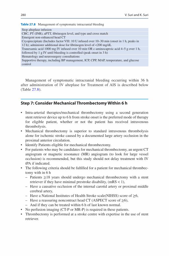
280
Management of symptomatic intracranial bleeding occurring within 36 h after administration of IV alteplase for Treatment of AIS is described below (Table 27.8).
Step 7: Consider Mechanical Thrombectomy Within 6 h
• Intra-arterial therapies/mechanical thrombectomy using a second generation stent retriever device up to 6 h from stroke onset is the preferred mode of therapy for eligible patient, whether or not the patient has received intravenous thrombolysis.
• Mechanical thrombectomy is superior to standard intravenous thrombolysis alone for ischemic stroke caused by a documented large artery occlusion in the proximal anterior circulation.
• Identify Patients eligible for mechanical thrombectomy.• For patients who may be candidates for mechanical thrombectomy, an urgent CT
angiogram or magnetic resonance (MR) angiogram (to look for large vessel occlusion) is recommended, but this study should not delay treatment with IV tPA if indicated.
• The following criteria should be fulfilled for a patient for mechanical thrombec-tomy with in 6 h – Patients ≥18 years should undergo mechanical thrombectomy with a stent
retriever if they have minimal prestroke disability, (mRS < 1), – Have a causative occlusion of the internal carotid artery or proximal middle
cerebral artery, – Have a National Institutes of Health Stroke scale(NIHSS) score of ≥6, – Have a reassuring noncontract head CT (ASPECT score of ≥6), – And if they can be treated within 6 h of last known normal.
• No perfusion imaging (CT-P or MR-P) is required in these patients.• Thrombectomy is performed at a stroke centre with expertise in the use of stent
retriever.
Table 27.8 Management of symptomatic intracranial bleeding
Stop alteplase infusionCBC, PT (INR), aPTT, fibrinogen level, and type and cross matchEmergent non-enhanced head CTCryoprecipitate (Includes factor Vlll: 10 U infused over 10–30 min (onset in 1 h, peaks in 12 h); administer additional dose for fibrinogen level of <200 mg/dLTranexamic acid 1000 mg IV infused over 10 min OR c-aminocaproic acid 4–5 g over 1 h, followed by 1 g IV until bleeding is controlled (peak onset in 3 h)Hematology and neurosurgery consultationsSupportive therapy, including BP management, ICP, CPP, MAP, temperature, and glucose control
V. Suri and K. Suri

281
Step 8: Consider Mechanical Thrombectomy Within 6–24 h
• In selected acute stroke patients within 6–24 h of last known normal who have evidence of a large vessel occlusion in the anterior circulation and obtain a perfu-sion imaging (CT-P or MR-P) and determine whether the patient is a candidate for mechanical thrombectomy.
• In selected patients with acute ischemic stroke within 6 to 16 h of last known nor-mal who have large vessel occlusion in the anterior circulation and meet the DAWN or DEFUSE 3 eligibility criteria, mechanical thrombectomy is recommended.
• In selected patients with acute ischemic stroke with in16 to 24 h of last known normal, who have a large vessel occlusion in the anterior circulation and meet the DAWN eligibility criteria, mechanical thrombectomy is recommended.
• Mechanical thrombectomy may also be considered for acute ischemic stroke caused by major posterior cerebral or vertebro basilar arterial occlusion but the benefits are uncertain.
• Second generation stent retriever device or catheter aspiration device should be used for mechanical thrombectomy.
Step 9: Shift the Patient to Stroke ICU
Initiate general Supportive Care and Treatment of Acute Complications
• Monitoring is recommended to screen for atrial fibrillation and other potentially serious cardiac arrhythmias that would necessitate emergency cardiac interven-tions. Cardiac monitoring should be performed for at least the first 24 h.
• Patients who have elevated blood pressure and are otherwise eligible for treat-ment with intravenous rtPA should have their blood pressure carefully lowered so that their systolic blood pressure is <185 mm Hg and their diastolic blood pressure is <110 mm Hg before thrombolytic therapy is initiated.
• If medications are given to lower blood pressure, blood pressure is stabilized at the lower level before beginning treatment with intravenous rtPA and maintained below 180/105 mm Hg for at least the first 24 h after intravenous rtPA treatment.
• If patient is not receiving tPA, lower blood pressure by 15% during the first 24 h after onset of stroke and antihypertensive medications should be withheld unless the systolic blood pressure is >220 mm Hg or the diastolic blood pressure is >120 mm Hg.
• Restarting antihypertensive medications is reasonable after the first 24 h for patients who have preexisting hypertension and are neurologically stable. Patients who have malignant hypertension or other medical indications for aggressive treatment of blood pressure should be treated accordingly.
27 Acute Ischemic Stroke (AIS)
282
• Optimise head of bed position: keep the head in neutral position to avoid kinking of jugular veins, and elevate the head to 30° to ensure optimising balance of cerebral venous drainage and cerebra perfusion
• Airways support and ventilator assistance are recommended for the treatment of patients with acute stroke who have decreased consciousness or who have bulbar dysfunction that causes compromise of the airway. Supplemental oxygen should be provided to maintain oxygen saturation > 94%.
• Sources of hyperthermia (temperature > 38 C) should be identified and treated, and antipyretic medications should be administered to lower temperature in hyperthemic patients with stroke.
• Hypoglycemia (blood glucose <60 mg/dL) should be treated in patients with acute ischemic stroke. The goal is to achieve normoglycemia.
• Hyperglycemia during the first 24 h. Treat hyperglycemia to achieve blood glu-cose levels in a range of 140 to 180 mg/dL and to closely monitor to prevent hypoglycemia.
• Hypovolemia should be corrected with intravenous normal saline, and cardiac arrhythmias should be corrected. But Volume Expansion, Vasodilators, and Induced Hypertension have no role. Avoid D5w and excessive fluid administration.
• Raised intracranial pressure should be managed as described in chapter.• Decompressive craniotomy with dural expansion is a reasonable when appropri-
ately planned and timed for patients with malignant MCA infarct who deterio-rates neurologically within 48 h despite medical therapy. It reduces mortality close to 50% with 55% of surgical survivors achieving moderate disability(able to walk, mRS 2 or 3), 18% achieving independence (mRS score2) at 12 months.
• Decompressive suboccipital craniotomy should also be performed for patients with cerebeller infarction with neurological decline due to brain stem compression.
• Patients with suspected pneumonia or urinary tract infections should be treated with appropriate antibiotics.
• Subcutaneous administration of anticoagulants for treatment of immobilized patients to prevent deep vein thrombosis when paralysed limbs have a power less than grade 2. Aspirin can be given for treatment of patients who cannot receive anticoagulants for prophylaxis of DVT. Intermittent external compression devices are less helpful and advised only for treatment of patients who cannot receive anticoagulants.
• Assessment of swallowing before the patient begins eating, drinking, or receiv-ing oral medications. Patients who cannot take solid food and liquids orally are given nasogastric or nasoduodenal, tube feedings to maintain hydration and nutrition while undergoing efforts to restore swallowing. If swallowing is impaired, nasogastric feeding is preferred to PEG for first 2–3 weeks after stroke.
• Early mobilization of less severely affected patients and measures to prevent sub-acute complications of stroke.
• Treatment of concomitant medical diseases should be started.
V. Suri and K. Suri
283
• Prophylactic antiepileptic drugs should not be used in ischemic stroke patient. Antiepileptic drugs should be used after a single unprovoked seizure occurring 7 or more days after stroke or patient with 2 or more seizures. Long term anticon-vulsant medications are not recommended for early symptomatic seizures occur-ring in less than 7 days of stroke.
• Discouraged: Routine use of indwelling catheter, prophylactic therapy, and nutri-tional supplements.
Suggested Reading
Khurana D, Suri V, et al. Recommendations for the early management of acute ischemic stroke: a consensus statement for healthcare professionals from the Indian Stroke Association. J Stroke Med. 2018;1(2):79–113. Indian guidelines on the early management of stroke.
Mokin M, et al. Indications for thrombectomy in acute ischaemic stroke from emergency large ves-sel occlusion (ELVO) repost of the SNIS standards and guideline committee. J Neurolntervent Surg. 2019;11:215–20. For anterior circulation AIS, thrombectomy is indicated in select patients up to 16 hours from symptom onset or time last known well for unwitnessed strokes, including wake-up strokes.
Powers WJ. 2018 Guidelines for EARLY management of patients with Acute Ischaemic Stroke- A Guideline for Healthcare professionals from the American Heart Association/American Stroke Association. Stroke. 2018;49:e46–99. https://doi.org/10.1161/STR.0000000000000158. A Comprehensive Guidelines for the management of stroke.
The National Institute of Neurological Disorders and Stroke rt-PA Stroke Study Group. Tissue plas-minogen activator for acute ischemic stroke. N Engl J Med. 1995;333(24):1581–7. Despite an increased incidence of symptomatic intracerebral hemorrhage, treatment with intravenous tPA within 3 h of the onset of ischemic stroke improved clinical outcome at 3 months. Intravenous alteplase administered between 3 and 4.5 h after the onset of symptoms significantly improved clinical outcomes but was more frequently associated with symptomatic intracranial hemor-rhage. Early intensive BP-lowering treatment is clinically feasible, well tolerated, and seems to reduce hematoma growth in ICH.
Warwick Pexmen JH. Use of the Albert Stroke Program Early CT score (ASPECTS) for assessing Ct Scan in patients with acute stroke Am J of. Neuroradiology. 2001;122:1534–42. ASPECTS is a practical method which can be applied to different axial baselines and is clinically superior to that of the 1/3 MCA rule.
Websites
aic.cuhk.edu.hk. Neurological teaching site for ICU residents.Stroke.ahajournals.org. Journal on cerebrovascular diseases published by American Heart
Association.library.med.utah.edu/neurologicexam. A comprehensive site for neurological examination.med.harvard.edu. A comprehensive atlas of brain imaging.
27 Acute Ischemic Stroke (AIS)
285© Springer Nature Singapore Pte Ltd. 2020R. Chawla, S. Todi (eds.), ICU Protocols, https://doi.org/10.1007/978-981-15-0898-1_28
V. D. Devaprasad (*) · N. Ramakrishnan Department of Critical Care Medicine, Apollo Hospitals, Chennai, India
28Spontaneous Intracerebral Hemorrhage
V. Dedeepiya Devaprasad and Nagarajan Ramakrishnan
Intracerebral hemorrhage (ICH) is the second most common stroke. Hypertension is the commonest cause of spontaneous ICH. Patients with acute intracerebral hemor-rhage (ICH) should be managed in an critical care unit or stroke unit. Older age, decreased Glasgow Coma Scale (GCS) score, increased ICH volume, presence of intraventricular hemorrhage and deep or infratentorial ICH location are predictor of bad outcome. The 30-day mortality from ICH varies from 35 to 52%.
Step 1: Assess for ABCD and Maintain Airway
• Airway assessment needs to be done. If the patient talks to the examiner, it is reasonably certain that the airway is patent. However, it needs to be remembered that in dominant hemisphere (left hemispheric) hemorrhagic stroke, Broca’s aphasia can be anticipated. In patients, with Broca’s aphasia, the inability to speak should not be mistakenly considered as an airway problem.
• Breathing and ventilation can be a problem if the stroke is extensive. An exten-sive hemispherical hemorrhagic stroke, in fact, can cause hypoventilation. A brain stem hemorrhage by involving the respiratory centers can result in ventila-tion disturbances. A patient with GCS of 8 or less will need intubation.
A 60-year-old hypertensive male was admitted with sudden-onset slurring of speech, vomiting, headache and rapidly progressive drowsiness. His blood pres-sure was measured as 190/100 mmHg. An urgent noncontrast CT brain scan suggested the occurrence of a hemorrhagic stroke.

286
• In patients with brainstem stroke, intubation is sometimes required for preven-tion of aspiration even with normal sensorium
• A baseline severity score should be performed as part of the initial evaluation of patients with ICH.
Step 2: Neuroimaging and Its Interpretation
• Rapid diagnosis with CT /MRI is recommended. CT is preferred over MRI because of ease of availability, lesser cost and ease of interpretation. CT also saves time over MRI.
• A hyperdense lesion (seen as white) suggests hemorrhagic stroke. CT scan brain can never ever be normal in an hemorrhagic stroke. It can however be normal early in an ischemic stroke.
• CT angiography/contrast enhanced CT is not routinely used or recommended. When done it identifies patients at risk of hematoma expansion. The contrast extravasates into the hematoma causing “spot sign” which predicts hematoma expansion.
Step 3: Reduce BP Judiciously
Blood pressure is often elevated in patients with ICH which may predispose to hematoma expansion. On the other hand rapid reduction of blood pressure may lead to decrease cerebral perfusion.Current guidelines for reducing Blood Pressure in ICH.
• Recent guidelines have recommended that in adults with ICH who present with SBP greater than 220 mm Hg, it is reasonable to use continuous intravenous drug infusion and close BP monitoring to lower SBP.
• Spontaneous ICH patients who present within 6 h of the acute event and who have an SBP between 150 mm Hg and 220 mm Hg, immediate lowering of SBP to less than 140 mm Hg can be potentially harmful and is not of benefit to reduce death or severe disability.
• The optimal goal blood pressure is uncertain, but a SBP of 140 to 160 mmHg is a reasonable target. Aggressive reduction can increase renal adverse events.
• IV labetalol is the drug of choice, preferably by IV infusion. Initial bolus dose of 10 mg IV followed by infusion of 2–8 mg/min, or intermittent boluses of 10 mg every half an hour. Maximum daily dose of IV labetalol is 300 mg/day.
• It is important to monitor heart rate, and hold infusion if it is less than 60/min.• Other agents that are recommended for treating hypertension in hemorrhagic
stroke are nicardipine, angiotensin-converting enzyme inhibitor, and hydrala-zine. Nitrates should be avoided because they tend to increase ICP.
Step 3: Consider Management of Raised ICP
• Raised ICP is expected to follow ICH and is defined as ICP ≥20 mmHg for >3 min. The therapeutic goal is to decrease ICP to <20 mmHg and maintain the cerebral perfusion pressure between 50 and 70 mmHg.
V. D. Devaprasad and N. Ramakrishnan
287
• Generally, it is recommended that a patient with GCS ≤8 or with rapidly deterio-rating GCS that is attributable to raised ICP or features of transtentorial hernia-tion may benefit from an ICP monitoring.
• Patients with intraventricular hemorrhage and hydrocephalus may also benefit from ICP measurement.
• This is not usually done in many centers for fear of infection and lack of outcome studies showing reduction in mortality with the use of ICP monitor.
• Raised ICP is treated with bolus therapy of 20% mannitol (1 mg/kg) IV given over 10–20 min repeated as frequently as needed or hypertonic saline.
• Maintain neck in neutral position to prevent impedance of jugular venous outflow.
• Elevate head of bed at 30°, mild sedation.• Use isotonic saline for maintenance fluid, Avoid hypotonic fluids.• In extreme case pharmacological paralysis, barbiturate coma is needed.
Step 4: Cautious Use of Blood Components
• All anticoagulants and antiplatelet drugs should be discontinued acutely and anticoagulant effect reversed acutely with appropriate agents.
• Current recommendations are against the routine use of platelets, FFP, recombi-nant Factor 7(rFVIIa) or Prothrombin complex concentrate (PCC).
• Patients with ICH who have been on therapy with oral anticoagulants such as warfarin, should have these drugs withheld, should receive preferably PCC or FFP to correct the INR and receive intravenous vitamin K.
• The usefulness of platelet transfusion in patients who have received antiplatelets is only investigational. However if the patient has severe thrombocytopenia, they should receive platelet transfusion.
• PCC has not shown to improve outcome compared to FFP. It however is eas-ily available in hospitals as it does not need blood banking facilities unlike FFP.
• PCC is costlier compared to FFP. PCC may have fewer complications. Volume overload is less with PCC than FFP and hence is a reasonable alter-native to FFP.
• Recombinant Factor VIIa (rFVIIa) is known to limit hematoma expansion even in non coagulopathic patients but also increases the thromboembolic complica-tions. rFVlla does not replace clotting factors, though it normalizes INR and is not routinely recommended. It however is to be considered in patients with hemophilia with hemorrhagic stroke or prior to surgical therapy.
• In patients when anticoagulation needs to be resumed urgently (such as mechani-cal valve prosthesis), continuous infusion of UFH with APTT monitoring with lower than the usual therapeutic goal may be instituted after stability of hemor-rhage is achieved.
• It is preferable to use unfractionated heparin because it can be easily reversed.
28 Spontaneous Intracerebral Hemorrhage
288
Step 5: Selected Use of Antiepileptics
• There is no role for prophylactic anticonvulsants, and hence they should not be used.
• However, if the patient has clinical seizures, they should receive antiepileptics and is reasonable to continue them for at least 1 month.
• If the patient has deterioration in mental status, that is, out of proportion to the degree of brain injury, a continuous electroencephalogram (EEG) monitor is indicated to exclude non-convulsive seizures, and should these patients have any evidence of seizures on EEG, they should receive anticonvulsants.
• In the absence of continuous EEG monitoring, introduction of antiepileptics in the presence of disproportionate mental status changes may be considered with continued close neurological monitoring.
Step 6: Consider Cerebral Angiography
• Angiography is not recommended in elderly patients with hypertension and who have hemorrhage in typical territories such as basal ganglia, thalamus, cerebel-lum, or brain stem and in whom CT scan is not suggestive of a structural lesion.
• However, if the patient is young, normotensive with no definite cause of hemor-rhage, an angiogram would be recommended especially if he/she is a candidate for surgical intervention.
Step 7: Consider Surgical Management
• Decompressive craniotomy and evacuation for all patients with ICH is not indicated.
• For Supratentorial ICH, surgery is usually avoided and should be considered on an individual basis based on assessment of prognosis with or without surgery.
Based on the STICH trial the following are the current indications for surgery.
• Cerebellar hemorrhage more than 3 cm diameter.• Anticipated high risk of brain stem compression and hydrocephalus without
evacuation.• Lobar clot of more than 30 mL volume located less than 1 cm below cortical
surface.• Decompressive craniectomy may also be considered in some patients who are
young and acutely deteriorating.• External ventricular drainage is used when there is intraventricular bleed with
raised ICP resulting in neurological deterioration and when there is hydrocephalus.
V. D. Devaprasad and N. Ramakrishnan
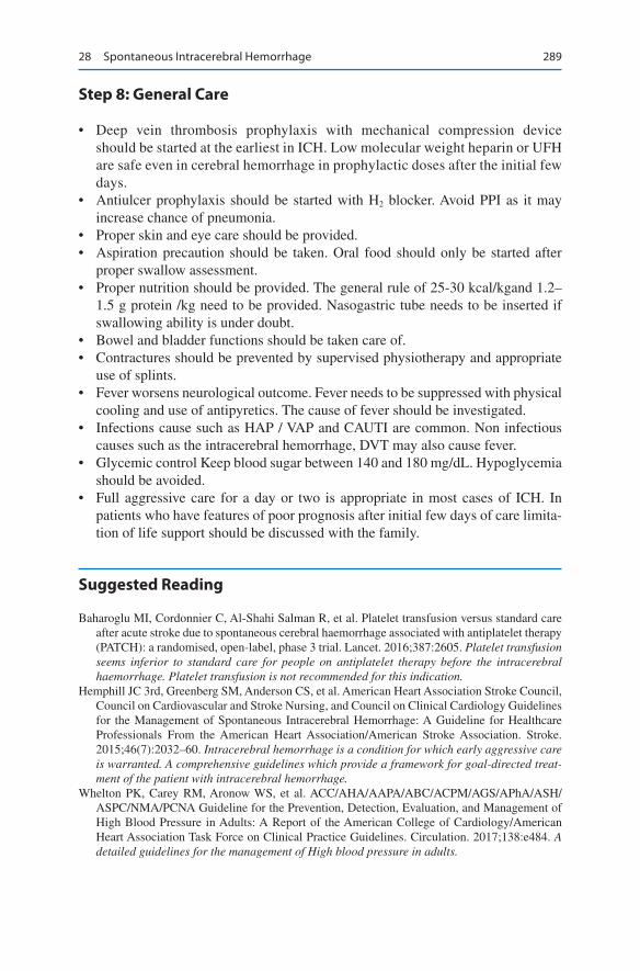
289
Step 8: General Care
• Deep vein thrombosis prophylaxis with mechanical compression device should be started at the earliest in ICH. Low molecular weight heparin or UFH are safe even in cerebral hemorrhage in prophylactic doses after the initial few days.
• Antiulcer prophylaxis should be started with H2 blocker. Avoid PPI as it may increase chance of pneumonia.
• Proper skin and eye care should be provided.• Aspiration precaution should be taken. Oral food should only be started after
proper swallow assessment.• Proper nutrition should be provided. The general rule of 25-30 kcal/kgand 1.2–
1.5 g protein /kg need to be provided. Nasogastric tube needs to be inserted if swallowing ability is under doubt.
• Bowel and bladder functions should be taken care of.• Contractures should be prevented by supervised physiotherapy and appropriate
use of splints.• Fever worsens neurological outcome. Fever needs to be suppressed with physical
cooling and use of antipyretics. The cause of fever should be investigated.• Infections cause such as HAP / VAP and CAUTI are common. Non infectious
causes such as the intracerebral hemorrhage, DVT may also cause fever.• Glycemic control Keep blood sugar between 140 and 180 mg/dL. Hypoglycemia
should be avoided.• Full aggressive care for a day or two is appropriate in most cases of ICH. In
patients who have features of poor prognosis after initial few days of care limita-tion of life support should be discussed with the family.
Suggested Reading
Baharoglu MI, Cordonnier C, Al-Shahi Salman R, et al. Platelet transfusion versus standard care after acute stroke due to spontaneous cerebral haemorrhage associated with antiplatelet therapy (PATCH): a randomised, open-label, phase 3 trial. Lancet. 2016;387:2605. Platelet transfusion seems inferior to standard care for people on antiplatelet therapy before the intracerebral haemorrhage. Platelet transfusion is not recommended for this indication.
Hemphill JC 3rd, Greenberg SM, Anderson CS, et al. American Heart Association Stroke Council, Council on Cardiovascular and Stroke Nursing, and Council on Clinical Cardiology Guidelines for the Management of Spontaneous Intracerebral Hemorrhage: A Guideline for Healthcare Professionals From the American Heart Association/American Stroke Association. Stroke. 2015;46(7):2032–60. Intracerebral hemorrhage is a condition for which early aggressive care is warranted. A comprehensive guidelines which provide a framework for goal-directed treat-ment of the patient with intracerebral hemorrhage.
Whelton PK, Carey RM, Aronow WS, et al. ACC/AHA/AAPA/ABC/ACPM/AGS/APhA/ASH/ASPC/NMA/PCNA Guideline for the Prevention, Detection, Evaluation, and Management of High Blood Pressure in Adults: A Report of the American College of Cardiology/American Heart Association Task Force on Clinical Practice Guidelines. Circulation. 2017;138:e484. A detailed guidelines for the management of High blood pressure in adults.
28 Spontaneous Intracerebral Hemorrhage

290
Websites
aic.cuhk.edu.hk. Neurological teaching site for ICU residents.library.med.utah.edu/neurologicexam. A comprehensive site for neurological examination.med.harvard.edu. A comprehensive atlas of brain imaging.Stroke.ahajournals.org. Journal on cerebrovascular diseases published by American Heart
Association.
V. D. Devaprasad and N. Ramakrishnan

291© Springer Nature Singapore Pte Ltd. 2020R. Chawla, S. Todi (eds.), ICU Protocols, https://doi.org/10.1007/978-981-15-0898-1_29
S. K. Iyer (*) · J. Shah · K. Vadapalli Department of Intensive Care and Critical Care Medicine, Bharati Vidyapeeth (DTU) Medical College, Pune, India
29Subarachnoid Hemorrhage
Shiva Kumar Iyer, Jignesh Shah, and Kiran Vadapalli
Subarachnoid hemorrhage (SAH) should be suspected in patients presenting with acute onset severe headache which they classically describe as the worst headache of their life. Most subarachnoid hemorrhages (SAH) are caused by rupture of intra-cranial saccular (berry) aneurysms. Other causes include trauma, vascular malfor-mations, hemorrhagic infarctions and hypertensive hemorrhages.
Risk factors include cigarette smoking, hypertension, estrogen deficiency, coarcta-tion of the aorta, hereditary diseases like polycystic kidney disease and Ehler- Danlos syndrome. The high morbidity and mortality associated with aneurysmal SAH (aSAH) mandate a high degree of suspicion to allow timely and appropriate treatment.
Step 1: Start Resuscitation
• Emergency airway protection is necessary for obtunded patients to avoid hyper-carbia and rise in intracranial pressure. Indications for endotracheal intubation include a Glasgow Coma Scale (GCS) score ≤8, elevated intracranial pressure (ICP), hemodynamic instability, poor oxygenation or hypoventilation and requirement for heavy sedation or paralysis.
A 24-year male cricket player complained of sudden headache followed by a brief period of loss of consciousness while playing. On arrival at the emergency department, he was drowsy but answering questions and reported that he had the worst headache ever. Examination revealed a blood pressure of 150/62 mm Hg and has neck stiffness. Rest all neurological examination was unremarkable. He does not give any significant past history and denies any trauma or addictions. Emergency noncontrast CT head revealed diffuse subarachnoid hemorrhage occupying the basal cisterns and bilateral Sylvian fissures.

292
• In patients with suspicion of SAH, volume loading and vasopressors should be avoided during initial resuscitation to prevent aneurysmal rebleeding. Resuscitation should focus on maintaining euvolemia and normal electrolyte balance.
Step 2: Take a Focused History and Perform Clinical Examination
• The hallmark of aneurysmal subarachnoid hemorrhage is a severe headache which the patient usually describes as “the worst headache of his life”
• Associated symptoms include nausea, vomiting, photophobia, neck pain and a brief loss of consciousness.
• Approximately 30–50% of patients report a history of a sudden and severe head-ache (the sentinel headache) that precedes a major aneurysmal bleed by 6 to 20 days which represents a warning leak.
• History of trauma, hypertension, bleeding diathesis, use of antiplatelet agents or anticoagulants and drug abuse like cocaine should be elicited.
• Examination might reveal neck stiffness and focal neurological deficits (if any) and preretinal subhyaloid hemorrhages may be seen on fundus examination.
• Strict neuro observation is required in these patients because of the high chances of sudden neurological deterioration.
Step 3: Assessment of Severity of Subarachnoid Hemorrhage
• Several severity assessment scales are used to categorize the patients with aSAH, the prominent among them are Hunt and Hess scale and World Federation of Neurological Surgeons (WFNS) grading scale.
• Hunt and Hess scale (Table 29.1) denotes the severity at the time of presentation and the higher grade denotes worse prognosis. The grade is advanced one level for the presence of serious systemic disease (hypertension, diabetes, severe arte-riosclerosis, chronic pulmonary disease) or vasospasm on angiography.
• World Federation of Neurological Surgeons (WFNS) grading scale (Table 29.2) is more in practice which has more objectivity.
Table 29.1 Hunt and Hess scale
Grade 1 Asymptomatic or mild headache and slight nuchal rigidityGrade 2 Moderate to severe headache, stiff neck, no neurologic deficit except cranial
nerve palsyGrade 3 Drowsy or confused, mild focal neurologic deficitGrade 4 Stupor, moderate or severe hemiparesisGrade 5 Deep coma, decerebrate posturing
S. K. Iyer et al.
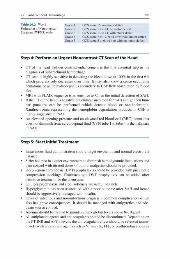
293
Step 4: Perform an Urgent Noncontrast CT Scan of the Head
• CT of the head without contrast enhancement is the first essential step in the diagnosis of subarachnoid hemorrhage.
• CT scan is highly sensitive in detecting the bleed close to 100% in the first 6 h which progressively decreases over time. It may also show a space-occupying hematoma or acute hydrocephalus secondary to CSF flow obstruction by blood clot.
• MRI with FLAIR sequence is as sensitive as CT in the initial detection of SAH.• If the CT of the head is negative but clinical suspicion for SAH is high then lum-
bar puncture can be performed which detects blood or xanthochromia. Xanthochromia representing the hemoglobin degradation products in CSF is highly suggestive of SAH.
• An elevated opening pressure and an elevated red blood cell (RBC) count that does not diminish from cerebrospinal fluid (CSF) tube 1 to tube 4 is the hallmark of SAH.
Step 5: Start Initial Treatment
• Intravenous fluid administration should target euvolemia and normal electrolyte balance.
• Strict bed rest in a quiet environment to diminish hemodynamic fluctuations and pain control with titrated doses of opioid analgesics should be provided.
• Deep venous thrombosis (DVT) prophylaxis should be provided with pneumatic compression stockings. Pharmacologic DVT prophylaxis can be added after definitive treatment for the aneurysm.
• GI ulcer prophylaxis and stool softeners are useful adjuncts.• Hyperglycemia has been associated with a poor outcome after SAH and hence
should be aggressively managed with insulin.• Fever of infectious and non-infectious origin is a common complication which
also has grave consequences. It should be managed with antipyretics and ade-quate source control.
• Anemia should be treated to maintain hemoglobin levels above 8–10 gm%• All antiplatelet agents and anticoagulants should be discontinued. Depending on
the PT INR and APTT levels, the anticoagulant effect should be reversed imme-diately with appropriate agents such as Vitamin K, FFP, or prothrombin complex
Table 29.2 World Federation of Neurological Surgeons (WFNS) scale
Grade 1 GCS score 15, no motor deficitGrade 2 GCS score 13 to 14, no motor deficitGrade 3 GCS score 13 to 14, with motor deficitGrade 4 GCS score 7 to 12, with or without motor deficitGrade 5 GCS score 3 to 6, with or without motor deficit
29 Subarachnoid Hemorrhage
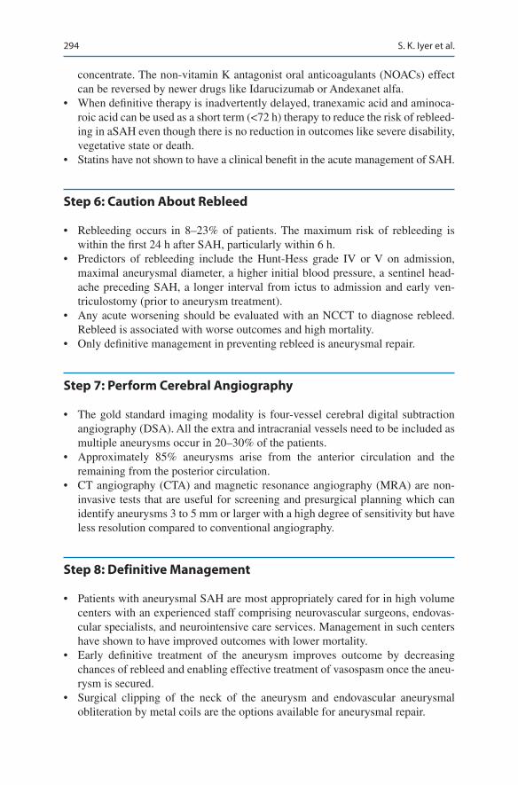
294
concentrate. The non-vitamin K antagonist oral anticoagulants (NOACs) effect can be reversed by newer drugs like Idarucizumab or Andexanet alfa.
• When definitive therapy is inadvertently delayed, tranexamic acid and aminoca-roic acid can be used as a short term (<72 h) therapy to reduce the risk of rebleed-ing in aSAH even though there is no reduction in outcomes like severe disability, vegetative state or death.
• Statins have not shown to have a clinical benefit in the acute management of SAH.
Step 6: Caution About Rebleed
• Rebleeding occurs in 8–23% of patients. The maximum risk of rebleeding is within the first 24 h after SAH, particularly within 6 h.
• Predictors of rebleeding include the Hunt-Hess grade IV or V on admission, maximal aneurysmal diameter, a higher initial blood pressure, a sentinel head-ache preceding SAH, a longer interval from ictus to admission and early ven-triculostomy (prior to aneurysm treatment).
• Any acute worsening should be evaluated with an NCCT to diagnose rebleed. Rebleed is associated with worse outcomes and high mortality.
• Only definitive management in preventing rebleed is aneurysmal repair.
Step 7: Perform Cerebral Angiography
• The gold standard imaging modality is four-vessel cerebral digital subtraction angiography (DSA). All the extra and intracranial vessels need to be included as multiple aneurysms occur in 20–30% of the patients.
• Approximately 85% aneurysms arise from the anterior circulation and the remaining from the posterior circulation.
• CT angiography (CTA) and magnetic resonance angiography (MRA) are non- invasive tests that are useful for screening and presurgical planning which can identify aneurysms 3 to 5 mm or larger with a high degree of sensitivity but have less resolution compared to conventional angiography.
Step 8: Definitive Management
• Patients with aneurysmal SAH are most appropriately cared for in high volume centers with an experienced staff comprising neurovascular surgeons, endovas-cular specialists, and neurointensive care services. Management in such centers have shown to have improved outcomes with lower mortality.
• Early definitive treatment of the aneurysm improves outcome by decreasing chances of rebleed and enabling effective treatment of vasospasm once the aneu-rysm is secured.
• Surgical clipping of the neck of the aneurysm and endovascular aneurysmal obliteration by metal coils are the options available for aneurysmal repair.
S. K. Iyer et al.

295
• Surgical clipping of the ruptured aneurysm requires craniotomy followed by identification of the aneurysm after gentle mobilization of the brain parenchyma. A titanium clip is placed across the neck of the aneurysm closing the sac at its neck while preserving blood flow through the adjacent normal arteries.
• Endovascular treatment involves fluoroscopically navigating a catheter till the parent artery of the aneurysm. A microcatheter is then advanced into the aneurys-mal sac and platinum coils are deposited into the aneurysmal lumen thereby arresting the blood flow and inducing thrombus formation. This reduces the chances of re-rupture of the aneurysm.
• The International Subarachnoid Aneurysm Trial (ISAT) and the Barrow Ruptured Aneurysm Trial (BRAT) comparing both treatment modalities have shown that despite higher rates of aneurysmal obliteration with open surgical treatment, functional outcomes (death or dependency) at the end of one year are better with endovascular treatment.
• Surgical clipping may be ideal for patients with large intracerebral hemato-mas(>50 mL) resulting in raised intracranial pressure or focal neurologic defi-cits, aneurysms which are difficult to visualize angiographically, and for patients younger than 40 years of age who have anterior circulation aneurysms and good neurologic status due to lower risk of rebleeding.
• Posterior circulation aneurysms, poor-grade WFNS classification (IV/V) aSAH, elderly are preferentially treated with endovascular coiling.
• Being minimally invasive, endovascular treatment is preferred over open surgical treatment. However the treatment should be individualized.
• After any aneurysm repair, all patients should undergo immediate cerebrovascu-lar imaging to identify remnants or recurrence of the aneurysm and if present should undergo retreatment.
• Combined endovascular and surgical techniques may be required with some very large or complex aneurysms.
Timing of surgery• Early surgery performed within 24–72 h of the hemorrhage has potential benefits
including prevention of rebleeding and management of vasospasm. In addition, the usual methods of treating vasospasm have deleterious consequences in the presence of an untreated aneurysm.
• In patients with aneurysmal SAH with Hunt and Hess grades-I to III, early aneu-rysm repair (within 24–72 h) results in good neurological recovery in 70–90% of patients with a mortality rate of 1.7–8%.
• Aneurysmal SAH with poor clinical grade (Hunt and Hess IV and V) may benefit from endovascular surgery, but the prognosis remains poor.
Step 9: Prevent, Identify and Manage Vasospasm and Delayed Cerebral Ischemia
• Delayed cerebral ischemia is a frequent complication of SAH contributing sub-stantially to morbidity and mortality. The most common cause of delayed cere-bral ischemia after SAH is assumed to be vasospasm.
29 Subarachnoid Hemorrhage

296
• Vasospasm generally starts 3 to 4 days after aneurysm rupture, peaks at 7 to 10 days, and resolves by 14 to 21 days.
• More than a third of the SAH patients, typically 4 to 14 days after aneurysm rupture develop focal neurologic deficits attributable to cerebral ischemia.
• Delayed cerebral ischemia does not necessarily occur in the same vascular terri-tory as that of the angiographically visible vasospasm. A variety of vascular and neural changes that take place after SAH may contribute to its pathogenesis of delayed cerebral ischemia.
• The modified Fisher scale (CT scan appearance) (Table 29.3) can be used to predict the likelihood of symptomatic cerebral vasospasm.
• Transcranial Doppler (TCD) sonography is useful for detecting and monitoring vasospasm in aSAH. Velocity changes detected by TCD typically precede the clinical sequelae of vasospasm but it is highly operator dependent.
• Prevention of vasospasm is crucial as treatment is difficult. In order to mitigate the effect of vasospasm, all patients with aSAH should receive Nimodipine, the only approved drug for prevention and treatment of vasospasm and euvolemia should be maintained.
• Nimodipine, an oral calcium channel antagonist, at 60 mg fourth hourly should be administered orally to all patients from the time of presentation to 21 days after the occurrence of aSAH. A Cochrane review indicated that Nimodipine reduced the risk of poor outcomes in patients with subarachnoid hemorrhage by one third. The benefit of Nimodipine may be due to its neuroprotective property rather than to its vasodilatory property.
• Early definitive treatment of aneurysm enables the maintenance of euvolemia which is crucial in the management of vasospasm.
• Hemodynamic augmentation does not prevent vasospasm, but it may be appro-priate in the treatment of symptomatic vasospasm.
• If clinically significant delayed cerebral ischemia, with or without vasospasm, is suspected, euvolemia and induced hypertension (with pressor agents such as nor-epinephrine, dopamine and phenylephrine) are recommended to improve cere-bral perfusion.
• Cardiac decompensation should be watched for in patients with high blood pressure or preexisting ischemic heart disease while inducing therapeutic hypertension.
• Clinical vasospasm that persists despite hyperdynamic therapy may be treated by balloon angioplasty for accessible lesions and/or intraarterial administration of vasodilators(nimodipine, nicardipine, milrinone) for more distal vessels.
Table 29.3 Modified Fisher scale
Grade CT finding0 No Subarachnoid Hemorrhage (SAH), No Intra Ventricular
Hemorrhage (IVH)1 Thin (<1 mm) diffuse or focal SAH, No IVH2 Thin (<1 mm) diffuse or focal SAH, with IVH3 Thick (>1 mm) diffuse or focal SAH, No IVH4 Thick (>1 mm) diffuse or focal SAH, with IVH
S. K. Iyer et al.
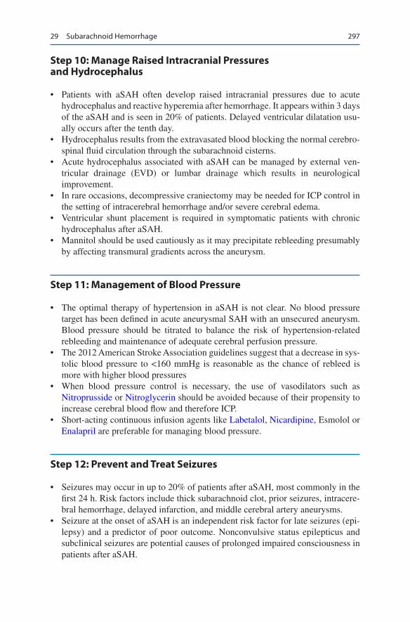
297
Step 10: Manage Raised Intracranial Pressures and Hydrocephalus
• Patients with aSAH often develop raised intracranial pressures due to acute hydrocephalus and reactive hyperemia after hemorrhage. It appears within 3 days of the aSAH and is seen in 20% of patients. Delayed ventricular dilatation usu-ally occurs after the tenth day.
• Hydrocephalus results from the extravasated blood blocking the normal cerebro-spinal fluid circulation through the subarachnoid cisterns.
• Acute hydrocephalus associated with aSAH can be managed by external ven-tricular drainage (EVD) or lumbar drainage which results in neurological improvement.
• In rare occasions, decompressive craniectomy may be needed for ICP control in the setting of intracerebral hemorrhage and/or severe cerebral edema.
• Ventricular shunt placement is required in symptomatic patients with chronic hydrocephalus after aSAH.
• Mannitol should be used cautiously as it may precipitate rebleeding presumably by affecting transmural gradients across the aneurysm.
Step 11: Management of Blood Pressure
• The optimal therapy of hypertension in aSAH is not clear. No blood pressure target has been defined in acute aneurysmal SAH with an unsecured aneurysm. Blood pressure should be titrated to balance the risk of hypertension-related rebleeding and maintenance of adequate cerebral perfusion pressure.
• The 2012 American Stroke Association guidelines suggest that a decrease in sys-tolic blood pressure to <160 mmHg is reasonable as the chance of rebleed is more with higher blood pressures
• When blood pressure control is necessary, the use of vasodilators such as Nitroprusside or Nitroglycerin should be avoided because of their propensity to increase cerebral blood flow and therefore ICP.
• Short-acting continuous infusion agents like Labetalol, Nicardipine, Esmolol or Enalapril are preferable for managing blood pressure.
Step 12: Prevent and Treat Seizures
• Seizures may occur in up to 20% of patients after aSAH, most commonly in the first 24 h. Risk factors include thick subarachnoid clot, prior seizures, intracere-bral hemorrhage, delayed infarction, and middle cerebral artery aneurysms.
• Seizure at the onset of aSAH is an independent risk factor for late seizures (epi-lepsy) and a predictor of poor outcome. Nonconvulsive status epilepticus and subclinical seizures are potential causes of prolonged impaired consciousness in patients after aSAH.
29 Subarachnoid Hemorrhage
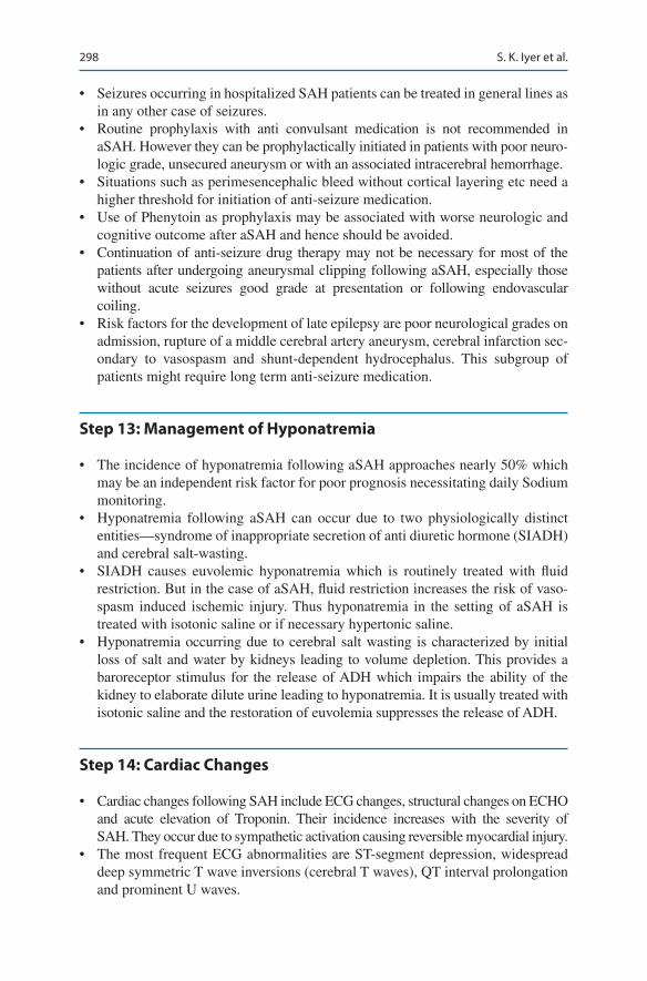
298
• Seizures occurring in hospitalized SAH patients can be treated in general lines as in any other case of seizures.
• Routine prophylaxis with anti convulsant medication is not recommended in aSAH. However they can be prophylactically initiated in patients with poor neuro-logic grade, unsecured aneurysm or with an associated intracerebral hemorrhage.
• Situations such as perimesencephalic bleed without cortical layering etc need a higher threshold for initiation of anti-seizure medication.
• Use of Phenytoin as prophylaxis may be associated with worse neurologic and cognitive outcome after aSAH and hence should be avoided.
• Continuation of anti-seizure drug therapy may not be necessary for most of the patients after undergoing aneurysmal clipping following aSAH, especially those without acute seizures good grade at presentation or following endovascular coiling.
• Risk factors for the development of late epilepsy are poor neurological grades on admission, rupture of a middle cerebral artery aneurysm, cerebral infarction sec-ondary to vasospasm and shunt-dependent hydrocephalus. This subgroup of patients might require long term anti-seizure medication.
Step 13: Management of Hyponatremia
• The incidence of hyponatremia following aSAH approaches nearly 50% which may be an independent risk factor for poor prognosis necessitating daily Sodium monitoring.
• Hyponatremia following aSAH can occur due to two physiologically distinct entities—syndrome of inappropriate secretion of anti diuretic hormone (SIADH) and cerebral salt-wasting.
• SIADH causes euvolemic hyponatremia which is routinely treated with fluid restriction. But in the case of aSAH, fluid restriction increases the risk of vaso-spasm induced ischemic injury. Thus hyponatremia in the setting of aSAH is treated with isotonic saline or if necessary hypertonic saline.
• Hyponatremia occurring due to cerebral salt wasting is characterized by initial loss of salt and water by kidneys leading to volume depletion. This provides a baroreceptor stimulus for the release of ADH which impairs the ability of the kidney to elaborate dilute urine leading to hyponatremia. It is usually treated with isotonic saline and the restoration of euvolemia suppresses the release of ADH.
Step 14: Cardiac Changes
• Cardiac changes following SAH include ECG changes, structural changes on ECHO and acute elevation of Troponin. Their incidence increases with the severity of SAH. They occur due to sympathetic activation causing reversible myocardial injury.
• The most frequent ECG abnormalities are ST-segment depression, widespread deep symmetric T wave inversions (cerebral T waves), QT interval prolongation and prominent U waves.
S. K. Iyer et al.

299
• The ECG changes are predominantly reflective of ischemic changes in the sub-endocardium of the left ventricle which is an independent predictor of poor out-come. Acute myocardial injury can develop in 20% of the cases with elevated CK MB and Troponin levels.
• Elevated peak Troponin levels have been associated with an increased risk of left ventricular dysfunction, pulmonary edema, and hypotension as well as with increased mortality and poor functional outcome.
• Cardiac arrhythmias like atrial flutter, fibrillation and torsades de pointes have been described along with sinus bradycardia and tachycardia in patients with SAH.
• Myocardial injury is most likely the result of a centrally mediated release of catecholamines within the myocardium due to hypoperfusion of the posterior hypothalamus but it may be multifactorial.
• Stress cardiomyopathy resulting from a primary neurologic condition is referred to as neurogenic stunned myocardium and is characterized by transient left ven-tricular dysfunction, electrocardiogram changes, and elevation in cardiac enzymes, in absence of significant coronary artery disease.
Step 15: Manage Neurogenic Pulmonary Edema
• Neurogenic pulmonary edema (NPE) characteristically presents within minutes to hours of a severe central nervous system insult such as subarachnoid hemor-rhage. Patients present with dyspnoea and on examination have tachycardia, tachypnoea, and bibasilar rales.
• Central transient sympathetic discharge following aSAH leading to abrupt alter-ations in cardiopulmonary hemodynamics and subsequent increased capillary hydrostatic pressure and altered permeability across the pulmonary vasculature results in pulmonary edema.
• Most patients with NPE are hypoxemic and require supplemental oxygen. Some patients may require mechanical ventilation. Treatment should focus on defini-tive treatment for aSAH while NPE management is predominantly supportive and usually resolves in 48–72 h.
• Special considerations in NPE include judicious use of positive end-expiratory pressure (PEEP) as high levels can reduce cerebral venous return and worsen intracranial hypertension. Hypercapnia, which is often tolerated in patients with ARDS, can cause cerebral vasodilatation, thereby increasing cerebral blood flow and potentially increasing ICP.
• In such circumstances, ICP monitoring may be considered to guide mechanical ventilation.
Step 16: Prognostication
• Risk factors for negative outcomes include advanced age, the clinical or radio-logic severity of the aSAH on presentation, and the occurrence and severity of complications.
29 Subarachnoid Hemorrhage

300
• Long-term complications of SAH include neurocognitive dysfunction, epilepsy, and other focal neurologic deficits.
• Patients who survive aneurysmal SAH have a small but enduring risk of a rebleed which can occur despite successful endovascular or surgical treatment of the ruptured aneurysm
• First-degree relatives of patients with aSAH have a two- to fivefold increased risk of SAH compared with the general population necessitating screening.
Suggested Reading
Grasso G, Alafaci C, Macdonald RL. Management of aneurysmal subarachnoid hemorrhage: State of the art and future perspectives. Surg Neurol Int. 2017;8:11. Review article of current sup-portive care and emerging therapies for the management of subarachnoid hemorrhage.
Lindgren A, Vergouwen MD, van der Schaaf I, Algra A, Wermer M, Clarke MJ, Rinkel GJ. Endovascular coiling versus neurosurgical clipping for people with aneurysmal subarach-noid haemorrhage. Cochrane Database Syst Rev. 2018;8:CD003085. Cochrane systematic review of randomised control trials to compare the effects of endovascular coiling versus neu-rosurgical clipping in people with aneurysmal SAH on poor outcome, rebleeding, neurological deficit, and treatment complications.
Macdonald RL, Schweizer TA. Spontaneous subarachnoid haemorrhage. Lancet. 2017;389(10,069):655–66. A comprehensive review of pathophysiology, investigations and management of subarachnoid haemorrhage.
Molyneux AJ, Birks J, Clarke A, Sneade M, Kerr RS. The durability of endovascular coiling versus neurosurgical clipping of ruptured cerebral aneurysms: 18 year follow-up of the UK cohort of the International Subarachnoid Aneurysm Trial (ISAT). Lancet. 2015;385(9969):691–7. Results of the 18 years follow up of patients enrolled in ISAT study which compared the results of endovascular coiling versus neurosurgical clipping for ruptured cerebral aneurysms.
Molyneux AJ, Kerr RS, Yu LM, Clarke M, Sneade M, Yarnold JA, Sandercock P. International subarachnoid aneurysm trial (ISAT) of neurosurgical clipping versus endovascular coiling in 2143 patients with ruptured intracranial aneurysms: a randomised comparison of effects on survival, dependency, seizures, rebleeding, subgroups, and aneurysm occlusion. Lancet. 2005;366(9488):809–17. Results of the follow up of patients enrolled in ISAT study which com-pared the results of endovascular coiling versus neurosurgical clipping for ruptured cerebral aneurysms.
Sander Connolly E Jr, Rabinstein AA, et al. Guidelines for the management of aneurysmal sub-arachnoid hemorrhage: a guideline for healthcare professionals from the American Heart Association/ American Stroke Association. Stroke. 2012;43(6):1711–37. AHA guidelines for the management of patients with aneurysmal subarachnoid hemorrhage.
Zhao J, Lin H, Summers R, Yang M, Cousins BG, Tsui J. Current treatment strategies for intra-cranial aneurysms: an overview. Angiology. 2018;69(1):17–30. Reviews various surgical and transluminal methods of securing an intracranial aneurysm.
S. K. Iyer et al.
301© Springer Nature Singapore Pte Ltd. 2020R. Chawla, S. Todi (eds.), ICU Protocols, https://doi.org/10.1007/978-981-15-0898-1_30
R. Chawla (*) · C. Madan Department of Respiratory, Critical Care & Sleep Medicine, Indraprastha Apollo Hospitals, New Delhi, India
30Status Epilepticus
Rajesh Chawla and Chirag Madan
Status epilepticus (CSE) for adults and older children (>5 years old) is “a con-tinuous, generalized, convulsive seizure lasting more than 5 min, or two or more seizures during which the patient does not return to baseline consciousness.”
• This definition is based on the observations that spontaneous cessation of gener-alized convulsive seizures is unlikely after 5 min.
• Initial assessment and treatment of status epilepticus should proceed simultaneously.
• For the purpose of standardization, initial pharmacotherapy of seizure has been divided into four stages (Table 30.1): 1. Stabilization phase (0–5 min) 2. Initial therapy phase (5–20 min) 3. Second therapy phase (20–40 min)
Third therapy phase (40–60 min) 4. Refractory status epilepticus (RSE) (>60 min)
Status epilepticus or recurrent seizures carry a mortality as high as 30% in adults and should be managed in a proper manner.
A 42-year-old male patient, with a known case of epilepsy for 6 years on irregu-lar treatment, was brought to the hospital with recurrent generalized tonic–clonic seizures and he was unconscious for 40 min.
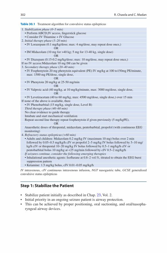
302
Step 1: Stabilize the Patient
• Stabilize patient initially as described in Chap. 23, Vol. 2.• Initial priority in an ongoing seizure patient is airway protection.• This can be achieved by proper positioning, oral suctioning, and oral/nasopha-
ryngeal airway devices.
Table 30.1 Treatment algorithm for convulsive status epilepticus
1. Stabilization phase (0–5 min) • Perform ABCD,IV access, fingerstick glucose • Consider IV Thiamine + IV Glucose2. Initial therapy phase (5–20 min) • IV Lorazepam (0.1 mg/kg/dose; max: 4 mg/dose, may repeat dose once,) OR • IM Midazolam (10 mg for >40 kg; 5 mg for 13-40 kg, single dose) OR • IV Diazepam (0.15-0.2 mg/kg/dose; max: 10 mg/dose, may repeat dose once,)If no IV access:Midazolam 10 mg IM can be given3. Secondary therapy phase (20–40 min) • IV Fosphenytoin 20 mg phenytoin equivalent (PE) IV mg/kg at 100 to150mg PE/minute,
max: 1500 mg PE/dose, single dose, OR • IV Phenytoin 20 mg/kg at 25-50 mg/min OR • IV Valproic acid (40 mg/kg, at 10 mg/kg/minute, max: 3000 mg/dose, single dose, OR • IV Levetiracetam (40 to 60 mg/kg, max: 4500 mg/dose, single dose,) over 15 minIf none of the above is available, then: • IV Phenobarbital (15 mg/kg, single dose, Level B) Third therapy phase (40–60 min) No clear evidence to guide therapy Intubate and start mechanical ventilation Repeat second line therapy repeat fosphenytoin if given previously (5 mg/kgPE), OR Anaesthetic doses of thiopental, midazolam, pentobarbital, propofol (with continuous EEG
monitoring)4. Refractory status epilepticus (>60 min) • Adults and children: Midazolam 0.2 mg/kg IV (maximum 10 mg) bolus over 2 min
followed by 0.05–0.5 mg/kg/h cIV or propofol 2–5 mg/kg IV bolus followed by 5–10 mg/kg/h cIV or thiopental 10–20 mg/kg IV bolus followed by 0.5–1 mg/kg/h cIV or pentobarbital bolus 10 mg/kg at <25 mg/min followed by cIV 0.5–2 mg/kg/h
If seizures continue, consider the following emerging therapies • Inhalational anesthetic agents: Isoflurane at 0.8–2 vol.%, titrated to obtain the EEG burst
suppression pattern • Ketamine: 1.5 mg/kg bolus, cIV 0.01–0.05 mg/kg/h
IV intravenous, cIV continuous intravenous infusion, NGT nasogastric tube, GCSE generalized convulsive status epilepticus
R. Chawla and C. Madan
303
• Assess oxygenation, give oxygen via nasal canula/mask. If necessary, the patient should be intubated.
• Urgent peripheral intravenous (IV) access should be established and simultane-ously collect sample for electrolytes (sodium, calcium, magnesium), LFT, Blood sugar hematology, toxicology screening.
• Check fingerstick blood glucose, if <60 mg/dL – Adults: Give 100 Thiamine IV then 50 mL 50% Dextrose – Children >2 yrs.: 2 mL/kg 25% D IV – Children<2 yrs.: 4 mL/kg 12.5% D IV
Step 2: Terminate the Seizure
• Immediate measures should be taken to end ongoing seizure activity (Table 30.1).
• When it is clear the seizure requires medical intervention, a benzodiazepine (spe-cifically IM midazolam, IV lorazepam, or IV diazepam) is recommended as the initial therapy of choice.
• When intravenous access is readily available intravenous lorazepam is the drug of choice. If seizure continues one additional dose of lorazepam can be given after waiting for a minute.
• Benzodiazepines (lorazepam, midazolam, and diazepam) are effective in termi-nating seizures in 59–78% of patients.
• In patients who are actively seizing despite two initial doses of lorazepam, intra-venous infusion of midazolam (preferred in patients with hypotension) or propo-fol (preferred in patients without hypotension)should be started along with the loading dose of a second line drug.
Step 3: Prevent Further Seizures (Table 30.1)
• Once the initial seizure is controlled, in addition to benzodiazepines a loading dose of second-line drug like fosphenytoin or Valproic acid or Levetiracetam should be given.
• Fosphenytoin is preferred to phenytoin because of its water solubility and neutral pH, thereby allowing more rapid intravenous administration with less adverse effects and its compatibility with all IV fluids.
• Phenytoin or fosphenytoin are incompatible with dextrose-containing solution.• IV levetiracetam is a efficacious and safe drug.• Phenytoin should be given through a larger vein and caution should be taken to
prevent extravasation as it is highly irritant.
Experience with IV valproic acid suggests that it is as effective as phenytoin/fosphenytoin in terminating SE in patients who have previously failed benzodiaze-pines and also as first-line treatment to prevent recurrent seizures.
30 Status Epilepticus
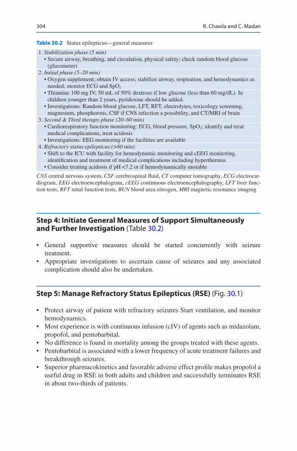
304
Step 4: Initiate General Measures of Support Simultaneously and Further Investigation (Table 30.2)
• General supportive measures should be started concurrently with seizure treatment.
• Appropriate investigations to ascertain cause of seizures and any associated complication should also be undertaken.
Step 5: Manage Refractory Status Epilepticus (RSE) (Fig. 30.1)
• Protect airway of patient with refractory seizures Start ventilation, and monitor hemodynamics.
• Most experience is with continuous infusion (cIV) of agents such as midazolam, propofol, and pentobarbital.
• No difference is found in mortality among the groups treated with these agents.• Pentobarbital is associated with a lower frequency of acute treatment failures and
breakthrough seizures.• Superior pharmacokinetics and favorable adverse effect profile makes propofol a
useful drug in RSE in both adults and children and successfully terminates RSE in about two-thirds of patients.
Table 30.2 Status epilepticus—general measures
1. Stabilization phase (5 min) • Secure airway, breathing, and circulation, physical safety; check random blood glucose
(glucometer)2. Initial phase (5–20 min) • Oxygen supplement; obtain IV access; stabilize airway, respiration, and hemodynamics as
needed; monitor ECG and SpO2
• Thiamine 100 mg IV, 50 mL of 50% dextrose if low glucose (less than 60 mg/dL). In children younger than 2 years, pyridoxine should be added.
• Investigations: Random blood glucose, LFT, RFT, electrolytes, toxicology screening, magnesium, phosphorous, CSF if CNS infection a possibility, and CT/MRI of brain
3. Second & Third therapy phase (20–60 min) • Cardiorespiratory function monitoring: ECG, blood pressure, SpO2; identify and treat
medical complications, treat acidosis • Investigations: EEG monitoring if the facilities are available4. Refractory status epilepticus (>60 min) • Shift to the ICU with facility for hemodynamic monitoring and cEEG monitoring,
identification and treatment of medical complications including hyperthermia • Consider treating acidosis if pH <7.2 or if hemodynamically unstable
CNS central nervous system, CSF cerebrospinal fluid, CT computer tomography, ECG electrocar-diogram, EEG electroencephalogram, cEEG continuous electroencephalography, LFT liver func-tion tests, RFT renal function tests, BUN blood urea nitrogen, MRI magnetic resonance imaging
R. Chawla and C. Madan

305
• Midazolam is an effective, short-acting benzodiazepine, which is given as an infusion, and has an efficacy in RSE.
• If available, continuous EEG monitoring should be performed.• Pharmacologic coma should be maintained for 12 h after the last seizure, with
EEG goal of attaining burst suppression, after which gradually taper off infusion of the anesthetic agent every 3 h with EEG monitoring, and if there are no clini-cal or electrographic seizures, then discontinue the infusion.
• Continue EEG monitoring for at least 24 h after end of infusion.• If clinical or electrographic seizures recur, reinstitute coma therapy with the
same anesthetic agent to which the seizures were responsive.• Make another attempt after 24 h of seizure freedom.• Look for complications and manage hypotension, bradycardia, pulmonary
edema, nosocomial sepsis, ileus, venous thromboemboli, skin breakdown, and exposure keratitis.
Step 6: Initiate Maintenance Treatment (Table 30.2)
• In parallel with emergency treatment, attention must be given to maintain anti-epileptic drug (AED) therapy to prevent recurrence of seizures in close consulta-tion with the neurologist.
• In patients known to have epilepsy, their usual AEDs should be maintained and dose adjustments should be made depending on AED levels.
• In patients presenting de novo, the AEDs, phenytoin/fosphenytoin, or valproic acid used to control the status can in principle be continued as oral maintenance therapy.
Refractory Status Epilepticus
Continuous EEG monitoring
IV Midazolam 0.2 mg/kg bolus followed by infusion at 0.1 mg/kg/hr; max: 3mg/kg/hr
IV Propofol infusion at 1-2 mg/kg loading dose over 5 minsMax: 10-12 mg/kg/hr but for <48 hrs
IV Pentobarbital 5 mg/kg over 10 mins followed by 1-5 mg/kg/hr for 24 hours till seizure free
Fig. 30.1 Recurrent seizure management
30 Status Epilepticus

306
• In others, unless relatively short-lived treatment is anticipated, the preference is to initiate oral maintenance therapy with valproic acid or carbamazepine or any of the newer AEDs, topiramate or levetiracetam.
• Duration of antiepileptic is variable, depending on reversibility of underlying etiology, and should be decided with neurology consultation.
Step 7: Identify and Manage the Nonconvulsive Status
• The nonconvulsive status may present as unexplained coma and fluctuating level of consciousness and is diagnosed by seizure activities in EEG monitoring.
• No concurrent motor activity is usually noticed.• IV benzodiazepines—lorazepam or diazepam—are the drugs of choice.• Allow 5 min to determine whether seizures terminate; if there is no response,
repeat benzodiazepines once.• If EEG monitoring still shows continuous electrographic seizures, consider val-
proic acid in case of absence type of nonconvulsive status epilepticus and con-sider phenytoin/fosphenytoin or valproic acid in case of other types of nonconvulsive status epilepticus. The alternative option, particularly in the elderly will be intravenous levetiracetam.
Suggested Reading
American Epilepsy Society issues guideline and treatment algorithm for convulsive status epilep-ticus 2016, https://www.aesnet.org/about_aes/press_releases/guidelines2016.
Bassin S, Smith TL, Bleck TP. Clinical review: status epilepticus. Crit Care. 2002;6(2):137–42. This review discusses current definitions of SE, as well as its clinical presentation and clas-sification. The recent literature on epidemiology is reviewed, including morbidity and mortality data. An overview of the systemic pathophysiologic effects of SE is presented. Finally, signifi-cant studies on the treatment of acute SE and refractory SE are reviewed, including the use of anticonvulsants, such as benzodiazepines and other drugs.
Brophy GM, Bell R, Claassen J, et al. Guidelines for the evaluation and management of status epilepticus. Neurocrit Care. 2012;17:3. A comprehensive guidelines for the management of status epilepticus.
Claassen J, Hirsch LJ, Emerson RC, et al. Treatment of refractory status epilepticus with pento-barbital, propofol, or midazolam: a systematic review. Epilepsia. 2002;41:146–53. Treatment with pentobarbitone, or any cIV-AED infusion to attain EEG background suppression, may be more effective than other strategies for treating RSE. However, these interventions were also associated with an increased frequency of hypotension, and no effect on mortality was seen.
Meierkord H, Holtkamp M. Non-convulsive status epilepticus in adults: clinical forms and treat-ment. Lancet Neurol. 2007;6:329–39. An excellent review on nonconvulsive status epilepticus.
Millikan D, Rice B, Silbergleit R. Emergency treatment of status epilepticus: current thinking. Emerg Med Clin North Am. 2009;27(1):101–13. Current thinking about the acute treatment of status epilepticus (SE) emphasizes a more aggressive clinical approach to this common life-threatening neurologic emergency. In this review, the authors consider four concepts that can accelerate effective treatment of SE. These include (1) updating the definition of SE to make it more clinically relevant, (2) consideration of faster ways to initiate first-line benzodiazepine
R. Chawla and C. Madan

307
therapy in the prehospital environment, (3) moving to second-line agents more quickly in refractory status in the emergency department, and (4) increasing detection and treatment of unrecognized nonconvulsive SE in comatose neurologic emergency patients.
Treiman DM, Meyers PF. Walton NY. A comparison of four treatments for generalized convul-sive status epilepticus: Veterans Affairs Status Epilepticus Cooperative Study Group. N Engl J Med. 1998;339:792–8. As initial intravenous treatment for overt generalized convulsive status epilepticus, lorazepam is more effective than phenytoin. Although lorazepam is no more effica-cious than phenobarbital or diazepam plus phenytoin, it is easier to use.
30 Status Epilepticus

309© Springer Nature Singapore Pte Ltd. 2020R. Chawla, S. Todi (eds.), ICU Protocols, https://doi.org/10.1007/978-981-15-0898-1_31
C. Gauba (*) · M. Varma Department of Neurology, Indraprastha Apollo Hospitals, New Delhi, India
P. Chopra Department of Critical Care Medicine, Fortis Memorial Research Institute, Gurugram, Haryana, India
31Acute Flaccid Paralysis
Charu Gauba, Mukul Varma, and Pooja Chopra
Patients with rapid onset of flaccid paralysis pose a diagnostic and therapeutic chal-lenge to the treating clinician. Systematic assessment, constructing a differential diagnosis and rational approach to this problem will help in preventing and treating complications as well as reducing hospital stay.
Step 1: Assess the Patient
• Initiate resuscitation (see Chap. 23, Vol. 2).• Acute weakness may be the primary reason for admission to the ICU (Table 31.1)
or may be secondary to an episode of critical illness.• Immediate assessment of the patient’s clinical condition and the duration of
symptoms are the most important factors for determining the need for ICU care.• Airway and breathing should be assessed for the need for airway protection and
ventilatory support (Tables 31.2 and 31.3).• Assess circulation by pulse rate, pulse volume and blood pressure. If tachycardia
is present or BP is low, IV fluid bolus should be given.
A 50-year-old male patient was admitted to hospital with a history of rapidly ascending paraparesis for the past 6 days followed by difficulty in breathing. He had an episode of acute gastroenteritis 2 weeks prior to the onset of the illness. His deep tendon reflexes were absent. Cerebrospinal fluid (CSF) examination showed raised protein with normal sugars and normal cell count.
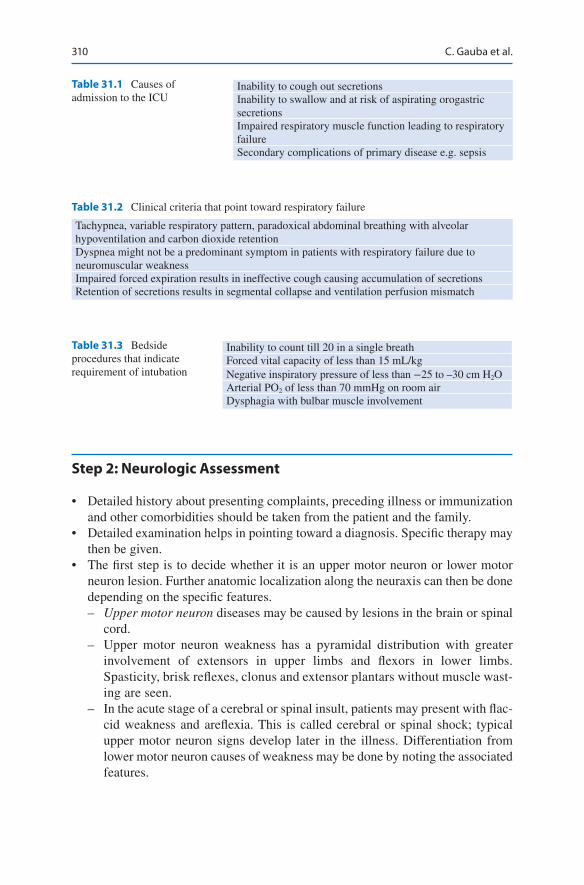
310
Step 2: Neurologic Assessment
• Detailed history about presenting complaints, preceding illness or immunization and other comorbidities should be taken from the patient and the family.
• Detailed examination helps in pointing toward a diagnosis. Specific therapy may then be given.
• The first step is to decide whether it is an upper motor neuron or lower motor neuron lesion. Further anatomic localization along the neuraxis can then be done depending on the specific features. – Upper motor neuron diseases may be caused by lesions in the brain or spinal
cord. – Upper motor neuron weakness has a pyramidal distribution with greater
involvement of extensors in upper limbs and flexors in lower limbs. Spasticity, brisk reflexes, clonus and extensor plantars without muscle wast-ing are seen.
– In the acute stage of a cerebral or spinal insult, patients may present with flac-cid weakness and areflexia. This is called cerebral or spinal shock; typical upper motor neuron signs develop later in the illness. Differentiation from lower motor neuron causes of weakness may be done by noting the associated features.
Table 31.1 Causes of admission to the ICU
Inability to cough out secretionsInability to swallow and at risk of aspirating orogastric secretionsImpaired respiratory muscle function leading to respiratory failureSecondary complications of primary disease e.g. sepsis
Table 31.2 Clinical criteria that point toward respiratory failure
Tachypnea, variable respiratory pattern, paradoxical abdominal breathing with alveolar hypoventilation and carbon dioxide retentionDyspnea might not be a predominant symptom in patients with respiratory failure due to neuromuscular weaknessImpaired forced expiration results in ineffective cough causing accumulation of secretionsRetention of secretions results in segmental collapse and ventilation perfusion mismatch
Table 31.3 Bedside procedures that indicate requirement of intubation
Inability to count till 20 in a single breathForced vital capacity of less than 15 mL/kgNegative inspiratory pressure of less than −25 to –30 cm H2OArterial PO2 of less than 70 mmHg on room airDysphagia with bulbar muscle involvement
C. Gauba et al.
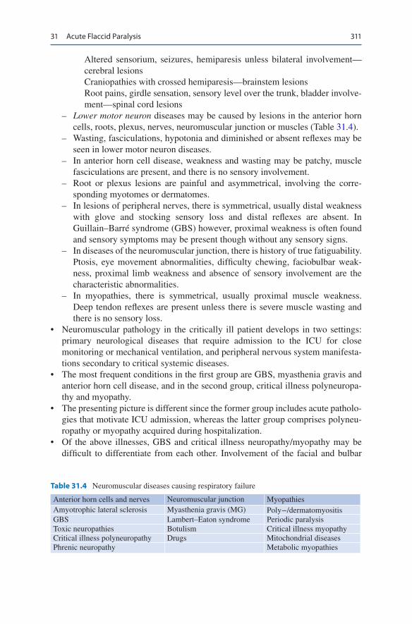
311
Altered sensorium, seizures, hemiparesis unless bilateral involvement—cerebral lesionsCraniopathies with crossed hemiparesis—brainstem lesionsRoot pains, girdle sensation, sensory level over the trunk, bladder involve-ment—spinal cord lesions
– Lower motor neuron diseases may be caused by lesions in the anterior horn cells, roots, plexus, nerves, neuromuscular junction or muscles (Table 31.4).
– Wasting, fasciculations, hypotonia and diminished or absent reflexes may be seen in lower motor neuron diseases.
– In anterior horn cell disease, weakness and wasting may be patchy, muscle fasciculations are present, and there is no sensory involvement.
– Root or plexus lesions are painful and asymmetrical, involving the corre-sponding myotomes or dermatomes.
– In lesions of peripheral nerves, there is symmetrical, usually distal weakness with glove and stocking sensory loss and distal reflexes are absent. In Guillain–Barré syndrome (GBS) however, proximal weakness is often found and sensory symptoms may be present though without any sensory signs.
– In diseases of the neuromuscular junction, there is history of true fatiguability. Ptosis, eye movement abnormalities, difficulty chewing, faciobulbar weak-ness, proximal limb weakness and absence of sensory involvement are the characteristic abnormalities.
– In myopathies, there is symmetrical, usually proximal muscle weakness. Deep tendon reflexes are present unless there is severe muscle wasting and there is no sensory loss.
• Neuromuscular pathology in the critically ill patient develops in two settings: primary neurological diseases that require admission to the ICU for close monitoring or mechanical ventilation, and peripheral nervous system manifesta-tions secondary to critical systemic diseases.
• The most frequent conditions in the first group are GBS, myasthenia gravis and anterior horn cell disease, and in the second group, critical illness polyneuropa-thy and myopathy.
• The presenting picture is different since the former group includes acute patholo-gies that motivate ICU admission, whereas the latter group comprises polyneu-ropathy or myopathy acquired during hospitalization.
• Of the above illnesses, GBS and critical illness neuropathy/myopathy may be difficult to differentiate from each other. Involvement of the facial and bulbar
Table 31.4 Neuromuscular diseases causing respiratory failure
Anterior horn cells and nerves Neuromuscular junction MyopathiesAmyotrophic lateral sclerosis Myasthenia gravis (MG) Poly−/dermatomyositisGBS Lambert–Eaton syndrome Periodic paralysisToxic neuropathies Botulism Critical illness myopathyCritical illness polyneuropathy Drugs Mitochondrial diseasesPhrenic neuropathy Metabolic myopathies
31 Acute Flaccid Paralysis
312
muscles and autonomic nervous system along with CSF albuminocytologic dis-sociation is common in GBS, which does not occur in critical illness polyneuropathy.
• GBS is typically an acute demyelinating neuropathy though axonal variants also occur. Critical illness neuropathy, on the other hand, is of the axonal type.
• The functional prognosis of primary muscle impairment tends to be quite good, but both critical illness polyneuropathy and myopathy resolve very slowly over weeks or months with the possibility of a significant residual deficit after 2 years in the most severe cases.
Step 3: Send Investigations
• Blood tests – Serum potassium, calcium, vitamin D levels and creatine phosphokinase are
required when muscle pathology is suspected. – Blood sugar levels, renal and liver function tests, vitamin B12 levels and
serum protein electrophoresis should be tested in diseases of the peripheral nerves. Toxin levels in blood may be tested where there is history of exposure.
– Thyroid function tests, HIV serology, vasculitic markers and paraneoplastic antibodies are useful in the case of both neuropathies and myopathies.
– Anti-acetylcholine receptor antibodies are a sensitive test for diagnosis of myasthenia gravis.
• Imaging: It is required in suspected CNS disease.• Electromyogram (EMG) and nerve conduction velocity (NCV) studies are
required to diagnose nerve and muscle disease. – In demyelinating neuropathy, distal latencies are prolonged with dispersed
compound muscle action potentials (CMAP). There may be conduction block and nerve conduction velocity may be decreased on nerve conduction studies.
– In axonal neuropathy, there is decrease in amplitude of action potential on nerve conduction studies. EMG shows spontaneous activity in the form of fibrillation potentials and positive sharp waves with decreased recruitment.
– In myopathies, small-amplitude, short-duration, polyphasic action potentials are seen on EMG with rapid recruitment.
– Repetitive nerve stimulation shows a decremental response in myasthenia gravis.
• CSF examination: Albuminocytologic dissociation (increased protein with nor-mal cell count and sugar) is seen in GBS. On the other hand, high protein levels with pleocytosis and normal or low sugars are seen in infectious diseases.
• Spirometry: It is required for the measurement of vital capacity and peak flow.• Muscle/nerve biopsy: It is required in selected cases of myopathy or
neuropathy.
C. Gauba et al.
313
Step 4: Management
1. Airway protection—indications• Bulbar palsy leading to dysarthria, dysphonia, dysphagia and poor gag reflex• Acute aspiration leading to respiratory arrest• Insidious aspiration leading to pneumonia and gradual respiratory
decompensation 2. Respiratory support
Bedside tests and clinical assessment to determine the need for respiratory support.• ICU admission is needed if vital capacity is less than 1 L or less than 50%
predicted value, respiratory rate more than 30 or if the patient is unable to maintain patent airway.
• Arterial blood gas analysis should be performed regularly. Hypoxia and hypercarbia with respiratory acidosis are late features. The patient may need assisted ventilation even with normal blood gas in neuromuscular respiratory failure.
• Early tracheostomy should be considered in patients who require prolonged ventilatory support.
• Physiotherapy should be encouraged. 3. Nutritional support
• Enteral nutrition should be initiated as soon as the patient becomes hemody-namically stable.
• Nasogastric route should be preferred. Proper precaution should be taken to prevent regurgitation and aspiration. Head end should be elevated. Give pro-kinetics. Give continuous feed through enteral pump and check residual vol-ume. Start nasojejunal feed in selected cases.
• Oral feed can be considered in some tracheostomized patients after proper swallowing assessment.
• Percutaneous endoscopic gastrostomy may be required for long-term use. 4. Venous thromboembolism prevention
• Due to immobilization, the risk of deep venous thrombosis and pulmonary embolism (PE) is high.
• Low-molecular-weight heparin or unfractionated heparin and gradient com-pression stockings are useful.
• Passive leg exercises should be initiated. 5. Pain control
• Both acute and chronic pain should be treated.• Opioid analgesics, anticonvulsants or antidepressants may be used for neuro-
pathic pains. 6. Autonomic disturbance
• Common in small fibre neuropathies and sometimes seen in GBS.• Depending on the symptoms—treat excessive secretions with
anticholinergics.
31 Acute Flaccid Paralysis
314
• β-blockers can be used for disproportionate tachycardia. Cardiac pacing can be done for severe symptomatic bradycardia. Autonomic disturbances in GBS can be fatal if not treated.
7. Pressure sores• Frequent turning should be done and pressure-relieving mattresses should be
used. 8. Physiotherapy
• It is important in the early course of the disease and in rehabilitation.• Passive exercises, cough assist devices and splints are useful.
9. Specific treatment• Depending on the etiology
Step 5: Identify and Manage Specific Illnesses
1. Critical illness neuropathy• History will be suggestive of some critical illness, for example sepsis, severe
trauma or burns.• Clinical signs include generalized flaccid weakness with distal predominance
and absent or decreased deep tendon reflexes. Facial and bulbar muscles are usually spared. Pain or paresthesia is not seen.
• Sensorium is intact.• It often presents as failure to wean from the ventilator.• Investigations: NCV studies will reveal decreased amplitude of motor and
sensory action potentials with preserved conduction velocity, suggestive of an axonal neuropathy. EMG will reveal fibrillations and decreased motor unit potentials. CSF is almost always normal.
• It is diagnosed after excluding other neuropathies or neuromuscular junction abnormalities.
• Treatment is supportive care, intense glucose control and early treatment of sepsis.
• Recovery is spontaneous in 3–6 months and is often partial.• Prognosis depends on the underlying illness.
2. Critical illness myopathy• It is difficult to distinguish clinically from its neuropathic counterpart and
both may occur concurrently.• It usually occurs in patients with acute respiratory distress syndrome or severe
asthma who have been treated with intravenous corticosteroids, aminoglyco-sides and/or nondepolarizing neuromuscular blocking agents.
• Plasma creatine kinase levels are transiently and marginally elevated.• EMG shows small-amplitude, short-duration, polyphasic motor unit poten-
tials and sensory nerve conduction studies are normal.• Repetitive nerve stimulation studies should be performed to exclude a defect
of neuromuscular transmission caused by defective clearance of neuromuscu-lar blocking agents.
C. Gauba et al.
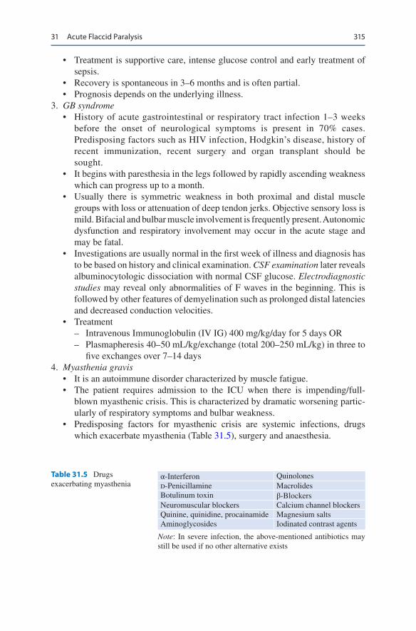
315
• Treatment is supportive care, intense glucose control and early treatment of sepsis.
• Recovery is spontaneous in 3–6 months and is often partial.• Prognosis depends on the underlying illness.
3. GB syndrome• History of acute gastrointestinal or respiratory tract infection 1–3 weeks
before the onset of neurological symptoms is present in 70% cases. Predisposing factors such as HIV infection, Hodgkin’s disease, history of recent immunization, recent surgery and organ transplant should be sought.
• It begins with paresthesia in the legs followed by rapidly ascending weakness which can progress up to a month.
• Usually there is symmetric weakness in both proximal and distal muscle groups with loss or attenuation of deep tendon jerks. Objective sensory loss is mild. Bifacial and bulbar muscle involvement is frequently present. Autonomic dysfunction and respiratory involvement may occur in the acute stage and may be fatal.
• Investigations are usually normal in the first week of illness and diagnosis has to be based on history and clinical examination. CSF examination later reveals albuminocytologic dissociation with normal CSF glucose. Electrodiagnostic studies may reveal only abnormalities of F waves in the beginning. This is followed by other features of demyelination such as prolonged distal latencies and decreased conduction velocities.
• Treatment – Intravenous Immunoglobulin (IV IG) 400 mg/kg/day for 5 days OR – Plasmapheresis 40–50 mL/kg/exchange (total 200–250 mL/kg) in three to
five exchanges over 7–14 days 4. Myasthenia gravis
• It is an autoimmune disorder characterized by muscle fatigue.• The patient requires admission to the ICU when there is impending/full-
blown myasthenic crisis. This is characterized by dramatic worsening partic-ularly of respiratory symptoms and bulbar weakness.
• Predisposing factors for myasthenic crisis are systemic infections, drugs which exacerbate myasthenia (Table 31.5), surgery and anaesthesia.
Table 31.5 Drugs exacerbating myasthenia
α-Interferon Quinolonesd-Penicillamine MacrolidesBotulinum toxin β-BlockersNeuromuscular blockers Calcium channel blockersQuinine, quinidine, procainamide Magnesium saltsAminoglycosides Iodinated contrast agents
Note: In severe infection, the above-mentioned antibiotics may still be used if no other alternative exists
31 Acute Flaccid Paralysis

316
• It commonly begins with fluctuating and asymmetric ptosis and weakness of extraocular muscles. This may be followed by weakness of bulbar, mastica-tory and proximal limb muscles. Respiratory muscle involvement indicates myasthenic crisis. Muscle bulk, tone, reflexes and sensory examination are normal.
• Investigations: Serum anti-acetylcholine receptor antibodies are positive in more than 90% cases of generalized myasthenia. Some seronegative patients have anti-muscle-specific tyrosine kinase antibodies. Edrophonium test: Pretreat with 0.5 mg atropine, give 2 mg edrophonium intravenously, then give 3 mg and then 5 mg edrophonium intravenously; observe at each dose for 1–3 min for increase in muscle strength. Electrodiagnostic tests reveal decre-mental (15% or more reduction in amplitude) response in compound muscle action potential on repetitive nerve stimulation (RNS). Chest imaging should be done to rule out thymoma.
• Treatment – Plasmapheresis 50 mL/kg/exchange (total 200–250 mL/kg) in five to six
exchanges over 7–14 days. This is for myasthenic crisis. – IV IG 400 mg/kg/day for 5 days (preferred over plasmapheresis in myas-
thenic crisis, if severe infection coexists). – Prednisolone 1 mg/kg/day. In about one-third of patients, myasthenic
weakness worsens temporarily 7–10 days after starting steroids and the patient may go into a crisis situation.
– Steroid sparing—azathioprine, cyclosporine, mycophenolate mofetil. – Pyridostigmine 60 mg oral; neostigmine 15 mg oral, 0.5 mg IV. (These
should be discontinued if the patient is ventilated and reintroduced on weaning.)
– Thymectomy may be useful especially in young (<50 years) females, but response is delayed. Its role is still being studied. It is mandatory in thymomas.
– Supportive treatment. 5. Anterior horn cell disease
• The commonest of these is amyotrophic lateral sclerosis.• It is usually sporadic and slightly more common in males.• Peak incidence is between 65 and 74 years.• Mean disease duration from onset to death is about 3 years.• Wasting and weakness are usually patchy. Fasciculations are prominent. Both
upper and lower motor neuron signs are seen and there is no sensory loss.• Bulbar and respiratory muscles are involved in later stages, and respiratory
failure is the usual mode of death.• So far, there is no known drug which significantly prolongs survival. Riluzole
or iv Edaravone may delay need for ventilatory assistance by 2-3 months.
C. Gauba et al.
317
Suggested Reading
Bolton CF, Laverty DA, Brown JD, Witt NJ, Hahn AF, Sibbald WJ. Critically ill polyneuropa-thy: electrophysiological studies and differentiation from Guillain–Barré syndrome. J Neurol Neurosurg Psychiatr. 1986;649:563–73.
Chawla J, Gruener G. Management of critical illness polyneuropathy and myopathy. Neurol Clin. 2010;28(4):961–77. A review article
Meena AK, Khadilkar SV, Murthy JM. Treatment guidelines for Guillain–Barré syndrome. Ann Indian Acad Neurol. 2011;14(Suppl 1):S73–81. A review article
Patwa HS, Chaudhry V, Katzberg H, et al. Evidence-based guideline: intravenous immunoglobu-lin in the treatment of neuromuscular disorders: report of the Therapeutics and Technology Assessment Subcommittee of the American Academy of Neurology. Neurology. 2012;78:1009. Guidelines for immunoglobulin administration. IVIg is as efficacious as plasmapheresis in Guillain-Barrésyndrome (GBS) in adults (Level A). IVIg is probably effective and should be considered for treating moderate to severe myasthenia gravis and multifocal motor neuropathy (Level B).
Spitzer AR, Giancarlo T, Maher L, Awerbuch G, Bowles A. Neuromuscular causes of prolonged ventilator dependency. Muscle Nerve. 1992;15:682–6.
Yuki N, Hartung HP. Guillain-Barré syndrome. N Engl J Med. 2012;366:2294. A review articleZochodne DW, Bolton CF, Thompson RT, Driedger AA, Hahn AF, Gilbert JJ. Myopathy in critical
illness. Muscle Nerve. 1986;9:652.
31 Acute Flaccid Paralysis

319© Springer Nature Singapore Pte Ltd. 2020R. Chawla, S. Todi (eds.), ICU Protocols, https://doi.org/10.1007/978-981-15-0898-1_32
K. Kadapatti (*) Department of Critical Care, Jehangir Hospital, Pune, India
S. K. Iyer Department of Intensive Care and Critical Care Medicine, Bharati Vidyapeeth (DTU) Medical College, Pune, India
32Coma
Kayanoosh Kadapatti and Shiva Kumar Iyer
The comatose patient in the ICU poses a diagnostic and therapeutic challenge to the physician. A broad range of differential diagnoses necessitate a systematic approach and judicious use of investigation in these patients.
Step 1: Assess and Stabilize the Patient
• In any unexplained coma, caution should be taken to avoid cervical spine move-ment while managing airway.
• Intubate if GCS <8• If there is hypoglycemia (<60 mg/dL), it should be urgently corrected with
50 mL of 25% dextrose intravenously.• Thiamine 100 mg IV should be given prior to glucose to avoid Wernicke’s
encephalopathy, especially in patients with malnutrition and alcoholism.• Control seizure, if present, with intravenous lorazepam.• In patients who show features of increased intracranial pressure (ICP), 100 mL
of 10% intravenous mannitol should be given (see Chap. 33, Vol. 1).• Give prompt antibiotics in patients with signs suggestive of meningitis.
A 30-year-old male patient, who was found unconscious in his room, was brought to the emergency department. On examination, he was found to be tachypneic and febrile with normal blood pressure. He was not opening his eyes and was withdrawing arms on pain.
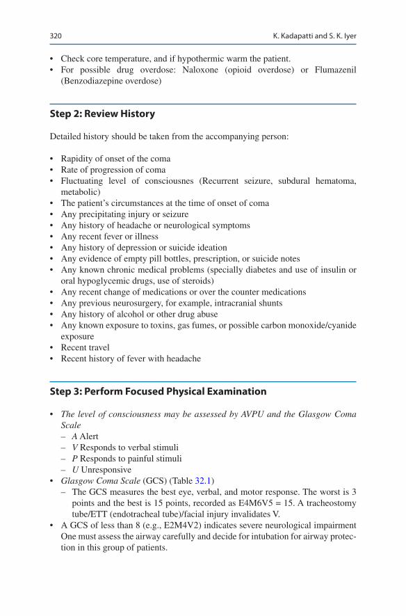
320
• Check core temperature, and if hypothermic warm the patient.• For possible drug overdose: Naloxone (opioid overdose) or Flumazenil
(Benzodiazepine overdose)
Step 2: Review History
Detailed history should be taken from the accompanying person:
• Rapidity of onset of the coma• Rate of progression of coma• Fluctuating level of consciousnes (Recurrent seizure, subdural hematoma,
metabolic)• The patient’s circumstances at the time of onset of coma• Any precipitating injury or seizure• Any history of headache or neurological symptoms• Any recent fever or illness• Any history of depression or suicide ideation• Any evidence of empty pill bottles, prescription, or suicide notes• Any known chronic medical problems (specially diabetes and use of insulin or
oral hypoglycemic drugs, use of steroids)• Any recent change of medications or over the counter medications• Any previous neurosurgery, for example, intracranial shunts• Any history of alcohol or other drug abuse• Any known exposure to toxins, gas fumes, or possible carbon monoxide/cyanide
exposure• Recent travel• Recent history of fever with headache
Step 3: Perform Focused Physical Examination
• The level of consciousness may be assessed by AVPU and the Glasgow Coma Scale – A Alert – V Responds to verbal stimuli – P Responds to painful stimuli – U Unresponsive
• Glasgow Coma Scale (GCS) (Table 32.1) – The GCS measures the best eye, verbal, and motor response. The worst is 3
points and the best is 15 points, recorded as E4M6V5 = 15. A tracheostomy tube/ETT (endotracheal tube)/facial injury invalidates V.
• A GCS of less than 8 (e.g., E2M4V2) indicates severe neurological impairment One must assess the airway carefully and decide for intubation for airway protec-tion in this group of patients.
K. Kadapatti and S. K. Iyer

321
• The GCS level is predictive of outcome-if eye opening is present within 6 hrs, the patient has one in five chance of achieving good recovery.
• Focused physical examination should be performed to assess the evidence of lateralizing signs like hemiplegia, features of increased ICP like pupillary changes, features of meningoencephalitis, or any systemic problem requiring immediate attention by laboratory investigation or by imaging and need of neu-rosurgical consultation.
• Severe hypertension is suggestive of Posterior reversible encephalopathy (PRES), hypertensive encephalopathy and intracranial hemorrhage
• Physical examination is useful in assessing the severity of coma and anatomical localization of neurological insult. These examinations need to be repeated fre-quently to elicit any deterioration of the patient’s status.
• Posture—look for any seizure activity, tremors, myoclonus, or spontaneous decerebration. Myoclonic jerks is suggestive of an anoxic cause of coma and portend a bad prognosis. Subtle rhythmic movements may suggest ongoing seizures.
• Skin—look for pallor, icterus, cyanosis of methemoglobinemia, petechiae, scars, burns, pigmentation, and needle marks.
• Facial muscles—look for asymmetry of the face.• Oral cavity—look for tongue bite, smell of alcohol or ammonia (liver disease),
gingival hyperplasia (phenytoin-induced), oral thrush (immunodeficient state).• HEENT—head, eye, ear, neck, and throat should be examined for the evidence
of injury, hematoma, bleeding (Battle’s sign, Raccoon eyes) or any purulent dis-charge, skull fractures, cerebrospinal fluid (CSF) rhinorrhea, or otorrhea. Neck stiffness examination should be avoided in patients with suspected cervical spine injury. Check pupils for symmetry and light reflex.
Table 32.1 Glasgow coma scale
Best eye opening (E)Nil 1Pain 2Verbal 3Spontaneous 4Best verbal response (V)Nil 1Incomprehensible sounds 2Inappropriate but recognizable words 3Confused conversation 4Oriented 5Best motor response (M)Nil 1Abnormal extension (decerebrate response) 2Abnormal flexion (decorticate response) 3Withdraws to pain 4Localizes to pain 5Obeys commands 6
32 Coma

322
• Temperature – Hypothermia can occur in ethanol or sedative drug intoxication, Wernicke’s
encephalopathy, hepatic encephalopathy, and myxedema. – Hyperthermia can occur in status epilepticus, pontine hemorrhage, heat
stroke, malignant hyperthermia, and anticholinergic intoxication.• Optic fundi—look for papilledema and vitreous or subhyaloid hemorrhage.• Specific neurological examination of a comatose patient should include mental
status, level of consciousness, awareness of self and environment, cranial nerve, motor response, and brainstem reflexes.
• Assess the brainstem reflexes – Check pupillary size and response to light. – Pupils that are equal in size, and react briskly to light, usually imply normal
brainstem function and suggest metabolic coma or cortical injury. – Poorly reactive (but equal) pupils offer no clinical clues to the etiology or
severity of the coma. – Small (1–2.5 mm) reactive pupils—diencephalon (thalamic hemorrhage),
metabolic encephalopathy. – Pinpoint pupils (<1 mm) with reaction—pontine pathology, narcotic or barbi-
turate overdose. – Midposition/large (5–7 mm), fixed pupils—midbrain lesion – Bilateral widely dilated, fixed pupils—severe midbrain damage, brain death,
rarely drugs, for example, barbiturates, atropine – Unilateral, dilated, fixed pupils—third nerve compression
• Ocular movements – Abducted eyes—third nerve dysfunction. – Adducted eyes—sixth nerve dysfunction; increased ICP. – Spontaneous horizontal eye movement, whether conjugate or disconjugate,
implies normal brainstem function and suggests a metabolic coma. – Induced conjugate eye movement, oculocephalic reflex (doll’s eye maneuver)
or the oculovestibular reflex (cold caloric test)—in patients with suspected cervical spine injury, cold caloric test is preferable.
– Resting position of the eyes on opening the eyelids—persistent conjugate deviation of the eyes toward one side suggests a stroke or seizure. Tonic con-jugate downward deviation of the eyes often implies thalamic upper brain-stem compression.
– Corneal reflex should be tested by putting a few drops of sterile saline in the eyes rather than cotton wool to avoid corneal injury.
• Motor response – Motor responses can be characterized as appropriate, posturing, or flaccid. – Posturing refers to stereotyped arm and leg movement occurring spontane-
ously or elicited by sensory stimulation. Abnormal posturing can occur in early brainstem compression due to increased ICP and transtentorial hernia-tion and manifest first with decorticate (arm flexion, leg extension) posturing due to diencephalon compression. The patient can then manifest decerebrate (arm extension, leg extension) posturing due to midbrain and upper pons
K. Kadapatti and S. K. Iyer
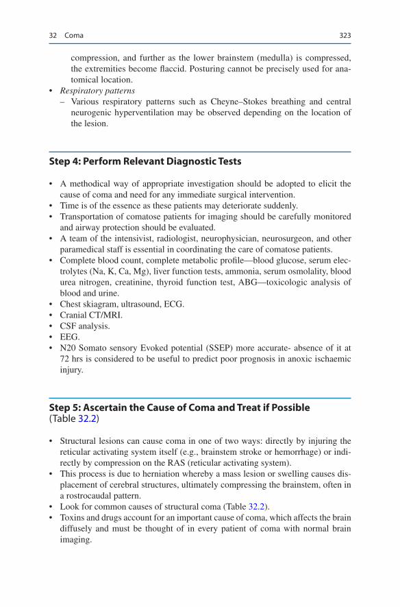
323
compression, and further as the lower brainstem (medulla) is compressed, the extremities become flaccid. Posturing cannot be precisely used for ana-tomical location.
• Respiratory patterns – Various respiratory patterns such as Cheyne–Stokes breathing and central
neurogenic hyperventilation may be observed depending on the location of the lesion.
Step 4: Perform Relevant Diagnostic Tests
• A methodical way of appropriate investigation should be adopted to elicit the cause of coma and need for any immediate surgical intervention.
• Time is of the essence as these patients may deteriorate suddenly.• Transportation of comatose patients for imaging should be carefully monitored
and airway protection should be evaluated.• A team of the intensivist, radiologist, neurophysician, neurosurgeon, and other
paramedical staff is essential in coordinating the care of comatose patients.• Complete blood count, complete metabolic profile—blood glucose, serum elec-
trolytes (Na, K, Ca, Mg), liver function tests, ammonia, serum osmolality, blood urea nitrogen, creatinine, thyroid function test, ABG—toxicologic analysis of blood and urine.
• Chest skiagram, ultrasound, ECG.• Cranial CT/MRI.• CSF analysis.• EEG.• N20 Somato sensory Evoked potential (SSEP) more accurate- absence of it at
72 hrs is considered to be useful to predict poor prognosis in anoxic ischaemic injury.
Step 5: Ascertain the Cause of Coma and Treat if Possible (Table 32.2)
• Structural lesions can cause coma in one of two ways: directly by injuring the reticular activating system itself (e.g., brainstem stroke or hemorrhage) or indi-rectly by compression on the RAS (reticular activating system).
• This process is due to herniation whereby a mass lesion or swelling causes dis-placement of cerebral structures, ultimately compressing the brainstem, often in a rostrocaudal pattern.
• Look for common causes of structural coma (Table 32.2).• Toxins and drugs account for an important cause of coma, which affects the brain
diffusely and must be thought of in every patient of coma with normal brain imaging.
32 Coma
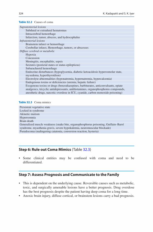
324
Step 6: Rule out Coma Mimics (Table 32.3)
• Some clinical entities may be confused with coma and need to be differentiated.
Step 7: Assess Prognosis and Communicate to the Family
• This is dependent on the underlying cause. Reversible causes such as metabolic, toxic, and surgically amenable lesions have a better prognosis. Drug overdose has the best prognosis despite the patient having deep coma for a long time.
• Anoxic brain injury, diffuse cortical, or brainstem lesions carry a bad prognosis.
Table 32.2 Causes of coma
Supratentorial lesions Subdural or extradural hematomas Intracerebral hemorrhage Infarction, tumor, abscess, and hydrocephalusInfratentorial lesions Brainstem infarct or hemorrhage Cerebellar infarct, Hemorrhage, tumors, or abscessesDiffuse cerebral or metabolic Hypoxia Concussion Meningitis, encephalitis, sepsis Seizures (postictal states or status epilepticus) Subarachnoid hemorrhage Endocrine disturbances (hypoglycemia, diabetic ketoacidosis hyperosmolar state,
myxedema, hyperthyroidism) Electrolyte abnormalities (hyponatremia, hypernatremia, hypercalcemia) Endogenous toxins or deficiencies (uremia, hepatic failure) Exogenous toxins or drugs (benzodiazepines, barbiturates, anticonvulsants, opiate
analgesics, tricyclic antidepressants, antihistamines, organophosphorus compounds, anesthetic drugs, narcotic overdose in ICU, cyanide, carbon monoxide poisoning)
Table 32.3 Coma mimics
Persistent vegetative stateLocked in syndromeAkinetic mutismHypersomniaBrain deathGeneralized muscle weakness (snake bite, organophosphorus poisoning, Guillain–Barré syndrome, myasthenia gravis, severe hypokalemia, neuromuscular blockade)Pseudocoma (malingering catatonia, conversion reaction, hysteria)
K. Kadapatti and S. K. Iyer
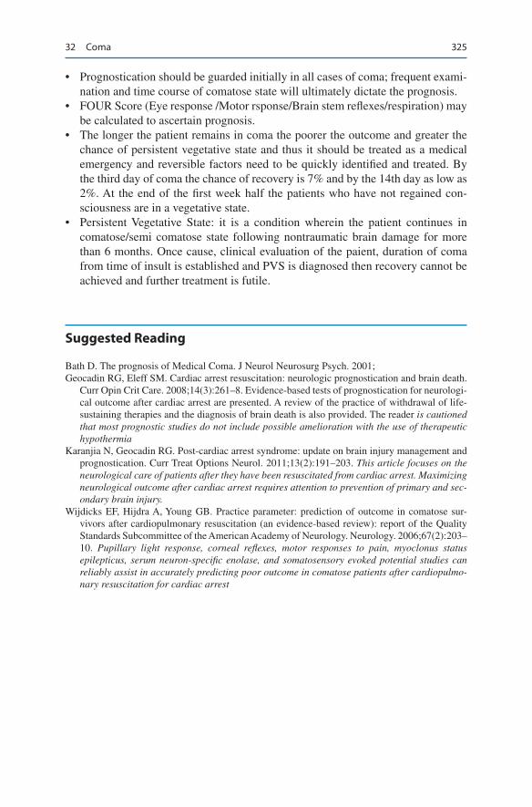
325
• Prognostication should be guarded initially in all cases of coma; frequent exami-nation and time course of comatose state will ultimately dictate the prognosis.
• FOUR Score (Eye response /Motor rsponse/Brain stem reflexes/respiration) may be calculated to ascertain prognosis.
• The longer the patient remains in coma the poorer the outcome and greater the chance of persistent vegetative state and thus it should be treated as a medical emergency and reversible factors need to be quickly identified and treated. By the third day of coma the chance of recovery is 7% and by the 14th day as low as 2%. At the end of the first week half the patients who have not regained con-sciousness are in a vegetative state.
• Persistent Vegetative State: it is a condition wherein the patient continues in comatose/semi comatose state following nontraumatic brain damage for more than 6 months. Once cause, clinical evaluation of the paient, duration of coma from time of insult is established and PVS is diagnosed then recovery cannot be achieved and further treatment is futile.
Suggested Reading
Bath D. The prognosis of Medical Coma. J Neurol Neurosurg Psych. 2001;Geocadin RG, Eleff SM. Cardiac arrest resuscitation: neurologic prognostication and brain death.
Curr Opin Crit Care. 2008;14(3):261–8. Evidence-based tests of prognostication for neurologi-cal outcome after cardiac arrest are presented. A review of the practice of withdrawal of life- sustaining therapies and the diagnosis of brain death is also provided. The reader is cautioned that most prognostic studies do not include possible amelioration with the use of therapeutic hypothermia
Karanjia N, Geocadin RG. Post-cardiac arrest syndrome: update on brain injury management and prognostication. Curr Treat Options Neurol. 2011;13(2):191–203. This article focuses on the neurological care of patients after they have been resuscitated from cardiac arrest. Maximizing neurological outcome after cardiac arrest requires attention to prevention of primary and sec-ondary brain injury.
Wijdicks EF, Hijdra A, Young GB. Practice parameter: prediction of outcome in comatose sur-vivors after cardiopulmonary resuscitation (an evidence-based review): report of the Quality Standards Subcommittee of the American Academy of Neurology. Neurology. 2006;67(2):203–10. Pupillary light response, corneal reflexes, motor responses to pain, myoclonus status epilepticus, serum neuron-specific enolase, and somatosensory evoked potential studies can reliably assist in accurately predicting poor outcome in comatose patients after cardiopulmo-nary resuscitation for cardiac arrest
32 Coma
327© Springer Nature Singapore Pte Ltd. 2020R. Chawla, S. Todi (eds.), ICU Protocols, https://doi.org/10.1007/978-981-15-0898-1_33
R. Chawla (*) Department of Respiratory, Critical Care & Sleep Medicine, Indraprastha Apollo Hospitals, New Delhi, India
R. Senthilkumar · N. Ramakrishnan Department of Critical Care Medicine, Apollo Hospitals, Chennai, India
33Intracranial Pressure Monitoring and Management
Rajesh Chawla, Rajagopal Senthilkumar, and Nagarajan Ramakrishnan
Increased intracranial pressure (ICP) should be suspected in all patients with altered mental state, especially due to an intracranial pathology. Normal ICP is below 15 mmHg. Intracranial hypertension (ICH) is defined as pressures ≥20 mmHg. Prompt assessment and management of this problem prevents secondary brain injury. Successful treatment of patients with elevated high ICP needs quick recogni-tion, the appropriate use of invasive monitoring, and treatment directed at both decreasing ICP and reversing its etiological cause.
Step 1: Initiate Resuscitation
• If elevated ICP is suspected, care should be taken to minimize its rise during intubation through careful positioning and adequate sedation.
• Avoid hypercapnia as it raises ICP by causing vasodilation.• Avoid succinylcholine during intubation as it may increase ICP.• Pretreat with mannitol if pupils are unequal.
A 30-year-old male with head injury was on a ventilator. He was withdrawing from painful stimulus. His pupillary responses were equal and brain CT scan showed bilateral frontal contusion and subarachnoid hemorrhage . His blood pressure (BP) was 100/60 mmHg, and SpO2 was 93% on 0.6 FiO2. His tempera-ture was 100 °F and blood sugar was 70 mg/dL. He had been nursed with the head elevated at 45°.
328
• Large shifts in blood pressure should be minimized, with particular care taken to avoid hypotension. Hypotension, especially in conjunction with hypoxemia, can induce reactive vasodilation and elevations in ICP.
• Vasopressors have been shown to be safe in most patients with intracranial hypertension and may be required to maintain cerebral perfusion pressure (CPP) of more than 50 mmHg.
Step 2: Recognize Features of Increased ICP
• Raised ICP may present with symptoms of headache, altered level of conscious-ness, weakness of extremities, or as respiratory arrest. Careful clinical examination would reveal one or more of the following nonspecific signs. Frequent neurologi-cal examination is essential as these patients may deteriorate suddenly.
• Cranial nerve VI palsies, papilledema• Dilatation of ipsilateral or contralateral pupil• Ptosis• Hemiparesis• Alteration of respiration• Spontaneous periorbital bruising• Decerebrate posturing• Cushings triad(bradycardia, respiratory depression, and hypertension)
Step 3: Urgently Manage Increased ICP
• Urgent measures may need to be instituted prior to a more detailed workup (e.g., imaging or ICP monitoring) in a patient who presents acutely with history or examination findings suggestive of elevated ICP.
• Many of these situations will rely on clinical judgment, but the following combi-nation of findings suggests the need for urgent intervention: – A Glasgow Coma Scale (GCS) ≤8 in the absence of other systemic problems
such as severe hypoxia, hypercapnia, hypotension, hypoglycemia, hypother-mia, or intoxication to explain the mental state.
– In such patientsOsmotic diuretics should be used urgently, 10–20% intravenous mannitol (1–1.5 g/kg)Head elevation to 30–45°Hyperventilation to a PCO2 of 26–30 mmHg
• In addition, standard resuscitation techniques should be instituted as soon as possible.
• Prolonged hyperventilation is contraindicated in the setting of traumatic brain injury and acute stroke as hypocapnia and respiratory alkalosis will cause cere-bral vasoconstriction and worsen perfusion.
R. Chawla et al.

329
• Ventriculostomy is a rapid means of simultaneously diagnosing (by measuring intraventricular pressure) and treating elevated ICP.
Step 4: Identify Causes of Raised ICP (Table 33.1)
• Brain is enclosed in a closed compartment formed of bony skull. It consists of three essential elements: brain matter (noncompressible: 80%), CSF: 10%, and blood (arteries and veins): 10%. Increase in any one of the elements will displace the others to keep ICP constant till a point when there will be an exponential rise of ICP. Displacement of brain will cause herniation syndromes. This interrela-tionship is known as Monroe Kellie doctrine
Step 5: Initiate ICP Monitoring
• An important early goal in management of the patient with presumed elevated ICP is placement of an ICP monitoring device. Brain injury results from com-pression of brainstem and a reduction in Cerebral blood flow(CBF).
• CBF = (CAP − JVP) ÷ CVR, where CAP is carotid arterial pressure, JVP is jugular venous pressure, and CVR is cerebrovascular resistance.
• Cerebral perfusion pressure (CPP) is a clinical surrogate for the adequacy of cerebral blood flow.
Table 33.1 Common reasons for raised intracranial lesions
1. Localized mass lesions • Traumatic hematomas (extradural, subdural, and intracerebral) • Abscess • Neoplasms • ICH and massive cerebral infarction • Ruptured aneurysm • Diffuse axonal injury2. Impaired CSF circulation • Obstructive and communicating hydrocephalus3. Obstruction to venous outflow • Cerebral venous thrombosis • Depressed fractures overlying major venous sinuses4. Diffuse brain edema • Infections and inflammations (encephalitis, meningitis, vasculitis) • Diffuse head injury • Hepatic encephalopathy • Hypertensive encephalopathy • Water intoxication • Near-drowning • Idiopathic intracranial hypertension (Pseudotumor cerebri)
33 Intracranial Pressure Monitoring and Management

330
• The only way to reliably determine cerebral perfusion pressure (CPP defined as the difference between mean arterial pressure [MAP] and ICP) is to continuously monitor both ICP and BP.
CPP MAP ICP= -
• Normal ICP is below 20 mmHg.• CPP should be kept between 50 and 70 mmHg in patients with elevated ICP in
an attempt to avoid hypoperfusion and ischemic injury.• Cerebral autoregulation maintains cerebral blood flow (CBF) by altering cere-
bral arteriolar diameter in response to changing CPP. Increase in CPP constricts the blood vessel, and decrease in CPP dilates the arterioles. Thus, CPP is a useful surrogate for CBF. In the injured brain, this autoregulation is lost, so CPP should be closely monitored and kept in a safe zone by monitoring ICP.
• CBF is normally maintained at a constant level by cerbrovascular autoregulaion of CVR over a wide range of CPP (50–100 mm Hg). This autoreguation becomes dysfunctional due to intracranial pathologies .
• In these situations. Brain becomes very sensitive to minor changes in CPP• In patients with chronic hypertension the set points for CBF regulation is set at
higher values and brain may become ischemic even in normal ranges of blood pressure.
• ICP monitoring is indicated in patients suspected to be at risk for elevated ICP, comatose (Glasgow Coma Scale [GCS] <8 and diagnosed with a process that merits aggressive medical care).
• Indications of ICP monitoring in severe head injury: – Comatose patients with Glasgow coma score (GCS) of 3–8, and with abnor-
mal cranial findings on computed tomographic (CT) scan. – Comatose patients with normal CT scans and more than 40 years of age. – Unilateral or bilateral motor posturing and systolic blood pressure (SBP) of
less than 90 mmHg.• ICP monitoring is not widely practiced for fear of risk of infection and absence
of definite outcome data on reduction of mortality.• There are four main anatomical sites used in the clinical measurement of ICP:
intraventricular, intraparenchymal, subarachnoid, and epidural.• Intraventricular monitors are considered the gold standard of ICP monitoring
catheters. They are surgically placed into the ventricular system and affixed to a drainage bag and pressure transducer with a three-way stopcock.
• Intraventricular monitoring has the advantage of accuracy, simplicity of mea-surement, and the unique characteristic of allowing for treatment of some causes of elevated ICP via drainage of cerebrospinal fluid (CSF). The primary disadvan-tage is infection, which may occur in up to 20% of patients. This risk increases the longer a device is in place. A further disadvantage of intraventricular systems includes a small (∼2%) risk of hemorrhage during placement, which is increased in coagulopathic patients. In addition, it may be technically difficult to place an intraventricular drain into a small ventricle, particularly in the setting of trauma and cerebral edema complicated by ventricular compression.
R. Chawla et al.

331
Step 6: Analyze ICP Waveform (Figs. 33.1 and 33.2)
• ICP is not a static value; it exhibits cyclic variation based on the superimposed effects of cardiac contraction, respiration, and intracranial compliance.
• The ICP wave is produced by pressure transmitted from the arterial pulse to the brain. This wave has 3 components: – P1—percussion (systolic) wave—produced by systolic pressure transmitted
to choroid plexus and is the mechanism by which CSF is produced. It repre-sents arterial pulsation .
– P2—elastance (tidal) wave—produced by the restriction of ventricular expan-sion by the rigid dura and scull, like an echo. It represents brain tissue compliance.
– P3—dicrotic wave—produced by closure of the aortic valve
Lundberg Waves
• Lundberg A (Plateau) waves are periodic, sustained increase in ICP (>50 mmHg) for 5 to15 min or more, and is associated with poor intracranial compliance and poor prognosis. A waves may come and go, spiking from temporary rises in tho-racic pressure or from any condition that increases ICP beyond the brain’s com-pliance limits. Such as sustained coughing or straining during defecation, can cause temporary elevations in thoracic pressure. The presence of A waves signifies a loss of intracranial compliance and heralds imminent decompensation of autoregulatory mechanisms and needs urgent intervention to lower ICP.
P1
P2
P3
Normal ICP waveform Abnormal noncompliant ICP waveform
P1
P2
P3
Fig. 33.1 Normal and abnormal ICP waveform. Intracranial pressure (ICP) waveforms. Percussionwave (PI) reflects arterial pulsation, tidal wave (P2) reflects brain tissue compliance, and dicrotic wave (P3) is due to closing of aortic valve. Under normal conditions, P1 > P2 .It is indica-tive of normal brain. P1:P2 ratio (i.e., P2 > P1) is a sensitive predictor of poor brain compliance
33 Intracranial Pressure Monitoring and Management

332
• Lundberg B waves are periodic, self-limited increases in ICP (20-50 mmHg) occurring every 1–2 min and lasting several seconds.
• Lundberg C waves are periodic, self-limited increases in ICP (~20 mm Hg) occurring every 4–8 min. Significance of these waves are unknown.
Step 7: Start Specific Management of Increased ICP (Fig. 33.3)
Intracranial hypertension (ICH) is a medical emergency. The best therapy for ICH is resolution of the proximate cause of elevated ICP. Examples include the evacua-tion of a blood clot, resection of a tumor, CSF diversion in the setting of hydro-cephalus, or treatment of an underlying metabolic disorder. Measures to lower ICP are generally applicable to all patients with suspected ICH. Some measures (par-ticularly glucocorticoids) are reserved for specific causes of ICH.
0
0
10
20
30
40
50
2 4
Minutes
Normal compliance
P1P2
P3
P1P2
P3
P1P2
P3
Reducedcompliance
cm H2O
Plateau wave
6 8 10
Fig. 33.2 Lundberg waves
R. Chawla et al.
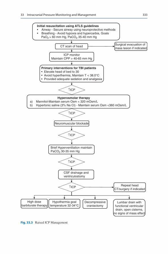
333
Initial resuscitation using ATLS guidelines• Airway - Secure airway using neuroprotective methods• Breathing - Avoid hypoxia and hypercarbia, Goals PaO2 > 60 mm Hg, PaCO2 35-40 mm Hg
CT scan of head
ICP monitorMaintain CPP > 40-65 mm Hg
Primary interventions for TBI patients• Elevate head of bed to 30• Avoid hyperthermia, Maintain T < 38.0°C• Provided adequate sedation and analgesia
↑ICP
↑ICP
↑ICP
↑ICP
↑ICP
Hyperosmolar therapya) Mannitol-Maintain serum Osm < 320 mOsm/Lb) Hypertonic saline (3% Na CI) - Maintain serum Osm <360 mOsm/L
Neuromuscular blockade
Brief Hyperventilation maintainPaCO3 30-35 mm Hg
CSF drainage andventriculostomy
Repeat headCT/surgery if indicated
High dosebarbiturate therapy
Hypothermia goaltemperature 32-34°C
Decompressivecraniectomy
Lumbar drain withfunctional ventriculardrain, open cisterns,
no signs of mass effect
Surgical evacuation ofmass lesion if indicated
Fig. 33.3 Raised ICP Management
33 Intracranial Pressure Monitoring and Management

334
Mannitol
• Osmotic diuretics reduce the brain volume by drawing free water out of the tis-sue and into the circulation, where it is excreted by the kidneys, thus dehydrating brain parenchyma.
• The most commonly used agent is 20% solution of mannitol given as a bolus of 1 g/kg.
• Repeated dosing can be given at 0.25–0.5 g/kg as needed, generally every 6–8 h.• Use of any osmotic agent should be carefully evaluated in patients with renal and
cardiac insufficiency.• Useful parameters to monitor in the setting of mannitol therapy include serum
sodium, serum osmolality, and renal function.• Concerned findings associated with the use of mannitol include serum sodium of
more than 150 mEq, serum osmolality of more than 320 mOsm, or rising blood, urea, and creatinine suggestive of evolving acute tubular necrosis (ATN).
• Mannitol can lower systemic BP, necessitating careful use if associated with a fall in CPP.
• It can cause massive diuresis and loss of potassium, magnesium, and phosphorus.
• In patients on mannitol therapy, euvolemia should be maintained by replacing volume loss with normal saline and additive electrolytes.
• Measuring osmolar gap (measured—calculated serum osmolality) may be useful in titrating mannitol therapy and should be kept below 18.
Loop Diuretics
• Furosemide, 0.5–1.0 mg/kg intravenously, may be given with mannitol to poten-tiate its effect. However, this effect can also exacerbate dehydration and hypokalemia.
Hypertonic Saline
• Hypertonic saline in bolus doses may acutely lower ICP.• Advantages of hypertonic saline are its use in hypotensive patients, reduced
potential to cause renal damage, and less hyponatremia.• The volume and tonicity of saline (3–23.4%) used in these reports have varied
widely.• Use of the central line is recommended for 23% saline to prevent venous
thrombosis.• In patients without central venous access, continuous infusion of hypertonic
saline (1.25–3%) may help to keep serum osmolality elevated.• Target the serum sodium level of 150–160 mEq/L.
R. Chawla et al.

335
• Weaning from osmotherapy should be gentle as sharp decrease in serum sodium may cause cerebral edema. Every day, 5–8 mEq decrease in serum sodium is generally recommended.
Glucocorticoids
• In general, glucocorticoids are not considered to be useful in the management of increased ICP due to cerebral infarction or intracranial hemorrhage.
• In contrast, glucocorticoids may have a role in the setting of intracranial hyper-tension caused by brain tumors and CNS infections.
Hyperventilation
• The use of mechanical ventilation to lower PaCO2 to 26–30 mmHg has been shown to rapidly reduce ICP through vasoconstriction and a decrease in the vol-ume of intracranial blood.
• The effect of hyperventilation on ICP is short-lived (1–24 h).• Therapeutic hyperventilation may be considered as an urgent intervention
when elevated ICP complicates cerebral edema, intracranial hemorrhage, and tumor.
• Hyperventilation should not be used on a chronic basis, regardless of the cause of ICH.
• Hyperventilation should be minimized in patients with traumatic brain injury or acute stroke. In these settings, vasoconstriction may cause a critical decrease in local cerebral perfusion and worsen neurological injury, particularly in the first 24–48 h.
• This might be used as a temporizing measure for patients awaiting a definitive therapy like surgical evacuation of a cerebral clot or tumor.
Barbiturates
• The use of barbiturates is predicated on their ability to reduce brain metabolism and cerebral blood flow, thus lowering ICP and exerting a neuroprotective effect. However, the therapeutic value of this remains unclear.
• Pentobarbital is generally used, with a loading dose of 5–20 mg/kg as a bolus, followed by 1–4 mg/kg/h.
• Treatment should be assessed based on ICP, CPP, and the presence of unaccept-able side effects.
• Continuous electroencephalogram (EEG) monitoring is generally recommended with EEG burst suppression as an indication of maximal dosing.
33 Intracranial Pressure Monitoring and Management
336
Glycerol and Urea
• These were used historically to control ICP via osmoregulation.• The use of these agents has decreased because equilibration between brain and
plasma levels occurs more quickly than with mannitol.• Furthermore, glycerol has been shown to have a significant rebound effect and to
be less effective in ICP control.
Therapeutic Hypothermia
• It is not currently recommended as a standard treatment for increased intracra-nial pressure.
Neuromuscular Paralysis
• This should generally be avoided unless the patient has refractory rise of ICP and is being closely monitored.
Removal of CSF
• When hydrocephalus is identified, a ventriculostomy should be inserted.• Rapid aspiration of CSF should be avoided because it may lead to obstruction of
the catheter opening by brain tissue.• In patients with aneurysmal subarachnoid hemorrhage, abrupt lowering of the
pressure differential across the aneurysm dome can precipitate recurrent hemorrhage.
• CSF should be removed at a rate of approximately 1–2 mL/min, for 2–3 min at a time, with intervals of 2–3 min in between. This should be done till a satisfactory ICP has been achieved (ICP < 20 mmHg) or till CSF is no longer easily obtained.
• Slow removal can also be accomplished by passive gravitational drainage through the ventriculostomy, and bag is positioned raised at the desired level of intracranial pressure.
• A lumbar drain is generally not recommended in the setting of high ICP due to the risk of transtentorial herniation.
Decompressive Craniectomy
• Decompressive craniectomy removes the rigid confines of the bony skull, increasing the potential volume of the intracranial contents.
• It has been demonstrated that in patients with elevated ICP, craniectomy alone lowers ICP up to 15%.
R. Chawla et al.

337
• Opening the dura in addition to the bony skull results in an average decrease in ICP of 70%.
• A recent study (DECRA) has shown the worse 6-month qualitative outcome with this procedure in the severe traumatic brain injury patient.
Step 8: Start General Measures of Management
• Fluid management – In general, patients with elevated ICP do not need to be severely fluid
restricted. – Patients should be kept euvolemic and normo- to hyperosmolar. – This can be achieved by avoiding free water and employing only isotonic
fluids (such as 0.9% saline). – Serum osmolality should be kept to more than 280 mOsm/L and often is kept
in the 295–305 mOsm/L range.• Sedation and pain management
– Keeping patients appropriately sedated and pain-free can decrease ICP by reducing metabolic demand, ventilator asynchrony, venous congestion, and the sympathetic responses of hypertension and tachycardia.
– Propofol has been utilized to good effect in this setting, as it is easily titrated and has a short half-life, thus permitting frequent neurological reassessment. Newer agents like dexmedetomidine may also be used for this purpose.
– Fentanyl should be used cautiously as it may raise ICP. – Agitated patients may be sedated with short-acting benzodiazepines. – The use of end-tidal CO2 should be performed frequently in sedated patients
with increased ICP to maintain normocarbia. Trending upward of end-tidal CO2 should warrant urgent attention.
• Blood pressure control – In general, BP should be sufficient to maintain CPP of more than 50 mmHg. – Vasopressors can be used safely without further increasing ICP. This is par-
ticularly relevant in the setting of sedation, when drug-induced hypotension can occur.
– Hypertension should generally only be treated when CPP is more than 120 mmHg and ICP is more than 20 mmHg.
– Labetalol and nicardipine are the ideal choice. – Nitrates should be avoided as they increase CBF.
• Position – Patients with elevated ICP should be positioned to maximize venous outflow
from the head and are traditionally managed with the head elevated above the heart (usually 30°).
– Important maneuvers include reducing excessive flexion or rotation of the neck, avoiding restrictive neck taping, and minimizing stimuli that could induce Valsalva responses, such as endotracheal suctioning.
33 Intracranial Pressure Monitoring and Management

338
• Temperature control – Fever increases brain metabolism and has been demonstrated to increase
brain injury. – Aggressive treatment of fever, including acetaminophen and mechanical
cooling, is recommended in patients with increased ICP.• Anticonvulsant therapy
– Seizures can complicate and contribute to elevated ICP. – Anticonvulsant therapy should be instituted if seizures are suspected. – Prophylactic treatment may be warranted in some cases. – There are no clear guidelines for prophylactic antiepileptic, but examples
include high-risk mass lesions, such as those within supratentorial cortical locations, or lesions adjacent to the cortex, such as subdural hematomas or subarachnoid hemorrhage.
Suggested Reading
Albanese J, Leone M, Alliez JR, et al. Decompressive craniectomy for severe traumatic brain injury: evaluation of the effects at 1 year. Crit Care Med. 2003;33(10):2535–8. In 40 patients with intractable intracranial hypertension and at very high risk of brain death, decompressive craniectomy allowed 25% of patients to attain social rehabilitation at 1 year. In adults with severe diffuse traumatic brain injury and refractory intracranial hypertension, early bifronto-temporoparietal decompressive craniectomy decreased intracranial pressure and the length of stay in the ICU but was associated with more unfavorable outcomes
Andrews PJ, Citerio G. Intracranial pressure. Part one: historical overview and basic concepts. Intensive Care Med. 2004;30(9):1730–3.
Bhatia A, Gupta AK. Neuromonitoring in the intensive care unit. I. Intracranial pressure and cere-bral blood flow monitoring. Intensive Care Med. 2007;33(7):1263–71. Authoritative review articles on management of increased ICP
Citerio G, Andrews PJ. Intracranial pressure. Part two: clinical applications and technology. Intensive Care Med. 2004;30(10):1882–5.
Cooper DJ, Rosenfeld JV, DECRA Trial Investigators, Australian and New Zealand Intensive Care Society Clinical Trials Group. Decompressive craniectomy in diffuse traumatic brain injury. N Engl J Med. 2011;364(16):1493.
Diringer MN, Zazulia AR. Osmotic therapy: fact and fiction. Neurocrit Care. 2004:219–33. This review examines the available data on the use of osmotic agents in patients with head injury and ischemic stroke, summarizes the physiological effects of osmotic agents, and presents the leading hypotheses regarding the mechanism by which they reduce ICP
Latorre JG, Greer DM. Management of acute intracranial hypertension: a review. Neurologist. 2009;15(4):193–207.
Websites
www.avon.nhs.uk/bristolitutrainees. Use of barbiturates in managing increased ICP.www.yorkshire-cancer-net.org.uk. Managing raised ICP in pediatrics.www.ihrfoundation.org. Homepage for intracranial hypertension research foundation.
R. Chawla et al.

339© Springer Nature Singapore Pte Ltd. 2020R. Chawla, S. Todi (eds.), ICU Protocols, https://doi.org/10.1007/978-981-15-0898-1_34
J. Shah (*) · S. K. Iyer Department of Intensive Care and Critical Care Medicine, Bharati Vidyapeeth (DTU) Medical College, Pune, India
34Acute Febrile Encephalopathy
Jignesh Shah and Shiva Kumar Iyer
A high index of suspicion should be maintained for a diagnosis of an infection involving the nervous system in any patient with fever and altered mental state. Time is essence in managing these patients as any delay could lead to irreversible brain damage (Fig. 34.1).
Step 1: Initiate Resuscitation and Assess Neurological Status
• In patients with neurological problems, assessment of airway and need for intu-bation is of paramount importance as these patients are at high risk of aspiration. Glasgow coma scale (GCS) below 8 usually requires airway protection (see Chap. 23, Vol. 2).
• Maintenance of adequate oxygenation and perfusion are equally important for preventing secondary brain injury.
• Checking the blood sugar level, arterial blood gases with electrolytes will help identify additional immediate priorities for resuscitation
A 40-year-old male patient was admitted to hospital with a 2-day history of fever, headache, and increasing confusional state. He was protecting his airway, had a respiratory rate of 22/min with an oxygen saturation of 95% and was febrile without hypotension. On neurological evaluation, he was disoriented to time and place, was opening eyes on verbal command, had an incomprehensible speech, and was moving all limbs spontaneously. He had neck stiffness and fundi were normal.
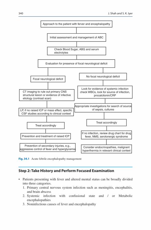
340
Step 2: Take History and Perform Focused Examination
• Patients presenting with fever and altered mental status can be broadly divided into three categories. 1. Primary central nervous system infection such as meningitis, encephalitis,
and brain abscess 2. Systemic infection with confusional state and / or Metabolic
encephalopathies 3. Noninfectious causes of fever and encephalopathy
Approach to the patient with ferver and encephalopathy
Initial assessment and management of ABC
Check Blood Sugar, ABG and serumelectrolytes
Evaluation for presence of focal neurological deficit
Focal neurological deficit
CT imaging to rule out primary CNSstructural lesion or evidence of infectiveetiology (contrast scan)
LP, if no raised ICP or mass effect, specificCSF studies according to clinical context
Treat accordingly
Prevention and treatment of raised ICP
Prevention of secondary injuries, e.g.,aggressive control of fever and hyperglycemia
Consider endocrinopathies, malignanthyperthermia in relevant clinical context
If no infection, review drug chart for drugfever, NMS, serotonergic syndrome
Treat accordingly
Appropriate investigations for search of sourceof sepsis, cultures
Look for evidence of systemic infectioncheck WBCs, look for source of infection,
procalcitonin/CRP
No focal neurological deficit
Fig. 34.1 Acute febrile encephalopathy management
J. Shah and S. K. Iyer

341
• History and physical examination should be carried out systematically to iden-tify the category in which the patients belong. – Travel history (malaria, dengue, typhus, arbovirus infection) – Drug history (steroids, other immunosuppressive) – Trauma (splenectomy) – Symptoms of ENT infection – Recent neurosurgery for suspicion of healthcare associated Meningitis and
ventriculitis. – Medical history of immunosuppressive disease, tuberculosis – History of tuberculosis in close family members
• Neurological examination should be performed. – Look for papilledema, airway reflexes, focal or lateralizing neurological
signs, and neck stiffness. Check for positive Kernig and/or Brudzinski’s sign.• Perform systemic examination to look for a source of sepsis.• Perform general examination for any skin rash, eschar, injection marks (intrave-
nous drug abuse).
Step 3: Initiate Empirical Treatment (Table 34.1)
• In patients with suspected primary neurological infection, time is of essence, so empirical therapy should be started, pending investigation such as CSF/Imaging especially in cases of suspected CNS Infections.
• Maintaining hydration is important as oral intake is often inappropriate. Correct any obvious metabolic causes.
Step 4: Send Basic Investigations
• General investigation workup, such as complete blood count (CBC), blood cul-ture, liver and renal profile, coagulation parameters, electrolyte panel, chest X-ray, echocardiogram (to exclude vegetation), should be performed in all patients.
• Exclude systemic infection. – In endemic areas, look for malaria (peripheral smear and antigen), leptospira
(antibody), enteric fever (blood culture, antibody), dengue (NS1 antigen, anti-body), typhus (antibody), and Japanese B encephalitis (antibody in serum and CSF). These will depend on geographic location of the patient.
Step 5: Send Specific Investigations
• It should be done expeditiously as time is of essence in these conditions.• Empirical treatment should be started pending the investigation to prevent fur-
ther brain damage.
34 Acute Febrile Encephalopathy
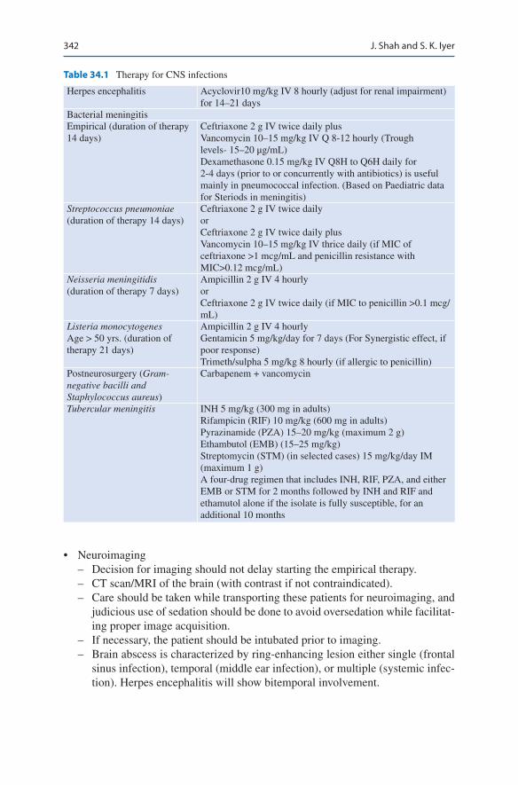
342
• Neuroimaging – Decision for imaging should not delay starting the empirical therapy. – CT scan/MRI of the brain (with contrast if not contraindicated). – Care should be taken while transporting these patients for neuroimaging, and
judicious use of sedation should be done to avoid oversedation while facilitat-ing proper image acquisition.
– If necessary, the patient should be intubated prior to imaging. – Brain abscess is characterized by ring-enhancing lesion either single (frontal
sinus infection), temporal (middle ear infection), or multiple (systemic infec-tion). Herpes encephalitis will show bitemporal involvement.
Table 34.1 Therapy for CNS infections
Herpes encephalitis Acyclovir10 mg/kg IV 8 hourly (adjust for renal impairment) for 14–21 days
Bacterial meningitisEmpirical (duration of therapy 14 days)
Ceftriaxone 2 g IV twice daily plusVancomycin 10–15 mg/kg IV Q 8-12 hourly (Trough levels- 15–20 μg/mL)Dexamethasone 0.15 mg/kg IV Q8H to Q6H daily for 2-4 days (prior to or concurrently with antibiotics) is useful mainly in pneumococcal infection. (Based on Paediatric data for Steriods in meningitis)
Streptococcus pneumoniae (duration of therapy 14 days)
Ceftriaxone 2 g IV twice dailyorCeftriaxone 2 g IV twice daily plusVancomycin 10–15 mg/kg IV thrice daily (if MIC of ceftriaxone >1 mcg/mL and penicillin resistance with MIC>0.12 mcg/mL)
Neisseria meningitidis (duration of therapy 7 days)
Ampicillin 2 g IV 4 hourlyorCeftriaxone 2 g IV twice daily (if MIC to penicillin >0.1 mcg/mL)
Listeria monocytogenesAge > 50 yrs. (duration of therapy 21 days)
Ampicillin 2 g IV 4 hourlyGentamicin 5 mg/kg/day for 7 days (For Synergistic effect, if poor response)Trimeth/sulpha 5 mg/kg 8 hourly (if allergic to penicillin)
Postneurosurgery (Gram- negative bacilli and Staphylococcus aureus)
Carbapenem + vancomycin
Tubercular meningitis INH 5 mg/kg (300 mg in adults)Rifampicin (RIF) 10 mg/kg (600 mg in adults)Pyrazinamide (PZA) 15–20 mg/kg (maximum 2 g)Ethambutol (EMB) (15–25 mg/kg)Streptomycin (STM) (in selected cases) 15 mg/kg/day IM (maximum 1 g)A four-drug regimen that includes INH, RIF, PZA, and either EMB or STM for 2 months followed by INH and RIF and ethamutol alone if the isolate is fully susceptible, for an additional 10 months
J. Shah and S. K. Iyer
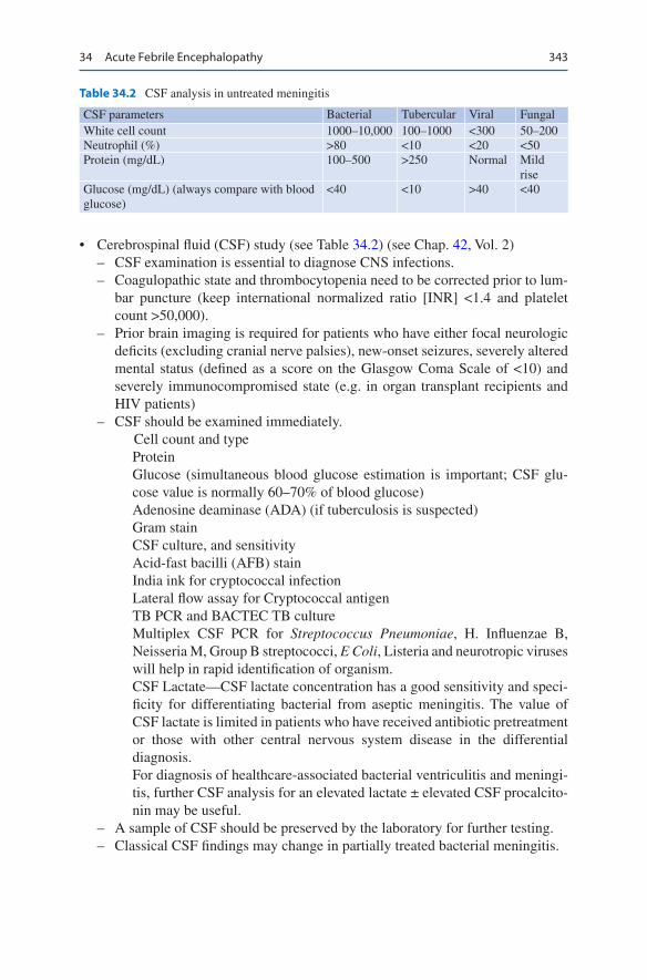
343
• Cerebrospinal fluid (CSF) study (see Table 34.2) (see Chap. 42, Vol. 2) – CSF examination is essential to diagnose CNS infections. – Coagulopathic state and thrombocytopenia need to be corrected prior to lum-
bar puncture (keep international normalized ratio [INR] <1.4 and platelet count >50,000).
– Prior brain imaging is required for patients who have either focal neurologic deficits (excluding cranial nerve palsies), new-onset seizures, severely altered mental status (defined as a score on the Glasgow Coma Scale of <10) and severely immunocompromised state (e.g. in organ transplant recipients and HIV patients)
– CSF should be examined immediately.Cell count and typeProteinGlucose (simultaneous blood glucose estimation is important; CSF glu-cose value is normally 60–70% of blood glucose)Adenosine deaminase (ADA) (if tuberculosis is suspected)Gram stainCSF culture, and sensitivityAcid-fast bacilli (AFB) stainIndia ink for cryptococcal infectionLateral flow assay for Cryptococcal antigenTB PCR and BACTEC TB cultureMultiplex CSF PCR for Streptococcus Pneumoniae, H. Influenzae B, Neisseria M, Group B streptococci, E Coli, Listeria and neurotropic viruses will help in rapid identification of organism.CSF Lactate—CSF lactate concentration has a good sensitivity and speci-ficity for differentiating bacterial from aseptic meningitis. The value of CSF lactate is limited in patients who have received antibiotic pretreatment or those with other central nervous system disease in the differential diagnosis.For diagnosis of healthcare-associated bacterial ventriculitis and meningi-tis, further CSF analysis for an elevated lactate ± elevated CSF procalcito-nin may be useful.
– A sample of CSF should be preserved by the laboratory for further testing. – Classical CSF findings may change in partially treated bacterial meningitis.
Table 34.2 CSF analysis in untreated meningitis
CSF parameters Bacterial Tubercular Viral FungalWhite cell count 1000–10,000 100–1000 <300 50–200Neutrophil (%) >80 <10 <20 <50Protein (mg/dL) 100–500 >250 Normal Mild
riseGlucose (mg/dL) (always compare with blood glucose)
<40 <10 >40 <40
34 Acute Febrile Encephalopathy
344
Step 6: Take Isolation Precaution
• Proper respiratory isolation precaution should be taken in patients with suspected bacterial meningitis .
• The patient should wear mask during transportation.
Step 7: Look for Noninfectious Causes of Fever with Associated Encephalopathy
• Structural brain lesions – Intracerebral hemorrhage, pontine bleeding, and subarachnoid hemorrhage
• Drugs and toxins – Organophosphorus, atropine, tricyclic antidepressants, phenothiazines,
cocaine, and amphetamines• Heat stroke
– Take history of exposure to extreme heat and exertion. Look for evidence of rhabdomyolysis.
• Neuroleptic malignant syndrome – It is caused by central dopamine antagonism. – Clinical features include hyperpyrexia with “lead-pipe” rigidity, extrapyrami-
dal effects, and seizures. – Other effects include rhabdomyolysis, renal failure, hepatic failure, and dis-
seminated intravascular coagulation. Metabolic acidosis and elevated trans-aminases are common.
– Typical agents involved are haloperidol, chlorpromazine, promethazine, and prochlorperazine.
– A few atypical agents such as risperidone, olanzapine, and quetiapine are also responsible.
– Onset of symptoms is usually days to weeks after the inciting agent is started. – Management includes withdrawal of inciting agents, cooling, dantrolene, and
bromocriptine.• Serotonergic syndrome
– It occurs within minutes to hours of initiating an offending agent. – Symptoms are hyperreflexia, myoclonus, and hyperthermia. – Inciting agents are selective serotonin receptor uptake inhibitors (SSRIs), tri-
cyclic antidepressants, and trazodone.• Malignant hyperthermia (MH)
– MH is a specific inherited muscle membrane disorder. – It causes a dangerous hypermetabolic state after anaesthesia with suxametho-
nium and/or volatile halogenated anesthetic agents. – Dantrolene sodium is a specific antidote and induces flaccidity of muscle due
to inhibition of excitation–contraction coupling in skeletal muscle.
J. Shah and S. K. Iyer
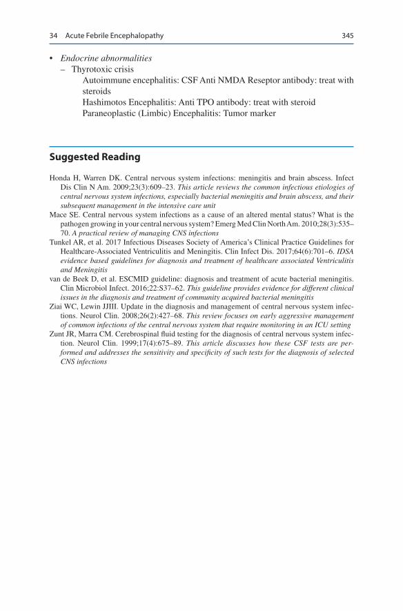
345
• Endocrine abnormalities – Thyrotoxic crisis
Autoimmune encephalitis: CSF Anti NMDA Reseptor antibody: treat with steroidsHashimotos Encephalitis: Anti TPO antibody: treat with steroidParaneoplastic (Limbic) Encephalitis: Tumor marker
Suggested Reading
Honda H, Warren DK. Central nervous system infections: meningitis and brain abscess. Infect Dis Clin N Am. 2009;23(3):609–23. This article reviews the common infectious etiologies of central nervous system infections, especially bacterial meningitis and brain abscess, and their subsequent management in the intensive care unit
Mace SE. Central nervous system infections as a cause of an altered mental status? What is the pathogen growing in your central nervous system? Emerg Med Clin North Am. 2010;28(3):535–70. A practical review of managing CNS infections
Tunkel AR, et al. 2017 Infectious Diseases Society of America’s Clinical Practice Guidelines for Healthcare-Associated Ventriculitis and Meningitis. Clin Infect Dis. 2017;64(6):701–6. IDSA evidence based guidelines for diagnosis and treatment of healthcare associated Ventriculitis and Meningitis
van de Beek D, et al. ESCMID guideline: diagnosis and treatment of acute bacterial meningitis. Clin Microbiol Infect. 2016;22:S37–62. This guideline provides evidence for different clinical issues in the diagnosis and treatment of community acquired bacterial meningitis
Ziai WC, Lewin JJIII. Update in the diagnosis and management of central nervous system infec-tions. Neurol Clin. 2008;26(2):427–68. This review focuses on early aggressive management of common infections of the central nervous system that require monitoring in an ICU setting
Zunt JR, Marra CM. Cerebrospinal fluid testing for the diagnosis of central nervous system infec-tion. Neurol Clin. 1999;17(4):675–89. This article discusses how these CSF tests are per-formed and addresses the sensitivity and specificity of such tests for the diagnosis of selected CNS infections
34 Acute Febrile Encephalopathy

347© Springer Nature Singapore Pte Ltd. 2020R. Chawla, S. Todi (eds.), ICU Protocols, https://doi.org/10.1007/978-981-15-0898-1_35
S. N. Myatra (*) · N. Baliga Department of Anaesthesia, Critical Care and Pain, Tata Memorial Hospital, Homi Bhabha National Institute, Mumbai, India
35Sedation and Analgesia
Sheila Nainan Myatra and Nishanth Baliga
Sedation and analgesia are often given to critically ill patients to facilitate mechani-cal ventilation and other ICU procedures. Pain is a common experience among ICU patients. Due to the presence of an endotracheal tube and sedation, patients receiv-ing mechanical ventilation are often unable to communicate their pain. Unrelieved pain has several adverse effects. It can lead to sympathetic stimulation which causes impaired tissue perfusion, increased myocardial oxygen consumption, hyperstimu-lation of catabolism leading to hyperglycaemia, breakdown of muscle, lipolysis and even impaired wound healing. Long term consequences may be depression, anxiety and chronic pain syndromes. Failure to recognize pain results in patient agitation often leading to the increased use of sedatives. Delirium in common entity in criti-cally ill patients. Inadequately treated pain, excessive sedation, delirium and decreased mobilization are risk factors for acute muscle wasting, weakness, physi-cal and cognitive dysfunction. Hence, both pain and sedation levels and the pres-ence of delirium needs to be assessed regularly and treatments individualized as per the requirement and clinical condition of the patient. The 2018 Pain, Agitation/
A 65 year old male was admitted to the ICU with sudden onset severe abdominal pain. He had history of recurrent burning pain in epigastric region since one week. On examination his pulse was 130/min, BP 110/70 mmHg, RR is 33/min, with a temperature of 101°F and oxygen saturation of 95% on room air. There was tenderness and guarding on abdominal examination. An abdominal CT scan revealed gas under the diaphragm with free fluid in the abdomen. He was planned for an exploratory laparotomy. He was intubated and received mechanical venti-lation . He had an intestinal perforation for which he underwent a resection anas-tomosis. His post operative course was complicated with sepsis and multiorgan failure. He remained on the ventilator for one week.

348
Sedation, Delirium, Immobility (rehabilitation/mobilization) and Sleep (disruption) (PADIS) guidelines provide an evidence based approach to meet these goals.
Step 1: Identify the Need for Sedation and Analgesia
• Follow an “Analgesia first” approach. On presentation the patient was conscious and in severe pain. Assess and treat pain first, since you will not be able to assess his pain intensity if you sedate the patient before giving analgesia. In addition, appropriate analgesia might obviate the need for sedation or decrease the seda-tion requirement. In critically ill adult patients who can self-report their pain, the Numeric Rating Scale (NRS) administered either verbally or visually is valid and recommended for use (Fig. 35.1)
• Following surgery since he remained mechanically ventilated, his pain is now more difficult to assess. Hence, pain scales recommended for sedated and mechanically ventilated patients in ICU should be used.
• In certain groups of patients, sedation is very important to prevent them from self-harm (removing lines/tubes or fall) or to decrease the work of breathing and oxygen demand.
• Remember, not surgical patients alone, but even medical patients need analgesia. Chronic immoblisation also causes pain. A majority of patients find even routine ICU procedures like tracheal suctioning uncomfortable and painful.
• Most mechanically ventilated patients will need analgesia, with or without sedation.• Common indications for analgesia and sedation are listed in Table 35.1
0No
painModerate
pain
PAIN SCORE 0 –10 NUMERICAL RATING
Worstpossible
pain
1 2 3 4 5 6 7 8 9 10
Fig. 35.1 Numerical Rating Scale (NRS) for awake patients. Scores range from 0 (no pain) to 10 (maximum pain)
Table 35.1 Indications of sedation and analgesia
Facilitation of endotracheal intubation and mechanical ventilation (for amnesia, anxiolysis, reduction of agitation, toleration of endotracheal tube (ETT) and to reduce cough, to alleviate dyspnea, to minimize pain, and to prevent self-extubation)While performing invasive proceduresTo facilitate nursing carePostoperative/trauma painTo tolerate ICU environmental influences (stressful environment, monitoring devices, noise, fear of illness, separation from family)During severe hypoxemia to tolerate certain ventilatory strategies (low tidal volume ventilation, high-frequency oscillation (HFO), and prone positioning)To bear underlying or hospital-acquired medical illnesses
S. N. Myatra and N. Baliga
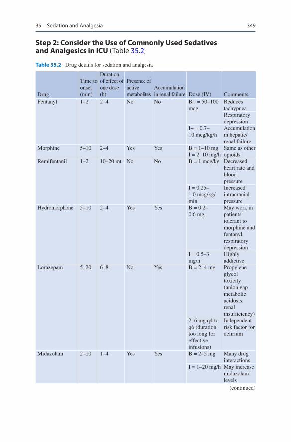
349
Step 2: Consider the Use of Commonly Used Sedatives and Analgesics in ICU (Table 35.2)
Table 35.2 Drug details for sedation and analgesia
Drug
Time to onset (min)
Duration of effect of one dose (h)
Presence of active metabolites
Accumulation in renal failure Dose (IV) Comments
Fentanyl 1–2 2–4 No No B+ = 50–100 mcg
Reduces tachypneaRespiratory depression
I+ = 0.7–10 mcg/kg/h
Accumulation in hepatic/renal failure
Morphine 5–10 2–4 Yes Yes B = 1–10 mg Same as other opioidsI = 2–10 mg/h
Remifentanil 1–2 10–20 mt No No B = 1 mcg/kg Decreased heart rate and blood pressure
I = 0.25–1.0 mcg/kg/min
Increased intracranial pressure
Hydromorphone 5–10 2–4 Yes Yes B = 0.2–0.6 mg
May work in patients tolerant to morphine and fentanyl, respiratory depression
I = 0.5–3 mg/h
Highly addictive
Lorazepam 5–20 6–8 No Yes B = 2–4 mg Propylene glycol toxicity (anion gap metabolic acidosis, renal insufficiency)
2–6 mg q4 to q6 (duration too long for effective infusions)
Independent risk factor for delirium
Midazolam 2–10 1–4 Yes Yes B = 2–5 mg Many drug interactions
I = 1–20 mg/h May increase midazolam levels
(continued)
35 Sedation and Analgesia

350
Drug
Time to onset (min)
Duration of effect of one dose (h)
Presence of active metabolites
Accumulation in renal failure Dose (IV) Comments
Propofol 30–50 s 3–10 (dose dependent)
No Insignificant B = 0.2–2 mg/kg
Hypotension
Increased serum triglyceride
I = 10–150 mcg/kg/min
Propofol infusion syndrome (>5 mg/kg/h for more than 48 h)
Dexmedetomidine 30 4 No No B = 1 μg/kg (should be given over 15–20 min)
Sedative, anxiolysis, with analgesic property (reduces opioid requirement by >50%)
I = 0.2–0.7 μg/kg/h
Notable adverse event is bradycardia
B+ bolus dose, I+ infusion dose
Table 35.2 (continued)
Step 3: Considerations for Choosing an Appropriate Agent
• There is no ideal agent for sedation and analgesia in ICU.• An ideal sedative and analgesic regimen should provide adequate sedation and
pain control, rapid onset of action, and allow rapid recovery after discontinua-tion, minimal adverse effects, minimal systemic accumulation, and minimal delirium without increasing overall health-care costs.
• In different clinical scenarios, some agents have been shown to be better than others. Have a patient-focused approach. Select analgesic and sedative drugs based on the clinical condition and the patients requirement.
• Remember that the degree of sedation/anxiolysis and analgesia required varies between patients and in the same patient over time (physiotherapy, weaning).
• Identify predisposing and precipitating factors—underlying medical conditions such as chronic pain or arthritis, history of alcohol, or substance abuse. Psychiatric illness can influence medication. For example, a patients who appear delirious or severely agitated in the ICU may become overmedicated with sedative drugs, simply because their medications have been omitted.
S. N. Myatra and N. Baliga
351
• In a nonintubated patient agitated due to pain, when opioid and sedatives are used, remember that they can cause respiratory depression. In patients with obstructive sleep apnea, even small doses should be used with caution. In hypo-volemic patients, these agents may cause precipitous fall in blood pressure and should be used with caution.
• A protocol based (analgesia/analgosedation), assessment-driven, stepwise approach for pain and sedation management compared with usual therapy is shown to reduce pain intensity, sedative requirements, duration of mechanical ventilation, and length of ICU stay.
• Analgosedation is either analgesia-first sedation (i.e., usually an opioid is used before a sedative to reach the sedation goal) or analgesia-based sedation (i.e., an opioid is used instead of a sedative to reach the sedatione goal).
• Opioids remain the cornerstone for pain management in most ICU settings.• However, there are concerns of sedation, delirium, respiratory depression, ileus,
and immunosuppression which may lengthen ICU stay and worsen outcomes. Hence a “multi-modal analgesia”approach shuld be used if feasible, especially in the perioperative setting. In addition to opioids, nonopioid analgesic (acetamino-phen, nonsteroidal anti-inflammatory drugs (NSAIDs), ketamine, lidocaine, neu-ropathic agents etc.) may be used with the goal of sparing opioid use and improving analgesic effectiveness and even combined with regional anesthetics and nonpharmacologic interventions known to reduce pain.
• Dexmedetomidine produces sedation, anxiolysis and has analgesic properties and is associated with less delirium in ventilated critically ill patients. It is rec-ommended for delirium in mechanically ventilated adults where agitation pre-cludes weaning/extubation.
• Benzodiazepines are prone to cause acute cognitive dysfunction. Lorazepam is an independent risk factor for delirium.
• Propofol or dexmedetomidine are recommended over benzodiazepines for seda-tion in critically ill, mechanically ventilated adults.
• Sedation with dexmedetomidine causes less delirium than with midazolam or propofol.
• Neuropathic pain medication (e.g., gabapentin, carbamazepine, and pregabalin) with opioids are recommended for neuropathic pain management in critically ill adults.
• Opioids should be used at the lowest effective dose for procedural pain manage-ment NSAIDS may also be considered as an alternative to opioids during ICU procedures.
• For postoperative pain/trauma, epidural analgesia, may be a suitable for select patients if the expertise is available. Patient contolled epidural or intravenous analgesia with opioids can be used if these patients are conscious and coopera-tive. Regional nerve blocks may also be considered in select patients if suitable, as a single dose or continuous infusion of local anaesthestic agents, through spe-cial catheters, provided the expertise is available.
• Massage, music therapy and relaxation therapy have may be used as adjuncts and been shown to be beneficial to reduce pain.
• Either continuous or intermittent boluses of sedative/analgesics may be used, depending upon the strength and training level of the ICU nursing staff and the sedation protocol followed in the ICU.
35 Sedation and Analgesia

352
Step 4: Routine Assessment of Pain and Sedation
• Management of pain and sedation in adult ICU patients should be guided by routine assessment for pain and sedation.
• Critically ill patients may not be able to verbally communicate their pain, as they may be intubated receiving mechanical ventilation, have altered level of con-sciousness or on high doses of sedatives or neuromuscular blocking agents. Therefore, traditional pain assessment tools like visual analog scale, numeric rating scale and verbal rating score, which are dependent on the patients ability to communicate may not be useful.
• Use of vital signs like heart rate, blood pressure, respiratory rate as individual measurement of assessment of pain is discouraged and should only be used as an indicator for further assessment of pain using validated assessment tools, as these clinical parameters in critically ill patients are not specific to pain.
• Use of sedation and pain scales are recommended to titrate the appropriate level of analgesia and sedation.
• The critical-care pain observation tool (CPOT) and behaviour pain scale (BPS) have been validated and recommended for pain assessment in sedated patients or those with altered sensorium in ICU (Tables 35.3 and 35.4).
• The Richmond agitation-sedation scale (RASS) and the Riker sedation-agitation scale (SAS) are validated and recommended scales for assessment of sedation levels in ICU. (Tables 35.5 and 35.6).
• Lighter levels of sedation, rather than deep sedation, are recommended to be targeted in critically ill, mechanically ventilated adult patients using these assess-ment scales.
Table 35.3 The critical care pain observation tool (CPOT)
1 Facial expression Relaxed, neutral 0Tense 1Grimacing 2
2 Body movements Absence of movement or normal position 0Protection 1Restlessness 2
3 Muscle tension Relaxed 0Tense, rigid 1Very tense or rigid 2
4 Compliance with ventilator(intubated patients)orVocalization (extubated patients)
Tolerating the ventilator or movement 0Coughing but tolerating 1Fighting the ventilator 2Talking in normal tone or no sound 0Sighing or moaning 1Crying out, sobbing 2
Score range is from 0 to 8 and target is 0 to 1
S. N. Myatra and N. Baliga
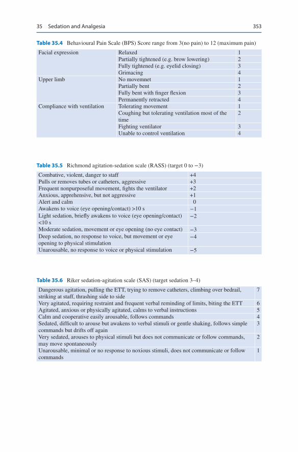
353
Table 35.4 Behavioural Pain Scale (BPS) Score range from 3(no pain) to 12 (maximum pain)
Facial expression Relaxed 1Partially tightened (e.g. brow lowering) 2Fully tightened (e.g. eyelid closing) 3Grimacing 4
Upper limb No movemnet 1Partially bent 2Fully bent with finger flexion 3Permanently retracted 4
Compliance with ventilation Tolerating movement 1Coughing but tolerating ventilation most of the time
2
Fighting ventilator 3Unable to control ventilation 4
Table 35.5 Richmond agitation-sedation scale (RASS) (target 0 to −3)
Combative, violent, danger to staff +4Pulls or removes tubes or catheters, aggressive +3Frequent nonpurposeful movement, fights the ventilator +2Anxious, apprehensive, but not aggressive +1Alert and calm 0Awakens to voice (eye opening/contact) >10 s −1Light sedation, briefly awakens to voice (eye opening/contact) <10 s
−2
Moderate sedation, movement or eye opening (no eye contact) −3Deep sedation, no response to voice, but movement or eye opening to physical stimulation
−4
Unarousable, no response to voice or physical stimulation −5
Table 35.6 Riker sedation-agitation scale (SAS) (target sedation 3–4)
Dangerous agitation, pulling the ETT, trying to remove catheters, climbing over bedrail, striking at staff, thrashing side to side
7
Very agitated, requiring restraint and frequent verbal reminding of limits, biting the ETT 6Agitated, anxious or physically agitated, calms to verbal instructions 5Calm and cooperative easily arousable, follows commands 4Sedated, difficult to arouse but awakens to verbal stimuli or gentle shaking, follows simple commands but drifts off again
3
Very sedated, arouses to physical stimuli but does not communicate or follow commands, may move spontaneously
2
Unarousable, minimal or no response to noxious stimuli, does not communicate or follow commands
1
35 Sedation and Analgesia
354
Step 5: Titrate Sedation and Analgesia
• The titration of the sedative/analgesic dose to a defined endpoint is recommended.
• Regular pain assessment helps to detect and treat pain, especially in sedated patient.• Sedatives should be titrated to achieve lighter levels of sedation unless
contraindicated.
Step 6: Daily Interruption of Sedation
• Critically ill patients often have deranged hepatic and renal function leading to pro-longed metabolism of medications used. Daily sedation interruption (DSI) helps give time for the accumulated sedative/analgesic drugs to metabolize, with resultant patient arousal and facilitation of neurological status assessment in some patients. This strategy when combined with a spontaneous breathing trial (SBT) has shown to decrease the duration of mechanical ventilation, ICU and hospital stay.
• The patient should not be left unattended after the sedation is stopped. This should be done in the morning hours, when there is better staffing, to avoid any tubes or lines from being pulled out by the patient.
• In the critically ill, intubated adults either DSI protocols or nurse targeted seda-tion can be used to achieve and maintain a light level of sedation. If a nurse tar-geted sedation is able to achieve lighter levels of sedation, DSI is not required.
• Early exercise and mobilization (physical and occupational therapy) during peri-ods of daily interruption of sedation have been shown to be safe, well tolerated and results in better functional outcomes at hospital discharge, a shorter duration of delirium, and more ventilator-free days compared with standard care.
Step 7: Delirium—Prevention, Assessment and Treatment
• Delirium is defined as a disturbance of consciousness with inattention accompa-nied by a change in cognition or perceptual disturbance that develops over a short period of time (hours to days) and fluctuates over time.
• Medications like benzodiazepenes, several patient factors for e.g. age, dementia, prior coma, emergency surgery or trauma, and increasing Acute Physiology and Chronic Health Evaluation (APACHE) and American Society of Anesthesiologists (ASA) scores etc. and environmental factors like sleep deprivation, immobility, ventilation etc. are associated with increased risk of delirium in ICU.
• The incidence of delirium is high in ICU (varies from 16% to 89%). Over 60% of ventilated patients develop delirium.
• Delirium has many untoward effects. Delirium is associated with poor outcomes due to increased ICU and hospital stay and cognitive impairment after ICU discharge.
• Delirium has been classified into subtypes. Hyperactive delirium manifestations include agitation, irritability, lack of concentration, and perseveration. However, though easy to detect, this occurs in less than 1% of the patients. Hypoactive
S. N. Myatra and N. Baliga
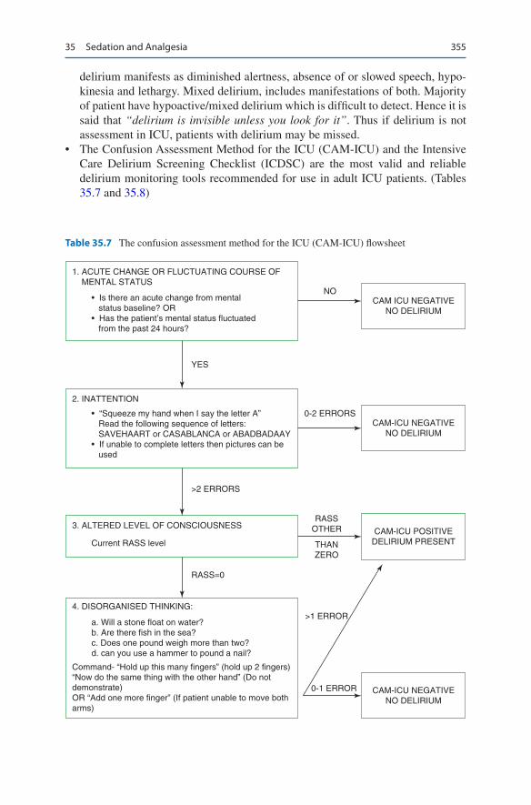
355
delirium manifests as diminished alertness, absence of or slowed speech, hypo-kinesia and lethargy. Mixed delirium, includes manifestations of both. Majority of patient have hypoactive/mixed delirium which is difficult to detect. Hence it is said that “delirium is invisible unless you look for it”. Thus if delirium is not assessment in ICU, patients with delirium may be missed.
• The Confusion Assessment Method for the ICU (CAM-ICU) and the Intensive Care Delirium Screening Checklist (ICDSC) are the most valid and reliable delirium monitoring tools recommended for use in adult ICU patients. (Tables 35.7 and 35.8)
1. ACUTE CHANGE OR FLUCTUATING COURSE OF MENTAL STATUS
2. INATTENTION
4. DISORGANISED THINKING:
3. ALTERED LEVEL OF CONSCIOUSNESS
• Is there an acute change from mental status baseline? OR• Has the patient’s mental status fluctuated from the past 24 hours?
• “Squeeze my hand when I say the letter A” Read the following sequence of letters: SAVEHAART or CASABLANCA or ABADBADAAY• If unable to complete letters then pictures can be used
Current RASS level
CAM-ICU NEGATIVENO DELIRIUM
CAM-ICU POSITIVEDELIRIUM PRESENT
CAM-ICU NEGATIVENO DELIRIUM
CAM ICU NEGATIVENO DELIRIUM
NO
YES
0-2 ERRORS
>2 ERRORS
RASS=0
RASSOTHER
THANZERO
>1 ERROR
0-1 ERROR
Command- “Hold up this many fingers” (hold up 2 fingers)“Now do the same thing with the other hand” (Do notdemonstrate)OR “Add one more finger” (If patient unable to move botharms)
a. Will a stone float on water?b. Are there fish in the sea?c. Does one pound weigh more than two?d. can you use a hammer to pound a nail?
Table 35.7 The confusion assessment method for the ICU (CAM-ICU) flowsheet
35 Sedation and Analgesia

356
• Three modifiable risk factors for ICU delirium are sedatives, immobility and sleep disturbances. Use multimodal strategies to prevent delirium in the ICU. Avoiding or limiting the use benzodiazepines, using dexmeditomidine for sedation, repeated reorientation, reducing noise, cognitive stimulation, adequate hydration providing visual and hearing aids and early mobilization can help reduce the incidence of deliriu m in ICU.
• Dexmedetomidine is recommended for delirium in mechanically ventilated adults where agitation precludes weaning/extubation.
• Routinely using of haloperidol, an atypical antipsychotic, or a HMG-CoA reduc-tase inhibitor (i.e., a statin) to treat delirium is not required.
• Physical restraints should only be used only in select patients to prevent removal of tubes/lines and prevent self harm. Whether efforts to reduce physical restraint use will increase patient exposure to potentially harmful antipsychotic medica-tions and sedative remains unclear.
Step 8: Mobilization and Rehabilitation
• Immobilisation results in ICU-acquired muscle weakness (ICUAW) which is seen in 25–50% of critically ill patients and associated with poor long-term sur-vival, physical functioning and quality of life.
• Thus mobilization and rehabilitation should be performed in critically ill patients whenever feasible and may also be beneficial as part of delirium management strategy.
Table 35.8 Intensive care delirium screening checklist (ICDSC)
1 Altered level of consciousness: coma (no response) or stupor (no response to loud voice and pain) • No response: No score
assigned • Response only to intense and repeated stimulation No score
assigned • Response to mild or moderate stimulation 1 point • Normal wakefulness or sleep with easy arousal 0 point • Exaggerated response to normal stimulation 1 point
2 Inattention: difficulty following conversation or instructions, easy distractions 1 point3 Disorientation: any obvious mistake in time, place or person 1 point4 Hallucination, delusion or psychosis 1 point5 Hyperactivity requiring sedation or restraints
orClinically important psychomotor slowing
1 point
6 Inappropriate speech or mood 1 point7 Disturbance in sleep or wake cycle: frequent spontaneous awakening, sleeping
less than 4 h/night1 point
8 Fluctuation in symptoms 1 point
A score of ≥4 is positive for delirium (with scores of 1 to 3 termed “subsyndromal delirium”)
S. N. Myatra and N. Baliga
357
• Rehabilitation comprises interventions to optimize functioning and reduce dis-ability in individual with illness.
• Rehabilitation/mobilization should be attempted only when there is reasonable stability in cardiovascular, respiratory, and neurologic status and should be stopped if there is any development of a new cardiovascular, respiratory or neu-rologic instability.
Step 9: Promotion of Sleep
• Patients who report poor quality of sleep or use of a medications for sleep aid at home are more likely to report poor quality of sleep in the ICU.
• Sleep disruption may contribute to ICU delirium, increased duration of mechanical ventilation, neurocognitive dysfunction and deranged immune function. Though there is an association between quality of sleep and delir-ium in critically ill adults, a cause-effect relationship has not yet been established.
• Pain, environmental factors (e.g. noise and light), healthcare procedures, psycho-logic factors, respiratory factors and medications all affect the quality of sleep in the ICU and should be addressed.
• A sleep promoting multicomponent protocol should be used in ICU. This may include the use of noise and light reduction strategies at night (offering earplugs and eyeshades to patients), use of assist-control ventilation at night and pressure support ventilation in the day when feasible, limiting ICU care procedure at night to only the essential ones, Keeping the patient awake in the day and orienting the patient, use of relaxing music etc.
• Sleep-promoting medication like melatonin, dexmedetomidine, or propofol are not recommended to improve sleep in critically ill adults.
Step 10: Follow the ABCDEF Sedation Bundle
A multimodal approach, the awakening and breathing coordination, delirium moni-toring/management, and early exercise/mobility (ABCDE) bundle has been shown to be significantly associated with less delirium.
• Assess, prevent and manage pain.• Both Spontaneous Awakening Trials (SATs) and Spontaneous Breathing Trials
(SBTs).• Attention to the Choice of analgesia and sedation.• Delirium monitoring and management.• Early mobility and exercise.• Family engagement and empowerment.
35 Sedation and Analgesia

358
Step 11: Weaning from Sedation and Analgesia
• Once the patient is ready for a SBT, converting the continuous infusion of the sedatives/analgesics into intermittent boluses may be an effective option for early weaning.
• Even after weaning from the mechanical ventilator, though sedatives may be discontinued, analgesics may still be required (especially in surgical patients).
Step 12: Reversal of Oversedation if Present
• For excessive sedation involving benzodiazepines flumazenil may be used. The dose is Dose—0.2 mg (2 mL) IV over 30 s. Wait 30 s and reassess. You may give additional 0.3 mg over 30 s, if needed, and reassess. Additional doses of 0.5 mg can be administered over 30 s at 1-min intervals as needed. Maximum cumula-tive dose is 3 mg.
• For opioid reversal, naloxone may be used. The dose is 0.1–2 mg IM/IV/SC. Titrate to patients’ response. It may be repeated at the interval of 2–3 min. Maximum dose is 10 mg.
Suggested Reading
Barnes-Daly MA, Phillips G, Ely EW. Improving hospital survival and reducing brain dysfunc-tion at seven California community hospitals:Implementing PAD guidelines via the ABCDEF bundle in 6,064 patients. Crit Care Med. 2017;45:171–8. Higher ABCDEF bundle compliance was independently associated with improved survival and more days free of delirium and coma after adjusting for age, severity of illness, and presence of mechanical ventilation
Devlin JW, Skrobik Y, Gélinas C, Needham DM, AJC S, Pandharipande PP, Watson PL, Weinhouse GL, Nunnally ME, Rochwerg B, Balas MC, van den Boogaard M, Bosma KJ, Brummel NE, Chanques G, Denehy L, Drouot X, Fraser GL, Harris JE, Joffe AM, Kho ME, Kress JP, Lanphere JA, McKinley S, Neufeld KJ, Pisani MA, Payen JF, Pun BT, Puntillo KA, Riker RR, BRH R, Shehabi Y, Szumita PM, Winkelman C, Centofanti JE, Price C, Nikayin S, Misak CJ, Flood PD, Kiedrowski K, Alhazzani W. Clinical practice guidelines for the prevention and management of pain, agitation/sedation, delirium, immobility, and sleep disruption in adult patients in the ICU. Crit Care Med. 2018;46(9):e825–73. Recent guidelines for the Prevention and Management of Pain, Agitation/Sedation, Delirium, Immobility, and Sleep Disruption in Adult Patients =
Morandi A, Brummel NE, Ely EW. Sedation, delirium and mechanical ventilation: the ‘ABCDE’ approach. Curr Opin Crit Care. 2011;17:43–9. Outcomes of critically ill patients can be improved by applying evidence based therapies for the 'liberation' from mechanical ventilation such as the'ABCDE' bundle to improve the management such patients
Morandi A, Piva S, Ely EW, Myatra SN, Salluh JIF, Amare D, Azoulay E, Bellelli G, Csomos A, Fan E, Fagoni N, Girard TD, Heras La Calle G, Inoue S, Lim CM, Kaps R, Kotfis K, Koh Y, Misango D, Pandharipande PP, Permpikul C, Cheng Tan C, Wang DX, Sharshar T, Shehabi Y,
S. N. Myatra and N. Baliga

359
Skrobik Y, Singh JM, Slooter A, Smith M, Tsuruta R, Latronico N. Worldwide survey of the “assessing pain, both spontaneous awakening and breathing trials, choice of drugs, delirium monitoring/management, early exercise/mobility, and family empowerment” (ABCDEF) bun-dle. Crit Care Med. 2017 Nov;45(11):e1111–22.
Website
www.icudelirium.org. A website of Vanderbilt University for update and practice parameters on ICU delirium.
35 Sedation and Analgesia

361© Springer Nature Singapore Pte Ltd. 2020R. Chawla, S. Todi (eds.), ICU Protocols, https://doi.org/10.1007/978-981-15-0898-1_36
B. K. Abraham (*) · N. Ramakrishnan Department of Critical Care Medicine, Apollo Hospitals, Chennai, India
36Brain Death
Babu K. Abraham and Nagarajan Ramakrishnan
Step 1: Establish the Underlying Cause for the Coma
• Never consider the diagnosis of brain stem death without a potential cause for brain damage.
• The potential cause for brain damage may be obvious, as in the scenario given above.
• If it is not evident, then the diagnosis of brain stem death should be made with great caution.
• Investigations such as brain imaging (CT scan and MRI) and cerebrospinal fluid (CSF) analysis may be useful in evaluation of etiology of coma but does not help with the confirmation of brain stem death.
A search for confounders that could mimic brain stem death should be made systematically, before considering the tests to evaluate brain stem function.
A 25-year-old male motorbike rider was brought to the intensive care unit (ICU) following a head-on collision with a car while traveling on a motorway. On arrival, he was found to have blood oozing out of his external auditory meatus and nostrils. Neurological assessment revealed Glasgow coma scale (GCS) of 2 T, pupils equal, 3 mm bilaterally, and not reacting to light. Family and col-leagues questioned whether he was “brain dead.”
362
Step 2: Provide an Appropriate Duration of Observation Before Assessing Brain Stem Function
• After sustaining an obvious brain injury it is prudent to wait for an appropriate duration time of before brain stem function is assessed. This is to make sure that all reversible causes for depressed cerebral function are corrected. There is no consensus about how long a patient needs to be observed before being certain that the brain functions have ceased irreversibly.
• The suggested time period for which patients should be observed before assess-ing brain stem is in given below in Table 36.1.
Step 3: Look for Confounders that Could Mimic Brain Stem Death Before Proceeding for Brain Death Verification
• Conditions that can mimic brain stem death are: – Complicating medical conditions like severe electrolyte abnormalities, hypo-
glycemia, and acid–base abnormalities. – Severe hypothermia (core temperature of ≤32 °C). – Severe hypotension (systolic blood pressure < 100 mmHg). – Drug intoxication including alcohol, poisoning, or recent use of sedation or
neuromuscular blocking agents.• If any of the confounders are present, they need to be corrected, and in case of
suspected poisoning, an extended observation period is needed before proceed-ing to further test.
Step 4: Identify Brain Death by Complete Neurological Examination
This consists of three essential documentations:
1. Documentation of coma• Absence of motor response to a standardized painful stimulus applied along a
cranial nerve path (pressing on supraorbital groove, temporomandibular joints, or Trapezius squeeze).
Pathology Duration of observationMajor neurosurgeryAnneursymal bleed
More than 4 h
Spontaneous intracerebral bleed More than 6 hTraumatic brain injury More than 6 hHypoxic brain injury from cardiac arrest or drowning More than 24 hAny cause for brain dysfunction with suspicion of drug intoxication More than 50 to 100 h
B. K. Abraham and N. Ramakrishnan

363
• Beware of local spinal reflexes causing spontaneous or stimulus-related motor movements.
2. Documentation of the absence of brainstem reflexes• As brain death occurs, reflexes are lost in a rostral-to-caudal direction with
the reflexes in medulla oblongata being the last to cease.• Absent pupillary reflex—the pupils will be round or oval, with midposition dila-
tation (4–9 mm) and no reaction to bright light. Remember they need not be equal or dilated to check for absence of reflex. This tests cranial nerves II and III.
• Oculocephalic movements (doll’s eye reflex) will be absent. Ensure integrity of the cervical spine. This test should be done by rapid turning of the head horizontally and vertically and looking for eye movements. There should be no movement of the eyes to the opposite side in brainstem dysfunction and it tests cranial nerves III and IV. This test may be difficult to interpret at times.
• Oculovestibular reflex—cold calorie test. This test is performed by irrigating the tympanum with 50 mL ice-cold water with the head tilted to 30°. Presence of clotted blood or cerumen in the external auditory canal can diminish this response even in the absence of brain death. Absence of provoked tonic eye movement toward the side of cold stimulus and absence of horizontal nystagmus toward contralateral side indicates brain stem dysfunction. This tests cranial nerves VIII, III and VI.
• Corneal reflex: This is checked by instilling a few drops of sterile saline over cornea and if absent it indicates brain stem dysfunction. This tests cranial nerve V and VII.
• Cough reflex: This is best tested by passing a suction catheter through the endotracheal tube and when absent it indicates brain stem dysfunction. This tests cranial nerve IX and X.
3. Documentation of apnea (apnea test)• This test is carried out only after documenting the absence of brainstem
reflexes and is performed to stimulate the respiratory center, which is in the medulla oblongata.
• The patient is first preoxygenated with 100% oxygen for 15 min, and an arte-rial blood gas analysis (ABG) is obtained.
• The patient is then disconnected from mechanical ventilation and oxygen-ation is continued through a catheter placed in the trachea with oxygen flow-ing at 6–10 L/min. Alternatives include using a T-piece system with oxygen flow at 12 L/min or using continuous positive airway pressure (CPAP) at 10 cm H2O with FiO2 titrated to keep oxygen saturation above 95%.
• PaCO2 is allowed to climb (usually at a rate of 3 mmHg/min). The threshold for maximal stimulation of the respiratory center is thought to be PaCO2 of 60 mmHg or a PaCO2 of 20 mmHg above the normal baseline value.
• ABG is repeated within about 8–10 min of disconnection from the mechani-cal ventilation, and the increase in the PaCO2 is documented.
• Visual observation for about 8–10 min is the standard method for detecting respiratory movement. During this period of observation, if the subject does not have spontaneous respiration and arterial PCO2 is 60 mmHg (or 20 mmHg
36 Brain Death
364
increase over the baseline arterial PCO2), the apnea test result is positive (i.e., supports the clinical diagnosis of brain stem death).
• This test should not be performed or should be terminated if the patient becomes hemodynamically unstable (systolic pressure ≤ 90 mm Hg, arryth-mia) or hypoxemic (SpO2 ≤ 85% for more than 30 s).
• If the test is inconclusive but the patient is hemodynamically stable during the procedure, it may be repeated for a longer period (10–15 min) after the patient is again adequately preoxygenated.
• If the apnea test cannot be performed or had to be terminated due to above mentioned reasons, consider doing ancillary confirmatory tests to confirm brain death. However it should be emphasised that the “Transplantation of Human Organs Act” (THOA), in India does not mandate the performance of ancillary confirmatory tests to diagnose brain stem death.
Step 5: Perform Ancillary Confirmatory Tests
In India, THOA does not mandate the performance of these tests. In certain other countries these tests are required by law to confirm brain death, while in others they are optional in adults but recommended in children younger than 1 year.
• Cerebral angiography (conventional) – Angiography of both the anterior and posterior circulations has to be carried
out. The absence of contrast flow into the intracerebral portions of the carotid and vertebral arteries at the level of their entry into the skull is taken as a sign of brain death.
– External carotid circulation should be patent. – Delayed filling of the superior longitudinal sinus may be seen. – Limitation of the test is the need for transporting the patient.
• Electroencephalography (EEG) – A complete absence of electroencephalographic activity to intense somato-
sensory and audiovisual stimulation is taken as a sign of brain death. – It is important to ensure that EEG recordings are done in a standardized
manner. – Limitations are presence of artifacts leading to an EEG pattern for false-
negative brain death diagnosis.• Transcranial Doppler (TCD) ultrasound
– The TCD abnormalities that indicate brain death are a lack of diastolic or reverberating flow and the documentation of small systolic peaks in early systole.
B. K. Abraham and N. Ramakrishnan
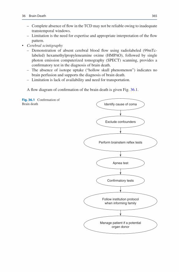
365
– Complete absence of flow in the TCD may not be reliable owing to inadequate transtemporal windows.
– Limitation is the need for expertise and appropriate interpretation of the flow pattern.
• Cerebral scintigraphy – Demonstration of absent cerebral blood flow using radiolabeled (99mTc-
labeled) hexamethylpropyleneamine oxime (HMPAO), followed by single photon emission computerized tomography (SPECT) scanning, provides a confirmatory test in the diagnosis of brain death.
– The absence of isotope uptake (“hollow skull phenomenon”) indicates no brain perfusion and supports the diagnosis of brain death.
– Limitation is lack of availability and need for transportation.
A flow diagram of confirmation of the brain death is given Fig. 36.1.
Apnea test
Exclude confounders
Identify cause of coma
Confirmatory tests
Perform brainstem reflex tests
Follow institution protocolwhen informing family
Manage patient if a potentialorgan donor
Fig. 36.1 Confirmation of Brain death
36 Brain Death

366
Step 6: Certification of Brain Stem Death—Follow the Protocol Set Up by Respective Governments
Institutes should follow the protocol as directed by their respective governments.
• In India “The Transplantation of Human Organs Act, 1994” has clearly laid down the guidelines for declaration of brain stem death and the detailed docu-mentation has to be done in Form 10 (see appendix). This form clearly lays down the method of performing brain stem evaluation.
• Two sets of brain death examinations are mandated, done by two teams of doc-tors and the second is typically done 6 h after the first, to declare brain stem death in adults. In neonates and pediatric patients, this period of observation is usually longer and is left to the discretion of the doctors performing the brain stem evaluation.
Step 7: Inform and Counsel the Family
• As soon as it is suspected that the patient is nearing brain death, the clinician should keep family members informed of his/her clinical status and manage the patient as per protocol (see below) making sure to maintain him/her hemody-namically stable.
• As declaring brain stem death is a sensitive issue, a senior member of the team, ideally the primary consultant, should counsel the next of kin and once the clini-cal criteria for brain stem death have been confirmed declare him/her dead.
• The family then can be approached to request for organ donation as per the regu-lations of the law.
• If the next of kin declines to donate organs, then it is good medical judgment to discontinue the mechanical ventilation.
Steps in Managing a Brain-Dead Patient
Step 1: Monitor and Manage Hemodynamics
• As brain stem injury is associated initially with a massive sympathetic storm fol-lowed by vasoplegia. Invasive monitoring for assessment of volume status and fluid responsiveness is essential.
• An arterial line, a central venous line, and a urinary catheter are minimum requirements.
• Hypovolumeia is very common and must be recognized and treated promptly, with an aim to maintain euvolemia.
• Resuscitation goals are summarized in Tables 36.1 and 36.2.• The initial volume resuscitation should be with crystalloids, preferably normal
saline or ringer lactate. Colloids containing hydroxyethyl starch should be avoided as its use has been associated with increased risk of delayed graft failure after renal transplantation.
B. K. Abraham and N. Ramakrishnan

367
Vasoactive drugs are initiated if shock persists even after adequate fluid resusci-tation. Dopamine and vasopressin are used as the first line drugs. Low dose infusion of Dopamine (4 mcg/kg/min) has been shown to reduce the need for post-transplan-tation dialysis, in renal recipients and vasopressin has been shown to be useful in increasing organ procurement, reducing inotrope use, treatment of diabetes insipi-dus and lowering sodium levels. Adrenaline, Noradrenaline, Phenylephrine, Dobutamine are used in severe shock Hormonal replacement therapy, as mentioned below, is initiated if severe shock persists even after initiating vasoactive drugs.
Step 2: Replace Hormones
• Brain death is associated with a panhypopituitary state that can lead to refractory hypotension.
• Hormone replacement therapy not only helps in correcting hemodynamic insta-bility but also decreases cardiovascular instability, improves organ procurement and graft function.
• The common hormones replaced are:• Thyroid hormones.• Recommendation is to start Levothyroxine (T4) in hemodynamically unstable
patients along with other hormones especially in potential cardiac donors with a left ventricular ejection fraction less than 45%.
• The recommended dosing of T4 is 20 mcg IV bolus followed by an infusion at 10 mcg/h. T4 has been shown to be more beneficial than using T3.
• Triiodothyronine (T3) when used is given initially as 4 mcg IV bolus followed by an infusion at 3 mcg/h.
• Bolus administration of T4 can cause hyperkalemia, and it is advisable to admin-ister 10 units of regular insulin and 50 mL of 50% dextrose prior to bolus admin-istration of T4, unless the serum glucose is greater than 300 mg/dL.
• If intravenous preparation is not available, thyroxine (300–400 mcg) can be replaced enterally.
• Corticosteriods
Table 36.1 Guidelines for resuscitation
Mean arterial pressure ≥ 60 mm HgUrine output ≥1 mL/kg/hLeft ventricular ejection fraction ≥45%Lowest dose of vasopressor
Rule of 100 • Systolic arterial pressure > 100 mmHg, • Urine output >100 mL/h, • PaO2 > 100 mmHg, • Haemoglobin concentration >100 g/L • Blood sugar 100% normal’
Table 36.2 Goals for resuscitation
36 Brain Death

368
– Recommendation is to imitate corticosteroids in patients with hemodynamic instability for presumed hypothalamo—pituitary—adrenal axis failure and to reduce inflammation associated with brain stem death.
– Methylprednisolone has been used mostly and is given in different dose regimens
15 mg/kg IV ODor 100 mg IV ODor 250 mg iv bolus followed by infusion of 100 mg/h
– Observational studies have shown to improve donor hemodynamics, increase organ procurement and improve graft and recipient survival.
– Hydrocortisone has also been tried in lower doses. This too decreases the need for vasopressors but did not shown any difference in organ procurement. The recommended doses are:
50 mg IV bolus followed by 10 mg/h infusionor 300 mg IV OD
– Whichever drug is used care should be taken to closely monitor patients blood sugar and that it is only given after sample for tissue typing has been obtained as they can interfere with HLA expression.
• Insulin• Hyperglycemia is common following brain stem death. It is associated with
decreased donor renal function, pancreatic allograft loss and severe hyperglyce-mia can cause osmotic dieresis leading to fluid and electrolyte imbalance
• The aim should be to maintain the blood glucose level below 140 mg/dL. Insulin is initiated at the rate of 1 unit/h IV infusion, and the infusion rate is adjusted appropriately to achieve this target.
• Frequent blood glucose analysis (hourly) may be required to maintain the target.
• Avoid using dextrose containing solutions as routine infusions• Vasopressin and Desmopressin• Consider vasopressin deficiency when hypotension persists despite adequate
fluid resuscitation and when diabetes insipidus (DI) is suspected.• DI is common in brain stem death and is an early sign of endocrinopathy• It should be suspected when
– urine output is greater than 3 to 4 L/day or 2.5 to 3 mL/kg/h. – urine specific gravity is less than 1.005 and urine osmolality is less than
200 mOsm/kg – serum osmolality is more than 300 mOsm/kg – serum sodium is more than 145 mEq/L – Once vasopressin deficiency is suspected it needs to be replaced.
In hypotensive patients start infusion of vasopressin at 0.01 to 0.04 IU/min. Higher doses should be used with caution.In DI with no hypotension, Desmopressin (DDAVP) can be given at a dose of 1–4 mcg IV followed by 1–2 mcg IV every 6 h. The dose should be titrated to urine output, urine osmolality and serum sodium, aiming for a urine output of 0.5–3 mL/kg/h and serum sodium to 135–145 mEq/L
B. K. Abraham and N. Ramakrishnan
369
Combination therapy with both infusion of vasopressin at 0.5 unit/h and DDAVP can be considered when patients are hypotensive and have severe hypernatremiaCaution—serum sodium values should be checked every 6 h to assist with titration of vasopressin/desmopressin dose. Side effects of vasopressin include hyponatremia, digital vasoconstriction, and thrombosis.Use of vasopressin in hypotensive patients has been associated with increased organ retrieval rates and inability to maintain serum sodium level less than 155 mEq/L has been reported to be associated with worse liver graft survival.
• There is still no clear consensus about when to initiate hormonal replacement therapy. Some prefer to initiate methylprednisolone and insulin components of hormonal replacement therapy soon after the first brain death declaration, while levothyroxine and/or vasopressin are initiated only if the patient becomes hypo-tensive or has diabetes insipidus. Others start all hormones simultaneously as soon as brain death is declared, even if they are hemodynamically stable.
Step 3: Provide Appropriate Respiratory & Ventilatory Support
• In brain stem death neurogenic pulmonary edema and ischemia—reperfusion injury lead to a ARDS like picture. The ensuing hypoxia, if not managed prop-erly can decrease organ procurement and improper ventilatory management can lead to damage to the lungs.
• Ventilatory management recommended is lung protective ventilation strategy consisting of: – Tidal volume of 6–8 mL/Kg of redicted body weight – PEEP of 8–10 cm H2O – Plateau pressure less than 30 cm H2O – Fraction of inspired oxygen (FiO2) as low as possible
• The targets to be maintained are: – pH–7.35–7.45 – PaO2–100 mm Hg – PaCO2–35–40 mm Hg – SpO2–95%
• Other recommendations include: – Using closed circuit tracheal suctioning – Employing alveolar recruitment after each disconnection – Using CPAP while performing apnoea test
• These ventilator management strategies have been associated with an increase in number of transplanted lungs.
36 Brain Death

370
Step 4: Other Issues that Need Consideration
Nutrition• The appropriate feeding strategy is not clear.• The present guidelines recommend to continue feeding as if brain death has not
happened.• Continuing enteric feed has been associated with optimization of allograft
function• Early parenteral feed is not advised due to concerns of increased risk of
infection.
Infection Management• Pre transplant donor infection screening and assessment is essential• Bacteremia and sepsis in the donor need to be treated with appropriate antibiotics
for atleast 48 h prior to organ procurement and is not a contraindication to dona-tion unless there is shock, multiorgan failure or poor response to antibiotic treatment.
• Bacterial meningitis too is not a contraindication for organ procurement once the patient has had adequate and appropriate antibiotic therapy for 24–48 h. This therapy needs to be continued in the recipient for further 5 to 10 days.
• There is no consensus on organ procurement from donors with meningoencepha-litis due to infection with rare pathogens.
• Organs should not be procured from donors who have fever of uncertain etiology with signs of meningitis, encephalitis or from those with global or focal deficits of unknown origin.
• Organs can be procured from donors who have HIV infection and transplanted into a HIV positive recipient.
Temperature Management• Brain stem death is associated with temperature dysregulation leading to hypo-
thermia, which is associated with intravascular coagulation, decreased myocar-dial contractility and arrhythmias.
• Hypothermia on admission, especially in trauma patients is associated with decreased organ procurement.
• The current consensus guidelines recommend maintaining a target body tem-perature of >35 °C.
• The presence of severe cardiovascular instability with high doses of vasopressors may warrant higher temperatures
Coagulation Abnormality• Brain stem death leads to activation of coagulation system and a prothrombotic
state with formation of microthrombi.• Disseminated intravascular coagulation (DIC) is also common, not a contraindi-
cation to transplantation and it does not affect either the early graft function or the long term outcome of the transplant recipients.
B. K. Abraham and N. Ramakrishnan

371
• Current recommendations suggest maintaining an INR < 1.5 and platelets >50,000/ mcL as therapeutic goals prior to surgery
• Imitating thromboprophylaxis in donors with normal coagulation and platelets is reasonable and low molecular weight heparins are the suggested drugs
Suggested Reading
Goila AK, Pawar M. The diagnosis of brain death. Indian J Crit Care Med. 2009;13(1):7–11.Jennifer A, Frontera TK. How I manage the adult potential organ donor: donation after neurologi-
cal death (Part 1). Neurocrit Care. 2010;12:103–10. A practical guide for managing brain dead organ donor
Kotloff RM, et al. Management of the potential organ donor in the ICU: Society of Critical Care Medicine/American College of Chest Physicians/Association of Organ Procurement Organizations Consensus Statement. Crit Care Med. 2015;43(6):1291–325. Guidelines for the critical care management of the potential organ donor
Maciel CB, Greer DM. ICU management of the potential organ donor: state of the art. Curr Neurol Neurosci Rep. 2016;16:86.
Meyfroidt G, Gunst J, Loeches IM, Smith M, Robba C, Taccone FS, Citerio G. Management of the brain-dead donor in the ICU: general and specific therapy to improve transplantable organ quality. Intensive Care Med. 2019;45:343–53.
Wijdicks EFM. The diagnosis of brain death. N Engl J Med. 2001;344:1215–21. A classic article on diagnosing brain death
Wijdicks EF, Varelas PN, Gronseth GS, Greer DM. Evidence-based guideline update: determining brain death in adults. Report of the Quality Standards Subcommittee of the American Academy of Neurology. Neurology. 2010;74:1911–8. An update of the 1995 American Academy of Neurology guideline on brain death
Wood KE, Becker BN, McCartney JG, D’Alessandro AM, Coursin DB. Care of the potential organ donor. N Engl J Med. 2004;351:2730–9. A classic article on management of brain death organ donor
Websites
www.aan.com. American Association of Neurology web sites for professional standards for deter-mining brain death.
www.thamburaj.com/brain_death.htm. Neurosurgeons insight into brain death criteria.www.neurologyindia.com. An archive of leading articles on brain death.https://isccm.org/guidelines.aspx
36 Brain Death

Part IV
Gastrointestinal System
375© Springer Nature Singapore Pte Ltd. 2020R. Chawla, S. Todi (eds.), ICU Protocols, https://doi.org/10.1007/978-981-15-0898-1_37
R. Banerjee (*) · D. N. Reddy Department of Gastroenterology, Asian Institute of Gastroenterology, Hyderabad, India
37Upper Gastrointestinal Bleeding
Rupa Banerjee and Duvvur Nageshwar Reddy
He was pale, sweaty, and restless with a marked tremor. His pulse was 110 beats/min and blood pressure was 90/50 mmHg. He was not jaundiced, but his abdomen was distended with shifting dullness in the flanks. The liver could not be palpated, but the spleen was palpable 5 cm below the costal margin. His hemoglobin was 8.5 g/L, with platelets 45 × 109/L, bilirubin 2.5 mg/L, and creatinine 1.2.
Upper gastrointestinal (UGI) bleeding originates proximal to the ligament of Treitz, from the esophagus, stomach, and duodenum. Acute UGI bleeding is a com-mon medical emergency, which carries a significant mortality risk. Initial triage, assessment, and prompt action can save lives.
Step 1: Initiate Resuscitation and Assessment of Hemodynamic Stability
• The first step in the management of such a patient is initial clinical assessment of the hemodynamic instability and the requirement for immediate resuscitation. Initial resuscitation is done as mentioned in Chap. 23, Vol. 2.
• Patients who present with hemodynamic instability and significant hematemesis will have resting tachycardia (pulse ≥100/min), hypotension (systolic blood pressure < 100 mmHg), or postural changes (increase in the pulse of ≥20 beats/min or a drop in systolic blood pressure of ≥20 mmHg on standing) (Table 37.1).
• In patients with exsanguinating bleeding or the patient who is delirious, airway should be protected by elective intubation. In conscious patients, give oxygen by nasal cannula.
A 46-year-old male patient was brought to hospital having several episodes of vomiting of bright red blood. He had no medical history, but he had a decade-long history of excess alcohol intake, consuming 60–70 units a week.
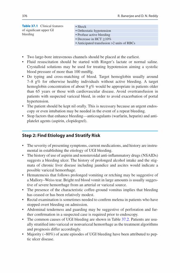
376
• Two large-bore intravenous channels should be placed at the earliest.• Fluid resuscitation should be started with Ringer’s lactate or normal saline.
Crystalloid solutions may be used for treating hypotension aiming a systolic blood pressure of more than 100 mmHg.
• Do typing and cross-matching of blood. Target hemoglobin usually around 7–8 g% for otherwise healthy individuals without active bleeding. A target hemoglobin concentration of about 9 g% would be appropriate in patients older than 65 years or those with cardiovascular disease. Avoid overtransfusion in patients with suspected variceal bleed, in order to avoid exacerbation of portal hypertension.
• The patient should be kept nil orally. This is necessary because an urgent endos-copy or even intubation may be needed in the event of a repeat bleeding.
• Stop factors that enhance bleeding—anticoagulants (warfarin, heparin) and anti-platelet agents (aspirin, clopidogrel).
Step 2: Find Etiology and Stratify Risk
• The severity of presenting symptoms, current medications, and history are instru-mental in establishing the etiology of UGI bleeding.
• The history of use of aspirin and nonsteroidal anti-inflammatory drugs (NSAIDs) suggests a bleeding ulcer. The history of prolonged alcohol intake and the stig-mata of chronic liver disease including jaundice and ascites would indicate a possible variceal hemorrhage.
• Hematemesis that follows prolonged vomiting or retching may be suggestive of a Mallory–Weiss tear. Bright red blood vomit in large amounts is usually sugges-tive of severe hemorrhage from an arterial or variceal source.
• The presence of the characteristic coffee-ground vomitus implies that bleeding has ceased or has been relatively modest.
• Rectal examination is sometimes needed to confirm melena in patients who have stopped overt bleeding on admission.
• Abdominal tenderness and guarding may be suggestive of perforation and fur-ther confirmation in a suspected case is required prior to endoscopy.
• The common causes of UGI bleeding are shown in Table 37.2. Patients are usu-ally stratified into variceal or nonvariceal hemorrhage as the treatment algorithms and prognosis differ accordingly.
• Majority (~80%) of acute episodes of UGI bleeding have been attributed to pep-tic ulcer disease.
Table 37.1 Clinical features of significant upper GI bleeding
• Shock • Orthostatic hypotension • Profuse active bleeding • Decrease in HCT ≥10% • Anticipated transfusion >2 units of RBCs
R. Banerjee and D. N. Reddy

377
• Peptic ulcer bleeding is predominant in the elderly, with 70% of patients older than 60 years.
• Majority of episodes are related to the use of aspirin and nonsteroidal anti- inflammatory drugs.
• The annual incidence of bleeding from peptic ulcer has shown a decline, but the mortality rate remains high at approximately 6–8%.
• Variceal bleeding is related directly to portal hypertension, and cirrhosis is the commonest etiology. The mortality rate for variceal bleeding is high at 30–40%, with a 70% risk of rebleeding in a year.
Risk Stratification
• Patients with hematemesis need to be stratified according to the risk of poor out-come including uncontrolled bleeding, rebleeding, need for intervention, and mortality.
• These factors should be taken into account when determining the need for ICU admission or suitability for discharge.
• The factors associated with a poor outcome are shown in Table 37.3.• Simple and widely validated scoring systems are needed to identify patients at
high risk of rebleeding, death, and active intervention for optimum management. Modified Glasgow–Blatchford bleeding score and the Rockall score are the two common scores used for risk stratification. Modified Glasgow–Blatchford bleed-ing score is a good tool, and it helps to decide the need for endoscopy.
Table 37.2 Common causes of UGI hemorrhage
• Peptic ulcer disease • Gastric erosions and ulcers (drug induced) • Esophageal or gastric varices (variceal bleeding) • Esophageal ulcers/esophagitis • Mallory–Weiss tear • Malignancy • Angiodysplasia and vascular malformations
Table 37.3 Factors associated with a poor outcome in UGI bleeding
1. Age Mortality increases with age among all age groups2. Comorbidity No comorbidity: low mortality; any comorbidity doubles risk3. Liver disease Cirrhosis increases mortality rate; mortality due to variceal bleeding
up to 14%4. Initial shock Increased mortality and need for intervention5. Continued bleeding Postadmission continued bleeding: ↑ intervention, almost 5 times ↑
mortality6. Hematemesis Presence of initial hematemesis doubles mortality7. High urea Poor outcome; increased need for intervention
NSAIDs and anticoagulants do not adversely affect clinical outcome of UGI bleeding
37 Upper Gastrointestinal Bleeding

378
• The Rockall score is the sum of each component, calculated before and after endoscopy (Table 37.4). This predicts rates of rebleeding and mortality and can be used in management algorithms.
• The full Rockall score comprises the initial score as well as additional points for endoscopic diagnosis (0–2 points), and endoscopic stigmata of recent hemor-rhage (0–2 points) giving a maximum score of 11 points.
• Postendoscopy risk scores of more than 2 are associated with a 4% risk of rebleed-ing and 0.1% mortality. Overall, patients with a score of 0, 1, and 2 have a lower risk of hemorrhage, whereas approximately 80% of patients with a postendos-copy score of 8 or more will rebleed. Accordingly, significant health savings could be achieved by early endoscopy and discharge of patients with low scores.
Step 3: Send Investigations
• Hemoglobin levels are required in all patients. It must however be noted that initial levels may be falsely high and underestimate true blood loss due to hemoconcentration.
• Blood should be sent urgently for cross-matching and availability.• Send blood parameters including prothrombin time, partial thromboplastin time,
and platelet count.• Blood urea, creatinine, and liver function tests may assist in diagnosing the cause
and severity of bleeding.• Baseline ECG.
Table 37.4 The Rockall score for prognostication of UGI bleeding
Score0 1 2 3
PreendoscopyAge (years) <60 60–79 >80Shock Blood
pressure (mmHg)
>100 >100 <100
Heart rate (per minute)
<100 >100 >100
Comorbidity None Heart failure, ischemic heart disease, and any major comorbidity
Renal/liver failure, disseminated malignancy
PostendoscopyDiagnoses Mallory–
Weiss/no lesion found
All other malignancy
Gastrointestinal malignancy
Major stigmata of bleeding
None/dark spot only
Bleeding in UGI tract, nonbleeding visible vessels, spurting vessels, adherent clot
R. Banerjee and D. N. Reddy

379
Step 4: Further Treatment
• Any coagulopathy found needs to be corrected by appropriate blood products. If prothrombin time/international normalized ratio is prolonged, give fresh frozen plasma/Prothrombin Complex Concentrate (PCC) or vitamin K injection.
• Reversal of anticoagulants with specific antidotes should be done• Role of platelet transfusion in patients on antiplatelet and NSAID is debatable.• Proton-pump inhibitors: 80 mg of Pantoprazole IV bolus should be administered
followed by intravenous infusion of 8 mg/h or 40 mg 12 hourly.• Randomised studies have failed to show superiority of continuous infusion of
PPI to intermittent boluses.• If there is known or suspected variceal bleeding (or known or suspected chronic
liver disease), empiric treatment with terlipressin 2 mg IV stat followed by 2 mg IV QDS and a dose of broad-spectrum antibiotics should be given.
• Somatostatin intravenous infusion or octreotide subcutaneous may also be tried in place of terlipressin.
• In patients on norepinephrine infusion for hypotension terlipressin or octreotide should be avoided.
• If there is history of active alcohol abuse, thiamine replacement should be started.• Prophylactic antibiotic with 1 g of ceftriaxone prior to endoscopy should be
given.
Step 5: Insert the Nasogastric Tube
• The nasogastric tube insertion is helpful in many ways. The type of aspirate such as fresh blood, bilious, or altered blood helps in determining whether bleeding is ongoing or has stopped.
• Gastric lavage before endoscopy also helps in giving a clear view for endoscopy.
• It may be avoided in uncooperative and delirious patients or in the patient who is retching as it may precipitate further bleeding.
• Nasogastric tube insertion is contraindicated in patients with severe midface trauma or recent nasal surgery, and is relatively contraindicated in patients with known coagulopathy, those with known esophageal varices or after recent vari-ceal banding.
Step 6: Carry Out Endoscopy
• Endoscopy is a mainstay for all cases of UGI bleeding. It enables: – Identification of the source of bleeding – Therapeutic intervention to achieve hemostasis if required
• UGI barium studies are contraindicated as they interfere with subsequent endos-copy or surgery. Also, the yield is very poor.
37 Upper Gastrointestinal Bleeding
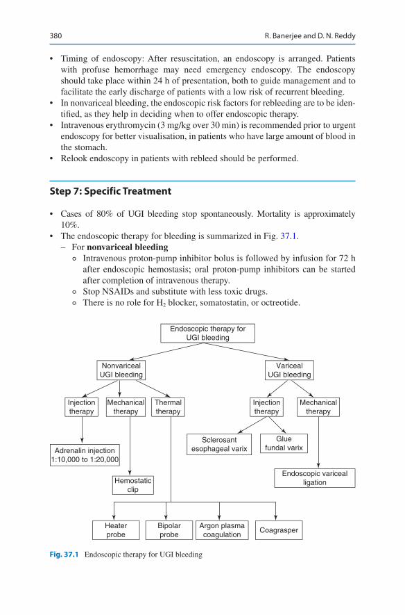
380
• Timing of endoscopy: After resuscitation, an endoscopy is arranged. Patients with profuse hemorrhage may need emergency endoscopy. The endoscopy should take place within 24 h of presentation, both to guide management and to facilitate the early discharge of patients with a low risk of recurrent bleeding.
• In nonvariceal bleeding, the endoscopic risk factors for rebleeding are to be iden-tified, as they help in deciding when to offer endoscopic therapy.
• Intravenous erythromycin (3 mg/kg over 30 min) is recommended prior to urgent endoscopy for better visualisation, in patients who have large amount of blood in the stomach.
• Relook endoscopy in patients with rebleed should be performed.
Step 7: Specific Treatment
• Cases of 80% of UGI bleeding stop spontaneously. Mortality is approximately 10%.
• The endoscopic therapy for bleeding is summarized in Fig. 37.1. – For nonvariceal bleeding
◦ Intravenous proton-pump inhibitor bolus is followed by infusion for 72 h after endoscopic hemostasis; oral proton-pump inhibitors can be started after completion of intravenous therapy.
◦ Stop NSAIDs and substitute with less toxic drugs. ◦ There is no role for H2 blocker, somatostatin, or octreotide.
Endoscopic therapy forUGI bleeding
NonvaricealUGI bleeding
Endoscopic varicealligation
Injectiontherapy
Injectiontherapy
Mechanicaltherapy
Mechanicaltherapy
Adrenalin injection1:10,000 to 1:20,000
Hemostaticclip
Heaterprobe
Bipolarprobe
Argon plasmacoagulation
Coagrasper
Sclerosantesophageal varix
Gluefundal varix
Thermaltherapy
VaricealUGI bleeding
Fig. 37.1 Endoscopic therapy for UGI bleeding
R. Banerjee and D. N. Reddy

381
◦ Patients with active bleeding and visible vessel should receive endoscopic therapy.
◦ Endoscopic hemostasis can be achieved with thermal coagulation, hemo-static clip with or without epinephrine injection. Epinephrine injection alone should be avoided to prevent rebleed.
◦ Endoscopic treatment of adherent clot should be done cautiously.Oral intake of clear liquids can be initiated 6 h after endoscopy in patients with hemodynamic stability.
◦ Helicobacter pylorus testing is required, and appropriate treatment should be started if the result is positive.
◦ Surgical or interventional radiologic consultation should be taken for angi-ography for selected patients with failure of endoscopic hemostasis or massive rebleeding.
– For variceal bleeding ◦ Vasoactive drug treatment should be continued (terlipressin for 48 h,
octreotide, or somatostatin each for 3 days). ◦ Pharmacological therapy should start at the time of presentation in ◦ suspected variceal bleed and should not wait till confirmation of
diagnosis. ◦ Terlipressin may have more sustained effect than octreotide. ◦ Antibiotic therapy should be commenced/continued. ◦ Balloon tamponade should be considered as a temporary salvage treatment
for uncontrolled bleeding. ◦ Esophageal band ligation rather than sclerotherapy is the preferred method
of endoscopic hemostasis. ◦ For patients with bleeding gastric varices cyanoacrylate injection is usu-
ally tried. ◦ In patients with rebleed a second session of endoscopy should be done
prior to TIPS. ◦ Transjugular intrahepatic portosystemic stent shunting is recommended as
the treatment of choice for uncontrolled variceal hemorrhage. ◦ Concurrent management of hepatic encephalopathy and hepato renal syn-
drome may be needed in some patient. ◦ An opinion of a hepatologist must be taken for further management of
chronic liver disease.
Suggested Reading
Blatchford O, Murray WR, Blatchford M. A risk score to predict need for treatment for upper- gastrointestinal hemorrhage. Lancet. 2000;356:1318–21. This score identifies patients who are at low or high risk needing treatment to manage their bleeding. This score should assist the clinical management of patients presenting with upper gastrointestinal hemorrhage, but requires external validation
Cremers I, Ribeiro S. Management of variceal and nonvariceal upper gastrointestinal bleeding in patients with cirrhosis. Ther Adv Gastroenterol. 2014;7(5):206–16. A thorough review on
37 Upper Gastrointestinal Bleeding

382
the standard of care management of variceal and non variceal upper gastrointestinal bleeding in patients with cirrhosis
Gralnek IM, Dumonceau JM, Kuipers EJ, Lanas A, Sanders DS, et al. Diagnosis and manage-ment of nonvariceal upper gastrointestinal hemorrhage: European Society of Gastrointestinal Endoscopy (ESGE) Guideline. Endoscopy. 2015;47(10):a1–46. The official statement of the European Society of Gastrointestinal Endoscopy (ESGE). It addresses the diagnosis and man-agement of nonvariceal upper gastrointestinal hemorrhage
Kumar NL, Cohen AJ, Nayor J, Claggett BL, Saltzman JR. Timing of upper endoscopy influ-ences outcomes in patients with acute nonvariceal upper GI bleeding. Gastrointest Endosc. 2017;85(5):945–52. Deliniates the differences between lower-risk versus high-risk bleeding and the different outcomes with urgent compared with nonurgent endoscopy
Rockall TA, Logan RF, Devlin HB, Northfield TC. Selection of patients for early discharge or outpatient care after acute upper gastrointestinal hemorrhage. Lancet. 1996;347:1138–40. This risk score identifies a large proportion of patients with acute upper gastrointestinal hemor-rhage who are at low risk of further bleeding or death. Early endoscopy and discharge of such patients could allow substantial resource savings
Stanley AJ, Laine L. Management of acute upper gastrointestinal bleeding. BMJ. 2019;364:l536. This review summarizes both medical and endoscopic interventions for patients with variceal and non variceal upper gastrointestinal bleeding
Sung JJ, Chiu PW, Chan FKL, Lau JY, Goh KL, Ho LH, et al. Asia-Pacific working group consen-sus on non-variceal upper gastrointestinal bleeding: an update 2018. Gut. 2018;67(10):1757–68. An up to date guidelines on the pre-endoscopic treatment, endoscopic haemostasis and post-endoscopic management of non-variceal upper gastrointestinal bleeding
Tripathi D, Stanley AJ, Hayes PC, Patch D, Millson C, Clinical Services and Standards Committee of the British Society of Gastroenterology, et al. U.K. guidelines on the management of variceal haemorrhage in cirrhotic patients. Gut. 2015;64(11):1680–704. These guidelines deal specifi-cally with the management of varices in patients with cirrhosis under the following subhead-ings: (1) primary prophylaxis; (2) acute variceal haemorrhage; (3) secondary prophylaxis of variceal haemorrhage; and (4) gastric varices
R. Banerjee and D. N. Reddy
383© Springer Nature Singapore Pte Ltd. 2020R. Chawla, S. Todi (eds.), ICU Protocols, https://doi.org/10.1007/978-981-15-0898-1_38
S. S. Rana (*) · D. K. Bhasin Department of Gastroenterology, Postgraduate Institute of Medical Education & Research, Chandigarh, India
38Lower Gastrointestinal Bleeding
Surinder S. Rana and Deepak Kumar Bhasin
Lower gastrointestinal bleed (LGIB) has traditionally been defined as bleed that occurs distal to the ligament of Treitz. However, with the advent of capsule endos-copy, small bowel bleeds are categorized as middle gastrointestinal tract bleed and LGIB are defined by some authors as blood loss from the colon and/or anorectum. Majority of patients with hematochezia bleed from the large bowel, but in 10–25% of patients, the small bowel is the source of bleeding, and it poses difficult diagnos-tic dilemma.
Also, some patients with massive upper GI bleeding can present with hemato-chezia. Lower GI bleeding represents a diverse range of bleeding sources and sever-ities, ranging from mild hemorrhoidal bleeding to massive blood loss from vascular small bowel tumors. Most patients with LGIB have spontaneous cessation of bleed-ing but patients with diverticular as well as angiodysplasia bleed tend to have recur-rent bleed that can be sometimes massive as well as life threatening. Therefore, risk stratification is of paramount importance but predictive scores for LGIB are not well defined.
A 70-year-old male patient presented to the emergency department with the massive acute painless passage of bright red blood per rectum with postural symptoms. He had no history of fever, weight loss, anorexia, or recent change in bowel habits. There was no significant history of drug ingestion. The patient was a chronic alcoholic and nonsmoker. There was no significant history of any medical or surgical illness.
384
Step 1: Initiate Evaluation and Resuscitation
The evaluation of the hemodynamic status and resuscitation is the most important step in the initial treatment of patients with lower gastrointestinal (GI) bleeding:
• Initiate resuscitation as mentioned in Chap. 23, Vol. 2.• Massive lower GI bleeding is defined as any bleeding requiring more than
3–5 units of blood during 24 h to maintain hemodynamic stability.• Effort should be made to identify patients with hemodynamic compromise.
Postural changes, pallor, dyspnea, tachycardia, and hypotension suggest hemo-dynamic compromise.
• Two large-caliber peripheral venous lines or a central venous line should be placed in patients with hemodynamic compromise.
• Resuscitation should be started with intravenous crystalloid fluid rather than colloid.
• Blood should be transfused if indicated. Restricting hemoglobin to 7 g/dL may be associated with improved mortality in bleeding patients. A threshold of 9 g/dL should be considered in patients with massive bleeding, significant co-morbid illness (especially cardiovascular ischemic diseases) or possible delay in receiv-ing therapeutic interventions.
Step 2: Make a Diagnosis
• An initial history and clinical evaluation should be done to arrive at a list of pos-sible differential diagnosis and plan further investigations.
• The use of anticoagulants or nonsteroidal anti-inflammatory drugs (NSAIDs), the presence of liver disease, and serious comorbid medical conditions like car-diac conditions should be assessed.
• The character and frequency of stool output should be noted, as it allows critical assessment of the severity of bleeding as well as likely source of bleeding.
• Hematochezia must be differentiated from melena, as melena is suggestive of an upper GI bleeding source (although bleeding from the cecum and right-sided colon occasionally presents with melena).
• Patients with brown or infrequent stools are unlikely to have brisk bleeding; those with frequent passage of red or maroon stool, however, may have ongoing bleeding.
• Careful digital rectal examination should be done to exclude anorectal pathology as well as confirm patient’s description of stool color. Hematochezia should also be differentiated from bloody diarrhea, by proper history.
• Bleeding from left side of colon usually appears bright red whereas bleeding from right colon appears dark or maroon colored and rarely as melena.
• Hematochezia associated with hemodynamic compromise could indicate upper GI bleed and therefore gastroduodenoscopy should be performed. A nasogastric aspirate/lavage may be used to rule out upper GI source if suspicion of UGIB is present.
S. S. Rana and D. K. Bhasin

385
• The most common etiologies of lower GI bleeding vary according to the age groups of the patients.
• In young adults and adolescents, the most common causes of bleeding are inflam-matory bowel disease, Meckel’s diverticulum, and polyps.
• In adults younger than 60 years, the most frequent source of lower GI bleeding includes colonic diverticula, inflammatory bowel disease, and neoplasms, whereas angiodysplasia, diverticula, neoplasms, and ischemia are the most com-mon cause of lower GI bleeding in the elderly.
• With history of 2–3 weeks of fever in the patient with lower GIT bleeding, rule out enteric fever in countries with high incidence.
Step 3: Send Investigations
• Initial laboratory studies should include a complete blood count, coagulation profile, blood grouping, renal function tests, and serum electrolytes.
• Coagulopathy (international normalized ratio > 1.5) or thrombocytopenia (<50,000 platelets/μL) should be treated using fresh frozen plasma or platelets, respectively.
• Endoscopic hemostatic therapy may be considered in patients with international normalized ratio of 1.5–2.5 prior to or concomitant with administration of anti- coagulation reversal agents. However, reversal agents should be considered prior to endoscopy if INR > 2.5. Decision to discontinue anticoagulants or use reversal agents should be made after a multidisciplinary approach balancing the risk of ongoing bleeding with the risk of thromboembolic events. Clinical status and underlying indication is used to determine duration and degree of reversal. Warfarin can be reversed within 10 min with prothrombin-complex concentrates. They may be useful in co-morbid conditions where large-volume transfusion is harmful. Fresh frozen plasma reversal of warfarin is slower and less durable than concentrate complex but is cheaper. Vitamin K can be used and it reverses anti- coagulation within 12–24 h.
• Prothrombin concentrates may partially reverse the effect of Direct oral antico-agulant (DOAC) but thromboembolic risk, cost, and lack of evidence limits its use to unstable patients only. Dabigatran can be reversed with idarucizumab. Oral factor Xa inhibitors (rivaroxaban, apixaban, edoxaban, and betrixaban) can-not be dialyzed but can be reversed with Andexanet alfa.
Step 4: Risk Stratification
• Risk stratification is important because diagnostic and therapeutic interventions are time- and resource-consuming and often involve risk and discomfort (i.e., bowel preparation) to the patient.
• In contrast to the upper GI bleeding, predictors of poor outcome in the lower GI bleeding are not well defined.
38 Lower Gastrointestinal Bleeding
386
• Hemodynamic instability at presentation (tachycardia, hypotension, syncope), ongoing bleeding (gross blood on initial digital rectal examination, recurrent hematochezia), comorbid illnesses, age > 60 years, a history of diverticulosis or angiodysplasia, an elevated creatinine, and anemia (initial hematocrit ≤35%) have been shown to be associated with poor outcome. Usually, the risk of adverse outcome increases with the number of risk factors present and therefore accord-ingly the risk stratification should be done.
• About 75% of patients stop bleeding spontaneously without any treatment and are unlikely to benefit from aggressive interventions.
Step 5: Rule Out the Upper GI Tract as the Source of Bleeding
• About 10–15% of patients presenting with severe hematochezia have the bleed-ing source localized in the upper GI tract.
• Patients with hemodynamic compromise and bleeding per rectum should at least have a nasogastric (NG) tube, and if the NG aspirate is bilious, an upper GI source of bleeding is unlikely.
• If the aspirate is nondiagnostic (no blood or bile), or if there is a strong suspicion of an upper bleeding source (i.e., history of previous peptic ulcer disease or fre-quent NSAID use), or an abdominal aortic surgery, then an upper GI endoscopy should be done.
• Nasogastric tube also helps in further preparation of the colon.• High blood urea nitrogen to creatinine ratio has also been shown to be helpful in
predicting an upper GI source of bleeding.
Step 6: Colonoscopy
• Colonoscopy is the preferred next diagnostic step after stabilization in most of the patients with lower GI bleeding as it can provide both a diagnosis and hemo-stasis. The diagnostic yield of colonoscopy is more than radiographic tests like tagged RBC scan or angiography, which requires active bleeding at the time of the radiological examination. Colonoscopy is also better than the flexible sig-moidoscopy, which visualizes only the left side of the colon.
• Advantage of colonoscopy is that diagnosis and therapy can be performed simultaneously.
• The diagnostic yield of urgent colonoscopy in acute lower GI bleeding has been reported to be between 75% and 97%, depending on the definition of the bleed-ing source, patient selection criteria, and timing of colonoscopy.
• Thoroughly clean the colon with bowel preparation in acute lower GI bleeding, as this procedure facilitates endoscopic visualization, improves diagnostic yield, and improves the safety of the procedure by decreasing the risk of perforation. Bowel preparation is not believed to dislodge clots or precipitate bleeding. In
S. S. Rana and D. K. Bhasin
387
patients intolerant to oral intake with low risk of aspiration, a nasogastric tube can be used for colon preparation.
• The cecum should be reached, if at all possible, because a substantial proportion of bleeding sites are located in the right hemicolon. The colonic mucosa should be carefully inspected during both insertion as well as withdrawal of the colono-scope with attempts being made to wash residual stool and blood in order to accurately identify bleeding site
• An attempt should be made to intubate the terminal ileum, especially in nondiag-nostic colonoscopy, as substantial number of causes of lower GI bleeding can be found in the terminal ileum.
• Unlike early endoscopy in upper GI bleeding, colonoscopy should be performed after bleeding has stopped owing to fear of increased complications, need for colon preparation, and lack of proven benefit. However, recent studies have sug-gested that performing colonoscopy shortly after presentation within 24 h is advantageous.
• Early colonoscopy also helps in identifying low-risk patients and thus reduces the need for prolonged hospitalization and costs of care.
• Currently, it is recommended that colonoscopy should be performed within 24 h of presentation in patients with high-risk clinical features and signs or symptoms of ongoing bleeding after a rapid bowel purge that is started immediately after hemo-dynamic resuscitation. However, colonoscopy can be performed next after prepa-ration in patients without high-risk clinical features or serious comorbid disease or those with high-risk clinical features without evidence of ongoing bleeding.
• The following criteria have been suggested for identifying the site of bleeding from colonoscopy: – Active colonic bleeding – Nonbleeding visible vessels – Adherent clot – Fresh blood localized to a colonic segment – Ulceration of diverticulum with fresh blood in adjoining area – Absence of fresh bleeding in the terminal ileum with fresh blood in the colon
Step 7: Achieve Hemostasis
• Endoscopic treatment modalities for lower GI bleeding include injection, contact and noncontact thermal coagulation, and mechanical devices such as metallic clips and band ligation.
• Endoscopic clipping is considered as a safer alternative to thermal contact meth-ods. Hemoclips can be applied directly to the stigmata, visible vessels, or used to oppose the sides of small diverticula or postpolypectomy defects.
• Thermal coagulation in the colon should be performed using moderately low power settings in 1- to 3-s bursts with light to moderate pressure. Thermal coagu-lation should be used carefully in the right colon, in the dome of diverticula, and in the presence of mucosal defects.
38 Lower Gastrointestinal Bleeding

388
• Epinephrine (dilution, 1:10,000 or 1:20,000) can be injected in 1–2-mL aliquots in four quadrants around the lesion in cases of active bleeding. However, it should not be used alone but used in combination with second hemostasis modal-ity including mechanical or contact thermal therapy to achieve definitive hemostasis.
• Argon plasma coagulation (APC) is useful for diffuse lesions such as radiation proctitis and large or multiple angiodysplasia.
• Ligation with bands is used for bleeding hemorrhoids and bleeding rectal vari-ces, and, in certain circumstances for treatment of focal lesions that are less than 2 cm in diameter on nonfibrotic tissue.
• The amount of tissue suctioned into the cap before the application of the rubber band must be carefully monitored to avoid perforation.
• Recently, there has been interest in use of topical haemostatic agents: Ankaferd Blood Stopper and Hemospray.
• Ankaferd Blood Stopper has been successfully used in lower GI bleeding after polypectomy, during bleeding from radiation colitis and malignant lesions.
• Small case series/reports have described successful use of hemospray in lower GI bleed.
• Repeat colonoscopy and endoscopic hemostasis if indicated should be used for recurrent bleeding.
Step 8: Investigate Further if Colonoscopy is Unhelpful or Not Possible
• The following radiological investigations are used in the management of severe lower GI bleeding who cannot be stabilized for colonoscopy or for ongoing bleeding of obscure etiology: – Angiography – Radionuclide scintigraphy – Computed tomography (CT) angiography – Magnetic resonance (MR) angiography
• They are useful in brisk bleeding as there is no need for bowel preparation. However, in contrast to colonoscopy, these investigations require active bleeding at the time of examination for diagnosis and treatment. Barium studies are not required in patients with lower GI bleeding.
• Computed tomography (CT) angiography and MR angiography are evolving as the modality of choice before digital subtraction angiography (DSA) and surgery if upper GI endoscopy and colonoscopy are normal.
Angiography
• Angiography is the only radiographic modality that is both diagnostic and thera-peutic but requires a bleeding rate of at least 0.5–1.0 mL/min to be positive.
S. S. Rana and D. K. Bhasin
389
• Systolic blood pressure of less than 90 mmHg and a requirement of at least 5 units of packed red blood cells within a 24-h period have been shown to predict positive mesenteric angiography.
• This should be reserved for patients who have massive bleeding with hemody-namic compromise that precludes colonoscopy or in patients where colonoscopy is nonconclusive and the patient continues to bleed.
• Vasopressin is the first therapeutic modality employed during angiography, and it controls bleeding in up to 91% of cases, but major complications occur in 10–20% of patients and include arrhythmias, pulmonary edema, hypertension, and ischemia.
• Rebleeding occurs in up to 50% of patients after cessation of the infusion, and, therefore, it is often used to stabilize a patient before surgery rather than as a definitive intervention.
• Early attempts at embolization occasionally cause bowel infarction, but techno-logic advances in coaxial microcatheters and embolic materials have enabled the embolization of specific distal arterial branches with increased success and fewer complications.
Radionuclide Scintigraphy
• Nuclear scintigraphy is a more sensitive method than angiography for detecting GI bleeding as it detects bleeding as low as 0.1 mL/min.
• The major disadvantage of nuclear imaging technique is that it localizes bleeding only to an area of the abdomen and also has high false localization rates because the intraluminal blood is moved away by intestinal motility.
• Currently, it is recommended that scintigraphy should be used as a screening test for patients before the angiography or colonoscopy.
CT Angiography/MR Angiography
• Multidetector row CT (MDCT), where scan time is considerably reduced, has brought CT also into the diagnostic armamentarium for patients with lower GI bleeding
• Reduction of scan time thus enables the accurate acquisition of arterial images, which can show contrast extravasation into any portion of the GI tract.
• Bleeding rates as low as 0.3–0.5 cc/min have been detected using MDCT.• The yield of MDCT is highest among patients with severe ongoing lower GI
bleeding.• The average yield of MDCT for lower GI bleeding is 60%, with yields ranging
from 25% to 95%.• Lack of therapeutic capability is a major limitation of MDCT. However, MDCT
can guide further angioembolization.• Recently, advances in MR have shown good results with MR angiography.
38 Lower Gastrointestinal Bleeding
390
Step 9: Surgery
• Surgery is usually reserved for patients who are having life-threatening bleeding, and other hemostatic techniques have failed to control the bleeding.
• An emergency operation for lower GI hemorrhage is ultimately required in 10–25% of patients.
• The usual indications for an operation are hemodynamic instability, clinical dete-rioration, transfusion requirements of more than 6 units, and persistent or recur-rent hemorrhage.
• In real-life situations, it is usually difficult to make decisions based solely on criteria, and, therefore, surgical consultation should be obtained early in the course of severe bleeding.
• Surgery is also used in patients with recurrent diverticular hemorrhage.• Surgery in lower GI bleeding is associated with high morbidity and mortality,
and localization of the bleeding source before surgery is important for better outcomes. For this, per operative panendoscopy or laparoscopic assisted endos-copy may be done.
Step 10: Prevention of Recurrence of Lower GI Bleed
• Non-aspirin NSAID should be avoided in patients with acute LGIB particularly if secondary to diverticulosis or angiodysplasia.
• Patients with lower GI bleed having high-risk cardiovascular disease aspirin should not be discontinued. However, aspirin for primary prevention of cardio-vascular events should be avoided in patients with LGIB.
• Non-aspirin anti-platelet therapy should be resumed as soon as possible and at least within 7 days of endoscopic therapy in patients who are on dual antiplatelet therapy or mono-therapy with non-aspirin anti-platelet agents (thienopyridine). This decision should be based upon multidisciplinary assessment of cardiovas-cular and GI risk and the adequacy of endoscopic therapy. It is important to remember that dual anti-platelet therapy should not be discontinued in patients with an acute coronary syndrome in past 90 days or coronary stenting in past 30 days because of risk of death and myocardial infarction.
Summary
• With the advent of capsule endoscopy, LGIB is defined as bleeding from the colon and/or anorectum
• Evaluation of hemodynamic status and resuscitation is the most important step in the initial treatment of patients with LGIB
• Hematochezia must be differentiated from melena, as melena is usually sugges-tive of an upper GI bleeding source
S. S. Rana and D. K. Bhasin

391
• Risk stratification is important and hemodynamic instability, ongoing hemato-chezia, and presence of comorbid illness have been associated with poor outcome.
• Colonoscopy is the preferred investigation after stabilization in most of the patients with LGIB
• It should be performed preferably after preparation and whole colon as well as terminal ileum should be evaluated.
• Endoscopic treatment modalities for achieving hemostasis in LGIB include injection, contact and noncontact thermal coagulation, and mechanical devices such as metallic clips and band ligation
• Further investigations like radiological investigations or Nuclear scintigraphy should be done if colonoscopy is unhelpful or not possible
• Surgery is usually reserved for patients who are having life-threatening bleeding, and other hemostatic techniques have failed to control the bleeding
Suggested Reading
Aoki T, Hirata Y, Yamada A, Koike K. Initial management for acute lower gastrointestinal bleed-ing. World J Gastroenterol. 2019;25:69–84. A comprehensive review on management of acute lower GIT bleeding
Gralnek IM, Neeman Z, Strate LL. Acute lower gastrointestinal bleeding. N Engl J Med. 2017;376:1054–63. A comprehensive review on acute lower GIT bleeding
Moss AJ, Tuffaha H, Malik A. Lower GI bleeding: a review of current management, controversies and advances. Int J Color Dis. 2016;31:175–88. A comprehensive review on management of acute lower GIT bleeding
Qayed E, Dagar G, Nanchal RS. Lower gastrointestinal hemorrhage. Crit Care Clin. 2016;32:241–54. A comprehensive review on acute lower GIT bleeding
Song WK, Baron TH. Endoscopic management of acute lower gastrointestinal bleeding. Am J Gastroenterol. 2008;103:1881–7. Although prior descriptions of LGI hemorrhage have encom-passed bleeding sources beyond the ligament of Treitz, small intestinal bleeding represents a distinct entity whose management algorithm has been redefined by the advent of capsule and double-balloon endoscopy
Strate LL, Gralnek IM. ACG clinical guideline: management of patients with acute lower gastroin-testinal bleeding. Am J Gastroenterol. 2016;111:459–74. Guidelines on management of lower GIT bleeding
Whitehurst BD. Lower gastrointestinal bleeding. Surg Clin North Am. 2018;98:1059–72. A com-prehensive review on management of acute lower GIT bleeding
38 Lower Gastrointestinal Bleeding
393© Springer Nature Singapore Pte Ltd. 2020R. Chawla, S. Todi (eds.), ICU Protocols, https://doi.org/10.1007/978-981-15-0898-1_39
M. K. Goenka (*) · S. Afzalpurkar Institute of Gastrosciences, Apollo Gleneagles Hospitals, Kolkata, India
39Acute Diarrhea
Mahesh Kumar Goenka and Shivaraj Afzalpurkar
Acute diarrhea can be defined as the passage of a greater number of stools of decreased form from the normal and lasting <14 days.
Acute severe diarrhea in the ICU may be seen in two situations: a patient with acute severe diarrhea gets admitted to the ICU or a patient admitted to the ICU for any other illness develops a new-onset diarrhea. Diarrhea can occur in almost 50% of ICU population sometimes during their ICU stay and is the most common non hemorrhagic gastrointestinal manifestation in ICU. If not managed properly it can lead to various complications, including fluid and electrolyte loss, non anion gap acidosis, malnutrition, increased risk of pressure sores, immobility, contamination of wounds and catheters leading to nosocomial infections.
Step 1: Initiate Resuscitation
• After taking care of airway and breathing, fluid and electrolyte resuscitation is the mainstay of therapy. It is important to assess the severity of dehydration and treat it.
• Check vital signs such as tachycardia, hypotension, orthostatic hypotension, skin turgor, sunken eyes, sensorium, and dry mucous membranes.
• Abdomen- bowel sounds, distension, and tenderness.
A 40-year-old male patient with no significant previous medical history was admitted to the ICU with 1-day history of passing watery diarrhea, about 20–25 episodes with crampy pain in the abdomen. On examination, he was afebrile with signs of dehydration in the form of dry skin, loss of skin turgor, pulse of 100/min, and BP of 90/60 mmHg. He was drowsy and decreased urinary output. His central venous pressure (CVP) was 3, and arterial blood gas (ABG) analysis revealed metabolic acidosis.

394
• Fluid resuscitation as per clinical judgment and features of hypoperfusion like tachycardia, low urine output, delayed capillary refill.
• Fluids to be used: – Ringer’s lactate – Normal saline with potassium chloride 20 mEq/L
• Additional potassium and magnesium are required as suggested by the biochem-istry results.
• Place Foley’s catheter and measure hourly urinary output.
Step 2: Take Detail History from the Patient/Family
Take a detail history of the following:
• Duration of symptoms, frequency, characteristics of stool, amount, and weight loss
• Medications (see Table 39.1)—very common causes of diarrhea in the ICU• Abdominal symptoms or constitutional symptoms
Table 39.1 Various causes of diarrhea
1. Infectious causes: (a) Bacteria—Vibrio cholerae, Shigella, Salmonella, Campylobacter, E. coli (b) Virus—Rotavirus, Adenovirus, Norovirus (c) Parasites—Giardia, amoeba, Microsporidium, Cryptosporidium, Cyclospora,
Strongyloides 2. Inflammatory causes: Inflammatory bowel disease, Ischemic colitis, Diverticulitis 3. Food poisoning/allergy 4. First presentation of any chronic diarrhea 5. Medications: (a) Acid-reducing agents (e.g., histamine H2-receptor antagonists and proton pump
inhibitors) and antacids (e.g., those that contain magnesium), potassium chloride (b) Antiarrhythmics (e.g., quinidine) (c) Antibiotics (most) (d) Anti-inflammatory agents (e.g., nonsteroidal anti-inflammatory drugs [NSAIDs], gold
salts, and 5-aminosalicylates) (e) Antihypertensive (e.g., β-adrenergic receptor blocking drugs) (f) Antineoplastic agents (many) (g) Antiretroviral agents (h) Colchicine (i) Herbal products, heavy metals (j) Theophylline, digitalis (k) Vitamin and mineral supplements (e.g., vitamin C and magnesium) (l) Recent antibiotic use or hospitalization (m) Prostaglandin (e.g., misoprostol) (n) Hyperosmolar feeds (o) Laxatives, Sorbitol
M. K. Goenka and S. Afzalpurkar
395
• Travel, food habits, and sexual activity• Water source• Sick contacts• Comorbidities (e.g., diabetes and pancreatitis)• Family history or past history of bowel disease (e.g., inflammatory bowel
disease)
Step 3: Identify the Cause of Diarrhea
Severe diarrhea is usually of infective origin. However, other etiologies should be kept in mind (Table 39.1).
Step 4: Send Investigations
Detailed investigations should be done if the patient has any one of the following:
• Profuse diarrhea with dehydration• Grossly bloody stools• Fever of more than 38 °C• Duration of more than 48 h without improvement• Recent antibiotic use• New community outbreak• Associated severe abdominal pain in the patient older than 50 years• Elderly (>70 years)• Immunocompromised patients
Investigations
• Hematology – Hemoglobin and hematocrit, total leukocyte count, and differential count – Biochemistry—ABG, renal and liver functions, electrolytes, and blood
glucose• Stool tests
– Fecal WBCs—suggest mucosal invasion, especially in Shigella, Campylobacter, EHEC (Entero Hemorrhagic Escherichia coli), and EIEC (Entero Invasive Escherichia coli)
– Absent fecal WBCs in viruses, ETEC (Enterotoxic Escherichia coli), amebia-sis, Giardiasis.
– C. difficile toxin A and B /GDH/PCR for C. difficile – Multiplex PCR for Multiple virus/bacterial and toxins – Aerobic culture—for bacteria (Enteropathogenic E. Coli.)
39 Acute Diarrhea

396
– Ova/parasites – The hanging drop for cholera
• Serology—amebic serology• Imaging—plain abdominal radiograph can detect partial obstruction, perfora-
tion, colonic dilatation, contrast-enhanced computed tomography (CECT) of the abdomen is advised in protracted cases
Optional Investigations
• Antigen—Giardia, rotavirus• ELISA and PCR for viruses• Dark field/phase contrast microscopy for Campylobacter• Stool osmolal gap = stool osmolality −2 × (Na+ + K+); gap of more than 40–60
suggests osmotic diarrhea• Endoscopy
Step 5: Specific Pharmacotherapy
• May be given empirically in all severe diarrheas• Usually Metronidazole, Rifaximin, Quinolones, Trimethoprim/sulfamethoxa-
zole, or Doxycycline are used• If Giardiasis or Amebiasis suspected—Metronidazole• If C. difficile suspected—Metronidazole or oral Vancomycin• Antivirals (Acyclovir, Ganciclovir)—herpes simplex virus, cytomegalovirus• Antihelminthics—Strongyloidiasis• Empirical antibiotics are usually recommended especially in:
(a) Elderly (b) Immunocompromised (c) Mechanical heart valves (d) Vascular grafts
Step 6: Symptom-Relief Agents
• If the cause of diarrhea is not known, palliative treatment can be started to decrease fluid loss and the patient’s discomfort.
• Reduced stool frequency and stool weight may even relieve cramps, but close monitoring is required for complications. – Opiate derivatives—loperamide 2–4 mg four times a day – Anticholinergics—diphenoxylate 2.5–5 mg four times a day – Racecadotril (enkephalinase inhibitor), antisecretory activity without affect-
ing intestinal transit—1.5 mg/kg thrice a day
M. K. Goenka and S. Afzalpurkar
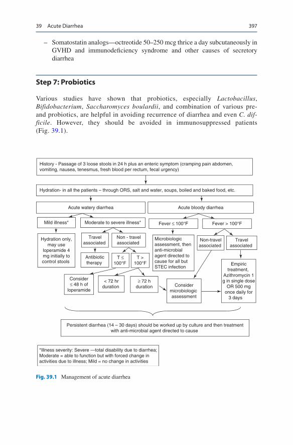
397
– Somatostatin analogs—octreotide 50–250 mcg thrice a day subcutaneously in GVHD and immunodeficiency syndrome and other causes of secretory diarrhea
Step 7: Probiotics
Various studies have shown that probiotics, especially Lactobacillus, Bifidobacterium, Saccharomyces boulardii, and combination of various pre- and probiotics, are helpful in avoiding recurrence of diarrhea and even C. dif-ficile. However, they should be avoided in immunosuppressed patients (Fig. 39.1).
History - Passage of 3 loose stools in 24 h plus an enteric symptom (cramping pain abdomen,vomiting, nausea, tenesmus, fresh blood per rectum, fecal urgency)
Hydration- in all the patients – through ORS, salt and water, soups, boiled and baked food, etc.
Acute watery diarrhea Acute bloody diarrhea
Mild illness* Moderate to severe illness* Fever > 100°FFever ≤ 100°F
Hydration only,may use
loperamide 4mg initially tocontrol stools
Travelassociated
Non - travelassociated
Antibiotictherapy
T ≤100°F
T >100°F
Consider≤ 48 h of
loperamide
< 72 hrduration
≥ 72 hduration
Microbiologicassessment, thenanti-microbialagent directed tocause for all butSTEC infection
Non-travelassociated
Travelassociated
Considermicrobiologic assessment
Empirictreatment,
Azithromycin 1g in single dose
OR 500 mgonce daily for
3 days
Persistent diarrhea (14 – 30 days) should be worked up by culture and then treatmentwith anti-microbial agent directed to cause
*Illness severity: Severe —total disability due to diarrhea;Moderate = able to function but with forced change inactivities due to illness; Mild = no change in activities
Fig. 39.1 Management of acute diarrhea
39 Acute Diarrhea

398
Case Scenario B
A 60-Year-old diabetic man, known diabetic and hypertensive, was admitted to the ICU with Cerebrovascular Accident. After 1 Week of Stay, he had developed watery diarrhea.
Step 1: Know the Causes of Acute-Onset Diarrhea in the ICU• Tube feeding—most likely to occur with calorie-dense formulas infused directly
into the small bowel, a variant of dumping syndrome• Antibiotic-associated diarrhea, other medications as listed before (Table 39.1)• Pseudomembranous enterocolitis—Clostridium difficile• Underlying disease related—diabetic autonomic neuropathy• Laxatives, lactose intolerance• Fecal impaction with overflow diarrhea• Ischemic colitis• Diverticulitis• First-time presentation of a disease such as malignancy, radiation colitis, inflam-
matory bowel disease, and Addison’s disease
Step 2: Management Is Same as Mentioned Above with a Few Changes
• There should be a lower threshold for performing sigmoidoscopy/colonoscopy in these patients.
• Withhold tube feed for few hours. Persistent diarrhea in spite of holding tube feed is suggestive of inflammatory or secretory diarrhea.
• Enteral feeding may be continued in patients with mild diarrhea.• For tube-feed-related diarrhea:
– Slow the rate of infusion. – Dilute the feeds. – Modify the formula to increase fiber. – Give an antidiarrheal agent such as loperamide or diphenoxylate.
• For antibiotic/laxative/other medication-related diarrhea, stop the offender.• For C. difficile, initiate oral vancomycin (125–500 mg QID) or metronidazole
(250–500 mg TDS, QID) for 10–14 days. Probiotics (Lactobacillus GG, S. bou-lardii) and prolonged course of vancomycin are required for relapsers. In most cases, treatment is started empirically as it takes time for the C. difficile toxin in stool report to come in positive. Consider oral rifaximin and fidaxomicin in recurrent C. difficile.
• Assess severity of infection with C. difficile infection. In severe infection start Intravenous metronidazole, and oral vancomycin.
• Do straight X-ray abdomen which may show features of toxic megacolon with dilation of descending colon and lack of haustrations and thumbprinting
• Bedside sigmoidoscopy will show pseudomembranous colitis.• If possible discontinue broad spectrum antibiotic.
M. K. Goenka and S. Afzalpurkar
399
• In patients with recurrent C. difficile try fecal transplant• Take proper isolation precaution and hand washing with soap and water rather
than chlorhexidine after handling patients with C. difficile.
Suggested Reading
Bartlet JG. Clinical practice. Antibiotic-associated diarrhea. N Engl J Med. 2002;346:334–9. This study discusses various issues related to antibiotic-associated diarrhea
Riddle MS, DuPont HL, Connor BA. ACG clinical guideline: diagnosis, treatment, and prevention of acute diarrheal infections in adults. Am J Gastroenterol. 2016;111(5):602. A comprehensive guidelines for the management of diarrhoea
Schiller LR. Diarrhea and malabsorption in the elderly. Gastroenterol Clin N Am. 2009;38:481–502. This article reviews the impact of diarrhea in the elderly, many of whom are less fit physi-ologically to withstand the effect of diarrhea on fluid balance and nutritional balance
Shane AL, Mody RK, Crump JA, Tarr PI, Steiner TS, Kotloff K, Langley JM, Wanke C, Warren CA, Cheng AC, Cantey J. 2017 Infectious Diseases Society of America clinical practice guidelines for the diagnosis and management of infectious diarrhea. Clin Infect Dis. 2017;65(12):e45–80. This guideline updates recommendations regarding epidemiology, diagnosis, treatment, and infection control and environmental management of Clostridium difficile infection
39 Acute Diarrhea
401© Springer Nature Singapore Pte Ltd. 2020R. Chawla, S. Todi (eds.), ICU Protocols, https://doi.org/10.1007/978-981-15-0898-1_40
N. Berry (*) Institute of Digestive and Liver Diseases, BLK Hospital, New Delhi, India
M. T. Noor · R. Kochhar Department of Gastroenterology, Postgraduate Institute of Medical Education and Research, Chandigarh, India
40Acute Abdominal Distension
Neha Berry, Mohd. Talha Noor, and Rakesh Kochhar
Abdominal distension in the ICU patients occurs due to many reasons. Acute colonic pseudo-obstruction is not an uncommon cause of acute abdominal disten-sion in this setting. This is characterized by clinical features of large bowel obstruc-tion but without any mechanical cause. Early recognition and appropriate management are critical in minimizing the morbidity and mortality from complica-tions. However, the diagnosis and treatment of such conditions can be challenging due to nonspecific clinical findings, concurrent complex disease processes, and altered mental status.
Step 1: Initial Resuscitation and Assessment
• After initial resuscitation (Chap. 23, Vol. 2), a detailed history should be obtained, and the patient should be carefully examined.
A 75-year-old male patient presented with respiratory distress after a road traffic accident. On examination, he was dyspneic. Computed tomography (CT) revealed features suggestive of massive haemothorax. He underwent urgent open thoracotomy following which his condition improved. On the third day of hospi-talization, he developed acute onset abdominal distension. The percussion note over the abdomen was tympanic, and bowel sounds were sluggish. Abdominal X-ray revealed dilated bowel loops with multiple air-fluid levels. The serum sodium level was 139 mEq/L, and the serum potassium level was 2.6 mEq/L.
402
• The bowel frequency, stool character, bowel sounds, abdominal distension and abdominal girth, and intra-abdominal pressure should be monitored Crampy abdominal pain and exaggerated bowel sound suggest the presence of mechani-cal obstruction.
• Ileus presents with abdominal distention and abdominal pain that is typically mild and poorly localized. Other features include hypoactive or absent bowel sounds, lack of passage of flatus and stool, intolerance of oral intake, and nausea and emesis. Physical examination reveals a distended, tympanic abdomen; hypo-active bowel sounds; and mild, diffuse abdominal tenderness. The patient may exhibit signs of dehydration, such as tachycardia, orthostatic hypotension, poor skin turgor, and dry mucous membranes.
• In critically ill patients, the possibility of a fecolith causing fecal impaction and obstruction should be kept in mind. Rectal examination should be performed with digital disimpaction if hard fecal matter is present.
• Send for the following investigations immediately: – Erect and supine abdominal X-ray
• Abdominal X-ray shows a cutoff point in mechanical obstruction. Free air under the diaphragm must be looked for perforation. Gas seen till the rectum rules out distal bowel obstruction.
• Serum electrolytes, particularly potassium and magnesium levels.• Thyroid function tests• Stool C. difficile toxin• Serum amylase and lipase• Serum lactate levels
Step 2: Make a Diagnosis
The following conditions commonly present with acute abdominal distension in the ICU:
• Acute colonic pseudo-obstruction (ACPO) or Ogilvie’s syndrome• Mechanical obstruction• Intestinal perforation• Ischemic bowel• Acute pancreatitis• Toxic megacolon
– Inflammatory bowel disease – C. difficile colitis
ACPO can be associated with a number of medical conditions (Table 40.1), which should be looked for and corrected if present.
• Correct important conditions: – Medications such as calcium channel blockers and narcotics can lead to para-
lytic ileus. These medications should be stopped or their dose should be reduced. – The presence of sepsis should be investigated, and samples for culture should
be sent.
N. Berry et al.
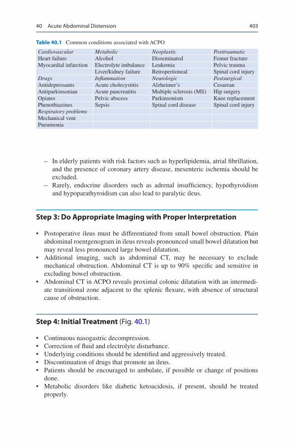
403
– In elderly patients with risk factors such as hyperlipidemia, atrial fibrillation, and the presence of coronary artery disease, mesenteric ischemia should be excluded.
– Rarely, endocrine disorders such as adrenal insufficiency, hypothyroidism and hypoparathyroidism can also lead to paralytic ileus.
Step 3: Do Appropriate Imaging with Proper Interpretation
• Postoperative ileus must be differentiated from small bowel obstruction. Plain abdominal roentgenogram in ileus reveals pronounced small bowel dilatation but may reveal less pronounced large bowel dilatation.
• Additional imaging, such as abdominal CT, may be necessary to exclude mechanical obstruction. Abdominal CT is up to 90% specific and sensitive in excluding bowel obstruction.
• Abdominal CT in ACPO reveals proximal colonic dilatation with an intermedi-ate transitional zone adjacent to the splenic flexure, with absence of structural cause of obstruction.
Step 4: Initial Treatment (Fig. 40.1)
• Continuous nasogastric decompression.• Correction of fluid and electrolyte disturbance.• Underlying conditions should be identified and aggressively treated.• Discontinuation of drugs that promote an ileus.• Patients should be encouraged to ambulate, if possible or change of positions
done.• Metabolic disorders like diabetic ketoacidosis, if present, should be treated
properly.
Table 40.1 Common conditions associated with ACPO
Cardiovascular Metabolic Neoplastic PosttraumaticHeart failure Alcohol Disseminated Femur fractureMyocardial infarction Electrolyte imbalance Leukemia Pelvic trauma
Liver/kidney failure Retroperitoneal Spinal cord injuryDrugs Inflammation Neurologic PostsurgicalAntidepressants Acute cholecystitis Alzheimer’s CesareanAntiparkinsonian Acute pancreatitis Multiple sclerosis (MS) Hip surgeryOpiates Pelvic abscess Parkinsonism Knee replacementPhenothiazines Sepsis Spinal cord disease Spinal cord injuryRespiratory problemsMechanical ventPneumonia
40 Acute Abdominal Distension
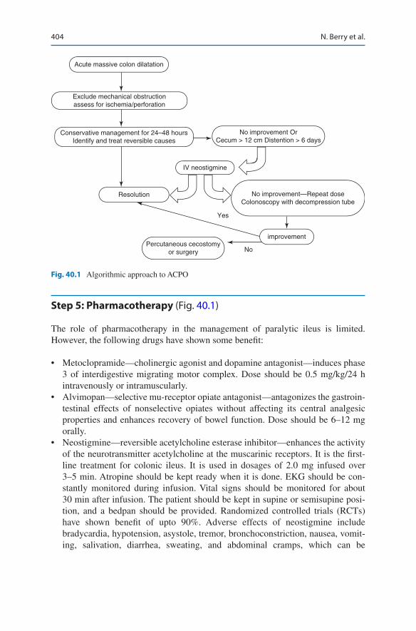
404
Step 5: Pharmacotherapy (Fig. 40.1)
The role of pharmacotherapy in the management of paralytic ileus is limited. However, the following drugs have shown some benefit:
• Metoclopramide—cholinergic agonist and dopamine antagonist—induces phase 3 of interdigestive migrating motor complex. Dose should be 0.5 mg/kg/24 h intravenously or intramuscularly.
• Alvimopan—selective mu-receptor opiate antagonist—antagonizes the gastroin-testinal effects of nonselective opiates without affecting its central analgesic properties and enhances recovery of bowel function. Dose should be 6–12 mg orally.
• Neostigmine—reversible acetylcholine esterase inhibitor—enhances the activity of the neurotransmitter acetylcholine at the muscarinic receptors. It is the first- line treatment for colonic ileus. It is used in dosages of 2.0 mg infused over 3–5 min. Atropine should be kept ready when it is done. EKG should be con-stantly monitored during infusion. Vital signs should be monitored for about 30 min after infusion. The patient should be kept in supine or semisupine posi-tion, and a bedpan should be provided. Randomized controlled trials (RCTs) have shown benefit of upto 90%. Adverse effects of neostigmine include bradycardia, hypotension, asystole, tremor, bronchoconstriction, nausea, vomit-ing, salivation, diarrhea, sweating, and abdominal cramps, which can be
Acute massive colon dilatation
Exclude mechanical obstructionassess for ischemia/perforation
IV neostigmine
Resolution
Percutaneous cecostomyor surgery No
Yes
improvement
Conservative management for 24–48 hoursIdentify and treat reversible causes
No improvement OrCecum > 12 cm Distention > 6 days
No improvement—Repeat doseColonoscopy with decompression tube
Fig. 40.1 Algorithmic approach to ACPO
N. Berry et al.

405
controlled with atropine or glycopyrrolate. Relative contraindications to its use include recent myocardial infarction, acidosis, asthma, bradycardia, peptic ulcer disease, and therapy with beta-blockers.
• Erythromycin- a macrolide antibiotic with prokinetic effects, has been used in ACPO at the dose of 250 mg IV every 8 h for 3 days or orally 250 mg four times daily for 10 days, with inconsistent results.
Step 6: Colonoscopic Decompression
• Colonoscopy is required in some patients to rule out distal obstructive lesions, but its role for colonic decompression is controversial.
• Its use has decreased after neostigmine has been accepted for treatment. Now it is resorted to if neostigmine fails.
• This is done without any preparation, and attempt has to be made to use mini-mum insufflation, preferably using carbon dioxide and suction maximum air from the colon.
• Some centres also use a decompression tube which should be placed in the trans-verse colon and kept inside to constantly decompress.
• This is initially successful in 70–90% of patients, but 10–20% may recur. In such patients, the second decompression can be tried.
• Colonic decompression has a small perforation rate of 3%, and is contraindicated in colonic perforation or peritonitis.
Step 7: Surgery
• Surgery is rarely required in patients with persistent colonic dilatation in spite of colonoscopic decompression and in patients with peritonitis.
• Surgery recommended in such cases is cecostomy or loop colostomy. If there is any nonviable bowel, it is resected. In patients who are unfit for surgery, percutaneous cecostomy just like percutaneous endoscopic gastrostomy is performed.
Suggested Reading
Batke M, Cappell MS. A dynamic ileus and acute colonic pseudo-obstruction. Med Clin North Am. 2008;92:649–70.
Johnson MD, Walsh RM. Current therapies to shorten postoperative ileus. Clev Clin J Med. 2009;76:641–8. To shorten the duration of postoperative ileus, one may need to establish stan-dard plans of care that favor earlier feeding, use of nasogastric tubes only on a selective basis, and prokinetic drugs as needed
40 Acute Abdominal Distension

406
Moss G. The etiology and prevention of feeding intolerance paralytic ileus—revisiting an old concept. Ann Surg Innov Res. 2009;3:3. Limiting inflow to match peristaltic outflow from the feeding site consistently prevented “feeding intolerance.” The patients who received immediate full enteral nutrition had most rapid resolution of postoperative paralytic ileus
Story SK, Chamberlain RS. A comprehensive review of evidence-based strategies to prevent and treat postoperative ileus. Dig Surg. 2009;26:265–75. When evidence-based strategies are used in combination as a part of a fast-track multimodal treatment plan, there is a significant decrease in time to return of normal bowel function and a shortened hospital stay
N. Berry et al.

407© Springer Nature Singapore Pte Ltd. 2020R. Chawla, S. Todi (eds.), ICU Protocols, https://doi.org/10.1007/978-981-15-0898-1_41
R. Chawla (*) · A. C. Jain Department of Respiratory, Critical Care & Sleep Medicine, Indraprastha Apollo Hospitals, New Delhi, India
S. K. Dash Department of Critical Care Medicine, Royal Hobart Hospital, Hobart, TAS, Australia
41Intra-abdominal Hypertension
Rajesh Chawla, Sananta K. Dash, and Aakanksha Chawla Jain
Raised intra-abdominal pressure and abdominal compartment syndrome are com-monly noticed in critically ill patients. In the recent past, their presence in a variety of medical and surgical conditions other than trauma has been emphasized. Early detection, prevention, and treatment reduce the morbidity and mortality in these critically ill patients.
Understand Definitions
• IAP (intra-abdominal pressure): It is the pressure concealed within the abdomi-nal cavity. Normal IAP is approximately 5–7 mmHg in critically ill adults. Physiological changes occur when the IAP raises up to 15 mmHg.
• APP (abdominal perfusion pressure) (MAP-IAP): It is a better reflection of gut perfusion.
A 53-year-old female patient was admitted to the ICU postoperatively after chole-cystectomy for a failed endoscopic retrograde cholangiopancreatography for retrieval of common bile duct stones. She had recent history of gallstone- induced pancreatitis. On the fifth postoperative day, she suddenly became tachypneic and complained of abdominal tightness. She continued to have respiratory distress and later on was intubated in view of severe respiratory distress and hypoxemia. While on the ventilator, her peak airway pressure and the plateau pressure were very high.
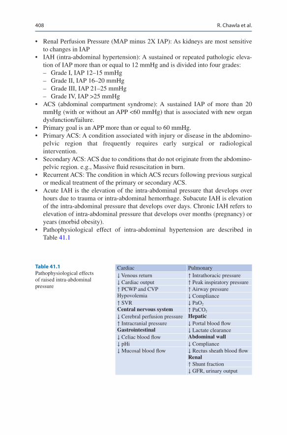
408
• Renal Perfusion Pressure (MAP minus 2X IAP): As kidneys are most sensitive to changes in IAP
• IAH (intra-abdominal hypertension): A sustained or repeated pathologic eleva-tion of IAP more than or equal to 12 mmHg and is divided into four grades: – Grade I, IAP 12–15 mmHg – Grade II, IAP 16–20 mmHg – Grade III, IAP 21–25 mmHg – Grade IV, IAP >25 mmHg
• ACS (abdominal compartment syndrome): A sustained IAP of more than 20 mmHg (with or without an APP <60 mmHg) that is associated with new organ dysfunction/failure.
• Primary goal is an APP more than or equal to 60 mmHg.• Primary ACS: A condition associated with injury or disease in the abdomino-
pelvic region that frequently requires early surgical or radiological intervention.
• Secondary ACS: ACS due to conditions that do not originate from the abdomino-pelvic region. e.g., Massive fluid resuscitation in burn.
• Recurrent ACS: The condition in which ACS recurs following previous surgical or medical treatment of the primary or secondary ACS.
• Acute IAH is the elevation of the intra-abdominal pressure that develops over hours due to trauma or intra-abdominal hemorrhage. Subacute IAH is elevation of the intra-abdominal pressure that develops over days. Chronic IAH refers to elevation of intra-abdominal pressure that develops over months (pregnancy) or years (morbid obesity).
• Pathophysiological effect of intra-abdominal hypertension are described in Table 41.1
Table 41.1 Pathophysiological effects of raised intra- abdominal pressure
Cardiac Pulmonary↓ Venous return ↑ Intrathoracic pressure↓ Cardiac output ↑ Peak inspiratory pressure↑ PCWP and CVP ↑ Airway pressureHypovolemia ↓ Compliance↑ SVR ↓ PaO2
Central nervous system ↑ PaCO2
↓ Cerebral perfusion pressure Hepatic↑ Intracranial pressure ↓ Portal blood flowGastrointestinal ↓ Lactate clearance↓ Celiac blood flow Abdominal wall↓ pHi ↓ Compliance↓ Mucosal blood flow ↓ Rectus sheath blood flow
Renal↑ Shunt fraction↓ GFR, urinary output
R. Chawla et al.

409
Approach to a Patient with IAH
Step 1: Initial Resuscitation
• Initiate resuscitation as mentioned in Chap. 23, Vol. 2.• The elevated IAP has a direct effect on pulmonary and cardiac functions.• Pulmonary compliance suffers with resultant progressive reduction in total lung
capacity, functional residual capacity, and residual volume and is manifested by hypoxia, hypercapnia, and increasing ventilatory pressure.
• Elevated IAP above 20 mmHg consistently correlates with reduction in cardiac output.
• IAP above 20 mmHg produces elevations in measured hemodynamic parameters including central venous pressure and pulmonary artery wedge pressure. Half of IAP should be subtracted from these measures of vascular pressures to arrive at an approximately true pressure.
Step 2: Assess the Possible Risk Factors for IAH/ACS
• IAH or ACS can be the result of abnormality in any of the four constituents that determine the IAP (Fig. 41.1): 1. Abdominal wall compliance. 2. Intraluminal contents. 3. Abdominal contents. 4. Capillary leak/fluid resuscitation.
Step 3: Take Focused Clinical History and Do Physical Examination
The mode of presentation and associated conditions many a times gives a clue to the possible cause of IAH and ACS in a patient. History should be taken on the basis of the background condition. Do detailed general and abdominal examination (Fig. 41.2).
Step 4: Measure IAP
• IAP can be measured either directly (through needle puncture of the abdomen during laparoscopy or peritoneal dialysis treatment) or indirectly (using intrave-sicular pressure or gastric pressure through a balloon catheter, intracolonic, as a surrogate of IAP).
• Measurement of bladder pressure is the standard method to screen for IAH and ACS.
41 Intra-abdominal Hypertension
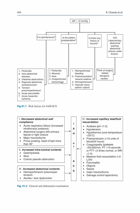
410
IAP > 12 mmHg
It is spontaneous? Is the patientpostoperative?
Is there anyhistory oftrauma?
H/OLaparoscopy,
abdominalpacking,
abdominalclosure under
tension
1. Peritonitis2. Intra-abdominal
abscess 3. Intestinal obstructions4. Ruptured abdominal
aorticaneurysm 5. Tension
pneumoperitoneum 6. Acute pancreatitis7. Acute mesentric
ischemia
1. Peritonitis2. Abscess3. Ileus4. Intraperitoneal
hemorrhage
1. Retroperitonealbleeding
2. Postresuscitation visceral oedema
3. Hemoperitoneum4. Vascular injury,
splenic rupture
Think of surgeryrelated
latrogenic causes
Fig 41.1 Risk factors for IAH/ACS
I. Decreased abdominal wall compliance• Acute respiratory failure (increased
intrathoracic pressure)• Abdominal surgery with primary
fascial or tight closure• Major trauma/burns• Prone positing, head of bed more
than 30°
II . Increased intra-luminal contents• Gastroparesis• Ileus• Colonic pseudo-obstruction
III . Increased abdominal contents• Hemoperitoneum /pneumoper
itoneum • Ascites / liver dysfunction
IV. Increased capillary leak/fluidresuscitation
• Acidosis (pH <7.2)• Hypotension• Hypothermia (core temperature
<33°C)• Polytransfusion (>10 units of
blood/24 hours)• Coagulopathy (platelets
<55,000/mm, PT >15 secondsor PTT >2 times normal, or INR>1.5)
• Massive fluid resuscitation (>5L/24)
• Pancreatitis• Oliguria• Sepsis• major trauma/burns• Damage control laparotomy
I
A
H
or
A
C
S
Fig. 41.2 General and abdominal examination
R. Chawla et al.

411
• The intravesical IAP measurement techniques are based on the same principle; namely, that a fluid column in the bladder catheter and tubing to the collecting bag serve as a pressure-transducing medium.
Method – An assembly is made, as shown in Fig. 41.3. – Clamp the urinary catheter distal to angiocath insertion site. – The tubing (B) is then attached to a pressure transducer (C). – Fill the syringe with saline and infuse upto 25 mL of saline in to the bladder. – Keep the second stop cork (D) opened to air and closed to patient. Calibrate the
transducer to zero at the level of midaxillary line. – IAP is measured 30–60 s after instillation to allow for bladder detrusor muscle
relaxation (for bladder technique), and it is ensured that there are no active abdominal muscle contractions.
– The pressure is measured at end-expiration in the supine position, with the midaxillary line taken as the zero level.
500 mLNS
20 mLSyringe
PressureTransducer
18 GAngiocath
StandardIV Tubing
Urinary Catheter
C
D
B
A
Fig. 41.3 (a) Intra-abdominal pressure monitoring an 18-gauge needle or an angiocath (A) with tubing is inserted into the urinary catheter port, as shown in the figure
41 Intra-abdominal Hypertension
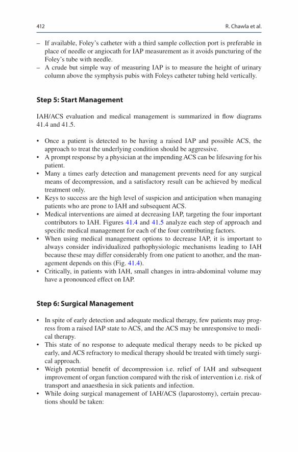
412
– If available, Foley’s catheter with a third sample collection port is preferable in place of needle or angiocath for IAP measurement as it avoids puncturing of the Foley’s tube with needle.
– A crude but simple way of measuring IAP is to measure the height of urinary column above the symphysis pubis with Foleys catheter tubing held vertically.
Step 5: Start Management
IAH/ACS evaluation and medical management is summarized in flow diagrams 41.4 and 41.5.
• Once a patient is detected to be having a raised IAP and possible ACS, the approach to treat the underlying condition should be aggressive.
• A prompt response by a physician at the impending ACS can be lifesaving for his patient.
• Many a times early detection and management prevents need for any surgical means of decompression, and a satisfactory result can be achieved by medical treatment only.
• Keys to success are the high level of suspicion and anticipation when managing patients who are prone to IAH and subsequent ACS.
• Medical interventions are aimed at decreasing IAP, targeting the four important contributors to IAH. Figures 41.4 and 41.5 analyze each step of approach and specific medical management for each of the four contributing factors.
• When using medical management options to decrease IAP, it is important to always consider individualized pathophysiologic mechanisms leading to IAH because these may differ considerably from one patient to another, and the man-agement depends on this (Fig. 41.4).
• Critically, in patients with IAH, small changes in intra-abdominal volume may have a pronounced effect on IAP.
Step 6: Surgical Management
• In spite of early detection and adequate medical therapy, few patients may prog-ress from a raised IAP state to ACS, and the ACS may be unresponsive to medi-cal therapy.
• This state of no response to adequate medical therapy needs to be picked up early, and ACS refractory to medical therapy should be treated with timely surgi-cal approach.
• Weigh potential benefit of decompression i.e. relief of IAH and subsequent improvement of organ function compared with the risk of intervention i.e. risk of transport and anaesthesia in sick patients and infection.
• While doing surgical management of IAH/ACS (laparostomy), certain precau-tions should be taken:
R. Chawla et al.
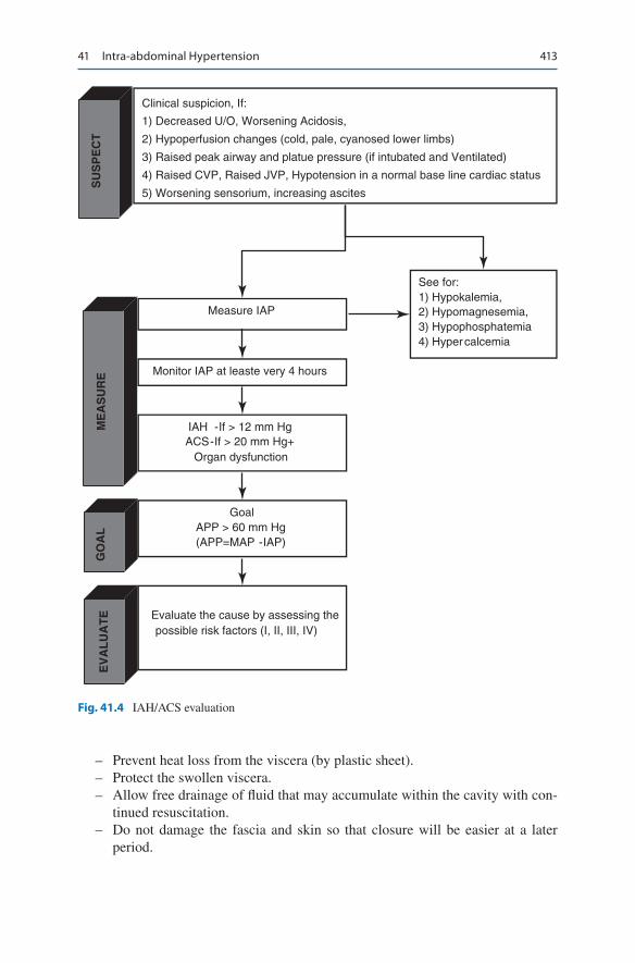
413
– Prevent heat loss from the viscera (by plastic sheet). – Protect the swollen viscera. – Allow free drainage of fluid that may accumulate within the cavity with con-
tinued resuscitation. – Do not damage the fascia and skin so that closure will be easier at a later
period.
SUSPECT
EVALUATE
Clinical suspicion, If:
1) Decreased U/O, Worsening Acidosis,
2) Hypoperfusion changes (cold, pale, cyanosed lower limbs)
3) Raised peak airway and platue pressure (if intubated and Ventilated)
4) Raised CVP, Raised JVP, Hypotension in a normal base line cardiac status
5) Worsening sensorium, increasing ascites
Measure IAP
MEASURE
GOAL
Monitor IAP at leaste very 4 hours
IAH -If > 12 mm HgACS-If > 20 mm Hg+
Organ dysfunction
GoalAPP > 60 mm Hg(APP=MAP -IAP)
Evaluate the cause by assessing the possible risk factors (I, II, III, IV)
See for:1) Hypokalemia,2) Hypomagnesemia,3) Hypophosphatemia4) Hypercalcemia
Fig. 41.4 IAH/ACS evaluation
41 Intra-abdominal Hypertension
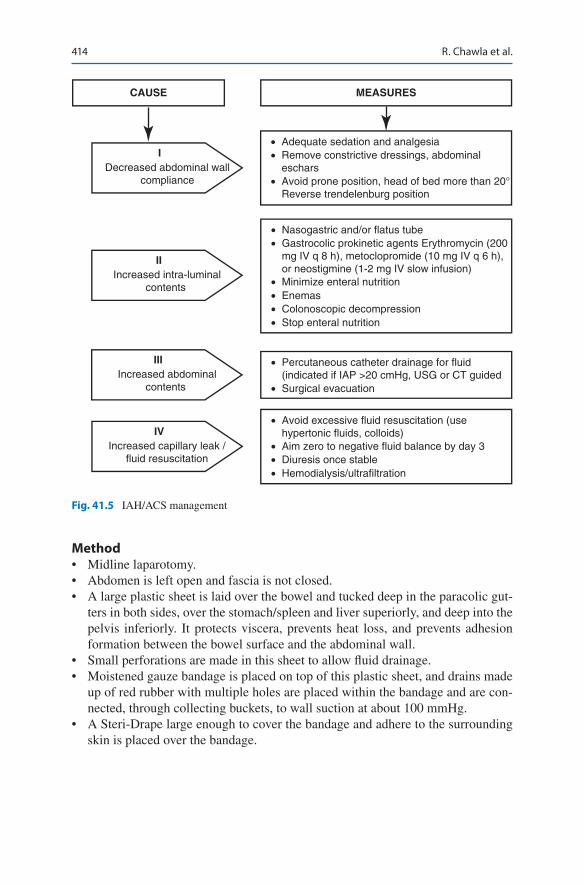
414
Method• Midline laparotomy.• Abdomen is left open and fascia is not closed.• A large plastic sheet is laid over the bowel and tucked deep in the paracolic gut-
ters in both sides, over the stomach/spleen and liver superiorly, and deep into the pelvis inferiorly. It protects viscera, prevents heat loss, and prevents adhesion formation between the bowel surface and the abdominal wall.
• Small perforations are made in this sheet to allow fluid drainage.• Moistened gauze bandage is placed on top of this plastic sheet, and drains made
up of red rubber with multiple holes are placed within the bandage and are con-nected, through collecting buckets, to wall suction at about 100 mmHg.
• A Steri-Drape large enough to cover the bandage and adhere to the surrounding skin is placed over the bandage.
CAUSE MEASURES
I Decreased abdominal wall
compliance
IIIncreased intra-luminal
contents
IIIIncreased abdominal
contents
IVIncreased capillary leak /
fluid resuscitation
• Adequate sedation and analgesia• Remove constrictive dressings, abdominal
eschars• Avoid prone position, head of bed more than 20°
Reverse trendelenburg position
• Nasogastric and/or flatus tube• Gastrocolic prokinetic agents Erythromycin (200
mg IV q 8 h), metoclopromide (10 mg IV q 6 h),or neostigmine (1-2 mg IV slow infusion)
• Minimize enteral nutrition• Enemas• Colonoscopic decompression• Stop enteral nutrition
• Percutaneous catheter drainage for fluid(indicated if IAP >20 cmHg, USG or CT guided
• Surgical evacuation
• Avoid excessive fluid resuscitation (usehypertonic fluids, colloids)
• Aim zero to negative fluid balance by day 3• Diuresis once stable• Hemodialysis/ultrafiltration
Fig. 41.5 IAH/ACS management
R. Chawla et al.
415
Suggested Reading
Cheatham ML. Nonoperative management of intraabdominal hypertension and abdominal com-partment syndrome. World J Surg. 2009;33(6):1116–22. This comprehensive review discusses the risk factors that predict the development of IAH/ACS, the appropriate measurement of IAP, and the current resuscitation options.
De Waele JJ, Kimball E, Malbrain M. Decompressive laparotomy for abdominal compartment syndrome. Br J Surg. 2016;103(6):709–15. A prospective cohort study of decompressive lapa-rotomy which showed significant improved oxygenation and urinary output. Survivors showed improvement in organ function scores. The overall 28-day mortality rate was 36 percent (12 of 33), which increased to 55 per cent (18 patients) at 1 year.
Irwin RS, Rippe JM. Irwin and Rippe’s intensive care medicine. 6th ed. Philadelphia: Lippincott Williams & Wilkins; 2008. p. 1795–803. The chapter discusses the practical application of IAP monitoring in detail, with pictorial depiction of pathophysiology of raised IAP.
Kirkpatrick AW, et al. Intra-abdominal hypertension and the abdominal compartment syndrome: updated consensus definitions and clinical practice guidelines from the World Society of the Abdominal Compartment Syndrome. Intensive Care Med. 2013;39:1190–206. Excellent review article and guideline on management of intra-abdominal hypertension.
Website
http://www.wsacs.org. The all-in-one website for intra-abdominal hypertension and abdominal compartment syndrome.
41 Intra-abdominal Hypertension
417© Springer Nature Singapore Pte Ltd. 2020R. Chawla, S. Todi (eds.), ICU Protocols, https://doi.org/10.1007/978-981-15-0898-1_42
A. Kumar (*) Department of Gastroenterology & Hepatology, BLK Hospital, New Delhi, India
A. Kumar Saint Peters University Hospital/Rutgers University, New Brunswick, NJ, USA
42Acute Pancreatitis
Ajay Kumar and Akshat Kumar
Acute Pancreatitis may be of two broad types: acute edematous, interstitial pancre-atitis and necrotising acute pancreatitis,
Step 1: Initiate Resuscitation and Take Focused History
• Initiate resuscitation, as mentioned in Chap. 23, Vol. 2.• Take detailed history.• Most of the patients (95%) present with acute epigastric abdominal pain. In
about 50% of these patients, pain will be referred to the back. This can be accom-panied by nausea, vomiting, and abdominal distension or fever. Symptoms may be out of proportion to signs.
• History of alcohol intake, previous gallstone disease, drug intake, hypertriglyc-eridemia or known malignancy.
A 45-year-old nonalcoholic male patient presented with severe continuous upper abdominal pain for 1 day, associated with vomiting and mild abdominal disten-sion. He had history of right upper abdominal pain 6 months back. His vital signs were stable. Abdomen examination showed mild tenderness and distension. Bowel sounds were sluggish. Investigations showed leukocytosis (13,000), ele-vated serum bilirubin of 2.5 mg/dL, and a fourfold increase in transaminases. Serum amylase was 1420 IU and serum lipase was 1200 IU. Ultrasonography of the abdomen showed 7-mm gallstones, normal common bile duct (CBD), and bulky pancreas with peri-pancreatic fluid collection. He was diagnosed to have biliary pancreatitis.
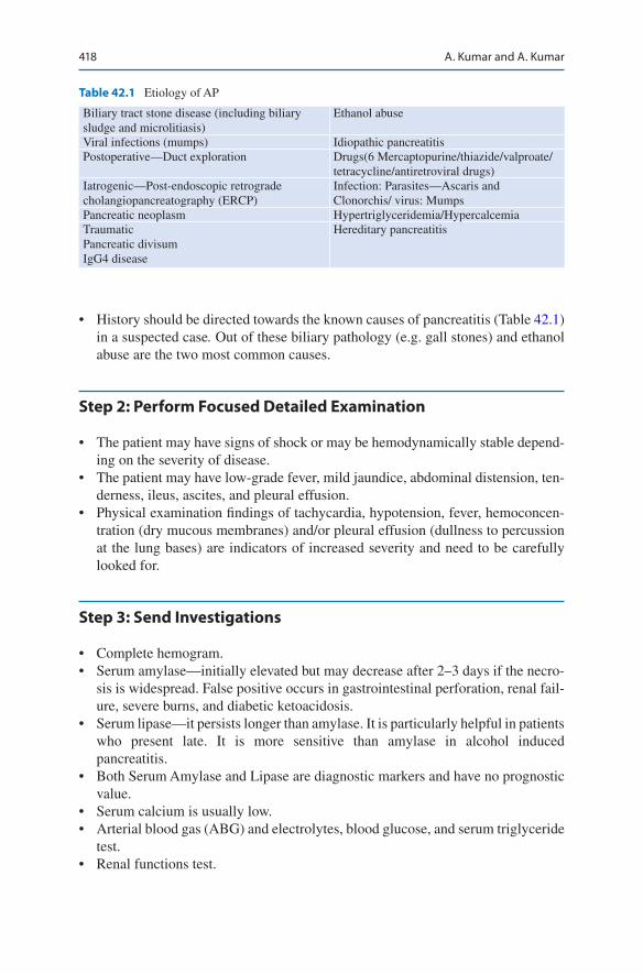
418
• History should be directed towards the known causes of pancreatitis (Table 42.1) in a suspected case. Out of these biliary pathology (e.g. gall stones) and ethanol abuse are the two most common causes.
Step 2: Perform Focused Detailed Examination
• The patient may have signs of shock or may be hemodynamically stable depend-ing on the severity of disease.
• The patient may have low-grade fever, mild jaundice, abdominal distension, ten-derness, ileus, ascites, and pleural effusion.
• Physical examination findings of tachycardia, hypotension, fever, hemoconcen-tration (dry mucous membranes) and/or pleural effusion (dullness to percussion at the lung bases) are indicators of increased severity and need to be carefully looked for.
Step 3: Send Investigations
• Complete hemogram.• Serum amylase—initially elevated but may decrease after 2–3 days if the necro-
sis is widespread. False positive occurs in gastrointestinal perforation, renal fail-ure, severe burns, and diabetic ketoacidosis.
• Serum lipase—it persists longer than amylase. It is particularly helpful in patients who present late. It is more sensitive than amylase in alcohol induced pancreatitis.
• Both Serum Amylase and Lipase are diagnostic markers and have no prognostic value.
• Serum calcium is usually low.• Arterial blood gas (ABG) and electrolytes, blood glucose, and serum triglyceride
test.• Renal functions test.
Table 42.1 Etiology of AP
Biliary tract stone disease (including biliary sludge and microlitiasis)
Ethanol abuse
Viral infections (mumps) Idiopathic pancreatitisPostoperative—Duct exploration Drugs(6 Mercaptopurine/thiazide/valproate/
tetracycline/antiretroviral drugs)Iatrogenic—Post-endoscopic retrograde cholangiopancreatography (ERCP)
Infection: Parasites—Ascaris and Clonorchis/ virus: Mumps
Pancreatic neoplasm Hypertriglyceridemia/HypercalcemiaTraumaticPancreatic divisumIgG4 disease
Hereditary pancreatitis
A. Kumar and A. Kumar
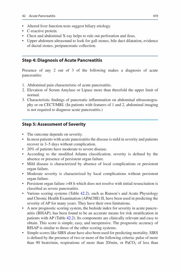
419
• Altered liver function tests suggest biliary etiology.• C-reactive protein.• Chest and abdominal X-ray helps to rule out perforation and ileus.• Upper abdomen ultrasound to look for gall stones, bile duct dilatation, evidence
of ductal stones, peripancreatic collection.
Step 4: Diagnosis of Acute Pancreatitis
Presence of any 2 out of 3 of the following makes a diagnosis of acute pancreatitis:
1. Abdominal pain characteristic of acute pancreatitis. 2. Elevation of Serum Amylase or Lipase more than threefold the upper limit of
normal. 3. Characteristic findings of pancreatic inflammation on abdominal ultrasonogra-
phy or on CECT/MRI. (In patients with features of 1 and 2, abdominal imaging is not required to diagnose acute pancreatitis.)
Step 5: Assessment of Severity
• The outcome depends on severity.• In most patients with acute pancreatitis the disease is mild in severity and patients
recover in 3–5 days without complication.• 20% of patients have moderate to severe disease.• According to the modified Atlanta classification, severity is defined by the
absence or presence of persistent organ failure.• Mild disease is characterized by absence of local complications or persistent
organ failure.• Moderate severity is characterized by local complications without persistent
organ failure.• Persistent organ failure >48 h which does not resolve with initial resuscitation is
classified as severe pancreatitis.• Various scoring systems (Table 42.2), such as Ranson’s and Acute Physiology
and Chronic Health Examination (APACHE) II, have been used in predicting the severity of AP for many years. They have their own limitations.
• A new prognostic scoring system, the bedside index for severity in acute pancre-atitis (BISAP), has been found to be an accurate means for risk stratification in patients with AP (Table 42.2). Its components are clinically relevant and easy to obtain. This score is simple, easy, and inexpensive. The prognostic accuracy of BISAP is similar to those of the other scoring systems.
• Simple scores like SIRS alone have also been used for predicting mortality. SIRS is defined by the presence of two or more of the following criteria: pulse of more than 90 beats/min, respirations of more than 20/min, or PaCO2 of less than
42 Acute Pancreatitis

420
32 mmHg, temperature of more than 100.4 °F or less than 96.8 °F, and white blood cell count of more than 12,000 or less than 4000 cells/mm3, or more than 10% immature neutrophils. It is very simple and inexpensive and can be done multiple times. SIRS of two or more for over 48 h predicts mortality of 25%. However SIRS is not very specific and can be positive from a number of other comorbid conditions.
• Cumulative SIRSS (Systemic Inflammatory Response Syndrome Score) is a recent marker of AP severity. It is the total number of cumulative SIRS positive days. It increases specificity of SIRS and has the potential to predict late onset complications of AP.
Table 42.2 Method to predict the severity of AP
(1) Ranson’s criteria0 h
Age >55 yearsWhite blood cell count >16,000/mm3
Blood glucose >200 mg/dL (11.1 mmol/L)Lactate dehydrogenase >350 U/LAspartate aminotransferase >250 U/L
48 hHematocrit Fall by ≥10%Blood urea nitrogen Increased by >5 mg/dL
(1.8 mmol/L) despite fluidsSerum calcium <8 mg/dL (2 mmol/L)PO2 <60 mmHgBase deficit >4 mEq/LFluid sequestration >6000 mLThe presence of one to three Ranson’s criteria represents mild pancreatitis; the mortality rate rises significantly with four or more criteriaThe presence of three or more Ranson’s criteria within the first 48 h is indicative of severe pancreatitis(2) The BISAP score for early mortality prediction (within the first 24 h)Five parameters(1) Blood urea nitrogen (BUN) >25 mg/dL(2) Impaired mental status(3) SIRS 2 or more(4) Age > 60 years(5) Pleural effusionPredictive accuracy of BISAP is similar to APACHE IIThe BISAP score of more than three predicts persistent organ failure (p < 0.0001) and necrosis (p < 0.0004)Mortality increases from BISAP score of 1–5Patients with predicted severe disease should be shifted to the specialist center or ICU, and aggressive management should be rapidly instituted
A. Kumar and A. Kumar

421
• American college of gastroenterology Guidelines 2018 have suggested advanced age, comorbidities, body mass index >30, pleural effusion or pulmonary infil-trate, Hematocrit >44, Blood urea nitrogen >20, rising BUN, high creatinine, initial SIRS score > 2, persistent SIRS and persistent organ failure as predictor of severe disease.
Step 6: Do Imaging US for Everyone, CT for Selected Few
• Ultrasound is noninvasive and the best tool available for the initial evaluation of pancreatitis. This may reveal gallstones or edema of the pancreas.
• CT scan should be postponed till 72 h or more. If the diagnosis of pancreatitis is established by the criteria given above, CT should be avoided.
• A contrast-enhanced CT (CECT) of the abdomen is one of the finest modalities available to morphologically diagnose and quantify the necrosis, which predicts prognosis (Table 42.3). These changes may not appear in the first few days.
• MRI of the abdomen should be done if CECT is contraindicated.• The only indication for early CT scan is when one is not certain about the
diagnosis.
Table 42.3 CT grading of AP
(A) Balthazar–Ranson’s grading system (can be done without contrast)A Normal appearing pancreasB Focal or diffuse enlargement of pancreasC Pancreatic gland abnormalities associated with mild peripancreatic inflammatory changes (stranding)D Fluid collection in a single location, usually within anterior pararenal spaceE Two or more fluid collections near the pancreas and/or presence of gas in or adjacent to the pancreas(B) Severity of APCT grade ScoreA 0B 1C 2D 3E 4Necrosis (needs IV contrast) ScoreNone 0<33% 233%–50% 4≥50% 6CT severity index (0–10)CT grade (0–4) + necrosis (0–6) = total score
42 Acute Pancreatitis
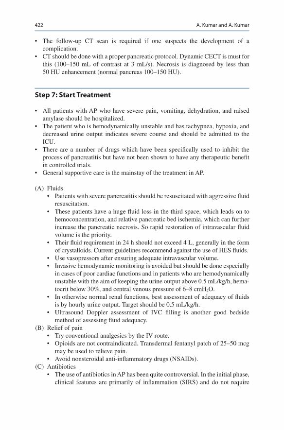
422
• The follow-up CT scan is required if one suspects the development of a complication.
• CT should be done with a proper pancreatic protocol. Dynamic CECT is must for this (100–150 mL of contrast at 3 mL/s). Necrosis is diagnosed by less than 50 HU enhancement (normal pancreas 100–150 HU).
Step 7: Start Treatment
• All patients with AP who have severe pain, vomiting, dehydration, and raised amylase should be hospitalized.
• The patient who is hemodynamically unstable and has tachypnea, hypoxia, and decreased urine output indicates severe course and should be admitted to the ICU.
• There are a number of drugs which have been specifically used to inhibit the process of pancreatitis but have not been shown to have any therapeutic benefit in controlled trials.
• General supportive care is the mainstay of the treatment in AP.
(A) Fluids• Patients with severe pancreatitis should be resuscitated with aggressive fluid
resuscitation.• These patients have a huge fluid loss in the third space, which leads on to
hemoconcentration, and relative pancreatic bed ischemia, which can further increase the pancreatic necrosis. So rapid restoration of intravascular fluid volume is the priority.
• Their fluid requirement in 24 h should not exceed 4 L, generally in the form of crystalloids. Current guidelines recommend against the use of HES fluids.
• Use vasopressors after ensuring adequate intravascular volume.• Invasive hemodynamic monitoring is avoided but should be done especially
in cases of poor cardiac functions and in patients who are hemodynamically unstable with the aim of keeping the urine output above 0.5 mL/kg/h, hema-tocrit below 30%, and central venous pressure of 6–8 cmH2O.
• In otherwise normal renal functions, best assessment of adequacy of fluids is by hourly urine output. Target should be 0.5 mL/kg/h.
• Ultrasound Doppler assessment of IVC filling is another good bedside method of assessing fluid adequacy.
(B) Relief of pain• Try conventional analgesics by the IV route.• Opioids are not contraindicated. Transdermal fentanyl patch of 25–50 mcg
may be used to relieve pain.• Avoid nonsteroidal anti-inflammatory drugs (NSAIDs).
(C) Antibiotics• The use of antibiotics in AP has been quite controversial. In the initial phase,
clinical features are primarily of inflammation (SIRS) and do not require
A. Kumar and A. Kumar
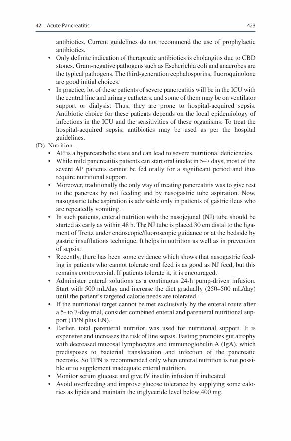
423
antibiotics. Current guidelines do not recommend the use of prophylactic antibiotics.
• Only definite indication of therapeutic antibiotics is cholangitis due to CBD stones. Gram-negative pathogens such as Escherichia coli and anaerobes are the typical pathogens. The third-generation cephalosporins, fluoroquinolone are good initial choices.
• In practice, lot of these patients of severe pancreatitis will be in the ICU with the central line and urinary catheters, and some of them may be on ventilator support or dialysis. Thus, they are prone to hospital-acquired sepsis. Antibiotic choice for these patients depends on the local epidemiology of infections in the ICU and the sensitivities of these organisms. To treat the hospital-acquired sepsis, antibiotics may be used as per the hospital guidelines.
(D) Nutrition• AP is a hypercatabolic state and can lead to severe nutritional deficiencies.• While mild pancreatitis patients can start oral intake in 5–7 days, most of the
severe AP patients cannot be fed orally for a significant period and thus require nutritional support.
• Moreover, traditionally the only way of treating pancreatitis was to give rest to the pancreas by not feeding and by nasogastric tube aspiration. Now, nasogastric tube aspiration is advisable only in patients of gastric ileus who are repeatedly vomiting.
• In such patients, enteral nutrition with the nasojejunal (NJ) tube should be started as early as within 48 h. The NJ tube is placed 30 cm distal to the liga-ment of Treitz under endoscopic/fluoroscopic guidance or at the bedside by gastric insufflations technique. It helps in nutrition as well as in prevention of sepsis.
• Recently, there has been some evidence which shows that nasogastric feed-ing in patients who cannot tolerate oral feed is as good as NJ feed, but this remains controversial. If patients tolerate it, it is encouraged.
• Administer enteral solutions as a continuous 24-h pump-driven infusion. Start with 500 mL/day and increase the diet gradually (250–500 mL/day) until the patient’s targeted calorie needs are tolerated.
• If the nutritional target cannot be met exclusively by the enteral route after a 5- to 7-day trial, consider combined enteral and parenteral nutritional sup-port (TPN plus EN).
• Earlier, total parenteral nutrition was used for nutritional support. It is expensive and increases the risk of line sepsis. Fasting promotes gut atrophy with decreased mucosal lymphocytes and immunoglobulin A (IgA), which predisposes to bacterial translocation and infection of the pancreatic necrosis. So TPN is recommended only when enteral nutrition is not possi-ble or to supplement inadequate enteral nutrition.
• Monitor serum glucose and give IV insulin infusion if indicated.• Avoid overfeeding and improve glucose tolerance by supplying some calo-
ries as lipids and maintain the triglyceride level below 400 mg.
42 Acute Pancreatitis
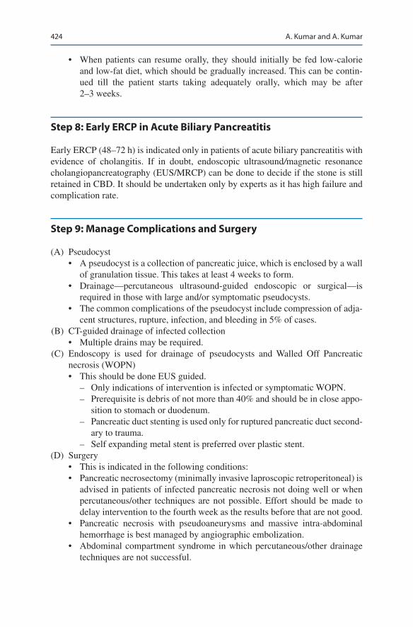
424
• When patients can resume orally, they should initially be fed low-calorie and low-fat diet, which should be gradually increased. This can be contin-ued till the patient starts taking adequately orally, which may be after 2–3 weeks.
Step 8: Early ERCP in Acute Biliary Pancreatitis
Early ERCP (48–72 h) is indicated only in patients of acute biliary pancreatitis with evidence of cholangitis. If in doubt, endoscopic ultrasound/magnetic resonance cholangiopancreatography (EUS/MRCP) can be done to decide if the stone is still retained in CBD. It should be undertaken only by experts as it has high failure and complication rate.
Step 9: Manage Complications and Surgery
(A) Pseudocyst• A pseudocyst is a collection of pancreatic juice, which is enclosed by a wall
of granulation tissue. This takes at least 4 weeks to form.• Drainage—percutaneous ultrasound-guided endoscopic or surgical—is
required in those with large and/or symptomatic pseudocysts.• The common complications of the pseudocyst include compression of adja-
cent structures, rupture, infection, and bleeding in 5% of cases. (B) CT-guided drainage of infected collection
• Multiple drains may be required. (C) Endoscopy is used for drainage of pseudocysts and Walled Off Pancreatic
necrosis (WOPN)• This should be done EUS guided.
– Only indications of intervention is infected or symptomatic WOPN. – Prerequisite is debris of not more than 40% and should be in close appo-
sition to stomach or duodenum. – Pancreatic duct stenting is used only for ruptured pancreatic duct second-
ary to trauma. – Self expanding metal stent is preferred over plastic stent.
(D) Surgery• This is indicated in the following conditions:• Pancreatic necrosectomy (minimally invasive laproscopic retroperitoneal) is
advised in patients of infected pancreatic necrosis not doing well or when percutaneous/other techniques are not possible. Effort should be made to delay intervention to the fourth week as the results before that are not good.
• Pancreatic necrosis with pseudoaneurysms and massive intra-abdominal hemorrhage is best managed by angiographic embolization.
• Abdominal compartment syndrome in which percutaneous/other drainage techniques are not successful.
A. Kumar and A. Kumar
425
• Local complications of the pseudocyst.• Bowel infarction.
(E) Controversial indications of surgery• Extensive (>50%) sterile pancreatic necrosis with persisting multiple-organ
failure despite intensive care therapy.• Most surgeons avoid surgery for this indication as this is associated with
increased mortality.
Step 10: Removal of the Gallbladder
• Removal of the gallbladder should be scheduled early to avoid recurrence of bili-ary pancreatitis (30%). Present AGA guidelines recommend cholecystectomy before discharge in mild cases.
• It should be performed both in patients with gall stones or biliary sludge.• In severe attacks, it is recommended to wait 4–6 weeks to allow inflammation to
subside, when patient is likely to require another intervention for fluid collection or WOPN.
• The laparoscopic approach has proved to be feasible and safe, even in cases where surgical debridement is required.
Suggested Reading
American Gastroenterological Association Institute. Guideline on initial management of acute pancreatitis. Gastroenterology. 2018;154:1096–101. Guidelines on the initial management of pancreatitis.
Balthazar EJ, Robinson DL, Megibow AJ, Ranson JH. Acute pancreatitis: value of CT in estab-lishing prognosis. Radiology. 1990;174:331–6. A CT severity index, based on a combina-tion of peripancreatic inflammation, phlegmon, and the degree of pancreatic necrosis as seen at initial CT study, was developed. Patients with a high CT severity index had 92% morbidity and 17% mortality; patients with a low CT severity index had 2% morbidity, and none died.
Gardner TB, Vege SS, Chari ST, Petersen BT, Topazian MD, Clain JE, et al. Faster rate of initial fluid resuscitation in severe acute pancreatitis diminishes in-hospital mortality. Pancreatology. 2009;9:770–6. Patients with severe acute pancreatitis who do not receive at least one-third of their initial 72-h cumulative intravenous fluid volume during the first 24 h are at risk of greater mortality than those who are initially resuscitated more aggressively.
Jafri NS, Mahid SS, Idstein SR, Hornung CA, Galandiuk S. Antibiotic prophylaxis is not protective in severe acute pancreatitis: a systematic review and meta-analysis. Am J Surg. 2009;197:806–13. Antibiotic prophylaxis of SAP does not reduce mortality or protect against infected necro-sis, or frequency of surgical intervention.
Petrov MS, Pylypchuk RD, Emelyanov NV. Systematic review: nutritional support in acute pan-creatitis. Aliment Pharmacol Ther. 2008;28:704–12. The use of either enteral or parenteral nutrition, in comparison with no supplementary nutrition, is associated with a lower risk of death in acute pancreatitis. Enteral nutrition is associated with a lower risk of infectious com-plications compared with parenteral nutrition.
Talukdar R, Vega SS. Early management of severe acute pancreatitis. Curr Gastroentrol Rep. 2011;13:123–30. A article on the early management of acute severe pancreatitis.
42 Acute Pancreatitis

426
Website
http://gut.bmjjournals.com/cgi/content/full/54/suppl_3/iii1
A. Kumar and A. Kumar
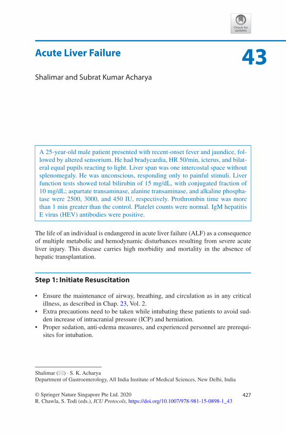
427© Springer Nature Singapore Pte Ltd. 2020R. Chawla, S. Todi (eds.), ICU Protocols, https://doi.org/10.1007/978-981-15-0898-1_43
Shalimar (*) · S. K. Acharya Department of Gastroenterology, All India Institute of Medical Sciences, New Delhi, India
43Acute Liver Failure
Shalimar and Subrat Kumar Acharya
The life of an individual is endangered in acute liver failure (ALF) as a consequence of multiple metabolic and hemodynamic disturbances resulting from severe acute liver injury. This disease carries high morbidity and mortality in the absence of hepatic transplantation.
Step 1: Initiate Resuscitation
• Ensure the maintenance of airway, breathing, and circulation as in any critical illness, as described in Chap. 23, Vol. 2.
• Extra precautions need to be taken while intubating these patients to avoid sud-den increase of intracranial pressure (ICP) and herniation.
• Proper sedation, anti-edema measures, and experienced personnel are prerequi-sites for intubation.
A 25-year-old male patient presented with recent-onset fever and jaundice, fol-lowed by altered sensorium. He had bradycardia, HR 50/min, icterus, and bilat-eral equal pupils reacting to light. Liver span was one intercostal space without splenomegaly. He was unconscious, responding only to painful stimuli. Liver function tests showed total bilirubin of 15 mg/dL, with conjugated fraction of 10 mg/dL; aspartate transaminase, alanine transaminase, and alkaline phospha-tase were 2500, 3000, and 450 IU, respectively. Prothrombin time was more than 1 min greater than the control. Platelet counts were normal. IgM hepatitis E virus (HEV) antibodies were positive.

428
Step 2: Identify ALF and Its Causes
• In a clinical setting, hepatic injury is usually recognized by appearance of jaun-dice, and liver failure is recognized by occurrence of encephalopathy, ascites, and coagulopathy.
• Proper history should be taken regarding medications, rash, needlestick injury, blood transfusion, previous surgery, and history of jaundice in family members, to identify the cause of fulminant hepatic failure (FHF) (Table 43.1).
• Other causes of fever with encephalopathy such as bacterial sepsis and tropical infections in endemic areas such as malaria, enteric fever, leptospira, dengue, meningitis, encephalitis, cholangitis, and underlying chronic liver disease should be ruled out.
While initial supportive therapy is being given, diagnostic workup should be sent. This includes the following:
• Complete blood count, blood glucose, blood urea nitrogen, creatinine, electro-lytes, liver function tests, and prothrombin time.
• Arterial blood gases, arterial ammonia and lactate.• Chest X-ray, ECG.• Endotracheal aspirate for aerobic culture in intubated patients, blood culture and
urine culture.• Serology including HBsAg, IgM anti-HBc, IgM anti-HEV, IgM anti-HAV, anti-
HCV, anti-HDV, and HIV serology.• Copper studies—serum Ceruloplasmin.• Autoimmune markers—Antinuclear Factor/LKM antibody.• Bedside abdominal ultrasound
Broad categoriesInfectionsMetabolic diseasesDrugsToxinsALF of pregnancyAutoimmune hepatitisAcute Budd–Chiari syndromeShock liverHemophagocytic syndromeIndividual etiological agentsHepatotropic viruses (A to E)Cytomegalovirus, herpes simplex virusWilson’s disease, galactosemiaParacetamol, isoniazid, rifampicin, sodium valproateAmanita phalloides
Table 43.1 Causes of FHF
Shalimar and S. K. Acharya
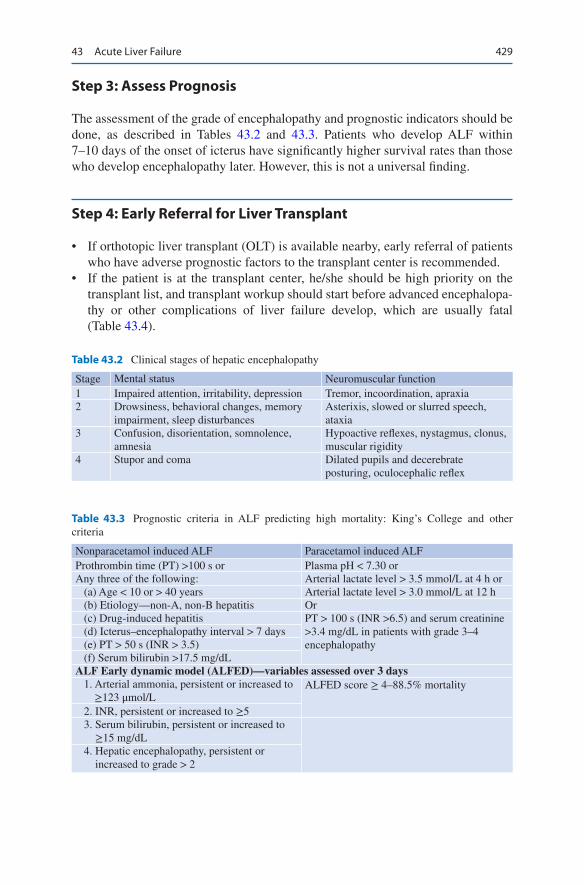
429
Step 3: Assess Prognosis
The assessment of the grade of encephalopathy and prognostic indicators should be done, as described in Tables 43.2 and 43.3. Patients who develop ALF within 7–10 days of the onset of icterus have significantly higher survival rates than those who develop encephalopathy later. However, this is not a universal finding.
Step 4: Early Referral for Liver Transplant
• If orthotopic liver transplant (OLT) is available nearby, early referral of patients who have adverse prognostic factors to the transplant center is recommended.
• If the patient is at the transplant center, he/she should be high priority on the transplant list, and transplant workup should start before advanced encephalopa-thy or other complications of liver failure develop, which are usually fatal (Table 43.4).
Table 43.2 Clinical stages of hepatic encephalopathy
Stage Mental status Neuromuscular function1 Impaired attention, irritability, depression Tremor, incoordination, apraxia2 Drowsiness, behavioral changes, memory
impairment, sleep disturbancesAsterixis, slowed or slurred speech, ataxia
3 Confusion, disorientation, somnolence, amnesia
Hypoactive reflexes, nystagmus, clonus, muscular rigidity
4 Stupor and coma Dilated pupils and decerebrate posturing, oculocephalic reflex
Table 43.3 Prognostic criteria in ALF predicting high mortality: King’s College and other criteria
Nonparacetamol induced ALF Paracetamol induced ALFProthrombin time (PT) >100 s or Plasma pH < 7.30 orAny three of the following: Arterial lactate level > 3.5 mmol/L at 4 h or (a) Age < 10 or > 40 years Arterial lactate level > 3.0 mmol/L at 12 h (b) Etiology—non-A, non-B hepatitis Or (c) Drug-induced hepatitis PT > 100 s (INR >6.5) and serum creatinine
>3.4 mg/dL in patients with grade 3–4 encephalopathy
(d) Icterus–encephalopathy interval > 7 days (e) PT > 50 s (INR > 3.5) (f) Serum bilirubin >17.5 mg/dLALF Early dynamic model (ALFED)—variables assessed over 3 days 1. Arterial ammonia, persistent or increased to
≥123 μmol/LALFED score ≥ 4–88.5% mortality
2. INR, persistent or increased to ≥5 3. Serum bilirubin, persistent or increased to
≥15 mg/dL 4. Hepatic encephalopathy, persistent or
increased to grade > 2
43 Acute Liver Failure
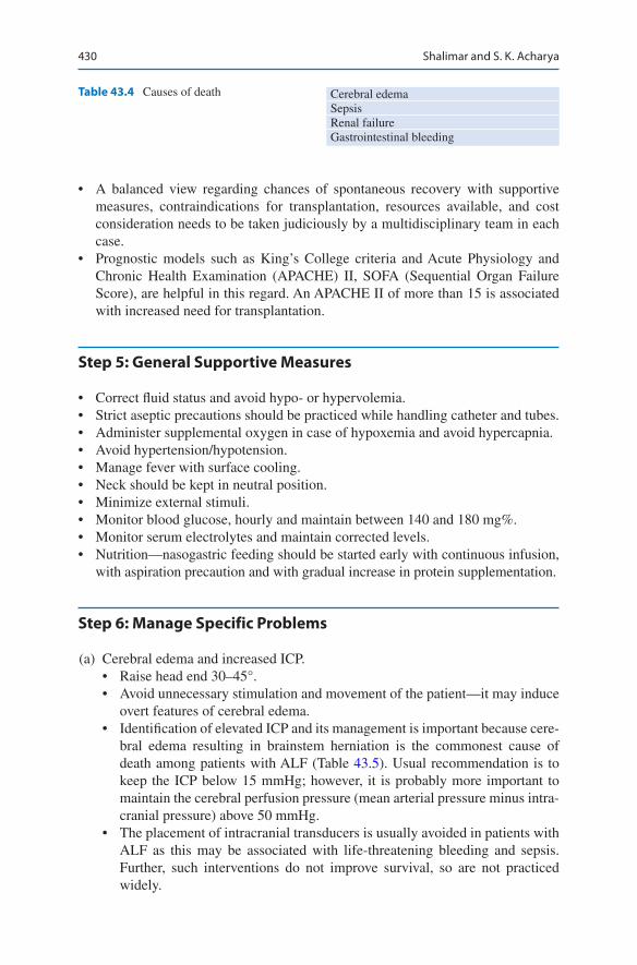
430
• A balanced view regarding chances of spontaneous recovery with supportive measures, contraindications for transplantation, resources available, and cost consideration needs to be taken judiciously by a multidisciplinary team in each case.
• Prognostic models such as King’s College criteria and Acute Physiology and Chronic Health Examination (APACHE) II, SOFA (Sequential Organ Failure Score), are helpful in this regard. An APACHE II of more than 15 is associated with increased need for transplantation.
Step 5: General Supportive Measures
• Correct fluid status and avoid hypo- or hypervolemia.• Strict aseptic precautions should be practiced while handling catheter and tubes.• Administer supplemental oxygen in case of hypoxemia and avoid hypercapnia.• Avoid hypertension/hypotension.• Manage fever with surface cooling.• Neck should be kept in neutral position.• Minimize external stimuli.• Monitor blood glucose, hourly and maintain between 140 and 180 mg%.• Monitor serum electrolytes and maintain corrected levels.• Nutrition—nasogastric feeding should be started early with continuous infusion,
with aspiration precaution and with gradual increase in protein supplementation.
Step 6: Manage Specific Problems
(a) Cerebral edema and increased ICP.• Raise head end 30–45°.• Avoid unnecessary stimulation and movement of the patient—it may induce
overt features of cerebral edema.• Identification of elevated ICP and its management is important because cere-
bral edema resulting in brainstem herniation is the commonest cause of death among patients with ALF (Table 43.5). Usual recommendation is to keep the ICP below 15 mmHg; however, it is probably more important to maintain the cerebral perfusion pressure (mean arterial pressure minus intra-cranial pressure) above 50 mmHg.
• The placement of intracranial transducers is usually avoided in patients with ALF as this may be associated with life-threatening bleeding and sepsis. Further, such interventions do not improve survival, so are not practiced widely.
Table 43.4 Causes of death Cerebral edemaSepsisRenal failureGastrointestinal bleeding
Shalimar and S. K. Acharya

431
• Propofol is the preferred sedative agent in ALF. Propofol reduces cerebral blood flow as well as ICP.
• CT Brain to exclude other intracranial pathology.• Transcranial doppler may be used to assess noninvasively increased ICP.
(b) Sepsis is the second major cause of death among ALF.• Gram-negative bacteria are the major cause of sepsis in these patients.• Prophylactic parenteral antibiotics with third generation antibiotics in
selected cases.• Targeted antibiotic as per sensitivity report should be instituted.• Fungal infection is also common in this population. Prophylactic fluconazole,
in selected cases with multiple-site colonization with yeast, may be used.• Surveillance cultures should be sent, and high index of suspicion and low
threshold for starting broad-spectrum antibiotics should be practiced. (c) Renal failure: Both intermittent hemodialysis and continuous renal replacement
therapy (CRRT) are equally effective, with the latter more suitable for unstable patients as it avoids fluctuation in ICP.
(d) Coagulopathy: Prophylactic use of fresh frozen plasma (FFP) is not helpful unless a planned procedure is followed. FFP and platelet transfusion should be given only if bleeding occurs. FFP can precipitate volume overload in these patients, and in selected cases, off-label use of recombinant factor VII may be considered.
(e) Seizures: Prophylactic therapy with phenytoin is not useful. Levetiracetam may be used for the treatment as it is not hepatotoxic.
Step 7: Manage Specific Situations
(a) Paracetamol overdose—N-acetyl cysteine (NAC): 150 mg/kg over 1 h, followed by 12.5 mg/kg/h for 4 h and then 6.25 mg/kg/h for 67 h. NAC has been found to be useful in both acetaminophen and non-acetaminophen-induced ALF.
(b) Special situation—pregnancy
Table 43.5 Cerebral edema and raised ICP
Symptoms/signs ManagementHyperventilationBradycardiaFocal seizuresDecerebrate posturesAbsent pupillary reflexes
Elevate the head and the trunk 35–40°Avoid vigorous endotracheal suctionAvoid hyperthermiaRemove constricting tapesSedate the patient100 mL mannitol (20%) stat followed by 1 g/kg q8 hourly (keeping serum osmolality <320 mosmol/l)Hypertonic saline for preventing increase in ICP (target serum sodium 145–155)Elective intubation in encephalopathy grades 3–4Hyperventilation (target PaCO2 30–32 mmHg) and hypothermia in selected casesBarbiturates
43 Acute Liver Failure
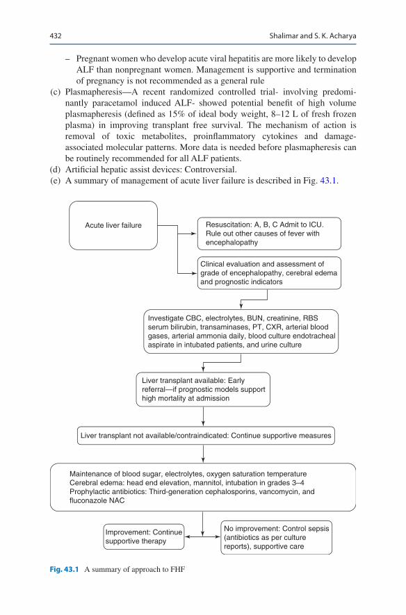
432
– Pregnant women who develop acute viral hepatitis are more likely to develop ALF than nonpregnant women. Management is supportive and termination of pregnancy is not recommended as a general rule
(c) Plasmapheresis—A recent randomized controlled trial- involving predomi-nantly paracetamol induced ALF- showed potential benefit of high volume plasmapheresis (defined as 15% of ideal body weight, 8–12 L of fresh frozen plasma) in improving transplant free survival. The mechanism of action is removal of toxic metabolites, proinflammatory cytokines and damage- associated molecular patterns. More data is needed before plasmapheresis can be routinely recommended for all ALF patients.
(d) Artificial hepatic assist devices: Controversial. (e) A summary of management of acute liver failure is described in Fig. 43.1.
Resuscitation: A, B, C Admit to ICU.Rule out other causes of fever withencephalopathy
Acute liver failure
Clinical evaluation and assessment ofgrade of encephalopathy, cerebral edemaand prognostic indicators
Investigate CBC, electrolytes, BUN, creatinine, RBSserum bilirubin, transaminases, PT, CXR, arterial bloodgases, arterial ammonia daily, blood culture endotrachealaspirate in intubated patients, and urine culture
Liver transplant available: Earlyreferral—if prognostic models supporthigh mortality at admission
Liver transplant not available/contraindicated: Continue supportive measures
Maintenance of blood sugar, electrolytes, oxygen saturation temperatureCerebral edema: head end elevation, mannitol, intubation in grades 3–4Prophylactic antibiotics: Third-generation cephalosporins, vancomycin, andfluconazole NAC
Improvement: Continuesupportive therapy
No improvement: Control sepsis(antibiotics as per culturereports), supportive care
Fig. 43.1 A summary of approach to FHF
Shalimar and S. K. Acharya
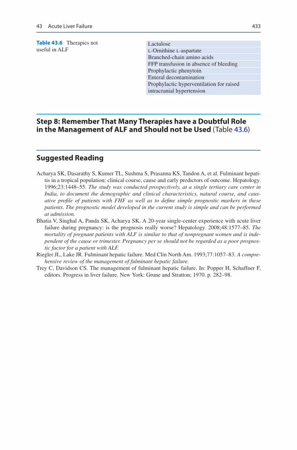
433
Step 8: Remember That Many Therapies have a Doubtful Role in the Management of ALF and Should not be Used (Table 43.6)
Suggested Reading
Acharya SK, Dasarathy S, Kumer TL, Sushma S, Prasanna KS, Tandon A, et al. Fulminant hepati-tis in a tropical population: clinical course, cause and early predictors of outcome. Hepatology. 1996;23:1448–55. The study was conducted prospectively, at a single tertiary care center in India, to document the demographic and clinical characteristics, natural course, and caus-ative profile of patients with FHF as well as to define simple prognostic markers in these patients. The prognostic model developed in the current study is simple and can be performed at admission.
Bhatia V, Singhal A, Panda SK, Acharya SK. A 20-year single-center experience with acute liver failure during pregnancy: is the prognosis really worse? Hepatology. 2008;48:1577–85. The mortality of pregnant patients with ALF is similar to that of nonpregnant women and is inde-pendent of the cause or trimester. Pregnancy per se should not be regarded as a poor prognos-tic factor for a patient with ALF.
Riegler JL, Lake JR. Fulminant hepatic failure. Med Clin North Am. 1993;77:1057–83. A compre-hensive review of the management of fulminant hepatic failure.
Trey C, Davidson CS. The management of fulminant hepatic failure. In: Popper H, Schaffner F, editors. Progress in liver failure. New York: Grune and Stratton; 1970. p. 282–98.
Table 43.6 Therapies not useful in ALF
Lactulosel-Ornithine l-aspartateBranched-chain amino acidsFFP transfusion in absence of bleedingProphylactic phenytoinEnteral decontaminationProphylactic hyperventilation for raised intracranial hypertension
43 Acute Liver Failure
435© Springer Nature Singapore Pte Ltd. 2020R. Chawla, S. Todi (eds.), ICU Protocols, https://doi.org/10.1007/978-981-15-0898-1_44
A. Kumar (*) Department of Gastroenterology & Hepatology, BLK Hospital, New Delhi, India
D. Amarapurkar (Deceased)Department of Gastroenterology, Bombay Hospital & Medical Research Centre, Breach Candy Medical Research Centre, Mumbai, India
44Acute Decompensation in Chronic Liver Failure
Ajay Kumar and Deepak Amarapurkar
Hepatic encephalopathy of any form is seen in 50–70% patients of cirrhosis. Mortality in patients of cirrhosis with encephalopathy ranges from 30% to 50% at the end of 1 year and 70% at the end of 3 years.
Step 1: Initiate Resuscitation
• All patients who have altered sensorium and cannot maintain their airway require immediate attention to airway. This assessment is done mainly by clinical means.
• They should be put on supplemental oxygen to increase SpO2 to above 90%. Patients who are unable to maintain their oxygenation are put on assisted ventilation.
• Circulation needs to be maintained by fluid infusion. If clinically there is evi-dence of cardiac impairment, fluids should be given cautiously.
A 54-year-old male patient, diagnosed to have cryptogenic cirrhosis 3 years back, was brought to the hospital, with abnormal behavior, inability to walk, edema over the feet, and distention of the abdomen for 3 days. On examination, the patient was found conscious but disoriented, having flapping tremors, icteric with edematous feet, and moderate ascites.
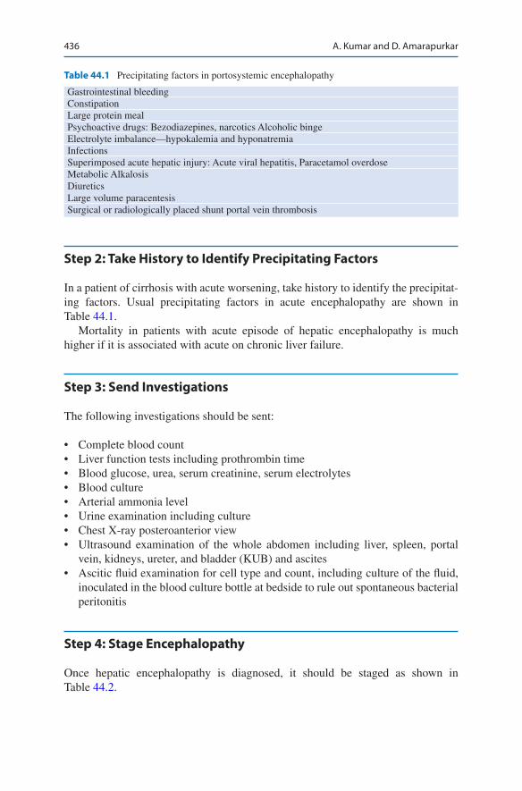
436
Step 2: Take History to Identify Precipitating Factors
In a patient of cirrhosis with acute worsening, take history to identify the precipitat-ing factors. Usual precipitating factors in acute encephalopathy are shown in Table 44.1.
Mortality in patients with acute episode of hepatic encephalopathy is much higher if it is associated with acute on chronic liver failure.
Step 3: Send Investigations
The following investigations should be sent:
• Complete blood count• Liver function tests including prothrombin time• Blood glucose, urea, serum creatinine, serum electrolytes• Blood culture• Arterial ammonia level• Urine examination including culture• Chest X-ray posteroanterior view• Ultrasound examination of the whole abdomen including liver, spleen, portal
vein, kidneys, ureter, and bladder (KUB) and ascites• Ascitic fluid examination for cell type and count, including culture of the fluid,
inoculated in the blood culture bottle at bedside to rule out spontaneous bacterial peritonitis
Step 4: Stage Encephalopathy
Once hepatic encephalopathy is diagnosed, it should be staged as shown in Table 44.2.
Table 44.1 Precipitating factors in portosystemic encephalopathy
Gastrointestinal bleedingConstipationLarge protein mealPsychoactive drugs: Bezodiazepines, narcotics Alcoholic bingeElectrolyte imbalance—hypokalemia and hyponatremiaInfectionsSuperimposed acute hepatic injury: Acute viral hepatitis, Paracetamol overdoseMetabolic AlkalosisDiureticsLarge volume paracentesisSurgical or radiologically placed shunt portal vein thrombosis
A. Kumar and D. Amarapurkar

437
Step 5: Manage Hepatic Encephalopathy
(A) Standard Therapeutic Measure in Hepatic Encephalopathy• Nutritional management:
– Normal protein diet for episodic hepatic encephalopathy – 1–2 g of protein per kg/day – Zinc replacement
• Reduction in nitrogenous load arising from the gut. Nonabsorbable disaccha-rides—lactulose is a first-line pharmacological treatment for hepatic encepha-lopathy. Lactulose should be given to have two to three loose stools per day.
• Recently lactulose was compared with the polythene glycol (PEG). The data showed that PEG resulted in faster patients recovery and this effect was independent of ammonia reduction.
• Bowel cleansing.• Rifaximin a nonabsorbable antibiotic is a therapeutic alternative to nonab-
sorbable disaccharides for treatment in acute and chronic encephalopathy and cirrhosis. Rifaximin is equally effective as lactulose. Rifaximin is used up to 1200 mg/day in divided doses.
• Albumin is traditionally considered as volume expander but has been shown to have anti-inflammatory properties. Albumin infusion did not improve hepatic encephalopathy but did improve the survival. Albumin is used in an extracorporeal circuit for dialysis. Patients treated with albumin dialysis showed improvement in hepatic encephalopathy.
• Ornithine aspartate in oral or intravenous form is only useful for a short duration. It should be avoided in renal dysfunction. This drug affects ammo-nia metabolism in multiorgan pathways. Ornithine drives the generation of glutamine in the muscle thus capturing a molecule of ammonia which is removed by phenyl acetate as phenyl acetyl glutamine
• Drugs that affect the neurotransmission—flumazenil and bromocriptine administration may have a therapeutic role in selected patients.
• Manipulation of the splanchnic circulation—closure of large portosystemic shunts.
(B) Renal Failure in Cirrhosis
Table 44.2 Clinical stages of hepatic encephalopathy
Stage Mental status Neuromuscular function1 Impaired attention, irritability, depression Tremor, incoordination, apraxia2 Drowsiness, behavioral changes, memory
impairment, sleep disturbancesAsterixis, slowed or slurred speech, ataxia
3 Confusion, disorientation, somnolence, amnesia
Hypoactive reflexes, nystagmus, clonus, muscular rigidity
4 Stupor and coma Dilated pupils and decerebrate posturing, absent oculocephalic reflex
44 Acute Decompensation in Chronic Liver Failure
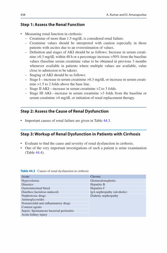
438
Step 1: Assess the Renal Function
• Measuring renal function in cirrhosis: – Creatinine of more than 1.5 mg/dL is considered renal failure. – Creatinine values should be interpreted with caution especially in those
patients with ascites due to an overestimation of values. – Definition and stages of AKI should be as follows: Increase in serum creati-
nine >0.3 mg/dL within 48 h or a percentage increase >50% from the baseline values (baseline serum creatinine value to be obtained in previous 3 months whenever available in patients where multiple values are available, value close to admission to be taken).
– Staging of AKI should be as follows: – Stage I—increase in serum creatinine >0.3 mg/dL or increase in serum creati-
nine >1.5 to 2 folds above the base line. – Stage II AKI—increase in serum creatinine >2 to 3 folds. – Stage III AKI—increase in serum creatinine >3 folds from the baseline or
serum creatinine >4 mg/dL or initiation of renal replacement therapy.
Step 2: Assess the Cause of Renal Dysfunction
• Important causes of renal failure are given in Table 44.3.
Step 3: Workup of Renal Dysfunction in Patients with Cirrhosis
• Evaluate to find the cause and severity of renal dysfunction in cirrhosis.• One of the very important investigations of such a patient is urine examination
(Table 44.4).
Table 44.3 Causes of renal dysfunction in cirrhosis
Acute ChronicHypovolemia GlomerulonephritisDiuretics Hepatitis BGastrointestinal bleed Hepatitis CDiarrhea (lactulose-induced) IgA nephropathy (alcoholic)Nephrotoxic drugs Diabetic nephropathyAminoglycosidesNonsteroidal anti-inflammatory drugsContrast agentsSepsis: Spontaneous bacterial peritonitisAcute kidney injury
A. Kumar and D. Amarapurkar
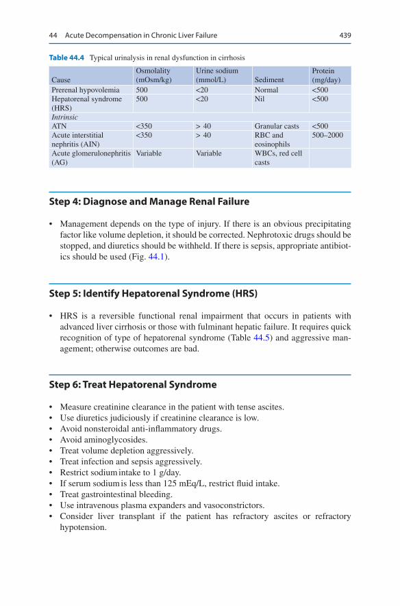
439
Step 4: Diagnose and Manage Renal Failure
• Management depends on the type of injury. If there is an obvious precipitating factor like volume depletion, it should be corrected. Nephrotoxic drugs should be stopped, and diuretics should be withheld. If there is sepsis, appropriate antibiot-ics should be used (Fig. 44.1).
Step 5: Identify Hepatorenal Syndrome (HRS)
• HRS is a reversible functional renal impairment that occurs in patients with advanced liver cirrhosis or those with fulminant hepatic failure. It requires quick recognition of type of hepatorenal syndrome (Table 44.5) and aggressive man-agement; otherwise outcomes are bad.
Step 6: Treat Hepatorenal Syndrome
• Measure creatinine clearance in the patient with tense ascites.• Use diuretics judiciously if creatinine clearance is low.• Avoid nonsteroidal anti-inflammatory drugs.• Avoid aminoglycosides.• Treat volume depletion aggressively.• Treat infection and sepsis aggressively.• Restrict sodium intake to 1 g/day.• If serum sodium is less than 125 mEq/L, restrict fluid intake.• Treat gastrointestinal bleeding.• Use intravenous plasma expanders and vasoconstrictors.• Consider liver transplant if the patient has refractory ascites or refractory
hypotension.
Table 44.4 Typical urinalysis in renal dysfunction in cirrhosis
CauseOsmolality (mOsm/kg)
Urine sodium (mmol/L) Sediment
Protein (mg/day)
Prerenal hypovolemia 500 <20 Normal <500Hepatorenal syndrome (HRS)
500 <20 Nil <500
IntrinsicATN <350 > 40 Granular casts <500Acute interstitial nephritis (AIN)
<350 > 40 RBC and eosinophils
500–2000
Acute glomerulonephritis (AG)
Variable Variable WBCs, red cell casts
44 Acute Decompensation in Chronic Liver Failure

440
Renal failure in patients of acutedec compensation in chronic
liver disease
ECF fluid losses; rapid or excessive diuresis,vomiting diarrhea, hemorrhage, recent LVP orhemodynamic changes due to use of NSAIDsor other drugs
Recent use of nephrotoxic medications(aminoglycosides, radiocontrast), hypotension(sepsis, hemorrhage)
Glomerular proteinuria and hematuria, i.e.dysmorphic RBC,s RBC casts
Imaging (ultrasound, CT scan) showshydronephrosis or urinary retention
Suggestive of obstructive uropathy. Unlesslong standing relief of obstruction shouldlead to improvement in renal function
Suspect glomerular disease. Depending onclinical scenario, further workup mayinclude cryoglobulins, C3, C4, and renalbiopsy
Toxic or ischemic ATN
Hold diuretics or other offendingmedications, trial of intravascular volumeexpander (albumin); if renal functionimproves, diagnosis of prerenal state ismade
Patient has evidence of portal hypertension(ascites). Cr > 1.5
Hepatorenal Syndrome
Yes
Yes
Yes
Yes
Yes
No
No
No
No
Fig. 44.1 Evaluation and management of renal failure
Table 44.5 Hepatorenal syndrome
Type 1 Type 2Rapid reduction in renal function in less than 2 weeks
Renal function slowly deteriorates over weeks to months
Doubling of initial serum creatinine to >2.5 mg/dL or 50% Reduction of the initial 24-h creatinine clearance to <20 mL/min
Increase in serum creatinine to more than 1.5 mg/dL or creatinine clearance less than 40 mL/mtOccurs in cirrhotic patients with refractory ascites
Severely ill patients Mild jaundiceJaundice Some degrees of coagulopathyCoagulopathy
• Recommendations for the use of vasoconstrictors in patients with type 2 hepa-torenal syndrome: – The goal of treatment is to reduce serum creatinine concentrations to ≤1.5 mg/
dL (130 μmol/L).
A. Kumar and D. Amarapurkar
441
– In patients who are critically ill: Norepinephrine infusion 0.5–3 mg/h or vaso-pressin infusion 0.01 units/mL should be started and titrating upwards and albumin 1 gm/kg/day for 2 days.
– In patient who are not critically ill Terlipressin Intravenously 1 to 2 mg every 4–6 h and albumin 1 g/kg/day for 2 days should be started followed by 25–50 g/day till terlipressin is discontinued.
– Alternatively a continuous infusion of terlipressin at a dose of 2 mg/day can be given. When the serum creatinine concentration does not reduce by at least 30%, the dose can be increased every 2 days up to 12 mg/day.
• Alternative drugs are midodrine, octreotide, the doses are as follows: – 2.5–7 mg midodrine should be given orally three times daily, increasing to
12.5 mg three times daily if needed. – 100 μg octreotide should be given subcutaneously or intravenously three
times daily, increasing to 200 μg three times daily if required. – Contraindications include coronary artery disease, peripheral vascular disease,
and/or cerebrovascular disease because of the potential risk of ischemic events.• Concomitant intravenous albumin infusion (1 g/kg body weight on the first day,
followed by 20–40 g per day) is recommended.• The duration of therapy should be approximately 7–14 days.
Suggested Reading
Ferenci P, Lockwood A, Mullen K, Tarter R, et al. Hepatic encephalopathy definition, nomencla-ture, diagnosis and quantification final report of the working party at the 11th World Congress of Gastroenterology, Vienna, 1998. Hepatology. 2002;35:716–21. The working party has sug-gested a modification of current nomenclature for clinical diagnosis of hepatic encephalopa-thy, proposed guidelines for the performance of future clinical trials in hepatic encephalopathy, and felt the need for a large study to redefine neuropsychiatric abnormalities in liver disease.
Gines P, Schrier R. Renal failure in cirrhosis. N Engl J Med. 2009;361:1279–90.Moriwaki H, Shiraki M, Iwasa J, Terakura Y. Hepatic encephalopathy as a complication of liver cir-
rhosis. An Asian perspective. J Gastroenterol Hepatol. 2010;25:658–63. This study concluded hepatic encephalopathy remains a serious complication of liver cirrhosis. Establishment of truly effective prevention modalities and broader application of liver transplantation will help rescue patients suffering from this complication of liver cirrhosis in the near future.
Nadim MK, Durand F, Kellum JA, Genyk YS, et al. Management of the critically ill patient with cirrhosis: a multidisciplinary perspective. J Hepatol. 2016;64:717–35.
Runyon B. Management of adult patients with ascites due to cirrhosis. An update. Hepatology. 2009;49:2087–107. European Association for the Study of the Liver (EASL) clinical practice guidelines on the management of ascites, spontaneous bacterial peritonitis, and hepatorenal syndrome. Guidelines for the management of ascites, spontaneous peritonitis.
Websites
www.aasld.orgwww.cco.orgwww.easl.org
44 Acute Decompensation in Chronic Liver Failure
443© Springer Nature Singapore Pte Ltd. 2020R. Chawla, S. Todi (eds.), ICU Protocols, https://doi.org/10.1007/978-981-15-0898-1_45
P. Amin (*) Department of Internal Medicine and Critical Care, Bombay Hospital Institute of Medical Sciences, Mumbai, India
45Nutrition Support
Pravin Amin
Nutritional support is an integral part of organ support in the ICU. A systematic and protocolized approach to nutrition support by a dedicated nutrition support team is ideal to minimize complications of the ICU stay.
Step 1: Initial Resuscitation
• Achieving hemodynamic stability is of paramount importance before consider-ing nutritional support to avoid complications like non occlusive mesenteric ischemia. Nutrition should be started as soon as the patient is resuscitated.
• Mild hemodynamic instability, e.g. low dose vasopressor is not a contraindica-tion for starting enteral nutrition.
Step 2: Nutritional Assessment and Screening Tools
• Numerous validated screening tools are available and appropriate for the hospi-talized patient. These include:
A 25-year-old male patient (175 cm in height and 80 kg in weight), involved in a motorcycle accident, was admitted to the ICU. He had several high rib frac-tures and a flail segment on his left chest wall with associated major lung contu-sion and hemopneumothorax on the left side. He had blunt injury to abdomen with large bruises in the epigastrium. He had fractured both tibia and femur in the right lower limb. His blood pressure on arrival was 70/40 mmHg, heart rate was 145/min, and respiratory rate was 42/min. He was fully conscious but in distress. You had been asked to formulate a nutritional plan for him.
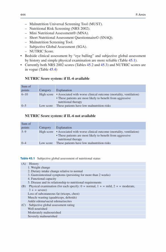
444
– Malnutrition Universal Screening Tool (MUST). – Nutritional Risk Screening (NRS 2002). – Mini Nutritional Assessment® (MNA). – Short Nutritional Assessment Questionnaire© (SNAQ). – Malnutrition Screening Tool. – Subjective Global Assessment (SGA). – NUTRIC Score.
• Bedside clinical assessment by “eye balling” and subjective global assessment by history and simple physical examination are more reliable (Table 45.1).
• Currently both NRS 2002 scores (Tables 45.2 and 45.3) and NUTRIC scores are in vogue (Table 45.4)
NUTRIC Score system: if IL-6 available
Sum of points Category Explanation6–10 High score • Associated with worse clinical outcome (mortality, ventilation)
• These patients are most likely to benefit from aggressive nutritional therapy
0–5 Low score These patients have low malnutrition risks
NUTRIC Score system: if IL-6 not available
Sum of points Category Explanation5–9 High score • Associated with worse clinical outcome (mortality, ventilation)
• These patients are most likely to benefit from aggressive nutritional therapy
0–4 Low score These patients have low malnutrition risks
Table 45.1 Subjective global assessment of nutritional status
(A) History 1. Weight change 2. Dietary intake change relative to normal 3. Gastrointestinal symptoms (persisting for more than 2 weeks) 4. Functional capacity 5. Disease and its relationship to nutritional requirements(B) Physical examination (for each specify: 0 = normal, 1 + = mild, 2 + = moderate,
3 + = severe) Loss of subcutaneous fat (triceps, chest) Muscle wasting (quadriceps, deltoids) Ankle edema/sacral edema/ascites(C) Subjective global assessment rating Well nourished Moderately malnourished Severely malnourished
P. Amin
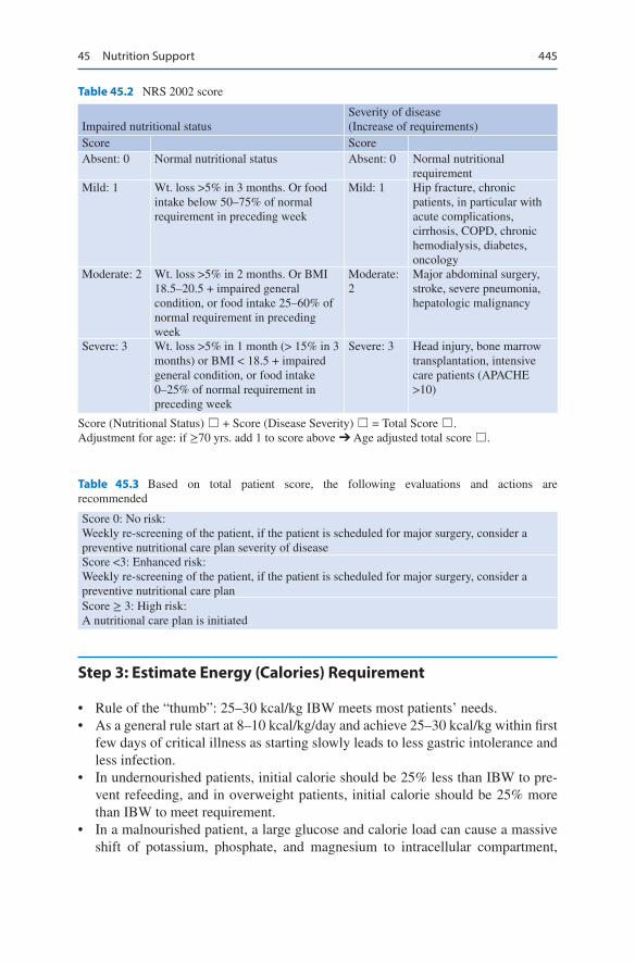
445
Step 3: Estimate Energy (Calories) Requirement
• Rule of the “thumb”: 25–30 kcal/kg IBW meets most patients’ needs.• As a general rule start at 8–10 kcal/kg/day and achieve 25–30 kcal/kg within first
few days of critical illness as starting slowly leads to less gastric intolerance and less infection.
• In undernourished patients, initial calorie should be 25% less than IBW to pre-vent refeeding, and in overweight patients, initial calorie should be 25% more than IBW to meet requirement.
• In a malnourished patient, a large glucose and calorie load can cause a massive shift of potassium, phosphate, and magnesium to intracellular compartment,
Table 45.2 NRS 2002 score
Impaired nutritional statusSeverity of disease(Increase of requirements)
Score ScoreAbsent: 0 Normal nutritional status Absent: 0 Normal nutritional
requirementMild: 1 Wt. loss >5% in 3 months. Or food
intake below 50–75% of normal requirement in preceding week
Mild: 1 Hip fracture, chronic patients, in particular with acute complications, cirrhosis, COPD, chronic hemodialysis, diabetes, oncology
Moderate: 2 Wt. loss >5% in 2 months. Or BMI 18.5–20.5 + impaired general condition, or food intake 25–60% of normal requirement in preceding week
Moderate: 2
Major abdominal surgery, stroke, severe pneumonia, hepatologic malignancy
Severe: 3 Wt. loss >5% in 1 month (> 15% in 3 months) or BMI < 18.5 + impaired general condition, or food intake 0–25% of normal requirement in preceding week
Severe: 3 Head injury, bone marrow transplantation, intensive care patients (APACHE >10)
Score (Nutritional Status) ☐ + Score (Disease Severity) ☐ = Total Score ☐.Adjustment for age: if ≥70 yrs. add 1 to score above ➔ Age adjusted total score ☐.
Table 45.3 Based on total patient score, the following evaluations and actions are recommended
Score 0: No risk:Weekly re-screening of the patient, if the patient is scheduled for major surgery, consider a preventive nutritional care plan severity of diseaseScore <3: Enhanced risk:Weekly re-screening of the patient, if the patient is scheduled for major surgery, consider a preventive nutritional care planScore ≥ 3: High risk:A nutritional care plan is initiated
45 Nutrition Support
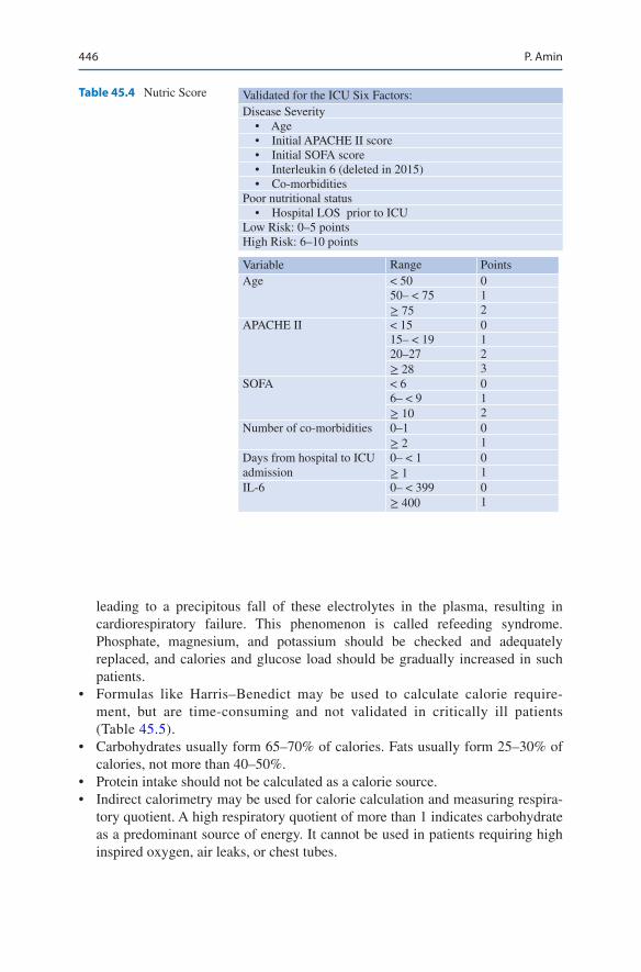
446
leading to a precipitous fall of these electrolytes in the plasma, resulting in cardiorespiratory failure. This phenomenon is called refeeding syndrome. Phosphate, magnesium, and potassium should be checked and adequately replaced, and calories and glucose load should be gradually increased in such patients.
• Formulas like Harris–Benedict may be used to calculate calorie require-ment, but are time-consuming and not validated in critically ill patients (Table 45.5).
• Carbohydrates usually form 65–70% of calories. Fats usually form 25–30% of calories, not more than 40–50%.
• Protein intake should not be calculated as a calorie source.• Indirect calorimetry may be used for calorie calculation and measuring respira-
tory quotient. A high respiratory quotient of more than 1 indicates carbohydrate as a predominant source of energy. It cannot be used in patients requiring high inspired oxygen, air leaks, or chest tubes.
Table 45.4 Nutric Score Validated for the ICU Six Factors:Disease Severity • Age • Initial APACHE II score • Initial SOFA score • Interleukin 6 (deleted in 2015) • Co-morbiditiesPoor nutritional status • Hospital LOS prior to ICULow Risk: 0–5 pointsHigh Risk: 6–10 points
Variable Range PointsAge < 50 0
50– < 75 1≥ 75 2
APACHE II < 15 015– < 19 120–27 2≥ 28 3
SOFA < 6 06– < 9 1≥ 10 2
Number of co-morbidities 0–1 0≥ 2 1
Days from hospital to ICU admission
0– < 1 0≥ 1 1
IL-6 0– < 399 0≥ 400 1
P. Amin

447
Indirect Calorimetry
• Calculation of Heat production by measuring Inspired Oxygen consumption and expired CO2 production.
• The goal is to calculate Resting Energy Expenditure (REE) and Respiratory Quotient (RQ).
• Some of the modern ventilators display Respiratory Quotient.• Calculate REE by using Abbreviated Weir Equation.
• REE (Kcals/day) = [(VO2 X 3.94) + (VCO2 X 1.11)] X 1440 min/day• Measuring Respiratory Quotient (RQ)
RQ =( )( )
VCO CO production
VO O consumption
2 2
2 2
• Energy is vital: Too much or too little is unsafe.• Energy matters for protein efficacy.• Measure Energy Expenditure whenever possible.
Step 4: Estimate Protein (Nitrogen) Requirement
• Rule of the “thumb”: 1.5–2 g of protein/kg IBW meets most patients’ needs (Table 45.6). – 6.25 g of protein is equal to 1 g of nitrogen. – Non Protein Calorie (NPC) – nitrogen ratio = 150 cal (NPC): 1 g nitrogen.
• Nitrogen requirement may also be assessed by calculating nitrogen balance: – Nitrogen balance = nitrogen intake – nitrogen output.
Table 45.5 Harris–Benedict equation with Long’s modification
Harris–Benedict formula for women Basal metabolic rate (BMR) = 655 + (9.6 × weight in kg) + (1.8 × height in cm)—(4.7 × age in years)
Harris–Benedict formula for men BMR = 66 + (13.7 × weight in kg) + (5 × height in cm)—(6.8 × age in years)
Actual energy needs = BMR × AF × IFActivity factor (AF) Use Injury factor
(IF)Use
Confined to bed 1.2 Minor surgery 1.2Out of bed 1.3 Skeletal trauma 1.3
Major sepsis 1.6
Table 45.6 Protein requirements
No stress 0.7–0.8 g/kg/dayMild stress 0.8–1.0 g/kg/dayModerate stress 1.0–1.5 g/kg/daySevere stress 1.5–2.0 g/kg/day
45 Nutrition Support

448
– Nitrogen intake = protein intake/6.25. – Nitrogen output = 24-h urinary urea nitrogen + 4 (nonurinary nitrogen). – This should be done once weekly in all severely ill patients with a normal
renal function. Try to achieve at least equal nitrogen balance.
Step 5: Supplement Micronutrients
• Vitamins and the trace of elements should be added as per recommended daily allowance, which are present in formula feed.
Step 6: Estimate Fluid and Electrolyte Requirement
• Rule of the “thumb”: 1 mL/cal is the minimum requirement of fluid to deliver isocaloric feed.
• Electrolyte should be tailored to individual patients’ requirement.
Step 7: Select Route of Delivering Nutrition
• Whenever the gut is working, use it. Advantages of enteral feed are as follows: – Preservation of the integrity and function of the gastrointestinal tract. – Maintenance of splanchnic blood flow. – Provision of nutrition to the enterocytes. – Fewer infectious and metabolic complications. – Ease of administration, lower costs, and avoiding the disadvantages of total
parenteral nutrition (TPN).• Enteral feeding may be given through nasogastric, nasojejunal, gastrostomy, or
jejunostomy tubes. There is no significant difference in the efficacy of postpylo-ric or jejunal versus gastric feeding in critically ill patients.
• Intragastric feeding should be the first choice because it is easy and relatively safe, and the majority of patients tolerate it: – The head of the bed should be elevated to 30–45°. – Give continuous rather than bolus feedings; start with 25 cc intragastrically
every hour to a target rate of 50–75 cc/h, in most adults preferably through a feeding pump.
– Check residual 2 h after initiating feeding and every 8 h thereafter. Though routine checking for residual volume is discouraged nowadays in asymptom-atic patient but only performed for indications like abdominal distension, abdominal pain, vomiting etc.
– IV administration of metoclopramide should be considered in patients with intolerance to enteral feeding, for example, with high gastric residuals (>300 in 4 h or more than one-third of enteral feed being aspirated).
P. Amin
449
• Postpyloric or jejunal feed can be tried if the patient has high gastric residue despite prokinetic and correction of electrolytes or in patients with high risk of aspiration and unprotected airway.
Step 8: Select the Type of Enteral Feed
• Blenderized diets: It may not be complete and balanced. Nutritive value is diffi-cult to estimate, and highly viscous solution needs a large-bore nasogastric tube. Large particles may block the feeding tubes, and nutrients are not predigested. Bacterial overgrowth is a distinct possibility.
• Polymeric diets: These contain nitrogen as whole protein and are balanced and compete. The carbohydrate source is partially hydrolyzed starch, and the fat con-tains long-chain triglycerides. Their fiber content is very variable.
• Predigested diets: These feeds contain nitrogen either as short peptides or, in the case of elemental diets, as free amino acids. Carbohydrate provides much of the energy. The rest of the calorie proportion is provided as long-chain triglycerides and medium-chain triglycerides. The aim of “predigested diets” is to improve nutrient absorption in the presence of significant malabsorption.
• Disease-specific diets: Concentrated feed formula for patients who requires vol-ume restriction. Patients with respiratory failure are often given feeds with a low carbohydrate-to-fat ratio to minimize carbon dioxide production. Renal patients often require modified protein, electrolyte, and volume feeds. Liver patients may need low sodium, low volume feeds. There is no good evidence that patients with hepatic encephalopathy should have low protein intakes, and the evidence for the benefit of feeds rich in branched-chain amino acids is weak.
• Immunonutrition: In general immunonutrition supplement has not been found to be useful in critically ill patient Glutamine may be added to a standard enteral formula in burned patients and trauma patients. The immune-modulating for-mula enriched with arginine, nucleotides, and ω-3 fatty acids may be given to selective upper gastrointestinal surgical patients but can be harmful to patients with severe sepsis and septic shock. Fibres should be avoided in patients on vasopressors.
Step 9: Look for Tolerance to Enteral Feed
• Signs of intolerance include bloating, nausea, cramps, abdominal distention, or diarrhea, but these signs are nonspecific.
• Intragastric feeds should not be stopped for residuals of less than 200 ml.• Significant distention or complaints of cramping by the patient should warrant
slowing or discontinuing the feeds.• In case of diarrhea, follow a standard protocol (see Fig. 45.1).
45 Nutrition Support

450
Step 10: Select Candidates for Parenteral Nutrition
• Parenteral nutrition is indicated for some critically ill patients in the following situations:
• Signs of abnormal gut function (complete intestinal obstruction, mesenteric isch-emia, bowel fistula).
• Patients cannot consume adequate amounts of nutrients by enteral feeding.• They are not expected to be able to eat orally by 3–7 days and are malnourished.
Yes
Is diarrhea present?
Is stool clinically significant?
Are medications responsible?
Is the patient on antibiotics?
Consider fiber or elemental diet
Diarrhea resolved
Decrease feed rates tilltolerance is achieved
NoContinue same feeds
Change medications
Continue same feeds
Advance to goal rate astolerance improves
Check for C.difficile.
No
No
No
Yes
Yes
Yes
Yes
Fig. 45.1 Resolving tube feeding-associated diarrhea
P. Amin
451
Step 11: Select the Route of Total Parenteral Nutrition (TPN)
TPN can be provided by peripheral or central route:
• Peripheral (partial or supplemental) parenteral nutrition: – Minimizes cost and complications of the central catheter. – Can provide supplemental calories and protein not met by enteral route. – Phlebitis, requires frequent site rotations and fluid overload.
• TPN by central route: – Preferably a multilumen catheter with a single lumen dedicated to TPN. – TPN being delivered as an “all-in-one” solution as a commercially available
standard solution, or made-to-order solution from a pharmacist-based mixing center.
– Supplementing parenteral glutamine to the TPN formula is known to reduce infectious complication in elective surgical patient, length of ICU stay, and mortality. However, glutamine in patients with septic shock, renal failure and hepatic failure is clearly more harmful and is contraindicated.
– Routinely supplement with trace elements and multivitamins.
Step 12: Monitor Patients on Parenteral Nutrition
• This consists of clinical examination, fluid balance, catheter care, and blood glu-cose monitoring.
• Renal, liver, and lipid profiles should be measured weekly.
Step 13: Look for Complications of TPN
• Mechanical complications: – Related to vascular access technique. – Venous thrombosis. – Catheter occlusion.
• Metabolic complications: – Hyper-/hypoglycemia. – Electrolyte abnormalities. – Acid–base disorders. – Hyperlipidemia. – Steatosis.
• Infectious complications.
45 Nutrition Support

452
Step 14: Prevent Overfeeding and Underfeeding (Table 45.7)
It is important to feed appropriately. Effect of underfeeding and overfeeding are described in Table 45.7 and Fig. 45.2.
Table 45.7 Effects of overfeeding and underfeeding
Insufficient energy intake Excessive energy intakeEarly signs Hypoglycemia
HypothermiaHyperglycemiaHyperlipidemia (triglycerides) Hypercapnia
Delayed signs
Infectious complicationsImpaired immunity impaired healingloss of lean and fat body massImpaired muscle function
Infectious complicationsImpaired immunity liver steatosis increased fat mass
Refeeding
Glucose ↑
Insulin ↑
Transcellular shift ofglucose &
electrolytes
Refeeding syndrome
Lipogenesis ↑
Thiamine ↓
Osmolarity ↑
Na+ ↑, hypervolaemia
K+↓, Mg2+↓, Ca2+↓
Steatohepatitis
Wernicke-Korsakoff syndrome, metabolic acidosis
Hyperglycaemic hyperosmolar coma, neutrophil function↓infection risk ↑
Congestive heart failure, oedema↑
PO4↓, ATP ↓
Spasms, tetany, arrhythmia
RBC: ATP & 2, 3 DPG↓, anaemia, haemolysis, O2delivery↓, ischaemia
Muscle weakness,myalgia,rhabdomyolysis,diaphragm, weakness,dyspnoea
Acute kidney injury
Respiratory system: hyperventilation, respiratoryalkalosis
Gastrointestinal system: anorexia, constipation
CNS: weakness, paresthesia, tremors, ataxia,delirium, areflexic paralysis, coma, death
Fig. 45.2 Refeeding syndrome: pathophysiology and clinical manifestations
P. Amin

453
Suggested Reading
Dhaliwal R, Cahill N, Lemieux M, Heyland DK. The Canadian critical care nutrition guidelines in 2013: an update on current recommendations and implementation strategies. Nutrit Clin Pract. 2014;29:29–43.
Elke G, van Zanten AR, Lemieux M, McCall M, Jeejeebhoy KN, Kott M, Jiang X, Day AG, Heyland DK. Enteral versus parenteral nutrition in critically ill patients: an updated systematic review and meta-analysis of randomized controlled trials. Crit Care. 2016;20(1):117. The use of EN as compared to PN has no effect on overall mortality but decreases infectious compli-cations and ICU LOS. This may be explained by the benefit of reduced macronutrient intake rather than the enteral route itself incritically ill patient.
Kreymann KG, Berger MM, Deutz NE, et al. ESPEN guidelines on enteral nutrition: intensive care. Clin Nutr. 2006;25:210–23.
Rosa M, Heyland DK, Fernandes D, Rabito EI, Oliveira ML, Marcadenti A. Translation and adap-tation of the NUTRIC score to identify critically ill patients who benefit the most from nutrition therapy. Clin Nutr ESPEN. 2016;14:31–6. The NUTRIC score has been successfully trans-lated into Portuguese and the prevalence of nutritionally-high risk patients is around 50% in Brazilian ICUs.
Singer P, Blasér AR, Berger MM, et al. ESPEN guideline on clinical nutrition in the intensive care unit. Clin Nutrit. 2018;S0261-5614(18):32432-4. Guidelines for clinical nutrition.
Society of Critical Care Medicine (SCCM) and American Society for Parenteral and Enteral Nutrition (A.S.P.E.N.), SA MC, Taylor BE, Martindale RG, Warren MM, Johnson DR, Braunschweig C, MS MC, Davanos E, Rice TW, Cresci GA, Gervasio JM, Sacks GS, Roberts PR, Compher C, Society of Critical Care Medicine, American Society for Parenteral and Enteral Nutrition. Guidelines for the provision and assessment of nutrition support therapy in the adult critically ill patient. JPEN J Parenter Enteral Nutr. 2016;40(2):159–211. Current guidelines for nutrition support in critically ill.
45 Nutrition Support

Part V
Renal System
457© Springer Nature Singapore Pte Ltd. 2020R. Chawla, S. Todi (eds.), ICU Protocols, https://doi.org/10.1007/978-981-15-0898-1_46
S. Todi (*) Department of Critical Care & Emergency Medicines, A.M.R.I. Hospital, Kolkata, India
A. Majumdar Department of Nephrology, A.M.R.I. Hospitals, Kolkata, West Bengal, India
46Acute Kidney Injury
Subhash Todi and Arghya Majumdar
The term acute kidney injury has replaced the earlier term acute renal failure to emphasize occurrence of small decrement in kidney function can occur earlier than overt kidney failure and may be amenable to therapy reversible. A systematic approach to oliguria in the critically ill patient is warranted, due to the multifactorial etiology of this problem, potential to deteriorate to anuria, and need for renal replacement therapy (RRT). Timely diagnosis and prompt intervention may decrease morbidity and mortality from this potentially preventable problem. Mortality rate in patients with acute kidney injury (AKI) in the ICU is in excess of 50%.
Step 1: Resuscitate
• Optimizing airway, oxygenation and hemodynamic stability is of prime impor-tance in preventing acute kidney injury (refer to Chap. 23, Vol. 2).
A 30-year-old hypertensive male victim of a road traffic accident was admitted with pelvic fracture. He had been on angiotensin receptor blockers (ARB). An external fixator was applied the same day. The following day he suffered a mas-sive pulmonary embolism requiring thrombolysis. He started becoming oliguric. On the fourth day, he developed retroperitoneal hemorrhage with an intra-abdominal pressure of 22 mmHg, as a complication of anticoagulation. He required surgical drainage. After return from the operation theatre he became anuric and his renal parameters deteriorated. He had developed Acute Kidney Injury and had to be commenced on renal replacement therapy.
458
Step 2: Differentiate Urinary Retention from Oliguria
• By definition, oliguria is less than 0.5 mL/kg/h urine output for at least 2 h.• Perform supra-pubic percussion for bladder fullness in all cases of low urine
output to exclude retention of urine.• Sudden drop of urine output, no urine or fluctuating levels of urine output in a
catheterized patient who is otherwise stable, may indicate a partial or complete catheter block with clot or debris or pericatheter leak. Ascertain this by physical examination, bladder wash, or replacing the catheter.
• Bedside ultrasonography differentiates retention from oliguria and at the same time confirms the catheter position.
Step 3: Ascertain the Cause of Oliguria (Table 46.1)
• First consider whether it is a pre-renal, renal, or post-renal cause.• Proper history should be taken including fluid loss, drug history (non-steroidal
anti-inflammatory drugs [NSAIDs]), exposure to contrast, urinary symptoms, and fever; perform physical examination for evidence of hypovolemia, abdomi-nal distension, focus of infection and skin rash.
• Pre-renal factors are by far the most frequent. Volume deficit is the commonest cause.
• For renal factors, pay attention to the nephrotoxic potential of antibiotics, anal-gesic, or radiocontrast agent.
• Post-renal factors like urethral injury and bladder outflow tract obstruction are common causes.
Step 4: Send Biochemical Investigations to Ascertain Severity and Cause of Acute Kidney Injury (AKI)
• Serum chemistry including sodium; potassium; creatinine; blood urea nitrogen (BUN); calcium; magnesium; phosphate; uric acid; creatine phosphokinase (if rhabdomyolysis is suspected); total protein, albumin, globulin, and unconjugated bilirubin (to exclude hemolysis); and lactate dehydrogenase (LDH) should be checked.
• If clinically indicated, antinuclear factor (ANA), antiglomerular basement mem-brane antibody, antineutrophil cytoplasmic antibodies (ANCA), complement levels (C3, C4), cryoglobulin, and hepatitis B and C serology test may be performed.
• Serum and urine protein electrophoresis should be performed in patients with bone pain, hypercalcemia, and hyperglobulinemia where paraproteinemia is suspected.
S. Todi and A. Majumdar
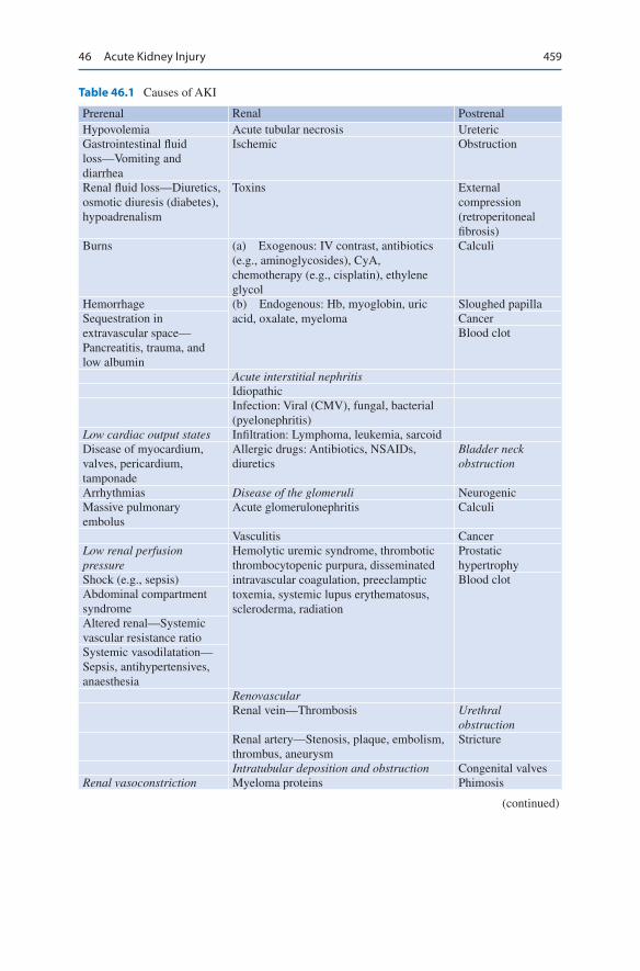
459
Table 46.1 Causes of AKI
Prerenal Renal PostrenalHypovolemia Acute tubular necrosis UretericGastrointestinal fluid loss—Vomiting and diarrhea
Ischemic Obstruction
Renal fluid loss—Diuretics, osmotic diuresis (diabetes), hypoadrenalism
Toxins External compression (retroperitoneal fibrosis)
Burns (a) Exogenous: IV contrast, antibiotics (e.g., aminoglycosides), CyA, chemotherapy (e.g., cisplatin), ethylene glycol
Calculi
Hemorrhage (b) Endogenous: Hb, myoglobin, uric acid, oxalate, myeloma
Sloughed papillaSequestration in extravascular space—Pancreatitis, trauma, and low albumin
CancerBlood clot
Acute interstitial nephritisIdiopathicInfection: Viral (CMV), fungal, bacterial (pyelonephritis)
Low cardiac output states Infiltration: Lymphoma, leukemia, sarcoidDisease of myocardium, valves, pericardium, tamponade
Allergic drugs: Antibiotics, NSAIDs, diuretics
Bladder neck obstruction
Arrhythmias Disease of the glomeruli NeurogenicMassive pulmonary embolus
Acute glomerulonephritis Calculi
Vasculitis CancerLow renal perfusion pressure
Hemolytic uremic syndrome, thrombotic thrombocytopenic purpura, disseminated intravascular coagulation, preeclamptic toxemia, systemic lupus erythematosus, scleroderma, radiation
Prostatic hypertrophy
Shock (e.g., sepsis) Blood clotAbdominal compartment syndromeAltered renal—Systemic vascular resistance ratioSystemic vasodilatation—Sepsis, antihypertensives, anaesthesia
RenovascularRenal vein—Thrombosis Urethral
obstructionRenal artery—Stenosis, plaque, embolism, thrombus, aneurysm
Stricture
Intratubular deposition and obstruction Congenital valvesRenal vasoconstriction Myeloma proteins Phimosis
(continued)
46 Acute Kidney Injury

460
Step 5: Examine the Urine (Table 46.2)
• Limitations of using these parameters for differentiating pre-renal from renal injury: use of diuretics, postcontrast i.v. contrast, CKD, elderly, acute glomeru-lonephritis, acute interstitial nephritis, hyperglycemia, and hepato-renal syndrome.
• This can yield vital clues with regard to the type of renal failure. Microscopic examination of the urinary sediment should not be neglected.
• In pre-renal failure, urine examination is usually bland, and no casts or cells will be obvious. Hyaline casts may be the only finding.
• In intrinsic renal disease, one may find RBC casts and dysmorphic RBCs indicat-ing glomerular hematuria, eosinophil casts in allergic interstitial nephritis, muddy brown epithelial casts in acute tubular damage, or coarse granular or broad casts, which might indicate background chronic kidney disease (CKD).
• Uric acid or calcium oxalate crystals may highlight a background metabolic problem or ethylene glycol poisoning.
• RBC and WBC casts are fragile, and their absence does not exclude underlying parenchymal disease.
Table 46.1 (continued)
Prerenal Renal PostrenalHypercalcemia Uric acidNorepinephrine, vasopressor agents
Oxalate
CyA, tacrolimus Acyclovir crystalsCirrhosis with hepatorenal syndrome
Methotrexate
Hyperviscosity SulfonamidesMultiple myeloma Renal allograft rejectionMacroglobulinemia
Table 46.2 Urinary biochemistry
Prerenal RenalUrine osmolality >500 mosmols/kg <350 mosmols/
kgUrine sodium <10 mEq/L >20 mEq/LUrine/plasma osmolality ratio >1.5 <1.2Urine/plasma urea nitrogen ratio >8 <3Urine/plasma creatinine ratio >40 <20Plasma BUN/creatinine ratio >20 <10Urine specific gravity >1.020 ∼1.010Fractional excretion of sodium (FeNa)% <1 >1Urine/plasma Na divided by urine/plasma creatinine × 100
S. Todi and A. Majumdar
461
• Dipstick test is vital. It might reveal proteinuria, hematuria, glycosuria, ketonu-ria, bilirubin, or urobilinogen. A positive dipstick for hemoglobin in the absence of RBCs in the urine sediment may suggest hemolysis or myoglobinuria (which can be confirmed by specific assays).
• A positive nitrite or leucocyte esterase test may indicate infection. High specific gravity and ascorbic acid may interfere with the test.
• Urine biochemistry test should be performed to differentiate pre-renal from renal failure (Table 46.2).
Step 6: Monitor the Patient Carefully
• Ensure continuous monitoring of urine output, by placing an indwelling urinary catheter.
• Continuous monitoring of hemodynamic parameters (CVP and arterial line) is also mandatory.
• Intra-abdominal pressure (IAP) monitoring is very important, especially when large volumes of fluids or blood products are infused, as they may third space into the peritoneal cavity if there is capillary leak due to systemic inflammatory response syndrome (SIRS) after trauma, sepsis, or abdominal surgery.
• IAP of less than 12 mmHg indicates normal condition, 12–20 mmHg indicates intra-abdominal hypertension, and more than 20 mmHg with organ dysfunction indicates abdominal compartment syndrome.
Step 7: Perform Renal Imaging
• Abdominal ultrasound or non-contrast CT scan would be useful to diagnose renal and post-renal oliguria.
• An ultrasonography may reveal echogenic or small kidneys with loss of cortico-medullary differentiation, which might indicate a background of chronic kidney disease. However, in some conditions such as diabetes, amyloidosis, and multi-ple myeloma, kidney size may be normal even with chronic disease. A discrep-ancy in kidney size (>2 cm) may suggest unilateral renal artery stenosis. Color Doppler flow can be used to assess renal perfusion and rule out thrombosis. and helps in calculating renal resistive index Multiple bilateral cortical cysts may indicate polycystic kidney disease.
• The presence of hydronephrosis and/or hydroureter is suggestive of a post-renal cause. However, one must remember that a dehydrated patient may not exhibit significant hydronephrosis. Post-voidal residual urine of more than 100 mL is suggestive of bladder outlet obstruction.
• Volume status can be assessed by checking the diameter and collapsibility of the infe-rior vena cava, passive leg raising or other measures of dynamic volume assessment.
46 Acute Kidney Injury
462
• Radiologic examination is useful for detecting renal stones and pelvic fractures. However, fecoliths may masquerade as stones, and without proper bowel prepa-ration, it is difficult to delineate stones.
• Occasionally an intravenous urogram, contrast CT (if renal function is normal), MRI, or retrograde/antegrade (percutaneous) contrast studies may be necessary to delineate the site or nature of obstruction or injury.
• Nuclear scans (DMSA, DTPA, MAG3) may be used to assess renal function, perfusion, or obstruction.
Step 8: Be Wary of Sepsis
• Always suspect sepsis, whether as the primary cause or as an intercurrent com-plication. In such instances, the promptness and appropriateness of antibiotics can save lives.
• In case of post-renal oliguria and urosepsis, urgent urological intervention such as urinary drainage using ureteric catheterization, DJ stents, or percutaneous nephrostomy may be required, as source control is vital. Peri-nephric collections of pus or blood may need drainage too (Fig. 46.1).
Step 9: Maintain Renal Perfusion Pressure
• Maintain mean arterial pressure (MAP) of more than 65 mmHg by adequate volume loading and with vasopressors if necessary. MAP may have to be kept higher if the patient is hypertensive or has a high IAP.
• Individual titration is necessary in this regard. In cases of high IAP, renal perfu-sion pressure is equal to MAP minus two times IAP, and a higher MAP is desir-able. Dose of vasopressors should be kept to minimum, which is necessary for maintaining an adequate MAP, and every attempt should be made to reduce dose of vasopressors.
• Invasive hemodynamic monitoring (arterial line and central line) is usually needed in these cases.
• There is no role of renal dose of dopamine to increase renal perfusion.• Dobutamine should be used to optimize cardiac output, provided there is no
tachycardia or arrhythmia (refer to Chap. 18, Vol. 1).• Medical (e.g., diuresis, drainage of ascites) and surgical measures (e.g., opening
up of abdominal sutures) should be taken up, where possible, to reduce intra- abdominal hypertension.
Step 10: Use Diuretics Judiciously
• Weigh the potential benefits versus risks of a trial of intravenous diuretics. The role of diuretic therapy has been questioned as it does not affect renal outcome. However, in non-oliguric renal failure, management of the patient becomes easier.
S. Todi and A. Majumdar
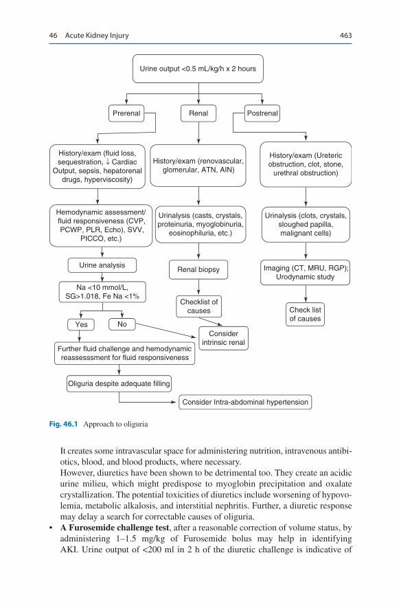
463
It creates some intravascular space for administering nutrition, intravenous antibi-otics, blood, and blood products, where necessary.However, diuretics have been shown to be detrimental too. They create an acidic urine milieu, which might predispose to myoglobin precipitation and oxalate crystallization. The potential toxicities of diuretics include worsening of hypovo-lemia, metabolic alkalosis, and interstitial nephritis. Further, a diuretic response may delay a search for correctable causes of oliguria.
• A Furosemide challenge test, after a reasonable correction of volume status, by administering 1–1.5 mg/kg of Furosemide bolus may help in identifying AKI. Urine output of <200 ml in 2 h of the diuretic challenge is indicative of
Urine output <0.5 mL/kg/h x 2 hours
Prerenal Renal
History/exam (renovascular,glomerular, ATN, AIN)
Postrenal
History/exam (Uretericobstruction, clot, stone,
urethral obstruction)
Urinalysis (clots, crystals,sloughed papilla,malignant cells)
Hemodynamic assessment/fluid responsiveness (CVP,PCWP, PLR, Echo), SVV,
PICCO, etc.)
Urinalysis (casts, crystals,proteinuria, myoglobinuria,
eosinophiluria, etc.)
Urine analysisRenal biopsy
Checklist ofcauses
Considerintrinsic renal
Yes No
Further fluid challenge and hemodynamicreassesssment for fluid responsiveness
Oliguria despite adequate filling
Consider Intra-abdominal hypertension
Na <10 mmol/L,SG>1.018, Fe Na <1%
Check listof causes
Imaging (CT, MRU, RGP);Urodynamic study
History/exam (fluid loss,sequestration, ↓ Cardiac
Output, sepsis, hepatorenaldrugs, hyperviscosity)
Fig. 46.1 Approach to oliguria
46 Acute Kidney Injury
464
need for RRT.. The sensitivity and specificity of this test for need of RRT is around 80%. This test should not be performed in the presence of clinical hypo-volemia and is not a screening tool to identify AKI. Its main role is to ascertain severity of tubular dysfunction in AKI and need for RRT. This should be used in conjunction with clinical parameters and renal biomarkers where available.
Step 11: Correct Metabolic Abnormalities
• Look for and manage metabolic abnormalities, which may be a result of renal impairment such as hyperkalemia, hyper/hyponatremia, and hypocalcemia.
• Look for causative factors such as hypercalcemia or hyperuricemia.• Frequent monitoring of electrolytes may be necessary.• The usual urea–creatinine ratio is 10:1. An unusually high urea–creatinine ratio
is suggestive of volume depletion, gastrointestinal bleeding, catabolic state, cor-ticosteroid use or high protein feed.
• A high creatinine–urea ratio is associated with rhabdomyolysis, diabetic ketoaci-dosis or may indicate chronic kidney disease (CKD).
• If rhabdomyolysis is suspected, the maintenance of urinary pH to more than 7 by systemic alkalinization is indicated.
Step 12: Avoid Any Potentially Nephrotoxic Agent
• In high-risk cases, along with hydration with 0.9% saline, N-acetyl cysteine at a dose of 1200 mg twice per day may be administered for 3 days from a day prior to elective radio-contrast imaging study.
• Low osmolality or preferably iso-osmolar contrast should be used.• Avoid nephrotoxic antibiotics. Aminoglycosides, if used, should be dosed once
daily.• Lipid formulations of amphotericin are preferable.• Intravenous use of voriconazole may cause nephrotoxicity due to β-cyclodextrin.• Intravenous acyclovir may cause crystal nephropathy.• High-dose mannitol may lead to osmotic nephropathy.• NSAIDs should be avoided.
Step 13: Strive to Detect Acute Kidney Injury (AKI) As Early as Possible (Table 46.3)
• RIFLE, AKIN and KDIGO are the three classification system for AKI.• All these classifications are based primarily on rise in serum creatinine and/or
decrease in urine output over time.• In all three classification if there is a discordance between serum creatinine or
urine output criteria, the highest (most severe) criteria is chosen.
S. Todi and A. Majumdar
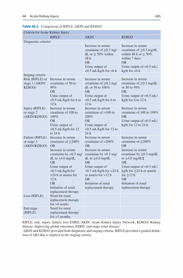
465
Table 46.3 Comparison of RIFLE, AKIN and KDIGO
Criteria for Acute Kidney InjuryRIFLE AKIN KDIGO
Diagnostic criteriaa
Increase in serum creatinine of ≥0.3 mg/dL or ≥ 50% within 48 hORUrine output of <0.5 mL/kg/h for >6 h
Increase in serum creatinine of ≥0.3 mg/dL within 48 h or ≥ 50% within 7 daysORUrine output of <0.5 mL/kg/h for >6 h
Staging criteriaRisk (RIFLE) or stage 1 (AKIN/KDIGO)
Increase in serum creatinine of 50 to 99%ORUrine output of <0.5 mL/kg/h for 6 to 12 h
Increase in serum creatinine of ≥0.3 mg/dL or 50 to 100%ORUrine output of <0.5 mL/kg/h for 6 to 12 h
Increase in serum creatinine of ≥0.3 mg/dL or 50 to 99%ORUrine output of <0.5 mL/kg/h for 6 to 12 h
Injury (RIFLE) or stage 2 (AKIN/KDIGO)
Increase in serum creatinine of 100 to 199%ORUrine output of <0.5 mL/kg/h for 12 to 24 h
Increase in serum creatinine of >100 to 200%ORUrine output of <0.5 mL/kg/h for 12 to 24 h
Increase in serum creatinine of 100 to 199%ORUrine output of <0.5 mL/kg/h for 12 to 24 h
Failure (RIFLE) or stage 3 (AKIN/KDIGO)
Increase in serum creatinine of ≥200%ORIncrease in serum creatinine by >0.5 mg/dL to >4.0 mg/dLORUrine output of <0.3 mL/kg/h for >24 h or anuria for 12 hORInitiation of renal replacement therapy
Increase in serum creatinine of >200%ORIncrease in serum creatinine by >0.5 mg/dL to ≥4.0 mg/dLORUrine output of <0.3 mL/kg/h for >24 h or anuria for >12 hORInitiation of renal replacement therapy
Increase in serum creatinine of ≥200%ORIncrease in serum creatinine by ≥0.3 mg/dL to ≥4.0 mg/dL¶ORUrine output of <0.3 mL/kg/h for ≥24 h or anuria for ≥12 hORInitiation of renal replacement therapy
Loss (RIFLE) Need for renal replacement therapy for >4 weeks
End stage (RIFLE)
Need for renal replacement therapy for >3 months
RIFLE: risk; injury, failure, loss ESRD; AKIN: Acute Kidney Injury Network; KDIGO: Kidney disease: Improving global outcomes; ESRD: end-stage renal disease.aAKIN and KDIGO provided both diagnostic and staging criteria. RIFLE provided a graded defini-tion of AKI that is implicit in the staging criteria.
46 Acute Kidney Injury

466
• KDIGO criteria differs from RIFLE as it does not consider change in GFR for staging
• The Kidney Disease: Improving Global Outcomes (KDIGO) AKI Workgroup definition and staging system is the preferred definition: – Increase in serum creatinine by ≥0.3 mg/dL (≥26.5 micromol/L) within 48 h,
or – Increase in serum creatinine to ≥1.5 times baseline, which is known or pre-
sumed to have occurred within the prior 7 days, or – Urine volume < 0.5 mL/kg/h for 6 h.
• Using the KDIGO criteria, AKI is staged as follows: – Stage 1—Increase in serum creatinine to 1.5 to 1.9 times baseline, or increase
in serum creatinine by ≥0.3 mg/dL (≥26.5 micromol/L), or reduction in urine output to <0.5 mL/kg/h for 6 to 12 h.
– Stage 2—Increase in serum creatinine to 2.0 to 2.9 times baseline, or reduc-tion in urine output to <0.5 mL/kg/h for ≥12 h.
– Stage 3—Increase in serum creatinine to 3.0 times baseline, or increase in serum creatinine to ≥4.0 mg/dL(≥353.6 micromol/L), or reduction in urine output to <0.3 mL/kg/h for ≥24 h, or anuria for ≥12 h, or the initiation of renal replacement therapy, or, in patients <18 years, decrease in estimated glomerular filtration rate (eGFR) to <35 mL/min/1.73 m2.
A comparison of RIFLE, AKIN and KDIGO is mentioned in Table 46.3.
• Once a patient is considered to be in failure according to this criterion, the issue of renal replacement therapy arises, and early nephrology consultation is useful.
• Serum and urinary biomarkers like NGAL (neutrophil gelatin-associated lipo-calin) have been found to be useful in early detection of AKI.
Suggested Reading
Acute Dialysis Quality Initiative (ADQI) Consensus Group. ADQI 7: the clinical management of the cardio-renal syndromes: work group statements from the seventh ADQI consensus confer-ence. Nephrol Dial Transpl. 2010;25(7):2077–89. A comprehensive review on this increasingly recognized clinical entity. Many patients with heart failure have underlying renal dysfunction, and similarly, patients with kidney failure are prone to cardiac failure. This has led to the con-cept of cardiorenal syndromes, which can be an acute or chronic cardiorenal syndrome, when cardiac failure causes deterioration in renal function, or acute and/or chronic renicardiac syndrome, when renal dysfunction leads to cardiac failure. These patients have typically been excluded from clinical trials.
Bellomo R. Defining, quantifying, and classifying acute renal failure. Crit Care Clin. 2005;21(2):223–37. A comprehensive review article on the subject.
Bellomo R, Ronco C, Kellum JA, Mehta RL, Palevsky P. Acute dialysis quality initiative work-group. Acute renal failure—definition, outcome measures, animal models, fluid therapy and information technology needs: the second international consensus conference of the acute
S. Todi and A. Majumdar

467
dialysis quality initiative (ADQI) group. Crit Care. 2004;8(4):R204–12. This article enunci-ates the RIFLE criteria, which has given us an early practical way of diagnosing AKI, which is internationally accepted and validated in various countries and scenarios.
Crowley ST, Peixoto AJ. Acute kidney injury in the intensive care unit. Clin Chest Med. 2009;30(1):29–43. This review focuses on the diagnosis and management of AKI.
Kidney Diseases: Improving Global Outcomes (KDIGO) Acute Kidney Injury Working Group. KDIGO clinical practice guideline for acute kidney injury. Kidney Int Suppl. 2012;2:1. KDIGO guidelines for clinical practice in acute kidney injury.
Koyner JL, Davison DL. Furosemide stress test and biomarkers for the prediction of AKI severity. J Am Soc Nephrol. 2015;26(8):2023–31. A Furosemide challenge test, after a reasonable cor-rection of volume status, by administering of Furosemide bolus may help in identifying AKI.
Mehta RL, Kellum JA, Shah SV, et al. Acute kidney injury network: report of an initiative to improve outcomes in acute kidney injury. Crit Care. 2007;11:R31. Copyright @ 2007 BioMed Central Ltd. AKIN initiative to improve outcomes in acute kidney injury.
Moore PK, Hsu RK, Liu KD. Management of acute kidney injury: core curriculum 2018. Am J Kid Ds. 2018;72(1):136–48. This article gives a complete overview of AKI
Websites
www.renalandurologynews.comA comprehensive site for clinical trial summary on AKI.www.ADQI.netHomepage for RIFLE criteria network.www.kidneycare.nhs.ukA site for information on preventive aspects of AKI.
46 Acute Kidney Injury
469© Springer Nature Singapore Pte Ltd. 2020R. Chawla, S. Todi (eds.), ICU Protocols, https://doi.org/10.1007/978-981-15-0898-1_47
S. Prakash (*) · B. Mishra Department of Nephrology and Renal Transplant, B L Kapur Superspeciality Institute, Delhi, India
A. Majumdar Department of Nephrology, A.M.R.I. Hospitals, Kolkata, West Bengal, India
47Renal Replacement Therapy
Sunil Prakash, Arghya Majumdar, and Bhanu Mishra
Acute kidney injury is a common occurrence in the ICU and often requires renal replacement therapy (RRT). ICU physicians should be aware of the different modal-ities of renal replacement therapy (RRT) with their advantages and disadvantages.
Step 1: Initiate Resuscitation and Decide on RRT
• Along with resuscitation measures with ventilatory and hemodynamic support, RRT should be considered in patients with acute kidney injury.
• Optimal timing of starting RRT remains controversial, and a joint decision between the nephrologist and the intensivist should be taken.
• The usual indications of commencing RRT are the following (Fig 47.1): – Volume overload/pulmonary edema. – Refractory hyperkalemia (>6.5 mEq/L). – Severe metabolic acidosis (pH <7.1). – Anuria. – Uremic encephalopathy. – Uremic pericarditis.
A 50-year-old diabetic and hypertensive male patient was admitted with acute pan-creatitis requiring ventilatory support. Despite aggressive volume resuscitation, he had a mean arterial pressure (MAP) of 60 mmHg on multiple vasopressors. Echo showed global hypokinesia. His intra-abdominal pressure was 20 mmHg. He was catheterized and had 100 mL of urine output in the past 12 h. Serum urea was 150 mg/dL creatinine was 3.5 mg/dL, and potassium was 6.5 mEq/L.
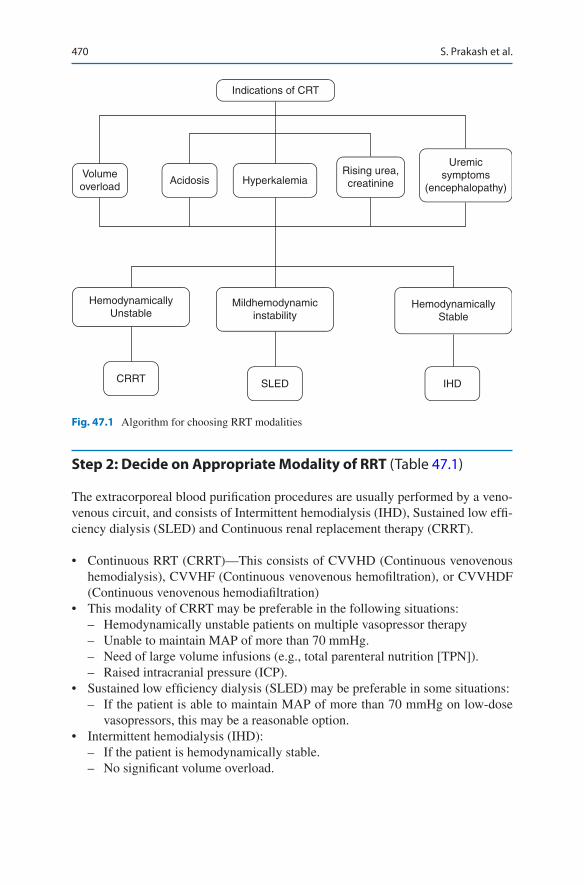
470
Step 2: Decide on Appropriate Modality of RRT (Table 47.1)
The extracorporeal blood purification procedures are usually performed by a veno-venous circuit, and consists of Intermittent hemodialysis (IHD), Sustained low effi-ciency dialysis (SLED) and Continuous renal replacement therapy (CRRT).
• Continuous RRT (CRRT)—This consists of CVVHD (Continuous venovenous hemodialysis), CVVHF (Continuous venovenous hemofiltration), or CVVHDF (Continuous venovenous hemodiafiltration)
• This modality of CRRT may be preferable in the following situations: – Hemodynamically unstable patients on multiple vasopressor therapy – Unable to maintain MAP of more than 70 mmHg. – Need of large volume infusions (e.g., total parenteral nutrition [TPN]). – Raised intracranial pressure (ICP).
• Sustained low efficiency dialysis (SLED) may be preferable in some situations: – If the patient is able to maintain MAP of more than 70 mmHg on low-dose
vasopressors, this may be a reasonable option.• Intermittent hemodialysis (IHD):
– If the patient is hemodynamically stable. – No significant volume overload.
Indications of CRT
Volumeoverload
HemodynamicallyUnstable
Mildhemodynamicinstability
HemodynamicallyStable
CRRT SLED IHD
Acidosis HyperkalemiaRising urea,creatinine
Uremicsymptoms
(encephalopathy)
Fig. 47.1 Algorithm for choosing RRT modalities
S. Prakash et al.

471
• Besides the medical indications, the selection of a particular modality of RRT is based on infrastructure, available resources, affordability, availability of appro-priate fluids, hemofilters, and preference of the physician.
Step 3: Understand Different Modalities of RRT (Table 47.1)
• CRRT - The advantages and disadvantages are described in (Tables 47.2 and 47.3)
CVVHD will probably be more effective than CVVH in the highly catabolic patient with a large solute load. CVVHDF with its convective removal of larger solutes is preferred in the patient with septic shock in whom the removal of
Table 47.1 Different modalities of RRT
Modality Mechanism MethodologyHemodialysis (HD)
Diffusion Here, the solute passively diffuses down its concentration gradient from one fluid compartment (either blood or dialysate) into the other. The dialysate is made to flow in a direction which is opposite to blood flow (countercurrent flow) through the hollow fiber dialyzer at a rate of 1–2 L/h, to maintain a continuous concentration gradient between the two compartments and therefore maximize solute removal. Diffusion-based dialysis mostly removes small molecular weight solutes of less than 1 kD (kilodalton)
Hemofiltration (HF)
Convection Hydrostatic pressure gradient is used to induce the filtration (or convection) of plasma water across the membrane of the hemofilter. The frictional forces between water and solutes (called “solvent drag”) result in the convective transport of small and middle molecular weight solutes (less than 5000 D) in the same direction as water and in the same concentration in the plasma. There is therefore no change in the plasma concentrations of these solutes. Dialysate fluid is not used. The amount of replacement fluid is determined by the net volume removal that is desired. Ultrafiltration rate is usually 20–25 ml/kg/h
Hemodiafiltration (HDF)
Diffusion and convection
This modality offers the maximum solute removal as it combines convection with diffusion for achieving this, as both dialysis and ultrafiltration occurs simultaneously. Ultrafiltration rate is usually 2-8 ml/min
Ultrafiltration (SCUF)
Hydrostatic pressure
Slow continuous removal of fluid alone, by steady hydrostatic pressure. This is used to treat isolated fluid overload, as solute removal is minimal. This modality can safely remove about 8 L of fluid/day
Peritoneal dialysis (PD)
Diffusion, convection, and osmosis
Solute removal is accomplished by diffusion, and most of the ultrafiltration is by osmosis
47 Renal Replacement Therapy
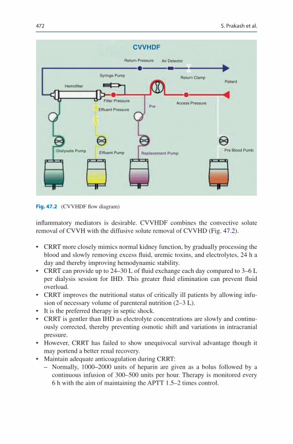
472
inflammatory mediators is desirable. CVVHDF combines the convective solute removal of CVVH with the diffusive solute removal of CVVHD (Fig. 47.2).
• CRRT more closely mimics normal kidney function, by gradually processing the blood and slowly removing excess fluid, uremic toxins, and electrolytes, 24 h a day and thereby improving hemodynamic stability.
• CRRT can provide up to 24–30 L of fluid exchange each day compared to 3–6 L per dialysis session for IHD. This greater fluid elimination can prevent fluid overload.
• CRRT improves the nutritional status of critically ill patients by allowing infu-sion of necessary volume of parenteral nutrition (2–3 L).
• It is the preferred therapy in septic shock.• CRRT is gentler than IHD as electrolyte concentrations are slowly and continu-
ously corrected, thereby preventing osmotic shift and variations in intracranial pressure.
• However, CRRT has failed to show unequivocal survival advantage though it may portend a better renal recovery.
• Maintain adequate anticoagulation during CRRT: – Normally, 1000–2000 units of heparin are given as a bolus followed by a
continuous infusion of 300–500 units per hour. Therapy is monitored every 6 h with the aim of maintaining the APTT 1.5–2 times control.
CVVHDF
Return Pressure Air Detector
Syringe Pump Return ClampPatient
Pre Blood Pumb
Hemofilter
Filter Pressure
Effluent Pressure
Effluent PumpDialysate Pump
Access PressurePre
Replacement Pump
Fig. 47.2 (CVVHDF flow diagram)
S. Prakash et al.
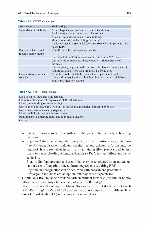
473
– Saline infusions sometimes suffice if the patient has already a bleeding diathesis.
– Regional Citrate anticoagulation may be used with custom-made, calcium- free dialysate. Frequent calcium monitoring and calcium infusion may be required. It is better than heparin in maintaining filter patency and is less likely to cause bleeding. Contraindication to RCA is liver failure and lactic acidosis ..
– Bivalirudin, fondaparinux and argatroban may be considered as an anticoagu-lant in cases of heparin-induced thrombocytopenia requiring RRT.
– Regional anticoagulation can be achieved with heparin and protamine. – Prostacyclin infusions are an option, but may cause hypotension.
• Continuous RRT must be provided with an effluent flow rate (the sum of hemo-filtration rate and dialysate flow rate) of at least 20 mL/kg/h.
• There is improved survival at effluent flow rates of 35 mL/kg/h but not much with 45 mL/kg/h (57% and 58%, respectively) as compared to an effluent flow rate of 20 mL/kg/h (41%) in patients with septic shock.
Table 47.2 CRRT advantages
Advantage MethodologyHemodynamic stability Avoids hypotension, which is seen in ultrafiltration
Avoids major swings in intravascular volumeAllows slow and continuous tissue refillingMaintains steady cardiac filling pressuresAvoids swings in intracranial pressures (beneficial in patients with raised ICP)
Easy to replenish and regulate fluid volume
Ultrafiltration is continuous and gentle
Can adjust ultrafiltration rate according to hourly MAP statusCan vary ultrafiltrate according to hourly variation in rate of infusatesCan accurately adjust it to the intravascular blood volume or stroke volume variation when such monitors are being used
Customize replacement solutions
According to the metabolic parameters, replacement fluid composition may be altered like high lactate, calcium, high/low potassium, high/low sodium
Table 47.3 CRRT disadvantages
Lack of rapid solute and fluid removalGlomerular filtration rate equivalent of 15–20 mL/minLimited role in drug overdose settingDuring filter clotting, entire system shuts down and the patient loses a lot of bloodNecessitates continuous anticoagulationLimits mobility for various investigationsRequirement of ultrapure fluids and high-flux dialyzersCostly
47 Renal Replacement Therapy
474
SLED
• SLED is not a continuous therapy and achieves lower solute clearances that are maintained for longer period.
• Treatments are deliberately intermittent rather than attempting to be continuous, with session longer in duration than conventional hemodialysis (HD).
• Solute and fluid removal is slower than conventional HD, but faster than CRRT. This allows for down timing of dialysis duration compared to CRRT without compromise in dialysis dose.
• They are easy to perform with modification of the standard dialysis machine, allow flexibility for procedure and diagnostic tests, allow a break in anticoagula-tion exposure, and are less staff intensive.
• This can be performed in the same machine used for IHD.• The time per session is usually 6–18 h. Depending on the needs of patient and
tolerance of ultrafiltration.• SLED and other hybrid therapies such as SLED-F have a lot of potential to be of
use when CRRT is not available.
Intermittent Hemodialysis
• This is the conventional hemodialysis modality.
Slow Continuous Ultrafiltration (SCUF)
• This modality does not require dialysate or any replacement fluids.• Here, therapeutic goal is to safely remove large volumes of fluid by hydrostatic
pressure, with no intent to substantially remove solute.• Ultrafiltration (UF) can be adjusted to cause dramatic fluid shifts; however, aver-
age UF rate ranges up to 2 L/h.• As SCUF is a longer duration therapy, blood flow rates are less than intermittent
HD, about 100–180 mL/min.• SCUF is primarily used when the fluid removal goals are gradual and modest.
Peritoneal Dialysis
• This modality of RRT utilizes the peritoneum as the dialyzer membrane.• The dialysate fluid is instilled periodically in the peritoneum and drained out to
achieve solute removal across a diffusion gradient.• Fluid removal is achieved by osmosis by changing the glucose content of the
dialysate fluid as necessary.
S. Prakash et al.

475
• This modality is effective when the patient is not too catabolic or hypotensive, or on vasopressor support.
• It is advantageous as it is gentle and continuous and it does not need anticoagula-tion, suitable for patients who have had a bleed, especially intracranial bleed.
• Due to infection risk, this is not practiced commonly.• Extracorporeal filters apart from dialysis filters like cytokine adsorption filter,
endotoxin adsorption filter etc. can be used with RRT machines.• However there is still not enough evidence to recommend it routinely.
Suggested Reading
Bagshaw SM, Berthiaume LR, Delaney A, Bellomo R. Continuous versus intermittent renal replacement therapy for critically ill patients with acute kidney injury: a meta-analysis. Crit Care Med. 2008;36(2):610617. Numerous issues related to study design, conduct, and quality that dispute the validity and question any inferences can be drawn from these trials. In the con-text of these limitations, the initial RRT modality did not seem to affect mortality or recovery to RRT independence. There is urgent need for additional high-quality and suitably powered trials to adequately address this issue.
Casey ET, Gupta BP, Erwin PJ, Montori VM, Murad MH. The dose of continuous renal replacement therapy for acute renal failure: a systematic review and meta-analysis. Ren Fail. 2010;32(5):555–61. Systematic review on the subject. Increased dosing of CRRT was not associated with a decrease in mortality of patients with ARF in an intensive care unit setting.
Davenport A. Renal replacement therapy in acute kidney injury: which method to use in the inten-sive care unit? Saudi J Kidney Dis Transpl. 2008;19(4):529–36. Comprehensive review on the subject. The choice of RRT modality should be guided by the individual patients’ clinical status, the medical and nursing expertise in the local intensive care unit, and the availability of RRT modality.
Fieghen H, Wald R, Jaber BL. Renal replacement therapy for acute kidney injury. Nephron Clin Pract. 2009;112(4):c222–9. Recent trials indicate that continuous renal replacement therapy does not confer a survival advantage as compared to intermittent hemodialysis. Furthermore, there is no evidence to support a more intensive strategy of renal replacement therapy in the setting of AKI. There is comparatively limited data regarding the ideal timing of renal replace-ment therapy initiation and the preferred mode of solute clearance.
Ronco C, Bellomo R, Homel P, Brendolan A, Dan M, Piccinni P, La Greca G. Effects of dif-ferent doses in continuous veno-venous haemofiltration on outcomes of acute renal fail-ure: a prospective randomised trial. Lancet 2000;356(9223):26–30. Klaus Kogelmann, Dominic Jarczak, Morten Scheller and Matthias Druner. Hemoadsorption by CytoSorb in septic patients: a case series. Critical care 2017, biomedcentral.com. doi.org/10.1186/s13054-017-1662-9.
Website
www.renalandurologynews.com
47 Renal Replacement Therapy

477© Springer Nature Singapore Pte Ltd. 2020R. Chawla, S. Todi (eds.), ICU Protocols, https://doi.org/10.1007/978-981-15-0898-1_48
A. Majumdar (*) Department of Nephrology, A.M.R.I. Hospitals, Kolkata, West Bengal, India
R. K. Mani Department of Pulmonology and Critical Care, Batra Hospital and Medical Research Centre, New Delhi, India
48Managing a Patient on Dialysis
Arghya Majumdar and Raj Kumar Mani
Acute kidney injury (AKI) is preventable in many instances and initial efforts should be directed towards this, with appropriate fluid and vasopressor therapy. If the patient does not respond one must make a judicious choice about com-mencing renal replacement therapy (RRT). Once on dialysis, patients need close monitoring due to their underlying unstable clinical condition, hemodynamic effects of the extracorporeal therapy, and technical problems commonly encoun-tered during dialysis.
Our initial endeavour should be to try and prevent acute kidney injury in such patients admitted to ICU, as the mortality doubles once AKI sets in.
A 65-year-old diabetic and hypertensive male patient, with a history of coro-nary artery bypass grafting a year ago, was admitted to the ICU, with pneumo-nia and septic shock. Previous records show that he had poor cardiac systolic function (LVEF 30%) and was on an angiotensin receptor blocker-neprilysin inhibitor (ARNI). ARNI was stopped. He was resuscitated with fluids and started on vasopressors. He remained oliguric (urine output <30 mL/h) for more than 12 h, with serum creatinine 3 mg/dL, potassium 6.5 mEq/L, and pH 7.1. He was commenced on continuous renal replacement therapy (CRRT).
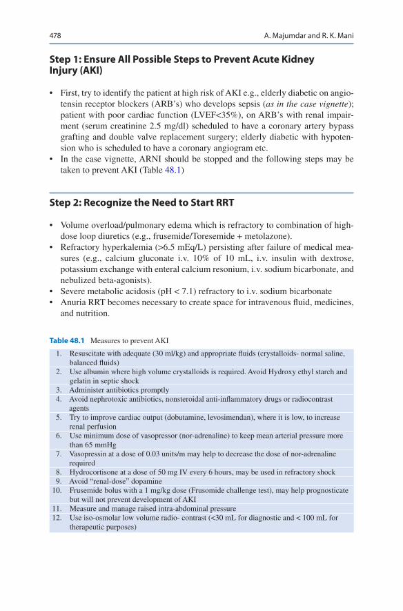
478
Step 1: Ensure All Possible Steps to Prevent Acute Kidney Injury (AKI)
• First, try to identify the patient at high risk of AKI e.g., elderly diabetic on angio-tensin receptor blockers (ARB’s) who develops sepsis (as in the case vignette); patient with poor cardiac function (LVEF<35%), on ARB’s with renal impair-ment (serum creatinine 2.5 mg/dl) scheduled to have a coronary artery bypass grafting and double valve replacement surgery; elderly diabetic with hypoten-sion who is scheduled to have a coronary angiogram etc.
• In the case vignette, ARNI should be stopped and the following steps may be taken to prevent AKI (Table 48.1)
Step 2: Recognize the Need to Start RRT
• Volume overload/pulmonary edema which is refractory to combination of high- dose loop diuretics (e.g., frusemide/Toresemide + metolazone).
• Refractory hyperkalemia (>6.5 mEq/L) persisting after failure of medical mea-sures (e.g., calcium gluconate i.v. 10% of 10 mL, i.v. insulin with dextrose, potassium exchange with enteral calcium resonium, i.v. sodium bicarbonate, and nebulized beta-agonists).
• Severe metabolic acidosis (pH < 7.1) refractory to i.v. sodium bicarbonate• Anuria RRT becomes necessary to create space for intravenous fluid, medicines,
and nutrition.
Table 48.1 Measures to prevent AKI
1. Resuscitate with adequate (30 ml/kg) and appropriate fluids (crystalloids- normal saline, balanced fluids)
2. Use albumin where high volume crystalloids is required. Avoid Hydroxy ethyl starch and gelatin in septic shock
3. Administer antibiotics promptly 4. Avoid nephrotoxic antibiotics, nonsteroidal anti-inflammatory drugs or radiocontrast
agents 5. Try to improve cardiac output (dobutamine, levosimendan), where it is low, to increase
renal perfusion 6. Use minimum dose of vasopressor (nor-adrenaline) to keep mean arterial pressure more
than 65 mmHg 7. Vasopressin at a dose of 0.03 units/m may help to decrease the dose of nor-adrenaline
required 8. Hydrocortisone at a dose of 50 mg IV every 6 hours, may be used in refractory shock 9. Avoid “renal-dose” dopamine10. Frusemide bolus with a 1 mg/kg dose (Frusomide challenge test), may help prognosticate
but will not prevent development of AKI11. Measure and manage raised intra-abdominal pressure12. Use iso-osmolar low volume radio- contrast (<30 mL for diagnostic and < 100 mL for
therapeutic purposes)
A. Majumdar and R. K. Mani
479
• Uremic encephalopathy.• Uremic pericarditis.• Ideal time to start RRT is controversial, and a joint decision between the nephrol-
ogist and the intensivist should be made, on the merit of each case.
Step 3: Establish Vascular Access for Dialysis
• Urgent vascular access may be achieved by inserting a double-lumen hemodialy-sis catheter, preferably under (USG) ultrasonographic guidance.
• Coagulation profile and platelet count should be checked.• A tunneled line may be preferable if the access is expected to be kept in for a
long time, if there are multiple malfunctioning temporary catheters, or if the patient is going to receive immunosuppressive treatment.
• Site – Central vein—internal jugular (IJ), subclavian (SC), or femoral (F). – The preferred route is- IJ. – SC route may be opted for, if the patient has a tracheostomy, but it should be
avoided to prevent subclavian vein stenosis, which may restrict future vascu-lar access for dialysis like fistula in the arm.
– Femoral route is accompanied by increased risk of infection and/or thrombo-sis, but it is preferred as a short-term measure in severely coagulopathic patient.
• Size: 11.5 Fr and length of catheter: 13.5 cm for (R) IJ or SC, 16.5 cm for (L) IJ or SC, and 19.5 cm for F.
• Meticulous sterile handling of dialysis line by health-care workers is mandatory.
• The hemodialysis line should not be used for any other purpose.• After each dialysis session, heparin lock should be used. In spite of heparin lock,
if there is a clot formation inside the catheter, attempt at clot lysis may be made by instilling thrombolytic agent like 2 mg of alteplase into each catheter lumen.
• Platelet counts should be followed periodically.
Patients commenced on dialysis need close supervision due to their underly-ing unstable clinical state, hemodynamic effects of extracorporeal circulation, and technical problems commonly encountered during renal replacement ther-apy (RRT).
Step 4: Start CRRT (See Chap. 47, Vol. 1)
Patients in shock with high dose of vasopressor should ideally be started on CRRT.
• Use central venous double-lumen hemodialysis catheter, which will ensure a blood flow of at least 200 mL/min.
• Before starting, check electrolytes, arterial blood gas, and lactate.
48 Managing a Patient on Dialysis

480
• If custom-made fluid is not available, one has to improvise on substitution fluids, maintaining the balance of essential electrolytes. Custom-made fluids often have lactate. If liver function is impaired, it may not be converted to bicarbonate and lead to worsening of hyperlactatemia.
• Electrolytes like Na, K, Ca, and Mg should be monitored frequently.• If the patient is very catabolic and higher solute clearance is required, substitu-
tion fluid should be added post-dilution (i.e., after the filter) to maintain a higher diffusion gradient of solutes. On the other hand, in situations where one needs to avoid anticoagulation, it may be added prefilter (i.e., pre-dilution). Heparin is the safest anticoagulant. Target an APTT of 1.5–2 times the control. If the patient has coagulopathy, regional anticoagulation may be tried with heparin/protamine or one may try prostacyclin. If citrate is used, ensure that all fluids are custom made and free of calcium. Calcium has to be added separately as an infusion, with regular monitoring.
• Convection at 20 mL/kg/h has been shown to be as effective as 35 mL/kg/h. Some studies have found a benefit of high volume ultrafiltration in sepsis.
• Adjust the dose of drugs especially antibiotics. Roughly one may use the dosing for the glomerular filtration rate at 20–50 mL/min, during CRRT.
• Hourly ultrafiltrate rate depends on central volume status (guided by invasive or non- invasive hemodynamic monitoring) and hourly fluid intake and output.
• Change the circuit, at least once in 72 h.• Protein intake should be planned, taking into account the daily loss on CRRT\
(40 g/day, over and above 1.5 g/kg/day).• Water-soluble vitamins should be replaced daily.
Step 5: Use Slow Extended Dialysis (SLED), as a Step- Down Dialytic Support or Initial Support in Less Hemodynamically Unstable Patients
• This can be done when the patient is recovering from shock.• A modified hemodialysis machine will suffice for SLED.• The dialysate flow rate can be adjusted to 100–200 mL/min.• SLED can be done for 8–10 h at one stretch, during the daytime, enabling better
utilization of skilled dialysis staff and resources.• The patient can be mobilized for investigations and procedures.• Drug dosing should be adjusted accordingly. Supplemental doses of most antibi-
otics are needed after a session.• Partial TPN may be given in between sessions when required.
A. Majumdar and R. K. Mani

481
Step 6: Manage the Patient with Conventional Intermittent Hemodialysis (HD) Once Hemodynamically Stable
• Ensure a vascular access blood flow of 250–400 mL/min.• Dialysate flow may be varied from 300 to 800 mL/min.• Pre-HD potassium should be checked. Dialysate fluids have a potassium level of
2.2 mmol/L. So if serum potassium is less than 3.5 mmol/L, potassium should be replaced accordingly.
• In cases of severe hyperkalemia or hypercalcemia, dialysate fluids without potas-sium or low calcium may be used.
• A bolus of heparin, usually 1000–2000 U, is followed by an infusion. Adjust according to APTT. In the patient with bleeding diathesis, dialysis without added heparin may be done.
• Check glucose, preferably hourly. Normal dialysate fluids do not contain glu-cose. Dialysate fluids containing glucose should be used, or it should be added when hypoglycemia is anticipated (e.g., in patients with liver dysfunction and sepsis).
Step 7: Assess Adequacy of a Dialysis Session
• Urea reduction ratio: predialysis urea—postdialysis urea/predialysis urea. The target is 65%.
• Kt/V: a dimensionless ratio representing volume of plasma cleared (Kt) divided by the urea distribution volume (V). The latest generation of hemodialysis machines is equipped with this measurement capability. The target is 1.2.
• In clinical practice, postdialysis urea and creatinine are compared with subse-quent predialysis levels. A steady state reflects recovering of renal function, and dialysis sessions may be spaced out accordingly.
Step 8: Optimize Adequacy of Dialysis
• Blood flow rate depends to a large extent on the position and patency of the cen-tral venous access catheter used for dialysis.
• Dialysate flow rate can be varied from 300 to 800 mL/min.• Dialyzer efficiency: a high-efficiency (high mass transfer area coefficient) dia-
lyzer with a thin, large-surface-area membrane, wide pores, and a design, which increases contact between blood and dialysate, will remove more waste products. However, the water used for dialysis in such situations needs to be ultrapure.
48 Managing a Patient on Dialysis

482
• Molecular weight (MW) of solute. Urea (MW 60) will be removed from blood more efficiently than creatinine (MW 113). Larger molecules like β2-microglobulin (MW 11,800) can only be removed by high-flux dialyzers.
• Access recirculation, which depends on the proximity of the “arterial” inflow and venous outflow of the dialysis catheter. Separate tunneled lines cause less recirculation.
• Hypercatabolic patients: high urea nitrogen generation rate from endogenous protein breakdown may give a false impression of “inadequate solute clearance.”
• Residual renal function when present may give an impression of “higher solute clearance.”
• Protein-bound molecules are not well removed by dialysis. Charcoal hemoperfu-sion is a better alternative.
Step 9: Assess Hypotension during Dialysis
• Hypotension is the commonest complication during dialysis. It occurs most commonly in intermittent hemodialysis.
• Commonest cause is reduction of intravascular volume due to mismatch of rate of ultrafiltration and tissue refilling.
• Assess central volume status (CVP).• Look for features of sepsis.• Look for underlying cardiac dysfunction, anemia, prior intake of antihyperten-
sive medications, arrhythmia (commonly atrial fibrillation), and autonomic neuropathy.
• Acute coronary syndrome, pericardial tamponade, or air embolism may also present as hypotension.
• Dialyzer reaction is rare now, with the use of biocompatible polysulfone membranes.
Step 10: Manage Hypotension Promptly
• Put the patient in the Trendelenburg position (with airway precaution), stop ultra-filtrate, and connect to a cardiac monitor.
• Do a 12-lead ECG and troponin I if clinical features are suggestive of ischemic heart disease. Do not treat supraventricular tachycardia or atrial fibrillation unless the patient is in shock or arrhythmia persists post-HD.
• A cautious bolus of 100 mL NS or more may be needed or 25% dextrose if blood glucose is low.
• Check Hb and transfuse if it is less than 8 g/dL.• Ensure that the dialysate fluid contains bicarbonate and not acetate, as the latter
causes more hypotension.• Dialysate temperature may be lowered to 36.5 °C to promote vasoconstriction.
A. Majumdar and R. K. Mani

483
• Reassess the dry weight and restart ultrafiltrate at a slower rate when stable.• Avoid antihypertensives pre-HD.• If the patient gets chills or fever, screen for an underlying infection.
Step 11: Monitor for Other Potential Complications (Table 48.2)
• Apart from hypotension, various other local and systemic complications may be seen in a patient on dialysis, which should be assessed and managed promptly.
Step 12: Assess Recovery of Renal Function and Try to Wean from RRT
• Urine output: increasing urine output especially after an oliguric phase and when the patient has not been on diuretics is considered the best sign of recovery.
• Change of urea or creatinine levels from postdialysis levels, when they remain steady or decline in between dialysis session, might be particularly helpful in nonoliguric patients.
Suggested Reading
Bellomo R, Ronco C. Indications and criteria for initiating renal replacement therapy in the inten-sive care unit. Kidney Int Suppl. 1998;66:S106–9. Even though there are several time-honored indications for initiating dialytic therapy in patients with near end-stage renal failure, such indications may not apply to the management of acute renal failure (ARF).
Table 48.2 Complications of dialysis
1. Hypotension 2. Muscle cramps 3. Nausea and vomiting 4. Headache 5. Chest pain or back pain 6. Itching 7. Disequilibrium syndrome 8. Dialyzer reactions 9. Bleedings—Gastrointestinal, epistaxis, intracranial10. Seizures11. Hemolysis12. Air embolism13. Visual or hearing loss14. Hypertension15. Hypoglycemia, hypothermia
48 Managing a Patient on Dialysis
484
Brochard L, Abroug F. ATS/ERS/ESICM/SCCM/SRLF ad hoc committee on acute renal fail-ure. An official ATS/ERS/ESICM/SCCM/SRLF statement: prevention and management of acute renal failure in the ICU patient: an international consensus conference in intensive care medicine. Am J Respir Crit Care Med. 2010;181(10):1128–55. The jury recommended the use of newly described definitions.
KIDGO AKI Workgroup. KDIGO clinical practice guideline for AKI. Kidney Int Suppl. 2012;2:1–138.
Kolhe NV, Reilly T, Leung J, et al. A simple care bundle for use in AKI: a propensity score- matched cohort study. Nephrol Dial Transplant. 2016;31:1846–54.
Moore PK, Hsu RK, Liu KD. Management of acute kidney injury: core curriculum 2018. Am J Kidney Dis. 2018;72(1):136–48. This Core Curriculum reviews the key principles regarding the diagnosis and general management of AKI for clinicians.
Ricci Z, Ronco C. Dose and efficiency of renal replacement therapy: continuous renal replacement therapy versus intermittent hemodialysis versus slow extended daily dialysis. Crit Care Med. 2008;36(4 Suppl):S229–37. Some broad concepts and a few suggestions for renal replace-ment therapy prescription derived from current evidence and from the available experience are given.
Websites
www.ADQI.netHomepage for ADQI network on acute kidney injury.www.crrtonline.comPractical aspects of managing CRRT, including nursing perspective.www.asn-online.orgAmerican Society of Nephrology webpage with information on various aspects of AKI.
A. Majumdar and R. K. Mani
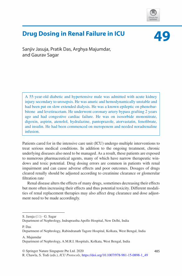
485© Springer Nature Singapore Pte Ltd. 2020R. Chawla, S. Todi (eds.), ICU Protocols, https://doi.org/10.1007/978-981-15-0898-1_49
S. Jasuja (*) · G. Sagar Department of Nephrology, Indraprastha Apollo Hospital, New Delhi, India
P. Das Department of Nephrology, Rabindranath Tagore Hospital, Kolkata, West Bengal, India
A. Majumdar Department of Nephrology, A.M.R.I. Hospitals, Kolkata, West Bengal, India
49Drug Dosing in Renal Failure in ICU
Sanjiv Jasuja, Pratik Das, Arghya Majumdar, and Gaurav Sagar
Patients cared for in the intensive care unit (ICU) undergo multiple interventions to treat serious medical conditions. In addition to the ongoing treatment, chronic underlying diseases also need to be managed. As a result, these patients are exposed to numerous pharmaceutical agents, many of which have narrow therapeutic win-dows and toxic potential. Drug dosing errors are common in patients with renal impairment and can cause adverse effects and poor outcomes. Dosages of drugs cleared renally should be adjusted according to creatinine clearance or glomerular filtration rate.
Renal disease alters the effects of many drugs, sometimes decreasing their effects but more often increasing their effects and thus potential toxicity. Different modali-ties of renal replacement therapies may also affect drug clearance and dose adjust-ment need to be made accordingly.
A 55-year-old diabetic and hypertensive male was admitted with acute kidney injury secondary to urosepsis. He was anuric and hemodynamically unstable and had been put on slow extended dialysis. He was a known epileptic on phenobar-bitone and levetiracetam. He underwent coronary artery bypass grafting 2 years ago and had congestive cardiac failure. He was on isosorbide mononitrate, digoxin, aspirin, atenolol, hydralazine, pantoprazole, atorvastatin, fenofibrate, and insulin. He had been commenced on meropenem and needed noradrenaline infusion.
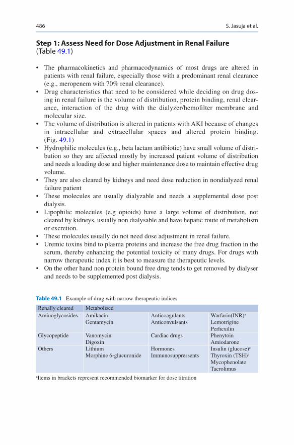
486
Step 1: Assess Need for Dose Adjustment in Renal Failure (Table 49.1)
• The pharmacokinetics and pharmacodynamics of most drugs are altered in patients with renal failure, especially those with a predominant renal clearance (e.g., meropenem with 70% renal clearance).
• Drug characteristics that need to be considered while deciding on drug dos-ing in renal failure is the volume of distribution, protein binding, renal clear-ance, interaction of the drug with the dialyzer/hemofilter membrane and molecular size.
• The volume of distribution is altered in patients with AKI because of changes in intracellular and extracellular spaces and altered protein binding. (Fig. 49.1)
• Hydrophilic molecules (e.g., beta lactam antibiotic) have small volume of distri-bution so they are affected mostly by increased patient volume of distribution and needs a loading dose and higher maintenance dose to maintain effective drug volume.
• They are also cleared by kidneys and need dose reduction in nondialyzed renal failure patient
• These molecules are usually dialyzable and needs a supplemental dose post dialysis.
• Lipophilic molecules (e.g opioids) have a large volume of distribution, not cleared by kidneys, usually non dialysable and have hepatic route of metabolism or excretion.
• These molecules usually do not need dose adjustment in renal failure.• Uremic toxins bind to plasma proteins and increase the free drug fraction in the
serum, thereby enhancing the potential toxicity of many drugs. For drugs with narrow therapeutic index it is best to measure the therapeutic levels.
• On the other hand non protein bound free drug tends to get removed by dialyser and needs to be supplemented post dialysis.
Table 49.1 Example of drug with narrow therapeutic indices
Renally cleared MetabolisedAminoglycosides Amikacin
GentamycinAnticoagulantsAnticonvulsants
Warfarin(INR)a
LemotriginePerhexilin
Glycopeptide VanomycinDigoxin
Cardiac drugs PhenytoinAmiodarone
Others LithiumMorphine 6-glucuronide
HormonesImmunosuppressents
Insulin (glucose)a
Thyroxin (TSH)a
MycophenolateTacrolimus
aItems in brackets represent recommended biomarker for dose titration
S. Jasuja et al.
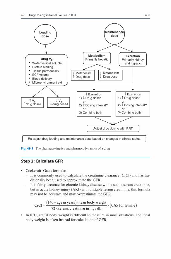
487
Step 2: Calculate GFR
• Cockcroft–Gault formula: – It is commonly used to calculate the creatinine clearance (CrCl) and has tra-
ditionally been used to approximate the GFR. – It is fairly accurate for chronic kidney disease with a stable serum creatinine,
but in acute kidney injury (AKI) with unstable serum creatinine, this formula may not be accurate and may overestimate the GFR.
CrCl
age in years lean body weight
serum. creatinin=
−( )××
140
72 ee in mg dL for female
/.×( )0 85
• In ICU, actual body weight is difficult to measure in most situations, and ideal body weight is taken instead for calculation of GFR.
Loadingdose
Adjust drug dosing with RRT
Re-adjust drug loading and maintenance dose based on changes in clinical status
↑ Vd↑ drug dose#
↓ Vd↓ drug dose#
Drug Vd
Water vs lipid soluble •
↑↑
↓↓
•••••
Protein bindingTissue permeabilityECF volumeBlood deliveryMicroenvironment pH
Maintenancedose
MetabolismPrimarily hepatic
ExcretionPrimarily kidney
and hepatic
Metabolism Drug dose
MetabolismDrug dose
↓ Excretion1) ↓ Drug dose*
or2)
3)
↑ Dosing interval**orCombine both
↑ Excretion1) ↑ Drug dose*
or2)
3)
↓ Dosing interval**orCombine both
Fig. 49.1 The pharmacokinetics and pharmacodynamics of a drug
49 Drug Dosing in Renal Failure in ICU
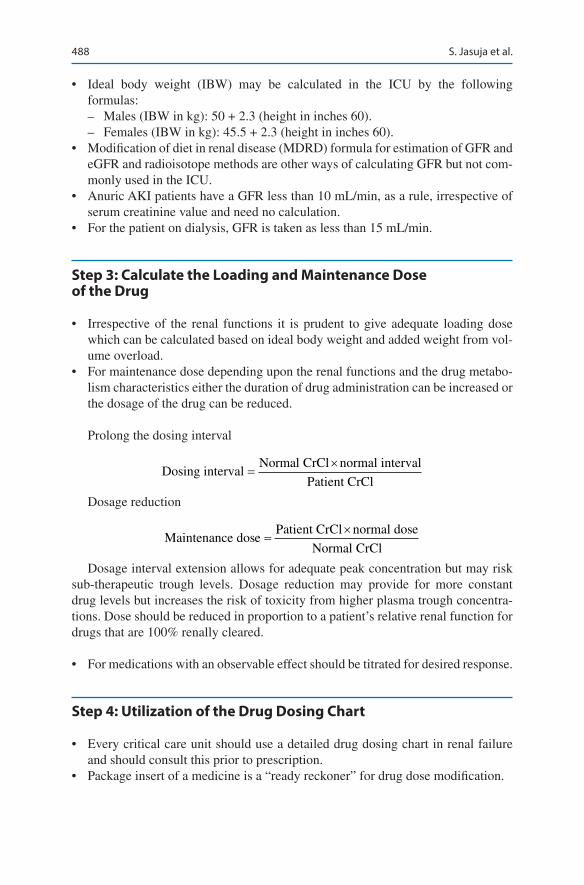
488
• Ideal body weight (IBW) may be calculated in the ICU by the following formulas: – Males (IBW in kg): 50 + 2.3 (height in inches 60). – Females (IBW in kg): 45.5 + 2.3 (height in inches 60).
• Modification of diet in renal disease (MDRD) formula for estimation of GFR and eGFR and radioisotope methods are other ways of calculating GFR but not com-monly used in the ICU.
• Anuric AKI patients have a GFR less than 10 mL/min, as a rule, irrespective of serum creatinine value and need no calculation.
• For the patient on dialysis, GFR is taken as less than 15 mL/min.
Step 3: Calculate the Loading and Maintenance Dose of the Drug
• Irrespective of the renal functions it is prudent to give adequate loading dose which can be calculated based on ideal body weight and added weight from vol-ume overload.
• For maintenance dose depending upon the renal functions and the drug metabo-lism characteristics either the duration of drug administration can be increased or the dosage of the drug can be reduced.
Prolong the dosing interval
Dosing interval
Normal normal interval
Patient
CrCl
CrCl=
×
Dosage reduction
Maintenance dose
Patient normal dose
Normal
CrCl
CrCl=
×
Dosage interval extension allows for adequate peak concentration but may risk sub-therapeutic trough levels. Dosage reduction may provide for more constant drug levels but increases the risk of toxicity from higher plasma trough concentra-tions. Dose should be reduced in proportion to a patient’s relative renal function for drugs that are 100% renally cleared.
• For medications with an observable effect should be titrated for desired response.
Step 4: Utilization of the Drug Dosing Chart
• Every critical care unit should use a detailed drug dosing chart in renal failure and should consult this prior to prescription.
• Package insert of a medicine is a “ready reckoner” for drug dose modification.
S. Jasuja et al.
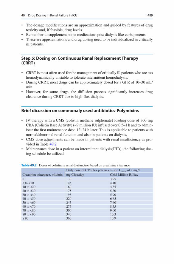
489
• The dosage modifications are an approximation and guided by features of drug toxicity and, if feasible, drug levels.
• Remember to supplement some medications post dialysis like carbapenems.• These are approximations and drug dosing need to be individualized in critically
ill patients.
Step 5: Dosing on Continuous Renal Replacement Therapy (CRRT)
• CRRT is most often used for the management of critically ill patients who are too hemodynamically unstable to tolerate intermittent hemodialysis.
• During CRRT, most drugs can be approximately dosed for a GFR of 10–30 mL/min.
• However, for some drugs, the diffusion process significantly increases drug clearance during CRRT due to high-flux dialysis.
Brief dicussion on commonaly used antibiotics-Polymixins
• IV therapy with a CMS (colistin methane sulphonate) loading dose of 300 mg CBA (Colistin Base Activity) (~9 million IU) infused over 0.5–1 h and to admin-ister the first maintenance dose 12–24 h later. This is applicable to patients with normal/abnormal renal function and also in patients on dialysis.
• CMS dose adjustments can be made in patients with renal insufficiency as pro-vided in Table 49.2.
• Maintenance dose in a patient on intermittent dialysis(IHD), the following dos-ing schedule be utilized:
Table 49.2 Doses of colistin in renal dysfunction based on creatinine clearance
Creatinine clearance, mL/minDaily dose of CMS for plasma colistin Css,avg of 2 mg/Lmg CBA/day CMS Million IU/day
0 130 3.955 to <10 145 4.4010 to <20 160 4.8520 to <30 175 5.3030 to <40 195 5.9040 to <50 220 6.6550 to <60 245 7.4060 to <70 275 8.3570 to <80 300 9.0080 to <90 340 10.3≥ 90 360 10.9
49 Drug Dosing in Renal Failure in ICU

490
– On a nondialysis day, administer a CMS dose of 130 mg CBA/ day (~3.95 million IU/day). On a dialysis day, administer a supplemental dose of CMS 40 mg CBA (~1.2 million IU) or 50 mg CBA (~1.6 mil- lion IU) for a 3- or 4-h IHD session, respectively.
– If possible, the supplement to the baseline (nondialysis) daily dose should be administered with the next regular dose, after the dialysis session has ended.
– Conduct IHD sessions as late as possible within a CMS dosage interval to minimize the amount of CMS and formed colistin lost to the extracorporeal system.
– In patients on sustained low-efficiency dialysis (SLED) 10% of the CMS dose be added to the baseline daily dose per 1 h of SLED.
– Patients on CRRT, for a plasma colistin Css, avg. of 2 mg/L, to administer CBA 440 mg/day (~13.3 million IU/day). This equates to 220 mg CBA every 12 h (~6.65 million IU every 12 h).
Polymixin B
• Start with a loading dose of 2.0–2.5 mg/kg for polymyxin B, based on total body weight (TBW) (equivalent to 20,000–25,000 IU/kg) over 1 h irrespective of renal function.
• Polymyxin B dose of 1.25–1.5 mg/kg (equivalent to 12,500–15,000 IU/kg TBW) every 12 h is infused over 1 h as a maintenance dose.
• Daily maintenance doses of polymyxin B should not be adjusted if the patient has renal impairment.
• For other commonly used drugs refer to Appendix Table A.2 Dosage modifica-tion in renal failure.
Suggested Reading
Claus BO, Hoste EA. Augmented renal clearance is a common finding with worse clinical out-come in critically ill patients receiving antimicrobial therapy. J Crit Care. 2013;28(5):695–700. ARC was documented in approximately 52% of a mixed ICU patient population receiving anti-biotic treatment with worse clinical outcome. Young age and male gender were independently associated with ARC presence.
Lewis SJ, Mueller BA. Antibiotic dosing in patients with acute kidney injury: “enough but not too much”. J Intensive Care Med. 2016;31(3):164–76. Review article on antibiotic dosing in RRT.
Tsuji BT, Pogue JM, Zavascki AP. International consensus guidelines for the optimal use of the polymyxins: Endorsed by the American College of Clinical Pharmacy (ACCP), European Society of Clinical Microbiology and Infectious Diseases (ESCMID), Infectious Diseases Society of America (IDSA), International Society for Anti-infective Pharmacology (ISAP), Society of Critical Care Medicine (SCCM), and Society of Infectious Diseases Pharmacists (SIDP). Pharmacotherapy. 2019;39(1):10–39. Current consensus guideline on the use of polymyxin.
S. Jasuja et al.

Part VI
Infection Diseases

493© Springer Nature Singapore Pte Ltd. 2020R. Chawla, S. Todi (eds.), ICU Protocols, https://doi.org/10.1007/978-981-15-0898-1_50
S. N. Myatra (*) · J. G. Pulinilkunnathil Department of Anaesthesia, Critical Care and Pain, Tata Memorial Hospital, Mumbai, India
50General Measures of Infection Control
Sheila Nainan Myatra and Jacob George Pulinilkunnathil
Health-care-associated infections are a common cause of increased morbidity, mor-tality, and cost of care in ICUs. A systematic and multidisciplinary approach to infection control practice goes a long way in minimizing this problem. Infectious patients needs to be isolated to prevent spread of infection to other patients and to healthcare staff. Apart from appropriate isolation precautions, infection control and judicious antibiotic use are the mainstay of management of these patients.
Step 1: Assess the Need for Isolation
• Screen all ICU patients for the following: – Neutropenia and immunological disorder. – Diarrhea. – Skin rashes. – Known communicable disease. – Known carriers of an epidemic strain of bacterium.
Step 2: Identify the Type of Isolation Needed
• There are two types of isolation in the ICU:
A 72-year-old male patient with non-Hodgkin’s lymphoma was neutropenic after chemotherapy and presented to the ICU with breathlessness and hypoten-sion. He was intubated and kept on a ventilator and received broad- spectrum antibiotics. He had a peripheral, central, and arterial line in place. A Foley’s catheter and a nasogastric tube were also placed.
494
– Protective isolation for neutropenic or other immunocompromised patients to reduce the chances of acquiring opportunistic infections.
– Source isolation of colonized or infected patients to minimize potential trans-mission to other patients or staff.
• Isolation rooms should have tight-fitting doors, glass partitions for observation, and both negative-pressure (for source isolation) and positive-pressure (for pro-tective isolation) ventilations.
Step 3: Identify the Patient at Risk of Nosocomial Infections
• There are patient-, therapy-, and environment-related risk factors for the devel-opment of nosocomial infection: – Age more than 70 years. – Shock. – Major trauma. – Acute renal failure. – Coma. – Prior antibiotics. – Mechanical ventilation. – Drugs affecting the immune system (steroids, chemotherapy). – Indwelling catheters. – Prolonged ICU stay (>3 days).
Step 4: Observe Hand Hygiene
• Hands are the most common vehicle for transmission of organisms, and “hand hygiene” is the single most effective means of preventing the horizontal trans-mission of infections among hospital patients and health-care personnel.
• When and why—follow WHO’s five moments for hand hygiene (Fig. 50.1):
1. Before touching a patient—to protect the patient from harmful germs carried on your hands
2. Before aseptic procedures—to protect the patient against harmful germs, includ-ing the patient’s own germs
3. After body fluid exposure/risk—to protect yourself and the health-care environ-ment from the harmful patient’s germs
4. After touching the patient—to protect yourself and the health-care environment from the harmful patient’s germs
5. After touching the patient’s surrounding—to protect yourself and the health-care environment from the harmful patient’s germs(Remember, there are two moments before and three moments after touching the patient)
S. N. Myatra and J. G. Pulinilkunnathil
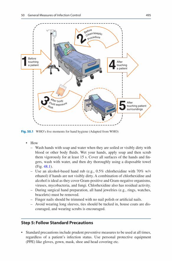
495
• How – Wash hands with soap and water when they are soiled or visibly dirty with
blood or other body fluids. Wet your hands, apply soap and then scrub them vigorously for at least 15 s. Cover all surfaces of the hands and fin-gers, wash with water, and then dry thoroughly using a disposable towel (Fig. 48.1).
– Use an alcohol-based hand rub (e.g., 0.5% chlorhexidine with 70% w/v ethanol) if hands are not visibly dirty. A combination of chlorhexidine and alcohol is ideal as they cover Gram-positive and Gram-negative organisms, viruses, mycobacteria, and fungi. Chlorhexidine also has residual activity.
– During surgical hand preparation, all hand jewelries (e.g., rings, watches, bracelets) must be removed.
– Finger nails should be trimmed with no nail polish or artificial nails. – Avoid wearing long sleeves, ties should be tucked in, house coats are dis-
couraged, and wearing scrubs is encouraged.
Step 5: Follow Standard Precautions
• Standard precautions include prudent preventive measures to be used at all times, regardless of a patient’s infection status. Use personal protective equipment (PPE) like gloves, gown, mask, shoe and head covering etc.
Beforetouchinga patient1 After
touchinga patient4
Aftertouching patientsurroundings
Fluid exposure
After body
Risk
Before
Clean/aseptic
Procedure
3
2
5
Fig. 50.1 WHO’s five moments for hand hygiene (Adapted from WHO)
50 General Measures of Infection Control

496
• Gloves – Sterile gloves should be worn after hand hygiene procedure while touching
mucous membrane and nonintact skin and performing sterile procedures (e.g., arterial, central line, and Foley catheter insertion).
– Clean, nonsterile gloves are safe for touching blood, other body fluids, con-taminated items, and any other potentially infectious materials.
– Change gloves between tasks and procedures in the same patient especially when moving from a contaminated body area to a clean body area.
– Never wear the same pair of gloves for the care of more than one patient. – Remove gloves after caring for a patient. – Practice hand hygiene whenever gloves are removed.
• Gown – Wear a gown to prevent soiling of clothing and skin during procedures that are
likely to generate splashes of blood, body fluids, secretions, or excretions. – The sterile gown is required only for aseptic procedures, and for the rest, a
clean, nonsterile gown is sufficient. – Remove the soiled gown as soon as possible, with care to avoid
contamination.• Mask, eye protection/face shield
– Wear a mask and adequate eye protection (eyeglasses are not enough) or a face shield to protect mucous membranes of the eyes, nose, and mouth during procedures and patient-care activities that are likely to generate splashes/sprays of blood, body fluids, etc.
– Patients, relatives, and health-care workers presenting with respiratory symp-toms should also use masks (e.g., cough).
• Shoe and head coverings – They are not required for routine care.
• Patient-care equipment – Used patient-care equipment soiled with blood, body fluids, secretions, or
excretions should be handled carefully to prevent skin and mucous membrane exposures, contamination of clothing, and transfer of microorganisms to health-care workers, other patients, or the environment.
– Ensure that reusable equipment is not used for the care of another patient until it has been cleaned and sterilized appropriately.
– Ensure that single-use items and sharps are discarded properly.
Step 6: Follow Transmission-Based Precautions
In addition to standard precautions, the following should be observed in those patients known or suspected to have airborne, contact, or droplet infections. See Table 50.1 for details.
S. N. Myatra and J. G. Pulinilkunnathil
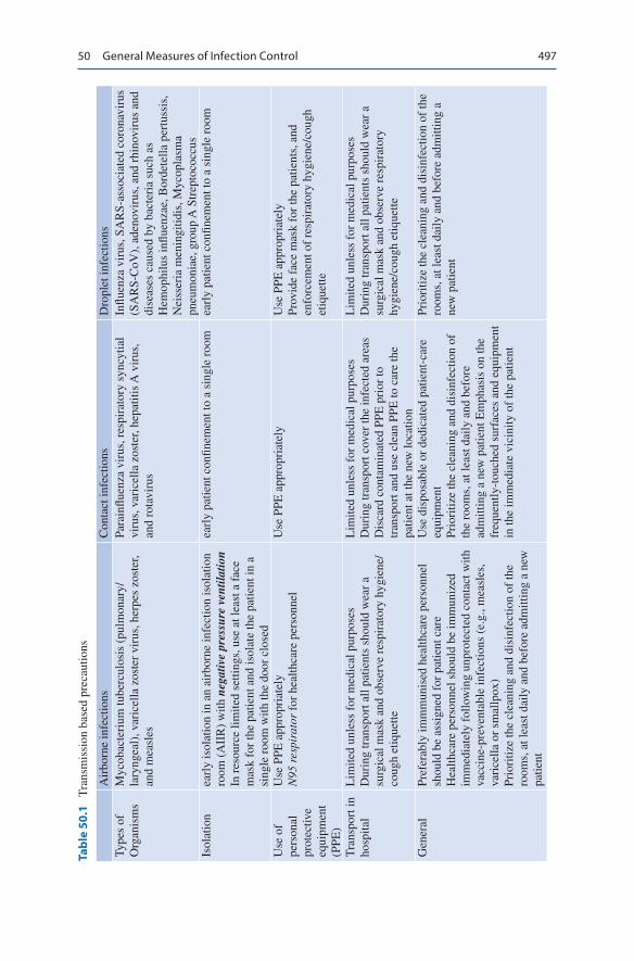
497
Tabl
e 50
.1
Tra
nsm
issi
on b
ased
pre
caut
ions
Air
born
e in
fect
ions
Con
tact
infe
ctio
nsD
ropl
et in
fect
ions
Type
s of
O
rgan
ism
sM
ycob
acte
rium
tube
rcul
osis
(pu
lmon
ary/
lary
ngea
l), v
aric
ella
zos
ter
viru
s, h
erpe
s zo
ster
, an
d m
easl
es
Para
influ
enza
vir
us, r
espi
rato
ry s
yncy
tial
viru
s, v
aric
ella
zos
ter,
hepa
titis
A v
irus
, an
d ro
tavi
rus
Influ
enza
vir
us, S
AR
S- as
soci
ated
cor
onav
irus
(S
AR
S-C
oV),
ade
novi
rus,
and
rhi
novi
rus
and
dise
ases
cau
sed
by b
acte
ria
such
as
Hem
ophi
lus
influ
enza
e, B
orde
tella
per
tuss
is,
Nei
sser
ia m
enin
gitid
is, M
ycop
lasm
a pn
eum
onia
e, g
roup
A S
trep
toco
ccus
Isol
atio
nea
rly
isol
atio
n in
an
airb
orne
infe
ctio
n is
olat
ion
room
(A
IIR
) w
ith n
egat
ive
pres
sure
ven
tilat
ion
In r
esou
rce
limite
d se
tting
s, u
se a
t lea
st a
fac
e m
ask
for
the
patie
nt a
nd is
olat
e th
e pa
tient
in a
si
ngle
roo
m w
ith th
e do
or c
lose
d
earl
y pa
tient
con
finem
ent t
o a
sing
le r
oom
earl
y pa
tient
con
finem
ent t
o a
sing
le r
oom
Use
of
pers
onal
pr
otec
tive
equi
pmen
t (P
PE)
Use
PPE
app
ropr
iate
lyN
95 r
espi
rato
r fo
r he
alth
care
per
sonn
elU
se P
PE a
ppro
pria
tely
Use
PPE
app
ropr
iate
lyPr
ovid
e fa
ce m
ask
for
the
patie
nts,
and
en
forc
emen
t of
resp
irat
ory
hygi
ene/
coug
h et
ique
tte
Tra
nspo
rt in
ho
spita
lL
imite
d un
less
for
med
ical
pur
pose
sD
urin
g tr
ansp
ort a
ll pa
tient
s sh
ould
wea
r a
surg
ical
mas
k an
d ob
serv
e re
spir
ator
y hy
gien
e/co
ugh
etiq
uette
Lim
ited
unle
ss f
or m
edic
al p
urpo
ses
Dur
ing
tran
spor
t cov
er th
e in
fect
ed a
reas
Dis
card
con
tam
inat
ed P
PE p
rior
to
tran
spor
t and
use
cle
an P
PE to
car
e th
e pa
tient
at t
he n
ew lo
catio
n
Lim
ited
unle
ss f
or m
edic
al p
urpo
ses
Dur
ing
tran
spor
t all
patie
nts
shou
ld w
ear
a su
rgic
al m
ask
and
obse
rve
resp
irat
ory
hygi
ene/
coug
h et
ique
tte
Gen
eral
Pref
erab
ly im
mm
unis
ed h
ealth
care
per
sonn
el
shou
ld b
e as
sign
ed f
or p
atie
nt c
are
Hea
lthca
re p
erso
nnel
sho
uld
be im
mun
ized
im
med
iate
ly f
ollo
win
g un
prot
ecte
d co
ntac
t with
va
ccin
e- pr
even
tabl
e in
fect
ions
(e.
g., m
easl
es,
vari
cella
or
smal
lpox
)Pr
iori
tize
the
clea
ning
and
dis
infe
ctio
n of
the
room
s, a
t lea
st d
aily
and
bef
ore
adm
ittin
g a
new
pa
tient
Use
dis
posa
ble
or d
edic
ated
pat
ient
-car
e eq
uipm
ent
Prio
ritiz
e th
e cl
eani
ng a
nd d
isin
fect
ion
of
the
room
s, a
t lea
st d
aily
and
bef
ore
adm
ittin
g a
new
pat
ient
Em
phas
is o
n th
e fr
eque
ntly
- tou
ched
sur
face
s an
d eq
uipm
ent
in th
e im
med
iate
vic
inity
of
the
patie
nt
Prio
ritiz
e th
e cl
eani
ng a
nd d
isin
fect
ion
of th
e ro
oms,
at l
east
dai
ly a
nd b
efor
e ad
mitt
ing
a ne
w p
atie
nt
50 General Measures of Infection Control
498
Airborne Infections
Pathogenic microorganisms may be suspended in the air as small particles, aerosols, or dust and remain infective over time and distance.
Contact Infections
Some infections spread by direct or indirect contact with an infected person, and also from the surfaces or patient-care items in the room.
Droplet Infections
Microorganisms can also be transmitted by large droplets >5 μm in size that are generated during coughing, sneezing or talking.
Step 7: Use specific Strategies Focused on Prevention of Specific Nosocomial Infections
In addition to the standard and transmission-based precautions, there are several strategies focused on prevention of specific nosocomial infections in critically ill patients. Of these, ventilator-associated pneumonia (VAP), catheter-related blood-stream infection (CRBSI), and urinary tract infection (UTI) are the most important.
General Strategies to Reduce VAP (SHEA/IDSA Practice Recommendation 2014)
• Avoid intubation whenever possible.• Consider noninvasive ventilation whenever possible.• Prefer oral intubations to nasal unless contraindicated.• Minimum sedation for patients, daily interruption of sedation, daily extubation
trials to be practiced together.• Keep head elevated at 30–45° in the semi-recumbent body position.• Avoid reintubation whenever possible.• Routine change of ventilator circuits is not required unless visibly soiled or
malfunctioning• Monitor endotracheal tube cuff pressure (keep it between 20–30 cmH2O) to
avoid air leaks around the cuff, which can allow entry of bacterial pathogens into the lower respiratory tract, while permitting capillary perfusion.
• Prefer endotracheal tubes with a subglottic suction port to prevent pooling of secretions around the cuff leading to microaspiration for patients expected to require greater than 48 or 72 hours of mechanical ventilation
S. N. Myatra and J. G. Pulinilkunnathil
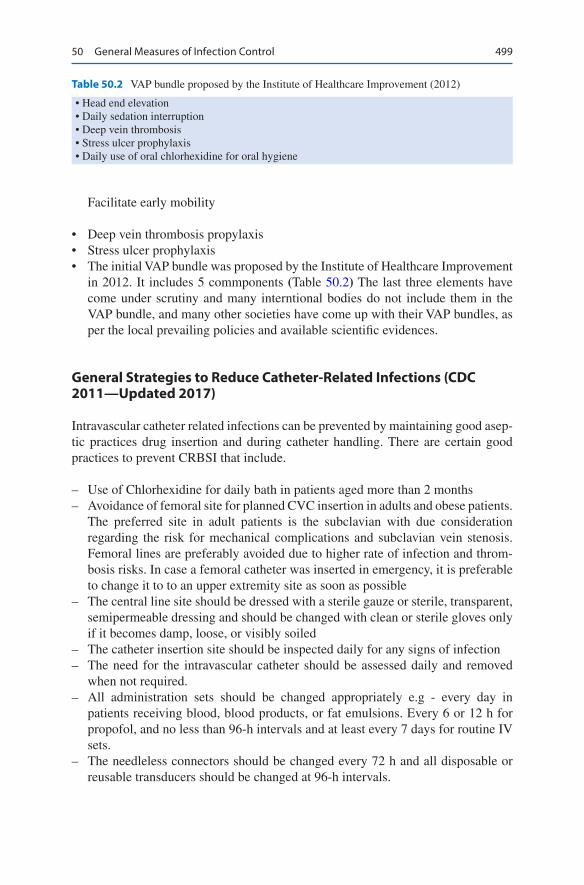
499
Facilitate early mobility
• Deep vein thrombosis propylaxis• Stress ulcer prophylaxis• The initial VAP bundle was proposed by the Institute of Healthcare Improvement
in 2012. It includes 5 commponents (Table 50.2) The last three elements have come under scrutiny and many interntional bodies do not include them in the VAP bundle, and many other societies have come up with their VAP bundles, as per the local prevailing policies and available scientific evidences.
General Strategies to Reduce Catheter-Related Infections (CDC 2011—Updated 2017)
Intravascular catheter related infections can be prevented by maintaining good asep-tic practices drug insertion and during catheter handling. There are certain good practices to prevent CRBSI that include.
– Use of Chlorhexidine for daily bath in patients aged more than 2 months – Avoidance of femoral site for planned CVC insertion in adults and obese patients.
The preferred site in adult patients is the subclavian with due consideration regarding the risk for mechanical complications and subclavian vein stenosis. Femoral lines are preferably avoided due to higher rate of infection and throm-bosis risks. In case a femoral catheter was inserted in emergency, it is preferable to change it to to an upper extremity site as soon as possible
– The central line site should be dressed with a sterile gauze or sterile, transparent, semipermeable dressing and should be changed with clean or sterile gloves only if it becomes damp, loose, or visibly soiled
– The catheter insertion site should be inspected daily for any signs of infection – The need for the intravascular catheter should be assessed daily and removed
when not required. – All administration sets should be changed appropriately e.g - every day in
patients receiving blood, blood products, or fat emulsions. Every 6 or 12 h for propofol, and no less than 96-h intervals and at least every 7 days for routine IV sets.
– The needleless connectors should be changed every 72 h and all disposable or reusable transducers should be changed at 96-h intervals.
Table 50.2 VAP bundle proposed by the Institute of Healthcare Improvement (2012)
• Head end elevation• Daily sedation interruption• Deep vein thrombosis• Stress ulcer prophylaxis• Daily use of oral chlorhexidine for oral hygiene
50 General Measures of Infection Control

500
– Other practices include adequate staffing with maintained nurse-to-patient ratio, use of antimicrobial ointments at hemodialysis catheter-insertion site and to have a continuous surveillance for CLABSI in both ICU and non-ICU settings.
In spite of adequate precautions, if the CLABSI rate is still high, then additional measures such as
– Use of antiseptic- or antimicrobial-impregnated CVCs in adult patients – Use chlorhexidine-containing dressings in patients over 2 months of age – Use of an antiseptic-containing hub/connector cap/port protector to cover con-
nectors may be tried.
The use of antimicrobial locks CVCs are presently recommended only for long- term hemodialysis catheters, in patients with limited venous access and a history of recurrent CLABSI and in patients who are at increased risk of severe complications from CLABSI (e.g patients with prosthetic valves or aortic graft).
The central line bundle by Institute of Healthcare Improvement is in Table 50.3.
General Strategies to Reduce UTI (CDC 2009)
• Insert catheters only for appropriate indications.• Follow aseptic insertion of the urinary catheter.• Maintain a closed drainage system.• Maintain unobstructed urine flow. At all times, the urinary catheter should be
placed and taped above the thigh and the urinary bag should hang below the level of the bladder.
• The urinary bag should never have floor contact.• Changing indwelling catheters or drainage bags at fixed intervals is not recom-
mended. Change only if there are clinical indications such as infection or obstruc-tion or when the closed system is compromised.
• Remove the catheter when it is no longer needed.
The Catheter Associated Urinary Tract Infection (CAUTI) Bundle from the International Federation of Infection Control (2016) is given in Table 50.4.
Table 50.3 The central line bundle by Institute of Healthcare Improvement
• Hand hygiene• Maximal barrier precautions• Chlorhexidine skin antisepsis• Optimal catheter site selection, with avoidance of using the femoral vein for central venous
access in adult patients• Daily review of line necessity, with prompt removal of unnecessary lines
S. N. Myatra and J. G. Pulinilkunnathil
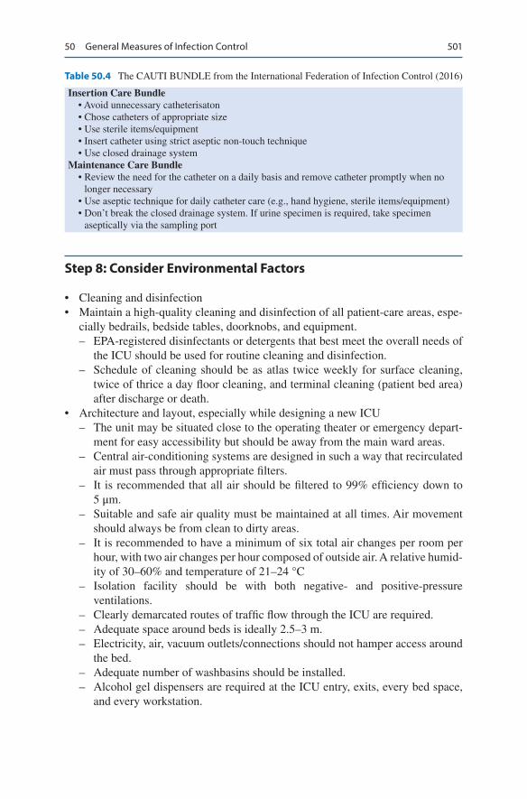
501
Step 8: Consider Environmental Factors
• Cleaning and disinfection• Maintain a high-quality cleaning and disinfection of all patient-care areas, espe-
cially bedrails, bedside tables, doorknobs, and equipment. – EPA-registered disinfectants or detergents that best meet the overall needs of
the ICU should be used for routine cleaning and disinfection. – Schedule of cleaning should be as atlas twice weekly for surface cleaning,
twice of thrice a day floor cleaning, and terminal cleaning (patient bed area) after discharge or death.
• Architecture and layout, especially while designing a new ICU – The unit may be situated close to the operating theater or emergency depart-
ment for easy accessibility but should be away from the main ward areas. – Central air-conditioning systems are designed in such a way that recirculated
air must pass through appropriate filters. – It is recommended that all air should be filtered to 99% efficiency down to
5 μm. – Suitable and safe air quality must be maintained at all times. Air movement
should always be from clean to dirty areas. – It is recommended to have a minimum of six total air changes per room per
hour, with two air changes per hour composed of outside air. A relative humid-ity of 30–60% and temperature of 21–24 °C
– Isolation facility should be with both negative- and positive-pressure ventilations.
– Clearly demarcated routes of traffic flow through the ICU are required. – Adequate space around beds is ideally 2.5–3 m. – Electricity, air, vacuum outlets/connections should not hamper access around
the bed. – Adequate number of washbasins should be installed. – Alcohol gel dispensers are required at the ICU entry, exits, every bed space,
and every workstation.
Table 50.4 The CAUTI BUNDLE from the International Federation of Infection Control (2016)
Insertion Care Bundle • Avoid unnecessary catheterisaton • Chose catheters of appropriate size • Use sterile items/equipment • Insert catheter using strict aseptic non-touch technique • Use closed drainage systemMaintenance Care Bundle • Review the need for the catheter on a daily basis and remove catheter promptly when no
longer necessary • Use aseptic technique for daily catheter care (e.g., hand hygiene, sterile items/equipment) • Don’t break the closed drainage system. If urine specimen is required, take specimen
aseptically via the sampling port
50 General Measures of Infection Control

502
– There should be separate medication preparation area. – There should be separate areas for clean storage, soiled and waste storage and
disposal. – Adequate toilet facilities should be provided.
Step 9: Organizational and Administrative Measures
• Work with hospital administration for better patient-to-nurse ratio in the ICU.• Policies for controlling traffic flow to and from the unit to reduce sources of
contamination from visitors, staff, and equipment.• Waste and sharp disposal policy.• Education and training for ICU staff about prevention of nosocomial infections.• ICU protocols for prevention of nosocomial infections.• Audit and surveillance of infections and infection control practices.• Infection control team (multidisciplinary approach).• Antibiotic stewardship.• Vaccination of health-care personnel.
Antibiotic Stewardship
Antibiotic stewardship has been defined as “coordinated interventions designed to improve and measure the appropriate use of antibiotic agents by promoting the selection of the optimal antibiotic drug regimen including dosing, duration of ther-apy, and route of administration”.
Antibiotic stewardship ensures the right drug in right dose at the right time for the right duration for any infection, thereby eradicating infection with minimial side effects. It consists of prospective audit and feedback, education, antibiotic restric-tion, antibiotics de-escalation, guideline use, optimal dosing and duration, microbi-ologist, and computer aided clinical support. Properly executed, this is associated with reduced drug resistance, improved patient outcome, and optimized resource utilization. These policies have shown to improve antibiotic prescribing and drug resistance patterns without an increase in patient mortality or hospital length of stay.
Suggested Reading
Johnson I. Banks V; Antibiotic stewardship in critical care. BJA Educ. 2017;17(4):111–6.Klompas M, Branson R, Eichenwald EC, Greene LR, Howell MD, Lee G, Magill SS, Maragakis
LL, Priebe GP, Speck K, Yokoe DS, Berenholtz SM. Strategies to prevent ventilator-associated pneumonia in acute care hospitals: 2014 update. Infect Control Hosp Epidemiol. 2014;35(Suppl 2):S133–54. Describes Strategies for prevention of ventilator associated pneumonia.
Ling ML, Apisarnthanarak A, Jaggi N, et al. APSIC guide for prevention of Central Line Associated Bloodstream Infections (CLABSI). Antimicrob Resist Infect Control. 2016;5:16. https://doi.
S. N. Myatra and J. G. Pulinilkunnathil

503
org/10.1186/s13756-016-0116-5. Guide for the prevention of central line associated blood stream infections.
Mehta Y, Gupta A, Todi S, Myatra SN, Samaddar DP, Patil V, Bhattacharya PK, Ramasubban S. Guidelines for prevention of hospital acquired infections. Indian J Crit Care Med. 2014;18:149–63. Describes Indian guidelines for prevention of hospital acquired infections.
Torres A, Niederman MS, Chastre J, Ewig S, Fernandez-Vandellos P, Hanberger H, Kollef M, Bassi GL, Luna CM, Martin-Loeches I, ArturPaiva J, Read RC, Rigau D, Timsit JF, Welte T, Wunderink R. Summary of the international clinical guidelines for the management of hospital- acquired and ventilator-acquired pneumonia. ERJ Open Research. 2018;4(2):00028–2018. https://doi.org/10.1183/23120541.00028-2018. Guidelines for the management of hospi-tal acquired and ventilator associated pneumonia.
Antoni Torres, Michael S. Niederman, Jean Chastre, Santiago Ewig, Patricia Fernandez-Vandellos, Hakan Hanberger, Marin Kollef, Gianluigi Li Bassi, Carlos M. Luna, Ignacio Martin-Loeches, J. ArturPaiva, Robert C. Read, David Rigau, Jean François Timsit, Tobias Welte, Richard Wunderink. International ERS/ESICM/ESCMID/ALAT guidelines for the manage-ment of hospital- acquired pneumonia and ventilator-associated pneumonia. Eur Respir J 2017;3:1700582. https://doi.org/10.1183/13993003.00582-2017. Guidelines for management of ventilator associated pneumonia.
Kalil AC, Metersky ML, Klompas M, Muscedere J, Sweeney DA, Palmer LB, Napolitano LM, O'Grady NP, Bartlett JG, Carratalà J, El Solh AA, Ewig S, Fey PD, File TM, Restrepo MI, Roberts JA, Waterer GW, Cruse P, Knight SL, Brozek JL. Management of adults with hospital- acquired and ventilator-associated pneumonia: 2016 clinical practice guidelines by the Infectious Diseases Society of America and the American Thoracic Society. Clin Infect Dis. 2016;63(5):61–e111. https://doi.org/10.1093/cid/ciw353. Guidelines for the management of hospital acquired and ventilator associated pneumonia.
Websites
http://whqlibdoc.who.int/hq/2009/WHO_IER_PSP_2009.07_eng.pdfWHO guidelines on hand hygiene in health care: a summary.http://www.cdc.gov/hicpac/pdf/guidelines/bsi-guidelines-2011.pdfGuidelines for the prevention of intravascular catheter-related infections.http://www.cdc.gov/hicpac/pdf/CAUTI/CAUTIguideline2009final.pdfGuidelines for prevention of catheter-associated urinary tract infections.http://theific.org/wp-content/uploads/2016/04/18-UTI_2016.pdfPrevention of CatheterAssociated Urinary Tract Infections.http://www.cdc.gov/hicpac/pdf/isolation/Isolation2007.pdfGuideline for isolation precautions: preventing transmission of infectious agents in healthcare
settings.https://www.cdc.gov/infectioncontrol/guidelines/bsi/recommendations.htmlUpdated guidelines forprevention of catheter related infections.https://www.cdc.gov/infectioncontrol/guidelines/environmental/appendix/air.html#tableb2Ventilation requirements for areas affecting patient care in hospitals and outpatient facilities.
50 General Measures of Infection Control

505© Springer Nature Singapore Pte Ltd. 2020R. Chawla, S. Todi (eds.), ICU Protocols, https://doi.org/10.1007/978-981-15-0898-1_51
S. Todi (*) Department of Critical Care & Emergency Medicines, A.M.R.I. Hospital, Kolkata, India
R. Chawla Department of Respiratory, Critical Care & Sleep Medicine, Indraprastha Apollo Hospitals, New Delhi, India
51Antibiotic Stewardship
Subhash Todi and Rajesh Chawla
Judicious antibiotic prescription along with infection control is the cornerstone for preventing emergence of drug-resistant organisms in the ICU. Appropriate (right choice) and adequate (right time, right dose, right route, right duration) antibiotic coverage improves outcome in infected patients.
Step 1: Formulate a Plan for Antibiotic Selection
• Antibiotics in the ICU are given either prophylactically, mainly perioperative or empirically for presumed infection while culture report is pending, or defini-tively when infection is documented with positive culture results.
• Appropriate antibiotics should be chosen depending on clinical presentation, severity of illness, likely pathogens and its anatomical source, recent antibiotic use, local epidemiology and resistance pattern and likelihood of an infection with resistant organism.
• Reason for antibiotic prescription and selection should be clearly documented in the antibiotic order form, which should be audited periodically for compliance and appropriateness.
• Early consultation with an Infectious disease consultant in difficult cases should be taken
A 70-year-old male patient had been admitted to the ICU with left-sided hemi-paresis for 4 days. He was catheterized on admission and intubated for airway protection. He had spiked a fever of 102 °F with chills and became drowsy. A broad-spectrum antibiotic was started empirically after sending blood cultures.
506
Step 2: Send Appropriate Cultures
• This should ideally be done prior to starting antibiotics.• Blood, urine, sputum, and endotracheal secretion should be promptly transported
in a proper media to the microbiology laboratory and expeditiously processed.
Step 3: Start Antibiotics Early
• Every hour delay in starting effective antibiotics from the onset of septic shock increases the risk of death from sepsis
• Antibiotics should be started within 1 h of recognition of septic shock as “Time is tissue.”
Step 4: Choose Empirical Antibiotics Appropriately
• Empirical antibiotics should be chosen carefully as initial wrong choice increases mortality, even if the antibiotic is changed appropriately after culture results are obtained.
• Initial antibiotic choice should be based on the patient’s history, underlying dis-ease or clinical syndrome, susceptibility pattern of the pathogen in the commu-nity and hospital, and previous colonization pattern.
• Recently used antibiotic class should generally be avoided.• Choose broad-spectrum antibiotics that have activity against most likely bacte-
rial pathogens.• Infections due to viral/fungal/parasitic pathogen should also be thought of in
appropriate clinical scenarios and relevant investigations should be sent.
Step 5: Stratify the Risk of Infection with Drug-Resistant Organisms (Table 51.1)
• The patient should be assessed for risk of infection with multidrug-resistant bacteria.
• Apart from the above mentioned classical risk factors the following risk factors are also increasingly recognised.
• Old Age.• Lack of functional independence/decreased cognition.• Comorbid conditions: Liver failure/Renal failure/diabetes/immunosuppression.• Presence of indwelling devices like urinary catheter.• If one or more risk factors are present, antibiotic choice should be broadened to
cover these organisms.
S. Todi and R. Chawla
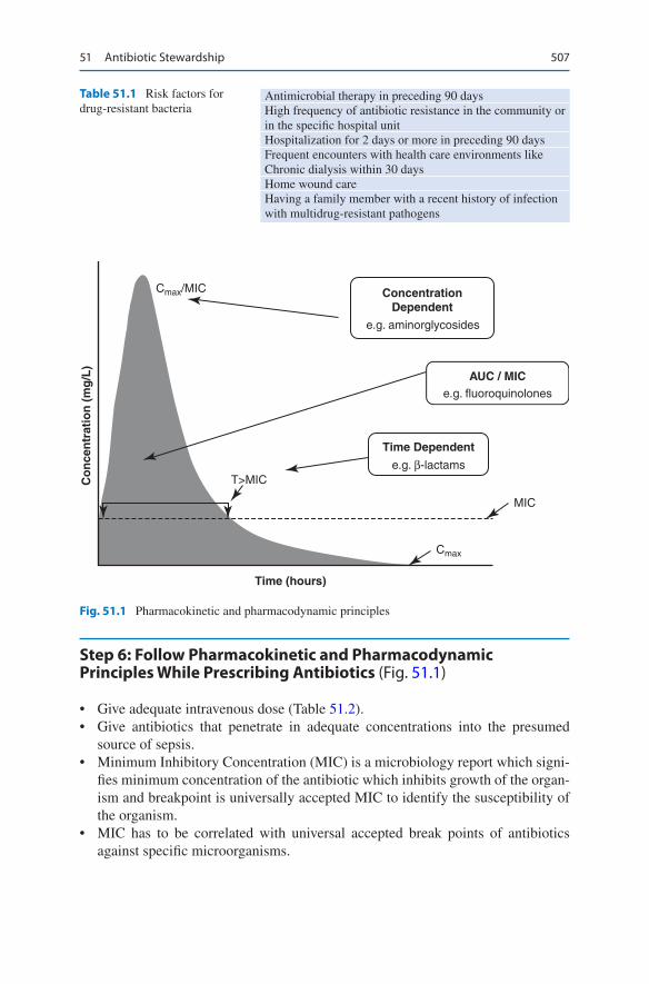
507
Step 6: Follow Pharmacokinetic and Pharmacodynamic Principles While Prescribing Antibiotics (Fig. 51.1)
• Give adequate intravenous dose (Table 51.2).• Give antibiotics that penetrate in adequate concentrations into the presumed
source of sepsis.• Minimum Inhibitory Concentration (MIC) is a microbiology report which signi-
fies minimum concentration of the antibiotic which inhibits growth of the organ-ism and breakpoint is universally accepted MIC to identify the susceptibility of the organism.
• MIC has to be correlated with universal accepted break points of antibiotics against specific microorganisms.
Table 51.1 Risk factors for drug-resistant bacteria
Antimicrobial therapy in preceding 90 daysHigh frequency of antibiotic resistance in the community or in the specific hospital unitHospitalization for 2 days or more in preceding 90 daysFrequent encounters with health care environments like Chronic dialysis within 30 daysHome wound careHaving a family member with a recent history of infection with multidrug-resistant pathogens
e.g. aminorglycosides
MIC
T>MIC
Cmax
Cmax/MIC ConcentrationDependent
e.g. fluoroquinolones
e.g. β-lactams
AUC / MIC
Time Dependent
Time (hours)
Co
nce
ntr
atio
n (
mg
/L)
Fig. 51.1 Pharmacokinetic and pharmacodynamic principles
51 Antibiotic Stewardship
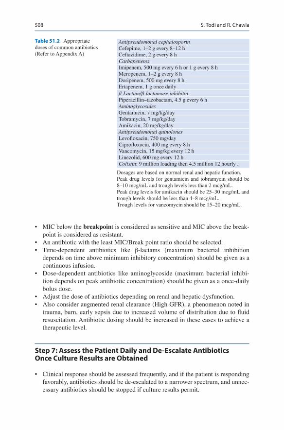
508
• MIC below the breakpoint is considered as sensitive and MIC above the break-point is considered as resistant.
• An antibiotic with the least MIC/Break point ratio should be selected.• Time-dependent antibiotics like β-lactams (maximum bacterial inhibition
depends on time above minimum inhibitory concentration) should be given as a continuous infusion.
• Dose-dependent antibiotics like aminoglycoside (maximum bacterial inhibi-tion depends on peak antibiotic concentration) should be given as a once-daily bolus dose.
• Adjust the dose of antibiotics depending on renal and hepatic dysfunction.• Also consider augmented renal clearance (High GFR), a phenomenon noted in
trauma, burn, early sepsis due to increased volume of distribution due to fluid resuscitation. Antibiotic dosing should be increased in these cases to achieve a therapeutic level.
Step 7: Assess the Patient Daily and De-Escalate Antibiotics Once Culture Results are Obtained
• Clinical response should be assessed frequently, and if the patient is responding favorably, antibiotics should be de-escalated to a narrower spectrum, and unnec-essary antibiotics should be stopped if culture results permit.
Table 51.2 Appropriate doses of common antibiotics (Refer to Appendix A)
Antipseudomonal cephalosporinCefepime, 1–2 g every 8–12 hCeftazidime, 2 g every 8 hCarbapenemsImipenem, 500 mg every 6 h or 1 g every 8 hMeropenem, 1–2 g every 8 hDoripenem, 500 mg every 8 hErtapenem, 1 g once dailyβ-Lactam/β-lactamase inhibitorPiperacillin–tazobactam, 4.5 g every 6 hAminoglycosidesGentamicin, 7 mg/kg/dayTobramycin, 7 mg/kg/dayAmikacin, 20 mg/kg/dayAntipseudomonal quinolonesLevofloxacin, 750 mg/dayCiprofloxacin, 400 mg every 8 hVancomycin, 15 mg/kg every 12 hLinezolid, 600 mg every 12 hColistin: 9 million loading then 4.5 million 12 hourly .
Dosages are based on normal renal and hepatic function.Peak drug levels for gentamicin and tobramycin should be 8–10 mcg/mL and trough levels less than 2 mcg/mL.Peak drug levels for amikacin should be 25–30 mcg/mL and trough levels should be less than 4–8 mcg/mL.Trough levels for vancomycin should be 15–20 mcg/mL.
S. Todi and R. Chawla
509
• Decisions to continue, narrow, or stop antimicrobial therapy must be made on the basis of clinician judgment and laboratory information like decrease in leukocy-tosis, decreasing C reactive protein, and a low procalcitonin level.
• Judicious use of antibiogram to differentiate coloniser vs. pathogen and appro-priate antibiotic choice should be done in consultation with the microbiologist and infectious disease consultant.
Step 8: Consider the Combination of Antibiotics in Specific Situations
• The combination of antibiotics (two appropriate antibiotics against the same organ-ism) is indicated in difficult to treat multidrug-resistant pathogens like Acinetobacter and Pseudomonas sp. Combination therapy is also indicated for neutropenic patient with severe sepsis and selected patients with severe Pseudomonas infection with respiratory failure and shock. Similarly, a combination of beta- lactam and macrolide is recommended for pneumococcal bacteremia.
• Avoid combinations like vancomycin with piperacillin/tazobactam, which may lead to increased renal toxicity.
Step 9: Decide on Duration of Antibiotic Therapy
• Duration of therapy should be individualised but shorter courses (4–7 days) have been found to be equivalent to longer courses (7–14 days) in majority of patients
• Longer courses may be appropriate in patients who have a slow clinical response, undrainable foci of infection, or immunologic deficiencies including neutropenia.
• If culture result is negative and there is a favorable clinical response, most anti-biotics can be stopped in 5 days.
• In Pseudomonas and Acinetobacter infection, severe sepsis should be treated for longer period.
Step 10: Implement Antibiotic Stewardship Program
• Constitute an antibiotic stewardship team along with the microbiologist, infec-tion control nurse, infectious disease consultant, and clinical pharmacist.
• Educating ICU staff the principles of antibiotic stewardship is of prime importance.
• Proper utilization of local antibiogram should be done.• Utilize optimally the information obtained from the microbiology laboratory.• Work in close collaboration with microbiologists and other physicians involved
in antibiotic prescribing.• Optimal use of computerised electronic health record and clinical decision sup-
port and timely reminder to physicians to descalate and stop antibiotics is extremely useful.
51 Antibiotic Stewardship
510
Suggested Reading
Dellinger RP, Levy MM, Carlet JM, et al. Surviving sepsis campaign: international guidelines for management of severe sepsis and septic shock. Intensive Care Med. 2017;43:304–77. A com-prehensive guidelines on the management of sepsis.
Ibrahim EH, Sherman G, Ward S, et al. The influence of inadequate antimicrobial treatment of bloodstream infections on patient outcomes in the ICU setting. Chest. 2000;118:146–55. Multiple logistic regression analysis identified the administration of inadequate antimicrobial treatment as an independent determinant of hospital mortality.
Kumar A, Roberts D, Wood KE, et al. Duration of hypotension prior to initiation of effective antimicrobial therapy is the critical determinant of survival in human septic shock. Crit Care Med. 2008;34:1589–96. Administration of an antimicrobial effective for isolated or suspected pathogens within the first hour of documented hypotension was associated with a survival rate of 79.9%. Each hour of delay in antimicrobial administration over the ensuing 6 h was associated with an average decrease in survival of 7.6%. Median time to effective antimicro-bial therapy was 6 h.
Website
www.survivingsepsis.org
S. Todi and R. Chawla
511© Springer Nature Singapore Pte Ltd. 2020R. Chawla, S. Todi (eds.), ICU Protocols, https://doi.org/10.1007/978-981-15-0898-1_52
S. Ramasubban (*) Department of Respiratory, Critical Care and Sleep Medicine, Apollo Gleneagles Hospital, Kolkata, India
52Septic Shock
Suresh Ramasubban
Sepsis is defined as “life-threatening organ dysfunction caused by a dysregulated host response to infection”. Organ dysfunction is assessed at the bedside by a change in SOFA score. Early identification of organ dysfunction with a quick SOFA score will help in early resuscitation with a resultant decrease in morbidity and mortality.
Septic shock is defined as a “subset of sepsis in which underlying circula-tory and cellular metabolism abnormalities are profound enough to substan-tially increase mortality”. Clinically, septic shock is sepsis that results in tissue hypoperfusion, with hypotension requiring vasopressors-and elevated lactate levels. The term severe sepsis has been eliminated in the new sepsis 3 definition.
Step 1: Take Care of Airway and Breathing
• Proper airway care and, if needed, assisted ventilation should be promptly initi-ated in all patients with severe sepsis and shock (see Chap. 23, Vol. 2). Taking early control of breathing decreases the oxygen consumption by the respiratory muscles and enables better perfusion of vital organs.
A 60-year-old diabetic male patient presented with a history of dysuria and fever. His vital signs on admission were as follows: pulse 120/min, BP 80/50 mmHg, and respiratory rate 28/min. He was disoriented and agitated.
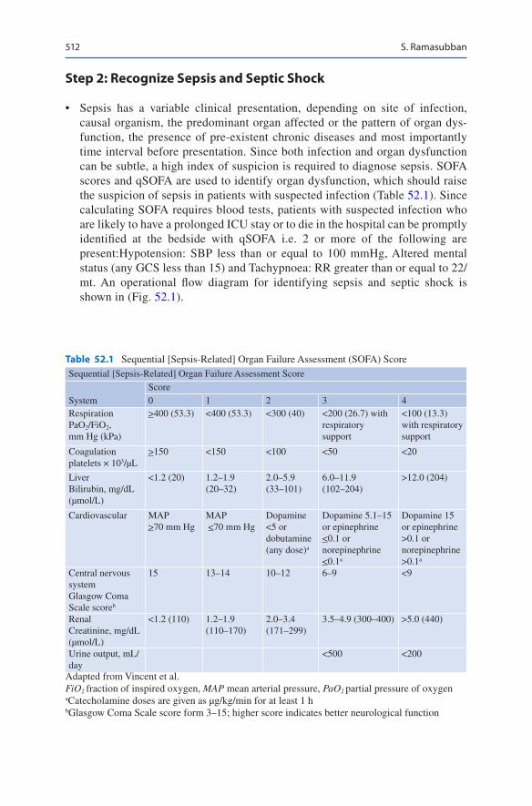
512
Step 2: Recognize Sepsis and Septic Shock
• Sepsis has a variable clinical presentation, depending on site of infection, causal organism, the predominant organ affected or the pattern of organ dys-function, the presence of pre-existent chronic diseases and most importantly time interval before presentation. Since both infection and organ dysfunction can be subtle, a high index of suspicion is required to diagnose sepsis. SOFA scores and qSOFA are used to identify organ dysfunction, which should raise the suspicion of sepsis in patients with suspected infection (Table 52.1). Since calculating SOFA requires blood tests, patients with suspected infection who are likely to have a prolonged ICU stay or to die in the hospital can be promptly identified at the bedside with qSOFA i.e. 2 or more of the following are present:Hypotension: SBP less than or equal to 100 mmHg, Altered mental status (any GCS less than 15) and Tachypnoea: RR greater than or equal to 22/mt. An operational flow diagram for identifying sepsis and septic shock is shown in (Fig. 52.1).
Table 52.1 Sequential [Sepsis-Related] Organ Failure Assessment (SOFA) ScoreSequential [Sepsis-Related] Organ Failure Assessment Score
SystemScore0 1 2 3 4
RespirationPaO2/FiO2, mm Hg (kPa)
>400 (53.3) <400 (53.3) <300 (40) <200 (26.7) with respiratory support
<100 (13.3) with respiratory support
Coagulationplatelets × 103/μL
>150 <150 <100 <50 <20
LiverBilirubin, mg/dL (μmol/L)
<1.2 (20) 1.2–1.9 (20–32)
2.0–5.9 (33–101)
6.0–11.9 (102–204)
>12.0 (204)
Cardiovascular MAP >70 mm Hg
MAP <70 mm Hg
Dopamine <5 or dobutamine (any dose)a
Dopamine 5.1–15 or epinephrine <0.1 or norepinephrine <0.1a
Dopamine 15 or epinephrine >0.1 or norepinephrine >0.1a
Central nervous systemGlasgow Coma Scale scoreb
15 13–14 10–12 6–9 <9
RenalCreatinine, mg/dL (μmol/L)
<1.2 (110) 1.2–1.9 (110–170)
2.0–3.4 (171–299)
3.5–4.9 (300–400) >5.0 (440)
Urine output, mL/day
<500 <200
Adapted from Vincent et al.FiO2 fraction of inspired oxygen, MAP mean arterial pressure, PaO2 partial pressure of oxygenaCatecholamine doses are given as μg/kg/min for at least 1 hbGlasgow Coma Scale score form 3–15; higher score indicates better neurological function
S. Ramasubban

513
• Sepsis can affect all organ systems acutely, especially the cardiovascular system and respiratory system manifesting as hypotension and acute respiratory distress syndrome (ARDS). Acute Kidney injury (AKI) manifests as oliguria and an increase in serum creatinine values. Obtundation and delirium are also very com-mon CNS manifestations of sepsis. The other manifestations of sepsis include paralytic ileus, abnormal liver enzymes, hyperglycemia, thrombocytopenia and disseminated intravascular coagulation, adrenal dysfunction and the euthyroid sick syndrome.
• Categorizing patients into sepsis and septic shock helps in triaging, prognostica-tion, and choosing appropriate therapy.
Step 3: Initial Resucitation
• Fluid resuscitation is of utmost importance in initial management of patients with sepsis induced tissue hypoperfusion and septic shock.
• Insert a wide-bore peripheral line and give initial fluid challenge of 1000 mL of crystalloids (normal saline or Ringer lactate) to achieve a minimum of 30 ml/kg of crystalloids over 3 hours with careful monitoring of vital signs. Following initial fluid resuscitation, further fluids should be guided by frequent reassess-ment of hemodynamic status.
Patient with suspected infection
(A) qSOFA variablesRespiratory rateMental statusSystolic blood pressure
(B) SOFA variablesPaO2/Fio2 ratioGlasgow Coma Scale scoreMean arterial pressureAdministration ofvasopressors with typeand dose rate of infusionSerum creatinine or urineoutputBilirubinPlatelet count
Monitor clinicalcondition;reevaluate forpossible sepsis if clinically indicated
Monitor clinical condition;reevaluate for possible
sepsis if clinically indicated
Sepsis
Assess for evidenceof organ dysfunction
Despite adequate fluidresuscitation,1. Vasopressors require to maintain MAP >65 mm Hg2. Serum lactate level> 2 mmol/L?
Septic shock
SOFA >2?(see (B))
qSOFA >2?(see (A))
Sepsis stillsuspected?
NoNo
No
Yes
Yes
No
Yes
Yes
Fig. 52.1 Identifying a patient of sepsis and septic shock. Operationalization of Clinical Criteria Identifying Patients With Sepsis and Septic Shock. The baseline Sequential [Sepsis-related] Organ Failure Assessment (SOFA) score should be assumed to be zero unless the patient is known to have preexisting (acute or chronic) organ dysfunction before the onset of infection. qSOFA quick SOFA, MAP mean arterial pressure
52 Septic Shock

514
• If type of shock is not clear, further hemodynamic monitoring is required.• One can do initially echocardiography for assessment.• Fluid responsiveness should be assessed by using dynamic indices preferably
over static indices. Dynamic variables have better diagnostic accuracy at pre-dicting a response to a fluid challenge by increasing stroke volume. These tech-niques include passive leg raising while measuring stroke volume in a spontaneously breathing patient, or the variations in systolic pressure, pulse pressure, or stroke volume using heart lung interactions during mechanical ventilation.
• Fluid responsiveness should always be assessed in conjunction with fluid toler-ance. Fluid tolerance can be assessed clinically or using ultrasound of the lungs.
• In septic shock requiring vasopressors, target means arterial pressure (MAP) of 65 mmHg.
• Target Urine output of at least 0.5 ml/kg/h.
Step 4: Send Initial Investigations
• As the patient is being resuscitated, send blood for complete hemogram, blood cultures (two sets), and other appropriate cultures depending on the clinical situ-ation, urea, creatinine, electrolytes, liver function test, ECG, and chest X-ray.
• Send arterial blood for arterial blood gas and lactate analysis. Increased lactate is a feature of global hypoperfusion and needs urgent attention.
• Serial lactate measurement should be done frequently as it may be useful in monitoring response to resuscitation, when initial lactates are high due to tissue hypoperfusion
• If lactate is not available, base deficit (metabolic acidosis) in the absence or renal failure, can be taken as a surrogate marker of lactic acidosis.
• Bio-markers of infection such as procalcitonin are not diagnostic of sepsis. However they have a very high negative predictive value and can be used to rule out sepsis. Serial Procalcitonin may be helpful in de-escalation of antibiotics. Testing for procalcitonin should not delay fluid resuscitation and antibiotic administration in patients with sepsis and septic shock.
Step 5: Start Antimicrobial Agent
• Appropriate broad-spectrum antibiotics as per hospital protocol should be started immediately, preferably within 1 h of presentation of septic shock.
• Appropriate cultures should be sent before starting antibiotics, but if these are delayed for logistic reasons beyond 45 min, antibiotics should be started.
• Selection of an optimal antimicrobial regimen is one of the key determinants of outcome in sepsis and septic shock. Thus one or more agents active against likely bacterial/fungal or viral pathogens and with good penetration into presumed source should be selected.
• Dosing of antibiotics should be based on pharmacokinetics/pharmacodynamic principles and specific properties of the antibiotic being used.
S. Ramasubban
515
• Risk factors for multidrug resistant gram negative organisms causing sepsis should be sought. The risk factors include prolonged hospital stay, recent antimi-crobial use, prior hospitalization, and prior colonization or infection with multidrug- resistant organisms.
• Combination therapy with supplemental gram negative coverage is recom-mended for Pseudomonas aeruginosa bacteremia. Combination therapy may also be used for patients at high risk for multidrug-resistant bacteria like Acinetobacter and in neutropenic and immunocompromised patients.
• A combination of beta-lactam and a macrolide should be used in patients with pneumococcal bacteremia with septic shock.
• The duration of therapy is typically 7–10 days; longer courses may be appro-priate in patients who have a slow clinical response, undrainable foci of infec-tion, some fungal or viral infection, or immunologic deficiencies including neutropenia.
• Antiviral therapy should be initiated as early as possible in patients with severe sepsis or septic shock of viral origin, when suspected, such as for severe influ-enza infection.
• In patients with risk factors for invasive Candida infections such as immuno-compromised status (neutropenia, chemotherapy, transplant, diabetes mellitus, chronic liver failure, chronic renal failure), prolonged invasive vascular devices (hemodialysis catheters, central venous catheters), total parenteral nutrition, nec-rotizing pancreatitis, recent major surgery (particularly abdominal), prolonged administration of broad-spectrum antibiotics, prolonged hospital/ICU admis-sion, recent fungal infection, and multisite colonization, empirical antifungal therapy should be started (refer to Chap. 55, Vol. 1).
Step 6: Source Control
• Efforts should be made to make a specific anatomic diagnosis amenable to source control as soon as possible. Efforts should specifically consider the drainage of an abscess, debridement of infected necrotic tissue, removal of a potentially infected device, and definitive control of a source of ongoing microbial contamination.
• Source control should be done as soon as medically and logistically practical. Ideally within 6–12 hours, a delay of more than 12 hours may lead to worse outcomes.
• Central lines and other intravascular devices should be promptly removed in case of septic shock after estabilishing other vascular access.
Step 7: Fluid Therapy
• Crystalloids are the fluids of choice in sepsis and septic shock. Avoiding hyper-chloremia may be an important issue with large volume resuscitation. Balanced salt solutions contain less chloride than normal saline and my be used as resuci-tation fluid.
52 Septic Shock

516
• 5% Albumin may be considered for initial resuscitation and subsequent intravas-cular volume replacement in patients with sepsis and septic shock when patients require large volumes of crystalloids to maintain perfusion.
• Hydroxyethyl starches have no role in resuscitation of sepsis and septic shock due to its effects on renal function and the coagulation system and the attendant increase in mortality in clinical trials.
Step 8: Optimize Vasopressor Use
• Vasopressors should be started as early as possible in septic shock as the duration and the degree of hypotension are associated with increased mortality.
• Vasopressor (norepinephrine) needs to be started to keep MAP more than 65 mmHg (see Chap. 18, Vol. 1) as a drug of choice.
• Intra-arterial line should be placed in all these patients.• Add low-dose vasopressin (0.03 unit/min) if the patient remains hypotensive on
catecholamine.• Epinehrine should be chosen alternative agents in septic shock that is poorly
responsive to norepinephrine.• Vasopressin should not be used as a first-line agent for hypotension.• Dopamine may be used as an alternative vasopressor agent to norepinephrine in
highly selected patients at very low risk of arrythmias and with low cardiac out-put and/or low heart rate.
• High-dose vasopressors should always be given through the central line.• All attempts should be made to taper off vasopressors once blood pressure
stabilizes.• Low-dose renal dopamine should not be used in managing these patients.• Dobutamine infusion should be administered or added to vasopressor (if in use)
in the presence of myocardial dysfunction as suggested by elevated cardiac fill-ing pressures and low cardiac output.
Step 9: De-escalation of Fluids and Vasoppressors
For patients who demonstrate response to therapy, the rate of fluid administration should be reduced. Titrate the fluid therapy down to maintenance level, move to enteral feeds.
• Try to maintain neutral fluid balance• If patient is not self diuresing, low dose diuretics in stable patient may achieve a
neutral fluid balance
Step 9: Corticosteroids
• Septic shock which does not respond to fluids and vasopressors should be con-sidered for IV hydrocortisone.
S. Ramasubban

517
• A dose of 200 mg/day, as continuous infusion over 24 hours or 50 mg six hourly I.V. is preferred to restore mean arterial pressure
• Steroid therapy may be continued till the patient is on vasopressor and gradually tapered off over a week.
• Replacement dose of steroid should be continued in patients on chronic steroid therapy.
• ACTH (adrenocorticotropic hormone) stimulation test is not routinely recommended.
Step 10: Maintain Glycemic Control
• Frequent monitoring of blood glucose needs to be done.• A protocolized approach to blood glucose management in ICU is recom-
mended in patients with severe sepsis, commencing insulin infusion when two consecutive blood glucose levels are equal to or more than 180 mg/dL. This protocolized approach should target an upper blood glucose less than or equal to 180 mg/dL rather than an upper target blood glucose greater than or equal to 110 mg/dL.Keep blood sugar between 140 and 180 mg/dL, preferably with intravenous insu-lin infusion.
Step 11: Other Adjuncts
• Blood transfusion goals are to keep a hemoglobin of >7.0 g/dL in adults with no extraneous issues like myocardial ischemia. There is no need to give fresh frozen plasma to correct clotting abnormalities in the absence of bleeding or planned invasive procedures.
• Immunomodulators like blood purification systems and ulinastatins have no major evidence of benefit and no recommendations are available for their rou-tine use.
• Lung protective mechanical ventilation should be used whenever mechanical ventilation is initiated in patients with sepsis and septic shock.
• Sedation should be minimized and intermittent doses are preferable as compared to continuous doses.
• Renal replacement therapy in sepsis and acute kidney injury should be in the form of continuous therapy or slow extended daily dialysis.
Step 12: Following Therapies Are no More Recommended in the Management of Severe Sepsis
• Activated protein C.• Immunoglobulins.• Intravenous selenium.
52 Septic Shock
518
Step 13: General Support
• General ICU support such as nutrition, stress ulcer prophylaxis, and deep vein thrombosis prophylaxis should be instituted.
Suggested Reading
Angus DC, Van der Poll T. Severe sepsis and septic shock. N Engl J Med. 2013;369:840–51. A review article on sepsis.
Dellinger RP, Levy MM, Carlet JM, et al. Surviving sepsis campaign: international guidelines for management of severe sepsis and septic shock. Intensive Care Med. 2017;43:304–77. A com-prehensive guidelines on the management of sepsis.
Gotts JE, Matthay MA. Sepsis: pathophysiology and clinical management. BMJ. 2016;353:1585–605. A review article on pathophysiology and management.
Kumar A, Roberts D, Wood KE, et al. Duration of hypotension prior to initiation of effective antimicrobial therapy is the critical determinant of survival in human septic shock. Crit Care Med. 2008;34:1589–96. Administration of an antimicrobial effective for isolated or suspected pathogens within the first hour of documented hypotension was associated with a survival rate of 79.9%. Each hour of delay in antimicrobial administration over the ensuing 6 h was asso-ciated with an average decrease in survival of 7.6%. Median time to effective antimicrobial therapy was 6 h.
Singer M, Deutschman CS, Seymour CW, et al. The third international consensus definition for sepsis and septic shock (Sepsis-3). JAMA. 2016;315(8):801–10. Sepsis 3 definition defined Sepsis as “life-threatening organ dysfunction caused by a dysregulated host response to infection”
S. Ramasubban

519© Springer Nature Singapore Pte Ltd. 2020R. Chawla, S. Todi (eds.), ICU Protocols, https://doi.org/10.1007/978-981-15-0898-1_53
D. Chaudhry (*) · D. Tyagi · S. Jakka Department of Pulmonary and Critical Care Medicine, Pt. B. D. Sharma Post Graduate Institute of Medical Sciences, Rohtak, Haryana, India
53Severe Tropical Infections
Dhruva Chaudhry, Diksha Tyagi, and Sushmitha Jakka
Severe tropical infections with multiorgan involvement, though common cause of admission in ICU but establishing etiology can be a challenge. Dengue, scrub typhus, encephalitis/meningitis, malaria etc. are the major tropical fevers in Indian ICUs. Close monitoring and supportive therapy with antibiotics where necessary, are the mainstay of treatment in most of these infections. Rapid identification of treatable infection is imperative for a better outcome.
A 26-year-old female patient presented with high-grade fever for 10 days followed by pedal edema and decreased urine output for 4 days. She devel-oped shortness of breath for last 2 days which was associated with dry cough. There was no history of paroxysmal nocturnal dyspnea and orthopnea. History of epistaxis was present for 1 day. No history of travel to another place was present. On examination, she was conscious, well oriented. Her pulse rate was 124/min, blood pressure was 90/60 mmHg and SpO2 was 86% on room air. Eschar was present under the left breast. On auscultation, bilat-eral crepitations were present. Cardiovascular examination revealed no abnormality. Blood Investigations revealed hemoglobin—9.0 mg/dl, platelet count—70,000/mm 3, blood urea—108 mg/dl, serum creatinine—1.6 mg/dl, blood sugar—91 mg/dl, SGOT/PT—440/235. X-ray chest was suggestive of bilateral diffuse alveolar shadows. Arterial blood gas analysis revealed pH—7.38, pO2—58 mmHg, pCO2—44 mmHg, HCO3—15 mmol/L, SaO2—86%.
520
Step 1: Initial Assessment and Resuscitation
• Assess for the airway, breathing and circulation.• Fluid resuscitation is the mainstay of initial management in most of the tropical
infections as they present late, may have predominant diarrheal component, and are usually dehydrated.
• Capillary leak syndrome is a known complication in many tropical infections.• At the same time, close monitoring for volume overload and pulmonary edema
should also be done.• Patients presenting with encephalopathy syndromes need closer airway assess-
ment and assisted ventilation.• While resuscitation is going on, send investigations:
– Complete blood count—neutropenia and thrombocytopenia are common fea-tures in many tropical infections. Leptospirosis, scrub typhus usually have leukocytosis.
– C-reactive protein (CRP). – Peripheral blood smear for Malarial parasite, dual malarial antigen. – Dengue antigen (NS1) and serology (IgM). – Blood, urine, and sputum cultures as appropriate. – Leptospira antibody (IgM). – Scrub typhus antibody (IgM)—Rapid immunochromatographic test. – Blood culture—preferably a pair. – Widal and typhi dot (if duration of fever >7 days). – Liver and renal profile. – USG to rule out liver abscess.
• Depending on local epidemiology, further specific investigations for appropriate organisms should be done.
Step 2: Take Focused History
• Tropical infections can have a variety of nonspecific presentations and general-ized constitutional symptoms.
• Specific symptoms characteristic of some organisms should be carefully looked for.
• Fever: – Many tropical infections have febrile episodes, which are nonspecific
(Table 53.1). Rarely, fever pattern can be diagnostic, such as alternate-day fever in tertian malaria (vivax or falciparum) and saddle back biphasic fever (dengue). Biphasic fever with the first phase lasting 5–7 days is followed by a second febrile phase for 1–2 days.
• Anorexia and weight loss: – History of severe weight loss is present in some tropical infections such as
tuberculosis, visceral leishmaniasis, brucellosis, giardiasis, and schistosomiasis.
D. Chaudhry et al.
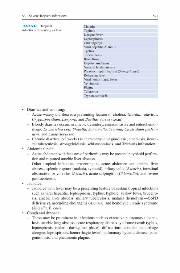
521
• Diarrhea and vomiting: – Acute watery diarrhea is a presenting feature of cholera, Giardia, rotavirus,
Cryptosporidium, Isospora, and Bacillus cereus (toxin). – Bloody diarrhea occurs in amebic dysentery, enteroinvasive and enterohemor-
rhagic Escherichia coli, Shigella, Salmonella, Yersinia, Clostridium perfrin-gens, and Campylobacter.
– Chronic diarrhea (>2 weeks) is characteristic of giardiasis, amebiasis, ileoce-cal tuberculosis, strongyloidiasis, schistosomiasis, and Trichuris infestation.
• Abdominal pain: – Acute abdomen with features of peritonitis may be present in typhoid perfora-
tion and ruptured amebic liver abscess. – Other tropical infections presenting as acute abdomen are amebic liver
abscess, splenic rupture (malaria, typhoid), biliary colic (Ascaris), intestinal obstruction or volvulus (Ascaris), acute salpingitis (Chlamydia), and severe gastroenteritis.
• Jaundice: – Jaundice with fever may be a presenting feature of certain tropical infections
such as viral hepatitis, leptospirosis, typhus, typhoid, yellow fever, brucello-sis, amebic liver abscess, miliary tuberculosis, malaria (hemolysis—G6PD deficiency), ascending cholangitis (Ascaris), and hemolytic uremic syndrome (Shigella, E. coli).
• Cough and dyspnea: – These may be prominent in infections such as extensive pulmonary tubercu-
losis, amebic lung abscess, acute respiratory distress syndrome (scrub typhus, leptospirosis, malaria during late phase), diffuse intra-alveolar hemorrhage (dengue, leptospirosis, hemorrhagic fever), pulmonary hydatid disease, para-gonimiasis, and pneumonic plague.
Table 53.1 Tropical infections presenting as fever
MalariaTyphoidDengue feverLeptospirosisChikungunyaViral hepatitis A and ETyphusTuberculosisBrucellosisHepatic amebiasisVisceral leishmaniasisParasitic hyperinfection (Strongyloides)Relapsing feverViral hemorrhagic feverYersiniosisPlagueTularemiaTrypanosomiasis
53 Severe Tropical Infections
522
• Headache: – Most febrile illnesses—especially malaria, typhoid, and dengue fever—are
accompanied by headache.• Sore throat:
– Severe sore throat with painful swallowing is characteristic of Corynebacterium diphtheriae infection.
• Hematuria, dysuria, and renal colic: – Some tropical infections such as schistosomiasis, renal tuberculosis, and chla-
mydial urethritis can present with hematuria.• History of skin rash (see Step 3).• Travel history: A list of places visited in recent past in chronological order should
be elicited. Endemic infections in different geographical areas should be consid-ered in differential diagnosis.
• Occupational history: Exposure to contaminated water source may be a clue to leptospirosis.
• Seasonal variation: Many tropical infections have propensity to occur during the monsoon season.
• Animal exposure: pet dogs (ticks—rickettsial infection, ehrlichiosis etc.)
Step 3: Perform Focused Physical Examination
• General examination: Examine for anemia, lymphadenopathy, jaundice, and edema.
• Skin: Many tropical infections present with skin manifestations, and these should be searched meticulously: – Maculopapular rash—dengue, typhus, measles, and rubella. – Urticaria—strongyloidiasis and schistosomiasis. – Petechial rash—typhus, meningococcemia, and viral hemorrhagic fever. – Vesicles—chicken pox, herpes simplex, and herpes zoster. – Eschar—Scrub typhus (Fig. 53.1).
• Abdomen: Examine for hepatosplenomegaly and abdominal distension: – Predominant hepatomegaly—viral hepatitis, amebic liver abscess, leptospiro-
sis, yellow fever, and brucellosis. – Predominant splenomegaly—malaria, typhoid, typhus, visceral leishmania-
sis, and hydatid disease. – Abdominal distension could be due to ascites (tuberculosis) or dilated bowel
loops (Shigella dysentery).• Cardiorespiratory system:
– Relative bradycardia is a feature of typhoid and typhus fever. – Pleural effusion (tuberculosis). – Appearance of new murmurs—especially regurgitant—and infective
endocarditis.• Central nervous system:
– Confusion and decreased conscious level: cerebral malaria, typhoid, dengue fever, leptospirosis, typhus, rabies, and viral hemorrhagic fever.
D. Chaudhry et al.
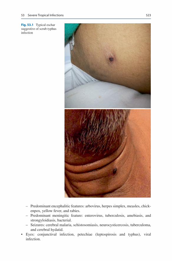
523
– Predominant encephalitic features: arbovirus, herpes simplex, measles, chick-enpox, yellow fever, and rabies.
– Predominant meningitic feature: enterovirus, tuberculosis, amebiasis, and strongyloidiasis, bacterial.
– Seizures: cerebral malaria, schistosomiasis, neurocysticercosis, tuberculoma, and cerebral hydatid.
• Eyes: conjunctival infection, petechiae (leptospirosis and typhus), viral infection.
Fig. 53.1 Typical eschar suggestive of scrub typhus infection
53 Severe Tropical Infections

524
Step 4: Syndromic Approach to the Diagnosis and Management of Tropical Infections
• Presenting syndrome – Undifferentiated fever (up to 2 weeks): Malaria (P. falciparum), scrub typhus,
leptospirosis, typhoid, dengue and other common viral illness. – Fever with rash/thrombocytopenia: Dengue, rickettsial infections, meningo-
coccal infection, malaria (usually falciparum), leptospirosis, measles, rubella and other viral exanthem.
– Fever with ARDS: Scrub typhus, falciparum malaria, influenza including H1N1, leptospirosis, hantavirus infection, melioidosis, severe community acquired pneumonias due to Legionella spp. and Streptococcus pneumoniae and diffuse alveolar haemorrhage due to collagen vascular diseases.
– Febrile encephalopathy: Encephalitis (Herpes simplex virus encephalitis, Japanese B and other viral encephalitis), meningitis (S. pneumoniae, Neisseria meningitidis, Haemophilus influenzae, enteroviruses), scrub typhus, cerebral malaria and typhoid encephalopathy.
– Fever with multiorgan dysfunction: Bacterial sepsis, falciparum malaria, lep-tospirosis, scrub typhus, dengue, hepatitis A or E with fulminant hepatic fail-ure and hepato-renal syndrome, Hanta virus infection, hemophagocytosis and macrophage activation syndrome.
• Send the investigation according to the presenting syndrome – Fever with a rash/thrombocytopenia: dengue serology, platelet count, chikun-
gunya serology, meningococcal serology, rickettsial serology, Widal test (typhoid), Epstein–Barr virus serology
– Fever with ARDS: Scrub typhus serology, malaria thick thin blood smears and antigen, H1N1 PCR testing, leptospirosis serology.
– Fever with multiorgan dysfunction: malaria parasite thick, thin blood smear and antigen, Leptospira serology, dengue serology, Legionella serology, vari-cella and influenza serology.
– Fever with encephalopathy—specific investigation: MP smear and antigen; lumber puncture; CT scan for tubercular meningitis, pyogenic meningitis, and viral encephalitis; herpesvirus serology and PCR.
• Specific tests – Dengue
Viral isolation, viral RNA (RT-PCR) and Dengue NS1 (immunoassay) antigen becomes positive within first 4–5 days.ELISA test for IgM antibodies (positive day 6)—IgG antibodies appear after 7–10 days and last for months to years. In the secondary dengue, IgG antibodies are present in high titer early in illness.Detection of antibodies by hemagglutination inhibition assay: at least four-fold rise in titer of neutralizing antibodies in paired samples.
– LeptospirosisSerology with the microscopic agglutination test is the gold standard with either a fourfold rise in titers between acute and convalescent serum or a single titer of more than 1:800 being diagnostic.
D. Chaudhry et al.
525
Other serological tests are IgM antibody by an enzyme-based dot immunoas-say with a sensitivity of 30% at 3 days and of 100% at 10 days into the illness.Polymerase chain reaction (PCR) test for Leptospira antigen shows con-siderable promise.Raised creatine phosphokinase levels is a supportive finding.
– MalariaThree thick and thin smears 12–24 h apart should be obtained. The highest yield of peripheral parasites occurs during or soon after a fever spike; how-ever, smears should not be delayed while awaiting fever spikes.Thick smears are 20 times more sensitive than thin smears, but speciation may be more difficult. The parasitemia can be calculated based on the number of infected RBCs.Thin smears are less sensitive than thick smears but facilitate speciation. This should be considered a qualitative test.The quantitative buffy coat is a technique that is as sensitive as thick smears.Malarial antigen—immunochromatographic tests based on antibodies to malarial antigen like histidine-rich protein-2 (Pf HRP2), parasite LDH (pLDH), or plasmodium aldolase appear to be very sensitive and specific.RDT has sensitivity and specificity of >95%. Malaria is ruled out if two RDTs respectively are negative.
– Rickettsial infectionLook for eschar (black crust, 5–20 mm)—found in 40% cases.IgM Scrub TyphusWeil Felix OXK: 1 in 320 or more (highly specific, poor sensitivity. Serology for typhus fever—Antibody for 65 KD antigen. Rapid immuno-chromatographic test has sensitivity and specificity of 80–90%.Indirect fluorescent antibody: “Gold standard”.
– Enteric feverThe diagnosis of typhoid fever is primarily clinical.Cultures are widely considered 100% specific. Cultures of bone marrow is 90% sensitive until at least 5 days after commencement of antibiotics. Blood, intestinal secretions and stool cultures are positive in 85–90% of patients who present within the first week of onset. Multiple blood cultures (>3) yield a sensitivity of 73–97%. Large volume (10–30 ml) blood culture may increase the likelihood of detection.
– Special investigationsProcalcitonin, ESR, CRP.Aspirates, scrapings, and pustular fluid may be obtained for Gram staining and culture. When a herpes simplex virus infection is suspected, a Tzanck test may be performed by unroofing a lesion and taking a scraping of the lesion base.Biopsy samples from nonhealing or persistent purpuric lesions: Biopsy of inflammatory dermal nodules, ulcers, and muscles (tropical pyomyositis) should be done.HIV serology.Imaging: chest x-ray, echo, ultrasound of abdomen, and CT scan (when indicated).
53 Severe Tropical Infections

526
Step 5: Start General Supportive Care and Specific Organ Support
• Many tropical infections are self-limiting. Close monitoring and general organ support in the initial days or weeks of viremia or parasitemia will salvage many patients.
Step 6: Initiate Empirical Therapy Based on Initial Presentation
• Specific therapy is available only for a few tropical infections.• Empirical antibiotics based on syndromic approach and the endemicity of a par-
ticular infection in the geographical region, should be started at the time of admission. De-escalation of antibiotics should be done once specific infection is identified.
• Ceftriaxone and doxycycline are empirical antibiotics of choice to cover typhoid fever, leptospirosis and scrub typhus, once malaria is excluded.
• In case of contraindication to doxycycline, azithromycin should be used.
Step 7: Start Specific Treatment Once the Diagnosis Is Confirmed
• Dengue – A protocol for intravenous fluid therapy has been developed by the World
Health Organization (WHO). – An initial bolus of 5% dextrose in normal saline or Ringer lactate (20 mL/kg
of body weight) is infused over 15 min, followed by continuous infusion (10–20 mL/kg/h, depending on the clinical response) until vital signs and urine output normalize.
– Crystalloids are equally effective as colloids in fluid resuscitation. – Normalization of the hematocrit is an important goal of early fluid repletion. – However, a normal or low hematocrit may be misleading in patients with
overt bleeding and severe hypovolemia. – Close clinical observation is essential, even after normal blood volume is
restored, because patients can develop shock for 1–2 days after initial fluid resuscitation, which represents the period of increased vascular permeability in dengue hemorrhagic fever.
– Management of fever:Control fever with paracetamol, cold sponging, and cold IV fluids.Avoid aspirin and nonsteroidal anti-inflammatory drugs due to bleeding risk and risk of developing Reye syndrome (encephalopathy).
– Manage shock and multiorgan failure.
D. Chaudhry et al.
527
– Manage secondary infections. – Manage complications. – Clinical guides for bleeding.
Bleeding timeFundus examination—retinal hemorrhagesHess capillary test/Tourniquet test: positive is more than 20 petechiae in an area of 1 in. when the blood pressure cuff is inflated midway between sys-tolic and diastolic blood pressure for 5 min.
– Platelet transfusions need to be given for symptomatic thrombocytopenia. – Platelet transfusions have not been shown to be effective in preventing or
controlling hemorrhage but may be warranted in patients with severe throm-bocytopenia (<10,000/mm3) and active bleeding. Prophylactic platelet trans-fusions in patients with severe thrombocytopenia but without active bleeding are generally not recommended.
– Manage complications of fluid therapy in dengue fever. – A decrease in hematocrit together with stable hemodynamic status and ade-
quate urine output indicates hemodilution and/or reabsorption of extravasated fluids.
– Judicious use of intravenous fluids with proper monitoring is recommended. – Fluid therapy may have to be discontinued if required, immediately, to avoid
pulmonary edema, electrolyte imbalance, hypo- or hypernatremia, and hyper-chloremic metabolic acidosis.
• Leptospirosis – Treatment involves the use of crystalline penicillin at a dose of six million
units daily or ceftriaxone 1 g every 12 h. – In penicillin-allergic patients, intravenous or oral doxycycline, 100 mg every
12 h, can be used. – Manage shock, disseminated intravascular coagulation, and multiorgan
failure.• Scrub typhus
– Doxycycline is the drug of choice. – In case of small children and pregnant women, azithromycin is the drug of
choice. – Rifampicin has also been used as an alternative drug.
• Enteric Fever – Ceftriaxone i.v. 50–75 mg/kg/day for 10–14 days is the drug of choice to
cover MDR S. typhi. Azithromycin and Ciprofloxacin are alternatives. – Consider dexamethasone 3 mg/kg followed by 1 mg/kg 6 hourly for 48 h in
selected cases with encephalopathy, hypotension or DIC.• Falciparum malaria
– For Plasmodium falciparum infections acquired in areas without chloroquine- resistant strains, patients should be treated with oral chloroquine. A chloroquine dose of 600 mg base (=1000 mg salt) should be given initially, followed by
53 Severe Tropical Infections

528
300 mg base (=500 mg salt) at 6, 24, and 48 h after the initial dose for a total chloroquine dose of 1500 mg base (=2500 mg salt).
– For chloroquine-resistant strains, treatment options are as follows:Artemisinin derivatives clear parasites very rapidly, are now a key compo-nent of malaria treatment worldwide, and have been shown to reduce mor-tality in severe malaria compared with parenteral quinine. Artemisinin-based combination therapies, including artesunate–mefloquine, artemether–lumefantrine, artesunate–amodiaquine, and dihydroartemisinin–pipera-quine, are highly efficacious.Under the CDC protocol, intravenous artesunate is administered in four equal doses of 2.4 mg/kg of body weight over a period of 3 days. The dos-ing schedule recommended by the WHO entails doses every 12 h on day 1 and then once daily.Up to 7 days of therapy may occasionally be indicated in very ill patients.
• Quinine sulfate: Quinine has a rapid onset of action and, in combination with tetracycline, doxycycline, or clindamycin, has been shown to be a very effica-cious treatment option for P. falciparum infections acquired in regions with chloroquine- resistant strains.
D. Chaudhry et al.
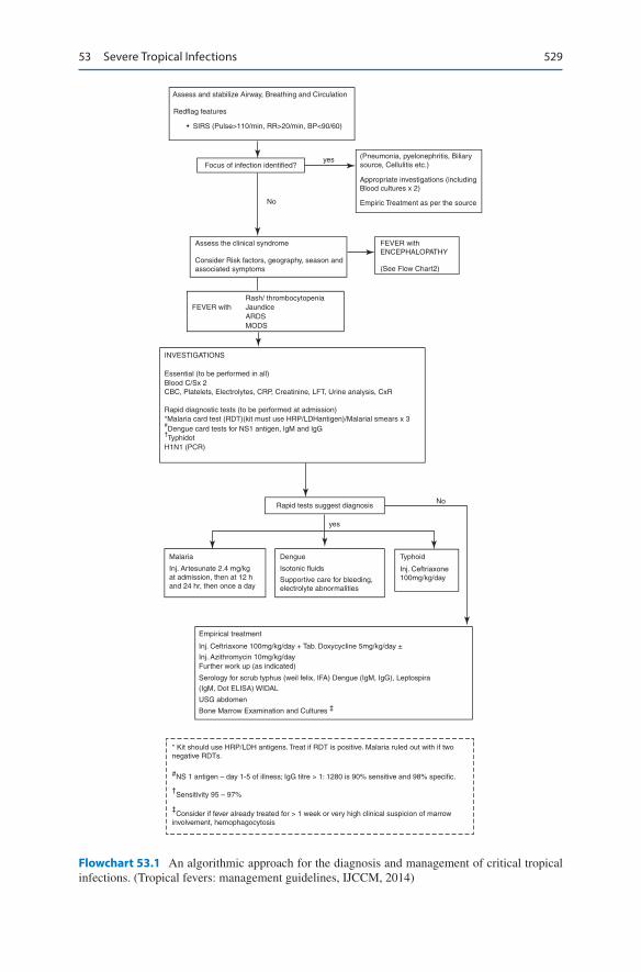
529
Assess and stabilize Airway, Breathing and Circulation
Redflag features
• SIRS (Pulse>110/min, RR>20/min, BP<90/60)
Focus of infection identified?(Pneumonia, pyelonephritis, Biliarysource, Cellulitis etc.)
Appropriate investigations (includingBlood cultures x 2)
Empiric Treatment as per the source
FEVER withENCEPHALOPATHY
(See Flow Chart2)
Assess the clinical syndrome
FEVER with
INVESTIGATIONS
Rapid tests suggest diagnosisNo
yes
Malaria Dengue Typhoid
Empirical treatment
* Kit should use HRP/LDH antigens. Treat if RDT is positive. Malaria ruled out with if twonegative RDTs.
#NS 1 antigen – day 1-5 of illness; IgG titre > 1: 1280 is 90% sensitive and 98% specific.
†Sensitivity 95 – 97%
‡Consider if fever already treated for > 1 week or very high clinical suspicion of marrow
involvement, hemophagocytosis
(IgM, Dot ELISA) WIDAL
USG abdomen
Bone Marrow Examination and Cultures ‡
Inj. Azithromycin 10mg/kg/dayFurther work up (as indicated)
Serology for scrub typhus (weil felix, IFA) Dengue (IgM, IgG), Leptospira
Inj. Ceftriaxone 100mg/kg/day + Tab. Doxycycline 5mg/kg/day ±
Inj. Ceftriaxone100mg/kg/day
Isotonic fluids
Supportive care for bleeding,electrolyte abnormalities
Inj. Artesunate 2.4 mg/kgat admission, then at 12 hand 24 hr, then once a day
Essential (to be performed in all)Blood C/Sx 2
H1N1 (PCR)
CBC, Platelets, Electrolytes, CRP, Creatinine, LFT, Urine analysis, CxR
Rapid diagnostic tests (to be performed at admission)*Malaria card test (RDT)(kit must use HRP/LDHantigen)/Malarial smears x 3#Dengue card tests for NS1 antigen, lgM and lgG
†Typhidot
Rash/ thrombocytopeniaJaundiceARDSMODS
Consider Risk factors, geography, season andassociated symptoms
No
yes
Flowchart 53.1 An algorithmic approach for the diagnosis and management of critical tropical infections. (Tropical fevers: management guidelines, IJCCM, 2014)
53 Severe Tropical Infections
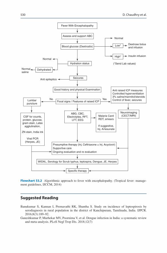
530
Fever With Encephalopathy
Normal
Normal
Anti epileptics
Lumbarpuncture
CSF for counts,protein, glucose,gram stain, Latex
agglutination,
ZN stain, India ink
Viral PCR(Herpes, JE)
Normalsaline
Dehydrated
Low*
High*
Dextrose bolusand infusion
Insulin infusion
(*Send Lab values)
Assess and support ABC
Blood glucose (Dextrostix)
Hydration status
Seizures
Good history and physical Examinaiton
Focal signs / Features of raised ICP
ABG, CBC,Electrolytes, RFT,
LFT, EEG
Presumptive therapy (Inj. Ceftriaxone ± Inj. Acyclovir)Supportive careOngoing evaluation and re evaluation
WIDAL, Serology for Scrub typhus, leptospira, Dengue, JE, Herpes
Specific therapy
No Yes
Neuroimaging(CECT/MRI)Malaria Card
RDT, smears
If suggestive,Inj. Artesunate
Anti raised ICP measuresControlled hyperventilation3% saline/mannitol/steroidsControl of fever, seizures
Flowchart 53.2 Algorithmic approach to fever with encephalopathy. (Tropical fever: manage-ment guidelines, IJCCM, 2014)
Suggested Reading
Banukumar S, Kannan I, Premavathi RK, Shantha S. Study on incidence of leptospirosis by serodiagnosis in rural population in the district of Kanchipuram, Tamilnadu, India. IJPCR. 2016;8(3):189–92.
Ganeshkumar P, Murhekar MV, Poornima V, et al. Dengue infection in India: a systematic review and meta-analysis. PLoS Negl Trop Dis. 2018;12(7)
D. Chaudhry et al.

531
Karnad DR, Richards GA, Silva GS, Amin P, Council of the World Federation of Societies of Intensive and Critical Care Medicine. Tropical diseases in the ICU: a syndromic approach to diagnosis and treatment. J Crit Care. 2018;46:119–26.
Luce-Fedrow A, Lehman ML, Kelly DJ, et al. A review of scrub typhus (Orientia tsutsugamushi and related organisms): then, now, and tomorrow. Trop Med Infect Dis. 2018;3(1):8. A review article on Scrub Typhus.
Singhi S, Chaudhary D, Varghese GM, Bhalla A, Karthi N. Tropical fevers: management guide-lines. Indian J Crit Care Med. 2014;18(2):62–9. Guidelines for the management of tropical fevers.
Websites
https://www.astmh.org/education-resources/tropical-medicine-q-a/major-tropical-diseaseshttps://www.who.int/topics/tropical_diseases/en/
53 Severe Tropical Infections
533© Springer Nature Singapore Pte Ltd. 2020R. Chawla, S. Todi (eds.), ICU Protocols, https://doi.org/10.1007/978-981-15-0898-1_54
S. Todi (*) Department of Critical Care & Emergency Medicines, A.M.R.I. Hospital, Kolkata, India
R. Chawla Department of Respiratory, Critical Care & Sleep Medicine, Indraprastha Apollo Hospitals, New Delhi, India
54New-Onset Fever
Subhash Todi and Rajesh Chawla
New onset of fever is an everyday problem encountered in the ICU. The reason could be manifold such as noninfectious cause, mild infection, or an initial presen-tation of severe infection. This should trigger a careful clinical assessment and a systematic approach to differentiate these possibilities.
Step 1: Record Temperature
• All patients in the ICU should have, as a minimum, hourly temperature recorded and charted in the nursing record as per the ICU protocol.
• The site of recorded temperature should be marked in the nursing chart (O = oral, R = rectal, A = axillary, T = tympanic).
• All ICUs should have access to a core temperature measurement device (tym-panic, rectal), properly calibrated and sterilized.
• Temperature may be recorded as centigrade or Fahrenheit.• The same method and site of measurement should be used repeatedly to facilitate
the trending of serial measurements.
A 70-year-old male patient was admitted to the ICU with hemorrhagic stroke. On the fifth day of admission, he spiked a fever of 100 °F orally. He was not intubated and had an indwelling urinary catheter, peripheral intravenous can-nula, and a nasogastric tube. His sensorium remained unchanged, and he was hemodynamically stable.

534
• In immunocompetent patients who are stable, core temperature below 101 °F (equivalent to oral temperature of 100 F) in which clinical examination does not reveal any definite source of fever may be observed for a few hours before initiat-ing investigation.
• As a general rule, core temperature of more than 38.3 °C (101 °F) warrants spe-cial attention in all patients.
• In immunocompromised patients, temperature of any degree should be investigated.
Step 2: Take a Detailed History
• Take proper history from the bedside nurse and do a thorough chart review.• Enquire about medications, blood transfusion, diarrhea, rash, new procedure,
dressing changes, and catheters and line manipulation (both insertion and removal),dialysis
• Duration of indwelling urinary catheter and central and arterial line placement.
Step 3: Perform Focused Clinical Examination
• Examine for any source of infection or noninfectious causes of fever (Tables 54.1 and 54.2).
• Perform systematic head-to-toe examination: – Purulent nasal discharge, sinus tenderness. – Parotid swelling, oral hygiene. – Chest auscultation (including bases). – New murmur.
Table 54.1 Noninfectious causes of fever in the ICU
Drug fever β-Lactam, antiepileptics, sulfonamides Antipsychotics (neuroleptic malignant syndrome, serotonin syndrome)Blood products, IV contrast, immunoglobulins, albuminCNS causes: Blood in cerebrospinal fluid, pontine bleedPulmonary/cardiac causes: Acute respiratory distress syndrome, pulmonary emboli, fat emboli, pericarditisAbdominal causes: Ischemic gut, pancreatitis, acalculous cholecystitisMetabolic: Adrenal insufficiency, thyroid storm, goutPostoperative fever (48 h), postprocedure (bronchoscopy)Thrombophlebitis, decubitus ulcer, hematoma, deep venous thrombosis (DVT)
Table 54.2 Infectious causes of new-onset fever in the ICU
Ventilator-associated pneumoniaSinusitisCatheter-related sepsisUrinary tract infectionClostridium difficile diarrheaComplicated wound infections
S. Todi and R. Chawla

535
– Abdominal examination, suprapubic tenderness. – Vascular device sites for purulence and erythema, note insertion date. – Urinary catheter site. – Surgical wounds, drain sites (take off dressings). – Skin rash. – Gynecological examination. – Painful leg swelling. – Decubitus ulcer.
Step 4: Send Investigations
• If clinical examination does not strongly suggest a noninfectious source, two sets of blood cultures should be sent in the following: – In patients with temperature above 101 °F. – In patients who are hemodynamically unstable or develop new organ dysfunc-
tion or immunosuppressed, with new onset of fever of any degree.• All ICUs should have a blood culture drawing protocol in consultation with
microbiology department: – Skin disinfection: 2% chlorhexidine or 2% iodine, give 30 s for drying. – Site: two peripheral venipunctures, or one from distal lumen of the central
line and another from periphery, If a blood sample cannot be drawn from a peripheral vein, then blood can be drawn from different lumens of multilumen catheter.
– Minimum two sets (each set containing two bottles, one aerobic another anaerobic)—at least 10 mL in each bottle —to be inoculated directly into the culture bottle.
– Labeling should be done carefully for site, date, and time.• If infection is suspected clinically and there is a focus on infection, the following
investigations should be sent: – Total and differential white blood cell count, C-reactive protein (CRP), pro-
calcitonin when presence of infection is in doubt. – Focused imaging such as chest X-ray, abdominal ultrasonography, CT scan of
the abdomen/chest. – If there is a history of diarrhea, send stool for occult blood, pus cells,
Clostridium difficile toxin and GDH. – Urinalysis and culture sensitivity. – Transthoracic/transesophageal echocardiogram—look for vegetation. – Sputum, endotracheal suction, Non bronchoscopy or bronchoscopy with
bronchoalveolar lavage sent for Gram stain, AFB stain, Fungal stain and quantitative bacterial culture and sensitivity.
– Avoid sending cultures of urinary catheter tips, superficial wound swabs and drains which have been in situ for >48 h.
• Trend of white blood cell count or CRP is valuable to ascertain any new infection.
54 New-Onset Fever

536
Step 5: Remove Lines if There is a Suspicion of Line Sepsis
• All patients with a central vascular access of some duration and persistent fever without any other obvious source of infection should have the line removed at the earliest if any of the following criteria are met: – Inflammation or purulence present at the insertion site or along the tunnel. – No other identifiable source of infection. – An abrupt onset, associated with fulminant shock. – Nonfunctioning lumen. – Fever on starting infusion/dialysis. – Persistent bacteremia or fungemia.
• The intracutaneous and tip of the central line should be sent for semiquantitative culture.
• Central line infection is considered significant for the following situations: – Culture of the same organism from both the catheter tips and at least one per-
cutaneous blood culture. – Multiple blood cultures containing organisms such as Staphylococci (espe-
cially coagulase-negative Staphylococci) and Candida. – Positive semiquantitative culture of the catheter tip (>15 cfu). – Differential time to positivity—growth detected from the catheter sample at
least 2 h before growth detected from the peripheral vein sample (Fig. 54.1).
Step 6: Make a Diagnosis
• In patients on the ventilator, pneumonia should be considered by clinical exami-nation, purulence of endotracheal secretions, raised white blood cell count, and new or worsening lung infiltrate on chest skiagram.
• Consider urosepsis in patients with an indwelling bladder catheter and increased pus cells in urine.
• Consider sinusitis in patients with the nasogastric tube and purulent nasal discharge.
• Consider inflammatory diarrhea (stool positive for occult blood) with abdominal distension, and in patients on antibiotics, investigate for C. difficile colitis.
• Consider gynecological infection if vaginal discharge is present.
Step 7: Start Treatment
• If infectious cause for fever is suspected, empirical antibiotic therapy should be started.
• The choice of antibiotics should be guided by the hospital antibiotic policy and suspected source of infection.
S. Todi and R. Chawla

537
• In patients with persistent fever despite broad-spectrum antibiotics, look for occult source of sepsis.
• Consider fungemia in patients colonized by fungus.• Never forget noninfectious causes of fever.
Suggested Reading
Kalil AC, Metersky ML, Klompas M, et al. Management of adults with hospital-acquired and ventilator associated pneumonia:2016 clinical practice guidelines by the infectious diseases Society of America and the American Thoracic Society. Clin Infect Dis. 2016;63(5):e61–e111. Comprehensive guidelines for the management of ventilator associated pneumonia.
Marik PE. Fever in ICU. Chest. 2000;117:855–69.O’Grady NP, Alexander M. Guidelines for the prevention of intravascular catheter-related infec-
tions. 2011. www.cdc.gov.
Confirm fever of > 100ºF
Look for noninfectious cause offever (Table1)
Send blood cultures for fever>101ºF
or immunocompromised
or hypotension
or new organ dysfunction
Persistent fever on broad-spectrum antibiotics
Reconsider noninfectioussource of fever (Table 1)
Fungemia––look for fungalcolonization, send fungalcultureStart preemptive antifungaltherapy
Occult source of sepsis––chestCT, USG/CT abdomen, echo,bronchoscopy, repeat bloodcultures, venous Doppler of legsas indicated
Focused clinical examand chart review
If Source of infectionsuspected appropriateinvestigation andtreatment (Table2)
If no obvious source of fever andthe patient has a central/arterialline placed for more than 2 daysconsider line sepsis
Fig. 54.1 An approach to new onset of fever in the ICU
54 New-Onset Fever

538
Websites
www.cdc.govwww.sccm.orgwww.isccm.org
S. Todi and R. Chawla
539© Springer Nature Singapore Pte Ltd. 2020R. Chawla, S. Todi (eds.), ICU Protocols, https://doi.org/10.1007/978-981-15-0898-1_55
S. Todi (*) Department of Critical Care & Emergency Medicines, A.M.R.I. Hospital, Kolkata, India
R. Chawla Department of Respiratory, Critical Care & Sleep Medicine, Indraprastha Apollo Hospitals, New Delhi, India
55Fungal Infections
Subhash Todi and Rajesh Chawla
With increasing incidence of elderly patient population, comorbidities, prolonged ICU stay, invasive therapies, major surgeries, solid organ and hematopoeitic stem cell trans-plant, use of broad-spectrum antibiotics and immunosuppresives, there is a rising inci-dence of fungal infection and use of antifungal drugs. Candidemia is the most common invasive fungal infection encountered in the ICU and is the fourth most common cause of bloodstream infection. Candida albicans is the most common species identified worldwide, but the incidence of non albicans candida is growing, like Candida tropi-calis (in the tropical countries), Candida glabrata and Candida krusei with the later two showing increasing azole resistance Attributable mortality with candidemia could be as high as 47%. Other fungal infections seen in the ICU are invasive aspergillosis and zygomycosis. With increasing threat of developing resistance, antifungal drugs should be used judiciously, either prophylactic, empiric, or as a definitive therapy.
Step 1: Take Focused History and Perform Physical Examination to Identify the Patient at Risk of Candidemia
• Classical risk factors described for candidemia are as follows: – Prolonged ICU stay.
A 50-year-old diabetic male patient was admitted to hospital with pancreatitis. He had received imipenem for 2 weeks. He had spiking fever and abdominal CT showed peripancreatic collection. Growth of Candida species was found in his urine.

540
– Use of broad-spectrum antibiotics. – Central venous catheters. – Parenteral nutrition. – Neutropenia. – Candida colonization. – Diabetes. – Renal replacement therapy. – Pancreatitis. – Implantable prosthetic devices. – Immunosuppressive agents (glucocorticoid, chemotherapeutic agents, immu-
nomodulating agents). – Abdominal surgery.
• All patients with suspected candidemia should have an ophthalmological exami-nation (with pupils dialated by an ophthalmologist) to rule out endophthalmitis.
• Cardiac examination should be performed to look for features suggestive of endocarditis.
• Abdominal examination is required for hepatosplenomegaly suggestive of vis-ceral candidiasis.
• Neurological examination is performed to look for features of meningoencepha-litis in patients with intraventricular catheters.
• Cutaneous examination to look for skin changes of disseminated candidiasis.
Step 2: Send Blood for Fungal Cultures for Persistent Fever
• Presence of Candida in blood culture is usually pathological.• Fungal cultures can be obtained in the aerobic culture bottles used for routine
bacterial cultures.• Blood cultures are positive in only 50–70% of patients with invasive
candidiasis.• In patients with documented candidemia, repeat blood cultures should be
obtained frequently, preferably daily or alternate days, till they become negative.
Step 3: Differentiate Colonization from Infection with Candida
• Growth of Candida from respiratory secretions usually represents colonization, and therefore, it should not be treated.
• In the absence of systemic features, growth of Candida in the urine often repre-sents colonization.
• Candida colonisation is frequent in surgical drains which have been in situ for some time and from superficial wound swab.
S. Todi and R. Chawla
541
Step 4: Identify Patients in Whom Empiric Antifungal Therapy Should be Considered
• Early empirical therapy in patients with candidemia has been proved to reduce mortality.
• Empirical antifungal therapy should be considered in patients with one or more risk factors for invasive candidiasis and if they show one or more of the following features: – Persistent fever without a definite source. – Fever not responding to antibiotics. – Positive biomarkers for systemic fungal infection (β-d-glucan for candida,
Galactomannan for aspergillus). – Candida colonization of one or more sites (e.g., urine, sputum, and skin).
Step 5: Get Familiar with Antifungal Agents Used in Patients with Suspected or Proven Candidemia
• Choice of antifungal agents in candidemia is guided by many factors: – History of recent azole exposure—avoid azoles. – Local epidemiological data from the ICU regarding predominant Candida
species and susceptibility pattern—choose most effective antifungal agents initially pending culture results.
– Severity of illness—use fungicidal drugs in severely ill patients. – Comorbidity—check renal and hepatic function and avoid amphotericin
deoxycholate and voriconazole respectively. – Involvement of CNS, cardiac valves, and eyes—choose antifungal agents for
penetration at the infected site. – History of intolerance to any antifungal agent or drug interactions.
• Antifungal agents used for candidemia are triazoles, echinocandins, and ampho-tericin B.
Triazoles
• Triazoles include fluconazole, itraconazole, voriconazole, posaconazole and isa-vuconazole. In the general medical/surgical ICU, fluconazole and voriconazole are used most often, whereas in hemato/onco ICU posaconazole use is more frequent
• They all have similar activity against Candida albicans and are fungistatic. They have less activity against Candida glabrata and Candida krusei and Candida auris. Voriconazole is fungicidal against aspergillus. Hepatotoxicity is a side effect among all the azoles and aminotransferases should be moni-tored closely. Azoles are not nephrotoxic but need dose modification in renal dysfunction.
55 Fungal Infections
542
• They inhibit cytochrome P450 and are prone to drug–drug interaction.• Fluconazole is most widely used azole and is available both as an oral and as
intravenous formulations. It is readily absorbed orally. It achieves high concen-tration in the urine. It has the greatest penetration into the cerebrospinal fluid and vitreous body. In patients with invasive candidiasis, fluconazole should be administered with a loading dose of 800 mg (12 mg/kg), followed by a daily dose of 400 mg (6 mg/kg); a lower dosage is required in patients with creatinine clear-ance of less than 50 mL/min.
• Voriconazole is available in both oral and intravenous forms. It is used mainly for infection with Aspergillus. Its clinical use in candidiasis has been primarily for step-down oral therapy for patients with infection due to C. krusei and fluconazole- resistant, voriconazole-susceptible C. glabrata.
• In adults, the recommended oral dosing regimen includes a loading dosage of 400 mg twice daily for 1 day, followed by 200 mg twice daily.
• Intravenous voriconazole is complexed to a cyclodextrin molecule; after two loading dosages of 6 mg/kg every 12 h, a maintenance dosage of 3–4 mg/kg every 12 h is recommended. Because of the potential for cyclodextrin accumu-lation among patients with significant renal dysfunction, intravenous voricon-azole is not recommended in patients with a creatinine clearance less than 50 mL/min.
• Oral voriconazole does not require dosage adjustment for renal insufficiency, but it is the only triazole that requires dosage reduction for patients with mild-to- moderate hepatic impairment.
• Common polymorphisms in the gene encoding the primary metabolic enzyme for voriconazole result in wide variability of serum levels. Therapeutic drug level monitoring is advisable when using voriconazole.
• Drug–drug interactions are common with voriconazole and should be considered when initiating and discontinuing treatment with this compound.
Echinocandins
• Echinocandins are caspofungin, anidulafungin, and micafungin and are available only as parenteral preparations.
• These are fungicidal drugs and have equal efficacy.• All echinocandins have a few adverse effects and minimal drug–drug
interaction.• The pharmacological properties in adults are also very similar, and echinocan-
dins are administered intravenously once daily.• None of the echinocandins require dosage adjustment for renal insufficiency or
dialysis.• Caspofungin is the only echinocandin for which dosage reduction is recom-
mended for patients with moderate-to-severe hepatic dysfunction.
S. Todi and R. Chawla
543
• Intravenous dosing regimens for invasive candidiasis with the three compounds are as follows: caspofungin, loading dose of 70 and 50 mg daily thereafter; anidulafun-gin, loading dose of 200 and 100 mg daily thereafter; and micafungin, 100 mg daily.
• All echinocandins have a broad-spectrum activity against most of the Candida species including Candida auris.
Amphotericin B (Amph B)
• This is available in non-lipid formulation (Amph B deoxycholate AmB-d) or lipid formulation (ABLC, ABCD, and L-AmB).
• These are fungicidal drugs.• The three lipid formulations have different pharmacological properties and rates
of treatment-related adverse events and should not be interchanged.• All amphotericin preparations have a very broad-spectrum activity against most
Candida species.• For most forms of invasive candidiasis, the typical intravenous dosage for AmB-d
is 0.5–0.7 mg/kg daily, but dosages as high as 1 mg/kg daily should be consid-ered for invasive Candida infections caused by less susceptible species, such as C. glabrata and C. krusei.
• The typical dosage for liposomal preparations of AmB is 3–5 mg/kg daily when used for invasive candidiasis.
• Nephrotoxicity is the most common serious adverse effect associated with AmB-d therapy, resulting in acute renal failure in up to 50% of recipients. This can be minimized by avoiding concomitant use of other nephrotoxic agents, proper hydration, and saline loading prior to use of AmB-d. Its use is associated with hypokalemia and hypomagnesemia due to renal wasting, and levels of these electrolytes should be monitored and replaced.
• Liposomal preparations of AmB are considerably more expensive than AmB-d, but all have considerably less nephrotoxicity.
• AmB-d and other liposomal preparations have infusion-related toxicity with fever and rigor and require pretreatment with antipyretics.
Step 6: Choose Appropriate Antifungal Regimen for Patients with Suspected or Proven Candidemia
• Echinocandins are recommended as a drug of choice in patients with moderately severe to severe illness or patients who have had recent azole exposure and in neutropenic patients.
• Fluconazole is recommended as an empiric or definitive therapy in patients who are less critically ill and who have no recent azole exposure and no known resis-tance to fluconazole.
55 Fungal Infections
544
• Amphotericin B, preferably lipid formulation, may replace echinocandins in patients with normal renal function, cost consideration, and nonavailability of echinocandins.
• Amphotericin B, preferably liposomal preparations, may be considered in neu-tropenic patients where invasive Aspergillus or mucormycosis is a possibility.
• Transition from an echinocandin to fluconazole is recommended for patients who have isolates that are likely to be susceptible to fluconazole (e.g., C. albi-cans) and who are clinically stable.
• Combination antifungal therapy is sometimes used in the following situations: – Invasive aspergillosis refractory to amphotericin B—voriconazole with
caspofungin. – Central nervous system infection (cryptococcal meningitis)—amphotericin B
with flucytosine.• Voriconazole is recommended as step-down oral therapy for selected cases of
candidiasis due to C. krusei or voriconazole-susceptible C. glabrata.• Recommended duration of therapy for candidemia without obvious metastatic
complications is 2 weeks after documented clearance of Candida species from the bloodstream (last negative blood culture) and resolution of symptoms attrib-utable to candidemia.
• Intravenous catheter removal is strongly recommended for nonneutropenic patients with candidemia.
• In patients with endophthalmitis, consider amphotericin B deoxycholate along with flucytosine for 4–6 weeks. Consider early partial vitrectomy in severe cases.
• Consider Echinocandins for the treatment of resistant Candida auris. One may have to resort to higher dose for adequate response.
Step 7: Manage Persistent Candiduria in a Catheterized Patient
• Avoid treating with antifungal drugs in an asymptomatic, afebrile, stable patient.• Remove the catheter if possible.• Echhinocandins are not recommended for candida urosepsis• In high-risk patients such as neutropenic, urological surgery, pregnancy consider
fluconazole therapy if the species is susceptible.
Step 8: Consider Central Nervous System Candidiasis in Patients with an Intraventricular Device
• Consider liposomal amphotericin B at a dosage of 3–5 mg/kg/day with or with-out flucytosine at a dosage of 25 mg/kg/dose four times daily.
• After initial response, deescalate to fluconazole 400–800 mg daily.• Remove the infected ventricular device.
S. Todi and R. Chawla

545
Step 9: Consider Azole Prophylaxis in the Selected Group of Patients
• Prophylactic antifungal therapy has not been proven to decrease mortality from invasive candidiasis in medical/surgical ICU patients and should be avoided.
• For high-risk patients such as neutropenic, solid organ transplant, or stem cell transplant, fluconazole 400 mg (6 mg/kg) daily, posaconazole 200 mg three times a day, or an echinocandin is recommended during the period of neutropenia.
Step 10: Consider Possibility of Invasive Aspergillosis in Some Situations
• Invasive aspergillosis should be suspected in the following group of patients: – Prolonged neutropenia more than 10 days. – Hematopoietic stem cell transplantation. – Solid organ transplantation. – Corticosteroid therapy.
• Look for involvement of lungs and paranasal sinuses by CT scan, which may show a “halo sign,” a haziness surrounding nodular pulmonary infiltrate.
• Serum galactomannan assay has a moderate sensitivity and specificity for diag-nosing invasive aspergillosis. It will be falsely positive in patients treated with piperacillin–tazobactam.
• Isolation of Aspergillus hyphae from nonsterile sites like respiratory secretions may represent colonization. Demonstration of the organism in tissue biopsy is considered a gold standard, but it is difficult to perform in ICU patients.
• Voriconazole is considered a first-line agent for treatment of invasive aspergil-losis. Echinocandins have in vitro sensitivity against Aspergillus and may be considered in selected cases.
• In nonresponder combination, antifungal therapy may be tried.
Step 11: Consider Zygomycosis (Mucormycosis) in Some Specific Situations
• Consider this mould infection in patients with uncontrolled diabetes presenting with rhinocerebral disease.
• This mould also infects immunosuppressed patients and mainly involves the lung.
• Diagnosis is based mainly on tissue biopsy.• High-dose amphotericin B is considered standard first-line therapy (amphoteri-
cin B deoxycholate 1–1.5 mg/kg body wt/day or lipid amphotericin preparation at 5 mg/kg body wt/day).
55 Fungal Infections
546
• Echinocandins fluconazole and voriconazole are ineffective against zygomyco-sis. However Posaconazole and Isavuconazole are broad spectrum azoles which can be used as step down therapy in patients who have responded to a lipid for-mulations of amphoteracin-B.
• Therapy is usually given for months.• Surgical intervention is usually required in this angioinvasive disease.• Mucormycosis carries extremely poor prognosis in the absence of early aggres-
sive treatment.
Suggested Reading
Darouiche RO. Candida in the ICU. Clin Chest Med. 2009;30(2):287–93. Although treatment for documented, deep-seated Candida infections in nonneutropenic patients has been studied extensively, guidelines for the management of suspected but undocumented cases of invasive Candida infections in critically ill patients have not been clearly established.
Hsueh PR, Graybill JR, et al. Consensus statement on the management of invasive candidiasis in intensive care units in the Asia-Pacific region. Int J Antimicrob Agents. 2009;34(3):205–9. Candida albicans remains the predominant cause of invasive candidiasis in ICUs, followed by Candida tropicalis, Candida glabrata, and Candida parapsilosis. Invasive isolates of Candida spp. remain highly susceptible to fluconazole (>90% susceptible), although among Asia-Pacific countries the susceptibility rate of C. glabrata to fluconazole varies widely from 22% to 72%.
Lam SW, Eschenauer GA. Evolving role of early antifungals in the adult intensive care unit. Crit Care Med. 2009;37(5):1580–93. The use of early antifungal therapy should be reserved for patients with a high risk (10–15%) of developing invasive candidiasis (IC). There is no sin-gle predictive rule that can adequately forecast IC in critically ill patients. Clinicians should assess patients on a case-by-case basis and determine the need for early antifungal treatment strategies based on frequent evaluations of risk factors and clinical status.
Peter GP, Carol AK, et al. Clinical practice guideline for the management of candidiasis: 2016 update by the Infectious Diseases Society of America. Clin Infect Dis. 2016;62(4):e1–50. IDSA Comprehesive guidelines for the management of candidiasis.
S. Todi and R. Chawla
547© Springer Nature Singapore Pte Ltd. 2020R. Chawla, S. Todi (eds.), ICU Protocols, https://doi.org/10.1007/978-981-15-0898-1_56
S. Todi (*) Department of Critical Care & Emergency Medicines, A.M.R.I. Hospital, Kolkata, India
56Infections in the Immunocompromised Host
Subhash Todi
Managing infection in an immunocompromised patient is a challenge to any ICU physician, as the presentation is varied and subtle and needs high index of suspicion for diagnosis and multiple possible etiologies. A methodological approach to inves-tigation and choice of empirical therapy is warranted in these patients. Complexity of the problem necessitates close liaison between the microbiologist, infectious dis-ease consultant, and hemato-oncologist.
Step 1: Resuscitate
• If assisted respiration is required, initially High Flow Nasal Oxygen (HFNC) or noninvasive ventilation should be tried.
• The patient should be closely monitored, and if no improvement or deterioration occurs in 2 h, invasive ventilation should be initiated.
• Urinary catheters and central lines should be avoided as these patients are coagu-lopathic and neutropenic and have a high risk of line sepsis.
• If absolutely necessary, invasive catheters and lines should be placed with utmost aseptic precautions by an experienced person.
• Careful maintenance of the peripheral line is extremely vital in these patients.• Use of Chlorhexidine patch at the central line insertion site is recommended.
A 29-year-old female patient, 2 years after kidney transplant, on mycophenolate, sirolimus, and prednisolone, presented with semiconsciousness, ulcerating lesions around right elbow, and oliguria for the past 6 h.

548
Step 2: Take a Focused History (Table 56.1)
• This should be done to determine the type and duration of the immunocompro-mised state.
• Disease states such as hematological malignancy (leukemia and lymphoma), solid organ tumor, and conditions associated with neutropenic state should be looked for.
• History of any organ transplant and duration since transplant should be taken.• HIV status should be determined, with proper consent.• History of chemotherapy or radiotherapy should be taken.• Detailed drug history should be elicited.• Neurological, respiratory, gastrointestinal symptoms need to be elicited to nar-
row down differential diagnosis of opportunistic infection.
Step 3: Perform Focused Physical Examination (Table 56.1)
• Any breach of skin or mucosal abrasion, skin ulcer, oral ulcer, oral thrush, and perianal lesion should be searched for.
• Look for skin rash.• All insertion sites of invasive lines should be inspected for tenderness or discharge.• All suture lines and drain sites should be inspected in postoperative patients after
removing the dressing.• Look at the back for decubitus ulcer.• Do neurological, respiratory, and gastrointestinal system examination to deter-
mine organ system involvement.
Table 56.1 Syndromic approach to infection in immunocompromised patients
Pneumonia Focal/nodular infiltrateBacteria, Aspergillus, mycobacteria, Nocardia, Histoplasma Diffuse infiltrateVirus (cytomegalo, herpes), Pneumocystis, StrongyloidesMeningoencephalitis Bacteria: Neisseria, Hemophilus, Pneumococcus,
Listeria Mycobacteria: Typical and atypical Fungus: CryptococcusFocal CNS lesions Toxoplasma Tuberculoma NocardiaSevere sepsis or septic shock Gram-positive and gram-negative bacteria CandidemiaGastroenteritis Strongyloides Cryptococcosis Amebiasis
S. Todi
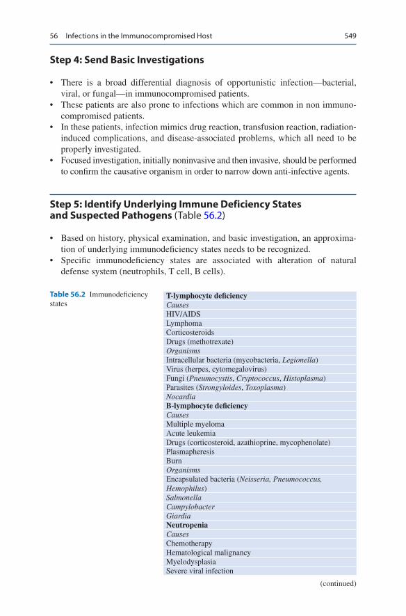
549
Step 4: Send Basic Investigations
• There is a broad differential diagnosis of opportunistic infection—bacterial, viral, or fungal—in immunocompromised patients.
• These patients are also prone to infections which are common in non immuno-compromised patients.
• In these patients, infection mimics drug reaction, transfusion reaction, radiation- induced complications, and disease-associated problems, which all need to be properly investigated.
• Focused investigation, initially noninvasive and then invasive, should be performed to confirm the causative organism in order to narrow down anti-infective agents.
Step 5: Identify Underlying Immune Deficiency States and Suspected Pathogens (Table 56.2)
• Based on history, physical examination, and basic investigation, an approxima-tion of underlying immunodeficiency states needs to be recognized.
• Specific immunodeficiency states are associated with alteration of natural defense system (neutrophils, T cell, B cells).
Table 56.2 Immunodeficiency states
T-lymphocyte deficiencyCausesHIV/AIDSLymphomaCorticosteroidsDrugs (methotrexate)OrganismsIntracellular bacteria (mycobacteria, Legionella)Virus (herpes, cytomegalovirus)Fungi (Pneumocystis, Cryptococcus, Histoplasma)Parasites (Strongyloides, Toxoplasma)NocardiaB-lymphocyte deficiencyCausesMultiple myelomaAcute leukemiaDrugs (corticosteroid, azathioprine, mycophenolate)PlasmapheresisBurnOrganismsEncapsulated bacteria (Neisseria, Pneumococcus, Hemophilus)SalmonellaCampylobacterGiardiaNeutropeniaCausesChemotherapyHematological malignancyMyelodysplasiaSevere viral infection
(continued)
56 Infections in the Immunocompromised Host

550
HypersplenismOrganismsGram-negative bacilli (enteric and nonenteric)Staphylococcus aureusCoagulase-negative StaphylococcusStreptococci, enterococciFungi (Aspergillus spp., Candida spp.)Neutrophil dysfunctionCausesDiabetesUremiaAlcoholismCirrhosisBurnOrganismsStaphylococcus aureusStreptococciMucor (Zygomycoses)Gram negative bacilli (enteric and non-enteric)Coagulase negative staphylococcusFungi (Aspergillus spp., Candida spp.)
Table 56.2 (continued)
• Patients with specific defense system alteration have propensity to be infected with certain groups of organisms, which need to be recognized.
Step 6: Initiate Empirical Anti-Infective Agents (Table 56.3)
• Guided by the type and duration of immunosuppression and primary organ sys-tem involvement, broad-spectrum anti-infective agents should be initiated against suspected organisms.
• These agents need to be deescalated once an organism is confirmed.• The dose of these agents needs to be modified depending on renal and liver func-
tion tests.• The duration of therapy with these agents depends on clinical response of the
patient.
Step 7: Beware of Infection Mimics in Immunocompromised Patient
• Immune reconstitution response Syndrome (IRIS) is a paradoxical response noted in HIV patient when HAART is started along with the treatment of an opportunistic infection like cryptococcosis or tuberculosis.
S. Todi
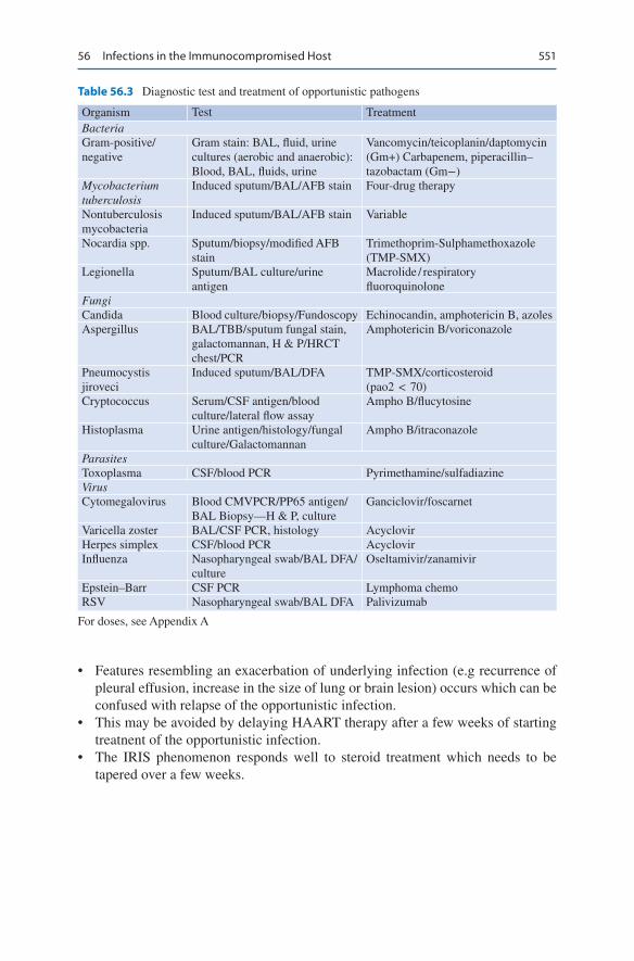
551
• Features resembling an exacerbation of underlying infection (e.g recurrence of pleural effusion, increase in the size of lung or brain lesion) occurs which can be confused with relapse of the opportunistic infection.
• This may be avoided by delaying HAART therapy after a few weeks of starting treatnent of the opportunistic infection.
• The IRIS phenomenon responds well to steroid treatment which needs to be tapered over a few weeks.
Table 56.3 Diagnostic test and treatment of opportunistic pathogens
Organism Test TreatmentBacteriaGram-positive/negative
Gram stain: BAL, fluid, urine cultures (aerobic and anaerobic): Blood, BAL, fluids, urine
Vancomycin/teicoplanin/daptomycin (Gm+) Carbapenem, piperacillin–tazobactam (Gm−)
Mycobacterium tuberculosis
Induced sputum/BAL/AFB stain Four-drug therapy
Nontuberculosis mycobacteria
Induced sputum/BAL/AFB stain Variable
Nocardia spp. Sputum/biopsy/modified AFB stain
Trimethoprim-Sulphamethoxazole (TMP-SMX)
Legionella Sputum/BAL culture/urine antigen
Macrolide / respiratory fluoroquinolone
FungiCandida Blood culture/biopsy/Fundoscopy Echinocandin, amphotericin B, azolesAspergillus BAL/TBB/sputum fungal stain,
galactomannan, H & P/HRCT chest/PCR
Amphotericin B/voriconazole
Pneumocystis jiroveci
Induced sputum/BAL/DFA TMP-SMX/corticosteroid (pao2 < 70)
Cryptococcus Serum/CSF antigen/blood culture/lateral flow assay
Ampho B/flucytosine
Histoplasma Urine antigen/histology/fungal culture/Galactomannan
Ampho B/itraconazole
ParasitesToxoplasma CSF/blood PCR Pyrimethamine/sulfadiazineVirusCytomegalovirus Blood CMVPCR/PP65 antigen/
BAL Biopsy—H & P, cultureGanciclovir/foscarnet
Varicella zoster BAL/CSF PCR, histology AcyclovirHerpes simplex CSF/blood PCR AcyclovirInfluenza Nasopharyngeal swab/BAL DFA/
cultureOseltamivir/zanamivir
Epstein–Barr CSF PCR Lymphoma chemoRSV Nasopharyngeal swab/BAL DFA Palivizumab
For doses, see Appendix A
56 Infections in the Immunocompromised Host
552
Step 8: Beware of Breakthrough Infection
• Antibiotic or antifungal prophylaxis is routinely used in many immunosup-pressed patients before initiation of chemotherapy. Breakthrough infections can occur with organisms which are resistant to the prophylactic agent used (e.g. Aspergillus infection in patients prophylaxed with fluconazole, or mucor infec-tion in patients prophylaxed with voriconazole).
Suggested Reading
Fishman JA. Infection in solid-organ transplant recipients. N Engl J Med. 2007;357(25):2601–14. An authoritative review on the subject.
Gea-Banacloche JC, Opal SM. Sepsis associated with immunosuppressive medications: an evidence- based review. Crit Care Med. 2004;32(11 Suppl):S578–90.
Penack O, Buchheidt D. Management of sepsis in neutropenic patients: guidelines from the infec-tious diseases working party of the German Society of Hematology and Oncology. Ann Oncol. 2011;22(5):1019–29.
Picazo JJ. Management of the febrile neutropenic patient: a consensus conference. Clin Infect Dis. 2004;39(Suppl 1):S1–6.
Rosen MJ, Narasimhan M. Critical care of immunocompromised patients: human immunodefi-ciency virus. Crit Care Med. 2006;34(9 Suppl):S245–50.
Website
cid.oxfordjournals.orgFree text available on many references on the subject.
S. Todi
553© Springer Nature Singapore Pte Ltd. 2020R. Chawla, S. Todi (eds.), ICU Protocols, https://doi.org/10.1007/978-981-15-0898-1_57
S. Samavedam (*) · R. Reddy Department of Critical Care, Virinchi Hospitals, Hyderabad, India
R. Pande Department of Critical Care, BLK Superspeciality Hospital, New Delhi, India
57Central Line Related Blood Stream Infections (CRBSI)
Srinivas Samavedam, Ramakrishna Reddy, and Rajesh Pande
Central line (Central Venous Catheter or CVC) is an intravascular access device or catheter that terminates at or close to the heart or in one of the great vessels like internal jugular vein, subclavian vein, superior vena cava, inferior vena cava, bra-chiocephalic veins, pulmonary artery, external iliac veins, common iliac veins or femoral veins. It can be short term catheter (<14 days) or long-term catheter (>14 days).
CVC infection implies central line colonization with accompanying clinical manifestations suggestive of infection which can be local (phlebitis) or systemic (CRBSI).CRBSI occurrence in the ICU is common.
45-year-old male patient presented to emergency department with abdominal pain and vomiting. On evaluation he was found to have pancreatic tumor and subsequently underwent whipples procedure (pancreato-duodenectomy). He was doing well till fifth postoperative day when he deteriorated and was reex-plored and found to have leak at pancreas jejunum junction which was cor-rected. Arterial line and internal jugular vein central line were inserted intraoperatively On seventh POD from re-exploration he developed fever spikes of 101 °F and was worked up for development of nosocomial infection. His chest X-ray did not show any infiltrates, surgical site was clean. Urinary catheter was changed. Paired blood cultures were sent with one set from periphery.

554
Central Line Associated Blood Stream Infections (CLABSI) Versus Central Line Related Blood Stream Infection (CRBSI)
• CLABSI is a surveillance definition used by the Centre for Disease Control, Atlanta, USA (CDC) and is defined as recovery of a pathogen from a blood cul-ture in a patient who had central line at the time of infection or within 48 h before development of infection and is not due to an infection at another site. Simply put CLABSI is a laboratory confirmed bloodstream infection where an eligible BSI organism is identified, and an eligible central lines present on the lab confirmed blood stream infection (LCBI) on the day of event (DOE) or the day before. This epidemiological definition overestimates the true incidence of BSI.
CRBSI: Catheter Related Blood Stream Infection is a diagnostic definition given by IDSA. It includes laboratory confirmed BSI in patient with an intravascular access device and at least
• 1 positive culture obtained from a peripheral vein, clinical manifestations of infection (fever, chills, hypotension, and no apparent cause of BSI except the vascular access) + one of the following:
• Positive semiquantitative culture (>15 CFU/catheter segment) with the same organism.
• Positive Quantitative culture (>103 CFU/catheter segment) with the same organism.
• Simultaneous quantitative blood cultures with a ≥ 5:1 ratio CVC vs. peripheral.• Differential period of CVC cultures vs. peripheral blood culture positivity of
>2 h. i.e blood culture is positive at least two hours earlier than the culture drawn from the peripheral blood.
Approach to CRBSI
Step 1: Suspect CRBSI and Diagnosis
One or more of the following should raise suspicion of CRBSI in a patient with central line.• Fever.• Hemodynamic instability.• Catheter dysfunction.• Catheter inserted in emergency situation.• Catheter in place for >7 days.• Femoral catheters.• Difficult catheter insertion.• Clinical signs of sepsis occurring abruptly after starting intravenous infusion
through the line.
S. Samavedam et al.
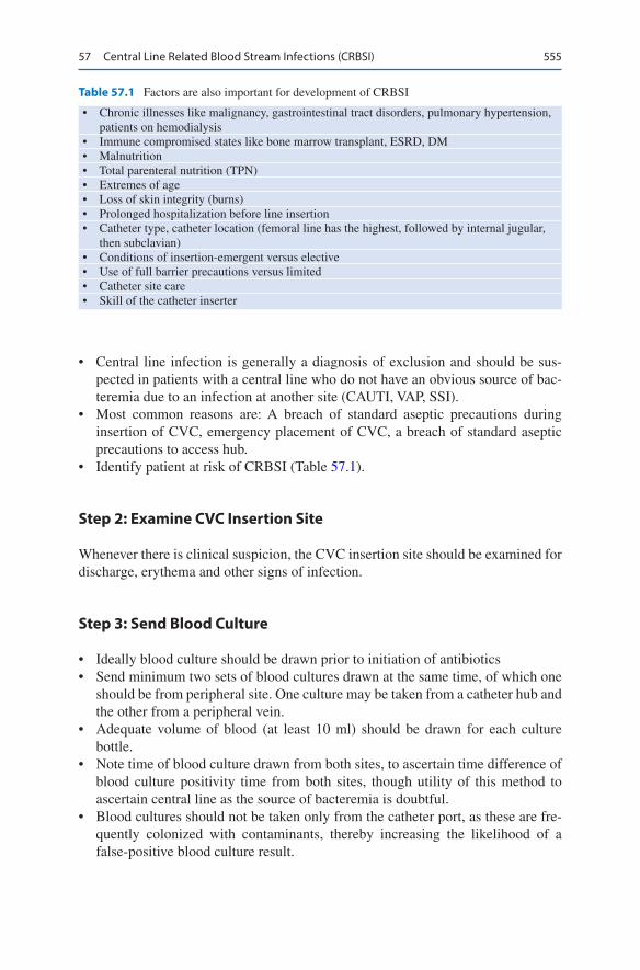
555
• Central line infection is generally a diagnosis of exclusion and should be sus-pected in patients with a central line who do not have an obvious source of bac-teremia due to an infection at another site (CAUTI, VAP, SSI).
• Most common reasons are: A breach of standard aseptic precautions during insertion of CVC, emergency placement of CVC, a breach of standard aseptic precautions to access hub.
• Identify patient at risk of CRBSI (Table 57.1).
Step 2: Examine CVC Insertion Site
Whenever there is clinical suspicion, the CVC insertion site should be examined for discharge, erythema and other signs of infection.
Step 3: Send Blood Culture
• Ideally blood culture should be drawn prior to initiation of antibiotics• Send minimum two sets of blood cultures drawn at the same time, of which one
should be from peripheral site. One culture may be taken from a catheter hub and the other from a peripheral vein.
• Adequate volume of blood (at least 10 ml) should be drawn for each culture bottle.
• Note time of blood culture drawn from both sites, to ascertain time difference of blood culture positivity time from both sites, though utility of this method to ascertain central line as the source of bacteremia is doubtful.
• Blood cultures should not be taken only from the catheter port, as these are fre-quently colonized with contaminants, thereby increasing the likelihood of a false-positive blood culture result.
Table 57.1 Factors are also important for development of CRBSI
• Chronic illnesses like malignancy, gastrointestinal tract disorders, pulmonary hypertension, patients on hemodialysis
• Immune compromised states like bone marrow transplant, ESRD, DM• Malnutrition• Total parenteral nutrition (TPN)• Extremes of age• Loss of skin integrity (burns)• Prolonged hospitalization before line insertion• Catheter type, catheter location (femoral line has the highest, followed by internal jugular,
then subclavian)• Conditions of insertion-emergent versus elective• Use of full barrier precautions versus limited• Catheter site care• Skill of the catheter inserter
57 Central Line Related Blood Stream Infections (CRBSI)
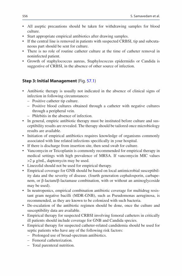
556
• All aseptic precautions should be taken for withdrawing samples for blood culture.
• Start appropriate empirical antibiotics after drawing samples.• If the central line is removed in patients with suspected CRBSI, tip and subcuta-
neous part should be sent for culture.• There is no role of routine catheter culture at the time of catheter removal in
noninfected patient.• Growth of staphylococcus aureus, Staphylococcus epidermidis or Candida is
suggestive of CRBSI, in the absence of other source of infection.
Step 3: Initial Management (Fig. 57.1)
• Antibiotic therapy is usually not indicated in the absence of clinical signs of infection in following circumstances: – Positive catheter tip culture. – Positive blood cultures obtained through a catheter with negative cultures
through a peripheral vein. – Phlebitis in the absence of infection.
• In general, empiric antibiotic therapy must be instituted before culture and sus-ceptibility results are revealed. The therapy should be tailored once microbiology results are available.
• Initiation of empirical antibiotics requires knowledge of organisms commonly associated with line related infections specifically in your hospital.
• If there is discharge from insertion site, then send swab for culture.• Vancomycin or Teicoplanin is commonly recommended for empirical therapy in
medical settings with high prevalence of MRSA. If vancomycin MIC values >2 μ g/mL, daptomycin may be used.
• Linezolid should not be used for empirical therapy.• Empirical coverage for GNB should be based on local antimicrobial susceptibil-
ity data and the severity of disease. (fourth generation cephalosporin, carbape-nem, or β-lactam/β-lactamase combination, with or without an aminoglycoside may be used).
• In neutropenics, empirical combination antibiotic coverage for multidrug resis-tant gram negative bacilli (MDR-GNB), such as Pseudomonas aeruginosa, is recommended, as they are known to be colonized with such bacteria.
• De-escalation of the antibiotic regimen should be done, once the culture and susceptibility data are available.
• Empirical therapy for suspected CRBSI involving femoral catheters in critically ill patients should include coverage for GNB and Candida species.
• Empirical therapy for suspected catheter-related candidemia should be used for septic patients who have any of the following risk factors: – Prolonged use of broad-spectrum antibiotics. – Femoral catheterization. – Total parenteral nutrition.
S. Samavedam et al.

557
Feb
rile
patie
nt w
ith in
dwel
ling
CV
C
Exa
min
e lo
cal C
VC
and
cul
ture
any
dra
inag
eO
btai
n si
mul
tane
ous
bloo
dcu
lture
s vi
a C
VC
and
per
iphe
ral
Che
ck fo
r so
urce
s of
infe
ctio
n
Loca
l sig
ns a
ndsy
mpt
oms
No
loca
l sig
ns a
ndsy
mpt
oms
Initi
ate
appr
opria
teth
erap
yC
heck
the
resu
lt of
blo
od
Pos
itive
CV
Cbl
ood
cultu
re;
nega
tive
perip
hera
lbl
ood
cultu
re
DT
P ≥
2 h
or
quan
titat
ive
bloo
dcu
lture
≥ 5
:1
DT
P <
2 h
or
quan
titat
ive
bloo
d cu
lture
< 2
:1
Pos
itive
per
iphe
ral
bloo
d cu
lture
;ne
gativ
e C
VC
bloo
d cu
lture
Con
side
ran
tibio
tic lo
ckso
lutio
n
CR
BS
Im
anag
emen
tLo
ok fu
rthe
r fo
r ot
her
sour
ces
of in
fect
ion
Neg
ativ
e bl
ood
cultu
re; n
egat
ive
CV
C ti
p
Fou
nd o
ther
sour
ceF
ound
no
othe
rso
urce
If po
ssib
le, e
xcha
nge
and
cultu
re C
VC
Initi
ate
appr
opria
teth
erap
y
Neg
ativ
e bl
ood
cultu
re; p
ositi
veC
VC
tip
Pos
itive
blo
odcu
lture
; pos
itive
CV
C ti
p
Mon
itor
clos
ely
and
repe
at b
lood
cultu
res
CR
BS
Im
anag
emen
t
Det
erm
ine
resu
lts o
f DT
P o
rqu
antit
ive
bloo
d cu
lture
*
Fig.
57.
1 D
iagn
osis
of
acut
e fe
brile
epi
sode
in a
pat
ient
with
a c
entr
al v
enou
s ca
thet
er. C
VC
cen
tral
ven
ous
cath
eter
, DT
P d
iffe
rent
ial t
ime
to p
ositi
vity
57 Central Line Related Blood Stream Infections (CRBSI)
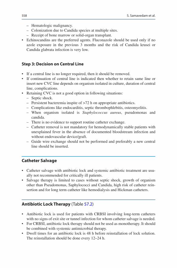
558
– Hematologic malignancy. – Colonization due to Candida species at multiple sites. – Receipt of bone marrow or solid-organ transplant.
• Echinocandins are the preferred agents. Fluconazole should be used only if no azole exposure in the previous 3 months and the risk of Candida krusei or Candida glabrata infection is very low.
Step 3: Decision on Central Line
• If a central line is no longer required, then it should be removed.• If continuation of central line is indicated then whether to retain same line or
insert new CVC line depends on organism isolated in culture, duration of central line, complications.
• Retaining CVC is not a good option in following situations: – Septic shock. – Persistent bacteremia inspite of >72 h on appropriate antibiotics. – Complications like endocarditis, septic thrombophlebitis, osteomyelitis. – When organism isolated is Staphylococcus aureus, pseudomonas and
candida. – There is no evidence to support routine catheter exchange. – Catheter removal is not mandatory for hemodynamically stable patients with
unexplained fever in the absence of documented bloodstream infection and without endovascular device/graft.
– Guide wire exchange should not be performed and preferably a new central line should be inserted.
Catheter Salvage
• Catheter salvage with antibiotic lock and systemic antibiotic treatment are usu-ally not recommended for critically ill patients.
• Salvage therapy is limited to cases without septic shock, growth of organism other than Pseudomonas, Saphylococci and Candida, high risk of catheter rein-sertion and for long term catheter like hemodialysis and Hickman catheters.
Antibiotic Lock Therapy (Table 57.2)
• Antibiotic lock is used for patients with CRBSI involving long-term catheters with no signs of exit site or tunnel infection for whom catheter salvage is needed.
• For CRBSI, antibiotic lock therapy should not be used as monotherapy. It should be combined with systemic antimicrobial therapy.
• Dwell times for an antibiotic lock is 48 h before reinstallation of lock solution. The reinstallation should be done every 12–24 h.
S. Samavedam et al.
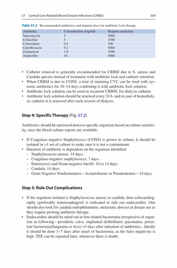
559
• Catheter removal is generally recommended for CRBSI due to S. aureus and Candida species instead of treatment with antibiotic lock and catheter retention.
• When CRBSI is due to CONS, a trial of retaining CVC can be tried with sys-temic antibiotics for 10–14 days combining it with antibiotic lock solution.
• Antibiotic lock solution can be used in recurrent CRBSI, for dialysis catheter.• Antibiotic lock solution should be renewed every 24 h. and in case of hemodialy-
sis catheter it is renewed after each session of dialysis.
Step 4: Specific Therapy (Fig. 57.2)
Antibiotics should be narrowed down to specific organism based on culture sensitiv-ity, once the blood culture reports are available.
• If Coagulase negative Staphylococci (CONS) is grown in culture, it should be isolated in >1 set of culture to make sure it is not a contaminant.
• Duration of antibiotic is dependent on the organism identified. – Staphylococcus aureus: 14 days. – Coagulase-negative staphylococci: 7 days. – Enterococci and Gram-negative bacilli: 10 to 14 days. – Candida: 14 days. – Gram Negative Nonfermenters—Acinetobacter or Pseudomonas—14 days.
Step 5: Rule Out Complications
• If the organism isolated is Staphylococcus aureus or candida, then echocardiog-raphy (preferably transesophageal) is indicated to rule out endocarditis. One should also look for candida endophthalmitis, metastatic abscess at distant site as they require prolong antibiotic therapy.
• Endocarditis should be ruled out in line related bacteremia irrespective of organ-ism in following—prosthetic valve, implanted defibrillator, pacemaker, persis-tent bacteremia/fungemia or fever >3 days after initiation of antibiotics.. Ideally it should be done 5–7 days after onset of bacteremia, as the false negativity is high. TEE can be repeated later, whenever there is doubt.
Table 57.2 Recommended antibiotics and heparin dose for antibiotic lock therapy
Antibiotic Concentration (mg/ml) Heparin (units/ml)Vancomycin 5 5000Cefazoline 5 2500Ceftazidime 0.5 100Ciprofloxacin 0.2 5000Gentamicin 1.0 2500Ampicillin 10 5000
57 Central Line Related Blood Stream Infections (CRBSI)

560
Coa
gula
se-n
egat
ive
Sta
phyl
ococ
cus
spp
Sta
phyl
ococ
cus
aure
usG
ram
-neg
ativ
e ba
cilli
Can
dida
spp
Non
-tun
nelle
d C
V• • • •
Rem
ove
CV
C a
ndtr
eat w
ith A
SA
for
5-7
days
May
ret
ain
CV
C a
ndtr
eat w
ith A
SA
for
7da
ys a
nd A
LT fo
r10
-14
days
Tun
nele
d/im
plan
tabl
eC
VC M
ay r
etai
n C
VC
and
trea
t with
AS
A fo
r 7
days
and
ALT
for
10-1
4 da
ys
If m
anife
stat
ions
wor
sen
or p
ersi
st,
rem
ove
CV
C tr
eat
for
5-7
days
Non
-tun
nelle
d C
VC
CV
C r
emov
al is
rec
omm
ende
dA
SA
for
>14
day
s, if
tran
soes
opha
geal
ech
ocar
diog
ram
is n
egat
ive
Tun
nelle
d/im
plan
tabl
e C
VC
If tr
anso
esop
hage
alec
hoca
rdio
gram
is n
egat
ive,
trea
tw
ith A
SA
and
ALT
for
> 1
4 da
ys
Rem
ove
CV
C if
infe
ctio
n is
com
plic
ted
by e
ndoc
ardi
tis(t
rans
oeso
phag
eal e
choc
ardi
ogra
mpo
sitiv
e) o
r se
ptic
thro
mbo
sis
orde
ep-s
eate
d in
fect
ion
and
trea
tw
ith in
trav
enou
s A
SA
for
4-6
wee
ks
Non
-tun
nelle
d C
VC
CV
C r
emov
al is
rec
omm
ende
dA
SA
for
10-1
4 da
ys
Tun
nelle
d/im
plan
tabl
e C
VC
CV
C r
emov
al is
rec
omm
ende
dIn
abs
ence
of o
ther
vas
cula
r si
tes,
trea
t with
AS
A a
nd A
LT fo
r 10
-14
days
and
ALT
for
14 d
ays
Rem
ove
CV
C if
ther
e is
dete
riora
tion
or n
o re
spon
se, a
ndtr
eat w
ith A
SA
for
10-1
4 da
ys
Non
-tun
nelle
d or
tunn
eled
CV
C CV
C r
emov
al is
reco
mm
ende
d
Tre
at w
ith a
ppro
pria
tean
tifun
gal a
gent
s fo
r 14
days
afte
r la
st p
ositi
vebl
ood
cultu
re
If ba
cter
aem
ia o
r fu
ngae
mia
per
sist
, or
lack
of r
espo
nse
afte
r 3
days
of C
VC
rem
oval
and
initi
atio
nap
prop
riate
ant
icio
tics
or a
ntifu
ngal
s,In
vest
igat
e fu
rthe
r th
e po
ssib
ility
of:
Any
oth
er m
etas
tatic
infe
ctio
n
• • • • •
• • • • •
• •
Fig.
57.
2 M
anag
emen
t of
cat
hete
r-re
late
d bl
oods
trea
m i
nfec
tions
. A
LT,
antib
iotic
-loc
k th
erap
y; A
SA,
appr
opri
ate
syst
emic
ant
ibio
tics;
CR
BSI
, ca
thet
er-
rela
ted
bloo
dstr
eam
infe
ctio
n; C
VC
, cen
tral
ven
ous
cath
eter
S. Samavedam et al.

561
Step 6: Prevention
• Education, training and adequate staffing for controlling CRBSI.• Use aseptic precautions while inserting catheters.• Follow hand hygiene.• Use clean gloves for peripheral IV catheters and sterile gloves for CVC.• Use maximum sterile barrier precautions: cap, mask, sterile gown, sterile gloves,
and a sterile full body drape.• Avoid femoral lines for CVC access.• Use subclavian vein to minimize infection risks as compared to IJV and Femoral
veins.• Use ultrasonic guidance (USG) for CVC insertion.• Use CVC with minimum number of ports necessary to manage the patient.• Replace CVC inserted in emergency within 48 h.• Prepare clean skin with a > 0.5% chlorhexidine preparation with alcohol before
CVC insertion. If there is a contraindication to chlorhexidine, tincture of iodine, an iodophor, or 70% alcohol can be used as alternatives.
• Do not use topical antibiotic ointment or creams on insertion sites, except for dialysis catheters, because of their potential to promote fungal infections and antimicrobial resistance.
• Replace dressings used on CVC sites at least every 7 days for transparent dress-ings and every two days for gauze dressings.
• When the CRBSI rate is not decreasing even after successful implementation of measures to reduce CRBSI, a chlorhexidine/silver sulfadiazine or minocycline/rifampin impregnated CVC should be used in patients whose catheter is expected to remain in place >5 days.
• Use a sutureless securement device to reduce the risk of infection for intravascu-lar catheters.
• Do not administer systemic antimicrobial prophylaxis routinely before insertion or during use of an intravascular catheter to prevent catheter colonization or CLABSI.
• Use prophylactic antimicrobial lock solution in patients with long term catheters who have a history of multiple CRBSI despite optimal maximal adherence to aseptic technique.
• Use a 2% chlorhexidine wash for daily skin cleansing to reduce CRBSI.• Replace IV sets that are continuously used, including secondary sets and add-on
devices, no more frequently than at 96-h intervals, but at least every 7 days in patients not receiving blood, blood products or fat emulsions.
• Replace tubing used to administer blood, blood products, or fat emulsions (those combined with amino acids and glucose in a 3-in-1 admixture or infused sepa-rately) within 24 h of initiating the infusion (IB).
• Propofol infusion tubing should be changed every 6 or 12 h.
57 Central Line Related Blood Stream Infections (CRBSI)
562
Suggested Reading
Bouzidi H, Emirian A. Differential time to positivity of central and peripheral blood cultures is inaccurate for the diagnosis of Staphylococcus aureus long-term catheter-related sepsis. J Hosp Infect. 2018;99(2):192–9. To ascertain accuracy of Differential time to positivity of cultures of blood drawn simultaneously from central venous catheter and peripheral sites which is widely used to diagnose catheter-related bloodstream infections without removing the catheter. Because of the high number of false-negative cases, the classic cut-off limit of 120 min showed 100% specificity but only 42% sensitivity for the diagnosis of catheter-related bloodstream infection due to S. aureus.
Central Line Associated Blood Stream Infections (CLABSI)–CDC. https://www.cdc.gov/nhsn/pdfs/training/2018/clabsi-508.pdf
Chong HY, Lai NM. Comparative efficacy of antimicrobial central venous catheters in reducing catheter-related bloodstream infections in adults: abridged cochrane systematic review and net-work meta-analysis. Clin Infect Dis. 2017;64(suppl_2):S131–40. A cochrane systematic review of antibiotic impregnanted central venous catheter.
Guenezan J, Drugeon B, Marjanovic N, et al. Treatment of central line associated blood stream infections. Crit Care. 2018;22:303.
Ling ML, Apisarnthanarak A, Jaggi N, Harrington G. APSIC guide for prevention of central line associated bloodstream infections (CLABSI). Antimicrob Resist Infect Control. 2016;5:16. A guide for the prevention of CLABSI.
O’Grady NP, et al. Healthcare infection control practices advisory committee (HICPAC). Guidelines for the prevention of intravascular catheter-related infections. Clin Infect Dis. 2011;52:e162–93. Guidelines for the prevention of CRBSI.
Raad I, Chaftari AM. Successful salvage of central venous catheters in patients with catheter-related or central line-associated bloodstream infections by using a catheter lock solution consisting of minocycline, EDTA, and 25% ethanol. Antimicrob Agents Chemother. 2016;60(6):3426–32. An article on use of antibiotic lock solution in cancer patients with central line.
Russell TA, Fritschel E, et al. Minimizing central line-associated bloodstream infections in a high-acuity liver transplant intensive care unit. Am J Infect Control. 2019;47(3):305–12. Multidisciplinary collaboratives are essential to minimize CLABSI and optimize value and quality in a challenging, high-acuity patient population.
S. Samavedam et al.
563© Springer Nature Singapore Pte Ltd. 2020R. Chawla, S. Todi (eds.), ICU Protocols, https://doi.org/10.1007/978-981-15-0898-1_58
S. Todi (*) Department of Critical Care & Emergency Medicines, A.M.R.I. Hospital, Kolkata, India
R. Chawla Department of Respiratory, Critical Care & Sleep Medicine, Indraprastha Apollo Hospitals, New Delhi, India
S. Swaminathan Department of Infectious Diseases, Global Hospital, Chennai, India
58Antibiotic Pearls: A Case Based Discussion
Subhash Todi, Rajesh Chawla, and Subramanian Swaminathan
Step 1: Identify Extended Spectrum Beta Lactamase (ESBL) Producing Organism by Antibiogram
• These are organisms which are resistant to Cephalosporins but susceptible to Piperacillin/Tazobactam and Carbapenem.
• They belong to Class A of Betalactamase producing gram negative organism (Ambler Classification).
Step 2: Identify Antibiotic of Choice for ESBL Organisms
• Piperacillin Tazobactam may be used for these organism if the disease severity is less urinary tract is the source of infection, E.coli is the culprit organism and MIC of the organism is low to Pip/Taz.
Case 1: A 60 year old man was admitted with fever and pain in left loin. He is hemodynamically stable. His urine and blood culture subsequently grew E. Coli which was resistant to cephalosporin but sensitive to Piperacillin tazobactam.
564
• Carbapenems are the drug of choice for ESBL specially in sicker group of patient
In a recent study (MERINO), Meropenem was found to be superior to Pip/Taz in reducing mortality in ESBL infected patients even if the illness severity was low.
Coproduction of AmpC enzymes is often seen, and in such cases, carbapen-ems would work better than BL/BLIs. It belongs to class C ambler classifi-cation. Case 2
A 50 year old man with biliary sepsis is growing Acinetobacter baumanii in blood culture which is resistant to carbapenem and sensitive to Colistin only.
Step 1: Identify Optimal Antibiotic Regimen for Carbepenem Resistant Acinetobacter (CRAB) Infection
• Colistin or Polymyxin B is the backbone antibiotic for CRAB infections.
Step 2: Identify Role of Mono or Combination Therapy with Colistin for CRAB Infection
• Combination therapy should be considered for CRAB bacteremia if two antibiot-ics which are susceptible can be identified.
• Monotherapy with Colistin/Polymyxin B should be used if the sensitivity is only to Colistin/Polymyxin B and MIC to other antibiotic too high. No added advan-tage for combination therapy.
Case 3
A 50 year old female is admitted with urosepsis and septic shock. Her blood culture is growing Klebsiella, sensitive to Colistin/Polymyxin B, (CRE) and tigecycline but Resistant to carbapenem, and aminoglycosides.
Step 1: Identify Optimal Antibiotic Regimen for Colistin Only Sensitive Enterobacteriacae (CRE)
• Colistin or Polymyxin B is the backbone antibiotic for colistin only sensitive enterobacteriacae (CRE) infection.
S. Todi et al.

565
Step 2: Identify Role of Mono or Combination Therapy with Colistin for CRE
• Colistin should be combined with another antibiotic if the organism is suscepti-ble to it specially in sick patient.
• In the absence of susceptibility to any other antibiotic combination therapy with a second and/or third nonsusceptible agent (e.g., a carbapenem). Preference should be given to a non- susceptible agent with the lowest MIC relative to the respective susceptibility breakpoint.
• Tigecycline should not be used in urinary tract infections (UTI) as it is not excreted in urine.
• Polymyxin B should not be used in UTI as it is not excreted in urine.
Case 4
A 60 year old man with Ventilator associated pneumonia and renal failure. He is hypotensive and requiring Renal replacement therapy with SLED. He is growing Pseudomonas aeruginosa in sputum and blood culture which is sensi-tive to Colistin only.
Step 1. Identify Optimal Dose of Colistin for this Patient
• Give standard Loading dose: initiating loading dose of 300 mg CBA (~nine mil-lion IU) infused over 0.5–1 h.
• Maintenance dose: Calculated for a GFR of <10 ml/min: 4.40 million IU/day.• Supplemental dose post dialysis: 10% of the CMS dose be added to the base-
line daily dose per 1 h of SLED. e.g. Patient on 8 hours of SLED should be supplemented with 80% of maintenance dose.
Case 5
A 60 year old man with Ventilator associated pneumonia and renal failure. He is hypotensive and requiring Renal replacement therapy with SLED. He is growing Pseudomonas in sputum and blood culture which is sensitive to Colistin and Polymyxin B only.
Step 1: Choose Between Polymyxin B or Colistin as the Preferred Antibiotic
• Polymyxin B is the preferred agent for routine systemic use in invasive infec-tions. The rationale for this recommendation is that polymyxin B has superior PK characteristics as well as a decreased potential to cause nephrotoxicity.
58 Antibiotic Pearls: A Case Based Discussion
566
• However colistin is the preferred polymyxin for the treatment of lower urinary tract infections. Since it is cleared by kidney as prodrug CMS and then converts to the active moiety colistin in the urinary tract.
Step 2: Identify Optimal Dose of Polymyxin B
• Loading dose: 2.0–2.5 mg/kg for polymyxin B, based on total body weight (TBW) (equivalent to 20,000–25,000 IU/kg) over 1 h.
• Maintenance dose: 1.25–1.5 mg/kg (equivalent to 12,500–15,000 IU/kg TBW) every 12 hours is infused over 1 hour in patient with normal renal function.
• Dose in renal failure: No dose adjustment is needed in daily maintenance dose of Polymyxin B in renal impatrment or on any form of dialysis.
Case 6
A 50 year old man with multiorgan failure due to septic shock. He was treated with Colistin to which he responded, he subsequently developed a ventilator associated pneumonia.
Step 1: Choose Empiric Antibiotic in Patients Already Exposed to Colistin
• Bacteria which are intrinsically resistant to Colistin are Burkholderia cepacia, Serratia marcescens, Proteus spp., Providencia and Morganella morganii.
• These bacteria may be sensitive to Carbapenem.
Case 7
A 70 year old man is being treated with intravenous fosfomycin for urosepsis caused by Klebsiella. He has become afebrile and CRP is decreasing but he has become confused, drowsy and very weak.
Step 1: Identify Electrolyte Abnormalities Associated with Fosfomycin Infusion
• Hypernatremia (due to increase sodium load) and Hypokalemia (due to renal potassium wasting) are the two common electrolyte abnormalities encountered with Fosfomycin infusion.
• Fosfomycin injection should be infused dissolved in hypotonic solution to pre-vent development of hypernatremia.
S. Todi et al.
567
Case 8
A 50 year old man was admitted with features suggestive of urosepsis. His blood culture is growing Carbapenem resistant Pseudomonas. He is diabetic and have borderline renal function.
Step 1: Identify Strategies to Spare Colistin in Carbapenem Resistant Infections
• Resistance to carbapenem may occur due to production of beta lactamases (OXA 48 like enzyme and NDM) or Porin Channel mutation or Efflux mechanism.
• Organisms which produce OXA 48 or NDM can be identified by Carba card test or PCR technique.
• Avibactam is a novel betalactamase inhibitor which can act against OXA 48 betalactamase enzyme.
• OXA 48 producing Carpapenem resistant Pseudomonas will be susceptible to a combination of ceftazidime-avibactam.
• NDM producing organism are susceptible to Aztreoonam and a combination of Ceftazidime-Avibactam and Aztreonam may be used for this.
Case 9
A 40 year old farmer was admitted with a cellulitis of left thigh and is in shock. His blood culture subsequently grew Staphylococcus aureus which is sensitive to Methicillin.
Step 1: Differentiate MSSA from MRSA
• Resistance to Oxacillin or Cefoxitin differentiates MRSA from MSSA.• Presence of Mec a gene identified by PCR also identifies MRSA.
Step 2: Decide Optimal Antibiotic for MSSA
• Flucloxacillin is the drug of choice for MSSA.• Cefazolin is an alternative choice for MSSA.• Anti MRSA therapy like Vancomycin/Teicoplanin/Linezolid is inferior to
Flucloxacillin or Cefazolin for MSSA.
58 Antibiotic Pearls: A Case Based Discussion
568
Case10
A 70 year old man with post H1N1 influenza was admitted with severe pneu-monia. His sputum culture has grown MRSA.
Step 1: Identify Optimal Therapy for MRSA Pneumonia
• Both IV Vancomycin, Teicoplanin and Linezolid are equally effective for MRSA pneumonia, though recent evidence suggests Linezolid is the preferred option for MRSA pneumonia due to better lung penetration.
• There is a trend of increasing MIC (>2 mcg) to Vancomycin and a need for higher loading dose (25–30 mg/kg) and a higher trough level of 15–20 mcg/l for therapeutic efficacy.
• IV Daptomycin does not work in pneumonia as it is inactivated by surfactant.
Case 11
65 year old male with cholangiocarcinoma is admitted with sepsis. He had a stent placed earlier, which is blocked. Blood culture shows carbapenem resis-tant K. pneumoneae.
Step 1
• Consider Colistin based therapy.• The second agent to colistin could be tigecycline or meropenem, based on the
MICs• Tigecycline monotherapy may be considered in less sick patients
Step 2
• Beware of Colistin Resistance.• Colistin resistance increases mortality.• Resistance cannot be tested by disc diffusion, automated methods or E-strip.• Broth micro-dilution is the best technique for testing, and is encouraged, as this
is a rising problem.
Step 3
Consider Ceftazidime Avibactam as an alternative.
S. Todi et al.
569
• Ceftazidime-avibactam (with aztreonam) could be useful choice.• Carba Resistance will determine need for aztreonam.• Need to add metronidazole for anaerobic activity.• Efficacy possibly as good as colistin based therapy, although emerging data
seems to indicate better outcomes.
Suggested Reading
Harris PNA, Tambyah PA, MERINO Trial Investigators and the Australasian Society for Infectious Disease Clinical Research Network (ASID-CRN). Effect of Piperacillin-Tazobactam vs Meropenem on 30-day mortality for patients with E. coli or Klebsiella pneumoniae bloodstream infection and ceftriaxone resistance: a randomized clinical trial. JAMA. 2018;320(10):984–94. A randomised control trial on ESBL producing enterobacetriacae showing superiority of Carbapenem over Piperacillin/Tazobactam.
Lucasti C, Popescu I. Comparative study of the efficacy and safety of ceftazidime/avibactam plus metronidazole versus meropenem in the treatment of complicated intra-abdominal infec-tions in hospitalized adults: results of a randomized, double-blind, Phase II trial. J Antimicrob Chemother. 2013;68(5):1183–92. A randomised study on Ceftazidime Avibactam a novel BL/BLI combination.
Theuretzbacher U, Leibovici L. Colistin alone versus colistin plus meropenem for treatment of severe infections caused by carbapenem-resistant gram-negative bacteria: an open-label, randomised controlled trial. Lancet Infect Dis. 2018;18(4):391–400. A randomised study of Colistin mono or combination therapy specially against Acinetobacter bacteremia.
Tsuji BT, Pogue JM. International consensus guidelines for the optimal use of the polymyxins. Pharmacotherapy. 2019;39(1):10–39. Current international guideline on polymyxins.
Youngster I, Shenoy ES. Comparative evaluation of the tolerability of cefazolin and nafcillin for treatment of methicillin-susceptible Staphylococcus aureus infections in the outpatient setting. Clin Infect Dis. 2014;59(3):369–75. A study looking at Cefazolin as an alternative to Nafcillin in MSSA infection.
58 Antibiotic Pearls: A Case Based Discussion
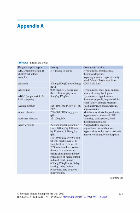
571© Springer Nature Singapore Pte Ltd. 2020R. Chawla, S. Todi (eds.), ICU Protocols, https://doi.org/10.1007/978-981-15-0898-1
Appendix A
Table A.1 Drugs and doses
Drug class/prototypes Dosing Common toxicitiesABCD (amphotericin B cholesteryl sulfate complex)
3–4 mg/kg IV q24h Hypotension, hypokalemia, thrombocytopenia, hypomagnesemia, hepatotoxicity, renal failure allergic reactions
Abacavir 300 mg PO q12h or 600 mg q24h
CNS, Skin Rash
Abciximab 0.25 mg/kg IV bolus, and then 0.125 mcg/kg/min
Hypotension, chest pain, nausea, minor bleeding, back pain
ABLC (amphotericin B lipid complex)
5 mg/kg IV q24h Hypotension, hypokalemia, thrombocytopenia, hepatotoxicity, renal failure, allergic reactions
Acetaminophen 325–1000 mg PO/IV q4–6h PRN
Rash, anemia, blood dyscrasias, hepatotoxicity
Acetazolamide 250–500 PO/IV mg given q8h
Metabolic acidosis, hypokalemia, hyponatremia, abnormal LFT
Activated charcoal 25–100 g PO Vomiting, constipation, fecal discoloration (black)
Acetylcysteine Acetaminophen poisoning Anaphylactoid reaction, angioedema, vasodilatation, hypotension, tachycardia, urticaria, nausea, vomiting, bronchospasm
Oral: 140 mg/kg followed by 17 doses of 70 mg/kg q4hIV: 150 mg/kg over 60 min f/b 300 mg/kg over 21 hNebulization: 3–5 mL of 20% solution three to four times a day, administer before chest physiotherapyPrevention of radiocontrast-induced renal injury: 600 mg PO q12h for 3 days starting 1 day before procedure, may be given intravenously
(continued)

572
Drug class/prototypes Dosing Common toxicitiesAcyclovir 10–15 mg/kg/dose q8h IV
(HSV encephalitis)800 mg PO every 4 h (Herpes Zoster)400 mg bolus
Reversible renal failureHepatic toxicityConfusional state
Adenosine 6 mg IV, if not effective in 1–2 min, can give 12 mg, may repeat 12 mg
Flushing, light headedness, headache, nervousness/anxiety
Adrenaline (see epinephrine)Albumin 0.5–1 g/kg/dose (5% in
hypovolemia)Hypervolemia, anaphylaxis, chills, fever, tachycardia, bronchospasm
Albuterol 5–10 mg nebulized over 30–60 min
Arrhythmias, chest discomfort, palpitation, CNS stimulation, drowsiness, diarrhea, dry mouth, micturition difficulty
Alfentanil IV bolus 500 mcg every 10 min as necessary
Respiratory depression, apnea, bradycardia, delayed gastric emptying, chest wall rigidityIV infusion: 1 mcg/kg/min
Allopurinol 600–800 mg/day in two to three divided doses
Rash
Alteplase 15 mg bolus, then 0.75 mg/kg (up to 50 mg) × 30 min, then 0.5 mg/kg (up to 35 mg) × 60 min (maximum 100 mg over 90 min)
Hypotension, bleeding, allergic reactions
Pulmonary embolism: 100 mg IV over 2 hStroke: 0.9 mg/kg 10% bolus, rest over 60 min (Max 90 mg)
Amantadine 100 mg PO q12h Nausea, vomiting, anorexia, xerostomia
Amikacin 15–20 mg/kg once a day Ototoxicity, neurotoxicity, nephrotoxicity
Aminophylline Loading dose; 5 mg/kg IV over 30 min
Tachycardia, arrhythmia, convulsions
Maintenance: 0.1–0.8 mg/kg/h
Amiodarone 150–300 mg bolus, then 1 mg/min for 6 h, then 0.5 mg/min for 18 h
Bradycardia, hypotension, AV block, nausea, photosensitivity, hypothyroidism, hyperthyroidism, coagulation abnormalities, hepatitis, visual disturbance, pulmonary fibrosis
Oral 200 mg 8 hourly, then titrate down to 200 mg q24h
Amitryptiline 25–75 mg PO q24h Confusion, dry mouth, retention of urine
Amlodipine 5–10 mg PO q24h Pedal edema, headache, nausea, vomiting
Table A.1 (continued)
Appendix A

573
Drug class/prototypes Dosing Common toxicitiesAmoxicillin/clavulanate 625 mg PO q12h/q8h Nausea, vomiting, diarrhea,
allergic reactionAmphotericin B deoxycholateAmphotericin B (Liposomal)
0.3–1.5 mg/kg IV q24h (diluted in 5% Dextrose)3–5 mg/kg /day (diluted in 5% dextrose)
Hypotension, hypokalemia, thrombocytopenia, hepatotoxicity, creatinine increase, allergic reactionsSame as Ampho B, Less nephrotoxicity
Ampicillin 250–500 mg IV q4–6h Fever, allergic reaction, penicillin encephalopathy, diarrhea, pseudomembranous colitis, agranulocytosis, anemia
Ampicillin/sulbactamAndexanet Alpha (Antidote to rivaroxaban and apixaban)
1.5–3 g IVq6h400–800 mg bolus @30 mg/min4–8 mg/min iv infusion for up to 120 min
Fever, allergic reaction, penicillin encephalopathy, diarrhea, pseudomembranous colitis, agranulocytosis, anemia
Anidulafungin 200 mg IV bolus, then 100 mg IV q24h
Elevated LFT
Aqueous penicillin G 2–4 million U IV q4h Fever, allergic reaction, penicillin encephalopathy, diarrhea
Argatroban 350 mcg/kg bolus, then 25 mcg/kg/min adjust based on aPTT (For PCI)Initial 2 mcg/kg/min, adjust based on aPTT measurements (For prophylaxis in heparin induced thrombocytopenia)
Bleeding, cardiac arrest, dyspnea
Arbekacin (Aminoglycoside)
150–200 mg once a day i.v. Nephrotoxicity, Ototoxicity
Ascorbic acid 1.5 g over 30 min every 6 h Oxalate nephropathyAspirin 160–325 mg PO q24h Bleeding, dyspepsiaAtropine 1 mg IV q3–5 min Dry eyes, dry mouth, urinary
retention, tachycardiaAtracurium 0.5 mg/kg IV Bolus then
0.08–0.12 mg/kg bolus every 20–30 min or 5–10 mcg/kg/min infusion
Flushing, allergic reactions, bradycardia, hypotension, bronchospasm, laryngospasm, seizures
Azithromycin 250–500 mg IV/PO q24h Headache, nausea, vomiting, diarrhea, allergic reactions, fungal infection
Bivalirudin 1 mg/kg IV bolus, then 1.75 mg/kg/h
Hypotension, hemorrhage, pain, headache, nausea, back pain
Bosentan 62.5 mg PO q12h × 1 month, then 125 mg PO q12h, as tolerated
Headache, anemia, transaminase increase, nasopharyngitis, flushing, pruritus
(continued)
Table A.1 (continued)
Appendix A

574
Drug class/prototypes Dosing Common toxicitiesBromocriptine 2.5–5 mg PO q8h (Max
45 mg for Neuroleptic Malignant syndrome)
Headache, dizziness, nausea, hypotension, nasal congestion
Bumetanide 0.5–2 mg/dose PO q24h Hyperuricemia, hypochloremia, hypokalemia, azotemia, hyponatremia, hyperglycemia, muscle cramps, creatinine increase
Calcitonin Initial 4 U/kg IM q12h, up to 8 U/kg IM q6h
Facial flushing, nausea, vomiting
Calcium gluconate/chloride (10 mL of 10%)
1 g IV over 2–5 min Hypercalcemia, constipation (oral)
Captopril 6.25–50 mg PO q8h Hypotension, dizziness, abnormal taste, cough, worsening renal function
Carvedilol 6.25 mg PO q12h, maximum 25 mg PO q12h
Hypotension, dizziness, fatigue, hyperglycemia, weight gain, diarrhea, bradycardia, syncope, deranged LFT, bronchospasm
Caspofungin 70 mg IV bolus, then 50 mg IV q24h
Headache, fever, chills, hypokalemia, flushing, tachycardia, anemia, eosinophilia, neutropenia, nephrotoxicity
Cefazolin 1–2 g IV q8h Allergic reaction, fever, Stevens–Johnson syndrome, nephrotoxicity, diarrhea, nausea, vomiting
Cefepime 500 mg to 2 g IV q8–12h Positive Coombs test, fever, headache, rash, pruritus, diarrhea, nausea, vomiting, agranulocytosis
Cefoxitin 1–2 g IV q6–8h Diarrhea, anaphylaxis, nausea, vomiting, headache, rash, pruritus, diarrhea, agranulocytosis, pseudomembranous colitis
Ceftazidime 500 mg IV to 2 g q8–12h Diarrhea, hypersensitivity reactions, candidiasis, nephrotoxicity, encephalopathy, headache, fever
Ceftazidime Avibactam 2.5 g every 8 h HypersensitivityCeftolazone Tazobactam 1.5–3 g every 8 hourly HypersensitivityCeftriaxone 1–2 g IV q12–24h headache, rash, pruritus, diarrhea,
nausea, vomiting, agranulocytosisCefuroxime 0.75–1.5 g IV 6–8 hourly C. difficile diarrhea
HypersensitivityTransient rise in liver function test
Chlordiazepoxide 10–30 mg PO q8h to q6h Muscle weakness, ataxia, confusion, hypotension
Cyclosporine IV: 1–5 mg/kg/day Increased urea, creatinineOral: 1.5 times IV dose q12h
HypertensionHirsutism, gingival hypertrophyHyperuricemia, tremor
Table A.1 (continued)
Appendix A
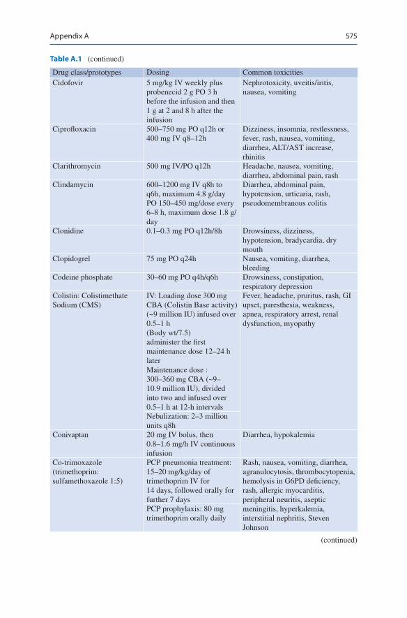
575
Drug class/prototypes Dosing Common toxicitiesCidofovir 5 mg/kg IV weekly plus
probenecid 2 g PO 3 h before the infusion and then 1 g at 2 and 8 h after the infusion
Nephrotoxicity, uveitis/iritis, nausea, vomiting
Ciprofloxacin 500–750 mg PO q12h or 400 mg IV q8–12h
Dizziness, insomnia, restlessness, fever, rash, nausea, vomiting, diarrhea, ALT/AST increase, rhinitis
Clarithromycin 500 mg IV/PO q12h Headache, nausea, vomiting, diarrhea, abdominal pain, rash
Clindamycin 600–1200 mg IV q8h to q6h, maximum 4.8 g/day PO 150–450 mg/dose every 6–8 h, maximum dose 1.8 g/day
Diarrhea, abdominal pain, hypotension, urticaria, rash, pseudomembranous colitis
Clonidine 0.1–0.3 mg PO q12h/8h Drowsiness, dizziness, hypotension, bradycardia, dry mouth
Clopidogrel 75 mg PO q24h Nausea, vomiting, diarrhea, bleeding
Codeine phosphate 30–60 mg PO q4h/q6h Drowsiness, constipation, respiratory depression
Colistin: Colistimethate Sodium (CMS)
IV: Loading dose 300 mg CBA (Colistin Base activity) (~9 million IU) infused over 0.5–1 h(Body wt/7.5)administer the first maintenance dose 12–24 h laterMaintenance dose : 300–360 mg CBA (~9–10.9 million IU), divided into two and infused over 0.5–1 h at 12-h intervals
Fever, headache, pruritus, rash, GI upset, paresthesia, weakness, apnea, respiratory arrest, renal dysfunction, myopathy
Nebulization: 2–3 million units q8h
Conivaptan 20 mg IV bolus, then 0.8–1.6 mg/h IV continuous infusion
Diarrhea, hypokalemia
Co-trimoxazole (trimethoprim: sulfamethoxazole 1:5)
PCP pneumonia treatment: 15–20 mg/kg/day of trimethoprim IV for 14 days, followed orally for further 7 days
Rash, nausea, vomiting, diarrhea, agranulocytosis, thrombocytopenia, hemolysis in G6PD deficiency, rash, allergic myocarditis, peripheral neuritis, aseptic meningitis, hyperkalemia, interstitial nephritis, Steven Johnson
PCP prophylaxis: 80 mg trimethoprim orally daily
Table A.1 (continued)
(continued)
Appendix A

576
Drug class/prototypes Dosing Common toxicitiesDalteparin 120 U/kg SC q12h Bleeding, wound hematoma, pain
at injection site, thrombocytopenia, allergic reactions, alopecia
Dantrolene 1–2.5 mg/kg IV; may repeat q5–10 min to maximum cumulative dose 10 mg/kg
Drowsiness, dizziness, diarrhea, nausea, vomiting
Daptomycin 4–6 mg/kg IV q24h Anemia, diarrhea, vomiting, peripheral edema, rash, insomnia, UTI, rise in CPK
DDAVP 0.3 mcg/kg slow IV/SC/IM Facial flushingIntranasally: 5–20 mcg daily
Deferoxamine 1 g IV bolus, then 500 mg IV q4h × 2 doses
Urine discoloration (orange–red)
Dexamethasone 10 mg IV prior to ACTH stimulation test, 4–6 mg IV q6h/q8h
Same as hydrocortisone
Dexmedetomidine 0.2–0.7 mcg/kg/h IV Hypotension, bradycardiaDextran (40) Maximum 20 mL/kg,
20–40 mL/min IVAllergic reaction, fluid overload, platelet dysfunction
Diazepam 5–10 mg IV over 2 min Apnea, respiratory depression, drowsiness, hypotension, bradycardia
Diclofenac 75 mg IM50 mg PO q8h
Digoxin Load: 10–15 mcg/kg; give 50% of load in initial dose, then 25% at 6–12 h intervals × 2
Bradycardia, heart block, arrhythmias, yellow vision, rash, muscle weakness
Maintenance:0.125–0.5 mg/day (dose should be reduced by 20–25% when changing from oral to IV)
Diltiazem 0.25 mg/kg bolus (may repeat 0.35 mg/kg bolus after 15 min), then 5–15 mg/h (PO (extended release) 60–120 mg q12h)
Bradycardia, hypotension, constipation (verapamil > diltiazem), headache, flushing, edema
Dobutamine 2.5–20 mcg/kg/min Tachycardia, hypertension, ventricular ectopics, headache, palpitations
Dopamine 5–20 mcg/kg/min Ectopic beats, tachycardia, arrhythmia, palpitations, angina, headache, dyspnea
Dopexamine 0.25–6 mcg/kg/min Tachycardia, hypotension, angina, hypokalemia, hyperglycemia
Table A.1 (continued)
Appendix A
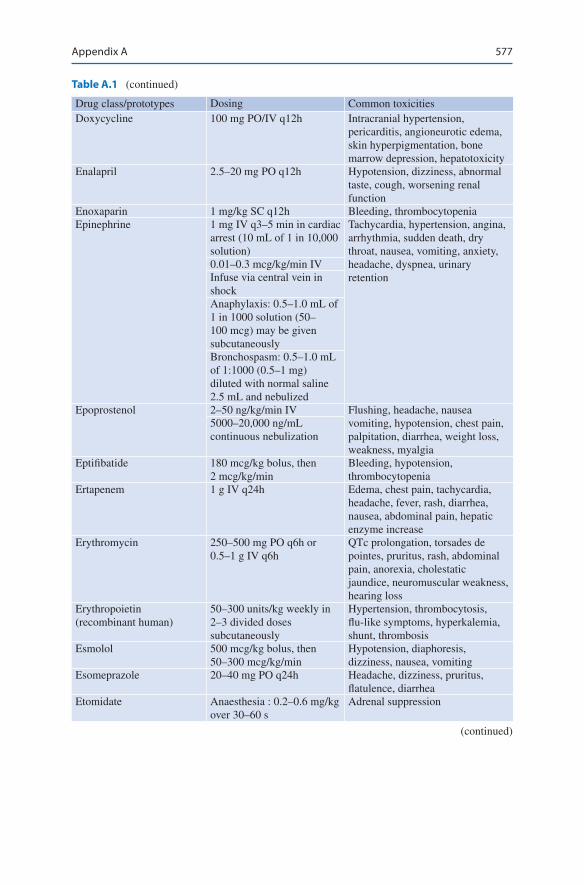
577
Drug class/prototypes Dosing Common toxicitiesDoxycycline 100 mg PO/IV q12h Intracranial hypertension,
pericarditis, angioneurotic edema, skin hyperpigmentation, bone marrow depression, hepatotoxicity
Enalapril 2.5–20 mg PO q12h Hypotension, dizziness, abnormal taste, cough, worsening renal function
Enoxaparin 1 mg/kg SC q12h Bleeding, thrombocytopeniaEpinephrine 1 mg IV q3–5 min in cardiac
arrest (10 mL of 1 in 10,000 solution)
Tachycardia, hypertension, angina, arrhythmia, sudden death, dry throat, nausea, vomiting, anxiety, headache, dyspnea, urinary retention
0.01–0.3 mcg/kg/min IVInfuse via central vein in shockAnaphylaxis: 0.5–1.0 mL of 1 in 1000 solution (50–100 mcg) may be given subcutaneouslyBronchospasm: 0.5–1.0 mL of 1:1000 (0.5–1 mg) diluted with normal saline 2.5 mL and nebulized
Epoprostenol 2–50 ng/kg/min IV Flushing, headache, nausea vomiting, hypotension, chest pain, palpitation, diarrhea, weight loss, weakness, myalgia
5000–20,000 ng/mL continuous nebulization
Eptifibatide 180 mcg/kg bolus, then 2 mcg/kg/min
Bleeding, hypotension, thrombocytopenia
Ertapenem 1 g IV q24h Edema, chest pain, tachycardia, headache, fever, rash, diarrhea, nausea, abdominal pain, hepatic enzyme increase
Erythromycin 250–500 mg PO q6h or 0.5–1 g IV q6h
QTc prolongation, torsades de pointes, pruritus, rash, abdominal pain, anorexia, cholestatic jaundice, neuromuscular weakness, hearing loss
Erythropoietin (recombinant human)
50–300 units/kg weekly in 2–3 divided doses subcutaneously
Hypertension, thrombocytosis, flu-like symptoms, hyperkalemia, shunt, thrombosis
Esmolol 500 mcg/kg bolus, then 50–300 mcg/kg/min
Hypotension, diaphoresis, dizziness, nausea, vomiting
Esomeprazole 20–40 mg PO q24h Headache, dizziness, pruritus, flatulence, diarrhea
Etomidate Anaesthesia : 0.2–0.6 mg/kg over 30–60 s
Adrenal suppression
Table A.1 (continued)
(continued)
Appendix A

578
Drug class/prototypes Dosing Common toxicitiesFactor VIIa (recombinant) Hemorrhagic stroke:
(Warfarin related) 10–100 mcg/kg IV: Bleeding episode: 90 mcg/kg every 2 h
Hypertension
Fentanyl 1–2 mcg/kg/dose Hypotension, bradycardia, CNS depression, confusion, chest wall rigidity, respiratory depression
Infusion: 1–3 mcg/kg/h
Flucloxacillin 1–2 g 6 h Interstitial nephritisHemolytic anaemiaCholestatic jaundice
Fluconazole 100–800 mg PO/IV q24h Headache, seizure, rash, angioedema, hypercholesterolemia, hypokalemia, hepatitis, cholestasis
Fondaparinux 2.5 mg SC q24h Bleeding, fever, nausea, anemiaFlucytosine 25–37.5 mg/kg PO q6h Nausea, vomiting, diarrhea, rashFludrocortisone 50–200 mcg PO q24h Hypertension, edema, acne,
hypokalemic alkalosis, hyperglycemia, peptic ulcer
Flumazenil 0.2–0.5 mg IV q1min, up to 3 mg
Vasodilatation, headache, agitation, dizziness, blurred vision, dyspnea, hyperventilation
Fomepizole 15 mg/kg/IV bolus, then 10 mg/kg IV q12h × 4 doses, then 15 mg/kg IV q12h until ethylene glycol or methanol level <20
Headache, nausea, bradycardia, hypotension, dizziness, metallic taste
Foscarnet 60 mg/kg IV q8h or 90 mg/kg IV q12h
Nephrotoxicity, electrolyte abnormalities (hypocalcemia, hypomagnesemia, hypokalemia, hypophosphatemia), nausea, vomiting, diarrhea, headache
Fosphenytoin Loading dose; 15–20 mgPE/kg IV Maintenance; 4–6 mg phenytoin equivalent (PE)/kg/day in two to three divided doses
IV form: hypotension, bradycardia, phlebitis, nystagmus, rash75 mg of
fosphenytoin = 50 mg of phenytoin
Furosemide 20–80 mg/day IV/PO in two to three divided doses
Hypotension, blurred vision, cutaneous vasculitis, gout, hyperglycemia, anorexia, allergic interstitial nephritis, fall in GFR, increased blood urea
Ganciclovir 5 mg/kg IV q12h Fever, rash, abdominal pain, nausea, vomiting, anemia, leucopenia, thrombocytopenia, confusion, neuropathy, pruritus, paresthesia, retinal detachment
Gemifloxacin 320 mg PO q24h Headache, dizziness, rash
Table A.1 (continued)
Appendix A
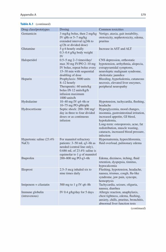
579
Drug class/prototypes Dosing Common toxicitiesGentamicin 3 mg/kg bolus, then 2 mg/kg
IV q8h or 5–7 mg/kg extended interval (q24h to q12h or divided dose)
Vertigo, ataxia, gait instability, ototoxicity, nephrotoxicity, edema, pruritus
Glutamine 5 g 6 hourly orally Increase in AST and ALT0.3–0.4 g/kg body weight IV
Haloperidol 0.5–5 mg 2–3 times/day/max 30 mg IV/PO 2–10 mg IV bolus, repeat bolus every 15–30 min with sequential doubling of dose
CNS depression, orthostatic hypotension, arrhythmia, alopecia, extrapyramidal symptoms, neuroleptic malignant syndrome, cholestatic jaundice
Heparin Prophylaxis: 5000 units 8–12 hourly
Bleeding, hyperkalemia, cutaneous necrosis, elevated liver enzymes, peripheral neuropathyTherapeutic: 60 units/kg
bolus f/b 12 units/kg/h infusion maximum 1000 units/h
Hydralazine 10–40 mg IV q4–6h or 10–75 mg PO q8h/q6h
Hypotension, tachycardia, flushing, headache
Hydrocortisone Septic shock: 200–300 mg/day in three to four divided doses or as continuous infusion
Hyperglycemia, mood changes, insomnia, gastrointestinal irritation, increased appetite, GI bleed, hypokalemia,Long-term: osteoporosis, acne, fat redistribution, muscle wasting, cataracts, increased blood pressure, infection
Hypertonic saline (23.4% NaCl)
For mannitol refractory patients: 3–50 mL q3–6h as needed (central line only), 0.686 mL of 23.4% saline is equimolar to 1 g of mannitol
Hypernatremia, hyperchloremia, fluid overload, pulmonary edema
Ibuprofen 200–800 mg PO q3–6h Edema, dizziness, itching, fluid retention, dyspepsia, tinnitus, hypocalcemia
IIioprost 2.5–5 mcg inhaled six to nine times daily
Flushing, hypotension, headache, nausea, trismus, cough, flu-like syndrome, jaw pain, syncope, hemoptysis
Imipenem + cilastatin 500 mg to 1 g IV q6–8h Tachycardia, seizure, oliguria, nausea, diarrhea
Immune globulin (intravenous)
IV 0.4 g/kg/day for 5 days Allergic reaction, anaphylaxis, chest tightness, edema, flushing, anxiety, chills, pruritus, bronchitis, abnormal liver function tests
Table A.1 (continued)
(continued)
Appendix A

580
Drug class/prototypes Dosing Common toxicitiesIpratropium 2–4 puffs (15 mcg/
actuation) q12h to q6hNebulised: 500 mcg 3–4 times/day
Bronchitis, upper respiratory tract infection, palpitation, dizziness, rash, nausea
Isavuconazole IV : initial 200 mg 8 hourly for six dosesMaintenance : 200 mg once daily
Peripheral edema
Isoprenaline Up to 20 mcg/min IV infusion, titrate according to heart rate
Tachycardia, arrhythmia, angina, hypotension
Isosorbide dinitrate 5–40 mg PO q8h Hypotension, headache, dizziness, flushing
Isosorbide mononitrate (Extended Release)
30–120 mg PO q24h Hypotension, headache, dizziness, flushing
ItraconazoleKayexalate
200 mg IV/PO q24hOral: 15 g 1–4 times dailyRectal : 30–50 g every 6 h
Pruritus, nausea, vomiting, chillsIschemic colitisBezoar
KetorolacKetamine
15–30 mg IV q6h Headache, abdominal pain, dyspepsia, nausea, edema, drowsiness, diarrheaTachycardiaHypertensionDelirium
20 mg PO f/b 10 mg q6–8hIntubation: 1–2 mg/kg iv over 1–2 min
Labetalol 100–400 mg PO q8–12h (max 2.4 g/day)
Dizziness, hypotension, bradycardia, nausea, vomiting, transaminase increase, paresthesia, flushing, headache
20–40 mg IV (maximum 80 mg) as bolus at 10–20 min intervals (Max 300 mg), then 0.5–2 mg/min infusion if needed
Lactulose 20–30 g (30–45 mL) PO q2h until initial stool, then adjust to maintain two to three soft stools/day
Diarrhea, flatulence, nausea
Lacosamide Status epilepticus: 200–600 mg IV daily
CNS
Lansoprazole 30–60 mg PO q12–24h Headache, abdominal pain, nausea, diarrhea
Lepirudin 0.5 mg/kg IV loading over 5 min as a loading dose (0.2 mg in renal failure)
Bleeding
0.15 mg/kg/h continuous IV infusion adjust based on aPPT measurements
Levalbuterol Adjust based on aPPT measurements 0.63–0.125 mg q8h 2–4 puff TID (45 mcg/actuation)
Hyperglycemia, hypokalemia, tremors, rhinitis, viral infection, headache, migraine, rash, abdominal cramps
Table A.1 (continued)
Appendix A

581
Drug class/prototypes Dosing Common toxicitiesLevetiracetam 500–1000 mg IV/PO q12h Behavior changes, somnolence,
nausea, vomiting, anorexia, weakness, cough, facial edema, bruising
Levofloxacin 500–750 mg IV/PO q24h Chest, pain, edema, nausea, vomiting, dyspnea, pharyngitis, rash, CNS stimulation, seizure, dizziness, somnolence
Levosimendan Loading dose (may be omitted) 6–12 mcg/kg given over 10 min0.1 mcg/kg/min continuous infusion
Levothyroxine 50–100 mcg IV Angina, arrhythmia, MI, palpitations, tachycardia, anxiety, headache, hyperactivity, insomnia, alopecia, tremors
Q6–8h × 24 h, then 100 mcg IV q24h
Lidocaine 1–1.5 mg/kg IV bolus (may repeat doses 0.5–0.75 mg/kg in 5–10 min up to maximum 3 mg/kg), then 1–4 mg/min
Arrhythmia, bradycardia, heart block, hypotension, edema, flushing, anxiety, hallucinations, seizures
Linezolid 600 mg IV/PO q12h Headache, diarrhea, insomnia, rash, nausea, constipation, thrombocytopenia, anemia, leucopenia, abnormal liver tests
Liothyronine 200–500 mcg (in myxedema coma)
Tachycardia, arrhythmia
Lisinopril 2.5–40 mg PO q24h Hypotension, dizziness, abnormal taste, cough, worsening renal function
Lorazepam Status epilepticus: 4 mg IV bolus, 0.5–4 mg/h
Sedation, hypotension, confusion, dermatitis, rash
Sedation: 0.02–0.06 mg/kg bolusInfusion: 0.01–0.1 mg/kg/h
Magnesium sulphate 4–6 g IV over 15–20 min, then 2 g/h infusion (1 g of Mg So4 = 98.6 mg of elemental Mg = 8.12 meq of elemental magnesium)
Hypermagnesemia, diarrhea (oral)
Mannitol (10–20%) 1–1.5 g/kg IV bolus, then 0.25–1 g/kg q3–6h as needed
Hypotension, acute renal failure, fluid and electrolyte imbalances
Meropenem 1 g IV q8h Headache, rash, diarrhea, anemia, phlebitis, agranulocytosis, TEN, glossitis
Meropenem Vaborbactam 4 g 8 hourly Hypersensitivity
Table A.1 (continued)
(continued)
Appendix A
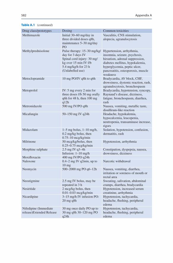
582
Drug class/prototypes Dosing Common toxicitiesMethimazole Initial 30–60 mg/day in
three divided doses q8h, maintenance 5–30 mg/day PO
Vasculitis, CNS stimulation, alopecia, agranulocytosis
Methylprednisolone Pulse therapy: 15–30 mg/kg/day for 3 days IV
Hypertension, arrhythmia, insomnia, seizure, psychosis, hirsutism, adrenal suppression, diabetes mellitus, hypokalemia, hyperglycemia, peptic ulcer, pancreatitis, osteoporosis, muscle weakness
Spinal cord injury: 30 mg/kg over 15 min IV f/b 5.4 mg/kg/h for 23 h (Unlabelled use)
Metoclopramide 10 mg PO/IV q8h to q6h Bradycardia, AV block, CHF, drowsiness, dystonic reaction, rash, agranulocytosis, bronchospasm
Metoprolol IV: 5 mg every 2 min for three doses f/b 50 mg orally q6h for 48 h, then 100 mg q12h
Bradycardia, hypotension, syncopy, Raynaud’s disease, dizziness, fatigue, bronchospasm, diarrhea, rash
Metronidazole 500 mg IV/PO q8h Nausea, vomiting, metallic taste, disulfiram-like reaction
Micafungin 50–150 mg IV q24h Headache, hypokalemia, hypocalcemia, leucopenia, neutropenia, transaminase increase, rigors
Midazolam 1–5 mg bolus, 1–10 mg/h, 0.2 mg/kg bolus, then 0.75–10 mcg/kg/min
Sedation, hypotension, confusion, dermatitis, rash
Milrinone 50 mcg/kg/bolus, then 0.25–0.75 mcg/kg/min
Hypotension, arrhythmia
Morphine sulphate 2.5 mg IV q3–4h Constipation, dyspepsia, nausea, drowsiness, dizzinessInfusion: 1–10 mg/h
Moxifloxacin 400 mg IV/PO q24hNaloxone 0.4–2 mg IV q2min, up to
10 mgNarcotic withdrawal
Neomycin 500–2000 mg PO q6–12h Nausea, vomiting, diarrhea, irritation or soreness of mouth or rectal area
Neostigmine 2.5 mg IV bolus, may be repeated in 3 h
Sweating, salivation, abdominal cramps, diarrhea, bradycardia
Nesiritide 2 mcg/kg bolus, then 0.01–0.03 mcg/kg/min
Hypotension, increased serum creatinine, arrhythmia
Nicardipine 3–15 mg/h IV infusion PO: 20 mg q8h
Hypotension, tachycardia, headache, flushing, peripheral edema
Nifedipine (Immediate release)Extended Release
30 mg once daily PO up to 30 mg q8h 30–120 mg PO q24h
Hypotension, tachycardia, headache, flushing, peripheral edema
Table A.1 (continued)
Appendix A
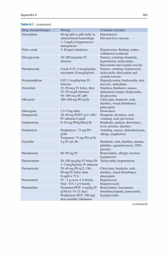
583
Drug class/prototypes Dosing Common toxicitiesNimodipine 60 mg q6h to q4h orally in
subarachnoid hemorrhageHypotensionElevated liver enzyme
1–2 mg/h in hypertensive emergencies.
Nitric oxide 5–40 ppm inhalation Hypotension, flushing, rashes, withdrawal syndrome
Nitroglycerin 10–200 mcg/min IV infusion
Nausea, vomiting, headache, hypotension, tachycardia, thiocyanate and cyanide toxicity
Nitroprusside Usual, 0.25–3 mcg/kg/min, maximum 10 mcg/kg/min
Nausea, vomiting, hypotension, tachycardia, thiocyanate and cyanide toxicity
Norepinephrine 0.02–3 mcg/kg/min IV infusion
Hyperglycemia, bradycardia, skin necrosis, arrhythmia
Octreotide 25–50 mcg IV bolus, then 25–50 mcg/h infusion
Diarrhea, flatulence, nausea, abdominal cramps, bradycardia, dysglycemia50–100 mcg SC q8h
Ofloxacin 200–400 mg PO q12h Chest pain, headache, rash, diarrhea, visual disturbance, pharyngitis
Olanzapine 2.5–5 mg daily DrowsinessOmeprazole 20–40 mg PO/IV q12–24h /
IV infusion 8 mg/hHeadache, dizziness, rash, vomiting, taste perversion
Ondansetron 8–10 mg PO/q24h/q12h Headache, malaise, drowsiness, fever, pruritus, diarrhea
Oseltamivir Prophylaxis: 75 mg PO q24h
Vomiting, nausea, abdominal pain, allergy, anaphylaxis
Treatment: 75 mg PO q12hOxacillin 2 g IV q4–5h Headache, rash, diarrhea, anemia,
phlebitis, agranulocytosis, TEN, glossitis
Pamidronate 60–90 mg IV Renal failure, allergic reaction, hypotension
Pancuronium 50–100 mcg/kg IV bolus f/b 1–2 mcg/kg/min IV infusion
Tachycardia, hypertension
Pantoprazole 20–40 mg PO q12–24h, 80 mg IV bolus, then 8 mg/h × 72 h
Chest pain, headache, rash, diarrhea, visual disturbance, pharyngitis
Paracetamol IV : 1 g every 4–6 hourlyOral : 0.5–1 g 6 hourly
HypotensionHepatotoxicity
Pentamidine Treatment PCP: 4 mg/kg IV q24h for 14–21 days Prophylaxis PCP: 300 mg/dose monthly inhalation
Renal failure, leucopenia, thrombocytopenia, pancreatitis, hypoglycemia
Table A.1 (continued)
(continued)
Appendix A
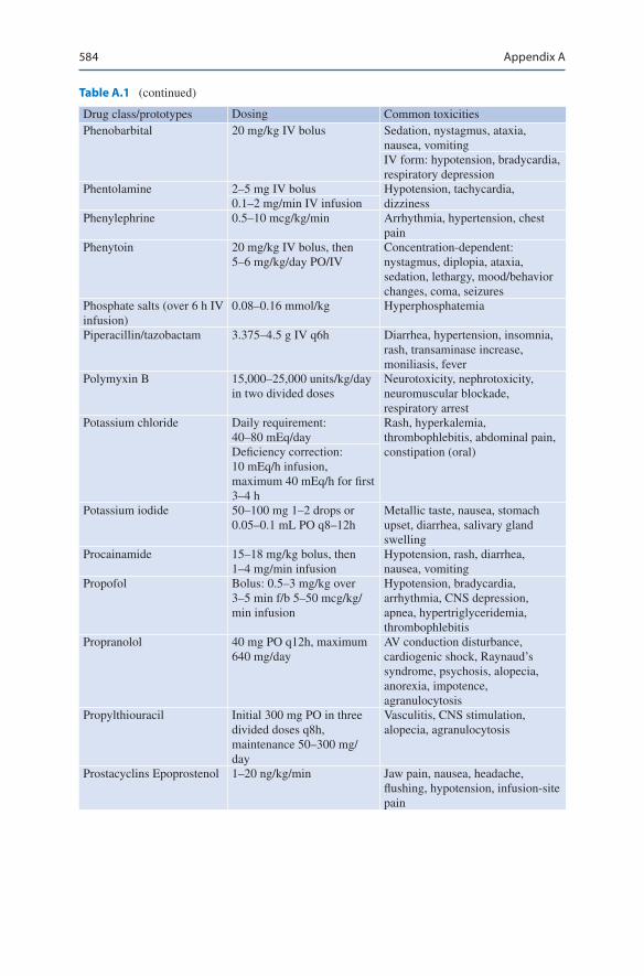
584
Drug class/prototypes Dosing Common toxicitiesPhenobarbital 20 mg/kg IV bolus Sedation, nystagmus, ataxia,
nausea, vomitingIV form: hypotension, bradycardia, respiratory depression
Phentolamine 2–5 mg IV bolus Hypotension, tachycardia, dizziness0.1–2 mg/min IV infusion
Phenylephrine 0.5–10 mcg/kg/min Arrhythmia, hypertension, chest pain
Phenytoin 20 mg/kg IV bolus, then 5–6 mg/kg/day PO/IV
Concentration-dependent: nystagmus, diplopia, ataxia, sedation, lethargy, mood/behavior changes, coma, seizures
Phosphate salts (over 6 h IV infusion)
0.08–0.16 mmol/kg Hyperphosphatemia
Piperacillin/tazobactam 3.375–4.5 g IV q6h Diarrhea, hypertension, insomnia, rash, transaminase increase, moniliasis, fever
Polymyxin B 15,000–25,000 units/kg/day in two divided doses
Neurotoxicity, nephrotoxicity, neuromuscular blockade, respiratory arrest
Potassium chloride Daily requirement: 40–80 mEq/day
Rash, hyperkalemia, thrombophlebitis, abdominal pain, constipation (oral)Deficiency correction:
10 mEq/h infusion, maximum 40 mEq/h for first 3–4 h
Potassium iodide 50–100 mg 1–2 drops or 0.05–0.1 mL PO q8–12h
Metallic taste, nausea, stomach upset, diarrhea, salivary gland swelling
Procainamide 15–18 mg/kg bolus, then 1–4 mg/min infusion
Hypotension, rash, diarrhea, nausea, vomiting
Propofol Bolus: 0.5–3 mg/kg over 3–5 min f/b 5–50 mcg/kg/min infusion
Hypotension, bradycardia, arrhythmia, CNS depression, apnea, hypertriglyceridemia, thrombophlebitis
Propranolol 40 mg PO q12h, maximum 640 mg/day
AV conduction disturbance, cardiogenic shock, Raynaud’s syndrome, psychosis, alopecia, anorexia, impotence, agranulocytosis
Propylthiouracil Initial 300 mg PO in three divided doses q8h, maintenance 50–300 mg/day
Vasculitis, CNS stimulation, alopecia, agranulocytosis
Prostacyclins Epoprostenol 1–20 ng/kg/min Jaw pain, nausea, headache, flushing, hypotension, infusion-site pain
Table A.1 (continued)
Appendix A
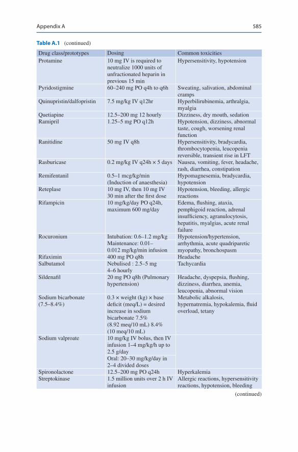
585
Drug class/prototypes Dosing Common toxicitiesProtamine 10 mg IV is required to
neutralize 1000 units of unfractionated heparin in previous 15 min
Hypersensitivity, hypotension
Pyridostigmine 60–240 mg PO q4h to q6h Sweating, salivation, abdominal cramps
Quinupristin/dalfopristin 7.5 mg/kg IV q12hr Hyperbilirubinemia, arthralgia, myalgia
Quetiapine 12.5–200 mg 12 hourly Dizziness, dry mouth, sedationRamipril 1.25–5 mg PO q12h Hypotension, dizziness, abnormal
taste, cough, worsening renal function
Ranitidine 50 mg IV q8h Hypersensitivity, bradycardia, thrombocytopenia, leucopenia reversible, transient rise in LFT
Rasburicase 0.2 mg/kg IV q24h × 5 days Nausea, vomiting, fever, headache, rash, diarrhea, constipation
Remifentanil 0.5–1 mcg/kg/min (Induction of anaesthesia)
Hypomagnesemia, bradycardia, hypotension
Reteplase 10 mg IV, then 10 mg IV 30 min after the first dose
Hypotension, bleeding, allergic reactions
Rifampicin 10 mg/kg/day PO q24h, maximum 600 mg/day
Edema, flushing, ataxia, pemphigoid reaction, adrenal insufficiency, agranulocytosis, hepatitis, myalgias, acute renal failure
Rocuronium Intubation: 0.6–1.2 mg/kg Hypotension/hypertension, arrhythmia, acute quadriparetic myopathy, bronchospasm
Maintenance: 0.01–0.012 mg/kg/min infusion
Rifaximin 400 mg PO q8h HeadacheSalbutamol Nebulised : 2.5–5 mg
4–6 hourlyTachycardia
Sildenafil 20 mg PO q8h (Pulmonary hypertension)
Headache, dyspepsia, flushing, dizziness, diarrhea, anemia, leucopenia, abnormal vision
Sodium bicarbonate (7.5–8.4%)
0.3 × weight (kg) × base deficit (meq/L) = desired increase in sodium bicarbonate 7.5% (8.92 meq/10 mL) 8.4% (10 meq/10 mL)
Metabolic alkalosis, hypernatremia, hypokalemia, fluid overload, tetany
Sodium valproate 10 mg/kg IV bolus, then IV infusion 1–4 mg/kg/h up to 2.5 g/dayOral: 20–30 mg/kg/day in 2–4 divided doses
Spironolactone 12.5–200 mg PO q24h HyperkalemiaStreptokinase 1.5 million units over 2 h IV
infusionAllergic reactions, hypersensitivity reactions, hypotension, bleeding
Table A.1 (continued)
(continued)
Appendix A

586
Drug class/prototypes Dosing Common toxicitiesSucralfate 1 g suspension 4 hourly ConstipationSuxamethonium 1.0–1.5 mg/kg IV bolus
Rapid sequence intubationHyperpyrexia, muscle pain, hyperkalemia
Teicoplanin 400 mg 12 hourly for three doses IV, then 400 mg q24h IV
Raised LFT, hypersensitivity
Tenecteplase One-time bolus over 5 s: Hypotension, bleeding, allergic reactions≤60 kg = 30 mg
61–70 kg = 35 mg71–80 kg = 40 mg81–90 kg = 45 mg≥90 kg = 50 mg
Terlipressin Hepatorenal syndrome: 0.5–1 mg q6h IV
Hypertension, abdominal cramps
Varices: 2 mg IV bolus, then 1–2 mg q4–6h IV
Theophylline Bolus: 5 mg/kg if no theophylline received in the previous 24 h
Arrhythmia, headache, seizure, nervousness, nausea, diarrhea, tremor, muscle cramp
Maintenance: 0.7 mg/kg/hThiopental 2.5–4 mg/kg IV bolus for
seizure controlApnea, bronchospasm, hypersensitivity
Thiourea drugs Initial 300–600 mg q24h PO Rash, arthralgias, fever, leucopenia, nausea, vomiting
Ticarcillin/clavulanate 3.1 g IV q4–6h Diarrhea, hypertension, insomnia, rash, transaminase increase, moniliasis, fever
Tigecycline 100 mg bolus, then 50 mg IV q12h
Nausea, hypertension, peripheral edema, phlebitis, fever, headache, insomnia, pruritus, hyperglycemia, hyperproteinemia, hyperkalemia, thrombocytopenia, leukocytosis, hepatic dysfunction, neuromuscular weakness
Tirofiban 0.4 mcg/kg/min × 30 min, then 0.1 mcg/kg/min (In unstable angina)0.4 mcg/kg/min × 3 min, then 0.1 mcg/kg/min (In PCI)
Bleeding, bradycardia, coronary artery dissection, dizziness, vasovagal reaction, thrombocytopenia
Torsemide 10–20 mg IV/PO daily Arrhythmia, chest pain, headache, ototoxicity, dizziness, hyperglycemia, hyperuricemia, hypokalemia
Maximum: 200 mg (PO, IV)
Tranexamic acid 500–1000 mg 8 hourly Thrombosis
Table A.1 (continued)
Appendix A

587
Drug class/prototypes Dosing Common toxicitiesValproate 1000–2500 mg/day IV/PO
q12h to q6h Maintenance Loading (in status epilepticus): 15–45 mg/kg IV at <6 mg/kg/min Infusion (in status epilepticus): 1–4 mg/kg/h
Somnolence, diplopia, nausea, vomiting, diarrhea
Vancomycin Loading (In severe infection): 25–30 mg/kg f/b 15–20 mg/kg IV q8–12h125–500 mg PO in C. difficile diarrhea
Bitter taste, nausea, vomiting, chills, fever, eosinophilia, interstitial nephritis, vasculitis, thrombocytopenia, red man syndrome
Vasopressin 40 units IV bolus Arrhythmia, asystole, decreased cardiac output, chest pain, MI, peripheral ischemia, venous thrombosis, urticaria, mesenteric ischemia
0.01–0.04 U/min IV infusion (in refractory septic shock) 0.2–0.4 U/min IV infusion (in variceal hemorrhage)
Verapamil 5–10 mg iv bolus BradycardiaVecuronium 100 mcg/kg iv Liver dysfunctionVitamin K 1–10 mg PO, SQ, or IV
q24hHemolysis in G6PD deficiency
Voriconazole i.v. 6 mg/kg for two doses q12h, fist day f/b 4 mg/kg IV q12hOral maintenance : 200 mg 12 hourly
Photophobia, agranulocytosis, thrombocytopenia, anemia, diarrhea, vomiting, hallucinations, tachycardia, hyper/hypotension, raised liver enzymes, cholestatic jaundice
Warfarin Initial 1–5 mg PO q24h, adjust based on INR measurements
Bleeding, angina, chest pain, hypotension, alopecia, skin necrosis, agranulocytosis, purple toe syndrome
Table A.1 (continued)
Appendix A

588
Tabl
e A
.2
Dos
age
mod
ifica
tion
in r
enal
fai
lure
Med
icat
ion
Dos
e fo
r no
rmal
ren
al
func
tion
Dos
e w
ith im
pair
ed r
enal
fun
ctio
n (G
FR m
L/m
in/1
.73
m2 )
Supp
lem
enta
l dos
e in
dia
lysi
s
30–5
010
–29
<10
Hem
odia
lysi
s (H
D)
Peri
tone
al
dial
ysis
(PD
)A
ntim
icro
bial
sA
cycl
ovir
PO: 8
0 m
g/kg
/day
, div
ided
q6
h10
mg/
kg, q
12h
10 m
g/kg
, q24
h5
mg/
kg, q
24h
Yes
CV
VH
D/C
VV
HD
F:
10 m
g/kg
q12
–24h
No
IV: 3
0 m
g/kg
/day
Am
ikac
in5–
7.5
mg/
kg/d
ose,
q8–
12h
or 1
5–20
mg/
kg I
V O
D5–
7.5
mg/
kg,
q12–
18h
5–7.
5 m
g/kg
, q2
4h5–
7.5
mg/
kg,
q48–
72h
Yes
CV
VH
D/D
F: L
oadi
ng
dose
10
mg/
kg f
/b
mai
nten
ance
7.5
mg/
kg
q24–
48h
Yes
Am
phot
eric
in B
(c
onve
ntio
nal)
0.5–
1.5
mg/
kg I
V, q
24h
No
chan
geN
o ch
ange
No
chan
geN
oN
o
Am
pici
llin
100–
200
mg/
kg/d
ay,
divi
ded
q6h
No
chan
geU
sual
dos
e q6
–12h
Usu
al d
ose
q12–
24h
Yes
CV
VH
D/D
F: L
oadi
ng
Dos
e 2
g fo
llow
ed b
y 1–
2 g
q6–8
h
No
Am
oxic
illin
20–5
0 m
g/kg
/day
IV
/PO
, di
vide
d q8
hN
o ch
ange
10–2
0 m
g/kg
/do
se, q
12h
10–2
0 m
g/kg
/dos
e,
q24h
Yes
No
Aza
thio
prin
e1–
3 m
g/kg
, q24
h PO
Red
uce
dose
by
25%
Red
uce
dose
by
25%
Red
uce
dose
by
50%
Yes
Yes
Azi
thro
myc
in10
mg/
kg/d
ay P
O/I
VN
o ch
ange
No
chan
geN
o ch
ange
No
No
Cas
pofu
ngin
70 m
g on
day
1, t
hen
50 m
g IV
, q24
hN
o ch
ange
No
chan
geN
o ch
ange
No
No
Co-
amox
icla
vIV
/PO
: 10–
20 m
g/kg
, q8h
No
chan
geIn
crea
se in
terv
al,
q12h
Incr
ease
inte
rval
, q2
4hY
esN
o
Cef
aclo
r20
–40
mg/
kg/d
ay I
V,
divi
ded
q8–1
2hN
o ch
ange
No
chan
geR
educ
e do
se b
y 50
%; d
ivid
ed q
12h
Yes
No
Cef
epim
e50
mg/
kg/d
ose
IV, q
12h
50 m
g/kg
/dos
e,
q24h
50 m
g/kg
/dos
e,
q24h
50 m
g/kg
/dos
e,
q48h
Yes
CV
VH
D/D
F: L
oadi
ng
dose
2 g
f/b
2 g
q12
hN
o
Appendix A
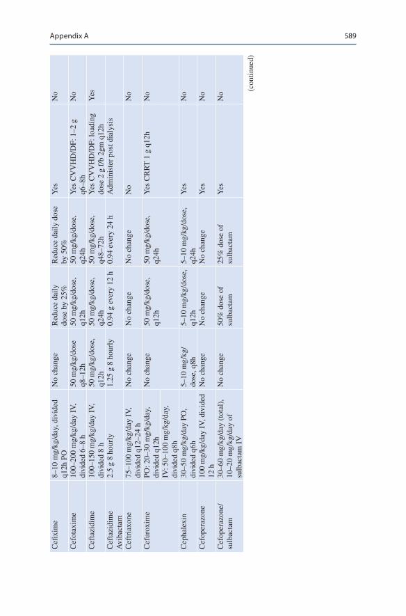
589
Cefi
xim
e8–
10 m
g/kg
/day
, div
ided
q1
2h P
ON
o ch
ange
Red
uce
daily
do
se b
y 25
%R
educ
e da
ily d
ose
by 5
0%Y
esN
o
Cef
otax
ime
100–
200
mg/
kg/d
ay I
V,
divi
ded
6–8
h50
mg/
kg/d
ose
q8–1
2h50
mg/
kg/d
ose,
q1
2h50
mg/
kg/d
ose,
q2
4hY
es C
VV
HD
/DF:
1–2
g
q6–8
hN
o
Cef
tazi
dim
e10
0–15
0 m
g/kg
/day
IV
, di
vide
d 8
h50
mg/
kg/d
ose,
q1
2h50
mg/
kg/d
ose,
q2
4h50
mg/
kg/d
ose,
q4
8–72
hY
es C
VV
HD
/DF:
load
ing
dose
2 g
f/b
2gm
q12
hY
es
Cef
tazi
dim
e A
viba
ctam
2.5
g 8
hour
ly1.
25 g
8 h
ourl
y0.
94 g
eve
ry 1
2 h
0.94
eve
ry 2
4 h
Adm
inis
ter
post
dia
lysi
s
Cef
tria
xone
75–1
00 m
g/kg
/day
IV
, di
vide
d q1
2–24
hN
o ch
ange
No
chan
geN
o ch
ange
No
No
Cef
urox
ime
PO: 2
0–30
mg/
kg/d
ay,
divi
ded
q12h
No
chan
ge50
mg/
kg/d
ose,
q1
2h50
mg/
kg/d
ose,
q2
4hY
es C
RR
T 1
g q
12h
No
IV: 5
0–10
0 m
g/kg
/day
, di
vide
d q8
hC
epha
lexi
n30
–50
mg/
kg/d
ay P
O,
divi
ded
q6h
5–10
mg/
kg/
dose
, q8h
5–10
mg/
kg/d
ose,
q1
2h5–
10 m
g/kg
/dos
e,
q24h
Yes
No
Cef
oper
azon
e10
0 m
g/kg
/day
IV
, div
ided
12
hN
o ch
ange
No
chan
geN
o ch
ange
Yes
No
Cef
oper
azon
e/su
lbac
tam
30–6
0 m
g/kg
/day
(to
tal)
, 10
–20
mg/
kg/d
ay o
f su
lbac
tam
IV
No
chan
ge50
% d
ose
of
sulb
acta
m25
% d
ose
of
sulb
acta
mY
esN
o (con
tinue
d)
Appendix A

590
Cip
roflo
xaci
nC
olis
tinPO
: 20
mg/
kg/d
ay, d
ivid
ed
q12h
No
chan
ge5.
9–6.
65 m
illio
n un
its/d
ay(1
95–2
20 m
g C
BA
/day
)
q12–
24h
4.85
–5.9
mill
ion
units
/day
s (1
60–1
95 m
g C
BA
/day
)
q24h
4.4
mill
ion/
day
(145
mg
CB
A/d
ay)
Yes
CV
VH
D/D
F:
200–
400
mg
q12–
24h
inte
rmit
tent
hem
odia
lysi
s (I
HD
): O
n a
nond
ialy
sis
day,
(~3.
95 m
illio
n IU
/day
).
130
mg
CB
A/d
ayO
n a
dial
ysis
day
, ad
min
iste
r a
supp
lem
enta
l do
se (
~1.2
mill
ion
IU)4
0 m
g C
BA
– (
~1.6
m
illio
n IU
) 50
mg
CB
A f
or
a 3-
or
4-h
IHD
ses
sion
, re
spec
tivel
ySu
stai
ned
low
-effi
cien
cy
dial
ysis
(SL
ED
) th
at 1
0%
of th
e C
MS
dose
be
adde
d to
the
base
line
daily
dos
e pe
r 1
h of
SL
ED
CR
RT,
(~1
3.3
mill
ion
IU/
day)
440
mg
CB
A/
day
= (
~6.6
5 m
illio
n IU
ev
ery
12 h
). 2
20 m
g C
BA
ev
ery
12 h
Yes
IV: 1
0 m
g/kg
/day
, div
ided
q1
2h9–
10.9
mill
ion
load
ing,
Mai
nten
ance
9 m
illio
n un
it da
ily d
ivid
ed in
two
dose
s30
0–36
0 m
g C
BA
load
ing
300
mg
CB
A d
aily
div
ided
in
two
dose
s
Co-
trim
oxaz
ole
6–10
mg/
kg/d
ay (
TM
P),
IV/P
O d
ivid
ed q
12h
No
chan
ge5–
10 m
g/kg
/dos
e,
q12h
Not
rec
omm
ende
dY
esN
o
Dap
tom
ycin
6 m
g/kg
/dos
e IV
, q24
hN
o ch
ange
4 PO
/IV
/kg/
dose
, q2
4h4
mg/
kg/d
ose,
q48
hN
o C
VV
HD
: 8 m
g/kg
q48
hN
o
Tabl
e A
.2
(con
tinue
d)
Med
icat
ion
Dos
e fo
r no
rmal
ren
al
func
tion
Dos
e w
ith im
pair
ed r
enal
fun
ctio
n (G
FR m
L/m
in/1
.73
m2 )
Supp
lem
enta
l dos
e in
dia
lysi
s
30–5
010
–29
<10
Hem
odia
lysi
s (H
D)
Peri
tone
al
dial
ysis
(PD
)
Appendix A
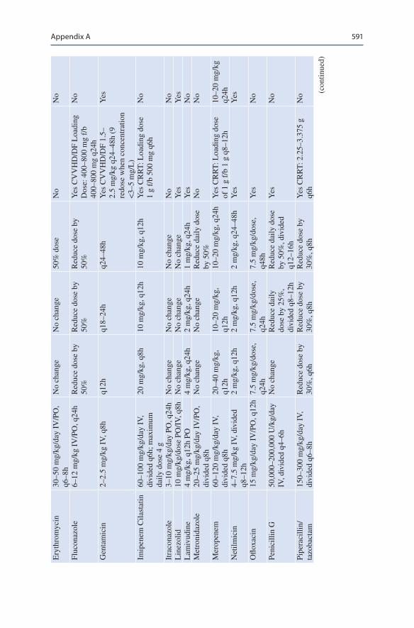
591
Ery
thro
myc
in30
–50
mg/
kg/d
ay I
V/P
O,
q6–8
hN
o ch
ange
No
chan
ge50
% d
ose
No
No
Fluc
onaz
ole
6–12
mg/
kg I
V/P
O, q
24h
Red
uce
dose
by
50%
Red
uce
dose
by
50%
Red
uce
dose
by
50%
Yes
CV
VH
D/D
F L
oadi
ng
Dos
e: 4
00–8
00 m
g f/
b 40
0–80
0 m
g q2
4h
No
Gen
tam
icin
2–2.
5 m
g/kg
IV
, q8h
q12h
q18–
24h
q24–
48h
Yes
CV
VH
D/D
F 1.
5–2.
5 m
g/kg
q24
–48h
(9
redo
se w
hen
conc
entr
atio
n <
3–5
mg/
L)
Yes
Imip
enem
Cila
stat
in60
–100
mg/
kg/d
ay I
V,
divi
ded
q6h;
max
imum
da
ily d
ose
4 g
20 m
g/kg
, q8h
10 m
g/kg
, q12
h10
mg/
kg, q
12h
Yes
CR
RT
: Loa
ding
dos
e 1
g f/
b 50
0 m
g q6
hN
o
Itra
cona
zole
3–10
mg/
kg/d
ay P
O, q
24h
No
chan
geN
o ch
ange
No
chan
geN
oN
oL
inez
olid
10 m
g/kg
/dos
e PO
/IV
, q8h
No
chan
geN
o ch
ange
No
chan
geY
esY
esL
amiv
udin
e4
mg/
kg, q
12h
PO4
mg/
kg, q
24h
2 m
g/kg
, q24
h1
mg/
kg, q
24h
Yes
No
Met
roni
dazo
le20
–25
mg/
kg/d
ay I
V/P
O,
divi
ded
q8h
No
chan
geN
o ch
ange
Red
uce
daily
dos
e by
50%
No
No
Mer
open
em60
–120
mg/
kg/d
ay I
V,
divi
ded
q8h
20–4
0 m
g/kg
, q1
2h10
–20
mg/
kg,
q12h
10–2
0 m
g/kg
, q24
hY
es C
RR
T: L
oadi
ng d
ose
of 1
g f
/b 1
g q
8–12
h10
–20
mg/
kg
q24h
Net
ilmic
in4–
7.5
mg/
kg I
V, d
ivid
ed
q8–1
2h2
mg/
kg, q
12h
2 m
g/kg
, q12
h2
mg/
kg, q
24–4
8hY
esY
es
Oflo
xaci
n15
mg/
kg/d
ay I
V/P
O, q
12h
7.5
mg/
kg/d
ose,
q2
4h7.
5 m
g/kg
/dos
e,
q24h
7.5
mg/
kg/d
ose,
q4
8hY
esN
o
Peni
cilli
n G
50,0
00–2
00,0
00 U
/kg/
day
IV, d
ivid
ed q
4–6h
No
chan
geR
educ
e da
ily
dose
by
25%
, di
vide
d q8
–12h
Red
uce
daily
dos
e by
50%
, div
ided
q1
2–16
h
Yes
No
Pipe
raci
llin/
tazo
bact
am15
0–30
0 m
g/kg
/day
IV
, di
vide
d q6
–8h
Red
uce
dose
by
30%
, q6h
Red
uce
dose
by
30%
, q8h
Red
uce
dose
by
30%
, q8h
Yes
CR
RT
: 2.2
5–3.
375
g q6
hN
o (con
tinue
d)
Appendix A
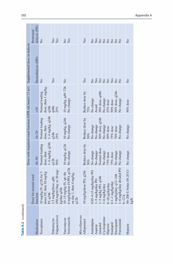
592
Teic
opla
nin
10 m
g/kg
IV
, q12
h fo
r 3
dose
s IV
, the
n 10
mg/
kg
IV, q
24h
Nor
mal
load
ing
dose
, the
n 1–
4 m
g/kg
, q24
h
Nor
mal
load
ing
dose
, the
n 1–
4 m
g/kg
, q24
h
Nor
mal
load
ing
dose
, the
n 1
mg/
kg,
q24h
No
No
Tobr
amyc
in2.
5 m
g/kg
/dos
e, q
8hq1
2hq2
4hq4
8hY
esY
esV
alga
ncic
lovi
r45
0 m
g/m
2 /da
y or
30
mg/
kg/d
ay q
24h
50%
25%
25%
Yes
Van
com
ycin
10–1
5 m
g/kg
IV
, q6–
8h10
mg/
kg, q
12h
10 m
g/kg
, q24
h10
mg/
kg, q
48–7
2hN
oN
oV
oric
onaz
ole
6 m
g/kg
/dos
e IV
/PO
, q12
h on
day
1, t
hen
4 m
g/kg
, q1
2h
No
chan
geN
o ch
ange
No
chan
geY
es
Mis
cell
aneo
usA
llopu
rino
l10
mg/
kg/d
ose
PO, q
24h
Red
uce
dose
by
50%
Red
uce
dose
by
50%
Red
uce
dose
by
75%
Yes
Yes
Am
lodi
pine
0.05
–0.1
5 m
g/kg
/day
PO
No
chan
geN
o ch
ange
No
chan
geN
oN
oA
spir
in1–
5 m
g/kg
/day
PO
No
chan
geN
o ch
ange
Avo
idY
esY
esA
teno
lol
1–3
mg/
kg P
O, q
24h
Nor
mal
dos
e50
% d
ose,
q24
h50
% d
ose,
q48
hY
esN
oC
yclo
spor
ine
3–6
mg/
kg/d
ayN
o ch
ange
No
chan
geN
o ch
ange
No
No
Dig
oxin
6–10
μg/
kg/d
ay75
% d
ose
50%
dos
e25
% d
ose
No
No
Ena
lapr
il0.
1–1
mg/
kg/d
ay75
% d
ose
75%
dos
e50
% d
ose
Yes
No
Eno
xapa
rin
1 m
g/kg
/day
, q12
–24h
No
chan
ge70
% d
ose
50%
dos
e, q
24h
No
No
Furo
sem
ide
1–6
mg/
kg/d
ay d
ivid
ed P
O
6–12
hN
o ch
ange
No
chan
geN
o ch
ange
No
No
Hep
arin
50–2
00 U
bol
us f
/b 2
0 U
/kg
/hN
o ch
ange
No
chan
ge50
% d
ose
No
No
Tabl
e A
.2
(con
tinue
d)
Med
icat
ion
Dos
e fo
r no
rmal
ren
al
func
tion
Dos
e w
ith im
pair
ed r
enal
fun
ctio
n (G
FR m
L/m
in/1
.73
m2 )
Supp
lem
enta
l dos
e in
dia
lysi
s
30–5
010
–29
<10
Hem
odia
lysi
s (H
D)
Peri
tone
al
dial
ysis
(PD
)
Appendix A
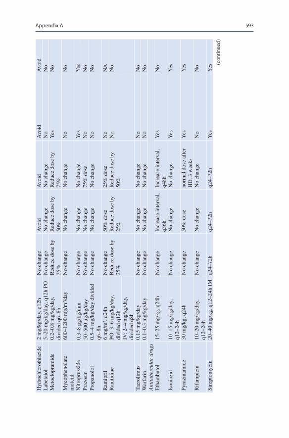
593
Hyd
roch
loro
thia
zide
2 m
g/kg
/day
, q12
hN
o ch
ange
Avo
idA
void
Avo
idA
void
Lab
etal
ol5–
20 m
g/kg
/day
, q12
h PO
No
chan
geN
o ch
ange
No
chan
geN
oN
oM
etoc
lopr
amid
e0.
2–0.
8 m
g/kg
/day
, di
vide
d q6
–8h
Red
uce
dose
by
25%
Red
uce
dose
by
50%
Red
uce
dose
by
75%
Yes
No
Myc
ophe
nola
te
mof
etil
600–
1200
mg/
m2 /
day
No
chan
geN
o ch
ange
No
chan
geN
oN
o
Nitr
opru
ssid
e0.
3–8 μg
/kg/
min
No
chan
geN
o ch
ange
No
chan
geY
esY
esPr
azos
in50
–500
μg/
kg/d
ayN
o ch
ange
No
chan
ge75
% d
ose
No
No
Prop
anol
ol0.
5–4
mg/
kg/d
ay d
ivid
ed
q6–8
hN
o ch
ange
No
chan
geN
o ch
ange
No
No
Ram
ipri
l6
mg/
m2 ,
q24h
No
chan
ge50
% d
ose
25%
dos
eN
oN
AR
aniti
dine
PO: 3
–6 m
g/kg
/day
, di
vide
d q1
2hR
educ
e do
se b
y 25
%R
educ
e do
se b
y 25
%R
educ
e do
se b
y 50
%N
oN
o
IV: 2
–4 m
g/kg
/day
, di
vide
d q8
hTa
crol
imus
0.15
mg/
kg/d
ayN
o ch
ange
No
chan
geN
o ch
ange
No
No
War
fari
n0.
1–0.
3 m
g/kg
/day
No
chan
geN
o ch
ange
No
chan
geN
oN
oA
ntit
uber
cula
r dr
ugs
Eth
ambu
tol
15–2
5 m
g/kg
, q24
hN
o ch
ange
Incr
ease
inte
rval
, q3
6hIn
crea
se in
terv
al,
q48h
Yes
No
Ison
iazi
d10
–15
mg/
kg/d
ay,
q12–
24h
No
chan
geN
o ch
ange
No
chan
geY
esY
es
Pyra
zina
mid
e30
mg/
kg, q
24h
No
chan
ge50
% d
ose
norm
al d
ose
afte
r H
D, 3
wee
ksY
esY
es
Rif
ampi
cin
10–2
0 m
g/kg
/day
, q1
2–24
hN
o ch
ange
No
chan
geN
o ch
ange
No
No
Stre
ptom
ycin
20–4
0 m
g/kg
, q12
–24h
IM
q24–
72h
q24–
72h
q24–
72h
Yes
Yes
(con
tinue
d)
Appendix A
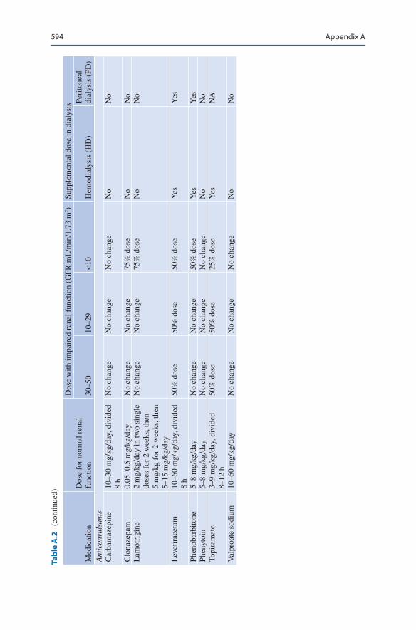
594
Ant
icon
vuls
ants
Car
bam
azep
ine
10–3
0 m
g/kg
/day
, div
ided
8
hN
o ch
ange
No
chan
geN
o ch
ange
No
No
Clo
naze
pam
0.05
–0.5
mg/
kg/d
ayN
o ch
ange
No
chan
ge75
% d
ose
No
No
Lam
otri
gine
2 m
g/kg
/day
in tw
o si
ngle
do
ses
for
2 w
eeks
, the
n 5
mg/
kg f
or 2
wee
ks, t
hen
5–15
mg/
kg/d
ay
No
chan
geN
o ch
ange
75%
dos
eN
oN
o
Lev
etir
acet
am10
–60
mg/
kg/d
ay, d
ivid
ed
8 h
50%
dos
e50
% d
ose
50%
dos
eY
esY
es
Phen
obar
bito
ne5–
8 m
g/kg
/day
No
chan
geN
o ch
ange
50%
dos
eY
esY
esPh
enyt
oin
5–8
mg/
kg/d
ayN
o ch
ange
No
chan
geN
o ch
ange
No
No
Topi
ram
ate
3–9
mg/
kg/d
ay, d
ivid
ed
8–12
h50
% d
ose
50%
dos
e25
% d
ose
Yes
NA
Val
proa
te s
odiu
m10
–60
mg/
kg/d
ayN
o ch
ange
No
chan
geN
o ch
ange
No
No
Tabl
e A
.2
(con
tinue
d)
Med
icat
ion
Dos
e fo
r no
rmal
ren
al
func
tion
Dos
e w
ith im
pair
ed r
enal
fun
ctio
n (G
FR m
L/m
in/1
.73
m2 )
Supp
lem
enta
l dos
e in
dia
lysi
s
30–5
010
–29
<10
Hem
odia
lysi
s (H
D)
Peri
tone
al
dial
ysis
(PD
)
Appendix A
595© Springer Nature Singapore Pte Ltd. 2020R. Chawla, S. Todi (eds.), ICU Protocols, https://doi.org/10.1007/978-981-15-0898-1
Appendix B
Common ICU Formulae
A. Pulmonary equations 1. Arterial oxygen tension (PaO2)
On room air = 100 − 1/3 (age) On supplemental oxygen = FiO2 (in decimals) × 500, Room air FiO2 = 21% (0.21), FiO2 increases by approximately 4% for each litre increase in Supplemental Oxygen
2. Alveolar gas equation
PAO FiO Patm PH OPaCO
R
PAO PaCO
2 2 22
2 2150 1 25
= × −[ ]( ) −
= ×( )– .
Normal = 100 mmHg (room air, at sea level)where PAO2 = alveolar partial pressure of oxygenFiO2 = fraction of inspired oxygen (in decimals)Patm = barometric pressure (760 mmHg at sea level)PH2O = water vapor pressure (47 mmHg at normal body temperature 37°C)PaCO2 = partial pressure of carbon dioxide in the bloodR = respiratory quotient, assumed to be 0.8
3. Alveolar–arterial oxygen gradientPAO2 − PaO2
• A-a gradient (on room air) = 2.5 + 0.21 × age in years• Normal value = 3–15 mmHgVaries with FiO2
• For FiO2 = 21%; A-a gradient = 5–15 mmHg• For FiO2 = 100%; A-a gradient = <150 mmHg
4. PaO2/FiO2 ratioNormal = 300–500 mmHg<300 = acute lung injury (previous definition)<200 = ARDS (previous definition)Berlin definition:• 200–300 (with PEEP/CPAP >5) = Mild ARDS• <200 (with PEEP >5) = Moderate ARDS• <100 (With PEEP >5) = Severe ARDS
596
5. Arteriolar–alveolar oxygen ratio = PaO2/PAO2Normal = 0.77–0.82 (most reliable when FiO2 <0.5)
6. Oxygenation index =
mean airway pressure cm H OFiO
PaOfractionofinspiredO
m
2
2
22
2( )× ( )
mm Hg
Good outcome
( )
×
− =
100
0 25
,
>25–40 = severe hypoxemia 7. Static lung compliance (Crs stat)
ComplianceTidal olume
Plateau pressure PEEP positive estatic = −v
nnd expiratory pressure−( )
Normal compliance in an intubated patient = 57–85 mL/cm H2O 8. Dynamic lung compliance (Crs dynamic)
ComplianceTidal olume
Peek pressure PEEP positive enddynamic = −v
−−( )expiratory pressure
Variable depending on peak pressure in an intubated patientLung + Thoracic wall compliance = 0.1 L (100 mL)/cm H2O
9. Airway resistance
Airway resistance
Peak inspiratory pressure plateau pressu=
− rre
Peak inspiratory flow
Normal resistance in an intubated patient is 4–6 cm H2O/L/s 10. PaCO2–PetCO2 gradientNormal = 4–5 mmHg
11. Dead space ventilation V
V
PaCO PetCO
PaCOD
T
=−2 2
2
VD = Dead Space Ventilation = 1 mL/lb (2.2 kg) of ideal body wt = 150 mLVT = Tidal Volume
PetCO end tidal CO measured by capnography
Normal V VD T
2 2
0 5 50
==
−/ . %(( )( )
in mechanically ventilated patients
in spontaneo0 3 30. % uusly breathing patients
12. Shunt equation (right to left shunt) Qs QtCcO CaO
CcO CvO/ =
−( )−( )
2 2
2 2
• Qs/Qt = shunt fraction• CcO2 is the end-capillary oxygen content (estimated from the PAO2)
Appendix B
597
• CaO2 is the arterial oxygen content• CvO2 is the mixed venous oxygen content• Normal = 5%Alternate equation (in patients breathing 100% oxygen for 20 min)
Qs Qt AaG AaG/ . / .= × ×( ) ×( ) +( )100 0 0031 0031 5
13. PaO2 + PaCO2 <150 mmHg at sea level breathing room air B. Hemodynamic equations
(see Chap. 16, Vol. 1)
Parameter Formula Normal rangePulse pressure Systolic − diastolic BP 40 mmHgMean arterial pressure (MAP)
1/3 pulse pressure + diastolic BP 65 mmHg
Cardiac output (CO) SV × HR 4–7 L/minCardiac index (CI) CO/BSA 3.5–4.5 L/min/m2
Stroke volume (SV) CO/HR × 1000 End diastolic volume (EDV) (120 mL) – End systolic volume (ESV) (50 mL)
60–80 mL
Stroke volume index (SVI) CI/HR × 1000, SV/BSA 33–47 mL/m2/beatSystemic vascular resistance (SVR)
[(MAP − CVP)/CO] × 80 900–1200 dyn s/cm5
Systemic vascular resistance index (SVRI)
(MAP – CVP) 80/CI 1970–2390 dyn s/cm5/m2
Pulmonary vascular resistance
[(MPAP − PAOP)/CO] × 80 80–120 dyn s/cm5
Pulmonary vascular resistance index
[(MPAP − PAOP)/CI] × 80 255–285 dyn s/cm5/m2
Oxygen delivery (DO2) CO (L) × CaO2 (mL/dL) × 10 700–1400 mL/minOxygen delivery index (DO2I)
CaO2 × CI × 10 500–600 mL/min/m2
Oxygen consumption (VO2) CO (L) × (CaO2 − CvO2) × 10 180–280 mL/minOxygen consumption index (VO2I)
CI × (CaO2 − CvO2) × 10 120–160 mL/min/m2
Oxygen extraction ratio (O2ER)
VO2/DO2 × 100 25%
Oxygen extraction index (O2EI)
[(SaO2 − SvO2)/SaO2] × 100 20–25%
Arterial oxygen content (CaO2)
(1.39 × Hb SaO2) + (0.003 × PaO2) 17–20 mL/dL
Mixed venous oxygen content (CvO2)
(1.39 × Hb × SvO2) + (0.003 × PvO2)
12–15 mL/dL
A-V oxygen content difference (C(a-v)O2)
CaO2 − CvO2 4–6 mL/dL
Systolic pressure variation (SPV)
[(SPmax − SPmin)/(SPmax + SPmin)/2] × 100
<5 mmHg unlikely to be preload responsive >5 mmHg likely to be preload responsive
Appendix B
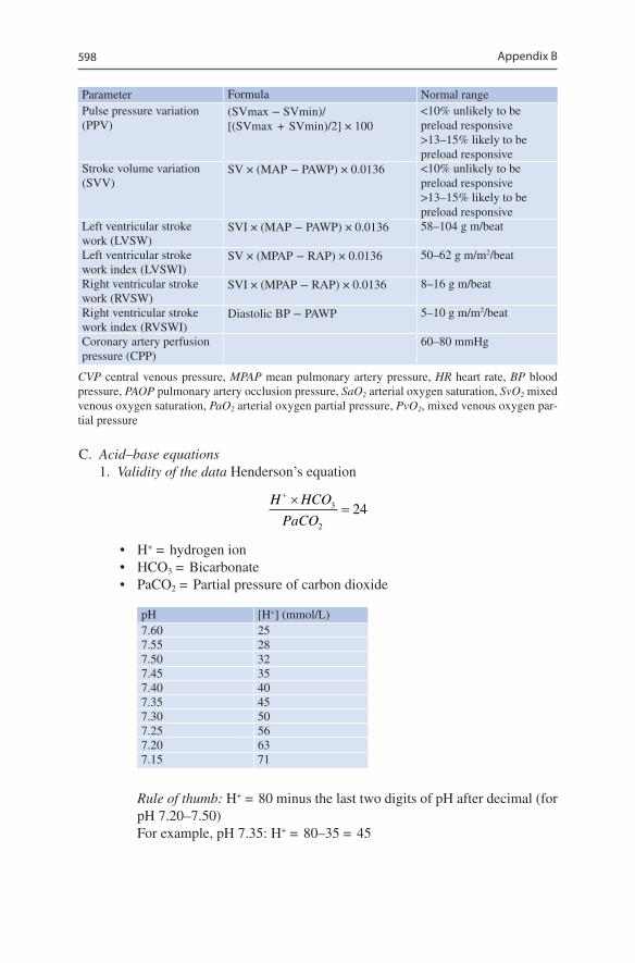
598
Parameter Formula Normal rangePulse pressure variation (PPV)
(SVmax − SVmin)/[(SVmax + SVmin)/2] × 100
<10% unlikely to be preload responsive >13–15% likely to be preload responsive
Stroke volume variation (SVV)
SV × (MAP − PAWP) × 0.0136 <10% unlikely to be preload responsive >13–15% likely to be preload responsive
Left ventricular stroke work (LVSW)
SVI × (MAP − PAWP) × 0.0136 58–104 g m/beat
Left ventricular stroke work index (LVSWI)
SV × (MPAP − RAP) × 0.0136 50–62 g m/m2/beat
Right ventricular stroke work (RVSW)
SVI × (MPAP − RAP) × 0.0136 8–16 g m/beat
Right ventricular stroke work index (RVSWI)
Diastolic BP − PAWP 5–10 g m/m2/beat
Coronary artery perfusion pressure (CPP)
60–80 mmHg
CVP central venous pressure, MPAP mean pulmonary artery pressure, HR heart rate, BP blood pressure, PAOP pulmonary artery occlusion pressure, SaO2 arterial oxygen saturation, SvO2 mixed venous oxygen saturation, PaO2 arterial oxygen partial pressure, PvO2, mixed venous oxygen par-tial pressure
C. Acid–base equations 1. Validity of the data Henderson’s equation
H HCO
PaCO
+ ×=3
2
24
• H+ = hydrogen ion• HCO3 = Bicarbonate• PaCO2 = Partial pressure of carbon dioxide
pH [H+] (mmol/L)7.60 257.55 287.50 327.45 357.40 407.35 457.30 507.25 567.20 637.15 71
Rule of thumb: H+ = 80 minus the last two digits of pH after decimal (for pH 7.20–7.50)For example, pH 7.35: H+ = 80–35 = 45
Appendix B
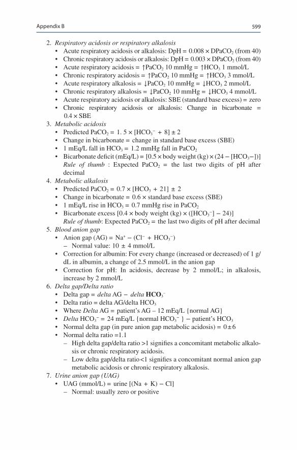
599
2. Respiratory acidosis or respiratory alkalosis• Acute respiratory acidosis or alkalosis: DpH = 0.008 × DPaCO2 (from 40)• Chronic respiratory acidosis or alkalosis: DpH = 0.003 × DPaCO2 (from 40)• Acute respiratory acidosis = ↑PaCO2 10 mmHg = ↑HCO3 1 mmol/L• Chronic respiratory acidosis = ↑PaCO2 10 mmHg = ↑HCO3 3 mmol/L• Acute respiratory alkalosis = ↓PaCO2 10 mmHg = ↓HCO3 2 mmol/L• Chronic respiratory alkalosis = ↓PaCO2 10 mmHg = ↓HCO3 4 mmol/L• Acute respiratory acidosis or alkalosis: SBE (standard base excess) = zero• Chronic respiratory acidosis or alkalosis: Change in bicarbonate =
0.4 × SBE 3. Metabolic acidosis
• Predicted PaCO2 = 1. 5 × [HCO3− + 8] ± 2
• Change in bicarbonate = change in standard base excess (SBE)• 1 mEq/L fall in HCO3 = 1.2 mmHg fall in PaCO2
• Bicarbonate deficit (mEq/L) = [0.5 × body weight (kg) × (24 − [HCO3−])] Rule of thumb : Expected PaCO2 = the last two digits of pH after
decimal 4. Metabolic alkalosis
• Predicted PaCO2 = 0.7 × [HCO3 + 21] ± 2• Change in bicarbonate = 0.6 × standard base excess (SBE)• 1 mEq/L rise in HCO3 = 0.7 mmHg rise in PaCO2
• Bicarbonate excess [0.4 × body weight (kg) × ([HCO3−] − 24)]
Rule of thumb: Expected PaCO2 = the last two digits of pH after decimal 5. Blood anion gap
• Anion gap (AG) = Na+ − (Cl− + HCO3−)
– Normal value: 10 ± 4 mmol/L• Correction for albumin: For every change (increased or decreased) of 1 g/
dL in albumin, a change of 2.5 mmol/L in the anion gap• Correction for pH: In acidosis, decrease by 2 mmol/L; in alkalosis,
increase by 2 mmol/L 6. Delta gap/Delta ratio
• Delta gap = delta AG − delta HCO3–
• Delta ratio = delta AG/delta HCO3
• Where Delta AG = patient’s AG – 12 mEq/L {normal AG}• Delta HCO3
− = 24 mEq/L {normal HCO3− } − patient’s HCO3
• Normal delta gap (in pure anion gap metabolic acidosis) = 0 ± 6• Normal delta ratio =1.1
– High delta gap/delta ratio >1 signifies a concomitant metabolic alkalo-sis or chronic respiratory acidosis.
– Low delta gap/delta ratio <1 signifies a concomitant normal anion gap metabolic acidosis or chronic respiratory alkalosis.
7. Urine anion gap (UAG)• UAG (mmol/L) = urine [(Na + K) − Cl]
– Normal: usually zero or positive
Appendix B
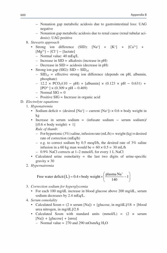
600
– Nonanion gap metabolic acidosis due to gastrointestinal loss: UAG negative
– Nonanion gap metabolic acidosis due to renal cause (renal tubular aci-dosis): UAG positive
8. Stewarts approach• Strong ion difference (SID): [Na+] + [K+] + [Ca2+] +
[Mg2+] − [Cl−] − [lactate] – Normal value: 40 mEq/L – Increase in SID = alkalosis (increase in pH) – Decrease in SID = acidosis (decrease in pH)
• Strong ion gap (SIG): SID − SIDeff
– SIDeff = effective strong ion difference (depends on pH, albumin, phosphate)
– 12.2 × PCO2/(10 − pH) + [albumin] × (0.123 × pH − 0.631) + [PO4−] × (0.309 × pH − 0.469)
– Normal SIG = 0 – Positive SIG = Increase in organic acid
D. Electrolyte equations 1. Hyponatremia
• Sodium deficit = (desired [Na+] − current [Na+]) × 0.6 × body weight in kg
• Increase in serum sodium = (infusate sodium − serum sodium)/[(0.6 × body weight) + 1]Rule of thumb:
– For hypertonic (3%) saline, infusion rate (mL/h) = weight (kg) × desired rate of correction (mEq/h)
– e.g. to correct sodium by 0.5 meq/l/h, the desired rate of 3% saline infusion in a 60 kg man would be = 60 × 0.5 = 30 mL/h
– 0.9% NaCl corrects at 1–2 mmol/L for every 1 L NaCl• Calculated urine osmolarity = the last two digits of urine-specific
gravity × 30 2. Hypernatremia
Free water deficit L body weightplasma Na( ) = × × −
+
0 4140
1.
3. Correction sodium for hyperglycemia• For each 100 mg/dL increase in blood glucose above 200 mg/dL, serum
sodium decreases by 2.4 mEq/L. 4. Serum osmolality
• Calculated Sosm = (2 × serum [Na]) + [glucose, in mg/dL]/18 + [blood urea nitrogen, in mg/dL]/2.8
• Calculated Sosm with standard units (mmol/L) = (2 × serum [Na]) + [glucose] + [urea]
– Normal value = 270 and 290 mOsm/kg H2O
Appendix B
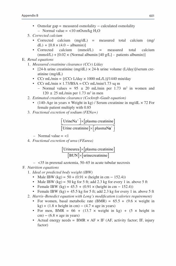
601
• Osmolar gap = measured osmolality − calculated osmolality – Normal value = <10 mOsm/kg H2O
5. Corrected calcium• Corrected calcium (mg/dL) = measured total calcium (mg/
dL) + [0.8 × (4.0 − albumin)]• Corrected calcium (mmol/L) = measured total calcium
(mmol/L) + [0.02 × (Normal albumin [40 g/L] – patients albumin)] E. Renal equations
1. Measured creatinine clearance (CCr) L/day• [24-h urine creatinine (mg/dL) × 24-h urine volume (L/day)]/serum cre-
atinine (mg/dL)• CCr mL/min = [(CCr L/day × 1000 mL/L)]/1440 min/day• CCr mL/min × 1.73/BSA = CCr mL/min/1.73 sq m
– Normal values = 95 ± 20 mL/min per 1.73 m2 in women and 120 ± 25 mL/min per 1.73 m2 in men
2. Estimated creatinine clearance (Cockroft–Gault equation)• (140- Age in years × Weight in kg) / Serum creatinine in mg/dL × 72 For
female patient multiply with 0.85 3. Fractional excretion of sodium (FENa+)
UrineNa plasma creatinine
Urine creatinine plasmaN
+ ×[ ][ ]× aa+
– Normal value = <1 4. Fractional excretion of urea (FEurea)
Urineurea plasma creatinine
BUN urinecreatinine
[ ]×[ ][ ]×[ ]
– <35 in prerenal azotemia, 50–65 in acute tubular necrosis F. Nutrition equations
1. Ideal or predicted body weight (IBW)• Male IBW (kg) = 50 + (0.91 × (height in cm − 152.4))• Male IBW (kg) = 50 kg for 5 ft; add 2.3 kg for every 1 in. above 5 ft• Female IBW (kg) = 45.5 + (0.91 × (height in cm − 152.4))• Female IBW (kg) = 45.5 kg for 5 ft; add 2.3 kg for every 1 in. above 5 ft
2. Harris–Benedict equation with Long’s modification (calories requirement)• For women, basal metabolic rate (BMR) = 65.5 + (9.6 × weight in
kg) + (1.8 × height in cm) − (4.7 × age in years)• For men, BMR = 66 + (13.7 × weight in kg) + (5 × height in
cm) − (6.8 × age in years)• Actual energy needs = BMR × AF × IF (AF, activity factor; IF, injury
factor)
Appendix B
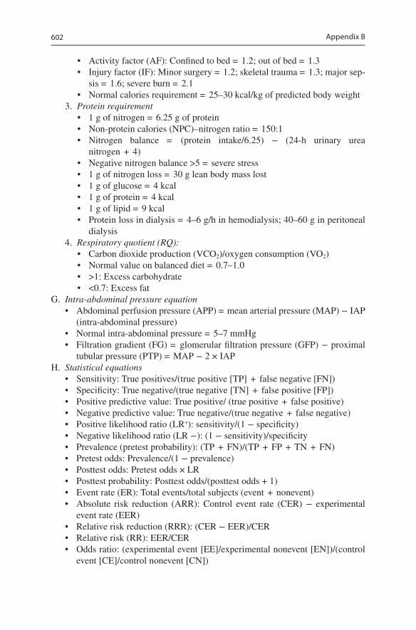
602
• Activity factor (AF): Confined to bed = 1.2; out of bed = 1.3• Injury factor (IF): Minor surgery = 1.2; skeletal trauma = 1.3; major sep-
sis = 1.6; severe burn = 2.1• Normal calories requirement = 25–30 kcal/kg of predicted body weight
3. Protein requirement• 1 g of nitrogen = 6.25 g of protein• Non-protein calories (NPC)–nitrogen ratio = 150:1• Nitrogen balance = (protein intake/6.25) − (24-h urinary urea
nitrogen + 4)• Negative nitrogen balance >5 = severe stress• 1 g of nitrogen loss = 30 g lean body mass lost• 1 g of glucose = 4 kcal• 1 g of protein = 4 kcal• 1 g of lipid = 9 kcal• Protein loss in dialysis = 4–6 g/h in hemodialysis; 40–60 g in peritoneal
dialysis 4. Respiratory quotient (RQ):
• Carbon dioxide production (VCO2)/oxygen consumption (VO2)• Normal value on balanced diet = 0.7–1.0• >1: Excess carbohydrate• <0.7: Excess fat
G. Intra-abdominal pressure equation• Abdominal perfusion pressure (APP) = mean arterial pressure (MAP) − IAP
(intra-abdominal pressure)• Normal intra-abdominal pressure = 5–7 mmHg• Filtration gradient (FG) = glomerular filtration pressure (GFP) − proximal
tubular pressure (PTP) = MAP − 2 × IAP H. Statistical equations
• Sensitivity: True positives/(true positive [TP] + false negative [FN])• Specificity: True negative/(true negative [TN] + false positive [FP])• Positive predictive value: True positive/ (true positive + false positive)• Negative predictive value: True negative/(true negative + false negative)• Positive likelihood ratio (LR+): sensitivity/(1 − specificity)• Negative likelihood ratio (LR −): (1 − sensitivity)/specificity• Prevalence (pretest probability): (TP + FN)/(TP + FP + TN + FN)• Pretest odds: Prevalence/(1 − prevalence)• Posttest odds: Pretest odds × LR• Posttest probability: Posttest odds/(posttest odds + 1)• Event rate (ER): Total events/total subjects (event + nonevent)• Absolute risk reduction (ARR): Control event rate (CER) − experimental
event rate (EER)• Relative risk reduction (RRR): (CER − EER)/CER• Relative risk (RR): EER/CER• Odds ratio: (experimental event [EE]/experimental nonevent [EN])/(control
event [CE]/control nonevent [CN])
Appendix B
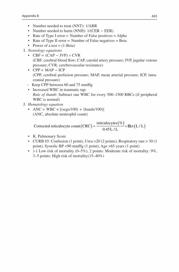
603
• Number needed to treat (NNT): 1/ARR• Number needed to harm (NNH): 1/(CER − EER)• Rate of Type I error = Number of False positives = Alpha• Rate of Type II error = Number of False negatives = Beta• Power of a test = (1-Beta)
I. Neurology equations• CBF = (CAP − JVP) ÷ CVR (CBF, cerebral blood flow; CAP, carotid artery pressure; JVP, jugular venous
pressure; CVR, cerebrovascular resistance)• CPP = MAP − ICP (CPP, cerebral perfusion pressure; MAP, mean arterial pressure; ICP, intra-
cranial pressure) – Keep CPP between 60 and 75 mmHg• Increased WBC in traumatic tap: Rule of thumb: Subtract one WBC for every 500–1500 RBCs (if peripheral
WBC is normal) J. Hematology equation
• ANC = WBC × [(segs/100) + (bands/100)] (ANC, absolute neutrophil count)
Corrected reticulocyte count CRC
reticulocytes
L L( ) = ( )
×%
. /0 45HHct L L/( )
• K. Pulmonary Score• CURB 65: Confusion (1 point), Urea >20 (2 points), Respiratory rate > 30 (1
point), Systolic BP <90 mmHg (1 point), Age >65 years (1 point)• )-1 Low risk of mortality (0–5%), 2 points: Moderate risk of mortality: 9%,
3–5 points: High risk of mortality(15–40%)
Appendix B
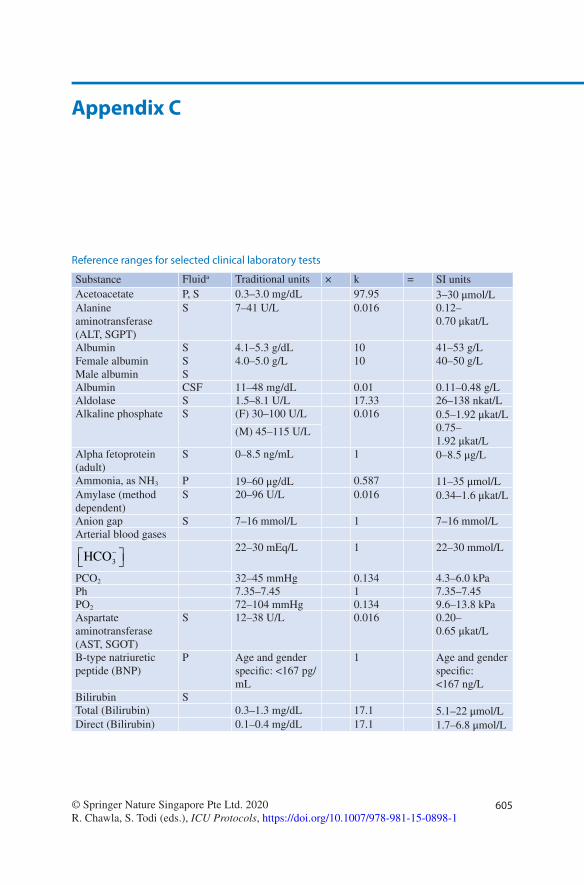
605© Springer Nature Singapore Pte Ltd. 2020R. Chawla, S. Todi (eds.), ICU Protocols, https://doi.org/10.1007/978-981-15-0898-1
Appendix C
Reference ranges for selected clinical laboratory tests
Substance Fluida Traditional units × k = SI unitsAcetoacetate P, S 0.3–3.0 mg/dL 97.95 3–30 μmol/LAlanine aminotransferase (ALT, SGPT)
S 7–41 U/L 0.016 0.12–0.70 μkat/L
AlbuminFemale albuminMale albumin
SSS
4.1–5.3 g/dL4.0–5.0 g/L
1010
41–53 g/L40–50 g/L
Albumin CSF 11–48 mg/dL 0.01 0.11–0.48 g/LAldolase S 1.5–8.1 U/L 17.33 26–138 nkat/LAlkaline phosphate S (F) 30–100 U/L 0.016 0.5–1.92 μkat/L
0.75–1.92 μkat/L
(M) 45–115 U/L
Alpha fetoprotein (adult)
S 0–8.5 ng/mL 1 0–8.5 μg/L
Ammonia, as NH3 P 19–60 μg/dL 0.587 11–35 μmol/LAmylase (method dependent)
S 20–96 U/L 0.016 0.34–1.6 μkat/L
Anion gap S 7–16 mmol/L 1 7–16 mmol/LArterial blood gases
HCO3−
22–30 mEq/L 1 22–30 mmol/L
PCO2 32–45 mmHg 0.134 4.3–6.0 kPaPh 7.35–7.45 1 7.35–7.45PO2 72–104 mmHg 0.134 9.6–13.8 kPaAspartate aminotransferase (AST, SGOT)
S 12–38 U/L 0.016 0.20–0.65 μkat/L
B-type natriuretic peptide (BNP)
P Age and gender specific: <167 pg/mL
1 Age and gender specific: <167 ng/L
Bilirubin STotal (Bilirubin) 0.3–1.3 mg/dL 17.1 5.1–22 μmol/LDirect (Bilirubin) 0.1–0.4 mg/dL 17.1 1.7–6.8 μmol/L
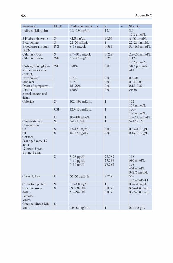
606
Substance Fluida Traditional units × k = SI unitsIndirect (Bilirubin) 0.2–0.9 mg/dL 17.1 3.4–
15.2 μmol/Lβ-Hydroxybutyrate S <1.0 mg/dL 96.05 <100 μmol/LBicarbonate S 22–26 mEq/L 1 22–26 mmol/LBlood urea nitrogen (BUN)
P, S 8–18 mg/dL 0.367 3.0–6.5 mmol/L
Calcium-Total S 8.7–10.2 mg/dL 0.252 2.2–2.6 mmol/LCalcium Ionized WB 4.5–5.3 mg/dL 0.25 1.12–
1.32 mmol/LCarboxyhemoglobin (carbon monoxide content)
WB >20% 0.01 >0.2 proportion of 1
Nonsmokers 0–4% 0.01 0–0.04Smokers 4–9% 0.01 0.04–0.09Onset of symptoms 15–20% 0.01 0.15–0.20Loss of consciousness and death
>50% 0.01 >0.50
Chloride S 102–109 mEq/L 1 102–109 mmol/L
CSF 120–130 mEq/L 1 120–130 mmol/L
U 10–200 mEq/L 1 10–200 mmol/LCholinesterase S 5–12 U/mL 1 5–12 kU/LComplementC3 S 83–177 mg/dL 0.01 0.83–1.77 g/LC4 S 16–47 mg/dL 0.01 0.16–0.47 g/LCortisolFasting, 8 a.m.–12 noon12 noon–8 p.m.8 p.m.–8 a.m.
S 5–25 μg/dL5–15 μg/dL0–10 μg/dL
27.58827.58827.588
138–690 nmol/L138–414 nmol/L0–276 nmol/L
Cortisol, free U 20–70 μg/24 h 2.758 55–193 nmol/24 h
C-reactive protein S 0.2–3.0 mg/L 1 0.2–3.0 mg/LCreatine kinase (total)FemalesMales
S 39–238 U/L51–294 U/L
0.0170.017
0.66–4.0 μkat/L0.87–5.0 μkat/L
Creatine kinase-MB SMass 0.0–5.5 ng/mL 1 0.0–5.5 g/L
Appendix C
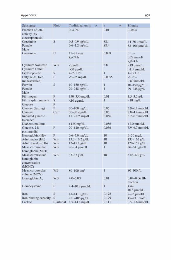
607
Substance Fluida Traditional units × k = SI unitsFraction of total activity (by electrophoresis)
0–4.0% 0.01 0–0.04
CreatinineFemaleMale
S 0.5–0.9 ng/mL0.6–1.2 ng/mL
88.488.4
44–80 μmol/L53–106 μmol/L
Creatinine U 15–25 mg/kg/24 h
0.009 0.13–0.22 mmol/kg/24 h
Cyanide: Nontoxic WB <μg/dL 3.8 <19 μmol/LCyanide: Lethal >30 μg/dL >114 μmol/LErythropoietin S 4–27 U/L 1 4–27 U/LFatty acids, free (nonesterified)
P <8–25 mg/dL 0.0355 <0.28–0.89 mmol/L
FerritinFemaleMale
S 10–150 ng/dL29–248 ng/mL
11
10–150 μg/dL29–248 μg/L
Fibrinogen P 150–350 mg/dL 0.01 1.5–3.5 g/LFibrin split products S <10 μg/mL 1 <10 mg/LGlucose PGlucose (fasting) P 70–100 mg/dL 0.06 3.9–6.1 mmol/LGlucose CSF 50–80 mg/dL 0.06 2.8–4.4 mmol/LImpaired glucose tolerance
111–125 mg/dL 0.056 6.2–6.9 mmol/L
Diabetes mellitus >125 mg/dL 0.056 >7.0 mmol/LGlucose, 2 h postprandial
P 70–120 mg/dL 0.056 3.9–6.7 mmol/L
Hemoglobin (Hb) P 0.6–5.0 mg/dL 10 6–50 mg/LAdult males (Hb) WB 13.3–16.2 g/dL 10 133–162 g/LAdult females (Hb) WB 12–15.8 g/dL 10 120–158 g/dLMean corpuscular hemoglobin (MCH)
WB 26–34 pg/cell 1 26–34 pg/cell
Mean corpuscular hemoglobin concentration (MCHC)
WB 33–37 g/dL 10 330–370 g/L
Mean corpuscular volume (MCV)
WB 80–100 μm3 1 80–100 fL
Hemoglobin Alc WB 4.0–6.0% 0.01 0.04–0.06 Hb fraction
Homocysteine P 4.4–10.8 μmol/L 1 4.4–10.8 μmol/L
Iron S 41–141 μg/dL 0.178 7–25 μmol/LIron-binding capacity S 251–406 μg/dL 0.179 45–73 μmol/LLactate P, arterial 4.5–14.4 mg/dL 0.111 0.5–1.6 mmol/L
Appendix C
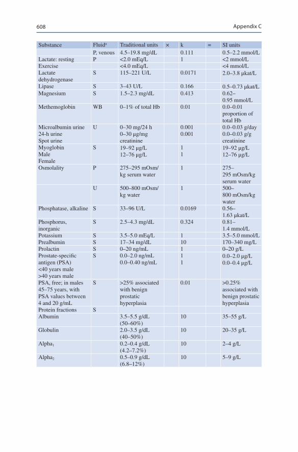
608
Substance Fluida Traditional units × k = SI unitsP, venous 4.5–19.8 mg/dL 0.111 0.5–2.2 mmol/L
Lactate: resting P <2.0 mEq/L 1 <2 mmol/LExercise <4.0 mEq/L <4 mmol/LLactate dehydrogenase
S 115–221 U/L 0.0171 2.0–3.8 μkat/L
Lipase S 3–43 U/L 0.166 0.5–0.73 μkat/LMagnesium S 1.5–2.3 mg/dL 0.413 0.62–
0.95 mmol/LMethemoglobin WB 0–1% of total Hb 0.01 0.0–0.01
proportion of total Hb
Microalbumin urine24-h urineSpot urine
U 0–30 mg/24 h0–30 μg/mg creatinine
0.0010.001
0.0–0.03 g/day0.0–0.03 g/g creatinine
MyoglobinMaleFemale
S 19–92 μg/L12–76 μg/L
11
19–92 μg/L12–76 μg/L
Osmolality P 275–295 mOsm/kg serum water
1 275–295 mOsm/kg serum water
U 500–800 mOsm/kg water
1 500–800 mOsm/kg water
Phosphatase, alkaline S 33–96 U/L 0.0169 0.56–1.63 μkat/L
Phosphorus, inorganic
S 2.5–4.3 mg/dL 0.324 0.81–1.4 mmol/L
Potassium S 3.5–5.0 mEq/L 1 3.5–5.0 mmol/LPrealbumin S 17–34 mg/dL 10 170–340 mg/LProlactin S 0–20 ng/mL 1 0–20 g/LProstate-specific antigen (PSA)<40 years male>40 years male
S 0.0–2.0 ng/mL0.0–0.40 ng/mL
11
0.0–2.0 μg/L0.0–0.4 μg/L
PSA, free; in males 45–75 years, with PSA values between 4 and 20 g/mL
S >25% associated with benign prostatic hyperplasia
0.01 >0.25% associated with benign prostatic hyperplasia
Protein fractions SAlbumin 3.5–5.5 g/dL
(50–60%)10 35–55 g/L
Globulin 2.0–3.5 g/dL (40–50%)
10 20–35 g/L
Alpha1 0.2–0.4 g/dL (4.2–7.2%)
10 2–4 g/L
Alpha2 0.5–0.9 g/dL (6.8–12%)
10 5–9 g/L
Appendix C
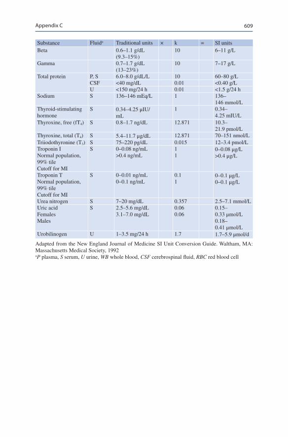
609
Substance Fluida Traditional units × k = SI unitsBeta 0.6–1.1 g/dL
(9.3–15%)10 6–11 g/L
Gamma 0.7–1.7 g/dL (13–23%)
10 7–17 g/L
Total protein P, S 6.0–8.0 g/dL/L 10 60–80 g/LCSF <40 mg/dL 0.01 <0.40 g/LU <150 mg/24 h 0.01 <1.5 g/24 h
Sodium S 136–146 mEq/L 1 136–146 mmol/L
Thyroid-stimulating hormone
S 0.34–4.25 μIU/mL
1 0.34–4.25 mIU/L
Thyroxine, free (fT4) S 0.8–1.7 ng/dL 12.871 10.3–21.9 pmol/L
Thyroxine, total (T4) S 5.4–11.7 μg/dL 12.871 70–151 nmol/LTriiodothyronine (T3) S 75–220 pg/dL 0.015 12–3.4 pmol/LTroponin INormal population, 99% tileCutoff for MI
S 0–0.08 ng/mL>0.4 ng/mL
11
0–0.08 μg/L>0.4 μg/L
Troponin TNormal population, 99% tileCutoff for MI
S 0–0.01 ng/mL0–0.1 ng/mL
0.11
0–0.1 μg/L0–0.1 μg/L
Urea nitrogen S 7–20 mg/dL 0.357 2.5–7.1 mmol/LUric acidFemalesMales
S 2.5–5.6 mg/dL3.1–7.0 mg/dL
0.060.06
0.15–0.33 μmol/L0.18–0.41 μmol/L
Urobilinogen U 1–3.5 mg/24 h 1.7 1.7–5.9 μmol/d
Adapted from the New England Journal of Medicine SI Unit Conversion Guide. Waltham, MA: Massachusetts Medical Society, 1992aP plasma, S serum, U urine, WB whole blood, CSF cerebrospinal fluid, RBC red blood cell
Appendix C
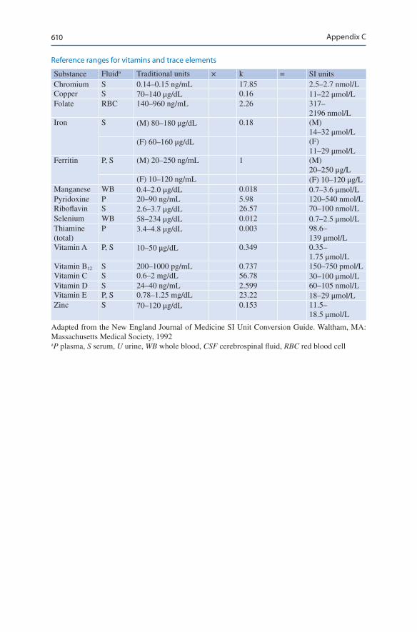
610
Reference ranges for vitamins and trace elements
Substance Fluida Traditional units × k = SI unitsChromium S 0.14–0.15 ng/mL 17.85 2.5–2.7 nmol/LCopper S 70–140 μg/dL 0.16 11–22 μmol/LFolate RBC 140–960 ng/mL 2.26 317–
2196 nmol/LIron S (M) 80–180 μg/dL 0.18 (M)
14–32 μmol/L(F) 60–160 μg/dL (F)
11–29 μmol/LFerritin P, S (M) 20–250 ng/mL 1 (M)
20–250 μg/L(F) 10–120 ng/mL (F) 10–120 μg/L
Manganese WB 0.4–2.0 μg/dL 0.018 0.7–3.6 μmol/LPyridoxine P 20–90 ng/mL 5.98 120–540 nmol/LRiboflavin S 2.6–3.7 μg/dL 26.57 70–100 nmol/LSelenium WB 58–234 μg/dL 0.012 0.7–2.5 μmol/LThiamine (total)
P 3.4–4.8 μg/dL 0.003 98.6–139 μmol/L
Vitamin A P, S 10–50 μg/dL 0.349 0.35–1.75 μmol/L
Vitamin B12 S 200–1000 pg/mL 0.737 150–750 pmol/LVitamin C S 0.6–2 mg/dL 56.78 30–100 μmol/LVitamin D S 24–40 ng/mL 2.599 60–105 nmol/LVitamin E P, S 0.78–1.25 mg/dL 23.22 18–29 μmol/LZinc S 70–120 μg/dL 0.153 11.5–
18.5 μmol/L
Adapted from the New England Journal of Medicine SI Unit Conversion Guide. Waltham, MA: Massachusetts Medical Society, 1992aP plasma, S serum, U urine, WB whole blood, CSF cerebrospinal fluid, RBC red blood cell
Appendix C
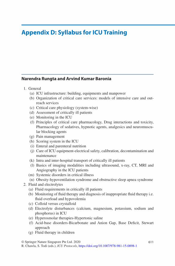
611© Springer Nature Singapore Pte Ltd. 2020R. Chawla, S. Todi (eds.), ICU Protocols, https://doi.org/10.1007/978-981-15-0898-1
Appendix D: Syllabus for ICU Training
Narendra Rungta and Arvind Kumar Baronia
1. General (a) ICU infrastructure: building, equipments and manpower (b) Organization of critical care services: models of intensive care and out-
reach services (c) Critical care physiology (system-wise) (d) Assessment of critically ill patients (e) Monitoring in the ICU (f) Principles of critical care pharmacology, Drug interactions and toxicity,
Pharmacology of sedatives, hypnotic agents, analgesics and neuromuscu-lar blocking agents
(g) Pain management (h) Scoring system in the ICU (i) Enteral and parenteral nutrition (j) Care of ICU equipment-electrical safety, calibration, decontamination and
maintenance (k) Intra and inter-hospital transport of critically ill patients (l) Basics of imaging modalities including ultrasound, x-ray, CT, MRI and
Angiography in the ICU patients (m) Systemic disorders in critical illness (n) Obesity-hypoventilation syndrome and obstructive sleep apnea syndrome
2. Fluid and electrolytes (a) Fluid requirements in critically ill patients (b) Monitoring of fluid therapy and diagnosis of inappropriate fluid therapy i.e.
fluid overload and hypovolemia (c) Colloid versus crystalloid (d) Electrolyte disturbances (calcium, magnesium, potassium, sodium and
phosphorus) in ICU (e) Hyperosmolar therapies-Hypertonic saline (f) Acid-base disorders-Bicarbonate and Anion Gap, Base Deficit, Stewart
approach (g) Fluid therapy in children

612
3. Renal disorders (a) Acute kidney injury (b) Renal tubular acidosis (c) Hepatorenal syndrome (d) Peritoneal dialysis, plasmapheresis and apheresis (e) Renal replacement therapy (f) Drugs in renal failure
4. Nervous system (a) Seizure disorders and status epileptics (b) Cerebrovascular accident (CVA) (c) Acute CNS infections (d) Intra-arterial pressure: Physiology, Intracranial hypertension, ICP
monitoring (e) Coma (f) Traumatic brain injury (g) Neuromuscular diseases (h) Acute Flaccid Paralysis-Guillain-Barré syndrome and other disorders (i) Tetanus (j) CNS drugs (k) Brain death (l) EEG in the ICU
5. Cardiovascular system (a) Acute coronary syndrome (b) Acute heart failure (c) ACLS guidelines (d) Rhythm disorders (e) Basics of echocardiography in the ICU (f) Valvular heart diseases (g) Cardiomyopathies (h) Postoperative cardiac care (i) Cardiogenic shock (j) Myocarditis (k) Hypertensive emergencies (l) Cardioversion (m) Cardiac drugs
6. Environmental disorders (a) Near-drowning (b) Thermal injuries (c) Biochemical hazards (d) Radiation hazards (e) Polytrauma (f) Disaster management guidelines (g) Envenomation (h) Acute poisoning
Appendix D: Syllabus for ICU Training

613
7. Endocrinal disorders (a) Thyroid storm and other thyroid disorder in critical care (b) Diabetic ketoacidosis (DKA) (c) Adrenal insufficiency (d) Cerebral salt wasting (e) Hyperglycemia and hypoglycemia in the ICU
8. Gastrointestinal disorders (a) Upper gastrointestinal bleeding (b) Lower gastrointestinal bleeding (c) Acute liver failure (d) Acute pancreatitis (e) Acute abdomen-medical and surgical emergencies (f) Stress ulcer prophylaxis (g) Postoperative care (h) Liver transplant: Basics
9. Respiratory disorders (a) Oxygen therapy (b) Airway adjuncts (c) Basics of mechanical ventilation and applied physiology (d) Disease-specific ventilation (e) Ventilator-Graphics, monitoring and Troubleshooting (f) High-frequency oscillation ventilation (g) Acute respiratory distress syndrome (h) Pulmonary thromboembolism (i) Pneumonias (j) Chronic obstructive pulmonary disease (k) Noninvasive ventilation (l) Chest physiotherapy (m) Pulmonary function test (PFT) (n) Extracorporeal membrane oxygenation (ECMO) + ECCO2 Elimination:
asics 10. Infections
(a) Hand hygiene (b) Asepsis guidelines (c) Sepsis syndrome: SIRS, sepsis, severe sepsis, septic shock and multiorgan
dysfunction syndrome (MODS) (d) Immunocompromised hosts (e) HIV and AIDS (f) Ventilator-associated pneumonia (VAP) (g) New outset fever in the ICU (h) Severe Tropical infections: Malaria, Typhoid, Scrub typhus and zoonosis (i) Nosocomial infections (j) Viral hemorrhagic fevers (k) Endocarditis (l) Opportunistic infections in the ICU
Appendix D: Syllabus for ICU Training
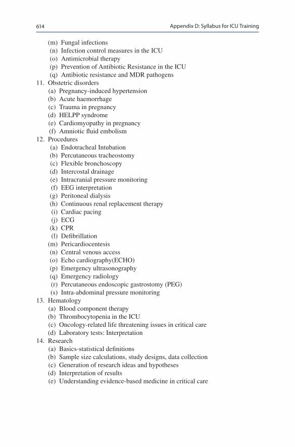
614
(m) Fungal infections (n) Infection control measures in the ICU (o) Antimicrobial therapy (p) Prevention of Antibiotic Resistance in the ICU (q) Antibiotic resistance and MDR pathogens
11. Obstetric disorders (a) Pregnancy-induced hypertension (b) Acute haemorrhage (c) Trauma in pregnancy (d) HELPP syndrome (e) Cardiomyopathy in pregnancy (f) Amniotic fluid embolism
12. Procedures (a) Endotracheal Intubation (b) Percutaneous tracheostomy (c) Flexible bronchoscopy (d) Intercostal drainage (e) Intracranial pressure monitoring (f) EEG interpretation (g) Peritoneal dialysis (h) Continuous renal replacement therapy (i) Cardiac pacing (j) ECG (k) CPR (l) Defibrillation (m) Pericardiocentesis (n) Central venous access (o) Echo cardiography(ECHO) (p) Emergency ultrasonography (q) Emergency radiology (r) Percutaneous endoscopic gastrostomy (PEG) (s) Intra-abdominal pressure monitoring
13. Hematology (a) Blood component therapy (b) Thrombocytopenia in the ICU (c) Oncology-related life threatening issues in critical care (d) Laboratory tests: Interpretation
14. Research (a) Basics-statistical definitions (b) Sample size calculations, study designs, data collection (c) Generation of research ideas and hypotheses (d) Interpretation of results (e) Understanding evidence-based medicine in critical care
Appendix D: Syllabus for ICU Training
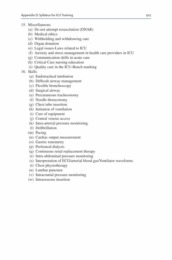
615
15. Miscellaneous (a) Do not attempt resuscitation (DNAR) (b) Medical ethics (c) Withholding and withdrawing care (d) Organ donation (e) Legal issues-Laws related to ICU (f) Anxiety and stress management in health care providers in ICU (g) Communication skills in acute care (h) Critical Care nursing-education (i) Quality care in the ICU-Bench marking
16. Skills (a) Endotracheal intubation (b) Difficult airway management (c) Flexible bronchoscopy (d) Surgical airway (e) Percutaneous tracheostomy (f) Needle thoracotomy (g) Chest tube insertion (h) Initiation of ventilation (i) Care of equipment (j) Central venous access (k) Intra-arterial pressure monitoring (l) Defibrillation (m) Pacing (n) Cardiac output measurement (o) Gastric tonometry (p) Peritoneal dialysis (q) Continuous renal replacement therapy (r) Intra-abdominal pressure monitoring (s) Interpretation of ECG/arterial blood gas/Ventilator waveforms (t) Chest physiotherapy (u) Lumbar puncture (v) Intracranial pressure monitoring (w) Intraosseous insertion
Appendix D: Syllabus for ICU Training
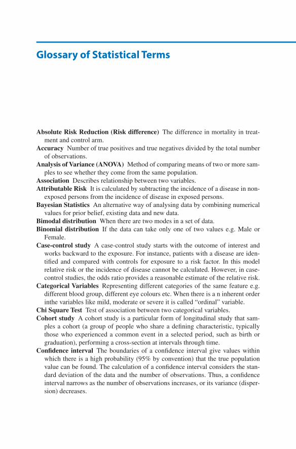
Glossary of Statistical Terms
Absolute Risk Reduction (Risk difference) The difference in mortality in treat-ment and control arm.
Accuracy Number of true positives and true negatives divided by the total number of observations.
Analysis of Variance (ANOVA) Method of comparing means of two or more sam-ples to see whether they come from the same population.
Association Describes relationship between two variables.Attributable Risk It is calculated by subtracting the incidence of a disease in non-
exposed persons from the incidence of disease in exposed persons.Bayesian Statistics An alternative way of analysing data by combining numerical
values for prior belief, existing data and new data.Bimodal distribution When there are two modes in a set of data.Binomial distribution If the data can take only one of two values e.g. Male or
Female.Case-control study A case-control study starts with the outcome of interest and
works backward to the exposure. For instance, patients with a disease are iden-tified and compared with controls for exposure to a risk factor. In this model relative risk or the incidence of disease cannot be calculated. However, in case-control studies, the odds ratio provides a reasonable estimate of the relative risk.
Categorical Variables Representing different categories of the same feature e.g. different blood group, different eye colours etc. When there is a n inherent order inthe variables like mild, moderate or severe it is called “ordinal” variable.
Chi Square Test Test of association between two categorical variables.Cohort study A cohort study is a particular form of longitudinal study that sam-
ples a cohort (a group of people who share a defining characteristic, typically those who experienced a common event in a selected period, such as birth or graduation), performing a cross-section at intervals through time.
Confidence interval The boundaries of a confidence interval give values within which there is a high probability (95% by convention) that the true population value can be found. The calculation of a confidence interval considers the stan-dard deviation of the data and the number of observations. Thus, a confidence interval narrows as the number of observations increases, or its variance (disper-sion) decreases.

618
Confounding Effect of a factor that cannot be separated out in an experiment.Continuous variable A variable which can take any value within a given range.Correlation When there is a linear relationship between two variables. Measured
on a scale of −1 (perfect negative correlation), 0 ( no correlation) to +1 ( perfect positive correlation).
Correlation Coefficient Measure of strength of the linear relationship between two variables.
Cox proportional hazards analysis Cox proportional hazards analysis is similar to logistic regression because it can account for many variables that are relevant for predicting a dichotomous outcome. However, unlike logistic regression, Cox proportional hazards analysis permits time to be included as a variable, and for patients to be counted only for the period of time in which they were observed.
Cox regression Model A method which explores the effect of different variables on survival.
Descriptive statistics Values which describe the data in a sample.Discrete variable The data can only be of certain values e.g. number of children
in a family.Fishers Exact Test Test for association between categorical variables.Histogram A graph of continuous data categorised in a number of classes.Incidence Number of new events that have occurred in a specific time interval
divided by the population at risk at the beginning of the time interval. The result gives the likelihood of developing an event in that time.
Intention to treat The central principle underlying intention-to-treat analysis is that study participants should be analyzed according to the groups in which they were randomized, even if they did not receive or comply with treatment. Such analysis is contrasted to “as treated” (or “per protocol”) analysis in which sub-jects are analyzed according to the actual treatment they received.
Intention to treat Analysis according to the group in which the patient were ran-domised even if they are withdrawn from the study or did not receive the treat-ment or crossed over.
Interquartile range The upper and lower values defining the central 50% of obser-vations. The boundaries are equal to the observations representing the 25th and 75th percentiles. The interquartile range is depicted in a box and whiskers plot.
Kaplan-Meier analysis Kaplan-Meier analysis measures the ratio of surviving patients (or those free from an outcome) divided by the total number of patients at risk for the outcome. Every time a patient has an outcome, the ratio is recalcu-lated. Using these calculations, a curve can be generated that graphically depicts the probability of survival as time passes.
Kappa Level of agreement between two categorical measures.Kruskal Wallis test Non Parametric test which compares two or more independent
groups.Likelihood Ratio Likelihood ratios are an expression of sensitivity and specificity
that can be used to estimate the odds that a condition is present or absent.LoG rank Test A non parametric test used for the comparison of survival estimates.
Glossary of Statistical Terms
619
Logistic regression analysis Models in which the outcome is dichotomous (eg, alive or dead, or a complication occurs or does not occur).
Mann Whitney U test A non parametric test to see whether there is a signifi-cant difference between two sets of data that has come from two different set of subject.
Mean Sum of observations divided by the number of observations.Median Observation in the middle, when all observations are ranked from smallest
to largest; when number of observations are even the median is defined as the mean of the middle two data points.
Mode Observation which occurs most frequently.Multivariate analysis (Regression analysis) Statistical methods that can simulta-
neously account for multiple variables are known as “multivariate” (or multivari-able) analysis. These methods help to “control” (or “adjust”) for variables that are extraneous to the main causal question and might confound it.
Negative likelihood ratio It is calculated by dividing 1 minus sensitivity by speci-ficity (1 − sensitivity)/specificity). Positive and negative likelihood ratios of 9 and 0.25, for example, can be interpreted as meaning that a positive result is seen 9 times as frequently while a negative test is seen 0.25 times as frequently in those with a specific condition than those without it.
Negative predictive value It represents the likelihood that a patient who has a negative test is free of disease. The predictive value depend upon the prevalence of a disease within a population.
Nominal Data Data that can be placed in a particular category but have no par-ticular order.
Non Parametric test A test which is not dependent on distribution of data.Normal distribution Distribution of data that is symmetrical and have a bell
shaped curve.Null hypothesis The null hypothesis is the theory that the exposure or intervention
that is being studied is not associated with the outcome of interest. Thus, if a cer-tain level of statistical significance is reached the null hypothesis will be rejected, otherwise the null hypothesis will not be rejected.
Number needed to Harm (NNH) The absolute side effects rate for placebo minus the absolute side effect for treated patients.
Number needed to treat (NNT) NNT is the reciprocal of the absolute risk reduc-tion (the absolute adverse event rate for placebo minus the absolute adverse event rate for treated patients). :1/ARR.
Odds ratio Odds that an individual with a specific condition has been exposed to a risk factor divided by the odds that a individual without that condition ( control) has been exposed. The odds ratio is used in case-control studies and is also used in multivariate analyses The relative risk and odds ratio are interpreted relative to the number one.
One tailed Test A test where the null hypothesis can only be rejected in one direc-tion ( better or worse).
Ordinal data Data that can allocated to categories in an ordered manner e.g. stages of malignancy.
Glossary of Statistical Terms
620
Parametric test Any test that assumes that the data needs to follow a certain distri-bution e.g. Normal distribution.
Pearson Correlation coefficient Calculating correlation coefficient if values are sampled from a normal population.
Percentile Percentage of a distribution that is below a specific value. As an exam-ple, a child is in 90th percentile for weight if only 10% of children the same age weigh more than she does.
Period prevalence Proportion of individuals with a condition during a specified interval (eg, a year).
Person-years Total number of years that each member of a study population has been under observation or treatment. multiplying the number of years by the number of members of a sample population studied.
Point prevalence Proportion of individuals with a condition at a specified point in time.
Poisson distribution Number of events occurring in a fixed time interval e.g. num-ber of deaths in a year.
Positive likelihood ratio It is calculated by dividing sensitivity by 1 minus speci-ficity (sensitivity/(1 − specificity)).
Positive Predictive Value Likelihood that a patient with a positive test has the disease.
Prevalence Number of individuals with a given disease at a given point in time divided by the population at risk at that point in time. Prevalence has been further defined as being “point” or “period.”
P-Value A p-value is a measure of the effect of chance within a study. It is not the probability that the result of the study is true or correct. Instead, it is the prob-ability that if the null hypothesis were true, and if the results were not affected by bias or confounding, that we would have seen a result as extreme or more extreme than the one seen in the study.
Randomized controlled trial A randomized controlled trial (RCT) is an experi-mental design in which patients are assigned to two or more interventions. One group of patients is often assigned to a placebo (placebo control) but a random-ized trial can involve two active therapies (active control).
Range Difference between the largest and smallest observation.Receiver Operating Curve (ROC ) curve It plots sensitivity on the Y axis, and
1-specificity (which is the false positive rate) on the X axis.Regression Finding a relationship between two variables where one is dependent
on the other.Relative risk (or risk ratio) Incidence in exposed individuals divided by the
incidence in unexposed individuals. This is used in cohort study.Sensitivity The number of patients with a positive test who have a disease divided
by all patients who have the disease. A test with high sensitivity will not miss many patients who have the disease (ie, few false negative results).
Spearman correlation coefficient An estimate of correlation used for non parametric variables.
Glossary of Statistical Terms

621
Specificity The number of patients who have a negative test and do not have the disease divided by the number of patients who do not have the disease.
Standard Deviation Variability of data around the mean. In “normal” distribution samples (ie, Gaussian), 68 and 95% of values fall within one and two standard deviations of the mean, respectively.
Standard Error of the Mean It describes how much variability can be expected when measuring the mean from several different samples.
tTest (Student tTest) It is a parametric test used to compare means of two groups.Two Tailed test A test where the null hypothesis can be rejected whether the treat-
ment is better or worse.Type I Error (also referred to as an “alpha error”) It is incorrectly concluding
that there is a statistically significant difference in a dataset when it is not pres-ent; the probability of making a type I error is called “alpha.” A typical value for alpha is 0.05. Thus, a p < 0.05 leads to a decision to reject the null hypothesis.
Type II error (also referred to as a “beta error”) It is incorrectly concluding that there was no statistically significant difference in a dataset and when actually it is present; the probability of making a type II error is called “beta.” This error often reflects insufficient power of the study.
Glossary of Statistical Terms

623© Springer Nature Singapore Pte Ltd. 2020R. Chawla, S. Todi (eds.), ICU Protocols, https://doi.org/10.1007/978-981-15-0898-1
AAbdominal perfusion pressure (APP),
407, 408Activated factor VII (aFVII), 100Acute colonic pseudo-obstruction (ACPO),
401–403Acute coronary syndromes (ACS)
ACE inhibitors and angiotensin receptor blockers, 256
anticoagulants, 255antiplatelets, 250–252, 255, 256arrhythmic complication, 256–257β-blockers, 252, 255clinical investigation, 248–249, 252diagnosis, 247, 249–250, 252drug orders, 252fibrinolysis, 252, 254–255GP IIb/IIIa inhibitors, 256hyperglycemia, 257invasive vs. conservative strategy, 251mechanical complication, 249, 251, 257nitroglycerine, 250, 255–256pain relief, 250patient history and physical
examination, 248patient resuscitation, 247–248reperfusion therapy, 250, 252sedatives and laxatives, 257statins, 252, 256STEMI patients, primary angioplasty,
252–254Abdominal distension
ACPO, 403, 404colonoscopic decompression, 405imaging, 403patient resuscitation, 401–402pharmacotherapy, 404–405surgery, 405treatment, 403
Acute febrile encephalopathyclinical investigations, 341–343CSF drugs and toxins (see Cerebrospinal
fluid)empirical treatment, 341endocrine abnormalities, 345general investigation workup, 341heat stroke, 344isolation precaution, 344malignant hyperthermia, 344management, 340neuroimaging, 342neuroleptic malignant syndrome, 344neurological status assessment, 339patient history and physical examination,
340–341patient resuscitation, 339serotonergic syndrome, 344structural brain lesions, 344systemic and general examination,
340–341Acute flaccid paralysis
airway protection, 309, 313anterior horn cell disease, 311, 316autonomic disturbance, 313clinical investigations, 309, 313–315critical illness myopathy, 311, 312, 314critical illness neuropathy, 311, 312, 314ICU admission, causes of, 310intubation requirement, 310myasthenia gravis, 311, 312, 315neurologic assessment, 310–312nutritional support, 313pain control, 313physiotherapy, 313, 314pressure sores, 314respiratory failure, clinical criteria, 310respiratory support, 313venous thromboembolism prevention, 313
Index

624
Acute heart failurecardiac tamponade, 227clinical history, 226coronary artery disease, 223general care, 229laboratory investigations, 227long-term care, 240patient resuscitation, 229, 230physical examination, 226pulmonary embolism, 228treatment
blood pressure control, 226, 228blood supply optimisation, 227diuretics, 224, 228, 229, 231inotropes, 226, 229intra-aortic balloon pump, 231oxygen adequacy, 225reduce demand, 223–225, 229vasodilators, 228, 229
Acute hepatic failure, 113Acute liver failure (ALF)
causes, 428cerebral edema, 430, 431coagulopathy, 431orthotopic liver transplant, 429, 430paracetamol overdose, 431patient resuscitation, 427pregnancy situation, 431prognosis, 429renal failure, 431seizures, 431sepsis, 431supportive measures, 430therapies, 433
Acute pancreatitis (AP)complications and surgery, 424–425CT grading, 421ERCP, 424gallbladder removal, 425laboratory investigations, 418–419patient resuscitation, 417–418physical examination, 418severity assessment, 419–421treatment, 422–424
Acute respiratory distress syndrome (ARDS), 27, 36, 39, 46, 51, 55–61, 63–71, 120–122, 130, 137, 169, 299, 314, 369, 513, 521, 524
Acute respiratory failure (ARF)airway evaluation and management, 20ARDS, 22asthma, 22cardiopulmonary resuscitation, 20circulation management, 20
clinical investigations, 23differential diagnosis, 21, 23NIV, 24patient history and physical
examination, 20pulmonary physiological
changes, 20, 21, 23type 1 hypercapnia
arterial blood gas analysis, 24pulse oximetry, 21
type 1 hypoxiacauses, 22clinical features, 20etiology, 20, 23
type 2 hypercapniaarterial blood gas analysis, 24causes, 22clinical features, 20, 21clinical investigation, 20pulse oximetry, 21
Alert/verbal/painful/unresponsive scale (AVPU), 320
Analgesia, 347, 348, 350Analgesia and sedation, 348Analgosedation, 351Angiography, 381, 388Angiotensin-converting enzyme (ACE)
inhibitors, 256, 257, 266, 267, 278, 286
Angiotensin receptor blockers (ARB), 256, 457, 478
Antibiotics, 422antibiotic stewardship program
implementation, 509combination therapy, 508cultures, 506, 514drugs and doses, 508duration of, 117, 124, 509, 559empirical antibiotics, 114, 125, 526,
536, 556patient assessment and de-escalation, 502,
514, 526, 556pharmacokinetic and pharmacodynamic
principles, 507risk of infection, drug-resistant organisms,
113–115, 506selection, 505septic shock, 125, 506
Anti-epileptic drug (AED) therapy, 283
APACHE II scoring system, 419, 430, 446
ARDS, Acute respiratory distress syndrome (ARDS), see
Index
625
Arterial blood gases (ABG)acid–base disorder assessment
metabolic acidosis, 514respiratory alkalosis, 103
analysis, 363Arterial catheterization
accuracy, 177arterial waveform, 174catheter and check perfusion, 173,
176–177MAP, 176phlebostatic axis, 175vascular complications, 176
Assessment of nutritional status, 444Assessment of pain, 352Assessment of severity, 419Assess neurological status, 339Assist-control mechanical ventilation
(ACMV), 48Assisted controlled mandatory ventilation
(ACMV), 48, 49Atelectasis, 9, 16, 22, 37, 40, 46, 51, 74, 120,
126, 158–159AVPU, Alert/verbal/painful/unresponsive
scale (AVPU), see
BBeer–Lambert law, 162Benzodiazepines, 88, 230, 266, 303, 305, 306,
320, 337, 351, 356, 358Brain death
cause, 361cerebral angiography, 364cerebral scintigraphy, 365electroencephalography, 364mimic agents, 361, 362neurological examination, 362–364patient management
hemodynamics, 366–367hormones replacement,
367–369patient’s family members, 366TCD ultrasound, 365–366
Bromocriptine, 344Bronchial artery embolization (BAE), 96
CCandidemia
antifungal agentsamphotericin B, 541, 543, 544echinocandins, 541–544triazoles, 541
azole prophylaxis, 545blood culture, 540in catheterized patient, 544central nervous system, 544colonization, 540, 541, 545empirical antifungal therapy, 541invasive aspergillosis, 539, 545patient history and physical examination,
539–540zygomycosis, 539
Capnographycapnogram, 164–169cardiac oscillations, 165, 167CO2 rise, 165curare effect, 168EtCO2, 165–167expired sample contamination, 165, 166obstruction, respiratory pathway, 165physiology, 164–168principle, 164rebreathing, insufficient gas
flows, 165, 166spontaneous respiratory efforts,
165, 168Cardiac arrhythmias
arrhythmias, 233–246atrial fibrillation, 237, 239–241atrial flutter, 236, 238, 241cardioversion, 234, 238–241, 243, 244ectopic atrial tachycardia, 237, 238first-degree heart block, 246general management, 239high-degree AV block, 24012-lead electrocardiogram, 234–237MAT, 241monomorphic ventricular
tachycardia, 242multifocal atrial tachycardia, 238, 241P waves, 235, 236, 238, 239, 241, 243patient history, 233, 243patient resuscitation, 233–234polymorphic ventricular tachycardia, 242,
244precipitating factors, 234QRS complex analysis, 233, 235–239,
241–243, 245second-degree AV block, 245, 246sinus bradycardia, 244sinus tachycardia, 237, 239supraventricular tachycardia, 237–238tachy/bradyarrhythmias, 234transvenous pacemaker, 245, 246ventricular fibrillation, 238–240ventricular rhythm analysis, 234–246
Index
626
Cardiogenic shockcardiac enzymes, 218catheterization and revascularization, 220causes, 218–219, 221ECG, 218hemodynamic management, 219IABP, 219–221mechanical complications, 220medical management, 219patient history and physical
examination, 218patient resuscitation, 217pericardial tamponade, 218, 219, 221rescue therapy, 221right ventricular infarction, 218, 2202D echocardiogram, 218
Cardiorespiratory arrestadvanced airway, 212cardiopulmonary resuscitation, 207,
209–213defibrillator, 208, 210–211drug therapy, 211–212emergency system activation, 208postcardiac arrest care, 213–214prognostication, 214–215rehabilitation services, 216responsiveness and breathing, 207, 209reversible causes, 212, 213
Cardioversion, anticoagulation, 240–241
Catheter-related blood-stream infection (CRBSI), 498, 499, 560
Cause of diarrhea, 394, 395Central line catheterization, antimicrobial-
impregnated catheter, 500Cerebral angiography, 364Cerebral edema, 430, 431Cerebral perfusion pressure, 408Cerebral scintigraphy, 365Cerebrospinal fluid (CSF)
acute febrile encephalopathy, meningoencephalitis, 548
ICP monitoring, 287, 299raised ICP causes and management,
286–287Cerebrovascular accident, 26Chronic liver failure
clinical stages, 436–437diagnosis and management, 439hepatic encephalopathy, 437hepatorenal syndrome, 439laboratory investigations, 436patient resuscitation, 435precipitating factors, 436
renal function and dysfunction in cirrhosis, 438
Cirrhosis, 130, 227, 377, 445Clinical pulmonary infection score (CPIS),
120, 124Coagulopathic state, 343Coagulopathy, 428, 431Colonoscopic decompression, 405Colonoscopy, 386Coma
causes of, 323–324coma mimics, 324diagnostic tests, 323patient assessment and stabilization,
319–320patient history, 320physical examination
AVPU, 320brainstem reflexes, 322GCS, 320–321hypothermia and hyperthermia, 322motor response, 322–323ocular movements, 322respiratory patterns, 323
prognosis assessment, 324–325Community-acquired pneumonia (CAP)
antibiotics, 116, 117clinical investigationsnoninfectious causes, 117nonresponders conditions, 116, 117patient resuscitation, 111, 114sepsis guidelines, 114severity assessment, 112steroids, 113, 116
Continous renal replacement therapy (CRRT)advantages and disadvantages, 473dialysis circuit, 470drug dosing, 489
Continuous positive airway, 363Continuous positive airway pressure (CPAP)
therapy, 57, 75, 78, 81, 88, 141, 225Controlled mandatory ventilation (CMV), 48Critical-care pain observation
tool (CPOT), 352Crystalloids and/colloids, 197Cultures, 431
DDefibrillation
AEDs, 211biphasic shocks, 211complications, 214drowning, 217
Index
627
electrodes, 210indications and contraindications, 213manual external, 210–211methods, 212monophasic shock, 211pediatric age group, 227
Delirium, 347, 357Dialysis
access recirculation, 482charcoal hemoperfusion, 482complications of, 483CRRT, 479dialyzer efficiency, 481hypercatabolic patients, 482hypotension, 482intermittent hemodialysis, 481postdialysis urea and creatinine vs.
predialysis, 481preventive measures, AKI, 477RRT, 478SLED usuage, 480urea reduction ratio, 481
Diarrheacauses, 395laboratory investigations, 395–396management, 398–399patient/family history, 394–395patient resuscitation, 393–394pharmacotherapy, 396probiotics, 397, 398symptom-relief agents, 396–397
Diazepam, 303
EEchocardiography
bedside echocardiogram, 187inferior vena cava, 194left ventricle, 189–190patient resuscitation, 187–188, 196pericardium, 194–196right atrium, 195right ventricle, 195valves, 191–192
Electroencephalography (EEG), 364Empirical treatment, 341,
379, 526Endobronchial brachytherapy, 97Endocrine abnormalities, 345End-of-life care, 40Endoscopic therapy, 380, 381Enteral feed, 449Estimate energy (calories), 445–447Estimate protein (nitrogen), 447–448
FFluid and electrolyte requirement, 448Fluid resuscitation, 520, 526Fluid therapy
clinical assessment, 198crystalloids, 198HES, 198hyperoncotic colloids, 197inotrope and vasopressors
agents, 202digitalis glycosides, 202doses and effects, 203limitations, 204patient resuscitation, 198selection, 200–201septic shock, 202titration, 201–202weaning, 204
Flumazenil, 358Fresh frozen plasma (FFP), 96, 379, 385,
432, 517
GGlasgow coma scale (GCS), 80, 273, 285, 291,
319–321, 328, 330, 339, 343, 361Glasgow–Blatchford bleeding score, 377Glomerular filtration rate (GFR)
anuric AKI patients, 488Cockroft–Gault formula, 487dialysis, 488ideal body weight, 487MDRD formula, 488
Glucose control, 214Guillain–Barré syndrome (GBS), 311–314
HHaloperidol, 344, 356Hand hygiene technique, 494Harris–Benedict equation, 447Heat stroke, 344HELLP syndrome, 266Hemodynamic monitoring
advanced monitoring, 174arterial blood gas, 174central venous oxygen saturation, 174central venous pressure, 174clinical examination, 173clinical findings, 185dynamic calibration, 176–177echocardiography screening, 174flushing system, 175hourly urine output, 219
Index
628
intra-arterial blood pressure, 174intra-arterial pressure waveform, 178lactate levels, 184–185noninvasive blood pressure, 174plethysmographic signals, 174pressure transducing, 174–175pulse oximetry, 174, 182pulse pressure variation and stroke
volume variation, 181ScvO2/SvO2, 183–184static calibration, 175–176
Hemodynamic stability, 375, 381Hemodynamics, 366, 368Hemolysis, elevated liver enzymes, low platelet
count (HELLP) syndrome, 266Hemostasis, 387Hepatic dysfunction, 184, 508Hepatic failure, 451Hormones, 367Hydrocephalus, 336Hypercalcemia, 464, 481Hyperglycemia, 204, 257, 282, 293, 368Hyperkalemia, 11, 212, 231, 234, 464, 478, 481Hypernatremia, 368, 527, 566, 567Hypertensive urgencies and emergency
acute aortic dissection, 265–266acute coronary syndrome, 266acute pulmonary edema, 266adrenergic crises, 266clinical investigations, 261drugs for, 264–265neurologic hypertensive emergencies,
265–267organ involvement assessment, 261–263pregnancy-induced hypertension, 266renal emergencies, 267severity of, 259treatment goals, 263
Hyperthermia, 234, 282, 304Hypoglycemia, 272, 282, 289, 319, 362, 452Hypokalemia, 87, 239, 243, 249, 334, 413,
543Hyponatremia, 230, 368Hypothermia, 322
IImmunocompromised patient
anti-infective agents, 551immune deficiency states and pathogens,
549–550
patient history, 548patient resuscitation, 547physical examination, 548
Implantable cardioverter defibrillators (ICD), 133, 136, 137
Infection, control measuresCRBSI, 554environmental factors, 501hand hygiene technique, 561isolation types, 545nosocomial infection patient identification,
553organizational and administrative
measures, 502patient screening test, 555standard precautions, 495transmission-based precautions, 496, 497UTI, 500VAP, 498
Initial assessment and resuscitation, 520Intermittent hemodialysis (IH), 431, 470, 482,
489Intra-abdominal hypertension (IAH), 408Intraabdominal pressure (IAP), 59, 407, 409,
412Intra-aortic balloon pump (IABP), 220, 231Intracerebral hemorrhage (ICH), see
Cerebrovascular accidentIntracranial pressure (ICP), 328, 329
raised ICP causes, 329raised ICP management, 333
barbiturates, 335CSF removal, 336decompressive craniectomy, 336–337glucocorticoids, 335glycerol and urea, 336hypertonic saline, 334–335hyperventilation, 335loop diuretics, 334mannitol, 334neuromuscular paralysis, 336therapeutic hypothermia, 336
JJaundice, 521
LLower Gastrointestinal (GI) bleeding, see
Gastrointestinal (GI) bleeding, 26
Hemodynamic monitoring (cont.)
Index
629
MMalignant hyperthermia, 344Massive hemoptysis
bleeding site localization, 93bronchial artery embolization, 96bronchoscopy, 95causes, 91, 93clinical assessment, 93clinical investigations, 94–95patient history and physical examination,
91pharmacotherapy, 95physical examination, 91, 93, 94surgical resection, 96
Mechanical ventilation, 347, 348alarms, 50arterial blood gas, 55, 59, 64, 74, 79complication, 45, 49, 51–52, 57, 60extubation, 53FiO2, 50, 58–60, 63, 76, 112, 157, 197initial ventilator setting, 50, 58, 76modes, 46–49oxygen therapy, 157patient monitoring and adjustments, 51PEEP, 46, 51, 52, 55, 56, 58, 59, 63, 76,
136, 137, 180PIP, 31postextubation period, 53resuscitation, 45sleep-disordered breathing (SDB), 143spontaneous breathing trial, 52, 53, 83–84weaning
checklist, 86components, 79extubation, 79, 83–86, 88failure, 85, 86home ventilation, 89noninvasive ventilation, 85pathophysiologic mechanism, 86patient monitoring, 51, 53predictors, 79–82protocols, 89readiness criteria, 52, 80reversible causes treatment, 87–88spontaneous breathing trial, 79, 81,
83–86, 89tracheostomy, 76, 79, 83, 88ventilation mode selection, 88
Meningitis, 340, 342, 343Meningoencephalitis, 321, 370Metabolic acidosis, 393Micronutrients, 448
Midazolam (MDZ), 303Monroe Kellie doctrine, 329
NNaloxone, 211, 212, 320Nasogastric tube, 379, 386National Institute of Neurological Disorders
and Stroke (NINDS) study, 272, 273
Neuroleptic malignant syndrome, 344
New-onset feverclinical investigations, 535detailed history of, 534diagnosis, 536infectious causes of, 534noninfectious causes of, 534sepsis, 536systematic head-to-toe
examination, 534temperature recording, 533treatment for, 536
Noninvasive positive-pressure ventilation (NIPPV)
application protocols, 27clinical criteria, 26complications, 31contraindications, 26critical care ventilator, 29discontinuation, 32–34effectiveness for, 27patient monitoring, 30pressure support/control
approach, 29, 30resuscitation, 25
Noninvasive ventilation (NIV), 10, 13, 17, 31, 35–37, 39, 40, 42, 43, 53, 57, 73, 75, 76, 85, 86, 89, 114, 139–143, 157, 225, 226, 498
Nonvariceal bleeding, 380Nutrition support
calories requirement estimation, 445enteral feed, 449–450fluid and electrolyte requirement
estimation, 448micronutrients, 448nitrogen estimation, 447–448nutritional status assessment, 441parenteral nutrition, 450patient resuscitation, 443total parenteral nutrition, 451
Index
630
OOliguria
biochemical investigations, 458causes of, 458diuretics usage, 462metabolic abnormalities, 464nephrotoxic agent avoidance, 464patient monitoring, 461patient resuscitation, 457renal imaging, 461renal perfusion pressure, 462RIFLE criteria, 466sepsis, 462urinary biochemistry, 460urinary retention differentiation, 458
Organ failure, 419, 421Orthotopic liver, 429Oxygen therapy
adequacy monitoring, 157cylinders and concentrators, 149face mask, 149mechanical ventilation, 157nasal cannula, 149, 150need for, 147–148NIPPV, 156non-rebreather mask, 149, 152risk, 148, 158, 159venturi masks, 153
PParacetamol overdose, 431Parapneumonic effusion, 117, 133, 134Parenteral nutrition, 450, 451PE, see Pulmonary embolism (PE)Peak inspiratory pressure (PIP), 31, 49, 50PEEP, Positive end-expiratory pressure
(PEEP), seePercutaneous tracheostomy (PCT), 88Pericardiocentesis, 221Peritoneal dialysis, 409Pharmacotherapy, 396, 404Phencyclidine (PCP), 260, 262Pleural diseases
causes, 130, 137clinical investigations, 130–131disease-specific management,
133–136etiology diagnosis, 132ICD removal, 136patient resuscitation, 129pleurocentesis, 131systemic examination, 130
Pleurocentesis, 131, 133
Pneumothorax, 16, 22, 23, 51, 52, 64, 66, 74, 100, 129, 134–137, 195, 212, 218, 234
Positive end-expiratory pressure (PEEP), 10, 13, 14, 29–31, 36, 37, 43, 46, 50–52, 55, 56, 58, 60, 61, 63, 65, 66, 69, 76–78, 80, 83, 121, 136, 137, 180, 197, 225, 299, 369
Pregnancy, 431acute respiratory failure (ARF)
airway evaluation and management, 20ARDS, 22asthma, 22circulation management, 20clinical investigations, 23differential diagnosis, 21, 23NIV, 24patient history and physical
examination, 20pulmonary physiological changes,
20, 21, 23jaundice, 521
Proton-pump inhibitor, 379, 380Pulmonary artery catheterization (PCA), 178,
179, 184Pulmonary artery occlusion pressure (POAP),
178–180, 199Pulmonary embolism (PE), 23, 93, 99, 117,
167, 169, 457anticoagulant treatment, 102clinical investigations, 103–105clinical probability, 101–102, 104diagnosis of, 104inferior vena caval filters, 108long-term treatment, 107–108low-molecular-weight heparin, 102myocardial dysfunction, 107patient resuscitation, 100percutaneous mechanical thrombectomy,
107prevention, 109risk factors, 100risk stratification, 105surgical embolectomy, 107thrombolysis, 105–106thrombolytic therapy, 105, 106vitamin K antagonist therapy, 107
Pulse oximetrydyshemoglobinopathies, 162, 163hypoventilation/hypercarbia
detection, 163patient examination, 161perfusion, 163principles, 164
Index

631
RRadionuclide scintigraphy, 389Rapid sequence intubation (RSI), 8, 11, 12,
14, 76Rebleeding, 377, 378, 381Refractory status epileptics (RSE), 301, 302,
304–305Renal failure, 87, 102, 107, 113, 115, 130,
131, 201, 227, 240, 264, 267, 430, 431, 451, 457, 460–462, 486, 488, 494, 514, 515
Renal replacement therapy (RRT), 231, 469, 473–475, 529
CRRT, anticoagulation, 480drug dosing
chart usage, 228, 488CRRT, 470, 473maintenance dose, 489
patient resuscitation, 469, 470peritoneal dialysis, 475
Resuscitation, 339, 393, 402, 409, 413, 417, 422, 427
Reversal of oversedation, 358Rockall score, 378RSI, see Rapid sequence intubation (RSI)
SSAH, see Subarachnoid hemorrhage (SAH)Scoring systems
APACHE II, 419SOFA, 430, 512
Sedation and analgesiaagent selection
analgosedation/co–sedation, 351lorazepam, 349
daily awakening trial, 354delirium assessment, 354–356drug details, 349–350flumazenil, 358indications, 348pain assessment, 352–354titration, 354weaning, 358
Seizures, 431Sepsis, 428, 430Sequential organ failure assessment (SOFA),
446, 511Severe sepsis and septic shock, Systemic
inflammatory response syndrome (SIRS), see
Severe tropical infectionsclinical investigations, 525empirical therapy, 526
fluid resuscitation, 526patient assessment and resuscitation, 520patient history, 520–522physical examination, 522–523specific tests, 524supportive care and organ support, 526treatment, 526–530
Single–port thoracoscopy, 134, 136SIRS, see Systemic inflammatory response
syndrome (SIRS)Sleep–disordered breathing (SDB)
acute acidemia, 141comorbid medical conditions, 143consciousness/coma, 141CPAP therapy, 141hemodynamic instability, 141intubation and mechanical ventilation, 143laboratory investigations, 142patient history and physical examination,
140–141polysomnography, 143–144refractory hypoxemia, 141respiratory failure, 141–142
Slow continuous ultra filtration (SCUF), 474SOFA, Sequential organ failure assessment
(SOFA), seeSpinal cord injury, 100, 260Status epilepticus
AED, 305, 306airway protection, 302anesthetic agents, 302, 305fosphenytoin, 302nonconvulsive status identification and
management, 306patient resuscitation, 309peripheral intravenous access, 303phenobarbitone, 302, 303, 305, 306, 485seizure control algorithm, 302seizures prevention, 297–298, 303, 305seizures termination, 303, 304, 306
ST elevation acute myocardial infarction (STEMI), 248, 251
angioplasty, 252–254features, 248fibrinolysis, 253
Structural brain lesions, 344Subarachnoid hemorrhage (SAH), 244,
291–300, 327, 336, 338, 344cerebral angiography, 288, 294ECG changes, 249, 298, 299hydrocephalus, 287, 288, 293, 297, 298hyponatremia, 298neurogenic pulmonary edema(NPE), 299noncontrast CT scan, head, 293
Index
632
patient history and clinical examination, 292
prognostication, 299–300rebleeding prevention, 292, 294, 295, 297seizure prevention and treatment, 297–298severity, 292–293, 298, 299vasospasm, 292, 294–296, 298
Subclavian vein cannulation, 479, 499, 553, 561Superior vena cava syndrome, 157, 181, 184Sustained low efficiency dialysis (SLED), 470,
471, 474, 490SVR, see Systemic vascular resistance (SVR)Symptom-relief agents, 396Syndrome of inappropriate secretion of anti
diuretic hormone (SIADH), 298Systemic infection, 340, 341Systemic inflammatory response syndrome
(SIRS), 419, 420, 422, 461Systemic vascular resistance (SVR), 178, 203,
261, 408
TThiamine replacement, 379Thrombocytopenia, 70, 100, 175, 287, 343,
385, 474, 513, 520, 524, 527Total parenteral nutrition (TPN), 451Tracheal intubation
airway maintenance, 16AMBU bag, 210analgesia and sedation, 16anticipated and unanticipated difficult
airway, 16arterial blood gas analysis, 4, 21, 25,
94, 125bag-mask ventilation, 13checklist, 6, 19chest radiograph, 16clinical signs, 4, 85, 92complications, 3, 4, 13, 16–18, 31, 43, 143confirm tracheal tube placement, 13difficult intubation assessment methods,
4–6, 10drug therapy, 11ETT cuff pressure, 13, 16, 17feeding, 16indication, 143mechanical ventilation, 4, 16, 27, 42,
45–52, 55–61, 75–76, 99, 119, 143noninvasive positive pressure ventilation,
25, 75preoxygenation, 6, 7, 9, 10, 12, 39, 76, 273
tube fixation, 13tube positioning, 13upper airway obstruction, 4, 11, 26
Transcranial Doppler (TCD), 365Trauma, ECG monitoring, 252
UUnfractionated heparin (UFH), 101–103, 105,
107, 256, 287, 289, 313Upper gastrointestinal (UGI) bleeding
clinical investigations, 378empiric treatment, 379–381endoscopic therapy, 379–380etiology, 376–377factors associated, 377Glasgow–Blatchford bleeding
score, 377hemodynamic stability, 375–376nasogastric tube, 379nonvariceal bleeding, 376patient resuscitation, 375–376proton-pump inhibitors, 380Rockall score, 378thiamine replacement, 379
Urinary tract infection (UTI), 282, 498, 500, 534
VVentilator-associated pneumonia (VAP), 52,
57, 64, 79, 289, 499, 534antibiotics
de-escalation, 126drug-resistant infection, 122early/late-onset pneumonia, 122policies, 124selection, 122
clinical investigation, 126drug-resistant infection, 122management of, 64, 120noninfectious conditions, 120, 535patient resuscitation, 119–122, 124–126prevention, 120, 127
Viral hepatitis, 432, 521, 522Vitamin K antagonist therapy, 108Vitamins and trace of elements, reference
ranges, 448
WWolff–Parkinson–White (WPW) syndrome,
237, 240
Subarachnoid hemorrhage (SAH) (cont.)
Index