Structural diversity of receptors for neuropeptide Y, peptide YY and pancreatic polypeptide
On the Structural Diversity of Sialoliths
António P. Alves de Matos,1,2,* Patrícia A. Carvalho,3 Arlindo Almeida,1 Luís Duarte,4
Rui Vilar,3 and Jorge Leitão1
1Department of Biomaterials/ITB, Dental Medical School, University of Lisbon, 1649-003 Lisbon, Portugal2Anatomic Pathology Department, Curry Cabral Hospital, R. da Beneficência 8, 1069-166 Lisbon, Portugal3Department of Materials Engineering, Technical University of Lisbon, 1049-001 Lisbon, Portugal4Maxilo-Facial Cirurgy Service, S. José Hospital, 1150-199 Lisbon, Portugal
Abstract: Sialoliths from parotid and submaxillar glands have been characterized. Fractured and polishedsurfaces revealed an intrinsic structural diversity across the calculi sections. In general, the calculi presentedhighly mineralized amorphous-looking cores surrounded by concentric alternating mineralized and organiclayers. The thickness of these layers decreased from the outer regions toward the center of the sialolith,illustrating a sequence of growth stages. Nevertheless, a significant variability could be detected among thespecimens. In some cases, the calculi displayed multiple cores and lacked concentric laminated structures. Inother instances, the specimens exhibited extensive regions of globular structures. In these cases, the globulediameter decreased across the radius toward the center of the sialoliths, and the globular structures tended toreorganize, forming bright and dark laminated layers surrounding the core. The participation of globularstructures in the layer formation process points to morphogenetic mechanisms not previously described.
Key words: salivary calculi, sialoliths, conchoidal fracture, laminated structures, sulfur, globular structures
INTRODUCTION
Sialolithiasis is a common disease of the salivary glandscaused by formation of calculi in the intra- or extraglandu-lar duct system ~Lustmann et al., 1990!, which frequentlyresults in inflammation and infection with accompanyingpain. Sialoliths exhibit some structural diversity but usuallyconsist of one or more highly mineralized cores surroundedby approximately concentric laminated structures with alter-nating layers of inorganic and organic substances of vari-able thickness and texture ~Faure et al., 1986!.
Chemically, sialoliths are condensations of calciumsalts—essentially calcium phosphate and calcium carbonatedisplaying the hydroxyapatite crystallographic structure—with small amounts of other inorganic and organic compo-nents ~Anneroth et al., 1975!. The main constituent of thecalcified regions is hydroxyapatite in the form of crystalswith platelike morphologies, which are deposited with vari-able density in a reticular organic matrix ~Tandler, 1965;Tohda et al., 1995!. Other crystallographic forms of calciumphosphate may also be present ~Teymoortash et al., 2003!.
The reported structural diversity of sialoliths is a reflec-tion of variations in the nucleation and growth processes.However, the fact that laminated layers are usually described
suggests a common underlying growth mechanism ~Anne-roth et al., 1978, Lustmann & Shteyer, 1981; Isacsson &Friskopp, 1984; Grases et al., 2003; Kasaboglu et al., 2004!.In spite of the extensive research carried out on salivarycalculi, the essential aspects behind the complex multifacto-rial formation of sialoliths remain obscure and demand fur-ther investigation ~Anneroth et al., 1978; Riesco et al., 1999!.The present work evaluates the structural diversity of sialolithsby ultrastructural characterization and microanalysis.
MATERIALS AND METHODS
Scanning electron microscopy ~SEM! and energy dispersivespectroscopy ~EDS! have been used to characterize ninesialoliths from parotid and submaxillar glands. The sialoliths,stored in air before processing, were fractured throughapproximate planes of symmetry, mounted on carbon sup-ports, and coated with carbon in a rotary vacuum evapora-tor. Selected samples were embedded in an epoxy resin,ground with SiC paper, and polished using alumina sus-pensions. Special care was used to minimize mechanicaldamage during the grinding and polishing steps. SEM ob-servations under secondary electron ~SE! and backscatteredelectron ~BSE! modes as well as EDS microanalyses werecarried out with a Hitachi S2400 instrument operated at25 kV and equipped with a standard Rontec Si~Li! detector.
Received November 28, 2005; accepted May 1, 2007.*Corresponding author. E-mail: [email protected]
Microsc. Microanal. 13, 390–396, 2007DOI: 10.1017/S1431927607070754 Microscopy AND
Microanalysis© MICROSCOPY SOCIETY OF AMERICA 2007
RESULTS
The observed sialoliths presented diameters in the 3–8-mmrange and exhibited an intrinsic structural diversity acrosstheir sections. Furthermore, a significant variability couldbe detected among the specimens ~listed in Table 1!. Thestructural diversity and variability resulted in contrast vari-ations under SE and BSE observation modes.
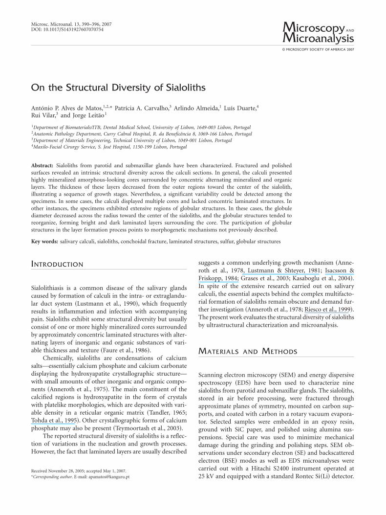
In general the calculi cores consisted of highly mineral-ized amorphous-looking masses. These regions exhibitedconchoidal fracture surfaces characteristic of brittle materi-als ~Fig. 1!. Typically, the samples presented concentricarrays of alternating brighter and dark thin layers surround-ing the central highly mineralized core ~Fig. 2!. These con-centric layers disclosed a laminated appearance, and theirthickness decreased from the outer regions toward the cen-ter of the sialolith ~Fig. 2c!. Similar to the central core, thebrighter layers were rich in Ca and P and therefore wereheavily mineralized. Dark layers lacked these mineral ele-ments and consisted of a sulphur-rich substance ~Fig. 2e!.The protrusion of mineralized brighter layers at the fracturesurfaces shows that the fracture tended to run through theinterfaces ~Fig. 2a,b!. This points to a weak adhesion be-tween the alternating bright and dark layers. The calculiwere frequently enclosed in a thick and amorphous-lookingshell ~20 mm . t . 1 mm! with a moderate mineralizationlevel ~Figs. 1a, 2a,b!.
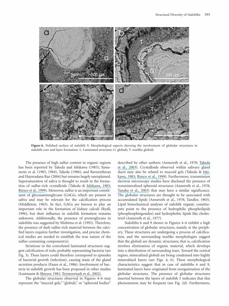
In spite of the typical underlying features describedabove, the specimens presented noticeable and significantstructural variations. Sialolith 8 shown in Figure 1b pre-sented multiple cores ~with conchoidal fracture surfaces!and lacked a clear laminated region. In this case, the fewdiscernible layers presented convoluted configurationsaround the multiple calcified cores ~Fig. 3! and often dis-closed a striated and palisade-like appearance, suggestingthat they may have resulted from bacterial growth ~regionsP in Fig. 3a,b!. Specimens 6 and 9 shown in Figures 4–6exhibited extensive regions of globular structures. The globulipresented diameters up to 50 mm and their internal sulfur-rich organic material displayed variable levels of calcifica-
tion ~Fig. 5a–c!. In some globuli, calcification occurred bothat the center and periphery ~Fig. 4b!, whereas in others itoccurred either at the periphery ~Fig. 4b! or at the center~Fig. 5a!. Tearlike structures surrounding major globuli
Table 1. Sialolith List
Sialolith Origin site Observed features
1 Parotid Single core; laminated structure2 Left submaxillar Multiple core; laminated structure; outer shell3 Parotid Single core; laminated structure; outer shell4 Right submaxillar Multiple core; laminated structure5 Left submaxillar Single core; laminated structure; outer shell6 Right submaxillar Single core; laminated structure; extensive globular morphology7 Right submaxillar Multiple core; convoluted laminated structure; outer shell8 Right submaxillar Multiple core; ~striated! convoluted laminated structure; outer shell9 Not available Multiple core; laminated structure; globular morphology
Figure 1. Fracture surface of sialolith 5 ~SE image!. a: Core exhib-iting a conchoidal fracture surface ~C!; L: laminated structure;S: outer shell. b: SE image of the fracture surface of sialolith 8: Inthis case the sialolith shows a complex pattern of multiple cores allexhibiting conchoidal fracture surfaces ~zones C!.
Structural Diversity of Sialoliths 391
suggest that organic materials were leaking from them~Figs. 4b, 5a!. Across the radius of the sialoliths, the globularstructures tended to reorganize into bright and dark lami-nated layers ~Figs. 4a, 6!. In sialolith 9 residual globularstructures are visible even within the core ~Fig. 6!. Globular
structures have also been found between the layers of sialolith5 ~see Fig. 2d!. The globular morphology was clearly ob-served in polished specimens studied under BSE mode;moreover, careful analysis disclosed the presence of globularstructures also at fractured surfaces ~Fig. 5d!.
Figure 2. a: SE image of the fracture surface of sialolith 5; L: laminated structure; S: outer shell. b: BSE image of samearea as a: dark and light layers correspond to different degrees of calcification. c: BSE image of polished surface ofsialolith 5, showing a decrease in layer thickness toward the center ~L!. d: Globular structures are sandwiched betweenthe layers in sialolith 5. e: Bright layer with a high P and Ca content ~L~B!! and dark layer with a high S content andreduced P and Ca contents ~L~D!!.
392 António P. Alves de Matos et al.
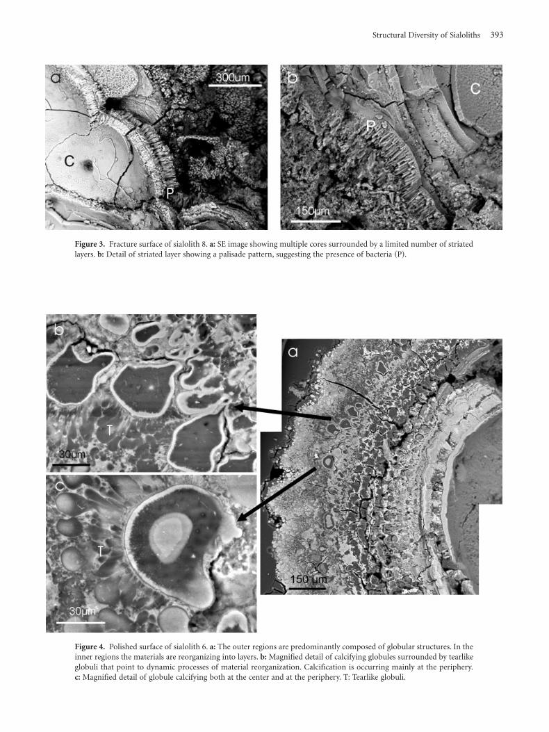
Figure 3. Fracture surface of sialolith 8. a: SE image showing multiple cores surrounded by a limited number of striatedlayers. b: Detail of striated layer showing a palisade pattern, suggesting the presence of bacteria ~P!.
Figure 4. Polished surface of sialolith 6. a: The outer regions are predominantly composed of globular structures. In theinner regions the materials are reorganizing into layers. b: Magnified detail of calcifying globules surrounded by tearlikeglobuli that point to dynamic processes of material reorganization. Calcification is occurring mainly at the periphery.c: Magnified detail of globule calcifying both at the center and at the periphery. T: Tearlike globuli.
Structural Diversity of Sialoliths 393

DISCUSSION
Sialoliths display a striking structural variability, probablyreflecting diverse mechanisms of formation and growth.The etiology of salivary calculi is still not fully understood,and several factors, working in parallel or in series, contrib-ute to the development of sialoliths, although the exactmorphogenetic mechanisms involved are in effect very un-clear ~Tanaka et al., 2003!. Several studies point to theexistence of distinct stages. Initially a core is formed bycalcification of any structure present at the duct, such as aprecipitated crystalloid, a foreign body, an inclusion body,or cellular remains ~Lustmann & Shteyer, 1981; Rodgers,1983; Takeda & Ishikawa, 1983; Cheng et al., 1987; Takeshitaet al., 1990; Tanaka et al., 2003!. This explains the fact thatmost of the observed specimens exhibit only one highlycalcified core. Sialoliths with multiple cores, like the onepresented in Figure 1b, may originate from an aggregationof several calcifying centers ~possibly microcrystalloids ormicroliths! during the calculus formation ~Takeda & Ishi-kawa, 1983; Harrison et al., 1997; Riesco et al., 1999!.Calcification may then be further assisted by factors suchas saliva composition ~Westhofen et al., 1984; Grases et al.,2003! and retention in the ductal system ~Harrison et al.,1997; Teymoortash et al., 2002, 2003!.
A “silent” growth period following the core formationstage is thought to generate the multilayered structure ob-served in most sialoliths. After this growth stage, the processeventually culminates at an inflammatory episode that de-termines the clinical symptoms and leads to the detectionand surgical removal of the calculus ~Faure et al., 1986!.
Laminated structures of bright and dark layers havebeen reported by several authors and, in fact, seem toconstitute the major organization type during growth ~Faureet al., 1986; Riesco et al., 1999!. The alternating heavilycalcified and organic layers are thought to result either froma rhythmic deposition of minerals similar to a Liesegangphenomenon ~Anneroth et al., 1975, 1978! or from a sim-pler and slow sequential deposition of layers during thecourse of the silent growth period ~Faure et al., 1986!. Onthe other hand, the current study shows that the layersdecrease in thickness across the sialolith radius toward thecenter. This indicates that the laminated structures evolvewith time through a dynamical material loss process and/orcompaction mechanism.
^
Figure 5. Polished and fracture surfaces of sialolith 6. a: Globulicalcifying at the center ~G!; tearlike surrounding structures ~T!indicate organic material replacement by mineral ~BSE image ofpolished surface!. b: Ca X-ray map of a. c: S X-ray map of a withenhanced contrast. d: SE image of fracture surface where anunpolished globule can be distinguished ~G!.
394 António P. Alves de Matos et al.
The presence of high sulfur content in organic regionshas been reported by Takeda and Ishikawa ~1983!, Yama-moto et al. ~1983, 1984!, Takeda ~1986!, and Raveenthiranand Hayavadana Rao ~2004! but remains largely unexplained.Supersaturation of saliva is thought to result in the forma-tion of sulfur-rich crystalloids ~Takeda & Ishikawa, 1983;Riesco et al., 1999!. Moreover, sulfur is an important constit-uent of glycosaminoglycans ~GAGs!, which are present insaliva and may be relevant for the calcification process~Middleton, 1965!. In fact, GAGs are known to play animportant role in the formation of kidney calculi ~Ryall,1996!, but their influence in sialolith formation remainsunknown. Additionally, the presence of proteoglycans insialoliths was suggested by Mishima et al. ~1992!. Therefore,the presence of dark sulfur-rich material between the calci-fied layers requires further investigation, and precise chem-ical studies are needed to establish the true nature of thesulfur-containing component~s!.
Striations in the convoluted laminated structures sug-gest calcification of rods, probably representing bacteria ~seeFig. 3!. These layers could therefore correspond to episodesof bacterial growth ~infection!, causing stasis of the glandsecretion products ~Faure et al., 1986!. Involvement of bac-teria in sialolith growth has been proposed in other studies~Lustmann & Shteyer, 1981; Teymoortash et al., 2002!.
The globular structures observed in Figures 4–6 mayrepresent the “mucoid gels,” “globuli,” or “spheroid bodies”
described by other authors ~Anneroth et al., 1978; Takedaet al., 2003!. Crystalloids observed within salivary glandducti may also be related to mucoid gels ~Takeda & Ishi-kawa, 1983; Riesco et al., 1999!. Furthermore, transmissionelectron microscopy studies have disclosed the presence ofnonmineralized spheroid structures ~Anneroth et al., 1978;Tanaka et al., 2003! that may have a similar significance.The globular structures are thought to be associated withaccumulated lipids ~Anneroth et al., 1978; Tandler, 1965!.Lipid histochemical analyses of sialolith organic constitu-ents point to the presence of hydrophilic phospholipids~phosphosphingosides! and hydrophobic lipids like choles-terol ~Anneroth et al., 1977!.
Sialoliths 6 and 9 shown in Figures 4–6 exhibit a highconcentration of globular structures, mainly at the periph-ery. These structures are undergoing a process of calcifica-tion, and the surrounding tearlike morphologies suggestthat the globuli are dynamic structures; that is, calcificationinvolves elimination of organic material, which developsinto a distribution of surrounding tears. Toward the centralregion, mineralized globuli are being condensed into highlymineralized layers ~see Figs. 4, 6!. These morphologicalcharacteristics suggest that in case of sialoliths 6 and 9,laminated layers have originated from reorganization of theglobular structures. The presence of globular structuresinserted between the layers of sialolith 5 indicates that thisphenomenon may be frequent ~see Fig. 2d!. Furthermore,
Figure 6. Polished surface of sialolith 9. Morphological aspects showing the involvement of globular structures insialolith core and layer formation. L: Laminated structure; G: globuli; T: tearlike globuli.
Structural Diversity of Sialoliths 395
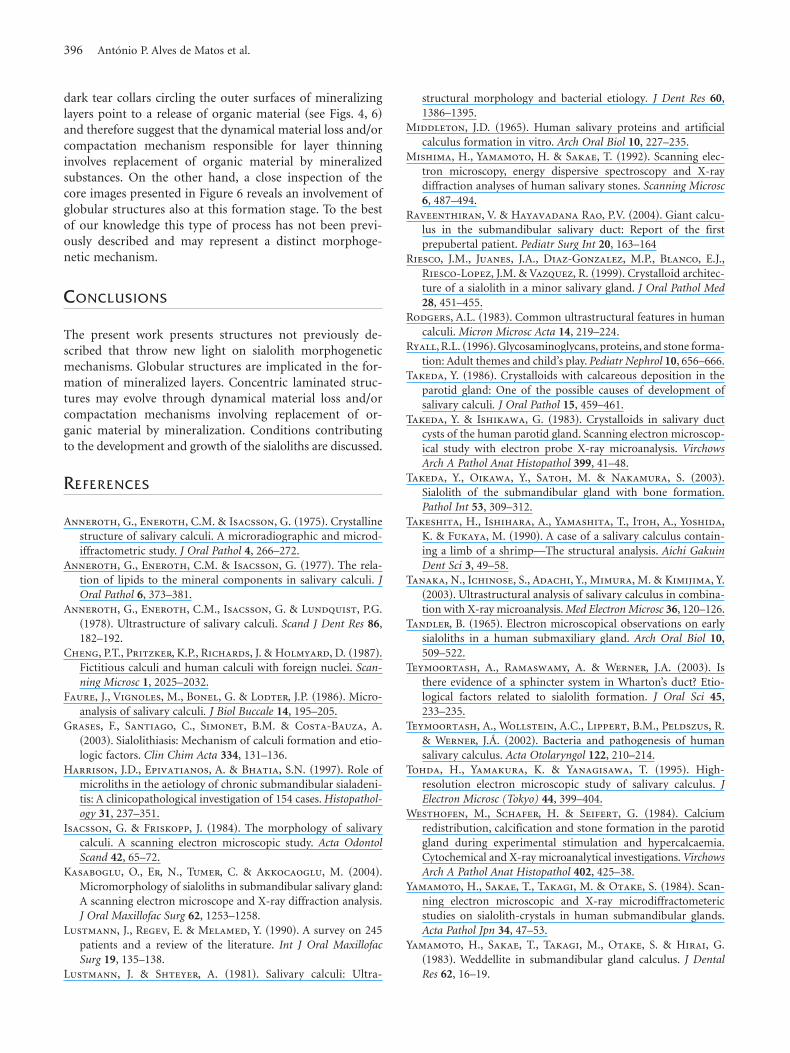
dark tear collars circling the outer surfaces of mineralizinglayers point to a release of organic material ~see Figs. 4, 6!and therefore suggest that the dynamical material loss and/orcompactation mechanism responsible for layer thinninginvolves replacement of organic material by mineralizedsubstances. On the other hand, a close inspection of thecore images presented in Figure 6 reveals an involvement ofglobular structures also at this formation stage. To the bestof our knowledge this type of process has not been previ-ously described and may represent a distinct morphoge-netic mechanism.
CONCLUSIONS
The present work presents structures not previously de-scribed that throw new light on sialolith morphogeneticmechanisms. Globular structures are implicated in the for-mation of mineralized layers. Concentric laminated struc-tures may evolve through dynamical material loss and/orcompactation mechanisms involving replacement of or-ganic material by mineralization. Conditions contributingto the development and growth of the sialoliths are discussed.
REFERENCES
Anneroth, G., Eneroth, C.M. & Isacsson, G. ~1975!. Crystallinestructure of salivary calculi. A microradiographic and microd-iffractometric study. J Oral Pathol 4, 266–272.
Anneroth, G., Eneroth, C.M. & Isacsson, G. ~1977!. The rela-tion of lipids to the mineral components in salivary calculi. JOral Pathol 6, 373–381.
Anneroth, G., Eneroth, C.M., Isacsson, G. & Lundquist, P.G.~1978!. Ultrastructure of salivary calculi. Scand J Dent Res 86,182–192.
Cheng, P.T., Pritzker, K.P., Richards, J. & Holmyard, D. ~1987!.Fictitious calculi and human calculi with foreign nuclei. Scan-ning Microsc 1, 2025–2032.
Faure, J., Vignoles, M., Bonel, G. & Lodter, J.P. ~1986!. Micro-analysis of salivary calculi. J Biol Buccale 14, 195–205.
Grases, F., Santiago, C., Simonet, B.M. & Costa-Bauza, A.~2003!. Sialolithiasis: Mechanism of calculi formation and etio-logic factors. Clin Chim Acta 334, 131–136.
Harrison, J.D., Epivatianos, A. & Bhatia, S.N. ~1997!. Role ofmicroliths in the aetiology of chronic submandibular sialadeni-tis: A clinicopathological investigation of 154 cases. Histopathol-ogy 31, 237–351.
Isacsson, G. & Friskopp, J. ~1984!. The morphology of salivarycalculi. A scanning electron microscopic study. Acta OdontolScand 42, 65–72.
Kasaboglu, O., Er, N., Tumer, C. & Akkocaoglu, M. ~2004!.Micromorphology of sialoliths in submandibular salivary gland:A scanning electron microscope and X-ray diffraction analysis.J Oral Maxillofac Surg 62, 1253–1258.
Lustmann, J., Regev, E. & Melamed, Y. ~1990!. A survey on 245patients and a review of the literature. Int J Oral MaxillofacSurg 19, 135–138.
Lustmann, J. & Shteyer, A. ~1981!. Salivary calculi: Ultra-
structural morphology and bacterial etiology. J Dent Res 60,1386–1395.
Middleton, J.D. ~1965!. Human salivary proteins and artificialcalculus formation in vitro. Arch Oral Biol 10, 227–235.
Mishima, H., Yamamoto, H. & Sakae, T. ~1992!. Scanning elec-tron microscopy, energy dispersive spectroscopy and X-raydiffraction analyses of human salivary stones. Scanning Microsc6, 487–494.
Raveenthiran, V. & Hayavadana Rao, P.V. ~2004!. Giant calcu-lus in the submandibular salivary duct: Report of the firstprepubertal patient. Pediatr Surg Int 20, 163–164
Riesco, J.M., Juanes, J.A., Diaz-Gonzalez, M.P., Blanco, E.J.,Riesco-Lopez, J.M. & Vazquez, R. ~1999!. Crystalloid architec-ture of a sialolith in a minor salivary gland. J Oral Pathol Med28, 451–455.
Rodgers, A.L. ~1983!. Common ultrastructural features in humancalculi. Micron Microsc Acta 14, 219–224.
Ryall, R.L. ~1996!. Glycosaminoglycans, proteins, and stone forma-tion: Adult themes and child’s play. Pediatr Nephrol 10, 656–666.
Takeda, Y. ~1986!. Crystalloids with calcareous deposition in theparotid gland: One of the possible causes of development ofsalivary calculi. J Oral Pathol 15, 459–461.
Takeda, Y. & Ishikawa, G. ~1983!. Crystalloids in salivary ductcysts of the human parotid gland. Scanning electron microscop-ical study with electron probe X-ray microanalysis. VirchowsArch A Pathol Anat Histopathol 399, 41–48.
Takeda, Y., Oikawa, Y., Satoh, M. & Nakamura, S. ~2003!.Sialolith of the submandibular gland with bone formation.Pathol Int 53, 309–312.
Takeshita, H., Ishihara, A., Yamashita, T., Itoh, A., Yoshida,K. & Fukaya, M. ~1990!. A case of a salivary calculus contain-ing a limb of a shrimp—The structural analysis. Aichi GakuinDent Sci 3, 49–58.
Tanaka, N., Ichinose, S., Adachi, Y., Mimura, M. & Kimijima, Y.~2003!. Ultrastructural analysis of salivary calculus in combina-tion with X-ray microanalysis. Med Electron Microsc 36, 120–126.
Tandler, B. ~1965!. Electron microscopical observations on earlysialoliths in a human submaxiliary gland. Arch Oral Biol 10,509–522.
Teymoortash, A., Ramaswamy, A. & Werner, J.A. ~2003!. Isthere evidence of a sphincter system in Wharton’s duct? Etio-logical factors related to sialolith formation. J Oral Sci 45,233–235.
Teymoortash, A., Wollstein, A.C., Lippert, B.M., Peldszus, R.& Werner, J.Á. ~2002!. Bacteria and pathogenesis of humansalivary calculus. Acta Otolaryngol 122, 210–214.
Tohda, H., Yamakura, K. & Yanagisawa, T. ~1995!. High-resolution electron microscopic study of salivary calculus. JElectron Microsc (Tokyo) 44, 399–404.
Westhofen, M., Schafer, H. & Seifert, G. ~1984!. Calciumredistribution, calcification and stone formation in the parotidgland during experimental stimulation and hypercalcaemia.Cytochemical and X-ray microanalytical investigations. VirchowsArch A Pathol Anat Histopathol 402, 425–38.
Yamamoto, H., Sakae, T., Takagi, M. & Otake, S. ~1984!. Scan-ning electron microscopic and X-ray microdiffractometericstudies on sialolith-crystals in human submandibular glands.Acta Pathol Jpn 34, 47–53.
Yamamoto, H., Sakae, T., Takagi, M., Otake, S. & Hirai, G.~1983!. Weddellite in submandibular gland calculus. J DentalRes 62, 16–19.
396 António P. Alves de Matos et al.
Copyright © 2022 FDOKUMEN


![[square brackets] - Convention on Biological Diversity](https://static.fdokumen.com/doc/165x107/631db8745ff22fc745065d61/square-brackets-convention-on-biological-diversity.jpg)