The annular-sum and Hale-McClellan methods of 3D wavefield extrapolation
Upload
independentCategory
view
4download
0
29 (2007) 242–250http://www.elsevier.com/locate/biophyschem
Biophysical Chemistry 1
On the NMR analysis of pKa values in the unfolded state of proteins byextrapolation to zero denaturant
Jeniffer Quijada, Gary López, Rodney Versace, Luis Ramírez, María Luisa Tasayco ⁎
Department of Chemistry, City College of New York, 138th Street and Convent Avenue, New York, NY 10031, United States
Received 9 February 2007; received in revised form 4 June 2007; accepted 4 June 2007Available online 14 June 2007
Abstract
Detailed knowledge of the pH-dependence in both folded and unfolded states of proteins is essential to understand the role of electrostatics inprotein stability. The increasing number of natively disordered proteins constitutes an excellent source for the NMR analysis of pKa values in theunfolded state of proteins. However, the tendency of many natively disordered proteins to aggregate via intermolecular hydrophobic clusters limitstheir NMR analysis over a wide pH range. To assess whether the pKa values in natively disordered polypeptides can be extrapolated from NMRmeasurements in the presence of denaturants, the natively disordered backbone of the C-terminal fragment 75 to 105 of Human Thioredoxin wasstudied. First, assignments using triple resonance experiments were performed to confirm lack of secondary structure. Then the pH-dependence ofthe amides and carboxylate side chains of Glu residues (Glu88, Glu95, Glu98, and Glu103) in the pH range from 2.0 to 7.0 was monitored using2D 1H15N HSQC and 3D C(CO)NH experiments, and the behavior of their amides and corresponding carboxyl groups was compared to confirmthe absence of nonlocal interactions. Lastly, the effect of increasing dimethyl urea concentration on the pKa values of these Glu residues wasmonitored. The results indicate that: (i) the dispersion in the pKa of carboxyl groups and the pH midpoints of amides in Glu residues is about 0.5pH units and 0.6 pH units, respectively; (ii) the backbone amides of the Glu residues exhibit pH midpoints which are within 0.2 pH units fromthose of their carboxylates; (iii) the addition of denaturant produces upshifts in the pKa values of Glu residues that are nearly independent of theirposition in the sequence; and (iv) these upshifts show a nonlinear behavior in denaturant concentration, complicating the extrapolation to zerodenaturant. Nevertheless, the relative ordering of the pKa values of Glu residues is preserved over the whole range of denaturant concentrationsindicating that measurements at high denaturant concentration (e.g. 4 M dimethyl urea) can yield a qualitatively correct ranking of the pKa of theseresidues in natively disordered proteins whose pH-dependence cannot be monitored directly by NMR.© 2007 Elsevier B.V. All rights reserved.
Keywords: NMR analysis; Denaturant; Unfolded state of proteins; pKa values
1. Introduction
The large body of experimental and computational studies ofwell-structured proteins has provided extensive knowledgeabout the folded state, but information on the unfolded state isstill limited. Central to the biophysical properties of proteins arehydrophobicity and electrostatics, but the latter is stillincompletely understood. Although progress has been madeby the recognition of ionizable groups on the surface ofthermophilic proteins which confer high thermal stability,structure-based calculations of protein energetics [1,2] require
⁎ Corresponding author.E-mail address: [email protected] (M.L. Tasayco).
0301-4622/$ - see front matter © 2007 Elsevier B.V. All rights reserved.doi:10.1016/j.bpc.2007.06.004
more information about the unfolded protein state to explain thepH-dependence of protein stability [3,4].
A widely used model of the contribution of electrostatics toprotein energetics predicts the pH-dependence of the ▵G forprotein unfolding with the aid of the pKa values of ionizableresidues in both the folded and unfolded states [5,6]. Awealth ofinformation has been accumulated on the pKa values ofionizable residues in folded proteins via NMR [7–9] andmutagenesis analysis [10–16]. In contrast, NMR analysis of thepH-dependence of chemical shifts in the unfolded state of mostwell-structured proteins is challenging due to the inability topopulate the unfolded state during the course of the pH-titration.This limitation has been overcome in a few cases: (a) unfoldedprotein segments [7,17,18], (b) the equilibrium mixture of theunfolded and folded states of an unstable protein [19], and (c)

243J. Quijada et al. / Biophysical Chemistry 129 (2007) 242–250
the unfolded monomers of a folded heterodimer [17,20]. Inother cases, the pKa values of ionizable residues in the unfoldedprotein have been indirectly determined using the pKa values inthe folded state, the pH-dependence of the ▵G for proteinunfolding, and the “zero interaction model” [18,21–23]. Insummary, few reports are available in the literature on the pKa
values of ionizable residues in unfolded protein states. Amongthem are the unfolded state of the N-terminal SH3 domain ofthe drk protein, which exchanges slowly with its folded state[19,24] and exhibits pKa values for Asp (3.75 to 4.13) and Glu(4.08 to 4.45) with dispersions of 0.38 and 0.37 pH units,respectively [19,25]; the unfolded monomers of the hetero-dimeric Leucine zipper which show pKa values for Glu (4.55 to4.76) with a dispersion of 0.21 pH units [20,26]; the shortnatively disordered segments of the N-terminal domain of theribosomal protein L9 [18,27] which exhibit pKa values for Aspand Glu with dispersions of up to 0.38 and 0.52 pH units,respectively; and one of the large unfolded monomers ofreassembled E. coli thioredoxin (ETrx) that shows pKa valuesfor Asp (3.77 to 3.94) and Glu (4.08 to 4.60) with dispersions of0.17 and 0.52 pH units, respectively [17]. The accumulatedresults indicate that the pKa values of Asp and Glu in theunfolded state of proteins show a dispersion of up to 0.5 pHunits and that these values are within 0.5 pH units from thoseobserved in host–guest tetrapeptides [28,29]. Whether thisdispersion results from local and/or nonlocal interactionsremains unclear and requires more mutagenesis analysis, asdone for the N-terminal SH3 domain of drk [19,25].
Considering the increasing number of natively disorderedproteins originated by the current proteomics efforts, it wouldbe interesting to know whether the dispersion in the pKa valuesof Asp and Glu residues can be greater than 0.5 pH units as aresult of nonlocal interactions. Recent reports [17,30] suggestthat medium range nonlocal interactions within the highlycharged segment of a natively disordered C-fragment of ETrxproduce significant discrepancies between the pH midpoints(pHm) of the backbone amides of Asp and Glu and the pKa oftheir own carboxylate side chains. In contrast, the moderatelycharged segment of the same fragment shows negligiblediscrepancies and provides no evidence for the presence ofnonlocal interactions [17].
A complication in any attempt to enlarge the database of pKa
values in natively disordered proteins by NMR analysis arisesfrom the tendency of amphipatic segments to aggregate viaintermolecular hydrophobic clusters in certain pH range. Tocounteract this aggregating tendency we propose to usechemical denaturants in an attempt to increase the populationof the monomeric state and obtain the pKa values by anextrapolation to zero denaturant. To gain confidence that thisproposal is feasible, we decided to start with the nativelydisordered C-terminal fragment encompassing residues 75 to105 of Human Thioredoxin (HC74), a control system for whichthe pKa values can be measured in the presence and absence ofdenaturants such as dimethyl urea (DMU). Our results indicatethat in fragments in which nonlocal interactions are insignif-icant: (i) the addition of DMU produces upshifts in the pKa
values of Glu residues that are nearly independent of their
position in the sequence, and consistent with previous reports[31–33]; and (ii) the upshifts in the pKa values are notproportional to the DMU concentration, complicating theextrapolation to zero DMU; nevertheless, the relative orderingof the pKa values of Glu residues is preserved over the wholerange of DMU concentrations.
2. Experimental
2.1. NMR analysis of fragment HC74
The fragment HC74 was generated using 15N–13C-labeledHTrx and previously reported expression, isolation, CNBrcleavage and purification procedures [34–36]. pH measure-ments were performed with an Accumet combination electrodeof 3 mm diameter and 1.5” flexible stem and used withoutcorrecting for either deuterium isotope effect [37] or denaturanteffect. Titrations were conducted with solutions of HCl orNaOH. NMR experiments were performed on a Varian INOVAAS600 spectrometer with a triple resonance probe. The 1Hchemical shift was referenced to the temperature dependentH2O frequency and corrected using sodium 2,2-dimethyl,2-silapentane,5-sulfonate (DSS) as an external reference. Sincethe addition of DMU in the strongly acidic regime affects theposition of the water resonance, DSS samples were prepared atdifferent pH s between pH 2 and 7 in the same buffer used forfragment HC74 that contains the same increasing concentra-tions of DMU (1 M, 2 M, 3 M and 4 M). The correspondingchemical shift corrections are available as Supplementarymaterial. The 15N and 13C chemical shifts were indirectlyreferenced [38]. The temperature was calibrated using standardprocedures with methanol [39]. A solution of 0.5 mM 15N–13C-labeled HC74 in 10 mM Potassium Phosphate buffer (KPi),containing 10% D2O and 4 M DMU at pH 6.44 and 25 °C wasused to acquire the 3D HNCACB, C DIPSY, CBCACONH, andC(CO)NH [40] experiments with 512 (t3), 38 (t2), and 48 (t1)complex points, spectral widths of 8000 (1H), 1600 Hz (15N),and 3400 Hz (13C), and a variable number of transients (8 to24). NMR data were processed and analyzed using NMRPipe[41] and SPARKY [42], respectively. The assignments of thebackbone and side chain of fragment HC74 in the absence ofDMU were obtained by transferring chemical shifts from highto low DMU concentrations (3 M, 2 M, 1 M and 0 M) via 2D1H15N HSQC and 3D C(CO)NH spectra at pH 6.44 and 25 °C.The secondary chemical shifts of the amide proton (HN),α-carbons (Cα) and amide nitrogen (N) of fragment HC74 in theabsence of DMUwere calculated using the random coil data base[43] and corrected for sequence [44] dependence.
2.2. pH-dependence of chemical shifts in the backbone amidesand carboxylate side chains of fragment HC74
Solutions of 0.1 or 0.3 mM of fragment HC74 in 10 mMKPi,10% D2O, and increasing DMU concentrations (0 M, 1 M, 2 M,3 M, and 4 M) at pH 4.25 or pH 6.5 were titrated with solutionsof HCl or NaOH between pH 2.0 and pH 7.0 at 25 °C to acquireNMR data. The pH-dependence in the chemical shifts of the

Fig. 1. Standard structural NMR analysis of the natively disordered HC74. Thesecondary chemical shifts of the HN (▵δ HN) and Cα (▵δ Cα) are defined as thedifferences between their observed chemical shift and their correspondingrandom coil values. The ▵δ HN and ▵δ Cα are given at pH≈6.44 and 25 °C inthe top and bottom panel, respectively.
244 J. Quijada et al. / Biophysical Chemistry 129 (2007) 242–250
amides and 13Cγ of Glu residues from 15N–13C-labeledfragment HC74 were monitored using 2D 1H15N HSQC and3D C(CO)NH experiments, respectively. Acquisitions of thelatter required 512 (t3), 38 (t2), and 48 (t1) complex points,spectral widths of 8000 (1H), 1600 Hz (15N), and 3400 Hz(13C), and a variable number of transients (8 to 24). Thechemical shifts of fragment HC74 were corrected to account forthe effect of DMU concentrations on the position of the waterpeak under strongly acidic conditions (see Supplementarymaterial). Typically, these corrections produce upshifts in thepKa values that amount to 0.01 and 0.03 pH units for 1 M DMUand 4 M DMU, respectively.
Data were fitted to a modified Hill equation [45]
d pHð Þ ¼ dbase þ dacid � 10n pHm�pHð Þ
1þ 10n pHm�pHð Þ : ð1Þ
The acidic (δacid) and basic (δbase) plateaus, Hill coefficient(n) and pHm were left as floating parameters when fitting theamides pH-dependence. For the carboxyl side chains twoseparate fits were actually performed. In the first case all fourparameters (pHm, n and the plateaus) were treated as freeparameters and only the data in the absence of denaturant wasused. In the second case, the plateaus were fixed to the valuesobtained from a “global” fit that considered all data, both in theabsence and presence of DMU. The rationale for performingthis second fit is that the chemical shifts of the 13Cγ in both theprotonated and unprotonated carboxyl group are thought to beinsensitive to the aqueous organic solvent composition and thusthe level of the plateaus should be independent of the DMUconcentration.
3. Results
3.1. Secondary chemical shift analysis of fragment HC74
To determine the degree of disorder of fragment HC74 undernativelike conditions, the first step was to assign this fragment athigh denaturant concentration and transfer the assignment toconditions with zero denaturant. The assignment of the amideswas straightforward with the exception of some residues thatexhibit similar chemical shifts (e.g.; F77 and K82, Q84 andE88, V86 and Q78, L104 and L97) or low signal/noise ratio(e.g., T76, G83, and S90 at pH close to 7). The second step wasto determine the secondary chemical shifts (differences betweenthe experimental values and those from a statistical random coilmodel) of the amide protons and α-carbons, which yieldedabsolute values of less than 0.3 ppm and 1.0 ppm, respectively(see Fig. 1). Inspection of these shifts reveals some segments(F77 to K81 and E95 to A99) where the secondary chemicalshifts of the α-carbons are predominantly negative andconsistent with a tendency to adopt β strands, while thesecondary chemical shifts of the corresponding amide protonslack any particular preference. This lack of conformationalpreference is typical of natively disordered segments.
In summary, the secondary chemical shift analysis of frag-ment HC74 confirms its natively disordered state.
3.2. pH-dependence of amide chemical shifts in fragmentHC74
Assessing the role of nonlocal interactions in natively dis-ordered fragments using standard NMR methods is difficult. Toassess their presence in the natively disordered fragment HC74,the pH-dependence of the amide chemical shifts of its ionizableand nonionizable residues were monitored in the acidic regimewithout the help of denaturants (see Fig. 2, Table 1 and Sup-plementary material) in search of correlations between themidpoint (pHm) values of Glu residues and its neighboringnonionizable residues [30].
In contrast to similar studies of natively disordered poly-peptides [30], the pH-dependence of the backbone amides in theionizable residues of HC74 is rather weak yielding differencesbetween the chemical shift of the unprotonated and protonatedGlu residues (▵δ) which are rather close to the cutoff values(0.8 ppm for nitrogens and 0.08 ppm for the protons). In onecase (amide proton of E88) the ▵δ even falls below the signal-to-noise ratio suggesting unexpected weak interactions with itscarboxyl group. With the exception of this residue, for whichthe fittings failed, analysis of the titration curves for Gluresidues in terms of a modified Hill equation [45] (see Eq. (1))yields pHm values with significant dispersion (0.60±0.10 pHunits) in both amide nitrogens and protons. In all cases the Hillcoefficients (n) are around 0.9 (within experimental uncertain-ties), revealing strong intra-residue interactions between thecarboxyl groups and the corresponding amides.
Moreover, the titration curves of these amide protons, withthe exception of E88, show the typical trend found for a Gluresidue in model tetrapeptides [29]: the acidic plateau is lowerthan the basic, which has been previously interpreted [29] asevidence for the presence of H-bonds between the carboxylgroup and the amide. As previously shown [30], nativelydisordered polypeptides encompassing Asp and/or Glu residues

Fig. 2. pH-dependence in the chemical shifts of Glu residues from the natively disordered fragment HC74. pH-dependence in the chemical shifts of HN (top left), amideN (top right), and 13Cγ (bottom) for E88 (squares); E95 (circles); E98 (up triangles); E103 (down triangles). The solid lines show the results of the nonlinear fittings tothe modified Hill equation.
245J. Quijada et al. / Biophysical Chemistry 129 (2007) 242–250
may also show pH-dependent nonionizable residues whichconstitute candidates for local and/or nonlocal interactionswith the ionizable residues. In this case, the amides of tennonionizable residues surrounding E88, E95, E98 and E103(K85, V86, K96, L97, T100, I101, N102, L104) are veryweakly pH-dependent; only the C-terminal V105 shows astrong dependence (see Supplementary material for fits to theHill equation for selected residues). Therefore is not possible
Table 1Fitting parameters for Glu residues in the natively disordered fragment HC74(see Eq. (1))
Residue pHm n ▵δ pHm (const)
E88 N 3.93±0.12 0.97±0.22 0.70±0.1E88 Cγ 4.02±0.05 0.91±0.10 3.76±0.2 4.06±0.01E95 H 4.17±0.10 0.94±0.21 0.21±0.1E95 N 4.25±0.11 0.96±0.25 0.90±0.1E95Cγ 4.18±0.03 0.98±0.08 3.50±0.1 4.20±0.01E98 H 4.33±0.14 0.95±0.33 0.13±0.1E98 N 4.47±0.11 0.95±0.23 0.80±0.1E98Cγ 4.25±0.03 0.85±0.06 3.74±0.1 4.31±0.01E103 H 4.72±0.15 1.04±0.35 0.12±0.1E103 N 4.58±0.08 0.89±0.14 0.86±0.1E103 Cγ 4.55±0.04 0.91±0.07 3.81±0.1 4.61±0.01
The pHm, n and▵δ parameters are the result of fitting to the data in the absenceof denaturant, with all parameters left floating. The last column gives the pHm
values from a fitting to the same data but constraining the plateaus to have thesame values for all DMU concentrations.
to distinguish the effect of short-, medium- or long-rangeinteractions.
In summary, there is no evidence for strong nonlocal inter-actions involving the Glu residues and nonionizable residues.The weak pH-dependencies observed in the latter are moreplausibly explained by local interactions with nearby Gluresidues.
3.3. pH-dependence of carboxylate side chain chemical shiftsin fragment HC74
To assess whether the pH-dependence of the backboneamides of Glu residues reflect that of their correspondingcarboxylates as expected in a natively disordered segmentwhere nonlocal interactions do not play a significant role [17],the chemical shifts of its Glu (13Cγ) residues were monitored inthe pH range from 2 to 7 using 2D 1H15N HSQC and 3D C(CO)NH experiments in the absence of chemical denaturants. Thetitration curves show a simple sigmoidal dependence (seeFig. 2) and were again fitted to Eq. (1), see Table 1. Theseanalysis indicate a dispersion in the pKa values of 0.54±0.10 pHunits, although they are within 0.3 pH units of those found inmodel tetrapeptides [29] (see Fig. 3). For the carboxylates twoseparate fits were actually performed (see Experimental): oneunconstrained (all parameters free) and one constraining theplateaus to be the same for all concentrations of denaturant. Theresults of the latter fit, which differ from the unconstrained fit by

Fig. 3. pH-dependence in the carboxylate side chains and backbone amides ofGlu residues from the natively disordered fragment HC74. Top: pHm of thecarboxylate side chains of Glu shown by vertical bars. Middle: pHm of backboneamide nitrogen of Glu shown by vertical bars. Bottom: pHm of backbone amideproton of Glu shown by vertical bars. The pKa of Glu in model host–guesttetrapeptides [28] are shown by solid horizontal lines. The sequence of HC74 isshown underneath the bottom panel.
246 J. Quijada et al. / Biophysical Chemistry 129 (2007) 242–250
about 0.06 pH units, are shown in the rightmost column inTable 1 and will be used in the following discussions.
In summary, the observed differences between the pHm ofthe amide (proton and/or nitrogen) of Glu residues and itscorresponding carboxylate's pKa are small, further confirmingthe lack of significant nonlocal interactions.
Fig. 4. Effect of DMU on the pH-dependence in the carboxylate side chains of Glu rechemical shifts of 13Cγ of E88 (left top panel); E95 (right top panel); E98 (bottom left(up triangles), 3 M (down triangles), and 4 M (right triangles) DMU concentrationequation, constraining the plateaus to be independent of the concentration of denatu
3.4. Denaturant effect on the pH-dependence of carboxylateside chain chemical shifts in fragment HC74
To determine whether the pKa values of carboxylate sidechains from Glu residues can be extrapolated from their valuesin the presence of denaturants, dimethyl urea (DMU) wasselected as a denaturant instead of the more commonly usedGuanidine HCl or urea for two reasons: (i) DMU is chemicallyless reactive than urea and less prone to modify amino andimino groups under strongly acidic conditions; and (ii) DMU isexpected to have less effect than guanidine HCl on chargescreening.
The chemical shifts of the 13Cγ of the carboxylate wereeasily monitored for E88, E98, and E103; however, monitoringE95 was more complicated due to overlapped cross-peaksthroughout the covered pH range in the C(CO)NH experiments,which become more pronounced as the DMU concentrationincreases (see Supplementary material). The resulting titrationcurves display simple sigmoidal pH-dependence (see Fig. 4)and they were analyzed using the modified Hill equation(Eq. (1)) just as the data in the absence of DMU.
Analysis of the constrained (to same plateaus) fitting of thefamily of titration curves for each individual Glu residue atincreasing DMU concentration (see Table 2) indicates upshiftsin the apparent pKa values of Glu residues (about 0.3 to 0.5 pHunits at 4 M DMU) and complex effects on the Hill coefficients
sidues from the natively disordered fragment HC74. The pH-dependence of thepanel); E103 (bottom right panel) is depicted at zero (squares), 1 M (circles), 2 Ms. The solid lines show the result of the nonlinear fittings to the modified Hillrant.
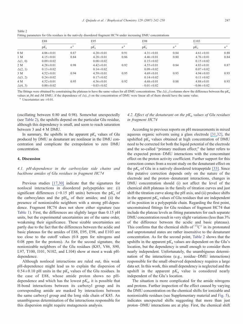
Table 2Fitting parameters for Glu residues in the natively disordered fragment HC74 under increasing DMU concentrations
E88 E95 E98 E103
pKa n a pKa n a pKa n a pKa n a
0 M 4.06±0.01 0.87 4.20±0.01 0.91 4.31±0.01 0.84 4.61±0.01 0.881 M 4.15±0.01 0.84 4.28±0.01 0.86 4.46±0.01 0.80 4.76±0.01 0.84▵(1, 0) 0.09±0.02 0.08±0.02 0.15±0.02 0.15±0.022 M 4.28±0.01 0.98 4.42±0.01 0.92 4.55±0.01 0.84 4.83±0.01 0.87▵(2, 1) 0.13±0.02 0.14±0.02 0.09±0.02 0.07±0.023 M 4.52±0.01 0.94 4.59±0.01 0.95 4.69±0.01 0.93 4.94±0.03 0.93▵(3, 2) 0.24±0.02 0.17±0.02 0.14±0.02 0.11±0.024 M 4.52±0.01 0.95 4.56±0.01 0.92 4.68±0.01 0.88 4.88±0.01 0.93▵(4, 3) 0.00±0.02 −0.03±0.02 –0.01±0.02 −0.06±0.02
The fittings were obtained by constraining the plateaus to have the same values for all DMU concentrations. The▵(i, j) columns show the difference between the pKa
values at jM and iM DMU; if the dependence of ▵(i, j) on the concentration of DMU were linear all of them should have the same value.a Uncertainties are ±0.01.
247J. Quijada et al. / Biophysical Chemistry 129 (2007) 242–250
(oscillating between 0.80 and 0.98). Somewhat unexpectedly(see Table 2), the upshifts depend on the particular Glu residue,although this dependency is small, and seem to reach saturationbetween 3 and 4 M DMU.
In summary, the upshifts in the apparent pKa values of Gluproduced by DMU as denaturant are nonlinear in the DMU con-centration and complicate the extrapolation to zero DMUconcentration.
4. Discussion
4.1. pH-dependence in the carboxylate side chains andbackbone amides of Glu residues in fragment HC74
Previous studies [17,30] indicate that the signatures fornonlocal interactions in disordered polypeptides are: (i)significant differences (N0.15 pH units) between the pKa ofthe carboxylates and the pHm of their amides; and (ii) thepresence of nonionizable neighbors with a strong pH-depen-dence. Fragment HC74 does not show either signature (seeTable 1). First, the differences are slightly larger than 0.15 pHunits, but the experimental uncertainties are of the same order,weakening their significance. These sizable uncertainties arepartly due to the fact that the differences between the acidic andbasic plateaus for the amides of E88, E95, E98, and E103 aretoo close to the cutoff values (0.8 ppm for nitrogens and0.08 ppm for the protons). As for the second signature, thenonionizable neighbors of the Glu residues (K85, V86, S90,L97, T100, I101, N102, and L104) show at most a weak pH-dependence.
Although nonlocal interactions are ruled out, this weakpH-dependence might lead us to explain the dispersion of0.54±0.10 pH units in the pKa values of the Glu residues. Inthe case of E88, whose amide proton shows no pH-dependence and which has the lowest pKa, it is possible thatH-bond interactions between its carboxyl group and itscorresponding amide are masked by interactions betweenthe same carboxyl group and the long side chain of K85. Anunambiguous determination of the interactions responsible forthis dispersion might require mutagenesis analysis.
4.2. Effect of the denaturant on the pKa values of Glu residuesin fragment HC74
According to previous reports on pH measurements in mixedaqueous organic solvents using a glass electrode [31,32], theupshifted pKa values obtained at high concentration of DMUneed to be corrected for both the liquid potential of the electrodeand the so-called “primary medium effect;” the latter refers tothe expected proton–DMU interactions with the concomitanteffect on the proton activity coefficient. Further support for thiscorrection comes from a recent study on the denaturant effect onthe pKa of Glu in a natively disordered tetrapeptide [33]. Sincethis putative correction depends only on the nature of theelectrode and the proton–denaturant interactions, changes inDMU concentration should (i) not affect the level of thechemical shift plateaus in the family of titration curves and justshift the titration curve along the pH axis, and (ii) produce shiftsin the apparent pKa values of Glu residues that are independentof its position in a polypeptide chain. Regarding the first point,unconstrained fittings for Glu residues of fragment HC74 thatinclude the plateau levels as fitting parameters for each separateDMU concentration result in very slight variations (less than 3%of the difference between the acidic and basic plateaus).This confirms that the chemical shifts of 13Cγ in its protonatedand unprotonated states are rather insensitive to the denaturantconcentration. As for the second point, Table 2 shows that theupshifts in the apparent pKa values are dependent on the Glu'slocation, but the dependency is small enough to consider themnearly independent. Unfortunately, the unambiguous determi-nation of the interactions (e.g., residue–DMU interactions)responsible for the small observed dependency requires a largedata set. In this work, this small dependency is neglected and theupshift in the apparent pKa value is considered nearlyindependent of the Glu's location.
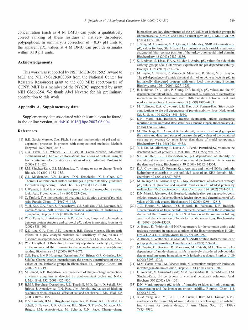
The situation is more complicated for the amide nitrogensand protons. Further inspection of the effect caused by varyingthe DMU concentration on the chemical shifts for ionizable andnonionizable residues (see Supplementary material and Fig. 5),indicates unexpected shifts suggesting that more than justproton–DMU interactions are at play. First, the chemical shift

Fig. 5. DMU-dependence in the backbone amides of selected residues from the natively disordered fragment HC74. Top and bottom left: overlay of portions of 1H15NHSQC spectra at pH 6.44. Top and bottom right: overlay of portions of 1H15N HSQC spectra at pH 2.0. The panels show the cross-peaks from uncorrected 1H15NHSQC spectra at zero (purple), 1 M (green), 2 M (orange), 3 M (red), and 4 M (cyan) DMU concentrations (see methodology). The labeled cross-peaks correspond to1H15N HSQC spectra at zero DMU concentration.
248 J. Quijada et al. / Biophysical Chemistry 129 (2007) 242–250
plateaus of the titration curves for the amides of ionizable residuesshow a strong dependency on the concentration of DMU. Thiseffect is quite pronounced, especially when considered as apercentage of the difference between the basic and acidic regimeplateaus, reaching values of up to 150% (compared with just 2–3% for the carboxylates). Second, the behavior of the chemicalshifts amides of nonionizable residues upon changes in theconcentration of DMU depends on the residue's identity andlocation in the sequence (e.g.; G87 versus T100 or A99 versusN102; and G87 versus G91) as shown in Fig. 5.
The strong dependency of the plateaus of the amide's titrationcurves on the DMU concentration suggests interactions betweenthe amides and the denaturant (possibly H-bonds), while the factthat there is a pH-dependence at all reflects interactions betweenthese amides and their corresponding carboxyl groups, presum-ably through H-bonds [29]. It is unclear which of these, if any,predominates, since the changes in the amide's acidic and basicplateaus are of the same order of magnitude as the differencebetween them. In contrast, the acidic and basic plateaus of thecarboxylate side chains show a small dependence on denaturantconcentration, suggesting low sensitivity to possible H-bondinteractions between the carboxyl group and the denaturant.
Although the upshifts in the pKa's of the Glu residues can beconsidered independent of their location, they are not linear in
the DMU concentration (see Table 2), complicating the extra-polation from high to zero concentration of the denaturant.Nevertheless, these upshifts appear to reach saturation between3 M and 4 M DMU and the addition of 4 M DMU producesupshifts in the apparent pKa values of Glu of about 0.37±0.10pH units. Interestingly, these upshifts are close to the ones foundfor a Glu residue in a host–guest tetrapeptide in an aqueoussolution of 6 M urea[33].
5. Conclusions
The addition of denaturant (DMU) produces the expectedupshifts in the pKa values of Glu residues that are nearlyindependent of their position in the sequence. However, theseupshifts are not linear in the DMU concentration and seem toreach saturation between 3 and 4 M DMU, complicating theextrapolation to zero DMU. This nonlinearity is in markedcontrast with the behavior found in simpler systems [31–33],possibly reflecting the complexity of interactions among Gluresidues, neighboring nonionizable residues, and the surround-ing aqueous organic solvent.
Nevertheless, the relative ordering of the pKa values of Gluresidues is preserved over the whole range of DMU con-centrations indicating that measurements at high denaturant
249J. Quijada et al. / Biophysical Chemistry 129 (2007) 242–250
concentration (such as 4 M DMU) can yield a qualitativelycorrect ranking of these residues in natively disorderedpolypeptides. In summary, a correction of −0.37 pH units tothe apparent pKa values at 4 M DMU can provide estimateswithin 0.10 pH units.
Acknowledgements
This work was supported by NSF (MCB-0517592) Award toMLT and NIH (5G12RR03060 from the National Center forResearch Resources) grant to the 600 MHz spectrometer ofCCNY. MLT is a member of the NYSBC supported by grantNIH GM66354. We thank Abel Navarro for his preliminarycontribution to this work.
Appendix A. Supplementary data
Supplementary data associated with this article can be found,in the online version, at doi:10.1016/j.bpc.2007.06.004.
References
[1] B.E. García-Moreno, C.A. Fitch, Structural interpretation of pH and salt-dependent processes in proteins with computational methods, MethodsEnzymol. 380 (2004) 20–51.
[2] C.A. Fitch, S.T. Whitten, V.J. Hilser, B. García-Moreno, Molecularmechanisms of pH-driven conformational transitions of proteins: insightsfrom continuum electrostatics calculations of acid unfolding, Proteins 63(2006) 113–126.
[3] J.M. Sánchez-Ruiz, G.I. Makhatadze, To charge or not to charge, TrendsBiotech. 19 (2001) 132–135.
[4] G.I. Makhatadze, V.V. Loladze, D.N. Ermolenko, X.-F. Chen, S.T.Thomas, Contribution of surface salt bridges to protein stability: guidelinesfor protein engineering, J. Mol. Biol. 327 (2003) 1135–1148.
[5] J. Wyman, Linked functions and reciprocal effects in myoglobin: a secondlook, Adv. Protein Chem. 19 (1964) 223–286.
[6] C. Tanford, The interpretation of hydrogen ion titration curves of proteins,Adv. Protein Chem. 17 (1962) 9–165.
[7] Y.-H. Kao, C.A. Fitch, S. Bhattacharya, C.J. Sarkisian, J.T.J. Lecomte, B.E.García-Moreno, Salt effects on ionization equilibria of histidines inmyoglobin, Biophys. J. 79 (2000) 1637–1654.
[8] W.R. Forsyth, J. Antosiewicz, A.D. Robertson, Empirical relationshipsbetween protein structure and carboxyl pKa values in proteins, Proteins 48(2002) 388–403.
[9] K.K. Lee, C.A. Fitch, J.T.J. Lecomte, B.E. García-Moreno, Electrostaticeffects in highly charged proteins: salt sensitivity of pKa values ofhistidines in staphylococcal nuclease, Biochemistry 41 (2002) 5656–5667.
[10] W.R. Forsyth, A.D. Robertson, Insensitivity of perturbed carboxyl pKa valuesin the ovomucoid third domain to charge replacement at a neighboringresidue, Biochemistry 39 (2000) 8067–8072.
[11] C.N. Pace, B.M.P. Huyghues-Despointes, J.M. Briggs, G.R. Grimsley, J.M.Scholtz, Charge–charge interactions are the primary determinants of the pKvalues of the ionizable groups in ribonuclease T1, Biophys. Chem. 101(2002) 211–219.
[12] M. Sundd, A.D. Robertson, Rearrangement of charge–charge interactionsin variant ubiquitins as detected by double-mutant cycles and NMR,J. Mol. Biol. 332 (2003) 927–936.
[13] B.M.P. Huyghues-Despointes, R.L. Thurlkill, M.D. Daily, D. Schell, J.M.Briggs, J. Antosiewicz, C.N. Pace, J.M. Scholtz, pK values of histidineresidues in ribonuclease Sa: effect of salt and net charge, J. Mol. Biol. 325(2003) 1093–1105.
[14] D.V. Laurents, B.M.P. Huyghues-Despointes, M. Bruix, R.L. Thurlkill, D.Schell, S. Newson, G.R. Grimsley, K.L. Shaw, S. Treviño, M. Rico, J.M.Briggs, J.M. Antosiewicz, M. Scholtz, C.N. Pace, Charge–charge
interactions are key determinants of the pK values of ionizable groups inribonuclease Sa (pI=3.5) and a basic variant (pI=10.2), J. Mol. Biol. 325(2003) 1077–1092.
[15] J. Song, M. Leskowski, M.A. Qasim, J.L. Markley, NMR determination ofpKa values for Asp, Glu, His, and Lys mutants at each variable contiguousenzyme-inhibitor contact position of the turkey ovomucoid third domain.Biochemistry 42 (2003) 2487–2856.
[16] S. Lindman, S. Linse, F.A.A. Mulder, I. Andre, pKa values for side-chaincarboxyl groups of a PGB1 variant explain salt and pH-dependent stability,Biophys. J. 92 (2007) 257–266.
[17] M. Pujato, A. Navarro, R. Versace, R. Mancusso, R. Ghose, M.L. Tasayco,The pH-dependence of amide chemical shift of Asp/Glu reflects its pKa inintrinsically disordered proteins with only local interactions, Biochim.Biophys. Acta 1764 (2006) 1227–1233.
[18] B. Kuhlman, D.L. Luisi, P. Young, D.P. Raleigh, pKa values and the pHdependent stability of theN-terminal domain of L9 as probes of electrostaticinteractions in the denatured state. Differentiation between local andnonlocal interactions, Biochemistry 38 (1999) 4896–4903.
[19] M. Tollinger, K.A. Crowhurst, L.E. Kay, J.D. Forman-Kay, Site-specificcontributions to the pH dependence of protein stability, Proc. Natl. Acad.Sci. U. S. A. 100 (2003) 4545–4550.
[20] D.N. Marti, H.R. Bosshard, Inverse electrostatic effect: electrostaticrepulsion in the unfolded state stabilizes a leucine zipper, Biochemistry 43(2004) 12436–12447.
[21] M. Oliveberg, V.L. Arcus, A.R. Fersht, pKa values of carboxyl groups inthe native and denatured states of barnase: the pKa values of the denaturedstate are on average 0.4 units lower than those of model compounds,Biochemistry 34 (1995) 9424–9433.
[22] Y.-J. Tan, M. Oliveberg, B. Davis, A.R. Fersht, Perturbed pKa values in thedenatured sates of proteins, J. Mol. Biol. 254 (1995) 980–992.
[23] S.T. Whitten, B.E. García-Moreno, pH dependence of stability ofstaphiloccal nuclease: evidence of substantial electrostatic interactions inthe denatured state, Biochemistry 39 (2000) 14292–14304.
[24] K.A. Crowhurst, J.D. Forman-Kay, Aromatic and methyl NOEs highlighthydrophobic clustering in the unfolded state of an SH3 domain, Bio-chemistry 42 (2003) 8687–8695.
[25] M. Tollinger, J.D. Forman-Kay, L.E. Kay, Measurement of side-chain carboxylpKa values of glutamate and aspartate residues in an unfolded protein bymultinuclear NMR spectroscopy, J. Am. Chem. Soc. 124 (2002) 5714–5717.
[26] D.N.Marti, I. Jelesarov, H.R. Bosshard, Interhelical ion pairing in coiled coils:solution structure of a heterodimeric leucine zipper and determination of pKa
values of Glu side chains, Biochemistry 39 (2000) 12804–12818.[27] J.C. Horng, V. Moroz, D.J. Rigotti, R. Fairman, D.P. Raleigh,
Characterization of large peptide fragments derived from the N-terminaldomain of the ribosomal protein L9: definition of the minimum foldingmotif and characterization of local electrostatic interactions, Biochemistry41 (2002) 13360–13369.
[28] A. Bundi, K. Wüthrich, 1H-NMR parameters for the common amino acidresidues measured in aqueous solutions of the linear tetrapeptides H-Gly-Gly-X-L-Ala-OH, Biopolymers 18 (1979) 285–297.
[29] A. Bundi, K. Wüthrich, Use of amide 1H-NMR titration shifts for studies ofpolypeptide conformation, Biopolymers 18 (1979) 299–311.
[30] M. Pujato, C. Bracken, R. Mancusso, M. Cataldi, M.L. Tasayco, pH-dependence of amide chemical shifts in natively disordered polypeptidesdetects medium-range interactions with ionizable residues, Biophys. J. 89(2005) 3293–3302.
[31] M.M.García-Mira, J.M. Sánchez-Ruiz, pH corrections and protein ionizationin water/guanidinium chloride, Biophys. J. 81 (2001) 3489–3502.
[32] O. Acevedo,M.Guzmán-Casado,M.M.García-Mira, B. Ibarra-Molero, J.M.Sánchez-Ruiz, pH corrections in chemical denaturant solutions, Anal.Biochem. 306 (2002) 158–1561.
[33] D.N. Marti, Apparent pKa shifts of titratable residues at high denaturantconcentration and the impact on protein stability, Biophys. Chem. 118(2005) 88–92.
[34] X.-M. Yang, W.-F. Yu, J.-H. Li, J.A. Fuchs, J. Rizo, M.L. Tasayco, NMRevidence for the reassembly of an α/β domain after cleavage of an α-helix:Implications for protein design, J. Am. Chem. Soc. 120 (1998)7985–7986.

250 J. Quijada et al. / Biophysical Chemistry 129 (2007) 242–250
[35] W.-F. Yu, C.-S. Tung, H. Wang, M.L. Tasayco, NMR analysis of cleavedE. coli thioredoxin (1–73/74–108) and its P76A variant: cis/trans peptideisomerization, Protein Sci. 9 (2000) 20–28.
[36] J.M. Louis, R.E. Georgescu, M.L. Tasayco, O. Tcherkasskaya, A.M.Gronenborn, Probing the structure and stability of a hybrid protein: thehuman-E. coli thioredoxin chimera, Biochemistry 40 (2001) 11184–11192.
[37] C.A. Bunton, V.J. Shiner, Isotope effects in deuterium oxide solution. I.Acid-base equilibria, J. Am. Chem. Soc. 83 (1961) 42–47.
[38] D.S. Wishart, C.G. Bigam, J. Yao, F. Abildgaard, H.J. Dyson, E. Oldfield,J.L. Markley, B.D. Sykes, 1H, 13C and 15N chemical shifts referencing inbiomolecular NMR, J. Biomol. NMR 6 (1995) 135–140.
[39] J. Cavanagh, W.J. Fairbrother, A.G. Palmer, N.J. Skelton, Protein NMRSpectroscopy: Principles and Practice, Academic Press, NewYork, NY,U.S.A.,1996.
[40] T.M. Logan, E.T. Olejniczak, R.-X. Xu, S.W. Fesik, A general method forassigning NMR spectra of denatured proteins using 3D HC(CO)NH-TOCSY triple resonance experiments, J. Biomol. NMR 3 (1993) 225–231.
[41] F. Delaglio, S. Grzesiek, G.W. Vuister, G. Zhu, J. Pfeifer, A.D. Bax,NMRPipe: a multidimensional spectral processing system based on UNIXpipes, J. Biomol. NMR 6 (1995) 277–293.
[42] T.D. Goddard, D.G. Kneller, SPARKY 3, University of California at SanFrancisco.
[43] BMRB (BioMagResBank), http://www.bmrb.wisc.edu.[44] S. Schwarzinger, G.J.A. Kroon, T.R. Foss, J. Chung, P.E. Wright, H.J.
Dyson, Sequence-dependent correction of random coil NMR chemicalshifts, J. Am. Chem. Soc. 123 (2001) 2970–2978.
[45] J.L. Markley, Observation of histidine residues in proteins by means ofnuclear magnetic resonance spectroscopy, Acc. Chem. Res. 8 (1975) 70–80.
Copyright © 2022 FDOKUMEN