On the Involvement of Single-Bond Rotation in the Primary Photochemistry of Photoactive Yellow...
9
On the Involvement of Single-Bond Rotation in the Primary Photochemistry of Photoactive Yellow Protein Andreas D. Stahl, †6 Marijke Hospes, ‡6 Kushagra Singhal, ‡ Ivo van Stokkum, † Rienk van Grondelle, † Marie Louise Groot, † and Klaas J. Hellingwerf †‡ * † Department of Physics and Astronomy, Faculty of Sciences, Vrije Universiteit Amsterdam, Amsterdam, The Netherlands; and ‡ Swammerdam Institute for Life Sciences, Science Faculty, University of Amsterdam, Amsterdam, The Netherlands ABSTRACT Prior experimental observations, as well as theoretical considerations, have led to the proposal that C 4 -C 7 single- bond rotation may play an important role in the primary photochemistry of photoactive yellow protein (PYP). We therefore synthesized an analog of this protein’s 4-hydroxy-cinnamic acid chromophore, (5-hydroxy indan-(1E)-ylidene)acetic acid, in which rotation across the C 4 -C 7 single bond has been locked with an ethane bridge, and we reconstituted the apo form of the wild-type protein and its R52A derivative with this chromophore analog. In PYP reconstituted with the rotation-locked chromo- phore, 1), absorption spectra of ground and intermediate states are slightly blue-shifted; 2), the quantum yield of photochemistry is ~60% reduced; 3), the excited-state dynamics of the chromophore are accelerated; and 4), dynamics of the thermal recovery reaction of the protein are accelerated. A significant finding was that the yield of the transient ground-state intermediate in the early phase of the photocycle was considerably higher in the rotation-locked samples than in the corresponding samples recon- stituted with p-coumaric acid. In contrast to theoretical predictions, the initial photocycle dynamics of PYP were observed to be not affected by the charge of the amino acid residue at position 52, which was varied by 1), varying the pH of the sample between 5 and 10; and 2), site-directed mutagenesis to construct R52A. These results imply that C 4 -C 7 single-bond rotation in PYP is not an alternative to C 7 ¼C 8 double-bond rotation, in case the nearby positive charge of R52 is absent, but rather facilitates, presumably with a compensatory movement, the physiological Z/E isomerization of the blue-light-absorbing chromophore. INTRODUCTION In addition to reproduction, and its inherent errors, which are cornerstones of evolution, the ability to perceive envi- ronmental signals and adjust accordingly is a key character- istic of (cellular) life. It is therefore of prime importance to understand the molecular basis of the elementary steps of such signal transduction processes and when necessary to repair or improve these processes. Photosensory receptors, i.e., proteins that can generate a biological signal upon the absorption of a photon of visible electromagnetic radiation, have traditionally played a key role as model systems for such studies (1). The accuracy with which these proteins can be activated in time and space is unrivalled. Several different families of these photosensory receptors have been characterized, each with their own specific (chro- mophore) structure and mechanism of primary photo- chemistry. Best known are the rhodopsins, in which the absorption of green light leads to Z/E isomerization of the retinal chromophore, and the LOV domains, which show blue-light-induced covalent adduct formation between the flavin chromophore and a nearby cysteine. Here, however, we have chosen yet another model system for such studies, photoactive yellow protein (PYP (2)), which functions as a photosensory receptor for a photophobic response in Halorhodospira halophila (3). This protein displays an exceptional chemical and photo- chemical stability and has been characterized in detail with respect to structure and function, as discussed in previous work (4–6). PYP is a relatively small protein (125 amino acids), containing a 4-hydroxy cinnamic acid (or p-couma- ric acid) chromophore, which is thiol-esterified to a cysteine residue, C69 of apo-PYP (7). The chromophore is buried inside the main hydrophobic core of this a/b-fold protein as a phenolate anion (8), stabilized by hydrogen bonding with E46 and Y42 (9) and in the trans configuration (10). It is significant that both hydrogen bonds show exceptional characteristics: they are very short and of the low-barrier and ionic-types, respectively (11). Light absorption by the chromophore of PYP induces primary photochemistry in a photocycle that leads to its cis configuration within a few picoseconds (12), with a quantum yield of 0.35 (13,14). The structural transition underlying this Z/E isomerization can only be completed in a complex reaction, in which also the hydrogen bond between the C¼O group of the chromophore and the nitrogen atom of the peptide bond between P68 and C69, is disrupted, after which the C¼O group starts to rotate around the long axis of the chromophore to stabilize the isomerization of the C 7 ¼C 8 ethylenic bond (15–17) to form the first cis ground-state intermediate (GSI), I 0 , of this photocycle. In parallel, a transient GSI (18) is formed that also may have an isomerized C 7 ¼C 8 ethylenic bond but has its carbonyl group still hydrogen-bonded to the nitrogen atom of the protein’s backbone (19). This GSI Submitted March 24, 2011, and accepted for publication June 17, 2011. 6 Andreas D. Stahl and Marijke Hospes contributed equally to this work. *Correspondence: [email protected] Editor: Leonid S. Brown. Ó 2011 by the Biophysical Society 0006-3495/11/09/1184/9 $2.00 doi: 10.1016/j.bpj.2011.06.065 1184 Biophysical Journal Volume 101 September 2011 1184–1192
-
Upload
independent -
Category
Documents
-
view
5 -
download
0
Transcript of On the Involvement of Single-Bond Rotation in the Primary Photochemistry of Photoactive Yellow...

1184 Biophysical Journal Volume 101 September 2011 1184–1192
On the Involvement of Single-Bond Rotation in the Primary Photochemistryof Photoactive Yellow Protein
Andreas D. Stahl,†6 Marijke Hospes,‡6 Kushagra Singhal,‡ Ivo van Stokkum,† Rienk van Grondelle,†
Marie Louise Groot,† and Klaas J. Hellingwerf†‡*†Department of Physics and Astronomy, Faculty of Sciences, Vrije Universiteit Amsterdam, Amsterdam, The Netherlands; and ‡SwammerdamInstitute for Life Sciences, Science Faculty, University of Amsterdam, Amsterdam, The Netherlands
ABSTRACT Prior experimental observations, as well as theoretical considerations, have led to the proposal that C4-C7 single-bond rotation may play an important role in the primary photochemistry of photoactive yellow protein (PYP). We thereforesynthesized an analog of this protein’s 4-hydroxy-cinnamic acid chromophore, (5-hydroxy indan-(1E)-ylidene)acetic acid, inwhich rotation across the C4-C7 single bond has been locked with an ethane bridge, and we reconstituted the apo form of thewild-type protein and its R52A derivative with this chromophore analog. In PYP reconstituted with the rotation-locked chromo-phore, 1), absorption spectra of ground and intermediate states are slightly blue-shifted; 2), the quantum yield of photochemistryis ~60% reduced; 3), the excited-state dynamics of the chromophore are accelerated; and 4), dynamics of the thermal recoveryreaction of the protein are accelerated. A significant finding was that the yield of the transient ground-state intermediate in theearly phase of the photocycle was considerably higher in the rotation-locked samples than in the corresponding samples recon-stituted with p-coumaric acid. In contrast to theoretical predictions, the initial photocycle dynamics of PYPwere observed to be notaffected by the charge of the amino acid residue at position 52, which was varied by 1), varying the pH of the sample between5 and 10; and 2), site-directed mutagenesis to construct R52A. These results imply that C4-C7 single-bond rotation in PYP isnot an alternative to C7¼C8 double-bond rotation, in case the nearby positive charge of R52 is absent, but rather facilitates,presumably with a compensatory movement, the physiological Z/E isomerization of the blue-light-absorbing chromophore.
INTRODUCTION
In addition to reproduction, and its inherent errors, whichare cornerstones of evolution, the ability to perceive envi-ronmental signals and adjust accordingly is a key character-istic of (cellular) life. It is therefore of prime importance tounderstand the molecular basis of the elementary steps ofsuch signal transduction processes and when necessary torepair or improve these processes. Photosensory receptors,i.e., proteins that can generate a biological signal upon theabsorption of a photon of visible electromagnetic radiation,have traditionally played a key role as model systems forsuch studies (1). The accuracy with which these proteinscan be activated in time and space is unrivalled.
Several different families of these photosensory receptorshave been characterized, each with their own specific (chro-mophore) structure and mechanism of primary photo-chemistry. Best known are the rhodopsins, in which theabsorption of green light leads to Z/E isomerization of theretinal chromophore, and the LOV domains, which showblue-light-induced covalent adduct formation between theflavin chromophore and a nearby cysteine. Here, however,we have chosen yet another model system for such studies,photoactive yellow protein (PYP (2)), which functions asa photosensory receptor for a photophobic response inHalorhodospira halophila (3).
Submitted March 24, 2011, and accepted for publication June 17, 2011.6Andreas D. Stahl and Marijke Hospes contributed equally to this work.
*Correspondence: [email protected]
Editor: Leonid S. Brown.
� 2011 by the Biophysical Society
0006-3495/11/09/1184/9 $2.00
This protein displays an exceptional chemical and photo-chemical stability and has been characterized in detail withrespect to structure and function, as discussed in previouswork (4–6). PYP is a relatively small protein (125 aminoacids), containing a 4-hydroxy cinnamic acid (or p-couma-ric acid) chromophore, which is thiol-esterified to a cysteineresidue, C69 of apo-PYP (7). The chromophore is buriedinside the main hydrophobic core of this a/b-fold proteinas a phenolate anion (8), stabilized by hydrogen bondingwith E46 and Y42 (9) and in the trans configuration (10).It is significant that both hydrogen bonds show exceptionalcharacteristics: they are very short and of the low-barrierand ionic-types, respectively (11).
Light absorption by the chromophore of PYP inducesprimary photochemistry in a photocycle that leads to itscis configuration within a few picoseconds (12), with aquantum yield of 0.35 (13,14). The structural transitionunderlying this Z/E isomerization can only be completedin a complex reaction, in which also the hydrogen bondbetween the C¼O group of the chromophore and thenitrogen atom of the peptide bond between P68 and C69,is disrupted, after which the C¼O group starts to rotatearound the long axis of the chromophore to stabilize theisomerization of the C7¼C8 ethylenic bond (15–17) toform the first cis ground-state intermediate (GSI), I0, ofthis photocycle. In parallel, a transient GSI (18) is formedthat also may have an isomerized C7¼C8 ethylenic bondbut has its carbonyl group still hydrogen-bonded to thenitrogen atom of the protein’s backbone (19). This GSI
doi: 10.1016/j.bpj.2011.06.065

FIGURE 1 Schematic representation of the structure of the chromophore
binding pocket of wild-type PYP before (A) and after (B) the two modifica-
tions characterized in this study, namely, the R52A mutation and replace-
ment of the coumaryl chromophore by a rotation-locked derivative,
(5-hydroxy indan-(1E)-ylidene)acetic acid.
Single-Bond Rotation in PYP Chromophore 1185
then rapidly (i.e., with a half-life of 6 ps) thermally relaxesback to the stable GSI.
The I0 intermediate converts with ~100% efficiency to I1(also denoted pR1), and subsequently to the pR2 intermediate,presumably by additional configurational relaxation of thecis chromophore. Next, a proton is transferred from E46 tothe chromophore (15,20) at a rate of ~103 s�1 to form the firstof a series of blue-shifted intermediates (i.e., pB0) in thisphotocycle, which leads to (partial) unfolding of the protein(21) to form the signaling state pB. A complex thermalrecovery process, which involves the pG0 intermediate, leadsto re-formation of the ground state within 1 s (22–26).
Formation of the electronically excited singlet statethrough absorption of a blue photon is accompanied by aprofound charge redistribution in the cinnamyl chromophoreof PYP, as demonstrated with Stark spectroscopy (27). It issignificant that in several polar organic dyes with a similarcharacteristic, based on a pronounced donor/acceptor asym-metry across their ethylenic bond, light absorption leads tothe formation of a so-called twisted-intramolecular chargetransfer (TICT) state (28). However, the structural transitionthat underlies formation of a TICT state may well be a rota-tion across a single rather than a C¼C double bond (29), e.g.,to position the p electron system of the tail of the coumarylchromophore orthogonally with respect to those of its aro-matic ring system (to which it is conjugated in the transconfiguration).
The above described similarities have led to discussions astowhether or not single-bond rotation (e.g., around the C4-C7
bond) would be involved in the primary photochemistryof PYP as well. This issue became even more relevantwhen it was reported that quantum mechanics/molecularmechanics calculations on its primary photochemistry pre-dicted that the positive charge of R52 is crucial to preventthe primary photochemistry of PYP from shifting frompartial Z/E isomerization to single-bond isomerization only(17,30). However, experimental observations made on site-directed mutant derivatives of PYP in which R52 was re-placed by a noncharged amino acid (31) have so far notprovided indications for such a dramatic transition.
Nevertheless, there is yet another reason why this single-bond rotation could be relevant for the primary photochem-istry of PYP: Besides the productive photochemistry of theprotein, which leads to sequential formation of the photo-cycle intermediates I0, I1/pR, pB
0, etc., a second GSI isformed in parallel to I0 directly from the decay of the (mani-fold of) singlet excited state(s) (18,32). This intermediate hasbeen characterized with both time-resolved ultraviolet-visible (UV-vis) and infrared spectroscopy (19), althoughthe configuration of the chromophore in this GSI inter-mediate has not yet been conclusively resolved. Suggestionsput forward range from a vibrationally hot ground state, viaa cis-like intermediate, to a twisted phenolate intermediate.
Here, we address this issue by reconstituting apo-PYPwith a chromophore analog in which rotation across the
C4-C7 single bond was locked with an ethane bridge, thuscreating a cyclopentane ring (see Fig. 1). For convenience,we refer to proteins reconstituted with this modified chro-mophore as rotation-locked. Furthermore, this modifiedchromophore was used not only in wild-type PYP but alsoin the R52A mutant, whereas the charge of R52 was alsoaltered by varying the buffer pH. The primary photochem-istry and subsequent photocycle transitions of these fourproteins were then characterized with UV-vis transientabsorption spectroscopy in the time domain ranging frompicoseconds to seconds.
The results obtained reveal that neither the net charge ofR52 nor C4-C7 single-bond rotation is critically required forthe primary photochemistry of PYP. Nevertheless, rotationalflexibility of the C4-C7 bond does contribute to an increasein the quantum yield of signaling state formation in PYP:restriction of this flexibility decreases the quantum yieldof product formation in PYP and dramatically increasesthe yield of the GSI.
MATERIALS AND METHODS
Set-up for ultrafast visible spectroscopy
The ultrafast transient absorption experiments in the visible spectral range
were performed with a setup described in more detail in Groot et al. (33).
Briefly, the output of a Ti:Sapphire regenerative-amplified laser system
operating at 1 kHz (Hurricane; SpectraPhysics, Mountain View, CA) is
divided into two beams. One beam is sent to a BBO crystal to generate
the excitation pulse at the desired center wavelength of 400 nm by
Biophysical Journal 101(5) 1184–1192

250 300 350 400 450 500 550
0.0
0.1
0.2
0.3
0.4
0.5
0.6
500
1000
1500
2000
WTWTRLR52AR52ARL
Ab
so
rp
tio
n (O
D)
lu
ore
sc
en
ce
(a
.u
.)
A
B
1186 Stahl et al.
frequency doubling. With the second beam, a white light continuum was
generated by focusing it on a 2-mm quartz plate. The excitation pulse
from the BBO crystal was focused in the sample to a diameter of ~130
mm and spatially overlapped with the smaller probe beam. The overlap
between pump beam and probe beam was carefully checked to be homoge-
neous by means of spectral shape comparison on variation of the pump spot
size. The absorption changes in the visible were recorded after dispersion in
a spectrograph, with a 256-element diode array (S4801-256Q; Hamamatsu,
Hamamatsu City, Japan). The pump beam was polarized at the magic-angle
orientation (54.7�) with respect to the probe beam using a Berek polarizer.
The sample was moved during the measurement in a Lissajous scanner to
ensure a fresh spot for every excitation pulse. A phase-locked chopper oper-
ating at 500 Hz was used to ensure that on every other shot the sample was
excited and the changes in transmission, and hence optical density, could be
measured. The time delay between the pump and probe beam was con-
trolled by sending the pump beam over a moveable delay line. The instru-
ment response function, determined via the cross correlation of pump and
probe, was ~150 fs. For each experiment, ~100 time-gated spectra were
recorded between �10 ps and 2.5 ns relative to the maximum of the instru-
ment response function. All experiments were performed at room
temperature.
450 500 550 600
0
Wavelength (nm)
F
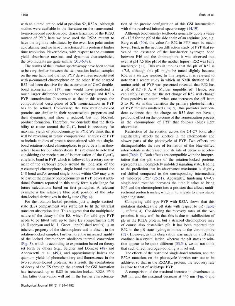
FIGURE 2 Characterization of the static UV-vis absorbance and fluo-
rescence emission of the four PYP derivatives. (A) Absorption spectra of
wild-type PYP (black), WTRL (red), R52A (blue), and R52ARL (green)
in 20 mM Tris buffer, pH 8.0. The absorption maxima are centered at
446, 443, 450, and 445 nm, respectively. (B) The corresponding emission
spectra, with excitation of all samples at their respective absorbance
maxima.
Set-up for ms/ms time-resolved visiblespectroscopy
An Edinburgh Instruments LP900 spectrometer (Livingston, West Lothian,
United Kingdom) equipped with a photomultiplier, in combination with
a Continuum Surelite OPO laser (for further details, see Hendriks et al.
(34)) was used for ms/ms time-resolved visible spectroscopy. PYP samples
were excited with 446-nm laser flashes of ~5–6 mJ/pulse (pulse duration
6 ns). Time traces were recorded for the four proteins at 360 nm,
~446 nm, and between 484 and 503 nm (dependent on the lmax of the partic-
ular protein), with the slow-board option of the photomultiplier (time reso-
lution ~2 ms). Optical interference filters were used before the sample to
minimize measurement artifacts induced by probe light.
Transient ms/s UV-vis spectroscopy
UV-vis spectra were recorded using an HP8453 UV-vis spectrophotometer
(Hewlett Packard, Palo Alto, CA). Time-resolved spectra were recorded
from 250 to 600 nm, using an integration time of 0.1 s. A Kraayenhof
cuvette was used to thermostat the sample at 25�C, with simultaneous pH
recording. A Schott KL1500 LCD lamp was used to illuminate the samples
with wild-type or R52A protein. For the rotation-locked proteins, a micro-
second flash lamp was used.
Steady-state fluorescence
Steady-state fluorescence spectra were recorded on a Fluorolog 3 spectrom-
eter (Spex, Metuchen, NJ) with a 450-W Xe lamp as the excitation light
source. The excitation wavelength was set at the absorption maximum of
the respective protein (see Fig. 2 below), and emission was recorded
from 450 to 600 nm for the two rotation-locked derivatives (i.e., the
wild-type (WTRL) and R52ARL) and from 455 to 600 nm (with a slit width
of 5 nm) for the two proteins reconstituted with p-coumaric acid (i.e., wild-
type and R52A). The slit width for the excitation and emission light was set
at 0.5 and 5 mm, respectively, for all four samples.
Synthesis of (5-hydroxy indan-(1E)-ylidene)aceticacid (rotation-locked chromophore)
After protection of its phenolic hydroxy group with tert-butyl-dimethyl-
silanyloxyl chloride, ~300 mg of (5-hydroxy-indan-(1E)-ylidene)-acetic
Biophysical Journal 101(5) 1184–1192
acid (190.20 mol wt) was synthesized from 5-hydroxy-1-indanone through
coupling with diethylphosphonoacetate. The phenolic hydroxyl group was
deprotected with tetra-n-butyl-ammonium fluoride. For this synthesis,
reagents were purchased at the highest commercial quality available and
used without further purification. Flash chromatography was carried out
with ACROS silica gel (particle size 35–70 mm). Infrared spectra were re-
corded using an IFS 28 spectrophotometer (Bruker, Billerica, MA), NMR
spectra were obtained using a Bruker ARX 400 spectrometer (400 MHz),
and accurate mass values of intermediates and the final product were
recorded on a JMS SX/SX102A four-sector mass spectrometer (JEOL,
Tokyo, Japan), coupled to a JEOL MS-MP7000 data system or a
TSQ7000 mass spectrometer (Thermo Finnigan, San Jose, CA) using
Xcaliber 1.2 software for acquisition and data processing.
Site-directed mutagenesis
Site-directed mutagenesis was performed using the QuikChange kit
(Stratagene, La Jolla, CA) and confirmed by DNA sequencing, as described
previously (35).
Sample preparation
Wild-type PYP and its R52A mutant derivative were produced and isolated
as described previously for wild-type PYP (36). Apo-PYP was reconstituted
with the 1,10-carbonyldiimidazole derivative of p-coumaric acid or rotation-
locked chromophore, as described by Hendriks et al. (37). The reconstituted
holoproteins were purified in two subsequent steps, with Ni-affinity and
anion-exchange chromatography, respectively (37). The purified holopro-
teins were used without removing the genetically introduced N-terminal
hexahistidine-containing tag, in a buffer containing 20 mM Tris-HCl,

Single-Bond Rotation in PYP Chromophore 1187
pH 8.0. The purity index for all samples was >0.5, except for R52A PYP
reconstituted with p-coumaric acid, in which case it was >0.75.
For measurements at pH 5, 8, and 10, buffer solutions containing 20 mM
acetic acid, 20 mMNa2HPO4, 20 mM Tris, and 20 mM boric acid, with the
same ionic strength, were prepared as described previously (17). For ultra-
fast spectroscopy, samples were concentrated to a final optical density of
~0.3 at 400 nm in a 50-mm-thick cell constructed from two CaF2 windows
separated by a Teflon spacer.
Data analysis
The transient difference absorption spectra were subjected to global and
target analysis, as reviewed in van Stokkum et al. (38). Briefly, the basis
of global analysis is the superposition principle, which states that the
measured j(t,l) data result from a superposition of the spectral properties,
εl(l), of the components present in the system of interest weighted by their
concentration cl(t).
jðt; lÞ ¼Xncomp
l¼ 1
clðtÞεlðlÞ
The cl(t) of all ncomp components are described by a compartmental
model that consists of first-order differential equations, with sums of expo-
nential decays as solution. We will consider two types of compartmental
model: 1), a sequential model with increasing lifetimes, also called an
unbranched unidirectional model, which results in evolution-associated
difference spectra (EADS); and 2), a full compartmental scheme, possibly
including branchings and equilibria, which yields species-associated differ-
ence spectra (SADS). The latter is most often referred to as target analysis,
where the target is the proposed kinetic scheme, including specific spectral
assumptions.
With ultrafast spectroscopy, the instrument response function can usually
adequately be modeled with a Gaussian with parameters for location and
full width at half-maximum (typically 100–250 fs). The dispersion of this
location parameter is described by a polynomial. All exponential decays
from the above models have to be convolved with this instrument response
function.
RESULTS
After heterologous overexpression in Escherichia coli andpurification, the four proteins studied in this investigation,i.e., wild-type and R52A-PYP reconstituted with 4-hydroxy-cinnamic acid and (5-hydroxy indan-(1E)-ylidene)acetic acid, respectively (see Fig. 1 for an overviewof the structure of their chromophore-binding pocket),were characterized with static UV-vis spectroscopy (Fig. 2A). The results show that whereas the R52A mutationcauses a slight red shift (4 nm for samples containing thep-coumaryl chromophore), the two rotation-lock PYPsshow a modest blue shift. The full width at half-maximumof the latter spectra, however, is significantly smaller thanthat of the two samples reconstituted with the endogenouschromophore, p-coumaric acid, presumably due to a muchsmaller contribution of the vibronic sideband of the mainelectronic transition of PYP. The decrease in the dynamicStokes shift in the fluorescence emission spectra ofthe two rotation-locked PYPs is even more pronounced,shifting from >500 nm for the endogenous chromophoreto <475 nm (Fig. 2 B) for the rotation-locked derivatives.
This figure also reveals that the already low fluorescencequantum yield of PYP is even lower (~50%) in the rota-tion-locked derivatives, whereas substitution of R52 by analanine results in an almost threefold increase of this yield.
Next, time-resolved visible pump/visible probe measure-ments in the 100 fs to 2 ns time domain were performed onall four samples, using 400-nm excitation. All four samplesshow rich dynamics in which some trends are immediatelyapparent, like the more extensive ultrafast relaxation inthe rotation-lock PYPs leading to a significantly shorter-lived signal than in wild-type PYP (see Fig. S1 in theSupporting Material for a set of transients of all foursamples collected at 446 nm, derived from the time-gatedspectra). However, much more detail is visible in the anal-ysis of the corresponding EADS (Fig. S2). An optimal fitto the data requires four lifetimes for each sample plusa component that does not decay on the timescale of ourexperiment (i.e., >>2.7 ns).
These EADS clearly show that the two rotation-lockedproteins have 1), a smaller red-shifted stimulated emissionwith respect to the 510-nm position at which the stimu-lated-emission band of the coumaryl derivatives is centered,consistent with their fluorescence characteristics (Fig. 2 B);2), significantly faster and monoexponential excited-statedecay (decay in 0.5 and 0.6 ps, respectively, versus the0.7–7 ps in wild-type), as can be concluded from the disap-pearance of the contribution of stimulated emission from thesubsequent spectra in each series of EADS; 3), both rota-tion-locked proteins form decreased amounts of the primaryphotoproducts (the blue and green spectra can be assigned tothe states I0 and I1, with product absorption bands at ~485and 475 nm, respectively). Note that the product bands ofthe rotation-locked proteins appear to be slightly blue-shifted, which may result in partial cancellation of their ab-sorbance with the bleached ground-state absorption, thusmaking a reliable estimate of the amount of these productsformed difficult; and finally, 4), the rotation-locked proteinsdisplay an intermediate spectrum with a 3-ps lifetime thatresembles the spectrum of the GSI of wild-type PYP re-solved in pump-dump-probe experiments (18,39) and targetanalysis (A. Rupenyan and M. L. Groot, unpublished).
However, to be able to characterize in more detail theseinitial photocycle steps and the distinct spectral characteris-tics of the various intermediates formed upon photoactiva-tion in these four proteins, a target analysis of the data isrequired. A complication in this analysis is the multiplicityof the excited state of each sample: the decay of the excitedstate has to be fitted by up to three time constants, and ina similar way, the appearance of I0 can be described withtwo to three time constants. From these excited states, theyield of I0 formation is largest for the shorter-lived excitedstates, in agreement with previous observations (see, e.g.,works by Larsen and colleagues (18,32)). However, thetarget analysis of the spectral data of the two rotation-lockedproteins already provides satisfactory results using a single
Biophysical Journal 101(5) 1184–1192

-100
-50
0
50
400 450 500 550 600
WT WTRL R52A R52ARL
400 450 500 550 600
-100
-50
0
50
400 450 500 550 600
-100
-50
0
50
400 450 500 550 600
-20
-10
0
10
Wavelength (nm)
Δm
OD
(a
.u
.)
ES I0
GSI I1
FIGURE 4 Species-associated difference spectra (SADS) of the four
PYP derivatives, obtained from fitting the target model of Fig. 3 to the
data. The SADS of wild-type PYP are in good agreement with earlier
reports in the literature (e.g., (18,39). The GSI spectrum of the rotation-
locked samples is easily resolved, as it appeared pronounced in the data,
as shown in Fig. S2. The quantum yields depicted in Fig. 3 were determined
by scaling the SADS of each state to that of the ES, which may result in
~10% uncertainty. An exception is the I1 state, for which we have used
a yield of 100% for the I0 to I1 transition, as determined with femtosecond
midinfrared spectroscopy (14,19). Color code as in Fig. 2.
1188 Stahl et al.
excited state. Therefore, to facilitate comparison betweenthe four samples here, we have fitted the datasets obtainedwith the two proteins reconstituted with the p-coumarylchromophore (i.e., wild-type PYP and R52A) with a singlecomponent for the excited-state decay as well, at the cost ofa small decrease in root-mean-squared value.
The results of this target analysis (see Fig. 3) and the cor-responding table with rate constants (see Table S1) providemore quantitative information on the systems dynamics inthe four proteins and give information beyond that derivedfrom the global analysis (e.g., the quantum yields). Fromthis target analysis, it is clear that rotation-locking of thechromophore causes not only much faster excited-statedecay but also an almost twofold lower quantum yield ofphotochemistry. We were surprised to find that in the lattertwo samples this is accompanied by the absence of internalconversion directly to the ground state, presumably becausethe free-energy barrier for the transition that leads to the GSIis even lower. The result is that in these two samples, thequantum yield of formation of the GSI state reaches valuesthat approach unity (i.e., 83%). The spectral characteristicsof these intermediates are consistent with the above obser-vations (Fig. 4): the stimulated emission of the rotation-locked proteins is blue-shifted to the extent that it is onlyvisible as a broadening of the low-energy shoulder of theground-state bleach. The corresponding I0 and GSI havenot only a blue-shifted absorption maximum (~25 nm forI0), but also a smaller molar extinction coefficient. For theGSI, it is difficult to estimate the lmax because of the exten-sive overlap with ground-state depletion. The I1 intermedi-
FIGURE 3 Target-model-based analysis of the initial stages of the photo-
cycle of the four PYP derivatives. I0 decays into I1 with a time constant of
~1 ns and a yield of 100%. The fitted lifetimes and relative populations of
the states accessible from the ES are shown. The excited states of wild-type
and R52A were found to decay biexponentially with time constants of 0.7
and 6.8 ps and 1.9 and 27 ps, respectively, with the quantum yield of I0formation largest for the fastest fraction, in agreement with Larsen et al.
(18). To facilitate comparison with the rotation-locked samples, the data
for excited-state decay for all four proteins were fitted here with single
exponents of 1.5 ps and 2.3 ps, respectively. The yield of the ground state
in WTRL and R52ARL is very low, and can be fitted with yields between
0 and 10%. The reduction of this yield and that of I0 in the rotation-locked
samples is due to a four- to sixfold increase in the rate of ES / GSI.
Biophysical Journal 101(5) 1184–1192
ates show the expected blue shift relative to theircorresponding I0 states, except in the case of the I1 stateof the R52A protein, which is almost isoenergetic with its I0.
The results obtained to date have revealed that within thisset of four proteins, R52A is the most similar to wild-typePYP. One would expect the rotation-locked samples to bemore similar to wild-type PYP, but different from R52APYP if the charge on R52 were steering the primary photo-chemistry of PYP toward single-bond rotation (see above).As this observation is clearly different from what has beenpredicted by theory (17,30), we tested a second approachin which the charge on R52 is varied.
By varying the pH of the buffer in the range 5–10, wemodulated the net charge of the side chain of R52 (seeDiscussion) and, by consequence, the environmental elec-trostatics of the p-coumaryl chromophore. The results pre-sented in Fig. 5 reveal that the primary photochemistry inPYP is totally invariant with pH in this pH range. Thisapplies to both the observed lifetimes and the shape of thedifference spectra.
Locking the rotation around the C4-C7 bond not only hasconsequences for the ultrafast photochemistry of PYP butmight also affect the slower processes that occur in thephotocycle of PYP. We therefore also analyzed the ms/mstime-resolved visible absorption changes at three differentwavelengths: 360 nm, at which the main contribution isfrom pB intermediates; in the range 484–503 nm, to recordthe contribution of the red-shifted intermediates (i.e., pR)(see also Table S2); and z446 nm, the absorbancemaximum of the four proteins, at which bleaching of pGand absorption of pR contribute to the dynamics of thechanges in UV-vis absorbance. The absorbance traces at360 nm are of particular interest, because at this wavelength

400 450 500 550 600
-8
-6
-4
-2
0
2
pH 5 pH 8 pH 10
Δm
OD
(a
.u
.)
Wavelength (nm)
FIGURE 5 pH dependence of the ultrafast kinetics of wild-type PYP. At
each pH, the dynamics could be fitted with four time constants. The EADS
for each time constant are indicated: 0.9, 0.9, and 0.8 ps (black); 6, 5, and
5.5 ps (red); 1.4, 1.7, and 1.4 ns (blue), and infinite (green), for wild-type
PYP at pH 5, 8, and 10, respectively.
Single-Bond Rotation in PYP Chromophore 1189
both the decay of the I1/pR state (which coincides withformation of pB0 (20)) and the thermal recovery of theground state can be observed. The results obtained(Fig. 6) clearly reveal that the R52A mutation acceleratespB0 formation (in combination with the p-coumaryl chromo-phore) and retards ground-state recovery. This is similar tothe effect observed by others (e.g., Hellingwerf et al. (4)and Cusanovich and Meyer (5)) in R52Q and several othermutant proteins. A global analysis of these data (Table S2,Fig. S3, and Fig. S4) furthermore shows that the effect of re-placing the p-coumaryl chromophore with the rotation-locked chromophore is an approximately five- and ninefoldretardation of the rate of formation of the first blue-shiftedstate for wild-type PYP and R52A, respectively, whereasthe recovery reaction (i.e., the re-formation of pG) for theseproteins is accelerated 8- and 16-fold, respectively. How-ever, the data for the two rotation-locked proteins in thistime domain are well-fitted with a sum of two exponentials
1E-6 1E-5 1E-4 1E-3 0.01 0.1 1 10
0.000
0.005
0.010
0.015
0.020
0.025
0.030 WT WTRL R52A R52ARL
Time (s)
ΔO
D
WT WTRL R52A R52ARL
FIGURE 6 Time traces of the absorbance changes during the later part of
the photocycle of the four PYP derivatives, recorded at 360 nm. The pB
intermediates absorb maximally at 360 nm. Their rise and decay can be
fitted with the time constants, as shown in Table S2 (see Fig. S1). Color
code as in Fig. 2.
(Table S2), whereas for the wild-type protein, at least threeexponentials are required (26). We therefore carried outa target analysis of this same data set based on the photo-cycle scheme pR 4 pB0 / pB / pG. For the wild-typeprotein, the results obtained confirm previous observations(5,24,26), whereas the data obtained with the R52A proteinshow that this mutation particularly accelerates protontransfer from E46 to the chromophore (i.e., pR / pB0)and also decelerates pG recovery (Table 1). We thereforetentatively conclude that single-bond rotation around theC4-C7 bond contributes to both of these processes. Theslower part of the photocycle of the two rotation-lockedproteins is well described by the sequential model pR /pB / pG, in which a single exponent suffices to describethe pR to pB transition. The estimated SADS (see Fig. S5)are consistent with the expected shapes for pR and pB,establishing the suitability of these two photocycle schemesfor the two sets of proteins (i.e., wild-type and R52A on theone hand and WTRL and R52ARL on the other.
DISCUSSION
In the past, the use of various rotation-locked chromophoreshas provided important information on the primary photo-chemistry in the two largest and most diverse families ofphotosensory receptors that use E/Z/E isomerization of theirchromophore to initiate the biological signaling process,i.e., the rhodopsins (40,41) and the phytochromes (42).For the xanthopsin family also, chromophore derivativeswith the C7¼C8 ethylenic bond of the chromophore lockedin the trans and cis states, respectively, have been exploitedto better understand the molecular basis of functioning ofthis family (43,44).
Here, we have made use of yet another rotation-lockedchromophore, i.e., one in which rotation of the C4-C7 singlebond is prevented by making it part of a cyclopentane ring.This restricts the flexibility within this chromophore evenmore than it does within derivatives in which a cyclohexanering is used instead (45). This rotation-locked analog hasbeen used to reconstitute wild-type PYP and a mutant
TABLE 1 Target analysis of the transitions between the
intermediates of the photocycle of PYP in the microsecond-
to-second time domain
PYP
derivative pR / pB0 pB0 / pR DG (meV)* pB0 / pB pB / pG
Wild-type 3.65 1.72 19 2.02 0.00366
R52A 15.2 0.97 71 2.71 0.00049
pR / pB
WTRL 1.37 0.0276
R52ARL 1.76 0.0076
Estimated rate constants (ms�1) of the transitions between intermediates.
Kinetic scheme for wild-type and R52A: pR 4 pB0 / pB / pG.
*DG of the reaction between pR and pB0. Kinetic scheme for WTRL and
R52ARL: pR / pB / pG.
Biophysical Journal 101(5) 1184–1192
1190 Stahl et al.
with an altered amino acid at position 52, R52A. Althoughstudies were available in the literature on the nanosecond-to-microsecond spectroscopic characterization of the R52Qmutant of PYP, here we have used the R52A mutant tohave the arginine substituted by the even less polar aminoacid alanine, and we have characterized this protein at highertime resolution. Nevertheless, with respect to the quantumyield, absorbance maxima, and dynamics characteristics,the two mutants are quite similar (31,46,47).
The results of the ultrafast spectroscopy have been shownto be very similar between the two rotation-locked sampleson the one hand and the two PYP derivatives reconstitutedwith p-coumaryl chromophore on the other. If the chargedR52 had been decisive for the occurrence of C¼C double-bond isomerization (17), one would have predicted amuch larger difference between the wild-type and R52APYP isomerization. It seems to us that in this aspect, thecomputational description of Z/E isomerization in PYPhas to be refined. Conversely, the two rotation-lockedproteins are similar in their spectroscopic properties andtheir dynamics, and show a reduced, but not blocked,product formation. Therefore, we conclude that the flexi-bility to rotate around the C4-C7 bond is necessary formaximal yields of photochemistry in PYP. We think that itwill be revealing in future computational analyses of PYPto include studies of protein reconstituted with this single-bond rotation-locked chromophore, to provide a firm theo-retical basis for our observations. It is relevant to note thatconsidering the mechanism of isomerization of the C7¼C8
ethylenic bond in PYP, which is followed by a rotary move-ment of the carbonyl group around the long axis of thep-coumaryl chromophore, single-bond rotation around theC-S bond and/or around single bonds within C69 may alsobe part of the primary photochemistry in PYP. Several addi-tional features reported in this study form a challenge forfuture calculations based on first principles. A relevantexample is the relatively blue peak position of the rota-tion-locked derivatives in the I0 state (Fig. 4).
For the rotation-locked proteins, just a single excited-state (ES) compartment was sufficient to fit the ultrafasttransient absorption data. This suggests that the multiphasicnature of the decay of the ES, which for wild-type PYPneeds to be fitted with up to three ES compartments (18)(A. Rupenyan and M. L. Groot, unpublished results), is aninherent property of the chromophore and is absent in therotation-locked samples. Furthermore, the increased rigidityof the locked chromophore abolishes internal conversion(Fig. 3), which is according to expectation based on theoryset forth by others (e.g., Seidner and Domcke (48) andAbbruzzetti et al. (49)), and approximately halves thequantum yields of photochemistry and fluorescence in thetwo rotation-locked proteins. As a result, the contributionof decay of the ES through the channel for GSI formationhas increased, up to 0.83 in rotation-locked R52A PYP.This latter observation will aid in the further characteriza-
Biophysical Journal 101(5) 1184–1192
tion of the precise configuration of this GSI intermediatewith time-resolved infrared spectroscopy (14,19).
Although biochemistry textbooks generally quote a valueof ~12.5 for the pK of the side chain of an arginine (see, e.g,Berg et al. (50)), the value for R52 in PYP must be muchlower. First, in the neutron diffraction study of PYP that re-vealed the existence of the low-barrier hydrogen bondbetween E46 and the chromophore, it was observed thateven at pH 7.5 (the pH of the mother liquor), R52 was fullyuncharged (11). This result implies that the pK of R52 is<7.5, although this pK might be tuned slightly becauseR52 is a surface residue. In this respect, it is relevant tonote that a recent study in which an NMR titration of allamino acids of PYP was presented revealed that R52 hasa pK of 8.7 (F. A. A. Mulder, unpublished). Hence, onecan safely assume that the net charge of R52 will changefrom positive to neutral when one increases the pH from5 to 10. As in this transition the primary photochemistryof PYP remains unaltered (Fig. 5), this provides indepen-dent evidence that the charge on R52 does not have aprofound effect on the outcome of the isomerization processin the chromophore of PYP that follows (blue) lightabsorption.
Restriction of the rotation across the C4-C7 bond alsosignificantly affects the kinetics in the intermediate andslower parts of the photocycle of PYP. Two effects aredistinguishable: the rate of formation of the blue-shiftedintermediate is decreased, and its rate of decay is acceler-ated (Table 1). Both effects are compatible with the interpre-tation that the pB state of the rotation-locked proteinsrepresents an incompletely unfolded signaling state, leadingto the prediction that its absorbance maximum is slightlyred-shifted compared to the corresponding intermediateof wild-type PYP (26,51). Apparently, hindering C4-C7single-bond rotation increases the time it takes to bringE46 and the chromophore into a position that allows unidi-rectional proton transfer, which in turn leads to a less stablesignaling state.
Comparing wild-type PYP with R52A shows that thismutation stabilizes the pB state with respect to pR (Table1, column 4). Considering the recovery rates of the twoproteins, it may well be that this is due to stabilization ofpB in the R52A protein, but a strained chromophore mayof course also destabilize pR. It has been reported thatR52 in the pB state hydrogen-bonds to the chromophore(52). However, as this observation was made on a pB stateconfined in a crystal lattice, whereas the pB states in solu-tion appear to be quite different (53,54), we do not thinkthat such direct hydrogen-bonding is involved.
The effects of the restricted single-bond rotation, and theR52A mutation, on the photocycle kinetics turn out to beadditive, so that in the R52ARL protein, the recovery rateis close to that of wild-type PYP.
A comparison of the maximal increase in absorbance at360 nm and the maximal decrease at 446 nm (Fig. 6 and

Single-Bond Rotation in PYP Chromophore 1191
Fig. S5) using the microsecond/millisecond data from wild-type PYP as a reference also allows an independent estimateof the quantum yield of these PYP derivatives. Consideringthe accuracy with which such data can be measured, the dataobtained are consistent with the results displayed in Fig. 3:An approximately twofold decrease is caused by the rota-tion-locked chromophore.
It will be of interest to test this and other chromophoreanalogs on the phototactic capabilities of H. halophila,similar to the way they were exploited in Idiomarinahalophila (44).
CONCLUSIONS
We have studied the isomerization pathway of PYP and therole of the charge of the side chain of amino acid R52therein by selectively locking one of the single bonds ofthe chromophore. Preventing rotation of the C4-C7 chromo-phore bond enhances the rate of formation of a nonproduc-tive intermediate state, GSI, and leads to reduction of thephysiological Z/E isomerization of the chromophore. Theseresults confirm that C4-C7 single-bond rotation in PYP isnot an alternative to C7¼C8 double-bond rotation butappears to optimize double-bond isomerization (e.g., witha compensatory movement). The initial photocycle dyn-amics of PYP were observed to be not significantly affectedby the charge of the amino acid residue at position 52, whichwas varied by 1), varying the pH of the sample between 5and 10; and 2), site-directed mutagenesis to construct R52A.
SUPPORTING MATERIAL
Two tables and five figures are available at http://www.biophysj.org/
biophysj/supplemental/S0006-3495(11)00836-8.
The authors thank Dr. Jan van Maarseveen (Van ’t Hoff Institute of
Molecular Sciences, University of Amsterdam) for synthesis of (5-hydroxy
indan-(1E)-ylidene)acetic acid and Mr. Jos Thieme and Mr. Jos Arents for
excellent technical support.
K.J.H. and R.v.G. acknowledge support from the Human Frontier Science
Program through grant number HFSP-RGP0038/2006.
REFERENCES
1. van der Horst, M. A., and K. J. Hellingwerf. 2004. Photoreceptorproteins, ‘‘star actors of modern times’’: a review of the functionaldynamics in the structure of representative members of six differentphotoreceptor families. Acc. Chem. Res. 37:13–20.
2. Meyer, T. E. 1985. Isolation and characterization of soluble cyto-chromes, ferredoxins and other chromophoric proteins from the halo-philic phototrophic bacterium Ectothiorhodospira halophila. Biochim.Biophys. Acta. 806:175–183.
3. Sprenger, W. W., W. D. Hoff, ., K. J. Hellingwerf. 1993. The eubac-terium Ectothiorhodospira halophila is negatively phototactic, witha wavelength dependence that fits the absorption spectrum of the photo-active yellow protein. J. Bacteriol. 175:3096–3104.
4. Hellingwerf, K. J., J. Hendriks, and T. Gensch. 2003. Photoactiveyellow protein, a new type of photoreceptor protein: will this
‘‘yellow lab’’ bring us where we want to go? J. Phys. Chem. A.107:1082–1094.
5. Cusanovich, M. A., and T. E. Meyer. 2003. Photoactive yellow protein:a prototypic PAS domain sensory protein and development ofa common signaling mechanism. Biochemistry. 42:4759–4770.
6. Imamoto, Y., and M. Kataoka. 2007. Structure and photoreaction ofphotoactive yellow protein, a structural prototype of the PAS domainsuperfamily. Photochem. Photobiol. 83:40–49.
7. Van Beeumen, J. J., B. V. Devreese, ., M. A. Cusanovich. 1993.Primary structure of a photoactive yellow protein from the phototrophicbacterium Ectothiorhodospira halophila, with evidence for the massand the binding site of the chromophore. Protein Sci. 2:1114–1125.
8. Kim, M., R. A. Mathies, ., K. J. Hellingwerf. 1995. ResonanceRaman evidence that the thioester-linked 4-hydroxycinnamyl chromo-phore of photoactive yellow protein is deprotonated. Biochemistry.34:12669–12672.
9. Borgstahl, G. E. O., D. R. Williams, and E. D. Getzoff. 1995. 1.4 Astructure of photoactive yellow protein, a cytosolic photoreceptor:unusual fold, active site, and chromophore. Biochemistry. 34:6278–6287.
10. Kort, R., H. Vonk, ., K. J. Hellingwerf. 1996. Evidence for trans-cisisomerization of the p-coumaric acid chromophore as the photochem-ical basis of the photocycle of photoactive yellow protein. FEBS Lett.382:73–78.
11. Yamaguchi, S., H. Kamikubo, ., M. Kataoka. 2009. Low-barrierhydrogen bond in photoactive yellow protein. Proc. Natl. Acad. Sci.USA. 106:440–444.
12. Gensch, T., C. C. Gradinaru, ., R. van Grondelle. 2002. The primaryphotoreaction of photoactive yellow protein (PYP): anisotropychanges and excitation wavelength dependence. Chem. Phys. Lett.356:347–354.
13. van Brederode, M. E., T. Gensch, ., S. E. Braslavsky. 1995.Photoinduced volume change and energy storage associated withthe early transformations of the photoactive yellow protein fromEctothiorhodospira halophila. Biophys. J. 68:1101–1109.
14. Groot, M. L., L. J. van Wilderen, ., R. van Grondelle. 2003. Initialsteps of signal generation in photoactive yellow protein revealed withfemtosecond mid-infrared spectroscopy. Biochemistry. 42:10054–10059.
15. Xie, A., W. D. Hoff,., K. J. Hellingwerf. 1996. Glu46 donates a protonto the 4-hydroxycinnamate anion chromophore during the photocycleof photoactive yellow protein. Biochemistry. 35:14671–14678.
16. Groenhof, G., M. F. Lensink,., A. E. Mark. 2002. Signal transductionin the photoactive yellow protein. I. Photon absorption and the isomer-ization of the chromophore. Proteins. 48:202–211.
17. Groenhof, G., L. V. Schafer,., M. A. Robb. 2008. Arginine52 controlsthe photoisomerization process in photoactive yellow protein. J. Am.Chem. Soc. 130:3250–3251.
18. Larsen, D. S., I. H. van Stokkum, ., R. van Grondelle. 2004. Inco-herent manipulation of the photoactive yellow protein photocyclewith dispersed pump-dump-probe spectroscopy. Biophys. J. 87:1858–1872.
19. van Wilderen, L. J., M. A. van der Horst,., M. L. Groot. 2006. Ultra-fast infrared spectroscopy reveals a key step for successful entry intothe photocycle for photoactive yellow protein. Proc. Natl. Acad. Sci.USA. 103:15050–15055.
20. Xie, A., L. Kelemen, ., W. D. Hoff. 2001. Formation of a new buriedcharge drives a large-amplitude protein quake in photoreceptor activa-tion. Biochemistry. 40:1510–1517.
21. Van Brederode, M. E., W. D. Hoff,., K. J. Hellingwerf. 1996. Proteinfolding thermodynamics applied to the photocycle of the photoactiveyellow protein. Biophys. J. 71:365–380.
22. Meyer, T. E., E. Yakali, ., G. Tollin. 1987. Properties of a water-soluble, yellow protein isolated from a halophilic phototrophic bacte-rium that has photochemical activity analogous to sensory rhodopsin.Biochemistry. 26:418–423.
Biophysical Journal 101(5) 1184–1192

1192 Stahl et al.
23. Hoff, W. D., I. H. van Stokkum, ., K. J. Hellingwerf. 1994. Measure-ment and global analysis of the absorbance changes in the photocycleof the photoactive yellow protein from Ectothiorhodospira halophila.Biophys. J. 67:1691–1705.
24. Hendriks, J., I. H. M. van Stokkum, and K. J. Hellingwerf. 2003. Deute-rium isotope effects in the photocycle transitions of the photoactiveyellow protein. Biophys. J. 84:1180–1191.
25. Hoersch, D., H. Otto, ., M. P. Heyn. 2007. Role of a conserved saltbridge between the PAS core and the N-terminal domain in the activa-tion of the photoreceptor photoactive yellow protein. Biophys. J.93:1687–1699.
26. Hendriks, J., and K. J. Hellingwerf. 2009. pH Dependence of the photo-active yellow protein photocycle recovery reaction reveals a new latephotocycle intermediate with a deprotonated chromophore. J. Biol.Chem. 284:5277–5288.
27. Premvardhan, L. L., M. A. van der Horst, ., R. van Grondelle. 2003.Stark spectroscopy on photoactive yellow protein, E46Q, and a noniso-merizing derivative, probes photo-induced charge motion. Biophys. J.84:3226–3239.
28. Grabowski, Z. R., K. Rotkiewicz, ., W. Baumann. 1979. Twistedintra-molecular charge-transfer states (TICT)—new class of excited-states with a full charge separation. Nouv. J. .Chim. 3:443–454.
29. Grabowski, Z. R., K. Rotkiewicz, and W. Rettig. 2003. Structuralchanges accompanying intramolecular electron transfer: focus ontwisted intramolecular charge-transfer states and structures. Chem.Rev. 103:3899–4032.
30. Ko, C., A. M. Virshup, and T. J. Martinez. 2008. Electrostatic control ofphotoisomerization in the photoactive yellow protein chromophore: abinitio multiple spawning dynamics. Chem. Phys. Lett. 460:272–277.
31. Changenet-Barret, P., P. Plaza, ., M. Kataoka. 2007. Role of arginine52 on the primary photoinduced events in the PYP photocycle. Chem.Phys. Lett. 434:320–325.
32. Larsen, D. S., and R. van Grondelle. 2005. Initial photoinduceddynamics of the photoactive yellow protein. ChemPhysChem.6:828–837.
33. Groot, M. L., L. J. G. W. van Wilderen, and M. Di Donato. 2007. Time-resolved methods in biophysics. 5. Femtosecond time-resolved anddispersed infrared spectroscopy on proteins. Photochem. Photobiol.Sci. 6:501–507.
34. Hendriks, J., W. D. Hoff, ., K. J. Hellingwerf. 1999. Protonation/deprotonation reactions triggered by photoactivation of photoactiveyellow protein from Ectothiorhodospira halophila. J. Biol. Chem.274:17655–17660.
35. van Aalten, D. M. F., A. Haker, ., W. Crielaard. 2002. Engineeringphotocycle dynamics. Crystal structures and kinetics of three photoac-tive yellow protein hinge-bending mutants. J. Biol. Chem. 277:6463–6468.
36. Kort, R., W. D. Hoff, ., K. J. Hellingwerf. 1996. The xanthopsins:a new family of eubacterial blue-light photoreceptors. EMBO J.15:3209–3218.
37. Hendriks, J., T. Gensch,., J. J. van Thor. 2002. Transient exposure ofhydrophobic surface in the photoactive yellow protein monitored withNile Red. Biophys. J. 82:1632–1643.
38. van Stokkum, I. H. M., D. S. Larsen, and R. van Grondelle. 2004.Global and target analysis of time-resolved spectra. Biochim. Biophys.Acta. 1657:82–104.
Biophysical Journal 101(5) 1184–1192
39. Changenet-Barret, P., P. Plaza,., M. Kataoka. 2009. Structural effects
on the ultrafast photoisomerization of photoactive yellow protein.
Transient absorption spectroscopy of two point mutants. J. Phys.
Chem. C. 113:11605–11613.
40. Birge, R. R., L. P. Murray, ., K. Nakanishi. 1985. Two-photon spec-
troscopy of locked-11-cis-rhodopsin: evidence for a protonated Schiff
base in a neutral protein binding site. Proc. Natl. Acad. Sci. USA.
82:4117–4121.
41. Akita, H.., 1980. Non-bleachable rhodopsins retaining the full
natural chromophore. J. Am. Chem. Soc. 102:6370–6372.
42. Inomata, K., M. A. Hammam,., T. Lamparter. 2005. Sterically locked
synthetic bilin derivatives and phytochrome Agp1 from Agrobacterium
tumefaciens form photoinsensitive Pr- and Pfr-like adducts. J. Biol.
Chem. 280:24491–24497.
43. Cordfunke, R., R. Kort, ., K. J. Hellingwerf. 1998. Trans/cis (Z/E)
photoisomerization of the chromophore of photoactive yellow protein
is not a prerequisite for the initiation of the photocycle of this photore-
ceptor protein. Proc. Natl. Acad. Sci. USA. 95:7396–7401.
44. van der Horst, M. A., T. P. Stalcup,., W. D. Hoff. 2009. Locked chro-
mophore analogs reveal that photoactive yellow protein regulates bio-
film formation in the deep sea bacterium Idiomarina loihiensis. J. Am.
Chem. Soc. 131:17443–17451.
45. Sheves, M., N. Friedman, ., M. Ottolenghi. 1985. Primary photo-
chemical event in bacteriorhodopsin: study with artificial pigments.
Biochemistry. 24:1260–1265.
46. Borucki, B., J. A. Kyndt,., M. P. Heyn. 2005. Effect of salt and pH on
the activation of photoactive yellow protein and gateway mutants
Y98Q and Y98F. Biochemistry. 44:13650–13663.
47. Philip, A. F., R. A. Nome,., W. D. Hoff. 2010. Spectral tuning in pho-
toactive yellow protein by modulation of the shape of the excited state
energy surface. Proc. Natl. Acad. Sci. USA. 107:5821–5826.
48. Seidner, L., and W. Domcke. 1994. Microscopic modelling of photoi-
somerization and internal-conversion dynamics. Chem. Phys.
186:27–40.
49. Abbruzzetti, S., R. Bizzarri, ., F. Beltram. 2010. Photoswitching of
E222QGFPmutants: ‘‘concerted’’ mechanism of chromophore isomer-
ization and protonation. Photochem. Photobiol. Sci. 9:1307–1319.
50. Berg, J. M., J. L. Tymoczko, and L. Stryer. 2012. Biochemistry, 7th ed.
W. H. Freeman, New York.
51. Yeremenko, S., I. H. van Stokkum, ., K. J. Hellingwerf. 2006. Influ-
ence of the crystalline state on photoinduced dynamics of photoactive
yellow protein studied by ultraviolet-visible transient absorption spec-
troscopy. Biophys. J. 90:4224–4235.
52. Genick, U. K., G. E. Borgstahl, ., E. D. Getzoff. 1997. Structure of
a protein photocycle intermediate by millisecond time-resolved crystal-
lography. Science. 275:1471–1475.
53. Vreede, J., W. Crielaard, ., P. G. Bolhuis. 2005. Predicting the
signaling state of photoactive yellow protein. Biophys. J. 88:3525–
3535.
54. Bernard, C., K. Houben, ., N. A. van Nuland. 2005. The solution
structure of a transient photoreceptor intermediate: delta25 photoactive
yellow protein. Structure. 13:953–962.