On the Comparison of NN-Based Architectures for Diabetic Damage Detection in Retinal Images
12
Journal of Circuits, Systems, and Computers Vol. 18, No. 8 (2009) 1369–1380 c World Scientific Publishing Company ON THE COMPARISON OF NN-BASED ARCHITECTURES FOR DIABETIC DAMAGE DETECTION IN RETINAL IMAGES VITOANTONIO BEVILACQUA ∗,†,‡ , LEONARDA CARNIMEO ∗ , GIUSEPPE MASTRONARDI ∗,† , VITO SANTARCANGELO ∗,† and ROCCO SCARAMUZZI ∗ ∗ Dipartimento di Elettrotecnica ed Elettronica, Politecnico di Bari, via Orabona, 4, Bari, 70125, Italy † e.B.I.S. s.r.l. (Electronic Business in Security), Spin-Off of Polytechnic of Bari, Via Pavoncelli, 139, Bari, Italy ‡ [email protected] The automatic screening of retinal images for an early detection of diabetic symptoms and an early prevention of diabetic retinopathies has been a prime focus in recent times. In this paper a contribution to improve diabetic damage detection in retinal images via neural networks is proposed by comparing two neural strategies. By considering the first architecture, fundus oculi symptomatic pale regions are firstly highlighted by enhanc- ing image contrast with a neurofuzzy subnet, which is synthesized using a Sparsely- Connected Neural Network. Then, obtained contrast-enhanced images with bimodal histograms are globally segmented, after an optimal thresholding performed by a neural subsystem. In output binary images, suspect diabetic areas are finally isolated. By con- sidering the second architecture, an EBP MLP neural net is synthesized, where a suitable training set of suspect patterns is developed by (5 × 5) windows centered on damaged pixels in gold standard images provided by clinicians. Performances are evaluated by percentage measures of exactness in the detection of suspect damaged areas via a com- parison with gold standard images provided by clinicians. Results of both strategies are discussed and compared with other researchers’ ones. Keywords : Sparsely-connected neural networks; EBP MLP; diabetic damage detection. 1. Introduction It is well known that diabetic retinopathy is the leading cause of blindness in the working population. 1 In the last years, the automatic screening of patients for early detection and prevention of diabetic retinopathy became very important due to the large ratio of patients to medical ophthalmologists. In the field of retinopathies attention has been recently focused on the problem of obtaining an accurate dia- betic ocular screening. In fact, specific retinal damages, such as exudates, drusen or cotton wool spots, which appear as vague pale areas in fundus oculi images, generally 1369
Transcript of On the Comparison of NN-Based Architectures for Diabetic Damage Detection in Retinal Images

October 20, 2009 16:12 WSPC/123-JCSC 00572
Journal of Circuits, Systems, and ComputersVol. 18, No. 8 (2009) 1369–1380c© World Scientific Publishing Company
ON THE COMPARISON OF NN-BASED ARCHITECTURESFOR DIABETIC DAMAGE DETECTION
IN RETINAL IMAGES
VITOANTONIO BEVILACQUA∗,†,‡, LEONARDA CARNIMEO∗,GIUSEPPE MASTRONARDI∗,†, VITO SANTARCANGELO∗,†
and ROCCO SCARAMUZZI∗
∗Dipartimento di Elettrotecnica ed Elettronica,Politecnico di Bari, via Orabona, 4,
Bari, 70125, Italy
†e.B.I.S. s.r.l. (Electronic Business in Security),Spin-Off of Polytechnic of Bari,Via Pavoncelli, 139, Bari, Italy
The automatic screening of retinal images for an early detection of diabetic symptomsand an early prevention of diabetic retinopathies has been a prime focus in recent times.In this paper a contribution to improve diabetic damage detection in retinal images vianeural networks is proposed by comparing two neural strategies. By considering the firstarchitecture, fundus oculi symptomatic pale regions are firstly highlighted by enhanc-ing image contrast with a neurofuzzy subnet, which is synthesized using a Sparsely-Connected Neural Network. Then, obtained contrast-enhanced images with bimodalhistograms are globally segmented, after an optimal thresholding performed by a neuralsubsystem. In output binary images, suspect diabetic areas are finally isolated. By con-sidering the second architecture, an EBP MLP neural net is synthesized, where a suitabletraining set of suspect patterns is developed by (5 × 5) windows centered on damaged
pixels in gold standard images provided by clinicians. Performances are evaluated bypercentage measures of exactness in the detection of suspect damaged areas via a com-parison with gold standard images provided by clinicians. Results of both strategies arediscussed and compared with other researchers’ ones.
Keywords: Sparsely-connected neural networks; EBP MLP; diabetic damage detection.
1. Introduction
It is well known that diabetic retinopathy is the leading cause of blindness in theworking population.1 In the last years, the automatic screening of patients for earlydetection and prevention of diabetic retinopathy became very important due tothe large ratio of patients to medical ophthalmologists. In the field of retinopathiesattention has been recently focused on the problem of obtaining an accurate dia-betic ocular screening. In fact, specific retinal damages, such as exudates, drusen orcotton wool spots, which appear as vague pale areas in fundus oculi images, generally
1369

October 20, 2009 16:12 WSPC/123-JCSC 00572
1370 V. Bevilacqua et al.
are revealed to be symptoms of diabetic retinopathy. Exudates detection is one ofthe main steps to prevent blindness due to diabetic retinopathy.2, 3 In Ref. 2 hardexudates are detected by employing domain knowledge about brightness. In Ref. 3exudates are detected using their high gray level variation and contours are deter-mined by means of morphological reconstruction techniques. Contributions for anautomatic detection of retinal damages using image filtering algorithms or statisti-cal ones have been recently presented.4–8 In Ref. 4 a first approach to the detectionof exudates by means of artificial neural networks is reported, but correspondingperformances greatly depend on the number of images collected in the training set.In Refs. 5 and 6 a recursive region growing segmentation algorithm is used for thedetection of exudates. In particular, each pixel of a retinal image is classified asbelonging to a suspect region only if: (i) it belongs to the neighborhood of a pixelwhich is in a suspect area and (ii) its gray level belongs to a pre-specified range of arepresentative center. In this way, clinicians’ recommended standards are achieved,but condition (ii) requires an a priori knowledge of image contrast or brightness.In Ref. 7 a three-step method is proposed for retinal images: blood vessels arefirstly segmented using a median filter; brighter regions are then extracted using amultilevel thresholding technique; location of optic disk and position of suspectedregions are finally determined. Unfortunately, drawbacks arise for the number ofvariables to be evaluated. In Ref. 8 hemorrhage areas restricted by finite windowin training images were used as input for the support vector machine. To detectdifferent sized hemorrhages a pyramid of images was generated by changing theresolution of fundus image. The principal component analysis was used to reducethe complexity of feature space.
Contributions to the detection of diabetic symptoms using neural networks havebeen successively developed.9–11
In Ref. 9 an approach based on neurofuzzy techniques, which requires selectivefeatures for training Multilayer Perceptron neural networks (MLP)12 working asclassifiers, has been proposed for fundus image segmentation. Color retinal imagesare segmented using a fuzzy C-means clustering technique in order to group closepixels with similar colors into the same region. This method is quite effective, butthe selection of features is based only on experimental results. In Ref. 10 MLPs havebeen considered for classifying symptoms, but proposed networks, even if effective,require heavy computational efforts.
In Ref. 11 a network of spiking neurons to detect symptoms in segmented retinalimages has been very recently introduced, but drawbacks arise for high memoryrequirements and long computation times.
Moreover, a Sparsely-Connected Neurofuzzy Network (SCNN) has been recentlyapplied to support diagnosis of ophthalmic pathologies such as diabetic damages.13
The sparsely-connected structure of these networks with fixed or adjustable weightsmakes them very suitable for VLSI implementation.
In this paper, a contribution to diabetic damage detection in retinal imagesvia the comparison of reliable methods for detection of pale areas with different

October 20, 2009 16:12 WSPC/123-JCSC 00572
On the Comparison of NN-Based Architectures for Diabetic Damage Detection 1371
architectures of neural networks is presented. At first, after introducing the imageprocessing steps of the whole detection method, a NN-based subsystem is firstlydesigned by a neurofuzzy synthesis technique to enhance contrast in diabetic fundusimages, thus highlighting symptomatic vague pale regions. A subsystem of MLPneural networks is successively trained for evaluating the optimal threshold whichcan minimize pixel classification errors. Contrast-enhanced images are then globallysegmented via a NN-based subsystem, providing binary output images, in whichsuspect diabetic damages are accurately isolated.
At the same time, the situation in which a reduced number of pathological retinalimages is available, is taken into account. For this purpose, a further NN-basedarchitecture is herein synthesized as an EBP MLP, for which a suitable trainingset of suspect patterns is developed by considering (5 × 5) windows centered onreference damaged pixels in gold standard images provided by clinicians. In bothstrategies bipolar output images show black regions which represent areas withsuspect lesions.
Performances of both networks are evaluated and compared by means of a Cor-rect Recognition Rate, which provides percentage measures of exactness in thedetection of suspect damaged areas. The capabilities of compared neural architec-tures are investigated by means of experimental examples. Results are discussedand compared with other techniques by considering experimental cases on humanretinal images.
2. Diabetic Damage Detection Method
After introducing the image processing steps of the whole detection method, aNN-based subsystem is firstly designed by a neurofuzzy synthesis technique toenhance contrast in diabetic fundus images, thus highlighting symptomatic vaguepale regions. A subsystem of MLP neural networks is successively trained for evalu-ating the optimal threshold which can minimize pixel classification errors. Contrast-enhanced images are then globally segmented via a NN-based subsystem, providingbinary output images, in which suspect diabetic damages are accurately isolated.
2.1. Sparsely-connected neural network
Three main retinal image processing steps are performed by the proposed NN-basednetwork for diabetic damage detection
• Neurofuzzy contrast enhancement,• NN-based optimal thresholding,• Global segmentation.
as reported in Fig. 1 and briefly described.The input of the whole system is given by the green layer I of an RGB reti-
nal image. Being vague pale areas considered as suspect diabetic symptoms, they

October 20, 2009 16:12 WSPC/123-JCSC 00572
1372 V. Bevilacqua et al.
Fig. 1. Block diagram of the diabetic damage detection method.
have to be detected in image I. For this purpose, an adequate image segmentationhas to be carried out in order to segment each fundus oculi image in two fuzzySuspect/Not-suspect sets, each one supposed as distinguishing a clinically signifi-cant area. Unfortunately, an automatic computation of a proper threshold is notfeasible for retinal image I, both for vagueness of pale regions and for the strongnonlinearity of their histograms. However, a threshold computation can be compu-tationally effective if the two sets to be identified have the maximum distance inbrightness space, that is, if histograms of analyzed images reveal bimodal. This canbe obtained via a neurofuzzy image contrast enhancement. For this purpose, in thispaper a Sparsely Connected Neural Network NNF , behaving as a fuzzy system,is proposed, with the aim of processing each image I and obtaining a contrast-enhanced image If with a bimodal histogram. Then, an optimal thresholding canbe developed by means of a subsystem comprising two MLP neural networks whichbehave as interpolators of histograms hD(g) and hP (g). Finally, a global segmenta-tion is performed on images If via a time-invariant NNTh, providing binary outputimages in which suspect diabetic areas are highlighted. The architecture of theproposed Neurofuzzy Network is reported in Fig. 2.
Fig. 2. Architecture of the proposed neurofuzzy network.

October 20, 2009 16:12 WSPC/123-JCSC 00572
On the Comparison of NN-Based Architectures for Diabetic Damage Detection 1373
The non-null discrete values of hD(g) and hP (g) can approximate the bimodalhistogram hf (g) if contemporary considered. The optimal threshold Th, that mini-mizes errors can be found by imposing14
hD(Th) = hP (Th) . (1)
Input layer and output layer of neural networks NND and NNP each consists inone neuron with a logarithmic sigmoid transfer function. Such MLP networks haveone hidden layer, whose optimal number of neurons can be computed by evaluatingthe minimum values of the following indices:
Mean Maximum Error Index , MME =1rMax{|hk(g) − h(g)|} (2)
and
Mean Percentage Error Index , MPE =100r
r∑l=1
255∑g=1
|hk(g) − h(g)|h(g)
, (3)
for k = P, D and g = 1, . . . , 255, r being the number of training phases carriedout with different initial weights. Neural networks NND and NNP behave as inter-polators of histograms hD(g) and hP (g) by fitting discrete values in both modeswhich form histogram hf (g). These networks are trained for r training phases anddifferent initial weights using the Levenberg-Marquardt Back-Propagation (LMBP)training algorithm.15
2.2. Proposed EBP neural network
In this case, the situation in which a reduced number of pathological retinal imagesis available, is taken into account. For this purpose, a further NN-based architectureis herein synthesized, for which a suitable training set of suspect smaller patterns iscollected by considering (5 × 5)-size windows in gold standard images providedby clinicians. In this case, gray output images show black regions when suspectdamages are detected. A proper Neural Network (Fig. 3) is synthesized, based onan EBP MLP, with the aim of classifying fundus image pixels as Pathological (Black)or Healthy (White) such as suggested by gold standard images.
The block diagram of the EBP MLP behavior is reported in Fig. 4.The developed training set is constituted by 136 patterns, divided in 40 patho-
logical examples and 92 healthy ones. The output yk of the 40 pathological patterns
I5x5
scaling
Iwindowing
Iout
yk = f(xi) Is
Fig. 3. Block diagram of the proposed EBP NN-based detection method.

October 20, 2009 16:12 WSPC/123-JCSC 00572
1374 V. Bevilacqua et al.
Training set
x = [x1,x2….,x25]
W
ykf(xi)
Learning phase
inputs x = [x1,x2….,x25]
output
Fig. 4. Block diagram of the learning phase.
can be calculated as follows
yk = f(xi) =
((∑25i=1 xi
)/25 × 1.1
)255
, (4)
where k indicates the kth pixel under evaluation of the (450 × 530)-size retinalimage. The output yk is set to 0 only for each one of the 92 healthy patterns, astaken from every gold standard image.
The training phase presented the following parameters: 50,000 epochs, 0.02Learning Rate, 0.02 Momentum and logsig functions for each layer (Fig. 5).
The developed MLP has 25 inputs that correspond to the pixels of the (5× 5)-size windows in the corresponding gold standard image. The green layer of eachRGB retinal image is preferred as input image I to the whole neural architecturealso in this case. Moreover, the synthesized neural architecture has a hidden layerwith 5 neurons, and 1 output neuron as shown in Fig. 6.
yk
Neural Network EBP
x
W
yGk
Fig. 5. Supervised learning of the EBP MLP neural network.
Fig. 6. Structure of the synthesized EBP MLP neural network.
October 20, 2009 16:12 WSPC/123-JCSC 00572
On the Comparison of NN-Based Architectures for Diabetic Damage Detection 1375
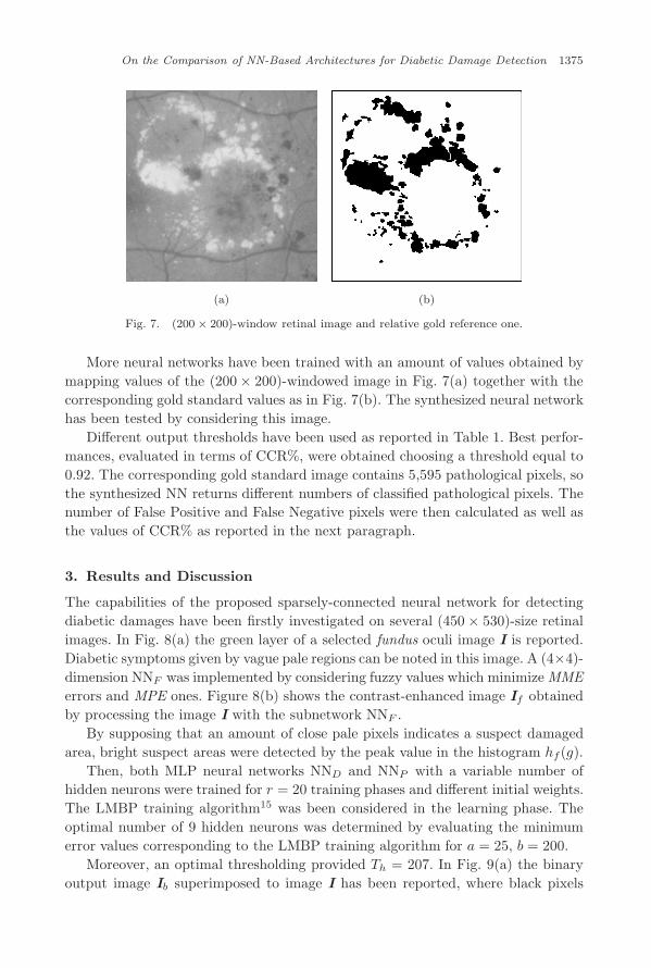
(a) (b)
Fig. 7. (200 × 200)-window retinal image and relative gold reference one.
More neural networks have been trained with an amount of values obtained bymapping values of the (200 × 200)-windowed image in Fig. 7(a) together with thecorresponding gold standard values as in Fig. 7(b). The synthesized neural networkhas been tested by considering this image.
Different output thresholds have been used as reported in Table 1. Best perfor-mances, evaluated in terms of CCR%, were obtained choosing a threshold equal to0.92. The corresponding gold standard image contains 5,595 pathological pixels, sothe synthesized NN returns different numbers of classified pathological pixels. Thenumber of False Positive and False Negative pixels were then calculated as well asthe values of CCR% as reported in the next paragraph.
3. Results and Discussion
The capabilities of the proposed sparsely-connected neural network for detectingdiabetic damages have been firstly investigated on several (450 × 530)-size retinalimages. In Fig. 8(a) the green layer of a selected fundus oculi image I is reported.Diabetic symptoms given by vague pale regions can be noted in this image. A (4×4)-dimension NNF was implemented by considering fuzzy values which minimize MMEerrors and MPE ones. Figure 8(b) shows the contrast-enhanced image If obtainedby processing the image I with the subnetwork NNF .
By supposing that an amount of close pale pixels indicates a suspect damagedarea, bright suspect areas were detected by the peak value in the histogram hf(g).
Then, both MLP neural networks NND and NNP with a variable number ofhidden neurons were trained for r = 20 training phases and different initial weights.The LMBP training algorithm15 was been considered in the learning phase. Theoptimal number of 9 hidden neurons was determined by evaluating the minimumerror values corresponding to the LMBP training algorithm for a = 25, b = 200.
Moreover, an optimal thresholding provided Th = 207. In Fig. 9(a) the binaryoutput image Ib superimposed to image I has been reported, where black pixels

October 20, 2009 16:12 WSPC/123-JCSC 00572
1376 V. Bevilacqua et al.
(a) (b)
Fig. 8. (a) Input fundus image I; (b) Contrast-enhanced image If .
(a) (b)
Fig. 9. (a) Superimposition of Ib to image I; (b) gold standard image.
belong to regions that the system classifies as suspect damaged areas. This imagehas been compared to a gold standard image, provided by expert clinicians andreported in Fig. 9(b). Then, the performances of the proposed neurofuzzy networkhave been evaluated and discussed by determining the values of False NegativesFN and False Positive FP, as defined in Ref. 3. More in detail, the values FP andFN give the number of pixels that the system wrongly classifies as retinal damageswith respect to reference ones or vice-versa.
A percentage measure of exactness is given by the Correct Recognition Rate(CCR), defined as10:
CCR% = 100(
1 − FP + FNTotal Number of image pixels
). (5)
This index gives a percentage measure of correctly classified regions in compar-ison with known results, given by expert clinicians in the gold standard image. Inthis paper pixels are classified as belonging to Suspect areas or Not Suspect ones.

October 20, 2009 16:12 WSPC/123-JCSC 00572
On the Comparison of NN-Based Architectures for Diabetic Damage Detection 1377
Table 1. Comparison of CCR% values computed by the: (a) Method inRef. 3; (b) SCNN; (c) Method in Ref. 7; (d) Proposed EBP MLP network.
A B C D
99.92 98.86 98.55 98.67
The values of FP and FN have been computed by considering a (200×200)-windowcentered on macula in image Ib, due to the fact that macula results to be the mainfundus oculi region from an ophthalmic point of view.
A comparison of the obtained value of CCR% to results obtained by the methodsdescribed in Ref. 3 and in Ref. 7 was carried out and is shown in Table 1.
The SCNN provides CCR% =98.86, being FP =2395 and FN = 317. It can benoted that the computed CCR% improves the value obtained using the methodpresented in Ref. 7. Moreover, it has to be pointed out that a further step ofimage processing is required to utilize the procedure of Case A with respect to theproposed one in Case B. Thus, the method proposed in Ref. 3 gives a better CCR%,but it seems to be more time-consuming. Furthermore, the herein proposed EBPMLP network provides CCR% = 98.67, being FP =731 and FN =2,645 in the caseof threshold Th = 0.92 as shown in Fig. 10.
It can be noticed that performances are quite robust, due to the fact that theydo not change much when varying threshold. It should be pointed out that the valueof FP reduces when Th increases, while maintaining performances approximatelyaround 98%.
The proposed MLP network presents satisfactory values of CCR%, besides hav-ing a very simple algorithmic structure. It is important to notice that performancesin terms of generalization of MLP approach reported in Table 2, are satisfactorybecause they were achieved by using a very small amount of training examples (notgreater than 3%) sampled by the image shown in Fig. 9. Moreover, a very significantvalue of CCR%, very close to 98%, was obtained by submitting the retinal image in
Fig. 10. Windowed retinal image obtained with Th = 0.92 where suspect pathological pixels areblack.

October 20, 2009 16:12 WSPC/123-JCSC 00572
1378 V. Bevilacqua et al.
Table 2. Comparison of CCR% values with different thresholds.
Th FP FN CCR%
0.34 4920 699 97.790.54 3799 905 98.150.61 3460 1032 98.230.70 2922 1195 98.380.89 1148 2368 98.610.92 731 2645 98.67
(a) (b)
Fig. 11. (a) Superposition of a selected retinal image with pathological black areas; (b) Super-position of the selected retinal image with its gold standard.
Fig. 12. Image Iout obtained with the proposed NN considering Th = 0.89 (CCR% = 97.96).
Fig. 11(a) to the MLP network designed by considering only image in Fig. 9. Bothpathological windowed image and its gold standard image are shown in Fig. 11.
In Fig. 12 the resulting image obtained considering a threshold Th = 0.89 isshown. The corresponding value of CCR% is equal to 97.96.

October 20, 2009 16:12 WSPC/123-JCSC 00572
On the Comparison of NN-Based Architectures for Diabetic Damage Detection 1379
4. Conclusions
In this paper, a contribution for improving the detection of diabetic damages hasbeen proposed by comparing two neural architectures. The first proposal concernswith the synthesis of a sparsely-connected neurofuzzy network for retinal imageprocessing. Vague pale regions in fundus images have been firstly highlighted byenhancing image contrast via a neurofuzzy network. Then, an optimal thresholdingwhich can minimize pixel classification errors has been carried out by synthesizing aNN-based MLP sub network with a fast convergence training algorithms. Contrast-enhanced images have been then globally segmented via a proper NN, providingbinary output images, in which suspect damaged areas are isolated. In the secondarchitecture an EBP MLP neural net has been synthesized, where a suitable trainingset of suspect patterns was developed by (5×5)-windows centered on damaged pixelsin gold standard images provided by clinicians.
Performances of both proposed NN-based architectures have been evaluated byconsidering gold standard images provided by clinicians and compared to each otherusing Correct Classification Rates. Results seem to be satisfactory and indicate thepossibility of further improvements in the field of diagnostic support tools.
References
1. J. Evans, C. Rooney and S. Ashgood, Blindness and partial sight in England andWales, April 1990–March 1991, Health Trends 28 (1996) 5–12.
2. W. Hsu, P. M. D. S. Pallawala, M. L. Lee and K. A. Eong, The role of domainknowledge in the detection of retinal hard exudates, IEEE Conf. Computer Visionand Pattern Recognition, Vol. 2, Kauai, HI, USA (2001), pp. 246–251.
3. T. Walter, J.-C. Klein, P. Massin and A. Erginay, A contribution of image processingto the diagnosis of diabetic retinopathy-detection of exudates in colour fundus imagesof the human retina, IEEE Trans. Med. Imaging 21 (2002) 1236.
4. G. G. Gardner, D. Keating, T. H. Williamson and A. T. Elliott, Automatic detectionof diabetic retinopathy using an artificial neural network: A screening tool, British J.Ophthalmol. 80 (1996) 940–944.
5. C. Sinthanayothin, J. F. Boyce, T. H. Williamson, H. L. Cook, E. Mensah, S. Laland D. Usher, Automated detection of diabetic retinopathy on digital fundus images,Diabet. Med. 19 (2002) 105–112.
6. C. Sinthanayothin, V. Kongbunkiat, S. Phoojaruenchanachai and A. Singalavanija,Automated screening system for diabetic retinopathy, IEEE Int. Symp. Image andSignal Processing and Analysis, Rome, Italy (2003), pp. 915–920.
7. D. Kavitha and S. S. Devi, Automatic detection of optic disc and exudates in retinalimages, IEEE Int. Conf. Intelligent Sensing and Information Processing, Chennai,India (2005), pp. 501–506.
8. X. Zhang and O. Chutatape, A SVM approach for detection of hemorrhages in back-ground diabetic retinopathy, Int. J. Conf. Neural Networks, Montreal, Canada (2005),pp. 2435–2440.
9. A. Osareh, M. Mirmehdi, B. Thomas and R. Markham, Automated identification ofdiabetic retinal exudates, in digital colour images, British. J. Ophthalmol. 87 (2003)1220–1223.

October 20, 2009 16:12 WSPC/123-JCSC 00572
1380 V. Bevilacqua et al.
10. C. M. Ennett, M. Frize and E. Charrette, Improvement and automation of artificialneural networks to estimate medical outcomes, Med. Eng. Phys. 26 (2004) 321–328.
11. W. Swiercz, K. J. Cios, K. Staley, L. Kurgan, F. Accurso and S. Sagel, A new synapticplasticity rule for networks of spiking neurons, IEEE Trans. Neural Networks 17(2006) 94.
12. S. Haykin, Neural Networks — A Comprehensive Foundations, 2nd edn. (PrenticeHall, New Jersey, 1999).
13. L. Carnimeo, Diabetic damage detection in retinal images via a sparsely-connectedneurofuzzy network, Lecture Notes Artif. Intell. 5227 (2008) 1175–1182.
14. R. C. Gonzalez and R. E. Woods, Digital Image Processing, 2nd edn. (Prentice Hall,New Jersey, 2002).
15. B. M. Wilamowski, O. Kaynak, S. Iplikci and M. O. Efe, An algorithm for fast con-vergence in training neural networks, IEEE Int. Joint Conf. Neural Networks, Wash-ington, DC (2001), pp. 1778–1782.