Of the multiple mechanisms leading to type 1 diabetes, T cell ...
10
Of the multiple mechanisms leading to type 1 diabetes, T cell receptor revision may play a prominent role (is type 1 diabetes more than a single disease?) D. H. Wagner Department of Medicine, Department of Neurology, Webb-Waring Center, University of Colorado Anschutz Medical Campus, Aurora, CO, USA Accepted for publication 31 May 2016 Correspondence: D. H. Wagner, Department of Medicine, Department of Neurology, Webb-Waring Center, University of Colorado Anschutz Medical Campus, 12850 East Montview Boulevard, Aurora, CO 80045, USA. E-mail: [email protected] Summary A single determinant factor for autoimmunity does not exist; disease development probably involves contributions from genetics, the environment and immune dysfunction. Type 1 diabetes is no exception. Genomewide-associated studies (GWAS) analysis in T1D has proved disappointing in revealing contributors to disease prediction; the only reliable marker has been human leucocyte antigen (HLA). Specific HLAs include DR3/DR4/DQ2/DQ8, for example. Because HLA molecules present antigen to T cells, it is reasonable that certain HLA molecules have a higher affinity to present self-antigen. Recent studies have shown that additional polymorphisms in HLA that are restricted to autoimmune conditions are further contributory. A caveat is that not all individuals with the appropriate ‘pro-autoimmune’ HLA develop an autoimmune disease. Another crucial component is autoaggressive T cells. Finding a biomarker to discriminate autoaggressive T cells has been elusive. However, a subset of CD4 helper cells that express the CD40 receptor have been described as becoming pathogenic. An interesting function of CD40 on T cells is to induce the recombination-activating gene (RAG)1/RAG2 T cell receptor recombination machinery. This observation is contrary to immunology paradigms that changes in TCR molecules cannot take place outside the thymic microenvironment. Alteration in TCR, called TCR revision, not only occurs, but may help to account for the development of autoaggressive T cells. Another interesting facet is that type 1 diabetes (T1D) may be more than a single disease; that is, multiple cellular components contribute uniquely, but result ultimately in the same clinical outcome, T1D. This review considers the process of T cell maturation and how that could favor auto-aggressive T cell development in T1D. The potential contribution of TCR revision to autoimmunity is also considered. Keywords: autoimmune diabetes, immune dysfunction, TCR revision Introduction Type 1 diabetes (T1D), unlike its far more prevalent counter- part, type 2 diabetes, is a classic autoimmune disease charac- terized by immune cell attacks of pancreatic islets resulting in hyperglycaemia subsequent to insulin loss. Islets are com- posed of five cell types that produce hormones and enzymes associated with digestion and nutrient uptake. Beta cells constitute approximately 80% of islets and produce insulin; alpha cells produce glucagon, delta cells produce somatostatin-D, gamma cells produce pancreatic polypeptide and epsilon cells produce ghrelin, the hunger hormone [1]. During diabetogenesis immune cells infiltrate islets creating an inflammatory condition called insulitis. This was first described more than 100 years ago in a 6-year-old girl who died from complications of ketoacidosis [2]. The first cells to infiltrate islets are probably neutrophils [3], followed by antigen-presenting cells (APC), including macrophages and dendritic cells (DC) [4,5]. In fact, merocytic DC that mediate cross-presentation of islet antigens to CD8 1 and CD4 1 cells in pancreatic lymph nodes could be instrumental in initiat- ing disease [6,7]. Eventually, lymphocytes including CD4 1 and CD8 1 , as well as B cells, infiltrate the islets and are V C 2016 British Society for Immunology, Clinical and Experimental Immunology, 185: 271–280 271 doi:10.1111/cei.12819 TYPE-1 DIABETES MINI-SERIES: PATHOGENESIS AND THERAPY Clinical and Experimental Immunology REVIEW ARTICLE Downloaded from https://academic.oup.com/cei/article/185/3/271/6412103 by guest on 16 March 2022
-
Upload
khangminh22 -
Category
Documents
-
view
1 -
download
0
Transcript of Of the multiple mechanisms leading to type 1 diabetes, T cell ...
Of the multiple mechanisms leading to type 1 diabetes, T cell receptor
revision may play a prominent role (is type 1 diabetes more than a
single disease?)
D. H. WagnerDepartment of Medicine, Department of
Neurology, Webb-Waring Center, University of
Colorado Anschutz Medical Campus, Aurora,
CO, USA
Accepted for publication 31 May 2016
Correspondence: D. H. Wagner, Department
of Medicine, Department of Neurology,
Webb-Waring Center, University of Colorado
Anschutz Medical Campus, 12850 East
Montview Boulevard, Aurora, CO 80045,
USA.
E-mail: [email protected]
Summary
A single determinant factor for autoimmunity does not exist; disease
development probably involves contributions from genetics, the
environment and immune dysfunction. Type 1 diabetes is no exception.
Genomewide-associated studies (GWAS) analysis in T1D has proved
disappointing in revealing contributors to disease prediction; the only
reliable marker has been human leucocyte antigen (HLA). Specific HLAs
include DR3/DR4/DQ2/DQ8, for example. Because HLA molecules present
antigen to T cells, it is reasonable that certain HLA molecules have a higher
affinity to present self-antigen. Recent studies have shown that additional
polymorphisms in HLA that are restricted to autoimmune conditions are
further contributory. A caveat is that not all individuals with the
appropriate ‘pro-autoimmune’ HLA develop an autoimmune disease.
Another crucial component is autoaggressive T cells. Finding a biomarker to
discriminate autoaggressive T cells has been elusive. However, a subset of
CD4 helper cells that express the CD40 receptor have been described as
becoming pathogenic. An interesting function of CD40 on T cells is to
induce the recombination-activating gene (RAG)1/RAG2 T cell receptor
recombination machinery. This observation is contrary to immunology
paradigms that changes in TCR molecules cannot take place outside the
thymic microenvironment. Alteration in TCR, called TCR revision, not only
occurs, but may help to account for the development of autoaggressive T
cells. Another interesting facet is that type 1 diabetes (T1D) may be more
than a single disease; that is, multiple cellular components contribute
uniquely, but result ultimately in the same clinical outcome, T1D. This
review considers the process of T cell maturation and how that could favor
auto-aggressive T cell development in T1D. The potential contribution of
TCR revision to autoimmunity is also considered.
Keywords: autoimmune diabetes, immune dysfunction, TCR revision
Introduction
Type 1 diabetes (T1D), unlike its far more prevalent counter-
part, type 2 diabetes, is a classic autoimmune disease charac-
terized by immune cell attacks of pancreatic islets resulting
in hyperglycaemia subsequent to insulin loss. Islets are com-
posed of five cell types that produce hormones and enzymes
associated with digestion and nutrient uptake. Beta cells
constitute approximately 80% of islets and produce
insulin; alpha cells produce glucagon, delta cells produce
somatostatin-D, gamma cells produce pancreatic polypeptide
and epsilon cells produce ghrelin, the hunger hormone [1].
During diabetogenesis immune cells infiltrate islets creating
an inflammatory condition called insulitis. This was first
described more than 100 years ago in a 6-year-old girl who
died from complications of ketoacidosis [2]. The first cells to
infiltrate islets are probably neutrophils [3], followed by
antigen-presenting cells (APC), including macrophages and
dendritic cells (DC) [4,5]. In fact, merocytic DC that mediate
cross-presentation of islet antigens to CD81 and CD41 cells
in pancreatic lymph nodes could be instrumental in initiat-
ing disease [6,7]. Eventually, lymphocytes including CD41
and CD81, as well as B cells, infiltrate the islets and are
VC 2016 British Society for Immunology, Clinical and Experimental Immunology, 185: 271–280 271
doi:10.1111/cei.12819 TYPE-1 DIABETES MINI-SERIES: PATHOGENESIS AND THERAPY
Clinical and Experimental Immunology REVIEW ARTICLE
Dow
nloaded from https://academ
ic.oup.com/cei/article/185/3/271/6412103 by guest on 16 M
arch 2022
presumed to be the primary players in loss of insulin. The
cellular phenotypes in animal models have been determined,
and some of the cellular phenotypes in human islets are
known. However, the aetiology of T1D remains a mystery:
why do cells traffic to the islets? Why do cells infiltrate the
islets? By what mechanism do these invaders cause loss of
insulin?
Development of type 1 diabetes (T1D) requires a series
of unfortunate events involving alignment of genetic, envi-
ronmental and immunological contributors. This argu-
ment, in fact, can be made for any autoimmune disease.
Developmental parameters for all autoimmune diseases are
similar in many ways, although the symptoms and clinical
outcomes vary dramatically. It has become clear that no
individual contributor can cause diabetes, but each con-
tributor acting in concert establishes danger and ultimately
causes disease. Interestingly, rates vary greatly, depending
upon geography. For example, incidence is high in Finland
and Sardinia at approximately one in 250 [8]; in the United
States the incidence is currently one in 300 by 18 years of
age [9]; and in Canada, Australia and New Zealand the
incidence is much lower, at one per 1750. In China and
Venezuela the incidence is approximately one in 100 000. A
2012 study of T1D trends in Europe during a 20-year
period ending in 2008 reported that 22 of 23 centres in 19
countries showed significant incidence increases, specifi-
cally reporting a 3�4% annual increase over the entire
period [10]. Another study reports that incidence in
Europe saw a yearly increase rate of 2�8% from 1990 to
1998, increasing to 3�2% and then 3�9% in 2010 [11]. The
increases are being reported in the very young and those
with moderate genetic susceptibility [11]. More and more
environmental factors are believed to be contributory, but
major determinants have yet to be defined.
Genetics
Surprisingly, while type 2 diabetes is reaching epidemic lev-
els in the United States and other developed countries,
T1D incidence is increasing similarly. Epidemiological pat-
terns indicate that the worldwide incidence has increased
by 2–5% during the last 20 years [9]. T1D occurs in familial
clusters, yet examination of monozygotic twins shows, sur-
prisingly, that concordance is less than 40% [12]. The
mouse model of T1D, the non-obese diabetic or NOD
mouse, develops intrinsically the same disease characteris-
tics as human T1D, including lymphocytic infiltrates in
islets, loss of insulin secretion and hyperglycaemia. NOD
colonies are bred to maintain high genetic susceptibility
loci multi-generationally to ensure disease, yet within any
given NOD colony disease incidence ranges from 50 to
90% in females and 20 to 50% in males [13–15]. Male and
female NOD mice are genetically identical, thus gender
bias, perhaps hormonal-driven, would appear to contribute
to disease. Studies show that early-life microbial exposures
determine sex hormone levels that modify progression to
autoimmunity in NOD mice [16,17]. Early studies in NOD
mice indicate that hormonal imprinting in neonates had
an influence on diabetes [18]. The situation for human
disease is different; gender bias does not occur in human
disease, and female to male disease ratios are virtually 1 : 1
[19,20].
GWAS studies
A concerted effort to address genetic contributions to T1D
has been attempted using genomewide analytical studies
(GWAS). In T1D, greater than 40 susceptibility loci have
been identified thus far [21]. The most significant indicator
is the human leucocyte antigen (HLA). HLA haplotypes
vary within diseases such that a specific HLA haplotype
associates with a particular autoimmune disease. Many of
the associated haplotypes are class II; HLA DRB1*1501
(referred to as DR2 or DR15) and DQB1*0601 (DQ6) asso-
ciate with multiple sclerosis [22], DRB1*0401 (DR4) and
DRB1*0101 (DR1) or DQB1*0302 (DQ8) associate with
rheumatoid arthritis [23,24], etc. The T1D-associated mol-
ecules include DRB1*0301 (DR3), DR4, DQB1*0201
(DQ2) and DQ8 [25]. Class I alleles in addition to, and
independently of, class II alleles associate with T1D. For
example A*2402, A*0201, B*1801 and C*0501 were consid-
ered predisposing; and A*1101, A*3201, A*6601, B*0702,
B*4403, B*3502, C*1601 and C*0401 appear to be protec-
tive [26]. An intriguing observation was that while DQ2
and DQ8 are linked independently to T1D development,
when subjects carry both DQ2 and DQ8 alleles the disease
incidence increases drastically [25]. Further study demon-
strates that a transposition of the DQ2 a chain to associate
with the DQ8 b chain occurs, creating a unique HLA-DQ
referred to as trans-DQ8 [25]. Thus autoimmune condi-
tions reflect the presence of unique polymorphisms in
MHC genes that further promote disease.
GWAS has generated numerous other indicators includ-
ing, but not limited to, interleukin (IL)-2R, cytotoxic T
lymphocyte antigen (CTLA)-4, IL-27, IL-2, IL-10, signal
transducer and activator of transcription (STAT)-4, C-C
chemokine receptor type 5 (CCR5) and the lymphocyte
marker, CD69 [27]. Typically, CD69 has been identified as
a lymphocyte ‘activation’ marker, but recent studies show
that CD69 plays an important decisional role in lymph
node migration, cell retention and memory formation
[28]. It was determined that CD69 and sphingosine-1-
phosphate receptor 1 (S1PR) expressions are intertwined
inversely. S1PR expression is required for lymphocyte
egress from secondary lymphoid tissues and thymus [29],
and CD69 expression increases as S1PR expression
decreases [28,29]. GWAS has been performed on a number
of autoimmune diseases in addition to T1D, including
lupus, multiple sclerosis, rheumatoid arthritis, coeliac dis-
ease and Crohn’s colitis [30]. After all these studies, thus
D. H. Wagner et al.
TYPE-1 DIABETES MINI-SERIES: PATHOGENESIS AND THERAPY
272 VC 2016 British Society for Immunology, Clinical and Experimental Immunology, 185: 271–280
Dow
nloaded from https://academ
ic.oup.com/cei/article/185/3/271/6412103 by guest on 16 M
arch 2022
far the only discriminating factor for disease prediction is
the HLA haplotype, which is not absolute. The hope was
that GWAS would discover a single gene or gene cluster
that would predict likelihood to develop T1D in much the
way that BRCA has done for breast cancer. This has not
happened. Given that no clearly predictive genes have yet
been discovered, one review summarized the question of
‘Have GWASs been a failure?’ with a qualified ‘yes’ [31].
The best conclusion from the information provided by
GWAS is that autoimmune diseases require immune
dysfunction.
Immune contribution
The reason that HLA is linked with autoimmunity is prob-
ably because of its function, the presentation of antigen(s)
to T cells. As such, HLA molecules dictate immune
response outcomes. HLA class I (HLA-A, B and C) mole-
cules are expressed on almost all cell types while HLA class
II molecules are restricted to what are known as professio-
nal APC, including B cells, macrophages and DCs [32], and
in humans, activated T cells express HLA class II [33].
Once professional APCs encounter exogenous antigens,
classically a non-viral antigen, the antigen is taken up via
phagocytosis involving membrane engulfment or by recep-
tor mediated uptake; for example, a B cell receptor binding
an antigen and the receptor/antigen becoming internalized
through membrane invagination. Once internalized the
antigen associates with an endosome to be broken down to
constituent parts, peptide fragments, nucleotide fragments
and lipids, etc. Peptides associate chemically with HLA to
be transported to the cell surface for external presentation.
HLA molecules are polymorphic, and those individuals
unfortunate enough to express HLA haplotypes that can
present self-antigens preferentially are clearly much more
at risk of developing disease. In addition it is possible that
the autoimmune background generates unique
autoimmune-favouring HLA haplotypes, such as trans-
DQ8 in T1D [25,34]. The physics of antigen presentation
leading to a T cell activation event that leads further to
inflammation requires that the antigen/HLA association be
sufficient to stimulate appropriate T cells. Thus, appropri-
ate T cells carrying T cell receptor (TCR) molecules that
can respond to self-antigens must be available, adding the
next ingredient in autoimmunity. Another layer of com-
plexity that could contribute to autoimmunity is post-
translational modification (PTM) of proteins that may
generate novel self-antigens. As much as 90% of proteins
produced by mammals undergo some form of post-
translational modification [35]. Modifications include gly-
cosylation, phosphorylation, citrullination, acetylation,
peroxidation and deamination, etc. [35]. Each of these
processes occur in the periphery, thus creating an environ-
ment for generation of novel antigens; in other words, pro-
teins and peptides that have not been seen in the thymus
by developing T cells. Further complicating this mecha-
nism, it is also possible that autoimmune backgrounds
have altered PTM mechanisms compared to non-
autoimmune backgrounds. For example, protein glycosyla-
tion patterns were different when the protein was isolated
from an autoimmune background than when the same
protein was examined from a non-autoimmune back-
ground [36,37]. A recently described mechanism for crea-
tion of novel epitopes is the hybrid insulin peptides (HIP)
model, which is acutely appropriate for T1D. Insulin pep-
tides have been considered potential self-antigens in T1D
for several years [38]. The HIP model suggests that during
type 1 diabetes insulin peptides hybridize with other pro-
teins, creating a unique set of epitopes that may be capable
of activating autoaggressive T cells [39].
Generation of rogue T cells and central tolerance
The immunological rules of inflammation are identical for
foreign antigen removal response to self-antigen-driven
autoaggressive responses: T cells carrying a TCR that have
affinity for antigen(s) recognize and react to the presented
antigen, leading to production and release of proinflamma-
tory cytokines that establish inflammation. The process by
which T cells are generated creates the possibility for devel-
opment of autoaggression. Pluripotent, immature lympho-
cytes (designated as double-negative, DN) migrate to the
thymus from the bone marrow. Developing T cells in the
thymus acquire TCR molecules after recombination-
activating gene (RAG)1 and RAG2 recombination proteins
become induced during the DN stage of development [40],
and this action takes place physically in the cortical region
(Fig. 1). The architecture of the thymus is similar to that of
secondary lymphoid organs, including lymph nodes, com-
posed of a cortical and a medullary region. Once RAGs are
induced, the TCR-b gene is rearranged and the resultant
molecule is expressed, associating with a prefabricated a
chain. This constitutes the pre-TCR [41]. By as-yet
unknown mechanisms, RAG1 and RAG2 proteins are
induced once again and the a locus is rearranged, remov-
ing the delta gene locus [42]. The newly rearranged a gene
is transcribed, translated and the protein derived displaces
the pre-a protein and associates with the b protein to cre-
ate a potentially functional TCR (ab). This process hap-
pens at least somewhat randomly within each developing T
cell, therefore any given T cell is as likely to express a self-
antigen reactive TCR as it is likely to express a foreign-
antigen reactive TCR. At this developmental stage cells
have become double-positive (DP) for expression of CD4
and CD8 and remain in the cortical region (Fig. 1). The
newly expressed TCR molecules on DP cells interact with
major histocompatibility complex (MHC) molecules,
including HLA-A, B and C, class I molecules, or HLA-D,
class II molecules that are located at the cortico–medullary
junction (Fig. 1), to undergo positive selection. Cells that
TYPE-1 DIABETES MINI-SERIES: PATHOGENESIS AND THERAPY
Genetics, environment and immune dysfunction contribute to T1D
VC 2016 British Society for Immunology, Clinical and Experimental Immunology, 185: 271–280 273
Dow
nloaded from https://academ
ic.oup.com/cei/article/185/3/271/6412103 by guest on 16 M
arch 2022
interact successfully are primed for survival; cells that can-
not interact with the available HLA molecules undergo
death-by-neglect (Fig. 1). Positive selection assures that
only T cells that are responsive to the available HLA are
maintained [43,44].
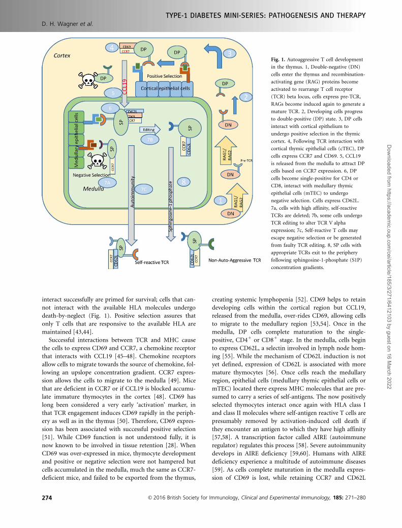
Successful interactions between TCR and MHC cause
the cells to express CD69 and CCR7, a chemokine receptor
that interacts with CCL19 [45–48]. Chemokine receptors
allow cells to migrate towards the source of chemokine, fol-
lowing an upslope concentration gradient. CCR7 expres-
sion allows the cells to migrate to the medulla [49]. Mice
that are deficient in CCR7 or if CCL19 is blocked accumu-
late immature thymocytes in the cortex [48]. CD69 has
long been considered a very early ‘activation’ marker, in
that TCR engagement induces CD69 rapidly in the periph-
ery as well as in the thymus [50]. Therefore, CD69 expres-
sion has been associated with successful positive selection
[51]. While CD69 function is not understood fully, it is
now known to be involved in tissue retention [28]. When
CD69 was over-expressed in mice, thymocyte development
and positive or negative selection were not hampered but
cells accumulated in the medulla, much the same as CCR7-
deficient mice, and failed to be exported from the thymus,
creating systemic lymphopenia [52]. CD69 helps to retain
developing cells within the cortical region but CCL19,
released from the medulla, over-rides CD69, allowing cells
to migrate to the medullary region [53,54]. Once in the
medulla, DP cells complete maturation to the single-
positive, CD41 or CD81 stage. In the medulla, cells begin
to express CD62L, a selectin involved in lymph node hom-
ing [55]. While the mechanism of CD62L induction is not
yet defined, expression of CD62L is associated with more
mature thymocytes [56]. Once cells reach the medullary
region, epithelial cells (medullary thymic epithelial cells or
mTEC) located there express MHC molecules that are pre-
sumed to carry a series of self-antigens. The now positively
selected thymocytes interact once again with HLA class I
and class II molecules where self-antigen reactive T cells are
presumably removed by activation-induced cell death if
they encounter an antigen to which they have high affinity
[57,58]. A transcription factor called AIRE (autoimmune
regulator) regulates this process [58]. Severe autoimmunity
develops in AIRE deficiency [59,60]. Humans with AIRE
deficiency experience a multitude of autoimmune diseases
[59]. As cells complete maturation in the medulla expres-
sion of CD69 is lost, while retaining CCR7 and CD62L
Fig. 1. Autoaggressive T cell development
in the thymus. 1, Double-negative (DN)
cells enter the thymus and recombination-
activating gene (RAG) proteins become
activated to rearrange T cell receptor
(TCR) beta locus, cells express pre-TCR,
RAGs become induced again to generate a
mature TCR. 2, Developing cells progress
to double-positive (DP) state. 3, DP cells
interact with cortical epithelium to
undergo positive selection in the thymic
cortex. 4, Following TCR interaction with
cortical thymic epithelial cells (cTEC), DP
cells express CCR7 and CD69. 5, CCL19
is released from the medulla to attract DP
cells based on CCR7 expression. 6, DP
cells become single-positive for CD4 or
CD8, interact with medullary thymic
epithelial cells (mTEC) to undergo
negative selection. Cells express CD62L.
7a, cells with high affinity, self-reactive
TCRs are deleted; 7b, some cells undergo
TCR editing to alter TCR V alpha
expression; 7c, Self-reactive T cells may
escape negative selection or be generated
from faulty TCR editing. 8, SP cells with
appropriate TCRs exit to the periphery
following sphingosine-1-phosphate (S1P)
concentration gradients.
D. H. Wagner et al.
TYPE-1 DIABETES MINI-SERIES: PATHOGENESIS AND THERAPY
274 VC 2016 British Society for Immunology, Clinical and Experimental Immunology, 185: 271–280
Dow
nloaded from https://academ
ic.oup.com/cei/article/185/3/271/6412103 by guest on 16 M
arch 2022
expression. These phenotypically mature cells can migrate
to populate lymph nodes through CCL19 and other signals,
and be retained in the node through CD62L interactions.
During the process of negative selection some thymo-
cytes destined for failure have an opportunity for redemp-
tion, utilizing a process called TCR editing [61–66]. Rather
than a single attempt at TCR generation, which was the
prevailing paradigm for many years, self-reactive thymo-
cytes can be induced to re-express the RAG proteins, which
will once again rearrange TCR genes [64]. Again, the mech-
anism of RAG induction in the thymus is unknown. Recep-
tor editing was first described in B cells, taking place
during development in bone marrow [67]. TCR editing
helps to account for repertoire expansion in the thymus
and argues in favour of cell conservation, thus reducing the
need for continual migration of bone marrow cells. The
particulars of negative selection have not yet been
explained fully. How are cells able to interact positively
with MHC to undergo positive selection and then the same
TCR again interacts with MHC to undergo negative selec-
tion? One explanation is that positional kinetics, i.e. posi-
tive selection, occurring in the cortex while negative
selection occurs in the medulla, create differential signals to
the developing T cell, although this hypothesis has not
been proven. Another viable option is that different affinity
or avidity thresholds between TCR molecules on develop-
ing T cells and HLA/MHC promote positive versus negative
selection. Autoimmunity can certainly create permissive
conditions allowing autoaggressive T cells to escape thymic
negative selection known as central tolerance, which has
been proposed [68,69]. One concern about central toler-
ance failure being the only means of generating autoaggres-
sion is that the vast majority of thymic output occurs early
in life. In mammals, thymic involution, loss of thymic
architecture and volume, occurs at or close to puberty. If
autoaggressive T cells arise solely by escaping negative
selection, then logically autoimmune disease would only
onset prior to or soon after puberty. This, however, is not
always the case.
In T1D the majority of disease onsets occur in juvenile
subjects; however, an ever-growing population is experi-
encing onset during the 3rd, 4th, 5th and even 6th decade
of life [70]. To account for this, either peripheral mecha-
nisms of tolerance are in place that become dysfunctional
over time, or an alternative mechanism of autoaggressive T
cell development occurs. Another intriguing option is that
early in life diabetes onset constitutes one type of disease,
perhaps associated closely with central tolerance failure,
while disease onset later in life constitutes a different type
of disease. The latter case would involve mechanisms to
develop autoaggressive T cells independently of thymic
control. It has been presumed that TCR editing is the final
point in TCR development. To the contrary, we and others
demonstrated that T cells are capable of inducing RAG1
and RAG2 proteins in the periphery and, subsequent to
that, alter TCR expression [71–86]. This process is known
as TCR revision. While it has not yet been determined
what induces RAGs in the thymus, the mechanisms of revi-
sion are the same as those for editing; the locale of the T
cell, in the periphery as opposed to the thymus, has dic-
tated the name change.
TCR revision
It has been shown that a subset of T cells, both in the thy-
mus and in the periphery, express the CD40 molecule
[36,37,71–73,87–98]. This was somewhat surprising, given
that CD40 expression has long been associated with only
APC. However, more extensive research demonstrated that
CD40 expression is ubiquitous, being expressed on all iden-
tified APC, on neural cells including microglia, on adipo-
cytes, on endothelial cells and on T cells, including CD41
and CD81 cells [36,37,71–73,80,88–94,99–101]. CD40-
expressing CD4 cells are referred to as Th40 cells, and have
been shown to become highly pathogenic in autoimmune
disease models [36,37,71–73,87–95,99]. Among its func-
tions, CD40 acts as a co-stimulus on T cells
[37,87,88,91–94,99]. This indicates that alternative, and
heretofore under-considered, co-stimulatory molecules
occur on T cells. Identifying these molecules could reshape
the understanding of T cell biology significantly.
An intriguing and surprising discovery was that CD40
engagement on Th40 cells induced the RAG1/RAG2 TCR
recombination machinery [71,73]. This was the first ever
demonstration in a primary T cell of a mechanism to
induce RAG proteins. RAG1 and RAG2 form heterodimers
that interact further with Ku proteins, DNA polymerases
and helicases, etc., leading to alteration of TCR expres-
sion [71–73,91,102,103]. In the periphery, at least, CD40
interacts directly with the RAG(s) complex in the
nucleus [71]. Following induction of RAGs, altered
expression of TCR-a [73,104] and TCR-b [83,84,105] mol-
ecules on long-standing peripheral T cells occurs
[71,74–83,85,102,106–119]. A central paradigm of immu-
nology holds that once T cells exit the thymus TCR mole-
cules do not undergo alteration. To the contrary, several
laboratories have shown that peripheral T cells re-express
RAG1 and RAG2 proteins and subsequently alter TCR
expression [81,83,85,109,120,121]. Importantly, revised T
cells are distinct from recent thymic emigrants [81]. There
is an evolutionary advantage to TCR revision, which is to
generate an expansive T cell repertoire able to respond to a
universe of foreign antigens (Fig. 2). Viruses are extremely
numerous, exhibiting a survival advantage by being highly
mutable. Success of any organism, in this case mammals, at
controlling viral infection will rely upon a highly adaptive
immune system. The estimated number of individual TCR
molecules achievable through RAG-mediated recombina-
tion is vast, approximately 1013 distinct molecules [122].
Maintaining and storing such a number of T cells would
TYPE-1 DIABETES MINI-SERIES: PATHOGENESIS AND THERAPY
Genetics, environment and immune dysfunction contribute to T1D
VC 2016 British Society for Immunology, Clinical and Experimental Immunology, 185: 271–280 275
Dow
nloaded from https://academ
ic.oup.com/cei/article/185/3/271/6412103 by guest on 16 M
arch 2022
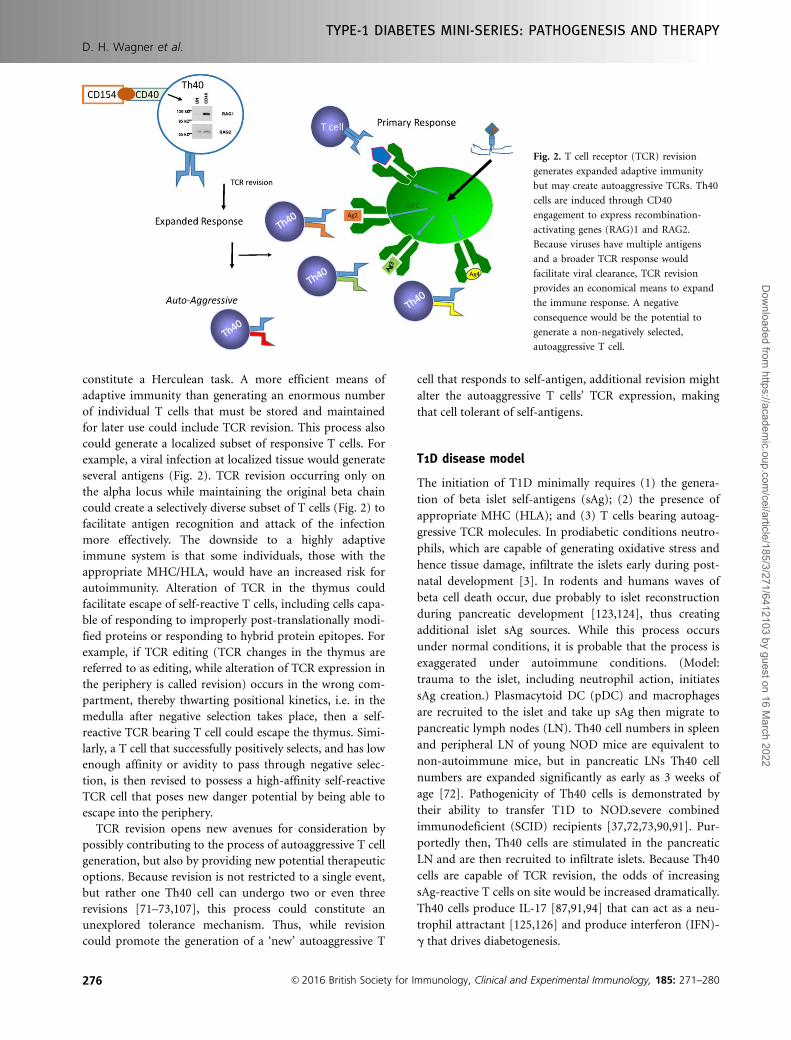
constitute a Herculean task. A more efficient means of
adaptive immunity than generating an enormous number
of individual T cells that must be stored and maintained
for later use could include TCR revision. This process also
could generate a localized subset of responsive T cells. For
example, a viral infection at localized tissue would generate
several antigens (Fig. 2). TCR revision occurring only on
the alpha locus while maintaining the original beta chain
could create a selectively diverse subset of T cells (Fig. 2) to
facilitate antigen recognition and attack of the infection
more effectively. The downside to a highly adaptive
immune system is that some individuals, those with the
appropriate MHC/HLA, would have an increased risk for
autoimmunity. Alteration of TCR in the thymus could
facilitate escape of self-reactive T cells, including cells capa-
ble of responding to improperly post-translationally modi-
fied proteins or responding to hybrid protein epitopes. For
example, if TCR editing (TCR changes in the thymus are
referred to as editing, while alteration of TCR expression in
the periphery is called revision) occurs in the wrong com-
partment, thereby thwarting positional kinetics, i.e. in the
medulla after negative selection takes place, then a self-
reactive TCR bearing T cell could escape the thymus. Simi-
larly, a T cell that successfully positively selects, and has low
enough affinity or avidity to pass through negative selec-
tion, is then revised to possess a high-affinity self-reactive
TCR cell that poses new danger potential by being able to
escape into the periphery.
TCR revision opens new avenues for consideration by
possibly contributing to the process of autoaggressive T cell
generation, but also by providing new potential therapeutic
options. Because revision is not restricted to a single event,
but rather one Th40 cell can undergo two or even three
revisions [71–73,107], this process could constitute an
unexplored tolerance mechanism. Thus, while revision
could promote the generation of a ‘new’ autoaggressive T
cell that responds to self-antigen, additional revision might
alter the autoaggressive T cells’ TCR expression, making
that cell tolerant of self-antigens.
T1D disease model
The initiation of T1D minimally requires (1) the genera-
tion of beta islet self-antigens (sAg); (2) the presence of
appropriate MHC (HLA); and (3) T cells bearing autoag-
gressive TCR molecules. In prodiabetic conditions neutro-
phils, which are capable of generating oxidative stress and
hence tissue damage, infiltrate the islets early during post-
natal development [3]. In rodents and humans waves of
beta cell death occur, due probably to islet reconstruction
during pancreatic development [123,124], thus creating
additional islet sAg sources. While this process occurs
under normal conditions, it is probable that the process is
exaggerated under autoimmune conditions. (Model:
trauma to the islet, including neutrophil action, initiates
sAg creation.) Plasmacytoid DC (pDC) and macrophages
are recruited to the islet and take up sAg then migrate to
pancreatic lymph nodes (LN). Th40 cell numbers in spleen
and peripheral LN of young NOD mice are equivalent to
non-autoimmune mice, but in pancreatic LNs Th40 cell
numbers are expanded significantly as early as 3 weeks of
age [72]. Pathogenicity of Th40 cells is demonstrated by
their ability to transfer T1D to NOD.severe combined
immunodeficient (SCID) recipients [37,72,73,90,91]. Pur-
portedly then, Th40 cells are stimulated in the pancreatic
LN and are then recruited to infiltrate islets. Because Th40
cells are capable of TCR revision, the odds of increasing
sAg-reactive T cells on site would be increased dramatically.
Th40 cells produce IL-17 [87,91,94] that can act as a neu-
trophil attractant [125,126] and produce interferon (IFN)-
g that drives diabetogenesis.
Fig. 2. T cell receptor (TCR) revision
generates expanded adaptive immunity
but may create autoaggressive TCRs. Th40
cells are induced through CD40
engagement to express recombination-
activating genes (RAG)1 and RAG2.
Because viruses have multiple antigens
and a broader TCR response would
facilitate viral clearance, TCR revision
provides an economical means to expand
the immune response. A negative
consequence would be the potential to
generate a non-negatively selected,
autoaggressive T cell.
D. H. Wagner et al.
TYPE-1 DIABETES MINI-SERIES: PATHOGENESIS AND THERAPY
276 VC 2016 British Society for Immunology, Clinical and Experimental Immunology, 185: 271–280
Dow
nloaded from https://academ
ic.oup.com/cei/article/185/3/271/6412103 by guest on 16 M
arch 2022
An issue in T1D is why is disease onset so disparate?
Some individuals experience onset as young children or
juveniles, while others do not experience onset until adult-
hood. Potential explanations are that all conditions for dis-
ease onset, including pre-existing autoaggressive T cells, are
present in juveniles, but not in adults. We hypothesize that
certain T cells, specifically Th40, alter TCR usage over time
to become autoaggressive (TCR revision). While the condi-
tions to create such cells would necessarily be determined
genetically, in some individuals those T cells either do not
yet exist or are regulated successfully through tolerance
mechanisms that eventually fail. A central paradigm of tol-
erance is the control of the CD40–CD154 dyad [127,128].
Multiple studies demonstrate that controlling CD40-
mediated signals is tolerogenic, including in T1D
[72,90,129]. Consequently, disruption of the CD40/CD154
balance becomes pathogenic. Induced increases in CD40-
bearing cells, specifically Th40 cells, would necessarily pro-
mote pathogenesis. When the balance of CD40 was broken
by adoptive transfer of high numbers of Th40 cells, diabetes
developed [72,73,95,97]. Such increases could be mediated
by any of a number of infectious agents. A major element
to explain why disease onset is so varied may focus upon
the generation of autoaggressive T cells. Even with all the
other disease-specific criteria, e.g. sufficient sAg, appropri-
ate HLA for presenting antigen, etc., until autoaggressive T
cells develop in sufficient numbers or are able to break tol-
erance, disease will not onset. If CD40 were engaged pur-
posefully on autoaggressive T cells, at the proper time it
may be possible to forestall T1D onset by altering autoag-
gressive TCR expression.
Acknowledgements
The author declares grant support from the following: the
American Diabetes Association and NIDDK.
Disclosure
D. H. W. is Chief Scientific Officer for Op-T-Mune, Inc.,
Denver, CO, USA and owns stock in the company Op-T-
Mune, Inc., Denver, CO. The current value of the company
is less than US$10 000. D. H. W. has received funding for
research but only from Federal and State Agencies, not
from private companies.
References
1 Elayat AA, el-Naggar MM, Tahir M. An immunocytochemical
and morphometric study of the rat pancreatic islets. J Anat
1995; 186:629–37.
2 In’t Veld P. Insulitis in human type 1 diabetes: the quest for an
elusive lesion. Islets 2011; 3:131–8.
3 Diana J et al. Crosstalk between neutrophils, B-1a cells and
plasmacytoid dendritic cells initiates autoimmune diabetes. Nat
Med 2013; 19:65–73.
4 Hanenberg H et al. Macrophage infiltration precedes and is a
prerequisite for lymphocytic insulitis in pancreatic islets of
pre-diabetic BB rats. Diabetologia 1989; 32:126–34.
5 Uno S et al. Macrophages and dendritic cells infiltrating islets
with or without beta cells produce tumour necrosis factor-
alpha in patients with recent-onset type 1 diabetes. Diabetolo-
gia 2007; 50:596–601.
6 Katz JD, Janssen EM. Breaking T cell tolerance to beta cell
antigens by merocytic dendritic cells. Cell Mol Life Sci 2011;
68:2873–83.
7 Katz JD et al. Cutting edge: merocytic dendritic cells break T
cell tolerance to beta cell antigens in nonobese diabetic mouse
diabetes. J Immunol 2010; 185:1999–2003.
8 Borchers AT, Uibo R, Gershwin ME. The geoepidemiology of
type 1 diabetes. Autoimmun Rev 2010; 9:A355–65.
9 Maahs D et al. Epidemiology of type 1 diabetes. Endocrinol
Metab Clin North Am 2010; 39:481–97.
10 Patterson CC et al. Trends in childhood type 1 diabetes inci-
dence in Europe during 1989-2008: evidence of non-
uniformity over time in rates of increase. Diabetologia 2012;
55:2142–7.
11 Vehik K, Dabelea D. The changing epidemiology of type 1 dia-
betes: why is it going through the roof? Diabetes Metab Res
Rev 2011; 27:3–13.
12 Knip M et al. Environmental triggers and determinants of type
1 diabetes. Diabetes 2005; 54 Suppl 2:S125–36.
13 Grattan M et al. Congenic mapping of the diabetogenic locus
Idd4 to a 5.2-cM region of chromosome 11 in NOD mice:
identification of two potential candidate subloci. Diabetes
2002; 51:215–23.
14 Casteels KM et al. Sex difference in resistance to
dexamethasone-induced apoptosis in NOD mice: treatment
with 1,25(OH)2D3 restores defect. Diabetes 1998; 47:1033–7.
15 Mathieu C et al. Prevention of autoimmune diabetes in NOD
mice by 1,25 dihydroxyvitamin D3. Diabetologia 1994; 37:552–
8.
16 Markle JG et al. Microbiome manipulation modifies sex-
specific risk for autoimmunity. Gut Microbes 2014; 5:485–93.
17 Markle JG et al. Sex differences in the gut microbiome drive
hormone-dependent regulation of autoimmunity. Science
2013; 339:1084–8.
18 Hawkins T, Gala RR, Dunbar JC. The effect of neonatal sex
hormone manipulation on the incidence of diabetes in non-
obese diabetic mice. Proc Soc Exp Biol Med 1993; 202:201–5.
19 Harjutsalo V, Forsblom C, Groop PH. Time trends in mortal-
ity in patients with type 1 diabetes: nationwide population
based cohort study. BMJ 2011; 343:d5364.
20 Harjutsalo V, Sjoberg L, Tuomilehto J. Time trends in the inci-
dence of type 1 diabetes in Finnish children: a cohort study.
Lancet 2008; 371:1777–82.
21 Bergholdt R et al. Identification of novel type 1 diabetes candi-
date genes by integrating genome-wide association data,
protein-protein interactions, and human pancreatic islet gene
expression. Diabetes 2012; 61:954–62.
22 Hillert J, Olerup O. Multiple sclerosis is associated with genes
within or close to the HLA-DR-DQ subregion on a normal
DR15,DQ6,Dw2 haplotype. Neurology 1993; 43:163–8.
23 Angelini G et al. Analysis of HLA DP, DQ, and DR alleles in
adult Italian rheumatoid arthritis patients. Hum Immunol
1992; 34:135–41.
TYPE-1 DIABETES MINI-SERIES: PATHOGENESIS AND THERAPY
Genetics, environment and immune dysfunction contribute to T1D
VC 2016 British Society for Immunology, Clinical and Experimental Immunology, 185: 271–280 277
Dow
nloaded from https://academ
ic.oup.com/cei/article/185/3/271/6412103 by guest on 16 M
arch 2022
24 Wordsworth BP et al. HLA-DR4 subtype frequencies in rheu-
matoid arthritis indicate that DRB1 is the major susceptibility
locus within the HLA class II region. Proc Natl Acad Sci USA
1989; 86:10049–53.
25 van Lummel M et al. Type 1 diabetes-associated HLA-DQ8
transdimer accommodates a unique peptide repertoire. J Biol
Chem 2012; 287:9514–24.
26 Noble JA et al. HLA class I and genetic susceptibility to type 1
diabetes: results from the Type 1 Diabetes Genetics Consor-
tium. Diabetes 2010; 59:2972–9.
27 Bakay M, Pandey R, Hakonarson H. Genes involved in type 1
diabetes: an update. Genes (Basel) 2013; 4:499–521.
28 Mackay LK et al. Cutting edge: CD69 interference with
sphingosine-1-phosphate receptor function regulates peripheral
T cell retention. J Immunol 2015; 194:2059–63.
29 Shiow LR et al. CD69 acts downstream of interferon-alpha/
beta to inhibit S1P1 and lymphocyte egress from lymphoid
organs. Nature 2006; 440:540–4.
30 Lettre G, Rioux JD. Autoimmune diseases: insights from genome-
wide association studies. Hum Mol Genet 2008; 17:R116–21.
31 Visscher PM et al. Five years of GWAS discovery. Am J Hum
Genet 2012; 90:7–24.
32 Parker DC. T cell-dependent B cell activation. Annu Rev
Immunol 1993; 11:331–60.
33 Casati C et al. Human lymphocyte activation gene-3 molecules
expressed by activated T cells deliver costimulation signal for
dendritic cell activation. J Immunol 2008; 180:3782–8.
34 Michels AW et al. Structure-based selection of small molecules
to alter allele-specific MHC class II antigen presentation.
J Immunol 2011; 187:5921–30.
35 Doyle HA, Mamula MJ. Autoantigenesis: the evolution of pro-
tein modifications in autoimmune disease. Curr Opin Immu-
nol 2012; 24:112–8.
36 Vaitaitis GM, Wagner DH Jr. Galectin-9 controls CD40 signal-
ing through a Tim-3 independent mechanism and redirects the
cytokine profile of pathogenic T cells in autoimmunity. PLOS
ONE 2012; 7:e38708.
37 Vaitaitis GM, Wagner DH Jr. CD40 glycoforms and TNF-
receptors 1 and 2 in the formation of CD40 receptor(s) in
autoimmunity. Mol Immunol 2010; 47:2303–13.
38 Zhang L, Nakayama M, Eisenbarth GS. Insulin as an autoanti-
gen in NOD/human diabetes. Curr Opin Immunol 2008; 20:
111–8.
39 Delong T et al. Pathogenic CD4 T cells in type 1 diabetes rec-
ognize epitopes formed by peptide fusion. Science 2016; 351:
711–4.
40 Yui MA, Rothenberg EV. Deranged early T cell development in
immunodeficient strains of nonobese diabetic mice.
J Immunol 2004; 173:5381–91.
41 von Boehmer H et al. Thymic selection revisited: how essential
is it? Immunol Rev 2003; 191:62–78.
42 Krangel MS. Mechanics of T cell receptor gene rearrangement.
Curr Opin Immunol 2009; 21:133–9.
43 Matsuki Y et al. Different role of Apaf-1 in positive selection,
negative selection and death by neglect in foetal thymic organ
culture. Scand J Immunol 2002; 56:174–84.
44 Cilio CM et al. Cytotoxic T lymphocyte antigen 4 is induced
in the thymus upon in vivo activation and its blockade pre-
vents anti-CD3-mediated depletion of thymocytes. J Exp Med
1998; 188:1239–46.
45 Nitta T et al. CCR7-mediated migration of developing
thymocytes to the medulla is essential for negative selection to
tissue-restricted antigens. Proc Natl Acad Sci USA 2009; 106:
17129–33.
46 Nitta T et al. Thymic microenvironments for T-cell repertoire
formation. Adv Immunol 2008; 99:59–94.
47 Kurobe H et al. CCR7-dependent cortex-to-medulla migration
of positively selected thymocytes is essential for establishing
central tolerance. Immunity 2006; 24:165–77.
48 Ueno T et al. CCR7 signals are essential for cortex-medulla
migration of developing thymocytes. J Exp Med 2004; 200:
493–505.
49 Ueno T et al. Role for CCR7 ligands in the emigration of
newly generated T lymphocytes from the neonatal thymus.
Immunity 2002; 16:205–18.
50 Paz Morante M et al. Activation-associated phenotype of CD3
T cells in acute graft-versus-host disease. Clin Exp Immunol
2006; 145:36–43.
51 Guyden JC, Pezzano M. Thymic nurse cells: a microenviron-
ment for thymocyte development and selection. Int Rev Cytol
2003; 223:1–37.
52 Feng C et al. A potential role for CD69 in thymocyte emigra-
tion. Int Immunol 2002; 14:535–44.
53 Yin X et al. CCR7 expression in developing thymocytes is
linked to the CD4 versus CD8 lineage decision. J Immunol
2007; 179:7358–64.
54 Kwan J, Killeen N. CCR7 directs the migration of thymocyte-
sinto the thymic medulla. J Immunol 2004; 172:3999–4007.
55 Brinkman CC et al. Peripheral tissue homing receptors
enable T cell entry into lymph nodes and affect the
anatomical distribution of memory cells. J Immunol 2013;
191:2412–25.
56 Carlson CM et al. Kruppel-like factor 2 regulates thymocyte
and T-cell migration. Nature 2006; 442:299–302.
57 Starr TK, Jameson SC, Hogquist KA. Positive and negative
selection of T cells. Annu Rev Immunol 2003; 21:139–76.
58 Shi Y, Zhu M. Medullary thymic epithelial cells, the indispen-
sable player in central tolerance. Sci China Life Sci 2013; 56:
392–8.
59 Michels AW, Gottlieb PA. Autoimmune polyglandular syn-
dromes. Nat Rev Endocrinol 2010; 6:270–7.
60 Matsumoto M. The role of autoimmune regulator (Aire) in
the development of the immune system. Microbes Infect 2009;
11:928–34.
61 Yang Y, Jacoby E, Fry TJ. Challenges and opportunities of allo-
geneic donor-derived CAR T cells. Curr Opin Hematol 2015;
22:509–15.
62 Holman PO, Walsh ER, Hogquist KA. The central tolerance
response to male antigen in normal mice is deletion and not
receptor editing. J Immunol 2003; 171:4048–53.
63 McGargill MA et al. A spontaneous CD8 T cell-dependent
autoimmune disease to an antigen expressed under the human
keratin 14 promoter. J Immunol 2002; 169:2141–7.
64 McGargill MA, Derbinski JM, Hogquist KA. Receptor editing
in developing T cells. Nat Immunol 2000; 1:336–41.
65 Osborn MJ et al. Evaluation of TCR gene editing achieved by
TALENs, CRISPR/Cas9 and megaTAL nucleases. Mol Ther
2016; 24:570–81.
66 Lugassy J et al. Modulation of TCR responsiveness by the
Grb2-family adaptor, Gads. Cell Signal 2015; 27:125–34.
D. H. Wagner et al.
TYPE-1 DIABETES MINI-SERIES: PATHOGENESIS AND THERAPY
278 VC 2016 British Society for Immunology, Clinical and Experimental Immunology, 185: 271–280
Dow
nloaded from https://academ
ic.oup.com/cei/article/185/3/271/6412103 by guest on 16 M
arch 2022
67 von Boehmer H, Melchers F. Checkpoints in lymphocyte
development and autoimmune disease. Nat Immunol 2010; 11:
14–20.
68 Kishimoto H, Sprent J. A defect in central tolerance in NOD
mice. Nat Immunol 2001; 2:1025–31.
69 Kishimoto H, Sprent J. The thymus and negative selection.
Immunol. Res 2000; 21:315–23.
70 Schwartz SS et al. The time is right for a new classification sys-
tem for diabetes: rationale and implications of the beta-cell-
centric classification schema. Diabetes Care 2016; 39:179–86.
71 Vaitaitis GM, Wagner DH Jr. CD40 interacts directly with RAG1
and RAG2 in autoaggressive T cells and Fas prevents CD40-
induced RAG expression. Cell Mol Immunol 2013; 10:483–9.
72 Waid DM, Vaitaitis GM, Wagner DH Jr. Peripheral
CD4loCD401 auto-aggressive T cell expansion during insulin-
dependent diabetes mellitus. Eur J Immunol 2004; 34:1488–97.
73 Vaitaitis GM et al. Cutting edge: CD40-induced expression of
recombination activating gene (RAG) 1 and RAG2: a mecha-
nism for the generation of autoaggressive T cells in the periph-
ery. J Immunol 2003; 170:3455–9.
74 Higdon LE et al. Receptor revision in CD4 T cells is influenced
by follicular helper T cell formation and germinal-center inter-
actions. Proc Natl Acad Sci USA 2014; 111:5652–7.
75 Simmons KB et al. Modulation of TCRbeta surface expression
during TCR revision. Cell Immunol 2012; 272:124–9.
76 Hale JS et al. Bcl-2-interacting mediator of cell death influen-
ces autoantigen-driven deletion and TCR revision. J Immunol
2011; 186:799–806.
77 Hale JS, Wubeshet M, Fink PJ. TCR revision generates func-
tional CD41 T cells. J Immunol 2010; 185:6528–34.
78 Hale JS, Fink PJ. T-cell receptor revision: friend or foe? Immu-
nology 2010; 129:467–73.
79 Hale JS et al. Cutting edge: Rag deletion in peripheral T cells
blocks TCR revision. J Immunol 2010; 184:5964–8.
80 Cooper CJ et al. Cutting edge: TCR revision occurs in germi-
nal centers. J Immunol 2004; 173:6532–6.
81 Cooper CJ et al. T cell receptor revision does not solely target
recent thymic emigrants. J Immunol 2003; 171:226–33.
82 Ali M et al. Differential regulation of peripheral CD41 T cell
tolerance induced by deletion and TCR revision. J Immunol
2003; 171:6290–6.
83 McMahan CJ, Fink PJ. Receptor revision in peripheral T cells creates
a diverse V beta repertoire. J Immunol 2000; 165:6902–7.
84 Fink PJ, McMahan CJ. Lymphocytes rearrange, edit and revise
their antigen receptors to be useful yet safe. Immunol Today
2000; 21:561–6.
85 McMahan CJ, Fink PJ. RAG reexpression and DNA recombina-
tion at T cell receptor loci in peripheral CD41 T cells. Immu-
nity 1998; 9:637–47.
86 Amrani A et al. CD154-dependent priming of diabetogenic
CD4(1) T cells dissociated from activation of antigen-
presenting cells. Immunity 2002; 16:719–32.
87 Waid DM et al. Defining a new biomarker for the auto-
immune component of Multiple Sclerosis: Th40 cells.
J Neuroimmunol 2014; 270:75–85.
88 Vaitaitis GM et al. A CD40-targeted peptide controls and reverses
type 1 diabetes in NOD mice. Diabetologia 2014; 57:2366–73.
89 Vaitaitis GM et al. An alternative role for Foxp3 as an effector
T cell regulator controlled through CD40. J Immunol 2013;
191:717–25.
90 Carter J et al. CD40 engagement of CD41 CD401 T cells in a
neo-self antigen disease model ablates CTLA-4 expression and
indirectly impacts tolerance. Eur J Immunol 2012; 42:424–35.
91 Vaitaitis G, Waid DM, Wagner D Jr. The expanding role of
TNF-receptor super family member CD40 (tnfrsf5) in auto-
immune disease: focus on Th40 cells. Curr Immunol Rev 2010;
6:130–7.
92 Waid DM et al. Disruption of the homeostatic balance between
autoaggressive (CD41CD401) and regulatory (CD41CD251
FoxP31) T cells promotes diabetes. J Leukoc Biol 2008; 84:
431–9.
93 Vaitaitis GM, Wagner DH Jr. High distribution of CD40 and
TRAF2 in Th40 T cell rafts leads to preferential survival of this
auto-aggressive population in autoimmunity. PLOS ONE 2008;
3:e2076.
94 Waid DM et al. A unique T cell subset described as
CD4loCD401 T cells (TCD40) in human type 1 diabetes. Clin
Immunol 2007; 124:138–48.
95 Wagner DH Jr. et al. Expression of CD40 identifies a unique
pathogenic T cell population in type 1 diabetes. Proc Natl
Acad Sci USA 2002; 99:3782–7.
96 Wagner DH Jr. et al. Increased expression of CD40 on thymo-
cytes and peripheral T cells in autoimmunity: a mechanism for
acquiring changes in the peripheral T cell receptor repertoire.
Int J Mol Med 1999; 4:231–42.
97 Baker RL, Wagner DH Jr. Haskins K. CD40 on NOD CD4 T
cells contributes to their activation and pathogenicity.
J Autoimmun 2008; 31:385–92.
98 Deng G et al. Pro-inflammatory T-lymphocytes rapidly infil-
trate into the brain and contribute to neuronal injury follow-
ing cardiac arrest and cardiopulmonary resuscitation.
J Neuroimmunol 2014; 274:132–40.
99 Siebert JC et al. An analytical workflow for investigating cyto-
kine profiles. Cytometry a 2008; 73:289–98.
100 Kobayashi T et al. TRAF6 is required for generation of the
B-1a B cell compartment as well as T cell-dependent and -inde-
pendent humoral immune responses. PLOS ONE 2009; 4:
e4736.
101 Munroe ME, Bishop GA. A costimulatory function for T cell
CD40. J Immunol 2007; 178:671–82.
102 Wagner DH Jr. Re-shaping the T cell repertoire: TCR editing
and TCR revision for good and for bad. Clin Immunol 2007;
123:1–6.
103 Wagner DH Jr. The specific antigen approach in multiple scle-
rosis: can it ever be enough? Clin Immunol 2012; 144:139–41.
104 Waid DM, Vaitaitis GM, Wagner DH Jr, Peripheral expansion
of CD4loCD401 auto-aggressive T cells during insulin-
dependent diabetes mellitus. Eur J Immunol 2004; 34:1488–97.
105 Blish C et al. Chronic modulation of the TCR repertoire in the
lymphoid periphery. J Immunol 1999; 162:3131–40.
106 Tsumiyama K, Miyazaki Y, Shiozawa S. Self-organized critical-
ity theory of autoimmunity. PLoS One 2009; 4:e8382.
107 Lantelme E et al. An in vitro model of T cell receptor revision in
mature human CD81 T cells. Mol Immunol 2008; 45:328–37.
108 Zehn D, Bevan MJ, Fink PJ. Cutting edge: TCR revision affects
predominantly Foxp3 cells and skews them toward the Th17
lineage. J Immunol 2007; 179:5653–7.
109 Takase M, Kanagawa EM, Kanagawa O. Age-dependent TCR
revision mediated by interaction between alphabeta TCR and
self-antigens. J Immunol 2007; 179:2163–9.
TYPE-1 DIABETES MINI-SERIES: PATHOGENESIS AND THERAPY
Genetics, environment and immune dysfunction contribute to T1D
VC 2016 British Society for Immunology, Clinical and Experimental Immunology, 185: 271–280 279
Dow
nloaded from https://academ
ic.oup.com/cei/article/185/3/271/6412103 by guest on 16 M
arch 2022
110 Kuklina EM. Revision of the antigen receptor of T-lympho-
cytes. Biochemistry (Mosc) 2006; 71:827–37.
111 Huang CY et al. Superantigen-induced TCR alpha locus sec-
ondary rearrangement: role in tolerance induction. J Immunol
2002; 168:3259–65.
112 Bohluli B et al. Trigeminocardiac reflex during Le Fort I oste-
otomy: a case-crossover study. Oral Surg Oral Med Oral Pathol
Oral Radiol Endod 2010; 110:178–81.
113 Lazic J et al. Immunoglobulin genes and T-cell receptors as
molecular markers in children with acute lymphoblastic leu-
kaemia. Srp Arh Celok Lek 2009; 137:384–90.
114 Zou HY et al. Expression of recombination-activating genes and
T cell receptor gene recombination in the human T cell leuke-
mia cell line. Chin Med J (Engl) 2007; 120:410–5.
115 Huang CY, Sleckman BP, Kanagawa O. Revision of T cell recep-
tor {alpha} chain genes is required for normal T lymphocyte
development. Proc Natl Acad Sci USA 2005; 102:14356–61.
116 Bynoe MS et al. T cells from epicutaneously immunized mice
are prone to T cell receptor revision. Proc Natl Acad Sci USA
2005; 102:2898–903.
117 Rosette C et al. The impact of duration versus extent of TCR
occupancy on T cell activation: a revision of the kinetic proof-
reading model. Immunity 2001; 15:59–70.
118 Lantelme E et al. Cutting edge: recombinase-activating gene
expression and V(D)J recombination in CD41CD3low mature
T lymphocytes. J Immunol 2000; 164:3455–9.
119 Rosat JP et al. CD1-restricted microbial lipid antigen-specific
recognition found in the CD81 alpha beta T cell pool.
J Immunol 1999; 162:366–71.
120 Giachino C, Padovan E, Lanzavecchia A. Re-expression of
RAG-1 and RAG-2 genes and evidence for secondary rear-
rangements in human germinal center B lymphocytes. Eur J
Immunol 1998; 28:3506–13.
121 Serra P et al. RAG-dependent peripheral T cell receptor diver-
sification in CD81 T lymphocytes. Proc Natl Acad Sci USA
2002; 99:15566–71.
122 Nikolich-Zugich J, Slifka MK, Messaoudi I. The many impor-
tant facets of T-cell repertoire diversity. Nat Rev Immunol
2004; 4:123–32.
123 Mathis D, Vence L, Benoist C. beta-Cell death during progres-
sion to diabetes. Nature 2001; 414:792–8.
124 Kassem SA et al. Beta-cell proliferation and apoptosis in the
developing normal human pancreas and in hyperinsulinism of
infancy. Diabetes 2000; 49:1325–33.
125 Feinen B et al. Critical role of Th17 responses in a murine
model of Neisseria gonorrhoeae genital infection. Mucosal
Immunol 2010; 3:312–21.
126 Watanabe H et al. Functional characterization of IL-17F as a
selective neutrophil attractant in psoriasis. J Invest Dermatol
2009; 129:650–6.
127 Karimi MH, Pourfathollah AA. CD40 and tolerance induction.
Iran J Allergy Asthma Immunol 2012; 11:1–13.
128 Quezada SA et al. CD40/CD154 interactions at the interface of
tolerance and immunity. Annu Rev Immunol 2004; 22:307–28.
129 Rigby MR et al. CD28/CD154 blockade prevents autoimmune
diabetes by inducing nondeletional tolerance after effector
T-cell inhibition and regulatory T-cell expansion. Diabetes
2008; 57:2672–83.
D. H. Wagner et al.
TYPE-1 DIABETES MINI-SERIES: PATHOGENESIS AND THERAPY
280 VC 2016 British Society for Immunology, Clinical and Experimental Immunology, 185: 271–280
Dow
nloaded from https://academ
ic.oup.com/cei/article/185/3/271/6412103 by guest on 16 M
arch 2022