of Hamza Habil Otondo of - MSpace - University of Manitoba
140
t!2 li-/ GENETICS oF VIRULENCE AND ToxIN PRODUCTION IN pyrenophora triticí-repentis, THE CAUSAL AGENT oF TAN spor oF WHEAT. A Thesis Submitted to the Faculty of Graduaæ Studies The University of Manitoba by Hamza Habil Otondo In Partial Fulfillment of the degr.ee of Doctor of Philosophy Department of Plant Science October 1994
-
Upload
khangminh22 -
Category
Documents
-
view
1 -
download
0
Transcript of of Hamza Habil Otondo of - MSpace - University of Manitoba
t!2li-/
GENETICS oF VIRULENCE AND ToxIN PRODUCTION IN pyrenophora
triticí-repentis, THE CAUSAL AGENT oF TAN spor oF WHEAT.
A Thesis
Submitted to the Faculty
of
Graduaæ Studies
The University of Manitoba
by
Hamza Habil Otondo
In Partial Fulfillment of the degr.ee
of
Doctor of Philosophy
Department of Plant Science
October 1994

l*l N,SonalLibrav
Acquisitions andBibliographic Services Branch
395 Wellington StreetOttawa, OntarioK1A ON4
The author has granted anirrevocable non-exclusive licenceallowing the National Library ofCanada to reproduce, loan,distribute or sell copies ofhis/her thesis by any means andin any form or format, makingthis thesis available to interestedpersons.
The author retains ownership ofthe copyright in his/her thesis.Neither the thesis nor substantialextracts from it may be printed orotherwise reproduced withouthis/her permission.
Bibliothèque nationaledu Canada
Direction des acquisitions etdes services bibliograPhiques
395, rue WellingtonOttawa (Ontario)K1A ON4
ISBN 0-612-13426-t
Your lile Volrc éfércnce
Ou hle Notrc éÍércnce
L'auteur a accordé une licenceirrévocable et non exclusivepermettant à la Bibliothèquenationale du Ganada dereproduire, prêter, distribuer ouvendre des copies de sa thèsede quelque manière et sousquelque forme que ce soit pourmettre des exemplaires de cettethèse à la disposition desperson nes intéressées.
L'auteur conserve la propriété dudroit d'auteur qui protège sathèse. Ni la thèse ni des extraitssubstantiels de celle-ci nedoivent être imprimés ouautrement reproduits sans sonautorisation.
C,anadä
Nome -*_---:-_-_-==-::Dissertotion Abslrocts InternoÍi_onalis.orronged by brood, generol subiectcoiegories. Pleose select the one subiect which moslneorly describes the content of your dissertotion. Enier the corresponding four-digit code in the spoces providi.
Btot0GtcÄt s(lENCtsAoricuhure" G"n"rol
AqronomyAãimol Ci¡lture ond
NuÌritìon .................Animol Polholoov .......Fæd Science oñil
Technoloov .............Forerry onäwildlife ..Plont CulturePlont Potholoov ..........Plont Phvsiol#vRonoe MonooËment ...Woãd Ïechnãlæv ......
BiologyLrenefol
.........0473
.........0285
.........0a75
.........0476
.........0359
.........0478
.........0479
.........0¿80
.........0817
.........0777
.........074ó
, ......030ó
03090379
Ancienl .............. ................. 0 57 IMedievol ........-................... 058 ìModern ......................,....... 058281ock ................. ..... .........0328Africon ............................... 033 IAsio, Austrolio ond Ocænio 0332Conodion ........................... 033¿turopæn ............................ 0335Lolin Americon .................... 033óMiddle Eostern,................... 0333United Stotes ....................... 0337
History o[ Science ..................... 05851ow.......................................... 0398Politicol Science
Generol .... -,-,...................... 0ó I 5lnlernotionol Low ond
Relotions .......................... 0ó I óPublic Administrotion ........... 0ól 7
Recreotìon ......081¿Sociol Work .............................0452Socioloov
Geriérol ............. ................. O 626Criminoloqy ond Penoloov ...0ó27Demoqrooïy ...............::..... 0938Ethnic-onä Éociol Studies ..... 0ó31lndividuol ond Fomilv
Studies .............. .'............. 0628lndustriol ond Lobor
Re|otions .......................... 0ó29Public ond Soc¡ol Welfore .... Oó30Sociol Structure ond
Development ................... 0700Theorv oird Methods ............ 0344
Tronsportôtion .......................... 0709UrboÀ ond Reqionol Plonninq ....0999Women's Studíes ...............I..... O¿SS
Enoineerino"Generof .............................. 053z4erospoce .......................... 0538Agricülturol ......................... 0539Aulomotive ......................... 0540Biomedicol .,........................ 054 ìChemicol ............................ 05¿2Civil ................................... 0543' Electronics ond Electricol ......0544Heot ond Thermodvnomics ...0348Hydroulic ............ j............... 05d5lndustriol ............................ 051óMorine .............. .......,.......,.0547Moteriols Science ................ 0794Mechonicol ......................... 0548Metollurgy ..........................07 A3Mininq ...............................0551Nucleõr .............................. 0552Pockooino .......................... 0549Pelroleum ...........................07 ó5Sonitory ond Municìpol .......0554Syslem Science .................... 0790
Geotächnoloov ......................... 0428Operolions RËleorch .................O796Plostics Technoloov ................... 0795Textile Technolog-y ..................... 0994
06210384o62206200623o624062509890349o6320¿5 I
@
Anolomy ............................0287Biostotislics ......................... 0308
åïill : :: ::Ecoloqv ............. ................. 0329Entomölogy ........................ 0353Genelics ............................. 03ó9Limnoloqy ........................... 0793Microbiãfosy. .. ....0¿lOMoleculor ........................... 0307Neuroscience ...................... 03 I ZOceonooroohy.................... 04 ì óPhysiololy I ..1..................... O¿¡¡Rodiolion ............................ 082 lVelerinory Science............... 0778
-. lool"sy .'.............................OA72Broohvslcs
'Gánerol ............ ..................078óMedicol .............................. 0Zó0
EARTH SCIEN(E5Bioqeochem i stry .. ...................... O 425Geõchemistry.1......... .... 099ó
?Ln-,u¡ Qn zr* ¿,c, 6flffi IJ.MISUBJECT ÏERM SUBJECf CODE
Subiect Cotegories
fHE HUNñAN¡ÍIES AND SOGTA¡. SCIENCE5COMTIUNICATIONS AHD THE ARTSArch itecture .............................. 07 29Art History ......... ....................... O37 7Cinemo ..1................................. 0900Donce ...................................... 0378Fine Artslnformotion Science
............u35/
._..........0723Journolism ......0391Librory Science ......................... 0399Moss Communicotions ............... 0708Music.....................,..-.............. 0¿l 3Sosh Communicotion ............. 0459Theoter .................................... 0¿ó5
rHE SG¡ENCEs AISD
Socioloqv of ............................. 0340Sp*io111................................... 0529Tèocher Troininq ....................... 0530Technology ..... i........................ 07 ì 0Tests ond Meosuremenls ............ 0288Vocotionol ......0747
I.ANGUAGÐ TITTRATURE AND
UNGUtSTt(SLonouooe
öen"ero I ............. ................. o 67 9,Ancient............................... 0289Linquistics ........................... 0290Moãern .............................. 0291
LiterotureGenerol ...... -....................... 040,lC1ossico1 ............ .................029AComporotive ....................... 0295Medievol ............................ 0297Modern .............................. 0298Africon ............................... 03 I óAmericon ...................,........ 059ìAsion ..-,............................. 0305Conodion (Enql;sh) .............. 0352Concdion (Freich) .............. 0355Enqlish ............................... 0593Ge-rmonic ........................... 03 I ìLotin Americon .................... 03 I 2Middle Eoslern .................... 03.l 5Romonce ............................ 03 I 3Slovic ond Eost Europeon.....03l 4
ENG!NEERIN6Geodesy ........O37OGeoloqi ............. ...................... 0372Geophlsics .............................. 0373Hydiolôsy ......0388Minero|oov ............................... 0¿ I ìPoleobotoîv ............................. 0345Poleoecoloóv ............................ 0A26Poleontolo<ìi ............................. 0¿ I 8Poleozooloäv ............................ 0985Polvnoloqv]l....... ...................... O A27Phfsicol öeogrophy . ................ O3ó8Physicol Oceonogrophy ............ 04ì 5
HTATTH AND TNVIRONMTNTAI.
SCIENCES
Environmentol Sciences ............. 07ó8Heolth Scìences
Generol ... -.......................... 05óó4udio|oov ........................... 0300Chemotñéropy ................... 0992Den1istny ............. ................ 0 567Educotion .,......................... 0350Hosoitol Monoqem enl .......... 07 69Humon Develoþment ........... 0758lmmunolooy ........................ 0982Medicine õhd Surqery ......... 0564Mentol Hæhh ....L...'............O5¿lNursing .............................. 05ó9Nutrition ............................. 0520Obstetrics ond Gvnecoloov ..0380Occupotionol Hólth ond'
Therooy ........................... 035¿Ophtholmolooy ................... 038 1
Porholoqy ........................... 0571Phormoibloqy ..................... 0¿ I 9Phormocy ........................... 0 572Phvsicol Îheroov ................. 0382public Hæhh .:.:................... 0523Rodiology ......................,.... 057¿Recrælion .........-..,-............ 0575
PHII.OSOPHY, REIIGION AND
THEOTOGY
Philosophy .......... ...................... O 422Relioion
öenero1 ............ .................031 IBiblicol Studies .................... 0321Clergy ...-............ ................ 03 I 9Hislory of ............................ 0320Philosôohv oÍ ......................0322
Theolosy....:.. ..............04ó9
50(lAL scrENCrSAmericon Studìes . -.... -............... 0323Anthropology
Archoeoloqy ....................... 0324Cuhurol ...11......................... 032óPhvsicol ............. ................. 0327
Busineis Administrotion. Genero|.,.....................,......03.l0Accountino ......................... 0272Bonkinq ..:.......... ................. 07 7 OMonogemenl ...................... 0¿5¿Morkelinq ........................... 0338
Conodion Stùdies ..................... 0385Economics
Generol .,.,.......................... 050 ìAgriculturol ......................... 0503Commerce-Busìness ............. 0505Finonce ....,-........................ 0508Hislory ................................ 0509Lobor'.................................05IOTheory ................................051 I
Folklore .................................... 0358Geoqrophy ............................... 03óóGero"ntcilogy ......................0351Historv
Gánero| .............................. 0578
Speech Potho|oqv ................ 0ló0Tåxicolosy . :.i............ ..0383
Home Economics ...................... 038ó
PHYST$t SCttNCtS
Pure SciencesChemistry
Genérol ...........,.................. 0485Agricu ltu ro I ......................... 07 49Anolyticol ........................... 0¿8óBìocliemistry .......................0A871norgonic ............................ 0488Nucleor .............................. 0738Or9onic .............................. 0¿90Phormoceuticol .................... 049 lPhvsìco1 ............. .................0494Polymer .............................. Od95Roðiotion ............................07 54
Molhemolics ..................,.......... 0¿05Phvsics
' Generol .............................. 0ó05Acoustics ...,........................ 098óAslronomy ond
Astrophysìcs..................... OóOóAtmospheric Science............0ó08Alomic ............................,..07 48Electronics ond EìectricìV ..... 0607Elemenlory Porticles ond
Hiqh Enãrqv....... ..............0798Fluidond Plõimo ................. 0759Mo|ecu|or ........................... 0ó09Nucleor .--,..................-....... 0óì 0Optics ................................07 52Rodiolion ............................ 07 56Solid Store ..........................0ól l
Stotistics ............... -.....,............. 04ó3Aoolied SciencesAþilied tvtechon;cs ................... 034óComputer Science ..................... 0984
PSYCHOTOGYGenerol .,.........,BehoviorolClinicol ..............DevelopmenlolExærimentollndustriolPersonolihv.PhvsiolooícolPsíchobioloovPsíchometriãíSdciol ................
.................. 05ì 5............. 051 ¿.............05 ì ó..-.-........0517
.._._,._0273
........0282
._...o275
.....0727
. . . ...0ó88
..................,..... 0 52A
........ -............... 0277
.......................051 8
rnd Counseling ......... 05 I 9,....0ó80
0745..................... 0520................ _.... o27 I
.......... 052 ì
.....-....0279
GENETICS OF VIRIILENCE AND TOXIN PRODITCTION IN Pyrenophora
triËici-repeptis, Tm CAUSAL AGENT OF TAN SPOT OF I{EEAT
BY
HAT'ZA EABIT OTONDO
A Thesis submitted to the Factrlty of Graduate Studies of the Universify of Manitobain partial fulfillment of the requirements of the degree of
DOCTOR OF PEITOSOPHY
o 1995
Pennission has been granted to the LIBRARY OF TlfE UNTVERSITY OF MANTIOBAto lend or sell copies of this thesis, to the NAIONAL LTBRARY OF CA-Ì.IADA tomic¡ofilm this thesis and to lend or sell copies of the filn, and LIBRÄRYùfICROFIT-lvfS to publish a¡r abstract of this thesis.
Ttre authot reserves other publication rights, and neithe¡ the thesis nor extensiveextracts from it may be printed o¡ othe¡-wise reproduced without the authoy's writtenper:rrission-
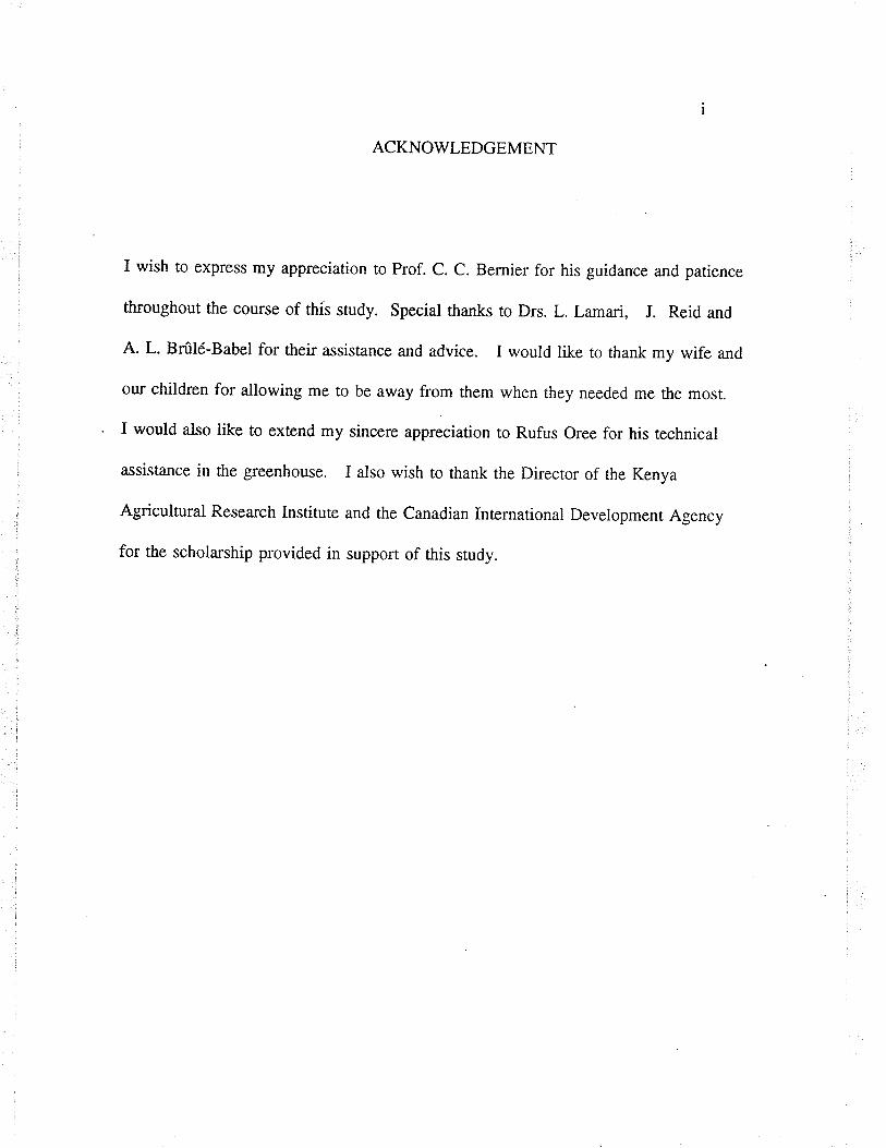
ACKNOWLEDGEMENT
I wish to express my appreciation to Prof. C. C. Bernier for his guidance and patience
throughout the course of this study. Special thanks to Drs. L. Lamari, J. Reid and
A. L. Brûté-Babel for their assistance and advice. I would like to thank my wife and
our children for allowing me to be away from them when they needed me the most.
I would also like to extend my sincere appreciation to Rufus Oree for his technical
assist¿nce in the greenhouse. I also wish to thank the Director of the Kenya
Agricultural Research Institute and the Canadian International Deveiopment Agency
for the scholarship provided in suppor.t of this study.
ll
FOREV/ARD
The materials, methods and results in this thesis are presented in the form of th¡ee
manuscripts inænded for publication. The style as well as the prepa-ration of tables
and figures comply with the requirements of Canadian Journal of Plant Pathology.
A general discussion and bibliography are included following the manuscripts.

l.)J.3.13.23.33.43.53.63.73.83.93.9.13.9.23.9.33.9.43.9.s3.9.63.9.74
4.t4.24.34.44.55
TABLE OF CONTENTS
List of tablesList of figuresGeneral abstractGeneral introductionLiterature Review
TaxonomyHost rangeEpidemiologySporulation in cultureConidia liberationlæaf wetness durationInfection processLeaf position and plant growrh srageControl of tân spot in wheatCultural methodsBiological controlCompetitionAntagonismResistance to tan spotHost-parasite interac tionGenetics of fungal pathogens
Manuscript l. Heterokaryon formation between iprodioneand hygromycin B resist¿nt mutants of pyrenophora títíci-repentisAbstractInuoductionMaterials and MethodsResultsDiscussionManuscript 2. Inheritance of virulence and toxin productionin Py reno phora tritici.-rep entis
5.1 Abstract5.2 Inrroduction5.3 Maærials and Methods5.4 Results5.5 Discussion6 Manuscript 3. Effect of host-selecrive ptr'-necrosis toxin on
the pathogenicity of avirulent recombinant progeny derived fromcrossing isolates 86-124 and Hy33l -9 of pyrettophora tritici-repenti.s
6.1 Abstract6.2 Introducrion6.3 Materials and Methods
llr
ivviI47
7
7
8
8
9
910
11
12
I213
t4I415
16
2t
29293l3439s0
5454555867
82
86
86889r
6.4 Results6.5 Discussion7 General Discussion8 References9 Appendices
iv
9399
102108
119
LIST OF TABLES
Table 1: Number of germinaæd conidia (vo) obøned from wild-type cultures,mutants, and from the meld formed between mutants 86-124 ipr+ and D308 hgm+of þrenophora titici-repentis on VSPDA amended with either iprodione orhygromycin B or both.
Table 2: Mean growth in diameter (mm) afær 7 days of wild typq culrures,mutants, and subcultures with restricæd mycelial growth obtainedfrom the meld formed between muranrs 86-124 ipr+ and D308 hgm+of P. triticïrepentis on VSPDA amended with either iprodione or hygromycin Bor both.
Table 3: Distribution of component genotypes in the subcultures withnormal mycelial growth obtained from the meld between mutants 86-124ipr+ and D308 hgm+ of P. trítici-repentis on VSPDA after 7 days ofgrowrh at 20-240C.
Table 4: Vi¡ulence on cultiva¡ Glenlea and line 68365 of the subcultureswith normal mycelial growth obtained from the meld formed betweenmutants 86-124 ipr+ and D308hgm+ of P. triticí-repentis (A) and the abitityof conidia reisolated from line 68365 ro grow on amended media (B).
Table 5: Colony characteristics of the P. tritici-repe¿t¿s isolates usedin the study.
Table 6: virulence patûerns of the isolaæs of P. tritici-repentis onthe twodifferential cultivars of wheat.
Table 7: Frequency of hybrid pseudothecia from a cross betweenisolates 86-124 and Hy33l-9 of P. tritíci-repentis as indicated by virulencephenotypes.
Table 8: segregation for vi¡ulence amongst randomly isolaæd ascosporesfrom hybrid pseudothecia in rhe cross between isolates 86-124 (nec+chl-)and Hy331-9 (nec-chl+) of P. tritici-repentis.
Table 9: Segregation for virulence amongst ascospores from five single asciin a cross between isolaæs 86-124 and Hy331-9 of P. tritici-repentis.
Table 10: Frequency of hybrid pseudothecia in the inter-crossof F, progenies (pathotype I and 4) from the cross of isolaæs 86-124 andHy33l-9 of P. triríci-repentis as indicated by virulence phenotypes.
42
43
47
49
63
64
70
72
73
76
vl
Table 1l: Segregation for virulence amongst randomly isolated ascospores fromhybrid pseudothecia in the inter-cross of F, progenies (pathotype I and 4)from the cross of isolaæs 86-124 and Hy33l -9 of P. ñticïrepentis.
Table 12: Segregation for virulence amongst ascospores from three singleasci in the inter-cross of F, progenies (pathotype I and 4) from the cross ofisolaæs 86-124 and Hy33l-9 of P. triticïrepentís.
Table 13: Segregation for production of Ptr-necrosis toxin amongst the¿ìscospore progeny from five asci in a cross between isolaæs 86-124and Hy331-9 of P. tritici-repentis.
Table 14: Segregation for production of Ptr-necrosis toxin amongst theascospore progeny from three single asci in the inter-cross of F,progenies (pathotype I and 4) from the cross of isolates 86-T24 andHy331-9 of P. titici-repentis.
Table 15: Effect of concentration of Ptr-necrosis toxin and incubationperiod on induction of tan necrosis in the susceptible cultivar Glenlea andon the pathogenicity of avirulent genotype of P.tritici-repentis.
Table 16: Effect of Pu'-necrosis toxin on vilulence of isolaæs ofP. tritici-repentís.
77
78
80
81
9s
96
v11
LIST OF FIGURES
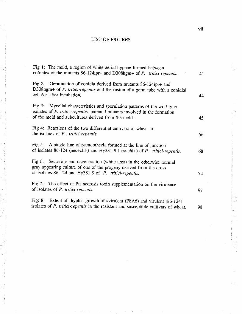
Fig 1: The meld, a region of white aerial hyphae formed betweencolonies of the mutants 86-l24ipr+ and D308hgm+ of P. tritíci-repentis. 4I
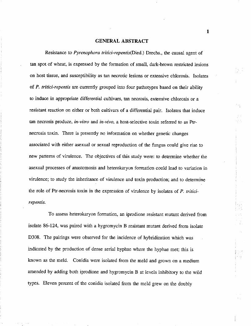
Fig2: Germination of conidia derived from mutants 86-124ipr+ andD308hgm+ of P. tritici-repentís and the fusion of a germ tube with a conidiatcell 6 h afær incubation. 44
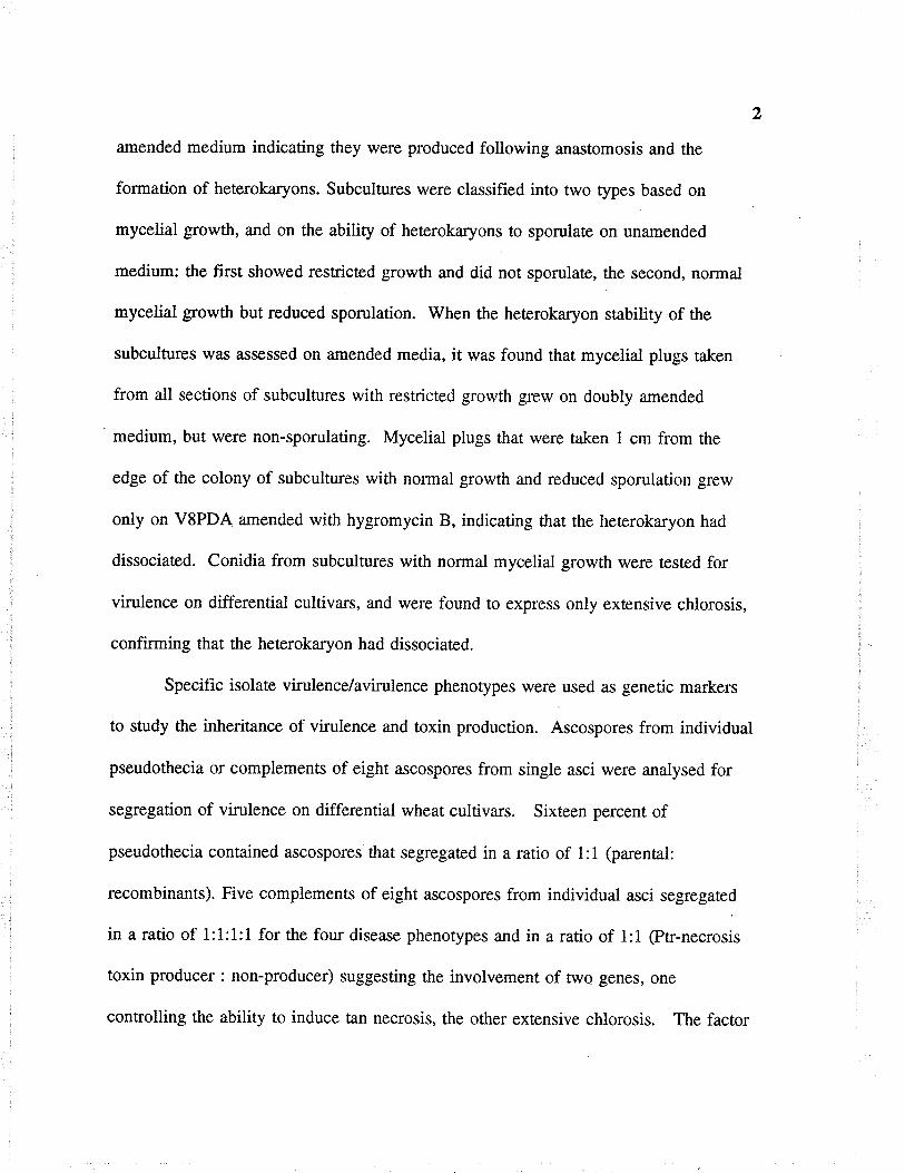
Fig 3: Mycelial characteristics and sporulation parterns of the wild-typeisolates of P. tritici-repentis, parental mutants involved in the folmation
. of the meld and subcultures derived from the meld. 45
Fig 4: Reactions of the two differential cultiva¡s of wheat tothe isolates of P . n'itíci-repenris 66
Fig 5 : A single line of pseudothecia formed ar rhe line of junctionof isolates 86-124 (nec+chl-) and Hy331-9 (nec-chl+) of P. tritici-repentis. 68
Fig 6: sectoring and degeneration (white area) in the otherwise normalgray appeating culture of one of the progeny derived from the crossof isolates 86-124 and Hy331-9 of P. tritici-repentis. 74
Fig 7: The eff-ect of Ptr-necrosis toxin supplementation on the vilulenceof isolates of P. n'itici-repentis. 97
Fig: 8: Extent of hyphal growrh of avirulenr (P846) and virulent (96-124)isolates of P. tritici-repentis in the resistant and susceptible cultivzus of wheat. 98
GENERAL ABSTRACT
Resistance to Pyrenophora trítíci-repentis(Died.) Drechs., the causal agent of
tan spot of wheat, is expressed by the formation of small, dark-brown restricæd lesions
on host tissue, and susceptibility as tan necrotic lesions or extensive chlorosis. Isolaæs
of P. triticïrepentis are currently grouped into four pathotypes based on their ability
to induce in appropriaæ differential cultivars, t¿n necrosis, extensive chlorosis or a
resist¿nt reaction on either or both cultivars of a differential pair. Isolaæs that induce
tan necrosis produce, in-vitro and ín-vívo, a host-selective toxin referred to as Ptr-
necrosis toxin. There is presently no information on whether genetic changes
associated with either asexual or sexual reproduction of the fungus could give rise to
new patterns of virulence. The objectives of this study were: to determine whether the
asexual processes of anastomosis and heterokaryon formation could lead to variation in
virulence; to study the inherit¿nce of virulence and toxin production; and to determine
the role of Ptr-necrosis toxin in the expression of virulence by isolates of P. tritici-
repentis.
To assess heüerokaryon formation, an iprodione resistant mut¿nt derived from
isolate 86-124, was paired with a hygromycin B resist¿nt mutant derived from isolaæ
D308. The pairings were observed for the incidence of hybridization which was
indicated by the production of dense aerial hyphae where the hyphae met; rhis is
known as the meld. Conidia were isolated from the meld and grown on a medium
amended by adding both iprodione and hygromycin B at levels inhibitory to the wild
types. Eleven percent of the conidia isolated from the meld grew on the doubly
2
amended medium indicating they were produced following anastomosis and the
fonnation of heterokaryons. Subcultures were classified into two types based on
mycelial growth, and on the ability of heterokaryons to sporulaæ on unamended
medium: the fust showed restricted growth and did not sporulate, the second, normal
mycelial growth but reduced sporulation. When the heterokaryon stabiliry of the
subcultures was assessed on amended media, it was found that mycelial plugs taken
from all sections of subcultures with restricted growth grew on doubly amended
medium, but were non-sporulating. Mycelial plugs that were taken 1 cm from the
edge of the colony of subcultures with normal growth and reduced sporulation grew
only on VSPDA amended with hygromycin B, indicating that the heterokaryon had
dissociated. Conidia from subcultures with normal mycelial growth were tested for
virulence on differential cultivars, and were found to express only extensive chlorosis,
confirming that the heterokaryon had dissociated
Specific isolate virulence/avirulence phenotypes were used as genetic markers
to study the inherit¿nce of virulence and toxin production. Ascospores from individual
pseudothecia or complements of eight Írscospores from single asci were analysed for
segregation of virulence on differential wheat cultivars. sixteen percent of
pseudothecia contained ascospores that segregated in a ratio of 1:l (parental:
recombinants). Five complements of eight ascospores from individual asci segregated
in a ratio of 1:1:1:1 for the four disease phenotypes and in a raúo of 1:l (Ptr-necrosis
toxin producer : non-producer) suggesting the involvement of two genes, one
controlling the ability to induce tan necrosis, the other extensive chlorosis. The factor
for Ptr-necrosis toxin was associaûed with the locus for tan necrosis.
Ptr- necrosis toxin was purified from culture filtaæs of isolate 86-124 and used
at several concentrations to test for its effect on the colonization of wheat leaves by
selected isolates of P. triticí-repentis. Untreaæd leaves, the leaves that were inoculaæd
only with spores of the avirulent isolaæ and those that were exposed only to the toxin
at less than 0.l¡rg/ml, remained symptomless. When Ptr-necrosis toxin was present at
0.1 ¡rglml-, the avirulent isolate invaded and coloniznd intercellular spaces of the
mesophyll in the susceptible cultivar Glenlea but not the resistant cultivar Salamouni,
and induced symptoms as rapidly as the virulent isolate. Leaves of the resistant
cultiva¡ Salamouni were not colonized by either the toxin-producing isolate 86-124 or
the avirulent isolate P8A6 3 supplemented with Ptr-necrosis toxin. This suggests that
resist¿nce to tan necrosis is based on insensitivity to the toxin produced by necrosis
inducing isolaæs. These observations support the hypothesis that Ptr-necrosis toxin is
a primary factor in the expression of virulence in the necrosis inducing pathotypes.
The occurrence of anastomosis followed by heærokaryon formation, and the
reconstitution of the four pathotypes by sexual recombination, indicates the poæntial
for P. tritici-repentis to evolve additional virulence that could overcome resistance
genes in the previously resist¿nf cultivars of wheat.
4
) GENERAL INTRODUCTION
Tan spot of wheat caused by þrenophora trítici-repentis (Died.) Drechs.
(anamorph : Drechslera títici-repentis (Died.l Shoemak er 1962) has become an
important disease world wide (Duff 1954; Valder and Shaw 1952; Misra and Singh
1972). The pathogen infects many grass species (Krupinsky 1982,1987) and has the
widest host range of any þrenophora species (Shoemaker 1962). The infection
causes reduced test weight and a pink discolouration of the kernels, resulting in
reduced grade. Under favourable conditions, epidemics of tan spot cause 12.9 to 49Vo
losses in grain yield of spring wheat (Hosford and Busch 1974; Rees et al. 1981).
Increased disease severity has been associated with change from conventional tillage to
practices that retain stubble on the soil surface (Pfender et aI. 1988).
Isolates of P. tritici-repentis are currently grouped into four pathotypes based
on the ability of isolates to induce in appropriaæ differential cultivars, tan necrosis or
extensive chlorosis or a resistant reaction in either or both cultivars of a differential
pair. Resistance to tan spot, expressed as small, dark-brown lesions, has been
identified at all ploidy levels of wheat (Lamari and Bernier 1989a) while
susceptibility is expressed as tan necrotic lesions or extensive chlorosis, or both.
Resist¿nce to tan necrosis and insensitivity to the Ptr-necrosis toxin produced
specifically by the isolaæs that induce tan necrosis is recessive, while resistance to
extensive chlorosis varies from dominant to partialty dominant (Lamari and Bernier
l99l).
5
Several methods have been tried in attempts to control tan spot of wheat.
Burying infected crop residue was considered because the fungus survives poorly on
buried stubble (Pfender and Wootke 1987), however, this practice leads to soil
erosion (Raymond et al. 1985). Conuol of t¿n spot using foliar fungicides is feasible
(Tekauz et aI. 1983; Watkins ¿r a/. lg82) but the cost may be prohibitive. The
incorporation of resist¿nce into susceptible wheat cultivars through breeding is now
possible. In order to determine how effective incorporation of resistance would be, we
require a better understanding of the host-pathogen interaction, durability of resistance
and the degree of variability of virulence patterns. Presently, there is no information
on whether asexual or sexual reproduction of the fungus can give rise to increased
levels or new patterns of virulence, and this study sought to provide such information.
Hybridization and recombination have been demonstrated in P. tritici-repentis.
Mutants resistant to either ttre fungicide iprodione or the antibiotic hygromycin B
were generated via ultraviolet light mutagenesis and used in sexual crosses (McCallum
1991; McCallum et al. 1994). Crosses were made in all possible combinations
between derived subcultures of isolate 86-124 which were resistant to either iprodione
or hygromycin B, and derived subcultures of isolate D308 which were resistant to
either iprodione or hygromycin B. Hybrid progeny were obtained only from crosses
between subcultures of isolate 86-124 resistant to iprodione and of 86-124 resist¿nt to
hygromycin B. Hybridization did not occur for all pairings, thus further work is
required to understand hybridization and recombination in P. ttitici-repentis.
The objectives of the present study were: i) to assess the occurrence of
6
anastomosis and heærokaryon formation as asexual means of variation in P .trirtcí-
repentis; (ii) to deærmine the inheritance of virulence and toxin production; and (iiÐ
to deærmine the role of the Ptr-necrosis toxin in virulence of isolates of P. tritici-
repentis.

7
3. REVIEW OF LITERATURE
3.1. Taxonomy oî forenophora trítici-repentis.
P. trítici-repentis, the causal agent of tan spot in wheat, is an ascomyceæ of
the class Loculoascomycetes, order Pleosporales, family Pleosporaceae (Luttrel L973).
The conidia are cylindrical, divided into 5-7 multinucleate cells (Shoemaker 1962;
Wehmeyer 1954) and measure 95-165 x 14-18 ¡rm. Pseudothecia are black and
produce abundant eight-spored asci which vary in size from 200-700¡rm (tPfender et al.
1988). Asci are bitunicate, cylindrical and narrow at the base. Ascospores are
hyaline at maturity, have three transverse septa and one longitudinal septum and
measure 47-65 x 20-26 pm.
3:2 Host range ol Pyrenophora tritici-repentis.
P. tritici-repentís is reported to have a broad host range among grass species
(Morrall and Howard 1975). At least 26 species of grasses a¡e infected by this
fungus (Krupinsþ 1982, lg87) and in moist weather it can cause yield losses of up
to 49Vo (Rees et aI. 1982). The wide host range of this fungus is in contrast to that
of other members within the genus e.g. P. teres Drechs., which only infects barley.
The ability of P. tritici-repentis to colonize such a large number of grasses, most of
which are perennial and grow in wheat producing areas, facilitates overwintering and
provides inoculum to initiate tan spot epidemics in farm fields (Krupinsþ 1982,
1987).
I3:3 Epidemiology
Several factors contribute to the production and spread of inoculum, and the
resulting disease severity in wheat. P. triticí-repentis can survive saprophytically on
infested residues of wheat and grasses between cropping seasons (Adee and Pfender
1989; Hosford l97l), and in infesæd seed (Schilder and Bergstrom 1990). Ascocarps
that are formed on infesæd stubble release ascospores as primary inoculum in spring.
Early season infection is largely initiaæd from inoculum formed on nearby stubble
since ascospores are not readily wind dispersed flVright and Sutton 1990; Krupinsky
1992). Following primary infection, conidia are produced on lesioned tissue and these
are wind disseminated and cause secondary infection cycles in the field.
3:4 SporulatÍon Ín culture
The conditions required for conidial production by P. triticí-repends have been
studied to some degree. Mycelial growth in culture is optimal at 20-250C and the
fungus is a diurnal sporulato¡ requiring light at wavelengths less than 350r1m to form
conidiophores (Odvody et aI. 1982). Production of conidia occurs over a temperature
range of 10-25t (ptatt etaI. 1977). High relative humidity (80-100 Vo)isessenrial
for conidia formation and promotes the formation of pseudothecia and ascospores
(Platt and Morrall 1980). Maximum pseudothecial formation is att¿ined at -0.2Mpa
and ceases under -2.4 Mpa (Pfender et aI. 1988). Ascospores develop most quickly at
150C (Summerell and Burgess 1988b).
3:5 Conidial lÍberation
Release of conidia has been shown to be dependent on wind speed and relative
humidity (RH) (Platt and Monall 1980). Wind speeds of 3.3 ms-I resulr in the
liberation of l00%o of spores at all RH values. Even at a relatively low wind speed,
60Vo spore liberation was obtained at 357o RH. Liberation was greater with changing
than with constant RH.
3:6 The effect of leaf wetness duration and temperature on infection.
By correlating the performance of wheat genotypes in the field with their post-
inoculation wet period requirement in the green house, Hosford and Busch (1974)
showed that moderately resistant genotypes required wet periods of 18-24 h alrd
susceptible cultivars wet periods of 6-12 h to develop similar amounts of disease as
measured by the number of lesions per cm2. In a similar study, Hosford et al. (1987)
showed that an increase in temperature andTor post-inoculation leaf wetness increased
disease severity in both a susceptible cultivar and, to a lesser extent also in a resistant
cultivar. The range of lesion size depended on the cultivar, with smaller lesions
occurring in a resistant cultiva¡ and larger lesions in a susceptible cultivar. Luz and
Bergstrom (1986) also showed that wheat cultivars differ in the amount of disease they
develop in response to changes in post-inoculation temperature. Thei¡ data indicates
that æmperature optima for disease development is between l8-280C, with the precise
range being dependent on the cultivar. On the cultivars Max and BR8, the number of
lesions per cm2 remained relatively high over a wide temperature range (18-240C)
whereas on the cultivar 8H1146, the range is narrow (25-280C) but the temperature is higher.
10
The reported breakdown of resist¿nce with increased temperature and/or post-
inoculation wetness period is contrary to the findings of Lamari and Bernier (1989a),
who found no significant differences in the percent leaf area infected for wetness
durations ranging from 24-72 h at 220C. The reason resistance was found to break
down with prolonged wetness can be explained by the way Hosford et aI. (1987)
characænzed resistance or susceptibility on the basis of lesions per cm2 or percent leaf
a¡ea infecæd. A large number of lesions is not necessarily synonymous with
susceptibility, which implies genetic action (or lack of it) and results in a "typical"
phenotype. Lamari and Bernier (1989a, b) showed that the tan spor syndrome
consists of two phenotypes, tan necrosis and chlorosis, and that resistance based on
lesion type did not change following post-inoculation wetness periods. The increase in
amounts of disease with prolonged wetness as reported by Hosford et aI. (1987) could
be due to a larger number of infectionVunit area as a result of multiple
infectionVconidium arising from branching and/or germination of several 'icells" per
conidium.
3:7 Infection process
Studies that have been conducted on the infection process of P. tritici-repentis
in the susceptible and resistant wheats reported no differences in Vo spore germination,
the number of appressoria produced, or the successful penetration of epidermal cells
between such wheats within 12 h post-inoculation at I8-220C and 90-l00%o W (Larez
et al. 1986; Loughman and Deverall 1986; Lamari and Bernier 1989b). However,
differences were observed between the susceptible and resistant wheats after 48 h post-

l1
inoculation when hyphae became established in the intercellular space of the
mesophyll tissue showing a susceptible reaction, but not in resist¿nt tissue. Whether
differences occur between the infection processes of virulent and avirulent isolaæs
have not been investigated.
3:8 The effect of leaf position and plant growth stage on disease development.
Wheat leaf position, or age, has been shown to affect significantty the
appearance and severity of disease symptoms. Both Raymond et al. (1985) and Riaz
et al. (1991) noted that despite the fact that leaves were inoculated at the same time,
lesions on lower (older) leaves coalesced ea¡lier than did the lesions on upper
(younger) leaves, indicating that older leaves a.re more susceptible to infection than
younger leaves. Raymond et al. (1985) also confi¡med that the growth stage at which
infection occurs can influence yield loss. Studies by Rees and Platz (1983) showed
that a l37o yield loss occurred at the seedling stage following infection, compared to a
35Vo loss at the jointing stage following infection. Shabeer and Bockus (1988)
reported that plants are most susceptible physiologically to infections occurring before
the boot or flowering stages, and that about half of the total yield loss due to such
infections results from multiple infections by secondary inoculum. Losses are a result
of a significant reduction in kernel weight and the number of grains per spike, and not
of a reduced number of spikes per plant.
t2
3:9 CONTROL OF TAN SPOT OF WHEAT.
3:9:1 Cultural methods for control.
Several cultural methods have been tried in att€mpts to control t¿n spot in
wheat. Control practices consisting of rot¿tions with non-host species have been
attempted to control tan spot in Australia (Rees and Pla¿ 1980, 1981). However;
since there is evidence that P. titici-repenr¡s infects several species of grasses which
may provide inoculum in the absence of wheat stubble (Krupinsky 1982, 1987),
rotations may not be always effective. Fungicides have been used to effectively
control tan spot in the field, but such practices are cost prohibitive (Tekauz et al.
1983).
Applications of nitrogen as either ammonium sulphate or calcium nitrate in the
field are reported to reduce disease severity (Huber et aI. 1987), and both the rate and
form of nitrogen influenced the severity of tan spot in winter wheats. Thus Huber ør
al. (1987) thought that management of nitrogen might provide a cultural control for
this disease. In a separate study, Bockus and Davis (1993) also found that fertilizer
applications significantly reduced disease severity in inoculated treatments. However,
the severe physiological necrosis and chlorosis from nitrogen deficiency in
nonfertilized, noninoculated checks were indistinguishable from the symptoms of the
disease. These symptoms were alleviated with both nitrogen forms in noninoculaæd
treatments. The apparent reduction of disease by nitrogen fertilizers in inoculated
treatments was due to alleviating physiological nêcrosis and chlorosis, and therefore
both ammonium sulphate and calcium nitrate reduced disease by delaying leaf
13
senescence but not by directly reducing the disease.
3:9:2 BiologÍcal control
Biological control involves both the manipulation of the environment to the
disadvantage of the harmful organisms, and the use of other organisms to reduce the
levels of the harmful agent on the host. Manipulation of the environment could
involve changes in water poæntial, temperature, competition for nutrients and release
of toxic substances into the environment (Campbell 1989).
The saprophytic sexual stage of P. tritici-repentís is apparently a complex one.
Mycelial growth and pseudothecial numbers have been shown to decline as the
exærnal water potentials decrease (Summerell and Burgess 1988a). The fungus
grows maximally at -0.2Mpa and produces the maximum number of pseudothecia at
this waær potential. Mycelial growth and pseudothecial formation is reduced ar -10.5
Mpa indicating that this fungus can not survive in water deficient areas.
The recovery of P. tritici-repenris from stubble, and the production of fertile
pseudothecia on stubble residues that have been weathered over a number of years in
various environments, have both been used as indicators of viability of the fungus
(Summerell and Burgess 1989; Adee and Pfender 1989). Incorporation and burial
of stubble is inimical to the survival of the fungus. The fungus is infrequently
recovered from stubble after 26 weeks of burial; 52 weeks of incorporation and 104
weeks on stubble that is retained on the soil surface.
l4
3:9:3 Competition and pathogen survÍval.
P. tritícïrep¿nr¿s survives well in association with both a number of other
parasiæs as well as primary saprophytes that occur on straw that remains on the soil
surface. Persistence of the pathogen in infested straw was correlaæd with the
composition of the microbial communitity in various macroenvironments in no till
fields (Zhang and Pfender 1992; Pfender et al. 1993a). The results showed that in
soil-borne straw, this pathogen was displaced by a community composed of
Actinomyceæs and soil-bome fungi. At water potentials higher than -39 Mpa, P.
tritíci-repentis was initially displaced by Pythiunt oligatzdrura. Drechs., at 10t and
Aspergillus terreus Thom., and Chaetomiunt globosum Kunze:Fr., at 200C and 300C
respectively, and fînally Penicillium spp and Fusarium solani (Mart.)Sacc., (Summerell
and Burgess 1989). No fungi other than P. tritici-repentis were recovered at -
150Mpa. Prolonged periods of moisture occurred in those microenvironments in
which the survival and pseudothecial production was poorest (Summerrel and Burgess
1989).
3:9:4 Ãntagonism
Several organisms have been reported to exhibit antagonism towards P. tritici
repentís. Such antagonism by Cochliobolus sativus (Ito & Kuribayashi ) Drechs. ex
Dastur., was observed when the two organisms were inoculated simultaneously, or
when C. sativus was inoculated up to six hours afær P. tritíci-repentis (Luz and
Bergstrom 1987). The antagonism of C. sativus towards P. tritici-repentis is
expressed as a suppression of conidial germination, appressorium formation, and germ
15
tube elongation on the leaf surface. Several straw-associated fungi, including
Límonomyces roseipel/rs Stalpers & Loerakker, are capable of inhibiting both growth
and pseudothecial production by P. títicí-repentis (Pfender 1988; Pfender et al.
I993a). These straw associaæd fungi reduce ascocarp and ascospore production, with
the degree of suppression varying between 50-90 Vo, depending on the test conditions.
The antagonism was thought to be due to mycoparasitism, because straw associaæd
fungi are known to have chitinolytic ability.
Antibiotic producing bacterial strains inhibit the growth of P. triticïrepentis in
culture and on wheat straw maintained under conditions of high humidity. Inhibition
by Pseudomoruß fluorescen..r (Trevisan) Migula, was expressed as a reduction in
mycelial growth and is due to pyrrolnitrin, one of the antibiotics produced by P.
fluorescens, strain Pf-5 (Pfender et aI. /993b).
. Among a group of herbicides tested for their ability to inhibit pseudothecial
formation in infested straw, the glyphosate containing herbicide Roundup was the most
effective, if applied before infection (Sharma er a/. 1989). However, it had no effect
on mycelial growth rate. This report did not specify whether the inhibitory effect on
pseudothecial formation was due specificatly to glyphosate or to some non-herbicide
component of the preparation.
3:9:5 Host resistance to tan spol
Resistance to tan spot in wheat has been reported at all ploidy levels of wheat
and is usually visually expressed by the formation of restricted small, da¡k-brown
lesions (Gough 1982; Lamari and Bernier 1989a). And while the incorporarion of
t6
resistånce into susceptible cultivars through breeding is now possible, we require a full
underst¿nding of the nature of the inæraction between wheat and P. triticí-repentís,
durability of resistance and the degree of variability of virulence patt€ms.
3:9:6 TIIE NATURE OF HOST-PARASITE INTERACTION.
Compatible and Íncompatible interactions
The interaction of the host and pathogen genotypes is influenced by the
envi¡onment over the course of time, and the result is a "typical" phenotype or
expression of disease symptoms. The phenotype of the interaction is used to
characænze the host as being resistant (the pathogen may penetrate the cuticle and
epidermal cells, but subsequent growth is restricæd making the interaction
incompatible) or susceptible (the pathogen penetrates and grows unimpeded making
the interaction compatibte). In the pathogen, the counterpart of resistance is avirulence
and for susceptibility it is vi¡ulence. Virulence is rhe ability of a pathogen to infect
and cause disease on different host genotypes while avi¡ulence is the inability to infect
and cause disease on differenr genorypes (Andrivon 1993).
Aggressiveness
The quantitative measure of disease expression by an isolate on a susceptible
cultivar is known as aggressiveness (Andrivon 1993). Aggressiveness is a
characteristic of the host-pathogen interaction as influenced by envi¡onmental
conditions. The same pathogen strain can induce different amounts of disease on a
series of susceptible hosts. Thus, aggressiveness depends on the partial resistance
t7
features of the host on which this cha¡acter is measured, and as it can be scored
repeatedly under standa¡d envi¡onmental conditions, this makes it a stable trait for a
given host-pathogen pair.
The traits of aggressiveness which can be measured directly in the disease
cycle are: (i) incubation and latent periods; (2) number of spores produced in a given
area or in a fixed time period; (3) the lesion sizes and (4) ttre duration of the
sporulation period. Short incubation and latent periods. long sporulation periods,
large cumulative spore counts, and large areas of infected tissue can be indicative of
an isolates high levels of aggressiveness on a specific cultivar if it is being compared
to another isolate. Isolates of P. tritici-repentís have been found to ditïer in
aggressiveness as measured by the percent leaf area infected (Krupinsky 1992b) but
not on the basis of individual components of disease reaction.
The Gene-for-Gene hypothesis
Many important advances in plant pathology have come from the genetic
analysis of the host and the pathogen. For example, the detailed studies of the
inheritance of virulence in flax rust pathogen Melantpsora lini (Ehrenb.) Desmaz., and
resistance in flax Linunt usitatissitnunt L-,led Flor (1942) to propose the gene-for-gene
hypothesis of specificity in host-pathogen interactions. The gene-for-gene hypothesis
states that for every resistance gene in the host there is a conesponding avirulence
gene in the pathogen. Genetic analysis of Venturia inaequalis (Cooke) G. Wint., on
apple (Bagga and Boone 1968; Boone and Keitt 1957) , Bremia lactucae Regel, on
lettuce (Michelmore et al. 1984; Norwood and Crute 1983) and Puccinia recondita
18
f. sp. títicr (Roberge ex Desmaz.), on wheat (Kolmer and Dyck 1994) among others
have supported the existence of the gene-for-gene relationships between fungi and
their hosts.
The Wheat i P. tritici-repentis system
The host
Resistance to tan spot in wheat, as reported by Hosford (1971), Hosford and
Busch (1974) and Raymond et al. (1985) was largely quantitative, and based on the
number of lesions per cm2 or percent disease severity. This did not provide a clea¡
concept of resistance or susceptibility, which imply genetic interactions with "typical"
phenotypes. The recognition of the interaction of the t¿n spot fungus with wheat
genotypes as a syndrome consisting of two phenotypes, necrosis and chlorosis (Lamari
and Bernier 1989a. b) led to studies on host-parasite interactions in this system.
Susceptible wheat cultivzus develop either tan necrosis or extensive chlorosis, while
resistance to both tan necrosis and extensive chlorosis is expressed as the formation of
small dark-brown lesions. Resistance to tån necrosis is controlled by one recessive
gene while resistance to extensive chlorosis varies from partial to completely dominant
(Lamari and Bernier 1991). However, preliminary results from an on-going study
(Duguid, s.; personal comm.) and from other reports (Nagle et al. 1982; sykes and
Bernier 1991) indicaæ that resistance to tan spot is controlled by more than one gene
in some cultivars of wheat.
t9
The pathogen
Va¡iation among isolates of P. tritíci-repentís has been reported for colony
morphology and the amounts of pigments produced in culture, and for virulence and
aggressiveness on wheat cultivars. While in culture most isolates form dark-gray
colonies that produce black-green pigments, on subculture some strains give rise to
variants that produce wine-red to purplish or reddish or bright orange pigments
(Wehmeyer 1954; Hosford 1971; Hunger and Brown 1987). While such variants
differ in morphology and growth rates, Wehmeyer (1954) also noted that the wine-red
va¡iants remained completely sterile and failed to form even the,"sclerotial" primordia
common to all other strains.
Misra and Singh (1972) reported that there were significant differences
between isolates based on the severity of the reactions they induced on the host plants,
and based on the percent leaf area infected and numbers of lesions formed per cm2,
Luz and Hosford (1980) distinguished 12 races amongst the 40 isolaæs they studied.
During an attempt to verify the potential of smooth brome grass to serve as an
alærnaúve source of inoculum, Krupinsky (1987) found he could not differentiate
between isolates from brome and wheat on the basis of reaction they induced on wheat
leaves; the reaction was measured by the number of lesions per cm2 and disease
severity. Smooth brome grass serves as a host to P. tritici-repentis, and therefore
inoculum can easily be transmitted between the two hosts.
Schilder and Bergstrom (1990a) showed that isolates they studied did not differ
widely in disease reaction as measured by percent leaf area infecæd, but had a
20
moderate degree of physiological specialization based on the amount of disease
induced on both winter and spring wheats. Winüer wheats had moderate to high levels
of resistance, while spring wheats were susceptible. Lamari et at. (1991) grouped
their isolaæs of P. tritící-repenr¿s into four pathotypes based on the ability of isolaæs
to induce either tan necrosis, extensive chlorosis or a combination thereof on
appropriaæ differential cultivars. Isolates of pathotype one induce both tan necrosis
and extensive chlorosis (nec+chl+), pathotype two only tan necrosis (nec+chl-),
pathotype tluee only extensive chlorosis (nec-chl+), while pathotype four, classified as
avirulent, induces only small dark-brown lesions without either characteristic tan
necrosis on Glenlea, or extensive chlorosis on line 68365.
Ptr-necrosÍs toxin
Isolaæs that induce tan necrosis produce a cultivar specific Ptr-necrosis toxin in
liquid culture (Tomas and Bockus 1987; Ballance et aI. 1989, Lamari and Bernier,
1989c). When the toxin is infiltrated into susceptible host leaves, it produces tan-
coloured necrotic lesions similar to those induced by the necrosis-inducing isolaæs
(nec+chl- and nec+ chl+), and isolates that lack the ability to induce tan necrosis
(nec-chl+ or nec-chl-) do not produce Ptr-necrosis toxin. Ptr-necrosis toxin was
characærized and found to be a high molecular weight (Mw 13,900) protein (Ballance
et al.I989), and Brown and Hunger (1993) isolated a low-molecular weight (Mw 800-
1800) toxin which induces the characæristic chlorosis that is associated with tan spot
disease, and also differentially inhibits elongation of wheat seedling coleoptiles. This
Ptr-toxin does not induce necrosis. The relationship between these toxins, and their
2l
roles in tan spot disease development, have not been investigaæd.
3:9:7 GENETICS OF FUNGAL PATHOGENS
Genetic studies of any organism require the use of characteristics which can be
precisely defined and easily scored, and that provide information about an organism's
genotype. The key to understanding variation in fungi lies in their reproductive
systems. Investigations in other fungi indicaæ that variation is caused by, and
distributed through, asexual and sexual processes (Burnett 1975; Fincham et aI.1979).
However, there is presently no information on whether either asexual or sexual
reproduction or both can give rise to increased levels or patterns of virulence in P.
tritici-repentis.
3:9:7:I Variation arising during asexual reproduction.
Mutations
. During asexual reproduction, the origination of variants chiefly involves
mutations, anastomoses followed by nuclear exchange to form heterokaryons, and
parasexuality. The advancement of fungal genetics has been based on the use of
mutations at specific loci to create genetic markers (Flor 1960; Griffiths and Carr
1961). As mut¿tions occur spontaneously at a low frequency of approximately 10-6 in
alt cell populations of fungi (Fincham et aI. 1979), mutagens such as X-rays, gamma
rays, ultraviolet light and certain chemicals have been used to increase mutation rates
in fungi. The rate and extent to which mutant genes can spread through, and between,
populations is of importance in determining how fast a species can evolve, whether it
will evolve eventually into two or more species and, for the plant pathologist, whether
22
new virulent forms could arise to overcome previously resistant genes.
The large size of most fungal populations means that mutant alleles arise
continuously despiæ the rarity of mutation. The absence of a distinct germline in
fungi ensures that mutations arising in virtually any tissue can be transmitted to
subsequent generations either sexually or asexually. The dominant haploid phase that
occurs in the life cycles of many fungi, means that new mutations can be expressed
immediately if they are segregated into uninucleate spores or monokaryotic hyphae
(Caten 1987). And although when they first arise new mutant alleles are so rare that
they generally pass undetected, the high asexual reproductive rate of most fungi
permits a rapid increase in number and fi'equency to those mutations that confer
benefit under natural selection. Genetic markers created by mutagenesis have been
used in several ascomycetous fungi including Saccharomyces cerevisiae (Meyen ex. E.
Hans), Aspergillus nidulans (Eidam.) G. Wint., and Neurospora crassa Shear and B.
O. Dodge, to develop genetic model systems (Fincham et aI. 1979).
HeterokaryosÍs
Intraspecific hyphal fusion and nuclear migration occurs regularly between
genetically identical (homogenic) hyphae of many fungi. And if such fusions and
nuclear migrations occurs between thalli whose nuclei differ, heterokaryons, (i.e.
mycelia cont¿ining a mixture of two or more types of nuclei) can be established.
(Esser and Blaich 1973; Esser and Meinhardt 1984; Puhalla 1985; Burnett 1975).
However, if hyphae a-re not genetically identical at certain loci (heterogenic), fusion
may be prevented, terminated or limited in its effects, and since veget¿tive
23
incompatibility is based on genetic differences, the term heterogenic vegetative
incompatibility is used to explain lack of fusion between hyphae. Heærogenic
incompatibility can also affect certain sexual fusions (Esser and Btaich L973; Bumett
1975; Fincham et al. 1979; Anagnostakis 1984). Vegetative incompatibility is
divided into fusion incompatibitity and post-fusion incompatibilty. In the former, cell
fusion between the two strains does not occur whereas in the latter, fusion reactions
cause "barrage" effects between colonies (Esser and Blaich 1973; Esser and
Meinha¡dt 1984; Anagnostakis 1984). Hyphal death or repulsion occurs within the
barrage zone in Basidiomycetes as well as N. crassa, while in most other
Ascomycetes, aversion or developtnent of reproductive structures occurs afær a period
of time within this region, depicting posr-fusion incompatibility.
Fusion and post-fusion incompatibility have been widely studied in fungal
species (Puhalla and Mayfield 1974; Pontecorvo and Sermonti 1954: Zambino and
Harrington 1990). ln each of the species studied, numerous vegetative
incompatibility groups (VIG) have been recognized, each of which has two alleles at
the controlling loci. An incompatibility reaction occurs when strains differing at one
or more vegetative incompatibility (VI) loci are brought together. Incompatibility due
to the interaction of alleles of a VI gene is temed allelic incompatibility. If a species
has n loci at which allelic incompatibility occurs, rhen T vIG are possible.
Inæraction between alleles at diffe¡ent VI loci is known as non-allelic incompatibility
and has only been described in Podospora anserina (Ces.ex Rob.) Niessl, (Esser and
Blaich 1973).
24
Studies of hyphal fusions and heterokaryon formation in some species of
fungi indicate that once est¿blished, heterokaryons can be maintained in several ways.
: In N. crassaand A. nidulans (Pontecorv o et aL 1953), and Penícillium chrysogenum
Thom., (Pontecorvo and Sermonti 1954), nuclei of paired strains migrate
reciprocally, a¡¿ then divide in the hyphae of each of the pair. Heærokaryons become
established in multinucleate hyphal tips, and thus the heterokaryon can be maintained
by further hyphal growth without recuffent anastomosis. In some other fungi,
. Verticillium dahliae KIeb., (Puhalla and Mayfield lg74), Gibberella fujikuroi
(Sawada) Ito., (Puhalla and Spieth 1985) and Leptographiunt wageneri (Kendrick) M.
J. Wingfield., (Zambino and Harrington 1990), recurrent anastomoses is required
probably because the nuclei do not migrate from the cell in which anastomosis has
occured, and the heterokaryon cannot be perpetuated without recuffent fusion.
The parasexual cycle
In the parasexual cycle, mycelium becomes heterokaryotic due to anastomosis
, or mutation. A portion of the haploid nuclei in the heterokaryon fuse to form diploid
' nuclei, and these divide mitotically and increase in number together with the
remaining haploid nuclei (Kafer 196l; Buxton 1956; Hartley and Williams 1971).
, At mitosis, some of the diploid nuclei undergo recombination due to crossing over.
: Qther diploid nuclei may enter spores which germinate and produce homokaryotic
mycelia. The diploid nuclei in these mycelia may be haploidized, and from those
which undergo mitotic recombination, haploid nuclei may result which differ in
genetic constitution from those of the original heterokaryotic mycelium. The various
25
processes of diploidization, haploidization, aneuploidy and recombination may all be
occurring simultaneously in the thallus, and not in any regular sequence or at any
specific stage (Kafer 1961). There is much speculation about the evolutionary
benefits of heærokaryosis as a means of exploiting hybrid vigour, storing genetic
variation and adjusting somatically to changing environment (Buxton 1956; Caæn and
Jinks 1966: Caæn 1987). The significance of heterokaryosis as a means of genetic
variation will depend not only upon its occurrence, but also upon the degree of the
diversity between the associaæd genomes.
3:9:7:2 Variation arising during sexual reproduction.
Mating types
Throughout most of the life of a fungus, fusion between genetically different
strains is prevented or severely limited by the presence of heærogenic VI systems.
However, when the sexual process occurs, these systems are largely or entirely
suppressed, so out-crossing is possible (Esser and Blaich 1973). In many fungi the
sexual process is initiated only as a result of an interaction between cells that differ
genetically with respect to mating type, and probably also in other ways, thus
promoting genetic exchange (Fincharn et aI. 1979; Burnett 1975; Anagnostakis
1984).
Heterothallism
Fungi in which the co-operation of genetically different strains is required for
the initiation or completion of the sexual process are referred to as self-sûerile or,
since different strains need to be involved, as heterothallic (Pontecorvo et aI. 1953;

26
Wheeler 1954; Olive 1958). The controlling mechanisms have been classed as either
homogenic or heærogenic incompatibility (Bumett 1975: Esser and Blaich 1973;
Esser and Kuenen 1967; Esser and Meinhardt l9S4). In fungi that have two mating
types based on alternate alleles at a single locus; homogenic incompatibility prevents
mating between genetically similar progeny. Heterogenic incompatibility prevents
mating between genetically dissimila¡ progeny.
Homothallism
A fungus in which every thallus is sexually self-fertite and can reproduce
sexually without the aid of another thallus is referred to as homothallic. Homothallism
is referred to as primary if there is no evidence for a heterothallic ancestor. If there
is evidence that an earlier heterothallic state has been circumvented, then it is referred
to as secondary homothallism or pseudocompatibility (Wheeler 1954; Olive 1958;
Carlile 1987). Secondary homothallism can result from the inclusion of two nuclei of
different mating type in a single spore, and the subsequent growth of a mycelium
heærokaryotic for mating type. Secondary homothallism due to mutation for mating
type in large-spored strains of Sclerotinia tríþIiorøm Eriks. is known (Uhm and Fuji
1983). In addition to S. triþIiorunt, the existence of both heærothallic and
homothallic strains of the same fungus has been reported in Ceratocystis ø/rd (Buism.)
Nannf. (Brasier and Gibbs 1975), Cryphonectría parasirlcø (Murrill) Barr
(Anagnostakis 1984, 1988), and Glomerella grantinicolaPotitis (Vaillancourt and
Hanau 1991). With any homothallic fungus, when two strains are grown in mixed
culture, the ascospores from any single ascus may derive their nuclei from a selfed
27
zygotÊ of one strain or the other, or from a hybrid zygota (Pontecorvo et aI. 1953).
Genetic analysis
For genetic analysis, the inærest is primarily in crossed asci. Ascospore colour
has been widely utilized to study the inheritance of spore colour, because progeny can
be analysed directly by viewing the progeny with a microscope. Ascospore colour
mutants have been utilized in the homothalic ascomycete Sordaria finúcola (Roberge
ex Desmaz.) Ces. and De Not. (Olive 1956) as well as in the heterothallic plant
pathogen y. inaequalis (Boone and Keitt 1957). Ascospore colour mutants have
been used to directly identify hybrid perithecia and cleistothecia. Since ascospores of
S. fimicola are produced in an ordered series, spore colour mutants make it possible
to visually analyse the segregation of the spore colour-loci. Ascospore colour is an
ideal marker for homothallic species because there is no need to germinate ascospores
and form thalli to deæct hybrid perithecia. Ascospores of P. triticí-repentis are
hyaline and indistinguishable on the basis of colour. The most useful approach to
genetic analysis is to germinate ascospores and score thalli for differences in colony
morphology, pigments, virulence or toxin production.
The only Pyrenophorø species that have been analysed genetically are P. teres
and P. graminea Ito & Kurabayashi (McDonald 1963; Smedegard-Peterson 1977).
Both species have bipolar heterothallic mating systems. P. teres, the causal agent of
net-blotch of barley, occurs in two forms: the net form which produces a net-like
pattern of necrotic lesions and the spot form which produces brown elliptical lesions.
In crosses between the spot and net forms, four classes based on lesion type were
28
found in the progeny, the parental spot and net lesion inducing types, and two
recombinant classes. The two recombinant classes were comprised of one class whose
strains induced lesions inærmediaæ between the spot type and the net type, while the
strains of the second class induced fleck or non-pathogenic lesions (Smedegard-
Peterson 1977). The two parental and tworecombinants occurred in a l: 1 : I : I
ratio indicating that the ability to produce spot and net lesions is conditioned by two
independent genes.

29
4. Heterokaryon formation between iprodione and hygromycÍn B resistant
mutants of þrenophora tritici-repentß.
ABSTRACT
Isolaæs of P. trítici-repentis, the causal agent of t¿n spot of wheat are
currently grouped into four pathotypes based on thei¡ ability to induce tan necrosis,
extensive chlorosis, or a combination of these or a resistant reaction on appropriaæ
differential cultivars of wheat. The objective of this study was to assess the
occurrence of anastomosis and heterokaryon formation as possible asexual means of
making variation in the virulence of this fungus. A mut¿nt of isolate 86-124 resist¿nt
to iprodione was paired with a mutant of isolate D308 resistant to hygromycin B. The
pairings were observed for the incidence of heterokaryon formation as indicated by the
presence of the meld. Conidia were isolated from the meld and grown on a medium
amended with both iprodione and hygromycin B at levels inhibitory to the wild-type
strains. Eleven percent of the conidia from the meld grew on the doubly amended
medium indicating tley were the result of anastomosis and heterokaryon formation.
Subcultures were classifîed into two types based on mycelial growth and the ability of
the heærokaryons to sporulaæ on unamended media: one with restricted growth and no
sporulation, the other with normal growth but reduced sporulation. When the
heterokaryon st¿bility of the subcultures was assessed on amended media, it was found
that mycelial plugs taken from all sections of subcultures with restricted growth grew
on doubly amended medium, but were non-sporulating. Mycelial plugs that were
taken I cm from the edge of the colony of subcultures with normal growth and
30
reduced sporulation grew on VSPDA amended with hygromycin B but not on VSPDA
amended with iprodione, indicating that the heærokaryon had dissociaæd. Mycelial
plugs that were taken 2 cm from the edge of the colony grew on the doubly amended
medium, and mycelial plugs taken 3 cm from the edge of the colony were a mosaic
that grew on media amended with either iprodione or hygromycin B, or both.
Conidia from subcultures with normal mycelial growth and reduced sporulation were
æsted for virulence on differential cultivars that develop either tan necrosis or
extensive chlorosis and were found to induce only extensive chlorosis also suggesting
that the heterokaryon was not stable.
3l
4.1. INTRODUCTION
þrenophora trítíci-repentis (Died.) Drechsler, the cause of tan spot disease of
wheat, has become an important disease world wide (Hosford 1971; Tekauz 19761;
Rees and Platz 1981; Duff 1954). The pathogen has a broad host range ÍLmong grass
species (Morrall and Howa¡d 1975; Krupinsky 1982, 1987) and causes yield losses
of up to 497o in moist weather (Rees et al. L982).
Isolaæs of P. triticïrepentis form either dark-gray or whitish-cre¿un colonies on
VBPDA medium and each type can form sectors that differ both morphologically and
in the appearance of the pigments secreted into the medium; the latter range from
dark-gray to orange or whitish cream (personal observations; Wehmeyer 1954;
Hunger and Brown 1987). When axenic cultures from these sectors were made by
subculturing hyphal tips and increased by growing colonies from single conidia, the
dark-gray and whitish cream sectors produced abundant conidia whereas the white or
albino sectors remained mycelial and did not sporulate.
Isolates of this fungus are currently grouped into four pathotypes based on
their specific inæractions with nvo differential wheat cultivars (Laman et al. 1991).
The inæraction is characterized by the development of tan necrosis on cv. Glenlea and
extensive chlorosis on line 68365. Isolates of pathotype 1 induce both t¿n necrosis
and extensive chlorosis (nec+chl+), pathotype 2 only tan necrosis (nec+chl-),
pathotype 3 only extensive chlorosis (nec-chl+) and pathotype 4 (nec-chl-) lacks the
ability to induce either symptom, and are classified as being avirulent.
Genetic analysis of this homothallic fungus was difficult until recently when mut¿nts
32
resistant to either the fungicide iprodione or the antibiotic hygromycin B were created
in isolaæs 86-124 (nec+chl-) and D308 (nec-chl+) via ultraviolet light mutagenesis and
then used in sexual crosses (McCallum et al. 1994). The mutants were crossed in all
possible combinations and the progeny analysed to deærmine the genetic basis of
chemical resistance. Hybrid progenies were obtained in crosses involving mutants of
isolate 86-124, but not of isolate D308. The results indicated that one locus controls
iprodione resistance and a second locus controls hygromycin B resistance. Why
hybridization did not occur between mutants of isolates 86-124 and D308 has not been
investigated.
Genotypic variants brought about by mutations are distribuæd among progeny
via asexual and sexual processes. The asexual process involves anastomosis and
nuclear migration between vegetatively compatible strains to form heærokaryons
(Tinline 1988; Heale 1988; Anagnostakis 1988). If hyphae are not genetically
identical, hyphal fusion may be prevented, ærminaæd, or limited in its effects by
vegeråúve incompatibility. Vegetative incompatibility prevents mixing of protoplasm
and nuclear migration (tæslie 1993). It is divided into fusion incompatibility and
post-fusion incompatibility (Esser and Blaich 1973; Anagnostakis 1984; Caæn
1986). In fusion incompaúbility, cell fusion between the two incompatible strains
does not occur and is usually expressed as aversion zones. In post-fusion
incompatibility, fusion reactions cause barrage zones (an area between colonies where
hyphae, after having attempted to anastomose, become curled, enlarged and
degenerate). The barrage line eventually consists of paralyzed dead cells (Esser and
4^JJ
Meinhardt 1984).
The frequency of heærokaryon formation is usually increased a thousandfold
by using either protoplast fusion or microinjection of donor protoplasmic fluids
including nuclei into recipient strains. This æchnique assures successful crosses
between strains in which crossing had failed previously, indicating that the hyphal wall
is a major siæ of incompatibility (Heale 1988).
Studies on anastomosis and heterokaryon formation in some species of fungi
indicate that heterokaryons once established can be maintained in several ways. In N.
crassa, A. nidulans and P. chrysogenurn (Pontecowo et aI. 1953), nuclei of both
strains migrate and divide in the hyphae of paired strains. Heterokaryons become
established in multinucleate hyphal tips, and are maintained by further hyphal growth
without recurrent anastomosis. In other fungi like V. dahliae (Puhalla and Mayfield
1974) and L. wageneri (Zambino and Harrington 1990), recurrent anastomosis is
required, probably because nuclei do not migrate from the cell in which anastomosis
has occurred, and the heterokalyon can not be propagated without recurrent fusion.
Heærokaryosis may contribute to fungal variation through the parasexual cycle
(Pontecorvo 1956; Hartley and Williams 1971; Buxton 1956).
This study was undertaken to investigaûe the natural occuflence of hyphal
fusion and heterokaryon formation between mut¿nts of isolates 86-124 and D308. In
order to assess the role of heærokaryosis as the basis of variation in the virulence of
this fungus, subcultures that were obtained from the zone of hyphal fusion (meld) were
assayed for the expression of resistance to both iprodione and hygromycin B as well
34
as for virulence on both the cultivar Glenlea and line 68365.
4.2 MATERIALS AND METHODS
4.2.L. Isolates
Mutant subcultures of isolates 86-124 and D308 of P. tritíci-repentís resistant to
either iprodione or hygromycin B (McCallum et aI. 1994) were used in this study.
Both isolates [86-124 (nec+chl-) and D308 (nec-chl+)J had previously been
characterized for virulence (Lamari and Bernier 1989b). The virulence of the
mutânts is simila¡ to that of their wild type parent. Isolate D308 is morphologically
unstable in culture and frequently sectors. Isolate 86-124, although relatively more
stable than D308, can sector to a fast growing, whiæ colony that produces an orange
pigment in the culture medium.
4.2.2 Medta
VSPDA (150 mL of V8 vegetable juice, 10 g Potato dextrose agar,3 g
calcium carbonate, 10 g Bacto agar, 850 mL water) was prepared as described by
Lamari and Bernier (1989a, b). The hygromycin B (hgm+) and iprodione (ipr+)
resistant mutants were assayed for growth on VSPDA amended with hygromycin B
(30 mg/L) and iprodione (60 mg/L) respectively (McCallum et al. 1994). Neither
wild-type isolate 86-124 nor D308 could grow on VSPDA amended with these
chemicals at these concentrations.
35
4.2.3 Pairing
Subcultures of each of the four mutants were grown on each of the following
media: VSPDA alone, VSPDA plus iprodione alone, VSPDA plus hygromycin B alone
and VSPDA plus iprodione and hygromycin B. Expectations were that all would grow
on unamended V8PDA, only mutant ipr+ would grow on VSPDA plus iprodione, only
mutant hgm+ on VSPDA plus hygromycin B and only a heærokaryon would grow on
the doubly amended medium (V8PDA plus iprodione and hygromycin B). Once it was
established that the mutants could grow only on unamended VSPDA and VSPDA
amended with the chemical to which each was resistant, subcultures of each of the
four mutants were paired in all combinations on unamended V8PDA.
There were four plates per pair of strains for each medium. The plates were
incubaæd for 4 d at 22-250C in the dark and examined for the presence of dense white
aerial hyphae (the meld) at the line of junction of the two colonies as an indication of
vegetative compatibility. The meld is a ærm that was coined by Votk and Leonard
(1989) to depict a mycelial inæraction in Morchella spp. (morels) which is distinct
from the "barrage" reaction. Barrage, as characterized in N. crassa, refers to a clear
aversion zone where hyphae simply pile up in opposed regions (Burnett 1975; Esser
and Meinhardt 1984). The meld, by conrast" is initiaæd as soon as contâct is made
between the two colonies in the centre of the plaæ and it involves hyphal fusion.
Analysis of heterokaryons
To æst whether the meld was indicative of anastomosis and heærokaryon
formation, the zone was well marked beneath the plate and the mycelia flattened with
36
the bottom of a særile test-tube carrying a drop of særile distilled water. The four
plates were incubated at 250C under light provided by 3 fluorescent tubes for 18 h to
induce formation of conidiophores and then transferred to 10-15t in the dark for 18 h
for conidiation. To isolaæ single conidia, 6 x 1 mm pieces of agar were cut from the
line of junction, inverted, ahd used to gently spread and thus separate conidia on the
surface of VSPDA amended with both iprodione and hygromycin B. Then, under a
dissecting microscope, pieces of agar carrying single conidia were cut, transferred to
similarly amended VSPDA and spatially separated. The germinated conidia were
counted, rescued by transferring to unamended VSPDA to assess growth
characæristics, stability and virulence.
Anastomosis
To ascertain whether hyphal fusion occurs between hyphae of this fungus, a
droplet of conidial suspension (3 x 10³ conidia/ml of waær) from each of the mut¿nts
86-I24ipr+ and D308hgm+ was initially paired I cm apart on glass slides coated with
a thin layer of water agar, and the pairs were then incubated in transparent plastic
boxes to maintain high moisture levels in the agar. The slides were observed every 2
h for conidial germination, germ-tube elongation and hyphal fusion. The high level
of moisture required for conidial germination wets the agar, causing conidia to run-off
of the slide before the mycelium is est¿blished. And because of the run-off, most of
the conidia were lost or comingled, making it diffrcult to trace the origin of the fusing
hyphae. Similar conidial pairs were placed I crir apart on the surface of leaf pieces
of cultivar Glenlea, and then incubated in moist plastic chambers. Conidia adhere
37
firmly on leaves after 2 h of incubation and because of this, they are retained for
prolonged periods of time at the inoculated sites even when placed in a staining
solution. The leaf pieces were sampled every 2 h and stained in cotton blue (8 ml
957o ethanol, I ml 85Vo lacuc acid, I ml glycerine, I ml water and 4 mg cotton blue).
The leaf pieces were then mounted in lactophenol and observed with a compound
microscope for conidial germination and hyphal fusion.
Test for Stability.
After 7 d of mycelial growth, the heterokaryotic colony was divided into 3
equal pie-shaped sections and assayed by taking mycelial plugs 3 mm in diameter at
one cm inærvals from the colony edge towards the centre of the colony, a method
slightly modified from that of Puhalla and Mayfield (1974). A total of twenry
hyphal plugs from each section were taken from the I cm band from the edge of the
colony, 15 plugs from each section were taken from the 2 cm band and 10 plugs from
each section were taken from the 3 cm band from the edge of the colony. All the
plugs from the one cm band of the first section were transferred to VSPDA plus
iprodione, all those from the 1 cm band of the second section were transferred to
VSPDA plus hygromycin B, and all those of the I cm band of the thi¡d section to
V8PDA plus iprodione and hygromycin B. The same procedure was repeaúed for the
plugs obtained from 2 cm and 3 cm bands from the edge of the colony of each of the
three sections. At least 3 replicate heterokaryotic colonies were sampled.
38
4.2.4 Test for virulence
Subcultures that were obtained from the meld and shown to be resist¿nt to .
both iprodione and hygromycin B, their mutant parents and wild type progenitors were
grown on unamended VSPDA and incubated for 4 d at 25oC in the dark or until the
colonies were 3-4 cm diameter. The plates were flooded with sterile distilled water
and the mycelia flattened with the bottom of a flamed test-tube. The water was then
decanæd and the cultures subjected to a regime of light (18 h) and dark (18 h) as
described by Lamari and Bernier 1989a, b). Særile distilled water was added to each
culture and the conidia dislodged using a wire loop. The resulting spore suspension
was adjusted to 3.5 x 10³ conidialmL using a cell counter (Hauser Sc.Blue Bell, Pa).
Ten drops of Tween 20 (polyoxyethylenesorbitan monolaurate) were added per L of
conidial suspension to reduce surface tension. Seedtings of cv. Glenlea and line
68365 at two leaf stage were sprayed with the conidial suspension until runoff, using
a Devilbis sprayer at approximately 69 Kpa (10 psi). After inoculation, the seedlings
were incubaæd for 24 h under continuous leaf wetness at220C. Leaf wetness was
provided by two compuûer controlled ultrasonic humidifiers frtled with distilled warer.
The plants were then transferred to a growth room bench and kept at 22/180C
(daylnight) with a 16 h photoperiod (ca :180¡rE m.-t s-t) for 7 d at which time they
were rated for presence or absence of t¿n necrosis or extensive chlorosis. Conidia
were induced to form on infecæd leaves by placing leaf pieces under fluorescent light
and then in the dark, as previously described. læaf pieces were then rubbed over
VSPDA to dislodge conidia which were individually transferred to separate VSPDA
39
plates for growth. Subcultures from these colonies were assayed for resistance to the
chemicals on VSPDA plaæs amended with either iprodione or hygromycin B or both.
4.3. R-ESULTS
4.3.1 Formation of the meld.
When mutants 86-I24ipr+ and D308hgm+ were paired on unamended
V8PDA, the meld formed at rhe line of junction of the two colonies (Fig.l).
Formation of the meld was initiated as soon as cont¿ct was made between the two
colonies. The meld grew from the initial point of cont¿ct at the centre towards the
edges of the plaæs along the line of junction. The meld never formed in pairings
between the same isolates (selfs) but insæad, the colonies simply intermingled, with
the separation line between them being barely visible.
Anastomosis.
Following initial hyphal contact, an opening occurred in the hyphal cell of
mut¿nt D308hgm+ n 90Vo of conidial pairs, and there appeared to be unidi¡ectional
protoplasmic movement from the apicat cell of mut¿nt 86-I24ipr+ into the conidial
cell of mutant D308hgm+. The hyphal cell of mutant 86-124ipr+ became vacuolated
while the hyphal cell of mutant D308hgm+ became granular, and later, formed hyphal
protrusions (Fig. 2). Hyphal fusion was observed between selfs of mutant 86-l24ipr+
as well as between selfs of mut¿nt D308hgm+, but no changes in protoplasmic
movements and appearance of the cells were observed.
40
Growth characteristics of the subcultures from the meld.
When conidia from the meld were sampled and transferred to VSPDA plates
amended with both iprodione and hygromycin B, only 1 IVo of the conidia genninaæd
(Table 1). None of the conidia from the various individuat mutants nor from thei¡
parent progenitors grew on this medium whereas they all grew on unamended V8PDA.
The conidia that germinaæd on VSPDA amended with both iprodione and hygromycin
B were rescued and transferred to unamended VSPDA to assess growth characteristics,
stability and vi¡ulence. The morphological features and sporulation ability of the
mycelium that developed was distinguishable from that of pa-rental isolates. Forty
percent of the subcultures obtained from the meld and shown to be resistant to both
iprodione and hygromycin B included cultures with clearly restricted growth, which
seemed arrested after attaining a diameter of 3 cm and were non-sporulating (Table 2).
The remaining were similar to parental types in growth, but sectored and sporulated
sparsely at the centre of the colony unlike the mutant parents and wild-type
progenitors which sporulated abundantly on the enti¡e surface of the culture (Fig. 3).
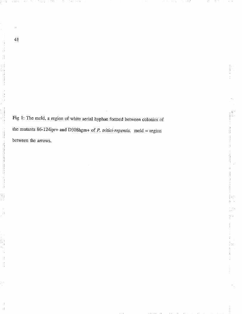
41
Fig 1: The meld, a region of white aerial hyphae formed between colonies of
the mutants 86-124ipr+ and D308hgm+ of p. triticirepentis. meld = region
between the arrows.
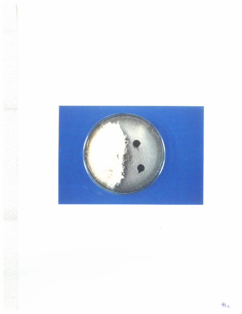
42
Table 1: Number of gerrninaæd conidia (Vo) obtatned from wild -type cultures, mutants,and from the meld formed between mutånts 86-l24ipr+ and D308hgm+ of P. tritici-repenrts on VSPDA amended with either iprodione or hygromycin B or both.
Germination (7o) on VSPDA amended with:
VSPDA ipr hgm ipr & hgmISOLATE alone
D308 wild type 100
D308ipr+ 100 100D308hgm+ 100 - 95 -86-124 wild type 10086-l24ipr+ 100 100
86-124hgm+ 100 - 10086-l24ipr+*D308hgm+ 100 25 40 11
ipr = iprodione, hgm - hygromycin B, ipr&hgm = both iprodione and hygromycin B.ipr+ = resistant to iprodione, hgm+ = resistant to hygromycin B, * = from the meld- = ûo germination.Vo = Of a tot¿l of 150 conidia obtained from 3 replicaæ heterokaryotic colonies.
43
Table 2: Mean growth in diameter (mm) afær 7 days of wild type cultures, mutants, and
subcultures with restricæd mycelial growth obtained from the meld between mutants 86-I24ipr+ and D308hgm+ of þrenophora tritíci-repentis on VSPDA amended with eitheriprodione or hygromycin B or both.
Diameter (mm)"on VSPDA amended with:
ISOLATE VSPDA ipr hgm iPr & hgmalone
D308 wild type 74 0 0 0
D308ipr+ L9 19 0 0
D308hgm+4404/.086-124 wild type 69 0 0 0
86-l24ipr+ 29 28 0 0
86-l24hgm+ 64 0 60 0
86-124ipr+*D308hgm+ 26 20
ipr = iprodione, hgm = hygromycin B, ipr & hgm - both iprodione and hygromycin B.ipr+ = resistant to iprodione, hgm+ = resistant to hygromycin B, * = from the meld- = no visible mycelial growth." mean of three replicate colonies.
44
Figure 2: Germination of conidia derived from mutanß g6-r24ipr+ (left) and
D308hgm+ (right) of Pyrenophora tritici-repentis and the fusion of a germ tube
with a conidial cell 6 h after incubation.
45
Fig 3: Mycelial characteristics and sporulation patterns of wild type isolates of
Pyrenophora triticì-repentis, parental mutants involved in the formation of the
meld and subculfures derived from the meld.
1) Heterokaryotic colony with sparse sporulation at the centre of the colony.
2) Heterokaryotic colony with sector
3) Illustration of both a wild-type and a mutant colony; such colonies have
similar characteristics with the sporulation over the enti¡e surface of the plate.
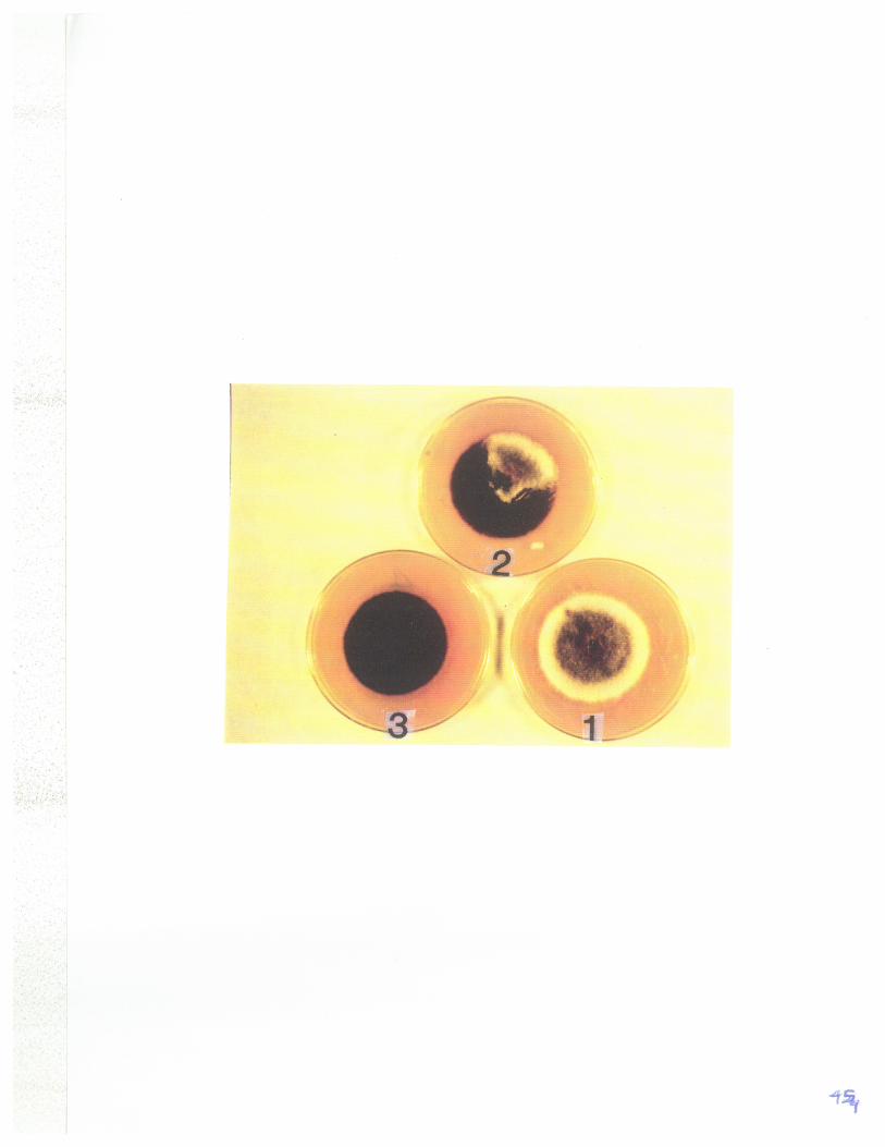
46
Stabílity
All hyphal plugs obtained from the subcultures with restricûed mycelial growth
consisæd of a single component that grew on VSPDA amended with both iprodione
and hygromycin B, and were heærokaryons. Hyphal plugs that were t¿ken from the
one cm band from the edge of the colony of all subcultures with normal mycelial
growth grew only on VSPDA plus hygromycin B (Table 3). Hyphal plugs that were
taken from the 1..5-2 cm band from the edge of the colony of all subcultures grew on
VBPDA plus both iprodione and hygromycin B. Hyphal plugs that were taken from
the 3 cm band from the edge of the colony were a mosaic that grew on VSPDA plus
iprodione, VSPDA plus hygromycin B and VSPDA plus both hygromycin B and
iprodione. The hyphal plugs that grew on VSPDA plus iprodione, did so after 7-10
days delay.
47
Table 3: Distribution of component genotypes in the subcultures with norrnal mycelialgrowth obtained from the meld betrveen mütants 86-124 ipr+ and D308 hgm+ of P.triticïrepentis on VSPDA afær 7 days of growth at 23-240C.
distance from Hyphal plugs (No.)growing on VSPDATcolony edge No. of amended with:to centre (cm) Plugs
tested/band ipr hgm ipr & hgm
200200150015t0 10* 10 10
ipr = iprodione, hgm = hygromycin B, ipr & hgm - both iprodione and hygromycin B.T - numbers a-re means of 3 replicate heterokaryotic colonies.* = 7-10 days delay in growth.
3
48
4.4 Test for virulence
Subcultures with reduced growth did not sporulate and were not tesred for
virulence. Since subcultures with normal mycelial growth sporulaæd poorly; conidia
from several plates were cornbined to provide an adequate inoculum concentration for
testing. The conidia induced both extensive chlorosis on line 68365 and small, da¡k-
brown spots on cv. Glenlea similar to that induced by D308hgm+ (Table.4). Conidia
that were reisolaæd from line 68365 and cultured on variously amended media, grew
on VSPDA plus hygromycin B, but not on VSPDA plus iprodione, or on VSPDA plus
both iprodione and hygromycin B. They also were similar to D308hgm+ in their
ability to grow on rhese media.
49
Table 4 : Virulence on cultivar Glenlea and line 68365 of the subcultures with normalmycelial growth obtained from the meld formed between mutants 86-124 ipr+ and D308hgm+ of P. tritici -repentis (A) and the ability of conidia reisolaæd from line 68365 togrow on amended media (B).
Cultivar reaction growth on media amended with:
Mutant Glenlea 68365 ipr hgm ipr & hgm
D308hgm+ - +
FIB-+
ipr = iprodione, hgm = hygromycin B, ipr & hgm = both iprodione and hygromycin B.ipr+ - resistant to iprodione, hgm+ = resistant to hygromycin B.HB = Heterokaryon with norrnal mycelial growth+ = virulent (A) or observed hyphal growth @)- = small dark-spots (A) or no visible hyphal growth (B)
s0
4:5 Discussion
The development of the meld between mutånts 86-l24ipr+ and D308hgm+
and the isolation of conidia from the meld that were able to grow on VSPDA amended
with both iprodione and hygromycin B demonstrate that anastomosis and heterokaryon
forrnation did occur between the paired strains. However, the incidence of
heærokaryosis was low since only I lbo ofthe conidia grew on the doubly amended
medium. Simila¡ findings were reported for C. sativus (Tinline 1962,1988), where
the heterokaryotic condition was caried by only 6-10 vo of the conidia.
Isolation of conidia with combined resistance to both iprodione and
hygromycin B suggests that the meld is associated with the transfer of genetic factors
between hyphal types, and the formation of heærokaryons in this fungus. The
dissociation of the initial cultures est¿blished by conidial transfer from the meld
indicaæd that the heterokaryon was not stable. The dissociation was demonstrated by
the mosaic pattern exhibited at the 3 cm band from the edge of the heærokaryotic
colony, and the absence of the 86-l24ipr+ component from the 1 cm bands. The
delayed growth exhibited by componentS6-l24ipr+ at the 3 cm band would explain
why it is eliminaæd from the I cm band. Since hyphae, conidia and ascospores of
this fungus are multinucleate (Whitehead and Dickson 1952; Wehmeyer 1954), the
instability could be due to differences in genetic make-up of the component nuclei or
to cultural conditions.
Several factors that alone or in combination could lead to .the formation of
unstable heærokaryons are: nuclear ratio imbalance; influence of cultural conditions;
51
limiæd anastomoses brought about by either a relatively slow radial growth of strains;
or allelic differences at single vegetative incompatibility loci (Pontecorvo et al. 1946;
Burnett 1975). Balanced heærokaryons are easily lost for a number of reasons
including back mut¿tions, non genetic adaptation, recombination through sexual
reproduction, formation of heterozygous diploid nuclei and accumulation in the
medium of metabolites enabling either or both components to grow independently
(Pontecorvo et al. 1953; Hartly and Williams l97l). Whether any of these reasons
is applicable in P. tritici-repenr¿s is not known and needs to be investigated.
The results of this study have demonstrated that two or more different nuclear
types can be associated in a mycelium by anastomosis between vegetatively
compatible hyphal types. The dissociation of the heterokaryon is indicative of the
const¿nt association and dissociation required for propagation of heterokaryons in
veget¿tively compatible hyphae. The phenotype of a heterokaryon is expressed as a
sum total of all nuclear types present in a mycelium and this suppresses the expression
of individual genotypes. The dissociation of the heærokaryon would allow the
expression of individual genotypes. These nuclear associations ensure exchange of
beneficial genotypes within the population through anastomosis, while vegetative
incompatibility would restrict sùch exchanges, leading to rhe formation of different
clones that would eventually differ through natural selection.
Heærokaryotic conidia or any other propagules are useful in ensuring survival
of an individual fungus or as a mechanism of bridging differences in environments.
For example, if a heærokalyotic mycelium is carrying vi¡ulence genes to certain
52
susceptible host genotypes that are currently out of season or not grown for some
specified reason, the virulence genes would be held within the heterokaryon until such
a time that favourable conditions do occur. Then, due to any of the reasons stated
above, the heærokaryon could dissociaæ and release the virulence genes within the
vicinity of the susceptible host cultivars. The occurrence of such a phenomenon in
nature would provide the pathogen with susceptible host tissues, on which it would
reproduce abundantly and cause severe epidemics, particularly in those crops which
are grown as monocultures of homozygous susceptible genotypes. Transient
heterokaryosis may therefore allow the pathogen to adapt to changes in the
environment.
There is no simple explanation for the failure of the heærokaryotic colonies to
induce both tan necrosis and extensive chlorosis on the respective differentials. In this
study, subcultures with normal mycelial growth sporulated sparsely at the centre of the
colony, and the resultant conidia.induced only extensive chlorosis. Conidia that were
reisolaæd from line 68365, could only grow on VSPDA plus hygromycin B but nor on
either V8PDA plus iprodione or VSPDA plus both iprodione and hygromycin B,
indicating that they were similar to D308 hgm+ but nor 86-124 ipr+.
These results may be explained by assuming that hyphae of the heærokaryotic
subcultures are homokaryotic diploids or haploids. If the two component nuclear
types fused to form diploids, then one type (D308hgm+) is dominant or epistatic to the
other. However, since the 86-l24ipr+ component is screened out in the 2 and 1 cm
bands, there is no possibility of these nuclei being diploids. The presence of haploid
53
nuclei would mean that there are post-fusion incompatibility factors in the
protoplasmic portion of the mycelium contributing to the instability of the
heærokaryon (Garnjobst and Wilson 1956; Boucherie et aI. 1976). Future research to
elucidaæ the incompatibility systems in this fungus will need.to address these issues.
The results of this study suggests that heærokaryosis can occur in P. trítici-
repentis, that the process has the poæntial of creating and concealing genotypic
variation and/or is a temporary reservoir of genetic potential whereby genotypes are
released in appropriate selective envi¡onments th-rough sectoring, and distributed via
conidia.
54
5. Inheritance of virulence and toxÍn production ín þrenophora tritíci-repentß.
ABSTRACT
Isolaæs of Pyrenophora tritíci-repentis,the causal agent of tan spot of wheat, are
currently grouped into four pathotypes [(nec+chl+, nec+chl-, nec-chl+ and nec-chl-)],
based on their ability to cause tan necrosis (nec) and chlo¡osis (chl) on differential
cultivars of wheat. Isolates that induce tan necrosis also produce Ptr-necrosis toxin.
To investigate the inheritance of virulence and toxin production, twelve isolates, at
least one from each pathotype, were paired in all combinations. The isolate pairs were
observed fo¡ the formation of fertile pseudothecia at the line of junction. Fertile
pseudothecia were obtained only from the pair of isolates 86-124 (nec+chl-) and
Hy331-9 (nec-chl+). Ascospores from individual pseudothecia and complements of 8
ascospores from single asci were analysed for virulence and for the production of Ptr-
necrosis toxin on differential cultivars of wheat. Sixæen percent of the 101
pseudothecia sampled contained ascospores that segregated in aratio of 1:1 (parental:
recombinants). Complements of 8 ascospores from each of five individual asci
segregated in a ratio of 1:l:1:1 for the four disease phenotypes and in a ratio of l:l(Ptr-necrosis toxin producer : non-producer). These data suggested the involvement of
two genes, one controlling the ability to induce t¿n necrosis, the other extensive
chlorosis. The factor for Ptr-necrosis toxin was associated with the factor for tan
necrosis. Progeny from the inter-cross of F, belonging to pathotype I and 4
segregated in a ratio of 1:1 (parentat : recombinants) and three single asci segregated
in a ratio of 1:1:1:1 for the four disease phenotypes and in a ratio of 1:l for (Ptr-
55
necrosis producer : non-producer) further supporting the involvement of two genes.
5.1. INTRODUCTION
Pyrenophora triticïrepentis (Died.) Drechs (Conidial state : Drechslera triticï
repentis (Died.) Shoem.) the cause of tan spot of wheat (Triticum aestivum L.) is now
wide spread throughout the world (Duff 1954: Hosford 1977; Morrall and Howard
1975; Summerell and Burgess 1989). P. triticïrep¿zns infects many grass species
(Hosford I97I; Krupinsky 1982, 1987; Morrall and Howard 1975) and has the
widest host range of any þrenophorø species (Shoemaker 1962).
In recent years, the prevalence and severity of t¿n spot have increased to
damaging levels and yield losses in spring wheat of 12.9 to 49Vo have been reported in
moist weather (Hosford and Busch 1974; Rees ¿r al. l98I). Ascospores discharged
from pseudothecia on wheat stubble are the main source of primary inoculum.
Increase in incidence and severity of t¿n spot in recent years has been associated with
the adoption of conservation tillage practices in North America and Australia whereby
stubble remains on the surface of ttre soil. In Australia, the high susceptibility of
most modern wheat cultivars has also been reported to be a contributing factor to
disease severity (Rees et aI. 1988).
Recent studies have shown that the tan spot syndrome consists of two host
phenotypes which are tan necrosis and chlorosis (Lamari and Bernier 1989a,b). These
are each the result of specific interactions between individual isolaæs of the fungus
and wheat genotypes. Susceptible wheat lines develop tan necrosis and/or extensive
chlorosis, whereas resist¿nt lines develop small, da¡k-brown lesions without

56
accompanying tan necrosis or extensive chlorosis (Lamari et al. 1991). Resistance to
tan necrosis is recessive and resistance to extensive chlorosis varies from dominant to
partially dominant. Each of these reactions is conrolled by one independenr gene
(Lamari and Bernier 1991). However, preliminary results from an on-going study
(Duguid, S.; personal comm.) and other reports (Nagle et aI . 1982;Sykes and Bernier
1991) indicaæ that resistance to tan necrosis in some cultivars of wheat is controlled
by more than one gene.
The existence of variability in aggressiveness and virulence has been reporæd
among isolates of P. tritíci-repentis (Ikupinsky 1992b). Luz and Hosford (1980)
tested 40 isolates from a wide geographic area and differentiated 12 races on the basis
of the amount of spotting caused on a differential set of six wheat and one barley
cultiva¡. Misra and Singh (1972) tesæd th¡ee isolates on 50 wheat genotypes and
found significant differences in the amount of disease caused by the different isolates.
More recently, isolates of P. tritici-repentis have been assigned to one of four
pathotypes based on the ability of isolates to induce in appropriate differential cultivars
either tan necrosis or exûensive chlorosis (compatible/virulent phenotypes) or small,
dark-brown lesions (incompatible/avirulent phenotypes) (Lamari and Bernier 1989b).
Isolates of pathotype I induce both tan necrosis and extensive chlorosis (nec+chl+),
pathotype 2 only tan necrosis (nec+ chl-), pathotype 3 only extensive chlorosis (nec-.
chl+) and pathotype 4, classified avirulent (nec-chl-), induces small, dark-brown
lesions without accompanying tan necrosis or chlorosis.
Susceptibility of wheat genotypes to necrosis inducing isolates and sensitivity
57
to the Ptr-necrosis toxin released specifically by these isolates are controlled by the
same dominant gene (Lamari and Bernier 1989c). Isolates that induce only extensive
chlorosis do not produce Ptr-necrosis toxin in-vitro.
The ability to sexually cross a pathogen is important as a means of
deærmining the genetic control of virulence within the population. The only
þrennphorc species that have been crossed and genetically analysed for virulence are
P. teres and P. gramínea. Both species have bipolar (two mating types required for
fertility) heærothallic mating systems (McDonald 1963; Smedegard-Pererson 1977)
whereas P. tritícïrepentis is homothallic.
Most isolates of P. triticí-repentís fsm fertile pseudothecia when grown singly
or in pairs. If there is any sexual differentiation of nuclei, then the multinucleate
character of the hyphae may distribute both types of.nuclei to all cells equally
(Whitehead and Dickson 1952: Wehmeyer 1954). In an attempt to cross P. tritíci-
repentis, Díaz de Ackerrnan et al. (1988) obtained fertile pseudothecia by culturing
isolaæs singly and in pairs on wheat seeds and corn leaves placed on water agar in
petri plates, and with one exception they found that all isolates formed pseudothecia.
However, they did not indicate whether all pseudothecia cont¿ined asci or whether the
¿rscospores they obtained from the pairings were hybrids or selfs.
Genetic analysis of this homothaltic fungus was difficult until recently when
mutants resistant to ttre fungicide iprodione or to the antibioúc hygromycin B were
created in isolaæs 86-124 (nec+chl-) and D308 (nec-chl+) using ultraviolet light
mutagenesis, and these were then used in sexual crosses (McCallum et at. 1994). The
58
mutants were crossed in all possible combinations and the progeny analysed to
deærrnine the genetic basis of chemical resist¿nce. Hybrid progenies were obtained
in crosses involving mutants of isolaæ 86-124 but not in crosses involving mutants of
isolaæ D308 nor in crosses between mutants of isolaæs 86-124 and D308 which
belong to different pathotypes. The ¡esults indicaæd that one locus controls resistance
to iprodione and a second independent locus controls resistance to hygromycin B. In
a subsequent study, analysis of conidia from the meld formed between mutants of the
two isolates (86-124 x Hy331-9) indicated that heærokaryons formed but dissociared
(In thesis, manuscript 1). The objectives of this study were to investigate the
possibility of crossing isolates from different pathotypes and to determine the genetic
control of virulence and toxin production in isolates of P. triticí-repentís using specific
host resistance/susceptibility phenotypes as genetic ma¡kers. The results of this work
are intended to complement studies on the inheritance of resistance to tan spot in the
host and to provide information on the potential for development of isolaæs with
inc¡eased virulence. Both are critical for the design of effective breeding programs
searching for durable resistance to tan spot in wheat.
5.2. MATERIALS AND METHODS
5.2.I Matings
The isolaæs of P. tritici-repenris used in this study were previously
cha¡acterized for virulence and production of Ptr-necrosis toxin by Lamari and Bernier
(1989b). The colony cha¡acteristics and virulence of the isolates used in this study are
shown in Tables 5 and 6. Each isolare was cultured on vSpDA (150 ml of vg
59
vegetable juice, 10 g potato dextrose agar,3 g calcium carbonate, l0 g of Bacto agar,
850 ml distilled waær) by inoculating the medium with a single conidium as described
by Lamari and Bernier (1989a, b). Matings were set up in polystyrene petri dishes on
autoclaved wheat leaves placed on Sachs' nutrient agar (Appendix 1), a method
slightty modified from McCallum et al. (1994). All of the twelve isolaæs comprising
at least one from each pathotype, were paired in all possible combinations by placing a
4- d-old mycelial plug (4 mm diameter) of each isolate pair at opposiæ ends of the
leaves. The plates were sealed with parafilm and incubated at 22-250C in the dark for
7 d and then tansferred to 10-150C in the dark for 8-12 weeks. The plates were
observed for formation of fertile pseudothecia at the line of junction of the paired
isolates.
5.2.2 Isolation of ascospores
Pseudothecia were uansfemed singly with a sterile forceps to plates of double
strength water agar (20 g agar/L water). lndividual pseudothecia we¡e crushed by
applying gentle pressure with the forceps to release asci. Mature asci were broken by
this process thus releasing ascospores randomly on the surface of the medium. læss
mature asci have harder bitunicaæ membranes which were not readily broken by the
pressure of the forceps. Under a dissecting microscope, these intact asci were selected
and separated from each other with a fine-tipped needle. To release ascospores from
such asci, a drop of cell-wall degrading enzyme (5Vo þ-glucuronidase) was applied
over the ascus (Yoder 1988) and allowed to digest the ascus wall until the ascospores
were released (approximaæly 30 minutes). Forty random ascospores were picked from
60
individual pseudothecia (pseudothecial analysis) and, whenever possible, complements
of eight ascospores from a single ascus were also obtained (ascal analysis). Individual
ascospores were subcultured on numbered V8PDA plates to identify the
pseudothecium and/or the ascus from which each was isolaæd. The numbering of the
spores from a single ascus was random since they are not produced in ordered fashion.
One half of each pseudothecium with a few intact asci and some ascospores was
retained and stored at -l7oc as described by McCallum ¿r aI. (1994) for possible
further analysis, if some of the ascospores sampled were found to be recombinant
types.
5.2.3 AnalysÍs of progeny for virulence
Seedlings of the differential wheat cv. Glenlea (susceptible only to tân necrosis) and
tine 68365 (susceptible only to extensive chlorosis) and of the resistant cvs. Erik and
Salamouni, were grown in 7.5 x 7.5 cm milk cartons containing soil-less mix
(Metromix 200, W. R. Grace and Co: Ajax, Ont¿rio) in growth chambers at 18-200C
with 16 h photoperiod (ca: 180¡rE.m.-2s't). Three seedlings of each culrivar were
planæd opposiæ each other in each of three cartons..
Subcultures of individual ascospores were incubaæd in the dark at 200C until
the colonies reached about 4-5 cm in diameter as described by Lama¡i and Bernier
(1989b). They were then flooded with sterile distilled water, the mycelium was
flatæned with a flamed test tube bottom, and excess water decanted. The cultures
were incubated for 18-24 h at room temperature (20-240C) under light (ca: 90 ¡rE.m.-
2.s-t) provided by th¡ee cool white fluorescent tubes, followed by 18-24 h in the da¡k
61
at 15 0C. Spores were suspended in distilled water by using a wire loop and, inoculum
concentration was measured with a cell counter (Hauser Scientific, Blue Bell, PA) and
adjusæd to 3.5 x 10³ per millimeter with distitled water.
Test seedlings of each of the four cultivars or line at the nvo leaf stage, were
inoculaæd with the conidial suspension containing æn drops of Tween 20
(polyoxyethlenesorbitan monolaurate) per lite of spore suspension, and the inoculum
applied with a Devilbis sprayer at 69 Kpa (10 psi) to the point of impending run-off as
described by Lamari and Bernier (1989a, b, 1991).
The seedlings were incubaæd for 24 h at 220C in a chamber under continuous
leaf wetness provided by two computer controlled ultrasonic humidifiers filled with
distilled water. The plants were then transferred to a growth room bench kept at
22ll80c (daylnight) and a 16 h photoperiod (ca: 180 ¡rE.m.-2.s-t), and scored after 6-7 d
for their reaction. Seedlings inoculated with conidia from parental isolates and non-
inoculated seedlings were used as checks.
Avirulence on cv. Glenlea was expressed as small, dark-brown lesions and
virulence as t¿n necrotic lesions. Avi¡ulence on line 68365 was expressed as small,
dark-brown lesions and virulence as extensive chlorosis (Fig. 4). Progenies found to
be vi¡ulent or avirulent on both differentials were considered to be recombinants and
the result of fertilization rather than selfing. A pseudothecium or ascus was
considered to be selfed if no segregation was observed amongst their ascospore
progenies and were therefore omitted from the analysis. The test for independence of
virulence factors was based on the hypothesis that if the factors are inherited
62
independently of each other, half of the progeny will be like the parents and half will
exhibit new combinations of factors. Chi-square values were calculaæd for the
deviations of the observed number of recombinants from the expecûed.
63
Table 5: Colony characæristics of the P. triticïrepenr¿s isolaæs used in the study.
ISOLATE COLOUR PIGMENT TISSUE" STABILN'P
ASCl86-r24D308331-1331-2331-3331-4331-533t-6331-8331-9331-12
dark-greygreywhiæ-greywhiæwhite-greywhite-greywhiæ-greywhiæ-greywhite-greygreywhiæwhite
dark-greydark-greygrey-orangecreamcreamcreamdark-greycreamgrey-orangedark-greycreamgrey
stablestablestablemoderatemoderateunstablemoderatemoderateunstableunstablemoderatemoderate
++
+
+
a) +l- = presence or absence of sclerotial tissue in cultureb refers to frequency of forming sectors.stable = no sectorsmoderate=afewsectorsunstable = many sectors

64
Table 6: Virulence patterns of the isolates of P. triticí-repentis on the two differentialcultivars of wheat.
ISOLATE CULTTVAR
Glenlea 6836s
ASCl86-124D30833r-r331-2331-3331-4331-533t-6331-8331-9331-r2
nec+nec+nec-nec-nec-nec+nec+nec-nec-nec+nec-nec-
chl+chl-chl+chl+chl+chl-chl+chl+chl+chl+chl+chl+
nec+chl+nec-chl-
presence of tan necrosis on Glenleapresence of chlorosis on 68365absence of tan necrosis on Glenleaabsence of chlorosis on 68365
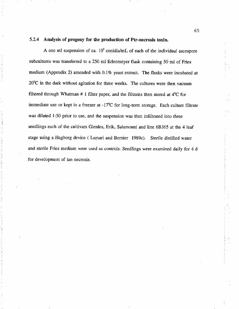
65
5.2.4 Analysis of progeny for the production of Ptr-necrosis toxin.
A one ml suspension of ca. 103 conidia/ml of each of the individual ascospore
subcultures was transferred to a 250 ml Erlenmeyer flask containing 50 ml of Fries
medium (Appendix 2) amended with 0.17o yeast extract. The flasks were incubated at
200C in the da¡k without agitation for th¡ee weeks. The cultures were then vacuum
filtered through Whatman # 1 filær paper, and the filtraæs then stored ar 40C for
immediate use or kept in a freezer at -l7oc for long-ûerln storage. Each culture filtrate
was diluted l:50 prior to use, and the suspension was then infiltrated into tluee
seedlings each of the cultiva¡s Glenlea, Erik, Salamouni and line 68365 at rhe 4leaf
stage using a Hagborg device ( Lqnari and Bernier 1989c). Sterile distilled water
and sterile Fries medium were used as controls. Seedlings were exarnined daily for 4 d
for development of tan necrosis.
66
Fig. 4- Reactions of the two differential cultiva¡s of wheat ro the isoiates of p. titicïrepentis.
a) Tan necrotic lesions induced by isolates of pathotype 1 (nec+chl+) (left)
and pathotype Z (nec+chl-) (righÐ on culriva¡ Glenlea.
b) Extensive chlorosis induced by isolates of pathorype 1 (nec+chl+) (left)
and 3 (nec-chl+) (right) on line 68365.
c) small, dark-brown lesions induced by isolates of pathotype 4
(avirulent) on culriva¡ Glenlea (left) and rine 68365 (righÐ.
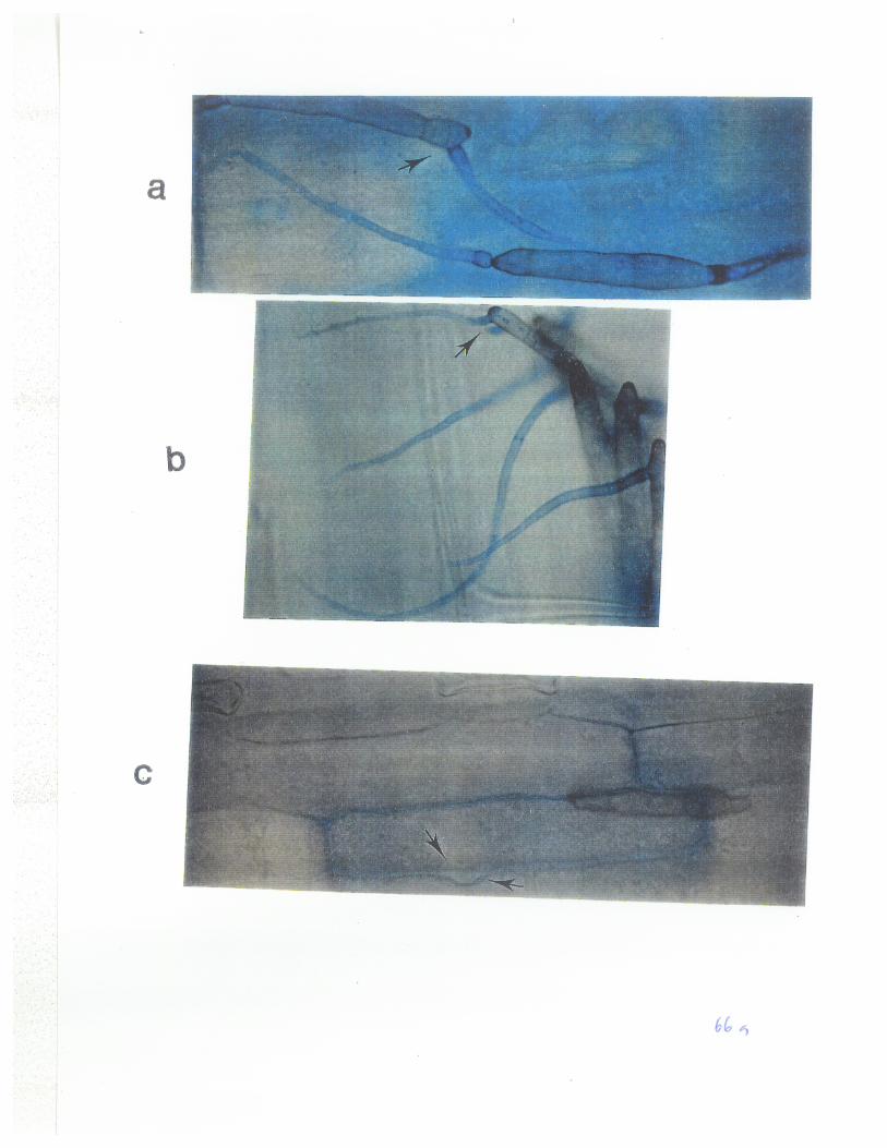
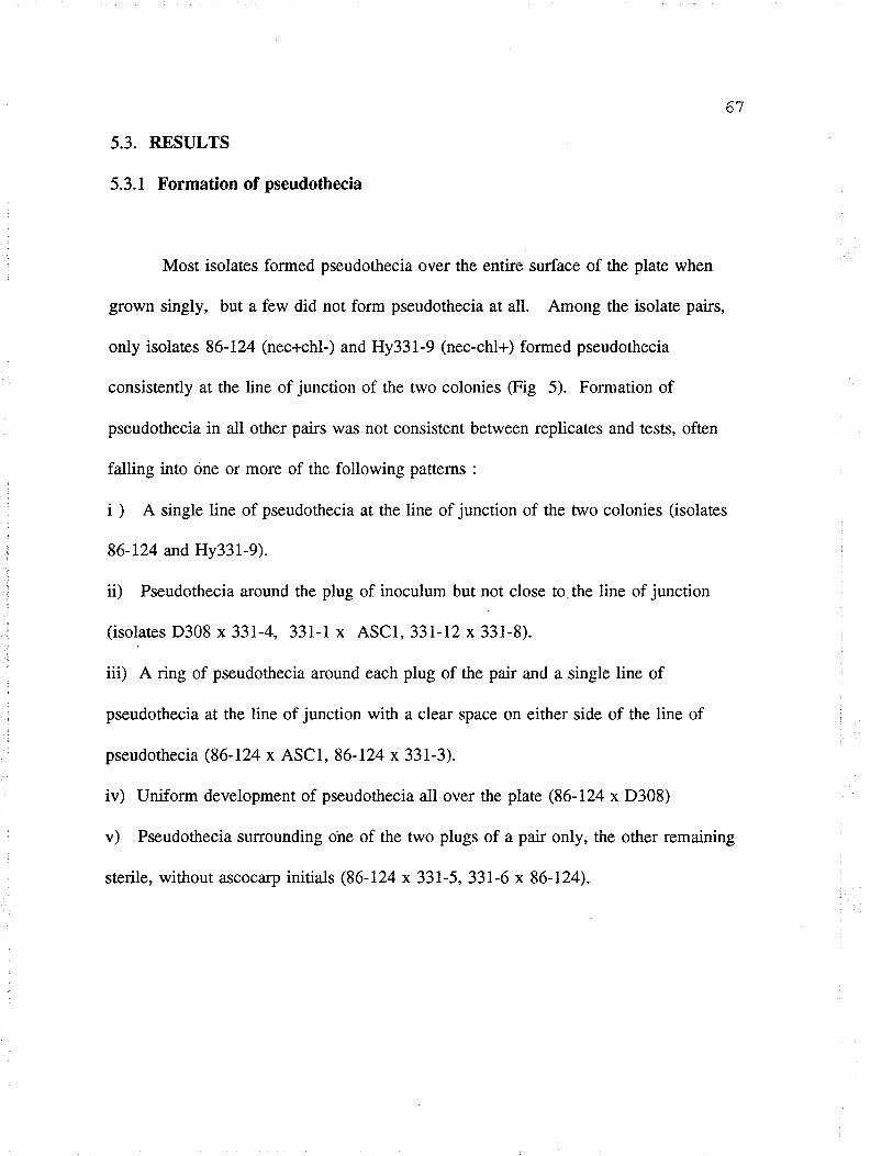
67
5.3. RESULTS
5.3.1 Formation of pseudothecia
Most isolaæs formed pseudothecia over the entire surface of the plaæ when
grown singly, but a few did not form pseudothecia at all. Among the isolate pairs,
only isolaæs 86-124 (nec+chl-) and Hy331-9 (nec-chl+) formed pseudothecia
consistently at the line of junction of the two colonies (Fig 5). Formation of
pseudothecia in all other pairs was not consistent between replicates and tests, often
fatling into one or more of the following patterns :
i ) A single line of pseudothecia at the line of junction of the two colonies (isolates
86-124 and Hy331-9).
ü) Pseudothecia around the plug of inoculum but not close to the line of junction
(isolates D308 x33l-4,331-1 x ASCI, 331-l2x 331-8).
iii) A ring of pseudothecia around each plug of the pair and a single line of
pseudothecia at the line of junction with a clear space on either side of the line of
pseudothecia (86-124 x ASCI, 86-124 x 331-3).
iv) Uniform development of pseudothecia all over the plate (86-124 x D308)
v) Pseudothecia surrounding ohe of the two plugs of a pair only, the other remaining
sterile, without ascocarp initials (86-124 x 331-5, 331-6 x 86-124).
68
Fig 5: A single line of pseudothecia formed ar the line of junction of isolates
86-124 (nec+chl-) and Hy331-9 (nec_chl+).
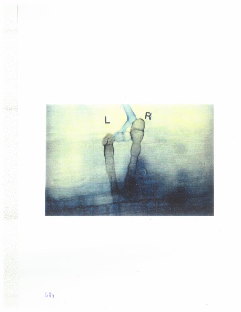
69
5.3.2 Analysis of pseudothecÍa
A tot¿l of 101 pseudothecia obtained from the line of juncrion of isolates 86-
I24 and Hy33l-9 were analysed forfertility (presence of asci and ascospores in a
pseudothecium) and fo¡ the virulence of individual ascospore progenies on the tan spot
differential cultivars of wheat. About 62.4Vo of the pseudothecia were sterile, forming
immature ascocarps or pseudothecia-like bodies without asci (Table 7). Only 167o of
the examined pseudothecia (42Vo of fertile pseudothecia) contained ascospores that
induced tan necrosis on cv. Glenlea and extensive chlorosis on line 68365 (nec+chl+)
or small, dark-brown lesions (nec-chl-) on both differentials, and were therefore
recombinants. Twenty two percent of all the pseudothecia (57.97o of the fertile)
contained ascospores that were virulent only on cv. Glenlea or line 68365 and were
therefore selfs. None of the ascospore progenies were virulent on cvs. Erik and
Salamouni. Analysis of the data indicated that random ascospores obtained from
individual hybrid pseudothecia segregaæd in a ratio of 1:l parentals: recombinants
(Xt = 1.10, P = 0.50-0.10) except for ascospores from pseudothecium P8 (Table 8)
which did not fit a l:l ratio.
70
Table 7: Frequency of hybrid pseudothecia from a cross between isolates 86-124 andHy331-9 of P. titicí-repentis as indicaæd by the virulence phenorypes.
Number of pseudothecia.
Plaæ # ASSAYED STERILET FrrBRlD* SELFED*
130194721282231510234137335171223614734TOTAL 101 62.4Vo I6Vo 2L.8Vo
t Absence of asci and ascospores* Contained ascospores that were virulent and avirulent to both cultivar
Glenlea and line 68365.* Contained ascospores that were virulent to either cultivar Glenlea or line 68365.
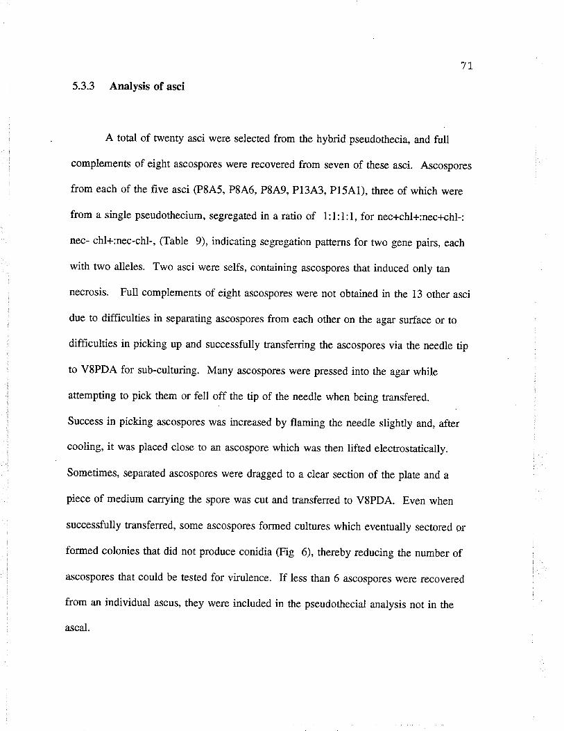
7t
5.3.3 Analysis of asci
A total of twenty asci were selecæd from the hybrid pseudothecia, and full
complemens of eight ascospores were recovered from seven of these asci. Ascospores
from each of the five asci (P8A5, P846, P849, P1343, P15Al), th¡ee of which were
from a single pseudothecium, segregated in a ratio of l:1:l:1, for nec+chl+:nec+chl-:
nec- chl+:nec-chl-, (Table 9), indicating segregation patterns for two gene pairs, each
with two alleles. Two asci were selfs, containing ascospores that induced only tan
necrosis. Full complements of eight ascospores were not obtained in the 13 other asci
due to difficulties in separating ascospores from each other on the agar surface or to
difficulties in picking up and successfully transferring the ascospores via the needle tip
to VSPDA for sub-culturing. Many ascospores were pressed into the agar while
attempting to pick them or fell off the tip of the needle when being transfered.
Success in picking ascospores was increased by flaming the needle slightly and, after
cooling, it was placed close to an ascospore which was then lifæd electrostatically.
Sometimes, separated ascospores were dragged to a clear section of the plaæ and a
piece of medium carrying the spore was cut and transferred to V8PDA. Even when
successfully transferred, some ascospores formed cultures which eventually sectored or
formed colonies that did not produce conidia (Fig 6), thereby reducing the number of
Ílscospores that could be tested for vi¡ulence. If less than 6 ascospores were recovered
from an individual ascus, they were included in the pseudothecial analysis not in the
ascal.
72
Table 8: Segregation for virulence ¿unongst randomly isolaæd ascospores from hybridpseudothecia in the cross between isolaæs86-124 (nec+chl-) and Hy331-9 (nec-chl+)of P. tritícïrepentís.
PSEUD".PROB.
#
PARENTAL RECOMBINANTS x2
(nec+chl-)(nec-chl+) (nec+chl+)(nec-chl-)
P2P3P6P8Pl0P13P15Total
t4t69
9
8
l36
75
2
J10
l110
1
7249
1l 7
1241636214 I1539583 25
0.110.250.005.140.270.250.s0r.10
0.s0-0.9s0.50-0.9s0.95-0.990.01-0.05*0.s0-0.9s0.s0-0.9s0.10-0.s00.10-0.50
* Ratio of parental : recombinants differ significantly (5 Vo level ) from the expected1 : I ratio. Some, not all, ¿rscospores did produce conidia.a = pSeudothecium
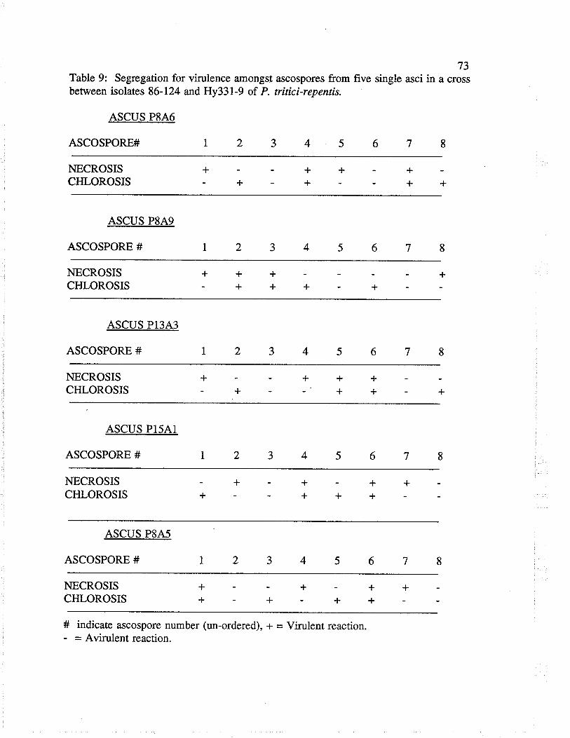
73Table 9: Segregation for virulence amongst ascospores from five single asci in a crossbetween isolates 86-124 and Hy331-9 of P. tritici-repentís.
ASCUS P8A6
ASCOSPORE#1234s678
NECROSISCHLOROSIS
ASCUS P8A9
ASCOSPORE #
++
++
+NECROSISCHLOROSIS
ASCUS P13A3
ASCOSPORE #
+NECROSISCHLOROSIS
ASCUS P15A1
ASCOSPORE #
NECROSISCHLOROSIS
ASCUS P8A5
ASCOSPORE #
NECROSISCHLOROSIS
# indicaæ ascospore number (un-ordered), * = Virulent reaction.- = Avirulent reaction.
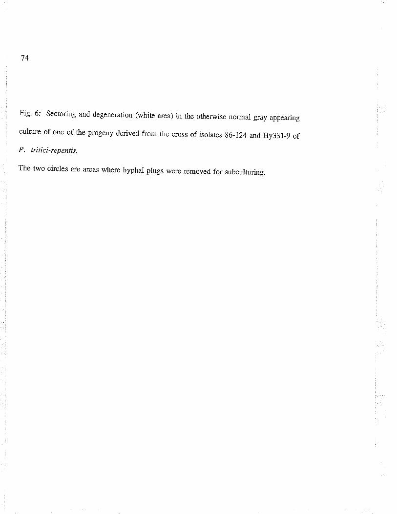
74
Fig' 6: Sectoring and degeneration (whiæ area) in the otherwise normal gray appearing
culture of one of the progeny derived from the cross of isolates g6-Lz4and Hy33l-9 of
P. tritici-repentis.
The two ci¡cles are a-reas where hyphal plugs were removed for subcuituring.

75
5.3.4 Backcross and inter-cross of F, progeny.
Backcrosses of F, progeny types nec+chl+ and nec-chl- to either parent (86-124
nec+chl- or Hy331-9 nec-chl+) never formed a single line of pseudothecia at the line
of junction. Pseudothecia were scattered over the entire surface of the plaæ with no
clear pattern. Some replicates of the inter-crosses of the two types of progeny
(nec+chl+ x nec-chl-) formed a single line of pseudothecia at the junction, but others
did not. To improve on the chances of recovering recombinant ascospores, only the
pseudothecia that formed along such lines of junction were analysed for ¿Ìscospore
fertility and virulence. Eighty nine percent of these pseudothecia were sterile as
compared to 62.4Vo in the initial cross of isolates 86-124 x Hy331-9, and only 25Vo of
the fertile pseudothecia were hybrids compared to 42Vo in the initial cross (Table l0).
This indicates a possible increase in sterility and a decrease in number of hybrid
pseudothecia with continued subculture of the strains of this fungus.
Analysis of the progeny from the inter-cross for virulence showed that the
same four disease phenotypes as in the initial cross of isolates 86-124 and Hy331-9
were expressed. Random ascospores obtained from these pseudothecia segregated in a
l:1 ratio, parental (nec+chl+, nec-chl-): recombinant (nec-chl+, nec+chl-) (Tabte l1)
and ascospores from 3 individual asci segregated in 1:1:l:l ratio for the four disease
phenotypes (fable 12), supporting the previous interpretation rhat each of the two
isolaæs has two loci, one controlling the ability to induce tan necrosis and a second
locus controlling the ability to induce extensive chlorosis.
76
Table 10: Frequency of hybrid pseudothecia in the inter-cross of F, progenies(pathotype 1 and 4) from the cross of isolates 86-724 and Hy331-9 of P. tritíci-repentisas indicated by the virulence phenorypes
Number of Pseudothecia.
Plate # Assayed Særilef Hybridf Selfed*
1604929257541333937024484134Total 204 897o 3Vo 9Vo
f Absence of asci and ascospores.
* Contained ascospores that were virulent and avi¡ulent to both cultivarGlenlea and line 68365.
* Contained ascospores that were virulent to either cultivar Glenlea or line6836s.
77
Table 1l : Segregation for virulence amongst randomly isolaæd ascospores from hybridpseudothecia in the inter-cross of F, progenies (pathotype 1 and 4) from the cross ofisolaæs 86-124 and Hy331-9 of P. tríticïrepentís.
Pseud#T. Parental type Recombinant type Ratio l:l
(nec+chl+) (nec-chl-) (nec+chl-) (nec-chl+) yz Prob.
P1
P2P3P4P5Total
96
310
7
35
10
t2t48
t764
49
610
5
34
t611
t27
18
64
0.50 .s0 - .702.00 .10 - .200.50 .50 - .700.s0 .s0 - .700.s0 .s0 - .700.50 .50 - .70
T = Pseudothecium
78
Table 12 : Segregation for virulence amongst ascospores from three single asci in theinter-cross of F, progenies (pathotype 1 and 4) from the cross of isolates 86-124 andHy331-9 of P. tritíci-repentis.
ASCUS (IC) PIA4
Ascospore# L 2
NecrosisChlorosis
ASCUS OC) P3A7
Ascospore # I 2
NecrosisChlorosis
++
++
+
ASCUS ûC) P3A1
Ascospore# I 2
NecrosisChlorosis
+ +
# indicaæ ascospore number (un-ordered), * = Virulent reaction- = Avirulent reaction
ty
5.3.5 Analysis of progeny for the production of Ptr-necrosis toxin.
To assess whether the factor for tan necrosis in recombinant progenies
possessing virulence to both cv. Glenlea and line 68365 (nec+chl+) was associated
with the ability to produce Ptr-necrosis toxin in culture, cultures derived from
individual ascospores from each of the eight asci (P8A5, P849, P1343, P1541,
P846, IC P144, IC P3A7 and IC P3A1) were assayed for the production of Ptr-
necrosis toxin. For each of the eight asci, four of the eight ascospore progenies æsted
produced Ptr-necrosis toxin (Tables 13 and 14). Each ascospore progeny that
produced Ptr-necrosis toxin also induced tan necrosis (Tables 9, 12, 13 and 14).
Those that did not produce Ptr-necrosis toxin did not induce tan necrosis. The Ptr-
necrosis toxin produced by these progenies had no effect on either cv. Erik or
Salamouni noron line 68365. The 64 ascospores segregated in aratio of l:1, Ptr-
necrosis toxin producers : non-producers, indicating that a single gene pair controls the
production of Ptr-necrosis toxin and that the ability to produce Pr-necrosis toxin is
associaæd with the ability to induce tan necrosis. This confirms previous findings by
Lamari and Bernier (1989c) that sensitivity to the Ptr-necrosis toxin in the host is
closely associaæd with susceptibility to tan necrosis.
80
Table 13: Segregation for production of Ptr-necrosis toxin amongst the ascosporeprogeny from five asci in a cross between isolaæs 86-124 and Hy33l-9 of p. trítici-repentis.
ASCUS P8A6
ASCOSPORE # 1
TOXIN +++
ASCUS P8A5
ASCOSPORE # 1
TOXIN ++
ASCUS P8A9
ASCOSPORE # 1 6
TOXIN
ASCUS PI3A3
ASCOSPORE # 1
TOXIN ++
ASCUS P15A1
ASCOSPORE # 1
TOXIN
+l- = presence or absence of Ptr-necrosis toxin in a culture filtraæ when tested oncultivar Glenlea. # indicate ascospore number (un-ordered)
+++
81
Table 14 : Segregation for production of Ptr-necrosis toxin Írmongst the ascosporeprogeny from three single asci in the inúer-cross of F, progenies (pathotype 1 and 4) fromthe cross of isolaæs 86-124 and Hy331-9 of P. triticí-repentis.
ASCUS GC) P1A4
Ascospore# I 2
Toxin
ASCUS ûC) P3A7
Ascospore # 1 2
Toxin
ASCUS (IC) P3AI
Ascospore# 1 2
Toxin
# indicate ascospore number (un-ordered)+/- = Presence/absence of Ptr-necrosis toxin when tested on cultiva¡ Glenlea.
+++
+
+
82
5.4 Discussion
The development of a single line of pseudothecia at the line of junction of
isolaæs 86-124 and Hy33l-9 and the subsequent recovery of both recombinant and
parental type ascospore progenies from individual asci and pseudothecia is significant
in that it indicaæs the existence of mechanisms that regulate out-crossing and selfing
in the population of this homothallic species. Analysis of ascospores from rwenry
individual asci indicaæd that five asci contained progenies that were either virulent
. to both cv. Glenlea and line 68365 or avirulent to both, and that the virulence of the
other ascospores was like that of the the parental strains. This shows that both
nec+chl+ and nec-chl- progeny were present in individual asci as well as in individual
pseudothecia and indicates conclusively that fertilization occurred between the two
parental isolates. This is important since the presence of both selfed and crossed asci
within a pseudothecium has been reported in P. tritíci repentis (McCaltum et at. 1994)
and within a perithecium in Cryphonectria parasitica (Anagnostakis 1984, 1988) and
a cleistothecium in Aspergillus nidulans (pontecorvo et al. 1953).
Matings amongst the 12 isolates showed that isolates 86-124 and Hy33l-9
formed pseudothecia consistently at the line of junction, from which progenies with
recombined virulence were obtained. The inconsistent patterns of pseudothecia
formation between most pairs of isolates indicated that there is an incompatibilty
syst€m operaring in the population of this fungus.
The recovery of both the nec-chl- and nec+chl+ progeny in addition to the
parental types from the cross of two virulent isolaæs 86-124 (nec+chl-) and Hy331-9
83
(nec-chl+), indicaæs that the parental isolates differ at two loci, and that there are two
genes for virulence in each isolate. An allele of one gene pair induces t¿n necrosis
(virulent) on cultivar Glenlea and the other induces small, dark-brown lesions
(avirulent) on line 68365. One allele of the other gene pair induces exûensive
chlorosis (virulent) on line 68365 and the other induces small, da¡k-brown lesions
(avi¡ulent) on cultivar Glenlea. This explains the recovery of four different disease
phenotypes from one meiotic event in the individual asci. Recombinant progenies that
were obtained from the initial cross of the parental isolates and those that were
obtained from the inter-cross of the F, progeny indicate that the four pathotypes can be
reconstituûed by sexual recombination.
The ratio of 1:1 for (parental : recombinant) segregation into random
ascospores of a pseudothecium would be expected on the basis of ascal analysis or if
this fungus was heterothallic. Since P. triticí-repentìs is homothallic, hybrid
pseudothecia contain both crossed and selfed asci and pseudothecial analysis would
therefore not be expected to show a 1:1 ratio. Pseudothecial analysis indicated a
2:l:2:1 ratio for the four disease phenotypes which is not consistenr with the
hypothesis of independent loci. This may be due to the large number of selfed asci in
the individual pseudothecia. Ascal analysis was consistent with a 1:1:1:l ratio for the
four disease phenotypes suggesting the involvement of two genes, each with two
alleles. The sample size of the asci that were to be analysed was reduced by the
difficulty involved in isolating all the eight ascospores of an ascus. It is not possible
to conclude from such a small sample whether the two genes are inherited
84
independently. An improvement in the methodology of isolating single ascospores is
required to increase the number of asci that can be analysed.
The occurrence of both out-crossing and selfing populations in the same fungal
species was reporte d in Cerastocystis ulmi (Brasrer and Gibbs lg75), A. nidulaw
(Ponæcorvo et al. 1953), Crypthonectria parasitica (Anagnostakis 1987) and
Colleøtrícum gramínicola (Yallancourt and Hanau 1991). For the occassional out-
crossing mechanism to be effective in generating variation within a predominantly
selfing population, out-crossed progeny would have to be competetively superior to the
selfed progeny. Ascospores with combined vi¡ulence could become more successful
pathogens by attacking cultivars that were previously resistant to one or the other of
the virulence genes. The recovery of recombinant progenies from the cross of isolates
86-124 and Hy331-9 indicates the potential of P. tritici-repentís to evolve new
virulence patterns that could overcome resistance genes in the previously resistant
cultivars. Theoretically, if a mutation from avirulence to vi¡ulence occurred,
recombination could associate the mutated gene with other virulence genes. The
resultant progenies could cause severe epidemics, if they encountered monocultures of
susceptible host plants. The virulent isolaæs could reproduce abundantly on the
susceptible cultivars and release more inocula to the detriment of the less fit isolaæs.
The severe epidemics that develop following the release of new virulence genes
usually calls for renewed efforts and strategies to improve the resistance of desirable
cultiva¡s.
Investigations into the inheritance of host resist¿nce to tan spot indicate that
85
resist¿nce to tan necrosis induced by isolate 86-124 on cultivar Glenlea is controlled
by a single recessive gene and that resistance to extensive chlorosis induced by isolate
D308 on line 68365 is controlled by a dominant or paniatly dominant gene (Lamari
and Bernier 1991). The fînding that two genes are involved in the virulence of p.
trirtcïrepentís on the two differential cultiva¡s of wheat suggests that ttrere is possibly
a "gene-for-gene" relationship in the wheat : P. títici-repentis system, whereby the
genes for resistance in the host a¡e matched by avirulence genes in the pathogen.
However, preliminary results from an going study (Duguid, S.; personal comm.) and a
previous report by Nagle et aI. ( 1982), indicate that resistance to tan necrosis in some
cultivars is controlled by more than one gene. In breeding for resist¿nce to tan spot,
resistance regulated by a single gene may not remain effective forever since haploid
recombinants with wider virulence may be able to overcome resistance genes in the
previously resistant cultiva¡s. More crosses and larger populations of asci are required
to fully assess the potential for increased virulence on a greater number of resist¿nt
wheat cultivars, and for aggressiveness on a range of susceptible cultivars.
6.1
86
The effect of Host-Selective Ptr-necrosis toxÍn on colonization of wheat leaves by
isolates o1 þrenophora tritici-rep entis.
ABSTRACT
Isolaæs of Pyrenophora tríticí-repentis, the causal agent of tan spot of wheat, are
ascribed to one of four pathotypes (nec+chl+, nec+chl-, nec-chl+, and nec-chl-) based
on their ability to cause necrosis (nec), chlorosis (chl) or a resistant reaction (small,
dark-brown spots) on the differential cultiva¡s of wheat. Necrosis inducing isolates
produce a cultivar specific toxin in- vivo and in-vitro. Ptr-necrosis toxin was purified
from culture filtrates of isolate 86-124(nec+chl-) and tested at several concentration
for its effect on colonization of wheat leaves by isolates of P. tritici-repentis. Various
concentrations of toxin, alone or added to suspensions of spores of an avirulent, non -
toxin producing isolate P8A6 3, and a virulent, toxin-producing isolate 86-124 were
sprayed on detached leaves of the susceptible cultivar Glenlea and the resistant cultiva¡
Salamouni. The leaves were visually rated for the development of tan necrosis and
whole leaf pieces were examined microscopically every 6 h for the exænt of fungal
growth in leaf tissues. Untreated leaves, those that were inoculaæd with the spores of
the avirulent isolate alone, and those that were sprayed with the toxin alone at less
than 0.1¡rg/ mL, remained symptomless. Ptr-necrosis toxin at 0.l¡rg/ml applied alone
or with spores of the avirulent isolate induced within 48 h a necrosis which was
simila¡ to that of the virulent isolate on the susceptible cultivar Glenlea. The initial
stages of fungal development were the same on both the susceptible and resistant
87
cultivars. Differences in development between virulent and avirulent isolates were
observed afær 24 h post-inoculation when hyphae of the virulent isolaæ became
established in the intercellular spaces of mesophyll of the susceptible cultivar Glenlea
but not in the resistant cultivar Salamouni. Hyphae of the avirulent isolate were
generally restricæd to the epidermal cells of both cultivars. When Ptr-necrosis toxin
was present at O.l¡rg/ml, the avi¡ulent isolate invaded and colonized the intercellula¡
spaces of the mesophyll in the susceptible cultivar Glenlea but not of the resistant
cultivar Salamouni and induced symptoms as rapidly as the virulent isolate. læaves of
the resistant cultivar Salamouni were not colonized by either the virulent toxin-
producing isolate 86-124 or the avirulent isolate P8A6 3 supplemenæd with Ptr-
necrosis toxin. This suggests that host resistance to tan necrosis is based on
insensitivity to the toxin produced by the necrosis inducing isolates. These results
support the hypothesis that Ptr-necrosis toxin is a primary factor in the virulence of the
necrosis inducing pathotypes.
88
6.2 INTRODUCTION
Pyrenophora tritici-repentís (Died.) Drechs. (anamorph: Drechslera tritici-
repentis (Died.) Shoem.) is a stubble borne pathogen that causes t¿n spot of wheat in
several regions of the world including the central plains of North America (Hosford
1971, 1982: Takauz 1976). P. triticïrepentis infects many grass species (Krupinsky
1982) and has the widest host range of any described þrenophora sp. (Shoemaker
1962).
Recent studies have shown that the tan spot syndrome consists of two
phenotypes: tan necrosis and chlorosis (Lamari and Bernier 1989a, b). These are
each the result of specific inæractions between individual isolates of the fungus and
wheat genotypes. Susceptible wheat lines develop tan necrosis and/or extensive
chlorosis, whereas resistant lines develop small, dark-brown lesions without
accompanying tan necrosis or extensive chlorosis. Resistance to tan necrosis is
recessive and resistance to extensive chlorosis varies from dominant to partially
dominant. Each of these reactions is controlled by a single gene (Lamari and Bernier
1991). Susceptibility of wheat genotypes to necrosis inducing isolaæs and sensitivity to
the Ptr- necrosis toxin released specificalty by these isolaæs are controlled by the same
dominant gene (Lamari and Bernier 1989c, 1991).
Isolaæs of P. tritici-repentis are ascribed to one of four pathotypes based on
their ability to induce, on appropriate differential cultivars, tan necrosis and extensive
chlorosis (Lamari and Bernier 1989c). Isolates from pathotype I induce both
chlorosis and necrosis (nec+chl+), pathotype 2 isolates only induce necrosis (nec+chl-),
89
pathotype 3 isolaæs only induce chlorosis (nec-chl+), and pathotype 4 isolates lack the
ability to induce either symptom (nec-chl-) and are classif,red as avinrlent.
Genetic analysis of this homothallic fungus was difficult until recently when
mutants resistant to the fungicide iprodione or the antibiotic hygromycin B were
generated in isolates 86-124 (nec+chl-) and D308 (nec-chl+) through ultraviolet light
mut¿genesis, and then used in sexual crosses (McCallum et aI. 1994). The mutants
were crossed in all possible combinations and the progeny analysed to determine the
genetic basis of chemical resistance. Hybrid progenies were obtained in crosses
involving mutants of isolate 86-124 but not of D308. The results indicated that one
locus controlled iprodione resistance and a second locus controlled hygromycin B
resist¿nce.
Investigations on the possibility of crossing isolates from different pathotypes
as well as the determination of the genetic basis of virulence and the production of
Ptr-necrosis toxin, showed that sexual crosses were possible benveen isolate 86-124
(nec+chl-) and Hy331-9 (nec-chl+). ln crosses between the necrotic and chlorotic
isolates, four classes were found in the progeny on the basis of lesion type induced on
differential cultivars; two parental and two recombinant classes. The two recombinant
classes were comprised of one class whose strains induced both tan necrosis and
extensive chlorosis (nec+ chl+) while the strains of the second class induced small,
dark-brown spots (nec- chl-) (in section 2 of this thesis).
Previous investigations on the infection process of P. tritici-repentis (Lamari
and Bernier 1989b; Larez et aI. 1986; Loughman and Deverall 1986) have indicaæd

90
that a susceptible host reaction was expressed only after the fungus had grown
inærcellularly in the mesophyll tissue of the host. Larez et al. (1986) and
Dushnicky (1993) reported that the fungus affected wheat host mesophyll cells beyond
the hyphae long before the hyphae came into contact with mesophyll tissue. The
effect was characænzed by the disruption and deformation of mesophyll cells and
chloroplast breakdown. The inærcellular hyphae were never observed to penetrate the
mesophyll cells. These obseruations suggested the involvement of toxic metabolites
in the disruption of the mesophyll tissue. The above studies utilized combinations of
virulent isolates alone with both susceptible and resistant host genotypes. Differences
in infectivity of avirulent and vi¡ulent isolates and the effect of Ptr-necrosis toxin on
pathogenicity of these isolates were not investigated.
Investigations on Ptr-necrosis toxin have been focussed on iæ purification and
characæization (Ballance et aI. 1989; Lama¡i and Bernier 1989; Tomas and Bockus
1987; Tomas et al. 1990). The Ptr-necrosis toxin has been isolated from culture
filtraæs of necrosis inducing isolates initiated from mycelia or conidia, and
intercellular washing fluids of wheat leaves infected with isolates which induce t¿n
necrosis but not from those which do not. It is not known whether Pu- necrosis toxin
is required in initiating early or late events of the infection process of nec+ isolates.
Presently, there is no information as to whether avirulent recombinants, which lost the
ability to produce toxin and induce tan necrosis, would exhibit virulence similar to that
of the necrosis inducing parent, if provided with an exogenous supply of Ptr- necrosis
toxin. The objective of this study was to determine the effect of Ptr-necrosis toxin on
91
colonization of wheat leaves by isolates of P. tritici-repentis.
6.3. MATERIALS AND METHODS
6.3. 1 \{heat cultivar/lÍnes
Seedlings of the differential wheat cultivars Glenlea (susceptible to necrosis)
and Salamouni (resistant) were grown in 7.5 x 7.5 cm milk cartons containing soil-less
mix (metromix 200, W.R Grace and Co: Ajax, Ontario) in growth chambers at 18-20
t with a 16 h photoperiod. Seedlings were grown until they reached the two leaf
stage. Leaves were det¿ched and placed on moist paper towels placed in 5.5 x 7.5
cm transparent plastic boxes. Three leaves of the susceptible cultivar Glenlea were
placed at one end of the box, and three leaves of the resistant cultivar Salamouni were
placed at the opposed end. Each box was treaûed with a spore suspension and/or
toxin, and was replicated three times.
6.3.2 The fungus
Avirulent recombinant strains PBA6 3 and P8A6 6 (nec-chl-) previously
obtained from the cross of isolates 86-124 (nec+ chl-) and Hy33l-9 (nec-chl+) and
their two parents ( thesis section 2) were selecæd for use in this experiment.
Avirulent isolate 90-2 (nec-chl-¡, which induces small dark-brown spots on both
differential wheat lines (Laman et aI. I99l) was included as a control.
6.3.3 Inoculum production
Mycelial plugs of isolaæs 86-124,331-9,90-2, P8A6 3 and P8A6 6 were
transferred onto VSPDA (150 mL V8 vegetable juice, 10 g calcium carbonate, 10 g
92
Bacto agar, 850 mL waûer) and incubated for 4 d at 20 o C in the dark or until the
colonies were 3-4 cm diameter. The plates were flooded with sterile distilled water
and the mycelia flattened with the bottom of a flamed test-tube as described by Lamari
and Bernier (1989a, b). The water was decanted and the cultures subjecæd to a
regime of tight (18 h) and dark (18 h). Sterile distilled water was added to each
culture and the conidia dislodged using a wire loop. The resulting spore suspension
was adjusted to 5x10³ conidia/ml using a cell counter (Hauser Sc. Blue, Bell, Pa). A
drop of Tween 20 (Polyoxyethlenesorbit¿n monolaurate) was added to the spore
suspension to reduce surface tension.
6.3.3 Ptr-necrosis toxin
Ptr-necrosis toxin was purified from culture filtrates of isolate 86-124 according to
Ballance et aI. (1989), and tested at several concentrations for its effect on
colonization of wheat leaves by isolates of P. títici-repentis. Ptr-necrosis toxin was
added to water or to spore suspensions (5xl03conidia/ml) of both nec+ and nec-
isolaæs to yield a final toxin concenrrarion of (0, 0.001, 0.01, 0.1 and I ¡rg/ml.). Two
ml of each toxin concentration in water or spore suspension was atomized onto the
surface of det¿ched leaves placed on moist paper towels in transparent plastic boxes.
Spore suspensions without the toxin and toxin without spores were used as controls.
Inoculaæd leaves were incubated in moist plastic chambers at 22l18oC (day/night) and
a 16 h photoperiod. The reactions of the leaves were scored every 12 h. Treaæd
leaf samples were taken every 6 h and stained in cotton blue ( 8 ml 95Vo ethanol, I
mI 85vo lactic acid, r ml 9070 phenol, 1 ml glycerine, 1 ml waær and 4 mg
93
cotton blue (Cla¡k 1981) for microscopic observation. The leaf pieces were mounted
in lactophenol and observed with a compound microscope for conidial germination,
appressorium formation and for the extent of hyphal development into leaf tissues.
6.4. RESULTS.
Cultivar response to Ftr-necrosis ûoxin and strains of the fungus
Untreated leaves, and those that were inoculated with spores of avirulent (nec-)
isolates or toxin at less than 0.1 pglmL, remained symptomless up to 96 h, when they
turned chlorotic due to senescence. Leaves of the susceptible cultivar Glenlea
inoculated with a suspension of spores of avirulent (nec-) isolate to which Ptr- necrosis
toxin was added at a concentration of 0.1 pglmL, developed necrosis within 48 h,
similar to that induced by isolaæ 86-124 alone (Fig. 7, Table 15). Leaves of the
resistant cultivar Salamouni developed small, da¡k-brown spots in response to infection
by alt isolates, and leaves of the susceptible cultivar Glenlea developed small, dark-
brown spots in response to infection by nec- isolaæs only. Ptr-necrosis toxin alone at
1 ¡rg/ml or spores of the virulent isolate (86-124) induced necrosis within 24 h ín the
susceptible but not in the resistant cultiva¡. Necrosis appeared 6 h ea¡lier and was
more severe in the susceptible cultivar when Ptr-necrosis toxin was added to the spore
suspensions of the virulent isolate 86-124. Addition of Ptr-necrosis toxin to isolate
86-124 had no effect on the resistant cultivar Salamouni.
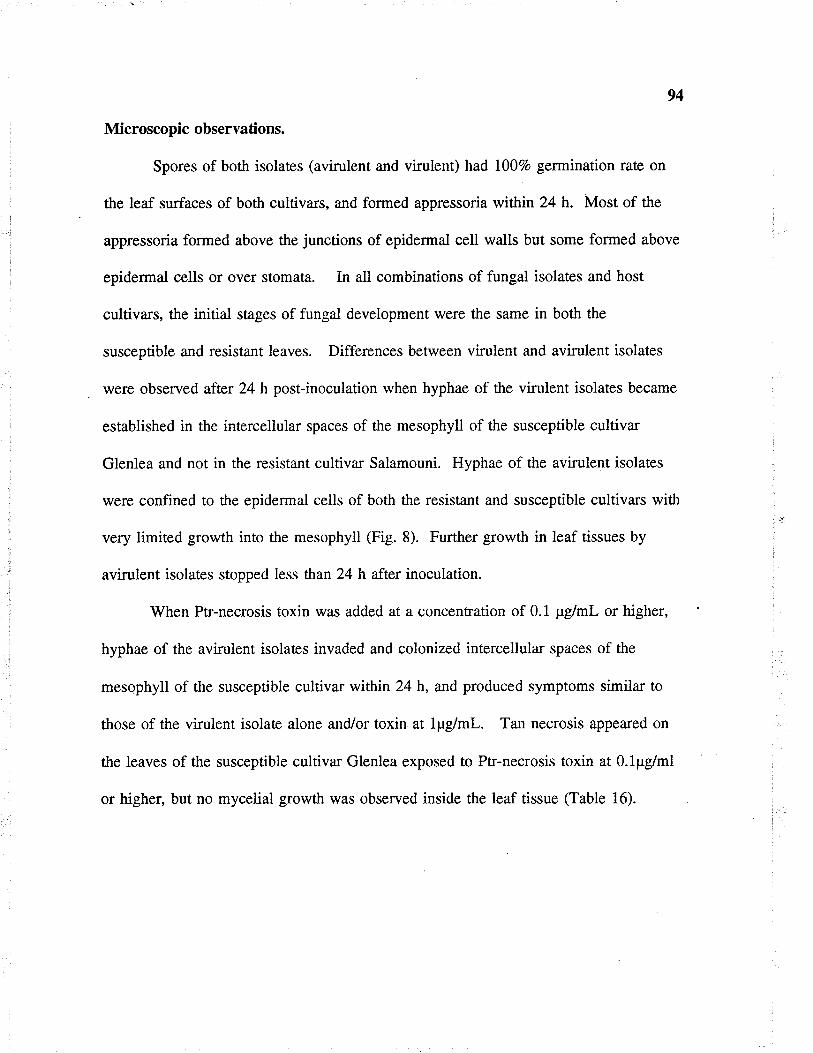
94
Microscopic observations.
Spores of both isolates (avirulent and virulenÐ had I007o germination rate on
the leaf surfaces of both cultivars, and formed appressoria within 24 h. Most of the
appressoria formed above the junctions of epidermal cell walls but some formed above
epidermal cells or over stomata. In all combinations of fungal isolates and host
cultivars, the initial stages of fungal development were the same in both the
susceptible and resistant leaves. Differences between virulent and avi¡ulent isolates
were observed after 24 h post-inoculation when hyphae of the virulent isolates became
established in the interceliular spaces of the mesophyll of the susceptible cultivar
Glenlea and not in the resistant cultivar Salamouni. Hyphae of the avi¡ulent isolates
were confined to the epidermal cells of both the resistant and susceptible cultivars with
very limited growth into the mesophyll (Fig. 8). Further growth in leaf tissues by
avi¡ulent isolates stopped less than 24 h afte.,r inoculation.
When Ptr-necrosis toxin was added at a concentration of 0.1 pglmL or higher,
hyphae of the avirulent isolaæs invaded and colonized intercellular spaces of the
mesophyll of the susceptible cultivar within 24 h, and produced symptoms similar to
those of the virulent isolaæ alone and/or toxin at l¡rg/ml. Tan necrosis appeared on
the leaves of the susceptible cultiva¡ Glenlea exposed to Ptr-necrosis toxin at O.l¡rg/ml
or higher, but no mycelial growth was observed inside the leaf tissue (Table 16).
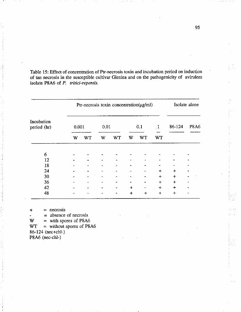
95
Table 15: Effect of concentration of Ptr-necrosis toxin and incubation period on inductionof tan necrosis in the susceptible cultivar Glenlea and on the pathogenicity of avirulentisolaæ P8A6 of P. triticïrepentis.
Ptr-necrosis toxin concentration(pglml) Isolaæ alone
Incubationperiod (h¡) 0.001 0.01 0.1 1 86-124 P8A6
wwTwwTwwTwT
6t218
24++30-++36++42+-++48++++
+ = necrosrs- = absence of necrosisW = with spores of P8A6WT = without spores of P8A686-124 (nec+chl-)P8A6 (nec-chl-)
%Tablel6: Effect of Ptr-necrosis toxin on virulence of isolaæs of P. triticïrepenrts.
HostReaction
Glenlea Salamouni
Extent of fungal growth
in leaf tissuesb
Glenlea SalamouniIsolaæ
Toxin(pdml)
86-124
86-124
331-9
33r-9
90-2
90-2
P8A6
P8A6
WS
watef
0.1
0
0.1
0
0.1
0
0.1
0
0.1
0
#+
++
+++
+
+++
+++
+
+
+
++
++
++
+
++
+
++
+
+
+
+
+
+
+
+
+
- = no visible symptoms afær 48 hrs+ = small dark-brown spos# = tan necrosis
+++ = very severe tan necrosisWS = toxin alone
b) Evaluaæd by examining hyphal growth under light microscope.- = no mycelial growth+ = spore germination and infection peg within epidermal cells.++ = abundant inærcellular mycelial development within leaf tissue.

9l
Fig '/: Effect of Ptr-necrosis toxin supplementation on the vi¡ulence of isolates
of P. tritici-repentis.
a) Tan necrosis on the susceptibre cultivar Glenlea, sprayed with 1 pglml
of ptr-necrosis toxin alone, 48 h post-inoculation
b) Tan necrosis on the susceptible cultiva¡ Glenlea, inoculated with spores
of the necrosis inducing isolate g6-124 (nec+chl-) alone, 4g h post-
inoculation.
c) Tan necrosis on the susceptible cultiva¡ Glenlea, inoculated with a
suspension of spores of the avirulent, non-toxin producing isolate
(P846 3), together with pr-necrosis toxin ar 0.1 ¡rglml, 4g h post-
inoculation.
d) Small, dark-brown spots on the susceptible cultivar Glenlea, inoculated
with spores of the avirulent, non-toxin producing isolate (p846 3) alone,
48 h posrinoculation.
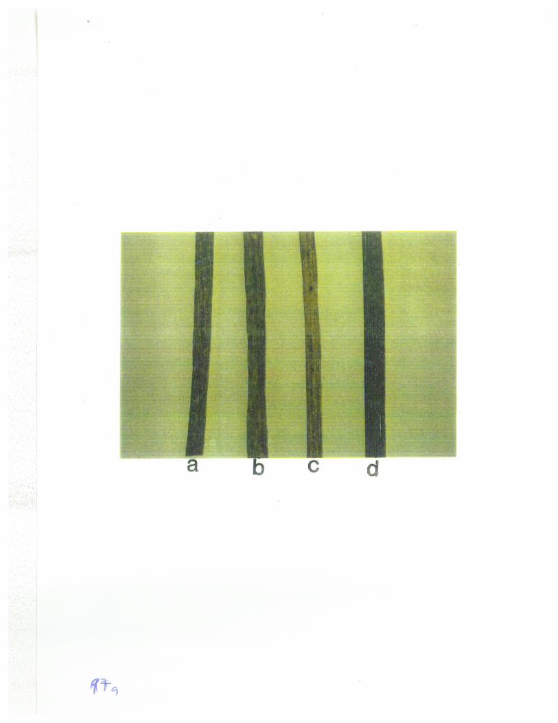
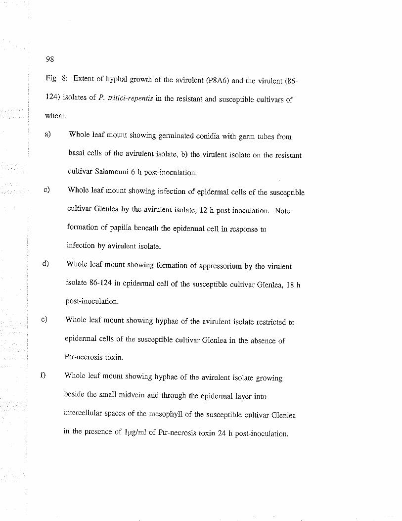
98
Fig 8: Extent of hyphal growth of the avirulent (P846) and the virulent (86-
124) isolates of P. tritici-repentis in the resistant and susceptible cultivars of
wheat.
a) whole leaf mount showing germinated conidia with germ tubes from
basal cells of the avirulent isolate, b) the virulent isolate on the resistânr
cultiva¡ Saiamouni 6 h posrinoculation.
c) Whole leaf mount showing infection of epidermal cells of the susceptible
cultiva¡ Glenlea by the avirulent isolate, 12 h post-inoculation. Note
formation of papilla beneath the epidermal cell in response to
infection by avirulent isolate.
whole leaf mount showing formation of appressorium by the virulent
isolate 86-124 in epidermal cell of the susceptible cultivar Glenlea, 1g h
post-inoculation.
whole leaf mount showing hyphae of the aviruient isolate restricted to
epidermal cells of the susceptible cuitivar Glenlea in the absence of
Ptr-necrosis toxin.
whole leaf mount showing hyphae of the avirulent isolate growing
beside the small midvein and through the epidermal layer into
intercellula¡ spaces of the mesophyll of the susceptible cultiva¡ Glenlea
in the prcsence of l¡rg/ml of ptr-necrosis toxin 24 h posrinocularion.
d)
e)
Ð
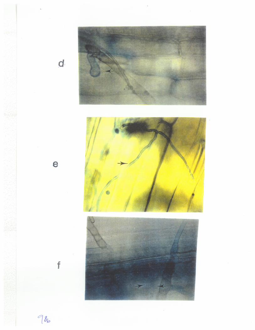
99
6.5 DISCUSSION
The major objective of this study was to determine the effect of Ptr-necrosis
toxin on colonization of wheat leaves by isolates of P. tritíci-repentis. The results of
this study established that, in the absence of Ptr-necrosis toxin, avirulent (nec-) isolaæs
failed to colonize the inærcellular spaces of the mesophyll tissue of both the resistant
and susceptible cultivars. However, by adding an exogenous supply of Ptr-necrosis
toxin produced by the virulent isolate 86-124, the avirulent isolates became as virulent
as the necrosis inducing isolate.
When Ptr-necrosis toxin wa$ sprayed alone on the susceptible cultivar Glenlea
at a concentration as low as O.l¡rg/ml, it induced a charateristic tan necrosis within 48
h, and at a higher concentlation of I ¡rg/ml, symptoms developed within 24 h.
Applications of Ptr'-necrosis toxin at 0.lprg/ml with spores of the vilulent isolate
enhanced the severity of the necrosis.
The present study and genetic evidence presented in thesis section 2, show that
Ptr-necrosis toxin is required for the colonization of the susceptible cultiva¡s.
Cultivar Glenlea, susceptible to isolate 86-124 and sensitive to Ptr-necrosis toxin
produced by this isolate, was colonized by the nec- isolate only when supplemented
with Ptr-necrosis toxin. Leaves of cultivar Salamouni, resistant to P. titicí-repentis
were not colonized by isolate 86-124 nor by the nec- isolates supplemenæd with Ptr-
necrosis toxin, suggesting that resistance to the fungus is based on the insensitivity to
the toxin produced by the fungus. These observations support the results of Lamari
100
and Bernier ( 1989c, 1991), that susceptibility of wheat genotypes to necrosis inducing
isolates and sensitivity to the Ptr-necrosis toxin released specifically by these isolaæs
a¡e controlled by the same dominant gene.
The early steps of the infection process were reported to be non-specific events
in the wheat/P. tritíci-repent¿.r system. No significant differences were observed
between compatible and incompatible interaction, until the invading hyphae had
reached the intercellula¡ spaces of the mesophyll (Loughman and Deverall 1986;
Lamari and Belnier 1989b, Larez et al. 1986). The effect was charactenzed by the
disruption and deformation of the mesophyll cells and breakdown of chloroplasts. This
disruption and breakdown was attributed to a metaboliæ suspected to be released by
the fungal isolates (Dushnicky 1993). These studies utilized combinations of vi¡ulent
isolates with susceptible and resistant host genotypes but did not investigate the
differences in the virulence of avirulent and virulent isolates and the effect of Ptr-
necrosis toxin on vi¡ulence of these isolates.
Results simila¡ to those of the present study were reported by Comstock and
Scheffer (1973) who showed that, when small amounts of Helnúnthosporium
carbonum (HC) toxin were added to the nonpathogen (H. victoriae) or to an avirulent
H. carbonunt isolatn which does not produce toxin, the isolaæs developed throughout
the susceptible leaves just as rapidly as the pathogenic or virulent ones.
The results of the present study imply that, if nec- isolates were supplemented
with Ptr-necrosis toxin, they could infect and colonize ptr-necrosis toxin sensitive
cultiva¡s to which they were previously avirulent. This could occur in nature if nec-
10I
and nec+ isolaæs were co-inoculated on the sensitive host cultivars. The in-vivo
production of Ptr-necrosis toxin by nec+ isolates could enable the nec- isolates to
acquire the ability to infect and colonize cultivars to which they were previously
avirulent. However, it is not known whether Ptr-necrosis toxin is required in initiating
early or late events of the infection process by nec+ isolates. The search for and the
production of anti-Ptr-necrosis toxin molecules could assist in the control of tan spot
in wheat. Splaying of the susceptible host genotypes with anti-PÍ-necrosis toxin
molecules as crop protectants could reduce the risk of epidemics caused by P. tritici-
repentís.
102
GENERAL DISCUSSION
The major goal of this study was to provide information on the potential
development of isolates of P. tritici-repenr¡s with increased virulence. Two approaches
were employed to obtain this information. The first was by examining the possibility
of obtaining variants through asexual processes of anastomosis and heærokaryon
formation, and the second was by sexual recombination. Results of the fîrst study
demonstrated that two mutants (86-124ipr+ and D308hgm+) of the four that were
paired, were veget¿tively compatible, and formed heterokaryons. Formation of the
heterokaryon between these two mut¿nts indicated ttre ability of two or more
genetically different nuclea¡ types to associate in the same mycelium. The phenotype
of such a heterokaryotic mycelium would be expressed as the sum total of all the
nuclear types present in the mycelium, and could suppress the expression of the
individuat genotypes. The presence of previous virulence genotypes or of new
genotypes brought about by mutation in such heterokaryons would constitute a genetic
pool from which new genotypes could be released by dissociation, in appropriate
selective environments. Vegetative compatibility and heterokaryon formation ensures
genetic exchange within the population, while veget¿tive incompatibility restricts such
exchanges, resulting in the formation of different clones.
The results of this study demonstrated that a heærokaryon did form as a result
of pairing two mutants of P. titícïrepentis, that it dissociated, but that new types
were not obtained. Heærokaryosis as a mechanism of variation could be useful if
different nuclear types associaæd in the same mycelium, and then through the
7.

103
parasexual cycle, produced new genotypes. Dissociation of the heærokaryon would
then allow the expression of the individual genotypes.
In the present study, two types of heærokaryons were obtained on the basis of
mycelial growth and the ability to sporulate: one with reduced mycelial growth and no
sporulation, and the other with normal mycelial growth and reduced sporulation. The
relevance of these two types to the pathogen was not addressed but, the heterokaryon
with reduced mycelial growth and no sporulation would not be expected to compete
effectively with the heterokaryon with normal mycelial growth and reduced
sporulation. The latter could out-compete the former for nutrients due to its faster
growth rate and by colonization of large areas of the substrate including susceptible
host cultiva¡s. In both the pathogenic and saprophytic phases of P. tritici-repentis, the
sporulating heterokaryon could reproduce and multiply to the exclusion of the non-
sporulating type.
The large size of fungal populations coupled with thei¡ high rates of
reproduction suggests that even a low frequency of anastomosis and heterokaryosis
could lead to significant changes in variation within the population of a fungus. The
formation of a heterokaryon in P. tritici-repentis followed by the release of a new
virulence genotype through dissociation may not be advantageous to the pathogen if
the new virulence genotype is released in an environment comprised of fully
susceptible wheat cultivars or grass hosts. The new virulence genotype would have to
compete against existing isolates to be able to infect the host. However, the presence
of a newly inuoduced resistence genotype in the host population would offer a
104
selective advantage to a new vi¡ulence genotype over the existing genotypes of the
pathogen which would not be able to infect the new host genotype. The new virulent
genotype would grow and produce a large quantity of inoculum that could lead to a
rapid buildup of disease particularly in crops which are grown as monocultures of
uniformly susceptible genotypes, and over wide areas. The occuffence of such a
process in nature would put added pressure on breeding and pathology programs to
monitor the population of the pathogen for variation in virulence. Such information
would be essential to the development of wheat cultivars with effective and durable
resistance.
The study on the role of Ptr-necrosis toxin in the expression of virulence by
isolates of P. tritici-repentis was based on the hypothesis that the toxin enhances the
colonization of host tissues by isolates of this fungus. The results indicated that Ptr-
necrosis toxin is a primary factor for infection and colonization of toxin sensitive
cultiva¡s. When supplemenæd with Ptr-necrosis toxin, nec- isolates acquired the ability
to infect and colonize the toxin sensitive cultivar Glenlea, but not the toxin insensitive
cultivar Salamouni. The enhancement of the colonization of host tissues by
supplementation of nec- isolates with Ptr-necrosis toxin, indicaæd that the toxin is
used to disrupt and disorganize living host tissues, so that nutrients leach out and
become available for fungal growth and subsequent colonization of the host tissues.
Ptr-necrosis toxin might also be used by the fungus to exclude other organisms
including saprophytes from the substrate. P. títici-repentis has a saprophytic phase,
but the ecological significance of Pt¡-necrosis toxin in saprophytism of this fungus has
105
not been investigaæd. This could be deærmined by assessing the sensitivity of other
leaf spot pathogens of wheat as well as several saprophytic fungi nonnally found on
wheat straw for sensitivity to Ptr-necrosis toxin in culture as well Íìs on straw.
The results of this study also suggest another approach to assessing the
poæntial of P. tritici-repentis to generate va¡iation, that is by the co-inoculation of
nec+ isolaûes with nec- isolates on a toxin sensitive host cultivar. The ín-vivo
production of Ptr-necrosis toxin by nec+ isolates could enable the nec- isolates to
infect and colonize host cultivars to which they were previously avirulent. However,
it is not known whether Ptr-necrosis toxin is required in initiating early or late events
of the infection process by nec+ isolaæs. In nature, the chance co-inoculation of nec+
and nec- isolaæs of P. títici-repentis could lead to the association of different nuclear
types in the same mycelium and, thus to variation through either anastomosis and
heærokaryon formation or through sexual recombination.
The possibility of obtaining variants in P. triticí-repentis through sexual
recombination was addressed in a separate study. Anastomosis and heterokaryon
formation provide an opportunity for the protoplasm and nuclei of different genotypes
including mating rypes to associate in the same cell. Heterokaryon formation is the
first step in out-crossing in homothallic and imperfecti fungi, whether. in sexual or
parasexual cycles (Ponærcovo et aI. 1953; Leslie 1993). Sexual recombination could
fail due to either nuclear or cytoplasmic post-fusion incompatibility factors, resulting
in særility. Sterility in fungi is associaæd with a number of genetic factors known to
block the sequence of sexual processes at various steps from vegetative mycelium to
106
ascospore fonnation (Wheeler 1954; Nelson 1959; Welz and læonard 1994). Based
on the dissociation of the heærokaryon as demonstrated in the frst study, and the
inconsistent formation of pseudothecia in most of the isolaæ pairs as shown in this
study, it would appear that there is a multi-sûep or mult-faceæd incompatibility sysrem
regulating out-crossing in the population of this fungus. The large proportion (65.7o)
of særile pseudothecia obtained from the pair of isolates 86-124 and Hy331-9 indicaæs
that there are genetic factors influencing additional steps of ascospore formation in
P.triticïrepentis.
In homothallic fungi including P. titici-repentis, when two strains are grown in
mixed culture, the ascospores from any single ascus may derive their nuclei from a
selfed zygota of one strain or the other, or from crossed zygotes (Pontecorvo et al.
1953). In the present study, ascospores were analysed in two ways; by sampling
random asospores from pseudothecia, and by isolating all the 8 ascospores of an ascus
from a pseudothecium shown to cont¿in hybrid ¿rscospores on the basis of virulence
phenotypes. Sixteeen percent only of pseudothecia taken from the line of junction
between isolaæs 86-124 and Hy331-9 were hybrids, since they contained recombinant
¿tscospores as evidenced by the recovery of new virulence phenotypes, and were thus
the result of fertilization. In the same hybrid pseudothecia, even when some asci were
truely hybrid, others were not. Sampling of random ascospores from a pseudothecium
only indicates the presence of new vi¡ulence phenotypes, and the results can not be
used to deærmine ratios of various phenotypes because of the presence of a large
number of selfed asci. However, in fungi like P. tritici-reperzr¿s which have hyaline
r07
ascospores and for which colour markers are not available, sampling of random
¿tscospores from individual pseudothecia is a useful and necessary method for the
identification of hybrid pseudothecia. Thus, analysis of ascospores from asci obtained
from known hybrid pseudothecia is presently the only method that allows the
deænnination of genetic ratios of virulence phenotypes and the number of genes
involved.
The recovery of recombinant progeny (nec+chl+ and nec-chl-) from the initial
cross of the parental isolates indicaæs that there is potential for P. tritícïrepentis to
evolve new virulence patterns through sexual recombination. These new virulence
types could infect cultivars that were previously resistant to one or the other of the
virulence genes. In the present study, recombinants that were virulent and avirulent to
both cultivar Glenlea and line 68365 were obtained from the cross of isolates 86-124
and 331-9, but progeny with increased virulence (i.e with ability to infect resistant
cultivars, Erik and Salamouni) were not recovered, indicating that recombination may
not necessarily creaæ new genotypes (or does so at very low frequencies) but merely
reshuffle existing ones. Unless there are suppressive or episøtic factors to mask the
expression of some of the virulence genes, all genes that are constitutively available
may be fully expressed. More crosses and larger populations of asci will need to be
used to fully assess the potential for increased vi¡ulence in P. tritici-repentis. The
development of fungal isolaæs with additional markers and tle use of molecular
æchniques will be required to assist in identifying recombinant genotypes. Progenies
will also need to be æsted for new virulence patterns on a greater number of resistant
108
wheat cultivars, and for aggressiveness on a range of susceptible cultivars in order to
effectively assess the generation of new genotypes.

109
REFER.ENCES
Adee, E. A, and Pfender, W. F. 1989. The effect of primary inoculum level ofþrenophora titicïrepentis on tan spot epidemic development in wheat.Phytopathology. 79: 873-877.
Anagnostakis, L. S. 1988. Cryphonectria parasitica, cause of Chestnut blighr pages123-136 in G. S. Sidhu, ed., Advances in Plant Pathology, Vol: 6. Academic Press.
566 pp.
Anagnostakis, L. S. 1984. The mycelial biology of the Endothia parasíticaII:Vegetative incompatibility. pages 500-507 in D. H. Jennings and A. D. M. Rayner,eds., The ecology and physiology of fungal mycelium. Cambridge University Press.
Andrivon, D. 1993. Nomenclature for pathogenicity and virulence: The need forprecision. Phytopathology 83: 889-890.
Bagga, H. S., and Boone, M. D. 1968. Genes inVenturiainaequalis controllingpathogenecity to crab apples. Phytopathology 58: 1176-1182.
Ballance, M. G., Lamari, L, and Bernier, C. C. 1989. Purification andcharacænzation of a host-selective necrosis toxin from Pyrenophora tritici-repentis.Physiol. Mol. Plant Pathol. 35:203-213.
Bockus, W. W., and Davis, M. A. Igg3. Effect of nitrogen fertilizers on severity oftån spot of winær wheat. Plant Dis. 77: 508-510.
Boone, M. D., and Keitt, W. G. 1957. Venturia inaequalis (Cooke) Wint: Genescontrolling pathogenicity of wild-type lines. Phytopathology 47: 403-409.
Boucherie, H., Begueret, J, and Bernet, J. 1976. The molecular mechanism ofprotoplasmic incompatibility and is relationship to the formation of protoperithecia inPodospora anserina. J. Gen. Microbiol. 92: 59-66
Brasier, M. C., and Gibbs, N. J. 1975. Highly fertile form of the aggressive strain ofCeratocystis ulmí. Nature 257: 128-131.
Brown, A. D., and Hunger, R. M. 1993. Production of a chlorosis-inducing, host-specific, low-molecular weight toxin by isolates of Pyrenophora tritici-repentis (cause
of tan spot of wheat). J. Phytopathol. 37:221-232.
Bumett, H. J. 1975. Mycogenetics. An inroduction to the general genetics of fungi.John Wiley and Sons, London, New York.375 pp.
110
Buxton, \ry. E. 1956. Heærokaryosis and Pa¡asexual recombination in pathogenicstrains of Fusarium orysporum. J. Gen. Microbiol. 15: 133-139.
Campbell, R. 1989. Biological control of microbial plant pathogens. CambridgeUniversity Press, london. 218 pp.
Carlile, J. M. 1987. Genetic exchange and gene flow : Thei¡ promotion andprevention. pages 203-214 in A. D . M. Rayner, M. C. Brasier and D. Moore, eds.,Evolutionary biology of fungi. Cambridge University Press.
Caten, E. C., and Jinks, L. J. 1966. Heærokaryosis: its significance in wildhomothallic ascomycetes and fungi imperfecti. Trans. Brit. Mycol. Soc. 49:81-93.
Caæn, E. C. 1987. The genetic integration of fungal life styles. pages 215-299 inA. D. M. Rayner, M. C. Braiser and D. Moore, eds., Evolutionary biology of fungi.Cambridge University Press.
Clark, G . 1981. Pathological anatomy and mycology. Staining procedures, pàges
367-373. Williams and Wilkins, Baltimore. 512 pp.
Comstock, C. J, and Scheffer, P. R . 1973. Role of host-selective toxin in colonizationof corn leaves by Helminthosporíum carbonum Phytopathology 63 :24-29.
Diaz de Ackermann, M., Hosford, R. M jr., Cox, D. J, and Hammond, J. J. 1988Resistance in winter wheats to geographically differing isolates of Pyrenophora triticïrepentis and observations on pseudothecia. Plant Dis. 72: L028-1031. .
Duff, A. D. S. 1954. A new disease of wheat in Kenya caused by a species ofPyrenophora. E. Afric. Agric. Journal: 19:225-228.
Dushnicky, L. G. 1993. A microscopy study of the infection process of þrernphoratritíci-repentis in susceptible and resistant wheat cultivars. M.Sc. Thesis, University ofManitoba. 150 pp.
Ellingboe, H. A. 1992. Segregation of avirulence/virulence on th¡ee rice cultivars in16 crosses of Magn^aporthe grisea. Phytopathology 82: 597-60.
Esser, K., and Blaich, R. 1973. Heterogenic incompatibility in plants and animals.pages 109-113 in E. W. Caspari, ed., Advances in Genetics Vol. 17. AcademicPress.
Esser, K., and Meinhardt, F. 1984. Barrage formation in Fungi. pages 351-361 in H.F. Linskens and J. Heslop-Harison, eds., Encyclopedia of Plant Physiology, Newseries; Cellula¡ inæractions, Vol. 17. Springer-Verlag.
111
Esser, K., and Kuenen, R. 1967. Genetics of Fungi . Springer Verlag, New York Inc.500 pp.
Fincham, J. R. S., Day, P. R, and Radford, A. 1979. Fungal genetics. 4ù ed.Botanical monographs, Vol. 4. Blackwell Scientific Publication, Berkely, Universityof California Press. 636 pp.
Flor, H. H. 1942. Inherit¿nce of pathogenecity n Melampsora lini. Phytopathology32:653-669.
Flor, H. H. 1946. Genetics of pathogenecity ín Melampsora lini. J. Agric. Res. 73:335-357.
Flor, H. H. 1960. The inheritance of X-ray induced mutations to virulence in aurediospore of race 7 of Melampsora lini. Phytopathology 50: 603-605.
Garnjobst, L., and Wilson, F. J. 1956. Heterocaryosis and protoplasmicincompatibility in Neurospora crassa. Proc. Natl. Acad. Sci. U.S.A. 42: 613-618.
Gough, F. J. 1982. Inherit¿nce of tan spot resistance from the Chilean wheat "Carifen12 ". pages 46-48 in R. M. Hosford jr. ed., Tan spot of wheat and related diseasesworkshop. N. D. St¿te University, Fargo. 166 pp.
Griffiths, D. J., and Carr, J. H. 196I. Induced mutations for pathogenecity inPuccinia coronata avenae. Trans. Brit. Mycol. Soc. 44:601-607.
Hartley, J. M, and Williams, G.P. I9TL lnteractions between strains of Pucciniagraminis f. sp. tritici in axenic culture. Trans. Br. Mycol. Soc. 57 (l) 129-136.
Heale, B. J. 1988. Verticilliutrl spp. The cause of vascular wilts in many species.pages 291-312 in G. S. Sidhu, ed., Advances in Plant pathology. Vol. 6. AcademicPress. 566 pp.
Hosford, M. R. jr. 197I. A form of Pyrenophora trichostonut pathogenic to wheatand other grasses. Phytopathology 6l:28-32.
Hosford, M. R. jr., and Busch, H. R. 1974. Losses in wheat caused by þrenophoratriticïrepentis and Leptosphaeria avennriaf.sp.titicea. Phytopathology 64 184-187.
Hosford, M. R. jr., Larez, C. R, and Hammond, J. J. 1987. Interaction of wet periodand æmperature on þrenophora titící-repentis infection and development in wheatsof differing resistance. Phytopathology 77: 385-390.
tt2
Huber, D. M., [æe, T. S., Ross, M. A, and Abney, T. S. 1987. Amelioration of tan
spot-infected wheat with nitrogen. Plant Dis. 7l:49-50.
Hunger, R. M., and Brown, D. A. 1987. Colony colour, growth, sporulation,fungicide sensitivity,and pathogenicity of Pyrenophora tritici-repentis. Plant Dis. 7l:907-910.
Kafer , E. 1961. The process of spontaneous recombination in vegetative nuclei ofAspergillus nidulans. Genetics 46: l58l-1609.
Kolmer, J. 4., and Dyck, P. L. 1994. Gene expression in the Triticum aestivum -Puccinia recondita f. sp. titíci gene-for-gene system. Phytopathology 84: 437-440.
Krupinsky, J. M. 1982. Observations on the host range of isolates of Pyrenophoratrichostoma. Can. J. Plant Pathol. 4: 42-46.
Krupinsky, J. M. 1987. Pathogenicity on wheat of Pyrenophora tritici-reperttisisolated from Bronuts ínermis. Phytopathology 77 : 760-765.
Krupinsky, J. M. I992a. Grass hosts of Pyrenophora tritici-repentis. Plant Dis.76:92-95.
Krupinsky, M. J. 1992b. Aggressiveness of isolates of Pyrenophora tritici-repentisobtained from wheat in the northern great plains. Plant Dis. 76:87-91.
Lamari, L., and Bernier, C. C. 1989a. Evaluation of wheat lines and cultivars to tan
spot (Pyrenophora tritici-repenris) based on lesion type. Can. J. Plant Pathol. ll: 49-56.
Lama¡i, L., and C. C. Bernier. 1989b. Virulence of isolates of þrenophora titici-repentis on 11 wheat cultivars and cytology of the differential host reactions.Can. J. Plant Pathol. Ll:284-290.
Lamari, L., and Bernier, C. C. 1989c. Toxin of þrenophora tritici-repentis: Host-specificity, significance in disease, and inherit¿nce of host reaction. Phytopathology79:740-7M.
Lamari, L., and Bernier, C. C. 1991. Genetics of tan necrosis and extensivechlorosis in tan spot of wheat, caused by Pyrenophora tritíci-repentis. Phytopathology81: 1092-1095.
Lamari, L., Bernier, C. C., and Smith, R. B. 1991. Wheat genotypes that developboth tan necrosis and extensive chlorosis in response to isolates of Pyrenophora trítici-repentis. Plant Dis. 75: l2l-I22.
113
Larez, C. R., Hosford. R. M. jr., and Freeman, T. P. 1986. Infection of wheat and
oats by Pyrenophora triticïrepentís and initial characænzation of resistance.Phytopathology 7 6: 93 I -938.
Leslie, F. J. Igg3. Fungal vegetative compatibility. Ann. Rev. Phytopathol. 31:r27-50.
Loughman, R., and Deverall. J. B. 1986. Infection of resist¿nt and susceptiblecultivars of wheat by þrenophora titicí-repentis. Plant Pathol. 35: M3-450.
Luttrel, S. E. 1973. Loculoascomycetes. pages 135-219 in G. C. Ainsworth, F. K.Sparrow and A. S. Sussman, eds., The fungi, lVA. A taxonomic review with keys.Academic Press, New York, NY. 62lpp.
Luz, W. C. da, and Bergstrom, G. C. 1987. Interactions between Cochliobolussatívus and Pyrenophora titici-repentis on wheat leaves. Phytopathology 77: 1355-1360.
Luz, W. C. da, and Bergstrom, G. C. 1986. Effect of temperature on tån spotdevelopment in spring wheat cultivars differing in resistance. Can. J. Plant Pathol. 8:
451-454.
Luz ,\iV. C. da, and R. M. Hosford, jr. 1980. Twelve þrenophora trichostotln racesfor virulence to wheat in the Central Plains of North America. Phytopathology 70:I 193-1 196.
McCallum, D. B. 1991. Generation and utilization of phenotypic mut¿nts in the t¿n
spot fungus Pyrenophora triticïrepentís. M.Sc. Thesis. University of Manitoba. T0 pp.
McCallum, D. 8., Bernier, C. C.,and Lamari, L. 1994. Generation and utilization ofchemical-resistant mutants in þrenophora triticí-repentis, the cause of t¿n spot ofwheat. Can. J. Bol 72: 100-105.
McDonald, W. C. 1963. Heærothallism in þrenophora teres. Phytopathology 53:77r-773.
Michelmore,W. R., Norwood, M. J., Ingram, S..D., Crute, R. I, and Nicholson, P.
1984. The Inheritance of virulence in Brenùa lactucae to match resistance factors 3,
4, 5, 6,8, 9, 10, and l1 in Lettuce (Lactuca sativa). Plant Pathol. 33: 301-315.
Misra, A. P., and Singh, R. A. 1972. Pathogenic differences amongst th¡ee isolates ofHelminthosporiunt trítici-repentis and the performance of wheat varieties against them.Indian Phytopathology 25: 350-353.
tt4Morrall, R. A. 4., and Howard, R. J. 1975. The epidemiology of leaf spot disease ina native prairie. II. Ai¡borne spore populations of Pyrenophora titicí-repentís.Can. J. Bot. 53: 2345-2353.
Nagle, B. J., Frohberg, R. C., and Hosford, R. M jr. 1982. Inheritance of resist¿nce totan spot of wheat. pages 40-45 in R. M. Hosford jr. ed., Tan spot of wheat andrelated diseases workshop. N. D. St¿te University, Fargo. 116 pp.
Nelson, R. R. I959a. Genetics of Cochliobolus heterostrophus U.. Genetic factorsinhibiting ascospore formation . Mycologia. 5l: 24-29.
Nelson, R. R. 1959b. Genetics of Cochliobolus heterostophus. IY. A mutant genethat prevents perithecial formation. Phytopathology 49: 384-386.
Odvody, G. W., Boosalis, M. G, and Watkins, J. E. 1982. Development ofpseudothecia during progressive colonization of wheat straw by þrenophoratrichostomn pages 33-35 in R. M. Hosford jr., ed., Tan spot of wheat and relateddiseases. North Dakota Staæ University, Fargo. 116 pp.
Olive, L. S. 1956. Genetics of Sordaria fimicola l. Ascospore colour mutants.American J. Bot.43: 97-107.
Olive, L. S. 1958. On the evolution of heterothallism in fungi. The AmericanNaturalist, Y ol 92, 865: 233-251.
Olive, L. S. 1963. Genetics of homothallic fungi. Mycologia, 55: 93-103.
Norwood, M. J.,and Crute, I. R. 1984. The genetic control and expression ofspecificity in Bremia lactucae (I-ettuce downy mildew). Plant pathol. 33: 385-400.
Person, C. 1959. Gene-for-Gene relationships in host:parasite systems. Can. J.
Bor 37: 1101-1131.
Pfender, W. F. 1988. Suppression of ascocarp formation in þrenophora triticïrepentis by Limonomyces roseipellis a basidiomycete from reduced tillage wheat straw.Phytopathology. 78: 1254-1258.
Ffender, W. F., Pacey, C. A, and Zhang, W. 1988. Saprophytic growth andpseudothecia production by þrenophora trítíci-repentis in plant tissue held atcontrolled water potentials. Phytopathology 78: 1205-1210.
Pfender, W. F., and Wootke, S. L. 1987. Production of pseudothecia and ascosporesby Pyrennphora tritici-repentis in response to macronutrient concentrations.Phytopathology 7 7 : 1213- 1216
115
Pfender, W. F., Zhang, W, and Nus, A. 1993a. Biological control to reduce inoculumof the tan spot pathogen Pyrenophora títicïrepentis in surface-borne residues ofwheat fields. Phytopathology 83: 371-375.
Pfender,W. F., Kraus. J, and Loper, J. E. 1993b. A genomic region fromPseudomonas fluorescens Pf-S required for pyrrolnitrin production and inhibition ofþrenophora tritíci-repentis in wheat straw. Phytopathology 83: 1223-1228.
Pitænger, H. T. 1964. Spontaneous alærations of heærokaryon compatibility factors inNeurospora. Genetics 46: 1645-1663.
Pittenger, H. T., and Atwood, C. K. 1955. Stability of nuclea¡ proportions duringgrowth of Neurospora heterokaryons. Genetics 4 : 227-24I.
Platt, H. W., Morrall, R. A. 4., and Gruen, H. E. 1977. The effects of substrate,
temperature and photoperiod on conidiation of Pyrenophora titici-repentis. Can. J.
Bor 55: 254-259.
Platt, H. W., and Morrall, R. A. A. 1980. Effects of light inænsity and relativehumidity on conidiation in þrenophora tritici -repentis. Can. J. Plant Pathol. 2: 53-57.
Pontecorvo , G. 1946. Genetic systems based on heærocaryosis. Cold Spr. Harb. Sym.
Quant Biol. 11: 193.
Pontecorvo, G. 1956. The parasexual cycle in fungi. Ann. Rev. Microbiol. 10: 393-400.
Pontecorvo, G., Roper, J.4., Hemmons L. M., MacDonald, R. D and Bufton, A. W.J.1953. The genetics of AspergíIlus nidulans. Advances in Genetics 5: l4I-238.
Pontecorvo, G, and Sermonti, G. 1954- Parasexual recombination in Penicilliumchrysogenum. J. Gen. Microbiol. 11:94-104.
Puhalla, J. E. 1985. Classification of strains of Fusarium orysporurn on the basis ofvegetåtive compatibility. Can. J. Bot. 63: 179-183.
Puhalla, J. E., and Mayfield, E. J. 1974. The mechanism of heærokaryotic growth inVerticillium dahliae. Genetics 76: 4lI-422.
Puhalla, J. E, and Spieth,T. P. 1985. A comparison of heterokaryosis and vegetativeincompatibility among varieties of Gibberella fujikuroi (Fusariurn miniliþrnrc).Experimental Mycology. 9: 39-47.

tt6
Raymond, P. J., Bockus, W. W., and Norman, B. L. 1985. Tan spot of winter wheat:Procedures to deærmine host response. Phytopathology 75: 686-690.
Rees, R. G., and Platz, G. J. 1980. The epidemiology of yellow leaf spot of wheat inSouthern Queensland. Aust. J. Agric. Res. 3l: 259-167.
Rees, R. G., Mayer, R. J., and Platz, G. J. 1981. Yield losses in wheat from yellowspot: A disease-loss relationship derived from single tillers. Aust. J. Agric. Res. 32:85r-859.
Rees, G. R., and Platz, G. J. 1990. Sources of resistance to þrenophora tritici-repentis in bread wheats. Euphytica 45: 59-69.
Rees, G. R., Platz, G. J., and Mayer, R. J. 1982. Yield losses in wheat from yellowspot. Comparison of estimates derived from single tillers and plots. Australian J.
Agric. Res. 33: 899-908.
Rees, R. G., and Platz, G. J. 1983. Effect of yellow leaf spot on wheat: Comparison ofepidemics at different stages of crop development. Aust. J. Agric. Res. 34: 39-46.
Rees, G. R., Platz, G. J., and Mayer, R. J. 1988. Susceptibility of Australian wheats
to þrenophora tritici-repentis. Australian J. Agric. Res 39: 141-151.
Riaz, M., Bockus,W. W.,and Davis, M. A. 1991. Effects of Wheat genotype, timeafær inoculaúon,and leaf age on conidia production by Drechslera titici-repentis.Phytopathology 81: 1298-1302
Schilder, M. C. 4., and Bergstrom, G. C. 1990a. Variation in virulence within thepopulation of Pyrenophora títici-repentis in New York. Phytopathology 80: 84-90.
Schilder, M. C. 4., and Bergstrom, G. C. 1990b. Wheat seed infection by and seed
transmission of Pyrenophora tritici-repentis. Phytopathology 80:123 Abstract.
Shabeer, 4., and Bockus, W. V/. 1988. Tan spot effects on yield components relativeto growth stage in winær wheat. Plant Dis. 72:599-602.
Sharma, U., Adee, E. A, and Pfender, W. F. 1989. Effect of glyphosaæ herbicide onpseudothecia formation by þrenophora tritícïrepentis in infested wheat straw. PlantDis. 73: 647-650.
Shoemaker, A. R. 1955. Biology, cytology and taxonomy of Cochliobolus sativus.Can. J. Bot. 33: 563-576.
Shoemaker, R. A. 1962. Drechslera lto. Can. J. Bot. 40: 809-836.
tltSmedegard-Peterson, V. 1977. Inherit¿nce of genetic factors for symptoms andpathogenecity in hybrids of Pyrenophora teres and þrenophora graminea.Phytopathol. Z. 89: 193-202.
Summerell, 4.8., and Burgess,W.L. 1989. Factors influencing survival ofþrenophora tritici-repentís:WatÊr poæntial and æmperature. Mycol. Res.93 (1)41-45.
Summerell, A. 8., and Burgess,W. L. 1988a. Saprophytic colonization of wheat andbarley by þrenophora tritici-repentis in the field. Trans. Brit. Mycol. Soc. 90 (4) 551-556.
Summerell, A. 8., and Burgess,W. L. 1988b. Factors influencing production ofpseudotheciaby þrenophora tritíci-repentis. Trans. Brit. Mycol. Soc. 90 (4) 557-562.
Sykes, E. E., and C. C. Bemier. 199I. Qualitative inheritance of t¿n spot resistance inhexaploid, tetraploid and diploid wheat. Can. J. Plant Pathol. 13:38-M.
Tekauz, A. 1976. Distribution, seïerity and relative importance of leaf spot diseasesof wheat in Wesærn Canada tn 1974. Can. Plant Dis. Survey 56:36-41.
Tekauz, 4., Samborski, D. J., Rourke, D. S. R, and Iverson, A. T. 1983. Diseases ofwinter wheat in Manitoba in 1983 pages 63-68. Proc. Man. Agronomists.
Tinline, D. R. 1962. Cochliobolus sativus, V. Heterokaryosis and parasexuality. Can.J. Bot. 40: 425-437.
Tinline, D. R. 1988. Cochlíobolus sativus, a pathogen of wide host range. pages 113-122 tn G. S . Sidhu, ed., Advances in Plant Pathology, Vol.6. Academic press. 566 pp.
Tomas, 4., and Bockus, W. W. 1987. Cultivar-specific toxicity of culture filuaæs ofPyreno phora trítci- re p entis. Phytopathology 7 7 : 1337 -1340.
Tomas, 4., Feng, G. H., Reeck, G. R., Bockus, W.W, and Leach, J..8. 1990.Purification of a culture-specific toxin from þrennphora titici-repentis, causal agentof tan spot of wheat. Mol. Plant-Microbe lnæract. 3:22I-224.
Uhm, J. Y., and Fuji. H. 1983. Ascospore dimorphism in Sclerotinia triþIiorum andcultural characters of strains from different sized spores. Phytopathology 73:565-569.
Valder, P. G., and Shaw, D. E. 1952. Yellow spot disease of wheat in Australia.Proc. Linn. Soc. N. S. W. 77:323-330.
118
Vaillancourt, L. J., and Hanau, R. M. 1991. A method for genetic analysis ofGlomzrella graminicol^a (Colletotricum gramínicola) from Maize. Phytopathology81:530-534.
VoIk, J. T., and Leona¡d, J. T. 1989. Experimental studies on the Morel, l.Heærokaryon fonnation between monoascosporous strains of Morchella. Mycologia81 (4) 523-s3r.
Walton, D. J., and D. G. Pa¡raccione. 1993. Host-selective toxins and diseasespecificity: Perspectives and progress. Ann. Rev. Phytopathol. 3l:275-303.
Watkins, J. E., Odvody, G.N., Boosalis, M. G, and Partridge, J. E. 1978. Anepidemic of tan spot of wheat in Nebraska. Plant Dis. Reporter 62:132-134.
Watkins J. E., Boosalis, M. G, and Doubnik, B. L. 1982. Foliar diseases ofNebraska's winter wheat. pages 53-61 in R. M. Hosford jr. ed., Tan spot of wheat andrelated diseases. North Dakota St¿æ University, Fargo. 116 pp.
Wehmeyer, E. L. 1952. Perithecial development tn Pleospora trichostomn.Bot¿nical Gazetæ ll5:297-310 .
Vy'heeler, E. H. 1954. Genetics and evolution of heterothallism in Glomerella.Phytopathology M: 342-345.
Welz, G. H., and K. J. Leonard. 1994. Genetic analysis of two race 0 x race 2crosses in Cochliobolus carbonum Phytopathology 84: 83-91.
\iVhiæhead, D. M., and Dickson, G. J. 1952. Pathology, morphology and nuclearcycle of two new species of Pyrenophora. Mycologia 44:747-758.
Wright, K. H., and J.C.Sutton. 1990. Inoculum of þrenophora tritici-repentis inrelation to epidemics of tan spot of winter wheat in Ontario. Can. J. Plant Pathol. 12:
t49-t57.
Yoder, C. O. 1988. Cochliobolus heterostrophus, cause of southern corn leaf btight.pages 93-Il2 in G. S. Sidhu, ed., Advances in Plant Pathology, Vol. 6. AcademicPress. 566 pp.
Zambino, P. J., and Harrington,T. C. 1990. Heterokaryosis and vegetativecompatibility in Leptographium wageneri. Phytopathology 80: 1460-1469.
Zhang, W., and Pfender,W. F. 1992- Effect of residue management on wetnessduration and ascocarp production by Pyrenophora tritici-repentis in wheat residue.Phytopathology 82: 1434-1439.
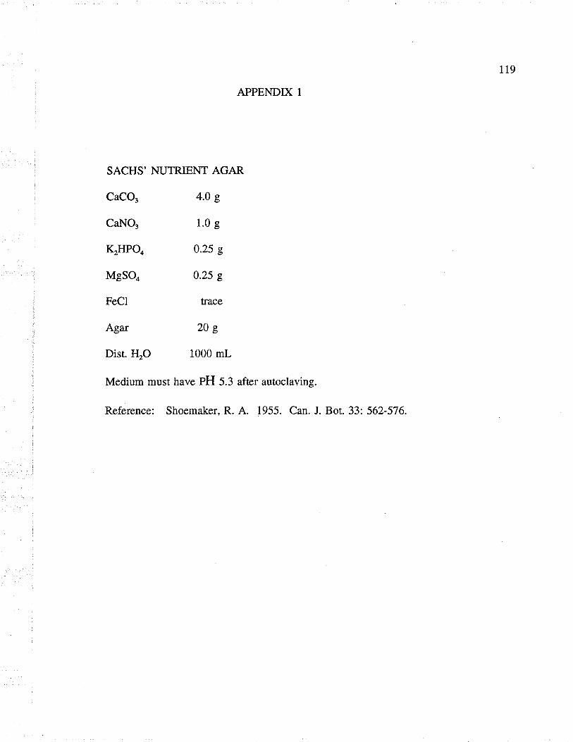
119
APPENDIX 1
SACHS' NUTRIENT AGAR
CaCO, 4.0 g
CaNO3 1.0 g
KTHPO.
MgSOo
FeCl
Agar
Dist. HrO
0.25 g
0.25 s
trace
2oe
1000 mL
Medium must have PH 5.3 after autoclaving.
Reference: Shoemaker, R. A. 1955. Can. J. Bot. 33: 562-576.

r20
APPENDIX 2
FRIES MEDIUM
NHotartraæ 5.0 g
NH4NO3 1.0 g
MgSOo.THrO 0.5 e
K2HPO4 2.6 e
KH2PO4 1.3 g
Sucrose 30.0 g
Yeast extract 1.0 g
Trace element soln. 2 mL
HrO 1000 mL
Stock solution/L.
LiCl 167 mg
CuClr.HrO 107 mg
HrMoOo 34 mg
MnClr.4HrO 72 mg
CoClr.4HrO 80 mg
Reference. Dhingra, O. D and Sinclair, J. B. 1983. Basic plant pathology methods
CRC Press, Boca Raton, Florida. 353pp.