OBSERVATIONS ON THE LUNGWORM, PNEUMOSTRONGYLUS CALCARATUS, IN IMP ALA (AEPYCEROS MELAMPUS) FROM...
7
76 Journal of Wildlife Diseases, 25/ 1 ), 1989. pp. 76-82 © Wildlife Disease Association 1989 OBSERVATIONS ON THE LUNGWORM, PNEUMOSTRONGYLUS CALCARA TUS, IN IMPALA (AEPYCEROS MELAMPUS) FROM SWAZILAND G. J. Gallivan, I. K. Barker,26 R. M. R. Alves,3 J. Culverwell,4 and R. Girdwood5 Department of Biology, University of Swaziland, Private Bag Kwaluseni, Swaziland 2 Department of Pathology, Ontario Veterinary College, University of Guelph, Guelph, Ontario, Canada N1G 2W1 Veterinary Research Institute, Onderstepoort 0110, Republic of South Africa Mbuluzi Nature Reserve, Tambankulu Estates, Private Bag Mhlume, Swaziland Mlawula Nature Reserve, Private Bag Simunye, Swaziland 6 Author to whom reprint requests should be addressed ABSTRACT: The lungworm, Pneurnostrongylus calcaratus, was found in 85% (164 of 193) of impala (Aepyceros melampus) collected in Mlawula Nature Reserve in Swaziland. Infection was confirmed at 4.5 mo of age, and the prevalence increased to 100% at 11 mo, with a prevalence of 98% in animals >1 yr of age. Pneumostrongylus calcaratus was usually found in firm, tan- grey nodules along the lobar borders of the lungs, although an extensive granulomatous pneumonia with miliary caseous abscesses and calcified nodules was observed in some older animals. In the primary infection in lambs, adult parasites, larvae and eggs were observed in the alveoli and bronchioles within the nodule. There was peribronchial and perivascular mononuclear cuffing, with infiltration of mononuclear cells in the alveolar septum in the vicinity of worms. In lesions in older animals, there was local consolidation with macrophages and giant cells, and foci of parenchymal necrosis associated with degenerating eosinophils, which appeared to lead to the formation of eosinophilic granulomas. Resolving lesions caused interstitial fibrosis, with mineralized nodules. Pneumostrongylosis does not appear to pose a significant threat to the health of impala in Swaziland. Key words: Pneurnostrongylus calcaratus, impala, Aepyceros melarnpus, Swaziland, preva- lence, pathology, field study. INTRODUCTION Pneumostrongylus calcaratus is a lung- worm of impala (Aepyceros melarn pus) and other antelope in eastern and southern Africa. This fine black parasite is found in yellowish-grey to bluish-grey raised em- physematous or firm areas on the dorsal caudal lobes and along the lobar borders of the lungs (Dinnik and Sachs, 1968; Young and Wagener, 1968; Heinichen- Anderson, 1982). Microscopic description of the lesions is limited to a general de- scription of the lesions associated with P. calcaratus and other small protostrongylid lungworms in six species of antelope (Moulton and Sachs, 1970). In the Seren- geti, Eastern Transvaal including Kruger National Park, and Natal the prevalence of P. calcaratus in impala ranges from 75 to 100% (Ortlepp, 1962; Dinnik and Sachs, 1968; Young and Wagener, 1968; Heini- chen, 1973; Anderson, 1983). Ortlepp (1962) did not find P. calcaratus in impala in Swaziland, yet Meeser (1952) stated that it had been reported. In the present study the prevalence of P. calcaratus in impala in Swaziland is reported, and the gross and microscopic lesions associated with infec- tion are described. MATERIALS AND METHODS The impala were collected during the culling program at Mlawula Nature Reserve (26#{176}12’S, 32#{176}00’E) in northeastern Swaziland. The reserve covers an area of approximately 23,000 ha, and is predominately Acacia nigrescens-Scierocar- ya caffra-Themeda triandra savanna (low veld vegetation type 10 of Acocks, 1975). The mean annual rainfall is 610 to 750 mm, with hot, wet summers and cool, dry winters. One hundred ninety-three impala were col- lected by shooting from early October 1985 to the end of August 1986, with monthly sample sizes ranging from six to 24 animals. Necropsies were performed at a central abattoir or in the field. The lungs were removed from the carcass, and the presence of P. calcaratus was deter-
-
Upload
independent -
Category
Documents
-
view
0 -
download
0
Transcript of OBSERVATIONS ON THE LUNGWORM, PNEUMOSTRONGYLUS CALCARATUS, IN IMP ALA (AEPYCEROS MELAMPUS) FROM...

76
Journal of Wildlife Diseases, 25/ 1 ), 1989. pp. 76-82© Wildlife Disease Association 1989
OBSERVATIONS ON THE LUNGWORM, PNEUMOSTRONGYLUS
CALCARA TUS, IN IMPALA (AEPYCEROS MELAMPUS)
FROM SWAZILAND
G. J. Gallivan, I. K. Barker,26 R. M. R. Alves,3 J. Culverwell,4 and R. Girdwood5Department of Biology, University of Swaziland, Private Bag Kwaluseni, Swaziland
2 Department of Pathology, Ontario Veterinary College, University of Guelph, Guelph,
Ontario, Canada N1G 2W1Veterinary Research Institute, Onderstepoort 0110, Republic of South AfricaMbuluzi Nature Reserve, Tambankulu Estates, Private Bag Mhlume, SwazilandMlawula Nature Reserve, Private Bag Simunye, Swaziland
6 Author to whom reprint requests should be addressed
ABSTRACT: The lungworm, Pneurnostrongylus calcaratus, was found in 85% (164 of 193) of
impala (Aepyceros melampus) collected in Mlawula Nature Reserve in Swaziland. Infection was
confirmed at 4.5 mo of age, and the prevalence increased to 100% at 11 mo, with a prevalenceof 98% in animals >1 yr of age. Pneumostrongylus calcaratus was usually found in firm, tan-grey nodules along the lobar borders of the lungs, although an extensive granulomatous pneumoniawith miliary caseous abscesses and calcified nodules was observed in some older animals. In theprimary infection in lambs, adult parasites, larvae and eggs were observed in the alveoli and
bronchioles within the nodule. There was peribronchial and perivascular mononuclear cuffing,with infiltration of mononuclear cells in the alveolar septum in the vicinity of worms. In lesionsin older animals, there was local consolidation with macrophages and giant cells, and foci ofparenchymal necrosis associated with degenerating eosinophils, which appeared to lead to theformation of eosinophilic granulomas. Resolving lesions caused interstitial fibrosis, with mineralizednodules. Pneumostrongylosis does not appear to pose a significant threat to the health of impalain Swaziland.
Key words: Pneurnostrongylus calcaratus, impala, Aepyceros melarnpus, Swaziland, preva-lence, pathology, field study.
INTRODUCTION
Pneumostrongylus calcaratus is a lung-
worm of impala (Aepyceros melarn pus)
and other antelope in eastern and southern
Africa. This fine black parasite is found in
yellowish-grey to bluish-grey raised em-
physematous or firm areas on the dorsal
caudal lobes and along the lobar borders
of the lungs (Dinnik and Sachs, 1968;
Young and Wagener, 1968; Heinichen-
Anderson, 1982). Microscopic description
of the lesions is limited to a general de-
scription of the lesions associated with P.
calcaratus and other small protostrongylid
lungworms in six species of antelope
(Moulton and Sachs, 1970). In the Seren-
geti, Eastern Transvaal including Kruger
National Park, and Natal the prevalence
of P. calcaratus in impala ranges from 75
to 100% (Ortlepp, 1962; Dinnik and Sachs,
1968; Young and Wagener, 1968; Heini-
chen, 1973; Anderson, 1983). Ortlepp
(1962) did not find P. calcaratus in impala
in Swaziland, yet Meeser (1952) stated that
it had been reported. In the present study
the prevalence of P. calcaratus in impala
in Swaziland is reported, and the gross and
microscopic lesions associated with infec-
tion are described.
MATERIALS AND METHODS
The impala were collected during the cullingprogram at Mlawula Nature Reserve (26#{176}12’S,32#{176}00’E) in northeastern Swaziland. The reservecovers an area of approximately 23,000 ha, andis predominately Acacia nigrescens-Scierocar-
ya caffra-Themeda triandra savanna (low veld
vegetation type 10 of Acocks, 1975). The meanannual rainfall is 610 to 750 mm, with hot, wetsummers and cool, dry winters.
One hundred ninety-three impala were col-lected by shooting from early October 1985 tothe end of August 1986, with monthly samplesizes ranging from six to 24 animals. Necropsieswere performed at a central abattoir or in thefield. The lungs were removed from the carcass,and the presence of P. calcaratus was deter-
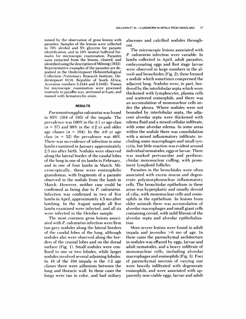
GALLIVAN ET AL.-LUNGWORM IN IMPALA FROM SWAZILAND 77
mined l)y the observation of gross lesions withparasites. Samples of the lesions were collectedin 70% alcohol and 5% glycerin for parasiteidentification, and in 10% neutral buffered for-
maIm for microscopic examination. Parasiteswere extracted from the lesions, cleared, andidentified using the description of Monnig(1933).Representative examples of the parasites are de-
posited in the Onderstepoort HelminthologicalCollection (Veterinary Research Institute, On-derstepoort 0110, Republic of South Africa;Accession numbers S.2444 and 5.2445). Tissuesfor microscopic examination were processedroutinely to paraffin wax, sectioned at 6 �m, and
stained with hematoxylin-eosin.
RESULTS
Pneumostrongylus calca rat us was found
in 85% (164 of 193) of the impala. The
prevalence was 100% in the � 1 yr age class
(n = 37) and 98% in the �2 yr and older
age classes (n = 104). In the �0 yr age
class (n = 52) the prevalence was 48%.
There was no evidence of infection in nine
lambs examined in January approximately
2.5 mo after birth. Nodules were observed
along the lateral border of the caudal lobes
of the lung in one of six lambs in February,
and in one of four lambs in March. Mi-
croscopically, these were eosinophilic
granulomas, with fragments of a parasite
observed in the nodule from the lamb in
March. However, neither case could be
confirmed as being due to P. calcaratus.
Infection was confirmed in two of five
lambs in April, approximately 4.5 mo after
lambing. In the August sample all five
lambs examined were infected, and all six
were infected in the October sample.
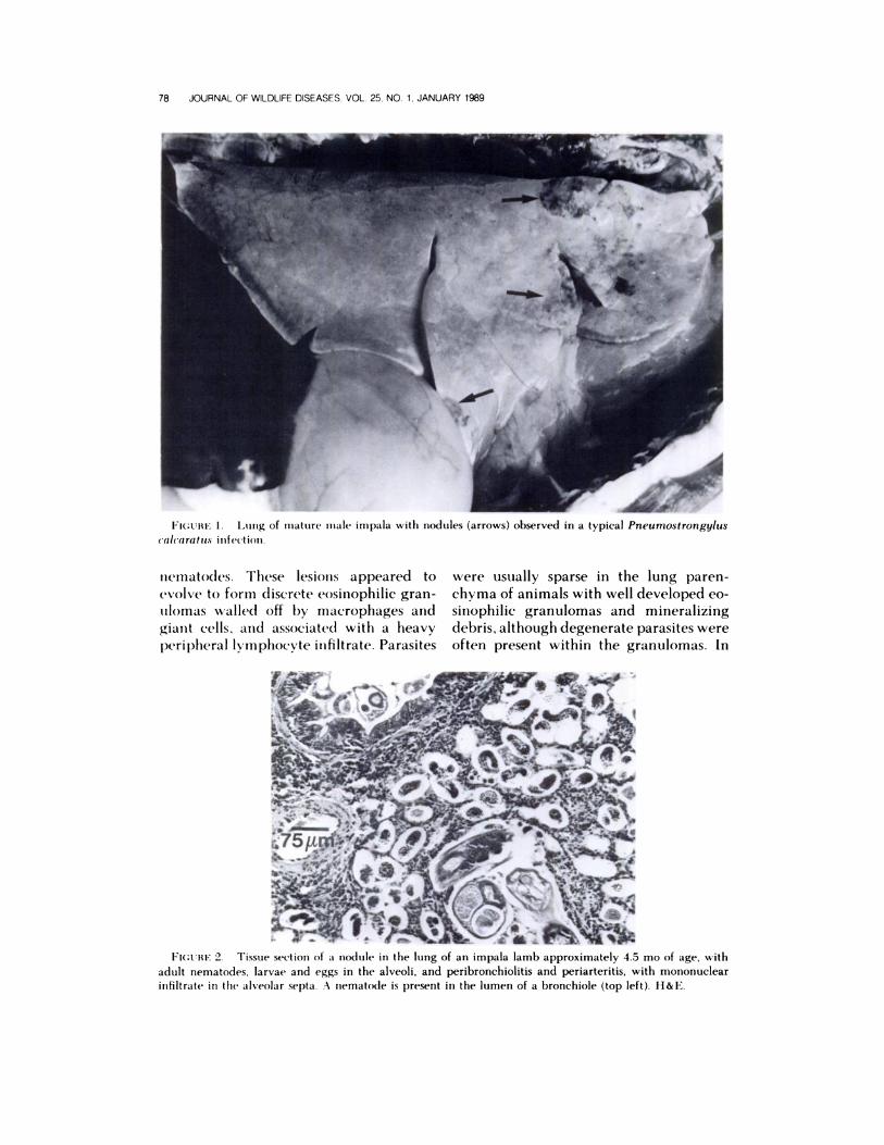
The most common gross lesions associ-
ated with P. calcaratus infection were firm
tan-grey nodules along the lateral borders
of the caudal lobes of the lung, although
nodules also were observed along the bor-
ders of the cranial lobes and on the dorsal
surface (Fig. 1). Small nodules were con-
fined to one or two lobules, while larger
nodules involved several adjoining lobules.
In 16 of the 104 impala in the �2 age
classes there were adhesions between the
lung and thoracic wall. In these cases the
lungs were tan in color, and had miliary
abscesses and calcified nodules through-
out.
The microscopic lesions associated with
P. calcaratus infection were variable. In
lambs collected in April, adult parasites,
embryonating eggs and first stage larvae
were observed in large numbers in the al-
veoli and bronchioles (Fig. 2); these formed
a nodule which sometimes compressed the
adjacent lung. Nodules were, in part, bor-
dered by the interlobular septa which were
thickened with lymphocytes, plasma cells
and scattered eosinophils, and there was
an accumulation of mononuclear cells un-
der the pleura. Where nodules were not
bounded by interlobular septa, the adja-
cent alveolar septa were thickened with
edema fluid and a mixed cellular infiltrate,
with some alveolar edema. In some areas
within the nodule there was consolidation
with a mixed inflammatory infiltrate, in-
cluding some macrophages and small syn-
cytia, but little reaction was evident around
individual nematodes, eggs or larvae. Therewas marked perivascular and peribron-
chiolar mononuclear cuffing, with prom-
inent lymphoid follicles.
Parasites in the bronchioles were often
associated with excess mucus and degen-
erate polymorphonuclear inflammatory
cells. The bronchiolar epithelium in these
areas was hyperplastic and usually devoid
of cilia, with mononuclear cells and eosin-ophils in the epithelium. In lesions from
older animals there was accumulation of
alveolar macrophages and small giant cells
containing ceroid, with mild fibrosis of the
alveolar septa and alveolar epithelializa-
tion.
More severe lesions were found in adult
impala and juveniles >6 mo of age. In
these cases the parenchymal architecture
in nodules was effaced by eggs, larvae and
adult nematodes, and a heavy infiltrate of
mononuclear cells, including alveolar
macrophages and eosinophils (Fig. 3). Foci
of parenchymal necrosis of varying sizewere heavily infiltrated with degenerate
eosinophils, and were associated with ap-
parently non-viable eggs, larvae and adult
_t
FI(;cRE 1. Lung of mature male impala with nodules (arrows) observed in a typical Pneumostrongylus
ealea ratus infection.
FIGcRE 2. Tissue section of a nodule in the lung of an impala lamb approximately 4.5 mo of age, with
adult nematodes, larvae and eggs in the alveoli, and peribronchiolitis and periarteritis, with mononuclear
infiltrate in the alveolar septa. .-� nematode is present in the lumen of a bronchiole (top left). H&E.
78 JOURNAL OF WILDLIFE DISEASES, VOL 25, NO 1, JANUARY 1989
nematodes. These lesions appeared to
evolve to form discrete eosinophilic gran-
ulomas walled off by rnacrophages and
giant cells, and associated with a heavy
peripheral km phocvte infiltrate. Parasites
were usually sparse in the lung paren-
chyma of animals with well developed eo-
sinophilic gran ulomas and mineralizing
debris, although degenerate parasites were
often present within the granulomas. In

FICcRE 3. Tissue section of a nodule in the lung of an older impala, with consolidation with macrophages
and giant cells, and areas of necrosis associated with degenerating eosinophils (arrows). H&E.
GALLIVAN ET AL.-LUNGWORM IN IMPALA FROM SWAZILAND 79
FIGURE 4. Tissue section of lung from an adult impala with a resolving parasitic pneumonia. There is
interlobular and alveolar septal fibrosis and a mineralized granuloma with a mantle of lymphocytes. H&E.
the lungs of several adult impala inter-
preted as having a resolving parasitic
pneumonia, there was marked alveolar
septal and interlobular fibrosis, with focal
aggregates of macrophages and giant cells,
eosinophils or mineralizing eosinophilic
granulomas with a well developed mantle
of lymphocytes (Fig. 4).

FIGURE 5. Group of adult parasites, presumably P. calcaratus, outside the typical nodule. There is some
compression of the surrounding lung parenchyma, and a mild edema and cellular response. H&E.
80 JOURNAL OF WILDLIFE DISEASES. VOL. 25, NO 1, JANUARY 1989
Animals with chronic lesions occasion-
ally had bronchioles obstructed by eosin-
ophils, larvae and necrotic debris, and there
was erosion or ulceration of the bronchio-
lar epithelium. In some severe cases this
had developed to bronchiolitis obliterans.
The cases with granulomatous lesions typ-
ically had associated villous proliferation
of the visceral pleura, with prominent me-
sothelial cells, and in most cases, adhesions
between the visceral and parietal pleura.
In several cases, eggs and larvae were
observed in alveoli outside the parasitic
nodtiles. These were usually accompanied
by edema and a local eosinophilic infil-
trate. Small groups of adult nematodes,
similar in microscopic appearance to those
in the large nodules, also were observed in
the lung parenchyma outside the nodules.
These nematodes were confined to discrete
foci (Fig. 5), which in some instances ap-
peared to be compressing the adjacent
parenchyrna, and were associated with
edema and a local eosinophilic infiltrate.
However, in other cases the foci were sur-
rounded by macrophages, fibroblasts and
lymphocytes. Mineralizing eosinophilic
granulomas centered on small aggregates
of degenerate nematodes and correspond-
ed to the palpable presence of miliary nod-
ules approximately 1 mm in diameter in
the lung parenchyma.
DISCUSSION
The high prevalence of P. calcaratus in
impala in Mlawula Nature Reserve is sim-
ilar to that reported in other areas of east-
ern and southern Africa (Ortlepp, 1962;
Dinnik and Sachs, 1968; Young and Wag-
ener, 1968; Heinichen, 1973; Anderson,
1983), but differs from Ortlepp’s (1962)
observation at an unspecified location in
Swaziland. Horak (1978, 1980) did not re-
port P. calcaratus from Nylsvley Nature
Reserve in the northern Transvaal or from
Pafuri in northeastern Kruger National
Park, although Young and Wagener (1968)
reported a high prevalence from southern
Kruger National Park. Horak (1981) has
suggested that P. calcaratus is restricted to
warm, moist regions. This may explain the
discrepancy between the findings in the
present study and that of Ortlepp (1962)
since several other areas of Swaziland in
which impala occur receive’considerably
less rainfall than Mlawula, or are consid-
erably cooler.
The youngest impala in which P. cal-
caratus infection was confirmed micro-
scopically were approximately 4.5 mo old.

GALLIVAN ET AL.-LUNGWORM IN IMPALA FROM SWAZILAND 81
These animals had fully developed adult
nematodes and first stage larvae in the
lungs. The observation of nodules in the
lungs of lambs 2.5 and 3.5 mo of age, and
of parasite fragments in a section of lung
from the 3.5-mo-old lamb, suggests that
infection occurs relatively early in life. The
yellow slug (Elisolimax flavescens) has
been suggested as the intermediate host of
P. calca rat us, with infection resulting from
the ingestion of infected slugs (Heinichen,
1974; Heinichen-Anderson, 1976, 1982).
In this situation, infection may occur as
early as 1.5 mo of age since lambs were
already consuming grass and browse in
January (G. J. Callivan, unpubl. obs.). Lit-
tle is known of the endogenous develop-
ment of P. calcaratus. Heinichen-Ander-
son (1976) did not find P. calcaratus in
impala 18 days after inoculation with third
stage larvae, but recorded lesions in a sheep
at 51 days and third stage larvae in the
lungs of a guinea pig 8 days postinocula-
tion (Heinichen-Anderson, 1982). How-
ever, since guinea pigs and sheep are ab-
normal hosts, the significance of these
findings is limited.
The typical gross lesions observed in the
present study were similar to those de-
scribed by Moulton and Sachs (1970) and
Heinichen-Anderson (1982). The emphy-
sematous areas described by Young and
Wagener (1968) were never observed.
Adult parasites, presumably P. calca rat us,
were observed in eosinophilic granulomas
and calcified nodules scattered in the lung
parenchyma, indicating that P. calcaratus
does not always localize in nodules along
the lobar borders. This was usually ob-
served in older animals, with only one case
in an animal <3-yr-old.
The microscopic lesions were similar to
those observed in other metastrongyloid
infections (Beresford-Jones, 1967; Moulton
and Sachs, 1970; Stockdale, 1976). The
wide range of lesions observed in the pres-
ent study may be due in part to differences
in the age of the host and the time of
infection coupled with variation in the host
response. The general pattern of response
to P. calcaratus appears to be similar to
that described for Muellerius capillaris in
sheep (Beresford-Jones, 1967). The initial
response involves lymphocytes and mac-
rophages, and a few eosinophils. Later, the
accumulation of eosinophils leads to tissue
necrosis and death of the parasites, with
the formation of granulomas.
The nature of the inflammatory re-
sponse suggests that impala may acquire
immunity to P. calcaratus, although im-
munity may take considerable time to de-
velop. The mechanism of immunity to
lungworms is poorly understood and ap-
pears to be dependent on the challenge
dose. Rose (1973) suggested that the rel-
atively poor immune response to M. cap-
illaris in sheep may be due to the fact that
under natural conditions they receive con-
tinual small doses, rather than the single
large doses often used in experimental
studies. A similar situation probably occurs
in impala, since the median number of
larvae in naturally infected E. flavescens
was only five, with a maximum of 93
(Heinichen-Anderson, 1976). Thus, a
widespread granulomatous reaction with
walling off of small groups of parasites was
observed only in older animals.
While P. calcaratus is a common par-
asite of impala in Mlawula Nature Re-
serve, it does not appear to be a significant
health problem and no clinical signs of
pneumonia were observed. In most cases
the lesions involved only a small portion
of the lung (Fig. 1). The most extensive
lesions were in animals with adhesions and
granulomatous pneumonia. However, the
body condition of these animals did not
appear to differ from that of other animals
of similar age and sex collected at the same
time.
ACKNOWLEDGMENTS
The assistance of C. Dlamini, T. Lapidos, H.Hunter and E. W. Eaton is greatly appreciated.Funding for this project was provided by theResearch and Publication Committee, Univer-sity of Swaziland to GJG. The Swaziland Na-tional Trust Commission, Tambankulu Estatesand Simunye Sugar Estates kindly provided lo-gistical support.

82 JOURNAL OF WILDLIFE DISEASES, VOL. 25, NO. 1, JANUARY 1989
LITERATURE CITED
ACOCKS, J. P. H. 1975. Veld types of South Africa.
Botanical Survey of South Africa, Memoirs 40:
1-128.
ANDERSON, I. G. 1983. The prevalence of helminths
in impala Aepyceros melampus (Lichtenstein
1812) under game ranching conditions. South
African Journal of Wildlife Research 13: 55-70.
BERESFORD-JONES, W. P. 1967. Observations on
Muellerius capillaris (Muller, 1899) Cameron,
1927. Ill-Experimental infection of sheep. Re-
search in Veterinary Science 8: 272-279.
DINNIK, J. A., AND R. SACHS. 1968. A gigantic Pro-
tostrongylus, P. africanus sp. nov., and other
lung nematodes of antelope in the Serengeti,
Tanzania. Parasitology 58: 819-829.
HEINICIIEN, I. G. 1973. Parasitological studies on
impala: Preliminary report. Journal of the South
African Veterinary Association 44: 265-269.1974. Preliminary note on the life cycle of
the lungworm, Pneumostrongylus calcaratus,
M#{246}nnig, 1932. Journal of the South African Vet-
erinary Association 45: 219-220.
HEINICHEN-ANDERSON, I. G. 1976. Studies on the
life cycle of the lungworm, Pneumostrongylus
calcaratus. M#{246}nnig, 1932. Journal of the South
African Veterinary Association 47: 23-27.
1982. The life cycle of the lungworm,
Pneumostrongylus calcaratus. Journal of the
South African Veterinary Association 53: 109-
114.
HORAK, I. C. 1978. Parasites of domestic and wild
animals in South Africa. X. Helminths in impala.
Onderstepoort Journal of Veterinary Research
45: 221-228.
1980. The incidence of helminths in pigs,
sheep, cattle, impala and blesbok in the Trans-
vaal. Ph.D. Dissertation. University of Natal, Pie-
termartizburg, Republic of South Africa, 201 pp.
1981. Host specificity and the distribution
of the helminth parasites in sheep, cattle, impala
and blesbok according to climate. Journal of the
South African Veterinary Association 52: 201-
206.
MEESER, M. J. N. 1952. A preliminary survey of
the endo- and ectoparasites of the impala-Ae-
pyceros melampus. Journal of the South African
Veterinary Medical Association 23: 221-223.
MONNIG, H. 0. 1933. Wild antelopes as carriers of
nematode parasites of domestic ruminants-Part
III. Onderstepoort Journal of Veterinary Science
and Animal Industry 1: 77-92.
MOULTON, J. E., AND R. SACHS. 1970. Verminous
pneumonia in East African antelopes. Journal of
Comparative Pathology 890: 169-172.
ORTLEPP, R. J. 1962. Lungworms from South Af-
rican antelopes. Onderstepoort Journal of Vet-
erinary Research 29: 173-181.
ROSE, J. H. 1973. Lungworms of the domestic pig
and sheep. Advances in Parasitology 11:559-599.
STOCKDALE, P. H. G. 1976. Pulmonary pathology
associated with metastrongyloid infections. Brit-
ish Veterinary Journal 132: 595-608.
YOUNG, E., AND L. J. J. WAGENER. 1968. The im-
pala as a source of food and by-products. Dataon production potential, parasites and pathology
of free-living impalas of the Kruger National
Park. Journal of the South African Veterinary
Medical Association 39: 81-86.
Received for publication 9 May 1988.