Nrf2 Is a Key Transcription Factor That Regulates Antioxidant Defense in Macrophages and Epithelial...
16
of December 22, 2015. This information is current as Effects of Diesel Exhaust Chemicals against the Proinflammatory and Oxidizing Macrophages and Epithelial Cells: Protecting Regulates Antioxidant Defense in Nrf2 Is a Key Transcription Factor That Constantinos Sioutas and Andre E. Nel Huang, Meiying Wang, Antonio H. Miguel, Arthur Cho, Ndaisha Slaughter, Erin Killeen, Xiaorong Wang, Aaron Eiguren-Fernandez, Debra Schmitz, Emma Di Stefano, Ning Li, Jawed Alam, M. Indira Venkatesan, Arantza http://www.jimmunol.org/content/173/5/3467 doi: 10.4049/jimmunol.173.5.3467 2004; 173:3467-3481; ; J Immunol References http://www.jimmunol.org/content/173/5/3467.full#ref-list-1 , 26 of which you can access for free at: cites 65 articles This article Subscriptions http://jimmunol.org/subscriptions is online at: The Journal of Immunology Information about subscribing to Permissions http://www.aai.org/ji/copyright.html Submit copyright permission requests at: Email Alerts http://jimmunol.org/cgi/alerts/etoc Receive free email-alerts when new articles cite this article. Sign up at: Print ISSN: 0022-1767 Online ISSN: 1550-6606. Immunologists All rights reserved. Copyright © 2004 by The American Association of 9650 Rockville Pike, Bethesda, MD 20814-3994. The American Association of Immunologists, Inc., is published twice each month by The Journal of Immunology by guest on December 22, 2015 http://www.jimmunol.org/ Downloaded from by guest on December 22, 2015 http://www.jimmunol.org/ Downloaded from
-
Upload
independent -
Category
Documents
-
view
0 -
download
0
Transcript of Nrf2 Is a Key Transcription Factor That Regulates Antioxidant Defense in Macrophages and Epithelial...
of December 22, 2015.This information is current as Effects of Diesel Exhaust Chemicals
against the Proinflammatory and Oxidizing Macrophages and Epithelial Cells: ProtectingRegulates Antioxidant Defense in Nrf2 Is a Key Transcription Factor That
Constantinos Sioutas and Andre E. NelHuang, Meiying Wang, Antonio H. Miguel, Arthur Cho, Ndaisha Slaughter, Erin Killeen, Xiaorong Wang, AaronEiguren-Fernandez, Debra Schmitz, Emma Di Stefano, Ning Li, Jawed Alam, M. Indira Venkatesan, Arantza
http://www.jimmunol.org/content/173/5/3467doi: 10.4049/jimmunol.173.5.3467
2004; 173:3467-3481; ;J Immunol
Referenceshttp://www.jimmunol.org/content/173/5/3467.full#ref-list-1
, 26 of which you can access for free at: cites 65 articlesThis article
Subscriptionshttp://jimmunol.org/subscriptions
is online at: The Journal of ImmunologyInformation about subscribing to
Permissionshttp://www.aai.org/ji/copyright.htmlSubmit copyright permission requests at:
Email Alertshttp://jimmunol.org/cgi/alerts/etocReceive free email-alerts when new articles cite this article. Sign up at:
Print ISSN: 0022-1767 Online ISSN: 1550-6606. Immunologists All rights reserved.Copyright © 2004 by The American Association of9650 Rockville Pike, Bethesda, MD 20814-3994.The American Association of Immunologists, Inc.,
is published twice each month byThe Journal of Immunology
by guest on Decem
ber 22, 2015http://w
ww
.jimm
unol.org/D
ownloaded from
by guest on D
ecember 22, 2015
http://ww
w.jim
munol.org/
Dow
nloaded from
Nrf2 Is a Key Transcription Factor That RegulatesAntioxidant Defense in Macrophages and Epithelial Cells:Protecting against the Proinflammatory and Oxidizing Effectsof Diesel Exhaust Chemicals1
Ning Li,* Jawed Alam,§ M. Indira Venkatesan,† Arantza Eiguren-Fernandez,‡ Debra Schmitz,‡
Emma Di Stefano,‡ Ndaisha Slaughter,* Erin Killeen,§ Xiaorong Wang,* Aaron Huang,*Meiying Wang,* Antonio H. Miguel,‡ Arthur Cho,‡ Constantinos Sioutas,‡¶ and Andre E. Nel2*
The proinflammatory effects of particulate pollutants, including diesel exhaust particles (DEP), are related to their content ofredox cycling chemicals and their ability to generate oxidative stress in the respiratory tract. An antioxidant defense pathway,which involves phase II enzyme expression, protects against the pro-oxidative and proinflammatory effects of DEP. The expressionof enzymes, including heme oxygenase-1 (HO-1) and GST, is dependent on the activity of a genetic antioxidant response elementin their promoters. In this study we investigated the mechanism by which redox cycling organic chemicals, prepared from DEP,induce phase II enzyme expression as a protective response. We demonstrate that aromatic and polar DEP fractions, which areenriched in polycyclic aromatic hydrocarbons and quinones, respectively, induce the expression of HO-1, GST, and other phaseII enzymes in macrophages and epithelial cells. We show that HO-1 expression is mediated through accumulation of the bZIPtranscription factor, Nrf2, in the nucleus, and that Nrf2 gene targeting significantly weakens this response. Nrf2 accumulation andsubsequent activation of the antioxidant response element is regulated by the proteasomal degradation of Nrf2. This pathway issensitive to pro-oxidative and electrophilic DEP chemicals and is also activated by ambient ultrafine particles. We propose thatNrf2-mediated phase II enzyme expression protects against the proinflammatory effects of particulate pollutants in the setting ofallergic inflammation and asthma. The Journal of Immunology, 2004, 173: 3467–3481.
T here is increasing evidence that particulate pollutants ex-ert adverse pulmonary effects by generating airway in-flammation as well as acting as adjuvants for IgE produc-
tion in the immune system (1–5). These actions could explainincreased asthma prevalence and airway hyper-reactivity in re-sponse to elevated levels of ambient particulate matter (PM)3 (1–5). However, fundamental uncertainty and disagreement still exist
about what physical and chemical properties determine PM healthrisks, what pathophysiological mechanisms are operative, andwhat air quality regulations should be adopted to deal with thesehealth risks (6). Particle size and composition are two key vari-ables that determine health impact (6–8). In this regard, we havedemonstrated that ultrafine PM (size range, �0.15 �m) exert moretoxicity in target cells than particles with larger aerodynamic di-ameter, i.e., fine (0.1–2.5 �m) and coarse (2.5–10 �m) PM (7).The increased toxicity of ultrafines can be explained by their smallsize, large airborne number concentration, high organic carbon(OC) content, high content of polycyclic aromatic hydrocarbons(PAHs), and ability to localize in subcellular organelles, such asmitochondria (7, 8). Moreover, ultrafine particles gain access to thesystemic circulation and may be involved in inflammatory eventsin the vessel wall, e.g., atherosclerosis (9).
To explain the proinflammatory effects of PM, a link has beenestablished with their ability to generate reactive oxygen species(ROS) (1, 10, 11). This includes superoxide radical (O2
.) produc-tion in macrophages, bronchial epithelial cells, and lung micro-somes incubated with diesel exhaust particles (DEP) or their or-ganic extracts (12–18). Moreover, we have demonstrated that thiolantioxidants, which suppress ROS production, interfere in the ad-juvant effects of DEP in vivo (19). How do ROS lead to airwayinflammation? When ROS production exceeds the cell’s ability toneutralize or inactivate these oxygen radicals, a state of oxidativestress ensues (20, 21). Oxidative stress activates a number of re-dox-sensitive signaling pathways, including several MAPK cas-cades (1, 22, 23). The MAPKs play a role in the expression ofproinflammatory cytokines, chemokines, and adhesion molecules(1). In the course of studying these redox signaling pathways, weobserved that MAPK activation and cytokine production require
*Division of Clinical Immunology and Allergy, Department of Medicine; †Institute ofGeophysics and Planetary Physics, and ‡Southern California Particle Center and Su-persite, Institute of the Environment, University of California, Los Angeles, CA90095; §Department of Molecular Genetics, Ochsner Clinic Foundation, New Or-leans, LA 70121; and ¶Department of Civil and Environmental Engineering, Univer-sity of Southern California, Los Angeles, CA 90089
Received for publication February 25, 2004. Accepted for publication June 21, 2004.
The costs of publication of this article were defrayed in part by the payment of pagecharges. This article must therefore be hereby marked advertisement in accordancewith 18 U.S.C. Section 1734 solely to indicate this fact.1 This work was supported by U.S. Public Health Service Grants AI50495 (funded byNational Institute of Allergy and Infectious Diseases and National Institute of Envi-ronmental and Health Science) and RO1ES10553 (National Institute of Environmen-tal and Health Science) and the U.S. Environmental Protection Agency STAR awardto the Southern California Particle Center and Supersite. This work has not beensubjected to the Environmental Protection Agency for peer and policy review andtherefore does not necessarily reflect the views of the agency; no official endorsementshould be inferred.2 Address correspondence and reprint requests to Dr. Andre E. Nel, Division of Clin-ical Immunology and Allergy, Department of Medicine, University of California, LosAngeles, CA 90095. E-mail address: [email protected] Abbreviations used in this paper: PM, particulate matter; ARE antioxidant responseelement; CHX, cyclohexamide; DAPI, 4�,6-diamido-2-phenylindole hydrochloride;DEP, diesel exhaust particle; EC, elemental carbon; HO-1, heme oxygenase-1; NAC,N-acetylcysteine; Nrf2, NF-E2-related factor-2; NQO1, NAD(P)H-quinone oxi-doreductase 1; O2
. , superoxide radical; OC, organic carbon; PAH, polycyclic aromatichydrocarbon; PKC, protein kinase C; ROS, reactive oxygen species; SOD, superoxidedismutase; UGT1a6, glucuronosyltransferase-1a6.
The Journal of Immunology
Copyright © 2004 by The American Association of Immunologists, Inc. 0022-1767/04/$02.00
by guest on Decem
ber 22, 2015http://w
ww
.jimm
unol.org/D
ownloaded from
PM doses intermediate between protective and toxic amounts (15,16). This led to the development of a hierarchical oxidative stressmodel, in which incremental PM doses induce protective and in-jurious effects (15, 16, 24). This model posits that at lower oxida-tive stress levels (tier 1), PM leads to the activation of a geneticantioxidant response element (ARE), resulting in the expression ofphase II and antioxidant enzymes (11, 24). Activation of the AREis dependent on the transcription factor, Nrf2 (11). Nrf2 inducesthe expression of heme oxygenase 1 (HO-1), NAD(P)H-quinoneoxidoreductase (NQO1), GST, superoxide dismutase 3 (SOD3),and glucuronosyltransferase-1a6 (UGT-1a6) (25). These enzymesexert cytoprotective, antioxidant, and anti-inflammatory effects inlungs (11, 24–26). If this protection fails, escalation of oxidativestress leads to MAPK activation, cytokine/chemokine production,and inflammation (tier 2) (15, 16, 24). At the highest level ofoxidative stress (tier 3), perturbation of the mitochondrial perme-ability transition pore and disruption of one-electron transfers leadto cellular apoptosis and necrosis (12, 27).
In addition to its role in heme catabolism (28), HO-1 hasemerged as an important phase II and anti-inflammatory enzymethat is highly up-regulated by oxidative stress (29). This includesits expression in bronchial epithelial cells and macrophages duringcoincubation with DEP as well as organic extracts prepared fromthese particles (15). The active chemical compounds in these ex-tracts include aromatic and polar substances, which can be ex-tracted from DEP by silica gel chromatography (26). Both thearomatic fraction, which is enriched in PAHs, and the polar frac-tion, which is enriched in quinones, are capable of inducing oxi-dative stress (26). Based on the analogy with other pro-oxidativechemicals that induce HO-1 expression, it is possible that thesefunctionalized DEP chemical compounds elicit HO-1 expressionvia activation of the b-ZIP transcription factor, Nrf2 (30). Oxidant-dependent regulation of Nrf2 activity is not completely understoodand may occur at multiple levels (30–38). A well-accepted modelproposes that under normal conditions, Nrf2 is anchored within thecytoplasm through its interaction with Keap1, a cytoskeleton-as-sociated protein (31–34). By interfering with this interaction, ox-idants are thought to deactivate cytoplasmic retention, resulting innuclear localization of Nrf2, dimerization with other transcriptionfactors, and, eventually, target gene activation (31–34). Addition-ally, several recent studies have shown that various oxidants in-duce Nrf2 expression by inhibiting its degradation by the ubiq-uitin-proteasome pathway (34–38). Finally, some agents mayregulate Nrf2 expression at the level of gene transcription (38).
In this communication we explore the mechanism by which pro-oxidative DEP chemicals regulate Nrf2 and phase II enzyme ex-pression in macrophages (RAW 264.7) and epithelial cells. Weshow that aromatic and polar DEP fractions induce mRNA expres-sion of a number of phase II enzymes. We demonstrate that theseresponses are mediated through an effect on Nrf2 proteasomal reg-ulation and nuclear accumulation. Finally, we show that concen-trated ambient particulates act in the same way as the organicchemicals to induce HO-1 expression via Nrf2. These findings areimportant in understanding the lung defense against the pro-oxi-dative and proinflammatory effects of particulate pollutants andmay help to characterize susceptible people who are more prone todevelop asthma due to a defect in phase II enzyme expression.
Materials and MethodsReagents
DMEM, penicillin-streptomycin, and L-glutamine were obtained from In-vitrogen Life Technologies (Gaithersburg, MD). Bronchial epithelialgrowth medium was purchased from Cambrex (Walkersville, MD). F12-Kmedium was obtained from American Type Culture Collection (Manassas,
VA). Type I rat tail collagen was purchased from Collaborative Research(Bedford, MA). FBS was purchased from Irvine Scientific (Santa Ana,CA). Anti-Nrf2 and anti-Keap1 Abs were obtained from Santa Cruz Bio-technology (Santa Cruz, CA). Anti-HO-1 mAb was purchased from Stress-gen (Victoria, Canada). Biotinylated swine anti-rabbit and rabbit anti-goatAbs were obtained from DakoCytomation (Carpinteria, CA). N-Acetylcys-teine (NAC) and cyclohexamide (CHX) were obtained from Sigma-Aldrich (St. Louis, MO). MG-132 was purchased from Calbiochem (SanDiego, CA). ECL reagents were purchased from Pierce (Rockford, IL).Effectene transfection reagent was obtained from Qiagen (Valencia, CA).The luciferase assay kit was purchased from Promega (Madison, WI). Ul-traspec RNA was obtained from Biotecx (Houston, TX). AlexaFluor 594-conjugated goat anti-rabbit Ab and 4�,6-diamido-2-phenylindole hydro-chloride (DAPI) were purchased from Molecular Probes (Eugene, OR).The iScript cDNA synthesis kit and iQ SYBR Green Supermix were ob-tained from Bio-Rad (Hercules, CA). Primers for traditional PCR wereobtained from Invitrogen Life Technologies. All primers used for real-timePCR were purchased from E-Oligos (Hawthorne, NY). All organic sol-vents used were of Optima grade (Fisher Scientific, Pittsburgh, PA), andthe solid chemicals were of analytical reagent grade.
Cell culture
The human bronchial epithelial cell line BEAS-2B, the murine bronchialepithelial cell line LA-4, and the murine macrophage cell line RAW 264.7were obtained from American Type Culture Collection. BEAS-2B cellswere cultured in bronchial epithelial growth medium in type I rat tail col-lagen-coated flasks or plates as previously described (15). LA-4 cells weregrown in F12-K medium plus 15% FBS and 1% penicillin/streptomycin.RAW 264.7 were cultured in DMEM supplemented with 10% FBS, 1%penicillin/streptomycin, and 1% glutamine. All cell cultures were con-ducted in a 37°C humidified incubator supplied with 5% CO2.
Preparation of DEP methanol extracts
Diesel exhaust particles were a gift from Dr. M. Sagai (National Institute ofEnvironment Studies, Tsukuba, Ibaraki, Japan). These particles were collectedfrom the exhaust in a 4JB1-type LD, 2.74 l, four-cylinder Isuzu diesel engine(Isuzu, Hokkaido, Japan) under a load of 10 torque onto a cyclone impactorequipped with a dilution tunnel constant volume sampler (12). DEP was col-lected on high capacity, glass-fiber filters, from which the scraped particleswere stored as a powder in a glass container under nitrogen gas. The particlesconsist of aggregates in which individual particles are �1 �m in diameter. Thechemical composition of these particles, including PAH and quinone analysis,was previously described (26). DEP methanol extracts were prepared as pre-viously described (12). Briefly, 100 mg of DEP were suspended in 25 ml ofmethanol and sonicated for 2 min. The DEP methanol suspension was centri-fuged at 2000 rpm for 10 min at 4°C. The methanol supernatant was trans-ferred to a preweighed polypropylene tube and dried under nitrogen gas. Thetube was reweighed to determine the amount of methanol-extractable DEPcomponents. Dried DEP extract was then dissolved in DMSO at a concentra-tion of 100 �g/�l. The aliquots were stored at �80°C in the dark until use.
Preparation of DEP fractions
DEP (1.2 g) was extracted by sonication with 200 ml of methylene chloride.The extract was filtered using a Millipore filtration system (Bedford, MA) witha 0.45-�m pore size nylon filter. Preparation of DEP fractions was conductedas previously described with some modifications (26). The methylene chlorideextract was concentrated by rotary evaporation and asphaltenes (insoluble,polar chemicals with S and O heteroatoms) were precipitated by adding 25 mlof hexane and shaking. The contents were left overnight in the freezer andcentrifuged, and the supernatant hexane was collected. The precipitate waswashed twice with hexane, and the washings were combined with the firsthexane extract, concentrated, and dried over anhydrous sodium sulfate. Theextract thus prepared was subjected to gravity-fed, silica gel column chroma-tography. Three 1.5 � 50-cm columns were packed with 26 g of activatedsilica gel between 1 cm of anhydrous sodium sulfate and conditioned withhexane. The extract was split into three equal aliquots and then applied to eachcolumn. Aliphatic, aromatic, and polar fractions were collected by successiveelution with 70 ml of hexane, 150 ml of hexane/methylene chloride (3/2, v/v),and 90 ml of methylene chloride/methanol (1/1, v/v), respectively. The flowrate was 1.5 ml/min. The elution of the aromatic fraction was monitored by anUV lamp at long wavelength (365 nm). The eluates from the three columnswere combined and concentrated by roto-evaporation and made up to 1 ml ina 4-ml graduated vial, the aliphatic fraction in hexane and the others in meth-ylene chloride. The vials were tightly sealed with a silicone-lined cap andstored in �80°C until further use. The weight of the fractions was determinedin a microbalance after evaporating off the hexane or methylene chloride froma known sample volume. Alkanes in the aliphatic fraction were characterized
3468 DEP CHEMICALS INCREASES Nrf2 PROTEIN STABILITY
by guest on Decem
ber 22, 2015http://w
ww
.jimm
unol.org/D
ownloaded from
by a GC (Varian 3400; Palo Alto, CA) with a Structure Probe (West Chester,PA) injector) equipped with a flame ionization detector and a DB-5 column(30 m, 0.25 mm inside diameter, 0.25-�m film). The fractions were dried withN2 gas and redissolved in DMSO for in vitro biological studies.
Elemental carbon (EC) and OC measurements
The EC and OC contents of PM were measured from a 1-cm2 punch takenfrom quartz-fiber filters using a thermo-optical transmittance analyzer(TOT; Sunset Laboratory, Tigard, OR), according to the procedure set bythe National Institute for Occupational Safety and Health (39).
PAH and quinone measurements
PAH content in the initial extract and in each fraction was determined byHPLC fluorescence with selective excitation/emission conditions. Themethod was optimized for the identification and quantification of the 16PAHs classified by the Environmental Protection Agency as hazardouspollutants (40). Quinone content was analyzed using the method describedby Cho et al. (41). Briefly, quinones in the samples were analyzed in theirmost stable diacetyl derivatives quantitated by gas chromatography/massspectrometry. Deuterated internal standards were added before extractionand derivatization. One hundred milligrams of zinc, anhydrous tetrahydro-furan, and 200 �l of acetic anhydride were added to samples. After heatingat 80°C for 15 min, samples were cooled to room temperature, and anadditional 100 mg of zinc was added, followed by an additional 15-minheating. The reaction was quenched with 0.5 ml of water and 3 ml ofpentane. After centrifugation at 750 � g for 10 min, the pentane layer wasevaporated to dryness, and the residue was reconstituted in 50–100 �l ofdry acetonitrile. 1,2-Naphthoquinone, 1,4-naphthoquinone, phenanthren-equinone, and anthraquinone were analyzed by the electron impact gaschromatography/mass spectrometry technique using a mass selective de-tector equipped with an automatic sampler (Hewlett-Packard, Palo Alto,CA) (41).
Ambient particle collection
Ambient coarse (2.5–10 �m) and ultrafine (�0.15 �m) particles were col-lected in the Los Angeles basin during the period of May 2003, using aparticle concentrator as described by Li et al. (7). These samples werecollected at Downey, an area in southeastern Los Angeles, impacted mostlyby vehicular emissions. To determine particle mass and chemical compo-sition, parallel samples were collected on Teflon and quartz filters with aMicro Orifice Uniform Deposit Impactor (MOUDI; MSP, Shoreview, MN)as previously described (7). We used Teflon filters to determine the massand metal and trace element contents by x-ray fluorescence and quartzfilters to determine the inorganic content (sulfate and nitrate) as well asorganic carbon (using a MnO2-catalyzed CO2 formation).
DTT assay
The DTT assay was used to quantitate the redox activity of PM as previ-ously reported (7, 42). This assay measures DTT oxidation by quinones inthe following net reaction: DTT � 2O23DTT-disulfide � 2O2
.. Briefly,the crude DEP extract or its fractions were incubated with 100 �M DTT ina phosphate buffer at pH 7.4 for 15–60 min. Aliquots of the incubation
mixture were mixed with Tris buffer, pH 8.9, and the 5,5�-dithiobis-(2-nitrobenzoic acid) solution, and the OD was read at 412 nm.
Preparation of cell lysates
Whole lysates of RAW 264.7 cells were prepared as previously described(26). To prepare nuclear extracts, RAW 264.7 cells were collected byscraping in cold PBS. The cell pellet was lysed in 10 mM HEPES, 1.5 mMMgCl2, 10 mM KCl, 0.5 mM DTT, 0.5 mM PMSF, and 0.3% NonidetP-40. Nuclear proteins were then extracted using a buffer containing 25%glycerol, 20 mM HEPES, 0.6 M KCl, 1.5 mM MgCl2, and 0.2 mM EDTA.Protein concentrations were determined using the Bradford method.
Western blotting analysis
Western blotting was conducted as previously described (26). One hundredto 150 �g of total protein was separated by SDS-PAGE before transfer topolyvinylidene difluoride membranes. HO-1 protein was detected by anti-HO-1 mAb at 0.3 �g/ml and rabbit anti-mouse Ab conjugated to HRPaccording to the manufacturer’s instructions. Anti-Nrf2 Ab was used at 0.6�g/ml. Biotinylated swine anti-rabbit Ab (1/1,000) was used as secondaryAb, followed by HRP-conjugated avidin-biotin complex (1:10,000). Anti-Keap1 Ab was used at 0.6 �g/ml, followed by biotinylated rabbit anti-goatAb as secondary Ab. HRP-conjugated avidin-biotin complex was used at1/10,000 dilution. Blots were developed with the ECL reagent according tothe manufacturer’s instruction.
RT, classical, and real-time PCR analyses
Total RNA was extracted using Ultraspec RNA according to manufactur-er’s instructions. RT was performed at 42°C in a total volume of 20 �lcontaining 5 �g of total RNA; 0.5 �g of oligo(dT)12–18; 10 mM DTT; 0.5mM each of dATP, dGTP, dCTP, and dTTP; and 10 U of Moloney leu-kemia virus reverse transcriptase (26). HO-1 primers for PCR amplificationof a 668-bp mouse HO-1 fragment (26) were obtained from Invitrogen LifeTechnologies. The primer sequences of mouse HO-1 are 5�-CTGTGTAACCTCTGCTGTTCC-3� and 5�-CCACACTACCTGAGTCTACC-3� (26). Thesequences of mouse �-actin primers are 5�-TGGAATCCTGTGGCATCCATGAAAC-3� and 5�-TAAAACGCAGCTCAGTAACAGTCCG-3�(26). The sequences for mouse Nrf2 primers are 5�-TCTCCTCGCTGGAAAAAGAA-3� and 3�-AATGTGCTGGCTGTGCTTTA-5� (43). PCRsfor HO-1, Nrf2, and �-actin were performed in a total reaction volume of25 �l containing 4 �l of cDNA template, 0.5 �M sense and antisenseprimers, 1.5 mM MgCl2, 0.2 mM dNTP, and 2.5 U of Taq DNA polymer-ase in a PerkinElmer thermal cycler (Norwalk, CT). Samples were heatedto 95°C for 2 min and subjected to 40 cycles of amplification (1 min at94°C, 1 min at 58°C, and 1 min at 72°C), followed by 10 min at 72°C forfinal extension. PCR products were electrophoresed in 2% agarose gels andviewed by ethidium bromide staining.
For real-time PCR analysis, RAW 264.7, LA-4, and BEAS-2B cellswere treated with 25 �g/ml crude DEP extract for 6 and 3 h, respectively,before RNA extraction. Total RNA was isolated as described above, andcDNA was generated using the iScript cDNA synthesis kit. Real-time PCRwas performed with iQ SYBR Green Supermix using PCR primers in aniCycler (Bio-Rad). The primers for real-time PCR are listed in Table I.Relative mRNA expression was determined by the Pfaffl equation.
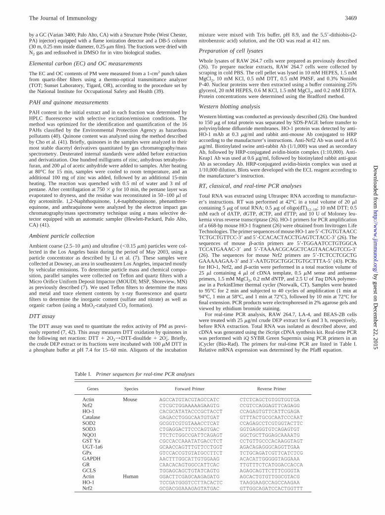
Table I. Primer sequences for real-time PCR analyses
Genes Species Forward Primer Reverse Primer
Actin Mouse AGCCATGTACGTAGCCATC CTCTCAGCTGTGGTGGTGANrf2 CTCGCTGGAAAAAGAAGTG CCGTCCAGGAGTTCAGAGGHO-1 CACGCATATACCCGCTACCT CCAGAGTGTTCATTCGAGACatalase GAGACCTGGGCAATGTGAT GTTTACTGCGCAATCCCAATSOD2 GCGGTCGTGTAAACCTCAT CCAGAGCCTCGTGGTACTTCSOD3 CTGAGGACTTCCCAGTGAC GGTGAGGGTGTCAGAGTGTNQO1 TTCTCTGGCCGATTCAGAGT GGCTGCTTGGAGCAAAATGGST Ya CGCCACCAAATATGACCTCT CCTGTTGCCCACAAGGTAGTUGT-1a6 GCAACCAGTTTGTTCCTGGT AGACAGAGGGCAGGTTGAAGPx GTCCACCGTGTATGCCTTCT TCTGCAGATCGTTCATCTCGGAPDH AACTTTGGCATTGTGGAAG ACACATTGGGGGTAGGAAAGR CAACACAGTGGCCATTCAC TTGTTTCTCATGGACCACCAGCLS TGGAGCAGCTGTATCAGTG AGAGCAGTTCTTTCGGGTAActin Human GGACTTCGAGCAAGAGATG AGCACTGTGTTGGCGTACGHO-1 TCCGATGGGTCCTTACACTC TAAGGAAGCCAGCCAAGAANrf2 GCGACGGAAAGAGTATGAC GTTGGCAGATCCACTGGTTT
3469The Journal of Immunology
by guest on Decem
ber 22, 2015http://w
ww
.jimm
unol.org/D
ownloaded from
RAW 264.7 transfection with SX2 plus wild-type or dominantnegative Nrf2 and luciferase assay
RAW 264.7 cells were plated at 2 � 105 cells/well in a 12-well plate 24 hbefore transfection. Cells were transfected with the Effectene transfectionreagent according to the manufacturer’s recommendations. Each well wastransfected with a DNA mixture containing 100 ng of pSX2-luc, 50 ng ofpCMV-�-galactosidase, and 50 ng of empty vector or the Nrf2 expressionplasmid (either wild-type or dominant negative mutant). The luciferaseactivity in each sample was corrected for variations in transfection effi-ciency with the corresponding �-galactosidase activity. Luciferase activitywas measured using a luciferase reagent kit from Promega (Madison, WI).
Confocal microscopy
Confocal microscopy was performed as previously described (44). RAW264.7 cells were treated with 50 �g/ml DEP extract for 2 h before washingin DMEM and attachment of 106 cells to each polylysine-coated coverslip.Cells were fixed in 4% paraformaldehyde in 30 mM sucrose for 10 min atroom temperature, followed by 10 min in 50 mM NH4Cl to neutralizealdehyde groups. For intracellular staining, coverslips were permeabilizedwith 0.1% Triton X-100/PBS for 30 min. After blocking in 1% BSA plus1% normal goat serum, cells were incubated with polyclonal anti-Nrf2 Ab(1/200) for 16 h. After washing in 0.1% Triton X-100/PBS, slides wereincubated with Alexa 594-conjugated goat anti-rabbit Ab for 60 min, fol-lowed by cellular nuclear staining with DAPI. Control slides were stainedwith rabbit nonimmune serum before addition of the secondary Ab. Sam-ples were examined under a Leica inverted TCS-SP confocal microscope(Deerfield, IL), using �100 and �43 objectives, respectively. Data acqui-sition was accomplished with Leica confocal software.
Preparation of thioglycolate-elicited peritoneal macrophagesfrom Nrf2�/� mice
Nrf2�/� animals on a mixed C57BL6/SV129 background were initiallyobtained from Dr. Y. Kan (University of California, San Francisco, CA)(45) and enriched 87.5% on a C57BL6 background by three rounds ofbreeding. Nrf2�/� littermate controls and wild-type C57BL/6 mice wereused as comparative controls. Peritoneal macrophages were collected byi.p. instillation of thioglycolate for 5 days before harvesting. Peritonealmacrophages were lavaged from the peritoneal cavity in complete DMEM,and 106 cells/well were used for stimulation with 50 �g/ml crude DEPextract. The macrophages were then lysed and processed for HO-1 blottingas described above.
ResultsOrganic DEP extracts induce mRNA expression of phase IIenzymes in BEAS-2B and RAW 264.7 cells
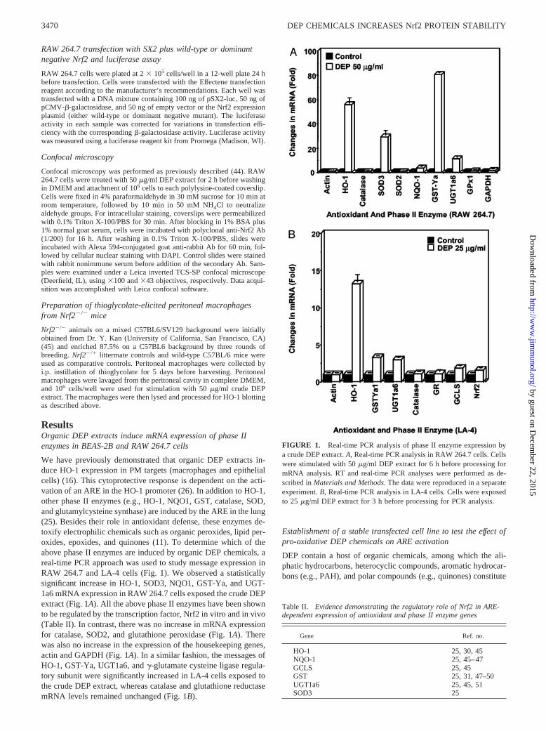
We have previously demonstrated that organic DEP extracts in-duce HO-1 expression in PM targets (macrophages and epithelialcells) (16). This cytoprotective response is dependent on the acti-vation of an ARE in the HO-1 promoter (26). In addition to HO-1,other phase II enzymes (e.g., HO-1, NQO1, GST, catalase, SOD,and glutamylcysteine synthase) are induced by the ARE in the lung(25). Besides their role in antioxidant defense, these enzymes de-toxify electrophilic chemicals such as organic peroxides, lipid per-oxides, epoxides, and quinones (11). To determine which of theabove phase II enzymes are induced by organic DEP chemicals, areal-time PCR approach was used to study message expression inRAW 264.7 and LA-4 cells (Fig. 1). We observed a statisticallysignificant increase in HO-1, SOD3, NQO1, GST-Ya, and UGT-1a6 mRNA expression in RAW 264.7 cells exposed the crude DEPextract (Fig. 1A). All the above phase II enzymes have been shownto be regulated by the transcription factor, Nrf2 in vitro and in vivo(Table II). In contrast, there was no increase in mRNA expressionfor catalase, SOD2, and glutathione peroxidase (Fig. 1A). Therewas also no increase in the expression of the housekeeping genes,actin and GAPDH (Fig. 1A). In a similar fashion, the messages ofHO-1, GST-Ya, UGT1a6, and �-glutamate cysteine ligase regula-tory subunit were significantly increased in LA-4 cells exposed tothe crude DEP extract, whereas catalase and glutathione reductasemRNA levels remained unchanged (Fig. 1B).
Establishment of a stable transfected cell line to test the effect ofpro-oxidative DEP chemicals on ARE activation
DEP contain a host of organic chemicals, among which the ali-phatic hydrocarbons, heterocyclic compounds, aromatic hydrocar-bons (e.g., PAH), and polar compounds (e.g., quinones) constitute
Table II. Evidence demonstrating the regulatory role of Nrf2 in ARE-dependent expression of antioxidant and phase II enzyme genes
Gene Ref. no.
HO-1 25, 30, 45NQO-1 25, 45–47GCLS 25, 45GST 25, 31, 47–50UGT1a6 25, 45, 51SOD3 25
FIGURE 1. Real-time PCR analysis of phase II enzyme expression bya crude DEP extract. A, Real-time PCR analysis in RAW 264.7 cells. Cellswere stimulated with 50 �g/ml DEP extract for 6 h before processing formRNA analysis. RT and real-time PCR analyses were performed as de-scribed in Materials and Methods. The data were reproduced in a separateexperiment. B, Real-time PCR analysis in LA-4 cells. Cells were exposedto 25 �g/ml DEP extract for 3 h before processing for PCR analysis.
3470 DEP CHEMICALS INCREASES Nrf2 PROTEIN STABILITY
by guest on Decem
ber 22, 2015http://w
ww
.jimm
unol.org/D
ownloaded from
the major groups (52–55). Previous studies have demonstrated thatthe polar and, to a lesser extent, the aromatic fractions are inducersof HO-1 expression in RAW 264.7 cells (26). To provide a rapidand reproducible readout of ARE activity, we constructed a RAW264.7 line with stable expression of an ARE-luciferase reporter
gene. This cell line exhibited a dose-dependent increase in lucif-erase activity in response to the crude DEP extract (Fig. 2A) andwas therefore suitable for comparing the aliphatic, aromatic, andpolar chemical fractions. The amounts and recovery of those frac-tions during fractionation of a crude DEP extract by silica gel
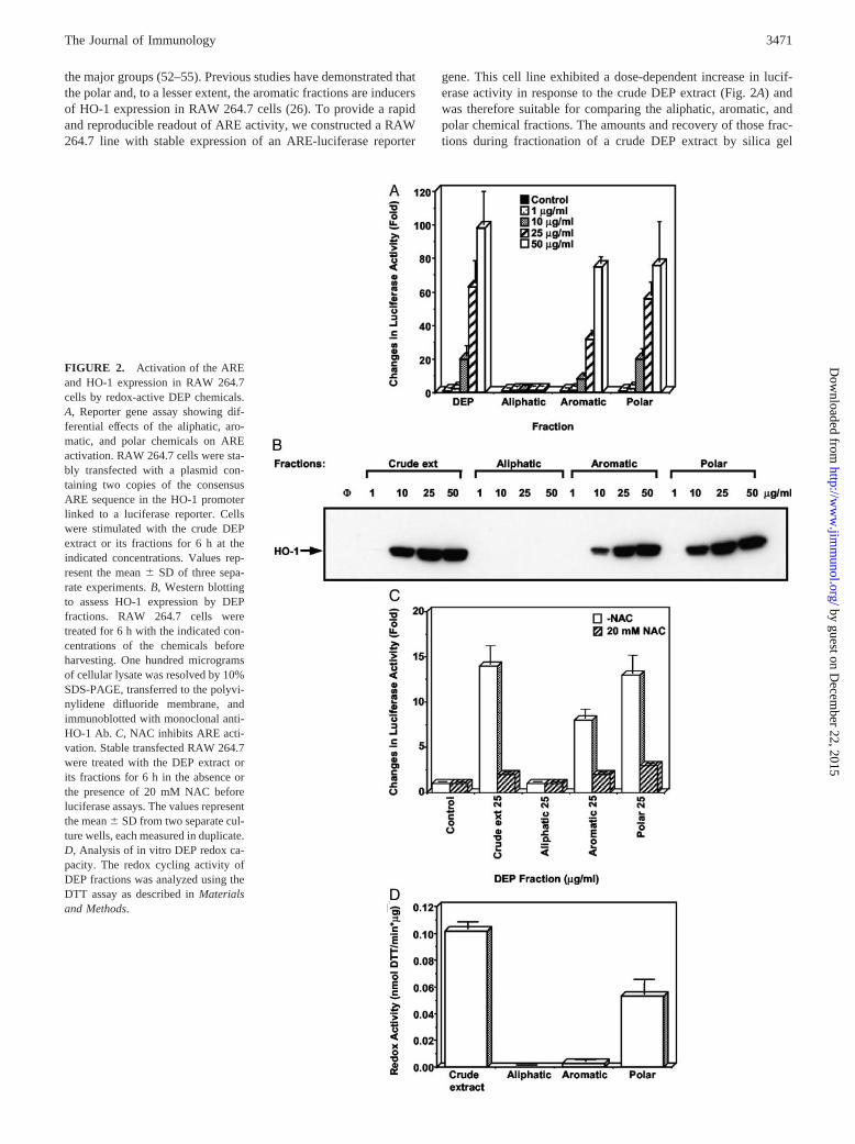
FIGURE 2. Activation of the AREand HO-1 expression in RAW 264.7cells by redox-active DEP chemicals.A, Reporter gene assay showing dif-ferential effects of the aliphatic, aro-matic, and polar chemicals on AREactivation. RAW 264.7 cells were sta-bly transfected with a plasmid con-taining two copies of the consensusARE sequence in the HO-1 promoterlinked to a luciferase reporter. Cellswere stimulated with the crude DEPextract or its fractions for 6 h at theindicated concentrations. Values rep-resent the mean � SD of three sepa-rate experiments. B, Western blottingto assess HO-1 expression by DEPfractions. RAW 264.7 cells weretreated for 6 h with the indicated con-centrations of the chemicals beforeharvesting. One hundred microgramsof cellular lysate was resolved by 10%SDS-PAGE, transferred to the polyvi-nylidene difluoride membrane, andimmunoblotted with monoclonal anti-HO-1 Ab. C, NAC inhibits ARE acti-vation. Stable transfected RAW 264.7were treated with the DEP extract orits fractions for 6 h in the absence orthe presence of 20 mM NAC beforeluciferase assays. The values representthe mean � SD from two separate cul-ture wells, each measured in duplicate.D, Analysis of in vitro DEP redox ca-pacity. The redox cycling activity ofDEP fractions was analyzed using theDTT assay as described in Materialsand Methods.
3471The Journal of Immunology
by guest on Decem
ber 22, 2015http://w
ww
.jimm
unol.org/D
ownloaded from
chromatography are shown in Table III (26). Chemical analysisconfirmed that the aromatic fraction contained an abundance ofPAHs (Table IV), whereas the polar fraction lacked PAHs but wasenriched with quinones (Table V; 26). Although exposure to thealiphatic fraction lacked any effect, the aromatic and polar chem-ical compounds induced a dose-dependent increase in luciferaseactivity (Fig. 2A). The polar was more active than the aromaticfraction on a dose-by-dose comparison (Fig. 2A). To confirm thebiological relevance of the reporter gene assay, we showed thatendogenous HO-1 expression by the polar fraction is more robustthan the aromatic fraction (Fig. 2B). The aliphatic fraction wasinactive (Fig. 2B).
ARE-mediated gene expression by heavy metals and pro-oxi-dative chemicals is dependent on electrophilic chemistry and pro-tein thiol modification (11). This includes modification of thiolgroups that control the function of the ARE-inducing transcriptionfactor, Nrf2 (14, 56). Thiol modification is important in under-standing the biological effect of DEP chemicals, as demonstratedby the ability of NAC to interfere in HO-1 expression (12, 15).This agent uses its Src homology group to directly neutralize elec-trophilic DEP chemicals (11, 24). It is of interest, therefore, thatthe NAC inhibitory effect also applies to the actions of the aro-matic and polar chemical fractions (Fig. 2C). NAC also suppressedARE-luciferase activity by the crude DEP extract (Fig. 2C).
As well as participating in electrophilic interactions, DEP en-gage in redox cycling reactions that lead to ROS production; this
effect may also be relevant to the activation of the ARE (12, 26).We have modified an in vitro assay developed by Kumagai et al.(42) that directly measures the capacity of intact particles and theirorganic compounds to redox cycle in the presence of DTT (seeMaterials and Methods). Quinones and possibly other redox cy-cling chemicals lead to DTT oxidation and O2
. generation (7, 42).When tested in the DTT assay, the polar fraction was considerablymore potent than the aromatic fraction, whereas aliphatic materialwas inactive (Fig. 2D). These results suggest that the increasedpotency of the polar fraction in the activation of the ARE may beexplained by its increased content of redox cycling chemicals.
Activation of the ARE by the organic DEP extract is dependenton Nrf2
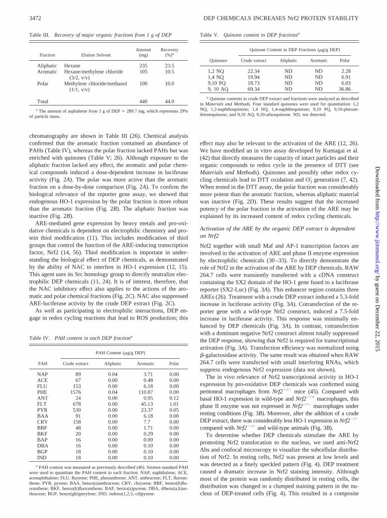
Nrf2 together with small Maf and AP-1 transcription factors areinvolved in the activation of ARE and phase II enzyme expressionby electrophilic chemicals (30–33). To directly demonstrate therole of Nrf2 in the activation of the ARE by DEP chemicals, RAW264.7 cells were transiently transfected with a cDNA constructcontaining the SX2 domain of the HO-1 gene fused to a luciferasereporter (SX2-Luc) (Fig. 3A). This enhancer region contains threeAREs (26). Treatment with a crude DEP extract induced a 5.3-foldincrease in luciferase activity (Fig. 3A). Cotransfection of the re-porter gene with a wild-type Nrf2 construct, induced a 7.5-foldincrease in luciferase activity. This response was minimally en-hanced by DEP chemicals (Fig. 3A). In contrast, cotransfectionwith a dominant negative Nrf2 construct almost totally suppressedthe DEP response, showing that Nrf2 is required for transcriptionalactivation (Fig. 3A). Transfection efficiency was normalized using�-galactosidase activity. The same result was obtained when RAW264.7 cells were transfected with small interfering RNAs, whichsuppress endogenous Nrf2 expression (data not shown).
The in vivo relevance of Nrf2 transcriptional activity in HO-1expression by pro-oxidative DEP chemicals was confirmed usingperitoneal macrophages from Nrf2�/� mice (45). Compared withbasal HO-1 expression in wild-type and Nrf2�/� macrophages, thisphase II enzyme was not expressed in Nrf2�/� macrophages underresting conditions (Fig. 3B). Moreover, after the addition of a crudeDEP extract, there was considerably less HO-1 expression in Nrf2�/�
compared with Nrf2�/� and wild-type animals (Fig. 3B).To determine whether DEP chemicals stimulate the ARE by
promoting Nrf2 translocation to the nucleus, we used anti-Nrf2Abs and confocal microscopy to visualize the subcellular distribu-tion of Nrf2. In resting cells, Nrf2 was present at low levels andwas detected as a finely speckled pattern (Fig. 4). DEP treatmentcaused a dramatic increase in Nrf2 staining intensity. Althoughmost of the protein was randomly distributed in resting cells, thedistribution was changed to a clumped staining pattern in the nu-cleus of DEP-treated cells (Fig. 4). This resulted in a composite
Table III. Recovery of major organic fractions from 1 g of DEP
Fraction Elution SolventAmount
(mg)Recovery
(%)a
Aliphatic Hexane 235 23.5Aromatic Hexane/methylene chloride
(3/2, v/v)105 10.5
Polar Methylene chloride/methanol(1/1, v/v)
100 10.0
Total 440 44.0
a The amount of asphaltene from 1 g of DEP � 289.7 mg, which represents 29%of particle mass.
Table IV. PAH content in each DEP fractiona
PAH
PAH Content (�g/g DEP)
Crude extract Aliphatic Aromatic Polar
NAP 89 0.04 3.71 0.00ACE 67 0.00 0.48 0.00FLU 153 0.00 6.18 0.00PHE 1576 0.04 110.87 0.00ANT 24 0.00 0.95 0.12FLT 678 0.00 45.13 1.01PYR 530 0.00 23.37 0.05BAA 91 0.00 6.18 0.00CRY 158 0.00 7.7 0.00BBF 48 0.00 1.71 0.00BKF 20 0.00 0.29 0.00BAP 16 0.00 0.00 0.00DBA 16 0.00 0.10 0.00BGP 18 0.00 0.10 0.00IND 18 0.00 0.10 0.00
a PAH content was measured as previously described (40). Sixteen standard PAHwere used to quantitate the PAH content in each fraction. NAP, naphthalene; ACE,acenaphthalene; FLU, fluorene; PHE, phenanthrene; ANT, anthracene; FLT, fluoran-thene; PYR, pyrene; BAA, benzo(a)anthracene; CRY, chrysene; BBF, benzo(b)flu-oranthene; BKF, benzo(k)fluoranthene; BAP, benzo(a)pyrene; DBA, dibenz(a,h)an-thracene; BGP, benzo(ghi)perylene; IND, indeno(1,2,3,-cd)pyrene.
Table V. Quinone content in DEP fractionsa
Quinones
Quinone Content in DEP Fractions (�g/g DEP)
Crude extract Aliphatic Aromatic Polar
1,2 NQ 22.34 ND ND 2.281,4 NQ 19.94 ND ND 6.919,10 PQ 18.73 ND ND 6.039, 10 AQ 69.34 ND ND 36.86
a Quinone contents in crude DEP extract and fractions were analyzed as describedin Materials and Methods. Four standard quinones were used for quantitation: 1,2NQ, 1,2-naphthoquinone; 1,4 NQ, 1,4-naphthoquinone; 9,10 PQ, 9,10-phenan-threnequinone; and 9,10 AQ, 9,10-athraquinone. ND, not detected.
3472 DEP CHEMICALS INCREASES Nrf2 PROTEIN STABILITY
by guest on Decem
ber 22, 2015http://w
ww
.jimm
unol.org/D
ownloaded from
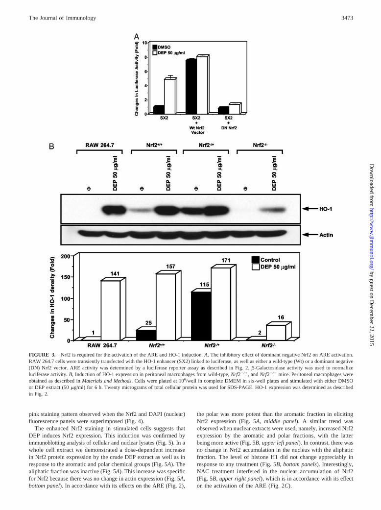
pink staining pattern observed when the Nrf2 and DAPI (nuclear)fluorescence panels were superimposed (Fig. 4).
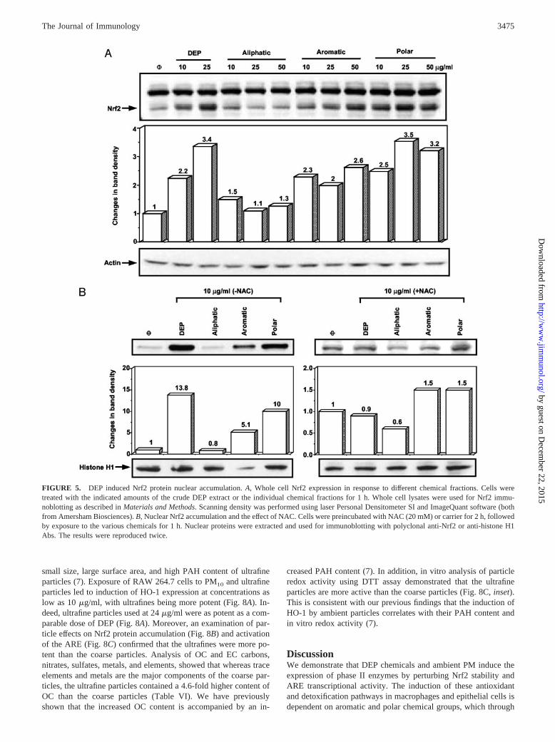
The enhanced Nrf2 staining in stimulated cells suggests thatDEP induces Nrf2 expression. This induction was confirmed byimmunoblotting analysis of cellular and nuclear lysates (Fig. 5). In awhole cell extract we demonstrated a dose-dependent increasein Nrf2 protein expression by the crude DEP extract as well as inresponse to the aromatic and polar chemical groups (Fig. 5A). Thealiphatic fraction was inactive (Fig. 5A). This increase was specificfor Nrf2 because there was no change in actin expression (Fig. 5A,bottom panel). In accordance with its effects on the ARE (Fig. 2),
the polar was more potent than the aromatic fraction in elicitingNrf2 expression (Fig. 5A, middle panel). A similar trend wasobserved when nuclear extracts were used, namely, increased Nrf2expression by the aromatic and polar fractions, with the latterbeing more active (Fig. 5B, upper left panel). In contrast, there wasno change in Nrf2 accumulation in the nucleus with the aliphaticfraction. The level of histone H1 did not change appreciably inresponse to any treatment (Fig. 5B, bottom panels). Interestingly,NAC treatment interfered in the nuclear accumulation of Nrf2(Fig. 5B, upper right panel), which is in accordance with its effecton the activation of the ARE (Fig. 2C).
FIGURE 3. Nrf2 is required for the activation of the ARE and HO-1 induction. A, The inhibitory effect of dominant negative Nrf2 on ARE activation.RAW 264.7 cells were transiently transfected with the HO-1 enhancer (SX2) linked to luciferase, as well as either a wild-type (Wt) or a dominant negative(DN) Nrf2 vector. ARE activity was determined by a luciferase reporter assay as described in Fig. 2. �-Galactosidase activity was used to normalizeluciferase activity. B, Induction of HO-1 expression in peritoneal macrophages from wild-type, Nrf2�/�, and Nrf2�/� mice. Peritoneal macrophages wereobtained as described in Materials and Methods. Cells were plated at 106/well in complete DMEM in six-well plates and stimulated with either DMSOor DEP extract (50 �g/ml) for 6 h. Twenty micrograms of total cellular protein was used for SDS-PAGE. HO-1 expression was determined as describedin Fig. 2.
3473The Journal of Immunology
by guest on Decem
ber 22, 2015http://w
ww
.jimm
unol.org/D
ownloaded from
Nrf2 abundance is post-translationally regulated
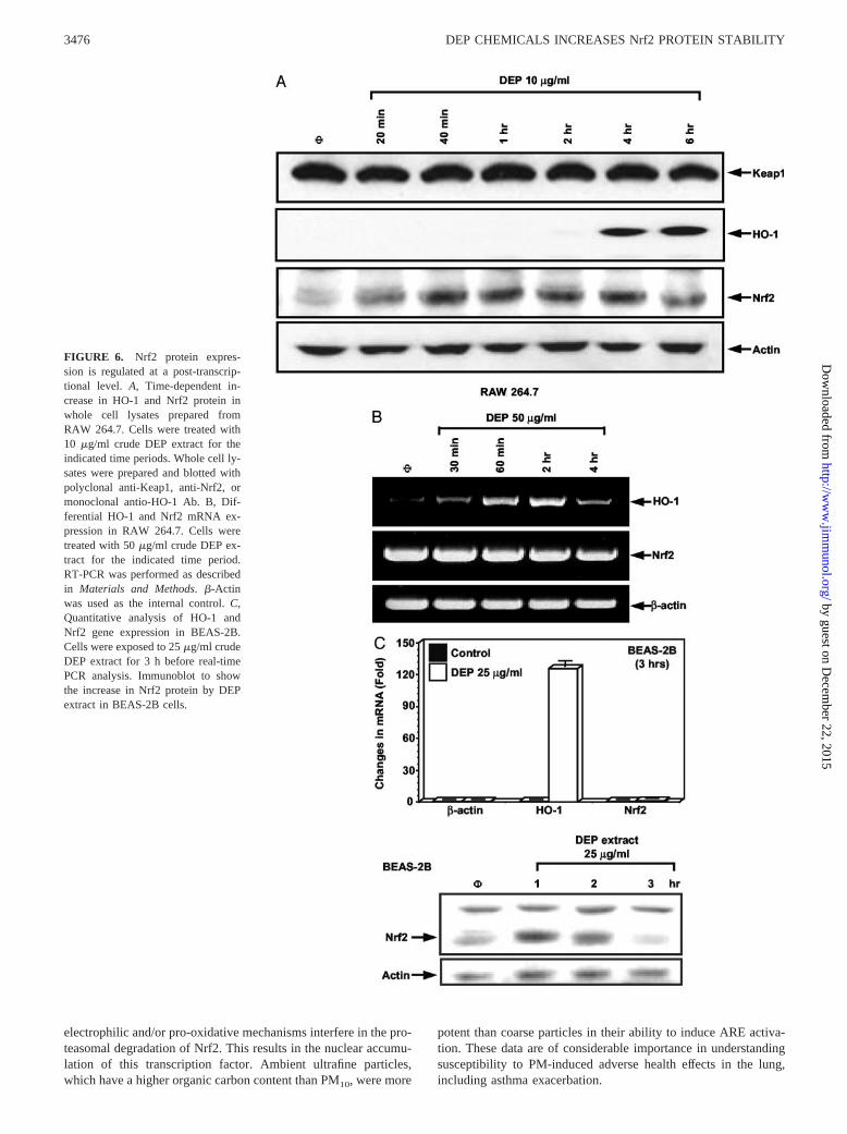
To investigate the mechanism by which DEP chemicals induceNrf2 expression, we monitored the temporal accumulation of Nrf2protein and mRNA after cellular stimulation. An increase in thelevel of Nrf2 protein was detected within 20 min after treatmentand was maintained for up to 6 h, the last time point tested (Fig.6A). Interestingly, this increase in Nrf2 protein was not accompa-nied by increased mRNA expression at any time point (Fig. 6B).By contrast, HO-1 mRNA was increased by the same stimulus(Fig. 6B), and this preceded the increase in protein expression at4 h (Fig. 6A). The initial appearance of the HO-1 message (�30min) is compatible with the prior accumulation (�20 min) of Nrf2protein (Fig. 6, A and B). DEP also stimulated Nrf2 protein ex-pression without affecting mRNA levels in BEAS-2B cells (Fig.6C). In contrast, expression of Keap1 protein (Fig. 6A) or �-actinmRNA/protein (Fig. 6) did not change in response to the chemicalexposure (Fig. 6A). Taken together, these results suggest that DEPregulates Nrf2 expression by post-transcriptional mechanisms.
DEP chemicals increase Nrf2 protein half-life by affectingproteasomal degradation
It has recently been reported that pro-oxidative chemicals, includ-ing �-naphthoflavone and cadmium, increase Nrf2 expression byinterfering in proteasomal degradation in HepG2 and Hepa cells(34, 38). To determine whether Nrf2 is similarly regulated in RAW264.7, cells were treated with MG-132, a 26S proteasome inhibi-tor, and whole cell lysates were subjected to Nrf2 immunoblotting.MG-132 induced a dose-dependent increase in Nrf2 protein abun-dance without requiring a costimulus (Fig. 7A). Similar resultswere obtained with another 26S proteasome inhibitor, lactacystin(not shown). We next determined the half-life of Nrf2 after inhi-
bition of protein synthesis by CHX. Cells treated with the DEPextract, followed by CHX exposure, showed a rapid disappearanceof the accumulated Nrf2 protein, with an estimated half-life of�30 min (Fig. 7B). The addition of MG-132 to CHX prolongedthe Nrf2 half-life to �90 min (Fig. 7B). Due to the low abundanceof Nrf2 in resting cells, it was not possible to estimate proteinhalf-life in the absence of DEP chemicals. However, we couldshow that combining the DEP extract with CHX prolonged Nrf2expression in MG-132-treated cells (Fig. 7C). This suggests thatthe crude extract enhanced the inhibitory effects of MG-132 onNrf2 proteasomal degradation. Analogous experiments with thearomatic and polar chemical fractions showed that similar to thecrude extract, these materials could induce Nrf2 expression with ahalf-life of �30 min (Fig. 7D). These results are consistent withpreviously published reports that assessed Nrf2 protein half-lifethrough the use of CHX and either immunoblotting or [35S]me-thionine pulse labeling (34).
Ambient particulate matter activates the ARE and induces HO-1expression via Nrf2
We asked whether the engagement of the antioxidant defense path-way by organic DEP chemicals apply to “real-life” ambient PM.Ambient PM consists of different particle sizes: coarse PM (PM10)are typically derived from soil, road dust, construction debris, oraggregation of smaller combustion particles, whereas ultrafine(�0.1 �m) PM are derived from the combustion of fossil fuelproducts, including diesel, as well as nucleation processes (54). Acomparison of the effects of ambient ultrafine and coarse particlescollected by particle concentrators in the Los Angeles basin dem-onstrated that the ultrafines were more potent than PM10 in gen-erating oxidative stress (7). This outcome is clearly related to the
FIGURE 4. Confocal microscopy to show intercellular Nrf2 expression. RAW 264.7 cells were treated with the crude DEP extract for 2 h. Confocalmicroscopy was conducted as described in Materials and Methods. Nrf2 was stained with polyclonal anti-Nrf2 Ab, followed by Alexa 594-conjugated goatanti-rabbit secondary Ab, whereas the nucleus was counterstained with DAPI.
3474 DEP CHEMICALS INCREASES Nrf2 PROTEIN STABILITY
by guest on Decem
ber 22, 2015http://w
ww
.jimm
unol.org/D
ownloaded from
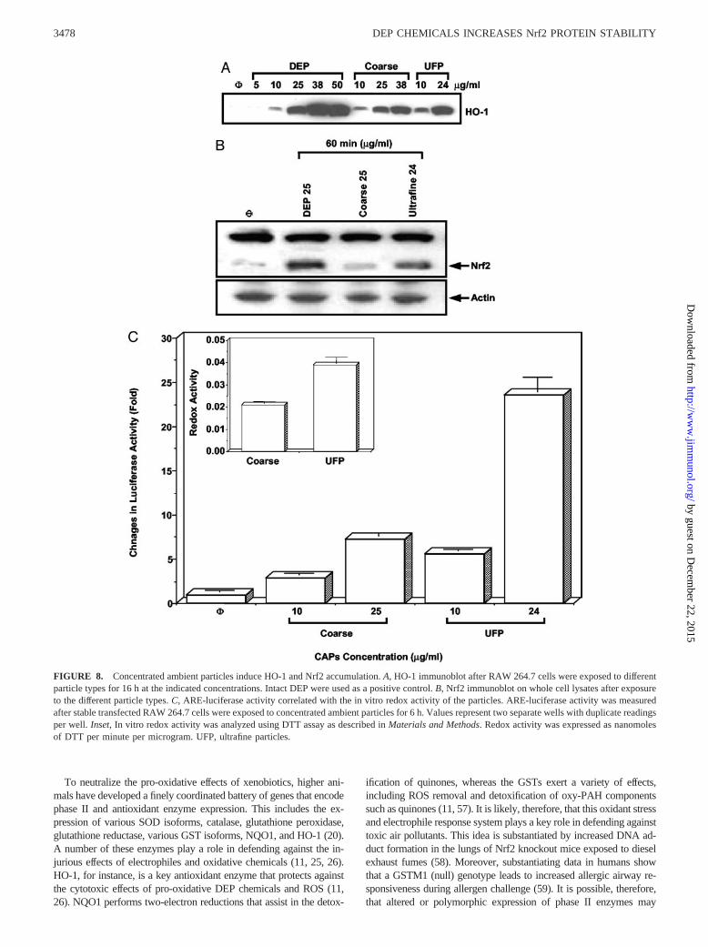
small size, large surface area, and high PAH content of ultrafineparticles (7). Exposure of RAW 264.7 cells to PM10 and ultrafineparticles led to induction of HO-1 expression at concentrations aslow as 10 �g/ml, with ultrafines being more potent (Fig. 8A). In-deed, ultrafine particles used at 24 �g/ml were as potent as a com-parable dose of DEP (Fig. 8A). Moreover, an examination of par-ticle effects on Nrf2 protein accumulation (Fig. 8B) and activationof the ARE (Fig. 8C) confirmed that the ultrafines were more po-tent than the coarse particles. Analysis of OC and EC carbons,nitrates, sulfates, metals, and elements, showed that whereas traceelements and metals are the major components of the coarse par-ticles, the ultrafine particles contained a 4.6-fold higher content ofOC than the coarse particles (Table VI). We have previouslyshown that the increased OC content is accompanied by an in-
creased PAH content (7). In addition, in vitro analysis of particleredox activity using DTT assay demonstrated that the ultrafineparticles are more active than the coarse particles (Fig. 8C, inset).This is consistent with our previous findings that the induction ofHO-1 by ambient particles correlates with their PAH content andin vitro redox activity (7).
DiscussionWe demonstrate that DEP chemicals and ambient PM induce theexpression of phase II enzymes by perturbing Nrf2 stability andARE transcriptional activity. The induction of these antioxidantand detoxification pathways in macrophages and epithelial cells isdependent on aromatic and polar chemical groups, which through
FIGURE 5. DEP induced Nrf2 protein nuclear accumulation. A, Whole cell Nrf2 expression in response to different chemical fractions. Cells weretreated with the indicated amounts of the crude DEP extract or the individual chemical fractions for 1 h. Whole cell lysates were used for Nrf2 immu-noblotting as described in Materials and Methods. Scanning density was performed using laser Personal Densitometer SI and ImageQuant software (bothfrom Amersham Biosciences). B, Nuclear Nrf2 accumulation and the effect of NAC. Cells were preincubated with NAC (20 mM) or carrier for 2 h, followedby exposure to the various chemicals for 1 h. Nuclear proteins were extracted and used for immunoblotting with polyclonal anti-Nrf2 or anti-histone H1Abs. The results were reproduced twice.
3475The Journal of Immunology
by guest on Decem
ber 22, 2015http://w
ww
.jimm
unol.org/D
ownloaded from
electrophilic and/or pro-oxidative mechanisms interfere in the pro-teasomal degradation of Nrf2. This results in the nuclear accumu-lation of this transcription factor. Ambient ultrafine particles,which have a higher organic carbon content than PM10, were more
potent than coarse particles in their ability to induce ARE activa-tion. These data are of considerable importance in understandingsusceptibility to PM-induced adverse health effects in the lung,including asthma exacerbation.
FIGURE 6. Nrf2 protein expres-sion is regulated at a post-transcrip-tional level. A, Time-dependent in-crease in HO-1 and Nrf2 protein inwhole cell lysates prepared fromRAW 264.7. Cells were treated with10 �g/ml crude DEP extract for theindicated time periods. Whole cell ly-sates were prepared and blotted withpolyclonal anti-Keap1, anti-Nrf2, ormonoclonal antio-HO-1 Ab. B, Dif-ferential HO-1 and Nrf2 mRNA ex-pression in RAW 264.7. Cells weretreated with 50 �g/ml crude DEP ex-tract for the indicated time period.RT-PCR was performed as describedin Materials and Methods. �-Actinwas used as the internal control. C,Quantitative analysis of HO-1 andNrf2 gene expression in BEAS-2B.Cells were exposed to 25 �g/ml crudeDEP extract for 3 h before real-timePCR analysis. Immunoblot to showthe increase in Nrf2 protein by DEPextract in BEAS-2B cells.
3476 DEP CHEMICALS INCREASES Nrf2 PROTEIN STABILITY
by guest on Decem
ber 22, 2015http://w
ww
.jimm
unol.org/D
ownloaded from
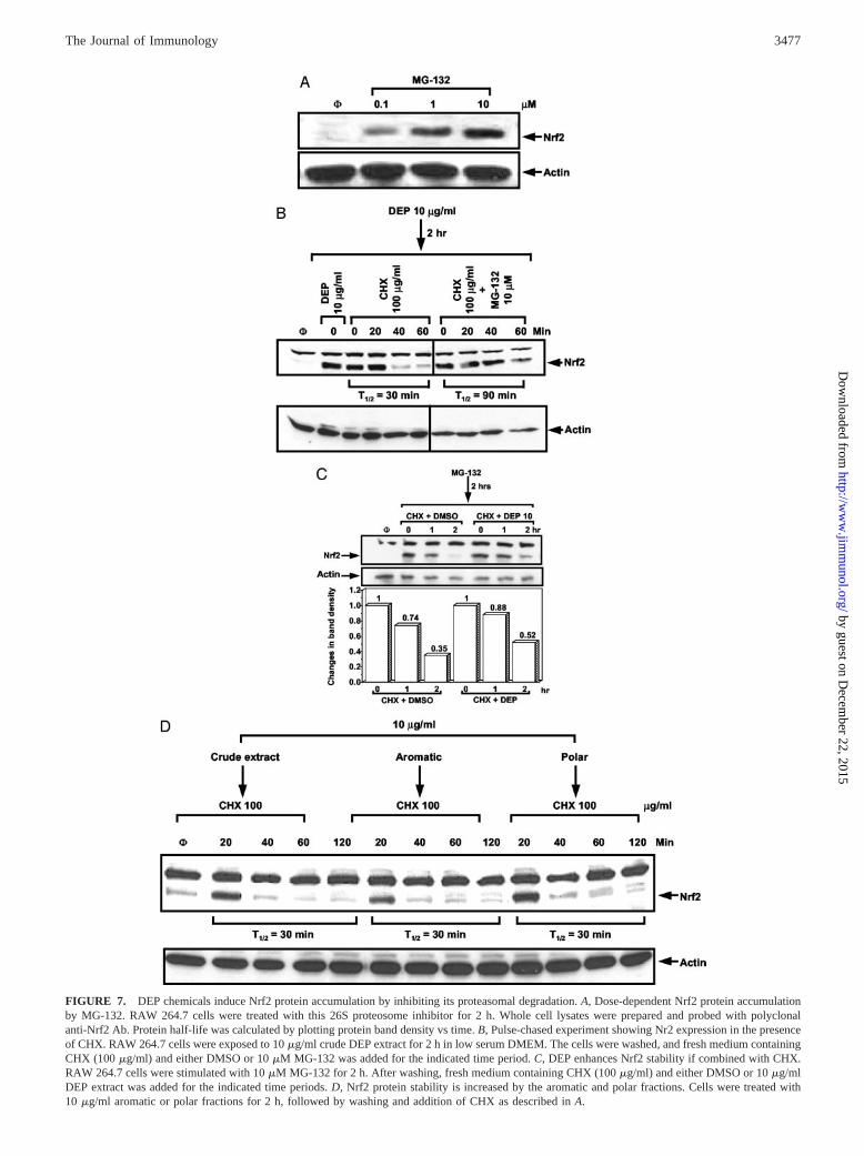
FIGURE 7. DEP chemicals induce Nrf2 protein accumulation by inhibiting its proteasomal degradation. A, Dose-dependent Nrf2 protein accumulationby MG-132. RAW 264.7 cells were treated with this 26S proteosome inhibitor for 2 h. Whole cell lysates were prepared and probed with polyclonalanti-Nrf2 Ab. Protein half-life was calculated by plotting protein band density vs time. B, Pulse-chased experiment showing Nr2 expression in the presenceof CHX. RAW 264.7 cells were exposed to 10 �g/ml crude DEP extract for 2 h in low serum DMEM. The cells were washed, and fresh medium containingCHX (100 �g/ml) and either DMSO or 10 �M MG-132 was added for the indicated time period. C, DEP enhances Nrf2 stability if combined with CHX.RAW 264.7 cells were stimulated with 10 �M MG-132 for 2 h. After washing, fresh medium containing CHX (100 �g/ml) and either DMSO or 10 �g/mlDEP extract was added for the indicated time periods. D, Nrf2 protein stability is increased by the aromatic and polar fractions. Cells were treated with10 �g/ml aromatic or polar fractions for 2 h, followed by washing and addition of CHX as described in A.
3477The Journal of Immunology
by guest on Decem
ber 22, 2015http://w
ww
.jimm
unol.org/D
ownloaded from
To neutralize the pro-oxidative effects of xenobiotics, higher ani-mals have developed a finely coordinated battery of genes that encodephase II and antioxidant enzyme expression. This includes the ex-pression of various SOD isoforms, catalase, glutathione peroxidase,glutathione reductase, various GST isoforms, NQO1, and HO-1 (20).A number of these enzymes play a role in defending against the in-jurious effects of electrophiles and oxidative chemicals (11, 25, 26).HO-1, for instance, is a key antioxidant enzyme that protects againstthe cytotoxic effects of pro-oxidative DEP chemicals and ROS (11,26). NQO1 performs two-electron reductions that assist in the detox-
ification of quinones, whereas the GSTs exert a variety of effects,including ROS removal and detoxification of oxy-PAH componentssuch as quinones (11, 57). It is likely, therefore, that this oxidant stressand electrophile response system plays a key role in defending againsttoxic air pollutants. This idea is substantiated by increased DNA ad-duct formation in the lungs of Nrf2 knockout mice exposed to dieselexhaust fumes (58). Moreover, substantiating data in humans showthat a GSTM1 (null) genotype leads to increased allergic airway re-sponsiveness during allergen challenge (59). It is possible, therefore,that altered or polymorphic expression of phase II enzymes may
FIGURE 8. Concentrated ambient particles induce HO-1 and Nrf2 accumulation. A, HO-1 immunoblot after RAW 264.7 cells were exposed to differentparticle types for 16 h at the indicated concentrations. Intact DEP were used as a positive control. B, Nrf2 immunoblot on whole cell lysates after exposureto the different particle types. C, ARE-luciferase activity correlated with the in vitro redox activity of the particles. ARE-luciferase activity was measuredafter stable transfected RAW 264.7 cells were exposed to concentrated ambient particles for 6 h. Values represent two separate wells with duplicate readingsper well. Inset, In vitro redox activity was analyzed using DTT assay as described in Materials and Methods. Redox activity was expressed as nanomolesof DTT per minute per microgram. UFP, ultrafine particles.
3478 DEP CHEMICALS INCREASES Nrf2 PROTEIN STABILITY
by guest on Decem
ber 22, 2015http://w
ww
.jimm
unol.org/D
ownloaded from
determine which human subjects are more prone to developingasthma during PM exposure.
The coordinated responses of phase II genes are regulatedthrough a cis element, known as the ARE or the electrophile re-sponse element (11). Nrf2 in association with small Maf proteins(e.g., Maf G) as well as AP-1 transcription factors play key rolesin the activation of the ARE (30–33). The importance of Nrf2 isillustrated by the failure to induce phase II enzyme expression inthe lungs and livers of Nrf2-deficient mice, thereby rendering theseanimals more susceptible to the effects of hyperoxia, pro-oxidativefood chemicals, and drugs (e.g., acetaminophen) (45, 58, 60, 61).Our results using peritoneal macrophages from Nrf2�/� mice alsoprovided direct evidence for the role of Nrf2 in regulating HO-1expression in response to pro-oxidative DEP chemicals (Fig. 3B).This is compatible with interference in phase II enzyme expressionunder conditions of hyperoxic lung injury (25). The ability of Nrf2to transcriptionally activate these genes is regulated in part by acytoplasmic protein, Keap1 (34–38). Keap1 contains a C-terminalKelch domain that interacts directly with an Nrf2 regulatory site,known as the Neh2 domain (36, 62). However, the molecular ex-planation for the cytoplasmic retention of Nrf2 is poorly under-stood. Several models have been proposed to explain the escape ofNrf2 from Keap1-mediated repression (34, 36, 38, 56). One sug-gestion is that electrophilic chemicals, such as the quinones, co-valently modify one or more of the 27 thiol groups present inKeap1 (56). Zhang et al. (56) identified two critical Keap1 cysteineresidues, C273 and C288, that are required for Nrf2 ubiquitination.This ubiquitination event is probably responsible for the rapid pro-teasomal degradation and short half-life of Nrf2 under homeostaticconditions (34–37). However, the ability of these cysteines to in-duce Nrf2 ubiquitination may be disrupted in the presence of elec-trophilic compounds, allowing Nrf2 to escape Keap1-dependentdegradation (56). This could explain the increased Nrf2 half-life byDEP chemicals (Fig. 7C). The thiol hypothesis also explains whyNAC interferes with Nrf2 accumulation and activation of the AREin our system (Figs. 5B and 2C). NAC has a number of actions thatmay explain this effect. In addition to its ROS scavenging effectsand action as a GSH precursor, NAC uses its Src homology groupto participate in electrophilic interactions. This may involve directbinding to electrophilic DEP chemicals (24).
However, whereas thiol modification may explain why aromaticand polar chemical compounds increase Nrf2 protein accumulation(Fig. 5A), this does not explain Nrf2 release from Keap1. Addi-tional hypotheses have been proposed to explain this effect. One isthe phosphorylation of Nrf2 by protein kinase C (PKC) (38). PKCphosphorylates a Nrf2 S40 residue and induces its in vitro disso-ciation from Keap1 (38). However, phosphorylation of this residuehas not been demonstrated in vivo, and the S40A-Nrf2 mutantbehaved identically with the wild-type protein in its interactionwith Keap1 (38). We could not demonstrate interference in the
activation of the ARE by PKC inhibitors in our system (notshown).
A third possibility, which is not mutually exclusive of the rolesof C273 and C288, is that DEP chemicals may target additionalcysteines on Keap1 or Nrf2. One example is the Keap1 residue,C151; this cysteine plays a unique role in the response of thispathway to oxidative stress (56). One possibility is that C151 actsas a redox-sensitive switch that is targeted by electrophilic DEPcompounds or by ROS that is generated as an independent event.Quinones are functionalized polar chemicals that induce O2
. gen-eration when added to lung microsomes (17). This redox cyclingactivity is catalyzed by NADPH-P450 reductase (17, 18) and couldexplain why the polar is more active than the aromatic fraction ininducing Nrf2 accumulation and the activation of the ARE (Figs.2A and 5B). This idea is also in keeping with the increased potencyof the polar fraction in the DTT assay (Fig. 2D). This assay is ameasure of the in vitro redox cycling capacity of the oxy-PAHcompounds present in ambient PM and DEP (7, 42). It is interest-ing, therefore, that ultrafine particulates, which have a higher invitro redox cycling capability than coarse particles (7), are morepotent inducers of HO-1 expression, Nrf2 accumulation, and acti-vation of the ARE (Fig. 8). A possible way to explain the lessereffect of the aromatic fraction is that the PAHs need to be con-verted to quinones, e.g., by cytochrome P450 1A1, before theybecome functionalized (63, 64).
It is important to point out that the release of Nrf2 from Keap1may not be an essential regulatory step in the Nrf2/ARE pathway.This idea is derived from the fact that because Nrf2 bound byKeap1 is targeted for proteasomal degradation, it does not accu-mulate to significant levels in the cytoplasm of unstimulated cells(34, 36). Furthermore, Nrf2 that accumulates to high levels in thenucleus of stimulated cells represents primarily, if not exclusively,de novo synthesized protein (34, 36). Given these observations, itis possible that oxidants promote nuclear accumulation of Nrf2 notby affecting the release of Nrf2, which in any case would representa minimal amount of Nrf2, but by preventing newly synthesizedNrf2 from associating with Keap1. In the absence of such associ-ation, Nrf2 will escape proteasomal degradation and by default bedirected to the nucleus. Interestingly, and consistent with this hy-pothesis, Zhang and Hannink (56) have observed minimal releaseof Nrf2 from Keap1 in response to electrophiles. Although molec-ular details about the roles of Nrf2 and Keap1 in phase II enzymeexpression is still unclear, proteasome-mediated Nrf2 degradationappears to be an essential step in both mouse and humancells (65–67).
Oxidative stress may be the central mechanism by which am-bient PM induce adverse health effects. We have recently demon-strated that PM-induced oxidative stress is a multi-tier response, inwhich cytoprotective responses transition to injurious effects as thelevel of oxidative stress increases (11, 16). The induction of Nrf2-mediated phase II enzyme expression is an integral component ofthe cytoprotective response, which is triggered at the lowest level(tier 1) of oxidative stress. This system is designed to preventfurther oxidative stress, which may escalate into inflammation andcytotoxicity (11, 30–33). In this role, phase II enzymes may beparticularly important in averting allergen sensitization and aller-gic airway inflammation in atopic people (1, 11). In this regard, itis well known that DEP and ambient PM act as adjuvants thatpromote TH2 immune deviation during cochallenge with an aller-gen (1, 11). Moreover, several animal and human studies show thatoxidative stress is important in asthma, and that antioxidant de-fense pathways are important in the prevention of disease progres-sion (1, 11).
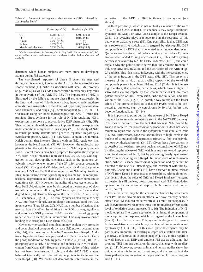
Table VI. Elemental and organic carbon content in CAPs collected inLos Angeles basina
Coarse, �g/m3 (%) Ultrafine, �g/m3 (%)
OC 1.786 (17.4) 6.911 (79.9)EC 0.817 (7.9) 0.844 (9.8)Nitrate 2.25 (21.9) 0 (0)Sulfate 0.972 (9.5) 0.381 (4.4)Metals and elements 5.638 (54.9) 1.689 (19.5)
a CAPs were collected in Downey, CA, in May 2003. The amounts of OC, EC,nitrate, sulfate, metals, and elements were analyzed as described in Materials andMethods.
3479The Journal of Immunology
by guest on Decem
ber 22, 2015http://w
ww
.jimm
unol.org/D
ownloaded from
There is growing evidence that genes involved in xenobioticdetoxification and antioxidant defense could serve as susceptiblegenes for asthma pathogenesis (59, 68–70). For instance, individ-uals who are homozygous for the GSTM1 (null) genotype, havebeen shown to have an increased risk for asthma development orallergic nasal responses (59). In contrast, homozygous GSTP1 ex-pression confers a protective effect on asthma and has also beenshown to protect against toluene di-isocyanate-induced asthma inspray painters (68). Although we still lack evidence for the in-volvement of Nrf2, it has recently been demonstrated that theHO-1 promoter expresses a polymorphism that defines gene in-ducibility under oxidative stress conditions (70). This polymor-phism impacts emphysema development in Japanese male smokers(70). We suggest that related polymorphisms in phase II enzymesdetermine their Nrf2 responses and could determine PM suscepti-bility. Although we are still uncertain about the cellular impact ofoxidative stress and antioxidant defense on Th2 pathways, we areexploring the possibility that the DEP adjuvant effect is mediatedthrough oxidative stress effects in the lung.
References1. Nel, A. E., D. Diaz-Sanchez, D. Ng, T. Hiura, and A. Saxon. 1998. Enhancement
of allergic inflammation by the interaction between diesel exhaust particles andthe immune system. J. Allergy Clin. Immunol. 102:539.
2. Pope, C. A., and D. W. Dockery. 1998. Epidemiology of particle effects. In AirPollution and Health. S. T. Holgate, J. M. Samet, H. S. Koren, andR. L. Maynard, eds. Academic Press, London, p. 673.
3. Takafuji, S., and T. Nakagawa. 2000. Air pollution and allergy. J. Invest. Aller-gol. Clin. Immunol. 10:5.
4. Oberdorster, G. 2001. Pulmonary effects of inhaled ultrafine particles. Int. Arch.Occup. Environ. Health 74:1.
5. Diaz-Sanchez, D., A. Tsien, J. Fleming, and A. Saxon. 1997. Combined dieselexhaust particulate and ragweed allergen challenge markedly enhances human invivo nasal ragweed-specific IgE and skews cytokine production to a T helper cell2-type pattern. J. Immunol. 158:2406.
6. National Research Council. 1998. Research Priorities for Airborne ParticulateMatter. I. Immediate priorities and a Long-Range Research Portfolio. NationalAcademy of Science National Research Council, Washington, D.C.
7. Li, N., C. Sioutas, A. Cho, D. Schmitz, C. Misra, J. Sempf, M. Wang, T. Oberley,J. Froines, and A. Nel. 2003. Ultrafine particulate pollutants induce oxidativestress and mitochondrial damage. Environ. Health Perspect. 111:455.
8. Oberdorster, G., and M. J. Utell. 2002. Ultrafine particles in the urban air: to therespiratory tract-and beyond. Environ. Health Perspect. 110:A440.
9. Nemmar, A., P. H. Hoet, B. Vanquickenborne, D. Dinsdale, M. Thomeer,M. F. Hoylaerts, H. Vanbilloen, L. Mortelmans, and B. Nemery. 2002. Passageof inhaled particles into the blood circulation in humans. Circulation 105:411.
10. Nel, A. E., D. Diaz-Sanchez, and N. Li. 2001. The role of particulate pollutantsin pulmonary inflammation and asthma: evidence for the involvement of organicchemicals and oxidative stress. Curr. Opin. Pulmonary Med. 7:20.
11. Li, N., M. Hao, R. F. Phalen, W. C. Hinds, and A. E. Nel. 2003. Particulate airpollutants and asthma: a paradigm for the role of oxidative stress in PM-inducedadverse health effects. Clin. Immunol. 3:250.
12. Hiura, T. S., M. P. Kaszubowski, N. Li, and A. E. Nel. 1999. Chemicals in dieselexhaust particles generate reactive oxygen radicals and induce apoptosis in mac-rophages. J. Immunol. 163:5582.
13. Marano, F., S. Boland, V. Bonvallot, A. Baulig, and A. Baeza-Squiban. 2002.Human airway epithelial cells in culture for studying the molecular mechanismsof the inflammatory response triggered by diesel exhaust particles. Cell Biol.Toxicol. 18:315.
14. Al-Humadi, N. H., P. D. Siegel, D. M. Lewis, M. W. Barger, J. Y. Ma,D. N. Weissman, and J. K. Ma. 2002. Alteration of intracellular cysteine andglutathione levels in alveolar macrophages and lymphocytes by diesel exhaustparticle exposure. Environ. Health Perspect. 110:349.
15. Li, N., M. Wang, T. D. Oberley, J. M. Sempf, and A. E. Nel. 2002. Comparisonof the pro-oxidative and proinflammatory effects of organic diesel exhaust par-ticle chemicals in bronchial epithelial cells and macrophages. J. Immunol.169:4531.
16. Li, N., S. Kim, M. Wang, J. Froines, C. Siouts, and A. Nel. 2002. Use of astratified oxidative stress model to study the biological effects of ambient con-centrated and diesel exhaust particulate matter. Inhal. Toxicol. 14:459.
17. Kumagai, Y., T. Arimoto, M. Shinyashiki, N. Shimojo, Y. Nakai, T. Yoshikawa,and M. Sagai. 1997. Generation of reactive oxygen species during interaction ofdiesel exhaust particle components with NADPH-cytochrome P450 reductase andinvolvement of the bioactivation in the DNA damage. Free Radical Biol. Med.22:479.
18. Arimoto, T., T. Yoshikawa, H. Takano, and M. Kohno. 1999. Generation ofreactive oxygen species and 8-hydroxy-2�-deoxyguanosine formation from dieselexhaust particle components in L1210 cells. Jpn. J. Pharmacol. 80:49.
19. Whitekus, M. J., N. Li, M. Zhang, M. Wang, M. A. Horwitz, S. K. Nelson,L. D. Horwitz, N. Brechun, D. Diaz-Sanchez, and A. E. Nel. 2002. Thiol anti-
oxidants inhibit the adjuvant effects of aerosolized diesel exhaust particles in amurine model for ovalbumin sensitization. J. Immunol. 168:2560.
20. Halliwell, B., and J. M. C. Gutteridge. 1999. Free Radicals in Biology and Med-icine. Oxford University Press, Oxford.
21. Bowler, R. P., and J. D. Crapo. 2002. Oxidative stress in allergic respiratorydiseases. J. Allergy Clin. Immunol. 110:349.
22. Gius, D., A. Botero, S. Shah, and H. A. Curry. 1999. Intracellular oxidation/reduction status in the regulation of transcription factors NF-�B and AP-1. Toxi-col. Lett. 106:93.
23. Lander, M. 1997. An essential role for free radicals and derived species in signaltransduction. FASEB J. 11:118.
24. Xiao, G. G., M. Wang, N. Li, J. A. Loo, and A. E. Nel. Use of proteomics todemonstrate a hierarchical oxidative stress response to diesel exhaust particles ina macrophage cell line. J. Biol. Chem. 278:50781.
25. Cho, H. Y., A. E. Jedlicka, S. P. Reddy, T. W. Kensler, M. Yamamoto,L. Y. Zhang, and S. R. Kleeberger. 2002. Role of NRF2 in protection againsthyperoxic lung injury in mice. Am. J. Respir. Cell Mol. Biol. 26:175.
26. Li, N., M. I. Venkatesan, A. Miguel, R. Kaplan, C. Gujuluva, J. Alam, andA. Nel. 2000. Induction of heme oxygenase-1 expression in macrophages bydiesel exhaust particle chemicals and quinones via the antioxidant-responsiveelement. J. Immunol. 165:3393.
27. Hiura, T. S., N. Li, R. Kaplan, M. Horwitz, J. Seagrave, and A. E. Nel. 2000. Therole of a mitochondrial pathway in the induction of apoptosis by chemicals ex-tracted from diesel exhaust particles. J. Immunol. 165:2703.
28. Maines, M. D. 1997. The heme oxygenase system: a regulator of second mes-senger gases. Annu. Rev. Pharmacol. Toxicol. 37:517.
29. Choi, A. M., and J. Alam. 1996. Heme oxygenase-1: function, regulation, andimplication of a novel stress-inducible protein in oxidant-induced lung injury.Am. J. Respir. Cell Mol. Biol. 15:9.
30. Alam, J., D. Stewart, C. Touchard, S. Boinapally, A. M. Choi, and J. L. Cook.1999. Nrf2, a Cap’n’collar transcription factor, regulates induction of the hemeoxygenase-1 gene. J. Biol. Chem. 274:26071.
31. Itoh, K., T. Chiba, S. Takahashi, T. Ishii, K. Iganashi, Y. Katoh, T. Oyake,N. Hayashi, K. Satoh, I. Hatayama, et al. 1997. An Nrf2/small Maf heterodimermediates the induction of phase II detoxifiying enzyme genes through antioxidantresponse elements. Biochem. Biophys. Res. Commun. 236:313.
32. Wild, A. C., H. R. Moinova, and R. T. Mulcahy. 1999. Regulation of �-glu-tamylcysteine synthetase subunit gene expression by the transcription factorNrf2. J. Biol. Chem. 274:33627.
33. Gong, P., B. Hu, D. Stewart, M. Ellerbe, Y. G. Figueroa, V. Blank,B. S. Beckman, and J. Alam. 2001. Cobalt induces heme oxygenase-1 expressionby a hypoxia-inducible factor-independent mechanism in Chinese hamster ovarycells: regulation by Nrf2 and MafG transcription factors. J. Biol. Chem.276:27018.
34. Stewart, D., E. Killeen, R. Naquin, S. Alam, and J. Alam. 2003. Degradation oftranscription factor Nrf2 via the ubiquitin-proteasome pathway and stabilizationby cadmium. J. Biol. Chem. 278:2396.
35. Sekhar, K. R., X. X. Yan, and M. L. Freeman. 2002. Nrf2 degradation by theubiquitin proteasome pathway is inhibited by KIAA0132, the human homolog toINrf2. Oncogene 21:6829.
36. McMahon, M., K. Itoh, M. Yamamoto, and J. D. Hayes. 2003. Keap1-dependentproteasomal degradation of transcription factor Nrf2 contributes to the negativeregulation of antioxidant response element-driven gene expression. J. Biol. Chem.278:21592.
37. Aono, J., T. Yanagawa, K. Itoh, B. Li, H. Yoshida, Y. Kumagai, M. Yamamoto,and T. Ishii. 2003. Activation of Nrf2 and accumulation of ubiquitinated A170 byarsenic in osteoblasts. Biochem. Biophys. Res. Commun. 305:271.
38. Kwak, M-K., K. Itoh, M. Yamamoto, and T. W. Kensler. 2002. Enhanced ex-pression of the transcription factor Nrf2 by cancer chemopreventive agents: roleof antioxidant response element-like sequences in the nrf2 promoter. Mol. Cell.Biol. 22:2883.
39. Birch, M. E., and R. A. Cary. 1996. Elemental carbon-based method for moni-toring occupational exposures to particulate diesel exhaust. Aerosol Sci. Technol.25:221.
40. Eiguren-Fernandez, A., and A. H. Miguel. 2003. Determination of semi-volatileand particulate PAHs in SRM 1649a and PM2.5 samples by HPLC-fluorescence.Polycyclic Aromatic Compounds 23:195.
41. Cho, A. K., E. Di Stefano, Y. You, C. E. Rodriguez, D. A. Schmitz, Y. Kumagai,A. H. Miguel, A. Eiguren-Fernandez, T. Kobayashi, E. Avol, et al. 2004. Deter-mination of four quinones in diesel exhaust particles, SRM 1649a and atmo-spheric PM2.5. Aerosol Sci. Tech. 38:68.
42. Kumagai, Y., S. Koide, K. Taguchi, A. Endo, Y. Nakai, T. Yoshikawa, andN. Shimojo. 2002. Oxidation of proximal protein sulfhydryls by phenanthraqui-none, a component of diesel exhaust particles. Chem. Res. Toxicol. 15:483.
43. Johnson, D. A., G. K. Andrews, W. Xu, and J. A. Johnson. 2002. Activation ofthe antioxidant response element in primary cortical neuronal cultures derivedfrom transgenic reporter mice. J. Neurochem. 81:1233.
44. Slaughter, N., I. Laux, X. Tu, J. Whitelegge, X. Zhu, R. Effros, P. Bickel, andA. Nel. 2003. The flotillins are integral membrane proteins in lipid rafts thatcontain TCR-associated signaling components: implications for T-cell activation.Clin. Immunol. 108:138.
45. Chan, K., and Y. W. Kan. 1999. Nrf2 is essential for protection against acutepulmonary injury in mice. Proc. Natl. Acad. Sci. USA 96:12731.
46. Baulig, A., M. Garlatti, V. Marchand, R. Barouki, F. Marano, andA. Baeza-Squiban. 2003. Involvement of reactive oxygen species in the meta-bolic pathways triggered by diesel exhaust particles in human airway epithelialcells. Am. J. Physiol. 285:L671.
3480 DEP CHEMICALS INCREASES Nrf2 PROTEIN STABILITY
by guest on Decem
ber 22, 2015http://w
ww
.jimm
unol.org/D
ownloaded from
47. Thimmulappa, R. K., K. H. Mai, S. Srisuma, T.W. Kensler, M. Yamamoto, andS. Biswal. 2002. Identification of Nrf2-regulated genes induced by the chemo-preventive agent sulforaphane by oligonucleotide microarray. Cancer Res.62:5196.
48. Hayes, J. D., S. A. Chanas, C. J. Henderson, M. McMahon, C. Sun, G. J. Moffat,C. R. Wolf, and M. Yamamoto. 2000. The Nrf2 transcription factor contributesboth to the basal expression of glutathione S-transferase in mouse liver and totheir induction by the chemopreventive synthetic antioxidants, butylated hy-droxyanisole and ethoxyquin. Biochem. Soc. Trans. 28:33.
49. Venugopal, R., and A. K. Jaiswal. 1998. Nrf2 and Nrf1 in association with Junproteins regulate antioxidant response element-mediated expression and coordi-nated induction of genes encoding detoxifying enzymes. Oncogene 17:3145.
50. Ishii, T., K. Itoh, S. Takahashi, H. Sato, T. Yanagawa, Y. Katoh, S. Bannai, andM. Yamamoto. 2000. Transcription factor Nrf2 coordinately regulates a group ofoxidative stress-inducible genes in macrophages. J. Biol. Chem. 275:16023.
51. Kwak, M. K., K. Itoh, M. Yamamoto, T. R. Sutter, and T. W. Kensler. 2001. Roleof transcription factor Nrf2 in the induction of hepatic phase 2 and antioxidativeenzymes in vivo by the cancer chemoprotective agent, 3H-1, 2-dimethiole-3-thione. Mol. Med. 7:135.
52. Schuetzle, D., F. S. Lee, and T. J. Prater. 1981. The identification of polynucleararomatic hydrocarbon (PAH) derivatives in mutagenic fractions of diesel partic-ulate extracts. Int. J. Environ. Anal. Chem. 9:93.
53. Cohen, A. J., and K. Nikula. 1998. The health effects of diesel exhaust: laboratoryand epidemiologic studies. In Air Pollution and Health. S. T. Holgate,J. M. Samet, H. S. Koren, and R. L. Maynard, eds. Academic Press, London,p. 707.
54. Li, H., C. D. Banner, G. G. Mason, R. N. Westerholm, and J. J. Rafter. 1996.Determination of polycyclic aromatic compounds and dioxan receptor ligandspresent in diesel exhaust particulate extracts. Atmos. Environ. 30:3537.
55. Alsberg, T., U. Stenberg, R. Westerholm, M. Strandell, U. Rannug, A. Sundvall,L. Romert, U. Bernson, B. Petersson, R. Toftgard, et al. 1985. Chemical andbiological characterization of organic material from gasoline exhaust particles.Environ. Sci. Technol. 19:43.
56. Zhang, D. D., and M. Hannink. 2003. Distinct cysteine residues in Keap1 arerequired for Keap1-dependent ubiquitination of Nrf2 and for stabilization of Nrf2by chemopreventive agents and oxidative stress. Mol. Cell. Biol. 23:8137.
57. Chesis, P. L., D. E. Levin, M. T. Smith, L. Ernster, and B. Ames. 1984. Muti-genecity of quinones: pathways of metabolic activation and detoxification. Proc.Natl. Acad. Sci. USA 81:1696.
58. Aoki, Y., H. Sato, N. Nishimura, S. Takahashi, K. Itoh, and M. Yamamoto. 2001.Accelerated DNA adduct formation in the lung of the Nrf2 knockout mouseexposed to diesel exhaust. Toxicol. Appl. Pharmacol. 173:154.
59. Fryer, A. A., A. Bianco, M. Hepple, P. W. Jones, R. C. Strange, and M. A. Spiteri.2000. Polymorphism at the glutathione S-transferase GSTP1 locus: a new markerfor bronchial hyperresponsiveness and asthma. Am. J. Respir. Crit. Care Med.161:1437.
60. Enomoto, A., K. Itoh, E. Nagayoshi, J. Haruta, T. Kimura, T. O’Connor,T. Harada, and M. Yamamoto. 2001. High sensitivity of Nrf2 knockout mice toacetaminophen hepatotoxicity associated with decreased expression of ARE-reg-ulated drug metabolizing enzymes and antioxidant genes. Toxicol. Sci. 59:169.
61. Chan, K., X. D. Han, and Y. W. Kan. 2001. An important function of Nrf2 incombating oxidative stress: detoxification of acetaminophen. Proc. Natl. Acad.Sci. USA 98:4611.
62. Itoh, K., N. Wakabayashi, Y. Katoh, T. Ishii, K. Igarashi, J. D. Engel, andM. Yamamoto. 1999. Keap1 represses nuclear activation of antioxidant respon-sive elements by Nrf2 through binding to the amino-terminal Neh2 domain.Genes Dev. 13:76.
63. Monks, T. J., R. P. Hanzlik, G. M. Cohen, D. Ross, and D. G. Graham. 1992.Quinone chemistry and toxicity. Toxicol. Appl. Pharmacol. 112:2.
64. Bolton, J. L., M. A. Trush, T. M. Penning, G. Dryhurst, and T. J. Monks. 2000.Role of quinones in toxicology. Chem. Res. Toxicol. 13:135.
65. Alam, J., C. Wicks, D. Stewart, P. Gong, C. Touchard, S. Otterbein, A. M. Choi,M. E. Burow, and J. S. Tou. 2000. Mechanism of heme oxygenase-1 gene acti-vation by cadmium in MCF-7 mammary epithelial cells: role of p38 kinase andNrf2 transcription factor. J. Biol. Chem. 275:27694.
66. Kataoka, K., H. Handa, and M. Nishizawa. 2001. Induction of cellular antioxi-dative stress genes through heterodimeric transcription factor Nrf2/Small Maf byantirheumatic gold (I) compounds. J. Biol. Chem. 276:34074.
67. Chen, X. L., S. E. Varner, A. S. Rao, J. Y. Grey, S. Thomas, C. K. Cook,M. A. Wasserman, R. M. Medford, A. K. Jaiswal, and C. Kunsch. 2003. Laminarflow induction of antioxidant response element-mediated genes in endothelialcells: a novel anti-inflammatory mechanism. J. Biol. Chem. 278:703.
68. Mapp, C. E., A. A. Fryer, N. De Marzo, V. Pozzato, M. Padoan, P. Boschetto,R. C. Strange, A. Hemmingsen, and M. A. Spiteri. 2002. Glutathione S-trans-ferase GSTP1 is a susceptibility gene for occupational asthma induced by iso-cyanates. J. Allergy Clin. Immunol. 109:867.
69. Spiteri, M. A., A. Bianco, R. C. Strange, and A. A. Fryer. 2000. Polymorphismsat the glutathione S-transferase, GSTP1 locus: a novel mechanism for suscepti-bility and development of atopic airway inflammation. Allergy 55(Suppl. 61):15.
70. Yamada, N., M. Yamaya, S. Okinaga, K. Nakayama, K. Sekizawa, S. Shibahara,and H. Sasaki. 2000. Microsatellite polymorphism in the heme oxygenase-1 genepromoter is associated with susceptibility to emphysema. Am. J. Hum. Genet.66:187.
3481The Journal of Immunology
by guest on Decem
ber 22, 2015http://w
ww
.jimm
unol.org/D
ownloaded from