Novel sulfur-oxidizing streamers thriving in perennial cold saline springs of the Canadian high...
15
NRC Publications Archive (NPArC) Archives des publications du CNRC (NPArC) Publisher’s version / la version de l'éditeur: Environmental Microbiology, 11, 3, 2009 Novel sulfur-oxidizing streamers thriving in a perennial cold saline springs of the Canadian High Arctic Niederberger, Thomas; Perreault, Nancy; Lawrence, John; Nadeau, Jay; Mielke, Randall; Greer, Charles; Andersen, Dale; Whyte, Lyle Contact us / Contactez nous: [email protected]. http://nparc.cisti-icist.nrc-cnrc.gc.ca/npsi/jsp/nparc_cp.jsp?lang=fr L’accès à ce site Web et l’utilisation de son contenu sont assujettis aux conditions présentées dans le site Web page / page Web http://dx.doi.org/10.1111/j.1462-2920.2008.01833.x http://nparc.cisti-icist.nrc-cnrc.gc.ca/npsi/ctrl?action=rtdoc&an=12429644&lang=en http://nparc.cisti-icist.nrc-cnrc.gc.ca/npsi/ctrl?action=rtdoc&an=12429644&lang=fr LISEZ CES CONDITIONS ATTENTIVEMENT AVANT D’UTILISER CE SITE WEB. READ THESE TERMS AND CONDITIONS CAREFULLY BEFORE USING THIS WEBSITE. Access and use of this website and the material on it are subject to the Terms and Conditions set forth at http://nparc.cisti-icist.nrc-cnrc.gc.ca/npsi/jsp/nparc_cp.jsp?lang=en
Transcript of Novel sulfur-oxidizing streamers thriving in perennial cold saline springs of the Canadian high...

NRC Publications Archive (NPArC)Archives des publications du CNRC (NPArC)
Publisher’s version / la version de l'éditeur: Environmental Microbiology, 11, 3, 2009
Novel sulfur-oxidizing streamers thriving in a perennial cold saline springs of the Canadian High ArcticNiederberger, Thomas; Perreault, Nancy; Lawrence, John; Nadeau, Jay; Mielke, Randall; Greer, Charles; Andersen, Dale; Whyte, Lyle
Contact us / Contactez nous: [email protected].
http://nparc.cisti-icist.nrc-cnrc.gc.ca/npsi/jsp/nparc_cp.jsp?lang=frL’accès à ce site Web et l’utilisation de son contenu sont assujettis aux conditions présentées dans le site
Web page / page Webhttp://dx.doi.org/10.1111/j.1462-2920.2008.01833.xhttp://nparc.cisti-icist.nrc-cnrc.gc.ca/npsi/ctrl?action=rtdoc&an=12429644&lang=enhttp://nparc.cisti-icist.nrc-cnrc.gc.ca/npsi/ctrl?action=rtdoc&an=12429644&lang=fr
LISEZ CES CONDITIONS ATTENTIVEMENT AVANT D’UTILISER CE SITE WEB.
READ THESE TERMS AND CONDITIONS CAREFULLY BEFORE USING THIS WEBSITE.
Access and use of this website and the material on it are subject to the Terms and Conditions set forth athttp://nparc.cisti-icist.nrc-cnrc.gc.ca/npsi/jsp/nparc_cp.jsp?lang=en

Novel sulfur-oxidizing streamers thriving in perennialcold saline springs of the Canadian high Arctic
Thomas D. Niederberger,1† Nancy N. Perreault,1,2†
John R. Lawrence,3 Jay L. Nadeau,4
Randall E. Mielke,5 Charles W. Greer,2
Dale T. Andersen6 and Lyle G. Whyte1*1Department of Natural Resource Sciences, McGill
University, 21, 111 Lakeshore Rd.,
Ste-Anne-de-Bellevue, QC, Canada H9X 3V9.2National Research Council Canada – Biotechnology
Research Institute, Montreal, Canada.3Environment Canada, 11 Innovation Blvd., Saskatoon,
Saskatchewan.4Department of Biomedical Engineering, McGill
University, Montreal, QC, Canada H3A 2B4.5Jet Propulsion Laboratory, California Institute of
Technology, 4800 Oak Grove Dr., Pasadena, CA 91109,
USA.6Carl Sagan Center, Mountain View, CA, USA.
Summary
The perennial springs at Gypsum Hill (GH) and Colour
Peak (CP), situated at nearly 80°N on Axel Heiberg
Island in the Canadian high Arctic, are one of the few
known examples of cold springs in thick permafrost
on Earth. The springs emanate from deep saline aqui-
fers and discharge cold anoxic brines rich in both
sulfide and sulfate. Grey-coloured microbial stream-
ers form during the winter months in snow-covered
regions of the GH spring run-off channels (-1.3°C to
6.9°C, ~7.5% NaCl, 0–20 p.p.m. dissolved sulfide,
1 p.p.m. dissolved oxygen) but disappear during the
Arctic summer. Culture- and molecular-based analy-
ses of the 16S rRNA gene (FISH, DGGE and clone
libraries) indicated that the streamers were uniquely
dominated by chemolithoautotrophic sulfur-oxidizing
Thiomicrospira species. The streamers oxidized both
sulfide and thiosulfate and fixed CO2 under in situ
conditions and a Thiomicrospira strain isolated from
the streamers also actively oxidized sulfide and thio-
sulfate and fixed CO2 under cold, saline conditions.
Overall, the snow-covered spring channels appear to
represent a unique polar saline microhabitat that
protects and allows Thiomicrospira streamers to form
and flourish via chemolithoautrophic, phototrophic-
independent metabolism in a high Arctic winter envi-
ronment characterized by air temperatures commonly
below -40°C and with an annual average air tempera-
ture of -15°C. These results broaden our knowledge
of the physical and chemical boundaries that define
life on Earth and have astrobiological implications for
the possibility of life existing under similar Martian
conditions.
Introduction
Various studies describe microbial communities inhabitingdiverse environments characterized by constant low orsubzero temperatures, with examples including cryptoen-doliths (de la Torre et al., 2003), polar mineral soils(Barrett et al., 2006; Niederberger et al., 2008) and lakes(Stackebrandt et al., 2004; Karr et al., 2006), salinesprings (Perreault et al., 2007), glacier-associatedsystems (Stibal et al., 2006; Mikucki and Priscu, 2007),cryopegs (Gilichinsky et al., 2005), ice shelf microbialmats (Bottos et al., 2008) and permafrost (Hansen et al.,2007; Steven et al., 2007a). Furthermore, significant evi-dence suggests that these habitats harbour active micro-bial ecosystems rather than dormant microbial survivors(Carpenter et al., 2000; Christner, 2002; Junge et al.,2004; Steven et al., 2007b; Wagner et al., 2007; Bottoset al., 2008; Rodrigues and Tiedje, 2008).
Two sets of springs, Gypsum Hill (GH) and Colour Peak(CP), located at nearly 80°N in the Expedition Fjord regionof Axel Heiberg Island in the Canadian high Arctic alsorepresent potential habitats for cold-active microbial com-munities. The springs originate from deep saline aquifersand flow through ~600 m of continuous permafrost result-ing in cold groundwater temperatures (Pollard et al.,1999). Despite an average annual air temperature of-15°C and with air temperatures below -40°C beingcommon during the winter months, the springs flowthroughout the entire year and maintain a constant dis-charge temperature ranging from -0.5 to 6.9°C, depend-ing on the spring source (Pollard et al., 1999; Perreaultet al., 2007). The geomorphology and chemistry of thesprings at GH and CP have been comprehensivelydescribed by Pollard and colleagues (1999) and Omelonand colleagues (2006) respectively. The GH and CP
Received 23 July, 2008; accepted 10 November, 2008. *For corre-spondence. E-mail [email protected]; Tel. (+1) 514 398 7889; Fax(+1) 514 398 7990. †Both authors contributed equally to the content ofthe manuscript.
Environmental Microbiology (2009) 11(3), 616–629 doi:10.1111/j.1462-2920.2008.01833.x
© 2008 The AuthorsJournal compilation © 2008 Society for Applied Microbiology and Blackwell Publishing Ltd
springs discharge waters that are moderately saline(7.5–15.8% salts), anoxic (mean ORP of -325 mV), near-neutral (pH 6.9–7.5), rich in both sulfate (2300–3724 mg l-1) and sulfide (25–100 p.p.m.), low in dissolvedinorganic carbon (13.1–17.2 mg l-1) with undetectable dis-solved organic carbon (Andersen, 2004) and display thebasic signature of seawater when the dissolved ions arenormalized to Na+ (Pollard et al., 1999; Perreault et al.,2007). An initial culture-independent, 16S rRNA gene-based survey (Perreault et al., 2007) revealed thatthe microbial communities inhabiting spring sedimentsconsisted of members of the phyla Actinobacteria,Bacteroidetes, Firmicutes, Gemmatimonadetes, Proteo-
bacteria, Spirochaetes and Verrucomicrobia with Proteo-
bacteria, Firmicutes and Bacteroidetes related signaturesaccounting for 97% of the sequences recovered fromspring CP-1. Archaeal signatures were also detected inboth springs with sequences from GH-4 and CP-1 con-sisting of Crenarchaeota (79% and 48% respectively)and Euryarchaeota (21% and 52% respectively). Mostsequences in the GH and CP libraries were related topotential sulfur-metabolizing bacteria, with the dominantsignature of the GH-4 (19%) and CP-1 (45%) bacteriallibraries being most closely related to the sulfur-oxidizingbacterium, Thiomicrospira psychrophila. Based on theseresults, it was postulated that sulfur-based metabolism isthe major source of energy production and maintenancein the sediments at the source of these springs.
Investigations into microbial-based cycling of sulfurcompounds has typically been focused on communitiesinhabiting sulfur- and sulfide-rich ecosystems such assewer and wastewater systems (Okabe et al., 2005;2007), hydrothermal vents (Teske et al., 2000; Brazeltonet al., 2006), and both hot (Yamamoto et al., 1998; Dillonet al., 2007) and cold springs (Rudolph et al., 2001). Theutilization and cycling of sulfur species also represents themajor form of energy production supporting some sulfur-rich cave ecosystems (Angert et al., 1998; Engel et al.,2003; 2004; Macalady et al., 2006). Likewise, microbialsulfur-oxidation may be an important subglacial processin the Antarctic (Mikucki and Priscu, 2007). Altogether, themicrobial assemblages involved in the above mentionedsulfur-cycling processes are typically dominated byvarious genera of the Gamma- and Epsilonproteobacteria
(Angert et al., 1998; Engel et al., 2003; 2004; Okabeet al., 2005; Brazelton et al., 2006; Campbell et al., 2006;Macalady et al., 2006; Mikucki and Priscu, 2007).
During previous expeditions to the perennial GHsprings, the removal of the snow covering the springsrun-off channels revealed that copious amounts of micro-bial streamers had formed in the snow-covered channelsduring the Arctic winter months. These microbial assem-blages subsequently dissipated or disappeared entirelyduring summer months (July, August) after the snow
covering the channels had melted. The presence of theseconspicuous microbial communities was surprising giventhe winter extreme subzero air temperatures, the lack ofexposure to sunlight and the oligotrophic nature of thesprings. This report describes the structure, compositionand metabolism of these streamers that were collectedduring a late winter expedition in 2007.
Results
Streamer occurrence and distribution
In early May 2007 (average air temperature of -14°C),snow covered much of the spring sources and the run-offchannels at GH. Some springs (e.g. GH-1, GH-2 andGH-3) were completely covered with ~25–50 cm of snowmaking them difficult to locate; however, GH-4 was clearof snow at the source, as were minor sections of its run-offchannel (Fig. 1A). The snow covering the run-off channelscreated small snow cave-like systems (Fig. 1B) startingfrom ~20–30 cm above channels containing warmerspring water (~5–6.9°C; Fig. 1C) down to ~1–2 cm abovethe coldest spring channels (GH-2, -1.3°C; Fig. 1C),which indicated that heat released from the spring watershad a significant effect on the snow cave morphology.The removal of the snow cover revealed conspicuousamounts of streamers, particularly abundant in springsGH-2, GH-4 and GH-6 (Fig. 1C and D). Streamers wereonly observed in the spring channels, starting approxi-mately > 1 m downstream from the spring source, butnever observed in any of the GH spring sources (Fig. 1a).The streamers were attached to the rocks and sedimentswithin the run-off channels of the springs and appeared aslong streamers within the outflow and ranged in size from~2–3 cm up to ~30 cm in length. The streamers weretypically greyish-white in colour with some purely whiteforms also present in GH-4. The water temperatures atthe sites of the streamers typically ranged from -1.3°C to6.9°C, which did not differ significantly from the springsource temperatures. However, dissolved sulfide wastypically lower and dissolved oxygen (DO) concentrationshigher (0–20 p.p.m. sulfide and 1 p.p.m. DO) at the loca-tion of the streamers, as compared with the source (25–100 p.p.m. sulfide and 0.05–0.2 p.p.m. DO). Streamerbiomass in the Arctic summer (July, August), based on sixprevious expeditions to the GH springs, was estimated tobe � 5% of that observed in April and May and alwaysconsisted of relatively short streamers (� 2–3 cm).
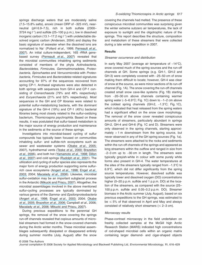
Microscopy results
Phase-contrast microscopy in the field undertaken onfreshly collected samples at the McGill high ArcticResearch Station (MARS) indicated high concentrationsof rod-shaped microbial cells within an organic matrixcontaining larger diamond- and cigar-shaped objects
S-oxidizing Thiomicrospira in Arctic springs 617
© 2008 The AuthorsJournal compilation © 2008 Society for Applied Microbiology and Blackwell Publishing Ltd, Environmental Microbiology, 11, 616–629

(Fig. 2A). The larger elongated (10–25 mm) structureswithin the streamers stained non-specifically with SYTOgreen and FUN 1 bacterial and fungal stains (results notshown). It was thus difficult to determine whether theyrepresented living organisms or mineral structures. Per-meabilization did not lead to a qualitative change in thelabelling of the large structures, although it removed muchof the organic material. SEM and EDAX analyses identi-fied the larger elongated structures within the streamers
as elemental sulfur and a variety of sulfur-containing min-erals including gypsum (CaSO4·2H2O) and halotrichite(FeAl2(SO4)4·22H2O) (Fig. S1; Table S1).
Confocal Scanning Laser Microscopy (CSLM) imagingof streamer material indicated that it was composed of aglobular substructure (Fig. 3A), which consisted of amatrix of exopolymeric substances binding the lectinsBanderia simplicifolia and Narcissus pseudonarcissus
(Fig. 3B). These lectins bind to N-acetyl-glucosamine and
Fig. 1. Field photographs of the run-offstreams and associated streamers of the GHsprings.A. Field photograph of GH-4; S indicates thespring source; the white arrow indicates thedirection of the run-off discharge from thesource.B. View underneath the snow cover from arun-off channel originating from GH-4.C. Streamers present following the removal ofthe snow cover from GH-4 (~5–6.9°C).D. Streamers present following the removal ofthe snow cover from GH-2 (-1.3°C). Scalebar represents 10 cm for all images.
618 T. D. Niederberger et al.
© 2008 The AuthorsJournal compilation © 2008 Society for Applied Microbiology and Blackwell Publishing Ltd, Environmental Microbiology, 11, 616–629
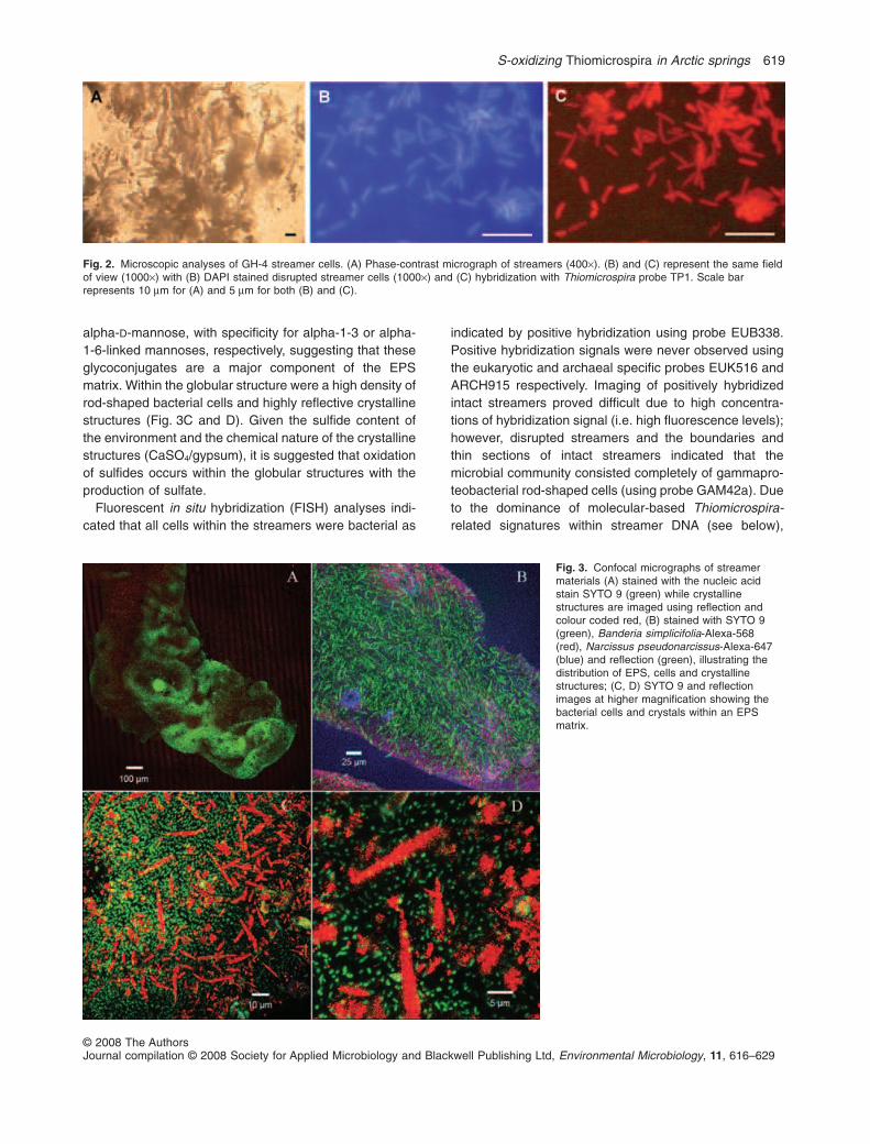
alpha-D-mannose, with specificity for alpha-1-3 or alpha-1-6-linked mannoses, respectively, suggesting that theseglycoconjugates are a major component of the EPSmatrix. Within the globular structure were a high density ofrod-shaped bacterial cells and highly reflective crystallinestructures (Fig. 3C and D). Given the sulfide content ofthe environment and the chemical nature of the crystallinestructures (CaSO4/gypsum), it is suggested that oxidationof sulfides occurs within the globular structures with theproduction of sulfate.
Fluorescent in situ hybridization (FISH) analyses indi-cated that all cells within the streamers were bacterial as
indicated by positive hybridization using probe EUB338.Positive hybridization signals were never observed usingthe eukaryotic and archaeal specific probes EUK516 andARCH915 respectively. Imaging of positively hybridizedintact streamers proved difficult due to high concentra-tions of hybridization signal (i.e. high fluorescence levels);however, disrupted streamers and the boundaries andthin sections of intact streamers indicated that themicrobial community consisted completely of gammapro-teobacterial rod-shaped cells (using probe GAM42a). Dueto the dominance of molecular-based Thiomicrospira-
related signatures within streamer DNA (see below),
Fig. 2. Microscopic analyses of GH-4 streamer cells. (A) Phase-contrast micrograph of streamers (400¥). (B) and (C) represent the same fieldof view (1000¥) with (B) DAPI stained disrupted streamer cells (1000¥) and (C) hybridization with Thiomicrospira probe TP1. Scale barrepresents 10 mm for (A) and 5 mm for both (B) and (C).
Fig. 3. Confocal micrographs of streamermaterials (A) stained with the nucleic acidstain SYTO 9 (green) while crystallinestructures are imaged using reflection andcolour coded red, (B) stained with SYTO 9(green), Banderia simplicifolia-Alexa-568(red), Narcissus pseudonarcissus-Alexa-647(blue) and reflection (green), illustrating thedistribution of EPS, cells and crystallinestructures; (C, D) SYTO 9 and reflectionimages at higher magnification showing thebacterial cells and crystals within an EPSmatrix.
S-oxidizing Thiomicrospira in Arctic springs 619
© 2008 The AuthorsJournal compilation © 2008 Society for Applied Microbiology and Blackwell Publishing Ltd, Environmental Microbiology, 11, 616–629

Thiomicrospira-specific FISH probes were constructedand tested. The utilization of an optimized Thiomicrospira
probe (TP1) indicated that ~100% of DAPI stained cells(Fig. 2B) from disrupted streamers also stained with theThiomicrospira-specific probe TP1 (Fig. 2C).
Phylogenetic analyses of streamers
Initial bacterial denaturing gradient gel electrophoresis(DGGE) comparison was undertaken between fourgreyish-white coloured streamer samples from GH-4,GH-2 and GH-6 as well as a white-coloured streamer fromGH-4. The bacterial DGGE profiles of the streamersamples were relatively simple (1–3 dominant bands) withGH-6 and GH-2 containing an additional dominant band(bands F and H respectively) that were not present for theGH-4 samples (Fig. S2); DGGE bands were excised andsequenced to identify major microbial members. Bothmajor (A, C, E and G) and minor bands (B and D) were nearidentical in sequence (99–100%, > 373 bp) to their closestNCBI BLAST relatives, namely, an uncultured clone G32(DQ521115) and a cultured Thiomicrospira sp. NP51(EU196304) both previously recovered from GH sourcesediment (Perreault et al., 2007). The DGGE bandsequences were classified as Thiomicrospira with 100%confidence by the RDP Classifier. Attempts to sequencebands F and H failed (Fig. S2).Although FISH analyses didnot indicate the presence of archaeal cells, positivearchaeal DGGE amplifications were obtained from stream-ers collected from GH-6 and GH-2 (i.e. not GH-4).ArchaealDGGE profiles were identical between GH-6 and GH-2(results not shown) and contained a single dominant bandthat contained a single nucleotide difference over 462 bpwith the closest NCBI BLAST relatives (99–100%; 461–476 bp) being uncultured Crenarchaeotal signaturesCP-A56 (DQ521209) and GH-A99 (DQ521150) previouslyrecovered from CP and GH source pool sediments respec-tively (Perreault et al., 2007).
Subsequent 16S rRNA gene cloning was undertaken tocorroborate DGGE results and to further resolve and char-acterize the diversity of the microbial communities withinthe streamers. A streamer sample from GH-2 was chosenas a representative sample for both archaeal and bacterial16S rRNA gene clone library construction due to botha higher complexity within its bacterial DGGE profile(Fig. S2) and the detection of archaea by DGGE. A total of24 bacterial 16S rRNA gene clones were screened with 22clones classified within the Thiomicrospira genus (RDPClassifier; 100%) with the closest BLAST match (99%,> 700 bp) being Thiomicrospira sp. NP51 (EU196304) pre-viously isolated from the GH sediments (Perreault et al.,2008). The closest validly described cultured relative (97%,> 700 bp) was Thiomicrospira arctica strain SVAL-ET
(AJ404731) (Knittel et al., 2005). DNAsequence alignment
against the dominant Thiomicrospira-related bacterialDGGE band (i.e. A, C, E and G) proved that thesesequences were 100% identical. Single bacterial clonesclassified within the Desulfuromusa genus (RDP Classifier;100%) and the uncultured Bacteroidetes (RDP Classifier;100%) were also represented in the bacterial library. Thephylogenetic positions of the bacterial 16S rRNA geneclones are presented in Fig. 4.All 23 of the archaeal clonesscreened were classified within the Crenarchaeota. Thearchaeal library consisted of 22 clones being identical(100%, > 484 bp) to clone GH-A8 (DQ521152) previouslyrecovered from GH source sediment (Perreault et al.,2007) and a single clone related to both GH-A99(DQ521150; 93%, 470/503 bp) and CP-A56 (DQ521209;93% 469/501) from GH and CP springs respectively(Perreault et al., 2007). These sequences do not haveclose phylogenetic relationships with any characterizedarchaeon and thus, no ecological role can be inferred.
Isolation of chemolithoautotrophic sulfur-oxidizing
bacteria
One colony morphotype and one RFLP pattern wereobtained on the thiosulfate medium and the MJ mediumand a single colony was randomly selected on eachmedium for sequencing of the near full-length 16S rRNAgene. DNA alignment showed that both sequences wereidentical and had a perfect sequence match to Thiomi-
crospira sp. NP51 (EU196304) previously isolated fromthe GH spring waters and sediments. The Thiomicrospira
sequences from the GH springs were also highly related(99% and 98% DNA identity over 1409 bp respectively) toThiomicrospira psychrophila and T. arctica, two psychro-philic sulfur-oxidizing bacteria isolated from the ArcticOcean (Knittel et al., 2005). The isolate grew on mediaused to culture both T. psychrophila and T. arctica (Knittelet al., 2005) and was capable of growth under aerobic andmicroaerophilic (6% O2) conditions, with thiosulfate andsulfide as sole electron donors. It was capable of growthat NaCl concentrations of 2.5% at 10°C and couldmetabolize sulfide at concentrations as high as125 p.p.m. in synthetic media.
In situ activity experiments and RuBisCO detection
In situ experiments were performed in the GH-4 springchannel to determine the capacity of the streamermicroorganisms to oxidize inorganic S compoundsand to fix inorganic carbon (Table 1). The lowest sulfideoxidation rate was obtained following in situ darkincubation (37.5 � 4.8 mmol cm-3 h-1) and the highest ratefollowing light incubation at room-temperature (62.5 �
8.8 mmol cm-3 h-1). Thiosulfate was also used as electrondonor by the streamer microbial community (Table 1) and
620 T. D. Niederberger et al.
© 2008 The AuthorsJournal compilation © 2008 Society for Applied Microbiology and Blackwell Publishing Ltd, Environmental Microbiology, 11, 616–629

Table 1. Rates of sulfide-oxidation, thiosulfate-oxidation and CO2 fixation of the streamers.
Sulfide-oxidation ratesa
Thiosulfate-oxidation ratesa
in situ-light
CO2 fixationb
RT-light RT-dark in situ-light in situ-dark in situ-light in situ-dark
62.5 � 8.8 45.7 � 5.5 51.6 � 5.2 37.5 � 4.8 21.2 � 1.8 235 � 42 317 � 59
a. mmol cm-3 h-1.b. nmol C l-1 h-1.RT, room-temperature (17–20°C) at the McGill Arctic Research Station (MARS).
Spitsbergen permafrost clone anE05 (EF034556)
Cold marine sediment Sva1038 (AJ240979)
Seafloor sediment clone GoM GB425 12B-2 (AY542556)
GH-2 bacterial clone (EU430108)
Mud volcano clone HMMVPog-1 (AJ704701)
Deep sea cold seep sediment clone JS624-9 (AB121104)
Bacteroidetes
58
90
100
100
99 p p ( )
Thiothrix fructosivorans strain I (L79963)
Thiothrix nivea DSM 5205 (L40993)
Cave sulfidic spring clone LKC3_19.29 (AY510231)
Cold sulfidic spring clone sipK4 (AJ307933)
Cave sulfidic spring clone LKC3_127.2 (AY510228)
M th l b t t d i l d SV96T (AJ414655)
100
100
66
100
Methylobacter tundripaludum SV96T (AJ414655)
Methylophaga marina DSM 5989 (X95459)
Thiomicrospira isolate NP20 (EU196336)
Thiomicrospira arctica SVAL-E (AJ404731)
Thiomicrospira psychrophila SVAL-D (AJ404732)
Sub-glacial Thiomicrospira sp. clone BF99_C55 (DQ677851)
Gammaproteobacteria
91
99
71
85
70
Thiomicrospira isolate NP51 (EU196304)
Thiomicrospira isolate (EU430112)
Arctic ocean clone Arctic96AD-9 (AF354608)
Thiomicrospira crunogena DSM 12354(AF069959)
Thioalcalomicrobium sibericum AL7 (AF126549)
Thiomicrospira pelophila DSM 1534 (L40809)
10059
100
99 100
Piscirickettsia salmonis LF-89 (U36941)
Antarctic sediment clone (AY177799)
Arctic Desulfuromonas isolate 60 (AY835390)
Mesophilic sulfide spring clone ZB5 (AY327186)
GH-2 bacterial clone (EU430107)
Deep sea sediment clone C1 B041 (AF420369)
Deltaproteobacteria
100
9996
89
Deep sea sediment clone C1_B041 (AF420369)
Desulfuromonas kysingii (X79414)
0.05
93
Fig. 4. Phylogenetic relationship of bacterial 16S rRNA gene sequences recovered from the streamers. The near full-length 16S rRNA gene ofthe Thiomicrospira isolate represents both the DGGE and the 22 16S rRNA gene clone library sequences classified within the Thiomicrospira
genus, as all sequences share 100% sequence homology over 380 bp of aligned sequence. The tree is rooted to Aquifex pyrophilus (M83548)with the branch removed from the figure. Bootstrap values � 50% of 1000 replicates are indicated at the nodes. The bar denotes theexpected number of changes per nucleotide position.
S-oxidizing Thiomicrospira in Arctic springs 621
© 2008 The AuthorsJournal compilation © 2008 Society for Applied Microbiology and Blackwell Publishing Ltd, Environmental Microbiology, 11, 616–629

autotrophic CO2 fixation detected by 14C-bicarbonateuptake under both light (235 � 42 nmol C l-1 h-1) and darkincubations (317 � 59 nmol C l-1 h-1). Radioactivity wasnot detected in any of the three controls. The abilityof the streamers to fix CO2 was further corroborated bythe successful PCR amplification of RuBisCO cbbM
sequences from the community DNA of the four streamersamples and from the Thiomicrospira isolate. DNAsequence analysis showed that all the sequences wereidentical and grouped with cbbM sequences from Thiomi-
crospira species and other Gammaproteobacteria (resultsnot shown).
Discussion
During previous summer month sampling expeditions toAxel Heiberg Island, patchy distributions of greyishstreamers were observed in the run-off channels of thesprings at GH. Conversely, during the winter months theconcentrations of greyish microbial streamers increasedmarkedly beneath the snow covering the perennially liquidrun-off channels. Although it is not known exactly whenthe streamers form during the Arctic winter, similar levelsof streamer biomass were observed in mid-April (averageair temperature approximately -25°C) in previous years.Streamers were never observed at the source or within~1 m from the source of the spring, even if the spring andits associated run-off channel were covered with snow.This phenomenon is hypothesized not to be due to tem-perature or sulfide, but rather levels of DO; as tempera-tures did not differ significantly between the spring sourceand the site of streamers and the Thiomicrospira isolatewas shown to grow in H2S concentrations higher(125 p.p.m.) than that measured at the spring source (25–100 p.p.m.) or in the spring run-off channel water.However, DO levels were consistently higher at the site ofthe streamers (1.0 p.p.m.) than at the source of thesprings (0.05–0.2 p.p.m.) and it has been shown that Thi-
omicrospira sp. require aerobic or microaerophilic condi-tions for growth (Knittel et al., 2005). Therefore, theThiomicrospira streamers may only develop in regions ofthe springs where the oxygen levels are sufficiently highenough for growth.
The construction, optimization and utilization of aThiomicrospira-specific FISH probe (TP1) conclusivelyproved that all cells (DAPI stained) of the streamers arespecies of Thiomicrospira. Therefore, FISH confirmed theresults of molecular analyses (both DGGE and clonelibraries) and indicated the complete dominance of thegammaproteobacterial genus Thiomicrospira. Cells unre-lated to the Gammaproteobacteria were not detected viaFISH analyses, i.e. all observed cells positively hybridizedwith the gammaproteobacterial-specific probe. Therefore,the unknown crenarchaeote and two non-Thiomicrospira
clones (an uncultured Bacteroidetes and a species ofDesulfuromonas) detected in streamers from GH-2 by16S rRNA gene cloning may exist below the detectionlevel of FISH. This is also exacerbated as minor propor-tions of the streamers microbial community may be over-looked by FISH analyses due to the high dominance of asingle type of microbial species and the subsequent brightsignal of the FISH hybridization. DNA sequence homolo-gies between the DGGE, the Thiomicrospira-related 16SrRNA clones and the near full-length 16S rRNA gene fromthe isolate were 100% identical over 380 bp of alignedsequence, indicating a high likelihood that they all repre-sent the same strain.
The reason or function of the S-containing crystalliteswithin the streamer matrix are currently unknown; however,they are morphologically and chemically similar to crystalsin microbial mats of Miette hot springs, Jasper NationalPark, Canada (Bonny and Jones, 2003). Bonny and Jones(2003) presented three processes whereby these crystalsform within the microbial mats of the Miette hot springs: (i)microbial extracellular polymeric substances passivelyconcentrate crystals via a trapping effect; (ii) microbialmats may act as nucleation sites for the growth of gypsumstructures; (iii) microbes within the mats influence mineralprecipitation by their metabolisms, e.g. CO2 degassing viaphotosynthetic pathways and the production of elementalS via sulfate reduction. These processes remain unknownin the GH streamers; however, crystal formation may occurdue to the production of elemental sulfur and SO4-mineralsvia S-oxidation due to the high concentrations of Thiomi-
crospira cells within the streamer matrix. The formation androle of the crystal structures within the streamers is acurrent avenue of further investigation, whereby sulfurisotopic analyses may determine the biogenic or abioticorigin of the crystallites.
Thiomicrospira species are obligate sulfur oxidizingbacteria (Knittel et al., 2005) and are typically distributedwhere sulfur-related metabolism is a primary energyprocess. They have been detected and cultured fromvarious environments including hydrothermal vents(Brinkhoff et al., 1999; Brazelton et al., 2006; Perneret al., 2007), marine sediments, coastal mud flats, hyper-saline ponds, sediment from a saline spring in Artern,Germany (Brinkhoff and Muyzer, 1997; Knittel et al.,2005) and a subglacial outflow in Antarctica (Mikucki andPriscu, 2007). Our previous culture-independent 16SrRNA gene-based study of the GH and CP sedimentsindicated that Thiomicrospira was the most abundant phy-lotype in both the GH and CP spring source sediments(Perreault et al., 2007). However, streamers have neverbeen observed in CP springs or run-off channels. Thereason for the lack of streamers at CP is not clear. TheCP springs also have snow covered run-off streams;however, the increased concentration of salts at CP
622 T. D. Niederberger et al.
© 2008 The AuthorsJournal compilation © 2008 Society for Applied Microbiology and Blackwell Publishing Ltd, Environmental Microbiology, 11, 616–629

(~15.5%) as compared with GH (~7.5%) may inhibit theformation of Thiomicrospira streamers as the tolerance ofvalidly characterized species of Thiomicrospira (includingthe psychrophilic species, T. arctica and psychrophila)are typically between ~0–1.24 M NaCl (0–7.2% NaCl)(Sorokin et al., 2006). The recently described T. halophila
can tolerate NaCl levels of up to 3.5 M NaCl (~20.5%NaCl); however, this isolate has an optimum growth tem-perature of 30°C and is not capable of growth at 0°C(Sorokin et al., 2006). The higher total discharge rate andflow rate of the CP springs (20–25 l s-1 and 1.5–1.8 l s-1
respectively) as compared with the GH springs (10–15 l s-1 and 0.9–10 l s-1 respectively) (Pollard et al., 1999)may also inhibit the establishment of optimal geochemicalgradients (e.g. H2S, O2) or the attachment of cells to solidmatrices that may be required for the formation of thestreamers.
The microbial streamers were shown to metabolize andto proliferate extensively under the in situ conditions ofcold (-1.3°C to 6.9°C) saline water within a microhabitatformed via snow cover protecting and insulating againstthe surrounding late-winter air temperatures (< -15°C).The snow cover may also allow the streamers to developunder mid-winter extreme air temperatures of -50°C asthey were already extensively established by the end ofwinter. Natural light had a positive effect on sulfide-oxidation while it appeared to be slightly detrimental forCO2 fixation, with CO2 fixation rates being 25% less thanfor dark incubation. The same phenomenon wasobserved previously for CO2 uptake in the GH springwater (Perreault et al., 2008). Previously rates for darkCO2 fixation have also been described from a moderatelysalty (~3% NaCl) subglacial outflow (Blood Falls)from the Taylor Glacier, Antarctica (Mikucki and Priscu,2007), although the rates were considerably lower(0.05 nmol C l-1 h-1) than those measured in this study(235 � 42 and 317 � 59 nmol C l-1 h-1 for light and darkincubations respectively) which is most likely due to thelow cell content (1 ¥ 104-7.6 ¥ 105 cells ml-1) of the sub-glacial water. Interestingly, the dominant clone detectedwithin the subglacial outflow water was also related toThiomicrospira arctica (46% of the library) (Mikucki andPriscu, 2007); however, streamers of Thiomicrospira cellswere not observed in this system.
Detection of both sulfide and thiosulfate oxidation andCO2 uptake was consistent with the presence and activityof chemolithoautotrophic sulfur-oxidizing bacteria such asThiomicrospira while CO2 uptake under dark incubationconfirmed that chemoautotrophic activity, and not photo-autotrophy, sustains the streamer community. This sug-gests that Thiomicrospira spp. have an advantage ininhabiting cold polar environments that contain reducedsulfur-compounds and experience long-term periods ofdarkness during polar winters or permanent darkness as
for example in Antarctic subglacial microhabitats (Mikuckiand Priscu, 2007).
Form II (cbbM) RuBisCO is found in several photo-synthetic bacteria, aerobic and facultative anaerobic,chemoautotrophic bacteria and dinoflagellates and isadapted to anoxic conditions (Portis and Parry, 2007;Badger and Bek, 2008). The anoxic cbbM genotype wasdetected in the sulfur-oxidizing streamers and the Thiomi-
crospira streamer strain, which is in accord with thehypoxic conditions in the brine of the GH spring channels.Among other Thiomicrospira species, T. crunogena
(Tourova et al., 2006), T. kuenenii (Tourova et al., 2006)and T. halophila (Sorokin et al., 2006) also possess atleast one cbbM gene. Altogether, the presence of aunique cbbM sequence, identical for both the streamersamples and the Thiomicrospira strain and its phyloge-netic affiliation with cbbM sequences from other Thiomi-
crospira, support the 16S rRNA gene data, suggestingthat Gammaproteobacteria sulfur-oxidizers of the genusThiomicrospira are responsible for the autotrophic carbonfixation in the GH microbial streamers. Form II enzymeshave a poor affinity for CO2 and a low specificity factor todiscriminate between CO2 and O2, implying that the formII enzymes operate exclusively at high CO2 concentra-tions and low O2 concentrations (Badger and Bek, 2008),hence, a large investment of energy is required for thismethod of CO2 fixation. Therefore, the sulfur-oxidizingstreamers theoretically have to oxidize high concentra-tions of reduced sulfur compounds as electron donors togenerate enough energy to grow autotrophically. There-fore, the snow covering the run-off streams may trap andconcentrate H2S gas and explain the marked increase ofstreamers during the winter; in fact bursts of H2S wereclearly perceived following the puncturing of the snowcovering the GH spring run-off streams.
An initial microbiology investigation of very low saline,sulfur springs in the Canadian high Arctic (EllesmereIsland) did not detect S-oxidizing bacteria. These springsdiffer to those of GH and CP as they discharge from thesurface of a glacier, rather than through a thick layer ofpermafrost (Grasby et al., 2003). This is the first and onlyreport of Thiomicrospira in a macroscopic streamer formand appears to be unique to the GH springs; however, themacroscopic appearance of the streamers is similar tostreamers previously described in hot spring environments(Yamamoto et al., 1998; Nakagawa and Fukui, 2003) andcave systems (Engel et al., 2004; Macalady et al., 2006).Microbial streamer assemblages of other non-thermalspring and cave systems rich in sulfur species are com-posed of more than one, and sometimes quite complexmicrobial types. For example, a Thiothrix-related bacte-rium in association with Archaea forms a macroscopicstructure morphologically comparable to a string of pearlsin cold (10°C) sulfidic springs located in Bavaria, Germany
S-oxidizing Thiomicrospira in Arctic springs 623
© 2008 The AuthorsJournal compilation © 2008 Society for Applied Microbiology and Blackwell Publishing Ltd, Environmental Microbiology, 11, 616–629

(Rudolph et al., 2001), six Epsilonproteobacteria-relatedtaxonomic groups dominate (68% of 16S rRNA gene clonelibraries) microbial populations of white streamer bundlesin sulfurous spring cave systems in Wyoming, USA (Engelet al., 2003; 2004), and Gammaproteobacteria related toThiothrix sp. and Beggiatoa sp. dominate white biofilmsthat form in sulfide-rich waters in an Italian cave system(Macalady et al., 2006).
The characterization of Thiomicrospira-dominatedsulfur-oxidizing streamers, which flourish during theextreme polar winter via phototrophic-independentchemolithotrophic-based metabolism within a snow-covered microenvironment characterized by low tempera-tures ranging from -1.3°C to 6.9°C, depending on thespring source, high salt concentrations (~7.5%), olig-otrophic and microaerophilic conditions, expands ourcurrent understanding of the limits of terrestrial life andhas implications for the search for extinct or extant life onother solar system bodies. At nearly 80°N, these highArctic cold saline springs and associated channels areamong the few known examples of cold, non-volcanicsprings in thick permafrost on Earth, and are consideredMars analogues as similar hydrological systems mayhave existed or still exist on Mars (Andersen et al., 2002;Andersen, 2004; Malin et al., 2006). For example, basalmelting of Martian polar ice caps could generate signifi-cant groundwater flow systems that flow through evapo-rate salt deposits and finally emerge through thickpermafrost as hypersaline springs (Grasby and Londry,2007). In this respect, Mars Global Surveyor imagesrecently detected new gully deposits, formed since 1999,providing exciting and compelling evidence that liquidwater (possibly eutectic brines from the Martian subsur-face) flowed on Mars during the past decade, under meansurface temperatures of -60°C and extensive permafrost(Malin et al., 2006). These observations fit with othermodels, suggesting that it may be possible for liquids toexist below the surface and be discharged under modernMartian conditions (Mellon and Phillips, 2001; Heldmannet al., 2005; Kraal et al., 2008). It is possible that suchgully formations may harbour similar subsurface microen-vironments (i.e. very cold, saline, shielded from sunlightand UV) to those observed at the Gypsum Hill springs andhypothetically capable of supporting chemolithotrophic,phototrophic-independent microbial life.
Experimental procedures
Site description and collection of samples
The location and setting of the perennial springs adjacent toGypsum Hill (GH) are described in detail by Pollard andcolleagues (1999). The springs from which streamer sampleswere collected include GH-2 and GH-4, as described in theinitial investigation by Perreault and colleagues (2007), and a
new spring, designated GH-6, located adjacent to GH-4. Dis-solved sulfide and oxygen (DO) concentrations of waterwithin the run-off channels were measured in triplicate viacolorimetric assay (CHEMtrics, Calverton, VA, USA).Streamer samples were collected in May 2007 using steriletweezers and were subsequently stored in spring waterinside sterile polypropylene tubes (Fisher). Streamersamples were transported to McGill University at subzerotemperatures and stored at both -5°C (for culture-basedanalyses) and -20°C (for DNA-based analyses).
DNA extraction and polymerase chain reaction (PCR)
Streamer samples were rinsed in 0.9% sterile NaCl and totalcommunity DNA extracted from 0.07 to 0.25 g of biomassusing the ZR Fungal/bacterial DNA Kit™ (ZYMO Research,USA). DNA was quantified using a Nanodrop ND-1000 spec-trophotometer (Nanodrop Technologies, DE, USA). All PCRreagents were supplied by Invitrogen Canada, Burlington,ON. Bacterial DGGE-PCR utilizing primers 341F-(GC) and758R and the associated thermocycling conditions wereundertaken as described by Steven and colleagues (2008)with the following modifications; 3 mM MgCl2 and 2.5 ml eachprimer (10 mM concentration) for a 50 ml reaction and theinclusion of a 30 min final extension at 72°C to remedy anydouble-banding phenomena (Janse et al., 2004). Bacterialpartial-length 16S rRNA gene amplification for cloning wasundertaken as outlined by Steven and colleagues (2007a)utilizing primers 27F and 758R and ~50 ng of template DNA.Archaeal DGGE-PCR and partial length 16S rRNA gene PCRfor cloning were undertaken using primers A344F-(GC) andA934R with ~50 ng template DNA as described previously(Perreault et al., 2007).
Denaturing gradient gel electrophoresis analyses
Denaturing gradient gel electrophoresis was performed aspreviously described by Steven and colleagues (2008) utiliz-ing an 8% acrylamide gel with a 35–65% denaturing gradientthat was run for 16 h at 60°C and 80 V and visualized byethidium bromide staining. DGGE bands of interest were alsoprocessed and sequenced as described by Steven and col-leagues (2008). Bacterial and archaeal DGGE sequenceswere deposited into the GenBank database as accessionnumbers EU430099 to EU430104 and EU430105 toEU430106 respectively.
Construction and sequencing of 16S rRNA
gene clone libraries
PCR amplicons from the three partial-length 16S rRNA genePCRs were combined and purified using a QIAquick PCRpurification kit (QIAGEN Sciences, MD, USA) and quantifiedvia ethidium bromide staining in an agarose gel and a 2:1molar ratio of insert to vector used for both bacterial andarchaeal clone libraries. The pGEM-T Easy vector systemwas used as per manufacturer’s instructions (Promega,Madison, USA) in conjunction with subcloning efficiencyDH5a competent cells (Invitrogen, CA, USA). Clones of inter-est were selected and vectors with the correct sized inserts
624 T. D. Niederberger et al.
© 2008 The AuthorsJournal compilation © 2008 Society for Applied Microbiology and Blackwell Publishing Ltd, Environmental Microbiology, 11, 616–629

checked as described by Bottos and colleagues (2008).Inserts were sequenced with their respective forward primersused for the original partial-length 16S rRNA gene PCR.Representative bacterial and archaeal 16S rRNA gene clonesequences were deposited into the GenBank database asaccession numbers EU430107 to EU430109 and EU430110to EU430111 respectively.
DNA sequence analyses
Sequencing was undertaken at the Genome Quebec Innova-tion Center (McGill University) with 3730XL DNA analysersystems (Applied Biosystems). 16S rRNA gene sequenceswere manually edited and subjected to CHIMERA_CHECK(Cole et al., 2003) and the Bellerophon server (Huber et al.,2003). Taxonomic affiliations were determined using theClassifier tool (Wang et al., 2007) of the RDP II (Cole et al.,2007). Sequences were also compared with the GenBankdatabase using the BLASTn algorithm (Altschul et al., 1990).Sequences of each clone library were aligned using ClustalWsoftware and neighbour-joining phylogenetic trees wereproduced with MacVector 7.0 software package (OxfordMolecular, Oxford, UK) using Jukes-Cantor modeling with1000 bootstrap re-samplings.
Microscopy procedures
Phase-contrast microscopy was undertaken on freshly col-lected streamers at the MARS, Axel Heiberg Island and micro-graphs taken using a Nikon COOLPIX 4500 digital camera(Nikon, Melville, NY, USA). Staining of samples with SYTOGreen and FUN 1 was performed by adding the dye (MolecularProbes™, Invitrogen, Eugene, OR, USA) to a final concentra-tion of 10 mM in a sample of streamers in a 1.5 ml Eppendorftube containing water from the spring. The sample was mixedby inversion at ambient temperature (5–10°C) but not other-wise processed and wash steps were not performed. Perme-abilization by osmotic shock (Sestak and Farkas, 2001) wasundertaken as follows: the streamer sample was suspended in10 volumes of buffer containing 50 mM Tris-HCl, pH 7.5, 1 mMEGTA, 1 mM beta-mercaptoethanol, 0.5 mM phenylmethyl-sulfonyl fluoride (TEGM buffer) supplemented with 33% (w/v,3.5 M) glycerol and allowing them to stand for 10 min on ice.The suspension was then allowed to sediment by gravity(~1 min), and the sedimented cells were washed once with thesame buffer without glycerol. Finally, the cells were suspendedin two volumes of the same buffer and stained as outlinedabove. Environmental scanning electron microscopy (ESEM)was carried out using a FEG XL30 (FEI, Hillsboro, OR, USA)with an accelerating voltage of 20 kV. A gaseous secondaryelectron detector (GSED) was used with a Peltier coolingstage at 4.0°C and a 3.9 torr chamber pressure. The wholemount bacterial samples were rinsed in ddH2O prior to beingplaced on aluminum stubs and into the ESEM. A Genesisenergy dispersive X-ray (EDAX) spectrometer (Ametek, Paoli,PA, USA) was used with an ultrathin window for elementalanalysis.
Fluorescent lectins with Alexa 568 and Alexa 647 labellingand the nucleic acid probe SYTO 9 were purchased(Invitrogen). The lectins Banderia simplicifolia-Alexa-568 and
Narcissus pseudonarcissus-Alexa647, were employed alone,together or with SYTO 9 as described by Neu and colleagues(2001); with the exception that the staining time wasextended to 12 h to allow penetration of the samples. Exami-nation of all stained and control materials was carried outusing an MRC 1024 CLSM (Zeiss, Jena, Germany) attachedto a Microphot SA microscope (Nikon, Tokyo, Japan). Duringmicroscopic analyses, the following lenses were used: 60¥,1.4 numerical aperture (NA) Planapochromat (Nikon) a 20¥,0.75 NA Fluor/Ph3-DL (Nikon) and a 6¥, 0.2 NA Fluorotar(Wild). Signals were recorded in the green channel (excita-tion 488 nm, emission 522/32), red channel (excitation568 nm, emission 605/32) and far red channel (excitation647 nm, emission 680/32). In addition, the reflection modewas used to detect crystal structures associated with thebacteria (Lawrence and Neu, 2007). Image stacks were pro-jected using the software provided by Bio-Rad. During allsample handling, staining and microscopy samples weremaintained at 0°C by mounting on frozen gel packs.
Streamer samples for FISH were immediately fixed in thefield by the method utilized for marine sediment as describedby Pernthaler and colleagues (2001) and transported frozento McGill University and stored at -15°C. FISH analyses wereperformed as described by Rudolph and colleagues (2001)using gelatin/KCr(SO4)2-coated slides (Pernthaler et al.,2001). FISH probes and their respective hybridization andwash buffers were used according to the respective refer-ences: universal bacterial and archaeal probes, EUB338 andARCH915 (Rudolph et al., 2001), labelled at the 5′-terminuswith Texas Red and fluorescein isothiocyanate respectively;universal eukaryotic probe, EUK516 (Baschien et al., 2001),labelled at the 5′-terminus with fluorescein isothiocyanate;gammaproteobacterial specific probe, GAM42a (Macaladyet al., 2006), labelled at the 5′-terminus with Texas Red. AllFISH staining procedures included positive control samplesof pure cultures of known microorganisms. OligonucleotideFISH probes specific for Thiomicrospira sp. NP51 isolatedfrom GH spring source sediment (Perreault et al., 2008), andthe related strains T. arctica and T. psychrophila (Knittelet al., 2005) were designed using the program Primrose(Ashelford et al., 2002). Probe candidates were initiallychecked for specificity against the sequences in GenBank viaa BLAST search with probe coverage and specificity confirmedin silico via probeCheck (Loy et al., 2008). Two oligonucle-otide probes both labelled at the 5′-terminus with Texas Red,TP1 (5′-CTC TAT CGT TTC CGT CCG-3′) corresponding toEscherichia coli 16S rRNA nucleotide positions 63–80 andTP7 (5′-CGC CTA GAA AAG CAA GC-3′) corresponding toE. coli 16S rRNA nucleotide positions 91–107 were chosenfor specific detection of the three Thiomicrospira strains.Specificity of the Thiomicrospira probes was tested by adjust-ing hybridization stringencies from 0% to 60% formamide in10% increments. Positive controls consisted of cells of Thi-
omicrospira strain NP51 and T. arctica and T. psychrophila
obtained from the DSMZ (Deutsche Sammlung van Mikroor-ganismen und Zellkulturen GmbH; http://www.dsmz.de).Probe TP1 provided the highest fluorescence of the testedprobes on Thiomicrospira cells at 30% formamide and waschosen for further testing of steamer and negative controlsamples at 30% formamide concentration. Cross-reactivity ofthe TP1 probe was not observed in > 15 microorganisms
S-oxidizing Thiomicrospira in Arctic springs 625
© 2008 The AuthorsJournal compilation © 2008 Society for Applied Microbiology and Blackwell Publishing Ltd, Environmental Microbiology, 11, 616–629

isolated from the GH springs and other high Arctic environ-ments, including genera of gammaproteobacteria, e.g.Pseudomonas. All FISH stained samples were mounted with4:1 mix (Pernthaler et al., 2001) of Citifluor (Citifluor,Leicester, UK) and Vectashield (Vecta Laboratories, Ontario,Canada) amended with 1 mg ml-1 DAPI for counterstainingpurposes (Pernthaler and Pernthaler, 2007). FISH resultswere viewed using a fluorescent Nikon Eclipse E600 micro-scope (Nikon, Melville, NY, USA) with appropriate filter sets(Chroma Technology Corporation, VT, USA) and imagestaken using a Nikon COOLPIX 4500 digital camera.
Culture and 16S rRNA gene analyses of
chemolithoautotrophic sulfur-oxidizing bacteria
A preliminary study on GH streamers collected in July 2004identified two 16S rRNA gene phylotypes related to thesulfur-oxidizing bacteria Thiomicrospira psychrophila andSulfurimonas autotrophica in a clone ratio of 25:1. Basedon this preliminary molecular study, media were selected totarget aerobic thiosulfate-oxidizers (thiosulfate medium) andmicroaerophilic sulfide-oxidizers (MJ medium). Ten millime-tres of streamers from GH-6 was roughly disrupted in 30 ml ofsterile water and 100 ml was inoculated in 60 ml serum bottlescontaining 20 ml of thiosulfate medium (per litre: 0.4 g NH4Cl,4.0 g KH2PO4, 4.0 g K2HPO4, 0.8 g MgSO47H2O, 0.03 gCaCl2, 0.02 g FeCl36H2O, 0.02 g MnSO4H2O, 5.0 gNa2S2O35H2O, 25.0 g NaCl) or 20 ml of medium 1011: MJmedium (DSMZ). A gas mixture of N2/CO2/O2 (77:17:6) wasused in the headspace of the MJ medium. The bottles wereincubated at 10°C until growth was visually detected(~10 weeks). Aliquots of the liquid cultures were dispensedon agar plates of the same medium. Ten colonies for eachmedia were randomly selected for 16S rRNA gene restrictionfragment length polymorphism (RFLP) as described previ-ously (Juck et al., 2003). Two colonies (one for each culturemedium) were selected for PCR and sequencing of the nearfull-length 16S rRNA gene (~1500 bp) using primers 8F and1492R as described previously (Lane, 1991). The 16S rRNAgene sequence was deposited in the GenBank database asaccession number EU430112.
Sulfur oxidation rates
Approximately 10 ml of streamer slurry collected from GH-4was mixed with 30 ml of GH-4 spring water and roughlydisrupted by vigorous shaking. The potential sulfur oxidationrates (SORs) of the microbial streamers were determined byinoculating 0.5 ml of the streamer slurry into sterile 120-mlserum bottles containing 50 ml of synthetic media (Asamiet al., 2005) supplemented with NaCl (7.5%) and Na2S(1.6 mM) or Na2S2O3 (20 mM) as the sole electron donor.Serum bottles with Na2S were incubated either directly in thespring (GH-4) outlet (~6.9°C) or at the MARS (17–20°C),under natural light or dark incubation (bottles covered withlayers of foil). Bottles with Na2S2O3 were incubated in thespring outlet, under natural light. Subsamples were with-drawn every 24 h for 5 days. S- consumption was measuredin the subsamples by spectrophotometric determination(Cline, 1969). S2O3
2- consumption was estimated by monitor-
ing sulfate increase by ion exchange high pressure liquidchromatography (Spectra-Physics, model SP8800). The con-centrations measured during the first 48 h incubation wereused to calculate SORs. Experiments were done in triplicateand controls with no biomass added were performed in par-allel to determine the abiotic SORs, which was subtractedfrom the overall SORs.
In situ 14C-bicarbonate uptake and the PCR
amplification of the RuBisCO gene
A total of 0.5 ml of the sediment slurry (as described abovefor the SORs assays) was added to 20 ml of spring water insterile acid-washed 25 ml serum bottle and amended with250 ml of 14C-labelled sodium bicarbonate (NaH14CO3; 5 mCi)(MP Biochemicals; specific activity 53.5 mCi mmol-1). Bicar-bonate uptake was performed based on the methoddescribed by Joint and colleagues (1993) with incubationcarried out in the GH-4 spring outlet (~6.9°C) for 24 h. Theprimer pair RuIIF1/RuIIR3 was used to target the partial gene(~800 bp) encoding the large subunit of the form II (cbbM)RuBisCO protein from the streamers and the Thiomicrospira
isolate as described previously (Spiridonova et al., 2004).
Acknowledgements
Logistic support was provided by the Canadian Polar Con-tinental Shelf Project (PCSP-08, 634-07, 664-06, 66) andMcGill University’s High Arctic Research Station. This workwas supported by grants from NASA’s Exobiology program(NAG5-12395), the Natural Sciences and EngineeringResearch Council of Canada (NSERC Discovery Program,Northern Supplements Program, Special Research Oppor-tunities Program, and the Canadian Space Agency Cana-dian Analogue Research Network program. Additionalfunding for student research was provided by the Depart-ment of Indian and Northern Affairs – Northern ScientificTraining Program, and the Fonds Québécois de la Recher-che sur la Nature et les Technologies (FQRNT). Jay L.Nadeau acknowledges support from the Canadian SpaceAgency CARN program and the NSERC Individual Discov-ery and NanoIP programs.
References
Altschul, S.F., Gish, W., Miller, W., Myers, E.W., and Lipman,D.J. (1990) Basic local alignment search tool. J Mol Biol
215: 403–410.Andersen, D.T. (2004) Perennial Springs in the Canadian
High Arctic: Analogues of Hydrothermal Systems on Mars.Montreal, Canada: McGill University.
Andersen, D.T., Pollard, W.H., McKay, C.P., and Heldmann,J. (2002) Cold springs in permafrost on earth and mars.J Geophys Res 107: 1–7.
Angert, E.R., Northup, D.E., Reysenbach, A.L., Peek, A.S.,Goebel, B.M., and Pace, N.R. (1998) Molecular phyloge-netic analysis of a bacterial community in Sulphur River,Parker Cave, Kentucky. Am Mineral 83: 1583–1592.
Asami, H., Aida, M., and Watanabe, K. (2005) Acceleratedsulfur cycle in coastal marine sediment beneath areas of
626 T. D. Niederberger et al.
© 2008 The AuthorsJournal compilation © 2008 Society for Applied Microbiology and Blackwell Publishing Ltd, Environmental Microbiology, 11, 616–629

intensive shellfish aquaculture. Appl Environ Microbiol 71:
2925–2933.Ashelford, K.E., Weightman, A.J., and Fry, J.C. (2002) PRIM-
ROSE: a computer program for generating and estimatingthe phylogenetic range of 16S rRNA oligonucleotideprobes and primers in conjunction with the RDP-II data-base. Nucleic Acids Res 30: 3481–3489.
Badger, M.R., and Bek, E.J. (2008) Multiple Rubisco forms inproteobacteria: their functional significance in relation toCO2 acquisition by the CBB cycle. J Exp Bot 59: 1525–1541.
Barrett, J.E., Virginia, R.A., Wall, D.H., Cary, S.C., Adams,B.J., Hacker, A.L., and Aislabie, J.M. (2006) Co-variation insoil biodiversity and biogeochemistry in northern andsouthern Victoria Land, Antarctica. Antarct Sci 18: 535–548.
Baschien, C., Manz, W., Neu, T.R., and Szewzyk, U. (2001)Fluorescence in situ hybridization of freshwater fungi. Int
Rev Hydrobiol 86: 371–381.Bonny, S., and Jones, B. (2003) Microbes and mineral
precipitation, Miette Hot Springs, Jasper National Park,Alberta, Canada. Can J Earth Sci 40: 1483–1500.
Bottos, E.M., Vincent, W.F., Greer, C.W., and Whyte, L.G.(2008) Prokaryotic diversity of arctic ice shelf microbialmats. Environ Microbiol 10: 950–966.
Brazelton, W.J., Schrenk, M.O., Kelley, D.S., and Baross,J.A. (2006) Methane- and sulfur-metabolizing microbialcommunities dominate the Lost City hydrothermal fieldecosystem. Appl Environ Microbiol 72: 6257–6270.
Brinkhoff, T., and Muyzer, G. (1997) Increased species diver-sity and extended habitat range of sulfur-oxidizing Thiomi-crospira spp. Appl Environ Microbiol 63: 3789–3796.
Brinkhoff, T., Sievert, S.M., Kuever, J., and Muyzer, G. (1999)Distribution and diversity of sulfur-oxidizing Thiomicrospiraspp. at a shallow-water hydrothermal vent in the AegeanSea (Milos, Greece). Appl Environ Microbiol 65: 3843–3849.
Campbell, B.J., Engel, A.S., Porter, M.L., and Takai, K.(2006) The versatile [epsi]-proteobacteria: key players insulphidic habitats. Nat Rev Microbiol 4: 458–468.
Carpenter, E.J., Lin, S., and Capone, D.G. (2000) Bacterialactivity in South Pole snow. Appl Environ Microbiol 66:
4514–4517.Christner, B.C. (2002) Incorporation of DNA and protein pre-
cursors into macromolecules by bacteria at -15°C. Appl
Environ Microbiol 68: 6435–6438.Cline, J.D. (1969) Spectrophotometric determination of
hydrogen sulfide in natural waters. Limnol Oceanogr 14:
454–458.Cole, J.R., Chai, B., Marsh, T.L., Farris, R.J., Wang, Q.,
Kulam, S.A., et al. (2003) The Ribosomal Database Project(RDP-II): previewing a new autoaligner that allows regularupdates and the new prokaryotic taxonomy. Nucleic Acids
Res 31: 442–443.Cole, J.R., Chai, B., Farris, R.J., Wang, Q., Kulam-Syed-
Mohideen, A.S., McGarrell, D.M., et al. (2007) The Ribo-somal Database Project (RDP-II): introducing myRDPspace and quality controlled public data. Nucleic Acids Res
35: D169–D172.Dillon, J.G., Fishbain, S., Miller, S.R., Bebout, B.M., Habicht,
K.S., Webb, S.M., and Stahl, D.A. (2007) High rates ofsulfate reduction in a low-sulfate hot spring microbial mat
are driven by a low level of diversity of sulfate-respiringmicroorganisms. Appl Environ Microbiol 73: 5218–5226.
Engel, A.S., Lee, N., Porter, M.L., Stern, L.A., Bennett, P.C.,and Wagner, M. (2003) Filamentous ‘Epsilonproteobacte-
ria’ dominate microbial mats from sulfidic cave springs.Appl Environ Microbiol 69: 5503–5511.
Engel, A.S., Porter, M.L., Stern, L.A., Quinlan, S., andBennett, P.C. (2004) Bacterial diversity and ecosystemfunction of filamentous microbial mats from aphotic (cave)sulfidic springs dominated by chemolithoautotrophic ‘Epsi-
lonproteobacteria’. FEMS Microbiol Ecol 51: 31–53.Gilichinsky, D., Rivkina, E., Bakermans, C., Shcherbakova,
V., Petrovskaya, L., Ozerskaya, S., et al. (2005) Biodiver-sity of cryopegs in permafrost. FEMS Microbiol Ecol 53:
117–128.Grasby, S.E., and Londry, K.L. (2007) Biogeochemistry of
hypersaline springs supporting a mid-continent marineecosystem: an analogue for Martian springs? Astrobiology
7: 662–683.Grasby, S.E., Allen, C.C., Longazo, T.G., Lisle, J.T., Griffin,
D.W., and Beauchamp, B. (2003) Supraglacial sulfursprings and associated biological activity in the canadianhigh arctic – signs of life beneath the ice. Astrobiology 3:
583–596.Hansen, A.A., Herbert, R.A., Mikkelsen, K., Jensen, L.L.,
Kristoffersen, T., Tiedje, J.M., et al. (2007) Viability, diver-sity and composition of the bacterial community in a highArctic permafrost soil from Spitsbergen, Northern Norway.Environ Microbiol 9: 2870–2884.
Heldmann, J.L., Toon, O.B., Pollard, W.H., Mellon, M.T.,Pitlick, J., McKay, C.P., and Andersen, D.T. (2005)Formation of Martian gullies by the action of liquid waterflowing under current Martian environmental conditions.J Geophys Res 110: E05004.
Huber, T., Faulkner, G., and Hugenholtz, P. (2003) Bellero-phon; a program to detect chimeric sequences in multiplesequence alignments. Bioinformatics 20: 2317–2319.
Janse, I., Bok, J., and Zwart, G. (2004) A simple remedyagainst artifactual double bands in denaturing gradient gelelectrophoresis. J Microbiol Methods 57: 279–281.
Joint, I., Pomroy, A., Savidge, G., and Boyd, P. (1993) Size-fractionated primary productivity in the northeast Atlantic inMay–July 1989. Deep Sea Res Part II: Top Stud Oceanogr
40: 423–440.Juck, D., Driscoll, B.T., Charles, T.C., and Greer, C.W. (2003)
Effect of experimental contamination with the explosivehexahydro-1,3,5-trinitro-1,3,5-triazine on soil bacterialcommunities. FEMS Microbiol Ecol 43: 255–262.
Junge, K., Eicken, H., and Deming, J.W. (2004) Bacterialactivity at -2 to -20°C in Arctic wintertime sea ice. Appl
Environ Microbiol 70: 550–557.Karr, E.A., Ng, J.M., Belchik, S.M., Sattley, W.M., Madigan,
M.T., and Achenbach, L.A. (2006) Biodiversity of metha-nogenic and other Archaea in the permanently frozen LakeFryxell, Antarctica. Appl Environ Microbiol 72: 1663–1666.
Knittel, K., Kuever, J., Meyerdierks, A., Meinke, R., Amann,R., and Brinkhoff, T. (2005) Thiomicrospira arctica sp. nov.and Thiomicrospira psychrophila sp. nov., psychrophilic,obligately chemolithoautotrophic, sulfur-oxidizing bacteriaisolated from marine Arctic sediments. Int J Syst Evol
Microbiol 55: 781–786.
S-oxidizing Thiomicrospira in Arctic springs 627
© 2008 The AuthorsJournal compilation © 2008 Society for Applied Microbiology and Blackwell Publishing Ltd, Environmental Microbiology, 11, 616–629

Kraal, E.R., van Dijk, M., Postma, G., and Kleinhans, M.G.(2008) Martian stepped-delta formation by rapid waterrelease. Nature 451: 973–976.
Lane, D.J. (1991) 16S/23S rRNA sequencing. In Nucleic Acid
Techniques in Bacterial Systematics. Stackebrandt, E., andGoodfellow, M. (eds). New York, USA: John Wiley andSons, pp. 115–175.
Lawrence, J.R., and Neu, T.R. (2007) Laser scanning micros-copy. In Methods for General and Molecular Microbiology.Reddy, C.A., Beveridge, T.J., Breznak, J.A., Marzluf, G.A.,Schmidt, T.M., and Snyder, L. (eds). Washington, DC,USA: American Society for Microbiology Press, pp. 34–53.
Loy, A., Arnold, R., Tischler, P., Rattei, T., Wagner, M., andHorn, M. (2008) probeCheck – a central resource for evalu-ating oligonucleotide probe coverage and specificity.Environ Microbiol 10: 2894–2898.
Macalady, J.L., Lyon, E.H., Koffman, B., Albertson, L.K.,Meyer, K., Galdenzi, S., and Mariani, S. (2006) Dominantmicrobial populations in limestone-corroding stream bio-films, Frasassi Cave System, Italy. Appl Environ Microbiol
72: 5596–5609.Malin, M.C., Edgett, K.S., Posiolova, L.V., McColley, S.M.,
and Dobrea, E.Z.N. (2006) Present-day impact crateringrate and contemporary gully activity on Mars. Science 314:
1573–1577.Mellon, M., and Phillips, R. (2001) Recent gullies on Mars
and the source of liquid water. J Geophys Res 106: 23823–23871.
Mikucki, J.A., and Priscu, J.C. (2007) Bacterial diversity asso-ciated with Blood Falls, a subglacial outflow from the TaylorGlacier, Antarctica. Appl Environ Microbiol 73: 4029–4039.
Nakagawa, T., and Fukui, M. (2003) Molecular characteriza-tion of community structures and sulfur metabolism withinmicrobial streamers in Japanese hot springs. Appl Environ
Microbiol 69: 7044–7057.Neu, T.R., Swerhone, G.D.W., and Lawrence, J.R. (2001)
Assessment of lectin-binding-analysis for in situ detectionof glycoconjugates in biofilm systems. Microbiology 147:
299–313.Niederberger, T.D., McDonald, I.R., Hacker, A.L., Soo, R.M.,
Barrett, J.E., Wall, D.H., and Cary, S.C. (2008) Microbialcommunity composition in soils of Northern Victoria Land,Antarctica. Environ Microbiol 10: 1713–1724.
Okabe, S., Ito, T., Sugita, K., and Satoh, H. (2005) Succes-sion of internal sulfur cycles and sulfur-oxidizing bacterialcommunities in microaerophilic wastewater biofilms. Appl
Environ Microbiol 71: 2520–2529.Okabe, S., Odagiri, M., Ito, T., and Satoh, H. (2007) Succes-
sion of sulfur-oxidizing bacteria in the microbial communityon corroding concrete in sewer systems. Appl Environ
Microbiol 73: 971–980.Omelon, C.R., Pollard, W.H., and Anderson, D.T. (2006) A
geochemical evaluation of perennial spring activity andassociated mineral precipitates at Expedition Fjord, AxelHeiberg Island, Canadian High Arctic. Appl Geochem 21:
1–15.Perner, M., Seifert, R., Weber, S., Koschinsky, A., Schmidt,
K., Strauss, H., et al. (2007) Microbial CO2 fixation andsulfur cycling associated with low-temperature emissionsat the Lilliput hydrothermal field, southern Mid-AtlanticRidge (9°S). Environ Microbiol 9: 1186–1201.
Pernthaler, A., and Pernthaler, J. (2007) Fluorescence in situ
hybridization for the identification of environmentalmicrobes. In Protocols for Nucleic Acid Analysis by Nonra-
dioactive Probes. Hilario, E., and MacKay, J. (eds).Totowa, NJ, USA: Humana Press, pp. 153–164.
Pernthaler, J., Glöckner, F.O., Schönhuber, W., and Amann,R. (2001) Fluorescence in situ hybridization. In Methods in
Microbiology: Marine Microbiology. Paul, J.H. (ed.). SanDiego, CA, USA: Academic Press, pp. 207–226.
Perreault, N.N., Anderson, D.T., Pollard, W.H., Greer,C.W., and Whyte, L.G. (2007) Characterization of theprokaryotic diversity in cold saline perennial springs in theCanadian high Arctic. Appl Environ Microbiol 73: 1532–1543.
Perreault, N.N., Greer, C.W., Andersen, D.T., Tille, S.,Lacrampe-Couloume, G., Lollar, B.S., and Whyte, L.G.(2008) Heterotrophic and autotrophic microbial populationsin cold perennial springs of the high arctic. Appl Environ
Microbiol 10: 3388–3403.Pollard, W., Omelon, C.R., Andersen, D.T., and McKay, C.P.
(1999) Perennial spring occurrence in the Expedition Fiordarea of western Axel Heiberg Island, Canadian high Arctic.Can J Earth Sci 36: 105–120.
Portis, A.R.J., and Parry, M.A.J. (2007) Discoveries inRubisco (Ribulose 1,5-bisphosphate carboxylase/oxygenase): a historical perspective. Photosynth Res 94:
121–143.Rodrigues, D.F., and Tiedje, J.M. (2008) Coping with our cold
planet. Appl Environ Microbiol 74: 1677–1686.Rudolph, C., Wanner, G., and Huber, R. (2001) Natural com-
munities of novel archaea and bacteria growing in coldsulfurous springs with a string-of-pearls-like morphology.Appl Environ Microbiol 67: 2336–2344.
Sestak, S., and Farkas, V. (2001) In situ assays of fungalenzymes in cells permeabilized by osmotic shock. Anal
Biochem 292: 34–39.Sorokin, D.Y., Tourova, T.P., Kolganova, T.V., Spiridonova,
E.M., Berg, I.A., and Muyzer, G. (2006) Thiomicrospira
halophila sp. nov., a moderately halophilic, obligatelychemolithoautotrophic, sulfur-oxidizing bacterium fromhypersaline lakes. Int J Syst Evol Microbiol 56: 2375–2380.
Spiridonova, E.M., Berg, I.A., Kolganova, T.V., Ivanovsky,R.N., Kuznetsov, B.B., and Tourova, T.P. (2004) Anoligonucleotide primer system for amplification of theribulose-1,5-bisphosphate carboxylase/oxygenase genesof bacteria of various taxonomic groups. Microbiology 73:
316–325.Stackebrandt, E., Brambilla, E., Cousin, S., Dirks, W., and
Pukall, R. (2004) Culture-independent analysis of bacterialspecies from an anaerobic mat from Lake Fryxell, Antarc-tica: prokaryotic diversity revisited. Cell Mol Biol 50: 517–524.
Steven, B., Briggs, G.M.C.P., Pollard, W.H., Greer, C.W., andWhyte, L.G. (2007a) Characterization of the microbialdiversity in a permafrost sample from the Canadian highArctic using culture-dependent and culture-independentmethods. FEMS Microbiol Ecol 59: 513–523.
Steven, B., Niederberger, T.D., Bottos, E.M., Dyen, M.R., andWhyte, L.G. (2007b) Development of a sensitive radiores-piration method for detecting microbial activity at subzerotemperatures. J Microbiol Methods 71: 275–280.
628 T. D. Niederberger et al.
© 2008 The AuthorsJournal compilation © 2008 Society for Applied Microbiology and Blackwell Publishing Ltd, Environmental Microbiology, 11, 616–629

Steven, B., Pollard, W., Greer, C.W., and Whyte, L.G. (2008)Microbial diversity and activity through a permafrost/ground ice core profile from the Canadian high Arctic.Environ Microbiol 10: 3388–3403.
Stibal, M., Šabacká, M., and Kaštovská, K. (2006) Microbialcommunities on glacier surfaces in Svalbard: impact ofphysical and chemical properties on abundance and struc-ture of Cyanobacteria and algae. Microbial Ecol 52: 644–654.
Teske, A., Brinkhoff, T., Muyzer, G., Moser, D.P., Rethmeier,J., and Jannasch, H.W. (2000) Diversity of thiosulfate-oxidizing bacteria from marine sediments and hydrother-mal vents. Appl Environ Microbiol 66: 3125–3133.
de la Torre, J.R., Goebel, B.M., Friedmann, E.I., and Pace,N.R. (2003) Microbial diversity of cryptoendolithic commu-nities from the McMurdo Dry Valleys, Antarctica. Appl
Environ Microbiol 69: 3858–3867.Tourova, T.P., Spiridonova, E.M., Berg, I.A., Kuznetsov, B.B.,
and Sorokin, D.Y. (2006) Occurrence, phylogeny andevolution of ribulose-1,5-bisphosphate carboxylase/oxygenase genes in obligately chemolithoautotrophicsulfur-oxidizing bacteria of the genera Thiomicrospira andThioalkalimicrobium. Microbiology 152: 2159–2169.
Wagner, D., Gattinger, A., Embacher, A., Pfeiffer, E.M.,Schloter, M., and Lipski, A. (2007) Methanogenic activityand biomass in Holocene permafrost deposits of the LenaDelta, Siberian Arctic and its implication for the globalmethane budget. Global Change Biol 13: 1089–1099.
Wang, Q., Garrity, G.M., Tiedje, J.M., and Cole, J.R. (2007)Naive Bayesian classifier for rapid assignment of rRNAsequences into the new bacterial taxonomy. Appl Environ
Microbiol 73: 5261–5267.
Yamamoto, H., Hiraishi, A., Kato, K., Chiura, H.X., Maki, Y.,and Shimizu, A. (1998) Phylogenetic evidence for the exist-ence of novel thermophilic bacteria in hot spring sulfur-turfmicrobial mats in Japan. Appl Environ Microbiol 64: 1680–1687.
Supporting information
Additional Supporting Information may be found in the onlineversion of this article:
Fig. S1. ESEM images of minerals from streamers collectedfrom GH-4. (A) and (B) are unprocessed samples. Samplespresented in (C) to (F) are treated via osmotic shock. Scalebars are given on each panel. Representative minerals arelabelled ‘G’ (gypsum), ‘S’ (sulfur) or ‘H’ (halotrichite); valuesfor each of the labelled regions are provided in Table S1.Fig. S2. Bacterial DGGE profiles of streamer samples. Lane1, GH-4 (white-coloured form); lane 2, GH-4 (grey-colouredform); lane 3, GH-6; lane 4, GH-2.Table S1. Results of energy-dispersive X-ray spectroscopy(EDAX) analyses performed on the structures within stream-ers as labelled in Fig. S1.
Please note: Wiley-Blackwell are not responsible for thecontent or functionality of any supporting materials suppliedby the authors. Any queries (other than missing material)should be directed to the corresponding author for thearticle.
S-oxidizing Thiomicrospira in Arctic springs 629
© 2008 The AuthorsJournal compilation © 2008 Society for Applied Microbiology and Blackwell Publishing Ltd, Environmental Microbiology, 11, 616–629


![An Inverted and More Oxidizing Isomer of [Fe IV (O)(tmc)(NCCH 3 )] 2](https://static.fdokumen.com/doc/165x107/631f4871dbf756400702ac6d/an-inverted-and-more-oxidizing-isomer-of-fe-iv-otmcncch-3-2.jpg)