First Evidence for the Presence of Iron Oxidizing Zetaproteobacteria at the Levantine Continental...
10
First Evidence for the Presence of Iron Oxidizing Zetaproteobacteria at the Levantine Continental Margins Maxim Rubin-Blum 1 *, Gilad Antler 2 , Rami Tsadok 1 , Eli Shemesh 1 , James A. Austin, Jr. 3 , Dwight F. Coleman 4 , Beverly N. Goodman-Tchernov 1 , Zvi Ben-Avraham 1,5 , Dan Tchernov 1 1 The Leon H. Charney School of Marine Sciences, University of Haifa, Haifa, Israel, 2 Department of Earth Sciences, University of Cambridge, Cambridge, United Kingdom, 3 Institute for Geophysics, Jackson School of Geosciences, University of Texas at Austin, Austin, Texas, United States of America, 4 Graduate School of Oceanography, The University of Rhode Island, Narragansett, Rhode Island, United States of America, 5 Department of Geophysical, Atmospheric and Planetary Sciences, Faculty of Exact Sciences, Tel Aviv University, Ramat Aviv, Israel Abstract During the 2010–2011 E/V Nautilus exploration of the Levantine basin’s sediments at the depth of 300–1300 m, densely patched orange-yellow flocculent mats were observed at various locations along the continental margin of Israel. Cores from the mat and the control locations were collected by remotely operated vehicle system (ROV) operated by the E/V Nautilus team. Microscopic observation and phylogenetic analysis of microbial 16S and 23S rRNA gene sequences indicated the presence of zetaproteobacterial stalk forming Mariprofundus spp. – like prokaryotes in the mats. Bacterial tag-encoded FLX amplicon pyrosequencing determined that zetaproteobacterial populations were a dominant fraction of microbial community in the biofilm. We show for the first time that zetaproteobacterial may thrive at the continental margins, regardless of crustal iron supply, indicating significant fluxes of ferrous iron to the sediment-water interface. In light of this discovery, we discuss the potential bioavailability of sediment-water interface iron for organisms in the overlying water column. Citation: Rubin-Blum M, Antler G, Tsadok R, Shemesh E, Austin JA Jr, et al. (2014) First Evidence for the Presence of Iron Oxidizing Zetaproteobacteria at the Levantine Continental Margins. PLoS ONE 9(3): e91456. doi:10.1371/journal.pone.0091456 Editor: Wei-Chun Chin, University of California, Merced, United States of America Received October 28, 2013; Accepted February 11, 2014; Published March 10, 2014 Copyright: ß 2014 Rubin-Blum et al. This is an open-access article distributed under the terms of the Creative Commons Attribution License, which permits unrestricted use, distribution, and reproduction in any medium, provided the original author and source are credited. Funding: Support for this project was provided by European Commission FP7 research infrastructure initiative program ‘‘Assemble 227799’’ and The Leon H. Charney School for Marine Sciences. The funders had no role in study design, data collection and analysis, decision to publish, or preparation of the manuscript. Competing Interests: The authors have declared that no competing interests exist. * E-mail: [email protected] Introduction The recently described class of marine microbial iron oxidizers (FeOB), the zetaproteobacteria [1], is represented by a stalk- forming prokaryote Mariprofundus ferrooxydans [2], prevails in the marine iron-oxidizing biofilms [1,3–7], and is mainly linked to hydrothermal activity [4,5,8] and exposed oceanic crust [9,10]. Moreover, zetaproteobacteria were recently shown to participate in corrosion processes in the nearshore environments [3,6] as well as exist in saline estuaries [11]. The autotrophic zetaproteobac- teria may play a considerable role in global Fe and carbon cycling [4], although their biogeography and metabolic potential are still not fully understood. Since zetaproteobacteria studies are mostly limited to the deep sea, the iron-dependent carbon fixation is yet to be suggested as a considerable source of organic material in the benthic environment, and therefore the effects of FeOB on benthic-pelagic iron exchange are yet unexplored. At sites of hydrothermal fluid emission and exposed ocean crust, high fluxes of ferrous iron from the crustal source provide metabolic energy to FeOB [4]. In the absence of the latter, diagenetic chemical reactions at the sediment-water interface and at the top sediment layers control fluxes of important metabolites, such as reduced iron [12,13]. Recently, significant benthic iron fluxes were recognized in the iron isotopic values measured within in situ benthic chambers, which emphasized the importance of the microbial iron reduction within sediment for the iron supply to the sediment surface and the water column [12]. Given significant benthic iron fluxes and oxygenated conditions at the sediment- water interface, a niche for the FeOB can be formed at the continental margin, regardless of the crustal iron source. The net outward flux of soluble iron from the sediment to the upper water column layers under upwelling or mixing conditions, measured at the continental margins, can provide supplementary nutrition to the primary producers and diazotrophs [12,14–16]. These findings challenge the paradigm stating that the iron supply to the surface waters is controlled by the iron-rich dust supply [12,17,18]. In turn, the flux of soluble iron is controlled by the organic load that is causing rapid depletion of oxygen and switch to manganous and ferruginous respiration closer to the sediment- water interface, or by bioturbation/bioirrigation that allow for a rapid advection of the ferrous iron to the sediment-water interface [12,13]. Moreover, physical or biological resuspension of the sediment also leads to the release of soluble particles to the medium overlaying the sediment [13,19]. On the other hand, the microbially-enhanced iron oxide precipitation at the sediment- water interface can alter the bio-availability of the sediment- recycled iron, due to different solubility properties of various iron oxides [20]. FeOB precipitate nanoparticulate ferrihydrite - like phases with short-range structural order [21], although more organized iron minerals, such as lepidocrocite, were found in association with FeOB [22]. FeOB formed iron oxides are known PLOS ONE | www.plosone.org 1 March 2014 | Volume 9 | Issue 3 | e91456
Transcript of First Evidence for the Presence of Iron Oxidizing Zetaproteobacteria at the Levantine Continental...

First Evidence for the Presence of Iron OxidizingZetaproteobacteria at the Levantine Continental MarginsMaxim Rubin-Blum1*, Gilad Antler2, Rami Tsadok1, Eli Shemesh1, James A. Austin, Jr.3,
Dwight F. Coleman4, Beverly N. Goodman-Tchernov1, Zvi Ben-Avraham1,5, Dan Tchernov1
1 The Leon H. Charney School of Marine Sciences, University of Haifa, Haifa, Israel, 2Department of Earth Sciences, University of Cambridge, Cambridge, United Kingdom,
3 Institute for Geophysics, Jackson School of Geosciences, University of Texas at Austin, Austin, Texas, United States of America, 4Graduate School of Oceanography, The
University of Rhode Island, Narragansett, Rhode Island, United States of America, 5Department of Geophysical, Atmospheric and Planetary Sciences, Faculty of Exact
Sciences, Tel Aviv University, Ramat Aviv, Israel
Abstract
During the 2010–2011 E/V Nautilus exploration of the Levantine basin’s sediments at the depth of 300–1300 m, denselypatched orange-yellow flocculent mats were observed at various locations along the continental margin of Israel. Coresfrom the mat and the control locations were collected by remotely operated vehicle system (ROV) operated by the E/VNautilus team. Microscopic observation and phylogenetic analysis of microbial 16S and 23S rRNA gene sequences indicatedthe presence of zetaproteobacterial stalk forming Mariprofundus spp. – like prokaryotes in the mats. Bacterial tag-encodedFLX amplicon pyrosequencing determined that zetaproteobacterial populations were a dominant fraction of microbialcommunity in the biofilm. We show for the first time that zetaproteobacterial may thrive at the continental margins,regardless of crustal iron supply, indicating significant fluxes of ferrous iron to the sediment-water interface. In light of thisdiscovery, we discuss the potential bioavailability of sediment-water interface iron for organisms in the overlying watercolumn.
Citation: Rubin-Blum M, Antler G, Tsadok R, Shemesh E, Austin JA Jr, et al. (2014) First Evidence for the Presence of Iron Oxidizing Zetaproteobacteria at theLevantine Continental Margins. PLoS ONE 9(3): e91456. doi:10.1371/journal.pone.0091456
Editor: Wei-Chun Chin, University of California, Merced, United States of America
Received October 28, 2013; Accepted February 11, 2014; Published March 10, 2014
Copyright: ! 2014 Rubin-Blum et al. This is an open-access article distributed under the terms of the Creative Commons Attribution License, which permitsunrestricted use, distribution, and reproduction in any medium, provided the original author and source are credited.
Funding: Support for this project was provided by European Commission FP7 research infrastructure initiative program ‘‘Assemble 227799’’ and The Leon H.Charney School for Marine Sciences. The funders had no role in study design, data collection and analysis, decision to publish, or preparation of the manuscript.
Competing Interests: The authors have declared that no competing interests exist.
* E-mail: [email protected]
Introduction
The recently described class of marine microbial iron oxidizers(FeOB), the zetaproteobacteria [1], is represented by a stalk-forming prokaryote Mariprofundus ferrooxydans [2], prevails in themarine iron-oxidizing biofilms [1,3–7], and is mainly linked tohydrothermal activity [4,5,8] and exposed oceanic crust [9,10].Moreover, zetaproteobacteria were recently shown to participatein corrosion processes in the nearshore environments [3,6] as wellas exist in saline estuaries [11]. The autotrophic zetaproteobac-teria may play a considerable role in global Fe and carbon cycling[4], although their biogeography and metabolic potential are stillnot fully understood. Since zetaproteobacteria studies are mostlylimited to the deep sea, the iron-dependent carbon fixation is yetto be suggested as a considerable source of organic material in thebenthic environment, and therefore the effects of FeOB onbenthic-pelagic iron exchange are yet unexplored.At sites of hydrothermal fluid emission and exposed ocean crust,
high fluxes of ferrous iron from the crustal source providemetabolic energy to FeOB [4]. In the absence of the latter,diagenetic chemical reactions at the sediment-water interface andat the top sediment layers control fluxes of important metabolites,such as reduced iron [12,13]. Recently, significant benthic ironfluxes were recognized in the iron isotopic values measured withinin situ benthic chambers, which emphasized the importance of themicrobial iron reduction within sediment for the iron supply to the
sediment surface and the water column [12]. Given significantbenthic iron fluxes and oxygenated conditions at the sediment-water interface, a niche for the FeOB can be formed at thecontinental margin, regardless of the crustal iron source.The net outward flux of soluble iron from the sediment to the
upper water column layers under upwelling or mixing conditions,measured at the continental margins, can provide supplementarynutrition to the primary producers and diazotrophs [12,14–16].These findings challenge the paradigm stating that the iron supplyto the surface waters is controlled by the iron-rich dust supply[12,17,18]. In turn, the flux of soluble iron is controlled by theorganic load that is causing rapid depletion of oxygen and switchto manganous and ferruginous respiration closer to the sediment-water interface, or by bioturbation/bioirrigation that allow for arapid advection of the ferrous iron to the sediment-water interface[12,13]. Moreover, physical or biological resuspension of thesediment also leads to the release of soluble particles to themedium overlaying the sediment [13,19]. On the other hand, themicrobially-enhanced iron oxide precipitation at the sediment-water interface can alter the bio-availability of the sediment-recycled iron, due to different solubility properties of various ironoxides [20]. FeOB precipitate nanoparticulate ferrihydrite - likephases with short-range structural order [21], although moreorganized iron minerals, such as lepidocrocite, were found inassociation with FeOB [22]. FeOB formed iron oxides are known
PLOS ONE | www.plosone.org 1 March 2014 | Volume 9 | Issue 3 | e91456
to be excellent substrates for the iron-reducing bacteria [9], yettheir bioavailability to other species is still unknown.During the 2010–2011 exploration season of the E/V Nautilus
we studied the deep benthic environment of the Levantine basin,the most oligotrophic part of the Mediterranean Sea [23]. Thecontrast between the high iron demand for required for nitrogenfixation [24] and the low iron availability in the surface waters [25]may explain the low nitrogen fixation rates in this area [26]. Theparadigm of Fe-rich dust supply to surface waters as the dominantiron source includes Levantine basin [27]. In this study we showthat zetaproteobacterial mats are present throughout Israel’scontinental margins, at depths of 300–1000 m and bring evidenceshowing that such mats are widespread along the Mediterraneancontinental margins. Our findings hint at the potential of theFeOB to alter sediment iron bioavailability to the water column.Moreover, we provide indirect evidence for the presence ofsignificant ferrous iron fluxes from the sediment to the sediment-water interface at the marginal areas.
Materials and Methods
No specific permissions were required for the locations/activities used in this study. We confirm that the field studies didnot involve endangered or protected species. The samples werecollected from an unprotected area: GPS coordinates defining thestudy area are: 32u44. 09459 North, 34u47. 01629 East; 32u42.26319 North, 34u33. 55569 East; 32u35. 32279 North, 34u42.92719 East; 32u34. 74689 North, 34u44. 48329 East.
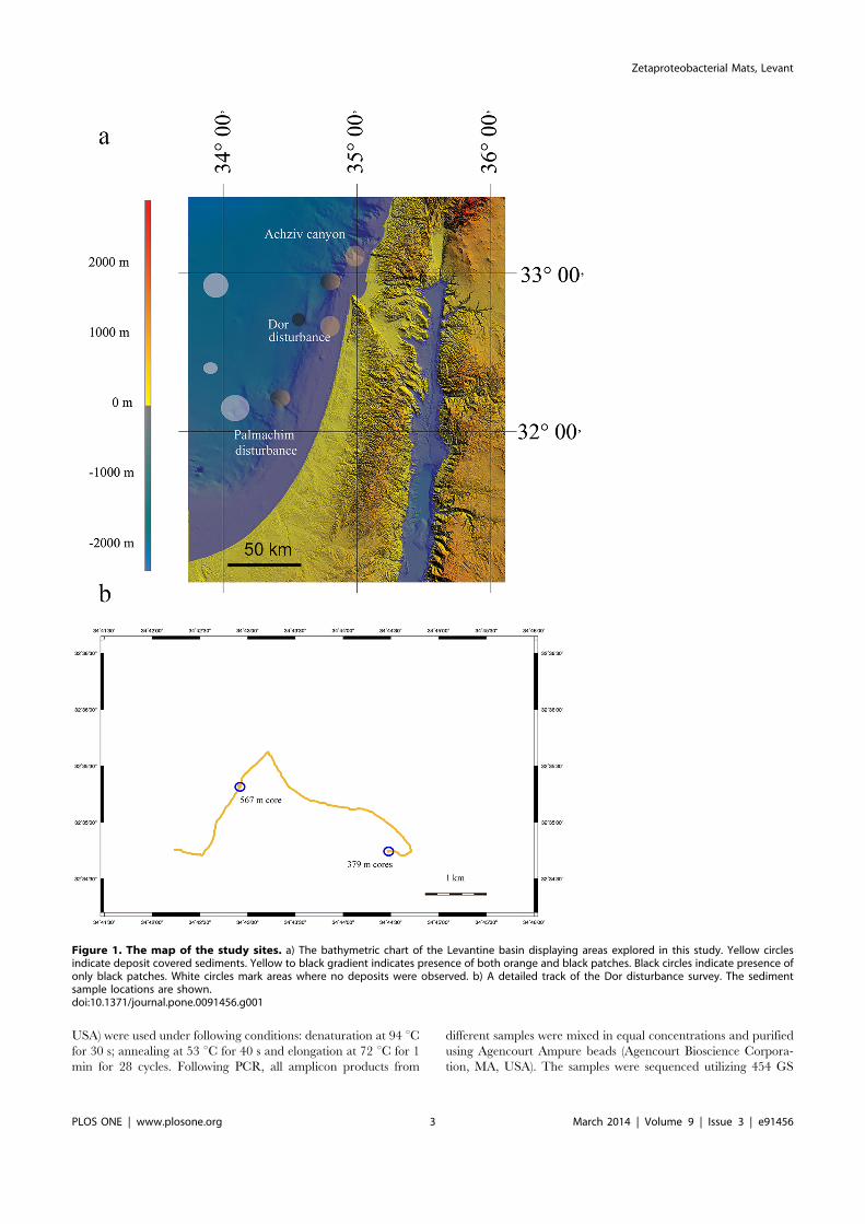
ROV imaging and sample collectionNautilus E/V is equipped with Hercules and Argus Remotely
Operated Vehicle (ROV) systems, which are able to collect high-resolution video, oceanographic data, and precision sampling. Theyellow biofilms at the Levantine sea floor were recorded duringtwo legs of the Nautilus E/V, in September 2010 and November2011 (NA-009 and NA-019) using the high definition imagingsystem mounted on the Hercules ROV. The areas explored werethe Achziv canyon at the depths of 500–1100 m; two locations inthe Acre area, one deep (1200–1700 m) and one shallower (1000–1200 m); Dor disturbance (both legs, 300–900 m) and Palmachimdisturbance (both legs, 600–1300 m) [28,29] (Fig. 1). Eachcontiguous survey covered approximately 5 km distance (e.g.Fig. 1b), while the field of view diameter was ca. 4 m, yielding ca.20000 m2 transect area. Bulk sediment (approximately top 5 cm)samples were collected during 2010 Nautilus E/V field season fromthe Dor disturbance at 32u44. 09459 North, 34u47. 01629 East at557 m and from a biofilm at the Achziv Canyon, 32u42. 26319North, 34u33. 55569 East at 1073 m. A subsample from Dordisturbance sediment was frozen for molecular analysis. All othersamples were collected during the 2011 Nautilus E/V field season.We used 70 mm diameter, 30 cm length cores to sample 9–10 cmprofiles at the Dor disturbance location near shore of Israel. A corewas taken from the yellow biofilm patch at 32u35. 32279 North,34u42. 92719 East at a depth of 567 m. The biofilm was collectedwith a pipette and flash-frozen and its pore-water was analyzed forFe2+. Additionally, a biofilm and a control core were collected at32u34. 74689 North, 34u44. 48329 East at a depth of 379 m. Thesecores were sliced to 1 cm sections, and the sediment was flash-frozen for further processing.
MicroscopyThe flocculent matter from the 567 m core was transferred with
a pipette to a slide and observed immediately under variousmagnifications. The structure of the biofilm was observed with an
Imager M2 (Carl Zeiss, Germany) microscope. The images weretaken with an AxioCam MRm camera (Carl Zeiss, Germany).
pH and O2 measurementsThe pH and O2 measurements were performed immediately
upon the retrieval of the 567 m core. The intact core with the insitu headspace water was subjected to a gentle air bubbling tomaintain steady state oxygenation. The FeOB biofilms were easilydisturbed during manipulation; hence the profiles were not takenspecifically through the biofilm. The pH and O2 concentrationwithin the sediment were measured with pH-100 and O2-100microsensors (Unisense, Denmark), respectively, according tomanufacturer’s instructions. We utilized a manually operatedmicromanipulator to obtain the profile at 1 mm resolution. ThepH and O2 concentrations were recorded at each depth followingthe stabilization of the signal. The signal was amplified with aMicrosensor Multimeter (Unisense, Denmark) and processed inthe SensorTrace Basic 3.0 software (Unisense, Denmark). Onlyone profile was obtained to allow immediate further processing ofthe core.
Fe2+ and total Fe determinationThe pore fluids were extracted by centrifugation under N2
atmosphere. Filtered subsamples were assayed immediately byferrozine reagent [30] and Fe2+ in the pore water was determinedspectrophotometrically [31]. Total Fe in the biofilm was alsodetermined with the ferrozine assay [31] as follows: 100 ml biofilmsubsample was acidified with 1 mol l21 HCl overnight, followedby reduction with hydroxylamine hydrochloride prepared in asolution of analytical grade HCl. Following the ferrozine reagentaddition, ammonium acetate solution adjusted to pH 9.5 wasadded to buffer pH. Only technical replicates were performed, dueto limited availability of samples. The sensitivity of this assay is0.1 nmol l21 [32], hence our results are several orders ofmagnitude above the detection limit.
DNA isolation, amplification and sequencingDNA was isolated from flash-frozen sediments or biofilm, using
PowerSoil DNA Isolation Kit (MoBio, Carlsbad, CA, USA)following manufacturer’s instructions. The template DNA wasamplified using GoTaq Green Master Mix (Promega, Madison,WI, USA) with addition of 1 ml bovine serum albumin per 50 mlreaction in T100 thermal cycler (Bio-Rad, Hercules, CA, USA).The 16S rRNA gene was amplified using 27F-CM and 1492-Rprimers [33], yielding ,1450 bp product; the 23S rRNA gene wasamplified with broad specificity forward primer L-0858-a-S-21and zetaproteobacteria specific L-C-Zeta-1611-A-22 reverseprimer [6], yielding ,750 bp product. The PCR conditions wereas follows: denaturation at 94uC for 30 s, annealing at for 30 s 50uC and extension at 72uC for 1min (1.5 min for 16S) for 35 cycles.PCR products were purified using the Wizard SV Gel and PCRClean-Up System (Promega, Madison, WI, USA) and subsequent-ly cloned in the pGEM-T Easy vector (Promega, Madison, WI,USA) using Escherichia coli JM109 (Promega, Madison, WI, USA)as a host. The sequencing was performed by HyLabs (Israel) usingpGEM-T specific primers T7 and SP6.
Bacterial tag-encoded FLX amplicon pyrosequencing(bTEFAP) and data analysis16S rRNA gene was amplified using 16S universal Eubacterial
primers 104F: 59 GGC GVA CGG GTG AGT AA; and 530R: 59CCG CNG CNG CTG GCA C [34]. A single-step 30 cycle PCRusing HotStarTaq Plus Master Mix Kit (Qiagen, Valencia, CA,
Zetaproteobacterial Mats, Levant
PLOS ONE | www.plosone.org 2 March 2014 | Volume 9 | Issue 3 | e91456
USA) were used under following conditions: denaturation at 94 uCfor 30 s; annealing at 53 uC for 40 s and elongation at 72 uC for 1min for 28 cycles. Following PCR, all amplicon products from
different samples were mixed in equal concentrations and purifiedusing Agencourt Ampure beads (Agencourt Bioscience Corpora-tion, MA, USA). The samples were sequenced utilizing 454 GS
Figure 1. The map of the study sites. a) The bathymetric chart of the Levantine basin displaying areas explored in this study. Yellow circlesindicate deposit covered sediments. Yellow to black gradient indicates presence of both orange and black patches. Black circles indicate presence ofonly black patches. White circles mark areas where no deposits were observed. b) A detailed track of the Dor disturbance survey. The sedimentsample locations are shown.doi:10.1371/journal.pone.0091456.g001
Zetaproteobacterial Mats, Levant
PLOS ONE | www.plosone.org 3 March 2014 | Volume 9 | Issue 3 | e91456
FLX titanium (Roche, Penzberg, Germany) following manufac-turer’s guidelines. The data derived from the sequencing wasprocessed using a proprietary analysis pipeline [35–38] at MRDNA (Shallowater, TX, USA). Barcodes and primers were deletedfrom the sequences, short sequences , 200bp were removed,sequences with ambiguous base calls were removed, and sequenceswith homopolymer runs exceeding 6bp were removed. Sequenceswere denoised and chimeras were removed using custom software[39]. Operational taxonomic units were defined after removal ofsingleton sequences, clustering at 3% divergence (97% similarity).OTUs were then taxonomically classified using BLASTn against acurated Greengenes database [40] and compiled into eachtaxonomic level. A total of 1659 valid sequences were established,yielding 97 OTUs.
Phylogenetic analysisEvolutionary history was deduced using the maximum likeli-
hood method based on Kimura 2-parameter model [41] (+I for454 16S rRNA genesequences (333 positions), +G for 23S rRNAgene sequences (798 positions)) and Data Specific model [42] (+I,1298 positions) for the cloned 16S rRNA gene sequences. Thebootstrap consensus tree inferred from 1000 replicates is taken torepresent the evolutionary history of the taxa analyzed [43]. Initialtrees for the heuristic search were obtained automatically asfollows: When the number of common sites was less than 100 orless than one-fourth of the total number of sites, the maximumparsimony method was employed; otherwise BIONJ method withMCL distance matrix was used. Codon positions included were1st+2nd+3rd+Noncoding. Evolutionary analyses were conductedin MEGA5 [44]. All sequences were submitted to GeneBankunder accession numbers KF199322-KF199336, KF651136-KF651143.
Results
The distribution of biofilm patches deduced from theROV imagerySediments at 300–800 m areas at the Achziv canyon, Dor
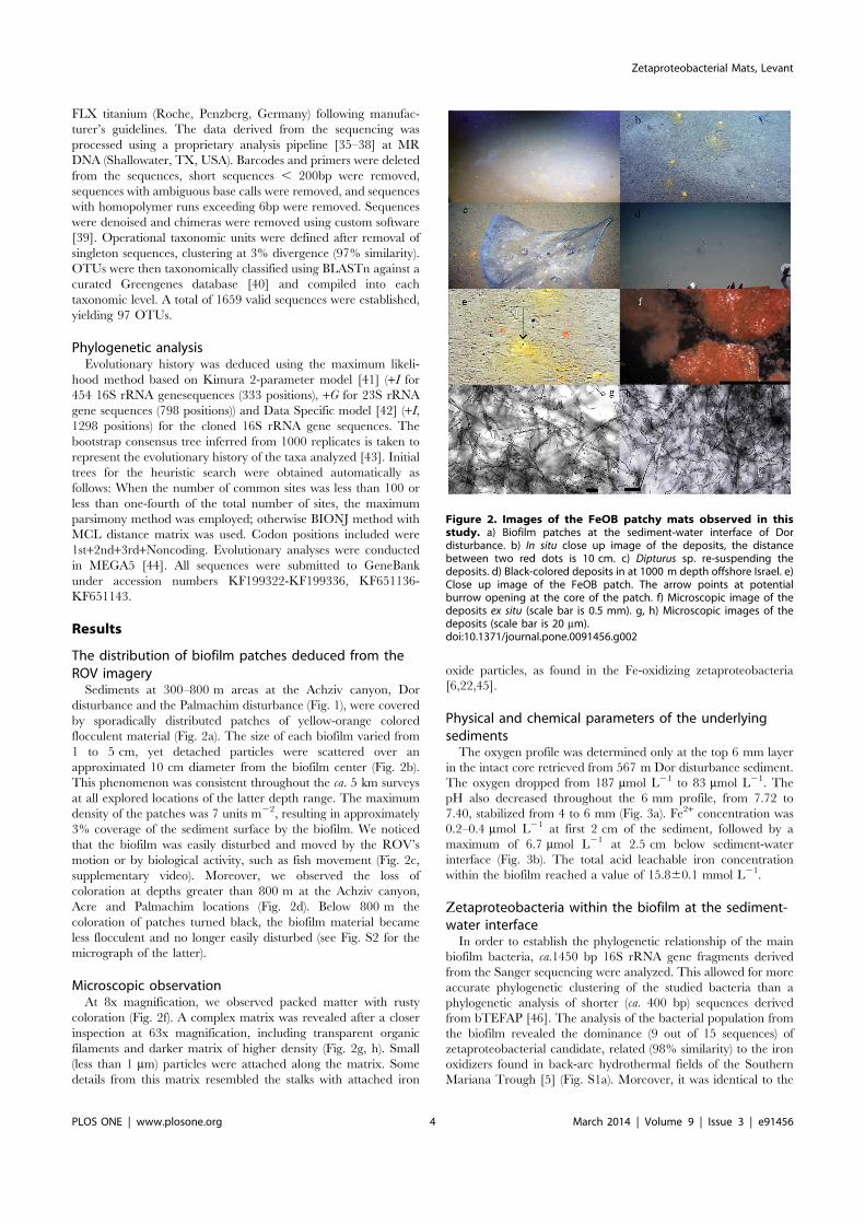
disturbance and the Palmachim disturbance (Fig. 1), were coveredby sporadically distributed patches of yellow-orange coloredflocculent material (Fig. 2a). The size of each biofilm varied from1 to 5 cm, yet detached particles were scattered over anapproximated 10 cm diameter from the biofilm center (Fig. 2b).This phenomenon was consistent throughout the ca. 5 km surveysat all explored locations of the latter depth range. The maximumdensity of the patches was 7 units m22, resulting in approximately3% coverage of the sediment surface by the biofilm. We noticedthat the biofilm was easily disturbed and moved by the ROV’smotion or by biological activity, such as fish movement (Fig. 2c,supplementary video). Moreover, we observed the loss ofcoloration at depths greater than 800 m at the Achziv canyon,Acre and Palmachim locations (Fig. 2d). Below 800 m thecoloration of patches turned black, the biofilm material becameless flocculent and no longer easily disturbed (see Fig. S2 for themicrograph of the latter).
Microscopic observationAt 8x magnification, we observed packed matter with rusty
coloration (Fig. 2f). A complex matrix was revealed after a closerinspection at 63x magnification, including transparent organicfilaments and darker matrix of higher density (Fig. 2g, h). Small(less than 1 mm) particles were attached along the matrix. Somedetails from this matrix resembled the stalks with attached iron
oxide particles, as found in the Fe-oxidizing zetaproteobacteria[6,22,45].
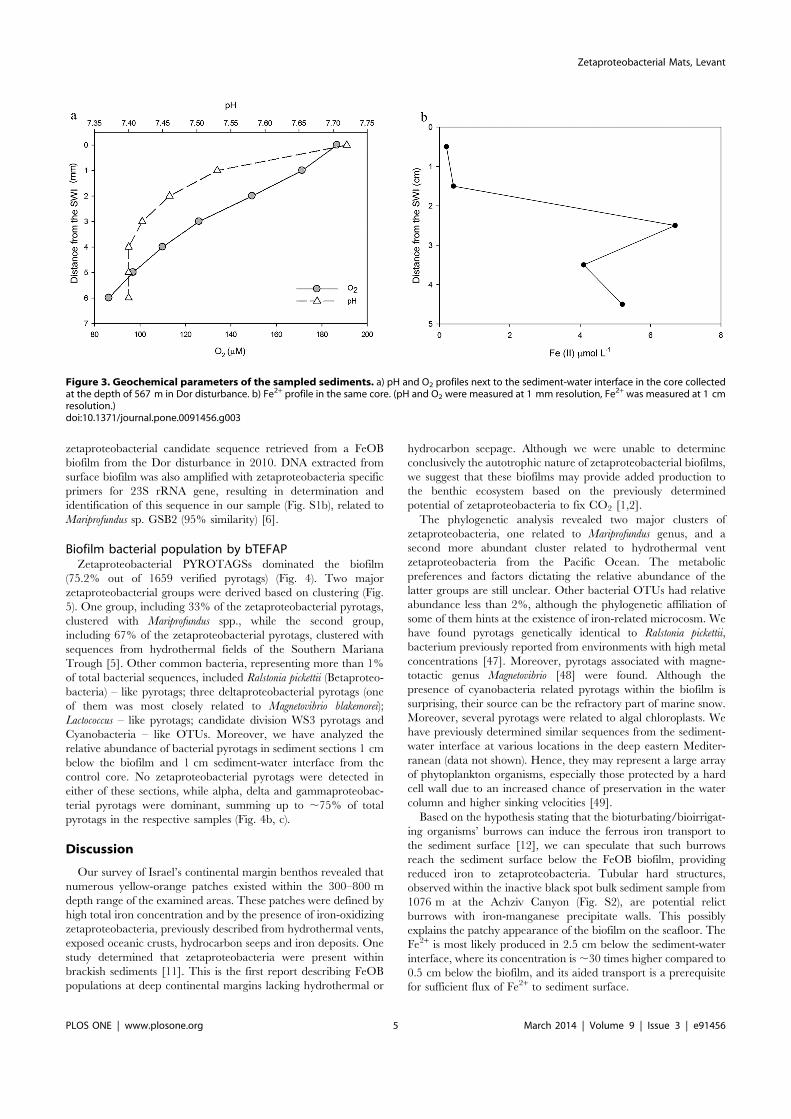
Physical and chemical parameters of the underlyingsedimentsThe oxygen profile was determined only at the top 6 mm layer
in the intact core retrieved from 567 m Dor disturbance sediment.The oxygen dropped from 187 mmol L21 to 83 mmol L21. ThepH also decreased throughout the 6 mm profile, from 7.72 to7.40, stabilized from 4 to 6 mm (Fig. 3a). Fe2+ concentration was0.2–0.4 mmol L21 at first 2 cm of the sediment, followed by amaximum of 6.7 mmol L21 at 2.5 cm below sediment-waterinterface (Fig. 3b). The total acid leachable iron concentrationwithin the biofilm reached a value of 15.860.1 mmol L21.
Fetaproteobacteria within the biofilm at the sediment-water interfaceIn order to establish the phylogenetic relationship of the main
biofilm bacteria, ca.1450 bp 16S rRNA gene fragments derivedfrom the Sanger sequencing were analyzed. This allowed for moreaccurate phylogenetic clustering of the studied bacteria than aphylogenetic analysis of shorter (ca. 400 bp) sequences derivedfrom bTEFAP [46]. The analysis of the bacterial population fromthe biofilm revealed the dominance (9 out of 15 sequences) ofzetaproteobacterial candidate, related (98% similarity) to the ironoxidizers found in back-arc hydrothermal fields of the SouthernMariana Trough [5] (Fig. S1a). Moreover, it was identical to the
Figure 2. Images of the FeOB patchy mats observed in thisstudy. a) Biofilm patches at the sediment-water interface of Dordisturbance. b) In situ close up image of the deposits, the distancebetween two red dots is 10 cm. c) Dipturus sp. re-suspending thedeposits. d) Black-colored deposits in at 1000 m depth offshore Israel. e)Close up image of the FeOB patch. The arrow points at potentialburrow opening at the core of the patch. f) Microscopic image of thedeposits ex situ (scale bar is 0.5 mm). g, h) Microscopic images of thedeposits (scale bar is 20 mm).doi:10.1371/journal.pone.0091456.g002
Zetaproteobacterial Mats, Levant
PLOS ONE | www.plosone.org 4 March 2014 | Volume 9 | Issue 3 | e91456
zetaproteobacterial candidate sequence retrieved from a FeOBbiofilm from the Dor disturbance in 2010. DNA extracted fromsurface biofilm was also amplified with zetaproteobacteria specificprimers for 23S rRNA gene, resulting in determination andidentification of this sequence in our sample (Fig. S1b), related toMariprofundus sp. GSB2 (95% similarity) [6].
Biofilm bacterial population by bTEFAPZetaproteobacterial PYROTAGSs dominated the biofilm
(75.2% out of 1659 verified pyrotags) (Fig. 4). Two majorzetaproteobacterial groups were derived based on clustering (Fig.5). One group, including 33% of the zetaproteobacterial pyrotags,clustered with Mariprofundus spp., while the second group,including 67% of the zetaproteobacterial pyrotags, clustered withsequences from hydrothermal fields of the Southern MarianaTrough [5]. Other common bacteria, representing more than 1%of total bacterial sequences, included Ralstonia pickettii (Betaproteo-bacteria) – like pyrotags; three deltaproteobacterial pyrotags (oneof them was most closely related to Magnetovibrio blakemorei);Lactococcus – like pyrotags; candidate division WS3 pyrotags andCyanobacteria – like OTUs. Moreover, we have analyzed therelative abundance of bacterial pyrotags in sediment sections 1 cmbelow the biofilm and 1 cm sediment-water interface from thecontrol core. No zetaproteobacterial pyrotags were detected ineither of these sections, while alpha, delta and gammaproteobac-terial pyrotags were dominant, summing up to ,75% of totalpyrotags in the respective samples (Fig. 4b, c).
Discussion
Our survey of Israel’s continental margin benthos revealed thatnumerous yellow-orange patches existed within the 300–800 mdepth range of the examined areas. These patches were defined byhigh total iron concentration and by the presence of iron-oxidizingzetaproteobacteria, previously described from hydrothermal vents,exposed oceanic crusts, hydrocarbon seeps and iron deposits. Onestudy determined that zetaproteobacteria were present withinbrackish sediments [11]. This is the first report describing FeOBpopulations at deep continental margins lacking hydrothermal or
hydrocarbon seepage. Although we were unable to determineconclusively the autotrophic nature of zetaproteobacterial biofilms,we suggest that these biofilms may provide added production tothe benthic ecosystem based on the previously determinedpotential of zetaproteobacteria to fix CO2 [1,2].The phylogenetic analysis revealed two major clusters of
zetaproteobacteria, one related to Mariprofundus genus, and asecond more abundant cluster related to hydrothermal ventzetaproteobacteria from the Pacific Ocean. The metabolicpreferences and factors dictating the relative abundance of thelatter groups are still unclear. Other bacterial OTUs had relativeabundance less than 2%, although the phylogenetic affiliation ofsome of them hints at the existence of iron-related microcosm. Wehave found pyrotags genetically identical to Ralstonia pickettii,bacterium previously reported from environments with high metalconcentrations [47]. Moreover, pyrotags associated with magne-totactic genus Magnetovibrio [48] were found. Although thepresence of cyanobacteria related pyrotags within the biofilm issurprising, their source can be the refractory part of marine snow.Moreover, several pyrotags were related to algal chloroplasts. Wehave previously determined similar sequences from the sediment-water interface at various locations in the deep eastern Mediter-ranean (data not shown). Hence, they may represent a large arrayof phytoplankton organisms, especially those protected by a hardcell wall due to an increased chance of preservation in the watercolumn and higher sinking velocities [49].Based on the hypothesis stating that the bioturbating/bioirrigat-
ing organisms’ burrows can induce the ferrous iron transport tothe sediment surface [12], we can speculate that such burrowsreach the sediment surface below the FeOB biofilm, providingreduced iron to zetaproteobacteria. Tubular hard structures,observed within the inactive black spot bulk sediment sample from1076 m at the Achziv Canyon (Fig. S2), are potential relictburrows with iron-manganese precipitate walls. This possiblyexplains the patchy appearance of the biofilm on the seafloor. TheFe2+ is most likely produced in 2.5 cm below the sediment-waterinterface, where its concentration is,30 times higher compared to0.5 cm below the biofilm, and its aided transport is a prerequisitefor sufficient flux of Fe2+ to sediment surface.
Figure 3. Geochemical parameters of the sampled sediments. a) pH and O2 profiles next to the sediment-water interface in the core collectedat the depth of 567 m in Dor disturbance. b) Fe2+ profile in the same core. (pH and O2 were measured at 1 mm resolution, Fe2+ was measured at 1 cmresolution.)doi:10.1371/journal.pone.0091456.g003
Zetaproteobacterial Mats, Levant
PLOS ONE | www.plosone.org 5 March 2014 | Volume 9 | Issue 3 | e91456
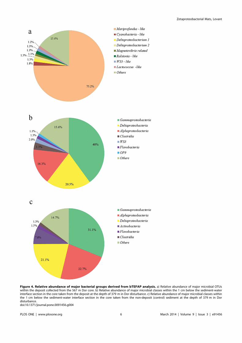
Figure 4. Relative abundance of major bacterial groups derived from bTEFAP analysis. a) Relative abundance of major microbial OTUswithin the deposit collected from the 567 m Dor core. b) Relative abundance of major microbial classes within the 1 cm below the sediment-waterinterface section in the core taken from the deposit at the depth of 379 m in Dor disturbance. c) Relative abundance of major microbial classes withinthe 1 cm below the sediment-water interface section in the core taken from the non-deposit (control) sediment at the depth of 379 m in Dordisturbance.doi:10.1371/journal.pone.0091456.g004
Zetaproteobacterial Mats, Levant
PLOS ONE | www.plosone.org 6 March 2014 | Volume 9 | Issue 3 | e91456

Interestingly, at water depth of 800–1300 m biofilm patcheswere observed, although their black coloration and lack offlocculence may indicate loss of zetaproteobacterial activity andprecipitation of manganese with iron oxides or pyrite formation.Several patches in transitional state (yellow to black) were alsopresent (Fig. S3a, b). No biofilms were observed between 1300–1700 m. The flux of organic matter and benthic infaunal biomassdecrease with depth [50–52], therefore the increased organic load
in shallow sediments can provide a larger pool of electrons forrespiration compared to deeper sediments, producing largerquantities of reduced metabolites. This organic load can alsosupport larger populations of bioturbating metazoans that mayinclude sedentary Polychaeta families such as Spionidae, Aphar-etidae, Pectinariidae and Maldanidae, potentially responsible forthe enhancement of Fe2+ transport. Hence, a barrier disabling thesediments from providing reduced metabolites to the overlaying
Figure 5. Molecular phylogenetic analysis by maximum likelihoodmethod of most common 16S rRNA pyrotags in the FeOB sample(grey color) and related sequences from the NCBI database. The number of pyrotags yielded for the respective OTU is shown in the brackets.Escherichia coli 16S sequence is used as an out-group. The numbers at nodes are bootstrap percent values based on 1000 resamplings. The scale barcorresponds to the number of substitutions per site.doi:10.1371/journal.pone.0091456.g005
Zetaproteobacterial Mats, Levant
PLOS ONE | www.plosone.org 7 March 2014 | Volume 9 | Issue 3 | e91456
water can be produced by insufficient organic matter flux to theseafloor. The seasonal fluctuations dependent on the primaryproductivity in the photic zone may explain the inactive ironoxidation state of 800–1300 m sediments. Both 2010 and 2011legs of the Nautilus E/V exploration in Levantine basin took placein late summer prior to water column mixing and it should benoted that the depth of biofilm activity would be expected toincrease following the spring phytoplankton bloom.Bioturbation/bioirrigation, sediment resuspension and chemical
processes at the sediment-water interface were suggested previ-ously as important mechanisms dictating the fluxes of sedimentiron into the water column [53]. In this study we established thatthe reduced Fe can reach the sediment surface and given thebiological resuspension of sediment (Fig 2c, Supp. Video) and deepwater mixing, can be transported up the water column. Aprerequisite for the ability of iron to travel up the water column isthe neutral buoyancy of the iron-containing particle. This can beachieved by detachment of small, colloidal iron particles.Moreover, the association of larger iron particles with positivelybuoyant organic matter can yield the same result [54,55]. Basedon existing data [56] we estimate that winter sea surfacetemperatures (SST) below 15uC are sufficient to mix the watercolumn to depths greater than 400 m. The average winter SST inthe eastern Mediterranean fluctuates around 15.5 uC, while thelowest average temperature of 15uC was detected during thewinter of 1992 [56]. During the last decade, there is an increasingtrend of SST values in the eastern Mediterranean Sea [56]. Theenhanced SST is expected to decrease iron supply from thesediment surface due to shallower mixing depth [57]. On thecontrary, increased temperature of eastern Mediterranean deepwater sources can result in the uplift of deep water [58] andincreased nutrient flux from the sediment surface to the photiczone. Another positive effect of the global change on the nutrientsupply may be the rare, extremely cold winter temperatures thatwere recently detected in this region [59]. Based on severalmodels, their frequency may increase in the future [60]. In suchcase, the significance of the sediment-water interface iron as abioavailable iron source for the water column may increase due toenhanced mixing during an extremely cold event.Unfortunately, water column iron profiles are still unavailable in
the Levantine basin, and present Mediterranean sampling islimited to shallow (,10 m) or deep (.1500 m) stations [61,62].Nonetheless, data from a Weddell Sea station, defined byproximity to the continental shelf and bathymetry resemblingour study, suggests iron enrichment close to the sediment-waterinterface [63], verifying that sediment is a potential source for thewater column iron. The bioavailability of the resuspendedparticulate iron originating from the sediment-water interfacecan be altered by biologically mediated processes such as microbialreductive dissolution [16,64–67] in the water column (Fig. 6). Ironminerals produced by the stalk forming FeOB are attached tocarboxyl-rich polysaccharides [22], providing the organic ligandsnecessary for the reductive dissolution.In the report we show that iron-oxidizing bacteria precipitate
the ferrous iron at the sediment-water interface along large areasof the continental margin, potentially altering its solubility. Thefate of iron precipitated by the zetaproteobacteria is still unclearand its bioavailability may play a crucial role in the future of eastMediterranean ecology under the global change.
Conclusions
The ‘‘plain’’ sediments at continental margins constitute aheavily understudied area of deep sea research. In this study we
report the presence of patchy FeOB mats, abundant in thesurveyed locations, unaffected by hydrothermal or hydrocarbonseepage, along the Levantine continental margin. Zetaproteobac-terial FeOB, represented by two main phylotypic groups derivedfrom the 16S rRNA gene phylogenetic analysis, constitute thedominant fraction of bacterial population within the biofilms. Wesuggest that Fe2+ concentration sufficient to maintain sediment-water interface FeOB community is most likely achieved byenhancement of pore water upward flux by bioturbating/bioirrigating infauna. Given the potential of FeOB to fix carbonand alter iron chemistry, they may play an important role in themarine environment, affecting both benthic and water columnecosystems.
Supporting Information
Figure S1 Molecular phylogenetic analysis by maxi-mum likelihood method of cloned bacterial genetic
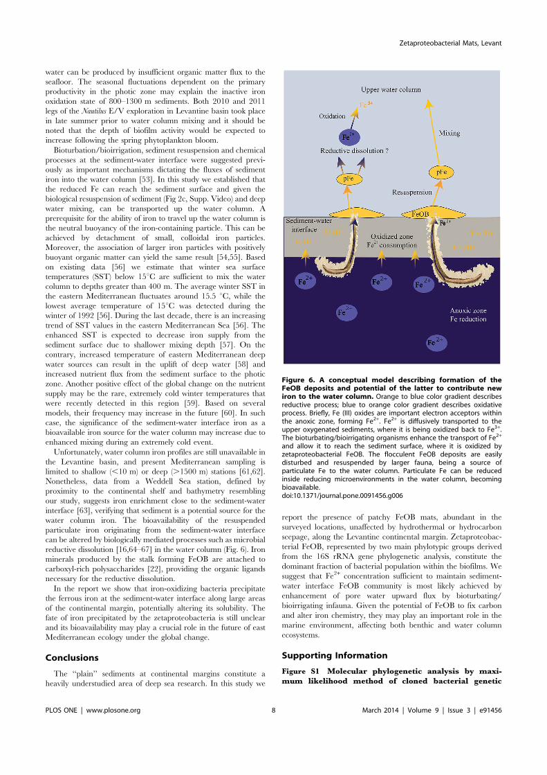
Figure 6. A conceptual model describing formation of theFeOB deposits and potential of the latter to contribute newiron to the water column. Orange to blue color gradient describesreductive process; blue to orange color gradient describes oxidativeprocess. Briefly, Fe (III) oxides are important electron acceptors withinthe anoxic zone, forming Fe2+. Fe2+ is diffusively transported to theupper oxygenated sediments, where it is being oxidized back to Fe3+.The bioturbating/bioirrigating organisms enhance the transport of Fe2+
and allow it to reach the sediment surface, where it is oxidized byzetaproteobacterial FeOB. The flocculent FeOB deposits are easilydisturbed and resuspended by larger fauna, being a source ofparticulate Fe to the water column. Particulate Fe can be reducedinside reducing microenvironments in the water column, becomingbioavailable.doi:10.1371/journal.pone.0091456.g006
Zetaproteobacterial Mats, Levant
PLOS ONE | www.plosone.org 8 March 2014 | Volume 9 | Issue 3 | e91456
markers. a) Evolutionary history of the 16S rRNA small subunitgene. Gamma-proteobacterial 16S sequence is used as an out-group. b) The evolutionary history of 23S rRNA large subunitgene. Nitrosomonas sp. 23S rRNA gene sequence is used as an out-group. The numbers are bootstrap percent values based on 1000resamplings. The scale bar corresponds to the number ofsubstitutions per site.(TIF)
Figure S2 Microscopic images of precipitated materialwithin sediments from a black spot. a) Biogenic tubes. b)Solid particles. Scale bar is 0.5 mm.(TIF)
Figure S3 Additional images of FeOB deposits. a) Adeposit in transitional state. b). Deposits next to a metal wreckhave both yellow-orange and black colorations. c) A spot in themoddle of FeOB deposit that can be an opening of metazoanburrow. d) Disturbance and resuspention of FeOB deposit bydeep-sea Pleuronectiformes specimen.(TIF)
Video S1 Re-suspension of deposit-covered sediment byDipturus specimen.(MOV)
Acknowledgments
We thank the European Commission FP7 research infrastructure initiativeprogram, ‘‘Assemble 227799,’’ for partial support of this project. Thisresearch used samples and data provided by the E/V Nautilus ExplorationProgram – expeditions, NA009 and NA019. The authors would like tothank all individuals who helped during the expedition, including onboardtechnical and scientific personnel, and the captain and crew of the E/VNautilus. We also thank the reviewers for constructive comments on anearlier version of this manuscript. Supplementary information is availableat Plos ONE website. All authors of this manuscript certify that they qualifyfor authorship because of substantial contribution to the work submitted.The authors declare that this manuscript has not been published nor isunder simultaneous consideration for publication elsewhere. The authorsagree to transfer the copyright to the Plos ONE journal to be effective ifand when the manuscript is accepted for publication and that themanuscript will not be published elsewhere in any other language withoutthe consent of the Plos ONE journal.
Author Contributions
Conceived and designed the experiments: MRB GA RT BGT JA DC ZBADT. Performed the experiments: MRB GA RT ES DT. Analyzed the data:MRB GA RT ES DT. Wrote the paper: MRB GA RT ES JA DC ZBABGT DT. Saw and approved the final manuscript: MRB GA RT ES JADC BGT ZBA DT.
References
1. Emerson D, Rentz JA, Lilburn TG, Davis RE, Aldrich H, et al. (2007) A novellineage of proteobacteria involved in formation of marine Fe-oxidizing microbialmat communities. PLoS One 2: e667. Available: http://www.plosone.org/article/info%3Adoi%2F10.1371%2Fjournal.pone.0000667. Accessed 2012March 9.
2. Singer E, Emerson D, Webb EA, Barco RA, Kuenen JG, et al. (2011)Mariprofundus ferrooxydans PV-1 the first genome of a marine Fe(II) oxidizingZetaproteobacterium. PLoS One 6: e25386. Available: http://www.plosone.org/article/info%3Adoi%2F10.1371%2Fjournal.pone.0025386. Accessed 2012March 1.
3. Dang H, Chen R, Wang L, Shao S, Dai L, et al. (2011) Molecularcharacterization of putative biocorroding microbiota with a novel nichedetection of Epsilon- and Zetaproteobacteria in Pacific Ocean coastal seawaters.Environ Microbiol 13: 3059–3074.
4. McAllister SM, Davis RE, McBeth JM, Tebo BM, Emerson D, et al. (2011)Biodiversity and emerging biogeography of the neutrophilic iron-oxidizingZetaproteobacteria. Appl Environ Microbiol 77: 5445–5457.
5. Kato S, Yanagawa K, Sunamura M, Takano Y, Ishibashi J, et al. (2009)Abundance of Zetaproteobacteria within crustal fluids in back-arc hydrothermalfields of the Southern Mariana Trough. Environ Microbiol 11: 3210–3222.
6. McBeth JM, Little BJ, Ray RI, Farrar KM, Emerson D (2011) Neutrophiliciron-oxidizing ‘‘zetaproteobacteria’’ and mild steel corrosion in nearshoremarine environments. Appl Environ Microbiol 77: 1405–1412.
7. Omoregie EO, Mastalerz V, de Lange G, Straub KL, Kappler A, et al. (2008)Biogeochemistry and community composition of iron- and sulfur-precipitatingmicrobial mats at the Chefren mud volcano (Nile Deep Sea Fan, EasternMediterranean). Appl Environ Microbiol 74: 3198–3215.
8. Kato S, Nakamura K, Toki T, Ishibashi J-I, Tsunogai U, et al. (2012) Iron-basedmicrobial ecosystem on and below the seafloor: a case study of hydrothermalfields of the southern mariana trough. Front Microbiol 3: 89. Available: http://www.pubmedcentral.nih.gov/articlerender.fcgi?artid=3304087&tool=pmcentrez&rendertype=abstract. Accessed 2012 Oct 19.
9. Emerson D, Fleming EJ, McBeth JM (2010) Iron-oxidizing bacteria: anenvironmental and genomic perspective. Annu Rev Microbiol 64: 561–583.
10. Emerson D, Moyer C (2010) Microbiology of seamounts: common patternsobserved in community structure. Oceanography 23: 148–163.
11. McBeth JM, Fleming EJ, Emerson D (2013) The transition from freshwater tomarine iron-oxidizing bacterial lineages along a salinity gradient on theSheepscot River, Maine USA. Environ Microbiol Rep 5: 453–463.
12. Severmann S, McManus J, Berelson WM, Hammond DE (2010) Thecontinental shelf benthic iron flux and its isotope composition. GeochimCosmochim Acta 74: 3984–4004.
13. Sundby B (2006) Transient state diagenesis in continental margin muds. MarChem 102: 2–12.
14. Chase Z (2005) Distribution and variability of iron input to Oregon coastalwaters during the upwelling season. J Geophys Res 110: C10S12. Available:http://www.agu.org/pubs/crossref/2005/2004JC002590.shtml. Accessed 2012Nov 11.
15. Elrod VA. (2004) The flux of iron from continental shelf sediments: A missingsource for global budgets. Geophys Res Lett 31: L12307. Available: http://
www.agu.org/pubs/crossref/2004/2004GL020216.shtml. Accessed 2012 Nov9.
16. Lohan MC, Bruland KW (2008) Elevated Fe(II) and dissolved Fe in hypoxicshelf waters off Oregon and Washington: an enhanced source of iron to coastalupwelling regimes. Environ Sci Technol 42: 6462–6468.
17. Lam PJ, Bishop JKB, Henning CC, Marcus MA, Waychunas GA, et al. (2006)Wintertime phytoplankton bloom in the subarctic Pacific supported bycontinental margin iron. Global Biogeochem Cycles 20: 1–12.
18. Lam PJ, Bishop JKB (2008) The continental margin is a key source of iron to theHNLC North Pacific Ocean. Geophys Res Lett 35: L07608. Available: http://www.agu.org/pubs/crossref/2008/2008GL033294.shtml. Accessed 2012 Nov11.
19. Katz T, Yahel G, Reidenbach M, Tunnicliffe V, Herut B, et al. (2012)Resuspension by fish facilitates the transport and redistribution of coastalsediments. Limnol Oceanogr 57: 945–958.
20. Rich HW, Morel FMM (1990) Availability of well-defined iron colloids to themarine diatom Thdassiosiru weissflogii. Limnol Oceanogr 35: 652–662.
21. Toner BM, Berquo TS, Michel FM, Sorensen J V, Templeton AS, et al. (2012)Mineralogy of iron microbial mats from loihi seamount. Front Microbiol 3: 118.Available: http://www.pubmedcentral.nih.gov/articlerender.fcgi?artid=3316996&tool=pmcentrez&rendertype=abstract. Accessed 2013 Dec 17.
22. Chan CS, Fakra SC, Emerson D, Fleming EJ, Edwards KJ (2011) Lithotrophiciron-oxidizing bacteria produce organic stalks to control mineral growth:implications for biosignature formation. ISME J 5: 717–727.
23. Tanaka T, Zohary T, Krom MD, Law CS, Pitta P, et al. (2007) Microbialcommunity structure and function in the Levantine Basin of the easternMediterranean. Deep Sea Res Part I Oceanogr Res Pap 54: 1721–1743.
24. Berman-Frank I, Cullen JT, Shaked Y, Sherrell RM, Falkowski PG (2001) Ironavailability, cellular iron quotas, and nitrogen fixation in Trichodesmium. LimnolOceanogr 46: 1249–1260.
25. Breitbarth E, Achterberg EP, Ardelan MV, Baker AR, Bucciarelli E, et al. (2010)Iron biogeochemistry across marine systems – progress from the past decade.Biogeosciences 7: 1075–1097.
26. Yogev T, Rahav E, Bar-Zeev E, Man-Aharonovich D, Stambler N, et al. (2011)Is dinitrogen fixation significant in the Levantine Basin, East MediterraneanSea? Environ Microbiol 13: 854–871.
27. Theodosi C, Markaki Z, Mihalopoulos N (2010) Iron speciation, solubility andtemporal variability in wet and dry deposition in the Eastern Mediterranean.Mar Chem 120: 100–107.
28. Coleman D, Austin Jr JA, Ben-Avraham Z (2011) Exploring the ContinentalMargin of Israel: ‘‘ Telepresence ’’ at Work. Eos Trans AGU 92. Available:http://www.agu.org/pubs/crossref/2011/2011EO100002.shtml. Accessed2012 June 24.
29. Coleman DF, Austin Jr JA, Ben-Avraham Z, Makovsky Y, Tchernov D (2012)Seafloor pockmarks, deepwater corals, and cold seeps along the continentalmargin of Israel. Oceanography 25 (supplement 1): 40–41.
30. Stookey LL (1970) Ferrozine-a new spectrophotometric reagent for iron. AnalChem 42: 779–781.
Zetaproteobacterial Mats, Levant
PLOS ONE | www.plosone.org 9 March 2014 | Volume 9 | Issue 3 | e91456
31. Viollier E, Inglett P, Hunter K, Roychoudhury A, Van Cappellen P (2000) Theferrozine method revisited: Fe(II)/Fe(III) determination in natural waters. ApplGeochemistry 15: 785–790.
32. Achterberg EP, Holland TW, Bowie AR, Mantoura RFC, Worsfold PJ (2001)Determination of iron in seawater. Anal Chim Acta 442: 1–14.
33. Frank JA, Reich CI, Sharma S, Weisbaum JS, Wilson BA, et al. (2008) Criticalevaluation of two primers commonly used for amplification of bacterial 16SrRNA genes. Appl Environ Microbiol 74: 2461–2470.
34. Wang Y, Qian P (2009) Conservative fragments in bacterial 16S rRNA genesand primer design for 16S ribosomal DNA amplicons in metagenomic studies.PLoS One 4: e7401. Available: http://www.plosone.org/article/info%3Adoi%2F10.1371%2Fjournal.pone.0007401. Accessed 19 August 2013.
35. Capone KA, Dowd SE, Stamatas GN, Nikolovski J (2011) Diversity of thehuman skin microbiome early in life. J Invest Dermatol 131: 2026–2032.
36. Dowd SE, Sun Y, Wolcott RD, Domingo A, Carroll JA (2008) Bacterial tag-encoded FLX amplicon pyrosequencing (bTEFAP) for microbiome studies:bacterial diversity in the ileum of newly weaned Salmonella-infected pigs.Foodborne Pathog Dis 5: 459–472.
37. Eren AM, Zozaya M, Taylor CM, Dowd SE, Martin DH, et al. (2011) Exploringthe diversity of Gardnerella vaginalis in the genitourinary tract microbiota ofmonogamous couples through subtle nucleotide variation. PLoS One 6: e26732.Available: http://www.plosone.org/article/info%3Adoi%2F10.1371%2Fjournal.pone.0026732. Accessed 2012 Nov 12.
38. Swanson KS, Dowd SE, Suchodolski JS, Middelbos IS, Vester BM, et al. (2011)Phylogenetic and gene-centric metagenomics of the canine intestinal micro-biome reveals similarities with humans and mice. ISME J 5: 639–649.
39. Gontcharova V, Youn E, Sun Y, Wolcott RD, Dowd SE (2010) A comparison ofbacterial composition in diabetic ulcers and contralateral intact skin. OpenMicrobiol J 4: 8–19.
40. DeSantis TZ, Hugenholtz P, Larsen N, Rojas M, Brodie EL, et al. (2006)Greengenes, a chimera-checked 16S rRNA gene database and workbenchcompatible with ARB. Appl Environ Microbiol 72: 5069–5072.
41. Kimura M (1980) A simple method for estimating evolutionary rate of basesubstitutions through comparative studies of nucleotide sequences. J Mol Evol16: 111–120.
42. Nei M, Kumar S (2000) Molecular Evolution and Phylogenetics. New York:Oxford University Press.
43. Felsenstein J (1985) Confidence limits on phylogenies: An approach using thebootstrap. Evolution (N Y) 39: 783–791.
44. Tamura K, Peterson D, Peterson N, Stecher G, Nei M, et al. (2011) MEGA5:molecular evolutionary genetics analysis using maximum likelihood, evolution-ary distance, and maximum parsimony methods. Mol Biol Evol 28: 2731–2739.
45. Hodges TW, Olson JB (2009) Molecular comparison of bacterial communitieswithin iron-containing flocculent mats associated with submarine volcanoesalong the Kermadec Arc. Appl Environ Microbiol 75: 1650–1657.
46. Wommack KE, Bhavsar J, Ravel J (2008) Metagenomics: read length matters.Appl Environ Microbiol 74: 1453–1463.
47. Konstantinidis KT, Isaacs N, Fett J, Simpson S, Long DT, et al. (2003)Microbial diversity and resistance to copper in metal-contaminated lakesediment. Microb Ecol 45: 191–202.
48. Bazylinski DA, Williams TJ, Lefevre CT, Trubitsyn D, Fang J, et al. (2013)Magnetovibrio blakemorei gen. nov., sp. nov., a magnetotactic bacterium(Alphaproteobacteria: Rhodospirillaceae) isolated from a salt marsh. Int J SystEvol Microbiol 63: 1824–1833.
49. Iversen MH, Ploug H (2010) Ballast minerals and the sinking carbon flux in theocean: carbon-specific respiration rates and sinking velocity of marine snowaggregates. Biogeosciences 7: 2613–2624.
50. Rijk S De, Jorissen FJ, Rohling EJ, Troelstra SR (2000) Organic flux control onbathymetric zonation of Mediterranean benthic foraminifera. Mar Micropa-leontol 40: 151–166.
51. Danovaro R, Croce N Della, Eleftheriou A, Fabiano M, Papadopoulou N, et al.(1995) Meiofauna of the deep Eastern Mediterranean Sea: distribution andabundance in relation to bacterial biomass, organic matter composition andother environmental factors. Prog Oceanogr 36: 329–341.
52. Danovaro R, Dinet A, Duineveld G, Tselepides A (1999) Benthic response toparticulate fluxes in different trophic environments: a comparison between theGulf of Lions–Catalan Sea (western-Mediterranean) and the Cretan Sea(eastern-Mediterranean). Prog Oceanogr 44: 287–312.
53. Pakhomova SV, Hall POJ, Kononets MY, Rozanov AG, Tengberg A, et al.(2007) Fluxes of iron and manganese across the sediment–water interface undervarious redox conditions. Mar Chem 107: 319–331.
54. Azetsu-Scott K, Passow U (2004) Ascending marine particles: Significance oftransparent exopolymer particles (TEP) in the upper ocean. Limnol Oceanogr49: 741–748.
55. Ortega-Osorio A, Scott SD (2001) Morphological and chemical characterizationof neutrally buoyant plume-derived particles at the Eastern Manus basinhydrothermal field, Papua New Guinea. Can Mineral 39: 17–31.
56. Herut B, Almogi-Labin A, Jannik N, Gertman I (2000) The seasonal dynamics ofnutrient and chlorophyll a concentrations on the SE Mediterranean shelf-slope.Oceanol Acta 23: 771–782.
57. Coma R, Ribes M, Serrano E, Jimenez E, Salat J, et al. (2009) Global warming-enhanced stratification and mass mortality events in the Mediterranean. ProcNatl Acad Sci 106: 6176–6181.
58. Danovaro R, Dell’Anno A, Fabiano M, Pusceddu A, Tselepides A (2001) Deep-sea ecosystem response to climate changes: the eastern Mediterranean casestudy. Trends Ecol Evol 16: 505–510.
59. Tolika K, Anagnostopoulou C, Maheras P, Velikou K (2013) Extremetemperature contrast of the year 2012 in Greece: An exceptionally cold winterand a record breaking summer. EGU General Assembly 2013. p. 2922.
60. Sanchez E, Gallardo C, Gaertner MA, Arribas A, Castro M (2004) Futureclimate extreme events in the Mediterranean simulated by a regional climatemodel: a first approach. Glob Planet Change 44: 163–180.
61. Guieu C (2002) Impact of high Saharan dust inputs on dissolved ironconcentrations in the Mediterranean Sea. Geophys Res Lett 29: 1911. Available:http://doi.wiley.com/10.1029/2001GL014454. Accessed 2013 June 3.
62. Sarthou G, Jeandel C (2001) Seasonal variations of iron concentrations in theLigurian Sea and iron budget in the Western Mediterranean Sea. Mar Chem 74:115–129.
63. Westerlund S, Ohman P (1991) Iron in the water column of the Weddell Sea.Mar Chem 35: 199–217.
64. Shaked Y, Lis H (2012) Disassembling iron availability to phytoplankton. FrontMicrobiol 3: 123.
65. Boyd PW, Ellwood MJ (2010) The biogeochemical cycle of iron in the ocean.Nat Geosci 3: 675–682.
66. Bruland K, Orians K, Cowen J (1994) Reactive trace metals in the stratifiedcentral North Pacific. Geochim Cosmochim Acta 58: 3171–3182.
67. Wu J, Boyle E (2002) Iron in the Sargasso Sea: Implications for the processescontrolling dissolved Fe distribution in the ocean. Global Biogeochem Cycles 16:33–1–33–38.
Zetaproteobacterial Mats, Levant
PLOS ONE | www.plosone.org 10 March 2014 | Volume 9 | Issue 3 | e91456