Novel signaling interactions between proteinase-activated receptor 2 and Toll-like receptors in...
21
Novel signaling interactions between proteinase-activated receptor 2 and Toll-like receptors in vitro and in vivo QM Nhu 1,2 , K Shirey 1 , JR Teijaro 1 , DL Farber 1,3 , S Netzel-Arnett 4,5 , TM Antalis 2,4,5 , A Fasano 2 , and SN Vogel 1,2 1 Department of Microbiology and Immunology, University of Maryland, Baltimore (UMB), School of Medicine, Baltimore, Maryland, USA. 2 Mucosal Biology Research Center, University of Maryland, Baltimore (UMB), School of Medicine, Baltimore, Maryland, USA. 3 Department of Surgery, University of Maryland, Baltimore (UMB), School of Medicine, Baltimore, Maryland, USA. 4 Department of Physiology, University of Maryland, Baltimore (UMB), School of Medicine, Baltimore, Maryland, USA. 5 Center for Vascular and Inflammatory Diseases, University of Maryland, Baltimore (UMB), School of Medicine, Baltimore, Maryland, USA. Abstract Toll-like receptors (TLRs) and proteinase-activated receptors (PARs) function as innate immune biosensors in mucosal epithelial cells (ECs). We previously reported the functional and physical interactions between TLR4 and PAR 2 . We have extended these findings herein by showing the cooperation between PAR 2 and TLR2, TLR3, or TLR4 for activation of nuclear factor-κB-dependent signaling in mucosal EC lines. In contrast, activation of PAR 2 negatively regulated TLR3-dependent antiviral pathway, blunting the expression of TLR3/interferon regulatory factor-3 (IRF-3)-driven genes, as well as activation of IRF-3 and STAT1. Consistent with these in vitro observations, PAR 2 −/− and TLR4 −/− mice, which were refractory to footpad edema induced by PAR 2 agonist peptide, were protected from mouse-adapted H1N1 influenza A virus-induced lethality when compared to wild-type (WT) mice. These data support and extend our recently described, novel model of PAR 2 -TLR4 “receptor cooperativity” and highlight the complexity of signaling integration between heterologous innate immune biosensors. Keywords proteinase-activated receptor 2; Toll-like receptor; IRF-3; interferon; H1N1 influenza A virus INTRODUCTION Pathogen recognition is a critical function of innate immunity. Distinct germline-encoded pattern-recognition receptors (PRRs) expressed on innate immune cells detect microbial Correspondence: Stefanie N. Vogel, Ph.D., Department of Microbiology and Immunology, University of Maryland, Baltimore, 685 W. Baltimore Street, Suite 380, Baltimore, MD 21201, USA, Tel: 410-706-4838; Fax: 410-706-8607, [email protected]. DISCLOSURE The authors declared no conflict of interest. NIH Public Access Author Manuscript Mucosal Immunol. Author manuscript; available in PMC 2010 April 8. Published in final edited form as: Mucosal Immunol. 2010 January ; 3(1): 29–39. doi:10.1038/mi.2009.120. NIH-PA Author Manuscript NIH-PA Author Manuscript NIH-PA Author Manuscript
-
Upload
montgomerycollege -
Category
Documents
-
view
3 -
download
0
Transcript of Novel signaling interactions between proteinase-activated receptor 2 and Toll-like receptors in...
Novel signaling interactions between proteinase-activatedreceptor 2 and Toll-like receptors in vitro and in vivo
QM Nhu1,2, K Shirey1, JR Teijaro1, DL Farber1,3, S Netzel-Arnett4,5, TM Antalis2,4,5, AFasano2, and SN Vogel1,21Department of Microbiology and Immunology, University of Maryland, Baltimore (UMB), School ofMedicine, Baltimore, Maryland, USA.2Mucosal Biology Research Center, University of Maryland, Baltimore (UMB), School of Medicine,Baltimore, Maryland, USA.3Department of Surgery, University of Maryland, Baltimore (UMB), School of Medicine, Baltimore,Maryland, USA.4Department of Physiology, University of Maryland, Baltimore (UMB), School of Medicine, Baltimore,Maryland, USA.5Center for Vascular and Inflammatory Diseases, University of Maryland, Baltimore (UMB), Schoolof Medicine, Baltimore, Maryland, USA.
AbstractToll-like receptors (TLRs) and proteinase-activated receptors (PARs) function as innate immunebiosensors in mucosal epithelial cells (ECs). We previously reported the functional and physicalinteractions between TLR4 and PAR2. We have extended these findings herein by showing thecooperation between PAR2 and TLR2, TLR3, or TLR4 for activation of nuclear factor-κB-dependentsignaling in mucosal EC lines. In contrast, activation of PAR2 negatively regulated TLR3-dependentantiviral pathway, blunting the expression of TLR3/interferon regulatory factor-3 (IRF-3)-drivengenes, as well as activation of IRF-3 and STAT1. Consistent with these in vitro observations,PAR2
−/− and TLR4−/− mice, which were refractory to footpad edema induced by PAR2 agonistpeptide, were protected from mouse-adapted H1N1 influenza A virus-induced lethality whencompared to wild-type (WT) mice. These data support and extend our recently described, novelmodel of PAR2-TLR4 “receptor cooperativity” and highlight the complexity of signaling integrationbetween heterologous innate immune biosensors.
Keywordsproteinase-activated receptor 2; Toll-like receptor; IRF-3; interferon; H1N1 influenza A virus
INTRODUCTIONPathogen recognition is a critical function of innate immunity. Distinct germline-encodedpattern-recognition receptors (PRRs) expressed on innate immune cells detect microbial
Correspondence: Stefanie N. Vogel, Ph.D., Department of Microbiology and Immunology, University of Maryland, Baltimore, 685 W.Baltimore Street, Suite 380, Baltimore, MD 21201, USA, Tel: 410-706-4838; Fax: 410-706-8607, [email protected] authors declared no conflict of interest.
NIH Public AccessAuthor ManuscriptMucosal Immunol. Author manuscript; available in PMC 2010 April 8.
Published in final edited form as:Mucosal Immunol. 2010 January ; 3(1): 29–39. doi:10.1038/mi.2009.120.
NIH
-PA Author Manuscript
NIH
-PA Author Manuscript
NIH
-PA Author Manuscript
structures and virulence factors, including microbial proteinases (reviewed in Gribar et al.1and Vroling et al.2). Toll-like receptors (TLRs) and proteinase-activated receptors (PARs)represent two structurally distinct classes of transmembrane receptors that have key roles inthe innate immune response to pathogens. For example, influenza A virus infection activatesmultiple PRRs, including TLR3,3, 4 but also generates extracellular proteinases5, 6 that couldactivate PARs.
“Classical” PRRs sense pathogen-associated molecular patterns (PAMPs), structuralmolecular motifs that are evolutionarily conserved and shared among members of a givenmicrobial class (reviewed in Janeway and Medzhitov,7 Akira and Takeda,8 and Akira et al.9). In mouse and man, the TLRs represent a family of >10 single-transmembrane classicalPRRs that detect chemically conserved microbial components, for example,lipopolysaccharide (LPS), lipopeptides, and RNA. Ligand engagement of TLR N-terminalectodomains induces receptor dimerization that brings the intracytoplasmic “Toll/interleukin-1(IL-1) receptor resistance” (TIR) domains into close proximity. This interaction facilitatessubsequent recruitment of TIR-domain-containing adapter proteins, kinases, and othersignaling molecules to the “signaling platform” generated by the interacting TIR domains ofthe TLR dimer.
The innate immune system also senses proteolytic enzymes generated during infection througha family of “nonclassical” PRRs, for example, PARs. The PARs are a family of four 7-transmembrane, G protein-coupled receptors (7-TM GPCRs) that detect serine proteinasesderived from pathogens and the host (reviewed in Steinhoff et al.10 and Ramachandran andHollenberg11). PAR1, PAR3, and PAR4 are activated by thrombin; PAR2 mediates the cellulareffects of trypsin and trypsin-like enzymes, including several microbial proteinases. PAR-activating enzymes cleave each PAR irreversibly at a specific site in the extracellular N-terminus to expose a tethered neo-ligand that binds to the second extracellular loop (ECL2) ofeach GPCR to trigger receptor activation. In this sense, the PARs function as a novel class ofnonclassical PRRs that might serve as additional pathogen/tissue damage biosensors. SyntheticPAR agonist peptides (AP), corresponding to the hexapeptide sequences of the tethered neo-ligands of PAR1, PAR2, and PAR4, activate the native, uncleaved PARs nonenzymatically bybinding directly to the corresponding PAR ECL2 to mediate signaling.
Proteinase-activated receptors and TLRs are distributed ubiquitously, yet strategically, in thebody. Of the four PARs, PAR2 has been most extensively studied with respect to theinflammatory response to microbial exposure. PAR2 is expressed highly in the respiratory andgastrointestinal (GI) tracts on epithelial cells (ECs), endothelial cells, macrophages, anddendritic cells (DCs) (reviewed in Steinhoff et al.10 and Ramachandran and Hollenberg11).Exposure of human ECs to proteinases purified from certain pathogens activates PAR2 toinduce antimicrobial and inflammatory responses.12–15 In mice, PAR2 deficiency reducesclearance of bacterial, parasitic, and fungal infections.16–18 Similar to PARs, TLRs are alsoexpressed on ECs, endothelial cells, macrophages, and DCs in the airway and GI tract. Ingeneral, TLR2 and TLR4 recognize Gram-positive and Gram-negative bacteria, respectively;TLR3 detects double-stranded RNA (dsRNA) from viruses (reviewed in Akira and Takeda8).Experimentally, TLR2 is activated by synthetic di- or triacylated lipopeptides that mimicbacterial cell wall constituents. TLR3 is stimulated by the synthetic dsRNA analog,polyinosine-polycytidylic acid (poly I:C). TLR4 is triggered by Gram-negative bacterial LPS.Signals originating from independently engaged, heterologous receptors may convergesynergistically or antagonistically to modify cellular responses to different exogenous stimuli(reviewed in Trinchieri and Sher19 and O’Neill20). PAR2 activation delivers intracellularsignals that intersect with TLR/IL-1R signaling pathways.21–24 We reported previously thatPAR2 AP augmented LPS-induced IL-8 secretion synergistically in SW620 human colonicECs.23 Our studies in HEK293T cells transfected with PAR2 and/or TLR4 revealed a novel
Nhu et al. Page 2
Mucosal Immunol. Author manuscript; available in PMC 2010 April 8.
NIH
-PA Author Manuscript
NIH
-PA Author Manuscript
NIH
-PA Author Manuscript
mechanism of “receptor cooperativity” in which PAR2 AP-induced NF-κB activation wassynergistically enhanced by TLR4 coexpression.23 These findings were strengthened by theobservation that PAR2 AP-induced NF-κB-dependent IL-1β mRNA expression in TLR4−/−
macrophages was diminished.23 Moreover, an AP-dependent, physical interaction betweenPAR2 and TLR4 was shown in HEK293T cells by co-immunoprecipitation.23
Given that TLRs and PARs are concurrently present on mucosal ECs (reviewed in Vroling etal.2), we hypothesized that intracellular signaling pathways utilized by TLRs and PAR2 wouldconverge either cooperatively or non-cooperatively when co-engaged. As the mucosalepithelium is the frontline innate immune barrier of the respiratory and GI tracts, we analyzedlung (A549) and colonic (SW620) ECs for responsiveness to stimulation of PAR2 and/or TLRs.Specifically, cellular responses to agonists of TLR2, TLR3, or TLR4 were examined todetermine the potential effects of PAR2 activation on the inflammatory responses associatedwith bacterial and viral infections in mucosal ECs. Cooperation between PAR2 and TLR2,TLR3, or TLR4 for nuclear factor-κB (NF-κB)-dependent IL-8 mRNA induction wasobserved. However, our data also revealed a novel role for PAR2 in the negative regulation ofTLR3 antiviral pathway, leading to reduced expression of TLR3-, interferon (IFN) regulatoryfactor-3 (IRF-3)-driven genes, and diminished activation of IRF-3 and signal transducer andactivator of transcription 1 (STAT1). In vivo, both PAR2
−/− and TLR4−/− mice were highlyrefractory to footpad edema induced by PAR2 AP, and less susceptible to lethality followingintranasal infection with a mouse-adapted H1N1 influenza A virus than wild-type (WT)C57BL/6J mice. Collectively, this study highlights the complexity of signaling integrationbetween heterologous innate immune biosensors that result in modulation of host inflammatoryresponses.
RESULTSWe reported recently that PAR2 AP synergized with the TLR4 agonist, LPS, to enhance IL-8secretion in SW620 human colonic ECs.23 As PAR2 is expressed highly and strategically onECs of the respiratory and GI tracts, we postulated that PAR2 might function as a novel,nonclassical PRR. We also hypothesized that cross talk between the two heterologous PRRclasses, that is, PAR2 and TLR2, 3, and 4, would modulate the inflammatory response.
PAR2 AP responsiveness in human mucosal EC linesHuman lung (A549) and colonic (SW620) ECs were examined for responsiveness to PAR2AP. Both responded rapidly to PAR2 AP with IL-8 mRNA induction that peaked rapidly at0.5–1 h after stimulation (Figure 1a); SW620 ECs responded similarly, but somewhat lessrobustly than A549 ECs. PAR2 AP-treated A549 ECs also expressed a broader array ofproinflammatory mediator genes examined, for example, MIP-3α, tumor necrosis factor-α(TNF-α), IL-6, IL-8, MIP-2α, MCP-1, and COX-2 (Figure 1b) than SW620 ECs (data notshown). A549 and SW620 ECs responded to PAR2 AP with peak AP activities at ~100 vs.~300 µM, respectively (Figure 1c). A control, reverse peptide (RP) was inactive in both celllines.
Cooperative PAR2-TLR signaling integration in human mucosal EC linesA549 and SW620 ECs were next examined for responsiveness to TLR2, TLR3, or TLR4agonists, that is, Pam2CSK4 (or P2C), poly I:C, or LPS, respectively, in the absence or presenceof PAR2 AP. In both EC lines, PAR2 AP enhanced IL-8 mRNA expression induced by theseTLR agonists (Figure 2); a control RP had no effect. In A549 ECs, the effect of concurrentPAR2 AP and LPS co-stimulation on IL-8 mRNA expression was additive (Figure 2a).However, in SW620 ECs, PAR2 AP potentiated LPS-induced IL-8 mRNA synergistically(Figure 2b). As for PAR2 and TLR2, cooperative signaling for IL-8 mRNA induction was
Nhu et al. Page 3
Mucosal Immunol. Author manuscript; available in PMC 2010 April 8.
NIH
-PA Author Manuscript
NIH
-PA Author Manuscript
NIH
-PA Author Manuscript
detected in both EC lines (Figure 2). In addition, PAR2 AP, together with the TLR3 agonist,poly I:C, induced robust, synergistic augmentation of IL-8 mRNA levels in both EC lines(Figure 2).
Cooperative and non-cooperative PAR2-TLR3 signaling integrationAs the synergy between PAR2 and TLR3 was the strongest observed for any of the agonistcombinations tested, we examined PAR2-TLR3 signaling interactions further. Poly I:Cstimulates MyD88-independent signaling by recruiting the adapter protein, TRIF, to the TLR3dimer. Although MyD88-dependent signaling is predominantly associated with NF-κBactivation, TRIF-dependent signaling results in the activation of IFN regulatory factor-3(IRF-3) and delayed NF-κB activation (reviewed in Akira and Takeda8 and Akira et al.9).Although NF-κB is most often associated with the induction of proinflammatory cytokines andchemokines, for example, IL-8 and MIP-3α, IRF-3 is a potent transcriptional activator of manyMyD88-independent genes, including IFN-β, IP-10, and RANTES.25,26 In both EC lines,PAR2 AP induced mRNA expression of IL-8 and MIP-3α, but did not stimulate the expressionof IFN-β, IP-10, or RANTES mRNAs (Figure 3). PAR2 AP synergistically enhanced poly I:C-induced mRNA expression of IL-8 and MIP-3α (Figure 3, top panels). In contrast, PAR2 APsignificantly downregulated poly I:C-induced mRNA expression of IFN-β, IP-10, andRANTES (Figure 3). In A549 ECs, poly I:C-induced IFN-β mRNA expression peaked at ~2h after stimulation (Figure 4); IP-10 mRNA expression was relatively delayed, consistent withits IFN-β dependence.25–27 PAR2 AP suppressed poly I:C-induced IFN-β mRNA levelssignificantly at 2–4 h and markedly inhibited poly I:C-induced mRNA expression of IP-10 andRANTES at 3–6 h after stimulation. In contrast, PAR2 AP enhanced poly I:C-induced IL-8and MIP-3α mRNAs at all time points examined (Figure 4). Poly I:C, but not PAR2 AP, inducedTLR3 mRNA expression (Figure 4),26,28 and the delayed induction kinetics reflects its IFN-β dependence.26 Consistent with its inhibitory effect on poly I:C-induced IFN-β geneexpression, PAR2 AP also inhibited poly I:C-induced TLR3 mRNA expression (Figure 4).Kinetic analysis of SW620 ECs stimulated with PAR2 AP and/or poly I:C yielded similarresults (data not shown). In contrast to its effects on poly I:C-induced IFN-β, IP-10, RANTES,and TLR3 mRNAs in A549 ECs, PAR2 AP enhanced mRNA expression of other TLR3-driven,NF-κB-regulated genes, that is, MCP-1, MIP-2α, TNF-α, IL-6, and IL-1β (data not shown).Consistent with the mRNA data, PAR2 AP augmented poly I:C-induced IL-8 and MIP-3αprotein production synergistically, but suppressed poly I:C-induced IP-10 and RANTESsecretion significantly in A549 ECs (Figure 5).
PAR2 AP differentially modulates TLR3-mediated activation of NF-κB and IRF-3Consistent with the observation that the NF-κB-responsive genes, for example, IL-8 andMIP-3α, were significantly upregulated by PAR2 and TLR3 co-stimulation, NF-κB p65activation (phospho-Ser536) was enhanced in A549 ECs co-stimulated with AP and poly I:Cvs. AP or poly I:C alone (Figure 6a). This observation was supported by a significant reductionin the levels of the negative regulator of NF-κB, IκBα (Figure 6b). In contrast to augmentedNF-κB activation, PAR2 AP downregulated poly I:C-induced IRF-3 activation (phospho-Ser396) significantly (Figure 6c). Total IRF-3 levels were unaffected (Figure 6a–c).Experiments in HEK293T cells transiently transfected with PAR2, TLR3, and luciferasereporter constructs driven by promoters having either interferon-stimulated response element(ISRE) or NF-κB binding sites showed similar results: PAR2 AP co-stimulation inhibited polyI:C-induced ISRE-luciferase activity, whereas AP and poly I:C co-treatments induced higherNF-κB-luciferase reporter activity than AP or poly I:C alone (data not shown).
Nhu et al. Page 4
Mucosal Immunol. Author manuscript; available in PMC 2010 April 8.
NIH
-PA Author Manuscript
NIH
-PA Author Manuscript
NIH
-PA Author Manuscript
PAR2 AP suppresses TLR3-induced STAT1 activationPoly I:C triggers the TLR3/IRF-3 pathway to induce the expression of IFN-β that then bindsto the type I interferon-α/β receptor (IFN-α/βR),25–27 resulting in the activation of severaltranscription factors, including STAT1. As PAR2 AP suppressed poly I:C-induced IRF-3activation and mRNA expression of IRF-3-dependent genes, including IFN-β, we predictedthat AP would also suppress poly I:C-induced STAT1 activation. In A549 ECs, STAT1 Tyr701phosphorylation (p-STAT1) was prominently induced at ~3 h after stimulation with poly I:C,but not with PAR2 AP at any time point examined; however, AP significantly inhibited p-STAT1 levels in poly I:C-treated ECs (Figure 6d). Total STAT1 levels were unaffected.Together, these data support the finding that PAR2 engagement can simultaneously augmentTLR3-driven NF-κB activation while inhibiting TLR3-mediated IRF-3 responses, leading todownregulation of the second wave of signaling induced by the IFN-β/IFN-α/βR/STAT1pathway.
We next asked whether PAR2 AP would also inhibit IFN-β-induced STAT1 activation. Incontrast to its inhibitory effect on poly I:C-induced STAT1 activation, PAR2 AP co-stimulationdid not inhibit recombinant IFN-β (rIFN-β)-induced p-STAT1 levels in A549 ECs (Figure 6e).
PAR2−/− and TLR4−/− mice are protected from influenza-induced lethalityAs PAR2 activation inhibited the TLR3/IRF-3 antiviral pathway in mucosal ECs, wehypothesized that PAR2
−/− mice would exhibit a de-repressed TLR3/IRF-3-mediated antiviralresponse and, thus, would exhibit increased protection against virus infection. Influenza Avirus infection activates multiple PRRs, including MDA-5, RIG-I, and TLR3,3,4 but alsogenerates significant tissue damage that produces extracellular proteinases,5,6 includingelastase,5 that could activate PAR2 (reviewed in Vroling et al.2). WT C57BL/6J andPAR2
−/− mice were infected intranasally with mouse-adapted H1N1 influenza virus, strain A/PR/8/34. At 200 p.f.u. (plaque-forming unit), ~80% of WT mice died by day 14 after infection;in contrast, most of the PAR2
−/− mice survived the infection (Figure 7a). At 600 p.f.u., all micefrom both strains succumbed equally (data not shown). We reasoned that if PAR2
−/− mice wereresistant to influenza virus-induced lethality secondary to a de-repressed TLR3/IRF-3/IFN-β-mediated antiviral response, IFN-β−/− mice would exhibit the opposite phenotype. Althoughmost of the PAR2
−/− mice survived the influenza infection (200 p.f.u.) vs. WT mice, all theIFN-β−/− mice died (Figure 7b). Taken together, these in vivo data support our observations inEC lines that PAR2 activation inhibits the antiviral response induced by innate immunebiosensors, such as TLR3. The absence of PAR2 conferred a protective phenotype on influenza-infected mice.
Previously, we showed receptor cooperativity between PAR2 and TLR4 in vitro.23 InHEK293T transfectants, PAR2 engagement by its AP resulted in NF-κB activation that wassynergistically enhanced by the coexpression of TLR4. TLR4-mediated enhancement ofPAR2 signaling was MyD88-dependent, whereas PAR2 signaling in the absence of TLR4 wasTRIF dependent.23 Conversely, PAR2 AP-treated TLR4−/− macrophages exhibitedsignificantly diminished expression of NF-κB-dependent IL-1β mRNA vs. WT macrophages.In the HEK293T transfection system, PAR2 AP induced a physical association betweenPAR2 and TLR4.23 We concluded from our earlier study that optimal PAR2 signaling leadingto NF-κB activation occurs when in complex with TLR4 and its adapter protein, MyD88.23
Given that optimal PAR2 signaling requires TLR4,23 and as PAR2−/− mice were resistant to
influenza-induced lethality, we hypothesized that TLR4−/− mice would be similarly protected.Similar to PAR2
−/− mice, most of the TLR4−/− mice survived H1N1 influenza A virus infection,under conditions in which most of the WT mice died (Figure 7c).
Nhu et al. Page 5
Mucosal Immunol. Author manuscript; available in PMC 2010 April 8.
NIH
-PA Author Manuscript
NIH
-PA Author Manuscript
NIH
-PA Author Manuscript
TLR4 contributes to PAR2-mediated inflammation in vivoTo test further the hypothesis that PAR2-TLR4 receptor cooperativity occurs in vivo, we alsoused a well-characterized footpad edema model.29 Injection of PAR2 AP, but not saline or aninactive, control (reverse) peptide (RP), into the hind footpads of WT C57BL/6J mice rapidlyinduced edema that peaked at 1 h (Figure 8). Neither PAR2 AP nor RP induced footpad edemain PAR2
−/− mice (Figure 8a). Compared to the WT response, PAR2 AP-induced footpad edemawas significantly diminished in both TLR4−/− and MyD88−/− mice (Figure 8a and b). Together,these in vivo data further support our recently described novel model of PAR2-TLR4 receptorcooperativity23 in which optimal PAR2 signaling leading to an inflammatory response requiresTLR4 and MyD88.
DISCUSSIONSignaling pathways coordinately triggered by distinct innate immune PRRs on activation bymicrobial components, for example, PAMPs, proteinases, have the potential to synergize withor antagonize one another to modulate an inflammatory response to infection. Results fromour recent study showed that PAR2 and TLR4 synergized in vitro to augment a MyD88-mediated, NF-κB-dependent inflammatory response.23 In this study, we have extended theseoriginal observations by studying signaling interactions between the classical PRRs, that is,TLR2, TLR3, and TLR4, and a nonclassical PRR, that is, PAR2, in A549 and SW620 ECsderived from human respiratory and colonic mucosa, respectively. Our data presented hereinsupport the conclusion that PAR2-TLR signaling integration drives “customized”inflammatory responses to combinatorial “danger” stimuli from the environment. We observedcooperative signaling convergence between PAR2 and TLR2, TLR3, or TLR4 for mRNAinduction of NF-κB-dependent IL-8, a potent neutrophil chemoattractant; cooperation betweenPAR2 and TLR3 was highly synergistic. We also showed, for the first time, that PAR2coactivation led to differential signaling outcomes in TLR3-stimulated mucosal ECs. Werevealed a novel role for PAR2 in the negative regulation of TLR/IRF-3 antiviral pathway,leading to reduced expression of TLR3-, IRF-3-driven genes, for example, IFN-β, IP-10, andRANTES. Mechanistically, these PAR2-mediated differential effects on TLR3 signaling weretraced to changes in the level of activation of the transcription factors, NF-κB and IRF-3. Theinhibitory effect of PAR2 AP on TLR3-driven IRF-3 activation resulted in reduced IFN-βexpression and significant suppression of TLR3-inducible STAT1 activation. These in vitroobservations in EC lines were supported by results showing that PAR2
−/− mice were moreresistant to lethality following intranasal infection with H1N1 influenza A virus than WTC57BL/6J mice, whereas IFN-β−/− mice were hypersusceptible.
During viral infection, RNA from viruses can be sensed by TLRs 3, 7, and 8, as well as theRNA helicase cytosolic sensors, RIG-I and MDA-5 (reviewed in Kawai and Akira30). Weexamined the cellular responses to TLR3 stimulation as a representative TLR-dependentantiviral pathway in mucosal ECs. Ligand-bound TLR3 activates several transcription factors,including IRF-3 and NF-κB.31 Ligand-activated TLR3 dimerizes, binds its sole adapter, TRIF,and recruits IκB kinase ε (IKKε) and TANK-binding kinase 1 (TBK1).32 In turn, IKKε/TBK1activates IRF-3 through C-terminal phosphorylation. The TLR3-TRIF complex also recruitsreceptor-interacting protein 1 (RIP1) to couple signaling to the IKKα/β/γ complex for NF-κBactivation.33 Although activated NF-κB translocates to the nucleus and induces transcriptionof many proinflammatory cytokine or chemokine genes, for example, IL-8, MIP-3α,phosphorylated IRF-3 homodimers translocate to the nucleus to induce transcription of type IIFNs and many other IRF-3-regulated genes, for example, IFN-β, IP-10, and RANTES.34Induction of type I IFNs is critical for evoking the expression of additional antiviral genesthrough the IFN-α/βR/STAT1 signaling pathway.25–27
Nhu et al. Page 6
Mucosal Immunol. Author manuscript; available in PMC 2010 April 8.
NIH
-PA Author Manuscript
NIH
-PA Author Manuscript
NIH
-PA Author Manuscript
Both IRF-3 and NF-κB are required for transcriptional activation of the potent antiviral protein,IFN-β (reviewed in Kawai and Akira30). Although PAR2 AP enhanced poly I:C-induced NF-κB activation, AP markedly inhibited poly I:C-driven IRF-3 activation, consistent with AP-mediated suppression of TLR3-driven IFN-β mRNA expression. The inhibitory effect of APon the TLR3/IRF-3 signaling pathway appears to be very early, at the level of IKKε/TBK1phosphorylation of IRF-3, rather than a result of altered IRF-3 protein stability. This is furthersupported by the observation that poly I:C-induced, but not rIFN-β-induced, phosphorylationof STAT1 was inhibited by concurrent stimulation of PAR2 by AP. The LPS-transducingreceptor, TLR4, is the only other TLR that signals through the TRIF/IKKε/TBK1/IRF-3pathway (reviewed in Akira and Takeda8). In SW620 ECs, LPS-induced expression of IFN-β and IP-10 mRNAs was attenuated significantly by PAR2 AP co-stimulation (SupplementaryFigure S1a online). In A549 ECs, PAR2 AP co-stimulation also inhibited LPS-induced IP-10mRNA expression significantly (Supplementary Figure S1b). Taken together, these datasuggest that PAR2 activation exerts a more generalized inhibitory effect on the TLR/TRIF, thatis, TLR3/TRIF and TLR4/TRIF, signaling pathway.
Consistent with previous reports attributing a positive, cooperative signaling outcome betweenPAR2 and TLRs,22–24 we showed positive cooperativity between PAR2 and TLRs at the levelof NF-κB activation. The cooperative convergence of NF-κB signaling derived from these twodistinct PRR families supports our contention of their proposed roles as biosensors of infection.Enhanced NF-κB activation is predicted to be critical for optimal induction of inflammatorymediators, including the expression of chemokines and cytokines necessary for the recruitmentand activation of circulating leukocytes to the nidus of infection. However, selectiveaugmentation and suppression of cytokine/chemokine expression likely has an important rolein the pathogenesis and persistence/clearance of a given infection. That PAR2 activationaugmented TLR3-driven expression of some chemokines, for example, IL-8 and MIP-3α, yetsuppressed the expression of others, for example, IP-10 and RANTES, suggests that proteinase-rich microenvironments might drastically alter the composition of infiltrating leukocytes and,thus, significantly alter the inflammatory outcome of an infection.
Under physiological conditions, extracellular proteinases are tightly regulated by manymechanisms, including the regulation of proteinase expression and their negative control byanti-proteinases. Reduced expression of anti-proteinases and/or enhanced local levels ofpathogen-and/or host-derived proteinases can lead to a dysregulated proteinase/anti-proteinasebalance at anatomical sites where extracellular proteolysis is undesirable (reviewed in Antaliset al.35). Given that PAR2 activation negatively regulated the TLR3/IRF-3 antiviral pathway,we speculated that in tissues in which both PAR2 and TLR3 are present, for example, theairway, disease processes with dysregulated and de-repressed extracellular PAR2-activatingtrypsin-like proteolytic activities might be more susceptible to infection with TLR3-triggeringviruses such as influenza A.3,4 We confirmed this hypothesis by infecting WT C57BL/6J andPAR2
−/− mice with mouse-adapted H1N1 influenza A virus that can activate TLR3 and alsogenerates extracellular proteinases that can trigger PAR2. In support of our in vitro observationsin EC cultures, in three separate experiments, PAR2
−/− mice were protected from challengewith influenza A virus. In contrast, IFN-β−/− mice were hypersusceptible to influenza virus-induced lethality. Taken together, both the in vitro and in vivo data support our model thatPAR2 activation inhibits TLR/IRF-3 antiviral pathway; the absence of PAR2 conferred aprotective phenotype in influenza-infected mice. The absence of PAR2 might also mitigate theNF-κB-mediated cytokine storm induced by influenza infection leading, in part, to theresistance of PAR2
−/− mice to influenza-induced lethality.
In contrast to our findings using 200 p.f.u. influenza virus per mouse, Khoufache et al.36
recently reported that PAR2−/− mice were more susceptible to intranasal infection with cell
culture-propagated H1N1 influenza A/PR/8/34 virus (30–60 p.f.u. per mouse). Differences in
Nhu et al. Page 7
Mucosal Immunol. Author manuscript; available in PMC 2010 April 8.
NIH
-PA Author Manuscript
NIH
-PA Author Manuscript
NIH
-PA Author Manuscript
infectious dose, method of virus propagation (i.e., cell culture vs. allantoic fluid of embyonatedeggs), and housing conditions may account for the discrepancies between results reported byus and Khoufache et al. and warrant further investigation. It is unlikely, though, that our viraldose (200 p.f.u.) was less infectious because it killed ~80% of the WT mice, whereas their doseof 30–60 p.f.u. was nonlethal in PAR2
+/+ mice. However, we hypothesize that differentinfectious doses used in the two studies (200 p.f.u. vs. 30–60 p.f.u.) perhaps led to differentiallevels of PAR2 receptor expression or activation that could potentially account for the oppositefindings. The extent of tissue damage, the level of PAR2-activating enzymes generated, andthe degree of PAR2 receptor activation secondary to influenza infection are likely to be differentat the two viral doses. In support of this hypothesis, it has been shown that differential stimulusdosage, for example, low vs. high agonist concentrations, can result in opposite biologicaleffects. For example, Eisenbarth et al.37 showed that low levels of LPS induced T-helper celltype 2 responses to an inhaled antigen, whereas high levels of inhaled LPS with the sameantigen resulted in strong Th1 responses. The genetic backgrounds of the PAR2
−/− mice usedin the two studies were also different. The different gene-targeted deletion approaches used togenerate PAR2
−/− mice and the extent of genetic backcross may also have contributed to theopposite observations.38,39 In addition, PAR2 activation has been reported to exhibit oppositeeffects in other host inflammatory responses,40–43 and perhaps this is attributable to the balanceof synergistic or antagonistic effects reported herein between PAR2 and activation of otherPRRs by PAMPs that are concurrently present in the environment. Future studies will benecessary to determine how differences in experimental designs might have contributed to thedifferent findings. Nevertheless, like PAR2
−/− mice, we observed that TLR4−/− mice weresimilarly protected from H1N1 influenza A virus infection, findings that are consistent withour recently described model of PAR2-TLR4 receptor cooperativity. Moreover, PAR2 APinduced footpad edema in WT C57BL/6J mice that was absent in PAR2
−/− mice andsignificantly diminished in both TLR4−/− and MyD88−/− mice. We also observed significantlydiminished secretion of KC and MIP-2α chemokines by TLR4−/− vs. WT C57BL/6J colonicintestinal tissues cultured ex vivo and treated with PAR2 AP (data not shown). Moretti et al.18 reported recently that PAR2 signaling in murine polymorphonuclear neutrophils dependedon the presence of TLR4.
Interference of host innate antiviral defense by influenza virus is known to be achieved by thenonstructural NS1 viral protein, which dampens the induction of IRF-3-responsive antiviralgenes, including IFN-β.44 However, infection of host cells by orthomyxoviruses andparamyxoviruses can also be enhanced by host/pathogen-derived proteinases45 (and reviewedin Kido et al.46). For example, co-infection with Staphylococcus aureus strains that secretetrypsin-like serine proteinases enhances influenza virus infectivity significantly by mediatingcleavage of viral fusion glycoproteins, for example, hemagglutinin.45 In this study, we haveprovided evidence for an additional mechanism by which host/pathogen-derived proteinasesmight diminish TLR-mediated host antiviral response through the activation of PAR2. It istempting to speculate that natural selection and host-virus co-evolution led to the utilization ofproteinases for enhanced virus infectivity by simultaneously facilitating host-virus membranefusion and interfering with host antiviral defense.
In summary, results from this study provide compelling data that suggest that regulating theextracellular proteinase/anti-proteinase balance might represent an effective therapeuticapproach to controlling orthomyxovirus and paramyxovirus infections. The results from thisstudy also represent a novel example of cooperative and non-cooperative signaling integrationbetween heterologous PRRs of the innate immune system.
Nhu et al. Page 8
Mucosal Immunol. Author manuscript; available in PMC 2010 April 8.
NIH
-PA Author Manuscript
NIH
-PA Author Manuscript
NIH
-PA Author Manuscript
METHODSReagents, virus, cell culture, and mice
Human PAR2 AP, SLIGKV-NH2, and an inactive control RP, VKGILS-NH2, were synthesized(>96% purity) by Phoenix Pharmaceuticals (Belmont, CA). Protein-free, phenol/waterextracted LPS from Escherichia coli strain K235 was purified as referenced.23 S-[2,3-bis(palmitoyloxy)-(2-RS)-propyl]-[R]-Cys-Ser-Lys4-OH (Pam2CSK4 or P2C) and poly I:C(pI:C) were purchased from Invivogen (San Diego, CA). Human rIFN-β 1a was purchasedfrom PBL InterferonSource (Piscataway, NJ). Mouse-adapted H1N1 influenza virus (A/PR/8/34) was purchased from American Type Culture Collection (ATCC) (Manassas, VA) andgrown in the allantoic fluid of 10-day old embryonated chicken eggs (Charles RiverLaboratories, Wilmington, MA) as previously described.47 Human A549 lung and SW620colonic ECs (ATCC) were propagated in Dulbecco’s modified Eagle’s medium (DMEM) orRPMI-1640 (CellGro, Herndon, VA), respectively. DMEM was supplemented with 10% heat-inactivated fetal calf serum (FCS; Hyclone, Logan, UT), 100 U ml−1 penicillin, 100 µg ml−1
streptomycin, and 2 mM L-glutamine. RPMI-1640 received the same supplements, but with2% heat-inactivated FCS. ECs were harvested from tissue culture flasks (Corning, Corning,NY) using CellStripper solution (CellGro) and seeded in 6-well plates at 4×10⁵ (A549) or1×106 (SW620) cells per well. Cells were allowed to rest for 2 days with a medium change onday 1, and treated with agonists, as indicated. WT C57BL6/J mice and PAR2
−/− micebackcrossed onto a C57BL/6 background (N5) were purchased from The Jackson Laboratories(Bar Harbor, ME). TLR4−/− and MyD88−/− mice (N≥8 on a C57BL/6 background), originallyobtained from Dr Shizuo Akira (Osaka University, Osaka, Japan), and IFN-β−/− mice (N≥8 ona C57BL/6 background), originally obtained from Dr Eleanor Fish (University of Toronto,Ontario, Canada), were bred at UMB. Mice were housed in a SPF barrier facility (UMB).Administration of AP or influenza to mice is described in the figure legends. All mice wereage matched and used between 6–10 weeks of age. All experiments were conducted withinstitutional approval.
Preparation of total RNA and cDNATotal RNA from EC cultures was extracted, and oligo(dT)-primed cDNA was synthesized aspreviously described.28
qPCRQuantitative real-time PCR (qPCR) primers (Table 1) were designed and synthesized aspreviously described.28 qPCR was carried out on ABI Prism 7500 Fast Real-time PCR System(Applied Biosystems, Foster City, CA) in a 25 µl reaction containing 20 ng cDNA, 0.3 µMeach of sense/anti-sense primers, and 12.5 µl of Fast SYBR Green Master Mix (AppliedBiosystems) under the manufacturer’s pre-set thermal conditions: 20 s at 95 °C, 40 cycles of3 s at 95 °C and 30 s at 60 °C, followed by a dissociation stage. Relative gene expression wascalculated using the 2−ΔΔCt method as previously referenced,28 with hypoxanthine guaninephosphoribosyltransferase as the housekeeping gene.
Western analysisA549 cells were seeded at 8×10⁵ cells per well in 6-well plates and allowed to rest for 3 days,with a medium change on day 1. After treatment, whole-cell lysates were collected, processed,resolved by gel electrophoresis, transferred to polyvinylidene difluoride membranes, probedwith antibodies, and target protein bands detected as previously described.28 Primary andhorseradish peroxidase-conjugated secondary antibodies used in this study were purchasedfrom Cell Signaling Technology (Danvers, MA) and used at 1:1,000 and 1:2,000 dilutions,respectively.
Nhu et al. Page 9
Mucosal Immunol. Author manuscript; available in PMC 2010 April 8.
NIH
-PA Author Manuscript
NIH
-PA Author Manuscript
NIH
-PA Author Manuscript
Analysis of secreted proteins by ELISAThe concentrations of secreted proteins were determined by enzyme-linked immunosorbentassay (ELISA) by the Cytokine Core Laboratory (UMB).
Statistical analysisUsing GraphPad PRISM 4.0 (GraphPad Software, San Diego, CA), oneway analysis ofvariance (ANOVA) with Tukey’s post-test or two-way ANOVA with Bonferroni post-test wasperformed to assess statistical significance (P-values < 0.05).
Supplementary MaterialRefer to Web version on PubMed Central for supplementary material.
AcknowledgmentsThe authors thank Ms Rachel Griffin for breeding TLR4−/−, MyD88−/−, and IFN-β−/− mice, and Dr PrasadRallabhandi for helpful discussion. This work was supported by the NIH Grants R37 AI-18797 (SNV), R01 DK048373(AF), R01 HL084387, R01 DK081376, R01 CA098369 (TA), and R01 AI050632 (DF). QMN was supported, in part,by the NIH Training Grant (T32 AI-07540) and is currently supported by AI-18797 (SNV). JRT was supported by aminority supplement R01 AI50632S1. This work was carried out in partial fulfillment of the PhD requirements (QMN).
REFERENCES1. Gribar SC, Richardson WM, Sodhi CP, Hackam DJ. No longer an innocent bystander: epithelial Toll-
like receptor signaling in the development of mucosal inflammation. Mol. Med 2008;14:645–659.[PubMed: 18584047]
2. Vroling AB, Fokkens WJ, Van Drunen CM. How epithelial cells detect danger: aiding the immuneresponse. Allergy 2008;63:1110–1123. [PubMed: 18699929]
3. Le Goffic R, et al. Cutting Edge: Influenza A virus activates TLR3-dependent inflammatory and RIG-I-dependent antiviral responses in human lung epithelial cells. J. Immunol 2007;178:3368–3372.[PubMed: 17339430]
4. Le Goffic R, et al. Detrimental contribution of the Toll-like receptor (TLR)3 to influenza A virus-induced acute pneumonia. PLoS Pathog 2006;2:e53. [PubMed: 16789835]
5. Buchweitz JP, Harkema JR, Kaminski NE. Time-dependent airway epithelial and inflammatory cellresponses induced by influenza virus A/PR/8/34 in C57BL/6 mice. Toxicol. Pathol 2007;35:424–435.[PubMed: 17487773]
6. Serkedjieva J, Toshkova R, Antonova-Nikolova S, Stefanova T, Teodosieva A, Ivanova I. Effect of aplant polyphenol-rich extract on the lung protease activities of influenza-virus-infected mice. Antivir.Chem. Chemother 2007;18:75–82. [PubMed: 17542152]
7. Janeway CA Jr, Medzhitov R. Innate immune recognition. Annu. Rev. Immunol 2002;20:197–216.[PubMed: 11861602]
8. Akira S, Takeda K. Toll-like receptor signalling. Nat. Rev. Immunol 2004;4:499–511. [PubMed:15229469]
9. Akira S, Uematsu S, Takeuchi O. Pathogen recognition and innate immunity. Cell 2006;124:783–801.[PubMed: 16497588]
10. Steinhoff M, et al. Proteinase-activated receptors: transducers of proteinase-mediated signaling ininflammation and immune response. Endocr. Rev 2005;26:1–43. [PubMed: 15689571]
11. Ramachandran R, Hollenberg MD. Proteinases and signalling: pathophysiological and therapeuticimplications via PARs and more. Br. J. Pharmacol 2008;153:S263–S282. [PubMed: 18059329]
12. Sun G, Stacey MA, Schmidt M, Mori L, Mattoli S. Interaction of mite allergens Der p3 and Der p9with protease-activated receptor-2 expressed by lung epithelial cells. J. Immunol 2001;167:1014–1021. [PubMed: 11441110]
13. Chung WO, Hansen SR, Rao D, Dale BA. Protease-activated receptor signaling increases epithelialantimicrobial peptide expression. J. Immunol 2004;173:5165–5170. [PubMed: 15470061]
Nhu et al. Page 10
Mucosal Immunol. Author manuscript; available in PMC 2010 April 8.
NIH
-PA Author Manuscript
NIH
-PA Author Manuscript
NIH
-PA Author Manuscript
14. Kida Y, Inoue H, Shimizu T, Kuwano K. Serratia marcescens serralysin induces inflammatoryresponses through protease-activated receptor 2. Infect. Immun 2007;75:164–174. [PubMed:17043106]
15. Dommisch H, et al. Protease-activated receptor 2 mediates human beta-defensin 2 and CC chemokineligand 20 mRNA expression in response to proteases secreted by Porphyromonas gingivalis. Infect.Immun 2007;75:4326–4333. [PubMed: 17591792]
16. Moraes TJ, et al. Role of PAR2 in murine pulmonary pseudomonal infection. Am. J. Physiol. LungCell. Mol. Physiol 2008;294:L368–L377. [PubMed: 18083764]
17. Devlin MG, Gasser RB, Cocks TM. Initial support for the hypothesis that PAR2 is involved in theimmune response to Nippostrongylus brasiliensis in mice. Parasitol. Res 2007;101:105–109.[PubMed: 17458579]
18. Moretti S, et al. The contribution of PARs to inflammation and immunity to fungi. Mucosal Immunol2008;1:156–168. [PubMed: 19079173]
19. Trinchieri G, Sher A. Cooperation of Toll-like receptor signals in innate immune defence. Nat. Rev.Immunol 2007;7:179–190. [PubMed: 17318230]
20. O'Neill LA. When signaling pathways collide: positive and negative regulation of Toll-like receptorsignal transduction. Immunity 2008;29:12–20. [PubMed: 18631453]
21. Fyfe M, Bergstrom M, Aspengren S, Peterson A. PAR-2 activation in intestinal epithelial cellspotentiates interleukin-1β-induced chemokine secretion via MAP kinase signaling pathways.Cytokine 2005;31:358–367. [PubMed: 16095910]
22. Ostrowska E, Sokolova E, Reiser G. PAR-2 activation and LPS synergistically enhance inflammatorysignaling in airway epithelial cells by raising PAR expression level and interleukin-8 release. Am.J. Physiol. Lung Cell. Mol. Physiol 2007;293:L1208–L1218. [PubMed: 17766588]
23. Rallabhandi P, et al. Analysis of proteinase-activated receptor 2 and TLR4 signal transduction: anovel paradigm for receptor cooperativity. J. Biol. Chem 2008;283:24314–24325. [PubMed:18622013]
24. Uehara A, Hirabayashi Y, Takada H. Antibodies to proteinase 3 prime human oral, lung, and kidneyepithelial cells to secrete proinflammatory cytokines upon stimulation with agonists to various Toll-like receptors, NOD1, and NOD2. Clin. Vaccine Immunol 2008;15:1060–1066. [PubMed:18495849]
25. Doyle S, et al. IRF3 mediates a TLR3/TLR4-specific antiviral gene program. Immunity 2002;17:251–263. [PubMed: 12354379]
26. Doyle SE, O’Connell R, Vaidya SA, Chowoyle EK, Yee K, Cheng G. Toll-like receptor 3 mediatesa more potent antiviral response than Toll-like receptor 4. J. Immunol 2003;170:3565–3571.[PubMed: 12646618]
27. Toshchakov V, et al. TLR4, but not TLR2, mediates IFN-β-induced STAT1α/β-dependent geneexpression in macrophages. Nat. Immunol 2002;3:392–398. [PubMed: 11896392]
28. Nhu QM, Cuesta N, Vogel SN. Transcriptional regulation of lipopolysaccharide (LPS)-induced Toll-like receptor (TLR) expression in murine macrophages: role of interferon regulatory factors 1 (IRF-1)and 2 (IRF-2). J. Endotoxin Res 2006;12:285–295. [PubMed: 17059692]
29. Vergnolle N, Hollenberg MD, Sharkey KA, Wallace JL. Characterization of the inflammatoryresponse to proteinase-activated receptor-2 (PAR2)-activating peptides in the rat paw. Br. J.Pharmacol 1999;127:1083–1090. [PubMed: 10455252]
30. Kawai T, Akira S. Innate immune recognition of viral infection. Nat. Immunol 2006;7:131–137.[PubMed: 16424890]
31. Jiang Z, Mak TW, Sen G, Li X. Toll-like receptor 3-mediated activation of NF-κB and IRF3 divergesat Toll-IL-1 receptor domain-containing adapter inducing IFN-β. Proc. Natl. Acad. Sci. USA2004;101:3533–3538. [PubMed: 14982987]
32. Fitzgerald KA, et al. IKKε and TBK1 are essential components of the IRF3 signaling pathway. Nat.Immunol 2003;4:491–496. [PubMed: 12692549]
33. Meylan E, et al. RIP1 is an essential mediator of Toll-like receptor 3-induced NF-κB activation. Nat.Immunol 2004;5:503–507. [PubMed: 15064760]
Nhu et al. Page 11
Mucosal Immunol. Author manuscript; available in PMC 2010 April 8.
NIH
-PA Author Manuscript
NIH
-PA Author Manuscript
NIH
-PA Author Manuscript
34. Yoneyama M, Suhara W, Fukuhara Y, Fukuda M, Nishida E, Fujita T. Direct triggering of the typeI interferon system by virus infection: activation of a transcription factor complex containing IRF-3and CBP/p300. EMBO J 1998;17:1087–1095. [PubMed: 9463386]
35. Antalis TM, Shea-Donohue T, Vogel SN, Sears C, Fasano A. Mechanisms of disease: proteasefunctions in intestinal mucosal pathobiology. Nat. Clin. Pract. Gastroenterol. Hepatol 2007;4:393–402. [PubMed: 17607295]
36. Khoufache K, et al. Protective role for protease-activated receptor-2 against influenza viruspathogenesis via an IFN-γ-dependent pathway. J. Immunol 2009;182:7795–7802. [PubMed:19494303]
37. Eisenbarth SC, Piggott DA, Huleatt JW, Visintin I, Herrick CA, Bottomly K. Lipopolysaccharide-enhanced, toll-like receptor 4-dependent T helper cell type 2 responses to inhaled antigen. J. Exp.Med 2002;196:1645–1651. [PubMed: 12486107]
38. Damiano BP, et al. Cardiovascular responses mediated by protease-activated receptor-2 (PAR-2) andthrombin receptor (PAR-1) are distinguished in mice deficient in PAR-2 or PAR-1. J. Pharmacol.Exp. Ther 1999;288:671–678. [PubMed: 9918574]
39. Schmidlin F, et al. Protease-activated receptor 2 mediates eosinophil infiltration and hyperreactivityin allergic inflammation of the airway. J. Immunol 2002;169:5315–5321. [PubMed: 12391252]
40. Kawabata A, et al. The protease-activated receptor-2 agonist induces gastric mucus secretion andmucosal cytoprotection. J. Clin. Invest 2001;107:1443–1450. [PubMed: 11390426]
41. Cenac N, et al. Induction of intestinal inflammation in mouse by activation of proteinase-activatedreceptor-2. Am. J. Pathol 2002;161:1903–1915. [PubMed: 12414536]
42. Afkhami-Goli A, et al. Proteinase-activated receptor-2 exerts protective and pathogenic cell type-specific effects in Alzheimer's disease. J. Immunol 2007;179:5493–5503. [PubMed: 17911636]
43. Laukkarinen JM, Weiss ER, van Acker GJ, Steer ML, Perides G. Protease-activated receptor-2 exertscontrasting model-specific effects on acute experimental pancreatitis. J. Biol. Chem2008;283:20703–20712. [PubMed: 18511423]
44. Geiss GK, et al. Cellular transcriptional profiling in influenza A virus-infected lung epithelial cells:the role of the nonstructural NS1 protein in the evasion of the host innate defense and its potentialcontribution to pandemic influenza. Proc. Natl. Acad. Sci. USA 2002;99:10736–10741. [PubMed:12149435]
45. Tashiro M, Ciborowski P, Klenk HD, Pulverer G, Rott R. Role of Staphylococcus protease in thedevelopment of influenza pneumonia. Nature 1987;325:536–537. [PubMed: 3543690]
46. Kido H, Murakami M, Oba K, Chen Y, Towatari T. Cellular proteinases trigger the infectivity of theinfluenza A and Sendai viruses. Mol. Cells 1999;9:235–244. [PubMed: 10420980]
47. Teijaro JR, et al. Costimulation modulation uncouples protection from immunopathology in memoryT cell responses to influenza virus. J. Immunol 2009;182:6834–6843. [PubMed: 19454679]
Nhu et al. Page 12
Mucosal Immunol. Author manuscript; available in PMC 2010 April 8.
NIH
-PA Author Manuscript
NIH
-PA Author Manuscript
NIH
-PA Author Manuscript
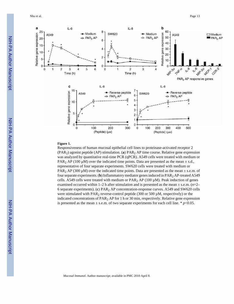
Figure 1.Responsiveness of human mucosal epithelial cell lines to proteinase-activated receptor 2(PAR2) agonist peptide (AP) stimulation. (a) PAR2 AP time course. Relative gene expressionwas analyzed by quantitative real-time PCR (qPCR). A549 cells were treated with medium orPAR2 AP (100 µM) over the indicated time points. Data are presented as the mean ± s.d.,representative of four separate experiments. SW620 cells were treated with medium orPAR2 AP (300 µM) over the indicated time points. Data are presented as the mean ± s.e.m. offour separate experiments. (b) Inflammatory mediator genes induced in PAR2 AP-treated A549cells. A549 cells were treated with medium or PAR2 AP (100 µM). Peak induction of genesexamined occurred within 1–2 h after stimulation and is presented as the mean ± s.e.m. (n=2–6 separate experiments). (c) PAR2 AP concentration-response curves. A549 and SW620 cellswere stimulated with PAR2 reverse-control peptide (300 or 500 µM, respectively) or theindicated concentrations of PAR2 AP for 1 h or 30 min, respectively. Relative gene expressionis presented as the mean ± s.e.m. of two separate experiments for each cell line. * p<0.05.
Nhu et al. Page 13
Mucosal Immunol. Author manuscript; available in PMC 2010 April 8.
NIH
-PA Author Manuscript
NIH
-PA Author Manuscript
NIH
-PA Author Manuscript
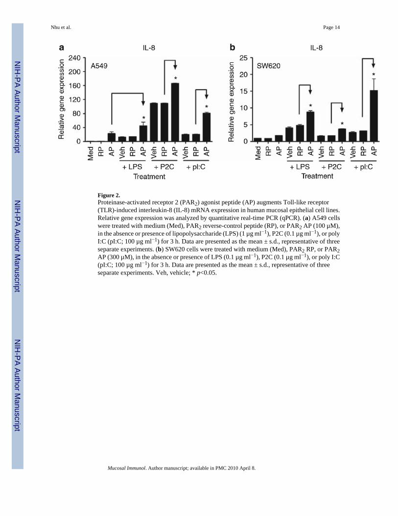
Figure 2.Proteinase-activated receptor 2 (PAR2) agonist peptide (AP) augments Toll-like receptor(TLR)-induced interleukin-8 (IL-8) mRNA expression in human mucosal epithelial cell lines.Relative gene expression was analyzed by quantitative real-time PCR (qPCR). (a) A549 cellswere treated with medium (Med), PAR2 reverse-control peptide (RP), or PAR2 AP (100 µM),in the absence or presence of lipopolysaccharide (LPS) (1 µg ml−1), P2C (0.1 µg ml−1), or polyI:C (pI:C; 100 µg ml−1) for 3 h. Data are presented as the mean ± s.d., representative of threeseparate experiments. (b) SW620 cells were treated with medium (Med), PAR2 RP, or PAR2AP (300 µM), in the absence or presence of LPS (0.1 µg ml−1), P2C (0.1 µg ml−1), or poly I:C(pI:C; 100 µg ml−1) for 3 h. Data are presented as the mean ± s.d., representative of threeseparate experiments. Veh, vehicle; * p<0.05.
Nhu et al. Page 14
Mucosal Immunol. Author manuscript; available in PMC 2010 April 8.
NIH
-PA Author Manuscript
NIH
-PA Author Manuscript
NIH
-PA Author Manuscript
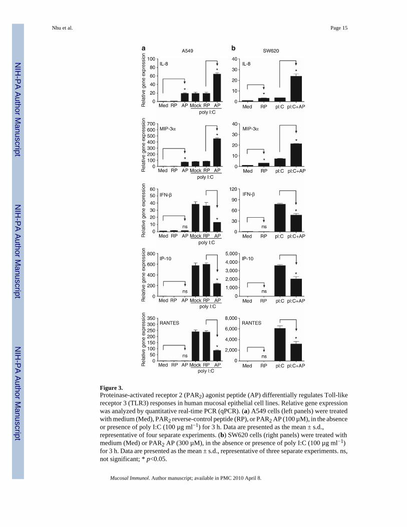
Figure 3.Proteinase-activated receptor 2 (PAR2) agonist peptide (AP) differentially regulates Toll-likereceptor 3 (TLR3) responses in human mucosal epithelial cell lines. Relative gene expressionwas analyzed by quantitative real-time PCR (qPCR). (a) A549 cells (left panels) were treatedwith medium (Med), PAR2 reverse-control peptide (RP), or PAR2 AP (100 µM), in the absenceor presence of poly I:C (100 µg ml−1) for 3 h. Data are presented as the mean ± s.d.,representative of four separate experiments. (b) SW620 cells (right panels) were treated withmedium (Med) or PAR2 AP (300 µM), in the absence or presence of poly I:C (100 µg ml−1)for 3 h. Data are presented as the mean ± s.d., representative of three separate experiments. ns,not significant; * p<0.05.
Nhu et al. Page 15
Mucosal Immunol. Author manuscript; available in PMC 2010 April 8.
NIH
-PA Author Manuscript
NIH
-PA Author Manuscript
NIH
-PA Author Manuscript
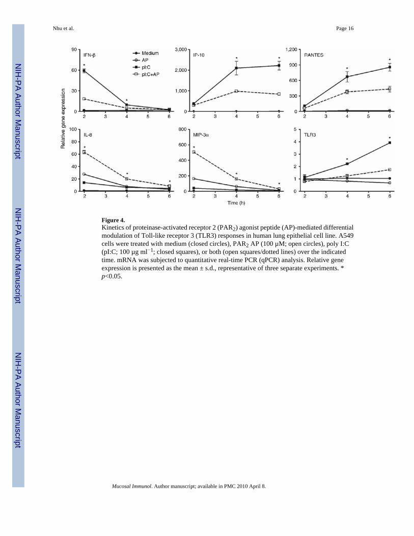
Figure 4.Kinetics of proteinase-activated receptor 2 (PAR2) agonist peptide (AP)-mediated differentialmodulation of Toll-like receptor 3 (TLR3) responses in human lung epithelial cell line. A549cells were treated with medium (closed circles), PAR2 AP (100 µM; open circles), poly I:C(pI:C; 100 µg ml−1; closed squares), or both (open squares/dotted lines) over the indicatedtime. mRNA was subjected to quantitative real-time PCR (qPCR) analysis. Relative geneexpression is presented as the mean ± s.d., representative of three separate experiments. *p<0.05.
Nhu et al. Page 16
Mucosal Immunol. Author manuscript; available in PMC 2010 April 8.
NIH
-PA Author Manuscript
NIH
-PA Author Manuscript
NIH
-PA Author Manuscript
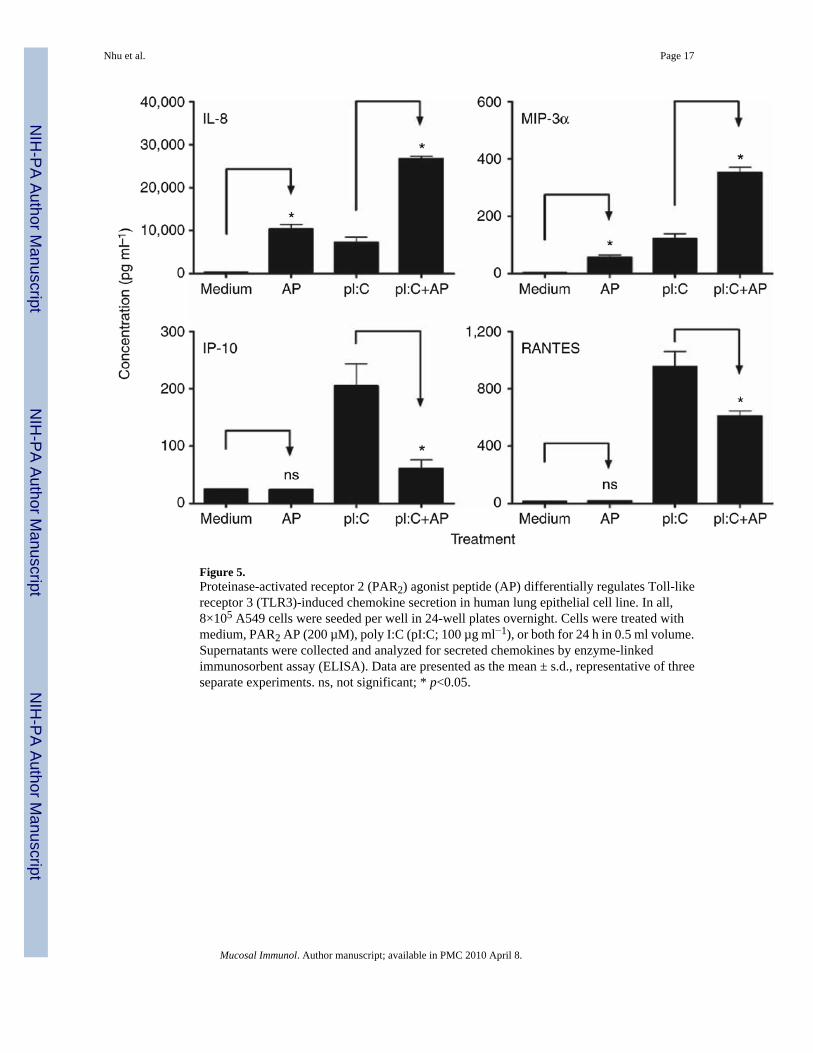
Figure 5.Proteinase-activated receptor 2 (PAR2) agonist peptide (AP) differentially regulates Toll-likereceptor 3 (TLR3)-induced chemokine secretion in human lung epithelial cell line. In all,8×105 A549 cells were seeded per well in 24-well plates overnight. Cells were treated withmedium, PAR2 AP (200 µM), poly I:C (pI:C; 100 µg ml−1), or both for 24 h in 0.5 ml volume.Supernatants were collected and analyzed for secreted chemokines by enzyme-linkedimmunosorbent assay (ELISA). Data are presented as the mean ± s.d., representative of threeseparate experiments. ns, not significant; * p<0.05.
Nhu et al. Page 17
Mucosal Immunol. Author manuscript; available in PMC 2010 April 8.
NIH
-PA Author Manuscript
NIH
-PA Author Manuscript
NIH
-PA Author Manuscript
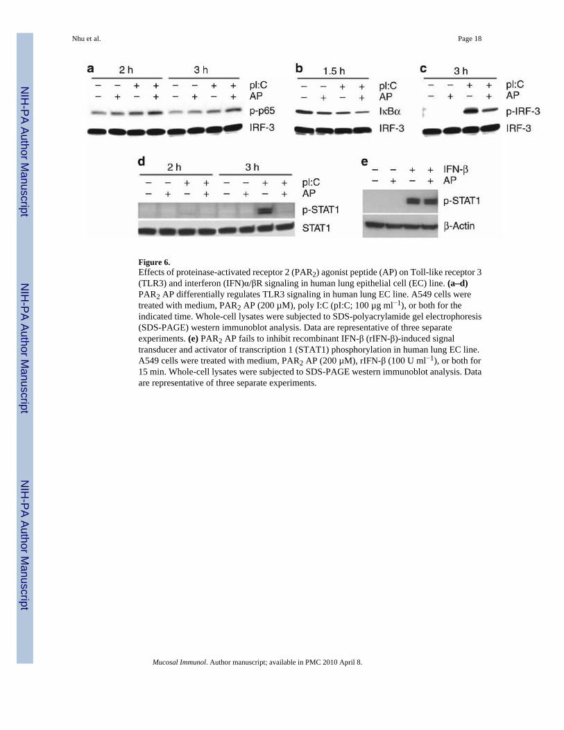
Figure 6.Effects of proteinase-activated receptor 2 (PAR2) agonist peptide (AP) on Toll-like receptor 3(TLR3) and interferon (IFN)α/βR signaling in human lung epithelial cell (EC) line. (a–d)PAR2 AP differentially regulates TLR3 signaling in human lung EC line. A549 cells weretreated with medium, PAR2 AP (200 µM), poly I:C (pI:C; 100 µg ml−1), or both for theindicated time. Whole-cell lysates were subjected to SDS-polyacrylamide gel electrophoresis(SDS-PAGE) western immunoblot analysis. Data are representative of three separateexperiments. (e) PAR2 AP fails to inhibit recombinant IFN-β (rIFN-β)-induced signaltransducer and activator of transcription 1 (STAT1) phosphorylation in human lung EC line.A549 cells were treated with medium, PAR2 AP (200 µM), rIFN-β (100 U ml−1), or both for15 min. Whole-cell lysates were subjected to SDS-PAGE western immunoblot analysis. Dataare representative of three separate experiments.
Nhu et al. Page 18
Mucosal Immunol. Author manuscript; available in PMC 2010 April 8.
NIH
-PA Author Manuscript
NIH
-PA Author Manuscript
NIH
-PA Author Manuscript
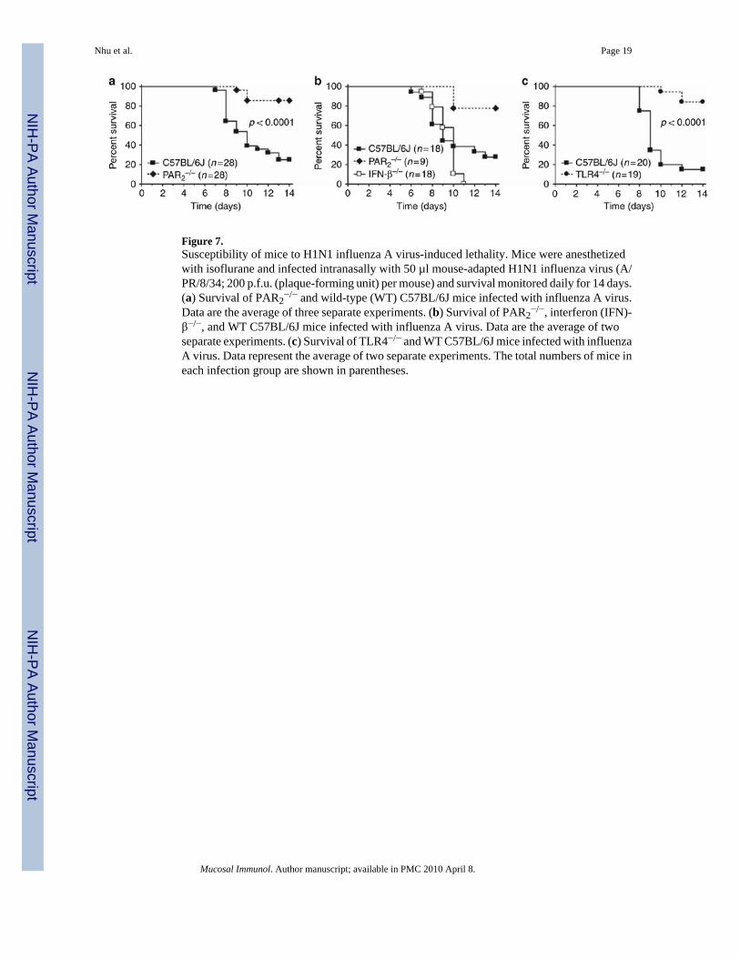
Figure 7.Susceptibility of mice to H1N1 influenza A virus-induced lethality. Mice were anesthetizedwith isoflurane and infected intranasally with 50 µl mouse-adapted H1N1 influenza virus (A/PR/8/34; 200 p.f.u. (plaque-forming unit) per mouse) and survival monitored daily for 14 days.(a) Survival of PAR2
−/− and wild-type (WT) C57BL/6J mice infected with influenza A virus.Data are the average of three separate experiments. (b) Survival of PAR2
−/−, interferon (IFN)-β−/−, and WT C57BL/6J mice infected with influenza A virus. Data are the average of twoseparate experiments. (c) Survival of TLR4−/− and WT C57BL/6J mice infected with influenzaA virus. Data represent the average of two separate experiments. The total numbers of mice ineach infection group are shown in parentheses.
Nhu et al. Page 19
Mucosal Immunol. Author manuscript; available in PMC 2010 April 8.
NIH
-PA Author Manuscript
NIH
-PA Author Manuscript
NIH
-PA Author Manuscript
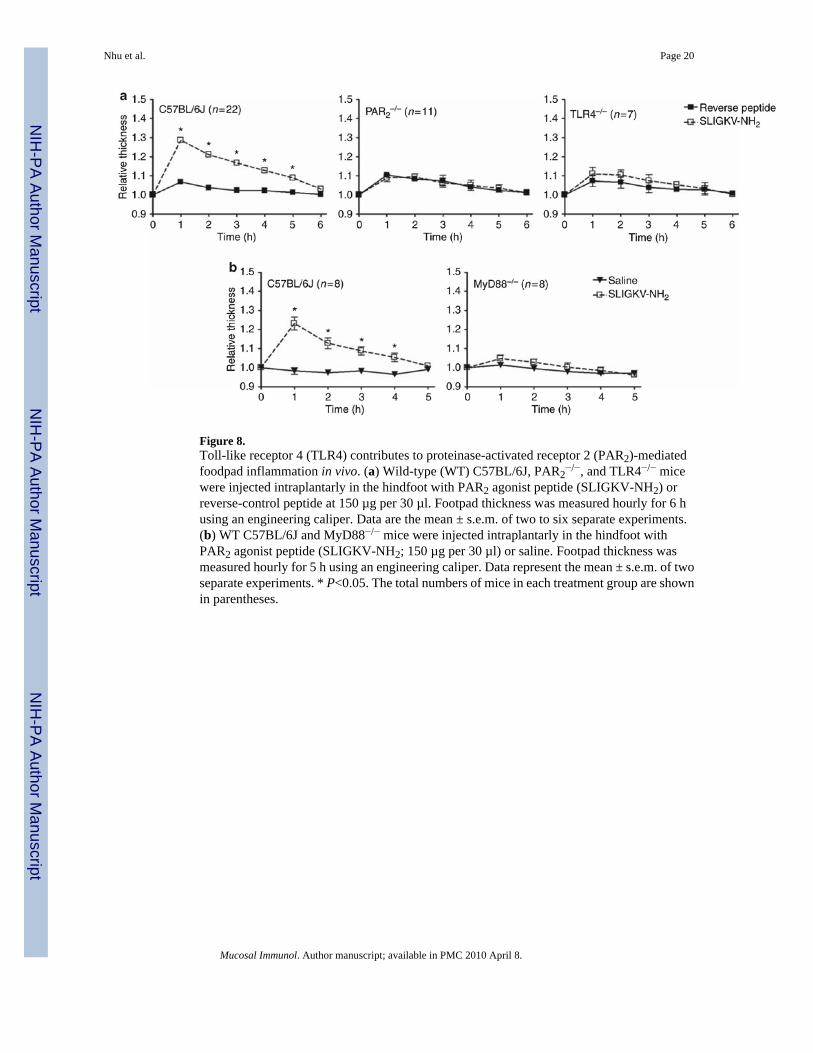
Figure 8.Toll-like receptor 4 (TLR4) contributes to proteinase-activated receptor 2 (PAR2)-mediatedfoodpad inflammation in vivo. (a) Wild-type (WT) C57BL/6J, PAR2
−/−, and TLR4−/− micewere injected intraplantarly in the hindfoot with PAR2 agonist peptide (SLIGKV-NH2) orreverse-control peptide at 150 µg per 30 µl. Footpad thickness was measured hourly for 6 husing an engineering caliper. Data are the mean ± s.e.m. of two to six separate experiments.(b) WT C57BL/6J and MyD88−/− mice were injected intraplantarly in the hindfoot withPAR2 agonist peptide (SLIGKV-NH2; 150 µg per 30 µl) or saline. Footpad thickness wasmeasured hourly for 5 h using an engineering caliper. Data represent the mean ± s.e.m. of twoseparate experiments. * P<0.05. The total numbers of mice in each treatment group are shownin parentheses.
Nhu et al. Page 20
Mucosal Immunol. Author manuscript; available in PMC 2010 April 8.
NIH
-PA Author Manuscript
NIH
-PA Author Manuscript
NIH
-PA Author Manuscript
NIH
-PA Author Manuscript
NIH
-PA Author Manuscript
NIH
-PA Author Manuscript
Nhu et al. Page 21
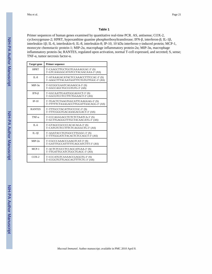
Table 1
Primer sequences of human genes examined by quantitative real-time PCR. AS, antisense; COX-2,cyclooxygenase-2; HPRT, hypoxanthine guanine phosphoribosyltransferase; IFN-β, interferon-β; IL-1β,interleukin-1β; IL-6, interleukin-6; IL-8, interleukin-8; IP-10, 10 kDa interferon-γ-induced protein; MCP-1,monocyte chemotactic protein-1; MIP-2α, macrophage inflammatory protein-2α; MIP-3α, macrophageinflammatory protein-3α; RANTES, regulated upon activation, normal T-cell expressed, and secreted; S, sense;TNF-α, tumor necrosis factor-α.
Target gene Primer sequence
HPRT 5’-CAAGCTTGCTGGTGAAAAGGAC-3’ (S)5’-GTCAAGGGCATATCCTACAACAAA-3’ (AS)
IL-8 5’-ATAAAGACATACTCCAAACCTTTCCAC-3’ (S)5’-AAGCTTTACAATAATTTCTGTGTTGGC-3’ (AS)
MIP-3α 5’-GCGGCGAATCAGAAGCA-3’ (S)5’-GGCCAGCTGCCGTGTG-3’ (AS)
IFN-β 5’-GGCAATTGAATGGGAGGCT-3’ (S)5’-GGCGTCCTCCTTCTGGAACT-3’ (AS)
IP-10 5’-TGACTCTAAGTGGCATTCAAGGAG-3’ (S)5’-TTTTTCTAAAGACCTTGGATTAACAGG-3’ (AS)
RANTES 5’-TTTGCCTACATTGCCCGC-3’ (S)5’-TTTCGGGTGACAAAGACGACT-3’ (AS)
TNF-α 5’-CCCAGGGACCTCTCTCTAATCA-3’ (S)5’-GCTTGAGGGTTTGCTACAACATG-3’ (AS)
IL-6 5’-GTAGCCGCCCCACACAGA-3’ (S)5’-CATGTCTCCTTTCTCAGGGCTG-3’ (AS)
IL-1β 5’-AAATACCTGTGGCCTTGGGC-3’ (S)5’-TTTGGGATCTACACTCTCCAGCT-3’ (AS)
MIP-2α 5’-CGCCCAAACCGAAGTCAT-3’ (S)5’-GATTTGCCATTTTTCAGCATCTTT-3’ (AS)
MCP-1 5’-ACTCTCGCCTCCAGCATGAA-3’ (S)5’-TTGATTGCATCTGGCTGAGC-3’ (AS)
COX-2 5’-CCCATGTCAAAACCGAGGTG-3’ (S)5’-CCGGTGTTGAGCAGTTTTCTC-3’ (AS)
Mucosal Immunol. Author manuscript; available in PMC 2010 April 8.