Implantation Serine Proteinase 1 Exhibits Mixed Substrate Specificity that Silences Signaling via...
13
Implantation Serine Proteinase 1 Exhibits Mixed Substrate Specificity that Silences Signaling via Proteinase-Activated Receptors Navneet Sharma 1 *, Rajeev Kumar 1 , Bernard Renaux 2 , Mahmoud Saifeddine 2 , Sandra Nishikawa 1 , Koichiro Mihara 2 , Rithwik Ramachandran 2 , Morley D. Hollenberg 2,3 , Derrick E. Rancourt 1 1 Department of Biochemistry and Molecular Biology, University of Calgary, Calgary, Canada, 2 Department of Physiology and Pharmacology University of Calgary, Calgary, Canada, 3 Department of Medicine, Faculty of Medicine, University of Calgary, Calgary, Canada Abstract Implantation S1 family serine proteinases (ISPs) are tryptases involved in embryo hatching and uterine implantation in the mouse. The two different ISP proteins (ISP1 and ISP2) have been detected in both pre- and post-implantation embryo tissue. To date, native ISP obtained from uterus and blastocyst tissues has been isolated only as an active hetero-dimer that exhibits trypsin-like substrate specificity. We hypothesised that in isolation, ISP1 might have a unique substrate specificity that could relate to its role when expressed alone in individual tissues. Thus, we isolated recombinant ISP1 expressed in Pichia pastoris and evaluated its substrate specificity. Using several chromogenic substrates and serine proteinase inhibitors, we demonstrate that ISP1 exhibits trypsin-like substrate specificity, having a preference for lysine over arginine at the P1 position. Phage display peptide mimetics revealed an expanded but mixed substrate specificity of ISP1, including chymotryptic and elastase activity. Based upon targets observed using phage display, we hypothesised that ISP1 might signal to cells by cleaving and activating proteinase-activated receptors (PARs) and therefore assessed PARs 1, 2 and 4 as potential ISP1 targets. We observed that ISP1 silenced enzyme-triggered PAR signaling by receptor-disarming. This PAR- disarming action of ISP1 may be important for embryo development and implantation. Citation: Sharma N, Kumar R, Renaux B, Saifeddine M, Nishikawa S, et al. (2011) Implantation Serine Proteinase 1 Exhibits Mixed Substrate Specificity that Silences Signaling via Proteinase-Activated Receptors. PLoS ONE 6(11): e27888. doi:10.1371/journal.pone.0027888 Editor: Haibin Wang, State Key Laboratory of Reproductive Biology, Institute of Zoology, Chinese Academy of Sciences, China Received June 13, 2011; Accepted October 27, 2011; Published November 23, 2011 Copyright: ß 2011 Sharma et al. This is an open-access article distributed under the terms of the Creative Commons Attribution License, which permits unrestricted use, distribution, and reproduction in any medium, provided the original author and source are credited. Funding: This work was supported by Canadian Institutes of Health Research Operating Grants awarded to DR and MH, who contributed equally to this project. DR is an AHFMR Senior Scholar. The funders had no role in study design, data collection and analysis, decision to publish, or preparation of the manuscript. Competing Interests: The authors have declared that no competing interests exist. * E-mail: [email protected] Introduction The implantation serine proteinases, ISP1 & 2, are two related S1-family serine proteinases that are tandemly localized in a cluster of tryptase genes found on mouse chromosome 17A3.3 [1]. Unlike many of the other tryptases, which are found primarily in mast cells, the ISPs are expressed in the embryo and the uterine decidua during the time of embryo implantation [2]. The first ISP gene to be characterized (ISP1) was initially detected in the pre- implantation embryo [3]. Anti-sense RNA disruption of ISP1 gene expression prevented embryo hatching and outgrowth in vitro [3]. Subsequently, ISP1 and ISP2 gene expression was detected in the uterine endometrial glands during the ‘window of implantation’ [4,5]. Artificial pregnancy experiments demonstrate that both ISP genes are up-regulated by progesterone [4,5]. Both ISPs are secreted from the endometrial glands into uterine fluid on day 4 of pregnancy, just prior to the commencement of implantation [6]. This appearance of enzyme in the glands and uterine fluid is negatively regulated by estrogen, such that both ISP proteins appear in the uterine fluid shortly after the estrogen spike synchronizes uterine-embryo receptivity to enable the commence- ment of implantation [6]. Interestingly, ISP2 antibodies have been found to abrogate murine embryo implantation, verifying an important role for ISPs [7]. Synthetic inhibitors of ISP activity have also demonstrated a potential role for ISPs in the early stages of murine embryo invasion in vitro and implantation in vivo [2,8]. ISP protein and proteolytic activity are found in uterine fluid at sites localized to embryo invasion [1,3]. The cognate tryptase is minimally a hetero-dimer comprised of both ISP1 and ISP2 en- zymes, although homo-dimers have not been ruled out [2]. Preliminary studies using chromogenic p-nitroanilide conjugat- ed synthetic peptide substrates have indicated that the ISP1/ISP2 hetero-dimer has ‘trypsin-like’ specificity [2]. Recent studies em- ploying a bacteriophage display of small peptides have not only confirmed this substrate specificity but have also demonstrated a preference for non-polar amino acid residues at the P19 as well as at the P2 positions [9]. Since these studies were performed using purified native ISP1/ISP2 enzyme complex [2], we have been interested in expressing recombinant ISPs in order to investigate the activity of each individual enzyme and to pursue enzyme structure-activity studies. A number of different high throughput screening methods including libraries of chromogenic or fluorogenic substrates (in solution and/or on chip), have been devised to determine the substrate specificity of proteinases. Phage display has been de- veloped as a more efficient and unbiased approach. So far, the phage display technology has been employed for displaying peptides/proteins for many different applications, including: (a) PLoS ONE | www.plosone.org 1 November 2011 | Volume 6 | Issue 11 | e27888
-
Upload
independent -
Category
Documents
-
view
1 -
download
0
Transcript of Implantation Serine Proteinase 1 Exhibits Mixed Substrate Specificity that Silences Signaling via...

Implantation Serine Proteinase 1 Exhibits MixedSubstrate Specificity that Silences Signaling viaProteinase-Activated ReceptorsNavneet Sharma1*, Rajeev Kumar1, Bernard Renaux2, Mahmoud Saifeddine2, Sandra Nishikawa1,
Koichiro Mihara2, Rithwik Ramachandran2, Morley D. Hollenberg2,3, Derrick E. Rancourt1
1 Department of Biochemistry and Molecular Biology, University of Calgary, Calgary, Canada, 2 Department of Physiology and Pharmacology University of Calgary,
Calgary, Canada, 3 Department of Medicine, Faculty of Medicine, University of Calgary, Calgary, Canada
Abstract
Implantation S1 family serine proteinases (ISPs) are tryptases involved in embryo hatching and uterine implantation in themouse. The two different ISP proteins (ISP1 and ISP2) have been detected in both pre- and post-implantation embryo tissue.To date, native ISP obtained from uterus and blastocyst tissues has been isolated only as an active hetero-dimer thatexhibits trypsin-like substrate specificity. We hypothesised that in isolation, ISP1 might have a unique substrate specificitythat could relate to its role when expressed alone in individual tissues. Thus, we isolated recombinant ISP1 expressed inPichia pastoris and evaluated its substrate specificity. Using several chromogenic substrates and serine proteinase inhibitors,we demonstrate that ISP1 exhibits trypsin-like substrate specificity, having a preference for lysine over arginine at the P1position. Phage display peptide mimetics revealed an expanded but mixed substrate specificity of ISP1, includingchymotryptic and elastase activity. Based upon targets observed using phage display, we hypothesised that ISP1 mightsignal to cells by cleaving and activating proteinase-activated receptors (PARs) and therefore assessed PARs 1, 2 and 4 aspotential ISP1 targets. We observed that ISP1 silenced enzyme-triggered PAR signaling by receptor-disarming. This PAR-disarming action of ISP1 may be important for embryo development and implantation.
Citation: Sharma N, Kumar R, Renaux B, Saifeddine M, Nishikawa S, et al. (2011) Implantation Serine Proteinase 1 Exhibits Mixed Substrate Specificity that SilencesSignaling via Proteinase-Activated Receptors. PLoS ONE 6(11): e27888. doi:10.1371/journal.pone.0027888
Editor: Haibin Wang, State Key Laboratory of Reproductive Biology, Institute of Zoology, Chinese Academy of Sciences, China
Received June 13, 2011; Accepted October 27, 2011; Published November 23, 2011
Copyright: � 2011 Sharma et al. This is an open-access article distributed under the terms of the Creative Commons Attribution License, which permitsunrestricted use, distribution, and reproduction in any medium, provided the original author and source are credited.
Funding: This work was supported by Canadian Institutes of Health Research Operating Grants awarded to DR and MH, who contributed equally to this project.DR is an AHFMR Senior Scholar. The funders had no role in study design, data collection and analysis, decision to publish, or preparation of the manuscript.
Competing Interests: The authors have declared that no competing interests exist.
* E-mail: [email protected]
Introduction
The implantation serine proteinases, ISP1 & 2, are two related
S1-family serine proteinases that are tandemly localized in a
cluster of tryptase genes found on mouse chromosome 17A3.3 [1].
Unlike many of the other tryptases, which are found primarily in
mast cells, the ISPs are expressed in the embryo and the uterine
decidua during the time of embryo implantation [2]. The first ISP
gene to be characterized (ISP1) was initially detected in the pre-
implantation embryo [3]. Anti-sense RNA disruption of ISP1 gene
expression prevented embryo hatching and outgrowth in vitro [3].
Subsequently, ISP1 and ISP2 gene expression was detected in the
uterine endometrial glands during the ‘window of implantation’ [4,5].
Artificial pregnancy experiments demonstrate that both ISP genes
are up-regulated by progesterone [4,5]. Both ISPs are secreted
from the endometrial glands into uterine fluid on day 4 of
pregnancy, just prior to the commencement of implantation [6].
This appearance of enzyme in the glands and uterine fluid is
negatively regulated by estrogen, such that both ISP proteins
appear in the uterine fluid shortly after the estrogen spike
synchronizes uterine-embryo receptivity to enable the commence-
ment of implantation [6]. Interestingly, ISP2 antibodies have been
found to abrogate murine embryo implantation, verifying an
important role for ISPs [7]. Synthetic inhibitors of ISP activity
have also demonstrated a potential role for ISPs in the early stages
of murine embryo invasion in vitro and implantation in vivo [2,8].
ISP protein and proteolytic activity are found in uterine fluid at
sites localized to embryo invasion [1,3]. The cognate tryptase is
minimally a hetero-dimer comprised of both ISP1 and ISP2 en-
zymes, although homo-dimers have not been ruled out [2].
Preliminary studies using chromogenic p-nitroanilide conjugat-
ed synthetic peptide substrates have indicated that the ISP1/ISP2
hetero-dimer has ‘trypsin-like’ specificity [2]. Recent studies em-
ploying a bacteriophage display of small peptides have not only
confirmed this substrate specificity but have also demonstrated a
preference for non-polar amino acid residues at the P19 as well as
at the P2 positions [9]. Since these studies were performed using
purified native ISP1/ISP2 enzyme complex [2], we have been
interested in expressing recombinant ISPs in order to investigate
the activity of each individual enzyme and to pursue enzyme
structure-activity studies.
A number of different high throughput screening methods
including libraries of chromogenic or fluorogenic substrates (in
solution and/or on chip), have been devised to determine the
substrate specificity of proteinases. Phage display has been de-
veloped as a more efficient and unbiased approach. So far, the
phage display technology has been employed for displaying
peptides/proteins for many different applications, including: (a)
PLoS ONE | www.plosone.org 1 November 2011 | Volume 6 | Issue 11 | e27888

generating highly specific antibodies, (b) studying protein-protein
interaction and (c) evaluating the substrate specificity of protein-
ases [10,11,12]. The T7 bacteriophage display system has been
employed successfully for determining the substrate specificities of
rat mast cell proteinases 4 and 5 [13,14], the native implantation
serine proteinase ISP1/ISP2 hetero-dimeric complex and human
kallikrein 6 [9].
Many physiological responses mediated by serine proteinases can
occur by cleaving and activating the PAR family of G-protein
coupled receptors (GPCRs) [15–21]. The four members of the PAR
family, PARs 1 to 4, have a unique mechanism of activation that
distinguishes them from other seven transmembrane GPCRs. PARs
carry their own activating molecule in a masked state and receptor
activation is achieved through proteolytic cleavage at a specific
arginine site within the receptor N-terminus to reveal a cryptic
tethered ligand that activates the receptor [15,17,18,21]. In addition,
PARs, with the exception of PAR3, are also activated by short
synthetic peptides (PAR-activating peptides, or PAR-APs) derived
from the sequences of the proteolytically revealed tethered ligand
[19,20]. In addition to the cleavage/activation of PARs, proteinases
can also negatively regulate signaling via the PARs by ‘disarming’
the receptor through cleavage at a downstream non-receptor ac-
tivating site to remove the tethered ligand. These truncated re-
ceptors, refractory to activation by their target enzymes (e.g. trypsin
for PAR2) are therefore unable to signal in a physiological setting;
but nonetheless remain responsive to PAR-APs [21].
In this study, we have used the methylotrophic yeast Pichia
pastoris in order to express recombinant ISP1, also known as
Mouse Prss28. Our aim was to evaluate the substrate specificity of
this enzyme acting on its own, in the absence of ISP2. Our data
demonstrate that recombinant ISP1 can exist in a monomeric
form. To evaluate the substrate preference of monomeric ISP1, we
studied: (a) the kinetics of cleavage of several small chromogenic
synthetic peptide substrates, (b) the effects of serine proteinase
inhibitors on this activity, (c) cleavage of a random hexameric
library of phage displayed peptides and (d) cleavage of synthetic
peptides with sequences based on the results obtained from the
phage display approach. Finally, in view of the tryptic activity of
ISP, we hypothesised that ISP1 could regulate PAR activity. Thus,
we also assessed the ability of the enzyme: (a) to regulate the
activity of PARs 1, 2 and 4 and (b) to cleave peptide sequences
derived from the cleavage-activation domain and from extracel-
lular loop-2 (ECL2) of PAR2, as we had done previously for trypsin
IV [22]. Our data indicate that the ISP1 monomer has mixed
substrate specificity with tryptic, chymotryptic and elastase cha-
racteristics and that ISP1 can target the PARs primarily by
disarming them. These actions of ISP1 may enable it to play a
physiological role in murine development or embryo implantation.
Results
Expression and Purification of recombinant ISP1Although the full length cDNA sequence of ISP1 suggests that it
is secreted as a pro-enzyme, we have previously only detected its
mature enzymatically active form as a complex with ISP2 (9),
when isolated from uterine fluid. Based upon this previous
observation, we sought to express the enzymatically active mature
form of ISP1 in the Pichia expression system using a protease
deficient strain of Pichia pastoris. As mentioned, the designed cDNA
was designed to generate the active enzyme, with an N-terminal
sequence (IVGG) found in active tryptases [4]. Accordingly we
introduced an in-frame fusion of mature protein cDNA sequence,
so as to place the mature protein downstream of the Pichia signal
peptide sequence in the vector PICZaB.
Recombinant ISP1 expression was seen after approximately
50 hours of fermentation and peaked at approximately 100 hours
(Figure S1C). The growth profile of the organism was also de-
monstrated by measuring packed cell volume (Figure S1A). A
steady rise in growth was observed after 36 hours of fermentation
until the end of the run. No difference in the fermentation para-
meters and expression profile was observed in the transition from
1.0 L to 10.0 L scale-up. Therefore these parameters may be
considered as optimum for the expression of rISP1 in Pichia pastoris.
Figure 1 shows the FPLC chromatograms obtained upon puri-
fication of rISP1 with DEAE Sepharose (Figure 1A) and Superdex-
75 (Figure 1B) columns. As seen in Figure 1A, ISP1 was eluted at
the last step of the elution buffer gradient (100% Buffer B). A
substantial amount of high molecular weight as well as low mole-
cular weight non-ISP1 impurities were separated in the subsequent
gel filtration step as shown in Figure 1B. Although only 3.9 fold
purification occurred in the ion exchange chromatography step, a
100-fold purification was achieved in the gel filtration steps
(Table 1 in Data S1). In total, a 340-fold purification was achieved
by following this methodology. Hence, the specific activity of
recombinant ISP1 increased from 0.54 units/mg protein to 180
units/mg. Figures 1C and 1D show the total protein profile of
different fractions of ISP1, before and after purification, con-
firming the results shown in Table 1. The gels shown in Figure 1C
(Lane 5) and 1D also demonstrate that the molecular weight of
ISP1 monomer is ,34 kDa. This size corresponded to the enzyme
detected by western blot analysis (Figure 1E). Since the protein
elutes at this size (,34 kDa) upon gel filtration chromatography
(calibration plot not shown), it appears that the recombinant
ISP1 exists as a monomeric protein under the conditions of
chromatography.
The purified ISP1 was also analyzed by native polyacrylamide
gel electrophoresis run under non-reducing and non-denaturing
conditions. Only one major band was observed (Figure 1F). As
mentioned above, we were also able to purify rISP1 using a Hitrap
Benzamidine affinity chromatography column, to yield a single
coomassie-stained band upon electrophoresis that co-migrated
with ISP1 immunoreactivity on a western blot (Fig. 1C, Lane 5
and Fig. 1E for the western blot detection). However the protein
recovered from the benzamidine column was enzymatically in-
active and we believe that the column elution conditions dena-
tured the protein. Thus, the affinity-purified enzyme could not be
used for functional studies. However, in accord with the gels and
ABP western blot data shown in Figure 1, the combined ion-
exchange/gel filtration purification procedure yielded a homo-
genously pure ISP1 preparation that generated only a single
biotin-labelled band when reacted with the activity based serine
proteinase probe, biotinyl-linker-Pro-Lys-diphenylphosphonate
(Bio-PK-DPP 4) [23–25] (Figure 1G, Lane 2). Thus, the enzyme
preparation obtained from the gel filtration column, containing a
single enzymatically active proteinase, was of sufficient purity
(180 U/mg) to be used for all further studies. The identity of the
purified enzyme was also confirmed by Mass Spectrometry (LC-
MS/MS) from Institute of Biodiagnostics (IBD), University of
Alberta (Data S1).
Enzyme activity measurements with different substratesand inhibitors
Various chromogenic peptide substrates conjugated with p-
nitroanilide/4-methoxy naphthylamide were used to characterize
the catalytic nature of rISP1 (Table 1). Amongst the different sub-
strates tested, only the ones having an Arginine/Lysine at the P1
position were hydrolyzed, namely N-Benzoyl-L- arginine p-nitroani-
lide hydrochloride (BAPNA), N-CBZ-arginine-arginine-arginine-4
ISP 1 Exhibits Mixed Substrate Specificity
PLoS ONE | www.plosone.org 2 November 2011 | Volume 6 | Issue 11 | e27888

-methoxy naphthylamide (RRRmN), N-Benzoyl-isoleucine-phenyl-
alanine-lysine p-nitroanilide hydrochloride (IFK-pNA) and N-
Benzoyl-phenylalanine-valine-arginine p-nitroanilide hydrochloride
(FVR-pNA). Towards IFK-pNA, ISP1 showed highest Km (9.6 mM)
and hence the lowest affinity followed by BAPNA (Km = 0.61 mM),
FVR-pNA (Km = 0.36 mM) and RRRmN (Km = ,0.1 mM). The
kcat and kcat/Km values are also shown in Table 1. Interestingly no
enzyme activity was observed with H-D-valine-leucine-lysine p-
nitroanilide hydrochloride (VLK-pNA) N(p-tosyl)-glycine-proline-
lysine p-nitroanilide acetate (GPK-pNA) and Valine-Phenylalanine-
Lysine p-nitroanilide (VFK-pNA). Similarly no reaction was observed
upon using Alanine p-nitroanilide (A-pNA) and N-Succinyl-alanine-
alanine-alanine p-nitroanilide (AAA-pNA).
In order to validate the results obtained with small chromogenic
substrates and to identify specific inhibitors, many general serine
protease inhibitors were tested employing 2 mM BAPNA as well
as 150 mM VPR-AMC as substrate (Table 2). As expected, PMSF,
a-1-Antitrypsin, Chymostatin, Gabexate mesylate and Soybean
Trypsin Inhibitor (STI) and TLCK inhibited the enzyme activity
of rISP1. Benzamidine hydrochloride, Leupeptin, Aprotinin and
recombinant human Secretory Leukocyte Protease Inhibitor
(rhSLPI) showed no noticeable effect.
Substrate specificity determination of ISP 1 with phagedisplay
2Having observed trypsin-like substrate specificity with small
chromogenic substrates, we next turned to the phage-display
approach, as outlined in Experimental Procedures, to assess ISP1
substrate specificity. The non-aligned unbiased amino acid
sequences deduced from the DNA sequences of the random
hexameric insert region from 24 plaques obtained from the
panning procedure are listed in Table 3. The data show that out of
24 plaque sequences, 20 (83%) had at least one arginine/lysine
residue in their sequences (Table 3, bold italic letters).
Figure 1. Purification of recombinant ISP1. (A) Ion exchange (DEAE-Sepharose) chromatography of harvested fermentation broth supernatantshowing elution of rISP1 by a step gradient ranging from 0–0.5 M Sodium Chloride, (B) Gel filtration chromatography of the ion exchange-purifiedfractions of rISP1 (the arrow points towards the ISP1 fraction with the inset showing western blot results obtained using monoclonal anti-ISP1antibodies), (C) Coomassie stained SDS-PAGE (10%) showing purification profile of fractions obtained at different steps of purification, lane 1 –Molecular weight marker (M/S Fermentas), lane 2 – fermentation broth supernatant (after removing cells), lane 3 – Eluted rISP1 fraction obtained afterIon exchange chromatography, lane 4 – Pooled fractions obtained after Superdex-200 Gel filtration chromatography step, lane 5 – Fraction obtainedafter purification with affinity chromatography (Benzamidine-Sepharose column), (D) Lane showing purified preparation of rISP1 obtained after 2nd
step of Gel Filtration Chromatography (Superdex-75), (E)Western blot analysis of purified rISP1 fraction using monoclonal anti-ISP1 antibodies, (F)Coomassie stained 8% native PAGE showing rISP1 as the major protein in the purified fraction after purification through ion exchange followed bygel filtration chromatography (G) Detection of ISP1 by activity based probe visualised by ECL plus, lane 1–20 mU Trypsin, lane 2–32 mU ISP1.doi:10.1371/journal.pone.0027888.g001
ISP 1 Exhibits Mixed Substrate Specificity
PLoS ONE | www.plosone.org 3 November 2011 | Volume 6 | Issue 11 | e27888

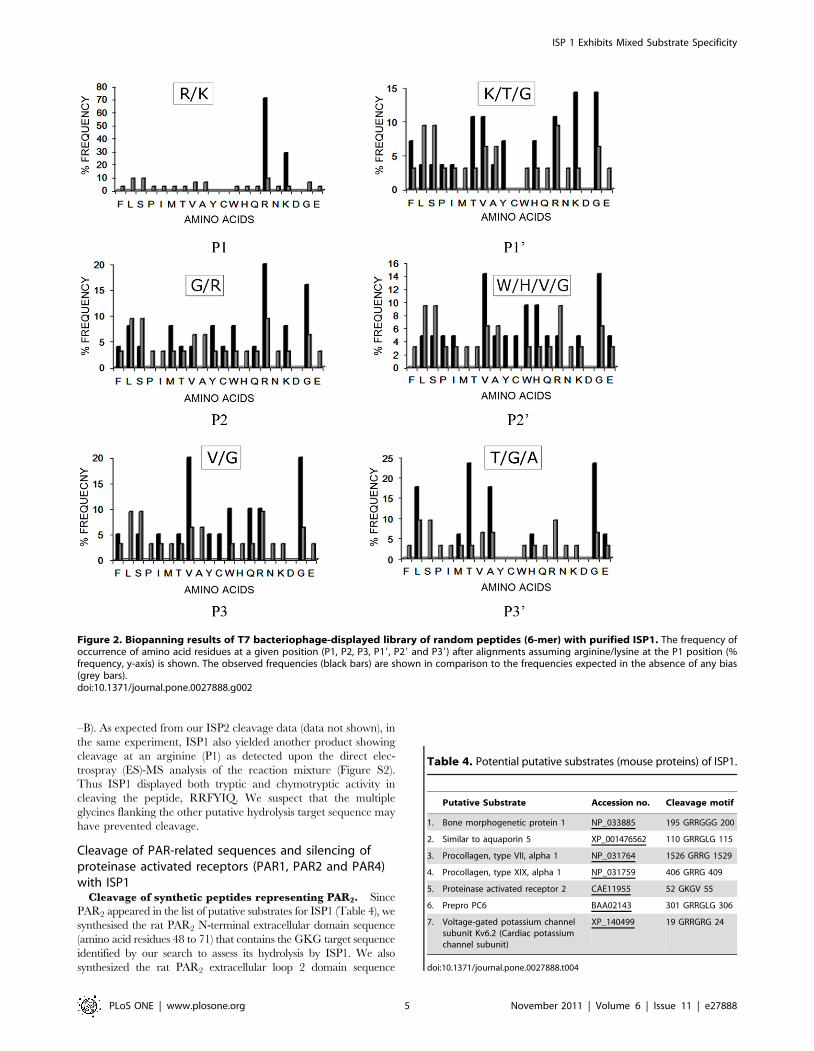
The biopanning data presented here validate in an unbiased
way, our findings with the small chromogenic substrates in-
dicating a trypsin-like activity. The biopanning data also
demonstrated the presence of arginine/lysine residues in 71%
of the phage plaques. Accordingly, resulting plaque sequences
were aligned assuming an arginine/lysine residue at the
P1 position. Upon analysis, lysine was found to be highly
preferred over arginine residue at P1 position. Ratio of Arginine
(Observed/Expected)/Lysine (Observed/Expected) is 1.28 in com-
parison to the same ratio of 0.38 in case of ISP1-ISP2 hetero-
dimer reported earlier [9]. The frequency of occurrence of an
amino acid at a given position (i.e. P1, P19, P2…..) in
comparison with its random probability at that position is
shown in Figure 2. Based on the above analysis, the following
preferred recognition/cleavage sequence motif was identified
for the recombinant ISP1:
P3 P2 P1 P19 P29 P39
V/G G/R R/K K/T/G W/H/V/G T/G/A
As already mentioned these results validate the results presented
above with synthetic chromogenic substrates and in addition
demonstrate a preference for dibasic residues around the cleavage
site at positions in the vicinity of cleavage site. To identify putative
substrates of ISP1, the above generated consensus target amino
acid sequence was compared with the NCBI non-redundant data-
base in different permutations and combinations. A list of probable
hits obtained with high degree of confidence is shown in Table 4,
wherein PAR2 surfaced as a possible enzyme target. In order to
validate our phage display results, we synthesized a number of
peptides based on the above results. To facilitate the identification
of the cleavage products by MALDI-TOF, the selected sequences
were flanked by glycine residues: a) GGGGRGWGGG; b)
GGGGKKGGGG; c) GGGGRKHGGG; d) GGVRKKVGGG.
These peptides were subjected to cleavage in vitro with ISP1 as
described in the methods section. To our surprise, although a
number of these peptides showed some cleavage according to the
HPLC analysis, we were unable to identify their sequences via
MALDI-TOF analysis (data not shown). On the other hand, a
target peptide (RRFYIQ) known to be cleaved by ISP2 (data not
shown) was also cleaved by ISP1. The major ISP1 cleavage site of
this peptide was found to be at a tyrosine (P1) residue (Figure 3A
Table 1. Turnover rate (kcat), Michaelis Menton Constant(Km) and kcat/Km ratio of recombinant ISP1 for somesynthetic chromogenic substrates.
Substrate
TurnoverNo. (kcat)sec21
MichaelisMentonConstant(Km) mM
kcat/Km(sec21 mM21)
1. N-Benzoyl Argininep-nitroanilide (BAPNA)
2.2 0.61 3.6
2. N-CBZ-Arginine-Arginine-Arginine-4 methoxynaphthylamide
0.7 0.099 7.1
3. Isoleucine-Phenylalanine-Lysine p-nitroanilide
3.9 9.6 0.41
4. Phenylalanine-Valine-Arginine p-nitroanilide
0.14 0.36 0.39
5. Valine-Leucine-Lysinep-nitroanilide
Nil Nil Nil
6. Tosyl-Glycine-Proline-Lysine p-nitroanilide
Nil Nil Nil
7. Succinyl-Alanine-Alanine-Alanine p-nitro anilide
Nil Nil Nil
8. Alanine p-nitroanilide Nil Nil Nil
9. Valine-Phenylalanine-Lysine p-nitroanilide
Nil Nil Nil
doi:10.1371/journal.pone.0027888.t001
Table 2. Dissociation constants (Apparent Ki) of generalserine proteinase inhibitors for a single substrate reaction ofrecombinant ISP1.
Inhibitor Substrate Ki (mM)
1. PMSF VPR-AMC (0.15 mM) 340
2. a-1-Antitrypsin BAPNA (2 mM) 2.0
3. Chymostatin BAPNA (2 mM) 220
4. Gabexate mesylate BAPNA (2 mM) 1700
5. Soybean Trypsin Inhibitor BAPNA (2 mM) 2.4
6. Benzamidine Hydrochloride BAPNA (2 mM) No inhibition
7. Leupeptin BAPNA (2 mM) No inhibition
8. TLCK VPR-AMC (0.15 mM) 30
9. Aprotinin BAPNA (2 mM) No inhibition
10. Recombinant human SLPI BAPNA (2 mM) No inhibition
doi:10.1371/journal.pone.0027888.t002
Table 3. Plaque sequences of random hexamer regionobtained after biopanning with recombinant ISP1.
Plaque No. AA1 AA2 AA3 AA4 AA5 AA6
1. P S T R K K
2. T G G W R F
3. Q W P M L V
4. W S G Q D I
5. Y R R G H L
6. R Y R T S L
7. R Y R T S L
8. S W G R R G
9. V R K V A A
10. V R K V A A
11. T G G W R F
12. S R S C T A
13. L E A L V V
14. S A C L T G
15. C L K T E G
16. F R R P G G
17. R G W G T V
18. L R Y V T Y
19. G G R V V T
20. R V Y L L S
21. R A Q M G E
22. P V Q K K H
23. V G R H L A
24. K G W E G C
doi:10.1371/journal.pone.0027888.t003
ISP 1 Exhibits Mixed Substrate Specificity
PLoS ONE | www.plosone.org 4 November 2011 | Volume 6 | Issue 11 | e27888
–B). As expected from our ISP2 cleavage data (data not shown), in
the same experiment, ISP1 also yielded another product showing
cleavage at an arginine (P1) as detected upon the direct elec-
trospray (ES)-MS analysis of the reaction mixture (Figure S2).
Thus ISP1 displayed both tryptic and chymotryptic activity in
cleaving the peptide, RRFYIQ. We suspect that the multiple
glycines flanking the other putative hydrolysis target sequence may
have prevented cleavage.
Cleavage of PAR-related sequences and silencing ofproteinase activated receptors (PAR1, PAR2 and PAR4)with ISP1
Cleavage of synthetic peptides representing PAR2. Since
PAR2 appeared in the list of putative substrates for ISP1 (Table 4), we
synthesised the rat PAR2 N-terminal extracellular domain sequence
(amino acid residues 48 to 71) that contains the GKG target sequence
identified by our search to assess its hydrolysis by ISP1. We also
synthesized the rat PAR2 extracellular loop 2 domain sequence
Figure 2. Biopanning results of T7 bacteriophage-displayed library of random peptides (6-mer) with purified ISP1. The frequency ofoccurrence of amino acid residues at a given position (P1, P2, P3, P19, P29 and P39) after alignments assuming arginine/lysine at the P1 position (%frequency, y-axis) is shown. The observed frequencies (black bars) are shown in comparison to the frequencies expected in the absence of any bias(grey bars).doi:10.1371/journal.pone.0027888.g002
Table 4. Potential putative substrates (mouse proteins) of ISP1.
Putative Substrate Accession no. Cleavage motif
1. Bone morphogenetic protein 1 NP_033885 195 GRRGGG 200
2. Similar to aquaporin 5 XP_001476562 110 GRRGLG 115
3. Procollagen, type VII, alpha 1 NP_031764 1526 GRRG 1529
4. Procollagen, type XIX, alpha 1 NP_031759 406 GRRG 409
5. Proteinase activated receptor 2 CAE11955 52 GKGV 55
6. Prepro PC6 BAA02143 301 GRRGLG 306
7. Voltage-gated potassium channelsubunit Kv6.2 (Cardiac potassiumchannel subunit)
XP_140499 19 GRRGRG 24
doi:10.1371/journal.pone.0027888.t004
ISP 1 Exhibits Mixed Substrate Specificity
PLoS ONE | www.plosone.org 5 November 2011 | Volume 6 | Issue 11 | e27888

(amino acid residues 229 to 245) in order to investigate its cleavage by
ISP1. Cleavage of this domain would prevent receptor activation
either by the activating peptide (SLIGRL-NH2) or trypsin. This rat
receptor sequence is highly homologous with the human PAR2
sequence. Further, as in previous work [22], we evaluated the ability
of ISP1 to cleave a sequence representing the cleavage/activation
site of rat PAR2 (30GPNSKGR/SLIGRLDT45P: tethered ligand
sequence, underlined). Although the cleavage/activation sequence of
PAR2 was not cleaved by ISP1 (data not shown), the 27 mer peptide
(I48TGKGAPVEPGFSVDEFSASVLTGKLT74-NH2), representing
the N-terminal extracellular domain of rat PAR2, downstream of the
tethered ligand sequence, S37LIGRLDTPPP47, was successfully
cleaved, as shown in Figure 3C–D. The cleavage products were
identified by MALDI-TOF as well by amino acid analysis
Figure 3. Cleavage of synthetic peptides in vitro by ISP1 and HPLC analysis of the peptide hydrolysis products followed by MALDI-TOF mass spectrometric analysis. The panels A–B represent the HPLC chromatograms of intact peptide –RRFYIQ-NH2 (A); and the peptideproducts after hydrolysis with ISP1 (B). The scales for time (minutes) and absorbance (E215: arbitrary absorbance units) are shown by the inserts(arrows) to the right of each chromatogram; (C) HPLC chromatogram of intact 27-mer peptide (ITGKGAPVEPGFSVDEFSASVLTGKLT-NH2) representingextracellular region of loop 1 in rat PAR2 downstream of the tethered ligand activating peptide (SLIGRL), (D) HPLC chromatogram showing cleavageproducts upon hydrolysis of the rat PAR2-derived 27-mer peptide sequence (above) after incubation with ISP1, as determined by MALDI-TOFspectrometry and amino acid analysis, (E) HPLC chromatogram of intact 17-mer peptide (VLPEEVLVGDMFSYFLS) representing extracellular region ofloop 2 in rat PAR2, (F) HPLC chromatogram showing cleavage products upon hydrolysis of the rat PAR2-derived 17-mer peptide sequence (above)after incubation with ISP1, as determined by MALDI-TOF spectrometry and amino acid analysis.doi:10.1371/journal.pone.0027888.g003
ISP 1 Exhibits Mixed Substrate Specificity
PLoS ONE | www.plosone.org 6 November 2011 | Volume 6 | Issue 11 | e27888

(SVLTGKLT-NH2 and ITGKGAPVEPGFSVDEFSA-NH2).
Interestingly the cleavage takes place between an alanine (P1 sub
site) residue and a serine (P19 sub site) residue. These results suggest an
elastase-like activity of ISP1 which has not been reported earlier.
Similarly the 17-mer synthetic peptide derived from the extracellular
loop 2 domain of rat PAR2 was successfully cleaved as shown in
Figure 3E–F. The cleavage products identified by MALDI-TOF
showed that the cleavage takes place at multiple sites namely between
a tyrosine (P1 sub site) residue and a phenylalanine (P19 sub site)
residue; a phenylalanine (P1 sub site) and a leucine (P19 sub site)
residue; a leucine (P1 sub site) and a serine (P19 sub site) residue. All
these cleavage sites represent a chymotrypsin-like activity of ISP1.
ISP1-mediated release of N-terminal PAR sequences from
cell-expressed receptors. In order to study the effects of ISP1 on
intact cell-expressed PARs, we examined the ability of ISP1 to release
the N-terminal domains of recombinant human PAR1, PAR2 and
PAR4 expressed in a COS cell background. As outlined in Methods,
the biarsenical binding, BAB site tag, was introduced at the N-
terminus region of each PAR. The ability of ISP1 to release the PAR
N-terminal BAB domains from the PARs, relative to trypsin (for
PAR2) and thrombin (for PARs 1 and 4) as positive controls, is shown
in Figure 4. The reaction was performed at 37uC for 30 minutes.
Relatively low concentrations of ISP1 such as 0.1 and 0.5 units per ml
showed significant concentration-dependent PAR cleavage activity for
all 3 PARs. These results confirm that the N-terminal domains of
PARs 1, 2 and 4 can be targets for ISP1 cleavage.
Disarming of PAR function by ISP1. The ability of ISP1 to
affect PAR function was evaluated with calcium signaling experi-
ments using (a) cell lines over-expressing one or other of PARs 1 & 2
(as explained in Data S1) and (b) rat platelets that express only
functional PAR4 but neither PAR1 nor PAR2. The results obtained
are shown in Figure 5. While rISP1 did not activate a calcium signal
via any of the PARs tested, the data (Figure 5A–C) clearly de-
monstrate the concentration–dependent disarming of PAR1, PAR2
and PAR4 by recombinant ISP1. Thus, pre-treatment of PAR-
expressing cells with ISP1 markedly attenuated PAR signaling by the
subsequent stimulation of the cells with an appropriate activating
proteinase (thrombin for PARs 1 and 4; trypsin for PAR2). The
results obtained in experiments wherein the cells were washed free
of ISP1 prior to a challenge with a PAR-activating enzyme were
equivalent to data from experiments wherein the cells were not
washed and the PAR-activating enzyme was added directly to the
ISP1-treated cells. Thus, an effect of ISP1 on the enzyme activity of
either thrombin or trypsin was ruled out.
PAR1 signaling was attenuated to a larger extent by a low
concentration (5 Units/ml) of ISP1 (20% residual PAR1 activity) in
Figure 4. Measurement of the proteolytic ISP1-triggered release of the N-terminal biarsenical fluorochrome binding motif fromPARs 1, 2 and 4 over-expressed on the cell surface of COS-1 cells. (A) a cartoon showing the details of biarsenical fluorochrome bindingmotif-tagged PARs; (B) Cleavage and release of the N-terminal PAR1-BAB motif; (C) Cleavage and release of the N-terminal PAR2-BAB motif; (D)Cleavage and release of the N-terminal PAR4-BAB motif; Trypsin and Thrombin are used as ‘positive control’ control proteinases in this experiment.The release of the N-terminal receptor BAB-containing domains was expressed in arbitrary fluorescence units, relative to the background fluorescencereleased by cells in the absence of proteinases [Relative Fluorescence = (signal with enzyme – signal in the absence of enzyme)/signal in the absenceof enzyme).doi:10.1371/journal.pone.0027888.g004
ISP 1 Exhibits Mixed Substrate Specificity
PLoS ONE | www.plosone.org 7 November 2011 | Volume 6 | Issue 11 | e27888

comparison with PAR2 (,80% residual PAR2 activity triggered
by trypsin after ISP1 pre-treatment) and PAR4 (,75% residual PAR4
activity triggered by thrombin after ISP1 pre-treatment). Higher
concentrations of rISP1 (20 units ISP1/ml) were able to eliminate
PAR1 activity completely, while in comparison, PAR2 and PAR4
activities were reduced to ,40% and ,20% of control respectively.
In addition to diminishing proteinase-mediated activation of the
PARs, pre-treatment of cells with ISP1 also diminished the cellular
response to the PAR-activating peptides (TFLLR-NH2 for PAR1,
SLIGRL-NH2 for PAR2, and AYPGKF-NH2 for PAR4). The
ability of the PAR-activating peptides to activate PARs was di-
minished by ISP1 in a concentration-dependent manner as shown
in Figure 5D. The effect on PAR1 was most highly pronounced in
comparison with PAR2 and PAR4 after pre-treatment with the
same concentration of ISP1 in the reaction milieu. These data
reflected the results shown in Figure 5A–C. Thus, in addition to
disarming the PARs to prevent enzyme activation, ISP1 was also
able to inactivate receptor signaling via the PAR-activating
peptides, presumably by cleaving one or more of the extracellular
loops of the receptors, where the peptides dock to trigger a signal.
This presumption is substantiated by our data demonstrating the
ISP1-mediated cleavage of the 17-mer synthetic peptide repre-
senting extracellular loop 2 of PAR2 (Figure 3E–F).
PAR-mediated ERK/MAP kinase activationIn order to explore the effect of ISP1 on the alternate routes of
PAR signaling, we performed the experiments as per Ramachan-
dran et al, 2009 [26] who showed a SLIGRL-NH2/PAR2-triggered
activation of ERK/MAPkinase in PAR2-expressing KNRK cells.
No change in phosphorylation status of ERK via MAP kinase
activation was observed upon incubation of rat PAR2-expressing
KNRK cells with ISP1 (Figure S3). Similar results were obtained
with human PAR2 expressing cells.
Discussion
ISP1 is expressed as a monomer and doesn’t form homo-dimers
Here we report the expression, purification, biochemical
characterization and assessment of the impact on PAR signaling
of recombinant implantation serine proteinase 1 (ISP1) produced by
the methylotrophic yeast, Pichia pastoris. The main finding of our
Figure 5. The disarming/inhibition of proteinase activated receptors (PARs) by ISP1 determined by a Calcium signaling assay. (A)Concentration-dependent disarming of human PAR1 is shown by measuring % residual PAR activation in response to the addition of Thrombin (0.5units/ml) after pre-treating cells with increasing concentrations of ISP1, (B) Concentration-dependent disarming of rat PAR2 is shown by measuringthe % residual PAR activation in response to the addition of Trypsin (1.0 units/ml) after pre-treating cells with increasing concentrations of ISP1, (C)Concentration-dependent disarming of rat PAR4 is shown by measuring the % residual PAR activation in response to the addition of Thrombin (0.5units/ml) after pre-treating cells with increasing concentrations of ISP1 (D) Concentration-dependent inactivation of PAR1, PAR2 and PAR4 is shown bymeasuring the % residual PAR activation in response to the addition of the PAR-activating peptides, TFLLR-NH2 (PAR1AP, 5 mM), SLIGR-NH2 (PAR2AP,10 mM) and AYPGKF-NH2 (PAR4AP, 100 mM) respectively after pre-treating cells with increasing concentrations of ISP1.doi:10.1371/journal.pone.0027888.g005
ISP 1 Exhibits Mixed Substrate Specificity
PLoS ONE | www.plosone.org 8 November 2011 | Volume 6 | Issue 11 | e27888

study was that the Pichia expression system yields a single en-
zymatically active monomeric form of ISP1 that can be covalently
labelled with the biotinylated activity-based serine proteinase probe,
BioPK-DPP4. Surprisingly, although our preliminary work with
small chromogenic substrates predicted a trypsin-like substrate
profile, the several approaches we used to assess substrate selectivity
demonstrated that ISP1 displays a ‘promiscuous’ substrate specific-
ity, targeting non-basic as well as basic amino acids in the P1
position. The expression profile for rISP1 demonstrated a typical
expression pattern for a recombinant protein expressed in Pichia.
rISP1 expression was observed within hours of starting the me-
thanol feed and peaked after approximately 50 hours. While the
Pichia expression system is notorious for expressing different protein
isoforms with varying degrees of glycosylation, in our work, only a
single enzymatically active species was expressed in a ,34 kDa
form, and not as a dimer. Since native ISP1 from uterine endo-
metrium and embryos has been found to exist only in a hetero-
dimeric form together with ISP2 [2], it is interesting that we did not
detect recombinant ISP1 as a homo-dimeric species.
The affinity chromatography (benzamidine column) was not
able to yield active enzyme, presumably because of the lability of
the enzyme under conditions of column elution. Notwithstanding,
the conventional purification approach involving ion exchange
chromatography followed by gel filtration resulted in a substantial
purification in terms of specific activity and the presence of only
one coomassie blue stained as well as ABP-labelled product
(Figure 1 and Data S1). The fact that we used a ‘proteinase free’
Pichia pastoris strain (SMD1168) to express a recombinant ISP1,
provides more confidence in using the purified enzyme prepara-
tion for further studies such as substrate specificity determination
and functional assays.
This purified preparation was used for characterizing the
biochemical nature of ISP1 employing a limited number of
synthetic low molecular weight chromogenic substrates and
inhibitors. Based on its apparent Km, kcat and kcat/Km ratio
for substrates having an arginine/lysine at P1 position (Table 1),
we confirmed that rISP1 possesses ‘trypsin like’ proteolytic activity,
similar to its native counterpart i.e. the ISP1–ISP2 hetero-dimer
[2]. Whereas the ISP1–ISP2 hetero-dimer showed a preference for
arginine over lysine at the P1 position, recombinant ISP1 ap-
peared to prefer lysine at the P1 position based upon results
obtained with phage display peptide mimetics. The inhibition
profile obtained with a number of serine proteinase inhibitors was
in common with the profile obtained using the ISP1–ISP2 hetero-
dimer isolated from natural sources [2]. PMSF, TLCK, a-1
antitrypsin, chymostatin, gabexate mesylate and trypsin soybean
inhibitor (TSI) all showed a strong inhibitory activity against
monomeric rISP1.
ISP1 is a serine proteinase with mixed substratespecificity
Using phage display peptide mimetics, we observed that re-
combinant ISP1 doesn’t observe strict substrate specificity norms as
might have been expected from the preliminary results with the
small substrates. Interestingly, we detected cleavage not only at P1
arginine residues as expected but also at P1 tyrosine and
phenylalanine residues (Figure 3B and 3F), showing that ISP1
exhibits both chymotrypsin-like as well as trypsin-like specificity.
Using the phage-displayed library, we did not detect any elastase-
like activity (cleavage with glycine/alanine/valine at the P1 position)
in these experiments. But the data obtained from cleavage of rat
PAR2 peptide (27 mer) demonstrates the modest but unequivocal
elastase-like cleavage specificity of ISP1 (Figure 3C–D). These
results demonstrate that no single method is sufficient to obtain
complete substrate specificity data of a proteolytic enzyme. How-
ever, upon putting together all the results obtained with chro-
mogenic substrates, inhibitors, phage-display, and in vitro cleavage
studies, it can be said that recombinant ISP1 is promiscuous and
displays mixed substrate specificity in vitro. Although the importance
of conserved S1 sub site (T216 and V226) has been emphasized in
case human elastases [27], in mouse elastases no such conserved
residues have been found. The ISP1 sequence also does not possess
this conserved S1 sub site.
Of note, catalytic promiscuity is widespread amongst enzymes,
especially amongst detoxifying enzymes like cytochrome P 450s,
Glutathione S-tranferases and other species [28,29]. In the case of
proteinases, as well, target promiscuity has been well documented
[30]. For instance, amongst the eight different proteases evaluated
in an individual study, Cruzain and Papain were found to be very
highly promiscuous, cleaving a variety of targets, in comparison
with very restricted target specificity in the case of Granzyme B
[30]. Amongst serine proteinases, human neutrophil elastase was
found to be the most promiscuous, followed by chymotrypsin,
plasmin, trypsin and thrombin in that order.
A distinct structure-activity relationship has previously been
seen with human lung tryptase, which exists in both monomeric
and tetrameric forms [31]. Similar studies done with human b-
tryptase [32] and granzyme A [33] have demonstrated that the
recombinant proteinases expressed in monomeric/multimeric
forms show diverse and expanded substrate specificity in com-
parison to their native multimeric counterparts. Different reasons
have been suggested for the increased substrate promiscuity ex-
hibited by monomeric as well as dimeric proteases in comparison
to tetrameric ones. However, ours is the first report describing a
change in the specificity of a proteolytic enzyme by virtue of its
existence in a monomeric versus dimeric form. Further work is
being done to express and purify recombinant ISP2 produced by
its heterologous expression in Pichia pastoris so as to characterize its
substrate specificity and to attempt to generate active ISP1/ISP2
hetero-dimers. In addition, we will try to obtain ISP1/ISP2 knock-
outs and then study the other enzyme. Hence, a more complete
picture should emerge about this structure-activity relationship
amongst the monomeric recombinant forms and the hetero-
dimeric form of the ISPs.
ISP1 can disarm and inactivate proteinase activatedreceptors in a concentration-dependent manner
According to our preliminary data establishing a trypsin-like
activity of ISP1 (above), we anticipated that like trypsin, ISP1
would activate PAR2 by cleavage at its R36/S37 bond to reveal the
receptor-activating tethered ligand. Utilising a novel PAR
construct expressing an N-terminal BAB domain that can be
removed by proteolytic cleavage we indeed found that rISP1 can
release the N-terminal BAB-containing sequence of PAR2 from
the C-terminal portion of its extracellular domain. Similarly, ISP1
was able to release N-terminal fragments from PARs 1 and 4
(Figure 4). Surprisingly, however, ISP1 did not cleave the synthetic
peptide representing the cleavage/activation sequence of rat PAR2
((30GPNSKGR/SLIGRLDT45P) either at the expected 36R/37S
bond or at the 41R/42L position, as does trypsin. Nonetheless, a
27 mer peptide (I48TGKGAPVEPGFSVDEFSASVLTGKLT74-
NH2), representing the N-terminal extracellular region of PAR2,
C-terminal to the cleavage-activation sequence shown above, was
proteolytically cleaved by ISP1. Interestingly recombinant ISP1
cleaves this PAR2 peptide at an alanine (P1) residue, representing
an elastase-like activity, unlike its trypsin-like activity observed
with simple chromogenic substrates with arginine/lysine at the P1
ISP 1 Exhibits Mixed Substrate Specificity
PLoS ONE | www.plosone.org 9 November 2011 | Volume 6 | Issue 11 | e27888

position (as described above). Trypsin-like activity was also found
for the native ISP1–ISP2 hetero-dimer as determined by the phage
display approach [9].
For the monomeric form of ISP1, however, our phage display
data combined with the results of the cleavage of putative substrate
peptides in vitro, including the PAR2 –derived peptide sequences,
indicate that the enzyme displays very mixed substrate specificity,
having a capability of acting as a trypsin-like, chymotrypsin-like as
well as an elastase-like serine proteinase. This kind of multiple
substrate specificity has been found for many serine proteinases
viz. human kallikreins KLK14 [34], KLK6 [9] and neutrophil
elastase.
One of the most significant findings of our study was that the
functional signaling through human, rat and murine PARs can be
regulated by ISP1 in a negative way. The results obtained clearly
demonstrate that recombinant ISP1 in a concentration-dependent
manner can suppress the activation of PAR1, PAR2 and PAR4 by
their activating proteinases (thrombin and trypsin). Since ISP1
treatment of PAR-expressing cells also attenuated subsequent PAR
activation by the agonist peptides, it would appear that ISP1
targets not only the N-terminal extracellular domain (release of the
N-terminal tetracystein BAB moiety) but also the extracellular
receptor loops responsible for mediating PAR agonist peptide
signalling. This hypothesis was confirmed by the successful
cleavage of 17-mer synthetic peptide derived from the extracellular
loop 2 region of PAR2, as shown in Figure 3E–F. In a separate
experiment it was found that ISP1 doesn’t have any inhibitory
effect on the enzyme activity of trypsin and thrombin (data not
shown), another possible explanation for this phenomenon.
Proteinase activated receptors are known to be expressed in the
uterine endometrium [35,36], myometrium [37], endometrial stro-
mal cells [38], cervix [39] and placenta [40]. They have been
implicated in several functions in uterus including thrombin/trypsin
induced myometrial contraction [37], endometriosis [36], pregnan-
cy and labour [41] and remodelling of the endometrium [42]. The
functional studies have confirmed that ISPs are important for the
successful hatching of the embryo and its implantation in the uterine
wall [2,7]. But, their molecular mechanism of action is not yet
resolved. Also the knock out studies can be performed in the murine
model to check the role of ISP1 alone in the absence of ISP2. Based
on the results obtained, we suggest that ISP1 may act on PARs in vivo
to inhibit signaling through these receptors. Since both PAR2 as well
as ISP are known to be expressed in the endometrium, this pheno-
menon may be important in processes such as endometrial tissue
remodelling [42] and maintenance of pregnancy [43] where the
PARs are implicated.
Materials and Methods
Molecular CloningPCR fragments were generated using Platinum Pfx DNA
polymerase (M/S Invitrogen) from the Strypt (ISP1) clone
previously generated in this lab [3]. Primers used to generate
PCR products for cloning into the Pichia expression vector pICZ
alpha B (M/S Stratagene) were as given below:
ISP1 forward: 59-CGC GAA TTC ATT GTG GGG GGT
CAA CGT ACC-39
ISP1 reverse: 59-ACG ACG GCG GCC GCC TGG ATG
TGT TGA TGG AT-39
The cloned construct was designed to start at the N-terminus with
the amino acid sequence, IVGG… that represents the N-terminus
of active tryptases. Thus, it was expected that the expressed enzyme
would be secreted in an enzymatically active form. The resulting
(744 bp) PCR product was inserted into the Xho I and EcoRI sites of
pICZ alpha B flanked by nucleotide sequence for Myc tag as well as
a (His)6 tag at the C-terminus. This plasmid was transfected into the
protease deficient strain of P. pastoris (SMD1168). Selection was
performed in YPDS (1% yeast extract, 2% peptone, 2% dextrose,
1 M Sorbitol, 62% agar and 100 ug/ml Zeocin) medium.
Expression StudiesExpression studies were performed in general as per ‘manufac-
turers’ instruction and as described in Data S1.
Protein purificationThe fermentation broth was centrifuged at 5000 g employing
Beckman centrifuge (rotor JA-10). The supernatant was subjected
to column chromatography using the Duo Flow FPLC system
(Bio-Rad) as per Sharma et al, 2006 [2]. Protein purification
involved sequential ion exchange (DEAE Sepharose) chromatog-
raphy (step-gradient) and two gel filtration steps a) Superdex-200
and b) Superdex-75. Affinity chromatography was also evaluated
using a nickel-containing NiNTA column and a HiTrapTM
Benzamidine column (GE Healthcare). Further details pertaining
to the isolation of ISP1 are found in the figure legends (Figure 1).
The initial attempts to purify the expressed ISP1 were pro-
blematic, since (a) the cMyc tag did not visualize on western blots
and (b) affinity purification with either the nickel or benzamidine
columns did not yield active enzyme, because of the non-functional
(His)6 tag and column elution conditions respectively. Notwith-
standing, the benzamidine column did yield highly purified protein
(see Figure 1C, lane 5), albeit enzymatically inactive. Thus, for
purifying active recombinant ISP1 we turned to the use of con-
ventional ion exchange chromatography followed by gel filtration as
was previously done for the purification of the native ISP complex
from biological fluids [2].
Protein concentrations were determined using the Bio-Rad DC
protein assay (modified Lowry’s assay) kit as per manufacturer’s
instructions. ISP1 was monitored by immunoblotting using 10%
PAGE under denaturing or non-denaturing conditions. Proteins
were transferred to nitrocellulose membrane and probed with
custom made anti-ISP1 monoclonal antibodies prepared by
Immuno-precise Antibodies Ltd., Victoria, Canada. A horseradish
peroxidase (HRP)-labelled anti-mouse IgG antibody (GE Health-
care) was used at a 1:10,000 dilution to generate the chemilumi-
nescence signal for detection on BioMax filmTM (Kodak).
Activity-based serine proteinase probe labelling of ISP1To label ISP1 with an activity-based probe (ABP), we used
a strategy previously described for the identification of serine
proteinases using biotinylated diphenyl phosphonate probes [23].
Based on the synthesis of probes with proline-lysine or asparagine-
lysine sequences in the P2 and P1 enzyme target positions [24], we
elected to use the biotinylated probe with the PK sequence as the
tryptic target (so-called, compound 4, Bio-PK-DPP 4). The
procedure followed in our experiments was essentially as per
Oikonomopoulou et al, 2008 [25], who used this same ABP to
label human tissue kallikreins. The reaction of the ABP was done
with ISP1 as well as trypsin (control) using varying concentrations
of ABP and enzyme. Enzyme was reacted at room temperature
with the ABP (100 mM final concentration of ABP dissolved in
DMSO) in 50 mM Tris.HCl (pH 7.4) in the presence of 1.5 mM
Calcium chloride and 0.2%(v/v) NP40. The reactions were done
at room temperature for 1 hour. The reaction (total volume 20 ml)
was terminated upon the addition of 20 ml of 26 electrophoresis
sample buffer containing b-mercaptoethanol and reaction prod-
ucts were heat-denatured for 3 minutes at 92uC. The samples
were resolved by SDS/PAGE, and transferred to poly-vinylidene
ISP 1 Exhibits Mixed Substrate Specificity
PLoS ONE | www.plosone.org 10 November 2011 | Volume 6 | Issue 11 | e27888

difluoride (Hybond-P, Amersham Pharmacia, Piscataway, NJ,
USA) membranes. Membranes were blocked with 2% ECL
Advance blocking solution (GE Healthcare, CPK1075) and 0.1%
Tween-20 in isotonic phosphate-buffered saline, pH 7.4 (PBST),
washed in PBST, treated with VectaStain (Vector Laboratories
Burlington, ON, Canada) in PBST, to detect ABP-biotinylated
enzyme. Membranes were washed, treated with ECL Plus reagents
(Amersham Pharmacia, Piscataway, NJ, USA), and ABP-biotiny-
lated enzyme was detected using the Storm 860 machine (Amer-
sham Biosciences, blue fluorescence mode) and Image Quant
software (Amersham Biosciences).
Enzyme Kineticsp-Nitroanilide substrates, Valine-Proline-Arginine-Amino meth-
yl coumarin (VPR-AMC), Benzamidine Hydrochloride, TLCK (N
-p-tosyl-L-lysine chloromethyl ketone), APMSF (4-Amidino-Phe-
nyl)-Methane-Sulfonyl Fluoride), Trypsin Soybean Inhibitor (TSI)
and Gabexate mesylate were obtained from Sigma Aldrich and
Co. Oakville, ON Canada. Chromogenic assays were performed
using the p-Nitroanilide/methyl-naphthylamide conjugated pep-
tidic substrates. The reaction conditions used were: 25 mM
Tris.Cl (pH 8.0) buffer with 10 mM EDTA reaction buffer, in a
total reaction volume of 0.5 mL including varying concentrations
of the substrates and enzyme sample. The reactions were done at
room temperature in a glass cuvette (1 cm path length), and
scanned at wavelength of 405 nm (in case of p-nitroanilide
conjugated substrates) and 540 nm (in case of methyl-naphthyla-
mide substrates) using a spectrophotometer. 1 Unit of enzyme is
defined as the amount of enzyme capable of the breakdown of 1
micromole of substrate/min.
The catalytic activity of recombinant ISP1 with various small
chromogenic peptide substrates was characterized by measuring
enzyme cleavage at differing substrate concentrations and plotting
enzyme activity [V] vs. substrate concentration [S] and calculating
Michaelis Menten constant (Km). The Km was calculated using
the Michaelis Menten equation (Km = [S] at K Vmax). Sigma
plot was used for plotting the values obtained and determining
Michaelis Menten Constant (Km). Inhibition of proteolytic activity
of recombinant ISP1 was determined by measuring enzyme ac-
tivity in the presence of varying concentrations of serine proteinase
inhibitors. 2 mM BAPNA and 150 uM VPR-AMC were em-
ployed for enzyme assays in these inhibition studies. Sigma plot
was used for plotting the values obtained in order to determine
dissociation constant (Ki). Enzyme concentration [E] used for
these experiments was 6.5 uM. This value was employed for cal-
culating turnover number (kcat) and enzyme efficiency represented
by kcat/Km ratio.
Substrate specificity determination by T7 phage displayThese studies were done essentially as described by Sharma et
al, 2008 [9]. The procedure is described in Data S1.
In vitro cleavage of synthetic peptides and identificationby HPLC-MS
Synthetic peptides (1 mg/ml) diluted in 25 mM Tris.Cl (pH 7.8)
were incubated with 2.0 units of purified recombinant ISP1 (total
volume of reaction mixture: 0.5 ml) at 37uC overnight. The reaction
was terminated by the addition of 0.1% trifluoroacetic acid and the
mixture was separated using HPLC with an acetonitrile gradient in
0.1% v/v trifluoroacetic acid. The positions of the hydrolysis
products monitored by ultraviolet absorption (E215) were compared
to the elution position of the intact peptide. The masses of the
individual peaks (E215) were determined by MALDI-TOF mass
spectroscopy and were used to deduce the predicted amino acid
sequences, based on the parent peptide sequence (amino acids are
abbreviated by their one-letter codes, e.g., A = alanine, R = argi-
nine). Except where indicated, all chemical reagents were from
VWR International (Mississauga, ON, Canada).
Studies with Proteinase activated receptor 1, 2 and 4(PAR1, PAR2 and PAR4)
The procedures followed were essentially those used by
Oikonomopolou et al, 2006 [25] and as described in brief in
Data S1.
Mass Spectral Analysis of Peptide proteolysis productsThe rat PAR2 sequence representing the cleavage/activation
domain (30GPNSKGR/SLIGRLDT45P: tethered ligand se-
quence, underlined), the 27-mer synthetic sequence derived from
the N-terminal residues 43 to 74 of rat PAR2 (I48TGKGAP-
VEPGFSVDEFSASVLTGKLT74) and a 17-mer synthetic pep-
tide sequence derived from extracellular loop 2 of rat PAR2
(V229LPEEVLVGDMFSYFLS245) were selected for the proteol-
ysis studies. In the rat receptor, the 48–74PAR2 sequence occurs
downstream from the revealed tethered ligand sequence
(S37LIGRL42 —) and C-terminal to a series of three prolines
that would render the domain exposed to proteinases. The
17-mer 229–245PAR2 sequence is the potential binding site of the
activating peptide. Comparable sequences are present in human
PAR2. These peptides at a concentration of 100 mM were
incubated with implantation serine proteinase 1 for varying times
up to overnight at 37uC. The incubation buffers were identical to
the buffers used for the measurements of enzymatic activities
using AMC-substituted peptide substrates. Reactions were
terminated either by rapid freezing in liquid nitrogen or by the
addition to the proteolysis sample of 2 volumes of a ‘‘stop
solution’’ comprising 50% acetonitrile and 0.1% trifluoroacetic
acid in water. Samples were either subjected immediately to
HPLC analysis, with collection of the E215 peak fractions, or were
stored at 280uC for further processing. The cleavage products in
peaks of absorption resolved by HPLC were quantified (E215),
collected and their identity was established by mass spectral
MALDI analysis. Some of the peaks recovered from the HPLC
columns, but not ascertained by mass spectral analysis (MALDI-
TOF), were identified by total amino acid analysis. Analyses were
done at the Southern Alberta Mass Spectrometry Facility at the
University of Calgary, Faculty of Medicine (Calgary, Alberta,
Canada).
Evaluation of activation of P44/42 MAP Kinase Signalingby ISP1
Western blot detection of P44/42 ERK was performed
essentially as described [26]. In brief, KNRK cells [25] that had
been transfected with the PAR2 expressing construct (PAR2 -
KNRK) or empty vector (pcDNA3-KNRK) were serum starved
and stimulated with ISP1 for 10 min. Cells were placed on ice and
agonists were removed prior to lysis with cold lysis buffer (Roche).
Protein samples were boiled for 10 min in Laemelis buffer and
resolved on SDS-PAGE gels. Activation of MAP Kinase was
monitored by immunoblotting with phospho-specific ERK anti-
bodies (Cell signaling) and relative increases in ERK phosphor-
ylation was quantified relative to total ERK signal detected in the
same samples. As a positive control cells were activated with
SLIGRL-NH2 that causes a PAR2-mediated increase in MAPki-
nase phosphorylation.
ISP 1 Exhibits Mixed Substrate Specificity
PLoS ONE | www.plosone.org 11 November 2011 | Volume 6 | Issue 11 | e27888

Supporting Information
Data S1 Supplementary data. a) Expression Studies b)
Substrate specificity determination by T7 phage display c) Studies
with Proteinase Activated Receptors (PAR1, PAR2 and PAR4) d)
Supplementary Results e) Supplementary Table 1 f) Mass
Spectrometry Results for rISP1 g) Supplementary References.
(DOC)
Figure S1 Expression of ISP1. (A) Fermentation parameters
including pH, temperature, % dissolved oxygen (DO) and packed
cell volume (PCV) recorded during a typical fermentation run
(10 litre working volume), (B) Feeding rates of glycerol, methanol
and oxygen shown in % figures during a typical fermentation run
(10 litre working volume), (C) Expression of recombinant ISP1 as
determined by Western blot analysis using monoclonal anti-mouse
ISP1 antibodies (samples were withdrawn at different time
intervals), lane 1–30 hrs., lane 2–40 hrs., lane 3–50 hrs., lane 4–
60 hrs., lane 5–70 hrs., lane 6–80 hrs., lane 7–90 hrs., lane 8–
100 hrs.
(DOC)
Figure S2 ES-MS analysis of reaction mixture showingthe detection of FYIQ as a cleavage product of RRFYIQwhen incubated with ISP1.
(DOC)
Figure S3 ERK activation assay with rat PAR2 andcontrol (pcDNA) transfected KNRK cells. No activation of
ERK is observed upon incubation of cells with ISP1for 10 min.
(DOC)
Acknowledgments
We would like to acknowledge the gracious help and support provided by
Prof. Hamid Habibi and Mehdi Dashtban, Department of Biosciences,
University of Calgary for assisting us with using their lab scale fermentor.
Author Contributions
Conceived and designed the experiments: NS DR. Performed the
experiments: NS RK BR MS SN KM RR. Contributed reagents/
materials/analysis tools: MH DR. Wrote the paper: NS. Reviewed the
paper: MDH DR.
References
1. O’Sullivan CM, Tang L, Xu H, Liu S, Rancourt DE (2004) Origin of the
murine implantation serine proteinase subfamily. Mol Reprod Dev 69: 126–36.
2. Sharma N, Liu S, Tang L, Irwin J, Meng G, et al. (2006) Implantation Serine
Proteinases heterodimerize and are critical in hatching and implantation. BMC
Dev Biol 6: 61.
3. O’Sullivan CM, Rancourt SL, Liu SY, Rancourt DE (2001) A novel murine
tryptase involved in blastocyst hatching and outgrowth. Reproduction 122(1):
61–71.
4. O’Sullivan CM, Liu SY, Rancourt SL, Rancourt DE (2001) Regulation of the
strypsin-related proteinase ISP2 by progesterone in endometrial gland
epithelium during implantation in mice. Reproduction 122(2): 235–44.
5. O’Sullivan CM, Liu SY, Karpinka JB, Rancourt DE (2002) Embryonic hatching
enzyme strypsin/ISP1 is expressed with ISP2 in endometrial glands during
implantation. Mol Reprod Dev 62(3): 328–34.
6. O’Sullivan CM, Ungarian JL, Singh K, Liu S, Hance J, et al. (2004) Uterine
secretion of ISP1 & 2 tryptases is regulated by progesterone and estrogen during
pregnancy and the endometrial cycle. Mol Reprod Dev 69(3): 252–259.
7. Huang ZP, Yu H, Yang ZM, Shen WX, Wang J, et al. (2004) Uterine expression
of implantation serine proteinase 2 during the implantation period and in vivo
inhibitory effect of its antibody on embryo implantation in mice. Reprod Fertil
16(3): 379–384.
8. Sun ZG, Shi HJ, Gu Z, Wang J, Shen QX (2007) A single intrauterine injection
of the serine protease inhibitor 4-(2-aminoethyl)benzenesulfonyl fluoride
hydrochloride reversibly inhibits embryo implantation in mice. Contraception
76: 250–255.
9. Sharma N, Oikonomopoulou K, Ito K, Diamandis EP, Hollenberg MD, et al.
(2008) Substrate specificity determination of mouse implantation serine
proteinase and human kallikrein-related peptidase 6 by phage display. Biol
Chem 389: 1097–1105.
10. Smith GP (1985) Filamentous fusion phage: novel expression vectors that display
cloned antigens on the virion surface. Science 228: 1315–1317.
11. McCafferty J, Griffiths AD, Winter G, Chriswell DJ (1990) Phage antibodies:
filamentous phage displaying antibody variable domains. Nature 348: 552–554.
12. Matthews DJ, Wells JA (1993) Substrate phage: selection of protease substrates
by monovalent phage display. Science 260: 1113–1117.
13. Karlson U, Pejler G, Froman G, Hellman L (2002) Rat mast cell protease 4 is a
beta-chymase with unusually stringent substrate recognition profile. J Biol Chem
277: 18579–18585.
14. Karlson U, Pejler G, Tomasini-Johansson B, Hellman L (2003) Extended
substrate specificity of rat mast cell protease 5, a rodent alpha-chymase with
elastase-like primary specificity. J Biol Chem 278: 39625–39631.
15. Steinhoff M, Buddenkotte J, Shpacovitch V, Rattenholl A, Moormann C, et al.
(2005) Proteinase-activated receptors: transducers of proteinase-mediated
signaling in inflammation and immune response. Endocr Rev 26: 1–43.
16. Wettschureck N, Offermanns S (2005) Mammalian G proteins and their cell
type specific functions. Physiol Rev 85: 1159–1204.
17. Hollenberg MD, Compton SJ (2002) International Union of Pharmacology.
XXVIII. Proteinase-activated receptors. Pharmacol Rev 54: 203–217.
18. Coughlin SR (2005) Protease-activated receptors in hemostasis, thrombosis and
vascular biology. J Thromb Haemost 3: 1800–1814.
19. Vu TK, Hung DT, Wheaton VI, Coughlin SR (1991) Molecular cloning of a
functional thrombin receptor reveals a novel proteolytic mechanism of receptor
activation. Cell 64: 1057–1068.
20. Scarborough RM, Naughton MA, Teng W, Hung DT, Rose J, et al. (1992)
Tethered ligand agonist peptides. Structural requirements for thrombin receptor
activation reveal mechanism of proteolytic unmasking of agonist function. J Biol
Chem 267: 13146–13149.
21. Ramachandran R, Hollenberg MD (2008) Proteinases and signalling: patho-
physiological and therapeutic implications via PARs and more. Br J Pharmacol
153: S263–82.
22. Knecht W, Cottrell GS, Amadesi S, Mohlin J, Skaregarde A, et al. (2007)
Trypsin IV or mesotrypsin and p23 cleave protease-activated receptors 1 and 2
to induce inflammation and hyperalgesia. J Biol Chem 282: 26089–26100.
23. Hawthorne S, Hamilton R, Walker BJ, Walker B (2004) Utilization of
biotinylated diphenyl phosphonates for disclosure of serine proteases. Anal
Biochem 326(2): 272–275.
24. Pan Z, Jeffery DA, Chehade K, Beltman J, Clark JM, et al. (2006) Development
of activity-based probes for trypsin-family serine proteases. Bioorg Med Chem
Lett 16: 2882–2885.
25. Oikonomopoulou K, Hansen KK, Saifeddine M, Tea I, Blaber M, et al. (2006)
Proteinase-activated receptors, targets for kallikrein signaling. J Biol Chem
281(43): 32095–112.
26. Ramachandran R, Mihara K, Mathur M, Rochdi MD, Bouvier M, et al. (2009)
Agonist-biased signaling via proteinase activated receptor-2: differential
activation of calcium and mitogen-activated protein kinase pathways. Mol
Pharmacol 76: 791–801.
27. Perona JJ, Craik CS (1997) Evolutionary divergence of substrate specificity
within the chymotrypsin-like serine protease fold. J Biol Chem 272(48):
29987–90.
28. Ekroos M, Sjorgen T (2006) Structural basis for ligand promiscuity in
cytochrome P450 3A4. PNAS 103: 13682–13687.
29. Griswold KE, Aiyappan NS, Iverson BL, Georgiou G (2006) The evolution of
catalytic efficiency and substrate promiscuity in human theta class 1-1
glutathione transferase. J Mol Biol 364: 400–410.
30. Nath A, Atkins WM (2008) A quantitative index of substrate promiscuity.
Biochemistry 47: 157–166.
31. Addington AK, Johnson DA (1996) Inactivation of human lung tryptase:
evidence for re-activatable tetrameric intermediate and active monomers.
Biochemistry 35: 13511–8.
32. Fukuoka Y, Schwartz LB (2007) Active monomers of human beta-tryptase have
expanded substrate specificities. Int Immunopharma 7: 1900–1908.
33. Bell JK, Goetz DH, Mahrus S, Harris JL, Fletterick RJ, et al. (2003) The
oligomeric structure of human granzyme A is a determinant of its extended
substrate specificity. Nature Structural Biol 10: 527–534.
34. Felber LM, Borgono CA, Cloutier SM, Kundig C, Kishi T, et al. (2005)
Enzymatic profiling of human kallikrein 14 using phage-display substrate
technology. Biol Chem 386: 291–298.
35. Krikun G, Schatz F, Taylor H, Lockwood CJ (2008) Endometriosis and tissue
factor. Ann N Y Acad Sci 1127: 101–105.
36. Jahan I, Fujimoto J, Alam SM, Sato E, Sakaguchi H, et al. (2007) Expression of
protease activated receptor-2 related to angiogenesis in tumor advancement of
uterine endometrial cancers. Oncol Rep 17(2): 345–50.
37. Shintani Y, Hirano K, Nakayama T, Nishimura J, Nakano H, et al. (2001)
Mechanism of trypsin-induced contraction in the rat myometrium: the possible
involvement of a novel member of protease-activated receptor. Br J Pharmacol
133(8): 1276–85.
ISP 1 Exhibits Mixed Substrate Specificity
PLoS ONE | www.plosone.org 12 November 2011 | Volume 6 | Issue 11 | e27888

38. Hague S, Oehler MK, MacKenzie IZ, Bicknell R, Rees MC (2002) Protease
activated receptor-1 is down regulated by levonorgestrel in endometrial stromal
cells. Angiogenesis 5(1–2): 93–8.
39. Mitchell SE, Robinson JJ, King ME, Williams LM (2005) Proteinase-activated
receptors in ovine cervical function. Reprod Fertil Dev 17(7): 693–9.
40. Even-Ram SC, Grisaru-Granovsky S, Pruss D, Maoz M, Salah Z, et al. (2003)
The pattern of expression of protease-activated receptors (PARs) during early
trophoblast development. J Pathol 200(1): 47–52.
41. O’Brien M, Morrison JJ, Smith TJ (2008) Expression of prothrombin and
protease activated receptors in human myometrium during pregnancy andlabor. Biol Reprod 78(1): 20–6.
42. Hirota Y, Osuga Y, Hirata T, Koga K, Yoshino O, et al. (2005) Evidence for the
presence of protease-activated receptor 2 and its possible implication inremodeling of human endometrium. J Clin Endocrin Metab 90(3): 1662–1669.
43. Isermann B, Sood R, Pawlinski R, Zogg M, Kalloway S, et al. (2003) Thethrombomodulin-protein C system is essential for the maintenance of pregnancy.
Nat Med 9(3): 331–7.
ISP 1 Exhibits Mixed Substrate Specificity
PLoS ONE | www.plosone.org 13 November 2011 | Volume 6 | Issue 11 | e27888