Novel mononuclear Ruthenium(II) compounds in cancer therapy. APJCP/1513-7368/2012; 13(7):...
6
Asian Pacific Journal of Cancer Prevention, Vol 13, 2012 3293 ` DOI:http://dx.doi.org/10.7314/APJCP.2012.13.7.3293 Novel Mononuclear Ruthenium(II) Compounds in Cancer Therapy Asian Pacific J Cancer Prev, 13, 3293-3298 Introduction For the survival of any organism, there should be a delicate balance between the cell growth and death. This balance can get disturbed in a number of ways, which may lead to either abnormal growth of tissue (Scott, 2003) or may develops into a lethal tumor or cancer (Folkman and Kalluri, 2004). According to WHO, cancer is a leading cause of death worldwide and accounted for 7.6 million deaths (around 13% of all deaths) in 2008 and deaths are projected to continue to rise to over 11 million in 2030. Current treatment of cancer like use of surgery and radiotherapy may cure it to the some extent but still many patients are dying as a result of metastasis (Verweij and Dejonge, 2000). Chemotherapy is an established course of therapy to treat malignancies but the most of the currently available drugs are not specific to tumor cells. That is, they damages the normal tissues that proliferate rapidly (bone marrow, hair follicles and intestinal epithelium), which often limits their usefulness (Brunton et al., 2005). Nowadays, metal complexes devoted to the vital research area in cancer diagnosis and therapy. The field of metal-based anticancer agents begins with discovery of cisplatin and many classes of compounds have been 1 Department of Pharmacology and Pharmaceutical Chemistry, S.R. College of Pharmacy, Ananthasagar, Warangal, 2 Department of Pharmacy, Acharya Nagarjuna University, Guntur, 3 Department of Pharmacology, Teegala Krishna Reddy College of Pharmacy, Meerpet, Hyderabad, Andhra Pradesh, India *For correspondence: [email protected] Abstract The present study was conducted to evaluate in vivo anticancer activity of two novel mononuclear ruthenium(II) compounds, namely Ru(1,10-phenanthroline) 2 (2-nitro phenyl thiosemicarbazone)Cl 2 (Compound R 1 ) and Ru (1,10-phenanthroline) 2 (2-hydroxy phenyl thiosemicarbazone)Cl 2 (Compound R 2 ) against Ehrlich ascites carcinoma (EAC) mice and in vitro cytotoxic activity against IEC-6 (small intestine) cell lines and Artemia salina nauplii using MTT [(3-(4,5-dimethylthiazol-2yl)-2,5-diphenyltetrazolium bromide)] and BLT [brine shrimp lethality] assays respectively. The tested ruthenium compounds at the doses 2 and 4 mg/kg body weight showed promising biological activity especially in decreasing the tumor volume, viable ascites cell counts and body weights. These compounds prolonged the life span (% ILS), mean survival time (MST) of mice bearing- EAC tumor. The results for in vitro cytotoxicity against IEC-6 cells showed the ruthenium compound R 2 to have significant cytotoxic activity with a IC 50 value of 20.0 µg/mL than R 1 (IC 50 =78.8µg/mL) in the MTT assay and the LC 50 values of R 1 and R 2 compounds were found to be 38.3 and 43.8 µg/mL respectively in the BLT assay. The biochemical and histopathological results revealed that there was no significant hepatotoxicity and nephrotoxicity associated with the ruthenium administration to mice. Keywords: BLT - cytotoxicity - EAC mice - MTT - ruthenium compounds RESEARCH ARTICLE Novel Mononuclear Ruthenium(II) Compounds in Cancer Therapy Shyam Sunder Anchuri 1,2 , Sreekanth Thota 1 , Rajeshwar Yerra 1 , Krishna Prasad Devarakonda 1 , Satyavati Dhulipala 3 * studied including coordination complexes (Gianferrara et al., 2009) and organometallic compounds (Hartinger and Dyson, 2009). Although cisplatin is most widely used antitumor drug, it has severe side effects which include the dose-limiting nephrotoxicity, neurotoxicity, ototoxicity, emetogenesis and resistance that cause relapse of cisplatin (Clarke et al., 1999). These limitations have promoted a search for more effective and less toxic non-platinum, transition-metal-based antitumor agents and ruthenium complexes have attracted much interest as alternative drugs to cisplatin in cancer chemotherapy. The first systematic investigation of ruthenium compounds and their antitumor property was done in beginning of 1980s. The Ru(II) compounds are kinetically more reactive than Ru(III) compounds (Clairs et al., 2001). So, we synthesized mononuclear Ruthenium(II) compounds of the type [Ru(A) 2 (B)]Cl 2 - , where A=1,10- phenanthroline; B=2-NO 2 - phenyl thiosemicarbazone (R 1 )/ 2-OH- phenyl thiosemicarbazone (R 2 ) and characterised by spectral analysis. Ruthenium complexes were prepared to ameliorate cisplatin activity, particularly on resistance tumors or to reduce host toxicity at active doses. Like platinum metallic compounds, several ruthenium compounds have been shown to inhibit DNA replication,
-
Upload
srcpwarangal -
Category
Documents
-
view
0 -
download
0
Transcript of Novel mononuclear Ruthenium(II) compounds in cancer therapy. APJCP/1513-7368/2012; 13(7):...
Asian Pacific Journal of Cancer Prevention, Vol 13, 2012 3293
` DOI:http://dx.doi.org/10.7314/APJCP.2012.13.7.3293 Novel Mononuclear Ruthenium(II) Compounds in Cancer Therapy
Asian Pacific J Cancer Prev, 13, 3293-3298
Introduction
For the survival of any organism, there should be a delicate balance between the cell growth and death. This balance can get disturbed in a number of ways, which may lead to either abnormal growth of tissue (Scott, 2003) or may develops into a lethal tumor or cancer (Folkman and Kalluri, 2004). According to WHO, cancer is a leading cause of death worldwide and accounted for 7.6 million deaths (around 13% of all deaths) in 2008 and deaths are projected to continue to rise to over 11 million in 2030. Current treatment of cancer like use of surgery and radiotherapy may cure it to the some extent but still many patients are dying as a result of metastasis (Verweij and Dejonge, 2000). Chemotherapy is an established course of therapy to treat malignancies but the most of the currently available drugs are not specific to tumor cells. That is, they damages the normal tissues that proliferate rapidly (bone marrow, hair follicles and intestinal epithelium), which often limits their usefulness (Brunton et al., 2005). Nowadays, metal complexes devoted to the vital research area in cancer diagnosis and therapy. The field of metal-based anticancer agents begins with discovery of cisplatin and many classes of compounds have been
1Department of Pharmacology and Pharmaceutical Chemistry, S.R. College of Pharmacy, Ananthasagar, Warangal, 2Department of Pharmacy, Acharya Nagarjuna University, Guntur, 3Department of Pharmacology, Teegala Krishna Reddy College of Pharmacy, Meerpet, Hyderabad, Andhra Pradesh, India *For correspondence: [email protected]
Abstract
The present study was conducted to evaluate in vivo anticancer activity of two novel mononuclear ruthenium(II) compounds, namely Ru(1,10-phenanthroline)2(2-nitro phenyl thiosemicarbazone)Cl2 (Compound R1) and Ru (1,10-phenanthroline)2(2-hydroxy phenyl thiosemicarbazone)Cl2 (Compound R2) against Ehrlich ascites carcinoma (EAC) mice and in vitro cytotoxic activity against IEC-6 (small intestine) cell lines and Artemia salina nauplii using MTT [(3-(4,5-dimethylthiazol-2yl)-2,5-diphenyltetrazolium bromide)] and BLT [brine shrimp lethality] assays respectively. The tested ruthenium compounds at the doses 2 and 4 mg/kg body weight showed promising biological activity especially in decreasing the tumor volume, viable ascites cell counts and body weights. These compounds prolonged the life span (% ILS), mean survival time (MST) of mice bearing-EAC tumor. The results for in vitro cytotoxicity against IEC-6 cells showed the ruthenium compound R2 to have significant cytotoxic activity with a IC50 value of 20.0 µg/mL than R1 (IC50=78.8µg/mL) in the MTT assay and the LC50 values of R1 and R2 compounds were found to be 38.3 and 43.8 µg/mL respectively in the BLT assay. The biochemical and histopathological results revealed that there was no significant hepatotoxicity and nephrotoxicity associated with the ruthenium administration to mice. Keywords: BLT - cytotoxicity - EAC mice - MTT - ruthenium compounds
RESEARCH ARTICLE
Novel Mononuclear Ruthenium(II) Compounds in Cancer Therapy
Shyam Sunder Anchuri1,2, Sreekanth Thota1, Rajeshwar Yerra1, Krishna Prasad Devarakonda1, Satyavati Dhulipala3*
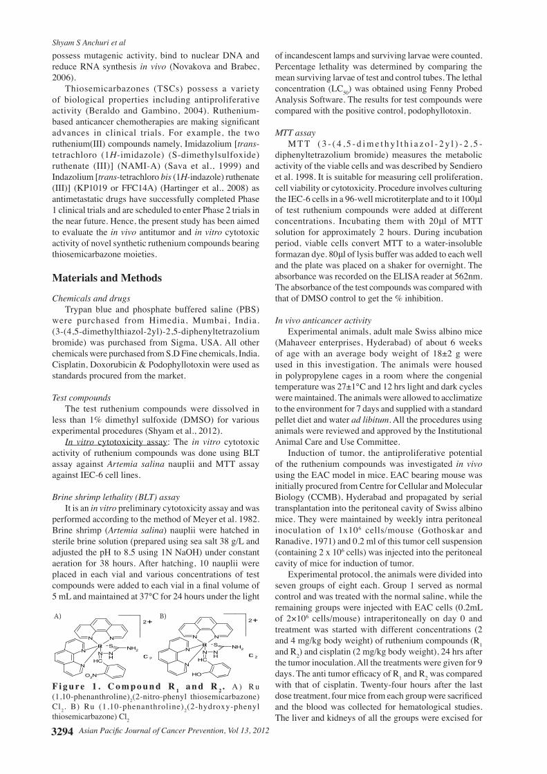
studied including coordination complexes (Gianferrara et al., 2009) and organometallic compounds (Hartinger and Dyson, 2009). Although cisplatin is most widely used antitumor drug, it has severe side effects which include the dose-limiting nephrotoxicity, neurotoxicity, ototoxicity, emetogenesis and resistance that cause relapse of cisplatin (Clarke et al., 1999). These limitations have promoted a search for more effective and less toxic non-platinum, transition-metal-based antitumor agents and ruthenium complexes have attracted much interest as alternative drugs to cisplatin in cancer chemotherapy. The first systematic investigation of ruthenium compounds and their antitumor property was done in beginning of 1980s. The Ru(II) compounds are kinetically more reactive than Ru(III) compounds (Clairs et al., 2001). So, we synthesized mononuclear Ruthenium(II) compounds of the type [Ru(A)2(B)]Cl2
-, where A=1,10-phenanthroline; B=2-NO2
- phenyl thiosemicarbazone (R1)/ 2-OH- phenyl thiosemicarbazone (R2) and characterised by spectral analysis. Ruthenium complexes were prepared to ameliorate cisplatin activity, particularly on resistance tumors or to reduce host toxicity at active doses. Like platinum metallic compounds, several ruthenium compounds have been shown to inhibit DNA replication,
Shyam S Anchuri et al
Asian Pacific Journal of Cancer Prevention, Vol 13, 20123294
possess mutagenic activity, bind to nuclear DNA and reduce RNA synthesis in vivo (Novakova and Brabec, 2006). Thiosemicarbazones (TSCs) possess a variety of biological properties including antiproliferative activity (Beraldo and Gambino, 2004). Ruthenium-based anticancer chemotherapies are making significant advances in clinical trials. For example, the two ruthenium(III) compounds namely, Imidazolium [trans-tetrachloro (1H-imidazole) (S-dimethylsulfoxide) ruthenate (III)] (NAMI-A) (Sava et al., 1999) and Indazolium [trans-tetrachloro bis (1H-indazole) ruthenate (III)] (KP1019 or FFC14A) (Hartinger et al., 2008) as antimetastatic drugs have successfully completed Phase 1 clinical trials and are scheduled to enter Phase 2 trials in the near future. Hence, the present study has been aimed to evaluate the in vivo antitumor and in vitro cytotoxic activity of novel synthetic ruthenium compounds bearing thiosemicarbazone moieties. Materials and Methods
Chemicals and drugs Trypan blue and phosphate buffered saline (PBS) were purchased from Himedia, Mumbai, India. (3-(4,5-dimethylthiazol-2yl)-2,5-diphenyltetrazolium bromide) was purchased from Sigma, USA. All other chemicals were purchased from S.D Fine chemicals, India. Cisplatin, Doxorubicin & Podophyllotoxin were used as standards procured from the market.
Test compounds The test ruthenium compounds were dissolved in less than 1% dimethyl sulfoxide (DMSO) for various experimental procedures (Shyam et al., 2012). In vitro cytotoxicity assay: The in vitro cytotoxic activity of ruthenium compounds was done using BLT assay against Artemia salina nauplii and MTT assay against IEC-6 cell lines.
Brine shrimp lethality (BLT) assay It is an in vitro preliminary cytotoxicity assay and was performed according to the method of Meyer et al. 1982. Brine shrimp (Artemia salina) nauplii were hatched in sterile brine solution (prepared using sea salt 38 g/L and adjusted the pH to 8.5 using 1N NaOH) under constant aeration for 38 hours. After hatching, 10 nauplii were placed in each vial and various concentrations of test compounds were added to each vial in a final volume of 5 mL and maintained at 37°C for 24 hours under the light
of incandescent lamps and surviving larvae were counted. Percentage lethality was determined by comparing the mean surviving larvae of test and control tubes. The lethal concentration (LC50) was obtained using Fenny Probed Analysis Software. The results for test compounds were compared with the positive control, podophyllotoxin.
MTT assay M T T ( 3 - ( 4 , 5 - d i m e t h y l t h i a z o l - 2 y l ) - 2 , 5 -diphenyltetrazolium bromide) measures the metabolic activity of the viable cells and was described by Sendiero et al. 1998. It is suitable for measuring cell proliferation, cell viability or cytotoxicity. Procedure involves culturing the IEC-6 cells in a 96-well microtiterplate and to it 100μl of test ruthenium compounds were added at different concentrations. Incubating them with 20μl of MTT solution for approximately 2 hours. During incubation period, viable cells convert MTT to a water-insoluble formazan dye. 80μl of lysis buffer was added to each well and the plate was placed on a shaker for overnight. The absorbance was recorded on the ELISA reader at 562nm. The absorbance of the test compounds was compared with that of DMSO control to get the % inhibition.
In vivo anticancer activity Experimental animals, adult male Swiss albino mice (Mahaveer enterprises, Hyderabad) of about 6 weeks of age with an average body weight of 18±2 g were used in this investigation. The animals were housed in polypropylene cages in a room where the congenial temperature was 27±1°C and 12 hrs light and dark cycles were maintained. The animals were allowed to acclimatize to the environment for 7 days and supplied with a standard pellet diet and water ad libitum. All the procedures using animals were reviewed and approved by the Institutional Animal Care and Use Committee. Induction of tumor, the antiproliferative potential of the ruthenium compounds was investigated in vivo using the EAC model in mice. EAC bearing mouse was initially procured from Centre for Cellular and Molecular Biology (CCMB), Hyderabad and propagated by serial transplantation into the peritoneal cavity of Swiss albino mice. They were maintained by weekly intra peritoneal inoculation of 1x10⁶ cells/mouse (Gothoskar and Ranadive, 1971) and 0.2 ml of this tumor cell suspension (containing 2 x 10⁶ cells) was injected into the peritoneal cavity of mice for induction of tumor. Experimental protocol, the animals were divided into seven groups of eight each. Group 1 served as normal control and was treated with the normal saline, while the remaining groups were injected with EAC cells (0.2mL of 2×10⁶ cells/mouse) intraperitoneally on day 0 and treatment was started with different concentrations (2 and 4 mg/kg body weight) of ruthenium compounds (R1 and R2) and cisplatin (2 mg/kg body weight), 24 hrs after the tumor inoculation. All the treatments were given for 9 days. The anti tumor efficacy of R1 and R2 was compared with that of cisplatin. Twenty-four hours after the last dose treatment, four mice from each group were sacrificed and the blood was collected for hematological studies. The liver and kidneys of all the groups were excised for
N
NRu
N N
N NH
CH
S NH2
O2N
2+
Cl 2
N
NRu
N N
N NH
CH
S NH2
OH
2+
Cl 2
F i g u re 1 . C o m p o u n d R 1 a n d R 2. A ) R u (1,10-phenanthroline)2(2-nitro-phenyl thiosemicarbazone) Cl2. B) Ru (1,10-phenanthroline)2(2-hydroxy-phenyl thiosemicarbazone) Cl2
A) B)
Asian Pacific Journal of Cancer Prevention, Vol 13, 2012 3295
` DOI:http://dx.doi.org/10.7314/APJCP.2012.13.7.3293 Novel Mononuclear Ruthenium(II) Compounds in Cancer Therapy
0
25.0
50.0
75.0
100.0
New
ly d
iagn
osed
with
out
trea
tmen
t
New
ly d
iagn
osed
with
tre
atm
ent
Pers
iste
nce
or r
ecur
renc
e
Rem
issi
on
Non
e
Chem
othe
rapy
Radi
othe
rapy
Conc
urre
nt c
hem
orad
iatio
n
10.3
0
12.8
30.025.0
20.310.16.3
51.7
75.051.1
30.031.354.2
46.856.3
27.625.033.130.031.3
23.738.0
31.3
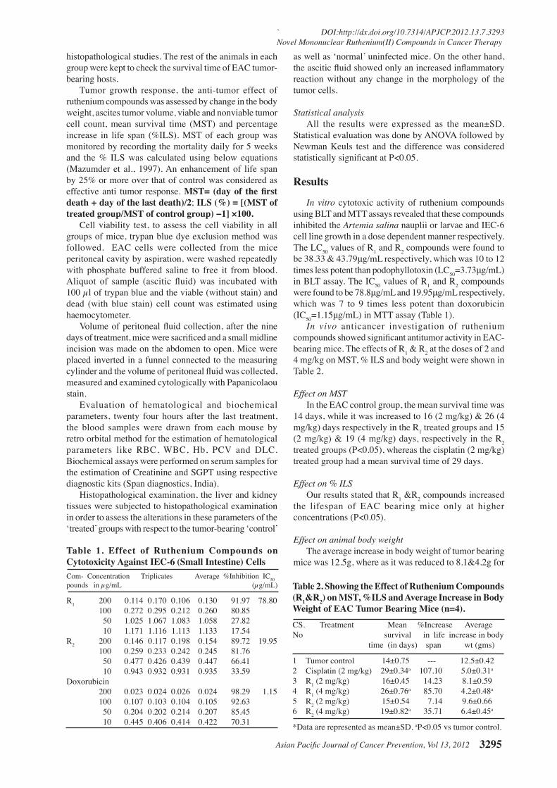
Table 1. Effect of Ruthenium Compounds on Cytotoxicity Against IEC-6 (Small Intestine) CellsCom- Concentration Triplicates Average %Inhibition IC50pounds in µg/mL (µg/mL)
R1 200 0.114 0.170 0.106 0.130 91.97 78.80 100 0.272 0.295 0.212 0.260 80.85 50 1.025 1.067 1.083 1.058 27.82 10 1.171 1.116 1.113 1.133 17.54R2 200 0.146 0.117 0.198 0.154 89.72 19.95 100 0.259 0.233 0.242 0.245 81.76 50 0.477 0.426 0.439 0.447 66.41 10 0.943 0.932 0.931 0.935 33.59Doxorubicin 200 0.023 0.024 0.026 0.024 98.29 1.15 100 0.107 0.103 0.104 0.105 92.63 50 0.204 0.202 0.214 0.207 85.45 10 0.445 0.406 0.414 0.422 70.31
0
25.0
50.0
75.0
100.0
New
ly d
iagn
osed
with
out
trea
tmen
t
New
ly d
iagn
osed
with
tre
atm
ent
Pers
iste
nce
or r
ecur
renc
e
Rem
issi
on
Non
e
Chem
othe
rapy
Radi
othe
rapy
Conc
urre
nt c
hem
orad
iatio
n
10.3
0
12.8
30.025.0
20.310.16.3
51.7
75.051.1
30.031.354.2
46.856.3
27.625.033.130.031.3
23.738.0
31.3
Table 2. Showing the Effect of Ruthenium Compounds (R1&R2) on MST, %ILS and Average Increase in Body Weight of EAC Tumor Bearing Mice (n=4).CS. Treatment Mean %Increase Average No survival in life increase in body time (in days) span wt (gms)
1 Tumor control 14±0.75 --- 12.5±0.422 Cisplatin (2 mg/kg) 29±0.34a 107.10 5.0±0.31a
3 R1 (2 mg/kg) 16±0.45 14.23 8.1±0.594 R1 (4 mg/kg) 26±0.76a 85.70 4.2±0.48a
5 R2 (2 mg/kg) 15±0.54 7.14 9.6±0.666 R2 (4 mg/kg) 19±0.82a 35.71 6.4±0.45a
*Data are represented as mean±SD. aP<0.05 vs tumor control.
histopathological studies. The rest of the animals in each group were kept to check the survival time of EAC tumor-bearing hosts. Tumor growth response, the anti-tumor effect of ruthenium compounds was assessed by change in the body weight, ascites tumor volume, viable and nonviable tumor cell count, mean survival time (MST) and percentage increase in life span (%ILS). MST of each group was monitored by recording the mortality daily for 5 weeks and the % ILS was calculated using below equations (Mazumder et al., 1997). An enhancement of life span by 25% or more over that of control was considered as effective anti tumor response. MST= (day of the first death + day of the last death)/2; ILS (%) = [(MST of treated group/MST of control group) −1] ×100. Cell viability test, to assess the cell viability in all groups of mice, trypan blue dye exclusion method was followed. EAC cells were collected from the mice peritoneal cavity by aspiration, were washed repeatedly with phosphate buffered saline to free it from blood. Aliquot of sample (ascitic fluid) was incubated with 100 µl of trypan blue and the viable (without stain) and dead (with blue stain) cell count was estimated using haemocytometer. Volume of peritoneal fluid collection, after the nine days of treatment, mice were sacrificed and a small midline incision was made on the abdomen to open. Mice were placed inverted in a funnel connected to the measuring cylinder and the volume of peritoneal fluid was collected, measured and examined cytologically with Papanicolaou stain. Evaluation of hematological and biochemical parameters, twenty four hours after the last treatment, the blood samples were drawn from each mouse by retro orbital method for the estimation of hematological parameters like RBC, WBC, Hb, PCV and DLC. Biochemical assays were performed on serum samples for the estimation of Creatinine and SGPT using respective diagnostic kits (Span diagnostics, India). Histopathological examination, the liver and kidney tissues were subjected to histopathological examination in order to assess the alterations in these parameters of the ‘treated’ groups with respect to the tumor-bearing ‘control’
as well as ‘normal’ uninfected mice. On the other hand, the ascitic fluid showed only an increased inflammatory reaction without any change in the morphology of the tumor cells.
Statistical analysis All the results were expressed as the mean±SD. Statistical evaluation was done by ANOVA followed by Newman Keuls test and the difference was considered statistically significant at P<0.05.
Results
In vitro cytotoxic activity of ruthenium compounds using BLT and MTT assays revealed that these compounds inhibited the Artemia salina nauplii or larvae and IEC-6 cell line growth in a dose dependent manner respectively. The LC50 values of R1 and R2 compounds were found to be 38.33 & 43.79μg/mL respectively, which was 10 to 12 times less potent than podophyllotoxin (LC50=3.73μg/mL) in BLT assay. The IC50 values of R1 and R2 compounds were found to be 78.8μg/mL and 19.95μg/mL respectively, which was 7 to 9 times less potent than doxorubicin (IC50=1.15μg/mL) in MTT assay (Table 1). In vivo anticancer investigation of ruthenium compounds showed significant antitumor activity in EAC-bearing mice. The effects of R1 & R2 at the doses of 2 and 4 mg/kg on MST, % ILS and body weight were shown in Table 2.
Effect on MST In the EAC control group, the mean survival time was 14 days, while it was increased to 16 (2 mg/kg) & 26 (4 mg/kg) days respectively in the R1 treated groups and 15 (2 mg/kg) & 19 (4 mg/kg) days, respectively in the R2 treated groups (P<0.05), whereas the cisplatin (2 mg/kg) treated group had a mean survival time of 29 days.
Effect on % ILS Our results stated that R1 &R2 compounds increased the lifespan of EAC bearing mice only at higher concentrations (P<0.05).
Effect on animal body weight The average increase in body weight of tumor bearing mice was 12.5g, where as it was reduced to 8.1&4.2g for
Shyam S Anchuri et al
Asian Pacific Journal of Cancer Prevention, Vol 13, 20123296
the groups treated with R1 at lower (2mg/kg) and higher (4mg/kg) doses respectively and 9.6 & 6.4g for the groups treated with R2 at lower and higher doses respectively (P<0.05).
Effect on tumor growth Treatment with ruthenium compounds significantly reduced (P<0.05) the tumor volume and viable tumor
cell count (P<0.01) in a dose-dependent manner as compared to that of the EAC control group. Furthermore, nonviable tumor cell count at different doses of ruthenium compounds were significantly increased in a dose-dependent manner (P<0.01).
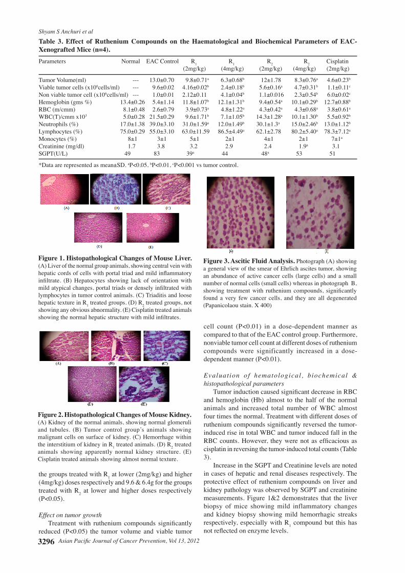
Evaluation of hematological , biochemical & histopathological parameters Tumor induction caused significant decrease in RBC and hemoglobin (Hb) almost to the half of the normal animals and increased total number of WBC almost four times the normal. Treatment with different doses of ruthenium compounds significantly reversed the tumor-induced rise in total WBC and tumor induced fall in the RBC counts. However, they were not as efficacious as cisplatin in reversing the tumor-induced total counts (Table 3). Increase in the SGPT and Creatinine levels are noted in cases of hepatic and renal diseases respectively. The protective effect of ruthenium compounds on liver and kidney pathology was observed by SGPT and creatinine measurements. Figure 1&2 demonstrates that the liver biopsy of mice showing mild inflammatory changes and kidney biopsy showing mild hemorrhagic streaks respectively, especially with R1 compound but this has not reflected on enzyme levels.
Table 3. Effect of Ruthenium Compounds on the Haematological and Biochemical Parameters of EAC- Xenografted Mice (n=4).Parameters Normal EAC Control R1 R1 R2 R2 Cisplatin (2mg/kg) (4mg/kg) (2mg/kg) (4mg/kg) (2mg/kg)
Tumor Volume(ml) --- 13.0±0.70 9.8±0.71a 6.3±0.68b 12±1.78 8.3±0.76a 4.6±0.23b
Viable tumor cells (x106cells/ml) --- 9.6±0.02 4.16±0.02b 2.4±0.18b 5.6±0.16a 4.7±0.31b 1.1±0.11c
Non viable tumor cell (x106cells/ml) --- 1.0±0.01 2.12±0.11 4.1±0.04b 1.1±0.016 2.3±0.54b 6.0±0.02c
Hemoglobin (gms %) 13.4±0.26 5.4±1.14 11.8±1.07b 12.1±1.31b 9.4±0.54a 10.1±0.29b 12.7±0.88b
RBC (m/cmm) 8.1±0.48 2.6±0.79 3.9±0.73a 4.8±1.22a 4.3±0.42a 4.3±0.68a 3.8±0.61a
WBC(T)/cmm x103 5.0±0.28 21.5±0.29 9.6±1.71b 7.1±1.05b 14.3±1.28a 10.1±1.30b 5.5±0.92b
Neutrophils (%) 17.0±1.38 39.0±3.10 31.0±1.59a 12.0±1.49b 30.1±1.3a 15.0±2.46b 13.0±1.12b
Lymphocytes (%) 75.0±0.29 55.0±3.10 63.0±11.59 86.5±4.49a 62.1±2.78 80.2±5.40a 78.3±7.12a
Monocytes (%) 8±1 3±1 5±1 2±1 4±1 2±1 7±1a
Creatinine (mg/dl) 1.7 3.8 3.2 2.9 2.4 1.9a 3.1SGPT(U/L) 49 83 39a 44 48a 53 51
*Data are represented as mean±SD. aP<0.05, bP<0.01, cP<0.001 vs tumor control.
Figure 1. Histopathological Changes of Mouse Liver. (A) Liver of the normal group animals, showing central vein with hepatic cords of cells with portal triad and mild inflammatory infiltrate. (B) Hepatocytes showing lack of orientation with mild atypical changes, portal triads or densely infiltrated with lymphocytes in tumor control animals. (C) Triaditis and loose hepatic texture in R1 treated groups. (D) R2 treated groups, not showing any obvious abnormality. (E) Cisplatin treated animals showing the normal hepatic structure with mild infiltrates.
Figure 2. Histopathological Changes of Mouse Kidney. (A) Kidney of the normal animals, showing normal glomeruli and tubules. (B) Tumor control group’s animals showing malignant cells on surface of kidney. (C) Hemorrhage within the interstitium of kidney in R1 treated animals. (D) R2 treated animals showing apparently normal kidney structure. (E) Cisplatin treated animals showing almost normal texture.
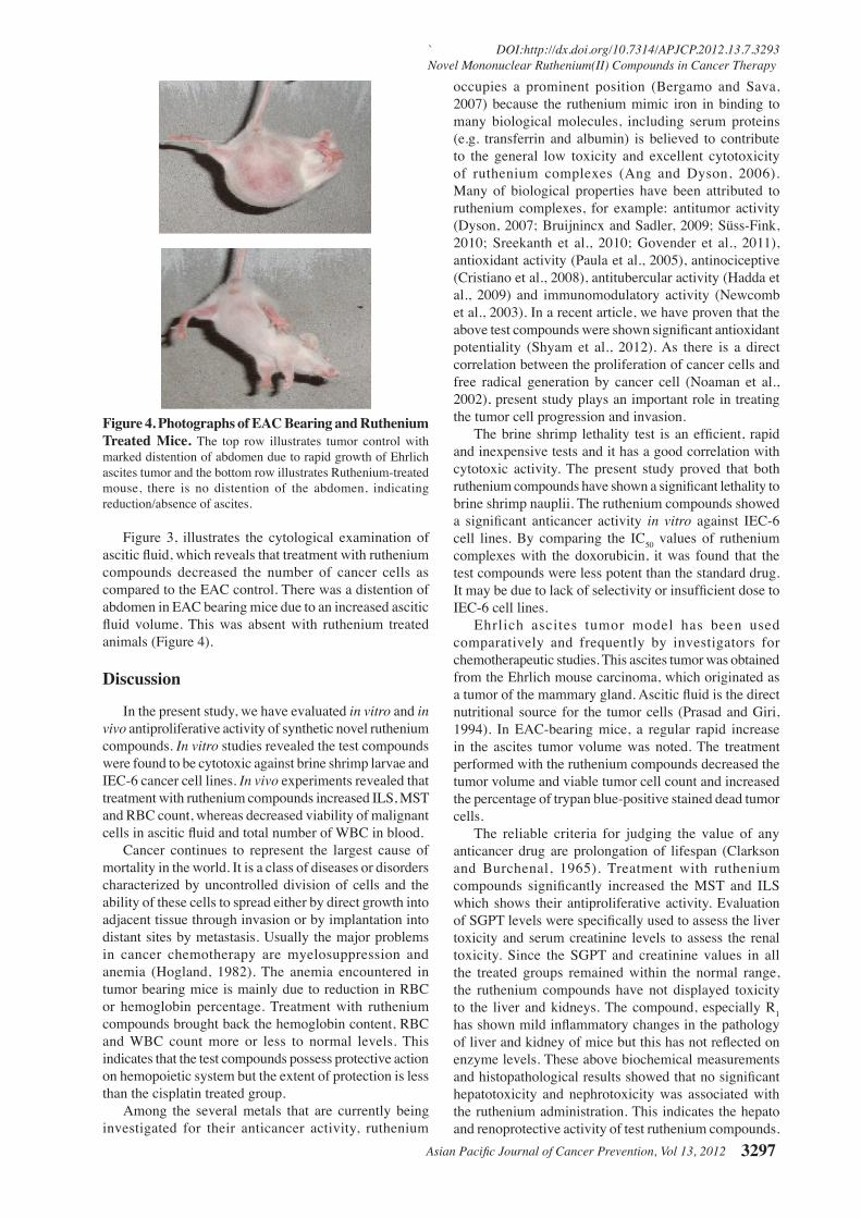
Figure 3. Ascitic Fluid Analysis. Photograph (A) showing a general view of the smear of Ehrlich ascites tumor, showing an abundance of active cancer cells (large cells) and a small number of normal cells (small cells) whereas in photograph B, showing treatment with ruthenium compounds, significantly found a very few cancer cells, and they are all degenerated (Papanicolaou stain. X 400)
Asian Pacific Journal of Cancer Prevention, Vol 13, 2012 3297
` DOI:http://dx.doi.org/10.7314/APJCP.2012.13.7.3293 Novel Mononuclear Ruthenium(II) Compounds in Cancer Therapy
Figure 3, illustrates the cytological examination of ascitic fluid, which reveals that treatment with ruthenium compounds decreased the number of cancer cells as compared to the EAC control. There was a distention of abdomen in EAC bearing mice due to an increased ascitic fluid volume. This was absent with ruthenium treated animals (Figure 4). Discussion
In the present study, we have evaluated in vitro and in vivo antiproliferative activity of synthetic novel ruthenium compounds. In vitro studies revealed the test compounds were found to be cytotoxic against brine shrimp larvae and IEC-6 cancer cell lines. In vivo experiments revealed that treatment with ruthenium compounds increased ILS, MST and RBC count, whereas decreased viability of malignant cells in ascitic fluid and total number of WBC in blood.
Cancer continues to represent the largest cause of mortality in the world. It is a class of diseases or disorders characterized by uncontrolled division of cells and the ability of these cells to spread either by direct growth into adjacent tissue through invasion or by implantation into distant sites by metastasis. Usually the major problems in cancer chemotherapy are myelosuppression and anemia (Hogland, 1982). The anemia encountered in tumor bearing mice is mainly due to reduction in RBC or hemoglobin percentage. Treatment with ruthenium compounds brought back the hemoglobin content, RBC and WBC count more or less to normal levels. This indicates that the test compounds possess protective action on hemopoietic system but the extent of protection is less than the cisplatin treated group.
Among the several metals that are currently being investigated for their anticancer activity, ruthenium
occupies a prominent position (Bergamo and Sava, 2007) because the ruthenium mimic iron in binding to many biological molecules, including serum proteins (e.g. transferrin and albumin) is believed to contribute to the general low toxicity and excellent cytotoxicity of ruthenium complexes (Ang and Dyson, 2006). Many of biological properties have been attributed to ruthenium complexes, for example: antitumor activity (Dyson, 2007; Bruijnincx and Sadler, 2009; Süss-Fink, 2010; Sreekanth et al., 2010; Govender et al., 2011), antioxidant activity (Paula et al., 2005), antinociceptive (Cristiano et al., 2008), antitubercular activity (Hadda et al., 2009) and immunomodulatory activity (Newcomb et al., 2003). In a recent article, we have proven that the above test compounds were shown significant antioxidant potentiality (Shyam et al., 2012). As there is a direct correlation between the proliferation of cancer cells and free radical generation by cancer cell (Noaman et al., 2002), present study plays an important role in treating the tumor cell progression and invasion.
The brine shrimp lethality test is an efficient, rapid and inexpensive tests and it has a good correlation with cytotoxic activity. The present study proved that both ruthenium compounds have shown a significant lethality to brine shrimp nauplii. The ruthenium compounds showed a significant anticancer activity in vitro against IEC-6 cell lines. By comparing the IC50 values of ruthenium complexes with the doxorubicin, it was found that the test compounds were less potent than the standard drug. It may be due to lack of selectivity or insufficient dose to IEC-6 cell lines.
Ehrlich ascites tumor model has been used comparatively and frequently by investigators for chemotherapeutic studies. This ascites tumor was obtained from the Ehrlich mouse carcinoma, which originated as a tumor of the mammary gland. Ascitic fluid is the direct nutritional source for the tumor cells (Prasad and Giri, 1994). In EAC-bearing mice, a regular rapid increase in the ascites tumor volume was noted. The treatment performed with the ruthenium compounds decreased the tumor volume and viable tumor cell count and increased the percentage of trypan blue-positive stained dead tumor cells.
The reliable criteria for judging the value of any anticancer drug are prolongation of lifespan (Clarkson and Burchenal, 1965). Treatment with ruthenium compounds significantly increased the MST and ILS which shows their antiproliferative activity. Evaluation of SGPT levels were specifically used to assess the liver toxicity and serum creatinine levels to assess the renal toxicity. Since the SGPT and creatinine values in all the treated groups remained within the normal range, the ruthenium compounds have not displayed toxicity to the liver and kidneys. The compound, especially R1 has shown mild inflammatory changes in the pathology of liver and kidney of mice but this has not reflected on enzyme levels. These above biochemical measurements and histopathological results showed that no significant hepatotoxicity and nephrotoxicity was associated with the ruthenium administration. This indicates the hepato and renoprotective activity of test ruthenium compounds.
Figure 4. Photographs of EAC Bearing and Ruthenium Treated Mice. The top row illustrates tumor control with marked distention of abdomen due to rapid growth of Ehrlich ascites tumor and the bottom row illustrates Ruthenium-treated mouse, there is no distention of the abdomen, indicating reduction/absence of ascites.
Shyam S Anchuri et al
Asian Pacific Journal of Cancer Prevention, Vol 13, 20123298
References
Ang WH, Dyson PJ (2006). Classical and non-classical ruthenium-based anticancer drugs: towards targeted chemotherapy. Euro J Inorg Chem, 20, 4003-18.
Beraldo H, Gambino D (2004). The wide pharmacological versatility of semicarbazones, thiosemicarbazones and their metal complexes. Mini-Rev Med Chem, 4, 31-9.
Bergamo A, Sava G (2007). Ruthenium complexes can target determinants of tumor malignancy. Dalton Trans, 13, 1267-72.
Bruijnincx PCA, Sadler PJ (2009). Controlling platinum, ruthenium, and osmium reactivity for anticancer drug design. Adv Inorg Chem, 61, 1-62.
Brunton LL, Lazo JS, Parker KL (2005). Pharmacological Basis of Therapeutics. MCGRAW- HILL, New York.
Clairs SA, Paul JD (2001). Ruthenium in medicine: current clinical uses and future prospects. Platinum Met Rev, 45, 62-9.
Clarke MJ, Zhu F, Frasca DR (1999). Non-platinum chemotherapeutic metallopharmaceuticals. Chem Rev, 99, 2511-34.
Clarkson BD, Burchenal JH (1965). Priliminary screening of antineoplastic drugs. Prog Clin Cancer, 1, 625-9.
Cristiano MP, Cardoso DC, Da Silva Paula MM, Costa-Campos L (2008). Antinociceptive effect of a ruthenium complex in mice. Auton & Autac Pharmacol, 28, 103-8.
Dyson PJ (2007). Systematic design of a targeted organometallic antitumour drug in pre-clinical development. CHIMIA Int J Chem, 61, 698-703.
Folkman J, Kalluri R (2004). Cancer without disease. Nature, 427, 787.
Gianferrara T, Bratsos I, Alessio E (2009). A categorization of metal anticancer compounds based on their mode of action. Dalton Trans, 37, 7588-98.
Gothoskar SV, Ranadive KJ (1971) Anticancer screening of SAN-AB: An extract of marking nut Semicarpus anacardium. Indian J Exp Biol, 9, 372-5.
Govender P, Renfrew AK, Clavel CM, et al (2011). Antiproliferative activity of chelating N, O- and N, N-ruthenium(II) arene functionalised poly(propyleneimine) dendrimer scaffolds. Dalton Trans, 40, 1158-67.
Hadda TB, Mehmetakkurt, Baba MF, et al (2009). Anti- tubercular Activity of Ruthenium(II) Complexes with Polypyridines. J Enzy Inhib Med Chem, 24, 457-63.
Hartinger CG, Dyson PJ (2009). Bioorganometallic chemistry- from teaching paradigms to medicinal applications. Chem Soc Rev, 38, 391-401.
Hartinger CG, Jakupec MA, Zorbas-Seifried S, et al (2008). KP1019, a new redox-active anticancer agent-preclinical development and results of a clinical phase I study in tumor patients. Chem and Biodiver, 5, 2140-55.
Hogland HC (1982). Hematological complications of cancer chemotherapy. Semin Oncol, 9, 95-102.
Mazumder UK, Gupta M, Maiti S, Mukherjee M (1997). Antitumor activity of Hygrophila spinosa on Ehrlich ascites carcinoma and sarcoma-180 induced mice. Indian J Exp Biol, 35, 473-7.
Meyer BN, Ferringin NR, Putman JE, et al (1982). Brine shrimp. A convenient general bioassay for active constituents. Planta Medica, 45, 31-4.
Newcomb JR, Rivnay B, Bastos CM, et al (2003). In vitro immunomodulatory activity of ruthenium complexes. Inflamm Res, 52, 263-71.
Noaman E, Zahran AM, Kamal AM, Omran MF (2002). Vitamin E and selenium administration as a modulator of antioxidant defense system: biochemical assessment and modification. Biol Trace Elem Res, 86, 55-64.
Novakova O, Brabec V (2006). DNA binding mode of Ruthenium complexes and relationship to tumour cell toxicity. Drug Res Updates, 9, 111-22.
Paula MM, Pich CT, Petronilho F, et al (2005). Antioxidant activity of new ruthenium compounds. Redox Rep, 10, 1-6.
Prasad SB, Giri A (1994). Antitumor effect of cisplatin against murine ascites Dalton’s lymphoma. Ind J Exp Biol, 32, 155-62.
Sava G, Clerici K, Capozzi I et al (1999b). Reduction of lung metastasis by ImH[trans-RuCl4(DMSO)Im]: mechanism of the selective action investigated on mouse tumours. Anti-Cancer Drugs, 10, 129-38.
Scott MP (2003). A twist in the Hedgehog’s tale. Nature, 425, 780-2.
Sendiero DA, Shoemaker RH, Paul KD (1998). Evaluation of soluble tetrazolium/formazan assay for cell growth % drug sensitivity in cultures using human and other cells lines. Cancer Res, 48, 4827-33.
Shyam Sunder A, Sreekanth T, Rajeshwar Y, Satyavati D (2012). In Vitro Antioxidant Activity of Some Novel Synthetic Mononuclear Ruthenium(II) Compounds. Lett Drug Design Disc, 9, 421-5.
Sreekanth T, Karki SS, Jayaveera KN et al (2010). Synthesis, antineoplastic and cytotoxic activities of some mononuclear Ru(II) complexes. J Enzyme Inhib Med Chem, 25, 513-9.
Süss-Fink G (2010). Arene ruthenium complexes as anticancer agents. Dalton Trans, 39, 1673-88.
Verweij J, Dejonge MJA (2000). Achievements and future of chemotherapy. Eur J Cancer, 36, 1479-87.
Acknowledgements
The authors acknowledge the kind help of Dr. Sistla. Ramakrishna, Principal Scientist, Division of Pharmacology, Indian Institute of Chemical Technology (IICT), Hyderabad and Dr. A. Rama Narsimha Reddy, Associate Professor, Vaageswari College of Pharmacy, Karimnagar, Andhra Pradesh, India. The authors are also grateful to Mr. A. Madhukar Reddy, Secretary and Correspondent, S. R. College of Pharmacy, Warangal, India for providing laboratory facilities for the present research work.