Noninvasive assessment of testicular torsion in rabbits using frequency-domain near-infrared...
7
Noninvasive assessment of testicular torsion in rabbits using frequency-domain near-infrared spectroscopy: prospects for pediatric urology Bertan Hallacoglu * Richard S. Matulewicz * Tufts University Department of Biomedical Engineering 4 Colby Street Medford, Massachusetts 02155 Harriet J. Paltiel Horacio Padua Children’s Hospital Boston Department of Radiology 300 Longwood Avenue Boston, Massachusetts 02115 Patricio Gargollo Glenn Cannon Children’s Hospital Boston Department of Urology 300 Longwood Avenue Boston, Massachusetts 02115 Ahmad Alomari Children’s Hospital Boston Department of Radiology 300 Longwood Avenue Boston, Massachusetts 02115 Angelo Sassaroli Sergio Fantini † Tufts University Department of Biomedical Engineering 4 Colby Street Medford, Massachusetts 02155 Abstract. We present a quantitative near-IR spectroscopy study of the absolute values of oxygen saturation of hemoglobin before and after surgically induced testicular torsion in adult rabbits. Unilateral testicu- lar torsions 0, 540, or 720 deg on experimental testes and contralat- eral sham surgery on control testes are performed in four adult rabbits. A specially designed optical probe for measurements at multiple source-detector distances and a commercial frequency-domain tissue spectrometer are used to measure absolute values of testicular hemo- globin saturation. Our results show: 1 a consistent baseline absolute tissue hemoglobin saturation value of 78±5%, 2 a comparable tis- sue hemoglobin saturation of 77±6% after sham surgery, and 3 a significantly lower tissue hemoglobin saturation of 36±2% after 540- and 720-deg testicular torsion surgery. Our findings demonstrate the feasibility of performing frequency-domain, multidistance near-IR spectroscopy for absolute testicular oximetry in the assessment of tes- ticular torsion. We conclude that near-IR spectroscopy has potential to serve as a clinical diagnostic and monitoring tool for the assessment of absolute testicular hemoglobin desaturation caused by torsion, with the possibility of serving as a complement to conventional color and spectral Doppler ultrasonography. © 2009 Society of Photo-Optical Instrumenta- tion Engineers. DOI: 10.1117/1.3253318 Keywords: near-infrared spectroscopy; testicular torsion; absolute tissue oximetry. Paper 09021R received Jan. 22, 2009; revised manuscript received Jul. 16, 2009; accepted for publication Aug. 11, 2009; published online Oct. 28, 2009. 1 Introduction Quantification of absolute or relative concentrations of chro- mophores in tissues through spectroscopic measurements of near-IR diffuse reflectance is an established method in the noninvasive examination of biological tissues. 1 Research in this area has led to applications that focused on different types of tissues such as brain 2 for monitoring hemodynamics asso- ciated with brain activation, skeletal muscles 3 for studying muscle metabolism, and the breast 4 for tumor detection, all indicative of the versatility and broad potential of near-IR spectroscopy NIRS to obtain physiological and diagnostic information from various tissues within the body. In this paper, we explore the potential of NIRS in pediatric urology, specifically in the detection, assessment, and evalua- tion of testicular torsion. Testicular torsion is the most serious cause of acute scrotal symptoms, with an incidence 5 of ap- proximately 1 in 4000. Torsion occurs at all ages, although it is most common in the pediatric population. Because of the risk of infarction, testicular torsion must be immediately ex- cluded in any patient who presents with acute scrotal symp- toms. Historically, about 50% of all torsive testes explored emergently are successfully salvaged, while the other 50% require orchiectomy or develop postoperative atrophy. 6 Color Doppler ultrasound US is routinely employed worldwide to elucidate the various causes of acute scrotal pain with a high degree of success due to its ability to directly visualize the testicular blood supply and to depict alterations in perfusion without the need for ionizing radiation. However, there are 1083-3668/2009/145/054027/7/$25.00 © 2009 SPIE *These authors contributed equally to this article. † Address all correspondence to: Sergio Fantini, Tufts University, Department of Biomedical Engineering, 4 Colby Street, Medford, MA 02155. Tel.: 617-627- 4356; E-mail: [email protected] Journal of Biomedical Optics 145, 054027 September/October 2009 Journal of Biomedical Optics September/October 2009 Vol. 145 054027-1 Downloaded from SPIE Digital Library on 13 Mar 2011 to 130.64.96.30. Terms of Use: http://spiedl.org/terms
Transcript of Noninvasive assessment of testicular torsion in rabbits using frequency-domain near-infrared...
Nup
BRTD4M
HHCD3B
PGCD3B
ACD3B
ASTD4M
1
Qmnntocmisi
*†
B4
Journal of Biomedical Optics 14�5�, 054027 �September/October 2009�
J
oninvasive assessment of testicular torsion in rabbitssing frequency-domain near-infrared spectroscopy:rospects for pediatric urology
ertan Hallacoglu*ichard S. Matulewicz*
ufts Universityepartment of Biomedical EngineeringColby Streetedford, Massachusetts 02155
arriet J. Paltieloracio Paduahildren’s Hospital Bostonepartment of Radiology00 Longwood Avenueoston, Massachusetts 02115
atricio Gargollolenn Cannonhildren’s Hospital Bostonepartment of Urology00 Longwood Avenueoston, Massachusetts 02115
hmad Alomarihildren’s Hospital Bostonepartment of Radiology00 Longwood Avenueoston, Massachusetts 02115
ngelo Sassaroliergio Fantini†
ufts Universityepartment of Biomedical EngineeringColby Street
Abstract. We present a quantitative near-IR spectroscopy study of theabsolute values of oxygen saturation of hemoglobin before and aftersurgically induced testicular torsion in adult rabbits. Unilateral testicu-lar torsions �0, 540, or 720 deg� on experimental testes and contralat-eral sham surgery on control testes are performed in four adult rabbits.A specially designed optical probe for measurements at multiplesource-detector distances and a commercial frequency-domain tissuespectrometer are used to measure absolute values of testicular hemo-globin saturation. Our results show: �1� a consistent baseline absolutetissue hemoglobin saturation value of 78±5%, �2� a comparable tis-sue hemoglobin saturation of 77±6% after sham surgery, and �3� asignificantly lower tissue hemoglobin saturation of 36±2% after 540-and 720-deg testicular torsion surgery. Our findings demonstrate thefeasibility of performing frequency-domain, multidistance near-IRspectroscopy for absolute testicular oximetry in the assessment of tes-ticular torsion. We conclude that near-IR spectroscopy has potential toserve as a clinical diagnostic and monitoring tool for the assessment ofabsolute testicular hemoglobin desaturation caused by torsion, withthe possibility of serving as a complement to conventional color andspectral Doppler ultrasonography. © 2009 Society of Photo-Optical Instrumenta-tion Engineers. �DOI: 10.1117/1.3253318�
Keywords: near-infrared spectroscopy; testicular torsion; absolute tissue oximetry.Paper 09021R received Jan. 22, 2009; revised manuscript received Jul. 16, 2009;accepted for publication Aug. 11, 2009; published online Oct. 28, 2009.
edford, Massachusetts 02155
Introduction
uantification of absolute or relative concentrations of chro-ophores in tissues through spectroscopic measurements of
ear-IR diffuse reflectance is an established method in theoninvasive examination of biological tissues.1 Research inhis area has led to applications that focused on different typesf tissues such as brain2 �for monitoring hemodynamics asso-iated with brain activation�, skeletal muscles3 �for studyinguscle metabolism�, and the breast4 �for tumor detection�, all
ndicative of the versatility and broad potential of near-IRpectroscopy �NIRS� to obtain physiological and diagnosticnformation from various tissues within the body.
These authors contributed equally to this article.Address all correspondence to: Sergio Fantini, Tufts University, Department ofiomedical Engineering, 4 Colby Street, Medford, MA 02155. Tel.: 617-627-356; E-mail: [email protected]
ournal of Biomedical Optics 054027-
Downloaded from SPIE Digital Library on 13 Mar 2011 to
In this paper, we explore the potential of NIRS in pediatricurology, specifically in the detection, assessment, and evalua-tion of testicular torsion. Testicular torsion is the most seriouscause of acute scrotal symptoms, with an incidence5 of ap-proximately 1 in 4000. Torsion occurs at all ages, although itis most common in the pediatric population. Because of therisk of infarction, testicular torsion must be immediately ex-cluded in any patient who presents with acute scrotal symp-toms. Historically, about 50% of all torsive testes exploredemergently are successfully salvaged, while the other 50%require orchiectomy or develop postoperative atrophy.6 ColorDoppler ultrasound �US� is routinely employed worldwide toelucidate the various causes of acute scrotal pain with a highdegree of success due to its ability to directly visualize thetesticular blood supply and to depict alterations in perfusionwithout the need for ionizing radiation. However, there are
1083-3668/2009/14�5�/054027/7/$25.00 © 2009 SPIE
September/October 2009 � Vol. 14�5�1
130.64.96.30. Terms of Use: http://spiedl.org/terms

pdpaplimacadar
moFtorsian
tdnsvsootoatrat7ssdfs�6Alqsq
ccuqms
Hallacoglu et al.: Noninvasive assessment of testicular torsion in rabbits using frequency-domain near-infrared spectroscopy…
J
ersistent limitations of conventional color Doppler US in theiagnosis of testicular torsion, especially in the pediatricopulation.7–9 Color Doppler diagnosis of torsion is based onsubjective impression of unilaterally diminished testicular
erfusion. However, in the prepubertal population in particu-ar, normal testicular flow is depicted with difficulty. A non-nvasive diagnostic method capable of providing quantitative
easurement of tissue oxygenation rather than a qualitativessessment of relative testicular perfusion as provided byolor Doppler US would be ideal for evaluating patients withcute scrotal symptoms and assessing testicular viability afteretorsion. Given the potential risks of anesthesia and surgery,n added benefit would be the avoidance of emergency explo-ation in patients with nonviable testes.
There is a perceived need by the pediatric urological com-unity for improved noninvasive methods for the diagnosis
f testicular torsion, particularly in the pediatric population. Inebruary 2005, the National Institute of Diabetes and Diges-
ive and Kidney Diseases �NIDDK� of the National Institutesf Health �NIH� sponsored a workshop to evaluate the state ofesearch in the field of pediatric urology and to formulate atrategic plan for the future. With respect to testicular torsion,t was noted that current diagnostic methods are imperfect,nd that “research addressing alternative strategies for diag-osis is greatly needed.”10
NIRS has been previously proposed for imaging humanestes with an envisioned application of testicular tumoretection,11 and it has been used to assess testicular hemody-amics and oxygenation in a boar model based on the occlu-ion of the spermatic vessel or the vas deference with itsessels12 and in a sheep model of testicular torsion.13 The boartudy showed the capability of NIRS in detecting the effectsf vascular occlusions in testes through relative measurementsf active testicular blood volume �ATBV�. Specifically, inheir boar study, Colier et al. found no measurable ATBV aftercclusion of the spermatic vessels, suggesting subsequenttrophy.12 In this boar study, NIRS measurements of activeesticular blood volume required a transient lowering of arte-ial saturation by reducing the fraction of inspired oxygen for
few minutes. The sheep study13 investigated the absoluteissue saturation �StO2� of testes at baseline, and following20-deg torsion �on one side� or sham surgery �on the otheride�, similar to the protocol reported in our study. NIRS mea-urements were performed every 15 min with a single source-etector distance, continuous wave spectrometer that allowedor absolute tissue saturation measurements. The results of theheep study found a median baseline tissue saturation of 59%interquartile range 57 to 69%� on the experimental side and7% �interquartile range 59 to 68%� on the control side.13
bout 2.5 h after torsion surgery, the torsioned testes stabi-ized to a lower median tissue saturation value of 14% �inter-uartile range 11 to 29%�, whereas the sham surgery testeshowed13 an increased median tissue saturation of 77% �inter-uartile range 77 to 94%�.
Our study was conducted on rabbit’s testes, whose sizeompare well to those found in the pediatric population. Inontrast to the boar and sheep studies already mentioned, wesed a multidistance, frequency-domain NIRS approach touantifying the absolute concentration and saturation of he-oglobin in testicular tissue. This approach separately mea-
ures the absorption and reduced scattering coefficients of tis-
ournal of Biomedical Optics 054027-
Downloaded from SPIE Digital Library on 13 Mar 2011 to
sue, and translates the absorption coefficients at twowavelengths �690 and 830 nm in this study� into absoluteconcentration and saturation of hemoglobin.
2 Experimental Methods2.1 Selection of AnimalsThe study was performed according to a protocol approved bythe Animal Care and Use Committee of Children’s HospitalBoston, Boston, Massachusetts, and conformed to guidelinesissued by the NIH for care of laboratory animals. Use of therabbit in experimental models of testicular ischemia is wellestablished.14–16 Four adult male New Zealand white rabbits�Millbrook Breeding Labs, Amherst, Massachusetts� with amean weight of 4.0 kg were examined.
2.2 Animal PreparationGeneral anesthesia was induced with glycopyrrolate0.04 mg /kg IM �intramuscular�, followed by ketamine10 mg /kg IV �intravenous�, and acepromazine 0.5 mg /kg IV.Endotracheal intubation was performed, and the animalsplaced on a ventilator. Anesthesia was maintained with 0.25 to3% isoflurane. A catheter was placed in one ear vein of eachexperimental animal for administration of maintenance fluids.A heparin flush was placed on the venous line. For pain con-trol prior to scrotal incision and testicular torsion, a spermaticcord block was administered containing bupivacaine/lidocaine��3 mg /kg total dose� and a dose of ketoprofen �1 mg /kg�IM. At the conclusion of the experiment, each rabbit was sac-rificed with an intravenous overdose of pentobarbital�1 mL /4.5 kg�.
2.3 Surgical ProcedureBilateral medial, ventral incisions were made under sterileconditions and the scrotal layers dissected to the tunica vagi-nalis. The testes were exposed, and following unilateral tor-sion and contralateral orchidopexy �sham surgery� the testeswere secured in place and the overlying scrotum closed. Shamsurgery follows the same exact protocol as the torsion surgeryexcept the twisting of the testis. It involves the opening of thescrotum, manipulation of the testis �remove and replace� andthen stitching of the scrotum. Through this protocol, we main-tained the surgical conditions so that the postoperative sequelis the same for both sham and torsion surgery, and this en-ables meaningful measurements of differences between testesthat underwent torsion and sham surgeries, respectively.
Baseline NIRS measurements of testicular oxygen satura-tion levels were obtained followed by unilateral testicular tor-sions of 720 deg �two twists� in rabbits 1 and 3; 540 deg �oneand a half twists� in rabbit 2; 0 deg �same as sham surgery� inrabbit 4 on the experimental testis and contralateral sham sur-gery on the control testis. Sham surgery on the experimentalside was referred to as 0-deg torsion due to the terminologyassociated with the clinical protocol.
2.4 Near-IR Tissue OximeterThe near-IR experiments were performed using one fiber-coupled photomultiplier tube �PMT� detector and two fiber-coupled laser diodes, one emitting at 690 nm and the other at830 nm �typical wavelengths used in near-IR tissue oximetry�
September/October 2009 � Vol. 14�5�2
130.64.96.30. Terms of Use: http://spiedl.org/terms
ftodtdlrmItce
2Mfsppsatfodstatmewttfir
FIaisib
Hallacoglu et al.: Noninvasive assessment of testicular torsion in rabbits using frequency-domain near-infrared spectroscopy…
J
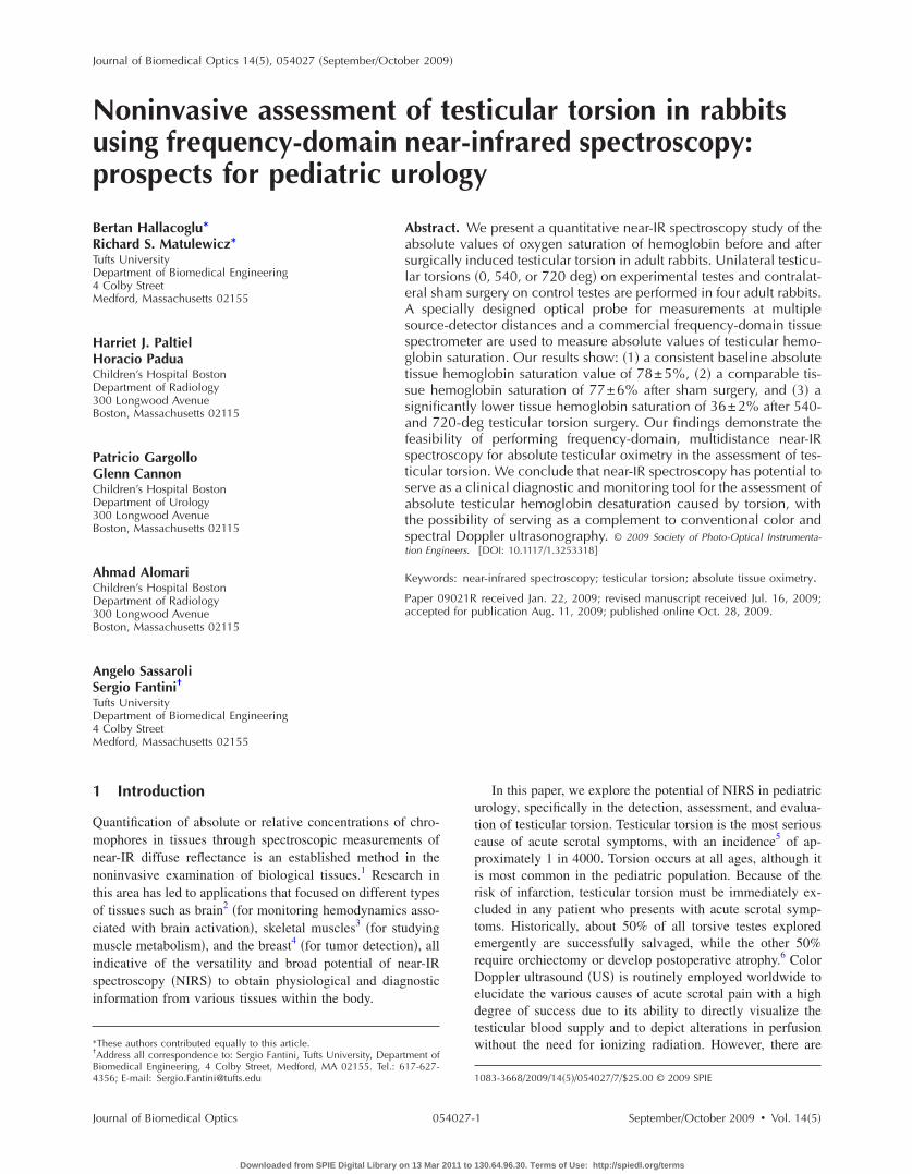
rom an OxiplexTS �ISS, Inc., Champaign, Illinois� opticalissue spectrometer.4 This is a frequency-domain instrumentperating at a modulation frequency of 110 MHz. Light waselivered to and from the tissue by a pair of single illumina-ion fibers, 400 �m in diameter, and by one 3.0-mm-diametector optical fiber bundle. The two source fibers carryingight at 690 and 830 nm were bundled together and positionedight next to each other at the emission end. A programmableechanical linear stage �Model XN10-0020-M01-71, Velmex,
nc., Bloomfield, New York� was used for linear scanning ofhe two illumination fibers toward and away from the fixedollection fiber bundle over each testis. Figure 1 shows thexperimental setup.
.5 Data Acquisitionultidistance, frequency-domain measurements were per-
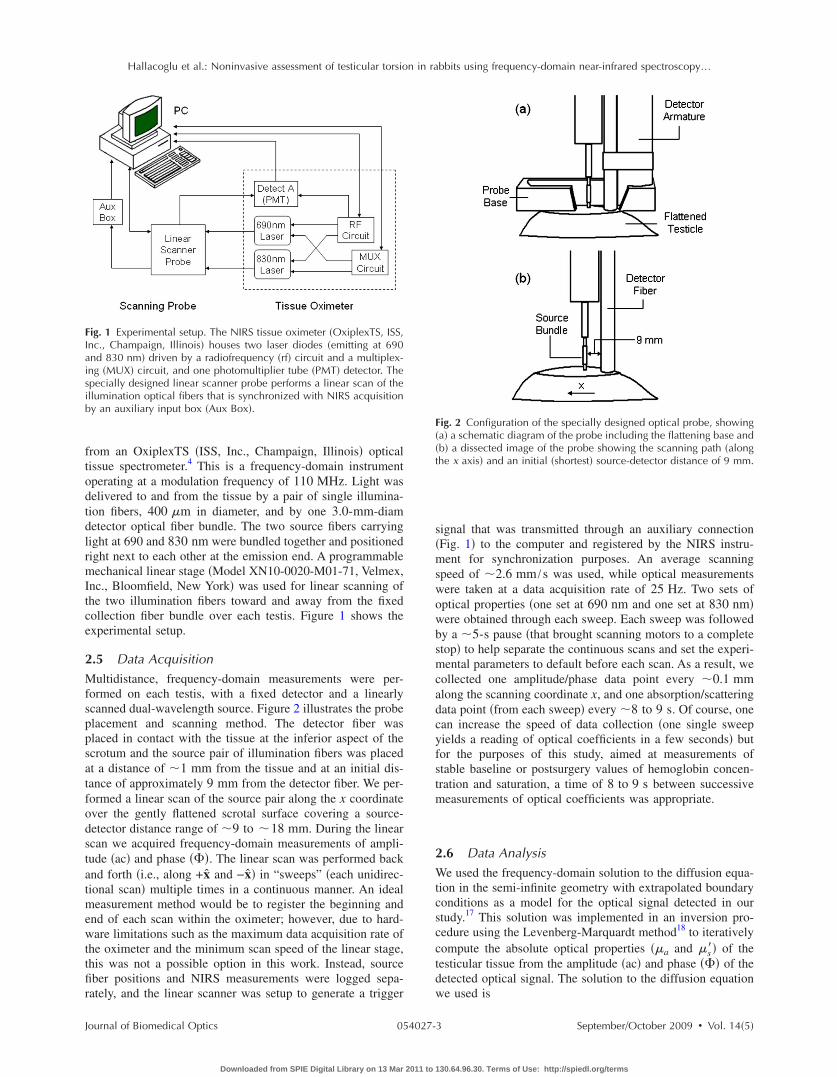
ormed on each testis, with a fixed detector and a linearlycanned dual-wavelength source. Figure 2 illustrates the probelacement and scanning method. The detector fiber waslaced in contact with the tissue at the inferior aspect of thecrotum and the source pair of illumination fibers was placedt a distance of �1 mm from the tissue and at an initial dis-ance of approximately 9 mm from the detector fiber. We per-ormed a linear scan of the source pair along the x coordinatever the gently flattened scrotal surface covering a source-etector distance range of �9 to �18 mm. During the linearcan we acquired frequency-domain measurements of ampli-ude �ac� and phase ���. The linear scan was performed backnd forth �i.e., along +x̂ and −x̂� in “sweeps” �each unidirec-ional scan� multiple times in a continuous manner. An ideal
easurement method would be to register the beginning andnd of each scan within the oximeter; however, due to hard-are limitations such as the maximum data acquisition rate of
he oximeter and the minimum scan speed of the linear stage,his was not a possible option in this work. Instead, sourceber positions and NIRS measurements were logged sepa-ately, and the linear scanner was setup to generate a trigger
ig. 1 Experimental setup. The NIRS tissue oximeter �OxiplexTS, ISS,nc., Champaign, Illinois� houses two laser diodes �emitting at 690nd 830 nm� driven by a radiofrequency �rf� circuit and a multiplex-ng �MUX� circuit, and one photomultiplier tube �PMT� detector. Thepecially designed linear scanner probe performs a linear scan of thellumination optical fibers that is synchronized with NIRS acquisitiony an auxiliary input box �Aux Box�.
ournal of Biomedical Optics 054027-
Downloaded from SPIE Digital Library on 13 Mar 2011 to
signal that was transmitted through an auxiliary connection�Fig. 1� to the computer and registered by the NIRS instru-ment for synchronization purposes. An average scanningspeed of �2.6 mm /s was used, while optical measurementswere taken at a data acquisition rate of 25 Hz. Two sets ofoptical properties �one set at 690 nm and one set at 830 nm�were obtained through each sweep. Each sweep was followedby a �5-s pause �that brought scanning motors to a completestop� to help separate the continuous scans and set the experi-mental parameters to default before each scan. As a result, wecollected one amplitude/phase data point every �0.1 mmalong the scanning coordinate x, and one absorption/scatteringdata point �from each sweep� every �8 to 9 s. Of course, onecan increase the speed of data collection �one single sweepyields a reading of optical coefficients in a few seconds� butfor the purposes of this study, aimed at measurements ofstable baseline or postsurgery values of hemoglobin concen-tration and saturation, a time of 8 to 9 s between successivemeasurements of optical coefficients was appropriate.
2.6 Data AnalysisWe used the frequency-domain solution to the diffusion equa-tion in the semi-infinite geometry with extrapolated boundaryconditions as a model for the optical signal detected in ourstudy.17 This solution was implemented in an inversion pro-cedure using the Levenberg-Marquardt method18 to iterativelycompute the absolute optical properties ��a and �s�� of thetesticular tissue from the amplitude �ac� and phase ��� of thedetected optical signal. The solution to the diffusion equationwe used is
Fig. 2 Configuration of the specially designed optical probe, showing�a� a schematic diagram of the probe including the flattening base and�b� a dissected image of the probe showing the scanning path �alongthe x axis� and an initial �shortest� source-detector distance of 9 mm.
September/October 2009 � Vol. 14�5�3
130.64.96.30. Terms of Use: http://spiedl.org/terms
ws�
a=gwblvsmsstti
wdf�=F�
3Itcavactaetpim
Hallacoglu et al.: Noninvasive assessment of testicular torsion in rabbits using frequency-domain near-infrared spectroscopy…
J
R̃��,�� = A1
4��z0� 1
r1+ �̃eff� exp�− r1�̃eff�
r12
+ �z0 + 2zb�� 1
r2+ �̃eff� exp�− r2�̃eff�
r22 � , �1�
here R̃ represents the complex output flux �or reflectance� atource-detector distance � and angular modulation frequency, so that the measured ac amplitude is given by
c= �R̃�� ,��� and the phase is given by �=argR̃�� ,��; z0
1 /�s� �with �s� reduced scattering coefficient�; r12 and r2
2 areiven, respectively by r1
2=z02+�2 and r2
2= �z0+2zb�2+�2,here zb is the distance between the real and the extrapolatedoundary;19 �̃eff= ��ac+ i�� /Dc1/2, where c is the speed ofight in the medium, and D is the optical diffusion coefficientD=1 / �3�s��; A is an amplitude factor used to match the acalues predicted by the model with those measured. We ob-erve that since the absolute phase shift � cannot be experi-entally measured, we measured the phase at a generic
ource-detector distance � relative to the phase at the shortestource-detector distance. From the absorption coefficient ob-ained with this fitting procedure, we calculate the concentra-ions of oxyhemoglobin HbO2 and deoxyhemoglobin Hbn tissue as follows:4
HbO2 =�a
�1�Hb�2 − �a
�2�Hb�1
�HbO2
�1 �Hb�2 − �HbO2
�2 �Hb�1
, �2�
Hb =�a
�2�HbO2
�1 − �a�1�HbO2
�2
�HbO2
�1 �Hb�2 − �HbO2
�2 �Hb�1
, �3�
here �Hb and �HbO2are the molar extinction coefficients of
eoxyhemoglobin and oxyhemoglobin �here we have used theollowing values:20 �Hb�690 nm�=4.854 mM−1 cm−1,
HbO2�690 nm�=0.956 mM−1 cm−1, �Hb�830 nm�
1.790 mM−1 cm−1, �HbO2�830 nm�=2.333 mM−1 cm−1�.
inally, the oxygen saturation of hemoglobin in the tissueStO2� is given by4
StO2 =HbO2
HbO2 + Hb. �4�
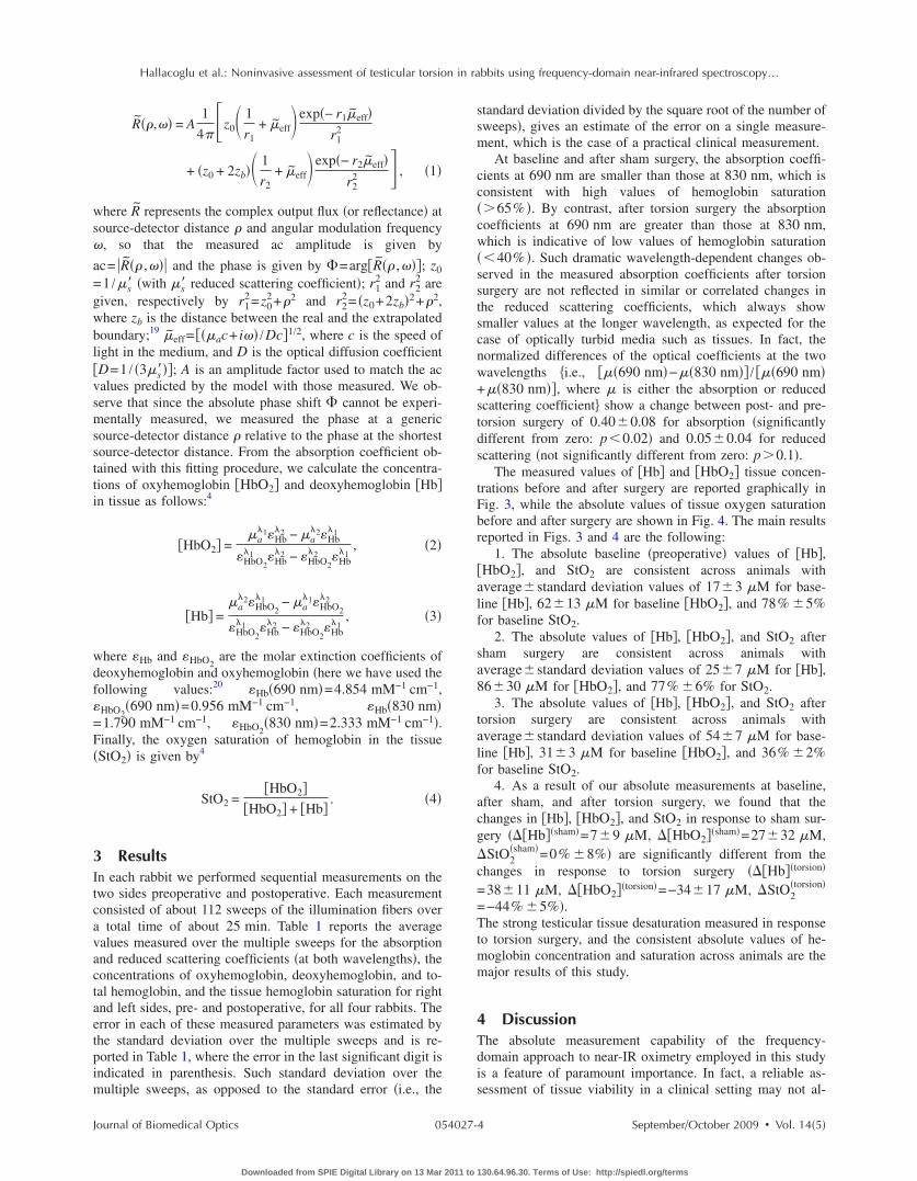
Resultsn each rabbit we performed sequential measurements on thewo sides preoperative and postoperative. Each measurementonsisted of about 112 sweeps of the illumination fibers overtotal time of about 25 min. Table 1 reports the average
alues measured over the multiple sweeps for the absorptionnd reduced scattering coefficients �at both wavelengths�, theoncentrations of oxyhemoglobin, deoxyhemoglobin, and to-al hemoglobin, and the tissue hemoglobin saturation for rightnd left sides, pre- and postoperative, for all four rabbits. Therror in each of these measured parameters was estimated byhe standard deviation over the multiple sweeps and is re-orted in Table 1, where the error in the last significant digit isndicated in parenthesis. Such standard deviation over the
ultiple sweeps, as opposed to the standard error �i.e., the
ournal of Biomedical Optics 054027-
Downloaded from SPIE Digital Library on 13 Mar 2011 to
standard deviation divided by the square root of the number ofsweeps�, gives an estimate of the error on a single measure-ment, which is the case of a practical clinical measurement.
At baseline and after sham surgery, the absorption coeffi-cients at 690 nm are smaller than those at 830 nm, which isconsistent with high values of hemoglobin saturation�65% �. By contrast, after torsion surgery the absorptioncoefficients at 690 nm are greater than those at 830 nm,which is indicative of low values of hemoglobin saturation�40% �. Such dramatic wavelength-dependent changes ob-served in the measured absorption coefficients after torsionsurgery are not reflected in similar or correlated changes inthe reduced scattering coefficients, which always showsmaller values at the longer wavelength, as expected for thecase of optically turbid media such as tissues. In fact, thenormalized differences of the optical coefficients at the twowavelengths �i.e., ��690 nm�−��830 nm� / ��690 nm�+��830 nm�, where � is either the absorption or reducedscattering coefficient� show a change between post- and pre-torsion surgery of 0.40�0.08 for absorption �significantlydifferent from zero: p0.02� and 0.05�0.04 for reducedscattering �not significantly different from zero: p0.1�.
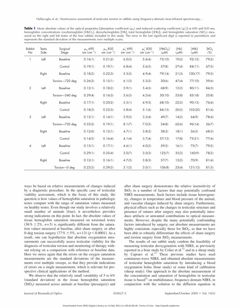
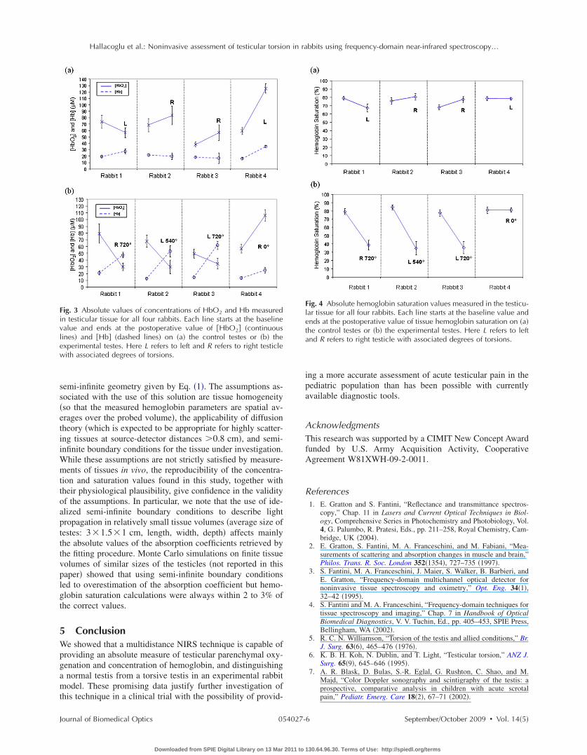
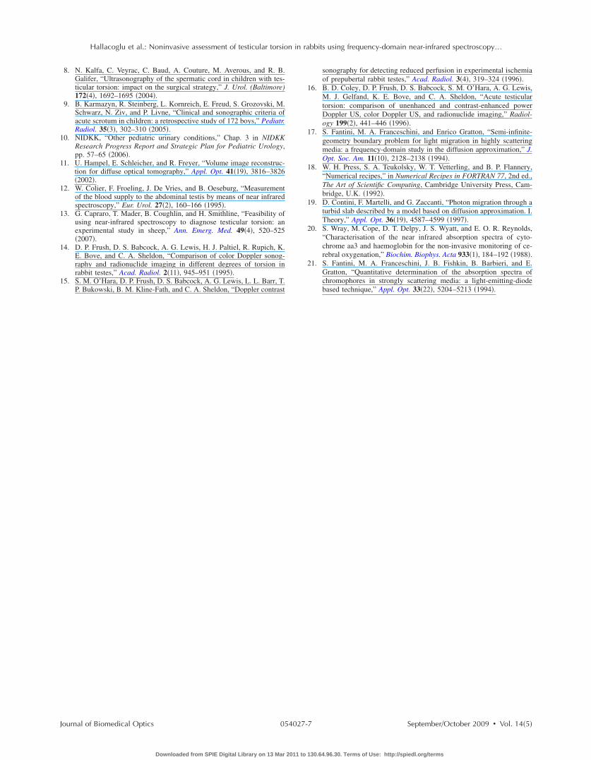
The measured values of Hb and HbO2 tissue concen-trations before and after surgery are reported graphically inFig. 3, while the absolute values of tissue oxygen saturationbefore and after surgery are shown in Fig. 4. The main resultsreported in Figs. 3 and 4 are the following:
1. The absolute baseline �preoperative� values of Hb,HbO2, and StO2 are consistent across animals withaverage�standard deviation values of 17�3 �M for base-line Hb, 62�13 �M for baseline HbO2, and 78% �5%for baseline StO2.
2. The absolute values of Hb, HbO2, and StO2 aftersham surgery are consistent across animals withaverage�standard deviation values of 25�7 �M for Hb,86�30 �M for HbO2, and 77% �6% for StO2.
3. The absolute values of Hb, HbO2, and StO2 aftertorsion surgery are consistent across animals withaverage�standard deviation values of 54�7 �M for base-line Hb, 31�3 �M for baseline HbO2, and 36% �2%for baseline StO2.
4. As a result of our absolute measurements at baseline,after sham, and after torsion surgery, we found that thechanges in Hb, HbO2, and StO2 in response to sham sur-gery ��Hb�sham�=7�9 �M, �HbO2�sham�=27�32 �M,�StO2
�sham�=0% �8%� are significantly different from thechanges in response to torsion surgery ��Hb�torsion�
=38�11 �M, �HbO2�torsion�=−34�17 �M, �StO2�torsion�
=−44% �5%�.The strong testicular tissue desaturation measured in responseto torsion surgery, and the consistent absolute values of he-moglobin concentration and saturation across animals are themajor results of this study.
4 DiscussionThe absolute measurement capability of the frequency-domain approach to near-IR oximetry employed in this studyis a feature of paramount importance. In fact, a reliable as-sessment of tissue viability in a clinical setting may not al-
September/October 2009 � Vol. 14�5�4
130.64.96.30. Terms of Use: http://spiedl.org/terms
wbvqtosst�t0rsdoHmmos
��
Thsr
Hallacoglu et al.: Noninvasive assessment of testicular torsion in rabbits using frequency-domain near-infrared spectroscopy…
J
ays be based on relative measurements of changes inducedy a diagnostic procedure. In the specific case of testiculariability assessment, which is the focus of this study, theuestion is how values of hemoglobin saturation in pathologicestes compare with the range of saturation values measuredn healthy testes. Even though our study involves a relativelymall number of animals �four�, it nevertheless providestrong indications on this point. In fact, the absolute values ofissue hemoglobin saturation measured on torsioned testes36% �2%, n=3� is significantly different from the satura-ion values measured at baseline, after sham surgery, or after-deg torsion surgery �77% �5%, n=13� �p0.0001�. As aesult, one can hypothesize that absolute oxygenation mea-urements can successfully assess testicular viability for theiagnosis of testicular torsion and monitoring of therapy with-ut relying on a comparison with reference or baseline data.ere we stress again that the errors on the oxygen saturationeasurements are the standard deviations of the measure-ents over multiple sweeps, so that they provide indications
f errors on a single measurement, which is relevant for pro-pective clinical applications of the method.
We observe that the relatively small variability of 5 to 6%standard deviation� in the tissue hemoglobin saturationStO � measured across animals at baseline �presurgery� and
able 1 Mean absolute values of the optical properties �absorption cemoglobin concentrations �oxyhemoglobin HbO2, deoxyhemogloured on the right and left testes of the four rabbits included in thisepresents the standard deviation of the measurements over multiple
RabbitNo
TestisSide
SurgicalStage
�a 690nm �cm−1�
�a 830nm �cm−1�
1 Left Baseline 0.16�1� 0.21�2�
Control 0.19�1� 0.19�1�
Right Baseline 0.18�2� 0.22�3�
Torsion—720 deg 0.26�2� 0.15�1�
2 Left Baseline 0.12�1� 0.18�2�
Torsion—540 deg 0.29�4� 0.16�3�
Right Baseline 0.17�1� 0.20�2�
Control 0.18�3� 0.23�3�
3 Left Baseline 0.12�1� 0.14�1�
Torsion—720 deg 0.33�2� 0.19�1�
Right Baseline 0.12�0� 0.12�1�
Control 0.14�2� 0.16�4�
4 Left Baseline 0.13�1� 0.17�1�
Control 0.29�1� 0.35�4�
Right Baseline 0.12�1� 0.16�1�
Torsion—0 deg 0.22�2� 0.29�2�
2
ournal of Biomedical Optics 054027-
Downloaded from SPIE Digital Library on 13 Mar 2011 to
after sham surgery demonstrates the relative insensitivity ofStO2 to a number of factors that may potentially confoundNIRS measurements. Such factors include tissue heterogene-ity, changes in temperature and blood pressure of the animal,and vascular changes induced by sham surgery. Furthermore,physical effects such as the changes in testicular structure andpresence of sutures after surgery may also potentially intro-duce artifacts or unwanted contributions to optical measure-ments. However, despite the many potentially confoundingfactors introduced by surgery, our absolute measurements arehighly consistent, especially those for StO2, so that we havebeen able to robustly differentiate the effects of sham surgeryand torsion surgery from StO2 measurements.
The results of our rabbit study confirm the feasibility ofmeasuring testicular deoxygenation with NIRS, as previouslyreported in a boar study by Colier et al.12 and in a sheep studyby Capraro et al.13 These previous studies have usedcontinuous-wave NIRS, and obtained absolute measurementsof testicular hemoglobin saturation by introducing a bloodoxygenation bolus �boar study� or some kind of calibration�sheep study�. Our approach to the absolute measurement ofthe concentration and saturation of hemoglobin in testiculartissue is based21 on multidistance, frequency-domain NIRS, inconjunction with the solution to the diffusion equation in
ent ��a� and reduced scattering coefficient ��s��� at 690 and 830 nm,�, total hemoglobin tHb�, and hemoglobin saturation �StO2� mea-The error in the last significant digit is reported in parenthesis and
s� 690�cm−1�
�s� 830nm �cm−1�
HbO2��M�
�Hb���M�
tHb��M�
StO2�%�
.0�3� 5.6�4� 73�10� 19�2� 92�12� 79�2�
.8�4� 5.6�3� 57�8� 27�4� 84�11� 67�5�
.3�3� 4.9�4� 79�14� 21�3� 100�17� 79�3�
.1�3� 5.2�2� 30�6� 47�4� 77�10� 39�6�
.9�1� 5.4�3� 68�9� 12�2� 80�11� 84�3�
.6�3� 4.2�6� 30�10� 53�8� 83�18� 35�8�
.5�1� 4.9�3� 68�10� 22�2� 90�13� 76�4�
.8�4� 5.1�6� 84�15� 20�5� 103�20� 81�4�
.9�3� 3.5�4� 49�7� 14�2� 64�9� 78�4�
.1�7� 7.0�3� 34�8� 62�6� 96�14� 36�7�
.7�1� 3.8�2� 38�3� 18�1� 56�5� 68�3�
.1�4� 3.7�4� 57�12� 17�8� 73�21� 77�4�
.6�1� 4.0�2� 59�5� 16�1� 75�7� 79�2�
.0�7� 3.0�3� 125�7� 35�2� 160�9� 78�2�
.7�3� 3.8�3� 57�7� 13�2� 70�9� 81�4�
.1�2� 3.0�1� 106�8� 25�4� 131�12� 81�3�
oefficibin �Hbstudy.
scans.
�nm
6
6
5
6
5
5
5
5
3
8
4
4
4
3
4
3
September/October 2009 � Vol. 14�5�5
130.64.96.30. Terms of Use: http://spiedl.org/terms
ss�etiiWmttoaptttvplgt
5Wpgamt
Fivlew
Hallacoglu et al.: Noninvasive assessment of testicular torsion in rabbits using frequency-domain near-infrared spectroscopy…
J
emi-infinite geometry given by Eq. �1�. The assumptions as-ociated with the use of this solution are tissue homogeneityso that the measured hemoglobin parameters are spatial av-rages over the probed volume�, the applicability of diffusionheory �which is expected to be appropriate for highly scatter-ng tissues at source-detector distances 0.8 cm�, and semi-nfinite boundary conditions for the tissue under investigation.
hile these assumptions are not strictly satisfied by measure-ents of tissues in vivo, the reproducibility of the concentra-
ion and saturation values found in this study, together withheir physiological plausibility, give confidence in the validityf the assumptions. In particular, we note that the use of ide-lized semi-infinite boundary conditions to describe lightropagation in relatively small tissue volumes �average size ofestes: 3 1.5 1 cm, length, width, depth� affects mainlyhe absolute values of the absorption coefficients retrieved byhe fitting procedure. Monte Carlo simulations on finite tissueolumes of similar sizes of the testicles �not reported in thisaper� showed that using semi-infinite boundary conditionsed to overestimation of the absorption coefficient but hemo-lobin saturation calculations were always within 2 to 3% ofhe correct values.
Conclusione showed that a multidistance NIRS technique is capable of
roviding an absolute measure of testicular parenchymal oxy-enation and concentration of hemoglobin, and distinguishingnormal testis from a torsive testis in an experimental rabbitodel. These promising data justify further investigation of
his technique in a clinical trial with the possibility of provid-
ig. 3 Absolute values of concentrations of HbO2 and Hb measuredn testicular tissue for all four rabbits. Each line starts at the baselinealue and ends at the postoperative value of HbO2 �continuousines� and �Hb� �dashed lines� on �a� the control testes or �b� thexperimental testes. Here L refers to left and R refers to right testicleith associated degrees of torsions.
ournal of Biomedical Optics 054027-
Downloaded from SPIE Digital Library on 13 Mar 2011 to
ing a more accurate assessment of acute testicular pain in thepediatric population than has been possible with currentlyavailable diagnostic tools.
AcknowledgmentsThis research was supported by a CIMIT New Concept Awardfunded by U.S. Army Acquisition Activity, CooperativeAgreement W81XWH-09-2-0011.
References1. E. Gratton and S. Fantini, “Reflectance and transmittance spectros-
copy,” Chap. 11 in Lasers and Current Optical Techniques in Biol-ogy, Comprehensive Series in Photochemistry and Photobiology, Vol.4, G. Palumbo, R. Pratesi, Eds., pp. 211–258, Royal Chemistry, Cam-bridge, UK �2004�.
2. E. Gratton, S. Fantini, M. A. Franceschini, and M. Fabiani, “Mea-surements of scattering and absorption changes in muscle and brain,”Philos. Trans. R. Soc. London 352�1354�, 727–735 �1997�.
3. S. Fantini, M. A. Franceschini, J. Maier, S. Walker, B. Barbieri, andE. Gratton, “Frequency-domain multichannel optical detector fornoninvasive tissue spectroscopy and oximetry,” Opt. Eng. 34�1�,32–42 �1995�.
4. S. Fantini and M. A. Franceschini, “Frequency-domain techniques fortissue spectroscopy and imaging,” Chap. 7 in Handbook of OpticalBiomedical Diagnostics, V. V. Tuchin, Ed., pp. 405–453, SPIE Press,Bellingham, WA �2002�.
5. R. C. N. Williamson, “Torsion of the testis and allied conditions,” Br.J. Surg. 63�6�, 465–476 �1976�.
6. K. B. H. Koh, N. Dublin, and T. Light, “Testicular torsion,” ANZ J.Surg. 65�9�, 645–646 �1995�.
7. A. R. Blask, D. Bulas, S.-R. Eglal, G. Rushton, C. Shao, and M.Majd, “Color Doppler sonography and scintigraphy of the testis: aprospective, comparative analysis in children with acute scrotalpain,” Pediatr. Emerg. Care 18�2�, 67–71 �2002�.
Fig. 4 Absolute hemoglobin saturation values measured in the testicu-lar tissue for all four rabbits. Each line starts at the baseline value andends at the postoperative value of tissue hemoglobin saturation on �a�the control testes or �b� the experimental testes. Here L refers to leftand R refers to right testicle with associated degrees of torsions.
September/October 2009 � Vol. 14�5�6
130.64.96.30. Terms of Use: http://spiedl.org/terms
1
1
1
1
1
1
Hallacoglu et al.: Noninvasive assessment of testicular torsion in rabbits using frequency-domain near-infrared spectroscopy…
J
8. N. Kalfa, C. Veyrac, C. Baud, A. Couture, M. Averous, and R. B.Galifer, “Ultrasonography of the spermatic cord in children with tes-ticular torsion: impact on the surgical strategy,” J. Urol. (Baltimore)172�4�, 1692–1695 �2004�.
9. B. Karmazyn, R. Steinberg, L. Kornreich, E. Freud, S. Grozovski, M.Schwarz, N. Ziv, and P. Livne, “Clinical and sonographic criteria ofacute scrotum in children: a retrospective study of 172 boys,” Pediatr.Radiol. 35�3�, 302–310 �2005�.
0. NIDKK, “Other pediatric urinary conditions,” Chap. 3 in NIDKKResearch Progress Report and Strategic Plan for Pediatric Urology,pp. 57–65 �2006�.
1. U. Hampel, E. Schleicher, and R. Freyer, “Volume image reconstruc-tion for diffuse optical tomography,” Appl. Opt. 41�19�, 3816–3826�2002�.
2. W. Colier, F. Froeling, J. De Vries, and B. Oeseburg, “Measurementof the blood supply to the abdominal testis by means of near infraredspectroscopy,” Eur. Urol. 27�2�, 160–166 �1995�.
3. G. Capraro, T. Mader, B. Coughlin, and H. Smithline, “Feasibility ofusing near-infrared spectroscopy to diagnose testicular torsion: anexperimental study in sheep,” Ann. Emerg. Med. 49�4�, 520–525�2007�.
4. D. P. Frush, D. S. Babcock, A. G. Lewis, H. J. Paltiel, R. Rupich, K.E. Bove, and C. A. Sheldon, “Comparison of color Doppler sonog-raphy and radionuclide imaging in different degrees of torsion inrabbit testes,” Acad. Radiol. 2�11�, 945–951 �1995�.
5. S. M. O’Hara, D. P. Frush, D. S. Babcock, A. G. Lewis, L. L. Barr, T.P. Bukowski, B. M. Kline-Fath, and C. A. Sheldon, “Doppler contrast
ournal of Biomedical Optics 054027-
Downloaded from SPIE Digital Library on 13 Mar 2011 to
sonography for detecting reduced perfusion in experimental ischemiaof prepubertal rabbit testes,” Acad. Radiol. 3�4�, 319–324 �1996�.
16. B. D. Coley, D. P. Frush, D. S. Babcock, S. M. O’Hara, A. G. Lewis,M. J. Gelfand, K. E. Bove, and C. A. Sheldon, “Acute testiculartorsion: comparison of unenhanced and contrast-enhanced powerDoppler US, color Doppler US, and radionuclide imaging,” Radiol-ogy 199�2�, 441–446 �1996�.
17. S. Fantini, M. A. Franceschini, and Enrico Gratton, “Semi-infinite-geometry boundary problem for light migration in highly scatteringmedia: a frequency-domain study in the diffusion approximation,” J.Opt. Soc. Am. 11�10�, 2128–2138 �1994�.
18. W. H. Press, S. A. Teukolsky, W. T. Vetterling, and B. P. Flannery,“Numerical recipes,” in Numerical Recipes in FORTRAN 77, 2nd ed.,The Art of Scientific Computing, Cambridge University Press, Cam-bridge, U.K. �1992�.
19. D. Contini, F. Martelli, and G. Zaccanti, “Photon migration through aturbid slab described by a model based on diffusion approximation. I.Theory,” Appl. Opt. 36�19�, 4587–4599 �1997�.
20. S. Wray, M. Cope, D. T. Delpy, J. S. Wyatt, and E. O. R. Reynolds,“Characterisation of the near infrared absorption spectra of cyto-chrome aa3 and haemoglobin for the non-invasive monitoring of ce-rebral oxygenation,” Biochim. Biophys. Acta 933�1�, 184–192 �1988�.
21. S. Fantini, M. A. Franceschini, J. B. Fishkin, B. Barbieri, and E.Gratton, “Quantitative determination of the absorption spectra ofchromophores in strongly scattering media: a light-emitting-diodebased technique,” Appl. Opt. 33�22�, 5204–5213 �1994�.
September/October 2009 � Vol. 14�5�7
130.64.96.30. Terms of Use: http://spiedl.org/terms