An Investigation into Maintaining Naso-gastric Feeding for ...
Upload
univ-lyon1Category
view
2download
0
Non-Invasive Exploration of Neonatal Gastric Epitheliumby Using Exfoliated Epithelial CellsBertrand Kaeffer1*, Arnaud Legrand2, Thomas Moyon1, Anne Frondas-Chauty2, Helene Billard1, Omar
Guzman-Quevedo1, Dominique Darmaun1,2, Jean-Christophe Roze2
1 Unite Mixte de Recherche-1280, Physiologie des Adaptations Nutritionnelles, Institut National Recherche Agronomique, Nantes, France, 2 University of Nantes, Hospital
of Mother and Child, Nantes, France
Abstract
Background & Aims: In preterm infants, exfoliated gastric epithelial cells can be retrieved from aspirates sampled throughthe naso-gastric feeding tube. Our aims were to determine (1) whether the recovery of exfoliated cells is feasible at any timefrom birth through the removal of the nasogastric tube, (2) whether they can be grown in culture in vitro, and (3) whetherthe physiological state of exfoliated cells expressing H+/K+ -ATPases reflects that of their counterparts remaining in situ atthe surface of the gastric epithelium in neonatal rat pups.
Methods: In infants, gastric fluid aspirates were collected weekly after birth or every 3 hours over 24-h periods, and relatedto clinical parameters (Biocollection PROG/09/18). In rat pups submitted to a single fasting/refeeding cycle, we exploredcircadian exfoliation with the cellular counter-parts in the gland. All samples were analyzed by confocal imaging andEnzyme-Linked Immunosorbent Assay.
Results: Epithelial cells were identified by microscopy using membrane-bound anti-H+/K+ ATPases antibody, assessed fornucleus integrity, and the expression of selected proteins (autophagy, circadian clock). On 34 infants, the H+/K+ -ATPase-positive cells were consistently found quiescent, regardless of gestational age and feeding schedule from day-5 of life to theday of removal of the naso-gastric tube. By logistic regression analysis, we did find a positive correlation between theintensity of exfoliation (cellular loss per sample) and the postnatal age (p,0.001). The H+/K+ ATPase-positive cellsestablished in culture retained the expression of a biomarker of progenitor status (Pouf5F1-Oct4). In rat pups, the expressionpattern of Survivin in H+/K+ ATPase-positive exfoliated cells paralleled that observed in cells remaining at the surface of thegastric gland.
Conclusions: Tracking parietal cells can improve clinical monitoring and understanding of the autophagic death via thephosphatidylinositol 3-kinase/Akt/survivin pathway.
Citation: Kaeffer B, Legrand A, Moyon T, Frondas-Chauty A, Billard H, et al. (2011) Non-Invasive Exploration of Neonatal Gastric Epithelium by Using ExfoliatedEpithelial Cells. PLoS ONE 6(10): e25562. doi:10.1371/journal.pone.0025562
Editor: Josef Neu, University of Florida, United States of America
Received July 6, 2011; Accepted September 5, 2011; Published October 18, 2011
Copyright: � 2011 Kaeffer et al. This is an open-access article distributed under the terms of the Creative Commons Attribution License, which permitsunrestricted use, distribution, and reproduction in any medium, provided the original author and source are credited.
Funding: The authors are grateful for receiving financial support from ANSSD-2008 Departement Alimentation Humaine (Inra, France); and PremaCol-2008(Region Pays de la Loire, France). The funders had no role in study design, data collection and analysis, decision to publish, or preparation of the manuscript.
Competing Interests: The authors have declared that no competing interests exist.
* E-mail: [email protected]
Introduction
Exfoliation has been described as an active biochemical process,
highly context-dependent and linked to epithelium homeostasis
[1,2,3,4]. It is believed that epithelial cells, loosing contact with
companion cells as well as extracellular matrix, enter anoikis [5].
The detachment of epithelial cells triggers both pro and
antiapoptotic signals, such as nuclear factor kappa-B and inhibitor
of apoptosis protein family members; these antiapoptotic mech-
anisms presumably delay the onset of apoptosis and allow cells to
survive [6,7]. The balance between these signals and the duration
of detachment further determine the ultimate fate of these cells.
Antiapoptotic signals presumably delay the onset of anoikis,
allowing cells to survive provided they can reestablish contact with
extracellular matrix in a timely manner [8]. Loss of extracellular
matrix contact induces autophagy in normal epithelial cells;
autophagy promotes the survival of detached cells during both
anoikis and lumen formation in 3D epithelial cell culture [9,10].
Under these assumptions, exfoliation may be understood as a
natural process to remove external cells from the luminal surface
of an epithelium. Consequently, exfoliation has a physiological
role in the architecture of epithelium by allowing the formation of
a lumen and we can surmise by providing sufficient flexibility to
preserve the physical integrity of epithelia and allow its growth. By
loosing contact with the original mucosa, exfoliated epithelial cells
have to activate autophagy as a survival mechanism to endure
starvation. Starving cells are degrading cytoplasmic material to
generate both nutrients and energy [11]. Aoyama et al (2008) have
shown on a rat model that the parietal cells are exfoliated into the
gastric pit of isolated rat gastric mucosa after stimulation of acid
secretion or under re-feeding conditions [2]. The renewal of pit
parietal cells of the gastric glands is of potential interest to monitor
PLoS ONE | www.plosone.org 1 October 2011 | Volume 6 | Issue 10 | e25562

the response of gastric epithelium to a stimulation by food intake
or pharmacological agents. In premature infants receiving enteral
nutrition, gastric fluids are routinely aspirated (and discarded)
every 3 hours in neonatal intensive care unit to ensure adequate
gastric emptying and the tolerance to enteral feeding. Exfoliated
epithelial cells therefore can be isolated from such samples3. Our
objective was to determine (1) whether the recovery of exfoliated
cells is consistently feasible at any time from birth until the time of
removal of the nasogastric tube, and (2) whether exfoliated cells
found positive for H+/K+ -ATPase are in a physiological state
comparable to their counterparts remaining in situ at the surface of
the gastric epithelium.
In this paper, we report analyzes performed on samples from 34
infants to trace specifically exfoliated H+/K+ ATPase-positive
cells. Such cells were consistently found in a state of quiescence
from the 5th day of life until the time of removal of naso-gastric
tube (Day-30), regardless of gestational age and feeding schedule.
In rat pups, exfoliated cells and cells within the gastric gland
expressing H+/K+ ATPases, had comparable levels of expression
of Survivin, a key member of the anti-apoptotic protein family.
Results
Exfoliated H+/K+ ATPase-positive cells of preterm infantsretain a biomarker of progenitor status in culture
To improve the yield of epithelial cells isolated by our previously
described procedure [3], we recommend the current procedure
which increases the yield of exfoliated cells by a tenfold factor
(5,000 cells per sample instead of 500) while preserving the typical
epithelial morphology of most cells (Figure 1 A and B). We also
observed the presence of epithelial cells adhering to the plastic
orogastric tube used to feed the babies (Figure 1 C). Samples
obtained before Day-5 were mostly made up of cellular aggregates
showing damaged nuclei. Later, all preparations contained typical
epithelial cells showing quiescent nuclei (Figure 1 D). After
isolation from gastric fluid aspirates of four preterm infants,
colonies were readily obtained a few hours after inoculation.
Inoculations were performed in P-6 or P-24 on tissue-culture
treated plastics. We did not use any specific coating to improve cell
adherence. Tissue culture medium was changed every third day.
Typical densities were between 1,000 to 5,000 cells per cm2. In the
inoculate, every other cell was expressing Pouf5F1Oct4 biomarker
suggesting their progenitor status (Figure 1 E). After 2 days in
culture, most cells were positively labeled by anti- Pouf5F1Oct4.
Primary cultures could be maintained for 16 days (Figure 1 F)
before microbial submersion.
Quality and Intensity of exfoliation over one month ofhospitalization
Microscopic examination allowed to circumvent the inherent
heterogeneity of clinical material by relating all results to the single
cell and to easily distinguish quiescent from apoptotic cells [3,12].
As shown in Figure 2, membrane-bound labeling by anti-H+/K+ATPase antibody was clearly obtained with only 37% (+/24.5%,
n = 113) of the cell population which was positively labeled along
with the expression of survivin, LC-3-b and CLOCK. Among 50
epithelial cells, 71% (+/26.4%) were positively labeled by anti-
survivin antibody. We did find only a weak labeling by anti-SLC-
26A7 antibody suggesting that the proteins were rapidly recycled
after the loss of cell-to-cell contact. Under microscope, we
performed double blind enumeration by two independent
investigators and found around 10,000 cells per gastric residual
fluid aspirate (minimum: 2,000 – maximum: 15,000). The average
size of quiescent nuclei was 1064 mm (n = 50) compared with
14 mm61.7 mm for Caco-2 cells, and 1162 mm for exfoliated
buccal cells from a healthy adult volunteer. The maximal
cytoplasmic length of epithelial gastric cell of infants was of
3762 mm, compared with 6662 mm for epithelial buccal cells.
On 178 analyzed samples from 34 infants (Table 1), we have
selected 72 samples obtained from 33 infants to create a database
and explore the relations between clinical parameters and the
intensity of exfoliation expressed as a semi-quantitative scale. As
shown in Table 2, we found a highly significant effect of post-
natal age and enteral volume on the intensity of exfoliation
(p = 0.001). Other clinical parameters like post-conceptional age,
gestational age, and weight at birth or at sampling day were all
slightly significant (p,0.05). The z scores were non-significant
(p = 0.36). We did not find any effect of gender (p = 0.65), of
feeding with Mother’s milk (p = 0.40) or with milks collected from
the lactarium facilities (p = 0.33). However, a statistical trend was
found with infants fed with Preterm milk formula (p = 0.058). On
these data, we selected 4 explicative variables ((enteral volume,
post-natal age, post-conceptional age, and preterm milk formula)
and realized a logistic regression analysis to explore any difference
between samples with low intensity of exfoliation (below or equal
to 2) and samples with high intensity of exfoliation (higher or equal
to 3). On Table 3, the Wald test indicated that 2 variables were
explicative: post-natal age (calculated on 7 days; p = 0.016), post-
conceptional age (p = 0.026). From database analysis, we found
that 6 infants (sex ratio: 1) were fed only on Preterm milk formula.
They had a higher intensity of exfoliation (Mean Intensity: 3.09,
Maximum: 5, Minimum: 1, on 11 samples) than the 26 infants
who were fed with breast milk or who received the milk of another
mother (Mean Intensity: 2.03, Maximum: 6, Minimum: 0, on 53
samples).
Kinetics of exfoliation from the gastric epithelium ofpreterm infants over two 24-hour cycles: Serial collection of
aspirates over 24-hours was performed to determine whether any
fluctuation occurred in the rate of cellular loss in infants fed with a
regular milk formula, and verify that exfoliation did occur even in
infants fed intravenously and without any enteral feeding.
Significant differences over time were found at a 5% level by
ANOVA (Figure 3, Figure S1 and Data S1). The oscillation of
CLOCK expression in the nuclei of buccal cells recovered by
mechanical exfoliation can be compared with these data (FigureS1).
H+/K+ ATPases- positive cells from the surface of the ratgastric gland or freshly exfoliated are expressing similarlevels of survivin
The amount of H+/K+ ATPase-positive exfoliated cells in
gastric contents of rat pups and cell supernatants of cultured
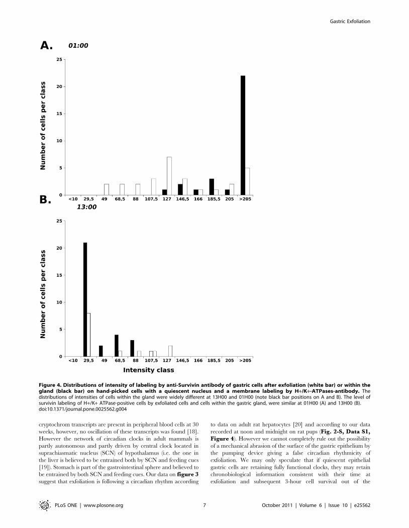
gastric glands is higher at 13:00 than at 01:00 (Figures S2 andS3). On samples of 30 hand-picked cells with a quiescent nucleus
and a membrane labeling by H+/K+-ATPase-antibody, the
distribution of labeling intensity by anti-Survivin antibody differed
widely between pups sampled at 13H00 and 01H00 (Figure 4 Aand B, solid black bars).
The level of survivin labeling of H+/K+ ATPase-positive
exfoliated cells and cells within the gastric gland, were similar as
shown figure 4 on data obtained from a rat pup with a bolus of
saline solution. At 01H00 (Figure 4 A), 78% of cells exfoliated in
vitro were in the same category as cells stained in situ within the
gastric gland (confidence interval: 64,28%–91,72%, at the p = 0.05
level n = 30). At 13H00 (Figure 4 B), 84% of cells exfoliated in
vitro were in the same category as cells stained in situ within the
gastric gland (confidence interval: 75,6–92,4%). The level of LC-3-
b or CLOCK labeling of H+/K+ ATPase-positive exfoliated cells
Gastric Exfoliation
PLoS ONE | www.plosone.org 2 October 2011 | Volume 6 | Issue 10 | e25562
and cells within the gastric gland were determined at 13H00, with
90% (9%) and 100%, respectively.
Discussion
The current study demonstrates that H+/K+ -APTase –
positive quiescent epithelial cells are consistently recovered from
any gastric fluid aspirate obtained from preterm infants from the
5th day of life, regardless of sex and gestational age between 25
weeks and 32 weeks.
A major concern with the use of exfoliated epithelial cells is to
be able to trace their origin in order to rule out any contamination
from the environment (i.e. exfoliated epithelial cells from the
mother’s mammary gland). We used the expression of H+/K+ATPase, or gastric pump, to minimize the risk of detecting
epithelial cells of the mother’s mammary gland. We therefore
chose to trace solely cells expressing membrane H+/K+ -ATPases
because such cells (1) belong to the pit parietal cell lineage, (2) can
be traced by their clear-cut membrane labeling by anti-H+/K+ -
ATPases both on infant and rat pup cells. Our results are in favor
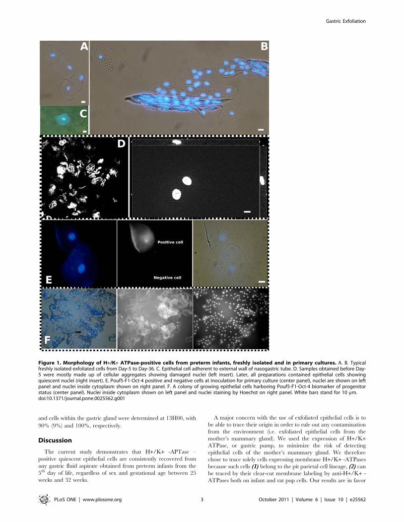
Figure 1. Morphology of H+/K+ ATPase-positive cells from preterm infants, freshly isolated and in primary cultures. A. B. Typicalfreshly isolated exfoliated cells from Day-5 to Day-36. C. Epithelial cell adherent to external wall of nasogastric tube. D. Samples obtained before Day-5 were mostly made up of cellular aggregates showing damaged nuclei (left insert). Later, all preparations contained epithelial cells showingquiescent nuclei (right insert). E. Pouf5-F1-Oct-4 positive and negative cells at inoculation for primary culture (center panel), nuclei are shown on leftpanel and nuclei inside cytoplasm shown on right panel. F. A colony of growing epithelial cells harboring Pouf5-F1-Oct-4 biomarker of progenitorstatus (center panel). Nuclei inside cytoplasm shown on left panel and nuclei staining by Hoechst on right panel. White bars stand for 10 mm.doi:10.1371/journal.pone.0025562.g001
Gastric Exfoliation
PLoS ONE | www.plosone.org 3 October 2011 | Volume 6 | Issue 10 | e25562

Figure 2. 3D-view of exfoliated epithelial cells of preterm infant showing membrane-bound labeling of H+/K+-ATPases (A, B, ingreen), and intact nuclei labeled by Hoechst (in blue). The cytoplasmic expression of Survivin (B, red), LC3-b (C, red) and CLOCK (D, red). Whitebars stand for 10 mm.doi:10.1371/journal.pone.0025562.g002
Table 1. Distribution of patients according to gestational and post-natal age with the number of patients per analysis performedon gastric residual fluid aspirates collected from neonates (25–32 weeks).
Gestational age(weeks) Number of patients (Sex ratio) Number of samples per type of analysis
Culture & immunofluorescenceImmunofluorescence(before D-5/after) ELISA
25 2 (1) - 3 (0/3) 24
27 2 (1) - 4 (0/4) 34
28 8 (1) - 29 (5/24) 23
29 9 (1.7) 1 10 (3/7) 3
30 3 (0.5) - 5 (1/4) -
31 5 (1.5) 3 10 (4/6) 17
32 5 (0.7) - 11 (0/11) 1
Total 34 (1.06) 4 72 (13/69) 102
Note that 13 samples were examined under microscope before 5 days of life. On 178 analyzed samples, we created a database of 72 samples tracing clinical informationand biological analyzes useful for multifactorial analyzes on 33 infants (Data S2).doi:10.1371/journal.pone.0025562.t001
Gastric Exfoliation
PLoS ONE | www.plosone.org 4 October 2011 | Volume 6 | Issue 10 | e25562

of a highly specific detection as infants who received only Preterm
milk formula are loosing more cells expressing H+/K+-ATPase
than infants who received breast milk or milk from another
mother; moreover unfed infant shown on figure 3 is showing low
level of cellular loss.
Epithelial stem cells in the neck region of the gastric gland are
responsible for the continuous renewal of the epithelium through
generation of multiple gastric cell lineages that populate the
epithelium. Some gastric progenitor cells could function as sensors
of damage, or reservoirs with high proliferative potential that can
regenerate portions of the gastric mucosa after injury [13,14,15].
In our study, the integrity of nuclei was systematically ascertained
by Hoechst staining to select for quiescent cells expressing H+/K+-APTases. To prove that exfoliated epithelial cells recovered from
infants were viable, we grew these cells in explants in culture. The
high level of expression of Pouf5-F1-Oct-4 by approximately every
other exfoliated cell (Figure 1-E) suggests that the postnatal
development of gastric gland in preterm infant is similar to that
described in neonatal mice [12]. In addition, most of these
quiescent epithelial cells can readily grow in culture suggesting that
detachment-induced autophagy contributes to the viability of these
cells. These data can be compared with those on human
mammary epithelial cells which retain some clonogenic potentials
after detachment and die after 24–48 hours in the environment
[10]. Quiescent exfoliated epithelial cells can be isolated from
different body fluids (breast milk, urines, digestive fluids). They are
believed to enter rapidly in anoikis after exfoliation. In cells having
lost contact with tissue structure, autophagy corresponds to the
recycling of cellular material as well as to the cell capacity to
mobilize reserves during periods of starvation. Autophagy is
viewed as a survival mechanism during fasting periods; autophagy
must, however, be controlled/stopped at the whole organism level
to prevent self-digestion. Anoikis can be considered as an
autophagic state promoting epithelial cell survival after a timely
loss of contact with extracellular matrix and cell neighbors [8].
Further molecular work on gastric exfoliated cells of preterm
infants would be needed to delineate anoikis from apoptosis. A
functional assay (like using some pHi indicator with a stimulation
by histamin) designed directly on the cell suspension right after the
isolation process would be highly desirable but difficult to perform
due to the small number of cells and the need to cross-check the
cellular phenotypes. Phenotype and physiological profiling by a
transcriptomic approach (like in [1] and [3]) will help to cross-
check the diversity of these H+/K+-ATPase-positive cells. Our
data (Figure 1-F) nevertheless pave the way to the selection of
primary cell cultures derived from human preterm gastric mucosa.
We cannot trace exactly the cellular position in the infant’s
stomach but it would be of crucial interest to know whether the
exfoliated gastric cells can be used to build long-lived gastric units
in vitro [16]. After solving the problem of heavy bacterial
contamination of cells, we believe that it would be informative
to design a functional assay on these cell cultures to ascertain
which part of the stomach (cardia, corpus, pylorus) they are their
representative?
Our study was undertaken to explore the time course of
exfoliation from birth to the time of removal of naso-gastric tube.
From samples obtained between the 5th day of life up to naso-
gastric tube removal (30–36 days later), we did not find any effect
of the gestational age nor of gender but we found a strong effect of
post-natal age and enteral volume (Table 2). Consequently, the
gastric epithelium is mature and functional within few days after
birth whatever the term of birth between 25 to 32 weeks. A non-
significant influence of milk formula on the intensity of exfoliation
has been found (p = 0.058) calling for future works on the influence
of feeding schedule or of various milk formulas on gastric
epithelium’s exfoliation. According to current works on the
nutritional induction of exfoliation by nutrients made on rodents,
gastric epithelial cell exfoliation may represent a direct way to
appreciate the nutritional or pharmacological stress induced by a
substance on the epithelium’s homeostasis.
As collection of gastric residual fluid aspirates is part of the
nursing care routine performed every three hours in many
neonatal intensive care units (NICU), the technique is highly
relevant for time series experiments in chronobiology. Rivkees et al
(2004) have shown that the Rest-Activity patterns of premature
infants are regulated by cycled lighting with an organization
detectable at 34 weeks [17]. Kaeffer et al (2007) have shown that
clock compounds are expressed by gastric exfoliated cells at 28
weeks [3]. Recently, Chen (2010) found that Bmal1 and
Table 2. Clinical parameters of the preterm infants.
Intensity of exfoliation p
0 1 2 3 .4
Post-natal age (days) 1.00 (0.89/6) 18.33 (13.53/21) 15.11 (10.00/18) 18.00 (8.72/19) 32.38 (9.77/8) 0.001
Age post-conceptional (days) 30.33 (1.63/6) 31.19 (2.29/21) 31.17 (1.58/18) 32.52 (2.27/19) 32.62 (1.30/8) 0.044
Gestational age (weeks) 30.00 (1.26/6) 28.62 (1.94/21) 29.06 (1.98/18) 29.95 (1.71/19) 28.00 (0.00/8) 0.036
Birth weight (g) 1403.33 (386.87/6) 1167.71 (334.73/21) 1295.00 (394.07/18) 1501.84 (424.06/19) 1144.38 (143.29/8) 0.044
Weight at sampling day (g) 1343.33 (426.95/6) 1361.00 (368.54/20) 1395.82 (289.55/17) 1730.89 (604.77/19) 1649.37 (246.60/8) 0.043
Birth weight Z-scores 20.5478 (1.12809/6) 21.07 (0.70132/20) 20.8716 (0.61411/17) 20.7217 (0.56185/19) 20.8909 (0.29333/8) 0.360
Enteral volume (ml/day/Kg) 33.18 (24.98/4) 104.48 (62.69/17) 132.93 (46.00/17) 139.79 (47.03/17) 160.04 (8.85/8) 0.001
doi:10.1371/journal.pone.0025562.t002
Table 3. Logistic regression analysis with 4 variables.
aOR 95% CI p
Enteral volume (by 100 ml/day/Kg) 1.9 [0.8–4.3] 0.143
Postnatal Age (by 7 days) 2.8 [1.2–6.7] 0.016
Postconceptional age (by week) 0.3 [0.1–0.9] 0.026
Milk formula 17.8 [0.7–399.3] 0.072
aOR: adjusted Odds ratio, CI: confidence interval.The test of Wald suggest that 2 variables are explicative: the postnatal age andthe postconceptional age.doi:10.1371/journal.pone.0025562.t003
Gastric Exfoliation
PLoS ONE | www.plosone.org 5 October 2011 | Volume 6 | Issue 10 | e25562

Figure 3. Kinetics of exfoliation of H+/K+-ATPase-positive cells over 24-hours followed by ELISA. Four infants were sampled every3 hours on two consecutive 24-h cycles. Infant 1(inverted triangle) and 3 (circle) had similar profiles for both cycles. Infant 3 (triangle) and 4 (diamond)were comparable for the second 24-h cycles. Note that infant 4 (diamond) was unfed but showing low level of cellular loss. White dotted arrow headsindicate acrophases and black arrow heads indicate bathyphases.doi:10.1371/journal.pone.0025562.g003
Gastric Exfoliation
PLoS ONE | www.plosone.org 6 October 2011 | Volume 6 | Issue 10 | e25562
cryptochrom transcripts are present in peripheral blood cells at 30
weeks, however, no oscillation of these transcripts was found [18].
However the network of circadian clocks in adult mammals is
partly autonomous and partly driven by central clock located in
suprachiasmatic nucleus (SCN) of hypothalamus (i.e. the one in
the liver is believed to be entrained both by SCN and feeding cues
[19]). Stomach is part of the gastrointestinal sphere and believed to
be entrained by both SCN and feeding cues. Our data on figure 3suggest that exfoliation is following a circadian rhythm according
to data on adult rat hepatocytes [20] and according to our data
recorded at noon and midnight on rat pups (Fig. 2-S, Data S1,Figure 4). However we cannot completely rule out the possibility
of a mechanical abrasion of the surface of the gastric epithelium by
the pumping device giving a false circadian rhythmicity of
exfoliation. We may only speculate that if quiescent epithelial
gastric cells are retaining fully functional clocks, they may retain
chronobiological information consistent with their time at
exfoliation and subsequent 3-hour cell survival out of the
Figure 4. Distributions of intensity of labeling by anti-Survivin antibody of gastric cells after exfoliation (white bar) or within thegland (black bar) on hand-picked cells with a quiescent nucleus and a membrane labeling by H+/K+-ATPases-antibody. Thedistributions of intensities of cells within the gland were widely different at 13H00 and 01H00 (note black bar positions on A and B). The level ofsurvivin labeling of H+/K+ ATPase-positive cells by exfoliated cells and cells within the gastric gland, were similar at 01H00 (A) and 13H00 (B).doi:10.1371/journal.pone.0025562.g004
Gastric Exfoliation
PLoS ONE | www.plosone.org 7 October 2011 | Volume 6 | Issue 10 | e25562
organism. The induction of gastric cell exfoliation by nutrient
cycle on lactating rat pups can be used in the future to address
questions about the stability of clock information during anoikis. In
our hands, exfoliated cells expressing H+/K+ ATPases can be
recovered from gastric fluids of rat pups in a physiological state
comparable to their counter part cells remaining in situ at the
surface of the gastric gland (Figure 4, Figure S3, Data S1).
In cancer cells like Prostate cancer PC3 cells, the balance
between survival and death is attributed to an interaction between
MAP-LC3-b and survivin [21]. In our work, exfoliated cells
expressing H+/K+ ATPases were found to constantly express high
levels of survivin (Figure 2 B) calling for future investigations to
correlate by Western blot analysis the direct interaction between
MP-LC3-b and survivin. We reported recently that perinatal
denutrition in rat pups has a wide impact on the metabolism and
behavior of young rats [22]. Direct exploration of autophagic and
circadian clock machinery on exfoliated epithelial cells isolated
from preterm infants of different nutritional status will allow to
detect early deficiency which might be corrected by appropriate
nutritional or pharmacological manipulations.
In conclusion, H+/K+ -ATPase positive cells harboring
markers of a progenitor status can be recovered from gastric fluid
aspirates in preterm infants whatever the term or the postnatal age
to measure the expression of specific genes of interest and explore
non-invasively the functioning of neonatal gastric epithelium.
Materials and Methods
Ethics statementsConcerning studies on preterm infants, all samples in Table 1 were
collected from preterm infants in the Neonatal Intensive Care Unit at
the ‘‘Hopital de la Mere et de l’Enfant’’ at Nantes, France. The
protocol was approved by the Nantes Hospital Ethics Committee
(PROG/09/18). Written informed consent were obtained from both
parents (or guardians) within PROG/09/18 and written informed
consent was independently obtained from the healthy volunteer
sampled for inner cheek mechanically exfoliated epithelial cells.
Concerning studies on rat pups, animal experiments were
realized according to the rules of the Nantes animal experimental
unit (in compliance with the European Communities Directive of
24 November 1986 (86/609/EEC) and the Principles of
laboratory animal care (NIH publication no. 85-23, revised
1985). The protocol was strictly non-invasive for rat pups and
mothers and as such was approved under the number P-2010-01
by the local review board of UMR-1280 animal husbandry
(members: Martine Champ, Guillaume Poupeau, Franck Doulay,
Isabelle Jicquel). Animals were euthanized by carbon dioxide
exposure.
Preparation of exfoliated epithelial cellsFluid aspirates of preterm infants or gastric contents of rat pups
were stored at 270uC and processed within one month of sampling
using identical procedure and buffers to recover exfoliated epithelial
cells. The procedure to obtain gastric fluid aspirates is standard in
our neonatalogy unit. Preterm infants equipped with a nasogastric
tube are fed over 3-hour periods, using, for instance 30 mL of milk
slowly instilled by a syringe pump device into the stomach lumen. At
the end of each 3-hour period, the nursing staff disconnects the
device and collects empties the infant’s gastric residues by manual
aspiration using a sterile syringe connected to the nasogastric tube.
Undigested milk and gastric fluids are then removed by a slow
depression of the syringe. The gastric fluid aspirate (0.5–1.5 ml) is
transferred into sterile plastic tubes for immediate processing or
direct storage at 270uC. Briefly, gastric samples were thawed and
diluted out in 15 ml EDTA/DTT buffer (3 mM EDTA and
0,05 mM DTT in PBS0 during 2 min on ice. Cellular material was
pelleted by centrifugation at 2000 rpm during 3 min. Supernatant
was discarded and cellular pellets were resuspended into 12 ml
PBS0, vigorously pipetted six times and pelleted again by
centrifugation at 2000 rpm during 3 min. Cellular pellets were
either stored at 270uC for ELISA or fixed and kept at 4uC up to
microscopic analysis. Fixation was performed with freshly made
formaldehyde 4% solution for one night and replaced by ethanol
70% at 4uC for storage up to labeling after rehydration, or by
paraformaldehyde 4% saline solution after rinsing by PBS0 and
stored at 4uC. Times series of exfoliated cells from the inner cheek of
an adult volunteer were obtained every 6 hours over 24 hours to
trace the nuclear expression of CLOCK factor and to follow LC-3-b
expression over 3 hours of fasting by maintaining cells in PBS-0
buffer. Colon cancer Caco-2 cells were also loaded into freshly
collected gastric fluid aspirates to assess the efficiency of recovery.
Selection of biomarkers, source and specificity of primaryantibodies
H+/K+ ATPase is specifically expressed by pit parietal cells of
the stomach [23] and can be detected with mouse monoclonal
antibodies as well-defined membrane labeling (Abcam (ab-2866)).
The H+/K+ ATPase, or gastric pump, share structural similarities
like an alpha and beta subunit, with the ubiquitous Na+/K+ATPase. The monoclonal antibody that we used is specific for the
beta-subunit which may play a role in maintaining the structural
and functional integrity of the complex. To our knowledge,
expression of H+/K+ ATPase has never been reported to occur in
exfoliated epithelial cells from the mammary gland. Survivin, a
member of the inhibitors-of-apoptosis protein (IAP) family, has
been described as expressed in gastric parietal cells of adult rats
and humans [24]. In situ detection of microtubule-associated
protein light chain 3b (LC3b) by primary antibodies (Santa-Cruz
(sc-28266) has been recommended when this protein constitutive
of the autophagosome is overexpressed during progressive
autophagy [25]. However, autophagy has been demonstrated to
occur in vivo in the surface epithelial cells of neonatal small
intestine of piglets [26]. Rabbit polyclonal anti-survivin (Santa-
Cruz, sc-10811) antibody was used to characterize non-apoptotic
status of our cells and the fraction of highly viable cells with a
status of progenitor cells [27] along with the expression of
POUF5F1-OCT4 [28] (Sigma – P0873 and Abcam, ab-49091).
Determination of cytokeratin 18 expression (a marker of epithelial
origin) was also performed on dot blot (Santa-Cruz, DC-10, sc-
6259 and H-80, sc-28264). SLC-26A7 is expressed mainly by
epithelial cells attached to the tissue [29] (detection by Santa-Cruz
sc-53960). In addition, we targeted CLOCK and Period1, both
involved in circadian rhythms. The transcription factor CLOCK
was detected by antibodies from Abcam ab3517 and Santa-Cruz,
sc-25361. CLOCK harbors a Histone-acetyl-transferase activity,
and histone acetylation is thought to play a key role in the effects of
early nutrition on gene expression, possibly mediating the long-
term effects of early nutrition (nutritional imprinting). Period1 is
another immediate response gene involved in the quick resetting of
the circadian clock (Santa-Cruz sc-25362). Antibodies were used in
serial dilution in confocal microscopy or ELISA according to
manufacturers’ requirements.
Primary culture of exfoliated epithelial cells from fourpreterm infants
Gastric cells were prepared from freshly sampled gastric fluid
aspirates and inoculated in DMEM with 20% fetal calf serum,
Gastric Exfoliation
PLoS ONE | www.plosone.org 8 October 2011 | Volume 6 | Issue 10 | e25562

2 mmol/L L-glutamine, penicillin-streptomycin (Invitrogen,
Cergy Pontoise, France), amphotericin-B, and gentamicin (Sig-
ma-Aldrich, St-Quentin Fallavier, France). We used P-24 (NuncHUpCell
TM
Surface cell culture multidish, 24 wells) and P-6 (NuncHNunclon Vita Multidish, 6 wells) tissue culture plates. Tissue
culture plates were maintained in a humidified incubator at 37uCunder 5% CO2. Trypan blue exclusion test was performed to
check for cell viability.
Nutritional induction of exfoliation in rat pupsFemale Wistar rats were housed either in a 12:12 light:dark (LD)
or dark:light (DL) cycle for a week before impregnation.
Subgroups of 6-day old (n = 40) and 12-day old rat pups (n = 16)
were separated for 5 hours from their mother but kept in eye-
contact in a container maintained in a 37uC-water bath. Pups
were then re-united with their mothers and allowed to feed for one
hour before sacrifice. Rat pups were sacrificed at 13H00 and at
01H00. Contents were recovered by soft massaging of the stomach
and stored at 270uC. Stomachs were used either for in vitro
exfoliation after rapid rinsing in 10 ml PBS0 or for immunohis-
tochemical examination. In vitro experiments were performed
after rat pups half-stomach was sectioned from cardia to pylorus,
and immersed in DMEM-GlutaMax+10% fetal calf serum with
antibiotics for one hour at 37uC under 5% CO2. Both halves of
stomach were recovered with a pincer and gently rinsed in PBS0.
The first half was fixed and placed in cassettes for cryosection,
embedded in DHE polymer taking care to orient the lumen of the
gland upward. The second half was placed overnight in Glucose :
Paraformaldehyde solution (4%) for fixation. The next day, tissues
were placed in cassettes for cryosection, embedded in DHE
polymer taking care to orient the lumen of the gland upward. In
order to realize immunostaining for microscopic examination,
gastric sections were rapidly thawned and saturated for one hour
at 4uC in PBS0+0,2% Bovine Serum Albumin fraction V (BSA).
Primary antibodies were diluted in PBS0+0,2% BSA and
incubated on sections overnight at 4uC in a special device. After
3 cycles of washing by PBS0, sections were incubated with
secondary antibodies and Hoechst 33258. Exfoliated cells in
supernatants were collected by centrifugation (2000 rpm, 3 min)
and stored at 270uC or fixed by freshly made paraformaldhyde
4% saline solution.
Immunocytochemistry and confocal imagingNuclear DNA was stained with Hoechst 33258 (Molecular
Probes) or DRAC-5 (In Vitrogen) fluorochromes. Hoechst staining
was systematically used to determine quiescent versus apoptotic
nuclei. Apoptotic figures were rarely seen and not included in
analysis. Cell and tissue preparations were mounted in Prolong
Gold (In Vitrogen) and visualized under Zeiss apotome micro-
scope or Leica confocal microscope. The intensity of labeling by
the primary – secondary antibodies complex was normalized by
the total surface of the cellular body at the best plane of acquisition
by densitometry with ImageJ software. Cell morphology was
compared with buccal epithelial cells of human adult exfoliated by
swabbing or a colon cancer cell line (Caco-2 cells, purchased from
the European Collection of Animal Cell Culture, UK: ECACC
#860120). With rat pups, exfoliated cells and surface cells of the
gastric gland harboring a quiescent nucleus, fixed as viable
according to a high intensity of MitoTracker Far Red labeling
[30], expressing a strong membrane-bound labeling with H+/K+ATPases antibody were identified manually under microscope.
The best plane of a stack of electronic sections was selected and
quantified for the expression of survivin performed on indepen-
dent preparations with proper corresponding controls. Incubations
of primary antibodies were carried out overnight at 4uC. After 3
washing cycles with PBS0, cell preparations were incubated with
Hoechst 33258 and secondary antibodies were either Goat Anti-
mouse-Alexa-488 or Goat-anti-rabbit-Alexa-568 (both from Mo-
lecular Probes) during one hour at 37uC. After 3 washing cycles
with PBS0, cellular preparations were mounted into ProLong
Gold. With preterm infants, cells expressing clear membrane
labeling by anti-H+/K+-ATPase antibody were selected to
quantify the expression of survivin or LC-3-b or CLOCK.
Preparations were observed under an Apotome-Zeiss microscope
with Axiovision 4.3 software, colocalization and quantification
were realized both under this system and with ImageJ 1.42. Image
stack were captured with a 1 mm z-resolution and 0.31 mm per
pixel as (x, y) resolution. Intensities of labeling expressed by every
cell were arranged in classes of intensity to calculate the frequency
by class (I. e. The number of cells belonging to every class gave the
frequency).
Enzyme linked-immunoassay (ELISA)To prepare antigens, cellular pellets were lyzed by 3 cycles of
freezing – thawing from 220uC to Room Temperature. An
aliquote of lysates was resuspended in saturation buffer (0,5 M
Na2CO3/NaHCO3, pH 9.6) and 100 mL were incubated over-
night on Greiner P-96 microplates. Well contents were discarded
and post-coated with 150 mL of PBS0+0.2% BSA for 2 hours at
4uC. Plaques were rinsed with 100 mL PBS0 and incubated
overnight with primary antibodies diluted according to suppliers’
requirements in PBS0+0.2% BSA, overnight at 4uC. After three
washing cycles by PBS0+0,05% Tween-20, plaques were incubat-
ed with secondary antibodies either Goat Anti-mouse-biotin
(Sigma, B-0529) or Goat-anti-rabbit-biotin (Vector-lab, BA-1000)
at 1/1000 during 2 hours at 37uC. After 3 washing cycles, plates
were incubated with Extravidin-horse-radish peroxidase (Sigma,
E-2886, 1/1000) for 30 min at 37uC. After 3 washing cycles, plates
were revealed by ABTS (Sigma A-9941), in citrate buffer PH 3
(Sigma) containing 2% hydrogen peroxide. Densitometry was
measured with a spectrophotometer Bio-Tek at 405 nm. Data
were expressed as a Relative Index (RI). RI = (Optical density
corrected from background – Mean Optical density of the serie/
Mean Optical density)6100 (expressed in percentage).
Statistics and DatabaseOn data obtained from immunofluorescence imaging, normal-
ity of distribution of the intensity of a specific labeling was tested
on 30 cells at a 5% level according to Kolmogorov test. Clinical
and biological measurements were organized into a database of 72
samples with clinical parameters and biological measurements to
realize contingency and logistic regression analyzes. In this
database, exfoliation was measured on every sample by concurring
techniques (confocal imaging and/or ELISA). As one of our aim
was to evaluate the number of H+/K+-ATPase-positive cells per
sample, we created a semi-quantitative index. The intensity of
exfoliation was defined as the cellular loss per sample. Logistic
regression analysis was performed on 2 groups of intensities of
exfoliation: low intensity from 1 to 3 and high intensity from 4 and
higher. Statistical data obtained by ELISA were analyzed by
ANOVA. All tests were two-tailed and the significance level was
set at .05% level.
Supporting Information
Figure S1 Kinetics of exfoliation of H+/K+-ATPase-positive cells over 24-hours followed by ELISA. Four
infants were sampled every 3 hours on two consecutive 24-h
Gastric Exfoliation
PLoS ONE | www.plosone.org 9 October 2011 | Volume 6 | Issue 10 | e25562
cycles. Note that infant 4 was unfed the first nycthemer of
sampling (orange diamond) but showing low level of cellular loss.
Profiles were obtained in parallel preparations of H+/K+-ATPases
detection, with antibodies against survivin (A, B), MAP-LC-3-b (C,
D)), Pouf5-F1-Oct4 (E, F), PERIOD1 (G, H) and CLOCK (I, J).
Note that by ELISA, we detect the antigens from any cellular
phenotypes in the preparation along with cell-free antigens.
(TIF)
Figure S2 Detection of biomarkers of exfoliated cells incell culture supernatants or stomach contents. The
detection of biomarkers was done by ELISA on preparations of
epithelial gastric cells exfoliated from gastric glands maintained in
culture for 60 min (black symbols) or recovered from gastric
contents (white symbols) of 12-day-old rat pups refed one hour
before sacrifice. Note the similarity of antigen detection profiles of
black and white symbols for all biomarkers. A significant difference
was found between 13:00 and 01:00 series for all biomarkers by
Student’t test (p,0.05) except LC-3-b in contents, and CLOCK
both in vivo and in vitro exfoliation.
(TIF)
Figure S3 Gastric exfoliated cells of rat pups. A. Typical
exfoliated cells expressing membrane-bound H+/K+ ATPase
(green, Alexa 488), showing a quiescent nucleus (blue, Hoechst). B.
Survivin (red, Alexa 568). C. LC-3-B (red, Alexa 568). D. CLOCK
(red, Alexa 568). E. Viable mitochondrial network in an exfoliated
cell (yellow, MitoTracker Far Red). F. Surface epithelial cells of
the gastric gland with viable mitochondries (yellow) showing a
gradient of MitoTracker Far Red labeling with a maximum on the
outside of the gastric sample and a minima inside the gastric
sample.White bars stand for 10 mm.
(TIF)
Data S1 Additional information on methodology andresults.(DOC)
Data S2 Raw data of clinical and biological variablesmeasured on preterm infants.(XLS)
Acknowledgments
We thank the following for their valuable help and discussion: Philippe
Hulin (IFR-26 Cellular and Tissular Imaging Core Facility of Nantes
University (MicroPICell)); Guillaume Poupeau and Isabelle Jicquel for
animal care (both from the Breeding facilities of UMR-1280); Laurent
Gouthiere (Euroestech, Esvres, France) for advice in using TSA Serial
Cosinor; Gael Mazeiras (CHU of Nantes for clinical management);
Christian Laboisse (CHU of Nantes) and Patricia Parnet (UMR-1280) for
valuable suggestions on the manuscript; Martine Champ (UMR-1280) for
encouragement and support; and our former Master2 students (Sciences de
l’aliment et nutrition humaine) of the University of Nantes: Fanny Morel,
Han Qi, Blandine Pourtier.
Author Contributions
Conceived and designed the experiments: BK AL AF-C OG-Q JCR.
Performed the experiments: BK AL AF-C OG-Q JCR. Analyzed the data:
BK JCR DD TM HB. Contributed reagents/materials/analysis tools: BK
OG-Q. Wrote the paper: BK DD JCR. Statistical analysis: BK DD TM
HB. Database processing: BK DD TM HB.
References
1. Chapkin RS, Zhao C, Ivanov I, Davidson LA, Goldsby JS, et al. (2010)
Noninvasive stool-based detection of infant gastrointestinal development usinggene expression profiles from exfoliated epithelial cells. Am J Physiol Gastro-
intest Liver Physiol 298: G582–G589.
2. Aoyama F, Sawaguchi A, Ide S, Kitamura K, Suganuma T, et al. (2008)Exfoliation of gastric pit-parietal cells into the gastric lumen associated with a
stimulation of isolated rat gastric mucosa in vitro: a morphological study by theapplication of cryotechniques. Histochem Cell Biol 129: 785–793.
3. Kaeffer B, Des Robert C, Alexandre-Gouabau MC, Pagniez A, Amarger V, et al.(2007) Recovery of exfoliated cells from the gastro-intestinal tract of premature
infants: a new tool to obtain ‘‘non-invasive biopsies’’? Pediatr Res 62: 564–569.
4. Kitazawa H, Nishihara T, Nambu T, Nishzawa H, Iwaki M, et al. (2004)Intectin, a novel small intestine-specific glycosylphosphatidylinositol-anchored
protein, accelerates apoptosis of intestinal epithelial cells. J Biol Chem 279:42867–42874.
5. Frisch SM, Francis H (1994) Disruption of epithelial cell-matrix interactions
induces apoptosis. J Cell Biol 124: 619–626.6. Gilemore AP (2005) Anoikis. Cell Death Differ 12: 1473–1477.
7. Liu Z, Li H, Wu X, Yoo BH, Yan SR, et al. (2006) Detachment-inducedupregulation of XIAP and cIAP2 delays anoikis of intestinal epithelial cells.
Oncogene 25: 7680–7690.8. Lock R, Debnath J (2008) Extracellular matrix regulation of autophagy. Curr
Opin Cell Biol 20: 583–588.
9. Debnath J, Mills KR, Collins NL, Reginato MJ, Muthuswamy SK, et al. (2002)The role of apoptosis in creating and maintaining luminal space within normal
and oncogene-expressing mammary acini. Cell 111: 29–40.10. Fung C, Lock R, Gao S, Salas SE, Debnath J (2008) Induction of autophagy during
extracellular matrix detachment promotes cell survival. Mol Biol Cell 19: 797–806.
11. Levine B, Klionsky DJ (2004) Development by self-digestion: molecularmechanisms and biological functions of autophagy. Dev Cell 6: 463–477.
12. Bandaletova T, Bailey N, Bingham SA, Loktionov A (2002) Isolation ofexfoliated colonocytes from human stools as a new technique for colonic
cytology. APMIS 110: 239–246.
13. Nomura S, Esumi H, Job C, Tan SS (1998) Lineage and clonal development ofgastric glands. Dev Biol 204: 124–135.
14. Dimaline R, Varro A (2007) Attack and defence in the gastric epithelium adelicate balance. Exp Physiol 92: 591–601.
15. Laine L, Takeuchi K, Tarnawski A (2008) Gastric mucosal defense andcytoprotection: bench to bedside. Gastroenterology 135: 41–60.
16. Barker N, Huch M, Kujala P, van de Wetering M, Snippert HJ, et al. (2010)
Lgr5+ve Stem Cells Drive Self-Renewal in the Stomach and Build Long-LivedGastric Units In Vitro. Cell Stem Cell 6: 25–36.
17. Rivkees SA, Mayes L, Jacobs H, Gross I (2004) Rest-Activity patterns of
premature infants are regulated by cycled lighting. Pediatrics 113: 833–839.18. Chen A (2010) The circadian rhythm of expression of Bmal1 and Cry1 in
peripheral blood mononuclear cells of preterm infants. J Matern Fetal Neonatal
Med 23: 1172–1175.19. Green CB, Takahashi JS, Bass J (2008) The meter of metabolism. Cell 134:
728–742.20. Sachdeva UM, Thompson CB (2008) Diurnal rhythms of autophagy. Autophagy
4: 581–589.21. Roca H, Varsos Z, Pienta KJ (2008) CCL2 protects prostate cancer PC3 cells
from autophagic death via phosphatidylinositol 3-kinase/AKT-dependent
survivin up-regulation. J Biol Chem 283: 25057–25073.22. Orozco-Solis R, Barbosa Matos RJ, Lopes de Souza S, Grit I, Kaeffer B, et al.
(2011) Perinatal nutrient restriction induces long-lasting alterations in thecircadian expression pattern of genes regulating food intake and energy
metabolism. International Journal of Obesity 35: 990–1000.
23. Sawaguchi A, McDonald KL, Forte JG (2004) High-pressure freezing of isolatedgastric glands provides new insight into the fine structure and subcellular
localization of H+/K+ -ATPase in gastric parietal cells. J Histochem Cytochem52: 77–86.
24. Chiou S-K, Moon WS, Jones MK, Tarnawski AS (2003) Survivin expression inthe stomach: implications for mucosal integrity and protection. Biochem Biophys
Res Commun 305: 374–379.
25. Martinet W, De Meyer GR, Andries L, Herman AG, Kockx MM (2006)Detection of autophagy in tissue by standard immunohistochemistry: possibilities
and limitations. Autophagy 2: 55–57.26. Godlewski MM, Hallay N, Bierea JB, Zabielski R (2007) Molecular mechanism
of programmed cell death in the gut epithelium of neonatal piglets. Journal of
Physiology and Pharmacology 58: 97–113.27. Tarnawski A, Pai R, Deng X, Ahluwalia A, Khomenko T, et al. (2007) Aging
gastropathy-novel mechanisms: hypoxia, up-regulation of multifunctionalphosphatase PTEN, and proapoptotic factors. Gastroenterology 133:
1938–1947.
28. Trosko JE (2006) From adult stem cells to cancer stem cells. Oct-4 gene, cell-cellcommunication, and hormones during tumor promotion. Ann N Y Acad Sci
1089: 36–58.29. Petrovic S, Ju X, Barone S, Seidler U, Alper SL, et al. (2003) Identification of a
basolateral Cl-/HCO3- exchanger specific to gastric parietal cells. Am J PhysiolGastrointest Liver Physiol 284: G1093–G1103.
30. Poot M, Zhang Y-Z, Kramer JA, Wells KS, Jones LJ, et al. (1996) Analysis of
mitochondrial morphology and function with novel fixable fluorescent stains.J Histochem Cytochem 44: 1363–1372.
Gastric Exfoliation
PLoS ONE | www.plosone.org 10 October 2011 | Volume 6 | Issue 10 | e25562
Copyright © 2022 FDOKUMEN