No evidence for structural brain changes in young adolescents at ultra high risk for psychosis
6
No evidence for structural brain changes in young adolescents at ultra high risk for psychosis Tim B. Ziermans a, ⁎, Sarah Durston a , Mirjam Sprong a , Hilde Nederveen a , Neeltje E.M. van Haren b , Hugo G. Schnack b , Bertine E. Lahuis a , Patricia F. Schothorst a , Herman van Engeland a a Department of Child and Adolescent Psychiatry, Rudolf Magnus Institute of Neuroscience, University Medical Center Utrecht, the Netherlands b Department of Psychiatry, Rudolf Magnus Institute of Neuroscience, University Medical Center Utrecht, the Netherlands article info abstract Article history: Received 14 August 2008 Received in revised form 7 April 2009 Accepted 12 April 2009 Available online 5 May 2009 Objective: The onset of psychosis is thought to be preceded by neurodevelopmental changes in the brain. However, the timing of these changes has not been established. We investigated structural brain changes in a sample of young adolescents (12–18 years) at ultra high-risk for psychosis (UHR). Methods: Structural MRI data from young UHR subjects (n = 54) and typically developing, matched controls (n =54) were acquired with a 1.5 Tesla scanner and compared. Results: None of the measures differed between UHR subjects and controls. Conclusions: Our results do not support the presence of gross neuroanatomical changes in young UHR subjects. This suggests that early changes are too subtle to detect with conventional imaging techniques. Therefore, changes observed in older cohorts may only onset later developmentally or occur secondary to prodromal symptoms. © 2009 Elsevier B.V. All rights reserved. Keywords: Psychosis Ultra high risk Adolescence Structural MRI Voxelbased morphometry Neurodevelopment 1. Introduction A growing body of evidence suggests early neurodevelop- mental brain changes preceding psychosis that are thought to progress into adolescence and adulthood (Rapoport et al., 2005). Recently, neuroimaging studies have focused on genetic and clinical high-risk cohorts to define the nature of these changes and to identify which of these may mark vulnerability for psychosis (Cannon, 2005). Subjects of clinical high-risk cohorts are commonly referred to as being at “ultra high-risk” (UHR), at “prodromal high-risk” or having an “at risk mental state” (ARMS) for psychosis. Several research groups have reported premorbid structural and functional brain changes in these cohorts. However, the timing of these changes is not established (for reviews see Pantelis et al., 2005; Wood et al., 2008): It is unclear whether they are truly premorbid or rather associated with prodromal symptoms. A large volumetric MRI study in a UHR population aged 20 years reported smaller whole brain volume for subjects at UHR for psychosis compared to controls (Velakoulis et al., 2006), while several voxelbased morphometry (VBM) studies have shown changes in both gray (GM; Borgwardt et al., 2007, 2008; Meisenzahl et al., 2008; Pantelis et al., 2003) and white matter (WM; Walterfang et al., 2008; Witthaus et al., 2008) clusters in young adults (20–25 years) at UHR, predominantly in (pre-)frontal and temporal lobe areas. Interestingly, long- itudinal reports suggest a differential development of changes in brain structure for individuals who convert to psychosis compared to those who do not (Pantelis et al., 2003; Walterfang et al., 2008). Reports to date of brain changes in subjects at UHR have focused on the age range of 20–25 years, when psychosis typically first occurs (Kessler et al., 2007). However, on average the earliest prodromal signs occur 4.8 years before onset (Hafner and Maurer, 2006). If neurobiological changes precede psychotic breakdown, these should be present in the at-risk period irrespective of the age at which psychotic breakdown occurs. To test whether this is indeed the case, we investigated brain structure volumes in a well-defined sample of young adolescents at UHR for psychosis (aged 12–18 years). We Schizophrenia Research 112 (2009) 1–6 ⁎ Corresponding author. Department of Child and Adolescent Psychiatry, University Medical Center, Heidelberglaan 100, HP A01.468, 3584 CX Utrecht, the Netherlands. E-mail address: [email protected] (T.B. Ziermans). 0920-9964/$ – see front matter © 2009 Elsevier B.V. All rights reserved. doi:10.1016/j.schres.2009.04.013 Contents lists available at ScienceDirect Schizophrenia Research journal homepage: www.elsevier.com/locate/schres
Transcript of No evidence for structural brain changes in young adolescents at ultra high risk for psychosis

Schizophrenia Research 112 (2009) 1–6
Contents lists available at ScienceDirect
Schizophrenia Research
j ourna l homepage: www.e lsev ie r.com/ locate /schres
No evidence for structural brain changes in young adolescents at ultra highrisk for psychosis
Tim B. Ziermans a,⁎, Sarah Durston a, Mirjam Sprong a, Hilde Nederveen a, Neeltje E.M. van Haren b,Hugo G. Schnack b, Bertine E. Lahuis a, Patricia F. Schothorst a, Herman van Engeland a
a Department of Child and Adolescent Psychiatry, Rudolf Magnus Institute of Neuroscience, University Medical Center Utrecht, the Netherlandsb Department of Psychiatry, Rudolf Magnus Institute of Neuroscience, University Medical Center Utrecht, the Netherlands
a r t i c l e i n f o
⁎ Corresponding author. Department of Child and AUniversity Medical Center, Heidelberglaan 100, HP A01the Netherlands.
E-mail address: [email protected] (T.B. Zi
0920-9964/$ – see front matter © 2009 Elsevier B.V.doi:10.1016/j.schres.2009.04.013
a b s t r a c t
Article history:Received 14 August 2008Received in revised form 7 April 2009Accepted 12 April 2009Available online 5 May 2009
Objective: The onset of psychosis is thought to be preceded by neurodevelopmental changes inthe brain. However, the timing of these changes has not been established. We investigatedstructural brain changes in a sample of young adolescents (12–18 years) at ultra high-risk forpsychosis (UHR).Methods: Structural MRI data from young UHR subjects (n=54) and typically developing,matched controls (n=54) were acquired with a 1.5 Tesla scanner and compared.Results: None of the measures differed between UHR subjects and controls.Conclusions: Our results do not support the presence of gross neuroanatomical changes inyoung UHR subjects. This suggests that early changes are too subtle to detect with conventionalimaging techniques. Therefore, changes observed in older cohorts may only onset laterdevelopmentally or occur secondary to prodromal symptoms.
© 2009 Elsevier B.V. All rights reserved.
Keywords:PsychosisUltra high riskAdolescenceStructural MRIVoxelbased morphometryNeurodevelopment
1. Introduction
A growing body of evidence suggests early neurodevelop-mental brain changes preceding psychosis that are thought toprogress into adolescence and adulthood (Rapoport et al.,2005). Recently, neuroimaging studies have focused ongenetic and clinical high-risk cohorts to define the nature ofthese changes and to identify which of these may markvulnerability for psychosis (Cannon, 2005). Subjects of clinicalhigh-risk cohorts are commonly referred to as being at “ultrahigh-risk” (UHR), at “prodromal high-risk” or having an “atrisk mental state” (ARMS) for psychosis. Several researchgroups have reported premorbid structural and functionalbrain changes in these cohorts. However, the timing of thesechanges is not established (for reviews see Pantelis et al.,2005; Wood et al., 2008): It is unclear whether they are trulypremorbid or rather associated with prodromal symptoms.
dolescent Psychiatry,.468, 3584 CX Utrecht,
ermans).
All rights reserved.
A large volumetric MRI study in a UHR population aged20 years reported smaller whole brain volume for subjects atUHR for psychosis compared to controls (Velakoulis et al.,2006), while several voxelbased morphometry (VBM) studieshave shown changes in both gray (GM; Borgwardt et al., 2007,2008; Meisenzahl et al., 2008; Pantelis et al., 2003) and whitematter (WM; Walterfang et al., 2008; Witthaus et al., 2008)clusters in young adults (20–25 years) at UHR, predominantlyin (pre-)frontal and temporal lobe areas. Interestingly, long-itudinal reports suggest a differential development of changesin brain structure for individuals who convert to psychosiscompared to thosewhodonot (Pantelis et al., 2003;Walterfanget al., 2008). Reports to date of brain changes in subjects at UHRhave focused on the age range of 20–25 years, when psychosistypically first occurs (Kessler et al., 2007). However, on averagethe earliest prodromal signs occur 4.8 years before onset(Hafner andMaurer, 2006). If neurobiological changes precedepsychotic breakdown, these should be present in the at-riskperiod irrespective of the age at which psychotic breakdownoccurs. To test whether this is indeed the case, we investigatedbrain structure volumes in a well-defined sample of youngadolescents at UHR for psychosis (aged 12–18 years). We

Table 1Inclusion criteria.
Attenuated positive symptoms (APS)
Presence of at least one of the following SIPS symptomswith a score between3 and 5 and an appearance of several times per week for a period of at leastone week:
• Unusual thought content/delusional ideas (P1)• Suspiciousness/persecutory ideas (P2)• Grandiosity (P3)• Perceptual abnormalities/hallucinations (P4)• Disorganized communication (P5)• Odd behaviour or appearance (D1)
Brief limited intermittent psychotic symptoms (BLIPS)
Presence of at least one of the following PANSS symptoms that resolvespontaneously in 7 days and an interval between episodes with thesesymptoms of at least one week (two episodes of BLIPS separated by lessthan one week are considered as being one episode; if the total durationthen becomes more than one week, the transition criterion is fulfilled):
• Hallucinations (PANSS P3 score ≥4)• Delusions (PANSS P1, P5, P6 score ≥4)• Formal thought disorder (PANSS P2 score ≥4)
Familial risk plus reduced functioning
A change in mental state or functioning leading to a reduction of 30% ormore on the Global Assessment of Functioning scale for at least one monthwithin the last year compared to the highest level of previous functioning,plus at least one of the following risk indicators:
• One first- or second-degree relative with a history of any DSM-IV psychoticdisorder (not due to a medical factor or substance induced)
• A schizotypal personality disorder of the index person according to DSM-IV
Basic symptoms
Presence of at least two of the following symptoms from the cluster“cognitive disturbances” for more than one year, with a BSABS-P score ≥3during the last three months:
• Inability to divide attention (A.8.4)• Thought interferences (C.1.1)• Thought pressure (C.1.3)• Thought blockages (C.1.4)• Disturbances of receptive speech (C.1.6)• Disturbances of expressive speech (C.1.7)• Disturbances of abstract thinking (“concretism”; C.1.16)• Unstable ideas of reference (“subject-centrism”; C.1.17)• Captivation of attention by details of the visual field (C.2.9)
SIPS – Structured Interview for Prodromal Syndromes; PANSS – Positive andNegative Syndrome Scale; BSABS-P – Bonn Scale for the Assessment of BasicSymptoms – Prediction List.
2 T.B. Ziermans et al. / Schizophrenia Research 112 (2009) 1–6
hypothesized that the UHR group would have smaller totalbrain volumeand lessGMandWMdensity in (pre-) frontal andmedial temporal lobe areas.
2. Methods
2.1. Subjects
Fifty-four adolescents (52 Caucasian, 2 Asian) meeting atleast 1 of 4 criteria for UHR were referred by generalpractitioners or other psychiatric clinics and included in thisstudy. A further 54 matched typically developing adolescents(52 Caucasian, 1 Asian, 1 Hispanic) were included. There wasalso a subgroup of nineteen (35%) UHR patients that metcriteria for pervasive developmental disorder – not otherwisespecified (PDD-NOS; American Psychiatric Association, 1994).While these subjects in general showed behavioral problemsat an earlier age (Sprong et al., 2008), they also met at leastone of the UHR criteria in the last year.
A complete overview of UHR inclusion criteria is displayedin Table 1. Briefly, the following criteria were applied: 1)attenuated positive symptoms, 2) brief, limited, or intermit-tent psychotic symptoms, 3) a 30% reduction in overall levelof social, occupational/school-, and psychological functioning(i.e. GAF-score) in the past year, combined with a genetic riskof psychosis, and 4) two or more of a selection of nine basicsymptoms, i.e. subjective deficits in cognitive, perceptual, andmotor functioning. The first three inclusion criteria wereassessed with the Structured Interview for ProdromalSyndromes (SIPS; McGlashan et al., 2001). The fourthinclusion criterion was assessed with the Bonn Scale for theAssessment of Basic Symptoms-Prediction List (BSABS-P;Schultze-Lutter and Klosterkötter, 2002). The numbers ofindividuals per UHR criterion are listed in Table 2. The studydesign allowed for repeatedmeasures to be performed at 9,18and 24 months after inclusion. At these assessments, subjectswere re-evaluated to determine possible transition topsychosis according to SIPS criteria (McGlashan et al.,2001). Additionally, transition was retrospectively confirmedby clinical expert consensus (HvE, PS).
The subgroup of patients with PDD-NOS had received aprepubertal DSM-IV diagnosis of PDD-NOS (American Psychia-tric Association, 1994), while also meeting criteria for MCDD(i.e. early childhood-onset impairments in affect regulation,social behavior/sensitivity, and cognition (Cohen et al., 1994)).Childrenwith PDD-NOS, MCDD subtype are at risk for develop-ing psychotic disorders later in life (Van Engeland and Van derGaag, 1994). The diagnosis was confirmed in a psychiatricexamination including the Autism Diagnostic Interview-Revised (Lord et al., 1994), as well as a parent interview basedon the diagnostic criteria for MCDD, which was developed forinternal use at the UMC. Diagnoses were confirmed by expertclinical opinion (HvE, PS). A more detailed description of thisUHR-subgroup is available elsewhere (Sprong et al., 2008).
Typically developing controls were recruited from sec-ondary schools in the region of Utrecht. They were excluded ifthey met one of the UHR-criteria, if they or any first degreerelative had a history of any psychiatric illness, or if there wasa second-degree relative with a psychotic disorder. Exclusioncriteria were assessed with SIPS & BSABS-P interviews and(parent) questionnaires.
In addition to the screening instruments a modifiedversion of the revised self-report Schizotypal PersonalityQuestionnaire (SPQ-R) was used to assess schizotypalpersonality traits (Raine, 1991; Vollema and Hoijtink, 2000).
All participants were aged between 12 and 18 years andnone of them were psychotic at the time of inclusion in thestudy. Subjects were excluded if there was evidence for anypast or present neurological disorder (e.g., epilepsy). Drug-and alcohol abusewere additional exclusion criteria, althoughpatients were allowed to have a history of drug use if symp-toms had also been present in the absence of drugs. Elevenpatients reported having used drugs at least five times withinthe last year (all Marijuana and two patients with additionaluse of psychostimulants). Three of these patients were con-sidered to be frequent users (at least once a week within thelast month) at the time of assessment. Also, all individuals
3T.B. Ziermans et al. / Schizophrenia Research 112 (2009) 1–6
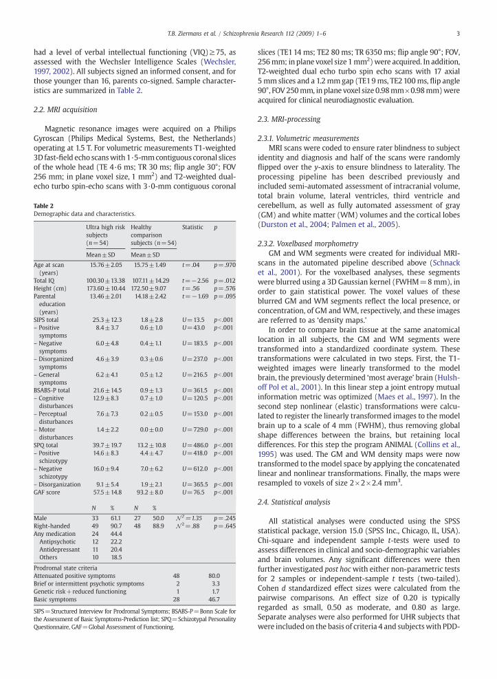
had a level of verbal intellectual functioning (VIQ)≥75, asassessed with the Wechsler Intelligence Scales (Wechsler,1997, 2002). All subjects signed an informed consent, and forthose younger than 16, parents co-signed. Sample character-istics are summarized in Table 2.
2.2. MRI acquisition
Magnetic resonance images were acquired on a PhilipsGyroscan (Philips Medical Systems, Best, the Netherlands)operating at 1.5 T. For volumetric measurements T1-weighted3D fast-field echo scanswith 1·5-mmcontiguous coronal slicesof the whole head (TE 4·6 ms; TR 30 ms; flip angle 30°; FOV256 mm; in plane voxel size, 1 mm2) and T2-weighted dual-echo turbo spin-echo scans with 3·0-mm contiguous coronal
Table 2Demographic data and characteristics.
Ultra high risksubjects(n=54)
Healthycomparisonsubjects (n=54)
Statistic p
Mean±SD Mean±SD
Age at scan(years)
15.76±2.05 15.75±1.49 t=.04 p=.970
Total IQ 100.30±13.38 107.11±14.29 t=−2.56 p=.012Height (cm) 173.60±10.44 172.50±9.07 t=.56 p=.576Parentaleducation(years)
13.46±2.01 14.18±2.42 t=−1.69 p=.095
SIPS total 25.3±12.3 1.8±2.8 U=13.5 pb .001– Positivesymptoms
8.4±3.7 0.6±1.0 U=43.0 pb .001
– Negativesymptoms
6.0±4.8 0.4±1.1 U=183.5 pb .001
– Disorganizedsymptoms
4.6±3.9 0.3±0.6 U=237.0 pb .001
– Generalsymptoms
6.2±4.1 0.5±1.2 U=216.5 pb .001
BSABS-P total 21.6±14.5 0.9±1.3 U=361.5 pb .001– Cognitivedisturbances
12.9±8.3 0.7±1.0 U=120.5 pb .001
– Perceptualdisturbances
7.6±7.3 0.2±0.5 U=153.0 pb .001
– Motordisturbances
1.4±2.2 0.0±0.0 U=729.0 pb .001
SPQ total 39.7±19.7 13.2±10.8 U=486.0 pb .001– Positiveschizotypy
14.6±8.3 4.4±4.7 U=418.0 pb .001
– Negativeschizotypy
16.0±9.4 7.0±6.2 U=612.0 pb .001
– Disorganization 9.1±5.4 1.9±2.1 U=365.5 pb .001GAF score 57.5±14.8 93.2±8.0 U=76.5 pb .001
N % N %
Male 33 61.1 27 50.0 N 2=1.35 p=.245Right-handed 49 90.7 48 88.9 N 2=.88 p=.645Any medication 24 44.4Antipsychotic 12 22.2Antidepressant 11 20.4Others 10 18.5
Prodromal state criteriaAttenuated positive symptoms 48 80.0Brief or intermittent psychotic symptoms 2 3.3Genetic risk+reduced functioning 1 1.7Basic symptoms 28 46.7
SIPS=Structured Interview for Prodromal Symptoms; BSABS-P=Bonn Scale forthe Assessment of Basic Symptoms-Prediction list; SPQ=Schizotypal PersonalityQuestionnaire, GAF=Global Assessment of Functioning.
slices (TE114 ms; TE2 80 ms; TR 6350 ms; flip angle 90°; FOV,256mm; inplane voxel size 1mm2)were acquired. In addition,T2-weighted dual echo turbo spin echo scans with 17 axial5 mm slices and a 1.2 mmgap (TE19ms, TE2 100ms, flip angle90°, FOV250mm, inplane voxel size 0.98mm×0.98mm)wereacquired for clinical neurodiagnostic evaluation.
2.3. MRI-processing
2.3.1. Volumetric measurementsMRI scans were coded to ensure rater blindness to subject
identity and diagnosis and half of the scans were randomlyflipped over the y-axis to ensure blindness to laterality. Theprocessing pipeline has been described previously andincluded semi-automated assessment of intracranial volume,total brain volume, lateral ventricles, third ventricle andcerebellum, as well as fully automated assessment of gray(GM) and white matter (WM) volumes and the cortical lobes(Durston et al., 2004; Palmen et al., 2005).
2.3.2. Voxelbased morphometryGM and WM segments were created for individual MRI-
scans in the automated pipeline described above (Schnacket al., 2001). For the voxelbased analyses, these segmentswere blurred using a 3D Gaussian kernel (FWHM=8mm), inorder to gain statistical power. The voxel values of theseblurred GM and WM segments reflect the local presence, orconcentration, of GM andWM, respectively, and these imagesare referred to as ‘density maps.’
In order to compare brain tissue at the same anatomicallocation in all subjects, the GM and WM segments weretransformed into a standardized coordinate system. Thesetransformations were calculated in two steps. First, the T1-weighted images were linearly transformed to the modelbrain, the previously determined ‘most average’ brain (Hulsh-off Pol et al., 2001). In this linear step a joint entropy mutualinformation metric was optimized (Maes et al., 1997). In thesecond step nonlinear (elastic) transformations were calcu-lated to register the linearly transformed images to the modelbrain up to a scale of 4 mm (FWHM), thus removing globalshape differences between the brains, but retaining localdifferences. For this step the program ANIMAL (Collins et al.,1995) was used. The GM and WM density maps were nowtransformed to the model space by applying the concatenatedlinear and nonlinear transformations. Finally, the maps wereresampled to voxels of size 2×2×2.4 mm3.
2.4. Statistical analysis
All statistical analyses were conducted using the SPSSstatistical package, version 15.0 (SPSS Inc., Chicago, IL, USA).Chi-square and independent sample t-tests were used toassess differences in clinical and socio-demographic variablesand brain volumes. Any significant differences were thenfurther investigated post hocwith either non-parametric testsfor 2 samples or independent-sample t tests (two-tailed).Cohen d standardized effect sizes were calculated from thepairwise comparisons. An effect size of 0.20 is typicallyregarded as small, 0.50 as moderate, and 0.80 as large.Separate analyses were also performed for UHR subjects thatwere included on the basis of criteria 4 and subjectswith PDD-
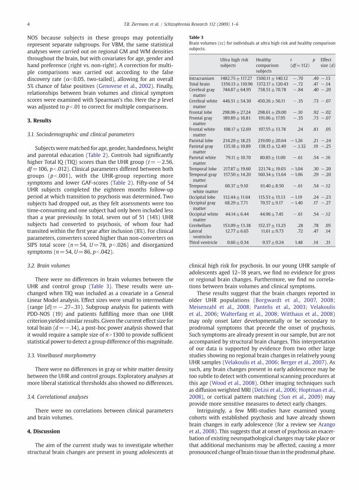
Table 3Brain volumes (cc) for individuals at ultra high risk and healthy comparisonsubjects.
Ultra high risksubjects
Healthycomparisonsubjects
t(df=112)
p Effectsize (d)
Intracranium 1482.75±117.27 1500.11±140.12 − .70 .49 − .13Total brain 1356.13±110.96 1372.17±120.43 − .72 .47 − .14Cerebral graymatter
744.87±64.95 758.51±70.78 − .84 .40 − .20
Cerebral whitematter
446.51±54.30 450.26±56.11 − .35 .73 − .07
Frontal lobe 298.06±27.24 298.61±29.00 − .10 .92 − .02Frontal graymatter
189.89±16.81 191.06±17.95 − .35 .73 − .07
Frontal whitematter
108.17±12.69 107.55±13.78 .24 .81 .05
Parietal lobe 214.29±18.25 219.00±20.64 −1.26 .21 − .24Parietal graymatter
135.18±10.89 138.15±12.49 − .1.32 .19 − .25
Parietal whitematter
79.11±10.70 80.85±11.00 − .61 .54 − .16
Temporal lobe 217.87±19.60 221.74±19.03 −1.04 .30 − .20Temporal graymatter
157.50±14.20 160.34±13.64 −1.06 .29 − .20
Temporalwhite matter
60.37±9.10 61.40±8.50 − .61 .54 − .12
Occipital lobe 112.44±11.64 115.53±15.13 −1.19 .24 − .23Occipital graymatter
68.29±7.71 70.57±9.17 −1.40 .17 − .27
Occipital whitematter
44.14±6.44 44.96±7.45 − .61 .54 − .12
Cerebellum 153.09±13.38 152.37±13.25 .28 .78 .05Lateralventricles
12.77±6.65 11.61±9.73 .72 .47 .14
Third ventricle 0.66±0.34 0.57±0.24 1.48 .14 .31
4 T.B. Ziermans et al. / Schizophrenia Research 112 (2009) 1–6
NOS because subjects in these groups may potentiallyrepresent separate subgroups. For VBM, the same statisticalanalyses were carried out on regional GM and WM densitiesthroughout the brain, but with covariates for age, gender andhand preference (right vs. non-right). A correction for multi-ple comparisons was carried out according to the falsediscovery rate (αb0.05, two-tailed), allowing for an overall5% chance of false positives (Genovese et al., 2002). Finally,relationships between brain volumes and clinical symptomscores were examined with Spearman's rho. Here the p levelwas adjusted to pb .01 to correct for multiple comparisons.
3. Results
3.1. Sociodemographic and clinical parameters
Subjectswerematched for age, gender, handedness, heightand parental education (Table 2). Controls had significantlyhigher Total IQ (TIQ) scores than the UHR group (t=−2.56,df=106, pb .012). Clinical parameters differed between bothgroups (pb .001), with the UHR-group reporting moresymptoms and lower GAF-scores (Table 2). Fifty-one of 54UHR subjects completed the eighteen months follow-upperiod at which transition to psychosis was determined. Twosubjects had dropped out, as they felt assessments were tootime-consuming and one subject had only been included lessthan a year previously. In total, seven out of 51 (14%) UHRsubjects had converted to psychosis, of whom four hadtransited within the first year after inclusion (8%). For clinicalparameters, converters scored higher than non-converters onSIPS total score (n=54, U=78, pb .026) and disorganizedsymptoms (n=54, U=86, pb .042).
3.2. Brain volumes
There were no differences in brain volumes between theUHR and control group (Table 3). These results were un-changed when TIQ was included as a covariate in a GeneralLinear Model analysis. Effect sizes were small to intermediate(range [d]=− .27–.31). Subgroup analysis for patients withPDD-NOS (19) and patients fulfilling more than one UHRcriterionyielded similar results. Given the current effect size fortotal brain (d=− .14), a post-hoc power analysis showed thatit would require a sample size of nN1300 to provide sufficientstatistical power to detect a group difference of thismagnitude.
3.3. Voxelbased morphometry
There were no differences in gray or white matter densitybetween the UHR and control groups. Exploratory analyses atmore liberal statistical thresholds also showed no differences.
3.4. Correlational analyses
There were no correlations between clinical parametersand brain volumes.
4. Discussion
The aim of the current study was to investigate whetherstructural brain changes are present in young adolescents at
clinical high risk for psychosis. In our young UHR sample ofadolescents aged 12–18 years, we find no evidence for grossor regional brain changes. Furthermore, we find no correla-tions between brain volumes and clinical symptoms.
These results suggest that the brain changes reported inolder UHR populations (Borgwardt et al., 2007, 2008;Meisenzahl et al., 2008; Pantelis et al., 2003; Velakouliset al., 2006; Walterfang et al., 2008; Witthaus et al., 2008)may only onset later developmentally or be secondary toprodromal symptoms that precede the onset of psychosis.Such symptoms are already present in our sample, but are notaccompanied by structural brain changes. This interpretationof our data is supported by evidence from two other largestudies showing no regional brain changes in relatively youngUHR samples (Velakoulis et al., 2006; Berger et al., 2007). Assuch, any brain changes present in early adolescence may betoo subtle to detect with conventional scanning procedures atthis age (Wood et al., 2008). Other imaging techniques suchas diffusionweighted MRI (DeLisi et al., 2006; Hoptman et al.,2008), or cortical pattern matching (Sun et al., 2009) mayprovide more sensitive measures to detect early changes.
Intriguingly, a few MRI-studies have examined youngcohorts with established psychosis and have already shownbrain changes in early adolescence (for a review see Arangoet al., 2008). This suggests that at onset of psychosis an exacer-bation of existing neuropathological changes may take place orthat additional mechanisms may be affected, causing a morepronounced changeof brain tissue than in theprodromal phase.
5T.B. Ziermans et al. / Schizophrenia Research 112 (2009) 1–6
In this light it would be relevant to compare individualswhere transition to psychosis takes place to those where itdoes not. However, the number of transitions (n=7; 14%)was too low in this study, to allow for such comparisons. Apossible explanation for our low conversion rate may be thatour subjects are relatively unexposed to environmental riskfactors associated with psychosis, such as unemployment,social isolation and cannabis (Reininghaus et al., 2008; Van Oset al., 2005). All subjects were still receiving some type offormal education at the time of assessment and/or wereliving with at least one parent/caretaker.
Although this study includes a relatively large cohort ofyoung adolescents at risk for psychosis, there are severallimitations that shouldbe considered in interpreting the results.First, our cohort includes a different type of high-risk subjectthan those typically included in other studies: Our groupconsisted of young adolescents of whom most had alreadysought help (Sprong et al., 2008), while most UHR cohorts donot have a history of contact with the mental health services.Accordingly, a relatively high percentage of our subjects wasalready using some form of psychotropic medication (44.4%),half of whom were using antipsychotic drugs. Antipsychoticmedication was primarily prescribed for impulse-regulationproblems. It is important to note that our medicated subjectsstill met UHR inclusion criteria. However, it is possible thatsome of their symptoms were ameliorated as a result of theirmedication. Other studies have reported on largely unmedi-cated samples. Nonetheless, our inclusion criteria conform tothose used by others (Simon et al., 2006) and therefore thephenotype is more or less comparable to other UHR studies inthe literature. If medication has a protective effect in (pre-)psychosis, this may be reflected in our findings, althoughanalyses including medication as a covariate did not confirmthis: they yielded similar results to the overall analyses.
Second, our UHR sample included a subgroup of subjectswith a diagnosis of PDD-NOS, MCDD subtype (35% of our UHRsample). The inclusion of these subjects could theoreticallyhave introduced a sample bias. However, these individualsmet full criteria for UHR and separate analysis of their datayielded results similar to the overall findings. As such, itseems unlikely that the inclusion of this group explains ourresults.
Finally, our groups were not matched for IQ, although bothgroups scored well within the normal range (85–115).Interestingly, IQ has been found to decline premorbidly inschizophrenia (Caspi et al., 2003). On average, converters inour study did show lower IQ scores at baseline (8 points), butthis did not reach significance due to limited power.
In sum, our results do not support the presence ofstructural brain changes in young adolescents at clinicalhigh risk for psychosis. They suggest that brain changespreceding psychosis may only onset later developmentally orbe secondary to prodromal symptoms. Alternatively, changesmay be too subtle to detect in adolescents, due to limitationsof morphometric imaging techniques. Longitudinal imagingstudies are needed to provide further insight into thedevelopmental aspects of prepsychotic symptoms and theirrelationship to structural brain changes. Furthermore, theywill permit the investigation of possible differential neuro-developmental trajectories (Shaw et al., 2008) in high riskadolescents.
Role of funding sourceThis work was funded by a grant from ZonMw – the Netherlands
organisation for health research and development. ZonMW had no furtherrole in study design; in the collection, analysis and interpretation of data; inthe writing of the report; and in the decision to submit the paper forpublication.
ContributorsDrs. Durston, van Engeland, Schothorst, and Mr. Ziermans conceived the
idea and methodology of this study. Drs. Durston, Lahuis, Schothorst, Sprongand Mr. Ziermans were involved in subject recruitment. Drs. Lahuis,Schothorst, Sprong, van Engeland and Mr. Ziermans were involved in clinicaland diagnostic assessments. Mr. Ziermans processed MRI images and wrotethe manuscript. Dr. Durston and Mr. Ziermans conducted the statisticalanalyses. Drs. van Haren and Schnack and Ms. Nederveen provided technicalsupport (processing). Dr. Durston contributed in the writing of the manu-script. All authors contributed to and have approved the final manuscript.
Conflict of interestThe authors have no competing financial interests to declare in relation
to the current work.
AcknowledgementsThe authors would like to thank Anneke J. Schouten and Petra W.
Klaassen who assisted with collecting the data for our analysis.
References
American Psychiatric Association, 1994. Diagnostic and Statistical Manual ofMental Disorders IV (DSM-IV). American Psychiatric Association,Washington DC.
Arango, C., Moreno, C., Martinez, S., Parellada, M., Desco, M., Moreno, D.,Fraguas, D., Gogtay, N., James, A., Rapoport, J., 2008. Longitudinal brainchanges in early-onset psychosis. Schizophr. Bull. 34, 341–353.
Berger, G.E., Wood, S.J., Velakoulis, D., Ang, A., Brewer, W.J., Phillips, L.J., Yung,A.R., Proffitt, T.M., Pantelis, C., McGorry, P.D., 2007. Ventricle volumes inemerging psychosis. A cross-sectional and longitudinal MRI study. Eur.Psychiatr. 22, S30–S31.
Borgwardt, S.J., McGuire, P.K., Aston, J., Berger, G., Dazzan, P., Gschwandtner,U., Pfluger, M., D'Souza, M., Radue, E.W., Riecher-Rossler, A., 2007.Structural brain abnormalities in individuals with an at-risk mental statewho later develop psychosis. Br. J. Psychiatr., Suppl. 51, s69–75.
Borgwardt, S.J., McGuire, P., Fusar-Poli, P., Radue, E.W., Riecher-Rossler, A.,2008. Anterior cingulate pathology in the prodromal stage of schizo-phrenia. Neuroimage 39, 553–554.
Cannon, T.D., 2005. Clinical and genetic high-risk strategies in understandingvulnerability to psychosis. Schizophr. Res. 79, 35–44.
Caspi, A., Reichenberg, A., Weiser, M., Rabinowitz, J., Kaplan, Z., Knobler, H.,Davidson-Sagi, N., Davidson, M., 2003. Cognitive performance inschizophrenia patients assessed before and following the first psychoticepisode. Schizophr. Res. 65, 87–94.
Cohen, D.J., Towbin, K.E., Mayes, L., Volkmar, F., 1994. Developmentalpsychopathology of Multiplex Developmental Disorder. In: Friedman, S.L.,Haywood, H.C. (Eds.), Developmental Follow-up: Concepts, Genres,Domains, and Methods. Academic Press Inc., New York, pp. 155–179.
Collins, D.L., Holmes, C.J., Peters, T.M., Evans, A.C., 1995. Automatic 3-Dmodel-based neuroanatomical segmentation. Hum. Brain Mapp. 3, 190–208.
DeLisi, L.E., Szulc, K.U., Bertisch, H.C., Majcher, M., Brown, K., Bappal, A.,Branch, C.A., Ardekani, B.A., 2006. Early detection of schizophrenia bydiffusion weighted imaging. Psychiatry Res. 148, 61–66.
Durston, S., Hulshoff Pol, H.E., Schnack, H.G., Buitelaar, J.K., Steenhuis, M.P.,Minderaa, R.B., Kahn, R.S., van Engeland, H., 2004. Magnetic resonanceimaging of boys with attention-deficit/hyperactivity disorder and theirunaffected siblings. J. Am. Acad. Child Adolesc. Psych. 43, 332–340.
Genovese, C.R., Lazar, N.A., Nichols, T., 2002. Thresholding of statistical mapsin functional neuroimaging using the false discovery rate. Neuroimage15, 870–878.
Hafner, H., Maurer, K., 2006. Early detection of schizophrenia: currentevidence and future perspectives. World Psychiatry 5, 130–138.
Hoptman, M.J., Nierenberg, J., Bertisch, H.C., Catalano, D., Ardekani, B.A.,Branch, C.A., DeLisi, L.E., 2008. A DTI study of white matter micro-structure in individuals at high genetic risk for schizophrenia. Schizophr.Res. 106, 115–124.
Hulshoff Pol, H.E., Schnack, H.G., Mandl, R.C., vanHaren, N.E., Koning, H., Collins,D.L., Evans, A.C., Kahn, R.S., 2001. Focal gray matter density changes inschizophrenia. Arch. Gen. Psychiatry 58, 1118–1125.
6 T.B. Ziermans et al. / Schizophrenia Research 112 (2009) 1–6
Kessler, R.C., Amminger, G.P., Aguilar-Gaxiola, S., Alonso, J., Lee, S., Ustun, T.B.,2007. Age of onset of mental disorders: a review of recent literature. Curr.Opin. Psychiatry 20, 359–364.
Lord, C., Rutter, M., Le Couteur, A., 1994. Autism Diagnostic Interview-Revised: a revised version of a diagnostic interview for caregivers ofindividuals with possible pervasive developmental disorders. J. AutismDev. Disord. 24, 659–685.
Maes, F., Collignon, A., Vandermeulen, D., Marchal, G., Suetens, P., 1997.Multimodality image registration by maximization of mutual informa-tion. IEEE Trans. Med. Imag. 16, 187–198.
McGlashan, T.H., Miller, T.J., Woods, S.W., 2001. Structured Interview forProdromal Syndromes (version 3.0). PRIME Research Clinic, Yale Schoolof Medicine, New Haven.
Meisenzahl, E.M., Koutsouleris, N., Gaser, C., Bottlender, R., Schmitt, G.J.,McGuire, P., Decker, P., Burgermeister, B., Born, C., Reiser, M., Moller, H.J.,2008. Structural brain alterations in subjects at high-risk of psychosis: avoxel-based morphometric study. Schizophr. Res. 102, 150–162.
Palmen, S.J., Hulshoff Pol, H.E., Kemner, C., Schnack, H.G., Durston, S., Lahuis,B.E., Kahn, R.S., Van Engeland, H., 2005. Increased gray-matter volume inmedication-naive high-functioning children with autism spectrumdisorder. Psychol. Med. 35, 561–570.
Pantelis, C., Velakoulis, D., McGorry, P.D., Wood, S.J., Suckling, J., Phillips, L.J.,Yung, A.R., Bullmore, E.T., Brewer, W., Soulsby, B., Desmond, P., McGuire,P.K., 2003. Neuroanatomical abnormalities before and after onset ofpsychosis: a cross-sectional and longitudinal MRI comparison. Lancet361, 281–288.
Pantelis, C., Yucel, M., Wood, S.J., Velakoulis, D., Sun, D., Berger, G., Stuart, G.W., Yung, A., Phillips, L., McGorry, P.D., 2005. Structural brain imagingevidence for multiple pathological processes at different stages of braindevelopment in schizophrenia. Schizophr. Bull. 31, 672–696.
Raine, A., 1991. The SPQ: a scale for the assessment of schizotypal personalitybased on DSM-III-R criteria. Schizophr. Bull. 17, 555–564.
Rapoport, J.L., Addington, A.M., Frangou, S., Psych, M.R., 2005. The neurode-velopmental model of schizophrenia: update 2005. Mol. Psychiatry 10,434–449.
Reininghaus, U.A., Morgan, C., Simpson, J., Dazzan, P., Morgan, K., Doody, G.A.,Bhugra, D., Leff, J., Jones, P., Murray, R., Fearon, P., Craig, T.K., 2008.Unemployment, social isolation, achievement–expectation mismatchand psychosis: findings from the AESOP Study. Soc. Psychiatry Psychiatr.Epidemiol. 43, 743–754.
Schnack, H.G., Hulshoff Pol, H.E., Baare, W.F., Staal, W.G., Viergever, M.A.,Kahn, R.S., 2001. Automated separation of gray and white matter fromMR images of the human brain. Neuroimage 13, 230–237.
Schultze-Lutter, F., Klosterkötter, J., 2002. Bonn Scale for the Assessment of BasicSymptoms-Prediction list (BSABS-P). University of Cologne, Cologne.
Shaw, P., Kabani, N.J., Lerch, J.P., Eckstrand, K., Lenroot, R., Gogtay, N.,Greenstein, D., Clasen, L., Evans, A., Rapoport, J.L., Giedd, J.N., Wise, S.P.,2008. Neurodevelopmental trajectories of the human cerebral cortex.J. Neurosci. 28, 3586–3594.
Simon, A.E., Dvorsky, D.N., Boesch, J., Roth, B., Isler, E., Schueler, P., Petralli, C.,Umbricht, D., 2006. Defining subjects at risk for psychosis: a comparisonof two approaches. Schizophr. Res. 81, 83–90.
Sprong,M., Becker, H.E., Schothorst, P.F., Swaab, H., Ziermans, T.B., Dingemans,P.M., Linszen, D., van Engeland, H., 2008. Pathways to psychosis: acomparison of the pervasive developmental disorder subtype MultipleComplex Developmental Disorder and the “At Risk Mental State”.Schizophr. Res. 99, 38–47.
Sun, D., Philips, L., Velakoulis, D., Yung, A.,McGorry, P.D.,Wood, S.J., van Erp, T.G.,Thompson, P.M., Toga, A.W., Cannon, T.D., Pantelis, C., 2009. Progressivebrain structural changes mapped as psychoses develops in ‘at risk’individuals. Schizophr. Res. 108, 85–92.
Van Engeland, H., Van der Gaag, R.J., 1994. MCDD in childhood: a precursor ofschizophrenic spectrum disorders. Schizophr. Res. 11, 197.
Van Os, J., Krabbendam, L., Myin-Germeys, I., Delespaul, P., 2005. Theschizophrenia environment. Curr. Opin. Psychiatry 18, 141–145.
Velakoulis, D., Wood, S.J., Wong, M.T., McGorry, P.D., Yung, A., Phillips, L., Smith,D., Brewer, W., Proffitt, T., Desmond, P., Pantelis, C., 2006. Hippocampal andamygdala volumes according to psychosis stage and diagnosis: a magneticresonance imaging study of chronic schizophrenia, first-episode psychosis,and ultra-high-risk individuals. Arch. Gen. Psychiatry 63, 139–149.
Vollema, M.G., Hoijtink, H., 2000. The multidimensionality of self-reportschizotypy in a psychiatric population: an analysis using multidimen-sional Rasch models. Schizophr. Bull. 26, 565–575.
Walterfang, M., McGuire, P.K., Yung, A.R., Phillips, L.J., Velakoulis, D., Wood, S.J.,Suckling, J., Bullmore, E.T., Brewer,W., Soulsby, B., Desmond, P.,McGorry, P.D.,Pantelis, C., 2008. White matter volume changes in people who developpsychosis. Br. J. Psychiatry 193, 210–215.
Wechsler, D., 1997. Wechsler Adult Intelligence Scale-III NL: Afname enscoringshandleiding [Manual]. The Psychological Corporation Ltd. HarcourtPublishers.
Wechsler, D., 2002. Wechsler Intelligence Scale for Children-III NL:Handleiding en verantwoording [Manual]. The Psychological Corpora-tion Ltd. Harcourt Assessment.
Witthaus, H., Brune, M., Kaufmann, C., Bohner, G., Ozgurdal, S., Gudlowski, Y.,Heinz, A., Klingebiel, R., Juckel, G., 2008. White matter abnormalities insubjects at ultra high-risk for schizophrenia and first-episode schizo-phrenic patients. Schizophr. Res. 102, 141–149.
Wood, S.J., Pantelis, C., Velakoulis, D., Yucel, M., Fornito, A., McGorry, P.D., 2008.Progressive changes in the development toward schizophrenia: studies insubjects at increased symptomatic risk. Schizophr. Bull. 34, 322–329.