Threshold Collision-Induced Dissociation of Hydrogen-Bonded Dimers of Carboxylic Acids
Upload
independentCategory
view
0download
0
Image reproduced by permission of Dr Michel Mons from Phys. Chem. Chem.
Phys., 2007, 9, 4491
This paper is published as part of a PCCP themed issue on
Spectroscopic probes of molecular recognition
Guest edited by Martin Suhm (Universität Göttingen)
Published in issue 32, 2007 of PCCP
Image reproduced by permission of
Professor Takayuki Ebata from Phys. Chem. Chem. Phys., 2007, 9, 4452
Other papers in this issue:
Carbohydrate molecular recognition: a spectroscopic investigation of carbohydrate–aromatic interactions John P. Simons et al., Phys. Chem. Chem. Phys., 2007, 9, 4444 (DOI: 10.1039/b704792d)
Laser spectroscopic study on the conformations and the hydrated structures of benzo-18-crown-6-ether and dibenzo-18-crown-6-ether in supersonic jets Takayuki Ebata et al., Phys. Chem. Chem. Phys., 2007, 9, 4452 (DOI: 10.1039/b704750a)
Conformational preferences of chiral molecules: free jet rotational spectrum of 1-phenyl-1-propanol Walther Caminati et al., Phys. Chem. Chem. Phys., 2007, 9, 4460 (DOI: 10.1039/b705114j) Electronic and infrared spectroscopy of jet-cooled (±)-cis-1-amino-indan-2-ol hydrates Anne Zehnacker-Rentien et al., Phys. Chem. Chem. Phys., 2007, 9, 4465 (DOI: 10.1039/b705650h)
A peptide co-solvent under scrutiny: self-aggregation of 2,2,2-trifluoroethanol Martin A. Suhm et al., Phys. Chem. Chem. Phys., 2007, 9, 4472 (DOI: 10.1039/b705498j) Intramolecular recognition in a jet-cooled short peptide chain: -turn helicity probed by a neighbouring residue M. Mons et al., Phys. Chem. Chem. Phys., 2007, 9, 4491 (DOI: 10.1039/b704573e)
NMR studies of double proton transfer in hydrogen bonded cyclic N,N -diarylformamidine dimers: conformational control, kinetic HH/HD/DD isotope effects and tunneling Hans-Heinrich Limbach et al., Phys. Chem. Chem. Phys., 2007, 9, 4498 (DOI: 10.1039/b704384h)
Molecular recognition in 1 : 1 hydrogen-bonded complexes of oxirane and trans-2,3-dimethyloxirane with ethanol: a rotational spectroscopic and ab initio study Nicole Borho and Yunjie Xu, Phys. Chem. Chem. Phys., 2007, 9, 4514 (DOI: 10.1039/b705746f)
Conformational study of 2-phenylethylamine by molecular-beam Fourier transform microwave spectroscopy Jose L. Alonso et al., Phys. Chem. Chem. Phys., 2007, 9, 4521 (DOI: 10.1039/b705614a)
Raman jet spectroscopy of formic acid dimers: low frequency vibrational dynamics and beyond P. Zielke and M. A. Suhm, Phys. Chem. Chem. Phys., 2007, 9, 4528 (DOI: 10.1039/b706094g)
Infrared spectroscopy of acetic acid and formic acid aerosols: pure and compound acid/ice particles Ruth Signorell et al., Phys. Chem. Chem. Phys., 2007, 9, 4535 (DOI: 10.1039/b704600f) Selectivity of guest–host interactions in self-assembled hydrogen-bonded nanostructures observed by NMR Hans Wolfgang Spiess et al., Phys. Chem. Chem. Phys., 2007, 9, 4545 (DOI: 10.1039/b704269h)
Molecular recognition in molecular tweezers systems: quantum-chemical calculation of NMR chemical shifts Christian Ochsenfeld et al., Phys. Chem. Chem. Phys., 2007, 9, 4552 (DOI: 10.1039/b706045a)
Molecular recognition in the gas phase. Dipole-bound complexes of benzonitrile with water, ammonia, methanol, acetonitrile, and benzonitrile itself David W. Pratt et al., Phys. Chem. Chem. Phys., 2007, 9, 4563 (DOI: 10.1039/b705679f)
Vibrational dynamics of carboxylic acid dimers in gas and dilute solution Brooks H. Pate et al., Phys. Chem. Chem. Phys., 2007, 9, 4572 (DOI: 10.1039/b704900e)
IR-UV double resonance spectroscopy of xanthine Mattanjah S. de Vries et al., Phys. Chem. Chem. Phys., 2007, 9, 4587 (DOI: 10.1039/b705042a)
Secondary structure binding motifs of the jet cooled tetrapeptide model Ac–Leu–Val–Tyr(Me)–NHMe M. Gerhards et al., Phys. Chem. Chem. Phys., 2007, 9, 4592 (DOI: 10.1039/b706519a)
UV resonance Raman spectroscopic monitoring of supramolecular complex formation: peptide recognition in aqueous solution Carsten Schmuck et al., Phys. Chem. Chem. Phys., 2007, 9, 4598 (DOI: 10.1039/b709142g)
www.rsc.org/pccp

NMR studies of double proton transfer in hydrogen bonded cyclic
N,N0-diarylformamidine dimers: conformational control, kinetic
HH/HD/DD isotope effects and tunneling
Juan Miguel Lopez,w Ferdinand Mannle,z Iwona Wawer,y Gerd Buntkowskyz and
Hans-Heinrich Limbach*
Received 26th March 2007, Accepted 14th May 2007
First published as an Advance Article on the web 25th June 2007
DOI: 10.1039/b704384h
Using dynamic NMR spectroscopy, the kinetics of the degenerate double proton transfer in cyclic
dimers of polycrystalline 15N,15N0-di-(4-bromophenyl)-formamidine (DBrFA) have been studied
including the kinetic HH/HD/DD isotope effects in a wide temperature range. This transfer is
controlled by intermolecular interactions, which in turn are controlled by the molecular
conformation and hence the molecular structure. At low temperatures, rate constants were
determined by line shape analysis of 15N NMR spectra obtained using cross-polarization (CP)
and magic angle spinning (MAS). At higher temperatures, in the microsecond time scale, rate
constants and kinetic isotope effects were obtained by a combination of longitudinal 15N and 2H
relaxation measurements. 15N CPMAS line shape analysis was also employed to study the non-
degenerate double proton transfer of polycrystalline 15N,15N0-diphenyl-formamidine (DPFA). The
kinetic results are in excellent agreement with the kinetics of DPFA and 15N,15N0-di-(4-
fluorophenyl)-formamidine (DFFA) studied previously for solutions in tetrahydrofuran. Two
large HH/HD and HD/DD isotope effects are observed in the whole temperature range which
indicates a concerted double proton transfer mechanism in the domain of the reaction energy
surface. The Arrhenius curves are non-linear indicating a tunneling mechanism. Arrhenius curve
simulations were performed using the Bell–Limbach tunneling model. The role of the phenyl
group conformation and hydrogen bond compression on the barrier of the proton transfer is
discussed.
Introduction
In complex biological systems the function of biomolecules is
often controlled by a molecular recognition process which in
turn is controlled by molecular conformation and hence
chemical constitution. In simple chemical systems such a series
of events is less common. However, their advantage is that
molecular details can be studied and understood. Symmetrically
substituted N,N-diarylformamidines Ar–NH–CHQN–Ar
(Fig. 1) and N,N-diaryltriazenes Ar–NH–NQN–Ar where
ArQX–C6H4 (Fig. 2) fall into this category. Here, the replace-
ment of the central CH group in the N,N-diarylformamidines
by a nitrogen atom leads to the class of N,N-diaryltriazenes.
Both types of molecules behave in a very different way with
respect to conformational isomerism, hydrogen bond associa-
tion and proton exchange as has been shown in a series
of liquid and solid state NMR studies from our laboratory.
N,N-diarylformamidines—which are white—form an S-cis
form and an S-trans form in tetrahydrofuran which are
hydrogen bonded to the solvent as illustrated in Fig. 1.1 At
room temperature, the conformational isomerism is rapid
within the NMR timescale, but slow exchange is achieved
around 190 K. The spectra reveal that only the S-trans form
can form cyclic dimers in which a double proton transfer takes
place. The equilibrium constants of the monomer–dimer
equilibrium and the concentration dependence of the rates of
the proton tautomerism of the S-trans form could be eluci-
dated, from which the intrinsic rate constants of the transfer in
the cyclic dimers were obtained. In addition, kinetic HH/HD
isotope effects were obtained for DPFA (X = H),1 and the
full kinetic HH/HD/DD isotope effects for DFFA (X = F)
which indicated a concerted double proton transfer in a
compressed dimer which takes place at low temperatures by
tunneling.2
When we wanted to study the yellow N,N-diaryltriazenes as
a reference we were surprised to find that these molecules were
not able to form cyclic dimers and exchange their protons.
However, in the presence of a base such as dimethylamine or
trimethylamine an intramolecular H-transfer was observed,
catalyzed by the base as illustrated in Fig. 2.3 A detailed study
Institut fur Chemie und Biochemie der Freien Universitat Berlin,Takustrasse 3, D-14195 Berlin. E-mail: [email protected];Fax: +49 30 8385 5310; Tel: +49 30 8385 5375w Present address: Leibniz-Institut fur Molekulare Pharmakologie(FMP), D-13125 Berlin-Buch, Germany.z Present address: SINTEF Materials and Chemistry, Forsknings-veien 1, N-0314 Oslo, Norway.y Present address: Faculty of Pharmacy, Medical University ofWarsaw, Ul. Banacha 1, Pl-02097 Warsaw, Poland.z Present address: Institut fur Physikalische Chemie, Friedrich Schil-ler Universitat Jena, Helmholtzweg 4, D-07743 Jena, Germany.
4498 | Phys. Chem. Chem. Phys., 2007, 9, 4498–4513 This journal is �c the Owner Societies 2007
PAPER www.rsc.org/pccp | Physical Chemistry Chemical Physics

of kinetic H/D isotope effects and quantum-mechanical calcu-
lations indicated a transition state where H is transferred to
the base.4 We argued that the aryl rings play a more important
role than anticipated in the beginning of our studies. Indeed,
the aryl groups of N,N-diaryltriazenes in the solid state are
coplanar with the triazene moieties which explains their yellow
Fig. 2 Base catalyzed intramolecular proton transfer of N,N-diaryltriazenes according to ref. 3 and 4. Cyclic dimers are not formed because of
steric repulsion between the aromatic CH protons.
Fig. 1 S-cis–S-trans isomerism, cyclic hydrogen bond formation and double proton transfer of N,N-diarylformamidines dissolved in
tetrahydrofuran according to ref. 1 and 2.
This journal is �c the Owner Societies 2007 Phys. Chem. Chem. Phys., 2007, 9, 4498–4513 | 4499

color. Moreover, the molecules only form linear hydrogen
bonded chains in which intermolecular steric interactions are
minimized.5 The fact that molecules cannot form cyclic hydro-
gen bonded dimers was discussed in terms of steric repulsion
between the aryl rings as illustrated in Fig. 2. Thus, this
repulsion should be absent in N,N-diarylamidines.
This is indeed the case as was shown by combined X-ray
crystallography and high resolution solid state 15N NMR
spectroscopy using the conditions of cross polarization (CP)
and magic angle spinning (MAS).6 All N,N-diarylamidines
studied formed cyclic dimers in the solid state as depicted for
selected cases in Fig. 3, exhibiting N� � �N distances of about
3 A. The aryl groups were found to be twisted from the
formamidine plane with angles f between 0 and 601. The
valence bond angles a increased when f decreased, confirming
the steric repulsion between the CH and the aromatic protons.
On the other hand, this intramolecular interaction enables the
formation of the cyclic dimers. Three different dimer types
were found by NMR. The first type showed only a single
tautomer in the solid state, from which it was concluded that
the equilibrium constant of tautomerism K = k12/k21 { 1, in
contrast to the liquid state where K = 1. k12 and k21 are the
forward and backward rate constants. An example is DFFA
with X = F whose X-ray crystal structure is depicted in
Fig. 3b. Here, the mobile protons are located on the amino
nitrogens where the adjacent aryl group is essentially coplanar
with the formamidine plane, i.e. exhibiting aryl torsional
angles f of 1.3 and 12.11. By contrast, torsional angles f of
44.3 and 58.71 were found for the imino nitrogen atoms. These
results can be rationalized in terms of resonance effects of the
nitrogen electron lone pairs with the adjacent aryl groups. By
contrast, for DBrFA with X = Br a proton tautomerism was
observed according to Fig. 3c which was degenerate within the
NMR time scale.6 Rate constants were obtained by line shape
analysis and longitudinal relaxation measurements.7 The crys-
tal structure (Fig. 3d) gave an explanation: the torsional angles
of the centro-symmetric dimer were very similar, i.e. 24.4 and
30.31, leading to a similar basicity of all nitrogen atoms and
hence to a degenerate double proton transfer process. Finally,
for the third type of dimers non-degenerate proton transfer
was observed with Ko 1. An example is DPFA where X=H.
Here, two very different torsional angles 59.3 and 9.31 were
observed for one molecule but similar values, i.e. 37.7 and
30.71 for the other in the dimer, as depicted in Fig. 3e.
In previous studies we have studied the role of NHN
hydrogen bond compression8–12 on the mechanisms of degen-
erate or quasi-degenerate single and multiple proton transfers.
It was shown that generally a reorganization energy is required
in order to reach a state where the barrier of proton transfer is
reduced by a decrease in the N� � �N distances. At about 2.5 A,
the NHN hydrogen bonds are so strong that the barrier for
proton transfer vanishes. Moreover, hydrogen bond compres-
sion assists the switch from a potential stepwise to a concerted
transfer.2
Fig. 3 Structure and tautomerism of cyclic dimers of crystalline N,N-diarylformamidines derived by 15N solid state NMR and X-ray
crystallography according to ref. 6. (a) Schematic structure of N,N-diarylformamidines dimers with localized NH protons. (b) Example: X-ray
crystallographic structure of DFFA. (c) Schematic structure and tautomerism of cyclic N,N-diarylformamidines dimers exhibiting a solid state
proton tautomerism. The degeneracy may be lifted by solid state interactions. (d) X-ray crystallographic structure of DBrFA exhibiting a
degenerate tautomerism with K = 1. (e) X-ray crystallographic structure of DPFA exhibiting a non-degenerate tautomerism with K o 1.
4500 | Phys. Chem. Chem. Phys., 2007, 9, 4498–4513 This journal is �c the Owner Societies 2007

Thus, we became interested again in the case of
N,N-diarylamidines because the torsional motions of the aryl
rings may not only control the degeneracy of the proton
tautomerism but also the hydrogen bond compression. There-
fore, we have undertaken a detailed dynamic solid state NMR
study of the degenerate tautomerism of DBrFA where all
torsional angles are similar. Using 15N CPMAS NMR line
shape analysis of partially deuterated DBrFA we have deter-
mined the full kinetic HH/HD/DD isotope effects below
150 K. At higher temperatures, these effects were determined
using a combination of 15N and 2H longitudinal relaxation
time measurements. For comparison, we also analyzed the 15N
CPMAS NMR line shapes of DPFA. Another goal of this
study was to contribute to the theoretical understanding of the
tautomerism in the amidines which are simple models for
nucleic acid bases. To date, only very few ab initio calculations
have been carried out on the unsubstituted compounds.13
This work is organized as follows: after a section describing
the experimental aspects the results of the variable tempera-
ture 15N CPMAS NMR line shape analyses and of the
relaxation measurements performed on DPFA and DBrFA
will be presented and analyzed. Finally, the non-linear
Arrhenius curves obtained will be compared with those ob-
tained previously for DPFA and DFFA using tetrahydrofuran
as solvent, and the information obtained by this analysis
discussed.
Experimental
15N enriched DPFA and DBrFA were synthesized according
to the method of Claisen14 from triethylorthoformate and
95% enriched aniline-15N (Chemotrade, Leipzig) and 4-bromo-
aniline-15N. The latter was obtained using methods described
for the unlabeled material.15,16
The 15N CPMAS spectra were recorded at 9.12 MHz and
30.41 MHz using Bruker CXP 100 and MSL 300 NMR
spectrometers equipped with a standard 7 mm and 5 mm
Doty probeheads. We used a normal CP sequence, which
minimizes ringing artifacts,17 with 3–8 ms CP times, 6–10 ms1H-901 pulse width, 3–10 s recycle delay. For the measurement
of the 15N longitudinal relaxation times T1 in connection with
the CP scheme, a pulse sequence described by Torchia18 was
employed. Due to phase cycling of the first proton 901 pulse
and of the receiver phase, the equilibrium magnetization is
zero in this experiment, which is contrary to the most fre-
quently applied inversion–recovery pulse sequence where equi-
librium magnetization approaches maximum intensity.
Between 500 and 2500 scans were accumulated on average,
with a contact time for CP of between 1.5 and 5.0 ms, and a
repetition time of 1–3 s. Low temperature measurements were
carried out by passing nitrogen gas through a home built heat
exchanger19 immersed in liquid nitrogen, thus allowing tem-
peratures as low as 90 K to be achieved, maintaining the
spinning speeds between 2 and 2.5 kHz which were large
enough for obtaining essentially rotational side band-free
spectra. Chemical shifts were referenced to external solid15NH4Cl. For the line shape calculations a home-made com-
puter program was used described previously,20 based on the
density matrix formalism proposed by Binsch et al.21
The measurements of the 2H T1 relaxation times for deut-
erated DBrFA were performed on a home-built 7 Tesla
spectrometer operating at a Larmor frequency of
46.03 MHz, equipped with a home-built low temperature2H probe. The measurements were carried out employing a
saturation recovery pulse sequence followed by a solid echo
sequence and recording of the echo. The saturation part
involved a string of 901 pulses (3.6 ms) with non-equal spacing
to avoid any undesired echo formations. The two 901 pulses of
the solid echo sequence were spaced by 35 ms. Finally, the echowas Fourier transformed, allowing the evaluation of T1 of
individual lines in the spectrum.
Results
Variable temperature 15N NMR line shape analyses of DPFA
and DBrFA
In Fig. 4a are depicted the superposed experimental and
simulated variable temperature 15N CPMAS NMR spectra
of polycrystalline DPFA recorded at 9.12 MHz. At 100 K, two
lines are observed at 180 ppm and at 84 ppm which are typical
for the non-protonated and protonated nitrogen atoms. When
temperature is increased, the lines broaden, shift towards each
other and sharpen again as the rate constants of proton
transfer increase. At room temperature the proton transfer is
fast and a splitting of dn = 22 ppm is observed, which is
strongly reduced compared to the intrinsic low-temperature
splitting of Dn E 96 ppm. As has been shown many times,22
this situation is typical for a non-degenerate proton tautomer-
ism, where the equilibrium constant of the tautomerism is
given in the fast exchange regime by
K12 ¼ ð1� dn=DnÞ=ð1þ dn=DnÞ ¼ k12=k21 ¼ x2=x1¼ expðDS12=R� DH12=RTÞ: ð1Þ
DH12 and DS12 represent the reaction enthalpy and reaction
entropy, R is the gas constant, k12 and k21 the forward and
backward rate constants of the tautomerism and x1 and x2 the
corresponding mole fractions. The spectra were simulated as
described previously for other systems.22 In order to simulate
the spectra the values of K12 were first determined at high
temperatures using eqn (1) and extrapolated to low tempera-
tures,
K12 ¼ 2:2� expð�307=TÞ;
DH12 ¼ 3:05 kJ mol�1; DS12 ¼ 6:36 J K�1 mol�1:ð2Þ
Then the exchange broadened spectra at lower temperatures
were simulated by varying only k12. The intrinsic chemical
shifts were taken from the low-temperature spectra and the
line width Wo in the absence of exchange from the spectra at
100 and 299 K. The rate and equilibrium constants obtained
are included in Fig. 4a and Table 1.
Fig. 4b depicts the superposed experimental and calculated
variable temperature 15N CPMAS NMR spectra of polycrys-
talline DBrFA recorded at 9.12 MHz. At room temperature,
only one singlet is observed at 134 ppm indicating that all
nitrogen sites are equivalent within the margin of error. When
This journal is �c the Owner Societies 2007 Phys. Chem. Chem. Phys., 2007, 9, 4498–4513 | 4501
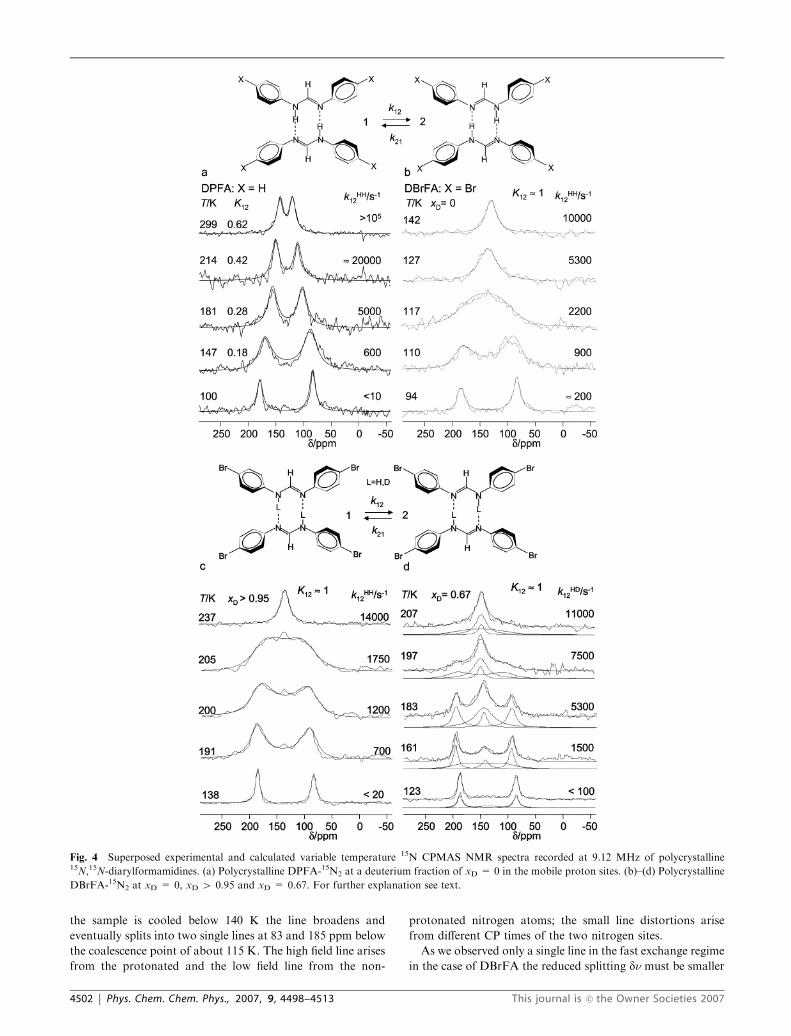
the sample is cooled below 140 K the line broadens and
eventually splits into two single lines at 83 and 185 ppm below
the coalescence point of about 115 K. The high field line arises
from the protonated and the low field line from the non-
protonated nitrogen atoms; the small line distortions arise
from different CP times of the two nitrogen sites.
As we observed only a single line in the fast exchange regime
in the case of DBrFA the reduced splitting dn must be smaller
Fig. 4 Superposed experimental and calculated variable temperature 15N CPMAS NMR spectra recorded at 9.12 MHz of polycrystalline15N,15N-diarylformamidines. (a) Polycrystalline DPFA-15N2 at a deuterium fraction of xD = 0 in the mobile proton sites. (b)–(d) Polycrystalline
DBrFA-15N2 at xD = 0, xD 4 0.95 and xD = 0.67. For further explanation see text.
4502 | Phys. Chem. Chem. Phys., 2007, 9, 4498–4513 This journal is �c the Owner Societies 2007

than the linewidth Wo in the absence of exchange. With Wo E4 ppm above 200 K, and DnE 102 ppm we estimate using eqn
(1) values of K12 between 0.9 and 1.0. Hence, the proton
tautomerism is degenerate within the margin of error, and, for
the simulation of the line shapes, K12 was set to 1 in the whole
temperature range.Wo was extrapolated from the line width in
the slow and fast exchange regime and the rate constants k =
k12 = k21 assembled in Table 2 were obtained by simulation of
the spectra as indicated in Fig. 4b.
Fig. 4c and d show the experimental and calculated 15N CP
MAS NMR spectra of DBrFA recorded at 9.12 MHz as a
function of temperature at two different deuterium fractions
xD of the mobile proton sites. For a statistical distribution, the
mole fractions of the isotopologs is given by
xHH ¼ ð1� xDÞ2; xHD ¼ 2ð1� xDÞxD; xDD ¼ x2D ð3Þ
At a deuterium fraction of xD 4 0.95, the mole fractions of the
isotopologs are given by xDD 4 0.9 and xHD o 0.1, xHH o0.0025. Thus, in good approximation, the line shape is deter-
mined only by kDD12 . The results of the simulations are
assembled in Table 2.
In order to obtain the rate constants kHD12 we measured a
sample with a deuterium fraction of xD E 0.67, where we
expect that xHH = 0.11, xHD = 0.445, xDD = 0.445. As the
values of kHH12 and of kDD
12 could be extrapolated from the
measurements at xD = 0 and 0.95, we only needed to adjust
the values of kHD12 and the line intensities of the different
isotopologs which can be different from the mole fractions
because of intensity distortions arising from CP. Therefore,
the line intensities are not discussed further. However, as
illustrated in Fig. 4d, the total line shapes can be described
well in terms of the sums of the line shape contributions of the
different isotopologs. The rate constants kHD12 obtained in this
way are included in Table 2.
Additional experiments were recorded at 30.41 MHz at
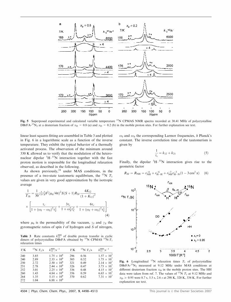
deuterium fractions of 0.8 and 0.2. Some typical results are
depicted in Fig. 5. The line shapes were again decomposed into
the contributions of the three isotopologs, and again only their
line intensities and the values of kHD12 needed to be adapted.
Spinning speeds were so high (5–9 kHz) that rotational side
bands could mostly be avoided. Because of sample heating in
the case of the high speed probe, a small quantity of 15N
labeled tetramethyltetraaza[14]annulene (TTAA) was added
to the rotors in a separate capsule in order to obtain the
sample temperatures from the four temperature-dependent15N chemical shifts of this compound23 marked by asterisks
in Fig. 5. Thus, by line shape analysis not only kHD12 but also
the temperature was obtained. The temperature error is,
however, a few degrees. Because of the higher resolution at
30.41 MHz we noticed that both the two amino as well as the
two imino nitrogen atoms of the cyclic dimers of DBrFA
exhibit slightly different chemical shifts as expected from the
crystal structure. These splittings were taken into account in
the line shape simulations.
We noticed that the rate constants kHD12 obtained at xD =
0.2 are slightly smaller than those obtained at xD = 0.8. In
view of the margin of error of the temperature we do not
discuss this difference further.
Variable temperature 15N NMR longitudinal relaxation times
and rates of HH-transfer in DBrFA
In order to obtain rate constants of the double proton transfer
at higher temperatures, longitudinal 15N relaxation times were
measured for non-deuterated DBrFA at 9.12 MHz. This
Larmor frequency is small enough so that only a 1H–15N
dipolar relaxation mechanism needs to be considered, i.e. that
relaxation contributions from the modulation of the chemical
shielding anisotropy by the proton motion can be neglected.24
The magnetization decays were found to be exponential in the
whole temperature range. The T1 values obtained by non-
Table 1 Equilibrium constants and forward and backward rateconstants k12 and k21 obtained for the double proton transfer in solidDPFA by 15N CPMAS line shape analysis
T/K K12 kHH12 /s�1 kHH
21 /s�1
100 — o10 o10147 0.18 600 3300181 0.28 5000 18 000214 0.42 E20 000 E48 000299 0.62 4105 4105
Table 2 Rate constants kHH12 , kHD
12 and kDD12 of double proton transfer
in cyclic dimers of polycrystalline DBrFA obtained by 15N CPMASNMR line shape analysis at different deuteron fractions xD
T/K xD no/MHz kHH12 /s�1 kHD
12 /s�1 kDD12 /s�1
110 0 9.12 900 — —117 0 9.12 2200 — —122 0 9.12 4200 — —127 0 9.12 5300 — —142 0 9.12 10 000 — —162 0.2 30.41 a 1200 —163 0.2 30.41 a 1400 —171 0.2 30.41 a 2500 —176 0.2 30.41 a 3500 —182 0.2 30.41 a 4700 —188 0.2 30.41 a 6000 —193 0.2 30.41 a 9100 —200 0.2 30.41 a 11 000 —204 0.2 30.41 a 12 000 —169 0.8 30.41 a 3900 o50176 0.8 30.41 a 4400 100b
189 0.8 30.41 a 8500 600b
197 0.8 30.41 a 13 000 950202 0.8 30.41 a 14 500 1300208 0.8 30.41 a a 2200161 0.67 9.12 a 1500 o50168 0.67 9.12 a 2100 o50173 0.67 9.12 a 3300 50b
183 0.67 9.12 a 5300 200b
197 0.67 9.12 a 7500 1100207 0.67 9.12 a 11 000 2200191 0.95 9.12 a a 700200 0.95 9.12 a a 1200205 0.95 9.12 a a 1750208 0.95 9.12 a a 2200211 0.95 30.41 a a 3000214 0.95 30.41 a a 3300217 0.95 30.41 a a 4200222 0.95 30.41 a a 5600237 0.95 30.41 a a 14 000
a Values larger than 20 000 s�1 without influence on the line widths.b Extrapolated values.
This journal is �c the Owner Societies 2007 Phys. Chem. Chem. Phys., 2007, 9, 4498–4513 | 4503
linear least squares fitting are assembled in Table 3 and plotted
in Fig. 6 in a logarithmic scale as a function of the inverse
temperature. They exhibit the typical behavior of a thermally
activated process. The observation of the minimum around
330 K allowed us to verify that the modulation of the hetero-
nuclear dipolar 1H–15N interaction together with the fast
proton motion is responsible for the longitudinal relaxation
observed, as described in the following.
As shown previously,24 under MAS conditions, in the
presence of a two-state tautomeric equilibrium, the 15N T1
values are given in very good approximation by the isotropic
average
1
T1S¼ 1
30g2I g
2S�h2ðm0=4pÞ2SðS þ 1ÞRSI
4K12
ð1þ K12Þ2
� tc1þ ðoI � oSÞ2t2C
þ 3tc1þ o2
St2C
þ 6tc1þ ðoI þ oSÞ2t2C
" #:
ð4Þ
where m0 is the permeability of the vacuum, gI and gS the
gyromagnetic ratios of spin I of hydrogen and S of nitrogen,
oI and oS the corresponding Larmor frequencies, h Planck’s
constant. The inverse correlation time of the tautomerism is
given by
1
tc¼ k12 þ k21 ð5Þ
Finally, the dipolar 1H–15N interaction gives rise to the
geometric factor
RSI ¼ RNH ¼ r�6NH þ r�6H���H þ r�3NHr�3H���Nð1� 3 cos2 aÞ ð6Þ
Fig. 5 Superposed experimental and calculated variable temperature 15N CPMAS NMR spectra recorded at 30.41 MHz of polycrystalline
DBrFA-15N2 at a deuterium fraction of xD = 0.8 (a) and xD = 0.2 (b) in the mobile proton sites. For further explanation see text.
Table 3 Rate constants kHH12 of double proton transfer in cyclic
dimers of polycrystalline DBrFA obtained by 15N CPMAS 15N-T1
relaxation times
T/K 15N T1/s kHH12 /s�1 T/K 15N T1/s kHH
12 /s�1
240 3.85 1.75 � 106 296 0.56 1.57 � 107
240 2.89 2.35 � 106 305 0.52 1.75 � 107
250 2.72 2.50 � 106 321 0.49 2.14 � 107
251 2.78 2.44 � 106 326 0.47 3.73 � 107
252 3.01 2.25 � 106 336 0.48 4.15 � 107
260 1.43 4.84 � 106 356 0.59 6.65 � 107
264 1.35 5.15 � 106 370 0.62 7.31 � 107
272 1.04 6.88 � 106
Fig. 6 Longitudinal 15N relaxation times T1 of polycrystalline
DBrFA-15N2 measured at 9.12 MHz under MAS conditions at
different deuterium fraction xD in the mobile proton sites. The HH
data were taken from ref. 7. The values of 15N T1 at 9.12 MHz and
xD 4 0.95 were 8.7 s, 5.5 s, 2.6 s at 298 K, 320 K, 334 K. For further
explanation see text.
4504 | Phys. Chem. Chem. Phys., 2007, 9, 4498–4513 This journal is �c the Owner Societies 2007

where rNH represents the short N–H bond length and rH� � �Nthe long hydrogen bond length, and a represents the jump
angle as depicted in Fig. 6, whereas the hydrogen bond angle bdoes not directly enter eqn (6).
In a first stage the data were fitted to eqn (4) assuming an
Arrhenius law of the degenerate double proton transfer from
which an energy of activation of 21.3 kJ mol�1 and an effective
pre-exponential factor log(A/s�1) = 10.9 was obtained,7 as
well as a value of RNH = 0.535 A�6. The calculated line
corresponds to the lower dashed line in Fig. 6. In a second
stage, the value of RNH was used in order to convert the 15N T1
values into the rate constants k12 using eqn (4). For this
conversion a computer program was written which varied
k12 until eqn (4) was fulfilled. The rate constants obtained in
this way are included in Table 3. Finally, in the last stage, the15N T1 values were recalculated using the non-linear Arrhenius
curve of the HH transfer depicted in Fig. 10, calculated using
the Bell–Limbach tunnel model as discussed below, giving rise
to the solid line in Fig. 6.
In order to obtain rate constants of the HD and the DD
transfer using this method we performed some 15N T1 mea-
surements at higher deuterium fractions. Some data obtained
at xD 4 0.95 are included in Fig. 6. However, because of the
much smaller gyromagnetic ratio of deuteron and because of
the large kinetic isotope effects the dipolar contribution to the
longitudinal 15N relaxation values is drastically reduced. This
leads to substantially larger values of T1 as illustrated in Fig. 6.
Under these conditions, the contribution to the longitudinal
relaxation arising from the modulation of the chemical shift
anisotropy by the double proton transfer can no longer be
neglected.24 Therefore, we did not try to obtain kinetic in-
formation from the longitudinal relaxation measurements
performed after partial H/D substitution.
Variable temperature2H NMR longitudinal relaxation times
and rates of HD/DD-transfer in DBrFA
In order to obtain rate constants of the HD and the DD
transfer we performed 2H NMR relaxation measurements. In
a first stage, 46.03 MHz 2H NMR spectra of DBrFA were
recorded at deuterium fractions in the mobile proton sites of
xD E 0.1 and 40.95 recorded under MAS conditions as
shown in Fig. 7. The spinning speed used was 6 kHz which
allowed us to considerably increase the signal to noise ratio as
compared to measurements of a static sample, i.e. to a con-
siderable reduction in the acquisition time, keeping the typical
double Pake 2H line shape. Assuming a statistical distribution
of isotopologs, the signal intensity of the DD species is given
by xD and of the HD species by 1 � xD. In other words, at
xD 4 0.95 the signal contribution of the HD species can be
neglected. At xD E 0.1, the signal stems to about 90% from
the HD and to about 10% from the DD species.
By the line shape analysis depicted in Fig. 7 a quadrupolar
coupling constant of qcc = 4/3qzz = 160 kHz and an asym-
metric factor Z = (qxx–qyy)/qzz = 0.18 were obtained for both
samples, indicating that substitution of the second H by D in a
cyclic dimer does not affect the components qxx, qyy, qzz of the
quadrupole coupling tensor within the margin of error. We
note that the values obtained do not refer to the intrinsic
quadrupole coupling tensor but to a tensor averaged by fast
deuteron transfer. This regime is reached as the motion of the
deuterons is highly anisotropic and as the rate constants of the
HD and DD transfer are of the order of 106 Hz as shown later.
In a second stage, longitudinal 2H relaxation times T1 were
measured using a conventional saturation recovery experiment
as depicted in Fig. 8a. An example is depicted in Fig. 8a. The
T1 values were obtained by fitting the signal intensities to
Int ¼Int1½xDð1� expð�t=TDD1 ÞÞ þ ð1� xDÞ
� ð1� expð�t=THD1 ÞÞ�
ð7Þ
where TDD1 and THD
1 represent the 2H longitudinal relaxation
times of the DD and the HD isotopologs. An illustration is
given in Fig. 8b. At xD 4 0.95 the signal decay is entirely
governed by the DD jumps and the contribution of the HD
isotopolog to eqn (7) can be neglected. The decay at xD E 0.1
is much faster because of the faster HD jumps. In order to
obtain THD1 , in the data analysis the second term eqn (7) was
taken into account, but the values of TDD1 were taken from
those obtained at xD 4 0.95. All relaxation times measured
are assembled in Table 4 and are plotted as a function of the
inverse temperature in Fig. 9.
In order to obtain rate constants of the HD and the DD
jumps of DBrFA we proceeded as follows. It has been shown
that the longitudinal relaxation times of a deuteron with spin
I = 1, jumping between two states can be expressed as:25
1
T1I¼ 3p2
10C
K12
ðK12 þ 1Þ2tc
1þ o2I t2cþ 4tc1þ 4o2
I t2c
� �ð8Þ
For axially symmetric quadrupole coupling constant tensors
the prefactor C can be expressed as25
C ¼ qccð1Þ2 þ qccð2Þ2 � qccð1Þqccð2Þð3 cos2 y� 1Þ ð9Þ
Fig. 7 Superposed experimental and calculated 2H NMR spectra of
DBrFA-15N2 measured at 46.03 MHz and a temperature of 298 K
under MAS conditions. Spinning speed 6 kHz. Quadrupolar echo
pulse sequence used. Variable delay VD = 12 s. (a) xD = 0.1. (b)
xD 4 0.95. For further explanation see text.
This journal is �c the Owner Societies 2007 Phys. Chem. Chem. Phys., 2007, 9, 4498–4513 | 4505

where qcc(1) = qcc(2) represent the intrinsic quadrupole cou-
pling constants in both tautomeric forms and y the jump angle
between the main axes of the quadrupolar coupling tensor in
the two states as defined in Fig. 9. However, eqn (9) is an
approximation and generally the factor C has to be deter-
mined experimentally from the value of T1 in the minimum.
Unfortunately, in the case of DBrFA, the latter could not be
reached because the melting point is around 440 K.15b
Therefore, we proceeded as follows. Firstly, we assumed
that the values of C are the same for the HD and the DD
isotopologs, in agreement with the finding that the 2H NMR
line shapes of the deuterons in both species are the same.
Secondly, we noticed that in the analysis of the Arrhenius
curves of the tautomerism of DBrFA described in the next
section the high temperature limit is reached for the HD and
the DD transfers, where exponential Arrhenius curves are
observed governed by the true pre-exponential factors A.
The latter are—within the margin of error—isotope insensi-
tive.10 From the HH data this value was determined to be
1012.4 s�1. Using this value and eqn (8), we could adapt the
solid lines in Fig. 9 to the experimental values by only adapting
the corresponding energies of activation. We obtained values
of 30.1 kJ mol�1 for the HD and of 33.1 kJ mol�1 for the DD
process from which we predict T1 minima around 475 K for
the DD and of 425 K for the HD isotopolog. Thus, we could
generate the rate constants of the HD and DD reactions in the
high-temperature limit listed in Table 4 as well as the value of
C = 9.84 � 108 s�2. Using eqn (9) i.e. a quadrupole coupling
constant of qcc(1) = qcc(2) = 160 kHz we predict a jump angle
of y = 1731. This value is in good agreement with a hydrogen
bond angle b = 51 defined in Fig. 6 and indicates the presence
of fairly linear hydrogen bonds. This feature is also the reason
why the 2H quadrupole relaxation mechanism is not much
more effective as compared to the dipolar 15N–1H relaxation
mechanism: the latter is operative even in the case of linear
hydrogen bonds whereas the former vanishes at y = 1801
according to eqn (9).
As the Arrhenius parameters were now known for the HD
and the DD reaction, we could convert the experimental T1
values into the corresponding rate constants, listed in Table 4.
Discussion
Using a combination of various methods of dynamic solid
state NMR we have measured the kinetic HH/HD/DD isotope
effects of the degenerate double proton transfer in cyclic
dimers of polycrystalline 15N,15N0-di-(4-bromophenyl)-forma-
midine (DBrFA) in a wide temperature range. Some rate
constants were also obtained for polycrystalline 15N,15N0-di-
(phenyl)-formamidine (DPFA) where the gas phase degener-
acy of the HH transfer is lifted by intermolecular interactions.
In this section, we will firstly describe the parameters used to
simulate the Arrhenius curves of double proton transfers in the
presence of tunneling. Then we discuss the parameters ob-
tained by these simulations and discuss them in comparison
with some other known related double proton transfer reac-
tions.
Arrhenius curve simulation parameters of degenerate double
proton transfer
Tunneling model. The Arrhenius curves of the double proton
transfers discussed below were calculated using a full Bell
Fig. 8 Longitudinal 2H relaxation time measurements of
DBrFA-15N2 measured under MAS conditions at 46.03 MHz at xDE 0.1 and xD 4 0.95. (a) Progressive saturation experiment at xD 40.95, (b) Signal intensities as a function of VD. For further explanation
see text.
Table 4 Rate constants kHD12 and kDD
12 of double hydron transfer incyclic dimers of polycrystalline DBrFA obtained from 2H T1 relaxa-tion times measured for static samples
T/K THD1 /s kHD
12 /s�1 TDD1 /s kDD
12 /s�1
301 1.4 5.5 � 106 6.52 7.1 � 105
316 1.7 4.4 � 106 3.34 1.3 � 106
336 0.5 1.2 � 107 1.82 2.8 � 106
356 0.31 2.6 � 107 0.94 5.5 � 106
Fig. 9 Longitudinal 2H relaxation times T1 of DBrFA-15N2 measured
under MAS conditions at 46.03 MHz at xD E 0.1 and xD 4 0.95 as a
function of the inverse temperature. For further explanation see text.
4506 | Phys. Chem. Chem. Phys., 2007, 9, 4498–4513 This journal is �c the Owner Societies 2007

tunneling model modified by Limbach et al. (‘‘Bell–Limbach-
model’’).10–12 In the framework of this model, the Arrhenius
curve of each isotopic reaction LL = HH, HD, DD depends
on five parameters, A, Em, ELLd , Dm, 2aLL. The following
parameters are experimental quantities: (i) the pre-exponential
factor A determines the intercept of the Arrhenius curve at
high temperatures; (ii) Em corresponds to the slope of the
Arrhenius curve in the strong tunneling regime at low tem-
peratures and (iii) the slope at high temperatures by Em +
ELLd where ELL
d is the barrier height in the tunneling configura-
tion. In the absence of pre-equilibria, A is in the usual range for
solid state proton transfers,11,12 which is of the order of
1012.6 s�1. Traditionally, Em has been neglected resulting in
temperature independent rate constants of tunneling at low
temperatures. However, it has been observed in various
hydrogen transfer reactions in organic molecules26,27 and in
enzyme reactions.28,29 It corresponds to any atomic and
molecular displacements preceding the tunnel process.
Therefore, it is isotope insensitive. Various sources contribute
to Em as has been discussed before.2,10–12,26,27 For example, in
a stepwise double proton transfer it contains a term necessary
to reach the energy of the intermediate state below which
tunneling cannot occur. For liquid solution solvent reorienta-
tion or non-recognized pre-equilibria can play a role.11,12
Furthermore, Em contains a term necessary for the reorgani-
zation of the molecular skeleton or the compression of
the hydrogen bond. This contribution is equivalent to the
‘‘work term’’ in Marcus theory of electron and proton
transfer.30
The hydron tunneling masses correspond to the sum of the
masses of the particles which tunnel, i.e. 1 and 2 for H and D
in a single hydron transfer and to 2, 3 and 4 in a HH/HD/DD
transfer and are not varied. However, there might be small
displacements of heavy atoms during the tunnel process which
enhances the tunneling mass, taken into account by the
parameter Dm. Finally, 2aLL corresponds to the width of the
barrier assumed to be parabolic.
Only in special cases is it possible to obtain all five para-
meters from the Arrhenius curve of a single isotopic reaction.
The situation is, however, different in the case of a set of three
Arrhenius curves for the HH, HD and DD reaction. The
Arrhenius curves of the HD and the DD reactions require each
only a single additional parameter, i.e. the modified barrier
heights EHDd and EDD
d . This is because the isotopic sensitive
barrier widths depend on the heights, i.e.11
2aHD ¼ 2aHH
ffiffiffiffiffiffiffiffiffiffiEHDd
EHHd
s; 2aDD ¼ 2aHH
ffiffiffiffiffiffiffiffiffiffiEDDd
EHHd
sð10Þ
In the presence of a concerted double proton transfer it has
been shown that often the barrier increase induced by sub-
stitution of the first H by D is similar to the substitution of the
second H by D, i.e. that11
EHDd ¼ EHH
d þ De; EDDd ¼ EHD
d þ De ð11Þ
This assumption leads at high temperatures where the over-
barrier dominates to the rule of the geometric mean for the
isotopic rate constants
kHH
kHD¼ kHD
kDD; kHD ¼
ffiffiffiffiffiffiffiffiffiffiffiffiffiffiffiffiffikHHkDDp
: ð12Þ
However, this rule is also fulfilled in good approximation at
low temperatures as barrier height, width, and tunneling mass
of the HD reaction correspond practically to the arithmetic
mean of the HH and the DD reaction. Only at intermediate
temperatures, where the tunneling contributions to the DD
and the HD reactions are smaller than to the HH reaction, the
first isotope effect is larger than the second, as has been
observed several times11
kHH
kHD4
kHD
kDDð13Þ
By contrast, for a stepwise degenerate double proton transfer
it has been shown11 that
kHD=kDD ¼ 2
1þ kDD=kHHffi 2 for kHH=kDD � 1: ð14Þ
Thus, only the six parameters A, Em, Dm, EHHd = Ed, 2a
LL =
2a and De are necessary to simulate the Arrhenius curves of the
three isotopic reactions. For these simulations a non-linear
least squares fitting routine was used, where each parameter
could be varied automatically or manually.
We are aware that this model is not perfect. It is only a
preliminary approach to interpreting experimental Arrhenius
curves until a fully quantum-mechanical description is avail-
able that will provide better quantitative support for the
qualitative interpretation of the Bell–Limbach model. Note
that in the case of the proton exchange between acetic acid and
methanol, the kinetic data were published in 1984,10a and a
quantum-mechanical model using an instanton approach was
published by Smedarchina et al. only in 2001.31
Arrhenius curve simulations. In Fig. 10 we have plotted the
Arrhenius diagram of the HH, HD and DD transfer in cyclic
dimers of polycrystalline DBrFA. For comparison, Fig. 11
depicts the corresponding graph for DFFA dissolved in
Fig. 10 Arrhenius diagram of the degenerate solid state HH, HD and
DD tautomerism of cyclic dimers of DBrFA. The solid lines were
calculated using the Bell–Limbach tunneling model. For further
explanation see text.
This journal is �c the Owner Societies 2007 Phys. Chem. Chem. Phys., 2007, 9, 4498–4513 | 4507

tetrahydrofuran2 and Fig. 12 the data obtained for DPFA in
the polycrystalline state and dissolved in tetrahydrofuran.1
The latter are assembled in Table 5. Moreover, we have
included in Fig. 11 the Arrhenius curve of the intramolecular
double proton transfer in the seven-membered bicyclic oxala-
midine OA7 (2,20-bis(4,5,6,7-tetrahydro)-1,3-diazepine,
Fig. 13a) dissolved in methylcyclohexane.32
The Arrhenius curve simulation parameters were derived as
follows. Firstly, the Arrhenius curves were simulated without
automatic parameter fit. As anticipated, we found that a
common value of 1012.4 s�1 for the pre-exponential factors A
of all isotopic reactions could well describe the experimental
data. For DBrFA, the isotope insensitive value of Em could be
determined without assumptions from the Arrhenius curve of
the HH reaction in Fig. 10. Then, EHHd and De was determined
from the slopes of the Arrhenius curves at high temperatures.
Finally, Dm and 2a were determined from the effective pre-
exponential factors of the HH and the HD reactions in the
low-temperature tunneling regime. The corresponding para-
meters for the other reactions in Table 6 were obtained
previously using a similar strategy.
In Table 6 we have included for comparison the parameters
obtained previously11 for the related double proton transfers
in solid benzoic acid dimers,33,34 derived from the data of
Horsewill et al.,34 and in 3,5-diphenyl-4-Br-pyrazole (DPBrP)
(Fig. 13b and c).9
Fig. 11 Arrhenius diagram of the degenerate HH, HD and DD
tautomerism inside cyclic dimers of DFFA-15N2 dissolved in tetrahy-
drofuran-d8. Adapted from ref. 2b. For further explanation see text.
Fig. 12 Arrhenius diagram of the degenerate HH and HD tautomer-
ism inside cyclic dimers of DPFA dissolved in tetrahydrofuran-d8.
Black triangles: Backward rate constants in polycrystalline DPFA.
Adapted from ref. 1. For further explanation see text.
Table 5 Rate constants k12 = k21 obtained for the double protontransfer in cyclic dimers of DPFA dissolved in THF-d8 according toref. 1
T/K kHH12 /s�1 kHD
12 /s�1 T/K kHH12 /s�1 kHD
12 /s�1
151 940 187 16 600 1100152 1570 188 14 800158 2750 191 24 300162 3540 152 193 18 900 —164 4620 — 194 21 900165 4170 243 198 32 200171 6240 305 198 23 800175 6450 428 199 32 000a 2150176 6900 536 205 37 700 —176 7570 — 209 59 000 5250178 7600 500 210 — 3548179 11 000 — 212 57 300 —181 — 1080 215 99 600 —183 8870 — 220 103600 8430185 15 600 870 230 128900 20 893186 12 900
a Extrapolated values.
Fig. 13 (a) Tautomerism of 2,20-bis(4,5,6,7-tetrahydro)-1,3-diazepine
(OA7) in organic solvents.32 (b) Solid state tautomerism of benzoic
acid dimers.33,34 (c) Solid state tautomerism of 3,5-diphenyl-4-Br-
pyrazole (DPBrP).9 (d) Photoinduced tautomerism of 7-azaindole
dimers.36,37
4508 | Phys. Chem. Chem. Phys., 2007, 9, 4498–4513 This journal is �c the Owner Societies 2007

Mechanisms of double proton transfer in amidines and related
systems
General remarks. Before we discuss the kinetic parameters
obtained in detail, let us make some general remarks. Firstly, we
note that the Arrhenius curves of DPFA, DFFA andDBrFA are
non-linear as expected for tunneling at low temperatures and for
over-barrier reactions at high temperatures. Furthermore, the
rate constants of the diarylamidines are very similar (Table 6), i.e.
major effects of substituents or liquid/solid state effects are
absent. This is demonstrated also in the use of a single pre-
exponential factor of 1012.4 s�1 for all diarylamidines in the solid
and liquid state. As mentioned above, this value is typical for
intramolecular H transfers in the absence of pre-equilibria and
major solvent reorientation during the proton transfer.11 We
note that in order to derive the intrinsic rate constants in the
hydrogen bonded dimers of DPFA and DFFA dissolved in
tetrahydrofuran the non-linear dependence of the pseudo-first
order rate constants as a function of concentration had been
analyzed.1,2 This analysis is now confirmed by the solid state
studies, especially in the case of DPFA. Here, the solid state lifts
the degeneracy of the tautomerism in the liquid state. Hence, the
forward rate constants kHH12 are a little bit smaller, and the
backward rate constants kHH21 a little bit larger than their average
kHH found for the liquid state, as is illustrated in Fig. 12.
Finally, we note a very large variation of the rate constants
in the different double proton transfers listed in Table 6. At
room temperature, the transfer is fastest in the OHO hydrogen
bonds of benzoic acid dimers, i.e. 1011 s�1, about 107 s�1 in the
amidines, 104 s�1 in 3,5-diphenyl-4-bromopyrazole DPBrP
and only 101 s�1 in the oxalamidine OA7.
Kinetic HH/HD/DD isotope effects. We obtain for the
amidines at high temperatures two large kinetic HH/HD and
HD/DD isotope effects showing that eqn (12) is fulfilled. The
same is true also for the dimers of benzoic acid (Fig. 13b)34
and of DPBrP (Fig. 13c)9 in the solid state. For the amidine
and the pyrazole dimers we found that the rule of geometric
mean is fulfilled within the margin of error, i.e. that the
increase De of the barrier height by isotopic substitution of
the first H by D is the same as of the second H by D (Table 6).
In the case of benzoic acid dimer (Fig. 13b) the second effect is
larger than the first. This could arise from a change of the H-
bond geometries after deuteration, a phenomenon which has
been observed recently for a solid exhibiting a strong OHN
hydrogen bond.35 Nevertheless, these findings are consistent
with a mechanism where both protons loose zero-point energy
in the transition state. This mechanism is often called ‘‘con-
certed’’, but it should be mentioned that this statement does
not refer to the time domain but to the energy reaction surface.
If this loss would arise from a single frequency, values of Debetween 2 and 3 kJ mol�1 would correspond to a frequency
shift between 1100 and 1700 cm�1. Thus, in the transition state
a substantial amount of zero-point energy is still present.
By contrast, eqn (14) is fulfilled for the tautomerism of the
oxalamidine OA7 (Fig. 13a) for which a stepwise tunneling
process has been demonstrated.11,26 Here, only the first sub-
stitution of H by D leads to an increase in the barrier height as
only a single proton is transferred in a given reaction step.
Thus, the overall kinetic HH/DD isotope effects are substan-
tially smaller than in the diarylamidines.
A stepwise photoinduced double proton transfer has been
demonstrated for 7-azaindole dimers (Fig. 13d) in condensed
phases. Evidence for a photoinduced double proton transfer in
7-azaindole dimers was obtained by Kasha et al.36 Femto-
second studies of Zewail et al.37 indicate two reactions steps as
illustrated in Fig. 13d, which was confirmed by high-level ab
initio calculations.38 The second step exhibited a primary
Table 6 Geometric data, kinetic data and Arrhenius curve simulation parameters of various degenerate double proton transfers in cyclichydrogen bonded dimers
System
Geometric and kinetic data Arrhenius curve simulation parameters
H-bondgeometry
kHH(298 K)/s�1 KIE(298 K)
Em/kJ mol�1
log(A/s�1)
Ed/kJ mol�1
Dm/a.m.u. 2aH /A
De/kJ mol�1
DFFA inTHF-d8 ref. 2
— 9.4 � 106 HH/HD 4.7 11.7 (5.4)a 12.4 (12.2)a 21.7 (26.4)a 0.5 (1.05)a 0.48 (0.55)a HH/HD 3.0HD/DD 4.0 HD/DD 3.0HH/DD 19
DPFA inTHF-d8 ref. 1
— 3.4 � 106 HH/HD 4.4 11.7 12.4 25.1 0.5 0.46 HH/HD 2.5HD/DD 3.5 HD/DD 2.5HH/DD 15
DBrFAthis work
rNN = 2.963 A6 1.3 � 107 HH/HD 5.6 9.2 12.4 24.7 0.5 0.44 HH/HD 2.9HD/DD 4.1 HD/DD 2.9HH/DD 23
OxalamidineOA7 inmethylcyclo-hexane ref. 32c
rNN = 2.8/3.0 Ac 14 HH/HD 3.1 52.7 12.6 27.2 1.5 0.2 H/D 2.9HD/DD 1.5
(PhCOOH)2crystal ref. 34
rOO = 2.606 A 9 � 1010 HH/HD 2.4 HH 0.84 11.6 5.4 1.8 HH 0.48 HH/HD 2.1q1 = 0.3115 Ab HD/DD 6 HD 0.84 HD 0.52 HD/DD 4.6q2 = 2.609 Ab DD 1.06 DD 0.44
3,5-diphenyl-4-Brpyrazol crystalref. 9
rNN = 2.84 A9 6500 HH/HD 5 5.6 12.65 47.5 2.3 0.55 HH/HD 3.4HD/DD 5 HD/DD 3.4HH/DD 25
a Values proposed in ref. 11. b Natural H-bond coordinates q1 = 12(rOH�rHO), q2 = rOH + rHO taken from ref. 41. c PM3-MO calculations of
syn/anti form according to ref. 32c.
This journal is �c the Owner Societies 2007 Phys. Chem. Chem. Phys., 2007, 9, 4498–4513 | 4509

kinetic H/D isotope effect of 4.7 and a secondary effect of 1.9,
which were related to rates of barrier crossing and tunne-
ling.37b,c We note that these values are of the same order as
found here for the amidines in spite of the difference of the
reaction time scales.
Barrier width and heavy atom tunneling
As mentioned above, in order to describe the low-temperature
part of the HH Arrhenius curve one needs to adjust the barrier
width 2aHH which was of the order of 0.5 A. The temperature-
independent kinetic isotope effect in this regime is then deter-
mined by the contribution of heavy atom displacements to the
tunneling mass, Dm, besides the values of De which was,
however, already fixed in order to reproduce the kinetic
isotope effects at high temperatures. We needed values of
Dm between 0.5 a.m.u. for the diarylamidines and 2.3 a.m.u.
for the pyrazoles. This result is surprising, but could arise both
from single–double bond interconversions during the proton
tunneling process and from small angle molecular flips.
For the barrier widths we needed values between 0.4 and
0.5 A in order to reproduce the kinetic data of the amidines.
Surprisingly, similar values were found before for benzoic acid
dimers (Table 6). The O� � �O distance of benzoic acid dimers is
2.606 A, and neutron structural data indicate a distance of
0.62 A between the two proton sites of the OHO hydrogen
bonds. If one substracts the contribution of the ground state
vibrational wave function which is of the order of 0.1–0.2 A we
find a good agreement with the barrier width derived from the
Arrhenius curves. Why then is the barrier width for the
amidines not larger, in view of the large N� � �N distance of
2.963 A, from which one can estimate that the proton sites are
separated by at least 0.7 A?
Minimum energy for tunneling to occur and barrier height. In
order to answer this question let us consider the minimum
energy Em for tunneling occur. The latter is very small in the
case of benzoic acid dimers, where it is dominantly arising
from the energy difference between the two tautomeric states
induced by the crystal fields.35 Hence, the molecular reorga-
nization energy in order to reach the tunneling configuration is
also small. In other words, no hydrogen bond compression is
needed in order to reach the tunneling configuration. Thus,
there is no large difference between the barrier width and
separation of the proton sites in the OHO hydrogen bond.
This is consistent with the small O� � �O distance of 2.606 A
which is not far away from the minimum distance of OHO
hydrogen bonds of about 2.4 A.11 The small value of the
barrier width in the amidines is then understandable in the
presence of substantial hydrogen bond compression, which
requires a substantial amount of energy contributing to Em. In
the case of DBrFA, the value of 9.2 kJ mol�1 (Table 6) could
be determined without assumptions from the slope of the
Arrhenius curve of the HH reaction at low temperatures. As
the crystal structure of DBrFA represents the best possible
configuration for the tautomerism, it is plausible that the
values of Em for DPFA and DFFA are somewhat larger than
Fig. 14 (a) Hydrogen bond geometries of various molecular systems containing NHN hydrogen bonds (taken from Table 5 of ref. 11). (b)
Calculated hydrogen bond geometries of various dimers of 7-azaindole in the ground state S0 and the first excited electronic state S1 according to
ref. 38. (c) Barrier heights of the H transfers calculated from the Arrhenius curves of the species in (a). The barrier heights of the transition states
are set to zero (taken from Table 5 of ref. 11). Abbreviations: TTAA: tetramethyldibenzotetraaza[14]annulene, DPBrP: 3,5-diphenyl-4-Br-pyrazole
dimers, DPP: 3,5-diphenyl-pyrazole tetramers, PMP: 3-phenyl-5-methyl-pyrazole tetramers, DMP: 3,5-dimethyl-pyrazole trimers, 4NO2P:
4-nitropyrazole trimers, 4BrP: 4-bromo-pyrazole trimers.
4510 | Phys. Chem. Chem. Phys., 2007, 9, 4498–4513 This journal is �c the Owner Societies 2007

the value of DBrFA. Using this condition, we derived a value
of 11.7 kJ mol�1 from the simulation of the Arrhenius curves
of DPFA and DFFA. This value is larger than anticipated
previously,2,11 where the data of DBrFA i.e. the condition
Em 4 9.2 kJ mol�1 were not yet available. We associate the
difference, i.e. 2.5 kJ mol�1, to the reorientation of the aryl
groups in tetrahydrofuran as discussed in the next section.
Hydrogen bond compression and aryl group reorientation.
Hydrogen bond compression as a major phenomenon in the
tautomerism of amidines has been discussed previously.2b
However, only in recent years hydrogen bond correlations
have been established by which the compression can be
quantified. Fig. 14a depicts the correlated NHN hydrogen
bond coordinates of various systems in their initial stationary
states containing NHN hydrogen bonds, taken from ref. 11. In
the case of porphyrin39 and its anion,40 the coordinates of the
transition states calculated using ab initio methods have been
included, otherwise the data were derived experimentally. The
solid line refers to the equilibrium geometries, i.e. to those of
the groundstates of harmonic oscillators, whereas the dotted
line includes a correction for anharmonic groundstate vibra-
tions. We note that all geometries are located on the predicted
correlation curve, especially the coordinates of the transition
states of porphyrin and of its anion, exhibiting values of 2.60
and 2.66 A. This means that hydrogen bond compression is
the most important heavy atom motion which enables H-
transfer; the transition state structures correspond to those
expected for the strongest possible NHN hydrogen bonds,
whereas the initial states do not show any sign of hydrogen
bonding.
In Fig. 14b we have made a similar graph for the structures
of dimers of isolated 7-azaindole in different molecular states
as calculated by Serrano-Andres and Merchan.38 The graph of
Fig. 14b demonstrates nicely the coupling of the proton
coordinate q1 to the heavy atom coordinate q2. The initial
state is the dimer in the S1 state, D(S1). A single proton
transfer occurs from the pyrrole to the pyridine ring forming
the zwitterionic intermediate ZI(S1), exhibiting stronger i.e.
shorter H-bonds as the parent dimer. Thus, this H-transfer is
accompanied by a heavy atom motion or hydrogen bond
compression. By contrast, the second H-transfer to the tauto-
mer dimer T(S1) involves smaller heavy atom motions. Hence,
if both process take place by tunneling, the first step will
exhibit a smaller kinetic H/D isotope effect as compared to the
second, because of the increase of the tunneling masses which
is consistent with the experimental findings37b,c discussed
above. A concerted transfer via the transition state TS(S1)
involves a larger energy and could be excluded. A covalent
biradical intermediate BI(S1) was proposed for the isolated
dimers; its hydrogen bonds are long and weak as indicated in
the graph.
The question arises whether and how the intrinsic barrier of
the symmetric H transfers depends on the hydrogen bond
geometries. In Fig. 14c, therefore, the experimental values of
the total barrier Ed + Em are plotted as a function of q2. As a
reference, the values of zero for the transition states calculated
for porphyrin and for the anion are included. As indicated by
Fig. 15 Aryl group torsion, hydrogen bonding and double proton transfer in diarylformamidines. One-dimensional schematic potential curves
for the double proton transfer are included.
This journal is �c the Owner Societies 2007 Phys. Chem. Chem. Phys., 2007, 9, 4498–4513 | 4511
the straight line, the total barrier of single H transfer and
stepwise H + H transfer increases with increasing q2. For the
HH transfer, stepwise HH + HH transfers and HHH trans-
fers the barriers are larger, and also increase on average with
q2 but there are deviations from the correlation. For example,
porphycene exhibits shorter hydrogen bonds than the N,N-
diarylformamidines, but the barrier is larger than in the latter.
This observation could arise from the balance between the
reduction of the barrier by hydrogen bond compression and
the energy needed for the compression. This balance can be
different for the different systems, leading to transition state
geometries which may not reach the maximum compression
point as reached in the case of porphyrins. In other words, a
system with shorter hydrogen bonds may exhibit a larger
barrier as the compression energy may be larger because of
a certain molecular structure.
We note that the energy needed for the compression will
show up both in the minimum energy Em for tunneling to
occur and in the barrier Ed, and is, therefore, experimentally
not easily obtained.
In the case of N,N-diarylformamidines, the conformation of
the aryl groups influences the double proton transfer as
illustrated in Fig. 15. As discussed in the Introduction, the
double proton transfer is degenerate only if the torsional
angles f of the aryl groups (Fig. 3) are all around 301, as is
the case for DBrFA. Deviations from these angles lift the
degeneracy of the transfer. It is plausible to assume that the
reorientation of the aryl groups is hindered in the solid state by
intermolecular interactions, leading then only to a few N,N-
diarylformamidines which can react in this phase. By contrast,
the aryl group reorientation energy will be much smaller in the
liquid state. Therefore, we find a surprisingly excellent agree-
ment of the rate constants of the N,N-diarylformamidines
tautomerism in the liquid and the solid state.
Conclusions
The tautomerism of cyclic N,N-diarylformamidines dimers is a
very good example of how molecular function is influenced by
intermolecular interactions, which are controlled by intra-
molecular conformation and hence molecular structure as
was illustrated in Fig. 1 and 2. The transfer exhibits two large
HH/HD and HD/DD isotope effects and is, therefore, con-
certed on the energy reaction surface. Tunneling plays an
important role; at low temperatures an isotope insensitive
minimum energy for tunneling is required for tunneling to
occur. Arrhenius curve simulations were performed using
the Bell–Limbach tunneling model which does not preclude
the application of more sophisticated models in the future.
Acknowledgements
We thank Prof. G. S. Denisov, St. Petersburg State University,
for helpful discussions and reading this manuscript. This work
was supported by the Deutsche Forschungsgemeinschaft,
Bonn, and the Fonds der Chemischen Industrie, Frankfurt.
References
1 L. Meschede, D. Gerritzen and H. H. Limbach, Ber. Bunsen-Ges.Phys. Chem., 1988, 92, 469–485.
2 (a) H. H. Limbach, L. Meschede and G. Scherer, Z. Naturforsch.,1989, 44a, 459–471; (b) L. Meschede and H. H. Limbach, J. Phys.Chem., 1991, 95, 10267–10280.
3 F. Mannle and H. H. Limbach,Angew. Chem., Int. Ed. Engl., 1996,35, 441–442.
4 H. H. Limbach, F. Mannle, C. Detering and G. S. Denisov, Chem.Phys., 2005, 319, 69–92.
5 R. Anulewicz, Acta Crystallogr., Sect. C, 1997, C53, 345–346.6 R. Anulewicz, I. Wawer, T. M. Krygowski, F. Mannle and H. H.Limbach, J. Am. Chem. Soc., 1997, 119, 12223–12230.
7 F. Mannle, I. Wawer and H. H. Limbach, Chem. Phys. Lett., 1996,256, 657–662.
8 (a) F. Aguilar-Parrilla, G. Scherer, H. H. Limbach, M. C. Foces-Foces, F. H. Cano, J. A. S. Smith, C. Toiron and J. Elguero, J. Am.Chem. Soc., 1992, 114, 9657–9659; (b) F. Aguilar-Parrilla, O.Klein, J. Elguero and H. H. Limbach, Ber. Bunsen-Ges. Phys.Chem., 1997, 101, 889–901; (c) O. Klein, M. M. Bonvehi, F.Aguilar-Parrilla, J. Elguero and H. H. Limbach, Isr. J. Chem.,1999, 34, 291–299.
9 O. Klein, F. Aguilar-Parrilla, J. M. Lopez, N. Jagerovic, J. Elgueroand H. H. Limbach, J. Am. Chem. Soc., 2004, 126, 11718–11732.
10 (a) D. Gerritzen and H. H. Limbach, J. Am. Chem. Soc., 1984, 106,869–879; (b) H. H. Limbach, O. Klein, J. M. Lopez del Amo and J.Elguero, Z. Phys. Chem., 2004, 217, 17–49.
11 H. H. Limbach, Single and multiple hydrogen/deuterium transferreactions in liquids and solids, in Hydrogen Transfer Reactions, ed.J. T. Hynes, J. Klinman, H. H. Limbach and R. L. Schowen,Wiley-VCH, Weinheim, 2007, vol. 1 & 2, ch. 6, pp. 135–221 andreferences cited therein.
12 H. H. Limbach, J. M. Lopez and A. Kohen, Philos. Trans. R. Soc.London, Ser. B, 2006, 361, 1399–1415.
13 (a) P. Svensson, N. A. Bergmann and P. Ahlberg, Z. Naturforsch.,1989, 44a, 473–479; (b) K. A. Nguyen, M. S. Gordon and D. G.Truhlar, J. Am. Chem. Soc., 1991, 113, 1596–1600.
14 L. Claisen, Ann. Chem., 1895, 287, 366–368.15 (a) T. Axenrod, P. S. Pregosin, M. J. Wieder, E. D. Becker, R. B.
Bradley and G. W. A. Milne, J. Am. Chem. Soc., 1971, 93,6536–6541; (b) J. Oszczapowicz, R. Orlinski and E. Hejchman,Polish J. Chem., 1979, 53, 1259–1265.
16 D. G. Ott, in Synthesis with Stable Isotopes, John Wiley & Sons,New York, 1981.
17 P. Du Bois Murphy, J. Magn. Reson., 1986, 70, 307–312.18 D. Torchia, J. Magn. Reson., 1978, 30, 613–616.19 R. D. Kendrick, S. Friedrich, B. Wehrle, H. H. Limbach and C. S.
Yannoni, J. Magn. Reson., 1985, 65, 159–161.20 H. H. Limbach, Dynamic NMR spectroscopy in the presence of
kinetic hydrogen deuterium isotope effects, in NMR Basic Princi-ples and Progress, Heidelberg, Berlin, 1990, vol 26, ch. 2.
21 (a) S. Alexander, J. Chem. Phys., 1962, 37, 974–980; (b) G. Binsch,J. Am. Chem. Soc., 1969, 91, 1304–1309; (c) D. A. Kleier and G.Binsch, J. Magn. Reson., 1970, 3, 146–160.
22 (a) H. H. Limbach, J. Hennig, R. D. Kendrick and C. S. Yannoni,J. Am. Chem. Soc., 1984, 106, 4059–4060; (b) B. Wehrle, H.Zimmermann and H. H. Limbach, J. Am. Chem. Soc., 1988, 110,7014–7024; (c) H. H. Limbach, B. Wehrle, M. Schlabach, R. D.Kendrick and C. S. Yannoni, J. Magn. Reson., 1988, 77, 84–100;(d) B. Wehrle and H. H. Limbach, Chem. Phys., 1989, 136,223–247.
23 (a) F. Aguilar-Parrilla, B. Wehrle, H. Braunling and H. H.Limbach, J. Magn. Reson., 1990, 87, 592–597; (b) B. Wehrle, F.Aguilar-Parrilla and H. H. Limbach, J. Magn. Reson., 1990, 87,584–591.
24 C. G. Hoelger, B. Wehrle, H. Benedict and H. H. Limbach, J.Phys. Chem., 1994, 98, 843–851.
25 W. Medycki, E. C. Reynhardt and L. Latanowicz, Mol. Phys.,1998, 93, 323–327.
26 G. Scherer and H. H. Limbach, J. Am. Chem. Soc., 1994, 116,1230–1239.
27 J. Braun, M. Schlabach, B. Wehrle, M. Kocher, E. Vogel and H.H. Limbach, J. Am. Chem. Soc., 1994, 116, 6593–6604.
4512 | Phys. Chem. Chem. Phys., 2007, 9, 4498–4513 This journal is �c the Owner Societies 2007

28 J. Basran, L. Masgrau, M. J. Sutcliffe and N. Scrutton,Solution and computational studies of kinetic isotope effects inflavoprotein and quinoprotein catalyzed substrate oxidations asprobes of enzymic hydrogen tunneling and mechanism, in IsotopeEffects in Chemistry and Biology, ed. A. Kohen and H. H.Limbach, Taylor & Francis, Boca Raton FL, 2006, ch. 25, pp.671–690.
29 M. J. Knapp, M. Meyer and J. P. Klinman, Nuclear tunneling inthe condensed phase: hydrogen transfer in enzyme reactions, inHydrogen Transfer Reactions, ed. J. T. Hynes, J. Klinman, H. H.Limbach and R. L. Schowen, Wiley-VCH, Weinheim, 2007, vol. 3& 4, ch. 10, pp. 1241–1284 and references cited therein.
30 R. A. Marcus, Faraday Discuss. Chem. Soc., 1982, 74, 7–15.31 A. Fernandez-Ramos, Z. Smedarchina and J. Rodrıguez-Otero, J.
Chem. Phys., 2001, 114, 1567–1574.32 (a) G. Scherer and H. H. Limbach, J. Am. Chem. Soc., 1989, 111,
5946–5947; (b) G. Scherer and H. H. Limbach, J. Am. Chem. Soc.,1994, 116, 1230–1239; (c) G. Scherer and H. H. Limbach, Croat.Chim. Acta, 1994, 67, 431–440.
33 (a) B. H. Meier, F. Graf and R. R. Ernst, J. Chem. Phys., 1982, 76,767–774; (b) A. Stockli, B. H. Meier, R. Kreis, R. Meyer and R. R.Ernst, J. Chem. Phys., 1990, 93, 1502–1520; (c) A. Heuer and U.Haeberlen, J. Chem. Phys., 1991, 95, 4201–4124; (d) J. L. Skinner
and H. P. Trommsdorff, J. Phys. Chem., 1988, 89, 897–907; (e) R.Meyer and R. R. Ernst, J. Chem. Phys., 1990, 93, 5518–5532; (f) Y.Kim, J. Am. Chem. Soc., 1996, 118, 1522–1528; (g) Y. Kim, J.Phys. Chem. A, 1998, 102, 3025–3036.
34 Q. A. Xue, A. J. Horsewill, M. R. Johnson and H. P. Tromms-dorff, J. Chem. Phys., 2004, 120, 11107–11119.
35 J. Zhou, Y. S. Kye and G. S. Harbison, J. Am. Chem. Soc., 2004,126, 8392–8393.
36 C. A. Taylor, M. A. El-Bayoumi and M. Kasha, Proc. Natl. Acad.Sci. U. S. A., 1969, 63, 253–260.
37 (a) A. Douhal, S. K. Kim and A. H. Zewail, Nature, 1995, 378,260–263; (b) T. Fiebig, M. Chachisvilis, M. Manger, A. H. Zewail,A. Douhal, I. Garcia-Ochoa and A. de La Hoz Ayuso, J. Phys.Chem. A, 1999, 103, 7419–7431; (c) O. H. Kwon and A. H. Zewail,Proc. Natl. Acad. Sci. U. S. A., 2007, 104, 8703–8708.
38 L. Serrano-Andres and M. Merchan, Chem. Phys. Lett., 2006, 418,569–575.
39 D. K. Maity, R. L. Bell and T. N. Truong, J. Am. Chem. Soc.,2000, 122, 897–906.
40 T. Vangberg and A. Ghosh, J. Phys. Chem. B, 1997, 101,1496–1497.
41 T. Emmler, S. Gieschler, H. H. Limbach and G. Buntkowsky, J.Mol. Struct., 2004, 700, 29–38.
This journal is �c the Owner Societies 2007 Phys. Chem. Chem. Phys., 2007, 9, 4498–4513 | 4513
Copyright © 2022 FDOKUMEN


![[VNMATHi com].On thi lop 10 theo CHUYEN DE-hh-DS](https://static.fdokumen.com/doc/165x107/631a7f8ffd704e1d390a282e/vnmathi-comon-thi-lop-10-theo-chuyen-de-hh-ds.jpg)