New heterodontosaurid specimens from the Lower Jurassic of southern Africa and the early...
17
Earth and Environmental Science Transactions of the Royal Society of Edinburgh http://journals.cambridge.org/TRE Additional services for Earth and Environmental Science Transactions of the Royal Society of Edinburgh: Email alerts: Click here Subscriptions: Click here Commercial reprints: Click here Terms of use : Click here New heterodontosaurid specimens from the Lower Jurassic of southern Africa and the early ornithischian dinosaur radiation Laura B. Porro, Richard J. Butler, Paul M. Barrett, Scott MooreFay and Richard L. Abel Earth and Environmental Science Transactions of the Royal Society of Edinburgh / Volume 101 / Special Issue 34 / September 2010, pp 351 366 DOI: 10.1017/S175569101102010X, Published online: 17 May 2011 Link to this article: http://journals.cambridge.org/abstract_S175569101102010X How to cite this article: Laura B. Porro, Richard J. Butler, Paul M. Barrett, Scott MooreFay and Richard L. Abel (2010). New heterodontosaurid specimens from the Lower Jurassic of southern Africa and the early ornithischian dinosaur radiation. Earth and Environmental Science Transactions of the Royal Society of Edinburgh, 101, pp 351366 doi:10.1017/S175569101102010X Request Permissions : Click here Downloaded from http://journals.cambridge.org/TRE, IP address: 137.222.114.247 on 23 Aug 2013
Transcript of New heterodontosaurid specimens from the Lower Jurassic of southern Africa and the early...
Earth and Environmental Science Transactions of the Royal Society of Edinburghhttp://journals.cambridge.org/TRE
Additional services for Earth and Environmental Science Transactions of the Royal Society of Edinburgh:
Email alerts: Click hereSubscriptions: Click hereCommercial reprints: Click hereTerms of use : Click here
New heterodontosaurid specimens from the Lower Jurassic of southern Africa and the early ornithischian dinosaur radiation
Laura B. Porro, Richard J. Butler, Paul M. Barrett, Scott MooreFay and Richard L. Abel
Earth and Environmental Science Transactions of the Royal Society of Edinburgh / Volume 101 / Special Issue 34 / September 2010, pp 351 366DOI: 10.1017/S175569101102010X, Published online: 17 May 2011
Link to this article: http://journals.cambridge.org/abstract_S175569101102010X
How to cite this article:Laura B. Porro, Richard J. Butler, Paul M. Barrett, Scott MooreFay and Richard L. Abel (2010). New heterodontosaurid specimens from the Lower Jurassic of southern Africa and the early ornithischian dinosaur radiation. Earth and Environmental Science Transactions of the Royal Society of Edinburgh, 101, pp 351366 doi:10.1017/S175569101102010X
Request Permissions : Click here
Downloaded from http://journals.cambridge.org/TRE, IP address: 137.222.114.247 on 23 Aug 2013

New heterodontosaurid specimens from theLower Jurassic of southern Africa and the earlyornithischian dinosaur radiation
Laura B. Porro1*, Richard J. Butler2*, Paul M. Barrett3, Scott Moore-Fay3,4
and Richard L. Abel5
1 Department of Organismal Biology and Anatomy, University of Chicago, Chicago, IL 60637, USA
2 Bayerische Staatssammlung fur Palaontologie und Geologie, Richard-Wagner-Straße 10, 80333 Munich, Germany
3 Department of Palaeontology, Natural History Museum, London SW7 5BD, UK
4 Wavecut Platform Ltd, 131 Bradbourne Vale Road, Sevenoaks, Kent TN13 3DJ, UK
5 Department of Mineralogy, Natural History Museum, London SW7 5BD, UK
*Corresponding authors
ABSTRACT: Heterodontosaurids are poorly understood early ornithischian dinosaurs withextensive geographic and stratigraphic ranges. The group is best known from the Lower Jurassicupper ‘Stormberg Group’ (upper Elliot and Clarens formations) of southern Africa, previouslyrepresented by at least three distinct species and ten described specimens. This paper describes fouradditional heterodontosaurid specimens from southern Africa. A partial skull of a large individualof Heterodontosaurus tucki (NM QR 1788) is approximately 70% longer than that of the typespecimen of Heterodontosaurus, and provides new information on allometric changes in mandibularmorphology during growth in this taxon. It is the largest known heterodontosaurid cranial specimen,representing an individual approximately 1·75 metres in length, and perhaps 10 kg in body mass.NHMUK R14161 is a partial skull that appears to differ from all other heterodontosaurids on thebasis of the proportions of the dentaries, and may represent an unnamed new taxon. Two additionalpartial skulls (NHMUK RU C68, NHMUK RU69) are referred to cf. Lycorhinus. At least four, andpossibly five or more, heterodontosaurid species are present in the upper ‘Stormberg’. This highdiversity may have been achieved by dietary niche partitioning, and suggests an adaptive radiationof small-bodied ornithischians following the end Triassic extinctions.
KEY WORDS: diversity, Elliot Formation, Heterodontosauridae, Ornithischia, South Africa,Stormberg Group, tooth replacement
Ornithischian dinosaurs were the dominant herbivores of thenorthern hemisphere during the Cretaceous Period, evolvinghigh morphological and taxonomic diversity, complex behav-ioural and social adaptations, and sophisticated feedingmechanisms (Sereno 1997, 1999; Weishampel et al. 2004).However, the evolution of ornithischians during the LateTriassic and much of the Jurassic is poorly understood (e.g.Sereno 1997, 1999; Parker et al. 2005; Butler et al. 2006, 2007;Irmis et al. 2007), primarily because relatively few earlyornithischian specimens are known, and because many ofthose that are known have been incompletely studied. Hetero-dontosauridae is a clade of small-bodied early ornithischianscharacterised by an unusual heterodont dentition (Weishampel& Witmer 1990; Norman et al. 2004a) that was first describedfrom the Lower Jurassic upper ‘Stormberg Group’ of southernAfrica. The majority of heterodontosaurid specimens are fromthis region (Broom 1911; Haughton 1924; Crompton & Charig1962; Thulborn 1970, 1974, 1978; Charig & Crompton 1974;Gow 1975, 1990; Hopson 1975, 1980; Santa Luca et al.1976; Santa Luca 1980; Porro 2007, 2009; Butler et al.2008a; Norman et al. in press) although additional speci-mens are known from the Late Triassic and Middle Jurassic
of Argentina (Baez & Marsicano 2001; Pol et al. 2011), theEarly and Late Jurassic of western North America (Attridgeet al. 1985; Sereno 1997; Butler et al. 2010), and the earliestCretaceous of the UK (Norman & Barrett 2002). A hetero-dontosaurid reported from China was initially described asfrom the late Early Cretaceous Yixian Formation (Zheng et al.2009); however, the age of the type and only describedspecimen appears to be uncertain and could be as early asMiddle Jurassic (H. You pers. comm. 2010). Heterodonto-saurids were thus both stratigraphically long-lived (>60 mil-lion years) and geographically widespread, although theirfossils are numerically rare and exhibit low species-richness inpost-Lower Jurassic faunal assemblages.
Recent work puts Heterodontosauridae at the forefront ofdebates on early ornithischian evolution. Reanalyses of orni-thischian phylogeny have placed heterodontosaurids as eitherbasal members of Marginocephalia (e.g. Xu et al. 2006; seealso Cooper 1985) or as the most basal clade of Ornithischia(Butler et al. 2007, 2008b, 2010); these alternative placementshave substantially different implications for the timing andpace of early ornithischian evolution (Butler et al. 2007; Butler2010). Heterodontosaurids included several diminutive species
Earth and Environmental Science Transactions of the Royal Society of Edinburgh, 101, 351–366, 2011 (for 2010)
� 2011 The Royal Society of Edinburgh. doi:10.1017/S175569101102010X
that appear to be the smallest known adult ornithischians(Butler et al. 2010), and at least one heterodontosaurid,Tianyulong, possessed filamentous integumentary structuresinterpreted as possible ‘protofeathers’ (Witmer 2009; Zhenget al. 2009).
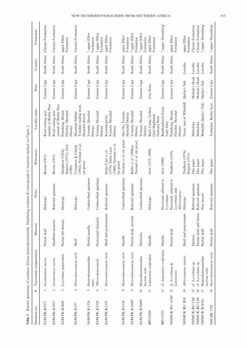
The upper Elliot and Clarens formations (upper parts ofthe informal ‘Stormberg Group’) have yielded nearly twentyheterodontosaurid specimens since the early twentieth century(Table 1, Fig. 1). These specimens have formed the basis forfive genera and species: Geranosaurus atavus Broom, 1911,Lycorhinus angustidens Haughton, 1924, Heterodontosaurustucki Crompton & Charig, 1962, Abrictosaurus consors(Thulborn 1974) and Lanasaurus scalpridens Gow, 1975.Geranosaurus, Lycorhinus and Lanasaurus were all based pri-marily upon fragmentary jaw material, and taxonomic assign-ments have been made largely upon differences in dentalmorphology, the ontogenetic and intraspecific significance ofwhich remains uncertain. This has led to a confused taxonomy,in which there has been little agreement upon the number ofvalid species and the specimens assigned to each taxon. Recentreviews have considered ‘Geranosaurus’ a nomen dubium, andLycorhinus, Heterodontosaurus and Abrictosaurus as valid dis-tinct taxa (Weishampel & Witmer 1990; Norman et al. 2004a).The status of Lanasaurus is uncertain: although it might bereferable to Lycorhinus (Gow 1990), the material preserved inthe holotypes of the two taxa does not overlap. Anotherspecimen, NHMUK RU A100, described by Thulborn (1970),has been controversial, and has been assigned to bothAbrictosaurus (Hopson 1975) and Lycorhinus (Gow 1990). Thepresent authors consider Heterodontosaurus and Abrictosaurusas valid, and both Lycorhinus and Lanasaurus as provisionallyvalid pending discovery of more complete specimens. Thetaxonomic assignment of NHMUK RU A100 is consideredto be uncertain; it may be referable to either Lycorhinus
or Lanasaurus or, alternatively, Lycorhinus, Lanasaurus andNHMUK RU A100 may all represent a single taxon (forwhich the valid name would be Lycorhinus). A provisionalreview of these historical taxa and specimens is presented byNorman et al. (in press). In addition to uncertainties over thetaxonomy and phylogenetic positions of the southern Africanheterodontosaurids, palaeobiological discussion has focusedon aspects of the jaw mechanics, dental replacement, diet, lifehistory and sexual dimorphism (Thulborn 1970, 1974, 1978;Hopson 1980; Weishampel 1984; Crompton & Attridge 1986;Galton 1986; Barrett 1998; Porro 2007, 2009; Butler et al.2008a; Norman et al. in press).
A major factor in prolonging these controversies has beenthe paucity of published anatomical data for heterodontosau-rids. Despite being known for the nearly 50 years, the cranialanatomy of Heterodontosaurus has not been described in detail(but see Norman et al. in press for a full description), andAbrictosaurus and NHMUK RU A100 require further prep-aration and study. Moreover, nearly half of the known south-ern African heterodontosaurid specimens have not beendescribed (Table 1). This paper partially redresses this situ-ation by describing four heterodontosaurid specimens thathave not previously been discussed in the literature. The aimsare to document this important material and provide newinsights into the morphology and diversity of southern Africanheterodontosaurids and, subsequently, to discuss the implica-tions of the southern African heterodontosaurid assem-blage for understanding the early evolution of ornithischiandinosaurs across the Triassic/Jurassic boundary.
Institutional abbreviations: BPI, Bernard Price Institute,University of the Witwatersrand, Johannesburg, South Africa;CAMSM, Sedgwick Museum of Earth Sciences, Cambridge,UK; NHMUK, Natural History Museum, London, UnitedKingdom; LACM, Natural History Museum of Los Angeles
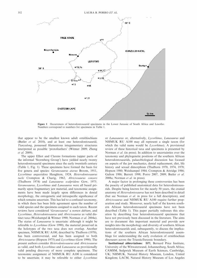
Figure 1 Occurrences of heterodontosaurid specimens in the Lower Jurassic of South Africa and Lesotho.Numbers correspond to numbers for specimens in Table 1.
352 LAURA B. PORRO ET AL.
Tab
le1
Kno
wn
spec
imen
sof
sout
hern
Afr
ican
hete
rodo
ntos
auri
ds.
Num
beri
ng(c
olum
n#
)co
rres
pond
sto
loca
tion
sm
arke
don
Fig
ure
1.
Spec
imen
no.
#T
axon
omic
assi
gnm
ent(
s)M
ater
ial
Not
esR
efer
ence
sL
ocal
ity
nam
eSt
ate
Cou
ntry
For
mat
ion
SAM
-PK
-K18
711
Ger
anos
auru
sat
avus
Par
tial
skul
lH
olot
ype
Bro
om(1
911)
Roa
dcu
ttin
gne
arsu
mm
itof
Bar
kly
Pas
sE
aste
rnC
ape
Sout
hA
fric
aC
lare
nsF
orm
atio
n
SAM
-PK
-K18
572
Ger
anos
auru
sat
avus
Hin
dlim
bm
ater
ial
Ref
erre
dsp
ecim
enB
room
(191
1)R
oad
cutt
ing
near
sum
mit
ofB
arkl
yP
ass
Eas
tern
Cap
eSo
uth
Afr
ica
Cla
rens
For
mat
ion
SAM
-PK
-K36
063
Lyc
orhi
nus
angu
stid
ens
Par
tial
left
dent
ary
Hol
otyp
eH
augh
ton
(192
4);
Hop
son
(197
5);
Gow
(199
0)
Pab
allo
ng,
Mou
ntF
letc
her,
Her
sche
lD
istr
ict
Eas
tern
Cap
eSo
uth
Afr
ica
uppe
rE
lliot
For
mat
ion
SAM
-PK
-K33
74
Het
erod
onto
saur
ustu
cki
Skul
lH
olot
ype
Cro
mpt
on&
Cha
rig
(196
2);
Nor
man
etal
.(i
npr
ess)
Mou
ntai
nbe
hind
Tyi
ndin
itr
adin
gst
ore,
Her
sche
ldi
stri
ct
Eas
tern
Cap
eSo
uth
Afr
ica
Cla
rens
For
mat
ion
SAM
-PK
-K13
265
Het
erod
onto
saur
idae
inde
t.P
arti
alm
axill
aU
ndes
crib
edsp
ecim
enV
oisa
na,
Her
sche
lD
istr
ict
Eas
tern
Cap
eSo
uth
Afr
ica
?up
per
Elli
otF
orm
atio
nSA
M-P
K-K
1328
6?H
eter
odon
tosa
urid
Pos
tcra
nial
mat
eria
lU
ndes
crib
edsp
ecim
enV
oisa
na,
Her
sche
lD
istr
ict
Eas
tern
Cap
eSo
uth
Afr
ica
?up
per
Elli
otF
orm
atio
nSA
M-P
K-K
1332
7H
eter
odon
tosa
urus
tuck
iSk
ull
and
post
cran
ium
Ref
erre
dsp
ecim
enSa
nta
Luc
aet
al.
(197
6);
Sant
aL
uca
(198
0);
Nor
man
etal
.(i
npr
ess)
Kro
mm
espr
uit,
Voi
sana
,H
ersc
hel
Dis
tric
t
Eas
tern
Cap
eSo
uth
Afr
ica
uppe
rE
lliot
For
mat
ion
SAM
-PK
-K13
348
Het
erod
onto
saur
ustu
cki
Max
illa
Und
escr
ibed
spec
imen
Nor
man
etal
.(i
npr
ess)
Site
18a,
Voi
sana
,H
ersc
hel
Dis
tric
tE
aste
rnC
ape
Sout
hA
fric
aup
per
Elli
otF
orm
atio
nSA
M-P
K-K
1048
79
Het
erod
onto
saur
ustu
cki
Par
tial
skul
l,ju
veni
leR
efer
red
spec
imen
But
ler
etal
.(2
008a
);N
orm
alet
al.
(in
pres
s)V
oisa
na,
Her
sche
lD
istr
ict
Eas
tern
Cap
eSo
uth
Afr
ica
?up
per
Elli
otF
orm
atio
nSA
M-P
K-K
1048
810
Het
erod
onto
saur
idae
ince
rtae
sedi
sD
enta
ries
Und
escr
ibed
spec
imen
Voi
sana
,H
ersc
hel
Dis
tric
tE
aste
rnC
ape
Sout
hA
fric
a?
uppe
rE
lliot
For
mat
ion
BP
/1/4
244
11L
anas
auru
ssc
alpr
iden
sM
axill
aH
olot
ype
Gow
(197
5,19
90)
Buc
kC
amp,
Gol
den
Gat
eH
ighl
ands
Nat
iona
lP
ark
Fre
eSt
ate
Sout
hA
fric
aup
per
Elli
otF
orm
atio
n
BP
/1/5
253
12cf
.L
anas
auru
ssc
alpr
iden
sM
axill
aP
revi
ousl
yre
ferr
edto
Lyc
orhi
nus
Gow
(199
0)B
ambo
eskl
oof
Far
m,
Lad
yG
rey
Eas
tern
Cap
eSo
uth
Afr
ica
?up
per
‘Sto
rmbe
rg’
NH
MU
KR
UA
100
13cf
.L
ycor
hinu
s&
Lan
asau
rus
Par
tial
skul
lP
revi
ousl
yre
ferr
edto
Lyc
orhi
nus
and
Abr
icto
saur
us
Thu
lbor
n(1
970)
Pab
allo
ng,
Mou
ntF
letc
her,
Her
sche
lD
istr
ict
Eas
tern
Cap
eSo
uth
Afr
ica
uppe
rE
lliot
For
mat
ion
NH
MU
KR
UB
5414
Abr
icto
saur
usco
nsor
sSk
ull
and
post
cran
ium
Hol
otyp
eT
hulb
orn
(197
4);
Hop
son
(197
5)N
oosi
,ea
stof
Whi
tehi
llQ
acha
’sN
ekL
esot
houp
per
Elli
otF
orm
atio
nN
HM
UK
RU
C68
15cf
.L
ycor
hinu
ssp
.D
enta
ryR
efer
red
spec
imen
Thi
spa
per
Mab
olok
aM
ohal
e’s
Hoe
kL
esot
hoC
lare
nsF
orm
atio
nN
HM
UK
RU
C69
16cf
.L
ycor
hinu
ssp
.P
arti
alsk
ull
and
fem
urR
efer
red
spec
imen
Thi
spa
per
Mab
olok
aM
ohal
e’s
Hoe
kL
esot
hoC
lare
nsF
orm
atio
nN
HM
UK
R14
161
17H
eter
odon
tosa
urid
aein
cert
aese
dis
Par
tial
skul
lN
ewta
xon?
Thi
spa
per
Whi
tehi
ll,Q
acha
’sN
ekQ
acha
’sN
ekL
esot
ho?
uppe
r‘S
torm
berg
’
NM
QR
1788
18H
eter
odon
tosa
urus
tuck
iP
arti
alsk
ull
Ref
erre
dsp
ecim
enT
his
pape
rT
ushi
elaw
,B
arkl
yE
ast
Eas
tern
Cap
eSo
uth
Afr
ica
?up
per
‘Sto
rmbe
rg’
NEW HETERODONTOSAURIDS FROM SOUTHERN AFRICA 353
County, Los Angeles, California, USA; NM, NationalMuseum, Bloemfontein, South Africa; SAM-PK, Iziko SouthAfrican Museum, Cape Town, South Africa.
1. Materials and methods
Preparation of NHMUK R14161 was carried out by SM-F.Due to the fragmented and fragile nature of the specimen,rotary diamond burrs were used to grind down the overlayingmatrix until <0·5 mm remained. This veneer of matrix wasthen carefully removed using a pneumatic airpen fitted with a1 mm diameter carbide pin at a very low air pressure(<20 PSI). Cracks, loose pieces and fragile areas were consoli-dated using a 5–15% solution of Paraloid B72 in acetone.Particularly thin or unsupported areas were strengthened bybrushing with molten Carbowax (polyethylene 400); this sup-port was further strengthened by adding layers of fine cottongauze. The Carbowax was later removed by either heating witha hot needle or by picking away with mounted pins. Theremaining wax residue was removed by gently brushing withwarm water.
NM QR 1788 was micro-CT scanned at the NHMUK byRLA with a HMX-ST CT 225 system and reconstructed withbeam hardening and noise corrections using CT-PRO 2.0(Metris X-Tek, Tring, UK). The resulting data set had anisotropic voxel size of 48·5 �m. The scan was visualised andsegmented by LBP (to extract bones and teeth) using Amira5.2.2 (Visage Imaging GmbH, Berlin, Germany).
2. Specimen NM QR 1788
2.1. Systematic palaeontology
Dinosauria Owen, 1842Ornithischia Seeley, 1887
Heterodontosauridae Kuhn, 1966Heterodontosaurus Crompton & Charig, 1962
Heterodontosaurus tucki Crompton & Charig, 1962
2.2. Material
NM QR 1788 is an incomplete articulated skull (Figs 2–3),comprising the incomplete dentaries (with in situ, but dam-aged, dentition), fragments of the postdentary bones, frag-ments and impressions of the maxillae and maxillary dentition,and a partial palate.
2.3. Locality and horizonCollected from an undocumented horizon within the‘Stormberg Group’ (presumably the upper part of thesequence, of Early Jurassic age) at Tushielaw Farm (30.78(S27.95(E, coordinates for the farmhouse), near Rhodes, BarklyEast District, Eastern Cape Province by P. J. Herselman in1975. The specimen was previously catalogued as an individualof the basal sauropodomorph dinosaur Massospondylus, andwas identified as a heterodontosaurid by Dr Adam Yates (BPI,Johannesburg).
2.4. DescriptionThe skull (Figs 2–3) has undergone shear such that the rightside of the skull is displaced ventrally and rostrally relative tothe left side; moreover, the cranium has been transverselycompressed at its rostral end. Most of the elements aredamaged and heavily eroded, complicating attempts to identifysutures.
Micro-CT scans of the upper jaw exhibited very poorcontrast between bone and matrix; as a result, the rostralcranial elements were not successfully segmented. The leftmaxilla is represented by a small sliver of bone from its medialsurface (Fig. 2c–d); the right maxilla is more complete, repre-sented by fragments of the medial maxillary shelf, alveolarmargin and dentition, and rostrally by an impression of themedial maxillary shelf (Fig. 2a–b). Digital moulds of thesesurfaces were created using Amira to aid visualisation. Themaxillary shelves are dorsoventrally expanded rostrally anddecrease in height caudally. Six left and six right maxillaryteeth are represented by fragments and impressions of thelingual crown surfaces. These fragments/impressions indicatethat the crowns are chisel-shaped and packed such that adja-cent crowns contacted each other at their apices. Severalimpressions (right crown fragments 2 and 5, left crown frag-ment 4) indicate that large apicolingually-facing planar wearfacets were present and covered the entire crown apex. Impres-sions (particularly of left crown fragments 2 and 3) indicate amedian ridge on the lingual surface of the maxillary crowns.
The palate is partially preserved and exposed in dorsal view(Fig. 2e–f). The vomers are present as several elongate rostro-caudally extending slivers of bone positioned along the midlinebetween the medial maxillary shelves (Fig. 2f: vom); theircaudal ends are not preserved, thus no information on theircontacts with other palatal elements is available. Fragments ofthe palatines are positioned between the preserved caudalends of the vomers and the maxillary shelves (Fig. 2f: pal).Rostrally, the right palatine contacts the medial surface ofthe right maxilla; caudally, the palatine fragments contact thelateral edges of the palatal processes of the pterygoids. Thepterygoids are partially preserved, including the palatal pro-cesses (Fig. 2f: rpt), which are not preserved in any otherknown specimen of Heterodontosaurus (LBP pers. obs.). Thepalatal processes of the pterygoids are short and triangular,tapering rostrally and diverging laterally from each other tocontact the medial edges of the palatines; caudally, the ptery-goids contact one another along the midline. There is anelongate ventral process of the right pterygoid (i.e., pterygoidflange) that projects ventrolaterally towards the mandibularfossa of the lower jaw and is arched (laterally convex) along itslength. The ectopterygoid is a robust element that extensivelyoverlaps the rostrodorsal aspect of the ventral process of thepterygoid (Fig. 2b, f: ect); it projects rostrally and laterally,contacting the medial surface of the caudal end of the maxilla.
The predentary is missing. Micro-CT scans of the lower jawexhibit good contrast between fossil material and matrix,revealing the dentaries, splenials, coronoids and prearticularson both sides (Fig. 3). Fragments of other postdentary bonesare present, but cannot be positively identified. Both dentaries(Figs 2a–d, g–h, 3a–b) are incomplete at their rostral ends. Thecaudal and ventral margins of the dentaries are damaged, andthe lateral surfaces (particularly that of the right dentary) havebeen eroded. The dentary is a robust and transversely thickelement. Caudally, it forms the rostral margin of the coronoideminence. The left dentary bifurcates caudally; the roundednotch between the dorsal and ventral processes forms therostral margin of the external mandibular fenestra (Fig. 3b).Rostrally, both dentaries exhibit a ventrally-projecting ‘flange’,resembling that reported in Psittacosaurus (Zhou et al. 2006;Sereno et al. 2007); however, because the ventral margins ofthe dentaries are heavily damaged, it is possible that this flangeis an artefact of preservation. The dentary symphyseal contactis a nearly flat, vertical surface.
The left and right splenials are present (Figs 2g–h, 3c–d),although the left splenial is better preserved. The splenial istransversely compressed and rostrocaudally elongated, and
354 LAURA B. PORRO ET AL.

medially overlies the Meckelian fossa. Micro-CT scans dem-onstrate that the splenial medially laps the dentary on theventromedial surface of the mandible. Rostrally, the ventraledge of the splenial is separated from the ventral mandibularmargin.
The coronoid is preserved on both left and right sides(Figs 2, 3c–d), and formed the dorsal margin of the coronoideminence. Unlike the condition in other basal ornithischians(e.g. Sereno 1991), it is a robust, dorsoventrally-expanded,sheet-like bone that extends rostrally below and medial to the
Figure 2 Photographs (a, c, e, g) and line drawings (b, d, f, h) of skull of Heterodontosaurus tucki (NM QR1788): (a–b) right lateral view; (c–d) left lateral view; (e–f) dorsal view; (g–h) ventral view. For line drawings, lightgrey indicates eroded bone; dark grey indicates matrix. Abbreviations: ci=caniniform tooth impression;cnd=coronoid bone; den=dentary; ect=ectopterygoid; mx=maxilla; mxsh=medial maxillary shelf; pal=palatine; pre=prearticular; ptf=(ventral) pterygoid flange; rpt=rostral (palatal) process of the pterygoid;spl=splenial; vom=vomers. Scale bars=10 mm; photographs are at the same scale as corresponding line-drawing.
NEW HETERODONTOSAURIDS FROM SOUTHERN AFRICA 355
tooth row. The coronoid eminence appears to be rela-tively taller in NM QR 1788 than in other Heterodontosaurusspecimens (SAM-PK-K337, SAM-PK-K1332).
Both prearticulars are present, although only the left pre-articular is visible within the external mandibular fenestra(Figs 2c–d, 3b). The prearticular slots dorsally into a troughformed by the dentary laterally and ventrally, and the splenialmedially.
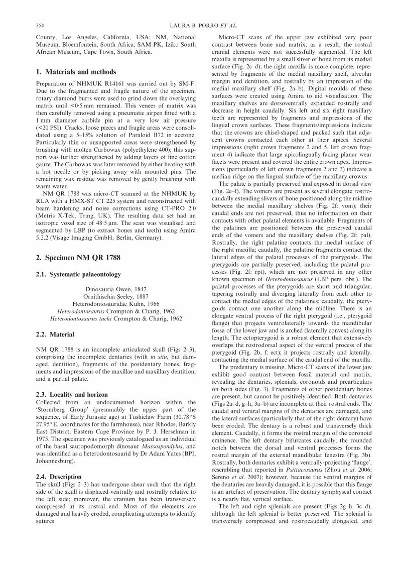
The dentary tooth row is virtually complete on the rightside, which preserves a partial impression of the lingual surfaceof the root of the dentary caniniform and 11 post-caniniformteeth (Fig. 3e, g, i), giving a complete dentary tooth count of 12in NM QR 1788. The height of the crowns has been accentu-ated by damage to the lateral surface of the dentary, which inmany cases has exposed the roots. Crowns 8–9 on the right sideare the best preserved. The left dentary tooth row containsseven badly preserved teeth corresponding to crowns 3–9
(Fig. 3f, h). Additionally, micro-CT scans demonstrate thepresence of a small eighth tooth (probably crown 11) lyingalmost horizontally within the caudal lower jaw, its apexdirected rostrally; this displaced tooth is separated from crown9 by a gap that probably marks the position of the missingcrown 10. The first post-caniniform crown (crown 2) is muchsmaller in size than subsequent crowns, and was clearlyseparated from the caniniform by a diastema approximatelyequal in length to the mesiodistal width of a single crown.Caudally, the crowns increase in mesiodistal width and apico-basal height, with the largest crowns (crowns 6–8) located atthe midpoint of the tooth row. Individual crowns are widestmesiodistally at their apices and taper gently towards theirroots, which extend nearly to the preserved ventral margin ofthe lower jaw; however, there is no clearly defined mesiodistalexpansion (‘neck’) or transverse expansion (‘cingulum’) of thecrown above the root. Large, planar, dorsolaterally-facing
Figure 3 Rostral part of mandible of Heterodontosaurus tucki (NM QR 1788) reconstructed from micro-CTdata: (a) right rostral mandible, lateral view; (b) left rostral mandible, lateral view; (c) right rostral mandible,medial view; (d) left rostral mandible, medial view; (e) right dentary tooth row, lateral view; (f) left dentary toothrow, lateral view; (g) right dentary tooth row, medial view; (h) left dentary tooth row, medial view; (i) rightdentary tooth row (bones removed) in oblique caudomedial view; (j) articulated rostral mandibles, dorsal view.Elements are colour coded as follows: dentary, red; splenial, blue; coronoid, orange; prearticular, grey; functionaldentary teeth, yellow; unresorbed tooth roots, green. The bones have been made transparent in several views(e–h) to better visualise dental anatomy. Abbreviations: ci=caniniform tooth impression; cr=crown; emf=external mandibular fenestra. Scale bars=10 mm and scale bar for (a) also applies to (b–h).
356 LAURA B. PORRO ET AL.
wear facets are present on several of the crowns and cover theapical portion of the labial crown surface. A prominentmedian ridge, mesially offset, is present on the lingual crownsurface, as demonstrated on CT scans and suggested by visibletooth wear; there is also a less pronounced distal ridge on thelingual crown surface. Adjacent crowns contact one anotherapically, but are separated from one another by small gapsbasally. The teeth are curved in mesiodistal view, being later-ally concave (Fig. 3i); furthermore, the dentary tooth rows arelaterally concave in dorsal view (Fig. 3j).
Several of the crowns (9 and 10 the right side and 8 and 9 onthe left) have unusual fragments positioned immediately mesialand labial to their crown bases (Fig. 3e–f); a fifth fragment ispositioned distal and labial to crown 9 on the left side and wasprobably associated with the missing tenth crown.
2.5. Taxonomic identityHeterodontosaurus tucki is characterised by the possession ofcolumnar maxillary and dentary teeth that lack a definedmesiodistal or labiolingual expansion above the root (Charig &Crompton 1974; Hopson 1975; Butler et al. 2008a; Normanet al. in press). The maxillary and dentary teeth are closelypacked, forming a continuous dental battery, with smallgaps between the teeth persisting only at the bases; moreover,they are transversely expanded relative to their mesiodistallength and exhibit heavier wear than either Lycorhinus orAbrictosaurus, with denticles rarely preserved except on themost mesial or distal dentary teeth (Butler et al. 2008a). Mostof these dental characters are present in NM QR 1788, and thisspecimen can be confidently referred to Heterodontosaurustucki.
3. Specimens NHMUK RU C68 and NHMUKRU C69
3.1. Systematic palaeontology
Heterodontosauridae Kuhn, 1966Lycorhinus Haughton, 1924
cf. Lycorhinus sp.
3.2. Material
NHMUK RU C68, partial right dentary, including elevenpartial crowns (Fig. 4). NHMUK RU C69, articulated butheavily damaged partial skull, with associated vertebra, femurand unidentified bone fragments (Figs 5–6).
3.3. Locality and horizonBoth specimens were found ‘‘on the 6,500# cave sst. plateaulying north of the eastern block of Mabloka Mt. [FrancesMountain]’’, Lesotho (unpublished field catalogue of K.Kermack and F. Mussett, NHMUK). The specimens werecollected by Prof. Kenneth Kermack and Mrs Frances Mussettas part of the 1968 University of London expedition. Althoughdetailed stratigraphic data are not available, the field notesindicate that these specimens were probably collected from theClarens Formation (=‘Cave Sandstone’). It is not clear howclose to one another the specimens were collected. Althoughwe have been unable to locate ‘Mabloka’ mountain on maps ofsouthern Lesotho, it seems likely that the locality in question is‘Maboloka’ (29.88(S, 27.35(E: Kitching & Raath 1984).
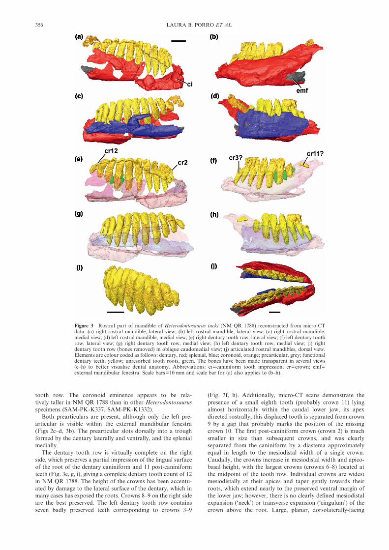
Figure 4 NHMUK RU C68, right dentary of a heterodontosaurid (cf. Lycorhinus sp.) from the ClarensFormation (Early Jurassic) of Lesotho: (a) medial view; (b) close-up of crowns 2 to 6; (c) line drawing of medialview. Abbreviations: can=caniniform; cing=‘cingulum’ at base of crown; cr=crown. For line drawings light greyindicates eroded bone; dark grey indicates matrix. Scale bars=10 mm.
NEW HETERODONTOSAURIDS FROM SOUTHERN AFRICA 357
3.4. DescriptionNHMUK RU C68 comprises a partial right dentary preservedwithin a block of reddish fine-grained sandstone. The blockand attached fossil material have broken into three pieces: themajority of the dentary is preserved in the largest of the three
blocks and is exposed in medial view (Fig. 4). Most of themedial surface of the dentary (and presumably the splenial), aswell as the lingual parts and/or impressions of the dentarycrowns are preserved on the two smaller blocks. The pre-dentary and the anteriormost dentary are either missing or
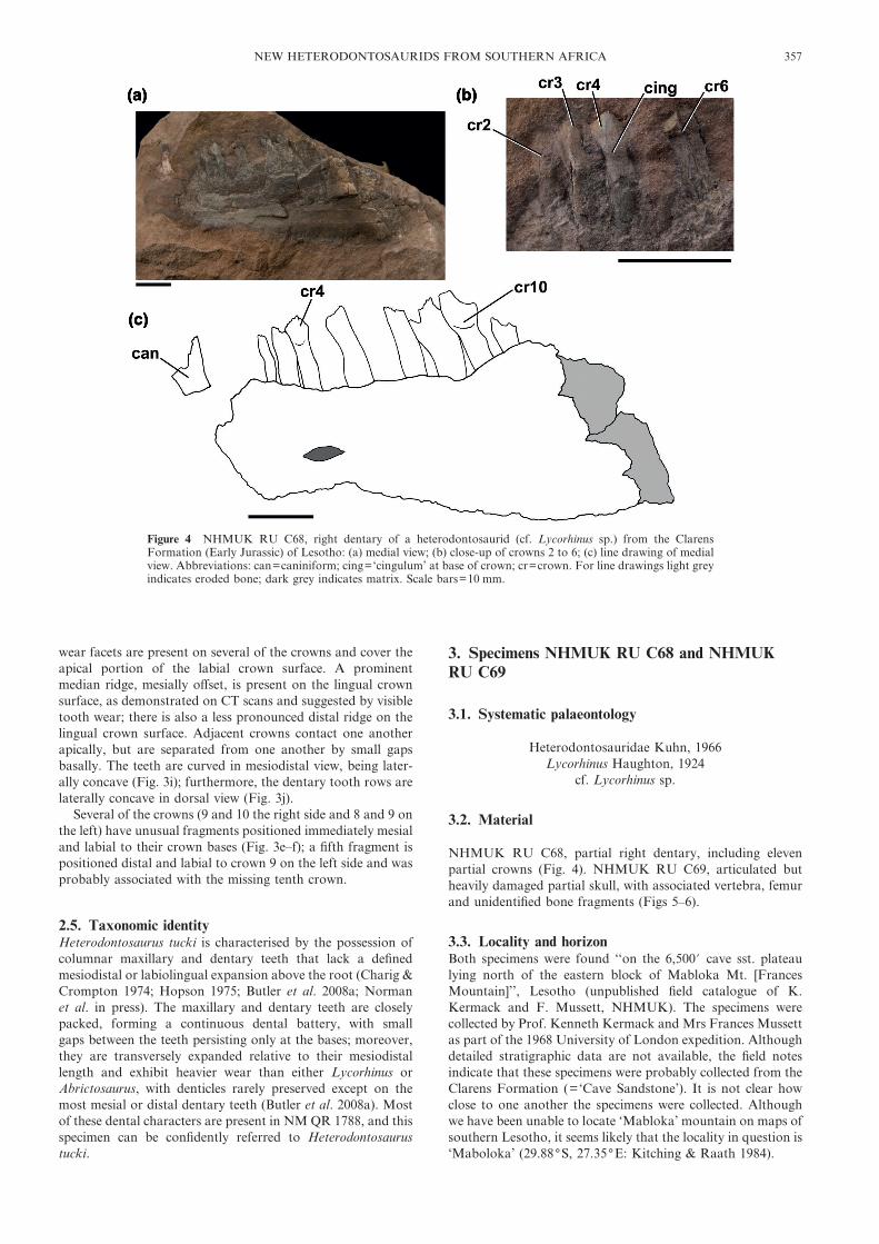
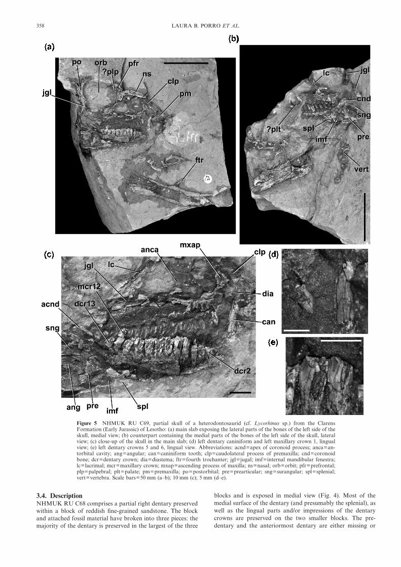
Figure 5 NHMUK RU C69, partial skull of a heterodontosaurid (cf. Lycorhinus sp.) from the ClarensFormation (Early Jurassic) of Lesotho: (a) main slab exposing the lateral parts of the bones of the left side of theskull, medial view; (b) counterpart containing the medial parts of the bones of the left side of the skull, lateralview; (c) close-up of the skull in the main slab; (d) left dentary caniniform and left maxillary crown 1, lingualview; (e) left dentary crowns 5 and 6, lingual view. Abbreviations: acnd=apex of coronoid process; anca=an-torbital cavity; ang=angular; can=caniniform tooth; clp=caudolateral process of premaxilla; cnd=coronoidbone; dcr=dentary crown; dia=diastema; ftr=fourth trochanter; jgl=jugal; imf=internal mandibular fenestra;lc=lacrimal; mcr=maxillary crown; mxap=ascending process of maxilla; ns=nasal; orb=orbit; pfr=prefrontal;plp=palpebral; plt=palate; pm=premaxilla; po=postorbital; pre=prearticular; sng=surangular; spl=splenial;vert=vertebra. Scale bars=50 mm (a–b); 10 mm (c); 5 mm (d–e).
358 LAURA B. PORRO ET AL.

unexposed, the ventral margin of the dentary is incomplete,and the dentary is broken caudally prior to the termination ofthe tooth row. Observable anatomical details of the dentaryare limited because of damage to the specimen, with theexception that the dentary increases in dorsoventral heightcaudally. A caniniform tooth is present at the mesial end; distalto this tooth there are seven incompletely preserved crowns,two gaps indicating the presence of additional crowns, and onebroken root at the distal end. The minimum tooth count forthe dentary is 11. The specimen is from a relatively largeindividual – the length from the mesial margin of the canini-form to the distal margin of crown 11 is 52 mm, and thepreserved cheek tooth row is approximately 39 mm in length.
Only the base of the dorsally-projecting caniniform is pre-served. Its size cannot be estimated, nor can it be determinedwhether or not serrations were present on the mesial and distalmargins. The caniniform tooth is separated by a diastema fromthe first post-caniniform crown. This diastema appears to belong compared to Heterodontosaurus, but it is possible thatanother small crown might have been present rostral to thefirst post-caniniform dentary tooth (crown 2); further mechan-ical preparation has not been carried out because of thefragility of the specimen in this region. Crowns 3–4 and 10have relatively well-preserved lingual surfaces; crowns 5 and 7are missing; crowns 6, 8 and 9 are broken in half through theirvertical axes; and crown 11 is represented by a broken root.The chisel-shaped crowns are expanded mesiodistally (forminga ‘neck’) and labiolingually (forming a ‘cingulum’) above theroot (Fig. 4b). Approximately five denticles are preserved (asimpressions on the smaller block) along the mesial margin ofcrown 3, and appear to be restricted to the apical third of thecrown. The mesial and distal margins of the crowns arethickened into low ridges that merge basally with the cingu-lum. A broad median swelling is present on the lingual surfaceand merges basally with the cingulum – this swelling is offsetslightly mesially and curves gently distally towards its apicalmargin. On either side of this swelling the crown surface is
gently depressed. There are no preserved secondary ridges onthe lingual crown surfaces. The labial surfaces are not exposed,and there is no available information on wear facets. Thedentary crowns are imbricated, resulting in an en echelonarrangement so that the mesial margin of each crown mediallyoverlaps the distal margin of the preceding crown. The crownscontact one another apically but gaps remain between adjacentcrowns basally.
NHMUK RU C69 includes an articulated partial skull andsome associated postcranial elements. The left side of the skullis exposed from the premaxilla to the postorbital/jugal bar(Figs 5–6). The skull is preserved as part and counterpart intwo red sandstone blocks, and has been split vertically suchthat one block (‘counterpart’) contains the medial parts of theleft maxilla, lacrimal, jugal, mandible and dentition (Figs 5b,6b), and the other block (‘main slab’) contains the lateral partsof these elements as well as additional elements (e.g. post-orbital, nasal, premaxilla; Figs 5a, c, 6a). The palate and rightside of the skull may also be present in the specimen, but arenot currently exposed. The blocks also contain a partial femurand a cross-section through a vertebra, as well as unidenti-fied bone fragments (Figs 5a–b, 6). The preservation of thespecimen limits the available anatomical data. The aim atpresent is to document, figure and briefly describe this speci-men; future preparation or CT imaging may provideadditional information.
Although parts of a premaxilla, including an elongatetapering caudolateral process (Fig. 5a, c: clp), are present, it ispoorly preserved and the oral margin of the element is notexposed. The premaxilla appears to make a point contact withthe lacrimal, but this cannot be confirmed with certainty, dueto cracks within the encasing sediment in this area. An archeddiastema (into which projects the dentary caniniform tooth)separates the premaxilla from the maxilla (Fig. 5c: dia). Theapproximate outline of an extensive external antorbitalfenestra is visible, although its margins are damaged and themedial wall of the antorbital cavity is missing. Only fragments
Figure 6 NHMUK RU C69, partial skull of a heterodontosaurid (cf. Lycorhinus sp.): (a) line drawing of mainslab exposing the lateral parts of the bones of the left side of the skull, medial view; (b) counterpart containingthe medial parts of the bones of the left side of the skull, lateral view. Abbreviations: anca=antorbital cavity;ang=angular; can=caniniform tooth; clp=caudolateral process of premaxilla; cnd=coronoid bone; den=dentary; dia=diastema; ftr=fourth trochanter; jgl=jugal; imf=internal mandibular fenestra; lc=lacrimal;mec=Meckelian canal; mxap=ascending process of maxilla; ns=nasal; orb=orbit; pfr=prefrontal; plt=palate;pm=premaxilla; po=postorbital; pre=prearticular; sng=surangular; spl=splenial; vert=vertebra. For linedrawings light grey indicates eroded bone, dark grey indicates matrix. Scale bar=50 mm.
NEW HETERODONTOSAURIDS FROM SOUTHERN AFRICA 359
and impressions of the maxilla are visible, but it has a short,rostrocaudally narrow ascending process that contacts thepremaxilla. Fourteen maxillary teeth appear to be preserved;most of these teeth are represented only by impressions (forexceptions, see below). The lacrimal is shaped like an inverted‘L’, and forms the dorsal and caudal margins of the externalantorbital fenestra (Fig. 5c: lc). It contacts the jugal ventrally.Fragments of the prefrontal, nasal and, possibly, the palpebralare present, but are not anatomically informative in theircurrent state of preparation (Fig. 5a). The rostral process ofthe jugal is present and visible in medial view, but the details ofits contacts with the lacrimal and maxilla are unclear. Theelongate tapering dorsal process of the jugal forms the caudo-ventral margin of the sub-circular orbit, and is overlappedlaterally by the descending process of the postorbital. Thecaudal process of the jugal is dorsoventrally expanded, butonly a cross-section of its base is visible. Because the lateralsurface of the jugal is not exposed, it is impossible to determineif a jugal boss is present or absent.
Elements of the lower jaw that are exposed include thedentary (in medial view), parts of the splenial and coronoid(mostly preserved in lateral view on the counterslab), andfragments of the surangular, angular and prearticular (Fig.5a–c). The dentary is poorly exposed, but is relatively deep,rising to form the rostral margin of the coronoid eminence (Fig.5c: acnd). The sheet-like splenial can be clearly distinguishedoverlying the caudoventral part of the dentary; its caudalmargin is notched for the internal mandibular fenestra (Fig. 5c:imf). The elongate strap-like coronoid extends immediatelyventromedial to the tooth row, along the entire length of thepreserved tooth row (Fig. 5b: cnd). Caudally it is expanded andlobe-like, forming the apex of the coronoid process. Theprearticular forms the caudal margin of the internal mandibularfenestra (Fig. 5c: pre), but other details of its anatomy areunclear. The surangular and angular are poorly preserved, andno particular anatomical details can be discerned.
Only parts of maxillary crowns 1 and 12–14 are preserved,the latter three crowns being heavily damaged. Crown 1 issmaller than more distal crowns, and has a sharply pointedapex (Fig. 5d). Denticles are visible along the distal surface; themesial surface is not completely exposed. The impressions ofthe remaining crowns indicate that they were chisel-shaped andcontacted each other apically. The dentary contains 13 crowns,including a caniniform tooth rostrally, but all are badlydamaged. The length of the ‘cheek tooth’ row is 46 mm. Thecaniniform tooth is elongate and tapers to a sharp tip (Fig. 5d).Fine serrations are present along its caudal margin, withapproximately three per millimetre. The serrations are squarein profile. The caniniform tooth is separated from crown 2 bya diastema, although its exact length cannot be determinedbecause the base of the tooth is not exposed. Crowns 2–13 haveelongate roots (partially exposed by breakage of the dentary)that curve labially, such that their lingual surface is apico-basally convex. The crowns are gently expanded labiolinguallyand mesiodistally above their roots, with a weak basal cingu-lum (Fig. 5e). The distal margin of the crown forms a distinctridge that merges with the basal cingulum. Although thelingual crown surfaces are badly damaged, a discrete primaryridge does not seem to be well-developed, although a medianeminence is present. The crowns are imbricated and separatedfrom one another by small gaps at their bases.
The proximal two thirds of the left femur is preserved. Thefemur is split vertically (lengthwise) revealing a cross-section ofthe central medullary cavity bounded on either side by corticalbone (Fig. 5a–b). The lateral margin of the proximal end isexpanded craniocaudally relative to the shaft; the cranial andgreater/dorsolateral trochanters cannot be clearly identified.
The shaft is bowed cranially along its length, and an elongateand very slender (‘rod-like’) pendant fourth trochanter ispresent, similar to Heterodontosaurus (SAM-PK-K1332).
3.5. Taxonomic identityThe caniniform tooth and the chisel-shaped crowns unambigu-ously indicate that NHMUK RU C68 and NHMUK RU C69represent heterodontosaurids. Assessing the affinities ofboth specimens within Heterodontosauridae is complicated byincompleteness and poor preservation. The dentary crowns ofLycorhinus (SAM-PK-3606: lingual crown surfaces describedby Haughton 1924), NHMUK RU A100 (Thulborn 1970) andHeterodontosaurus (SAM-PK-K1332) have lingual surfaceswith mesial and distal ridges and a weak median eminence thatis slightly offset mesially. NHMUK RU C68 and NHMUKRU C69 appear to be more similar to Lycorhinus and NHMRU A100 in possessing weak but distinct basal cingula, whichare absent in Heterodontosaurus, and in lacking secondaryridges, which occur on some dentary crowns of Heterodonto-saurus. The lingual surfaces of the dentary crowns are notexposed in the only known specimen of Abrictosaurus(NHMUK RU B54), preventing comparisons. Comparisons toLanasaurus are limited by the absence of the maxilla inNHMUK RU C68 and the scarcity of observable anatomicaldata for the maxilla of NHMUK RU C69. On the basis of thesimilarities between NHMUK RU C69 and Lycorhinus, thisspecimen is provisionally referred to cf. Lycorhinus sp. Ifcorrect, this referral substantially increases the amount of datafor Lycorhinus, which was previously known only from impres-sions of the lower jaw and a fragment of the caniniform tooth,and shows that the general form of its skull was similar to thatof Heterodontosaurus. NHMUK RU C68 most closely re-sembles Lycorhinus, based on the presence of weak basalcingula and absence of secondary ridges on the dentarycrowns. However, the curved median ridge and denticles ofNHMUK RU C68 are not preserved in NHMUK RU C69,and only impressions of the buccal surfaces of the dentaryteeth are preserved in the type of Lycorhinus. The absence ofthe curved median ridge in NHMUK RU C69 and Lycorhinusmay be preservational, and NHMUK RU C68 is provisionallyreferred to cf. Lycorhinus sp. although it is possible that futurediscoveries, further preparation of other heterodontosauridspecimens or better characterisation of the taxon Lycorhinusmay invalidate this referral.
4. Specimen NHMUK R14161
4.1. Systematic palaeontology
Heterodontosauridae Kuhn, 1966Heterodontosauridae incertae sedis
4.2. Material
NHMUK R14161, partial dentaries, maxillae and left lacrimal,with fragmentary dentition and fragments of other cranialbones (Fig. 7).
4.3. Locality and horizonThe only locality data currently available are ‘Whitehill’,Qacha’s Nek District, Lesotho. Whitehill (or White Hill) is asmall settlement on the Orange, or Senqu, River (30.05(S,28.47(E, coordinates from Google Earth), located within a fewkilometres of the type locality of Abrictosaurus consors (seeThulborn 1974, fig. 1). It is unclear what stratigraphic level the
360 LAURA B. PORRO ET AL.

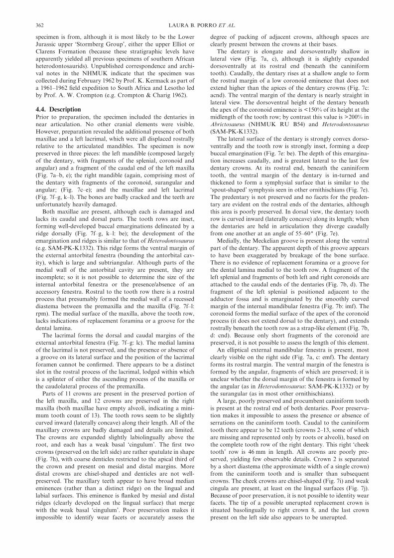
Figure 7 NHMUK R14161, partial skull of a heterodontosaurid (Heterodontosauridae incertae sedis) from the?upper ‘Stormberg Group’ of Lesotho: (a) left dentary, lateral view; (b) left dentary, medial view; (c) rightdentary, lateral view; (d) right dentary, medial view; (e) both dentaries in articulation, dorsal view; (f) left maxillaand lacrimal, lateral view; (g) line drawing of left maxilla an lacrimal, lateral view; (h) left maxillary crowns 1 and2, labial view; (i) right mid-dentary crowns, labial view; (j) right dentary crowns 11 and 12, lingual view; (k) rightmaxilla in lateral view, left maxilla in medial view; (l) line drawing of right maxilla in lateral view, left maxillain medial view. Abbreviations: acnd=apex of coronoid process; ang=angular; be=buccal emargination;can=caniniform; cnd=coronoid bone; cr=crowns; cvm=complete ventral margin of dentary; emf=externalmandibular fenestra; imf=internal mandibular fenestra; lc=lacrimal; lmx=left maxilla; mgr=mandibular groove;mx=fragment of the caudal end of the left maxilla; rmx=right maxilla; rpm=rostral process of maxilla;spl=splenial; sng=fragments of the surangular; sym=symphysial surface. For line drawings light grey indicateseroded bone or sediment. Scale bars=10 mm (a–g, k–l); 2 mm (h–j).
NEW HETERODONTOSAURIDS FROM SOUTHERN AFRICA 361
specimen is from, although it is most likely to be the LowerJurassic upper ‘Stormberg Group’, either the upper Elliot orClarens Formation (because these stratigraphic levels haveapparently yielded all previous specimens of southern Africanheterodontosaurids). Unpublished correspondence and archi-val notes in the NHMUK indicate that the specimen wascollected during February 1962 by Prof. K. Kermack as part ofa 1961–1962 field expedition to South Africa and Lesotho ledby Prof. A. W. Crompton (e.g. Crompton & Charig 1962).
4.4. DescriptionPrior to preparation, the specimen included the dentaries innear articulation. No other cranial elements were visible.However, preparation revealed the additional presence of bothmaxillae and a left lacrimal, which were all displaced rostrallyrelative to the articulated mandibles. The specimen is nowpreserved in three pieces: the left mandible (composed largelyof the dentary, with fragments of the splenial, coronoid andangular) and a fragment of the caudal end of the left maxilla(Fig. 7a–b, e); the right mandible (again, comprising most ofthe dentary with fragments of the coronoid, surangular andangular; (Fig. 7c–e); and the maxillae and left lacrimal(Fig. 7f–g, k–l). The bones are badly cracked and the teeth areunfortunately heavily damaged.
Both maxillae are present, although each is damaged andlacks its caudal and dorsal parts. The tooth rows are inset,forming well-developed buccal emarginations delineated by aridge dorsally (Fig. 7f–g, k–l: be); the development of theemargination and ridges is similar to that of Heterodontosaurus(e.g. SAM-PK-K1332). This ridge forms the ventral margin ofthe external antorbital fenestra (bounding the antorbital cav-ity), which is large and subtriangular. Although parts of themedial wall of the antorbital cavity are present, they areincomplete; so it is not possible to determine the size of theinternal antorbital fenestra or the presence/absence of anaccessory fenestra. Rostral to the tooth row there is a rostralprocess that presumably formed the medial wall of a recesseddiastema between the premaxilla and the maxilla (Fig. 7f–l:rpm). The medial surface of the maxilla, above the tooth row,lacks indications of replacement foramina or a groove for thedental lamina.
The lacrimal forms the dorsal and caudal margins of theexternal antorbital fenestra (Fig. 7f–g: lc). The medial laminaof the lacrimal is not preserved, and the presence or absence ofa groove on its lateral surface and the position of the lacrimalforamen cannot be confirmed. There appears to be a distinctslot in the rostral process of the lacrimal, lodged within whichis a splinter of either the ascending process of the maxilla orthe caudolateral process of the premaxilla.
Parts of 11 crowns are present in the preserved portion ofthe left maxilla, and 12 crowns are preserved in the rightmaxilla (both maxillae have empty alveoli, indicating a mini-mum tooth count of 13). The tooth rows seem to be slightlycurved inward (laterally concave) along their length. All of themaxillary crowns are badly damaged and details are limited.The crowns are expanded slightly labiolingually above theroot, and each has a weak basal ‘cingulum’. The first twocrowns (preserved on the left side) are rather spatulate in shape(Fig. 7h), with coarse denticles restricted to the apical third ofthe crown and present on mesial and distal margins. Moredistal crowns are chisel-shaped and denticles are not well-preserved. The maxillary teeth appear to have broad medianeminences (rather than a distinct ridge) on the lingual andlabial surfaces. This eminence is flanked by mesial and distalridges (clearly developed on the lingual surface) that mergewith the weak basal ‘cingulum’. Poor preservation makes itimpossible to identify wear facets or accurately assess the
degree of packing of adjacent crowns, although spaces areclearly present between the crowns at their bases.
The dentary is elongate and dorsoventrally shallow inlateral view (Fig. 7a, c), although it is slightly expandeddorsoventrally at its rostral end (beneath the caniniformtooth). Caudally, the dentary rises at a shallow angle to formthe rostral margin of a low coronoid eminence that does notextend higher than the apices of the dentary crowns (Fig. 7c:acnd). The ventral margin of the dentary is nearly straight inlateral view. The dorsoventral height of the dentary beneaththe apex of the coronoid eminence is <150% of its height at themidlength of the tooth row; by contrast this value is >200% inAbrictosaurus (NHMUK RU B54) and Heterodontosaurus(SAM-PK-K1332).
The lateral surface of the dentary is strongly convex dorso-ventrally and the tooth row is strongly inset, forming a deepbuccal emargination (Fig. 7e: be). The depth of this emargina-tion increases caudally, and is greatest lateral to the last fewdentary crowns. At its rostral end, beneath the caniniformtooth, the ventral margin of the dentary is in-turned andthickened to form a symphysial surface that is similar to the‘spout-shaped’ symphysis seen in other ornithischians (Fig. 7e).The predentary is not preserved and no facets for the preden-tary are evident on the rostral ends of the dentaries, althoughthis area is poorly preserved. In dorsal view, the dentary toothrow is curved inward (laterally concave) along its length; whenthe dentaries are held in articulation they diverge caudallyfrom one another at an angle of 55–60( (Fig. 7e).
Medially, the Meckelian groove is present along the ventralpart of the dentary. The apparent depth of this groove appearsto have been exaggerated by breakage of the bone surface.There is no evidence of replacement foramina or a groove forthe dental lamina medial to the tooth row. A fragment of theleft splenial and fragments of both left and right coronoids areattached to the caudal ends of the dentaries (Fig. 7b, d). Thefragment of the left splenial is positioned adjacent to theadductor fossa and is emarginated by the smoothly curvedmargin of the internal mandibular fenestra (Fig. 7b: imf). Thecoronoid forms the medial surface of the apex of the coronoidprocess (it does not extend dorsal to the dentary), and extendsrostrally beneath the tooth row as a strap-like element (Fig. 7b,d: cnd). Because only short fragments of the coronoid arepreserved, it is not possible to assess the length of this element.
An elliptical external mandibular fenestra is present, mostclearly visible on the right side (Fig. 7a, c: emf). The dentaryforms its rostral margin. The ventral margin of the fenestra isformed by the angular, fragments of which are preserved; it isunclear whether the dorsal margin of the fenestra is formed bythe angular (as in Heterodontosaurus: SAM-PK-K1332) or bythe surangular (as in most other ornithischians).
A large, poorly preserved and procumbent caniniform toothis present at the rostral end of both dentaries. Poor preserva-tion makes it impossible to assess the presence or absence ofserrations on the caniniform tooth. Caudal to the caniniformtooth there appear to be 12 teeth (crowns 2–13, some of whichare missing and represented only by roots or alveoli), based onthe complete tooth row of the right dentary. This right ‘cheektooth’ row is 46 mm in length. All crowns are poorly pre-served, yielding few observable details. Crown 2 is separatedby a short diastema (the approximate width of a single crown)from the caniniform tooth and is smaller than subsequentcrowns. The cheek crowns are chisel-shaped (Fig. 7i) and weakcingula are present, at least on the lingual surfaces (Fig. 7j).Because of poor preservation, it is not possible to identity wearfacets. The tip of a possible unerupted replacement crown issituated basolingually to right crown 8, and the last crownpresent on the left side also appears to be unerupted.
362 LAURA B. PORRO ET AL.

4.5. Taxonomic identityNHMUK R14161 cannot be clearly assigned to any existingheterodontosaurid taxon. It is distinguished from Heterodon-tosaurus and Abrictosaurus by a low coronoid eminence andthe presence of basal ‘cingula’ on the maxillary and dentarycrowns. It is further distinguished from Heterodontosaurus bythe absence of strongly developed primary, mesial and distalridges on the labial surfaces of the maxillary crowns; thestrongly divergent mandibles also suggest that the skull ofNHMUK R14161 was wider than that of Heterodontosaurus.NHMUK R14161 is further distinguished from Abrictosaurus(NHMUK RU B54) by the inwardly curved maxillary anddentary tooth rows (although the apparent absence of thisfeature in NHMUK RU B54 might reflect taphonomic com-pression of the skull), the well-developed buccal emarginationon the maxilla, and the presence of a dentary caniniform.Comparisons to Lycorhinus are difficult (because of the highlyfragmentary nature of the Lycorhinus holotype specimen,SAM-PK 3606); the dentary caniniform teeth of both speci-mens are identical in size, but the dentary of Lycorhinus isdeeper than that of NHMUK R14161. Furthermore, themaximum depth of the dentary beneath the tooth row is 40%of the length of the ‘cheek teeth’ row in SAM-PK 3606 andapproximately 30% in NHMUK R14161, although it shouldbe noted that the tooth row might be incomplete in SAM-PK3606. Overall, it appears that the dentary of NMHUK R14161was more slender than that of the Lycorhinus type, despite thespecimens being of similar size, and despite the missing ventralmargin of the dentary and the coronoid eminence of SAM-PK3606. As the dentary appears to become deeper and morerobust during ontogeny in Heterodontosaurus (see Discussion,section 5), it is not expected that two similarly-sized Lycorhinusindividuals would exhibit such different mandibular propor-tions. NHMUK R14161 appears to differ from Lanasaurus(and from the enigmatic specimen NHMUK RU A100) inlacking sharply-developed distal ridges on the labial surface ofthe maxillary crowns (a possible autapomorphy of Lanasaurus:Norman et al. in press), although it is emphasised that the poorpreservation of the maxillary dentition of NHMUK R14161makes it impossible for this difference to be confirmed withcertainty.
Because NHMUK R14161 cannot be clearly referred to anyexisting heterodontosaurid taxon, it may represent a newspecies that is characterised primarily by its shallow dentaryand coronoid eminence. The dentition shows some similarities(particularly the presence of basal ‘cingula’) to Lanasaurus andLycorhinus, but comparisons to these taxa are particularlydifficult. NHMUK R14161 and NHMUK RU C69 (describedabove as cf. Lycorhinus sp.) clearly differ substantially in thedepth of the coracoid eminence, indicating that at least twospecies must be present within the known Lycorhinus/Lanasaurus-like material. Given the fragmentary nature ofNHMUK R14161, the difficulties of comparisons to Lana-saurus and Lycorhinus and the already murky taxonomy of the‘Stormberg’ heterodontosaurids, the present authors refrainfrom erecting a new species name for this specimen, pend-ing the discovery of more complete material and furtherpreparation of key specimens (notably NHMUK RU A100).
5. Discussion
5.1. Maximum body size and cranial ontogeny inHeterodontosaurusThe right dentary ‘cheek’ tooth row (i.e. crowns 2–12) of NMQR 1788 measures 56·8 mm in length. By comparison, the
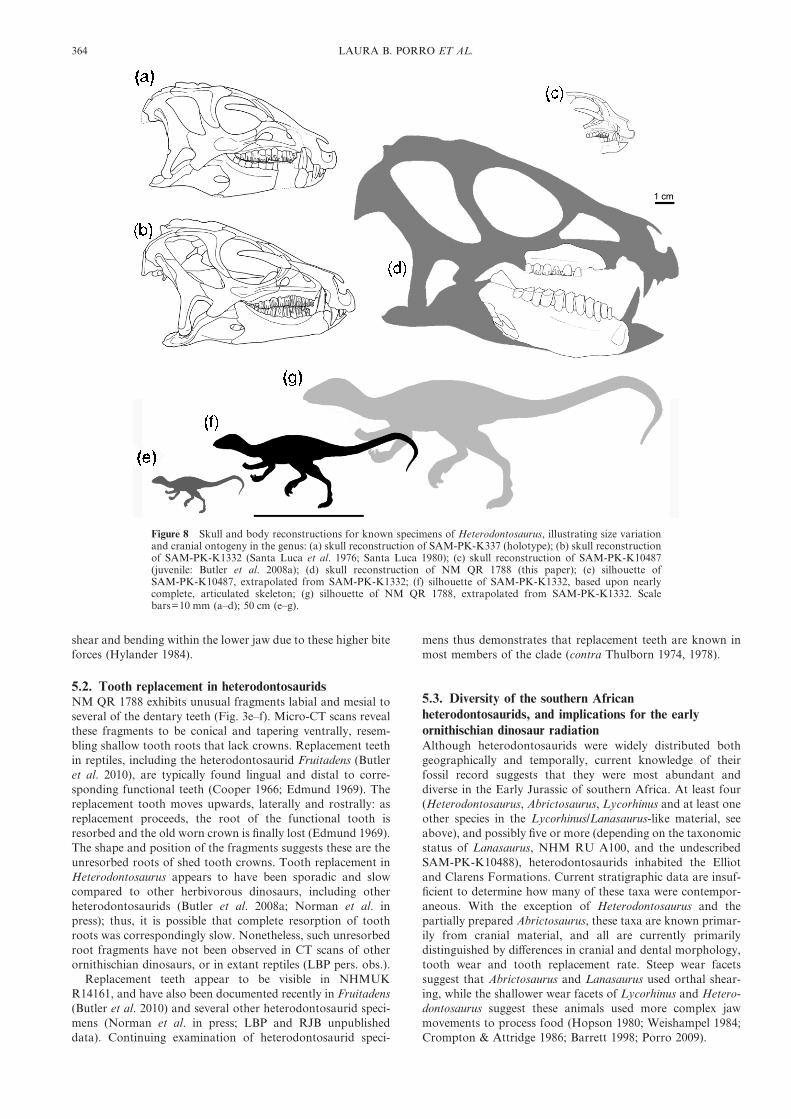
dentary ‘cheek’ tooth row of SAM-PK-K1332, the largestspecimen previously referred to Heterodontosaurus, measures32·5 mm. Based upon a basal skull length (premaxilla–quadrate) of 95 mm for SAM-PK-K1332, a basal skull lengthof 166 mm is estimated for NM QR 1788. SAM-PK-K1332 hasan approximate body length of 1 metre (Santa Luca 1980). Anisometric scaling relationship would give a total body length of1·75 metres for NM QR 1788, making this the largest hetero-dontosaurid specimen yet described. Henderson (in Butleret al. 2010: electronic supplementary material) estimated abody mass of 2·59 kg for Heterodontosaurus tucki, based upona body length of 1·12 metres, while Seebacher (2001) estimateda mass of 1·8 kg based upon a body length of 1 metre. Scalingthese estimates to a body length of 1·75 metres suggests a bodymass for NM QR 1788 of just under 10 kg, four to five timesheavier than previously known specimens of Heterodontosau-rus tucki. Furthermore, NM QR 1788 is over three timeslonger than the smallest known individual of Heterodontosau-rus tucki (SAM-PK-K10487; Butler et al. 2008a; Fig. 8).Extensive skeletal fusion (Santa Luca 1980), including com-plete closure of all neurocentral sutures, suggests that SAM-PK-K1332 is an adult specimen (although see Irmis 2007). Themuch larger body size of NM QR 1788 might demonstrate thatsubstantial growth occurred after individuals reached maturity(and following closure of neurocentral sutures). However, itmight also reflect sexual dimorphism, temporal, geographic orintraspecific variation, or indicate that two closely relatedspecies of Heterodontosaurus of different sizes inhabited theenvironments of the upper ‘Stormberg’. At the moment, it isnot possible to distinguish between these alternatives.
SAM-PK-K1332 has a complete dentary tooth count of 11(including the caniniform) while the larger NM QR 1788possesses 12 dentary teeth. Seven dentary teeth are preserved inthe juvenile specimen SAM-PK-K10487 (Butler et al. 2008a).Addition of teeth during ontogeny is common among extantreptiles, and usually occurs by the eruption of new teeth at thedistal end of the tooth row (Cooper & Poole 1973; Kline &Cullum 1984). Although tooth count increases with age, dentalmorphology is nearly identical in all described specimens ofHeterodontosaurus tucki.
NM QR 1788 provides information on allometric changesduring growth in Heterodontosaurus tucki. The dorsoventralheight of the right dentary below crown 6 is 34% of the lengthof the tooth row in SAM-PK-K1332; by comparison, theheight of the dentary below the crown 6 is 44% of the length ofthe tooth row in NM QR 1788. Thus, the preserved mandibleof NMQR 1788 is proportionately deeper than that of SAM-PK-K1332. The coronoid eminence appears taller and morepronounced in NM QR 1788 than in either the holotype(SAM-PK-K337) or SAM-PK-K1332; unfortunately, both thecoronoid eminence and the dentary below it are incomplete inthe new specimen, making quantitative comparisons difficult.
The dentition of Heterodontosaurus indicates that it ateprimarily plants, although some degree of omnivory may haveoccurred (Barrett 1998, 2000; Butler et al. 2008a; Porro 2009);furthermore, emarginated tooth rows (implying the presence offleshy cheeks) and heavy tooth wear suggest Heterodontosaurusprocessed its food by chewing (Thulborn 1978; Hopson 1980;Crompton & Attridge 1986; Galton 1986; Barrett 1998; Porro2007, 2009). Larger individuals would be capable of generatinggreater absolute muscle force, and allometric changes in man-dibular morphology, such as increased relative height of thecoronoid eminence, may have further increased mechanicaladvantage and bite force in older and larger animals. Thegreater relative depth of the mandible in larger specimens ofHeterodontosaurus may reflect the need to counter increased
NEW HETERODONTOSAURIDS FROM SOUTHERN AFRICA 363
shear and bending within the lower jaw due to these higher biteforces (Hylander 1984).
5.2. Tooth replacement in heterodontosauridsNM QR 1788 exhibits unusual fragments labial and mesial toseveral of the dentary teeth (Fig. 3e–f). Micro-CT scans revealthese fragments to be conical and tapering ventrally, resem-bling shallow tooth roots that lack crowns. Replacement teethin reptiles, including the heterodontosaurid Fruitadens (Butleret al. 2010), are typically found lingual and distal to corre-sponding functional teeth (Cooper 1966; Edmund 1969). Thereplacement tooth moves upwards, laterally and rostrally: asreplacement proceeds, the root of the functional tooth isresorbed and the old worn crown is finally lost (Edmund 1969).The shape and position of the fragments suggests these are theunresorbed roots of shed tooth crowns. Tooth replacement inHeterodontosaurus appears to have been sporadic and slowcompared to other herbivorous dinosaurs, including otherheterodontosaurids (Butler et al. 2008a; Norman et al. inpress); thus, it is possible that complete resorption of toothroots was correspondingly slow. Nonetheless, such unresorbedroot fragments have not been observed in CT scans of otherornithischian dinosaurs, or in extant reptiles (LBP pers. obs.).
Replacement teeth appear to be visible in NHMUKR14161, and have also been documented recently in Fruitadens(Butler et al. 2010) and several other heterodontosaurid speci-mens (Norman et al. in press; LBP and RJB unpublisheddata). Continuing examination of heterodontosaurid speci-
mens thus demonstrates that replacement teeth are known inmost members of the clade (contra Thulborn 1974, 1978).
5.3. Diversity of the southern Africanheterodontosaurids, and implications for the earlyornithischian dinosaur radiationAlthough heterodontosaurids were widely distributed bothgeographically and temporally, current knowledge of theirfossil record suggests that they were most abundant anddiverse in the Early Jurassic of southern Africa. At least four(Heterodontosaurus, Abrictosaurus, Lycorhinus and at least oneother species in the Lycorhinus/Lanasaurus-like material, seeabove), and possibly five or more (depending on the taxonomicstatus of Lanasaurus, NHM RU A100, and the undescribedSAM-PK-K10488), heterodontosaurids inhabited the Elliotand Clarens Formations. Current stratigraphic data are insuf-ficient to determine how many of these taxa were contempor-aneous. With the exception of Heterodontosaurus and thepartially prepared Abrictosaurus, these taxa are known primar-ily from cranial material, and all are currently primarilydistinguished by differences in cranial and dental morphology,tooth wear and tooth replacement rate. Steep wear facetssuggest that Abrictosaurus and Lanasaurus used orthal shear-ing, while the shallower wear facets of Lycorhinus and Hetero-dontosaurus suggest these animals used more complex jawmovements to process food (Hopson 1980; Weishampel 1984;Crompton & Attridge 1986; Barrett 1998; Porro 2009).
Figure 8 Skull and body reconstructions for known specimens of Heterodontosaurus, illustrating size variationand cranial ontogeny in the genus: (a) skull reconstruction of SAM-PK-K337 (holotype); (b) skull reconstructionof SAM-PK-K1332 (Santa Luca et al. 1976; Santa Luca 1980); (c) skull reconstruction of SAM-PK-K10487(juvenile: Butler et al. 2008a); (d) skull reconstruction of NM QR 1788 (this paper); (e) silhouette ofSAM-PK-K10487, extrapolated from SAM-PK-K1332; (f) silhouette of SAM-PK-K1332, based upon nearlycomplete, articulated skeleton; (g) silhouette of NM QR 1788, extrapolated from SAM-PK-K1332. Scalebars=10 mm (a–d); 50 cm (e–g).
364 LAURA B. PORRO ET AL.
In addition to heterodontosaurids, there are at least twosmall-bodied ‘fabrosaurid’ (basal genasaurian) ornithischiansthat come from the upper Elliot and Clarens Formations(Galton 1978; Butler 2005) and are known from more than 40specimens (RJB unpublished data). Knoll et al. (2010) sug-gested that the upper Elliot Formation was deposited over ashort time interval, and used this supposition as partial sup-port for their argument that only a single ‘fabrosaurid’ taxon(Lesothosaurus) is present in this unit. However, the length oftime over which the Elliot Formation was deposited is com-pletely unknown due to poor stratigraphic control, and thehigh diversity of heterodontosaurids demonstrates that there isno a priori reason to assume that the diversity of other earlysmall-bodied ornithischians was low. Compared to hetero-dontosaurids, these ‘fabrosaurids’ exhibit more gracile skullsand lightly-worn, leaf-shaped teeth that were more rapidlyreplaced, and probably utilised orthal puncture-crushing orshearing to process food (Thulborn 1971; Sereno 1991; Barrett1998; Norman et al. 2004b).
In contrast to most other Lower and Middle Jurassiclocalities, the number of small ornithischian species foundwithin the Elliot and Clarens formations is high (at leastsix, and possibly greater), suggesting a short-lived adaptiveradiation of small-bodied basal ornithischians. The variabilityin cranial and dental morphology, tooth replacement ratesand inferred jaw mechanisms may have been important inniche partitioning among the ‘Stormberg’ ornithischians; forexample, Heterodontosaurus may have fed upon tough,fibrous vegetation, while Abrictosaurus and ‘fabrosaurids’selected more nutritious, less abrasive plants or engaged morefrequently in omnivory.
There is a striking difference between the ornithischianfauna of the lower Elliot Formation (presumed to be UpperTriassic in age), which has yielded only a single specimen(Butler et al. 2007), and the relatively abundant (more than 60specimens known) and diverse (at least six species) fauna of theLower Jurassic upper Elliot and Clarens formations. Thisdifference between Late Triassic and Early Jurassic ornithis-chian faunas is also documented elsewhere globally (forexample, ornithischians are apparently absent in Late TriassicNorth America, but represented by abundant material in theLower Jurassic Kayenta Formation of Arizona; Tykoski 2005;Irmis et al. 2007) and has been taken as evidence for a globalradiation of ornithischians following the end-Triassic extinc-tion events (e.g. Butler et al. 2007). Traditionally the entire‘Stormberg’ sequence was considered Late Triassic in age, butthe upper Elliot and Clarens were re-dated as Lower Jurassicbased upon biostratigraphic arguments (see Olsen & Galton1984). The position of these units within the Lower Jurassicremains poorly constrained. Recently, Smith et al. (2009) havesuggested, on the basis of new ichnological data, that the entireupper Elliot Formation could be Late Triassic in age. Thesediffering interpretations of the position of the Triassic/Jurassicboundary impact dramatically upon our understanding of theearly ornithischian radiation; a fundamental requirement forestablishing the reality and timing of this inferred radiation isbetter constrained dating for the ‘Stormberg Group’.
6. Acknowledgements
We thank Jennifer Botha-Brink and Elize Butler (NM) for theloan of NM QR 1788 and providing locality data, Adam Yates(BPI) for alerting the authors to the existence of this specimen,and Fernando Abdala and Bernard Zipfel (BPI) for facilitat-ing the loan. For access to specimens used for comparativepurposes we additionally thank Sheena Kaal and Roger
Smith (SAM-PK), Sandra Chapman (NHMUK), BernardZipfel, Michael Raath and Adam Yates (BPI), Paul Sereno(University of Chicago), and Luis Chiappe, Paige Johnson andSam McLeod (LACM). David Norman (CAMSM) is thankedfor invaluable discussions on heterodontosaurid anatomy andpalaeobiology. Angela Milner (NHMUK) provided infor-mation on the provenance of NHMUK R14161. Thanks toPhil Crabb (NHMUK) for photography and Robert Laws(NHMUK) for line drawings used in Figures 5 and 8. Wewould like to thank two anonymous reviewers for their helpfulcomments and insights. RJB is funded by an Alexander vonHumboldt Postdoctoral Fellowship.
7. References
Attridge, J., Crompton, A. W. & Jenkins Jr, F. A. 1985. The southernAfrican Liassic prosauropod Massospondylus discovered in NorthAmerica. Journal of Vertebrate Paleontology 5, 128–32.
Baez, A. M. & Marsicano, C. A. 2001. A heterodontosaurid orni-thischian dinosaur from the Upper Triassic of Patagonia.Ameghiniana 38, 271–79.
Barrett, P. M. 1998. Herbivory in the non-avian Dinosauria.Unpublished PhD Thesis. University of Cambridge.
Barrett, P. M. 2000. Prosauropod dinosaurs and iguanas: speculationson the diets of extinct reptiles. In Sues, H.-D. (ed.) Evolution ofHerbivory in Terrestrial Vertebrates. Perspectives from the FossilRecord, 42–78. Cambridge, UK: Cambridge University Press.
Broom, R. 1911. On the dinosaurs of the Stormberg, South Africa.Annals of the South African Museum 7, 291–308.
Butler, R. J. 2005. The ‘fabrosaurid; ornithischian dinosaurs of theUpper Elliot Formation (Lower Jurassic) of South Africa andLesotho. Zoological Journal of the Linnean Society 145, 175–218.
Butler, R. J. 2010. The anatomy of the basal ornithischian dinosaurEocursor parvus from the lower Elliot Formation (Late Triassic)of South Africa. Zoological Journal of the Linnean Society 160(4),648–84.
Butler, R. J., Porro, L. B. & Heckert, A. B. 2006. A supposedheterodontosaurid tooth from the Rhaetian of Switzerland and areassessment of the European Late Triassic record of Ornithischia(Dinosauria). Neues Jahrbuch fur Geologie und Palaontologie,Monatshefte 10, 613–33.
Butler, R. J., Smith, R. M. H. & Norman, D. B. 2007. A primitiveornithischian dinosaur from the Late Triassic of South Africa,and the early evolution and diversification of Ornithischia.Proceedings of the Royal Society B 274, 2041–46.
Butler, R. J., Porro, L. B. & Norman, D. B. 2008a. A juvenile skull ofthe primitive ornithischian dinosaur Heterodontosaurus tuckifrom the ‘Stormberg’ of southern Africa. Journal of VertebratePaleontology 28, 702–11.
Butler, R. J., Upchurch, P. & Norman, D. B. 2008b. The phylogeny ofthe ornithischian dinosaurs. Journal of Systematic Palaeontology6, 1–40.
Butler, R. J., Galton, P. M., Porro, L. B., Chiappe, L. M., Henderson,D. M. & Erickson, G. M. 2010. Lower limits of ornithischiandinosaur body size inferred from a diminutive new Upper Jurassicheterodontosaurid from North America. Proceedings of the RoyalSociety B 277, 375–81.
Charig, A. J. & Crompton, A. W. 1974. The alleged synonymy ofLycorhinus and Heterodontosaurus. Annals of the South AfricanMuseum 64, 167–89.
Cooper, J. S. 1966. Tooth replacement in the slow worm (Anguisfragilis). Journal of Zoology, London 150, 235–48.
Cooper, J. S. & Poole, D. F. G. 1973. The dentition and dental tissuesof the agamid lizard, Uromastyx. Journal of Zoology, London 169,85–100.
Cooper, M. R. 1985. A revision of the ornithischian dinosaurKangnasaurus coetzeei Haughton, with a classification of theOrnithischia. Annals of the South African Museum 95, 281–317.
Crompton, A. W, & Attridge, J. 1986. Masticatory apparatus of thelarger herbivores during Late Triassic and Early Jurassic times. InPadian, K. (ed.) The Beginning of the Age of Dinosaurs, 223–36.Cambridge, UK: Cambridge University Press.
Crompton, A. W. & Charig, A. J. 1962. A new ornithischian from theUpper Triassic of South Africa. Nature 196, 1074–77.
Edmund, A. G. 1969. Dentition. In Gans, C., Bellairs, A. & Parsons,T. S. (eds) Biology of the Reptilia, Volume 1, 117–200. London,UK: Academic Press.
NEW HETERODONTOSAURIDS FROM SOUTHERN AFRICA 365

Galton, P. M. 1978. Fabrosauridae, the basal family of ornithischiandinosaurs (Reptilia: Ornithopoda). Palaontologische Zeitschrift52, 138–59.
Galton, P. M. 1986. Herbivorous adaptations of Late Triassic andEarly Jurassic dinosaurs. In Padian, K. (ed.) The Beginning of theAge of Dinosaurs, 203–21. Cambridge, UK: Cambridge UniversityPress.
Gow, C. E. 1975. A new heterodontosaurid from the Red Bedsof South Africa showing clear evidence of tooth replacement.Zoological Journal of the Linnean Society 57, 335–39.
Gow, C. E. 1990. A tooth-bearing maxilla referable to Lycorhinusangustidens Haughton, 1924 (Dinosauria, Ornithischia). Annals ofthe South African Museum 99, 367–80.
Haughton, S. H. 1924. The fauna and stratigraphy of the StormbergSeries. Annals of the South African Museum 12, 323–497.
Hopson, J. A. 1975. On the generic separation of the ornithischiandinosaurs Lycorhinus and Heterodontosaurus from the StormbergSeries (Upper Triassic of South Africa). South African Journal ofScience 71, 302–05.
Hopson, J. A. 1980. Tooth function and replacement in earlyMesozoic ornithischian dinosaurs: implications for aestivation.Lethaia 13, 93–105.
Hylander, W. L. 1984. Stress and strain in the mandibular symphysisof primates: a test of competing hypotheses. American Journal ofPhysical Anthropology 64, 1–46.
Irmis, R. B. 2007. Axial skeleton ontogeny in the Parasuchia (Archo-sauria: Pseudosuchia) and its implications for ontogenetic deter-mination in archosaurs. Journal of Vertebrate Paleontology 27,350–61.
Irmis, R. B., Parker, W. G., Nesbitt, S. J. & Liu, J. 2007. Earlyornithischian dinosaurs: the Triassic record. Historical Biology 19,3–22.
Kitching, J. W. & Raath, M. A. 1984. Fossils from the Elliot andClarens formations (Karoo sequence) of the northeastern Cape,Orange Free State and Lesotho, and a suggested biozonationbased on tetrapods. Palaeontologia Africana 25, 111–25.
Kline, L. W. & Cullum, D. 1984. A long-term study of the toothreplacement phenomenon in the young green iguana, Iguanaiguana. Journal of Herpetology 18, 176–85.
Knoll, F., Padian, K. & de Ricqle#s, A. 2010. Ontogenetic change andadult body size of the early ornithischian dinosaur Lesothosaurusdiagnosticus: implications for basal ornithischian taxonomy.Gondwana Research 17, 171–79.
Kuhn, O. 1966. Die Reptilien, System und Stammesgeschichte.Krailling bei Munchen: Verlag Oeben.
Norman, D. B. & Barrett, P. M. 2002. Ornithischian dinosaurs fromthe Lower Cretaceous (Berriasian) of England. In Milner, A. R.& Batten, D. J. (eds) Life and environments in Purbeck times.Special Papers in Palaeontology 68, 161–89. London: ThePalaeontological Association. 268 pp.
Norman, D. B., Sues, H.-D., Witmer, L. M. & Coria, R. A. 2004a.Basal Ornithopoda. In Weishampel, D. B., Dodson, P. &Osmolska, H. (eds) The Dinosauria (2nd edn), 393–412. Berkeley:University of California Press.
Norman, D. B., Witmer, L. M. & Weishampel, D. B. 2004b. BasalOrnithischia. In Weishampel, D. B., Dodson, P. & Osmolska, H.(eds) The Dinosauria (2nd edn), 325–34. Berkeley: University ofCalifornia Press.
Norman, D. B., Crompton, A. W., Butler, R. J., Porro, L. B. &Charig, A. J. in press. The Lower Jurassic ornithischian dinosaurHeterodontosaurus tucki Crompton and Charig, 1962: cranialanatomy, functional morphology, taxonomy and relationships.Zoological Journal of the Linnean Society.
Olsen, P. E. & Galton, P. M. 1984. A review of the reptile andamphibian assemblages from the Stormberg of southern Africa,with special emphasis on the footprints and the age of theStormberg. Palaeontologia Africana 25, 87–110.
Owen, R. 1842. Report on British fossil reptiles, part II. Report for theBritish Association for the Advancement of Science, Plymouth1841, 60–294.
Parker, W. G., Irmis, R. B., Nesbitt, S. J., Martz, J. W. & Browne, L.S. 2005. The Late Triassic pseudosuchian Revueltosaurus callend-
eri and its implications for the diversity of early ornithischiandinosaurs. Proceedings of the Royal Society B 272, 963–69.
Pol, D., Rauhut, O. W. M. & Becerra, M. 2011. A Middle Jurassicheterodontosaurid dinosaur from Patagonia and the evolution ofheterodontosaurids. Naturwissenschaften. DOI: 10.1007/500114-011-0780-5.
Porro, L. B. 2007. Feeding and jaw mechanism in Heterodonto-saurus tucki using finite element analysis. Journal of VertebratePaleontology 27 (3-suppl.), 131A.
Porro, L. B. 2009. Cranial biomechanics in the early dinosaur Hetero-dontosaurus. Unpublished PhD Thesis. University of Cambridge.
Santa Luca, A. P. 1980. The postcranial skeleton of Heterodontosaurustucki (Reptilia, Ornithischia) from the Stormberg of South Africa.Annals of the South African Museum 79, 159–211.
Santa Luca, A. P., Crompton, A. W. & Charig, A. J. 1976. A completeskeleton of the late Triassic ornithischian Heterodontosaurustucki. Nature 264, 324–28.
Seebacher, F. 2001. A new method to calculate allometric length-massrelationships of dinosaurs. Journal of Vertebrate Paleontology 21,51–60.
Seeley, H. G. 1887. On the classification of the fossil animals com-monly named Dinosauria. Proceedings of the Royal Society 43,165–71.
Sereno, P. C. 1991. Lesothosaurus, ‘fabrosaurids’, and the earlyevolution of Ornithischia. Journal of Vertebrate Paleontology 11,168–97.
Sereno, P. C. 1997. The origin and evolution of dinosaurs. AnnualReview of Earth and Planetary Sciences 25, 435–89.
Sereno, P. C. 1999. The evolution of dinosaurs. Science 284, 2137–47.Sereno, P. C., Zhao, X., Brown, L. & Tan, L. 2007. New psittaco-
saurid highlights skull enlargement in horned dinosaurs. ActaPalaeontologica Polonica 52, 275–84.
Smith, R. M. H., Marsicano, C. A. & Wilson, J. A. 2009. Sedi-mentology and paleoecology of a diverse Early Jurassic tetrapodtracksite in Lesotho, southern Africa. Palaios 24, 672–84.
Thulborn, R. A. 1970. The systematic position of the Triassic ornithis-chian dinosaur Lycorhinus angustidens. Zoological Journal of theLinnean Society 49, 235–45.
Thulborn, R. A. 1971. Tooth wear and jaw action in the Triassicornithischian dinosaur Fabrosaurus. Journal of Zoology 164,165–79.
Thulborn, R. A. 1974. A new heterodontosaurid dinosaur (Reptilia:Ornithischia) from the Upper Triassic Red Beds of Lesotho.Zoological Journal of the Linnean Society 55, 151–75.
Thulborn, R. A. 1978. Aestivation among ornithopod dinosaurs of theAfrican Triassic. Lethaia 11, 185–98.
Tykoski, R. S., 2005. Vertebrate paleontology in the Arizona Jurassic.In McCord, R. D. (ed.) Vertebrate Paleontology of Arizona. MesaSouthwest Museum Bulletin 11, 72–93. Mesa, Arizona: MesaSouthwest Museum.
Weishampel. D. B. 1984. Evolution of jaw mechanisms in ornithopoddinosaurs. Advances in Anatomy, Embryology and Cell Biology 87,1–110.
Weishampel, D. B. & Witmer, L. M. 1990. Heterodontosauridae. InWeishampel, D. B., Dodson, P. & Osmolska, H. (eds) TheDinosauria, 486–97. Berkeley: University of California Press.
Weishampel, D. B., Dodson, P. & Osmolska, H. (eds) 2004. TheDinosauria (2nd edn). Berkeley: University of California Press,861 pp.
Witmer, L. M. 2009. Dinosaurs: fuzzy origins for feathers. Nature 458,293–95.
Xu, X., Forster, C. A., Clark, J. M. & Mo, J. 2006. A basal ceratopsianwith transitional features from the Late Jurassic of northwesternChina. Proceedings of the Royal Society B 273, 2135–40.
Zheng, X.-T., You, H.-L., Xu, X. & Dong, X.-M. 2009. An EarlyCretaceous heterodontosaurid dinosaur with integumentarystructures. Nature 458, 333–36.
Zhou, C.-F., Gao, K.-Q., Fox, R. C. & Chen, S.-H. 2006. A newspecies of Psittacosaurus (Dinosauria: Ceratopsia) form the EarlyCretaceous Yixian Formation, Liaoning, China. Palaeoworld 15,100–14.
MS received 30 December 2009. Accepted for publication 7 October 2010.
366 LAURA B. PORRO ET AL.