
Neutron-irradiation-induced near-infrared emission in a-Al2O3
6
March 21, 2014 16:43 Philosophical Magazine Letters R1-tPHL-Rahman Philosophical Magazine Letters Vol. 00, No. 00, March 2014, 1–6 Neutron-irradiation-induced near-infrared emission in α-Al 2 O 3 Abu Zayed Mohammad Saliqur Rahman a∗ , Xingzhong Cao a , Long Wei a ,Baoyi Wang a , Haiming Ji b , Tao Yang b , Qiu Xu c and Kozo Atobe d a Key Laboratory of Nuclear Analytical Techniques, Institute of High Energy Physics, Chinese Academy of Sciences, 19B Yuquanlu Shijingshan District Beijing 100049, China, b Key Laboratory of Semiconductor Materials Science, Institute of Semiconductors, Chinese Academy of Sciences, Beijing 100083, China, c Reactor Research Institute, Kyoto University, 2, Asashiro-Nishi, Kumatori-cho, Sennan-gun, Osaka 590-0494, Japan, d Nuclear Safety Technology Center, 9-15, 1-chome, Utsubohonmachi, Nishi Ku, Osaka 550-0004, Japan (Received 21 May 2013; accepted 20 December 2013) Near-infrared spectroscopic study of neutron-irradiated α-Al 2 O 3 was conducted. Scanning- electron and optical microscopic measurements were also done. Sample irradiated to the flu- ence of 2.8 x 10 18 n/cm 2 shows strong emission band at 1180, 1700, 1945 and 2225 nm. Pho- toluminescence (PL) intensity of these bands reduced greatly with higher neutron fluences. From reduction of PL intensity, it can be reasonably assumed that nonradiative recombination centers such as extended defects or dislocations may be formed in α-Al 2 O 3 after exposing to very high neutron fluences. Keywords: near-infrared spectroscopy; neutron irradiation; lattice defects; dislocations; alumina 1. Introduction Alumina (α-Al 2 O 3 ) commonly known as corundum for crystal structure has wide variety of technological applications such as high-temperature structural materi- als, dielectric insulators, substrate for microelectronics and many more. This is also a good candidate material for high-strength window and blanket in fusion reactor, inert nuclear fuel host [1] due to resistance against radiation. Neutron- irradiation can induce defects[2] which modify the structural and optical properties of α-Al 2 O 3 . Most of these defects are associated with electron trapping oxygen va- cancies. Gamma-ray irradiation can not induce these kind of atomic displacement defects[3, 4]. Electron-irradiation can cause phase transformation in alumina[5, 6]. Many studies were done on the optical properties of neutron-irradiation-induced defects in α-Al 2 O 3 [7–11]. At low temperatures some of these optical bands be- come vibronic and show temperature dependence[12–15]. Although many stud- ies on the optical properties were done, still there are many unresolved research problems such as irradiation-induced formation of defect clusters or dislocations. Near-infrared (NIR) spectroscopy is nondestructive and very useful tool for vari- ous physical and chemical analysis which can be used to detect defects in α-Al 2 O 3 . The study of NIR spectroscopy for investigating irradiation-induced defects in α- Al 2 O 3 is very few to the best of our knowledge [16]. In this study, we used NIR, * Corresponding author. Email: [email protected], [email protected] ISSN: 0950-0839 print/ISSN 1362-3036 online c ⃝ 2014 Taylor & Francis DOI: 10.1080/09500839.2014.885180 http://www.informaworld.com
Transcript of Neutron-irradiation-induced near-infrared emission in a-Al2O3

March 21, 2014 16:43 Philosophical Magazine Letters R1-tPHL-Rahman
Philosophical Magazine LettersVol. 00, No. 00, March 2014, 1–6
Neutron-irradiation-induced near-infrared emission in α-Al2O3
Abu Zayed Mohammad Saliqur Rahmana∗, Xingzhong Caoa, Long Weia,Baoyi Wanga,
Haiming Jib, Tao Yangb, Qiu Xuc and Kozo Atobed
aKey Laboratory of Nuclear Analytical Techniques, Institute of High Energy Physics,
Chinese Academy of Sciences, 19B Yuquanlu Shijingshan District Beijing 100049, China,bKey Laboratory of Semiconductor Materials Science, Institute of Semiconductors,
Chinese Academy of Sciences, Beijing 100083, China, cReactor Research Institute, Kyoto
University, 2, Asashiro-Nishi, Kumatori-cho, Sennan-gun, Osaka 590-0494, Japan,dNuclear Safety Technology Center, 9-15, 1-chome, Utsubohonmachi, Nishi Ku, Osaka
550-0004, Japan
(Received 21 May 2013; accepted 20 December 2013)
Near-infrared spectroscopic study of neutron-irradiated α-Al2O3 was conducted. Scanning-electron and optical microscopic measurements were also done. Sample irradiated to the flu-ence of 2.8 x 10¹⁸n/cm2 shows strong emission band at 1180, 1700, 1945 and 2225 nm. Pho-toluminescence (PL) intensity of these bands reduced greatly with higher neutron fluences.From reduction of PL intensity, it can be reasonably assumed that nonradiative recombinationcenters such as extended defects or dislocations may be formed in α-Al2O3 after exposing tovery high neutron fluences.
Keywords: near-infrared spectroscopy; neutron irradiation; lattice defects; dislocations;alumina
1. Introduction
Alumina (α-Al2O3) commonly known as corundum for crystal structure has widevariety of technological applications such as high-temperature structural materi-als, dielectric insulators, substrate for microelectronics and many more. This isalso a good candidate material for high-strength window and blanket in fusionreactor, inert nuclear fuel host [1] due to resistance against radiation. Neutron-irradiation can induce defects[2] which modify the structural and optical propertiesof α-Al2O3. Most of these defects are associated with electron trapping oxygen va-cancies. Gamma-ray irradiation can not induce these kind of atomic displacementdefects[3, 4]. Electron-irradiation can cause phase transformation in alumina[5, 6].Many studies were done on the optical properties of neutron-irradiation-induceddefects in α-Al2O3[7–11]. At low temperatures some of these optical bands be-come vibronic and show temperature dependence[12–15]. Although many stud-ies on the optical properties were done, still there are many unresolved researchproblems such as irradiation-induced formation of defect clusters or dislocations.Near-infrared (NIR) spectroscopy is nondestructive and very useful tool for vari-ous physical and chemical analysis which can be used to detect defects in α-Al2O3.The study of NIR spectroscopy for investigating irradiation-induced defects in α-Al2O3 is very few to the best of our knowledge [16]. In this study, we used NIR,
∗Corresponding author. Email: [email protected], [email protected]
ISSN: 0950-0839 print/ISSN 1362-3036 onlinec⃝ 2014 Taylor & FrancisDOI: 10.1080/09500839.2014.885180http://www.informaworld.com
March 21, 2014 16:43 Philosophical Magazine Letters R1-tPHL-Rahman
2 A. Z. M. S. Rahman et. al.
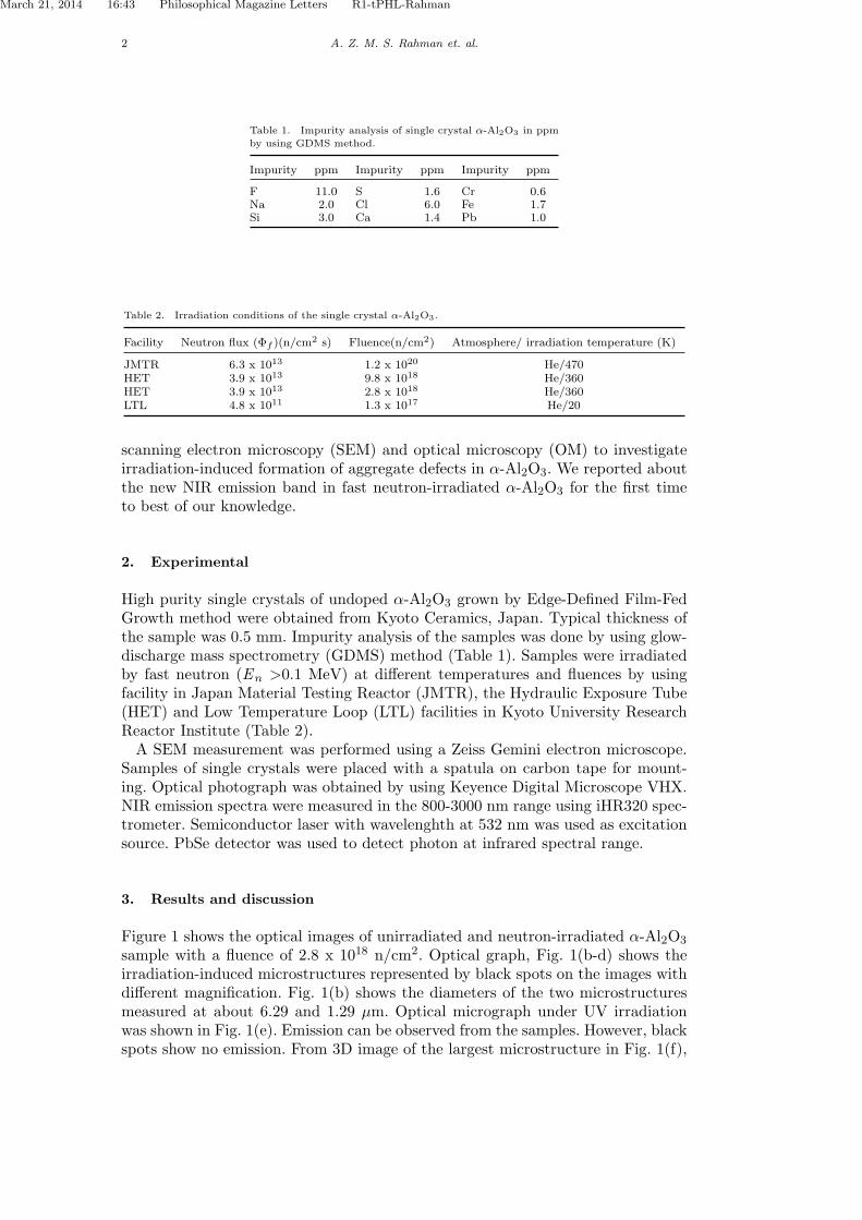
Table 1. Impurity analysis of single crystal α-Al2O3 in ppm
by using GDMS method.
Impurity ppm Impurity ppm Impurity ppm
F 11.0 S 1.6 Cr 0.6Na 2.0 Cl 6.0 Fe 1.7Si 3.0 Ca 1.4 Pb 1.0
Table 2. Irradiation conditions of the single crystal α-Al2O3.
Facility Neutron flux (Φf )(n/cm2 s) Fluence(n/cm2) Atmosphere/ irradiation temperature (K)
JMTR 6.3 x 10¹³ 1.2 x 10²⁰ He/470HET 3.9 x 10¹³ 9.8 x 10¹⁸ He/360HET 3.9 x 10¹³ 2.8 x 10¹⁸ He/360LTL 4.8 x 10¹¹ 1.3 x 10¹⁷ He/20
scanning electron microscopy (SEM) and optical microscopy (OM) to investigateirradiation-induced formation of aggregate defects in α-Al2O3. We reported aboutthe new NIR emission band in fast neutron-irradiated α-Al2O3 for the first timeto best of our knowledge.
2. Experimental
High purity single crystals of undoped α-Al2O3 grown by Edge-Defined Film-FedGrowth method were obtained from Kyoto Ceramics, Japan. Typical thickness ofthe sample was 0.5 mm. Impurity analysis of the samples was done by using glow-discharge mass spectrometry (GDMS) method (Table 1). Samples were irradiatedby fast neutron (En >0.1 MeV) at different temperatures and fluences by usingfacility in Japan Material Testing Reactor (JMTR), the Hydraulic Exposure Tube(HET) and Low Temperature Loop (LTL) facilities in Kyoto University ResearchReactor Institute (Table 2).A SEM measurement was performed using a Zeiss Gemini electron microscope.
Samples of single crystals were placed with a spatula on carbon tape for mount-ing. Optical photograph was obtained by using Keyence Digital Microscope VHX.NIR emission spectra were measured in the 800-3000 nm range using iHR320 spec-trometer. Semiconductor laser with wavelenghth at 532 nm was used as excitationsource. PbSe detector was used to detect photon at infrared spectral range.
3. Results and discussion
Figure 1 shows the optical images of unirradiated and neutron-irradiated α-Al2O3
sample with a fluence of 2.8 x 10¹⁸ n/cm2. Optical graph, Fig. 1(b-d) shows theirradiation-induced microstructures represented by black spots on the images withdifferent magnification. Fig. 1(b) shows the diameters of the two microstructuresmeasured at about 6.29 and 1.29 µm. Optical micrograph under UV irradiationwas shown in Fig. 1(e). Emission can be observed from the samples. However, blackspots show no emission. From 3D image of the largest microstructure in Fig. 1(f),

March 21, 2014 16:43 Philosophical Magazine Letters R1-tPHL-Rahman
Philosophical Magazine Letters 3
Figure 1. Optical micrographs of unirradiated (a) and fast neutron-irradiated (b-f) α-Al2O3 samples. FastNeutron fluence was 2.8 x 10¹⁸ n/cm2.
height and width were measured at about 41.8 and 63.4 µm, respectively. Fig. 1 (a)represents the image of the unirradiated sample for comparison with no observationof black spot on it. However, more investigation is needed to understand thesemicrostructures as size is very large compare to the voids reported elsewhere[17, 18].Figure 2 shows the NIR PL spectra of the unirradiated, neutron-irradiated and
annealed samples under 532 nm laser excitation. Unirradiated sample shows noobvious emission in the range of 800-3000 nm. Sample irradiated at 20 K showsonly a very negligible emission near at 1800 nm. However, after irradiating at thefluence of 2.8 x 10¹⁸ n/cm2, the α-Al2O3 sample shows very strong and broademission band at 1180 nm with FWHM of 234 nm. It also shows three overlappingband at 1700, 1945 and 2225 nm. These bands were separated by using Gaussianfit (Fig. 3). At a fluence of 9.8 x 10¹⁸, the intensity of emission band at 1180 nm

March 21, 2014 16:43 Philosophical Magazine Letters R1-tPHL-Rahman
4 A. Z. M. S. Rahman et. al.
1000 1500 2000 2500 3000
0.00
0.05
0.10
0.15
0.20
0.25
0.30
0.35
0.40
PL in
tens
ity (a
. u.)
wavelength (nm)
RT
(a)(b)
(c)
(d)(e) (f)
Figure 2. NIR PL spectra of (a) unirradiated, fast neutron-irradiated with fluence of (b) 1.3 x 10¹⁷ (c)2.8 x 10¹⁸ (d) 9.8 x 10¹⁸ (e) 1.2 x 10²⁰ and (f) annealed α-Al2O3 samples. Excitation source was fixed at532 nm.
1000 1500 2000 2500 3000
0.00
0.05
0.10
0.15
0.20
0.25
0.30
0.35
0.40
PL in
tens
ity (a
. u.)
Wavelength (nm)
(a) Experimental(b) Total Fit(c) Peak separation
(a)
(b)
(c)
Figure 3. NIR PL spectra of fast neutron-irradiated α-Al2O3 samples with Gaussian curve fitting. FastNeutron fluence was 2.8 x 10¹⁸ n/cm2.
with other three overlapping bands reduced greatly. Three emission bands at 1700,1945 and 2225 nm are seemed to be centered approximately at 1800 nm.Fig. 4 shows the SEM images of unirradiated and fast neutron-irradiated α-
Al2O3 to a fluence of 2.8 x 10¹⁸ n/cm2 after annealing at 1100◦C . Image of theunirradiated sample was shown for comparison in Fig. 4(a). Images of the annealedsample with different magnification were shown in Fig. 4(b-c). Although, there is adistinct difference between unirradiated and fast neutron-irradiated samples after

March 21, 2014 16:43 Philosophical Magazine Letters R1-tPHL-Rahman
Philosophical Magazine Letters 5
Figure 4. SEM images of unirradiated (a) and fast neutron-irradiated (b-c) α-Al2O3 samples to fluenceof 2.8 x 10¹⁸ n/cm2 after annealing at 1100◦C.
annealing, more investigation is needed to understand the SEM result.To understand the origin of NIR emission bands, we compare it with PL spectra
of unirradiated and fast neutron-irradiated sample at low temperature (LT). As noobvious peak was observed in unirradiated sample, it is clearly from irradiation-induced defects. LT irradiation was used to produce simple type defects such as For F+ centers[19]. The concentration of F aggregate centers are usually very low byLT irradiation. Therefore, the origin of these 1180, 1700, 1945 and 2225 nm may beassigned to F aggregate centers or clusters of F type centers. Excitation wavelength532 nm falls between the 450 nm and 570 nm absorption band which were reportedas F2
+ center[20]. So the NIR emission spectra may be tentatively assigned to F2+
center. However, further study is necessary to assign these bands appropriately.After annealing at 1100◦C all these bands are completely disappeared. On theother hand, the intensity of these bands decreased greatly at a fluence of 9.8 x10¹⁸ n/cm2 and almost disappeared at a fluence of 1.2 x 10²⁰ n/cm2. PL intensitydecreases may be attributed to the extended defects induced at higher fluence asin ref [21, 22]. Probably extended defect and dislocation loops affect the radiativerecombination. From disappearance of NIR PL emission spectra after annealing at1100◦C, it can be said that samples may be recovered from point defects. However,the extended type defects such as dislocation, microstructure and void may bestill present in the samples. Annealing at more higher temperature is necessary toeradicate these types of defects.
4. Conclusions
We studied neutron-irradiation-induced formation of lattice defects in α-Al2O3 byusing NIR, SEM and OM. Sample irradiated at a fluence of 2.8 x 10¹⁸ n/cm2
shows strong emission band at NIR region. Intense Emission band at 1180 nm

March 21, 2014 16:43 Philosophical Magazine Letters R1-tPHL-Rahman
6 REFERENCES
is expected to have good promise for application as color center laser. Extendeddefects or dislocations may be formed after irradiating the α-Al2O3 single crystalswith very high fluence of fast neutron.
Acknowledgments
We acknowledge Mr. Zhaojing Wen of Beijing Chemical University and Keyencestaff for their help with the SEM and OM experiments, respectively.This workwas partially supported by the NSFC (Grant No.91026006) and Chinese Academyof Sciences Fellowships for Young International Scientists under Grant No.2012Y1JB0007.
References
[1] M. Akiyoshi, J. Nucl. Mater. 386-388 (2009) p. 303.[2] S. J. Zinkle and G.S. Was, Acta Mater. 61 (2013) p.735.[3] A. K. Islamov, E. M. Ibragimova and I. Nuritdinov, J. Nucl. Mater. 362 (2007) p.222.[4] A. Z. M. S. Rahman , T. Kobayashi, T. Awata and K. Atobe, Radiat. Eff. Def. Solids 165 ( 2010)
p.290.[5] C. L. Chen, K. Arakawa, J. G. Lee and H. Mori, Scr. Mater. 63 (2010) p.1013.[6] C. L. Chen, K. Arakawa and H. Mori, Scr. Mater. 63 (2010) p.355.[7] M. Izerrouken and T. Benyahia, Nucl. Inst. Methods B 268 (2010) p.2987.[8] I. K. Abdukadyrova, Inorganic Mater. 41 (2005) p.1080.[9] B. D. Evans, G. J. Pogatshnik and Y. Chen, Nucl. Inst. Methods B 91 (1994) p.258.
[10] M. S.Kulkarni, N. S. Rawat, S. V. Thakare, K. C. Jagadeesan, D. R. Mishra, K. P. Muthe, B. C.Bhatt, S. K. Gupta and D. N. Sharma, Radiat. Meas. 46 (2011) p.1704.
[11] S. V. Solovev, I. I. Milman and A. I. Syurdo. Physics of the Solid State 54 (2012) p.726.[12] B. D. Evans and M. Stapelbroek, Solid State Commun 33 (1980) p.765.[13] L. S. Welch, A. E. Hughes and G. P. Pells, J Phys C: Solid St. Phys. 13 (1980) p.1805.[14] A. Z. M. S. Rahman, T. Awata, N. Yamashita, Y. Inada, K. Oshima, Q. Xu and K. Atobe, Physics
Procedia 2 ( 2009) p.551.[15] A. Z. M. S. Rahman , T. Awata, N. Yamashita, Q. Xu and K. Atobe, Radiat. Eff. Def. Solids 164
(2009) p.692.[16] R. Bauer, U. Bogner and M. Maier, Appl. Phys. B 60 (1995) p. 507.[17] F. W. Clinard, Jr. and G. F. Hurley, J. Nucl. Mater. 108-109 (1982) p. 655.[18] S. J. Zinkle and G. P. Pells, J. Nucl. Mat. 253 (1998) p. 120.[19] M. Okada, S. Kanazawa, T. Nozaki, M. Nakagawa, K. Atobe, E. Kuramoto, K. Matsumura and T.
Sano, Nucl. Inst. Methods A 463 (2001) p.213.[20] K. Atobe, N. Nishimoto and M. Nakagawa, Phys. Stat. Sol. A 89 (1985) 155.[21] A. Y. Polyakov, N. B. Smirnov, A. V. Govorkov, H. Amano, S. J. Pearton, I. H. Lee, Q. Sun, J. Han
and S. Y. Karpov, Appl. Phys. Lett. 98 (2011) p.072104.[22] I Burud, A. S. Flo and E. Olsen, AIP Advances 2 (2012) p.042135.