Neuropeptide immunoreactivity and co-existence in cardiovascular nerves and autonomic ganglia of the...
15
ELSEVIER Regulatory Peptides 58 (1995) 25-39 Neuropeptide immunoreactivity and co-existence in cardiovascular nerves and autonomic ganglia of the estuarine crocodile, Crocodylus porosus, and cardiovascular effects of neuropeptides Paul Karila a,*, Michael Axelsson a Craig E. Franklin c, Regina Fritsche a Ian L. Gibbins b, Gordon C. Grigg c, Stefan Nilsson a, Susanne Holmgren a a DeparJ.mentof Zoophysiology, GOteborg University, Medicinaregatan 18, S-413 90 Gfteborg, Sweden b Department of Anatomy and Histology, School of Medicine, Flinders University of South Australia, Bedford Park, SA 5042, Australia c Department of Zoology, University of Queensland, Brisbane, QLD 4072, Australia Received 26 September 1994; revised 20 January 1995; accepted 31 March 1995 Abstract The two aortas of tile crocodile are in open connection at two sites, the foramen of Panizzae immediately outside the ventricles, and the arterial anastomosis at the level of the gut. The present study was performed to elucidate the innervation of the cardiovascular structures of the crocodile, in part to provide a further basis for the assumption that .the apertures of the foramen and the anastomosis may be altered, possibly leading to changes in the flow profiles of the central vessels. The presence of smooth muscle arranged at the circumference of the foramen and in the walls of the anastomosis was demonstrated. The cardiovascular structures were innervated by nerves containing co-existing tyrosine hydroxylase, NPY and somatostatin immunoreactivities, which also occurred in neurons of the sympathetic ganglia. CGRP and substance P immunoreactive material co-existed in cardiovascular nerves, and in the nodose ganglion. In addition, bombesin, VIP and galanin immunoreactive, nerves were found. Effects of neuropeptides oh blood flows and blood pressures were studied in vivo. Substance P increased all blood flows measured, NPY increased the flow through the arterial anastomosis while neurotensin caused an initial decrease in the flow through the arterial anastomosis. In conclusion, there is a rich innervation of the heart and major vessels of the estuarine crocodile, including the foramen of Panizza and the arterial anastomosis. These nerves possibly :regulate the distribution of blood in the cardiovascular system, which is further suggested by the results of the injection of neuropeptides. Keywords: CGRP; SubstanceP; NeuropeptideY; Somatostatin;Tyrosine hydroxylase;Galanin;VIP; Bombesin;Neurotensin 1. Introduction * Correspondingauthor. E-mail:[email protected]: + 46 31 7733807. The reptilian circulation, and in particular the crocodilian circulation, displays several interesting 0167-0115/95/$09.50 © 1995 Elsevier Science B.V. All rights reserved SSDI 0167-0115(95)01)055-0
Transcript of Neuropeptide immunoreactivity and co-existence in cardiovascular nerves and autonomic ganglia of the...

ELSEVIER Regulatory Peptides 58 (1995) 25-39
Neuropeptide immunoreactivity and co-existence in cardiovascular nerves and autonomic ganglia of the estuarine crocodile, Crocodylus porosus, and cardiovascular effects of
neuropeptides
Paul Karila a,*, Michael Axelsson a Craig E. Franklin c, Regina Fritsche a Ian L. Gibbins b, Gordon C. Grigg c, Stefan Nilsson a, Susanne Holmgren a
a DeparJ.ment of Zoophysiology, GOteborg University, Medicinaregatan 18, S-413 90 Gfteborg, Sweden b Department of Anatomy and Histology, School of Medicine, Flinders University of South Australia, Bedford Park, SA 5042, Australia
c Department of Zoology, University of Queensland, Brisbane, QLD 4072, Australia
Received 26 September 1994; revised 20 January 1995; accepted 31 March 1995
Abstract
The two aortas of tile crocodile are in open connection at two sites, the foramen of Panizzae immediately outside the ventricles, and the arterial anastomosis at the level of the gut. The present study was performed to elucidate the innervation of the cardiovascular structures of the crocodile, in part to provide a further basis for the assumption that .the apertures of the foramen and the anastomosis may be altered, possibly leading to changes in the flow profiles of the central vessels. The presence of smooth muscle arranged at the circumference of the foramen and in the walls of the anastomosis was demonstrated. The cardiovascular structures were innervated by nerves containing co-existing tyrosine hydroxylase, NPY and somatostatin immunoreactivities, which also occurred in neurons of the sympathetic ganglia. CGRP and substance P immunoreactive material co-existed in cardiovascular nerves, and in the nodose ganglion. In addition, bombesin, VIP and galanin immunoreactive, nerves were found. Effects of neuropeptides oh blood flows and blood pressures were studied in vivo. Substance P increased all blood flows measured, NPY increased the flow through the arterial anastomosis while neurotensin caused an initial decrease in the flow through the arterial anastomosis. In conclusion, there is a rich innervation of the heart and major vessels of the estuarine crocodile, including the foramen of Panizza and the arterial anastomosis. These nerves possibly :regulate the distribution of blood in the cardiovascular system, which is further suggested by the results of the injection of neuropeptides.
Keywords: CGRP; Substance P; Neuropeptide Y; Somatostatin; Tyrosine hydroxylase; Galanin; VIP; Bombesin; Neurotensin
1. Introduct ion
* Corresponding author. E-mail: [email protected]. Fax: + 46 31 7733807.
The reptilian circulation, and in particular the crocodilian circulation, displays several interesting
0167-0115/95/$09.50 © 1995 Elsevier Science B.V. All rights reserved SSDI 0167-0115(95)01)055-0
26 P. Karila et al. / Regulatory Peptides 58 (1995) 25-39
features in the organization of the heart and major vessels. Crocodilians have a heart with a complete separation of the left and right ventricles and hence a complete separation between the lung circulation and the systemic circulation. The crocodilian heart is unique in that its left aortic arch, the left aorta, originates together with the pulmonary artery from the right ventricle. A right aorta runs from the left ventricle and communicates with the left aorta at two places; through the foramen of Panizza (hence called the foramen) immediately outside the aortic valves in the outflow tract [1,2] and through an arterial anasto- mosis in the abdomen (see, e.g., [3-5]). Posterior to the arterial anastomosis the right and left aortas continue as the dorsal aorta and coeliac artery, re- spectively.
The left ventricle of the heart normally generates a higher pressure than the right ventricle. This leads to a high pressure in the right aorta and, since there are open connections between the right and left aorta through the foramen and the arterial anastomosis, the pressure in the left aorta actually equals that in the right aorta. Therefore, under normal conditions, the left aortic blood pressure exceeds that in the right ventricle, and the left aortic valves (between the right ventricle and left aorta) remain closed. It is only under circumstances that lead to an elevated pul- monary arterial resistance, a decreased systemic re- sistance or an increased venous return to the right ventricle, that the pressure of the right ventricle exceeds that in the left aorta and the left aortic valves open [3-8]. It has been concluded that the flow in the left aorta is normally obtained only via the foramen. The flow profile is complex with both anterograde and retrograde phases [3,9] derived at least partly from a closure of the foramen by the cusp of the right aortic valve during part of the systole [3,5,10]. The low net flow measured in the left aorta is thought to be insufficient as a supply of oxygenated blood to the coeliac artery and the greater part of the blood is believed to reach the gut via the arterial anastomosis [11]. The oxygenated blood to the gut in the crocodile can thus enter the coeliac artery via two pathways: (1) From the right aorta through the foramen and via the left aorta or (2) From the right aorta via the arterial anastomosis to the coeliac artery.
To explain their observations of blood pressure
profiles in the heart and major arteries of Crocodylus porosus under different circumstances, Grigg and Johansen [5] suggested that the diameter of the fora- men is variable. However, no experiments designed to uncover possible control mechanisms have been made. There is also another possible central site of vascular control, the arterial anastomosis. On the basis of preliminary studies of flow patterns in the coeliac artery, Axelsson et al. [11] suggested that a portion of the blood flowing to the gut is derived from the right aorta via this anastomosis. The aim of this study was therefore to elucidate, by histological and immunohistochemical techniques, the possibility of an autonomic nervous control of the foramen and anastomosis, and to investigate the hemodynamic changes caused by some neuropeptides.
Parts of the results have been published elsewhere [121.
2. Materials and methods
Crocodylus porosus (body mass 0.09-6.6 kg) were obtained from the Edward River Crocodile Farm, Cape York, Australia and flown to Brisbane. They were kept in freshwater heated to 28 ° C in 4 m diameter fibre-glass tanks, and had free access to platforms on which they could bask. The animals were fed once a week. During the physiological experiments, the crocodiles were kept at 28-30 ° C in a tank with shallow water (20-30 cm) where they could submerge at will.
2.1. Immunohistochemistry
Tissue samples were taken from 9 crocodiles with a body mass of 90-4000 g; 6 of these had previously been used for physiological ~ studies (see below), but since there is no known uptake mechanism for neu- ropeptides into neurons, the previous injections of neuropeptides into the animals were most unlikely to have affected the immunohistochemical results [13]. The animals were killed by injection of an overdose of anaesthetics (sodium pentabarbitone, Sigma). Pieces of the heart and outflow tract, the foramen, the arterial anastomosis and adjoining vessels, the lower nodose ganglion, sympathetic chain ganglia and the intestinal canal were dissected out and pinned

P. Karila et al. / Regulatory Peptides 58 (1995) 25-39 27
flat to dental wax. The preparations were fixed float- ing in 15% saturated picric acid and 2% formalde- hyde in 0.1 M phosphate-buffered saline (PBS, pH 7.3) overnight at 4 ° C. They were rinsed and dehy- drated through a series of ethanol, treated with Histo- clear (Hinze) for 30 min and then rehydrated. The tissues were stored ;at 4 ° C in PBS containing 30% sucrose until sectioned on a cryostat [14]. The 10 p m thick sections were dried onto Vectabond (Vec- tor) coated slides.
The sections were pre-incubated with normal don- key serum (1:10) and incubated in a moist chamber at room temperature with the primary antisera listed in Table 1 for single staining or for simultaneous double or triple labelling for 24 to 72 h. The prepara- tions were rinsed in hypertonic PBS (1.7% NaCI) for three times 10 min and incubated with 1 - 3 species- specific secondary antibodies for 1 - 2 h. The sec- ondary antibodies were conjugated to fluorescein isothiocyanate (FITC), dichlorotriazinyl amino fluo-
rescein (DTAF), Texas Red (TR), 7-amino-4-methyl- coumarin-3-acetic acid (AMCA) or biotin. The bi- otinylated secondary antibodies were coupled to streptavidin (SA) conjugated to TR or A M C A (both 1:100, Jackson) in a third incubation (for details see [15]). Al l antibodies were diluted in hypertonic PBS to reduce non-specific binding to the tissue sections [16]. The preparations were viewed with a Leitz Laborlux fluorescence microscope with N2 (TR), L3 (FITC or DTAF) filter blocks and a modified A2 (AMCA) filter block [15]. Preparations were pho- tographed with a Leitz Orthomat camera using Ko- dak T - M A X 400 film rated at 1000 ASA.
The specificity of the immunoreactivity was tested by pre-absorption of the antisera with the corre- sponding peptides. Al l primary and secondary anti- bodies used in double and triple labellings have been tested for cross-reactivity in prior studies [15], for further references see also Table 1). The secondary antibodies used in double labellings were sheep
Table 1 Primary antibodies
Serum Code Raised in Dilution Source (reference)
Anti-5HT 932 rabbit 1:400 Immuno Nuclear Corp. * * Anti-BM 07-09-0400 rabbit 1:1000 Nova Biochem [34] Anti-CCK-8 CA-08-225 rabbit 1:100 Cambridge (CRB) [35] Anti-CGRP RAS-6006-N rabbit 1:1600 Peninsula [36] Anti-DBH Hilda rabbit 1:400 W. Kummer, Heidelberg * * Anti-DBH 2D4 rabbit 1:400 R. Rush [37] Anti-ENK L146 rabbit 1:100 Dockray [35] Anti-GAL CA-08-250 rabbit 1:500 CRB [38] Anti-GAL P8504-8 rabbit 1:3000 Peninsula [39] Anti-NPY CA-08-295 rabbit 1:100 CRB [12] Anti-NPY E2210 sheep 1:800 Oliver/Blessing [37] Anti-NPY RMJ266 rabbit 1 : 1 6 0 0 Jarrott/Maccarone [40] Anti-NSE A 589 rabbit 1:1000 Dakopatts [38] Anti-NT B-44-1 rabbit 1:100 Milab [41] Anti-NT CA-08-305 rabbit 1:100 CRB * * Anti-SOM $8 mouse * 1:20 A. Buchan [37] Anti-SP G10 rabbit 1:100 A.-C. J6nsson [42] Anti-SP NC1/34HL rat * 1:800 Sera Lab [43] Anti-SP RMSP-I/II rabbit 1:2000 R. Murphy [43] Anti-TH 1012 rabbit 1:240 Eugene Tech [15] Anti-TH 1017 381 mouse * 1:40 Boehringer [15] Anti-TH 22941 mouse * 1:1000 Incstar * * Anti-VIP FI III rat 1:800 R. Murphy [44] Anti-VIP 9535-0504 mouse * 1:500 Biogenesis * * Anti-VIP 8726017 rabbit 1 : 1 0 0 0 Immunonuclear [45] Anti-VIP B34-1 rabbit 1:400 Milab [35]
* monoclonal antibodies, * * suppliers/manufacturers reference.

28 P. Karila et al. / Regulatory Peptides 58 (1995) 25-39
anti-rabbit-FITC (1:150, Wellcome) together with sheep anti-rat-biotin: SA-TR (1:100, Amersham) or horse anti-mouse-biotin: SA-TR (1:50, Vector) and in the triple labellings donkey anti-rabbit-DTAF (1:100, Jackson) was used in combination with don- key anti-sheep-biotin: SA-TR (1:100, Jackson) and horse anti-mouse-AMCA (1:50, Vector) or with don- key anti-sheep-AMCA (1:25, Jackson) and horse anti-mouse-biotin: SA-TR (1:50, Vector).
Because the precise sequences of the neuropep- tides in crocodiles are largely unknown, we use the term 'immunoreactivity' or 'immunoreactive' (both abbreviated as 'IR') when referring to the immuno- histochemical localization of the neuropeptides.
2.2. In vivo studies
Surgical procedure Seven crocodiles (3.8-6.6 kg) were used for
physiological studies. The animals were anaes- thetized as described by Shelton and Jones [9], using lignocaine (20 mg ml-1) for initial local anaesthesia of the glottis. A mixture of 4-5% halothane in O 2 was used to induce the anaesthesia, which was then maintained using 1-2% halothane in 0 2 from an Ohmeda Fluotec 3 dispenser.
A ventral midline incision was made to expose the heart region. The outflow tract, and the major arteries as they extend from the anterior portion of the outflow tract were freed of surrounding tissue. Sterilized surgical equipment was used, and post- surgical daily injections of amoxycillin (ca. 30 mg kg-1 body mass, i.m.) were given to prevent infec- tions.
After surgery the crocodiles were allowed to re- cover for at least 48 h before any experiments were conducted. In a few animals that were briefly re-op- erated to change a malfunctioning flow probe or pressure cannula, at least 24 h recovery was allowed before experiments were re-started.
Blood flow recordings Blood flow recordings were performed using a
Doppler flow system (Iowa University). In one ani- mal, a two channel Transonic system was used in combination with the Doppler system.
Cuff-type Doppler flow probes (~b 2.5-3.5 mm), were placed around the right aorta (RAo), left aorta
(LAo) and left pulmonary artery (PA) immediately anterior to the outflow tract. In 5 animals, the lobes of the liver were parted just posterior to the tip of the heart. The abdominal portions of the LAo and RAo, at the level of the arterial anastomosis connecting the two, were located and freed of fat and connective tissue to allow the fitting of additional flow probes on the coeliac artery, and/or on the arterial anasto- mosis. The Doppler probes were attached to a four- channel Doppler flow meter (Iowa University), and the Transonic flow probes were attached to a Tran- sonic two channel flow meter (mod T201).
Blood pressure recordings Blood pressure recordings were made using
polyurethane cannulae (ID 0.90 mm) filled with heparinized (ca. 100 I.U. m1-1) 0.9% NaC1. The cannulae were non-occlusively implanted into the RAo, LAo and common pulmonary artery through the wall of the outflow tract as described by Axels- son et al. [3] and were attached to titanium injection ports. The ports were sutured onto the dorsal skin of the crocodile, allowing needle-tipped polyethylene (PE90) cannulae to be attached during the experi- ments. These cannulae were in turn attached to Statham pressure transducers, which had been simul- taneously calibrated using a static column of water.
Data acquisition and presentation The flow meter(s) and the pressure transducers
were connected to Grass Polygraph recorders (model 79), and heart rate was derived from the pulsatile blood pressure or blood flow signal using a Grass tachograph preamplifier (rood 7P4 or 7P44).
The Transonic flow probes are pre-calibrated and show absolute values for volume flow, which makes analyses of instantaneous flow changes possible. The Doppler flowmeter measures velocity and there is a direct relation between the blood velocity and instan- taneous volume flow. In those cases where in situ calibrations of volume flow have been made using this system, there was a good linear correlation between the Doppler signal and mean volume flow [17,18]. However, routine calculation of absolute volume flow was not performed in this study, and relative changes only are displayed.
In addition to the Grass polygraph recordings, flow and pressure signals and heart rate were sam-

P. Karila et ai. /Regulatory Peptides 58 (1995) 25-39 29
Table 2
Distribution of immunoreact ivi t ies to transmitters in autonomic ganglia and cardiovascular nerves of the estuarine crocodile, Crocodylus porosus
Symp. gangl. Nodose gangl. Heart Heart arter. Vena cava Foramen Anastomosis
cells fibres cells fibres myocard (car, pulm) adv a / m adv a / m
adv a / m
SP - + ÷ 46% + + + + + + + + + + + + + + + + + + + + CGRP - + ÷ 74% + + + + + + + + + + + + + + + + + + + + coex (%) 46 100 ~-- 90 -- 90 = 90 -~ 90 100 100
DBH 100% + + 32% + + + + + + + - - + +
TH 93% + + - + + + + + + + + + + + + + + + + +
NPY 88% + + 34% + + + + + + + + + + + + + + + + + +
SOM 93% + 2% + + + + + + + + + + + + + + + + + + coex (%) 77 -~ 95 = 95 = 95 = 95 = 95 = 95
GAL - + - + + . . . . . . + + -
VIP - + - + + - + + + - + + + + + + + + + +
BM - + + - + + - + - + + + + + + + 5HT - - - + + . . . . . + - -
The immunoreact ivi ty (IR) was evaluated on a 4-graded scale where dense IR is given + + + , moderate IR + + , sparse IR + and lack of
IR - . The percentages of IR cells in the sympathetic and nodose ganglia are given in their respective columns. The degree of co-existence between C G R P / S P - I R neurons in the nodose ganglion and N P Y / S O M / T H - I R neurons in the sympathetic ganglia is given as percent of
total neurons. The degree of co-existence in fibres is given where estimated. Antibodies raised against enkephalin, neurotensin and
cholecystokinin could not discern any immunoreactivity. Symp., sympathetic; gangl., ganglion, ganglia; arter., arteries; car., carotid; pulm, pulmonary; adv, adventitia; a / m , adventit io-medial
border; Foramen, foramen of Panizza; Anastomosis, arterial anastomosis; coex, co-existence; 5-HT, 5-hydroxy-tryptamine-IR; BM,
bombesin-IR; CGRP, calcitonin gene-related peptide-IR; DBH, dopamine-g-hydroxylase-IR; GAL, galanin-IR; NPY, neuropeptide Y-IR;
SOM, somatostatin-IR; SP, substance P-IR; TH, tyrosine hydroxylase-IR; VIP, vasoactive intestinal polypeptide-IR.
A
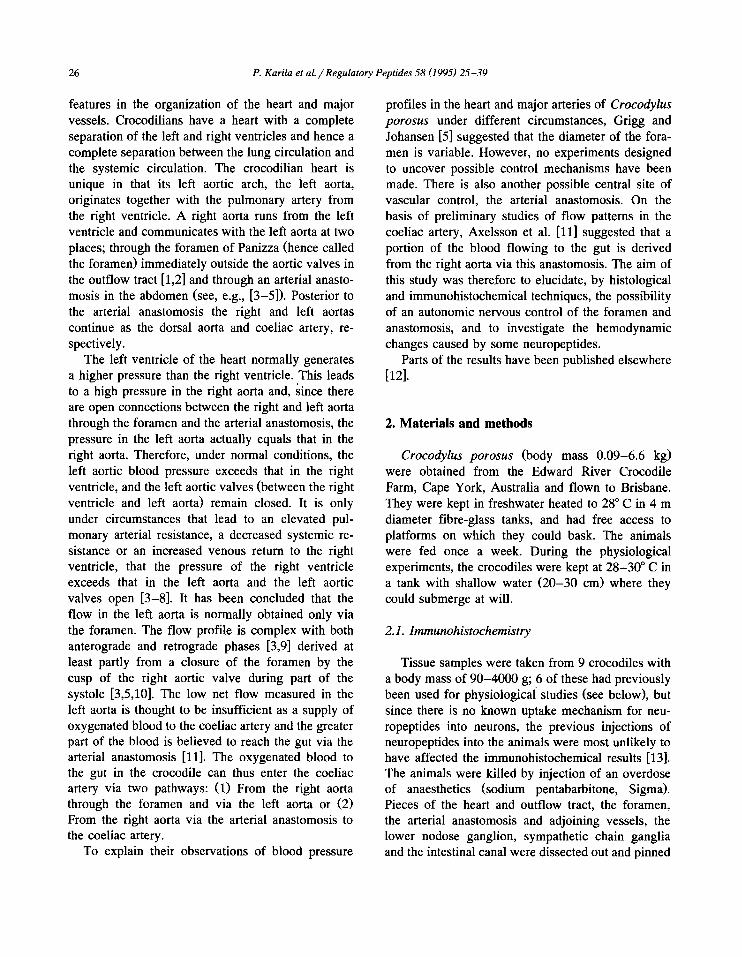
Fig. 1. Co-localization of :immunoreactivity (IR) for neuropeptide Y, (A); somatostatin, (B); and tyrosine hydroxylase (TH), (C) in nerve cell bodies in a sympathet ic ~;anglion of the estuarine crocodile, Crocodylus porosus. The majority of the cell bodies express IR to all three
antisera whereas a few o~ly show TH-IR (arrow in C). Bar: 50 /zm.

30 P. Karila et al. / Regulatory Peptides 58 (1995) 25-39
Fig. 2. Immunoreactivity (IR) in the nodose ganglion of the estuarine crocodile, Crocodylus porosus. (A,B) Co-existence of calcitonin gene related peptide (CGRF)-IR, (A); and substance P-IR (B) in nerve cell bodies and a varicose fibre. Some cells contain only CGRP-IR (arrow in A). Note that the peptides seem to be stored in the same structures in the cells with co-existing IR (double arrows). In the nodose ganglion cells immunoreactive to dopamine-/3-hydroxylase, (C); and neuropeptide Y (D) were also found. Bar: 50 p,m,
Fig. 3. Immunoreactivity (IR) in the heart and vessels of the cardiac region in the estuarine crocodile, Crocodylus porosus. (A-E) Co-localization of IR in the left atrium. Total overlap was seen between calcitonin gene related peptide-IR, (A); and substance P (SP)-IR, (B) in varicose fibres of the myocardium. Co-existence of neuropeptide Y (NPY)-IR, (C); somatostatin (SOM)-IR, (D); and tyrosine hydroxylase (TH)-IR, (E) in fibres of the myocardium. Some fibres are lacking TH-IR (arrows). (F-H) Fibres in the wall of superior vena cava showing co-localization of NPY-IR, (F); SOM-IR, (G); and TH-IR, (H). (I-N) IR in the foramen of Panizzae. Dense IR to vasoactive intestinal polypeptide inside the sphincter-like smooth muscle, (I); Moderate NPY-IR just outside the smooth muscle, (K); SP-IR outside the smooth muscle, (L); bombesin-IR in the smooth muscle layer, (M); and 5-hydroxy-tryptamine-lR, (N) were also found in the foramen of Panizzae. Bars: A-L, 50 /zm; M,N, 25 /xm.

P. Karila et al. / Regulatory Peptides 58 (1995) 25-39 31
C
, i i ~ ¸

32 P. Karila et al. / Regulatory Peptides 58 (1995) 25-39
pled and recorded using a Toshiba 3200SX micro- computer running AD/DATA (Doc. Peter Thor6n, Karolinska Institute, Stockholm). Mean flow and pressure recordings were made using AD/DATA sampling at 5 Hz and storing mean values every 5 s.
Experimental protocol All drug injections were made through the RAo
cannula. The injections were performed only after the water in the holding tank had been lowered to prevent voluntary submergence and related circula- tory changes that would obscure the direct effects of the injected substances. When the effects of different peptides were surveyed in the same animal, the peptides were injected in random order, and the animal was left to recover for at least 45 min or until the recorded variables had returned to their pre-injec- tion values before the following injection was given.
Drugs Substance P, neuropeptide Y, and neurotensin
were obtained from Sigma. The peptides were dis- solved in 0.9% NaC1, injected in boluses and flushed in by 0.5 ml 0.9% NaC1.
3. Results
3.1. Immunohistochemistry
Autonomic ganglia The results of the immunohistochemical labelling
are presented in Table 2. In the sympathetic ganglia, there was co-existence in the majority of the gan- glion cells of the catecholamine synthesizing enzyme tyrosine hydroxylase (TH) -IR, neuropeptide Y (NPY) -IR and somatostatin (SOM) -IR (Fig. 1), while minor populations of neurons contained TH-IR (7%), co-existing NPY-IR and SOM-IR (7%), TH-IR and NPY-IR (3%), TH-IR and SOM-IR (3%), NPY- IR (2%) or SOM-IR (1%). Only occasional cells lacking immunoreactivity to these substances were observed. DBH-IR detected with the antiserum anti- DBH 2D4 appeared to be present in all ganglion cells. In the nodose ganglion (the vagal trunk gan- glion), calcitonin gene-related peptide (CGRP)-IR and substance P (SP) IR co-existed in 46% of the nerve cell bodies, and another 28% showed CGRP-IR
but no SP-IR (Fig. 2A, B). DBH-IR (Fig. 2C), NPY- IR (Fig. 2D) or SOM-IR were found in other cells. The antisera raised against other transmitters (cf. Table 2) failed to reveal positive nerve cell bodies in the ganglia.
In addition to nerve fibres displaying immunore- activity to TH, NPY and SOM in the sympathetic ganglion and to SP and CGRP in the nodose gan- glion, which could possibly be attributed to pro- cesses from local ganglion cells, a number of fibre populations were observed. In the sympathetic gan- glia, SP-IR fibres were seen surrounding nerve cells, while varicose CGRP-IR, vasoactive intestinal polypeptide (VIP)-IR, galanin (GAL)-IR, 5-HT-IR or bombesin (BM)-IR fibres were passing through the ganglia. In the nodose ganglion, VIP- and NPY-IR fibres surrounded nerve cell bodies. Fibres im- munoreactive to antisera against the other transmit- ters were only seen running through the ganglia (TH-IR, BM-IR) or along the outside of the ganglion (GAL-IR, 5-HT-IR).
Heart and vessels of the cardiac region In the myocardium, no regional difference was
observed in the distribution of the immunoreactive material. SP-IR and CGRP-IR co-existed in one population of nerves (Fig. 3A,B), while TH-IR, NPY-IR and SOM-IR were found in a second popu- lation of nerves (Fig. 3C-E). The presence of DBH- IR in nerve fibres was also confirmed.
The same types of fibres (SP/CGRP-IR and TH/NPY/SOM-IR) were present in the carotid and pulmonary arteries and in the superior vena cava (Fig. 3F-H). The IR fibres were evenly distributed from the adventitio-medial ( a /m) border throughout the adventitia. In addition, VIP-IR and BM-IR were observed in populations of fibres with partly overlap- ping distributions (see Table 2).
In the foramen, smooth muscle cells formed a sphincter-like structure surrounded by connective tis- sue and heart muscle. The VIP-IR (Fig. 31) was stronger here than in the myocardium and the fibres more abundant. In the foramen there was also mod- erate NPY-IR (Fig. 3K), CGRP-IR, SP-IR (Fig. 3L) and BM-IR (Fig. 3M) and weak TH-IR, SOM-IR and 5-HT-IR (Fig. 3N). The VIP-IR fibres were distinct from the SP-IR and the TH-IR fibres, but showed some co-existence with CGRP-IR.

P. Karila et al. / Regulatory Peptides 58 (1995) 25-39 33
C
t 2'
Fig. 4. Immunoreactivity (]R) in the arterial anastomosis of the estuarine crocodile, Crocodylus porosus. (A,B) Colocalization of calcitonin gene related peptide-IR, ( /0 and substance P-IR, (B) in varicose fibres with complete overlap in the adventitio-medial border and in the adventitia. (C-E) Colocalization of IR for neuropeptide Y, (C); somatostatin, (D); and tyrosine hydroxylase, (E) in nerve fibres in the adventitio-medial border of the arterial anastomosis. IR to galanin was found in the adventitia, (F); and IR to vasoactive intestinal polypeptide, (G); and bombesin, (H) were found in both the adventitia and the adventitio-medial border. In H the branching point between the left aorta and the arterial anastomosis has been sectioned longitudinally. The arrow indicates the anastomosis where it branches from the left aorta. Bars: A-E,G,H, 50 /.Lm; F, 25 ~m.

34 P. Karila et al. / Regulatory Peptides 58 (1995) 25-39
Abdominal vessels The vessel walls of the arterial anastomosis be-
tween the LAo and the RAo had a well developed adventitia and media. The arterial anastomosis and the 4 connecting vessels (LAo, RAo, dorsal aorta and coeliac artery) had a similar distribution of nerve fibres. There were at least 4 different populations: (1) SP-IR and CGRP-IR co-existed in nerve fibres of the adventitia and the adventitio-medial border of the vessel walls (Fig. 4A,B). The SOM-IR was separate from the CGRP-IR in the adventitio-medial border, which indicates that they belong to different popula- tions. (2) The TH-IR, NPY-IR and SOM-IR co-ex- isted in a dense nerve net in the adventitio-medial border and in weaker immunoreactive nerves of the adventitia (Fig. 4C-E). (3) In the adventitia of the arterial anastomosis galanin (GAL)-IR fibres oc- curred (Fig. 4F). The GAL-IR fibres were separate from the VIP-IR fibres. (4) Dense VIP-IR (Fig. 4G) and moderate BM-IR (Fig. 4H) was found in the adventitia as well as in the adventitio-medial border. VIP- and BM-IR showed the same distribution pat- tern as the SP-IR and CGRP-IR but the VIP-IR did not co-exist with SP-IR and it was also separate from the TH-IR.
Neurotensin
FLA°° t ~ . . . . . . ~
Control + 60 sec + 600 sec
5 sec
Fig. 6. Simultaneous recordings of the effect of neurotensin injection (1 nmol /kg) on the blood flow through the arterial anastomosis (Ana), the coeliac artery (CoA), the left aorta (LAo) and the right aorta (RAo) in the estuarine crocodile, Crocodylus porosus. The flows through the arterial anastomosis and the CoA decreases immediately after injection but the effect is later re- versed to an increased flow.
3.2. In vivo studies
Substance P
° t .............
F o01 ............ Control + 40 sec + 120 sec
2 sec
Fig. 5. Simultaneous recordings of the effect of substance P injection (1 nmol /kg) on the blood flow through the arterial anastomosis (Ana), the coeliac artery (CoA), the left aorta (I2Ao) and the right aorta (RAo) in the estuarine crocodile, Crocodylus porosus. Note the increase in net flow through the arterial anasto- mosis.
Before injections, blood flow in the arterial anas- tomosis, coeliac artery and RAo showed pulses of positive flow correlated to individual heart beats. The flow profile in the left aorta was mixed, with a low anterograde flow during diastole. At the onset of systole there was a transient small increase in the anterograde flow, followed by a reversal of flow into a phase of retrograde flow (Figs. 5-7, see also Axelsson et al. [3,11]).
The responses to injections of substance P, NPY, and neurotensin (0.1-10 nmol /kg) were tested and the dose giving the clearest response was chosen for the study. We have previously reported effects of substance P on blood flows in the RAo and LAo and in the coeliac artery, but no measurements of the flow in the arterial anastomosis or of blood pressures were made [11]. In the present study, substance P (1 nmol/kg; n = 5; Table 3) caused an increase in all flows measured including that of the arterial anasto- mosis (Fig. 5). The pressures in the RAo and LAo were reduced, whereas no consistent pressure effects were obtained in the PA.

P. Karila et al. /Regulatory Peptides 58 (1995) 25-39 35
NPY
FCoA 1 . . . . . . . . . . . . .
FLAo°t ~ " ~ ~
Control + 60 sec + 300 sec
2 sec
Fig. 7. Simultaneous recordings of the effect of neuropeptide Y injection •10 nmol/kg) on the blood flow through the arterial anastomosis (Ana), the coeliac artery (CoA), the left aorta (LAo) and the right aorta (RAo) in the estuarine crocodile, Crocodylus porosus. The flow to the gut via the RAo, the arterial anastomosis and the CoA increases.
Neurotensin (1 nrnol/kg; n = 6; Table 4, Fig. 6) caused an initial reduction in blood flow in the arterial anastomosis and the coeliac artery, w h i c h after some minutes was reversed into a sustained increase in flow. Heart rate and pressure in the pulmonary circuit increased during the first few min-
Table 3 In vivo effects of substance P (1 nmol/kg) on blood flow and pressure in the estuarine crocodile, Crocodylus porosus
Variable Increase Decrease No effect ,~
FRA o 5 0 0 5 FLA o 5 0 0 5 Fco a 4 0 0 4 FAn a 3 0 0 3 PRAo 1 4 0 5 PLAo 0 4 1 5 PPA 2 2 1 5 fH 3 1 1 5
These changes are most likely due to a decrease in coeliac vascular resistance as indicated by the increase in coeliac and anastomosis blood flow in combination with the decreased perfu- sion pressure (PLAo)- Substance P also shows a direct effect on the heart in three of the five animals tested. F, blood flow; RAo, right aorta; LAo, left aorta; Coa, coeliac artery; Aria, arterial anastomosis; P , blood pressure; PA, pul- monary artery; f~, heart rate.
Table 4 In vivo effects of neurotensin (1 nmol/kg) on blood flow and pressure in the estuarine crocodile, Crocodylus porosus
Variable Increase Decrease No effect .Y
FRAo 1 5 0 6 FLAo 2 3 1 6 Fco . 0 4 0 4 FA. a 0 4 0 4 PRAo 3 2 1 6 PLAo 3 2 1 6 PPA 5 0 0 5 Fpa 0 3 0 3 / . 5 o o 5
These changes are most likely due to an increase in coeliac and pulmonary vascular resistance as indicated by the decrease in coeliac, anastomosis and lung blood flow in combination with the increased perfusion pressure (PLAo, PPA)' The effect of neu- rotensin on the heart may be direct or in the case were the blood pressure decreases an effect of a barostatic reflex. F, blood flow; RAo, right aorta; LAo, left aorta; Coa, coeliac artery; Ana, arterial anastomosis; P, blood pressure; PA, pul- monary artery; fn , heart rate.
utes, while f low in the RAo decreased. Pressures in the LAo and RAo and flow in the LAo showed inconsistent responses. The changes in the left aortic flow profile, with an increased foramen spike, are compatible with a vasoconstriction of the anastomo- sis.
NPY (10 nmol /kg; n = 2; Fig. 7) increased the flow in the arterial anastomosis, the coeliac artery and the RAo, but had no conclusive effect on the flow in the LAo. The heart rate was increased, and the pressures in the right and left aortas decreased.
4. Discussion
The presence of smooth muscle cells in a sphinc- ter-like structure around the foramen, and the inner- vation of the smooth muscle cells strongly suggest that the size of the aperture may be controlled, and consequently that the shunting of blood over the foramen may be affected. Similarly, although it may look insignificant to the naked eye [9], the arterial anastomosis between the aortas has a heavily inner- vated muscular wall, which allows for active changes in wall tension and consequently resistance to flow. It must be pointed out, however, that these effects may be small and overruled by more general varia-

36 P. Karila et aL / Regulatory Peptides 58 (1995) 25-39
tions in cardiac output and overall resistance changes in different vascular beds. The control of the tonus of the foramen as well as the arterial anastomosis may also be of importance in preventing these structures from disruption at high blood pressures.
Two populations of fibres, the SP/CGRP-IR fi~ bres and the T H / N P Y / S O M - I R fibres occurred in all parts of the cardiovascular system studied. This corresponds well with the predominating types of nerve cells occurring in the nodose ganglion and the sympathetic chain ganglia, respectively. A spinal autonomic nature of the T H / N P Y / S O M - I R fibres may be concluded. The nodose ganglion in crocodiles is a mixed ganglion, containing both autonomic and sensory components [19]. In analogy with mammals and amphibians, the SP /CGRP neurons may be sensory [20-22], and furthermore in the snake it has been shown that SP /CGRP fibres in vessels are capsaicin sensitive and therefore considered sensory [23].
The VIP-IR nerves had a similar distribution in the vessel walls but were separate from the above populations. The co-existence of the BM-like mate- rial with other neuropeptides was not tested, but the GAL-IR does not seem to be co-localized with any peptide since the distribution pattern differs from that of the other types of immunoreactive nerves observed. Thus, at least 4 types of nerve populations appear to innervate the crocodile cardiovascular sys- tem. The origin of the VIP, BM and GAL-IR nerve fibres could not be determined, since no correspond- ing nerve cell bodies were found in the present study. The nerve fibre populations in the aortic anastomosis and the adjoining vessels are summa- rized in Fig. 8.
Most vascular beds in mammals, crocodiles and the amphibian, Bufo marinus, are innervated by noradrenergic spinal autonomic (sympathetic) nerves, and these often contain NPY in addition to nora- drenaline [21,24,25]. The co-existence of SOM with N A / N P Y in sympathetic, cardiovascular neurons may be common in crocodiles ([24], present study) and is also reported in pigs [26], but this does not appear to be a general feature neither in reptiles nor in vertebrates in general. Somatostatin immunoreac- tive nerves in the heart of the snake Elaphe obsoleta are of an intrinsic parasympathetic population, and are not correlated to the extrinsic catecholamine and
Lower nodose ganglion
~ 4 6 %
28%
Sympathetic chain ganglion
7Ojo
CGRP/SP
BM, VIP
NPY/SOM/TH
GAL
Anastomosis
Fig. 8. Summary of the nerve fibre populations found in the arterial anastomosis and the adjoining vessels of the estuarine crocodile, Crocodylus porosus and the suggested origin for the CGRP/SP-immunoreactive and NPY/SOM/TH-immunoreactive fibres. Legend: CGRP, calcitonin gene-related peptide; SP, sub- stance P; NPY, neuropeptide Y; SOM, somatostatin; TH, tyrosine hydroxylase; BM, bombesin; VIP, vasoactive intestinal polypep- tide; GAL, galanin.
NPY-containing innervation [27], and the weak SOM-IR in snake vessels is not co-localized with any other peptide [23]. In the guinea-pig, NA/SOM- IR neurons do not project to blood vessels [15,28].
Adrenaline injected into the cardiovascular system of Crocodylus porosus causes a marked reduction in blood flows [11], while NPY in our preliminary experiments increases the flow to the gut (from the RAo via the arterial anastomosis). This agrees with results from the spiny dogfish, Squalus acanthias, in vivo [29]. In both the crocodile and the spiny dog- fish, the reason for the increase of flow to the gut appears to be a selective reduction of flow resistance in the gut vasculature caused by NPY. A presynaptic inhibitory effect of NPY on a sympathetic vasocon- strictor tone has been described in several mam- malian systems (see [30]), and a similar effect of NPY may be predominant during the experimental conditions of the present study.
Although sensory in nature, SP /CGRP fibres may also affect the cardiovascular smooth muscle. The injection of substance P caused flow increases in the

P. Karila et al. /Regulatory Peptides 58 (1995) 25-39 37
coeliac artery in agreement with our previous study [11]. Similarly, an increase in left aortic flow is obtained, suggesting an opening of the left aortic valves, which initiate, s the operation of a right-to-left shunt. In the present study, we could also demon- strate an increased supply of blood to the gut from the RAo via the arterial anastomosis. The main reason for this shunting of blood into the gut is probably a reduced vascular resistance of the gut, which may also include a reduced vascular resistance in the arterial anastomosis and main vessels to the gut. This agrees with the potent vasodilator effect of substance P reported in mammals [31,32].
Neurotensin produced profound and consistent ef- fects on the heart rate and the flow through the arterial anastomosis to the gut, but we were not able to demonstrate neurotensin-IR nerves in the corre- sponding tissues. However, neurotensin-IR nerve fi- bres and mucosal endocrine cells are present in the gut wall (P. Karila, I. Gibbins and S. Holmgren, unpublished data), and the obtained effects may thus imitate the hormonal effects of endogenous circulat- ing neurotensin. In mammals, neurotensin is nor- mally released after a meal, and produces an in- creased flow to the gut [31] and an increase in blood pressure and heart rate [33]. A similar effect on heart rate was observed in the crocodile, but the initial effects on the gut blood flow were opposite to those in mammals. An irLcreased flow to the gut was observed in the crocodile, but at a later stage and probably due to a reactive hyperaemia after the initial decrease in flow in the coeliac artery vascular bed. The results suggest an increase in resistance in the arterial anastomosis and possibly also the coeliac vascular bed and in the pulmonary vasculature, which may cause a shunting of blood into the systemic vasculature. The increase in heart rate may be a barostatic reflex.
In conclusion, the anatomy of the foramen and the arterial anastomosis fill the criteria for a direct regu- lation of blood flow: They contain smooth muscle, in the foramen forming a sphincter-like structure, and they are richly innerlated by several populations of nerve fibres. In general, there is a rich neuropeptide innervation of the heart and major vessels of the estuarine crocodile. Amongst the different nerve populations, putative sensory and sympathetic com- ponents were identified. The neuropeptides may af-
fect the cardiac performance and the distribution of blood in the cardiovascular system as further sug- gested by the injections of SP, NT and NPY.
Acknowledgements
We are grateful to Ms. Sue Matthew and Mrs. Christina Hagstr6m for expert technical assistance with immunohistochemistry and photography. The study was supported by project grants from the Swedish Natural Science Research Council (S. Nils- son, S. Holmgren), a University of Queensland Re- search Grant (G. Grigg) and the Australian Research Council (I. Gibbins). Travel grants were obtained from The Royal Society for Arts and Science of the University of G6teborg (S. Nilsson, M. Axelsson), the University of Queensland (S. Nilsson), the Swedish Natural Science Research Council (S. Nils- son), The Paul and Marie Berghaus Foundation (R. Fritsche, P. Karila), The Knut and Alice Wallenberg Foundation (R. Fritsche), The Hierta-Retzius Foun- dation (The Royal Swedish Academy of Sciences; P. Karila) and the l_~ngmanska Foundation.
References
[1] Panizza, B., Sulla struttura del cuore e sulla circolazione del sangue del Crocodilus lucius, Bilioth. Ital., 70 (1833) 87- 91.
[2] Webb, G.J.W., Comparative cardiac anatomy of the reptilia, J. Morph., 161 (1979) 221-240.
[3] Axelsson, M., Holm, S. and Nilsson, S., Flow dynamics of the crocodilian heart, Am. J. Physiol., 256 (1989) R875- R879.
[4] Grigg, G.C., Central cardiovascular anatomy and function in Crocodilia. In Physiological adaptations in vertebrates: Res- piration, circulation and metabolism. Lung biology in health and disease, Vol. 56, Marcel Dekker, New York, 1991, pp. 339-354.
[5] Grigg, G.C. and Johansen, K., Cardiovascular dynamics in Crocodylus porosus breathing air and during voluntary aero- bic dives, J. Comp. Physiol., 157 (1987) 381-392.
[6] Franklin, C.E. and Axelsson, M., The intrinsic properties of an in situ perfused crocodile heart, J. Exp. Biol., 186 (1994) 269-288.
[7] Jones, D.R. and Sheiton, G., The physiology of the alligator aeart - left aortic flow patterns and right-to-left shunts, J. Exp. Biol., 176 (1993) 247-269.
[8] White, F., Redistribution of cardiac output in the diving alligator, Copeia, 3 (1969) 567-570.
38 P. Karila et al. / Regulatory Peptides 58 (1995) 25-39
[9] Shelton, G. and Jones, D.R., The physiology of the alligator heart: the cardiac cycle, J. Exp. Biol., 158 (1991) 539-564.
[10] Greenfield, L.J. and Morrow, A.G., The cardiovascular hemodynamics of crocodila, J. Surg. Res., 1 (1961) 97-103.
[11] Axelsson, M., Fritsche, R., Holmgren, S., Grove, D.J. and Nilsson, S., Gut blood flow in the estuarine crocodile, Crocodylus porosus, Acta Physiol. Scand., 142 (1991) 509- 516.
[12] Karila, P., Holmgren, S., Gibbins, I.L., Grigg, G.C., Franklin, C.E., Axelsson, M., Fritsche, R. and Nilsson, S., Innervation of the foramen Panizzae sphincter and the aortic anastomosis in the crocodile, Crocodylus porosus, Regul. Pept. 39 :2-3 (1992) 272.
[13] Checler, F., Peptidases and neuropeptide-inactivating mecha- nisms in the circulation and in the gastrointestinal tract, Neuropeptide function in the gastrointestinal tract, CRC press, Boca Raton, 1991, pp. 273-307.
[14] Costa, M., Buffa, R., Furness, J.B. and Solcia, E., Immuno- histochemical localization of polypeptides in peripheral auto- nomic nerves using whole mount preparations, Histochem- istry, 65 (1980) 157-165.
[15] Gibbins I.L., Vasoconstrictor, vasodilator and pilomotor pathways in sympathetic ganglia of guinea-pigs, Neuro- science, 47 (1992) 657-672.
[16] Grube, D., Immunoreactivities of gastrin (G-) cells. II. Non- specific binding of immunoglobulins to G-cells by ionic interactions, Histochemistry, 87 (1980) 149-167.
[17] Axelsson, M. and Fritsche, R., Effects of exercise, hypoxia and feeding on the gastrointestinal blood flow in the Atlantic cod, Gadus morhua, J. Exp. Biol., 158 (1991) 181-191.
[18] Sundin, L. and Nilsson, S., Arterio-venous branchial blood flow in the Atlantic cod Gadus morhua, J. Exp. Biol., 165 (1992) 73-84.
[19] Gaskell, W.H., On the structure, distribution and function of the nerves which innervate the visceral and vascular systems, J. Physiol. (London), 7 (1886) 1-80.
[20] Gibbins, I.L., Furness, J.B., Costa, M., Maclntyre, I., Hill- yard, C.J. and Girgis, S., Co-localization of calcitonin gene- related peptide-like immunoreactivity with substance P in cutaneous, vascular and visceral sensory neurons of guinea pigs, Neurosci. Lett., 57 (1985) 125-130.
[21] Morris, J.L., Gibbins, I.L., Campbell, G., Murphy, R., Fur- ness, J.B. ~Ind Costa, M., Innervation of the large arteries and heart of the toad (Bufo marinus) by adrenergic and peptide- containing neurons, Cell Tissue Res., 243 (1986) 171-184.
[22] Wiesenfeld-Hallin, X., Hrkfelt, T., Lundberg, J.M., Forss- mann, W.G., Reinecke, M., Tschopp, F.A. and Fischer, J.A., Immunoreactive calcitonin gene-related peptide and sub- stance P coexist in sensory neurones of the spinal cord and interact in spinal behavioural responses of the rat, Neurosci. Lett., 52 (1984) 199.
[23] Davies, P.J. and Donald, J.A., The distribution and colocal- ization of neuropeptides in perivascular nerves innervating the large arteries and veins of the snake, Elaphe obsoleta, Cell Tissue Res., 269 (1992) 495-504.
[24] Gibbins, I.L., Morris, J.L., Furness, J.B. and Costa, M., Innervation of systemic blood vessels. In Non-adrenergic
innervation of blood vessels. Volume II: Regional innerva- tion, CRC press, Boca Raton, 1988, pp. 1-36.
[25] Mione, M.C., Ralevic, V. and Bumstock, G., Peptides and vasomotor mechanisms, Pharmac. Ther., 46 (1990) 429-668.
[26] Lacroix, J.S., Auberson, S., Morel, D.R., Theodorsson, E., Hrkfelt, T. and Lundberg, J.M., Vascular control of the pig nasal mucosa: distribution and effect of somatostatin in relation to noradrenaline and neuropeptide Y, Regul. Pept., 40 (1992) 373-387.
[27] Donald, J.A., O'Shea, J.E. and Lillywhite, H.B., Somato- statin and innervation o f the heart of the snake (Elaphe obsoleta), Am. J. Physiol., 258 (1990) mi-mvii.
[28] Costa, M. and Furness, J.B., Somatostatin is present in a subpopulation of noradrenergic nerve fibres supplying the intestine, Neuroscience, 13 (1984) 911-920.
[29] Holmgren, S., Axelsson, M. and Farrell, A.P., The effects of neuropeptide Y and bombesin on blood flow to the gut in dogfish, Squalus acanthias, Regul. Pept., 40 (1992) 169.
[30] Morris, J.L. and Gibbins, I.L., Co-transmission and Neuro- modulation. In G. Burnstock and C.H.V. Hoyle (Eds.), The autonomic nervous system. Autonomic neuroeffector mecha- nisms, Harwood Academic Publishers, Reading, 1992, pp. 33-119.
[31] Dahlstrrm, A., Nilsson, O., Lundgren, O. and AMman, H., Nonadrenergic, non-cholinergic innervation of gastrointesti- nal vessels: morphological and physiological aspects. In Nonadrenergic innervation of blood vessels. Volume II. Re- gional innervation, CRC Press, Boca Raton, 1988, pp. 143- 172.
[32] Pernow, B., Substance P, Pharmacol. Rev., 35 (1983) 85-141. [33] Rioux F. and Lcmieux M., Haemodynamic and abdominal
motor reflexes elicited by neurotensin in anaesthetized guinea-pigs, Br. J. Pharmacol., 106 (1992) 187-195.
[34] Bjenning, C., Farrell, A.P. and Holmgren, S., Bombesin-like immunoreactivity in the gut and heart region of elasmo- branch fish (skates), and the in vitro effect of bombesin on coronary vessels from the longnose skate, Raja rhina, Regul. Pept., 35 (1991) 207-219.
[35] Jrnsson, A.C., Regulatory peptides in the pancreas of two species of elasmobranchs and in the Brockmann bodies of four teleost species, Cell Tissue Res., 266 (1991) 163-172.
[36] Kummer, W., Gibbins, I.L., Stefan, P. and Kapoor, V., Catecholamines and catecholamine-synthesizing enzymes in guinea-pig sensory ganglia, Cell Tissue Res., 261 (1990) 595-606.
[37] Morris, J.L. and Gibbins, I.L., Neuronal colocalization of peptides, catecholamines, and catecholamine-synthesizing enzymes in guinea-pig paracervical ganglia, J. Neuroscience, 7 (1987) 3117-3130.
[38] Karila, P., J/Jnsson, A.C., Jensen, J. and Holmgren, S., Galanin-like immunoreactivity in extrinsic and intrinsic nerves to the gut of the Atlantic cod, Gadus morhua, and the effect of galanin on the smooth muscle of the gut, Cell Tissue Res., 271 (1993) 537-544.
[39] Morris, J.L., Gibbins, I.L. and Holmgren, S., Galanin is more common than NPY in vascular sympathetic neurons of the brush-tailed possum, Regul. Pept., 37 (1992) 101-109.

P. Karila et al. / Regulatory Peptides 58 (1995) 25-39 39
[40] Morris, J.L., Murphy, R., Furness, J.B. and Costa, M., Partial depletion of neuropeptide Y from noradrenergic perivascular and cardiac axons by 6-hydroxydopamine and reserpine, Regul. Pept., 13 (1986) 147-162.
[41] Holmgren, S., Fritsche, R., Karila, P., Gibbins, I.G., Axels- son, M., Franklin, C., Grigg, G. and Nilsson, S., Neuropep- tides in the Australian lungfish Neoceratodus forsteri: effects in vivo and presence in autonomic nerves, Am. J. Physiol., 266 (1994) R1568-R1577.
[42] Jensen, J., Holmgren, S. and JiSnsson, A.C., Substance P-like immunoreactivity and the effects of tachykinins in the intes- tine of the Atlantic cod, Gadus morhua, J. Auton. Nerv. Syst., 20 (1987) 25-33.
[43] Gibbins, I.L., Furness, J.B. and Costa, M., Pathway-specific
[441
[451
patterns of the coexistence of substance P, calcitonin gene-re- lated peptide, cholecystokinin and dynorphin in neurons of the dorsal root ganglia of the guinea pig, Cell Tissue Res., 248 (1987) 417-437. Morris, J.L., Gibbins, I.L. and Furness, J.B., Increased levels of dopamine-beta-hydroxylase-like immunoreactivity in non-noradrenergic axons supplying the guinea-pig uterine artery after 6-hydroxydopamine treatment, J. Auton. Nerv. Syst., 21 (1987) 15-27. Bowden, J.J. and Gibbins, I.L., Vasoactive intestinal peptide and neuropeptide Y coexist in non-noradrenergic sympathetic neurons to guinea pig trachea, J. Auton. Nerv. Syst., 38 (1992) 1-20.