Neural stem cells: ready for therapeutic applications?
17
REVIEW Open Access Neural stem cells: ready for therapeutic applications? Simona Casarosa, Yuri Bozzi and Luciano Conti * Abstract Neural stem cells (NSCs) offer a unique and powerful tool for basic research and regenerative medicine. However, the challenges that scientists face in the comprehension of the biology and physiological function of these cells are still many. Deciphering NSCs fundamental biological aspects represents indeed a crucial step to control NSCs fate and functional integration following transplantation, and is essential for a safe and appropriate use of NSCs in injury/disease conditions. In this review, we focus on the biological properties of NSCs and discuss how these cells may be exploited to provide effective therapies for neurological disorders. We also review and discuss ongoing NSC-based clinical trials for these diseases. Keywords: Neural stem cells, Pluripotent stem cells, Cell therapy, Neuron, Neurodegenerative diseases, Neurodevelopmental disorders Review Introduction Conventional pharmacological treatments for most neuro- degenerative conditions relieve some symptoms but rarely vary the course of the disease or halt its progression. Graft- ing of human fetal tissue has provided a proof of concept for cell therapy approaches to neurodegenerative diseases in a number of clinical studies, including treatment of Parkinson’ s and Huntington’ s disease patients [1]. Nonethe- less, this does not represent a practical route for large-scale therapeutic applications due to limited availability and quality of human fetal tissue, as well as for ethical considerations. To this regard, in the last years, great media consider- ation has brought neural stem cell (NSC) research into the spotlight. Most of this attention has been raised by the stimulating prospects of NSCs application for cell replace- ment therapies for neurological disorders, engendering hopes and expectations in the public and, particularly, in patients. Despite the evident benefits pledged by the NSC field and some encouraging preliminary studies in animal models, there still remains a gap between theory and practice. Indeed, while stem cell-based therapies are the current standard of care for blood tumors and are gaining agreement in the treatment of epidermal and corneal disorders, applications for diseases affecting the nervous system yet represent a pioneering field, being in the early phases of clinical scrutiny. What is still missing to effect- ively translate NSC research into clinical applications? Al- though important scientific progresses in the field have been achieved, we still lack a profound understanding of the basic biology of NSCs and how to manipulate these cells to provide reliable, safe and effective outcomes in cell-replacement approaches. NSCs are immature cells present in the developing and adult Central Nervous System (CNS). Typically, NSCs are defined by three cardinal characteristics: self-renewal po- tential, neural tripotency (i.e., the capability to give rise to all of the major neural lineages: neurons, astrocytes and oligodendrocytes) and competence for in vivo regener- ation (Figure 1; [2]). They have the potential to generate both neurons and glia of the developing brain and they also account for the limited regenerative potential in the adult brain. In the adult CNS, NSCs reside in defined regions (“neurogenic niches”) that sustain their multi- potency and regulate the balance between symmetrical self-renewal and fate-committed asymmetric divisions [3]. Several studies have shown that NSCs can be extracted from neural tissue or generated from pluripotent cellular sources, genetically manipulated and differentiated in vitro [2]. In the past two decades, a variety of protocols for NSCs * Correspondence: [email protected] Center for Integrative Biology, Università degli Studi di Trento, Via Sommarive 9, Povo-Trento 38123, Italy © 2014 Casarosa et al.; licensee BioMed Central Ltd. This is an Open Access article distributed under the terms of the Creative Commons Attribution License (http://creativecommons.org/licenses/by/4.0), which permits unrestricted use, distribution, and reproduction in any medium, provided the original work is properly credited. The Creative Commons Public Domain Dedication waiver (http://creativecommons.org/publicdomain/zero/1.0/) applies to the data made available in this article, unless otherwise stated. Casarosa et al. Molecular and Cellular Therapies 2014, 2:31 http://www.molcelltherapies.com/content/2/1/31
-
Upload
independent -
Category
Documents
-
view
4 -
download
0
Transcript of Neural stem cells: ready for therapeutic applications?

Casarosa et al. Molecular and Cellular Therapies 2014, 2:31http://www.molcelltherapies.com/content/2/1/31
REVIEW Open Access
Neural stem cells: ready for therapeuticapplications?Simona Casarosa, Yuri Bozzi and Luciano Conti*
Abstract
Neural stem cells (NSCs) offer a unique and powerful tool for basic research and regenerative medicine. However,the challenges that scientists face in the comprehension of the biology and physiological function of these cells arestill many. Deciphering NSCs fundamental biological aspects represents indeed a crucial step to control NSCs fateand functional integration following transplantation, and is essential for a safe and appropriate use of NSCs ininjury/disease conditions. In this review, we focus on the biological properties of NSCs and discuss how these cellsmay be exploited to provide effective therapies for neurological disorders. We also review and discuss ongoingNSC-based clinical trials for these diseases.
Keywords: Neural stem cells, Pluripotent stem cells, Cell therapy, Neuron, Neurodegenerative diseases,Neurodevelopmental disorders
ReviewIntroductionConventional pharmacological treatments for most neuro-degenerative conditions relieve some symptoms but rarelyvary the course of the disease or halt its progression. Graft-ing of human fetal tissue has provided a proof of conceptfor cell therapy approaches to neurodegenerative diseasesin a number of clinical studies, including treatment ofParkinson’s and Huntington’s disease patients [1]. Nonethe-less, this does not represent a practical route for large-scaletherapeutic applications due to limited availability andquality of human fetal tissue, as well as for ethicalconsiderations.To this regard, in the last years, great media consider-
ation has brought neural stem cell (NSC) research into thespotlight. Most of this attention has been raised by thestimulating prospects of NSCs application for cell replace-ment therapies for neurological disorders, engenderinghopes and expectations in the public and, particularly, inpatients. Despite the evident benefits pledged by the NSCfield and some encouraging preliminary studies in animalmodels, there still remains a gap between theory andpractice. Indeed, while stem cell-based therapies are thecurrent standard of care for blood tumors and are gaining
* Correspondence: [email protected] for Integrative Biology, Università degli Studi di Trento, ViaSommarive 9, Povo-Trento 38123, Italy
© 2014 Casarosa et al.; licensee BioMed CentrCommons Attribution License (http://creativecreproduction in any medium, provided the orDedication waiver (http://creativecommons.orunless otherwise stated.
agreement in the treatment of epidermal and cornealdisorders, applications for diseases affecting the nervoussystem yet represent a pioneering field, being in the earlyphases of clinical scrutiny. What is still missing to effect-ively translate NSC research into clinical applications? Al-though important scientific progresses in the field havebeen achieved, we still lack a profound understanding ofthe basic biology of NSCs and how to manipulate thesecells to provide reliable, safe and effective outcomes incell-replacement approaches.NSCs are immature cells present in the developing and
adult Central Nervous System (CNS). Typically, NSCs aredefined by three cardinal characteristics: self-renewal po-tential, neural tripotency (i.e., the capability to give rise toall of the major neural lineages: neurons, astrocytes andoligodendrocytes) and competence for in vivo regener-ation (Figure 1; [2]). They have the potential to generateboth neurons and glia of the developing brain and theyalso account for the limited regenerative potential in theadult brain. In the adult CNS, NSCs reside in definedregions (“neurogenic niches”) that sustain their multi-potency and regulate the balance between symmetricalself-renewal and fate-committed asymmetric divisions [3].Several studies have shown that NSCs can be extracted
from neural tissue or generated from pluripotent cellularsources, genetically manipulated and differentiated in vitro[2]. In the past two decades, a variety of protocols for NSCs
al Ltd. This is an Open Access article distributed under the terms of the Creativeommons.org/licenses/by/4.0), which permits unrestricted use, distribution, andiginal work is properly credited. The Creative Commons Public Domaing/publicdomain/zero/1.0/) applies to the data made available in this article,

Figure 1 Cardinal neural stem cell properties.
Casarosa et al. Molecular and Cellular Therapies 2014, 2:31 Page 2 of 17http://www.molcelltherapies.com/content/2/1/31
purification, generation and expansion in floating or adher-ent conditions have been described. However, beside theseimportant progresses, the identification of the best sourcesfor NSCs derivation and the optimization of approaches tostably proliferate them clonally in vitro still represents amajor goal of NSC research.Yet, for a realistic exploitation of NSCs for cell therapies,
clinically-suitable NSC systems should hold specific keyproperties including (i) standardized production and scal-ability to good medical practice (GMP), (ii) karyotypicstability, (iii) ability to correctly integrate in the host tissueand (iv) differentiate into the required functional neuralcells. In addition, NSCs should exhibit a reproducible, pre-dictable and safe behavior following in vivo injection.
NSCs in brain organogenesis and homeostasisIn the developing and adult CNS, different NSC popu-lations dynamically appear following predeterminedspatio-temporal developmental programs. Molecular andbiological characteristics of NSCs greatly vary dependingon the region and developmental stage considered [4].Development of the vertebrate CNS starts with neural
plate folding to originate the neural tube, consisting ofradially elongated neuroepithelial cells (NEPs) [5]. NEPsdevelop definite identities and different fates dependingon their positions along the rostrocaudal (R-C) and dorso-ventral (D-V) axes of the neural tube. Patterning along theR-C axis leads to the initial distinction into prosencephalon,mesencephalon, rhombencephalon and spinal cord territor-ies. NEPs are accountable for the first wave of neurogenesisin the neural tube. As development proceeds, NEPs convertthemselves into another transitory NSC type, the so-called“radial glia” (RG) [6,7]. This rapidly constitutes the mainprogenitor cell population in mid/late development andearly postnatal life while disappearing at late postnatal and
adult stages. Besides their ability to divide asymmetric-ally and to serve as progenitors of neurons and glia, RGcells constitute a scaffold on which neurons migrate inthe developing brain. RG differentiation potential is lessextensive compared to that of NEPs. Along with RG, an-other population of immature neural cells is constituted byBasal Progenitors (BPs) [8]. They are generated at earlyphases of development by NEPs and at later stages by RG.BPs mostly undergo one or two rounds of division, gener-ating one or two pairs of neurons. Hence, BPs may beconsidered neurogenic transit-amplifying progenitors thatspecifically increase the production of neurons during re-stricted developmental time periods in definite brain areas(i.e. cerebral cortex).At the end of neurogenesis (roughly at birth in mice),
neurogenic RG cells are exhausted and residual RG cellsare converted into a unique astrocyte-like subpopulation[9]. This population will make up the NSC pool of theadult brain, endorsed with neurogenesis and gliogenesismaintainance throughout adult life.The concept that the adult brain retains the ability to
self-renew some of its neurons has been broadly recog-nized in the last two decades and has represented a break-through in neurosciences. Pioneering studies from Altmanand Das already reported the generation of new neuronsin a variety of structures in the adult rat and cat includ-ing the olfactory bulb, hippocampus, and cerebral cor-tex [10]. However, their results were widely neglected untilthe early 1990s, when the formation of new neurons inadult rodent brain was clearly demonstrated [11,12]. Thisled to the identification of the germinal zones of the adultbrain. These are specialized niches located in the subven-tricular zone (SVZ) of the lateral ventricle wall and in thesubgranular zone (SGZ) of the dentate gyrus of the hippo-campus [3]. Whether NSCs reside in other regions of the

Casarosa et al. Molecular and Cellular Therapies 2014, 2:31 Page 3 of 17http://www.molcelltherapies.com/content/2/1/31
adult mammalian brain is still disputed. Neuroblasts pro-duced in the rodent SVZ migrate to the olfactory bulb fol-lowing the rostral migratory stream (RMS), an anatomicstructure well characterized in the rodent brain. The NSCslocated in the SVZ, also called type B cells, generate activelydividing intermediate cells, named type C cells, whichfurther divide giving rise to neuroblasts, referred to as typeA cells that migrate away from the SVZ. These migratingneuroblasts are organized in chains that connect the SVZto the olfactory bulb (constituting the RMS) where theygradually mature into functional GABAergic granuleneurons. Fate-mapping studies actually reveal that type Bcells are not developmentally restricted to neuronal lineagesbut can give rise also to glial progenies, suggesting they areauthentic tripotent NSCs. The second germinal zone of theadult mammalian brain is the dentate gyrus of the hippo-campus. Astrocyte-like NSCs, called type I progenitors,have been identified within the SGZ facing the dentategyrus hilus. They share several properties with the typeB cells of the adult SVZ, although they apparently exhibit anarrower developmental potential. Type I progenitors likelydivide asymmetrically to produce immature proliferatingprogenitors, type II cells. These gradually differentiate intomigrating neuroblasts that travel into the granule celllayer of the dentate gyrus, where they progressively matureinto functional granule neurons. Differently from the typeB cells of the SVZ, the progeny of type I progenitors doesnot migrate long distances, but remains localized in clus-ters closely connected to the parent cell. Additionally, hip-pocampal NSCs appear to be developmentally restrictedto become granule neurons; currently, there is no evidencethat type I progenitors can generate mature glial deriva-tives in vivo.The discovery of NSCs and neurogenesis in the adult
mammalian CNS has tremendously changed our view ofthe plasticity and function of the brain. This has promptedexcitement for the possible exploitement of intrinsicneurogenic activity to cure brain diseases and rescue brainfunction after injury. Mobilization of endogenous NSCshas thus emerged as a potential therapeutic approach forneural repair. It is known that brain injury promotes theproliferation of adjacent NSCs, generating new astrocytesand neurons [13]. For example, focal ischemia transientlyinduces forebrain SVZ cell proliferation and neurogenesis.The NSCs in the SVZ and DG are also stimulated aftertraumatic brain injury or seizures [14], suggesting thatadult neurogenesis may play a role in self-recoverymechanisms of the brain. However, the amount ofspontaneously produced neuroblasts after brain injuryis highly limited, and their survival and differentiationinto mature neurons are far from obtaining regenera-tive effects.It should be emphasized that, although our understand-
ing of NSCs has increased dramatically over the past few
years, there are still many major gaps regarding theirin vivo control.
NSCs for cell replacement approaches: requirements &available in vitro systemsA large number of studies have explored grafting behaviorof several NSCs typologies (and their progeny) in a varietyof preclinical studies and in some clinical investigations.Nevertheless, NSCs used for clinical applications shouldbe safe, effective and accessible in large amount in GMPconditions. A variety of different sources for NSCs havebeen tested, including fetal- and adult CNS-derived NSCs,neural progenitors derived from pluripotent cells, anda range of non-neural stem cells, such as mesenchymal(MSCs) and bone marrow-derived (BMDSCs) stem cells.With these issues in mind, it should be remarked that upto now an ideal NSC system is not yet available to theclinic. Here, we will restrict our discussion to NSCs de-rived from neural tissue and from pluripotent stem cells.Advantages and disadvantages of each source and recentexperimental evidence that highlight their potential usefor clinical applications will be presented.
Fetal- and adult-derived NSCsThe isolation of NSCs from their natural niches and theirexpansion in culture have been challenging issues, becausethe requirements to maintain these cells in their physio-logical state are yet poorly understood. In the early ‘90s,the identification of EGF and FGF-2 as key mitogensfor NSCs led to set up culture conditions that supportextended cell division of cells with NSCs properties [11,12].Since then, several studies reported that NSCs can beisolated from various regions of rodent (mouse and rat)and human brain at several developmental stages as well asfrom germinative areas of the adult brain. A widely usedmethod is to culture NSCs as neurospheres (Figure 2; [15]).These are free-floating aggregates of neural progenitors,each, in theory, deriving from a single NSC. Their gen-eration relies on neural tissue micro-dissection followedby exposure to defined mitogen-supplemented media.In such a procedure, primary cells are plated in low-attachment culture flasks in serum-free media supple-mented with EGF and/or FGF-2. In these conditions,differentiating or differentiated cells are supposed to die,whereas NSCs respond to mitogens, divide and form float-ing aggregates (primary neurospheres) that can be dissoci-ated and re-plated to generate secondary neurospheres.This procedure can be sequentially repeated several timesto expand a NSC population.Complementary to neurosphere culture is adherent cul-
ture, in which cells are more easily monitored and havebetter access to growth factors (Figure 2). In the last dec-ade, several groups reported the generation and expansionof adherent NSC lines from neural tissue of rodent and

Figure 2 Sources and in vitro growth protocols for neural stem cell generation and expansion.
Casarosa et al. Molecular and Cellular Therapies 2014, 2:31 Page 4 of 17http://www.molcelltherapies.com/content/2/1/31
human origin. According to this procedure, NSCs can becompetently expanded as adherent clonal homogeneousNSC lines by exposure to specific mitogens, such as EGFand/or FGF2 [16,17]. In these conditions, cells divide sym-metrically, retaining their tripotential differentiation cap-acity. Adherent culture regimens have been shown to allowfor cultures with less differentiated cells compared tothe neurosphere assay, where cell–cell contacts and non-uniform mitogens exposure is thought to stimulate differ-entiation programs [2].Although several studies have attempted to provide
comparisons between fetal- and adult-derived NSCs, sys-tematic side-by-side analyses are still few and do not allowto draw any solid conclusions. In fact, results might be
hampered by culture conditions, especially for humanNSCs. Nonetheless, major differences between fetal- andadult-derived human NSCs have been reported in termsof both biological and molecular properties. Fetal-derivedhuman NSCs generally exhibit a shorter doubling time, amore extensive expansion potential in vitro and betterintegrative potential following grafting in animal models[18-21]. Noteworthy, substantial differences have been alsodescribed when comparing human NSCs derived from dif-ferent brain areas of the same fetus [22].
NSCs from pluripotent stem cellsNeuralization protocols applied to mouse and humanpluripotent stem cells, including embryonic stem cells

Casarosa et al. Molecular and Cellular Therapies 2014, 2:31 Page 5 of 17http://www.molcelltherapies.com/content/2/1/31
(ESCs) and induced pluripotent stem cells (iPSCs), allowfor the generation of NSCs populations. ESCs are derivedfrom the inner cell mass (ICM) of blastocyst stage mamma-lian embryos [23,24]. They are characterized by an intrinsiccapacity for self-renewal and the ability to generate allcell types derived from the three embryonic germ layers(pluripotency). In the last years, the advent of iPSC technol-ogy has completely revolutioned the “pluripotency” field,avoiding the requirement of embryos as source of rodentand, most importantly, of human pluripotent stem cells.Moreover, the use of iPSC opens new possibilities forstudies of human development and disorders, furtherincreasing the potential biomedical applications of thistype of cells [25]. iPSCs are the product of a reprogram-ming procedure that allows the conversion of somaticcells directly into pluripotent cells [26,27]. This tech-nology is straightforward, robust and since its discoveryhas been implemented in terms of efficiency and repro-ducibility. iPSCs closely resemble ESCs with respect toexpression of pluripotency markers, self-renewal poten-tial, and multilineage differentiation potential. Both murineand human pluripotent stem cells can be exposed to neur-alizing in vitro protocols to generate a large amount ofNSCs or progenitor cells. In these conditions, pluripotentstem cells undergo progressive lineage restrictions similarto those observed during normal fetal development, lead-ing to the generation of a range of distinct neural precur-sor populations [2].Generally, two main procedures to generate NSCs from
pluripotent stem cells have been developed. The firststrategy relies on the formation of embryoid bodies (EBs),three-dimensional (3D) aggregates. EBs recapitulate manyaspects of cell differentiation occurring during early mam-malian embryogenesis and give rise to cells of the threegerm layers, including neural cells. EBs dissociated andplated in adhesion on coated plastic surfaces in definedmedia will produce rosette-like neural cells correspondingto the NEPs of the developing brain [28,29]. This NSCpopulation can be subsequently enriched, although no effi-cient methods for their extensive expansion have beenreported. EB-independent procedures based on adherentmonolayer protocols have been also described [30,31].Electrophysiology studies have shown that pluripotent-
derived NSCs efficiently generate fully mature neuronsin vitro, as well as functionally integrated neurons aftertransplantation in the mammalian CNS [32,33]. Never-theless, major limitations to therapeutic applications ofpluripotent-derived NSCs are represented by safety con-cerns and caveats about their clinical-grade production.Indeed, grafted pluripotent cells can form teratomas, im-plying that in a clinical setting residual undifferenti-ated pluripotent stem cells should be excluded fromthe cell preparation before grafting. Protocols for avoidingteratocarcinoma formation in vivo after transplantation of
ESC/iPSCs-derived cells are under scrutiny. In this view,recent studies have reported the direct conversion ofadult somatic cells into NSCs, thus opening a new path togenerate NSCs without contamination of undifferentiatedpluripotent stem cells [34].The ability to generate patient-specific iPSCs and NSCs
clearly provides enormous prospective for future personal-ized medicine, although too little is yet known about thesecells to make any firm prediction.
Possible therapeutic actions of grafted NSCs in differentneurodegenerative conditionsAlthough the capacity of NSCs to divide and appropriatelydifferentiate in vitro has attracted much attention for clin-ical translation, it does not assure that these cells func-tionally incorporate into the recipient tissue and produceefficient restoration of compromised functions after graft-ing. In order to generate therapeutic benefits in specificneurological diseases, grafted cells have to accomplish acertain grade of morphological, anatomical and functionalintegration into the impaired host CNS tissue.Neurodegenerative disorders embody a heterogeneous
collection of chronic and progressive diseases charac-terized by distinct aetiologies, anatomical impairmentsand symptoms [35]. Some of these disorders, such asHuntington’s disease (HD), are acquired in an entirely gen-etic manner. Alzheimer’s disease (AD), amyotrophic lateralsclerosis (ALS), and Parkinson’s disease (PD) mainly occursporadically, although familiar forms caused by inheritanceof gene mutations are known. On the other hand, the CNScan also be affected by other non-degenerative conditions,such as spinal cord injury and stroke, with no genetic herit-able components.By virtue of this extreme heterogeneity, different spe-
cific requirements should be envisaged when consideringcell replacement as a possible therapeutic strategy. We candistinguish between (i) “neuronal” CNS degenerative disor-ders caused by a prominent loss of specific neuronal popula-tions and (ii) “non neuronal” CNS degenerative conditionscharacterized by loss of non neuronal elements.In the case of neuronal degeneration, the success of cell
replacement strictly depends on the complexity and accur-acy of the pattern of connectivity that needs to be restored.In PD, affected dopaminergic neurons in the substantianigra (SN) exert a modulatory action on striatal target cir-cuits mostly through the release of dopamine. This systemis defined as “paracrine” and even a partial pattern repairmay lead to a significant functional recovery in such condi-tions. Indeed, in PD the donor cells can be transplanteddirectly into the target region to circumvent the problemof long-distance neuritic growth in the adult CNS. Despitethe ectopic location, if grafted cells are able to re-establisha regulated and efficient release of dopamine, they can leadto a clinically relevant functional recovery. However, cell-

Casarosa et al. Molecular and Cellular Therapies 2014, 2:31 Page 6 of 17http://www.molcelltherapies.com/content/2/1/31
based treatment strategies are extremely difficult for otherdiseases such as HD, ALS, trauma, stroke, and AD, whichare characterized by the need of a complex pattern repair.Differently, “non neuronal” CNS degenerative syndromes
such as multiple sclerosis (MS) characterized by severe in-flammation, oligodendroglial degeneration and axonal de-myelination, represent a good target for cell replacement,due to their limited requirements for pattern repair [36]. InMS, grafted cells should produce oligodendrocytes able torestore axonal myelination in order to lead to a functionalrescue.It should also be emphasized that although pattern re-
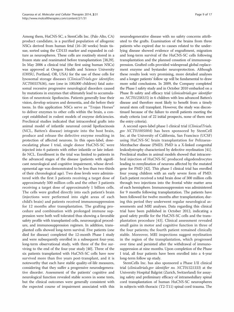
pair is critical to obtain permanent efficacy in the brain,cells transplanted into the brain may also be beneficialvia the release of molecules that may either stimulate theregenerative potential of local cells (where present) orincrease the survival of the remaining host elements,thus slowing disease progression (Figure 3; [37]). Also,immunomodulatory activity of the grafted cells couldbe of benefit in diseases such as MS where a prominentdisease-associated inflammation contributes to the estab-lishment and progression of the disease (Figure 3).
Current clinical trials involving NSCs forneurodegenerative diseasesA large number of studies have explored the grafting be-havior of several NSCs typologies (and their progenies)
Figure 3 Therapeutical strategies for neural stem cells exploitation in
in preclinical studies. Moreover, a few attempts are alreadybeing made to translate these discoveries into the clinicalsetting. Currently, the registry and results database ofpublicly and privately supported clinical studies withhuman participants conducted around the world (http://www.clinicaltrial.gov/) reports 880 international clinical tri-als employing the use of stem cells for treatment of patientsaffected by several CNS disorders (query terms: “stem cellsAND nervous system”) among which 89 are testing NSCinjection approaches (query terms: “neural stem cell ANDinjection AND nervous system disease”). If restrictingthe search to non-tumor diseases, the database returns51 studies (query terms: “neural stem cell AND injectionAND nervous system disease NOT tumor”) with only 27of these currently open. Interestingly, if we analyze theseresults more carefully, only 5 studies are actually open toexplore the potential clinical relevance of NSCs while theremaining are testing the regenerative potential of mesen-chymal stem cells whose actual uselfness in brain diseasesis far from being a solid preclinical reality.To date, few Phase I and II clinical studies employing
NSCs (a dozen in highly debilitating CNS disorders; de-tails reported in Table 1) have been performed, with themain objective to demonstrate safety and practicability andto explore the potential effectiveness of the treatments.These clinical trials are testing only few NSCs products,mainly consisting of fetal-tissue-derived allogenic NSCs.
CNS diseases.

Table 1 Clinical trials involving NSCs for neurodegenerative diseases
NCT number Title Recruitment Conditions Interventions Sponsor/Collaborators Phases Enrollment Startdate
Completiondate
NCT00337636 Study of HuCNS-SC Cells inPatients With Infantile or LateInfantile Neuronal CeroidLipofuscinosis (NCL)
Completed Neuronal CeroidLipofuscinosis
Biological: HuCNS-SC StemCells, Inc. Phase1
6 May2006
September2009
NCT01005004 Study of Human CentralNervous System (CNS) StemCells Transplantation inPelizaeus-Merzbacher Disease(PMD) Subjects
Completed Pelizaeus-MerzbacherDisease
Biological: HuCNS-SCcells implantation
StemCells, Inc. Phase1
4 November2009
December2012
NCT01151124 Pilot Investigation of StemCells in Stroke
Active, notrecruiting
Stroke Biological: CTX0E03neural stem cellsimplantation
ReNeuron Limited Phase1
12 June2010
March2015
NCT01217008 Safety Study of GRNOPC1in Spinal Cord Injury
Completed Spinal Cord Injury Biological: hES-derivedGRNOPC1 implantation
Asterias Biotherapeutics, Inc. Phase1
5 October2010
July2013
NCT01238315 Safety and Efficacy Study ofHuCNS-SC in Subjects WithNeuronal Ceroid Lipofuscinosis
Withdrawn Neuronal CeroidLipofuscinosis
Biological: HuCNS-SCcells implantation
StemCells, Inc. Phase1
0 November2010
April2011
NCT01321333 Study of Human CentralNervous System Stem Cells(HuCNS-SC) in Patients WithThoracic Spinal Cord Injury
Active, notrecruiting
Thoracic Spinal CordInjury|Spinal Cord
Injury|Spinal Cord InjuryThoracic|Spinal Cord
Trauma
Biological: HuCNS-SCcells implantation
StemCells, Inc. Phase1-2
12 March2011
December2015
NCT01348451 Human Spinal Cord DerivedNeural Stem Cell Transplantationfor the Treatment of AmyotrophicLateral Sclerosis
Active, notrecruiting
Amyotrophic LateralSclerosis
Biological: humanneural spinal cordstem cells implantation
Neuralstem Inc. Phase1
18 January2009
August2014
NCT01640067 Human Neural Stem CellTransplantation in AmyotrophicLateral Sclerosis (ALS)
Recruiting Amyotrophic LateralSclerosis
Biological: HumanNeural Stem Cellsimplantation
Azienda Ospedaliera SantaMaria, Terni, Italy|AziendaOspedaliero Universitaria
Maggiore della Carita|Universitàdi Padova Italy
Phase1
18 December2011
September2016
NCT01725880 Long-Term Follow-Up ofTransplanted Human CentralNervous System Stem Cells(HuCNS-SC) in Spinal CordTrauma Subjects
Enrolling byinvitation
Spinal Cord Injury Observation StemCells, Inc. Phase1-2
12 November2012
March2019
NCT01730716 Dose Escalation and SafetyStudy of Human Spinal CordDerived Neural Stem CellTransplantation for the
Treatment of AmyotrophicLateral Sclerosis
Enrolling byinvitation
Amyotrophic LateralSclerosis
Biological: Humanspinal cord stemcell implantation
Neuralstem Inc. Phase2
18 May2013
April2014
Casarosa
etal.M
olecularand
CellularTherapies
2014,2:31Page
7of
17http://w
ww.m
olcelltherapies.com/content/2/1/31

Table 1 Clinical trials involving NSCs for neurodegenerative diseases (Continued)
NCT01772810 Safety Study of HumanSpinal Cord-derivedNeural Stem CellTransplantation forthe Treatment of Chronic SCI
Not yetrecruiting
Spinal Cord Injury (SCI) Biological: Humanspinal cord stemcells implantation
Neuralstem Inc. Phase1
8 May2014
March2016
NCT02117635 Pilot Investigation of StemCells in Stroke Phase II Efficacy
Not yetrecruiting
Ischaemic Stroke|Cerebral Infarction|Hemiparesis|Arm
Paralysis
Biological: CTX DP ReNeuronLimited
Phase2
41 June2014
June2017
Casarosa
etal.M
olecularand
CellularTherapies
2014,2:31Page
8of
17http://w
ww.m
olcelltherapies.com/content/2/1/31
Casarosa et al. Molecular and Cellular Therapies 2014, 2:31 Page 9 of 17http://www.molcelltherapies.com/content/2/1/31
Among them, HuCNS-SC, a StemCells Inc. (Palo Alto, CA)product candidate, is a purified population of allogenicNSCs derived from human fetal (16–20 weeks) brain tis-sue, sorted using the CD133 marker and expanded in cul-ture as neurospheres. These cells are routinely stored in afrozen state and reanimated before transplantation [38,39].In May 2006 a clinical trial (the first using human NSCs)was approved at Oregon Health and Science University(OHSU, Portland, OR, USA) for the use of these cells forlysosomal storage diseases (ClinicalTrials.gov identifier,NCT00337636), rare (one in 100,000 children) fatal auto-somal recessive progressive neurological disorders causedby mutations in enzymes that ultimately lead to accumula-tion of neurotoxic lipofuscins. Patients generally lose theirvision, develop seizures and dementia, and die before theirteens. In this application NSCs serve as “Trojan Horses”to deliver enzymes to other cells within the brain, a con-cept established in rodent models of enzyme deficiencies.Preclinical studies indicated that intracerebral grafts intoanimal model of infantile neuronal ceroid lipofuscinosis(NCL, Batten’s disease) integrate into the host brain,produce and release the defective enzyme resulting inprotection of affected neurons. In this open-label, dose-escalating phase I trial, single donor HuCNS-SC wereinjected into 6 patients with either infantile or late infant-ile NCL. Enrollment in the trial was limited to patients inthe advanced stages of the disease (patients with signifi-cant neurological and cognitive impairment, whose devel-opmental age was demonstrated to be less than two-thirdsof their chronological age). Two dose levels were adminis-tered with the first 3 patients receiving a target dose ofapproximately 500 million cells and the other 3 patientsreceiving a target dose of approximately 1 billion cells.The cells were grafted directly into each patient’s brain(injections were performed into eight areas of eachchild’s brain) and patients received immunosuppressionfor 12 months after transplantation. The grafting pro-cedure and combination with prolonged immune sup-pression were both well tolerated thus showing a favorablesafety profile with transplanted cells, neurosurgical proced-ure, and immunosuppression regimen. In addition, trans-planted cells showed long-term survival. Five patients (onedied for disease) completed the 12-month Phase I studyand were subsequently enrolled in a subsequent four-year,long-term observational study, with three of the five sur-viving to the end of the four-year study [40]. Three of thesix patients transplanted with HuCNS-SC cells have nowsurvived more than five years post-transplant, and it isnoteworthy that each have stable quality-of-life measures,considering that they suffer a progressive neurodegenera-tive disorder. Assessment of the patients’ cognitive andneurological function revealed stable scores in some tests,but the clinical outcomes were generally consistent withthe expected course of impairment associated with this
neurodegenerative disease with no safety concerns attrib-uted to the grafts. Examination of the brains from threepatients who expired due to causes related to the under-lying disease showed evidence of engraftment, migrationand long-term survival of the HuCNS-SC cells followingtransplantation and the planned cessation of immunosup-pression. Grafted cells provided widespread global replace-ment enzyme and bystander neuroprotection. Althoughthese results look very promising, more detailed analysesand a longer patients’ follow up will be fundamental to drawmore solid conclusions. In 2009, the Company completedthe Phase I safety study and in October 2010 embarked on aPhase Ib safety and efficacy trial (clinicaltrials.gov identifierno. NCT01238315) in 6 children with less advanced Batten’sdisease and therefore most likely to benefit from a timelyneural stem cell transplant. However, the study was discon-tinued because of the failure to enroll patients meeting thestudy criteria (out of 22 initial prospects, none of them metthe entry criteria).A second open-label phase I clinical trial (ClinicalTrials.
gov NCT01005004) has been sponsored by StemCellInc. at the University of California, San Francisco (UCSF)using HuCNS-SC brain transplantation for Pelizaeus-Merzbacher disease (PMD). PMD is a X-linked congenitalleukodystrophy characterized by defective myelination [41].Preclinical studies in animal models showed that intracere-bral injection of HuCNS-SC produced oligodendrocytesleading to remylination of neurons affected by the mutatedgene for PMD [42]. This phase I clinical trial has enrolledfour young children with an early severe form of PMD.Each patient received a total brain dose of 300 million cellsthrough two injections into the frontal white matter areaof each hemisphere. Immunosuppression was administeredfor 9 months following transplantation. The patients havebeen followed for twelve months after transplantation; dur-ing this period they underwent regular neurological as-sessments and MRI analyses. Data regarding this clinicaltrial have been published in October 2012, indicating agood safety profile for the HuCNS-SC cells and the trans-plantation procedure [43]. Clinical assessment revealedsmall gains in motor and cognitive function in three ofthe four patients; the fourth patient remained clinicallystable. Moreover, MRI inspections suggest myelinationin the region of the transplantation, which progressedover time and persisted after the withdrawal of immuno-suppression at nine months. Upon completion of the PhaseI trial, all four patients have been enrolled into a 4-yearlong-term follow up study.StemCells Inc. has also sponsored a Phase I/II clinical
trial (clinicaltrials.gov identifier no. NCT01321333) at theUniversity Hospital Balgrist (Zurich, Switzerland) for assay-ing safety and preliminary efficacy of intramedullary spinalcord transplantation of human HuCNS-SC neurospheresin subjects with thoracic (T2-T11) spinal cord trauma. The

Casarosa et al. Molecular and Cellular Therapies 2014, 2:31 Page 10 of 17http://www.molcelltherapies.com/content/2/1/31
study began in March 2011 and includes 12 subjects whosuffered spinal cord injury (SCI) in the 3 to 12 monthsprior to cell transplantation. Each subject received a totalfixed dose of approximately 20 million cells injected dir-ectly into the thoracic spinal cord near the injury. The firstpatients have been transplanted with no safety concernsarising concerning the surgery and the cellular transplant[44]. This trial is estimated to end in March 2016. In No-vember 2012 the consequential long-term follow up of the12 patients subjected to HuCNS-SC transplantation hasstarted and it will last until March 2018 (clinicaltrials.govidentifier no. NCT01725880).Recently, also Neuralstem Inc. has received FDA ap-
proval to initiate a Phase I safety trial using proprietaryNSCs for chronic spinal cord injury (clinicaltrials.govidentifier no. NCT01772810). The cell line used in thistrial (NSI-566RSC) is derived from the cervical and upperthoracic regions of the spinal cord from a single 8-weekhuman fetus after an elective abortion [45]. The cells areserially expanded as monolayer in serum-free media sup-plemented with FGF-2 as the sole mitogen to maintainproliferation and prevent differentiation. This trial willenroll up to eight SCI patients with thoracic lesions (T2-T12) one to two years post-injury with no motor or sen-sory function in the relevant segments at and below injury(complete paralysis). All patients in the trial will receivesix injections close to injury site with the first four patientsreceiving 100,000 cells per injection and the second fourpatients 200,000 cells per injection. The patients willalso receive immunosuppressive therapy, which will lastfor three months, as tolerated. Late this year, NeuralstemInc. will also start an acute spinal cord injury trial in Seoulin collaboration with their South Korean partner CJCheilJedang.NSI-566 NSCs have also been used in clinical trials
for the treatment of ALS (Lou Gehrig’s disease). In 2009Neuralstem Inc. sponsored the first NSC-based phaseI safety trial in ALS (clinicaltrials.gov identifier no.NCT01348451) at the Emory University School of Medicine(Atlanta, GA, USA). The trial enrolled 18 patients dividedin 5 groups characterized by slightly different inclusion cri-teria and receiving different procedures in terms of site andnumber of injections (number of cells/injection: 100,000).The first group (cohort A) included patients with advancedALS who are unable to walk, and received transplant injec-tions in one or both sides of the lower spinal cord. The trialthen progressed to patients who were still ambulatory. Thefirst three of these patients (Cohort B) received five unilat-eral injections while the next three patients (Cohort C) re-ceived ten bilateral injections in the same lumbar region.In 2011 Neuralstem received approval to move into thecervical (upper back) stage of the trial. In this stage,two groups of three patients each were included. Thefirst three of these patients (Cohort D) were ambulatory
with some arm dysfunction and received grafts on oneside of their neck. The last group (Cohort E) includedALS patients still ambulatory, who received both injec-tions on one side of their neck and injections on bothsides of their lower spine. The last patient in this PhaseI trial was treated in August 2012 and the trial was con-cluded in February 2013. The clinical assessments onthe first 12 grafted patients demonstrated no evidenceof acceleration of disease progression with the planned18 months post-transplantation follow up [46]. In April2013, following conclusion of its Phase I FDA-approvedtrial, Neuralstem received approval to start a Phase II doseescalation and safety trial (clinicaltrials.gov identifier no.NCT01730716) to be performed in three centers: EmoryUniversity Hospital in Atlanta, Georgia, site of Phase I;ALS Clinic at the University of Michigan Health System,in Ann Arbor, Michigan, and Massachusetts General Hos-pital in Boston. This Phase II trial is designed to treat upto 15 ambulatory patients, in five different dosing cohorts,advancing up to a maximum of 40 injections, and 400,000cells per injection based on safety. The first 12 patientswill receive injections in the cervical region of the spinalcord only, where the stem cells could help preservebreathing function. The final three patients will receiveboth cervical and lumbar injections.Another ALS Phase I clinical trial using allogenic fetal
brain-derived human NSCs has been approved to theAzienda Ospedaliera Santa Maria (Terni, Italy) in June2012. This study includes a total of 18 ALS patientsthat will be treated with intraspinal implanted allogeneicfoetal-derived neurospheres (clinicaltrials.gov identifier no.NCT01640067).Fetal NSCs are also being used for treatment of disabled
ischemic stroke patients by the company ReNeuron Lim-ited (UK). For this clinical application, ReNeuron usesits proprietary allogenic foetal-derived brain human NSCs(CTX0E03), which were derived from human fetal braintissue following genetic modification with a conditionalimmortalizing gene, c-mycER [47]. This transgene gener-ates a fusion protein that stimulates cell proliferation inthe presence of a synthetic drug, 4-hydroxy-tamoxifen(4-OHT). The cell line is clonal, expands rapidly in cultureand has a normal human karyotype (46 XY). In the absenceof growth factors and 4-OHT, the cells undergo growtharrest and differentiate into neurons and astrocytes.The Phase I clinical trial (clinicaltrials.gov identifier no.NCT01151124) started on June 2012 at the GlasgowSouthern General Hospital (Glasgow, Scotland). The studyis designed to test the safety of CTX0E03 NSCs productby direct single dose injection into the damaged brains of12 male patients 60 years of age or over who remain mod-erately to severely disabled 6 months to 5 years followingan ischemic stroke. The trial will consider four ascendingdoses of CTX0E03 cells (4 dosage groups of three patients

Casarosa et al. Molecular and Cellular Therapies 2014, 2:31 Page 11 of 17http://www.molcelltherapies.com/content/2/1/31
at each dose level receiving 2 million, 5 million, 10 millionor 20 million cells). Clinical outcomes will be measuredover 24 months and patients will be invited to participatein a long-term follow-up trial for a further 8 years.To date, only one study has exploited the use of human
ES-derived neural cells for the treatment of CNS injury ina clinical setting. In 2010, Geron Corporation started aPhase I Safety clinical trial with human ES cell-derivedoligodendrocyte progenitors (GRNOPC1 cells) [48-50]in patients with neurologically complete, subacute spinalcord injury (ClinicalTrials.gov identifier: NCT01217008).The study was designed to test the safety of the GeronGRNOPC1 cell product by direct single dose (2 millioncells) injection into the damaged spinal cord of five patientswith neurologically complete spinal cord injuries (between7 and 14 days post injury). The enthusiasm about this studywas abated only a year later, when Geron suddenly and sur-prisingly stopped the trial and decided to give up on thestem cell division. In 2013 Asterias Biotherapeutics, Inc., asubsidiary of BioTime, purchased Geron’s stem cell divisionand announced that it will resume the spinal cord trial.
Is there a rationale for stem cell-based treatments forneurodevelopmental disorders?An increasing number of recent studies indicate thatneurological and neuropsychiatric disorders including epi-lepsy, autism and schizophrenia may arise from alteredbrain development. According to this view, neurodevelop-mental disorders may emerge from altered neurogeneticprocesses that lead to misplacement or loss of neuronsand their connections in the postnatal brain. It is thereforenot surprising that, in recent years, much attention hasbeen paid to the possible use of NSCs to understand (andpossibly treat) these pathologies. Currently, NSCs trans-plantation strategies have been essentially tested in rodentmodels of temporal lobe epilepsy (TLE). In both humansand rodents, TLE is accompanied by massive cell loss inthe hippocampus and limbic areas [51]. In the past tenyears, NSCs transplantation (both during the latent andchronic phases) has been widely tested as a tool to coun-teract cell loss and ameliorate TLE symptoms in rodents.NSCs are attractive as donor cells for grafting in TLE for anumber of reasons: 1) they can be expanded in culture forextended periods; 2) they migrate extensively into thehippocampal layers; 3) they can differentiate into inhibi-tory GABAergic neurons as well as astrocytes secretinganticonvulsant factors (such as the glial cell-line derivedneurotrophic factor GDNF); 4) they produce neurotrophicfactors that can stimulate hippocampal neurogenesis fromendogenous pools of NSCs [52]. Different strategies havebeen used to test the protective effects of transplantedNSCs, including transplantation of embryonic or adulthippocampal NSCs in both the KA and pilocarpinemodels of TLE [52]. Antiepileptic and neuroprotective
effects have also been reported after transplantation ofdifferent types of stem cells. Grafting of human umbilicalcord stem cells [53] or genetically-engineered bone marrowmesenchymal stem cells [54] have been shown to amelior-ate seizures in the pilocarpine rat model of TLE.Currently, research performed on animal models does
not provide a rationale for the use of stem cells in neu-rodevelopmental disorders, with the only exception ofepilepsy. Indeed, massive cell loss is detected in the epi-leptic brain, thus offering a rationale for cell replacementtherapies. On the contrary, current research does notsupport the idea that stem cell transplantation may workin the case of other neurodevelopmental disorders such asautism, since no massive cell loss is observed in the brainof autistic patients. Accordingly, a Phase I trial to test theeffect of stem cells transplantation in 20 patients withtemporal lobe epilepsy is currently ongoing (clinicaltrials.gov identifier no. NCT00916266). The study, authorizedon July 2008 to the Instituto do Cerebro de Brasilia (Brazil),has the aim to evaluate autologous bone marrow derivedstem cells (BMDSCs) transplantation as a safe and poten-tially beneficial treatment for patients with temporal loberefractory epilepsy. As primary outcomes were consideredevaluation of seizure frequency, hippocampal volume andcognitive performance. Although the study should havebeen completed in June 2012, no results are currentlyavailable. Beyond the lack of solid results, it should beemphasized that the rationale for using BMDSCs trans-plantation in TLE patients is rather inappropriate, sincethese cells can not replace neural cell lost in these pa-tients (see also below, “Clinical testing of non-neural cellsfor CNS diseases”).More surprisingly, one completed and five ongoing tri-
als have been approved to test the efficacy of stem celltransplantation in patients affected by autism spectrumdisorders (query terms: “stem cells AND autism”) (detailsare reported in Table 2). None of these studies is actuallyinjecting NSCs or pluripotent cells derivatives: theyare focussed on using autologous BMSC (clinicaltrials.gov identifier no. NCT01740869, NCT01974973), adipose-derived MSC (clinicaltrials.gov identifier no. NCT01502488)and human cord blood mononuclear cells (clinicaltrials.govidentifier no. NCT01343511, NCT01638819, NCT01836562).Considering the young age and the overall number ofpatients enrolled (more than 350), the poor rationaleand the total absence of published reports from thesestudies, great attention should be payed to these trials.
Retinal dystrophies and stem cell treatmentsAnother field in which a strong interest has been raised forcell treatments based on NSCs or cell derivatives of humanpluripotent stem cells is represented by retinopathies. Manystudies are undergoing in order to characterize the appro-priate source of cells for these therapeutic approaches. The

Table 2 Clinical trials for neurodevelopmental disorderss
NCT number Title Recruitment Conditions Interventions Sponsor/Collaborators
Phases Enrollment Start date Completiondate
NCT01343511 Safety and Efficacy ofStem Cell Therapy inPatients With Autism
Completed Autism Biological: human cord bloodmononuclear cells implantation|Biological: human cord bloodmononuclear cells and humanumbilical cord mesenchymalstem cells Injection
Shenzhen Beike Bio-Technology Co., Ltd.|Shandong JiaotongHospital|Associationfor the Handicapped
Of Jinan
Phase 1-2 37 March 2009 May 2011
NCT01502488 Adipose Derived StemCell Therapy for Autism
Not yetrecruiting
Autism Procedure: Fat Harvesting andStem Cell Injection
Ageless RegenerativeInstitute|Instituto de
Medicina Regenerativa,S.A. de C.V.
Phase 1-2 10 October 2014 January2017
NCT01638819 Autologous Cord BloodStem Cells for Autism
Recruiting Autism Biological: Autologous CordBlood Stem Cells Injection|Biological: Placebo
Sutter Health Phase 2 30 August 2012 August2014
NCT01740869 Autologous Bone MarrowStem Cells for Children WithAutism Spectrum Disorders
Recruiting Autism|AutismSpectrum
Biological: Stem cells Injection Hospital UniversitarioDr. Jose E. Gonzalez
Phase 1-2 30 November 2012 Notspecified
NCT01836562 A Clinical Trial to Study theSafety and Efficacy of BoneMarrow Derived AutologousCells for the Treatment ofAutism
Recruiting Autism Biological: Autologous CordBlood Stem Cells Injection
Chaitanya Hospital,Pune
Phase 1-2 100 March 2011 April 2014
NCT01974973 Stem Cell Therapy in AutismSpectrum Disorders
Recruiting Autism SpectrumDisorders
Procedure: Autologous bonemarrow mononuclear celltransplantation|Procedure:Autologous bone marrowmononuclear cell transplantationin patients with autism
Neurogen Brain andSpine Institute
Phase 1 150 August 2009 October2014
Casarosa
etal.M
olecularand
CellularTherapies
2014,2:31Page
12of
17http://w
ww.m
olcelltherapies.com/content/2/1/31

Casarosa et al. Molecular and Cellular Therapies 2014, 2:31 Page 13 of 17http://www.molcelltherapies.com/content/2/1/31
retina is subject to different degenerative diseases, whichcan be both age related (such as AMD, Age-relatedMacular Degeneration) and inherited (LCA, Leber’s Con-genital Amaurosis, and RP, Retinitis pigmentosa). These areall characterized by degeneration of photoreceptors and/orretinal pigmented epithelium (RPE cells). A number of dif-ferent therapeutical approaches have been meaningfullyexplored to cure human photoreceptor degenerations.These include environmental modifications (nutrient sup-plementation and avoidance of light), drugs (i.e. neuro-trophic factors), gene therapy to provide the healthyversion of the mutated gene, retinal prostheses [55].A large body of transplantation studies has been carried
out in order to assess the molecular features characterizingcells with the ability to integrate and generate functionalphotoreceptors in degenerating retinae. These studies de-fined that, in rodents, integration of donor cells is bestobtained by using post-mitotic photoreceptor precursors[56,57]. They also showed that, for donor cells to integratein retinal tissue, they should have specific molecular char-acteristics and should be derived from retinae that havenot reached complete maturation. Despite the promise,the low numbers of integrating cells impaired a real func-tional recovery in the transplanted eyes, even if some res-toration of vision was observed [58]. Moreover, a possibletransition to the clinic would be blocked by the obviousethical concerns in the use of human retinal progenitorcells. For these reasons, many reseachers turned their at-tention to obtaining postmitotic photoreceptor precursorsin vitro, by differentiation of pluripotent cells such as ESCsand iPSCs. Integration capabilities of the in vitro differen-tiated cells have also been tested by subretinal injectionsin mice [59-61]. All these studies assessed terminal differ-entiation and integration of pluripotent cells-derivedphotoreceptors and, when possible, functionality, althoughshowing alternative results.To date, the clinicaltrials.gov registry reports 50 inter-
national clinical trials employing different stem cells types(query terms: “stem cells AND retinopathy”), mostly au-tologous BMSCs, for treatment of patients affected byretinal dystrophies. Among these, an age related maculardegneration (AMD) Phase I/II trial sponsored by Stem CellInc. is ongoing in three US centers: (Byers Eye Instituteat Stanford, Stanford Hospital and Clinics, Palo Alto,California; New York Eye and Ear Infirmary, New York;Retina Foundation of the Southwest Recruiting Dallas,Texas) (clinicaltrials.gov identifier no. NCT01632527). Thisstudy, involving 16 patients affected by geographic atrophysecondary to AMD, is designed to investigate the safetyand preliminary efficacy of unilateral subretinal transplant-ation of HuCNS-SC cells by means of a single transplantprocedure. Immunosuppressive agents will be administeredorally to all subjects for a period of three months after sur-gery. Participants will be monitored for complications as
well as structural evidence of successful engraftment andchanges to vision.Interestingly, the field of retinopathies represents the
arena where the largest number of human ESC-derivedproducts is being tested in the clinics. Currently, seventrials are open employing terminally differentiated hu-man ESC-derived hRPE (details are reported in Table 3).Six of these studies are sponsored by Advanced Cell Tech-nology (ACT; USA) and CHA Bio & Diostech (SouthKorea) for the use of the ACT’s MA09-hRPE cells for treat-ment of Stargardt’s Macular Dystrophy patients (SMD;clinicaltrials.gov identifier no. NCT01469832, NCT01345006and NCT01625559), dry AMD patients (clinicaltrials.govidentifier no. NCT01674829, NCT01344993) or MyopicMacular Degeneration patients (MMD; clinicaltrials.govidentifier no. NCT02122159). In these studies, the num-ber of grafted cells varies between 50,000 and 200,000cells in order to define the optimal dosage. The prelim-inary results of two of these trials have been published,showing safety and some promising efficacy in visionrestoration. For this reason, MA09-hRPE cells from ACThave been recently granted by the FDA the orphan drugdesignation for use in the treatment of Stargardt’s MacularDystrophy (SMD). This represents the first time thatorphan drug status has been granted for the use of anembryonic stem cell derived therapy in treating an un-met medical need. As a result, ACT is entitled to obtainseveral benefits aimed at forstering clinical exploitationof this cell product, including tax credits, access to grantfunding for clinical trials, accelerated FDA approval andallowance for marketing exclusivity after drug approval fora period of as long as seven years.The remaining study is represented by a non-randomized
safety/efficacy study phase I open label trial of RPE replace-ment aiming at evaluating the safety and feasibility/efficacyof treating subjects with wet AMD in whom there israpidly progressing vision loss (clinicaltrials.gov identifierno. NCT01691261). The study has been sponsored byPfizer and will be performed at University College,London (UK). It will include 10 patients that will receivePf-05206388 RPE cells immobilized on a polyester mem-brane as a monolayer, derived from human ESCs. The im-planted membrane is approximately 6 mm × 3 mm and isintended to be life-long.
Clinical testing of non-neural cells for CNS diseasesWhile in this review we have mainly discussed ongoingclinical studies based on human NSCs or neural deriva-tives of pluripotent stem cells, most of the currentlyopen Phase I/II clinical trials for treatment of brain dis-eases are actually testing therapeutic potential of cells ofnon-neural origin. Mesenchymal stem cells (MSCs) de-rived from bone marrow (BMD-MSCs) or adipose tissueand bone marrow derived stem cells (BMDSCs) have been

Table 3 Clinical trials involving stem cells for retinal dystrophies
NCT number Title Recruitment Conditions Interventions Sponsor/Collaborators
Phases Enrollment Start date Completiondate
NCT01344993 Safety and Tolerability ofSub-retinal Transplantationof hESC Derived RPE(MA09-hRPE) Cells in PatientsWith Advanced Dry Age RelatedMacular Degeneration
Recruiting Dry Age Related MacularDegeneration
Biological:MA09-hRPEimplantation
Advanced CellTechnology
Phase 1-2 16 April2011
December2014
NCT01345006 Sub-retinal Transplantationof hESC Derived RPE (MA09-hRPE)Cells in Patients With Stargardt’sMacular Dystrophy
Recruiting Stargardt’s MacularDystrophy
Biological:MA09-hRPEimplantation
Advanced CellTechnology
Phase 1-2 16 April2011
December2014
NCT01469832 Safety and Tolerability ofSub-retinal Transplantationof Human Embryonic StemCell Derived Retinal PigmentedEpithelial (hESC-RPE) Cells inPatients With Stargardt’sMacular Dystrophy (SMD)
Recruiting Stargardt’s MacularDystrophy|Fundus
Flavimaculatus|JuvenileMacular Dystrophy
Biological:MA09-hRPEimplantation
Advanced CellTechnology
Phase 1-2 16 November2011
December2014
NCT01625559 Safety and Tolerability ofMA09-hRPE Cells in PatientsWith Stargardt’s MacularDystrophy (SMD)
Recruiting Stargardt’s MacularDystrophy
Biological:MA09-hRPEimplantation
CHA Bio &Diostech
Phase 1 3 September2012
October2014
NCT01632527 Study of Human CentralNervous System Stem Cells(HuCNS-SC) in Age-RelatedMacular Degeneration (AMD)
Recruiting Age Related MacularDegeneration|MacularDegeneration|AMD
Drug: HuCNS-SC cellsimplantation
StemCells, Inc. Phase 1-2 16 June2012
June2014
NCT01674829 A Phase I/IIa, Open-Label,Single-Center, Prospective Studyto Determine the Safety andTolerability of Sub-retinalTransplantation of HumanEmbryonic Stem Cell DerivedRetinal Pigmented Epithelial(MA09-hRPE) Cells in PatientsWith Advanced Dry Age-relatedMacular Degeneration (AMD)
Recruiting Dry Age RelatedMacular Degeneration
Biological:MA09-hRPEimplantation
CHA Bio &Diostech
Phase 1-2 12 September2012
April2016
NCT01691261 A Study Of Implantation OfHuman Embryonic Stem CellDerived Retinal PigmentEpithelium In Subjects WithAcute Wet Age RelatedMacular Degeneration AndRecent Rapid Vision Decline
Not yet recruiting Age Related MacularDegeneration
Biological:PF-05206388implantation
Pfizer|UniversityCollege, London
Phase 1 10 April2014
July2016
NCT02122159 Research With Retinal CellsDerived From Stem Cells forMyopic Macular Degeneration
Not yet recruiting Myopic MacularDegeneration
Biological:MA09-hRPEimplantation
University of California,Los Angeles|AdvancedCell Technology, Inc.
Phase 1-2 Notspecified
Notspecified
Notspecified
Casarosa
etal.M
olecularand
CellularTherapies
2014,2:31Page
14of
17http://w
ww.m
olcelltherapies.com/content/2/1/31

Casarosa et al. Molecular and Cellular Therapies 2014, 2:31 Page 15 of 17http://www.molcelltherapies.com/content/2/1/31
proposed by some scientists and many clinicians as re-liable extra-neural sources of multipotential stem cells forbrain repair [62,63]. From a clinical point of view, thesenon-neural cell sources provide the advantage of easyaccessibility and of autologous approaches, thus minimizingimmune reactions. Nonetheless, although these cells havebeen widely used in clinical settings for non neural diseasesand their safety has been demonstrated when injected inseveral body districts (different from the CNS), particularlywith autologous transplants, their sustained therapeuticbenefit for brain disorders has not been consistently ob-tained, neither in preclinical nor in clinical studies. Severaloptimistic reports have been published regarding MSCsconversion into NSCs or neurons but actually the proof offunctional neurons generation from MSCs is still missing.Indeed, these reports based their conclusions on morpho-logical inspection or on the expression of few neuronalmarkers rather than demonstrating that these neuronal-like cells exhibit all the morphological, antigenic andfunctional key properties of neurons (i.e. presence ofmature synaptic structures, electrical excitability, controlledneurotransmitter release) or showing an effect in diseasemodels [64,65]. Overall, the currently available evidencesindicate that the number of BMDSCs or MSCs able todifferentiate into neurons, if any, is extremely low andirrelevant when thinking of their potential clinical exploit-ation. However, several laboratories continue to declare thatMSCs can be converted into neurons, thus encouragingmany unsubstantiated clinical trials testing these cells forbrain repair. To this regard, the clinical outcome publishedin 2010 regarding the first open-label pilot clinical trial withautologous BMD-MSCs transplanted into the striatum ofpatients with advanced Parkinson’s Disease is emblematic[66]. Seven PD patients aged 22 to 62 years received asingle-dose (1 million per kg body weight) unilateral trans-plantation of autologous cells into the sublateral ventricularzone by stereotaxic surgery. Patients were followed up for aperiod that ranged from 10 to 36 months and the resultsindicate that the protocol seems to be safe with no adverseevents occurring during the observation period. Nonethe-less, the number of patients recruited and the uncontrolledtype of trial did not permit demonstration of effectivenessof the treatment. Indeed, the clinical improvements wereonly marginal and possibly due to a placebo effect. Regard-less of the capability of MSCs to differentiate into neurons,many trials have been pushed by the belief that MSCsmight provide benefit to patients affected from braindiseases by virtue of neuroprotective/immunomodulatoryproperties, thus supporting diseased cells and controllingor adjusting inflammation within the patient’s CNS. Assuch, many preclinical studies have been performed withMSCs but at the present time it is yet unproven that theymight exert substantial and enduring effects in any neuro-logical condition. Finally, long-term follow up should be
performed in order to be confident about the safety ofinjecting MSCs (or any other non-neural cell type) in thebrain. Indeed, it is still unknown (i) what eventuallyhappens to these cells in the brain, (ii) if they survive longterm in the lesioned region and (iii) if their “ectopic nature”might induce adverse effects in the future.Beside the use of MSCs and BD-MSCs, hematopoietic
stem cells (HSCs) represent an interesting source of nonneural cells, exploitable for treating some CNS disorders.Indeed, HSCs have shown very promising clinical resultsfor gene therapy treatments of congenital leukodystrophies[67,68].
ConclusionsNSCs have become one of the most intensively studiedcell types in biology. Our knowledge of their identitiesand properties has been radically revolutionized by thepossibility to isolate and expand them in vitro. One cananticipate that a rigorous assessment of the functional qual-ities of NSCs combined with emerging knowledge of CNSmicroenvironment following injury will allow us to exploitthe advantages offered by these cells in the clinical setting.It should be emphasized that the NSCs potential to regen-erate the brain relies also on the competence of other cellsto contribute to repairing processes. NSCs and pluripotentstem cells neural derivatives are now under scrutiny in earlyPhase I/II trials for CNS disorders. Also new systems basedon direct conversion of non neural cells into NSCs andinto specific neuronal populations are bringing new pos-sible strategies of intervention. While it is yet too early topredict the outcome of these trials, initial results indicateno safety concerns. Although the field is moving forwardevery year and new trials are continuously being plannedand started, to date none was successful, thus reducing thehope that stem cells may be a valid CNS therapy in thenext few years.Before envisaging any therapeutic application of such
cells for CNS disorders, we need to confront several, andstill unsolved, problems: (i) the ideal stem cell source fortransplantation in each specific disease context; (ii) the ap-propriate number of cells to transplant; (iii) a clinically-applicable transplantation strategy; (iv) the right diseasestage for cell transplantation; and, finally, (v) the mostappropriate in vivo and/or in vitro manipulations to obtainthe proper cells to be transplanted. With time, progresswill be made, but it is only by following a correct preclin-ical research and well-established methods of translationfrom the laboratory to the clinic that this can happen. Thegrowing interest and participation in stem cell therapiesof big pharmaceutical companies and their collaboratingpartners worldwide will represent an important step inorder to increase the number of new well-defined clinicaltrials in the next few years. It is mandatory that scientistsand clinicians should work side by side in order to pay

Casarosa et al. Molecular and Cellular Therapies 2014, 2:31 Page 16 of 17http://www.molcelltherapies.com/content/2/1/31
attention not to abandon these principles in the hurryto move to clinic and to responsibly communicate tothe public and patients [69]. The real risk is that the en-tire field of stem cell research and therapies might becomea fertile ground for commercially driven illusion sellersthat peddle the poisoning concept of stem cell research asan alchemic science.
Competing interestThe authors declare that they have no competing interests.
Authors’ contributionSC, YB and LC organized, composed sections, and edited the finalmanuscript. LC prepared the figures and tables, which were edited by all theauthors. All authors read and approved the final manuscript.
AcknowledgementsData reported in this review were identified by PubMed searches (as of April20, 2014). Abstracts and reports from meetings were not included, and onlypapers published in English were reviewed. Data about ongoing clinical trialswere obtained from http://www.clinicaltrial.gov/ (as of April 20, 2014). Due tothe large amount of bibliographic material available on this subject, weapologize to those authors whose studies have not been cited in this review.This work was funded by grants from the University of Trento (CIBIO start-upto L.C., Y.B. and S.C.).
Received: 2 June 2014 Accepted: 5 October 2014Published: 15 October 2014
References1. Lindvall O, Barker RA, Brustle O, Isacson O, Svendsen CN: Clinical translation
of stem cells in neurodegenerative disorders. Cell Stem Cell 2012, 10:151–155.2. Conti L, Cattaneo E: Neural stem cell systems: physiological players or
in vitro entities? Nat Rev Neurosci 2010, 11:176–187.3. Fuentealba LC, Obernier K, Alvarez-Buylla A: Adult neural stem cells bridge
their niche. Cell Stem Cell 2012, 10:698–708.4. Gage FH, Temple S: Neural stem cells: generating and regenerating the
brain. Neuron 2013, 80:588–601.5. Grabel L: Developmental origin of neural stem cells: the glial cell that
could. Stem Cell Rev 2012, 8:577–585.6. Malatesta P, Hartfuss E, Gotz M: Isolation of radial glial cells by
fluorescent-activated cell sorting reveals a neuronal lineage.Development 2000, 127:5253–5263.
7. Pollard SM, Conti L: Investigating radial glia in vitro. Prog Neurobiol 2007,83:53–67.
8. Miyata T, Kawaguchi A, Saito K, Kawano M, Muto T, Ogawa M: Asymmetricproduction of surface-dividing and non-surface-dividing cortical progenitorcells. Development 2004, 131:3133–3145.
9. Alvarez-Buylla A, Lim DA: For the long run: maintaining germinal nichesin the adult brain. Neuron 2004, 41:683–686.
10. Altman J: Are new neurons formed in the brains of adult mammals?Science 1962, 135:1127–1128.
11. Reynolds BA, Tetzlaff W, Weiss S: A multipotent EGF-responsive striatalembryonic progenitor cell produces neurons and astrocytes. J Neurosci1992, 12:4565–4574.
12. Reynolds BA, Weiss S: Generation of neurons and astrocytes from isolatedcells of the adult mammalian central nervous system. Science 1992,255:1707–1710.
13. Christie KJ, Turnley AM: Regulation of endogenous neural stem/progenitor cells for neural repair-factors that promote neurogenesis andgliogenesis in the normal and damaged brain. Front Cell Neurosci 2012,6:70.
14. Parent JM, Kron MM: Neurogenesis and Epilepsy. In Jasper’s BasicMechanisms of the Epilepsies. 4th edition. Edited by Noebels JL, Avoli M,Rogawski MA, Olsen RW, Delgado-Escueta AV. Bethesda (MD), USA: Publishedby Oxford University Press; 2012.
15. Reynolds BA, Rietze RL: Neural stem cells and neurospheres–re-evaluatingthe relationship. Nat Methods 2005, 2:333–336.
16. Conti L, Pollard SM, Gorba T, Reitano E, Toselli M, Biella G, Sun Y, Sanzone S,Ying QL, Cattaneo E, Smith A: Niche-independent symmetrical self-renewal of a mammalian tissue stem cell. PLoS Biol 2005, 3:e283.
17. Koch P, Opitz T, Steinbeck JA, Ladewig J, Brustle O: A rosette-type, self-renewing human ES cell-derived neural stem cell with potential forin vitro instruction and synaptic integration. Proc Natl Acad Sci U S A 2009,106(9):3225–3230.
18. Palmer TD, Schwartz PH, Taupin P, Kaspar B, Stein SA, Gage FH: Cellculture. Progenitor cells from human brain after death. Nature 2001,411:42–43.
19. Rossi F, Cattaneo E: Opinion: neural stem cell therapy for neurologicaldiseases: dreams and reality. Nat Rev Neurosci 2002, 3:401–409.
20. Maisel M, Herr A, Milosevic J, Hermann A, Habisch HJ, Schwarz S, Kirsch M,Antoniadis G, Brenner R, Hallmeyer-Elgner S, Lerche H, Schwarz J, Storch A:Transcription profiling of adult and fetal human neuroprogenitors identifiesdivergent paths to maintain the neuroprogenitor cell state. Stem Cells 2007,25:1231–1240.
21. Marei HE, Ahmed AE, Michetti F, Pescatori M, Pallini R, Casalbore P,Cenciarelli C, Elhadidy M: Gene expression profile of adult humanolfactory bulb and embryonic neural stem cell suggests distinctsignaling pathways and epigenetic control. PLoS One 2012, 7:e33542.
22. Fan Y, Marcy G, Lee ES, Rozen S, Mattar CN, Waddington SN, Goh EL,Choolani M, Chan JK: Regionally-specified second trimester fetal neuralstem cells reveals differential neurogenic programming. PLoS One 2014,9:e105985.
23. Evans MJ, Kaufman MH: Establishment in culture of pluripotential cellsfrom mouse embryos. Nature 1981, 292:154–156.
24. Thomson JA, Itskovitz-Eldor J, Shapiro SS, Waknitz MA, Swiergiel JJ, Marshall VS,Jones JM: Embryonic stem cell lines derived from human blastocysts. Science1998, 282:1145–1147.
25. Yamanaka S: Induced pluripotent stem cells: past, present, and future.Cell Stem Cell 2012, 10:678–684.
26. Takahashi K, Tanabe K, Ohnuki M, Narita M, Ichisaka T, Tomoda K, Yamanaka S:Induction of pluripotent stem cells from adult human fibroblasts bydefined factors. Cell 2007, 131:861–872.
27. Takahashi K, Yamanaka S: Induction of pluripotent stem cells from mouseembryonic and adult fibroblast cultures by defined factors. Cell 2006,126:663–676.
28. Okabe S, Forsberg-Nilsson K, Spiro AC, Segal M, McKay RD: Development ofneuronal precursor cells and functional postmitotic neurons from embryonicstem cells in vitro. Mech Dev 1996, 59:89–102.
29. Zhang SC, Wernig M, Duncan ID, Brustle O, Thomson JA: In vitrodifferentiation of transplantable neural precursors from humanembryonic stem cells. Nat Biotechnol 2001, 19:1129–1133.
30. Chambers SM, Fasano CA, Papapetrou EP, Tomishima M, Sadelain M, Studer L:Highly efficient neural conversion of human ES and iPS cells by dualinhibition of SMAD signaling. Nat Biotechnol 2009, 27:275–280.
31. Ying QL, Stavridis M, Griffiths D, Li M, Smith A: Conversion of embryonicstem cells into neuroectodermal precursors in adherent monoculture.Nat Biotechnol 2003, 21:183–186.
32. Emborg ME, Liu Y, Xi J, Zhang X, Yin Y, Lu J, Joers V, Swanson C, Holden JE,Zhang SC: Induced pluripotent stem cell-derived neural cells survive andmature in the nonhuman primate brain. Cell Rep 2013, 3:646–650.
33. Kriks S, Shim JW, Piao J, Ganat YM, Wakeman DR, Xie Z, Carrillo-Reid L,Auyeung G, Antonacci C, Buch A, Yang L, Beal MF, Surmeier DJ, Kordower JH,Tabar V, Studer L: Dopamine neurons derived from human ES cells efficientlyengraft in animal models of Parkinson’s disease. Nature 2011, 480:547–551.
34. Thier M, Wörsdörfer P, Lakes YB, Gorris R, Herms S, Opitz T, Seiferling D,Quandel T, Hoffmann P, Nöthen MM, Brüstle O, Edenhofer F: Directconversion of fibroblasts into stably expandable neural stem cells.Cell Stem Cell 2012, 10(4):473–479.
35. Lindvall O, Kokaia Z: Stem cells in human neurodegenerative disorders–timefor clinical translation? J Clin Invest 2010, 120:29–40.
36. Giusto E, Donega M, Cossetti C, Pluchino S: Neuro-immune interactions ofneural stem cell transplants: from animal disease models to humantrials. Exp Neurol 2013, 260C:19–32.
37. Martino G, Pluchino S, Bonfanti L, Schwartz M: Brain regeneration inphysiology and pathology: the immune signature driving therapeuticplasticity of neural stem cells. Physiol Rev 2011, 91:1281–1304.
38. Tamaki S, Eckert K, He D, Sutton R, Doshe M, Jain G, Tushinski R, Reitsma M,Harris B, Tsukamoto A, Gage F, Weissman I, Uchida N: Engraftment of

Casarosa et al. Molecular and Cellular Therapies 2014, 2:31 Page 17 of 17http://www.molcelltherapies.com/content/2/1/31
sorted/expanded human central nervous system stem cells from fetalbrain. J Neurosci Res 2002, 69:976–986.
39. Uchida N, Buck DW, He D, Reitsma MJ, Masek M, Phan TV, Tsukamoto AS,Gage FH, Weissman IL: Direct isolation of human central nervous systemstem cells. Proc Natl Acad Sci U S A 2000, 97:14720–14725.
40. Selden NR, Al-Uzri A, Huhn SL, Koch TK, Sikora DM, Nguyen-Driver MD,Guillaume DJ, Koh JL, Gultekin SH, Anderson JC, Vogel H, Sutcliffe TL, Jacobs Y,Steiner RD: Central nervous system stem cell transplantation for childrenwith neuronal ceroid lipofuscinosis. J Neurosurg Pediatr 2013, 11:643–652.
41. Garbern JY: Pelizaeus-Merzbacher disease: genetic and cellularpathogenesis. Cell Mol Life Sci 2007, 64:50–65.
42. Uchida N, Chen K, Dohse M, Hansen KD, Dean J, Buser JR, Riddle A,Beardsley DJ, Wan Y, Gong X, Nguyen T, Cummings BJ, Anderson AJ,Tamaki SJ, Tsukamoto A, Weissman IL, Matsumoto SG, Sherman LS, Kroenke CD,Back SA: Human neural stem cells induce functional myelination in micewith severe dysmyelination. Sci Transl Med 2012, 4:155ra136.
43. Gupta N, Henry RG, Strober J, Kang SM, Lim DA, Bucci M, Caverzasi E,Gaetano L, Mandelli ML, Ryan T, Perry R, Farrell J, Jeremy RJ, Ulman M,Huhn SL, Barkovich AJ, Rowitch DH: Neural stem cell engraftment andmyelination in the human brain. Sci Transl Med 2012, 4:155ra137.
44. Tsukamoto A, Uchida N, Capela A, Gorba T, Huhn S: Clinical translation ofhuman neural stem cells. Stem Cell Res Ther 2013, 4:102.
45. Guo X, Johe K, Molnar P, Davis H, Hickman J: Characterization of a humanfetal spinal cord stem cell line, NSI-566RSC, and its induction to functionalmotoneurons. J Tissue Eng Regen Med 2010, 4:181–193.
46. Glass JD, Boulis NM, Johe K, Rutkove SB, Federici T, Polak M, Kelly C,Feldman EL: Lumbar intraspinal injection of neural stem cells in patientswith amyotrophic lateral sclerosis: results of a phase I trial in 12 patients.Stem Cells 2012, 30:1144–1151.
47. Pollock K, Stroemer P, Patel S, Stevanato L, Hope A, Miljan E, Dong Z,Hodges H, Price J, Sinden JD: A conditionally immortal clonal stem cellline from human cortical neuroepithelium for the treatment of ischemicstroke. Exp Neurol 2006, 199:143–155.
48. Lebkowski J: GRNOPC1: the world’s first embryonic stem cell-derivedtherapy. Interview with Jane Lebkowski. Regen Med 2011, 6:11–13.
49. Okamura RM, Lebkowski J, Au M, Priest CA, Denham J, Majumdar AS:Immunological properties of human embryonic stem cell-derivedoligodendrocyte progenitor cells. J Neuroimmunol 2007, 192:134–144.
50. Zhang YW, Denham J, Thies RS: Oligodendrocyte progenitor cells derivedfrom human embryonic stem cells express neurotrophic factors.Stem Cell Dev 2006, 15:943–952.
51. Majores M, Schoch S, Lie A, Becker AJ: Molecular neuropathology oftemporal lobe epilepsy: complementary approaches in animal modelsand human disease tissue. Epilepsia 2007, 48(Suppl 2):4–12.
52. Shetty AK: Neural Stem Cell Therapy for Temporal Lobe Epilepsy. InJasper’s Basic Mechanisms of the Epilepsies. 4th edition. Edited by Noebels JL,Avoli M, Rogawski MA, Olsen RW, Delgado-Escueta AV. Bethesda (MD), USA:Published by Oxford University Press; 2012.
53. Costa-Ferro ZS, de Borba CF, de Freitas Souza BS, Leal MM, da Silva AA, deBellis Kuhn TI, Forte A, Sekiya EJ, Soares MB, Dos Santos RR: Antiepilepticand neuroprotective effects of human umbilical cord bloodmononuclear cells in a pilocarpine-induced epilepsy model.Cytotechnology 2014, 66:193–199.
54. Long Q, Qiu B, Wang K, Yang J, Jia C, Xin W, Wang P, Han R, Fei Z, Liu W:Genetically engineered bone marrow mesenchymal stem cells improvefunctional outcome in a rat model of epilepsy. Brain Res 2013, 1532:1–13.
55. Stone EM: Progress toward effective treatments for humanphotoreceptor degenerations. Curr Opin Genet Dev 2009, 19:283–289.
56. Lakowski J, Han YT, Pearson RA, Gonzalez-Cordero A, West EL, Gualdoni S,Barber AC, Hubank M, Ali RR, Sowden JC: Effective transplantation ofphotoreceptor precursor cells selected via cell surface antigen expression.Stem Cells 2011, 29:1391–1404.
57. MacLaren RE, Pearson RA, MacNeil A, Douglas RH, Salt TE, Akimoto M,Swaroop A, Sowden JC, Ali RR: Retinal repair by transplantation ofphotoreceptor precursors. Nature 2006, 444:203–207.
58. Pearson RA, Barber AC, Rizzi M, Hippert C, Xue T, West EL, Duran Y, Smith AJ,Chuang JZ, Azam SA, Xue T, West EL, Duran Y, Smith AJ, Chuang JZ, Azam SA,Luhmann UF, Benucci A, Sung CH, Bainbridge JW, Carandini M, Yau KW,Sowden JC, Ali RR: Restoration of vision after transplantation ofphotoreceptors. Nature 2012, 485:99–103.
59. Hambright D, Park KY, Brooks M, McKay R, Swaroop A, Nasonkin IO: Long-term survival and differentiation of retinal neurons derived from humanembryonic stem cell lines in un-immunosuppressed mouse retina.Mol Vis 2012, 18:920–936.
60. Tucker BA, Park IH, Qi SD, Klassen HJ, Jiang C, Yao J, Redenti S, Daley GQ,Young MJ: Transplantation of adult mouse iPS cell-derived photoreceptorprecursors restores retinal structure and function in degenerative mice.PLoS One 2011, 6:e18992.
61. West EL, Gonzalez-Cordero A, Hippert C, Osakada F, Martinez-Barbera JP,Pearson RA, Sowden JC, Takahashi M, Ali RR: Defining the integration capacityof embryonic stem cell-derived photoreceptor precursors. Stem Cells 2012,30:1424–1435.
62. Sadan O, Melamed E, Offen D: Bone-marrow-derived mesenchymal stemcell therapy for neurodegenerative diseases. Expert Opin Biol Ther 2009,9:1487–1497.
63. Wislet-Gendebien S, Laudet E, Neirinckx V, Rogister B: Adult bone marrow:which stem cells for cellular therapy protocols in neurodegenerativedisorders? J Biomed Biotechnol 2012, 2012:601560.
64. Sanchez-Ramos JR: Neural cells derived from adult bone marrow andumbilical cord blood. J Neurosci Res 2002, 69:880–893.
65. Wislet-Gendebien S, Wautier F, Leprince P, Rogister B: Astrocytic andneuronal fate of mesenchymal stem cells expressing nestin.Brain Res Bull 2005, 68:95–102.
66. Venkataramana NK, Kumar SK, Balaraju S, Radhakrishnan RC, Bansal A, Dixit A,Rao DK, Das M, Jan M, Gupta PK, Totey SM: Open-labeled study of unilateralautologous bone-marrow-derived mesenchymal stem cell transplantationin Parkinson’s disease. Transl Res 2010, 155:62–70.
67. Biffi A, Montini E, Lorioli L, Cesani M, Fumagalli F, Plati T, Baldoli C, Martino S,Calabria A, Canale S, Benedicenti F, Vallanti G, Biasco L, Leo S, Kabbara N,Zanetti G, Rizzo WB, Mehta NA, Cicalese MP, Casiraghi M, Boelens JJ, Del Carro U,Dow DJ, Schmidt M, Assanelli A, Neduva V, Di Serio C, Stupka E, Gardner J,von Kalle C, et al: Lentiviral hematopoietic stem cell gene therapy benefitsmetachromatic leukodystrophy. Science 2013, 341:1233158.
68. Cartier N, Hacein-Bey-Abina S, Bartholomae CC, Veres G, Schmidt M,Kutschera I, Vidaud M, Abel U, Dal-Cortivo L, Caccavelli L, Mahlaoui N,Kiermer V, Mittelstaedt D, Bellesme C, Lahlou N, Lefrère F, Blanche S, Audit M,Payen E, Leboulch P, l’Homme B, Bougnères P, Von Kalle C, Fischer A,Cavazzana-Calvo M, Aubourg P: Hematopoietic stem cell gene therapy with alentiviral vector in X-linked adrenoleukodystrophy. Science 2009, 326:818–823.
69. Cattaneo E, Bonfanti L: Therapeutic potential of neural stem cells: greaterin people’s perception than in their brains? Front Neurosci 2014, 8:79.
doi:10.1186/2052-8426-2-31Cite this article as: Casarosa et al.: Neural stem cells: ready fortherapeutic applications? Molecular and Cellular Therapies 2014 2:31.
Submit your next manuscript to BioMed Centraland take full advantage of:
• Convenient online submission
• Thorough peer review
• No space constraints or color figure charges
• Immediate publication on acceptance
• Inclusion in PubMed, CAS, Scopus and Google Scholar
• Research which is freely available for redistribution
Submit your manuscript at www.biomedcentral.com/submit