Iron Labeling and PreClinical MRI Visualization of Therapeutic Human Neural Stem Cells in a Murine...
12
Iron Labeling and Pre-Clinical MRI Visualization of Therapeutic Human Neural Stem Cells in a Murine Glioma Model Mya S. Thu 1 *, Joseph Najbauer 1 , Stephen E. Kendall 2 , Ira Harutyunyan 4 , Nicole Sangalang 1 , Margarita Gutova 1 , Marianne Z. Metz 1 , Elizabeth Garcia 1 , Richard T. Frank 1 , Seung U. Kim 5,6 , Rex A. Moats 4. , Karen S. Aboody 1,3. * 1 Department of Hematology and Hematopoietic Cell Transplantation, Beckman Research Institute, City of Hope National Medical Center, Duarte, California, United States of America, 2 Division of Molecular Medicine, Beckman Research Institute, City of Hope National Medical Center, Duarte, California, United States of America, 3 Division of Neuroscience, Beckman Research Institute, City of Hope National Medical Center, Duarte, California, United States of America, 4 Radiology MS 81, Children’s Hospital of Los Angeles, Keck School of Medicine, University of Southern California, Los Angeles, California, United States of America, 5 Division of Neurology, Department of Medicine, UBC Hospital, University of British Columbia, Vancouver, British Columbia, Canada, 6 Institute for Regenerative Medicine, Gachon University Gil Hospital, Inchon, Korea Abstract Background: Treatment strategies for the highly invasive brain tumor, glioblastoma multiforme, require that cells which have invaded into the surrounding brain be specifically targeted. The inherent tumor-tropism of neural stem cells (NSCs) to primary and invasive tumor foci can be exploited to deliver therapeutics to invasive brain tumor cells in humans. Use of the strategy of converting prodrug to drug via therapeutic transgenes delivered by immortalized therapeutic NSC lines have shown efficacy in animal models. Thus therapeutic NSCs are being proposed for use in human brain tumor clinical trials. In the context of NSC-based therapies, MRI can be used both to non-invasively follow dynamic spatio-temporal patterns of the NSC tumor targeting allowing for the optimization of treatment strategies and to assess efficacy of the therapy. Iron- labeling of cells allows their presence to be visualized and tracked by MRI. Thus we aimed to iron-label therapeutic NSCs without affecting their cellular physiology using a method likely to gain United States Federal Drug Administration (FDA) approval. Methodology: For human use, the characteristics of therapeutic Neural Stem Cells must be clearly defined with any pertubation to the cell including iron labeling requiring reanalysis of cellular physiology. Here, we studied the effect of iron- loading of the therapeutic NSCs, with ferumoxide-protamine sulfate complex (FE-Pro) on viability, proliferation, migratory properties and transgene expression, when compared to non-labeled cells. FE-Pro labeled NSCs were imaged by MRI at tumor sites, after intracranial administration into the hemisphere contralateral to the tumor, in an orthotopic human glioma xenograft mouse model. Conclusion: FE-Pro labeled NSCs retain their proliferative status, tumor tropism, and maintain stem cell character, while allowing in vivo cellular MRI tracking at 7 Tesla, to monitor their real-time migration and distribution at brain tumor sites. Of significance, this work directly supports the use of FE-Pro-labeled NSCs for real-time tracking in the clinical trial under development: ‘‘A Pilot Feasibility Study of Oral 5-Fluorocytosine and Genetically modified Neural Stem Cells Expressing Escherichia coli Cytosine Deaminase for Treatment of Recurrent High-Grade Gliomas’’. Citation: Thu MS, Najbauer J, Kendall SE, Harutyunyan I, Sangalang N, et al. (2009) Iron Labeling and Pre-Clinical MRI Visualization of Therapeutic Human Neural Stem Cells in a Murine Glioma Model. PLoS ONE 4(9): e7218. doi:10.1371/journal.pone.0007218 Editor: Pedro R. Lowenstein, Cedars-Sinai Medical Center and University of California Los Angeles, United States of America Received February 23, 2009; Accepted August 5, 2009; Published September 29, 2009 Copyright: ß 2009 Thu et al. This is an open-access article distributed under the terms of the Creative Commons Attribution License, which permits unrestricted use, distribution, and reproduction in any medium, provided the original author and source are credited. Funding: This research work is made possible by the H.L. Snyder Medical Foundation, National Brain Tumor Foundation, UCSF Brain Tumor SPORE, The Rosalinde and Arthur Gilbert Foundation, Neidorf Family Foundation, Stop Cancer Foundation, Ziman Family Foundation. The funders had no role in study design, data collection and analysis, decision to publish or preparation of the manuscript. Competing Interests: The authors have declared that no competing interests exist. * E-mail: [email protected] (MYT); [email protected] (KSA) . These authors contributed equally to this work. Introduction Glioblastoma multiforme (GBM) is the most common primary malignant brain tumor in adults, with a mean survival of less than one year following diagnosis, despite advances in surgical, radiation and chemotherapeutic approaches [1,2,3]. The diffuse, highly invasive nature of glioma cells contributes to tumor recurrence, treatment failure, and lethality. New treatment modalities, including immunotherapy, gene therapy and drug delivery across the blood-brain barrier (BBB), have yet to achieve significant clinical success due to shortcomings in effectively targeting these invasive tumor cells, while minimizing toxicity to PLoS ONE | www.plosone.org 1 September 2009 | Volume 4 | Issue 9 | e7218
-
Upload
independent -
Category
Documents
-
view
0 -
download
0
Transcript of Iron Labeling and PreClinical MRI Visualization of Therapeutic Human Neural Stem Cells in a Murine...

Iron Labeling and Pre-Clinical MRI Visualization ofTherapeutic Human Neural Stem Cells in a MurineGlioma ModelMya S. Thu1*, Joseph Najbauer1, Stephen E. Kendall2, Ira Harutyunyan4, Nicole Sangalang1, Margarita
Gutova1, Marianne Z. Metz1, Elizabeth Garcia1, Richard T. Frank1, Seung U. Kim5,6, Rex A. Moats4.,
Karen S. Aboody1,3.*
1 Department of Hematology and Hematopoietic Cell Transplantation, Beckman Research Institute, City of Hope National Medical Center, Duarte, California, United States
of America, 2 Division of Molecular Medicine, Beckman Research Institute, City of Hope National Medical Center, Duarte, California, United States of America, 3 Division of
Neuroscience, Beckman Research Institute, City of Hope National Medical Center, Duarte, California, United States of America, 4 Radiology MS 81, Children’s Hospital of
Los Angeles, Keck School of Medicine, University of Southern California, Los Angeles, California, United States of America, 5 Division of Neurology, Department of
Medicine, UBC Hospital, University of British Columbia, Vancouver, British Columbia, Canada, 6 Institute for Regenerative Medicine, Gachon University Gil Hospital, Inchon,
Korea
Abstract
Background: Treatment strategies for the highly invasive brain tumor, glioblastoma multiforme, require that cells whichhave invaded into the surrounding brain be specifically targeted. The inherent tumor-tropism of neural stem cells (NSCs) toprimary and invasive tumor foci can be exploited to deliver therapeutics to invasive brain tumor cells in humans. Use of thestrategy of converting prodrug to drug via therapeutic transgenes delivered by immortalized therapeutic NSC lines haveshown efficacy in animal models. Thus therapeutic NSCs are being proposed for use in human brain tumor clinical trials. Inthe context of NSC-based therapies, MRI can be used both to non-invasively follow dynamic spatio-temporal patterns of theNSC tumor targeting allowing for the optimization of treatment strategies and to assess efficacy of the therapy. Iron-labeling of cells allows their presence to be visualized and tracked by MRI. Thus we aimed to iron-label therapeutic NSCswithout affecting their cellular physiology using a method likely to gain United States Federal Drug Administration (FDA)approval.
Methodology: For human use, the characteristics of therapeutic Neural Stem Cells must be clearly defined with anypertubation to the cell including iron labeling requiring reanalysis of cellular physiology. Here, we studied the effect of iron-loading of the therapeutic NSCs, with ferumoxide-protamine sulfate complex (FE-Pro) on viability, proliferation, migratoryproperties and transgene expression, when compared to non-labeled cells. FE-Pro labeled NSCs were imaged by MRI attumor sites, after intracranial administration into the hemisphere contralateral to the tumor, in an orthotopic human gliomaxenograft mouse model.
Conclusion: FE-Pro labeled NSCs retain their proliferative status, tumor tropism, and maintain stem cell character, whileallowing in vivo cellular MRI tracking at 7 Tesla, to monitor their real-time migration and distribution at brain tumor sites. Ofsignificance, this work directly supports the use of FE-Pro-labeled NSCs for real-time tracking in the clinical trial underdevelopment: ‘‘A Pilot Feasibility Study of Oral 5-Fluorocytosine and Genetically modified Neural Stem Cells ExpressingEscherichia coli Cytosine Deaminase for Treatment of Recurrent High-Grade Gliomas’’.
Citation: Thu MS, Najbauer J, Kendall SE, Harutyunyan I, Sangalang N, et al. (2009) Iron Labeling and Pre-Clinical MRI Visualization of Therapeutic Human NeuralStem Cells in a Murine Glioma Model. PLoS ONE 4(9): e7218. doi:10.1371/journal.pone.0007218
Editor: Pedro R. Lowenstein, Cedars-Sinai Medical Center and University of California Los Angeles, United States of America
Received February 23, 2009; Accepted August 5, 2009; Published September 29, 2009
Copyright: � 2009 Thu et al. This is an open-access article distributed under the terms of the Creative Commons Attribution License, which permits unrestricteduse, distribution, and reproduction in any medium, provided the original author and source are credited.
Funding: This research work is made possible by the H.L. Snyder Medical Foundation, National Brain Tumor Foundation, UCSF Brain Tumor SPORE, The Rosalindeand Arthur Gilbert Foundation, Neidorf Family Foundation, Stop Cancer Foundation, Ziman Family Foundation. The funders had no role in study design, datacollection and analysis, decision to publish or preparation of the manuscript.
Competing Interests: The authors have declared that no competing interests exist.
* E-mail: [email protected] (MYT); [email protected] (KSA)
. These authors contributed equally to this work.
Introduction
Glioblastoma multiforme (GBM) is the most common primary
malignant brain tumor in adults, with a mean survival of less than
one year following diagnosis, despite advances in surgical,
radiation and chemotherapeutic approaches [1,2,3]. The diffuse,
highly invasive nature of glioma cells contributes to tumor
recurrence, treatment failure, and lethality. New treatment
modalities, including immunotherapy, gene therapy and drug
delivery across the blood-brain barrier (BBB), have yet to achieve
significant clinical success due to shortcomings in effectively
targeting these invasive tumor cells, while minimizing toxicity to
PLoS ONE | www.plosone.org 1 September 2009 | Volume 4 | Issue 9 | e7218
normal tissue [2]. New tumor-selective, targeted strategies are
needed to achieve a significant impact on long-term survival of
GBM patients.
Neural stem cells (NSCs) have inherent tumor-tropic properties to
primary and invasive tumor foci, and therefore offer much promise as
cellular delivery vehicles to effectively target therapeutic gene
products to these invasive glioma cells [4,5,6]. This provides the
basis for developing novel NSC-mediated treatment approaches to
localize therapeutic gene products specifically to malignant primary
and invasive tumor foci. Genetic modifications to NSCs by our
laboratory and others have shown 70–90% therapeutic efficacy
following intracranial administration of NSCs in animal models of
orthotopic glioma, medulloblastoma, and melanoma brain metasta-
ses, and following intravascular administration in a mouse model of
disseminated neuroblastoma [4,6,7,8,9,10,11]. In addition, the
identification of molecular mechanisms and factors that are involved
in NSC-tumor tropism have been shown in in vitro studies
[12,13,14,15,16,17,18], and thus offer the further possibility of fine-
tuning the tumor-selective localization of NSCs. This NSC-mediated
therapeutic approach would overcome current limitations of standard
therapies, while minimizing toxicity to normal tissues [19,20].
As with any cell-based therapy, the efficacy of this treatment
largely depends on the ability of the stem cells to adequately target
and distribute throughout tumor sites. To maximize therapeutic
benefit, optimal timing of treatment regimens must be determined
according to the spatio-temporal migration of stem cells to tumor
sites. We have previously quantitatively analyzed NSC-glioma
distribution using 3-D modeling and mathematical algorithms.
Assuming a 50 micrometer radius of action around the NSCs, this
model predicts a minimum of 70–90% coverage of the primary
tumor mass and invasive tumor foci [21]. However, dynamic
determination of NSC migration and tumor distribution in real
time is essential for optimizing treatments in pre-clinical models
and designing clinical protocols. Bioluminescence and optical
fluorescent imaging have been employed as non-invasive methods
to track NSC migration and monitor therapeutic efficacy in
animal models [22]. However, the clinical utility of these imaging
modalities is limited by poor tissue penetration and low spatial
resolution, making them impractical for use in patient trials.
Although positron emission tomography is commonly used in pre-
clinical and clinical studies for visualization of various tumors and
drug interactions and for understanding tumor metabolism with
high specificity, its low spatial resolution (.3–4 cm), radiation
dose, and relatively short-term signal production make it a non-
ideal technique for clinical tracking of cells to tumors [23], which
requires extended periods of observation.
Clinical Magnetic Resonance Imaging (MRI) at 3 Tesla,
however, has high spatial resolution (approximately 1 mm3) with
excellent soft tissue contrast for non-invasive, dynamic in vivo
assessment of cellular trafficking at multiple time points. Research
on MRI cellular tracking is rapidly expanding, and many studies
have been published during the last decade. Relevant studies
include mouse or rat-derived stem or progenitor cells transplanted
into the brain, spinal cord or vasculature of laboratory animals using
strongly T1-weighted paramagnetic contrast labels such as
gadolinium [24,25], and using T2 and T2*-weighted super-
paramagnetic iron oxide nanoparticles (SPIOs) [26].
Results
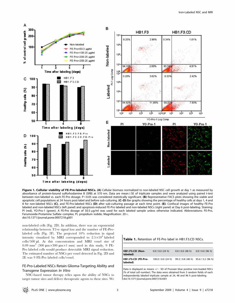
Viability and Proliferation Potential of FE-Pro LabeledNSCs
To determine the cellular viability and proliferation potential of
FE-Pro labeled NSC lines (HB1.F3, HB1.F3.CD), we employed
the sulforhodamine B (SRB) colorimetric assay and YO-Pro-1/PI
to assess cytotoxicity and apoptosis, respectively. Adherent NSCs
in a 96-well culture plate were labeled at various concentrations of
FE-Pro complex (FE at 50, 100, 200 and 250 mg/ml, each of
which was complexed with increasing Pro at 3, 10, and 25 mg/ml).
The highest labeling concentration studied (FE:Pro = 250:25 mg/
ml) was greater than 5x the dosage (FE:Pro = 50:3 mg/ml) for MR
cellular tracking in pre-clinical and future clinical settings. We
performed SRB cytotoxicity assays 1, 3, 5 and 7 days after the
NSCs were labeled with FE-Pro. No significant difference in SRB
staining intensity was observed between labeled and unlabeled
cells (P.0.05) at each time point as measured by optical
absorbance (Fig. 1A). This led us to conclude that labeling NSCs
with FE-Pro did not significantly affect cell viability and
proliferation over 7 days in cell culture.
To determine the plasma membrane integrity of labeled NSCs
(FE:Pro = 50:3 mg/ml), we performed the YO-Pro-1/PI apoptosis
assay on FE-Pro-labeled and non-labeled NSCs on days 1, 4, and
8 post-labeling. Flow cytometry analysis of YO-Pro-1/PI double-
stained FE-Pro-labeled NSCs showed a slight (3–9%) increase in
the apoptotic population compared to the non-labeled control at
day 1 (Fig. 1B). However, long-term monitoring of sub-cultured
labeled and non-labeled NSCs revealed similar percentages of
healthy cells over time (Fig. 1C, D), indicating that labeling with
FE-Pro causes no long term toxicity or effect on cellular integrity.
Confocal images of both labeled and non-labeled NSCs treated
with the apoptosis-inducing agent, staurosporine (STS) showed
100% cell death, whereas non-treated NSC populations reflect
viabilities detected by flow cytometry (Fig. 1E). These results
suggest that FE-Pro labeling has no significant effect on NSC
survival (P.0.05).
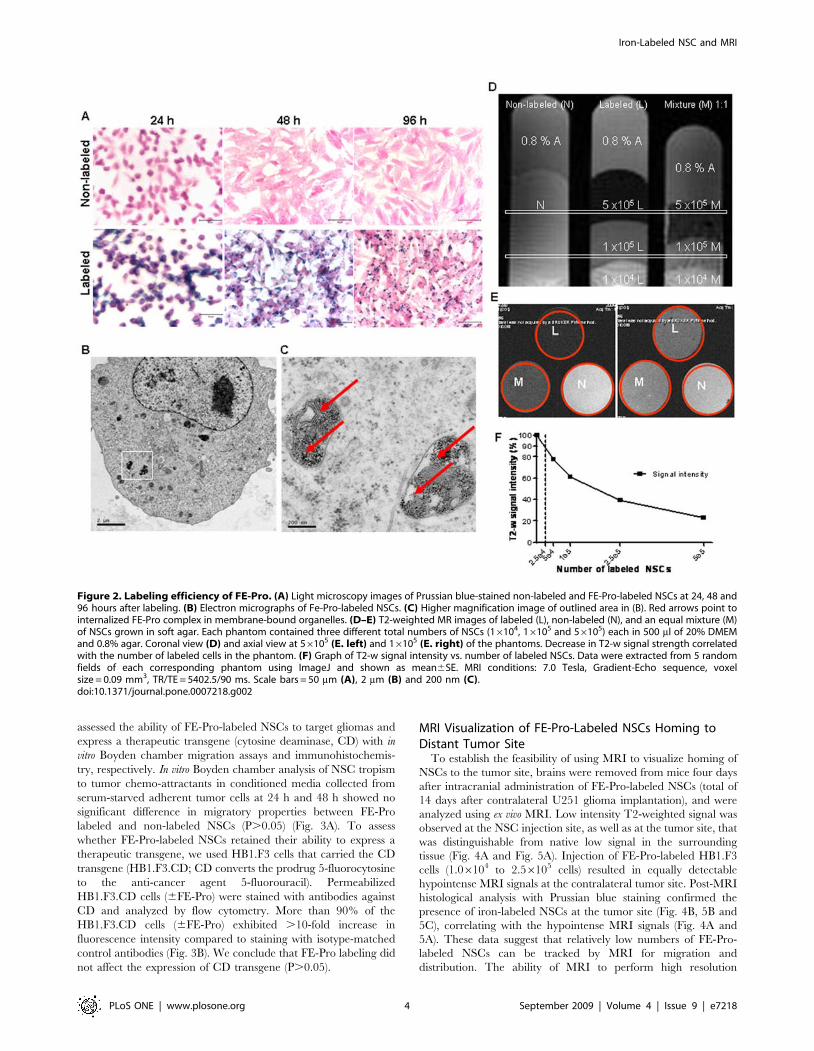
Labeling Efficiency and In Vitro MRI Visualization of FE-Pro-Labeled NSCs
To determine whether the FE-Pro label was retained by NSCs,
internalized and detectable by MRI, we used Prussian blue (PB)
staining, electron microscopy (EM) and in vitro MRI assessment,
respectively. Prussian blue staining of FE-Pro-labeled NSCs
indicated that greater than 95% of FE-Pro-labeled NSCs retained
the iron cores over 96 h in tissue culture, with predicted dilution due
to cell division (Table 1 and Fig. 2A). Unequal distribution of iron
particles during cell division appeared to contribute to the small
population (,5%) of PB-negative NSCs. Bright-field images of PB-
stained NSCs revealed similar cell adherence and proliferation
properties between FE-Pro labeled and non-labeled samples
(Fig. 2A). Transmission EM micrographs showed the FE-Pro
complex as an electron-dense structure in the cytoplasm of labeled
NSCs (Fig. 2B). No residual extracellular FE-Pro complex was
observed in the labeled samples following the final PBS/heparin
wash. The iron-cores of SPIOs were localized to membrane-bound
structures (red arrows; Fig. 2C), indicating that FE-Pro complexes
were encapsulated rather than dispersed throughout the cytoplasm.
We performed T2-weighted (T2-w) multi-spin multi-echo
(MSME) MRI of FE-Pro-labeled NSCs that were suspended in
an equal volume of 0.8% agarose gel and (DMEM+20% FBS)
24 h after labeling. A TE of 90 ms was optimal for observing the
T2-w signal gradient because it correlated with the number of
labeled cells in the phantom. Longer T2 values saturated the
magnetic susceptibility of the SPIOs, thereby wiping out the
signals, whereas shorter T2 values did not allow optimal detection
of differences in hypointense signal intensities. Equal mixtures of
FE-Pro-labeled and non-labeled NSCs (16105 cells/500 ml),
which mimicked one cell division, resulted in a detectable low
intensity signal that could be distinguished from 100% labeled or
Iron-Labeled NSC and MRI
PLoS ONE | www.plosone.org 2 September 2009 | Volume 4 | Issue 9 | e7218
non-labeled cells (Fig. 2D). In addition, there was an exponential
relationship between T2-w signal loss and the number of FE-Pro-
labeled cells (Fig. 2F). The projected 10% reduction in signal
intensity visualized by MRI corresponded to 2.56104 labeled
cells/500 ml. At this concentration and MRI voxel size of
0.09 mm3 (300 mm6300 mm61 mm) used in this study, 9 FE-
Pro labeled cells would produce detectable MRI signal reduction.
The estimated number of NSCs per voxel detected in Fig. 2D and
2E was 9 FE-Pro labeled cells/voxel.
FE-Pro-Labeled NSCs Retain Glioma-Targeting Ability andTransgene Expression In Vitro
NSC-based tumor therapy relies upon the ability of NSCs to
target tumor sites and deliver therapeutic agents to these sites. We
Figure 1. Cellular viability of FE-Pro-labeled NSCs. (A) Cellular biomass normalized to non-labeled NSC cell growth at day 1 as measured byabsorbance of protein-bound sulforhodamine B (SRB) at 570 nm. Data are mean6SE of triplicate samples and were analyzed using paired t-testbetween non-labeled vs. each FE-Pro dosage. P,0.05 was considered statistically significant. (B) Representative FACS plots showing the viable andapoptotic cell populations at 24 hours post-label and before sub-culturing. (C–D) Bar graphs showing the percentage of healthy cells at days 1, 4 and8 for non-labeled NSCs (C), and FE-Pro-labeled NSCs (D) after sub-culturing passage at each time point. (E): Confocal images of healthy FE-Prolabeled and non-labeled NSCs (left panel) and apoptosis-induced FE-Pro labeled and non-labeled NSCs (right panel) at Day 6 post-labeling. Staining:PI (red), YO-Pro-1 (green). A FE-Pro dosage of 50:3 mg/ml was used for each labeled sample unless otherwise indicated. Abbreviations: FE-Pro,Ferumoxide-Protamine Sulfate complex; PI, propidium iodide; Magnification: 206.doi:10.1371/journal.pone.0007218.g001
Table 1. Retention of FE-Pro label in HB1.F3.CD NSCs.
HB1.F3.CD (Non-labeled)
0.060.0 (24 h) 0.060.0 (48 h) 0.060.0 (96 h)
HB1.F3.CD (FE-Pro-labeled)
100.060.0 (24 h) 99.360.8 (48 h) 95.665.5 (96 h)
Data is displayed as means +/2 SD of Prussian blue positive iron-loaded NSCs(% of total cell number). The data were obtained from 5 random fields of eachindependently labeled triplicate sample at 24, 48 and 96 h post-labeling.doi:10.1371/journal.pone.0007218.t001
Iron-Labeled NSC and MRI
PLoS ONE | www.plosone.org 3 September 2009 | Volume 4 | Issue 9 | e7218
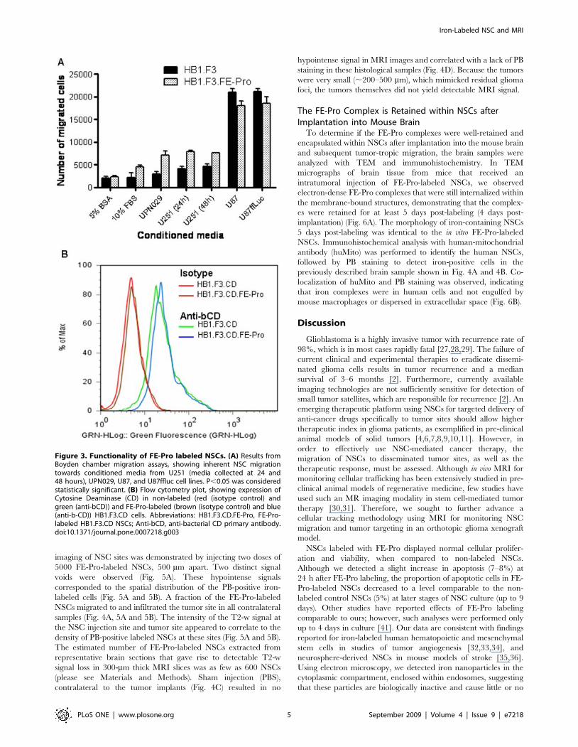
assessed the ability of FE-Pro-labeled NSCs to target gliomas and
express a therapeutic transgene (cytosine deaminase, CD) with in
vitro Boyden chamber migration assays and immunohistochemis-
try, respectively. In vitro Boyden chamber analysis of NSC tropism
to tumor chemo-attractants in conditioned media collected from
serum-starved adherent tumor cells at 24 h and 48 h showed no
significant difference in migratory properties between FE-Pro
labeled and non-labeled NSCs (P.0.05) (Fig. 3A). To assess
whether FE-Pro-labeled NSCs retained their ability to express a
therapeutic transgene, we used HB1.F3 cells that carried the CD
transgene (HB1.F3.CD; CD converts the prodrug 5-fluorocytosine
to the anti-cancer agent 5-fluorouracil). Permeabilized
HB1.F3.CD cells (6FE-Pro) were stained with antibodies against
CD and analyzed by flow cytometry. More than 90% of the
HB1.F3.CD cells (6FE-Pro) exhibited .10-fold increase in
fluorescence intensity compared to staining with isotype-matched
control antibodies (Fig. 3B). We conclude that FE-Pro labeling did
not affect the expression of CD transgene (P.0.05).
MRI Visualization of FE-Pro-Labeled NSCs Homing toDistant Tumor Site
To establish the feasibility of using MRI to visualize homing of
NSCs to the tumor site, brains were removed from mice four days
after intracranial administration of FE-Pro-labeled NSCs (total of
14 days after contralateral U251 glioma implantation), and were
analyzed using ex vivo MRI. Low intensity T2-weighted signal was
observed at the NSC injection site, as well as at the tumor site, that
was distinguishable from native low signal in the surrounding
tissue (Fig. 4A and Fig. 5A). Injection of FE-Pro-labeled HB1.F3
cells (1.06104 to 2.56105 cells) resulted in equally detectable
hypointense MRI signals at the contralateral tumor site. Post-MRI
histological analysis with Prussian blue staining confirmed the
presence of iron-labeled NSCs at the tumor site (Fig. 4B, 5B and
5C), correlating with the hypointense MRI signals (Fig. 4A and
5A). These data suggest that relatively low numbers of FE-Pro-
labeled NSCs can be tracked by MRI for migration and
distribution. The ability of MRI to perform high resolution
Figure 2. Labeling efficiency of FE-Pro. (A) Light microscopy images of Prussian blue-stained non-labeled and FE-Pro-labeled NSCs at 24, 48 and96 hours after labeling. (B) Electron micrographs of Fe-Pro-labeled NSCs. (C) Higher magnification image of outlined area in (B). Red arrows point tointernalized FE-Pro complex in membrane-bound organelles. (D–E) T2-weighted MR images of labeled (L), non-labeled (N), and an equal mixture (M)of NSCs grown in soft agar. Each phantom contained three different total numbers of NSCs (16104, 16105 and 56105) each in 500 ml of 20% DMEMand 0.8% agar. Coronal view (D) and axial view at 56105 (E. left) and 16105 (E. right) of the phantoms. Decrease in T2-w signal strength correlatedwith the number of labeled cells in the phantom. (F) Graph of T2-w signal intensity vs. number of labeled NSCs. Data were extracted from 5 randomfields of each corresponding phantom using ImageJ and shown as mean6SE. MRI conditions: 7.0 Tesla, Gradient-Echo sequence, voxelsize = 0.09 mm3, TR/TE = 5402.5/90 ms. Scale bars = 50 mm (A), 2 mm (B) and 200 nm (C).doi:10.1371/journal.pone.0007218.g002
Iron-Labeled NSC and MRI
PLoS ONE | www.plosone.org 4 September 2009 | Volume 4 | Issue 9 | e7218
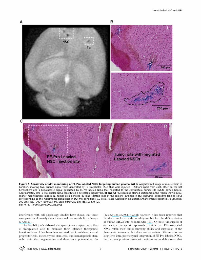
imaging of NSC sites was demonstrated by injecting two doses of
5000 FE-Pro-labeled NSCs, 500 mm apart. Two distinct signal
voids were observed (Fig. 5A). These hypointense signals
corresponded to the spatial distribution of the PB-positive iron-
labeled cells (Fig. 5A and 5B). A fraction of the FE-Pro-labeled
NSCs migrated to and infiltrated the tumor site in all contralateral
samples (Fig. 4A, 5A and 5B). The intensity of the T2-w signal at
the NSC injection site and tumor site appeared to correlate to the
density of PB-positive labeled NSCs at these sites (Fig. 5A and 5B).
The estimated number of FE-Pro-labeled NSCs extracted from
representative brain sections that gave rise to detectable T2-w
signal loss in 300-mm thick MRI slices was as few as 600 NSCs
(please see Materials and Methods). Sham injection (PBS),
contralateral to the tumor implants (Fig. 4C) resulted in no
hypointense signal in MRI images and correlated with a lack of PB
staining in these histological samples (Fig. 4D). Because the tumors
were very small (,200–500 mm), which mimicked residual glioma
foci, the tumors themselves did not yield detectable MRI signal.
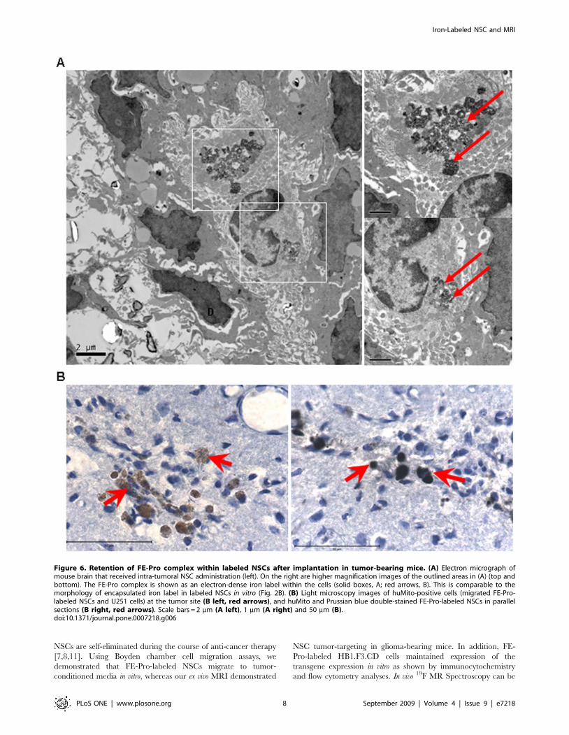
The FE-Pro Complex is Retained within NSCs afterImplantation into Mouse Brain
To determine if the FE-Pro complexes were well-retained and
encapsulated within NSCs after implantation into the mouse brain
and subsequent tumor-tropic migration, the brain samples were
analyzed with TEM and immunohistochemistry. In TEM
micrographs of brain tissue from mice that received an
intratumoral injection of FE-Pro-labeled NSCs, we observed
electron-dense FE-Pro complexes that were still internalized within
the membrane-bound structures, demonstrating that the complex-
es were retained for at least 5 days post-labeling (4 days post-
implantation) (Fig. 6A). The morphology of iron-containing NSCs
5 days post-labeling was identical to the in vitro FE-Pro-labeled
NSCs. Immunohistochemical analysis with human-mitochondrial
antibody (huMito) was performed to identify the human NSCs,
followed by PB staining to detect iron-positive cells in the
previously described brain sample shown in Fig. 4A and 4B. Co-
localization of huMito and PB staining was observed, indicating
that iron complexes were in human cells and not engulfed by
mouse macrophages or dispersed in extracellular space (Fig. 6B).
Discussion
Glioblastoma is a highly invasive tumor with recurrence rate of
98%, which is in most cases rapidly fatal [27,28,29]. The failure of
current clinical and experimental therapies to eradicate dissemi-
nated glioma cells results in tumor recurrence and a median
survival of 3–6 months [2]. Furthermore, currently available
imaging technologies are not sufficiently sensitive for detection of
small tumor satellites, which are responsible for recurrence [2]. An
emerging therapeutic platform using NSCs for targeted delivery of
anti-cancer drugs specifically to tumor sites should allow higher
therapeutic index in glioma patients, as exemplified in pre-clinical
animal models of solid tumors [4,6,7,8,9,10,11]. However, in
order to effectively use NSC-mediated cancer therapy, the
migration of NSCs to disseminated tumor sites, as well as the
therapeutic response, must be assessed. Although in vivo MRI for
monitoring cellular trafficking has been extensively studied in pre-
clinical animal models of regenerative medicine, few studies have
used such an MR imaging modality in stem cell-mediated tumor
therapy [30,31]. Therefore, we sought to further advance a
cellular tracking methodology using MRI for monitoring NSC
migration and tumor targeting in an orthotopic glioma xenograft
model.
NSCs labeled with FE-Pro displayed normal cellular prolifer-
ation and viability, when compared to non-labeled NSCs.
Although we detected a slight increase in apoptosis (7–8%) at
24 h after FE-Pro labeling, the proportion of apoptotic cells in FE-
Pro-labeled NSCs decreased to a level comparable to the non-
labeled control NSCs (5%) at later stages of NSC culture (up to 9
days). Other studies have reported effects of FE-Pro labeling
comparable to ours; however, such analyses were performed only
up to 4 days in culture [41]. Our data are consistent with findings
reported for iron-labeled human hematopoietic and mesenchymal
stem cells in studies of tumor angiogenesis [32,33,34], and
neurosphere-derived NSCs in mouse models of stroke [35,36].
Using electron microscopy, we detected iron nanoparticles in the
cytoplasmic compartment, enclosed within endosomes, suggesting
that these particles are biologically inactive and cause little or no
Figure 3. Functionality of FE-Pro labeled NSCs. (A) Results fromBoyden chamber migration assays, showing inherent NSC migrationtowards conditioned media from U251 (media collected at 24 and48 hours), UPN029, U87, and U87ffluc cell lines. P,0.05 was consideredstatistically significant. (B) Flow cytometry plot, showing expression ofCytosine Deaminase (CD) in non-labeled (red (isotype control) andgreen (anti-bCD)) and FE-Pro-labeled (brown (isotype control) and blue(anti-b-CD)) HB1.F3.CD cells. Abbreviations: HB1.F3.CD.FE-Pro, FE-Pro-labeled HB1.F3.CD NSCs; Anti-bCD, anti-bacterial CD primary antibody.doi:10.1371/journal.pone.0007218.g003
Iron-Labeled NSC and MRI
PLoS ONE | www.plosone.org 5 September 2009 | Volume 4 | Issue 9 | e7218
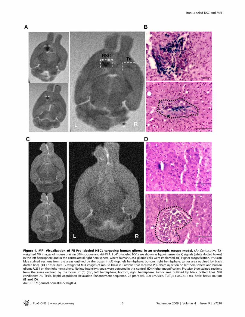
Figure 4. MRI Visualization of FE-Pro-labeled NSCs targeting human glioma in an orthotopic mouse model. (A) Consecutive T2-weighted MR images of mouse brain in 30% sucrose and 4% PFA. FE-Pro-labeled NSCs are shown as hypointense (dark) signals (white dotted boxes)in the left hemisphere and in the contralateral right hemisphere, where human U251 glioma cells were implanted. (B) Higher magnification, Prussianblue stained sections from the areas outlined by the boxes in (A) (top, left hemisphere; bottom, right hemisphere, tumor area outlined by blackdotted line). (C) Consecutive T2-weighted MRI images of mouse brain in Fomblin that received PBS sham injection on left hemisphere and humanglioma U251 on the right hemisphere. No low-intensity signals were detected in this control. (D) Higher magnification, Prussian blue stained sectionsfrom the areas outlined by the boxes in (C) (top, left hemisphere; bottom, right hemisphere, tumor area outlined by black dotted line). MRIconditions: 7.0 Tesla, Rapid Acquisition Relaxation Enhancement sequence, 78 mm/pixel, 300 mm/slice, TR/TE = 1500/23.1 ms. Scale bars = 100 mm(B and D).doi:10.1371/journal.pone.0007218.g004
Iron-Labeled NSC and MRI
PLoS ONE | www.plosone.org 6 September 2009 | Volume 4 | Issue 9 | e7218
interference with cell physiology. Studies have shown that these
nanoparticles ultimately enter the normal iron metabolic pathways
[37,38,39].
The feasibility of cell-based therapies depends upon the ability
of transplanted cells to maintain their intended therapeutic
functions in vivo. It has been demonstrated that iron-labeled neural
progenitor cells, mesenchymal stem cells, and hematopoietic stem
cells retain their regenerative and therapeutic potential in vivo
[32,33,34,35,36,40,41,42,43]; however, it has been reported that
Feridex complexed with poly-L-lysine blocked the differentiation
of human MSCs into chondrocytes [44]. Of note, the success of
our cancer therapeutic approach requires that FE-Pro-labeled
NSCs retain their tumor-targeting ability and expression of the
therapeutic transgene, but does not necessitate differentiation or
long-term intra-parenchymal integration of FE-Pro-labeled NSCs.
Further, our previous results with solid tumor models showed that
Figure 5. Sensitivity of MRI monitoring of FE-Pro-labeled NSCs targeting human glioma. (A) T2-weighted MR image of mouse brain inFomblin, showing two distinct signal voids generated by FE-Pro-labeled NSCs that were injected ,200 mm apart from each other on the lefthemisphere and a hypointense signal generated by FE-Pro-labeled NSCs that migrated to the contralateral tumor site (white dotted boxes).Approximately 600 FE-Pro-labeled NSCs constituted a detectable signal void. (B and C) Prussian blue stained section from the region shown in (A).Higher magnification images (B, tumor area denoted by black dotted line) of the regions outlined in (C), showing PB-positive labeled NSCscorresponding to the hypointense signal sites in (A). MRI conditions: 7.0 Tesla, Rapid Acquisition Relaxation Enhancement sequence, 78 mm/pixel,300 mm/slice, TR/TE = 1500/23.1 ms. Scale bars = 200 mm (B), 500 mm (C).doi:10.1371/journal.pone.0007218.g005
Iron-Labeled NSC and MRI
PLoS ONE | www.plosone.org 7 September 2009 | Volume 4 | Issue 9 | e7218
NSCs are self-eliminated during the course of anti-cancer therapy
[7,8,11]. Using Boyden chamber cell migration assays, we
demonstrated that FE-Pro-labeled NSCs migrate to tumor-
conditioned media in vitro, whereas our ex vivo MRI demonstrated
NSC tumor-targeting in glioma-bearing mice. In addition, FE-
Pro-labeled HB1.F3.CD cells maintained expression of the
transgene expression in vitro as shown by immunocytochemistry
and flow cytometry analyses. In vivo 19F MR Spectroscopy can be
Figure 6. Retention of FE-Pro complex within labeled NSCs after implantation in tumor-bearing mice. (A) Electron micrograph ofmouse brain that received intra-tumoral NSC administration (left). On the right are higher magnification images of the outlined areas in (A) (top andbottom). The FE-Pro complex is shown as an electron-dense iron label within the cells (solid boxes, A; red arrows, B). This is comparable to themorphology of encapsulated iron label in labeled NSCs in vitro (Fig. 2B). (B) Light microscopy images of huMito-positive cells (migrated FE-Pro-labeled NSCs and U251 cells) at the tumor site (B left, red arrows), and huMito and Prussian blue double-stained FE-Pro-labeled NSCs in parallelsections (B right, red arrows). Scale bars = 2 mm (A left), 1 mm (A right) and 50 mm (B).doi:10.1371/journal.pone.0007218.g006
Iron-Labeled NSC and MRI
PLoS ONE | www.plosone.org 8 September 2009 | Volume 4 | Issue 9 | e7218
used to determine expression of CD transgene in patients by
measuring the conversion of 5-fluorocytosine prodrug into 5-
fluorouracil during a therapeutic regimen.
A concern with using SPIO labeling for MRI cellular tracking is
dilution of the contrast agent due to cellular division and the
consequent decrease in the ability to detect the signal by MRI.
Indeed, SPIO-labeled cells show a gradual reduction in intracellular
iron particles with increased post-labeling incubation time. This
reduction has been attributed to cell division and/or exocytosis
[45,46]. Additionally, signal dilution can also occur as a result of
biodegradation and entry of iron into metabolic pathways [45,47].
Our data show that .99% of NSCs were labeled with FE-Pro up to
48 h post-labeling, and that .95% of FE-Pro-labeled NSCs
retained Prussian blue-positive iron nanoparticles at 96 h post-
labeling. The amount of iron appeared to be diluted, as expected,
due to cell division. Furthermore, Neri et al. reported 80% labeling
efficiency of NSCs with Resovist alone or Resovist complexed with
poly-L-lysine in vitro [48], thus, our FE-Pro labeling protocol shows
superior iron labeling efficiency of NSCs.
To establish feasibility of MRI for visualization of FE-Pro labeled
NSCs, it was important to determine the in vitro detection level by
MRI. We were able to detect 9 FE-Pro-labeled NSCs per
300 mm6300 mm61 mm size voxel. Furthermore, a difference in
T2-weighted signal reduction was observed between the 100%
labeled NSCs and non-labeled NSCs, as well as in an equal mixture
of labeled and non-labeled NSCs. This shows that after one cycle of
cell division, we are still able to detect the FE-Pro-labeled NSCs.
Significantly, our previous studies show that NSCs do not divide in
vivo, when implanted into the brain of glioma-bearing mice (Aboody
et al, unpublished results). These findings suggest that FE-Pro-
labeled NSCs will retain the iron nanoparticles to a sufficient degree
to produce hypo-intense signal, contrast-enhanced from surrounding
tissue in T2-weighted MRI. However, due to heterogeneity in the
structure and composition of brain tissue and differences in MRI
sequence and parameters, it is difficult to directly extrapolate the
exact number of labeled cells in vivo based on quantitative assessment
from in vitro studies. We verified in ex vivo MRI that FE-Pro labeled
NSCs injected contra-lateral to frontal lobe tumor in the mouse
brain, migrated along white matter tracts to the tumor site and were
detected 4 days after administration of NSCs as hypointense signal.
Studies of CD34-positive hematopoietic stem cell incorporation into
tumor neovasculature and of neural progenitor cell migration to sites
of spinal cord injury showed that such labeled stem cells were visible
by MRI up to 1–2 weeks after their administration [33,49,50,51,52].
In addition, transplanted SPIO-labeled pancreatic islets in patients
have been tracked by MRI for up to 6 months post-transplantation
[39]. We intend to further verify the successful use of FE-Pro labeling
of NSCs for in vivo MRI cellular tracking in pre-clinical animal
models for the planned clinical trial in glioma patients.
In addition to internalization and retention of the FE-Pro label
by NSCs, it is important to establish the sensitivity of MRI FE-Pro-
labeled NSCs at a particular resolution with MRI sequence field
strength. It has been reported that even a single cell can be tracked
in MRI in case of highly phagocytic, large cells, such as
macrophages [53]. Because iron nanoparticles produce hypoin-
tense signals by causing magnetic susceptibility in surrounding
water molecules, the labeled cells can cause a ‘blooming effect’ in
an area of interest due to iron overload [31]. In our studies, similar
numbers of NSCs migrated to tumor sites within 4 days after
administration, independent of the dosage of cells implanted. A
total of 10,000 NSCs administered to the left frontal lobe at two
sites 500 mm apart appeared in MRI as distinct hypointense
signals. A fraction of NSCs that migrated to the tumor implant in
the opposite hemisphere were visible at 600 FE-Pro-labeled NSCs
per site with an MRI resolution of 76 mm676 mm6300 mm.
Qualitative analysis of the spatio-temporal targeting of glioma
by NSCs in MRI requires further confirmation that the detected
MRI signal arose from FE-Pro-labeled NSCs. A concern is the
possible false positive interpretation of the MRI signal, which may
be produced by macrophages that have engulfed non-viable
labeled NSCs or freely-dispersed iron nanoparticles in the brain
tissue [54]. Although 10–20% of macrophages, when mixed with
stem cells, took up iron in in vitro Boyden chamber assays [54],
Arbab et al. found no co-localization of mouse macrophages and
iron-containing (Prussian blue-positive) areas of brain tissue
[33,34]. Here, our TEM data confirmed that the iron label was
encapsulated within endosomes in vivo after migration and/or
distribution at the tumor sites. Histological analysis revealed co-
localization of anti-human mitochondrial antibody and Prussian
blue staining, suggesting that the iron-containing cells are of
human origin. Although tumor cells may take up iron nanopar-
ticles released from non-viable NSCs, the enclosure of iron in
endosomes, as evidenced in our TEM data, suggest this process is
unlikely. Further support for tumor-specific targeting comes from
our data showing no identifiable presence of iron-labeled NSCs in
non-tumor-containing healthy brain tissues. Likewise earlier
reports have shown that NSCs, inoculated into non-tumor-bearing
brains of laboratory animals, did not randomly dissipate into
adjacent normal tissue or to the contra-lateral hemisphere [55].
The study of NSC trafficking in glioma therapy will help us
define the optimal time-frame for NSC delivery, the dose of anti-
cancer therapeutic, dosing regimen, and route of administration.
Our MRI cell tracking methodology will also aid the interpretation
of data obtained in clinical trials. In combination with standard
MRI techniques (e.g., gadolinium-enhanced MRI, diffusion-
weighted MRI, or MR Spectroscopy), our method will allow us
to better assess disease activity as related to the presence of
therapeutic NSCs.
Materials and Methods
Human Neural Stem CellsThe HB1.F3 human neural stem cell line was established by
retroviral transduction of v-myc into primary human neural stem
cells isolated from fetal telencephalon of 15 weeks gestation [56].
This parental HB1.F3 cell line was further transduced retrovirally
to stably express the E. coli cytosine deaminase gene (CD; EC
3.5.4.1), designated as HB1.F3.CD NSCs. Both the HB1.F3 and
HB1.F3.CD lines are well-characterized and multipotent, non-
tumorigenic and non-immunogenic [10,56,57,58]. NSCs were
cultured as an adherent monolayer in Dulbecco’s Modified Eagle’s
Medium (DMEM) (Invitrogen, Carlsbad, CA) containing high
glucose (4.5 g/l), 1 mM sodium pyruvate, 2 mM L-glutamine,
100 mg/ml streptomycin and 100 units/ml penicillin, supplement-
ed with 10% fetal bovine serum (FBS) in 6% CO2 at 37uC. (Note:
the HB1.F3.CD cell line has been approved for clinical use by the
NIH Recombinant DNA Advisory Committee).
FE-Pro Complex and Labeling of NSCsNSCs were labeled with FE-Pro as described by Arbab et al. [59]
with some modifications. Briefly, Ferumoxide (FE) (Feridex IV,
Berlex Laboratories, Wayne, NJ) with a total iron content of
11.2 mg/ml was diluted to the desired concentration (100–
500 mg/ml) in serum-free DMEM. Protamine Sulfate (Pro)
solution (1 mg/ml) was freshly prepared in distilled water from a
10 mg/ml stock solution (AM Pharm Partner, Schaumburg, IL),
and added to the serum-free DMEM-FE solution to achieve 2X
Iron-Labeled NSC and MRI
PLoS ONE | www.plosone.org 9 September 2009 | Volume 4 | Issue 9 | e7218
the desired final FE-Pro concentration. The solution was agitated
for 1 min to allow the FE-Pro complex to form and was
immediately added to adherent NSCs (80% confluence). Following
incubation for 2 h at 37uC, 6% CO2, an equal volume of
DMEM/10% FBS was then added to the NSCs. After 24 h of
further incubation, the NSCs were washed three times in sterile
PBS, and one time in PBS containing 10 U/ml heparin (Abraxis,
Schaumburg, IL) to remove the residual FE-Pro from the cell
surface, followed by a rinse in PBS and re-suspension in DMEM/
10% FBS for in vitro experiments or sterile PBS for intracranial
injections into glioma-bearing mice.
Analysis of Cellular ViabilityThe sulforhodamine B (SRB) cytotoxicity assay was used to assess
the viability of FE-Pro-labeled NSCs [60]. Briefly, the samples
(plated in a 96-well culture plate) were fixed with ice-cold 10%
trichloroacetic acid at 4uC for 1 h, stained with 100 ml of 0.4% SRB
in 1% acetic acid per well for 15 min at room temperature (RT),
rinsed 3 to 4 times with 1% acetic acid, and solubilized in 100 ml of
10 mM Tris-base. The intensity of SRB staining was evaluated
using a Spectra Max 250 Microplate Spectrophotometer at
570 nm. For early and late apoptosis analysis, NSCs were labeled,
trypsinized and re-suspended at 16106 cells/ml and stained with
1 mM YO-Pro-1 (Invitrogen) for 20 min and 1.5 mM propidium
iodide (PI) (Invitrogen) for 5 min. NSCs were re-plated for further
study time points and the same procedures repeated. The dye
uptake was analyzed using flow cytometry. The flow cytometry data
were confirmed with confocal imaging using a Zeiss LSM 510
confocal microscope (Carl Zeiss Microimaging Inc., Thornwood,
NY). As a positive control, 1 h staurosporine (100 nM) treatment
was used as an apoptosis-inducing agent.
Labeling Efficiency StudiesLabeling efficiency studies were performed with iron-sensitive
Prussian blue (PB) staining, electron microscopy and magnetic
resonance imaging. NSCs were cultured in 6-well culture plates to
80% confluence, labeled with FE-Pro for 24 h, and stained with
PB at 24, 48 and 96 h post-FE-Pro-labeling. Briefly, NSCs were
fixed with 4% paraformaldehyde (PFA) for 5–10 min, washed with
distilled H2O (dH2O) and incubated in freshly prepared equal
volumes of 4% w/v K4Fe(CN)6 and 1.2 mM HCl (Sigma-Aldrich,
St. Louis, MO) for 30 min at RT, followed by a rinse with dH2O
and counterstained with nuclear fast red (Sigma-Aldrich, St. Louis,
MO). Bright-field micrographs of PB-stained, FE-Pro labeled and
unlabeled samples were taken and the percentages of PB-positive
versus -negative NSCs were calculated from 5 random fields. For
MRI analysis of labeling efficiency, see MRI section below.
Expression of the Cytosine Deaminase TransgeneAdherent NSCs were labeled with FE-Pro, as described above,
and re-suspended at 56106 cells/ml in staining/wash buffer
(SWB) (94% PBS [without Ca2+ and Mg2+], 5% FBS and
0.001% w/v NaN3. NSCs were fixed and permeabilized (Fix and
Perm kit, Caltag), rinsed and immunostained with anti-bCD
primary antibody (0.01 mg/ml; BD Pharmingen, San Diego, CA)
or mouse IgG1 kappa isotype control antibody (0.01 mg/ml; BD
Pharmingen) for 20 min at RT. The samples were then washed
twice with SWB, stained with 10 mg/ml of goat anti-mouse-IgG/
IgM-FITC (BD Pharmingen) and incubated for 20 min at RT in
the dark. After two final rinses with SWB, the cell pellet was re-
suspended to 2.56104 cells/ml in SWB. The number of CD-
positive cells was analyzed by flow cytometry (Guava Technolo-
gies, Hayward, CA).
Cell Migration AssayIn vitro cell migration assays were performed using 96-well cell
culture plates with polycarbonate inserts (Millipore, Billerica, MA)
with 8-mm pore diameter. Tumor cell-conditioned media were
prepared by addition of serum-free media (SFM) to adherent tumor
cells (,75% confluence), followed by incubation at 37uC, 6% CO2
for 24 or 48 hours. 5% BSA/DMEM, 10% FBS/DMEM and
conditioned media from tumor cell lines were added to the lower
chamber of 96-well plates (150 ml/well, triplicate samples). Inserts
were placed into wells and suspensions of labeled and non-labeled
NSCs were added in the upper chamber (36104 cells/100 ml
suspended in 5% BSA/DMEM to each well). After incubation for
4 hours at 37uC, the cells that did not migrate were removed from
the inner surface of the filter. The membrane tray was then placed
in a new lower chamber containing pre-warmed detachment buffer
(Accutase, Sigma-Aldrich, St. Louis, MO) at 37uC for 10 min.
Detached cells in the buffer were then transferred to a V-bottom 96
well plate and centrifuged at 1500 rpm for 5 min, buffer aspirated
and froze at 220uC overnight. The data were analyzed using the
CyQuant Green according to the manufacturer’s recommended
protocol. Standard curve was generated using known number of
labeled and non-labeled cells and their respective fluorescence
intensities (triplicate samples).
Human Glioma XenograftsNude/nude mice (Charles River) were anesthetized with an
intra-peritoneal injection of 132 mg/kg Ketamine and 8.8 mg/kg
Xylazine. Animals were then immobilized in a stereotactic
apparatus and received stereotactically-guided intracranial injec-
tions of U251 human glioma cells (56104 cells/2 ml sterile PBS)
into the right frontal lobe (2 mm lateral, 0.5 mm anterior to
bregma, tracked from a depth of 2.5 mm to 2.25 mm to 2.0 mm;
0.667 ml of cell suspension was injected at each level for optimal
tumor/host brain contact and engraftment). Tumor cell injections
were performed with a 30-gauge 5-ml Hamilton syringe over 3–5
minutes. After retracting the needle over 2–4 minutes, bone-wax
was used to occlude the burr hole, Betadine was applied to the
surgical area, and skin was closed with skin glue or sutures.
Buprenorphine analgesic was administered intra-peritoneally at
0.05 mg/kg to relieve post-operative pain. Tumor implants grew
to the size of ,0.2–0.5 mm by Day 10, mimicking small, residual
tumor foci. All animal protocols were approved by the City of
Hope and/or Children’s Hospital, Los Angeles IACUC. When
mice appeared to be in discomfort or distress as judged by
independent animal care personnel with no knowledge of the
protocol design, animals were euthanized.
Administration of FE-Pro Labeled NSCsFE-Pro labeled HB1.F3 and HB1.F3.CD NSCs were detached
by trypsinization and centrifuged for 5 min at 1200 rpm. The
supernatant was discarded and NSCs were resuspended in sterile
PBS (16104, 16105, or 2.56105 cells/2 ml). On day 10 post-tumor
injection, the NSCs were injected intratumorally and contralaterally
at similar coordinates of injection site of tumor cells. Prior to NSC
injections, the mice were anesthetized, immobilized in a stereotactic
frame. FE-Pro labeled NSCs, non-labeled NSCs, or PBS alone was
then administered as described above.
In Vitro and Ex Vivo MRIFor in vitro MRI analysis, equal volumes of different concentra-
tions (16104 cells/500 ml, 16105 cells/500 ml, 56105 cells/
500 ml) of non-labeled, FE-Pro-labeled and an equal mixture of
labeled and non-labeled NSCs were suspended in 0.8% w/v
Iron-Labeled NSC and MRI
PLoS ONE | www.plosone.org 10 September 2009 | Volume 4 | Issue 9 | e7218
agarose gel and visualized using a 7.0 Tesla Bruker Pharmascan
with Paravision 4.0 software at the Children’s Hospital of Los
Angeles Small Animal Imaging Core. Images were reconstructed
using Bruker Paravision 4.0. T2-weighted MRI was performed
with a multi-spin multi-echo (MSME) sequence of TR = 5300 ms
and 10 echos (TE = 15–150 ms). The matrix size was 2566256
with field of view (FOV) of 363 cm. Average (binning) was 1 with
slice thickness of 1 mm. The T2-weighted signal strength as
correlated to the number of labeled cells was then obtained from 5
random fields of each corresponding phantom using NIH Image J
and displayed as percent signal loss versus number of labeled cells.
For ex vivo MRI analysis, mice were euthanized at day 14 (4 days
post-NSC injection) and perfused with PBS, followed by 4% PFA.
Brains were harvested and post-fixed in 4% PFA supplemented with
30% (w/v) sucrose. MR imaging of ex vivo brain samples was
conducted using a 19 mm horizontal magnetic bore in a 7.0 Tesla
Bruker Pharmascan with Paravision 4.0. MR images were
reconstructed using the standard Paravision software, and exported
in DICOM format to E-film 1.8.2 for visualization and conversion
to 2-D tiff files. During imaging, harvested brains were immersed in
Fomblin, an MRI negative agent, to reduce the background noise.
T2-weighted rapid acquisition with relaxation enhancement was
used with average ranging for 4–24 for image quality, slice thickness
of 300 mm, FOV of 262 cm, TR/TE = 1500–2000/23.1–33.3 ms.
In-plane resolution was 76 mm/pixel. For estimation of the number
of FePro-labeled cells detectable by ex vivo MRI in mouse brain, we
collected thirty 10-mm thick sections from the brain, and randomly
selected 6 sections that spanned 300 mm of distance corresponding
to MRI image in Fig. 5A. Prussian blue-positive NSCs were counted
in these 6 sections, and the total number of NSCs within the 300-
micrometer thick brain tissue was extrapolated. Immunohistochem-
ical analysis with human-mitochondrial antibody (huMito) (Chemi-
con, MAB 1273) was performed to identify the human NSCs as
previously described [7], and double-stained with PB to visualize
iron-positive cells.
Electron MicroscopyInternalization of the FE-Pro complex was visualized with
electron microscopy using standard techniques. FE-Pro-labeled
NSC pellets (fixed in 1.5% glutaraldehyde in 0.1 M cacodylate
buffer) or harvested brains (post-fixed in 4% PFA supplemented
with 30% sucrose) were processed with standard electron
microscopy techniques and imaged with an FEI Tecnai G2
transmission electron microscope (TEM).
Statistical AnalysisThe following statistical analyses were used: two-tailed Student’s
t-test and ANOVA. See figure legends for specific details.
GraphPad Prism and/or Excel were used to determine the
statistical significance. The data are shown as mean6SD or
mean6SEM. P,0.05 was considered statistically significant.
Acknowledgments
We thank the Electron Microscopy core at City of Hope and Small Animal
Imaging Core at Children’s Hospital, Los Angeles (CHLA) for their
technical assistance. We would like to express thanks to Gevorg
Karapetyan, MD (CHLA) and Babak Kateb, BS, for their invaluable
advice. We are grateful to Dr. Keely L. Walker for critical evaluation and
editing of the manuscript.
Author Contributions
Conceived and designed the experiments: MST RAM KSA. Performed the
experiments: MST SEK IH NS MG MM EG. Analyzed the data: MST JN
SEK IH MG MM RTF RAM KSA. Contributed reagents/materials/
analysis tools: MST SUK KSA. Wrote the paper: MST JN SEK RAM
KSA. Designed the experiments: SEK.
References
1. Castro MG, Cowen R, Williamson IK, David A, Jimenez-Dalmaroni MJ, et al.(2003) Current and future strategies for the treatment of malignant brain tumors.
Pharmacol Ther 98: 71–108.
2. Ehtesham M, Stevenson CB, Thompson RC (2005) Stem cell therapies for
malignant glioma. Neurosurg Focus 19: E5.
3. Benedetti S, Pirola B, Pollo B, Magrassi L, Bruzzone MG, et al. (2000) Gene
therapy of experimental brain tumors using neural progenitor cells. Nat Med 6:447–450.
4. Aboody KS, Brown A, Rainov NG, Bower KA, Liu S, et al. (2000) Neural stemcells display extensive tropism for pathology in adult brain: evidence from
intracranial gliomas. Proc Natl Acad Sci U S A 97: 12846–12851.
5. Noble M (2000) Can neural stem cells be used to track down and destroymigratory brain tumor cells while also providing a means of repairing tumor-
associated damage? Proc Natl Acad Sci U S A 97: 12393–12395.
6. Brown AB, Yang W, Schmidt NO, Carroll R, Leishear KK, et al. (2003)
Intravascular delivery of neural stem cell lines to target intracranial andextracranial tumors of neural and non-neural origin. Hum Gene Ther 14:
1777–1785.
7. Aboody KS, Bush RA, Garcia E, Metz MZ, Najbauer J, et al. (2006)
Development of a tumor-selective approach to treat metastatic cancer. PLoS
ONE 1: e23.
8. Danks MK, Yoon KJ, Bush RA, Remack JS, Wierdl M, et al. (2007) Tumor-
targeted enzyme/prodrug therapy mediates long-term disease-free survival ofmice bearing disseminated neuroblastoma. Cancer Res 67: 22–25.
9. Dickson PV, Hamner JB, Burger RA, Garcia E, Ouma AA, et al. (2007)Intravascular administration of tumor tropic neural progenitor cells permits
targeted delivery of interferon-beta and restricts tumor growth in a murine
model of disseminated neuroblastoma. J Pediatr Surg 42: 48–53.
10. Kim SK, Kim SU, Park IH, Bang JH, Aboody KS, et al. (2006) Human neural
stem cells target experimental intracranial medulloblastoma and deliver atherapeutic gene leading to tumor regression. Clin Cancer Res 12: 5550–5556.
11. Aboody KS, Najbauer J, Schmidt NO, Yang W, Wu JK, et al. (2006) Targetingof melanoma brain metastases using engineered neural stem/progenitor cells.
Neuro Oncol 8: 119–126.
12. Schmidt NO, Przylecki W, Yang W, Ziu M, Teng Y, et al. (2005) Brain tumor
tropism of transplanted human neural stem cells is induced by vascularendothelial growth factor. Neoplasia 7: 623–629.
13. Ehtesham M, Yuan X, Kabos P, Chung NH, Liu G, et al. (2004) Glioma tropic
neural stem cells consist of astrocytic precursors and their migratory capacity is
mediated by CXCR4. Neoplasia 6: 287–293.
14. Sun L, Lee J, Fine HA (2004) Neuronally expressed stem cell factor induces
neural stem cell migration to areas of brain injury. J Clin Invest 113: 1364–1374.
15. Heese O, Disko A, Zirkel D, Westphal M, Lamszus K (2005) Neural stem cell
migration toward gliomas in vitro. Neuro Oncol 7: 476–484.
16. Lesniak MS (2006) Targeted therapy for malignant glioma: neural stem cells.
Expert Rev Neurother 6: 1–3.
17. Kendall SE, Najbauer J, Johnston HF, Metz MZ, Li S, et al. (2008) Neural stem
cell targeting of glioma is dependent on phosphoinositide 3-kinase signaling.
Stem Cells 26: 1575–1586.
18. Gutova M, Najbauer J, Frank RT, Kendall SE, Gevorgyan A, et al. (2008)
Urokinase plasminogen activator and urokinase plasminogen activator receptor
mediate human stem cell tropism to malignant solid tumors. Stem Cells 26:
1406–1413.
19. Dwain I, Xiangpeng Y, Zeng Z, Patricia T, Joh SY (2006) Neural stem cells—a
promising potential therapy for brain tumors. Curr Stem Cell Res Ther 1:
79–84.
20. Yip S, Aboody KS, Burns M, Imitola J, Boockvar JA, et al. (2003) Neural stem
cell biology may be well suited for improving brain tumor therapies. Cancer J 9:
189–204.
21. Lin D, Najbauer J, Salvaterra PM, Mamelak AN, Barish ME, et al. (2007) Novel
method for visualizing and modeling the spatial distribution of neural stem cells
within intracranial glioma. Neuroimage 37 Suppl 1: S18–26.
22. Shah K, Bureau E, Kim DE, Yang K, Tang Y, et al. (2005) Glioma therapy and
real-time imaging of neural precursor cell migration and tumor regression. Ann
Neurol 57: 34–41.
23. Modo M (2006) Understanding stem cell-mediated brain repair through
neuroimaging. Curr Stem Cell Res Ther 1: 55–63.
24. Modo M, Cash D, Mellodew K, Williams SC, Fraser SE, et al. (2002) Tracking
transplanted stem cell migration using bifunctional, contrast agent-enhanced,
magnetic resonance imaging. Neuroimage 17: 803–811.
25. Modo M, Mellodew K, Cash D, Fraser SE, Meade TJ, et al. (2004) Mapping
transplanted stem cell migration after a stroke: a serial, in vivo magnetic
resonance imaging study. Neuroimage 21: 311–317.
Iron-Labeled NSC and MRI
PLoS ONE | www.plosone.org 11 September 2009 | Volume 4 | Issue 9 | e7218
26. Corot C, Robert P, Idee JM, Port M (2006) Recent advances in iron oxide
nanocrystal technology for medical imaging. Adv Drug Deliv Rev 58:1471–1504.
27. Price SJ, Jena R, Burnet NG, Carpenter TA, Pickard JD, et al. (2007) Predicting
patterns of glioma recurrence using diffusion tensor imaging. Eur Radiol 17:1675–1684.
28. Stupp R, Weber DC (2005) The role of radio- and chemotherapy inglioblastoma. Onkologie 28: 315–317.
29. Hochberg FH, Pruitt A (1980) Assumptions in the radiotherapy of glioblastoma.
Neurology 30: 907–911.30. Zhang Z, Jiang Q, Jiang F, Ding G, Zhang R, et al. (2004) In vivo magnetic
resonance imaging tracks adult neural progenitor cell targeting of brain tumor.Neuroimage 23: 281–287.
31. Brekke C, Williams SC, Price J, Thorsen F, Modo M (2007) Cellularmultiparametric MRI of neural stem cell therapy in a rat glioma model.
Neuroimage 37: 769–782.
32. Arbab AS, Jordan EK, Wilson LB, Yocum GT, Lewis BK, et al. (2004) In vivotrafficking and targeted delivery of magnetically labeled stem cells. Hum Gene
Ther 15: 351–360.33. Arbab AS, Pandit SD, Anderson SA, Yocum GT, Bur M, et al. (2006) Magnetic
resonance imaging and confocal microscopy studies of magnetically labeled
endothelial progenitor cells trafficking to sites of tumor angiogenesis. Stem Cells24: 671–678.
34. Anderson SA, Glod J, Arbab AS, Noel M, Ashari P, et al. (2005) NoninvasiveMR imaging of magnetically labeled stem cells to directly identify neovascu-
lature in a glioma model. Blood 105: 420–425.35. Guzman R, Bliss T, De Los Angeles A, Moseley M, Palmer T, et al. (2008)
Neural progenitor cells transplanted into the uninjured brain undergo targeted
migration after stroke onset. J Neurosci Res 86: 873–882.36. Guzman R, Uchida N, Bliss TM, He D, Christopherson KK, et al. (2007) Long-
term monitoring of transplanted human neural stem cells in developmental andpathological contexts with MRI. Proc Natl Acad Sci U S A 104: 10211–10216.
37. Arbab AS, Wilson LB, Ashari P, Jordan EK, Lewis BK, et al. (2005) A model of
lysosomal metabolism of dextran coated superparamagnetic iron oxide (SPIO)nanoparticles: implications for cellular magnetic resonance imaging. NMR
Biomed 18: 383–389.38. Pawelczyk E, Arbab AS, Pandit S, Hu E, Frank JA (2006) Expression of
transferrin receptor and ferritin following ferumoxides-protamine sulfate labelingof cells: implications for cellular magnetic resonance imaging. NMR Biomed 19:
581–592.
39. Toso C, Vallee JP, Morel P, Ris F, Demuylder-Mischler S, et al. (2008) Clinicalmagnetic resonance imaging of pancreatic islet grafts after iron nanoparticle
labeling. Am J Transplant 8: 701–706.40. Lepore AC, Walczak P, Rao MS, Fischer I, Bulte JW (2006) MR imaging of
lineage-restricted neural precursors following transplantation into the adult
spinal cord. Exp Neurol 201: 49–59.41. Kim DE, Schellingerhout D, Ishii K, Shah K, Weissleder R (2004) Imaging of
stem cell recruitment to ischemic infarcts in a murine model. Stroke 35:952–957.
42. Chu K, Kim M, Jeong SW, Kim SU, Yoon BW (2003) Human neural stem cellscan migrate, differentiate, and integrate after intravenous transplantation in
adult rats with transient forebrain ischemia. Neurosci Lett 343: 129–133.
43. Arbab AS, Yocum GT, Rad AM, Khakoo AY, Fellowes V, et al. (2005) Labelingof cells with ferumoxides-protamine sulfate complexes does not inhibit function
or differentiation capacity of hematopoietic or mesenchymal stem cells. NMR
Biomed 18: 553–559.
44. Kostura L, Kraitchman DL, Mackay AM, Pittenger MF, Bulte JW (2004)
Feridex labeling of mesenchymal stem cells inhibits chondrogenesis but not
adipogenesis or osteogenesis. NMR Biomed 17: 513–517.
45. Arbab AS, Bashaw LA, Miller BR, Jordan EK, Lewis BK, et al. (2003)
Characterization of biophysical and metabolic properties of cells labeled with
superparamagnetic iron oxide nanoparticles and transfection agent for cellular
MR imaging. Radiology 229: 838–846.
46. Daldrup-Link HE, Rudelius M, Oostendorp RA, Settles M, Piontek G, et al.
(2003) Targeting of hematopoietic progenitor cells with MR contrast agents.
Radiology 228: 760–767.
47. Pawelczyk E, Arbab AS, Pandit S, Hu E, Frank JA (2006) Expression of
transferrin receptor and ferritin following ferumoxides-protamine sulfate labeling
of cells: implications for cellular magnetic resonance imaging. NMR in
Biomedicine 19: 581–592.
48. Neri M, Maderna C, Cavazzin C, Deidda-Vigoriti V, Politi LS, et al. (2008)
Efficient in vitro labeling of human neural precursor cells with super-
paramagnetic iron oxide particles: relevance for in vivo cell tracking. Stem
Cells 26: 505–516.
49. Frank JA, Miller BR, Arbab AS, Zywicke HA, Jordan EK, et al. (2003) Clinically
applicable labeling of mammalian and stem cells by combining superparamag-
netic iron oxides and transfection agents. Radiology 228: 480–487.
50. Jendelova P, Herynek V, DeCroos J, Glogarova K, Andersson B, et al. (2003)
Imaging the fate of implanted bone marrow stromal cells labeled with
superparamagnetic nanoparticles. Magn Reson Med 50: 767–776.
51. Sykova E, Jendelova P (2006) Magnetic resonance tracking of transplanted stem
cells in rat brain and spinal cord. Neurodegener Dis 3: 62–67.
52. Sykova E, Jendelova P (2007) Migration, fate and in vivo imaging of adult stem
cells in the CNS. Cell Death Differ.
53. Heyn C, Ronald JA, Mackenzie LT, MacDonald IC, Chambers AF, et al. (2006)
In vivo magnetic resonance imaging of single cells in mouse brain with optical
validation. Magn Reson Med 55: 23–29.
54. Pawelczyk E, Arbab AS, Chaudhry A, Balakumaran A, Robey PG, et al. (2008)
In vitro model of bromodeoxyuridine or iron oxide nanoparticle uptake by
activated macrophages from labeled stem cells: implications for cellular therapy.
Stem Cells 26: 1366–1375.
55. Ehtesham M, Kabos P, Kabosova A, Neuman T, Black KL, et al. (2002) The use
of interleukin 12-secreting neural stem cells for the treatment of intracranial
glioma. Cancer Res 62: 5657–5663.
56. Kim SU, Nakagawa E, Hatori K, Nagai A, Lee MA, et al. (2002) Production of
immortalized human neural crest stem cells. Methods Mol Biol 198: 55–65.
57. Flax JD, Aurora S, Yang C, Simonin C, Wills AM, et al. (1998) Engraftable
human neural stem cells respond to developmental cues, replace neurons, and
express foreign genes. Nat Biotechnol 16: 1033–1039.
58. Kim DE, Tsuji K, Kim YR, Mueller FJ, Eom HS, et al. (2006) Neural stem cell
transplant survival in brains of mice: assessing the effect of immunity and
ischemia by using real-time bioluminescent imaging. Radiology 241: 822–830.
59. Arbab AS, Yocum GT, Kalish H, Jordan EK, Anderson SA, et al. (2004)
Efficient magnetic cell labeling with protamine sulfate complexed to ferumoxides
for cellular MRI. Blood 104: 1217–1223.
60. Vichai V, Kirtikara K (2006) Sulforhodamine B colorimetric assay for
cytotoxicity screening. Nat Protoc 1: 1112–1116.
Iron-Labeled NSC and MRI
PLoS ONE | www.plosone.org 12 September 2009 | Volume 4 | Issue 9 | e7218