Workspace and singularity determination of a 7-DoF wrist ...
Upload
independentCategory
view
0download
0
Network structure and dynamics of themental workspaceAlexander Schlegel1, Peter J. Kohler, Sergey V. Fogelson, Prescott Alexander, Dedeepya Konuthula, and Peter Ulric Tse
Department of Psychological and Brain Sciences, Dartmouth College, Hanover, NH 03755
Edited by Michael S. Gazzaniga, University of California, Santa Barbara, CA, and approved August 28, 2013 (received for review June 11, 2013)
The conscious manipulation of mental representations is central tomany creative and uniquely human abilities. How does the humanbrain mediate such flexible mental operations? Here, multivariatepattern analysis of functional MRI data reveals a widespreadneural network that performs specific mental manipulations onthe contents of visual imagery. Evolving patterns of neural activitywithin this mental workspace track the sequence of informationaltransformations carried out by these manipulations. The networkswitches between distinct connectivity profiles as representationsare maintained or manipulated.
occipital | frontoparietal | posterior parietal | precuneus |dorsolateral prefrontal
Albert Einstein described the elements of his scientificthought as “certain signs and more or less clear images
which can be ‘voluntarily’ reproduced or combined” (1). Creativethought in science as well as in other domains such as the visualarts, mathematics, music, and dance requires the capacity tomanipulate mental representations flexibly. Cognitive scientistsrefer to this capacity as a “mental workspace” and suggest that itis a key function of consciousness (2) involving the distribution ofinformation among widespread, specialized subdomains (3).How does the human brain mediate these flexible mental
operations? Behavioral studies of the mental workspace, such asShepard and Metzler’s work on mental rotation (4), have foundthat many mental operations closely resemble their corre-sponding physical operations. This finding supports the view thatthe mental workspace can simulate the physical world. Recentwork in neuroscience has focused on mental representationsinstead of operations, showing that the contents of visual per-ception (5), visual imagery (6), and even dreams (7) can bedecoded from activity in visual cortex. These results suggest thatthe same regions that mediate representations in sensory per-ception also are involved in mental imagery. However, how themind can manipulate these representations remains unknown.Many studies have found increased activity in frontal and pari-etal regions associated with a range of high-level cognitive abil-ities (8, 9) including mental rotation (10), analogical reasoning(11), working memory (12), and fluid intelligence (13). Together,these findings suggest that a frontoparietal network may form thecore of the mental workspace. We therefore hypothesized thatoperations on visual representations in the mental workspace arerealized through the coordinated activity of a distributed net-work of regions that spans at least the frontal, parietal, and oc-cipital cortices. A strong test of this hypothesis would be to askwhether patterns of neural activity in these regions contain in-formation about specific mental operations and whether thesepatterns evolve over time as mental representations aremanipulated.In the present study, we tested this hypothesis by asking 15
participants to engage in either maintenance or manipulation ofvisual imagery while we collected functional MRI (fMRI)measurements of their neural activity. As stimuli, we developed100 abstract parts that could be combined into 2 × 2 figures (Fig.1 A and C). In a series of trials, participants mentally maintaineda set of parts or a whole figure, mentally constructed a set of four
parts into a figure, or mentally deconstructed a figure into itsfour parts (Fig. 1B). Stimuli were presented briefly at the be-ginning of each trial, followed by a task prompt and a 6-s delayduring which the participant performed the indicated mentaloperation. At the end of the delay, the target output of the op-eration was presented along with three similar distractors, andthe participant indicated the correct target (Fig. 1D). Adjustingthe complexity of the stimuli allowed us to equate for task dif-ficulty by maintaining an accuracy of two out of three correctresponses for each participant in each of the four conditions(chance would be 1 out of 4 correct; Fig. 1E).
ResultsAs an initial procedure for selecting regions of interest (ROI) onthe fMRI blood oxygenation level-dependent (BOLD) data, wecarried out a whole-brain univariate general linear model(GLM) analysis to identify regions in which neural activity levelsdiffered between mental manipulation (construct parts or de-construct figure) and mental maintenance (maintain parts ormaintain figure) conditions. This analysis revealed 11 bilateralcortical and subcortical ROIs (Fig. 2), suggesting that a wide-spread network mediates the manipulation tasks. All but two ofthe ROIs showed greater activation in manipulation than inmaintenance conditions; the exceptions were the medial tem-poral lobe (MTL) and medial frontal cortex. In a separate con-trol GLM analysis, we evaluated whether any regions showeddifferences in activity between the two manipulation conditions.No voxels were significant in this analysis, suggesting that overallactivity levels were well matched between the manipulationtasks. We did not see a univariate effect in occipital cortex. Thisresult is expected, because visual stimuli were equated across the
Significance
We do not know how the human brain mediates complex andcreative behaviors such as artistic, scientific, and mathematicalthought. Scholars theorize that these abilities require consciousexperience as realized in a widespread neural network, or“mental workspace,” that represents and manipulates images,symbols, and other mental constructs across a variety ofdomains. Evidence for such a complex, interconnected networkhas been difficult to produce with current techniques thatmainly study brain activity in isolation and are insensitive todistributed informational processes. The present work takesadvantage of emerging techniques in network and informationanalysis to provide empirical support for such a widespreadand interconnected information processing network in thebrain that supports the manipulation of visual imagery.
Author contributions: A.S. and P.J.K. designed research; A.S., P.J.K., P.A., and D.K. per-formed research; A.S. and P.J.K. contributed new reagents/analytic tools; A.S., S.V.F., P.A.,and D.K. analyzed data; and A.S., P.J.K., S.V.F., and P.U.T. wrote the paper.
The authors declare no conflict of interest.
This article is a PNAS Direct Submission.1To whom correspondence should be addressed. E-mail: [email protected].
This article contains supporting information online at www.pnas.org/lookup/suppl/doi:10.1073/pnas.1311149110/-/DCSupplemental.
www.pnas.org/cgi/doi/10.1073/pnas.1311149110 PNAS Early Edition | 1 of 6
PSYC
HOLO
GICALAND
COGNITIVESC
IENCE
S

four conditions. However, because we hypothesized that visualcortex plays a role in mediating operations on visual imagery, weincluded an anatomically defined occipital mask in our set ofROIs. Thus we had 12 ROIs to investigate for informationalcontent relevant to the mental operations.We then attempted to decode the particular mental operations
performed by participants based on spatiotemporal patterns ofBOLD responses in each of these 12 ROIs. We carried outa multivariate pattern-classification analysis (5) within each ROI.In this analysis, a classifier algorithm first is trained by providingit with a set of BOLD response patterns from the ROI along withthe mental operation associated with each pattern. Thena holdout pattern not involved in the training is used to test theclassifier. If the classifier can predict above chance the mentaloperation associated with the holdout pattern, the ROI containsinformation specific to that particular mental operation andlikely is involved in mediating that operation. We carried outtwo-way classifications in each ROI between construct-parts anddeconstruct-figure conditions and between maintain-parts andmaintain-figure conditions, with results shown in Fig. 3A. Toevaluate the informational content of each ROI in a singleanalysis, we constructed the model confusion matrix that wouldbe expected for regions that mediated the mental operations(Fig. 3B). A confusion matrix indicates the similarity betweenpatterns from different conditions; if patterns are more similar,the classifier will be more likely to confuse them. In this case, weexpected high similarity between patterns from the same condi-tion, moderate similarity when both patterns were from eithertwo manipulation or two maintenance conditions, and low sim-ilarity when one pattern was from a manipulation condition andthe other was from a maintenance condition. We then carried
out correlation analyses between this model and the actualconfusion matrix in each ROI derived from four-way classi-fications among the conditions (Fig. 3C). These analyses iden-tified a subset of the ROIs, consisting of occipital cortex,posterior parietal cortex (PPC), precuneus, posterior inferiortemporal cortex, dorsolateral prefrontal cortex (DLPFC), andfrontal eye fields, in which we could decode the specific mentaloperations from patterns of neural activity. Additional controlanalyses confirmed that our results were not affected by ROI sizeor differences in response times between conditions (Fig. S1 andTable S1).Each of the four operations followed a three-stage temporal
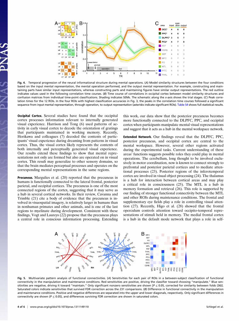
sequence in which participants encoded an input into a mentalrepresentation, performed a mental operation (construct, de-construct, or maintain) on that representation, and produced anoutput mental representation. Each of these stages entaileda unique relationship among the mental states associated withthe four conditions (Fig. 4A). For example, the inputs to theconstruct-parts condition were similar to those of the maintain-parts condition, the operation performed during the construct-parts condition was similar to that of the deconstruct-figurecondition, and the outputs from the construct-parts conditionwere similar to those of the maintain-figure condition. Thus, therelationship among the conditions evolved throughout the trialand provided a means of further exploring the informationalcontent of the mental workspace. To do so, we carried outa four-way classification among the conditions at each time pointand correlated the resulting confusion matrices with each of thethree model similarity structures in Fig. 4A. High correlationbetween a confusion matrix and one of the model structureswould indicate that a particular region was carrying out thecorresponding stage of processing at that time. Fig. 4B shows thetime course of correlations with each model in occipital cortex.In Fig. 4C, we report peak correlation times in each of the 12ROIs. In the four regions with highest classification accuracies inFig. 3A, correlation peaks progressed from input through oper-ation to output, providing strong evidence that these four areasdirectly mediated the mental operations as they unfolded overtime. It should be noted that the differences between test stimulicould have affected the output correlation time course (orangetrace in Fig. 4B) because the output mental representations weresimilar to the stimuli presented during the test phase. Our ex-perimental design did not allow us to evaluate the relative con-tributions of the output mental representations and of the teststimuli to the output correlation time course.The above analyses show that a subset of ROIs supports the
temporal evolution of information necessary to carry outparticular mental operations. However, they do not provide
Fig. 1. Experimental design. (A) Parts could be constructed into 2 × 2 fig-ures, and figures could be deconstructed into parts. (B) Participants per-formed four mental operations on stimuli: construct parts into figure,deconstruct figure into parts, maintain parts, or maintain figure. (C) Thestimulus set of 100 abstract parts, ordered from simple to complex. (D) Ex-ample of figures. Parts and figures ranged from simple to complex accordingto an index, d. This index allowed the difficulty of the task to be equatedacross conditions. (E) Trial schematic. Trials begin with a figure and fourunrelated parts presented for 2 s, followed by a task prompt for 1 s con-sisting of an arrow indicating the figure or the parts and a letter indicatingthe task. In this case, the participant is instructed to maintain the figure inmemory. The task prompt is followed by a 5-s delay period during which nostimulus is shown and the participant performs the indicated operation. Fi-nally, a test screen appears for 2.5 s. Depending on the task, four figures orfour sets of parts are presented, and the participant indicates the correctoutput of the operation.
Fig. 2. Eleven ROIs showing differential activity levels in manipulation andmaintenance conditions. An additional occipital cortex ROI was definedanatomically. CERE, cerebellum; DLPFC, dorsolateral prefrontal cortex; FEF,frontal eye fields; FO, frontal operculum; MFC, medial frontal cortex; MTL,medial temporal lobe; OCC, occipital cortex; PCU, precuneus; PITC, posteriorinferior temporal cortex; PPC,: posterior parietal cortex; SEF, supplementaryeye field; THA, thalamus.
2 of 6 | www.pnas.org/cgi/doi/10.1073/pnas.1311149110 Schlegel et al.

evidence about how these regions communicate within themental workspace network. We investigated this communicationby analyzing patterns of functional connectivity between the ROIs.For each condition, participant, and region, we constructed atime course by concatenating the mean BOLD signal within thatregion across the participant’s correct-response trials for thatcondition. We calculated the functional connectivity, defined asthe correlation between pairs of time courses, for each condition,participant, and pair of regions (14). This procedure yielded onenetwork-wide pattern of functional connectivity for each condi-tion and participant. A cross-subject classification analysis onthese connectivity patterns successfully predicted whether par-ticipants mentally manipulated or maintained imagery with61.7% accuracy [t(14) = 2.4, P = 0.029], thus indicating thatpatterns of connectivity between the network componentschanged depending on the operation that participants performedon the contents of their mental imagery. Investigating the weightsthat the classifier assigned to each pair of regions allowed us todetermine which connections were most informative (Fig. 5A).Increases in connectivity between pairs with positive weights drovethe classifier toward the manipulation conditions, whereas in-creases between pairs with negative weights drove it toward themaintenance conditions. Thus, stronger connectivities withthe precuneus and with left posterior inferior temporal cortexindicated manipulation conditions, and stronger connectivitiesprimarily with the MTL indicated maintenance conditions. InFig. 5B, we plot the difference in functional connectivity betweenconditions. During manipulation conditions the precuneus andposterior inferior temporal cortex showed stronger connectivitywith several frontal and parietal regions, whereas connectivitybetween the MTL and many regions became weaker. Thus, ourdata show not only that a distributed set of regions mediates men-tal operations but also that these regions communicate in aninformation-processing network. The network switches betweentwo connectivity profiles depending on whether mental repre-sentations are maintained or manipulated.
DiscussionOur findings reveal a widespread cortical and subcortical net-work that operates on visual representations in the mentalworkspace. This network includes four core regions spanning theDLPFC, PPC, posterior precuneus, and occipital cortex thatmanipulate the contents of visual imagery. Within these regions
we decoded and tracked the evolution of mental operations overtime. Several other areas showed a difference in BOLD re-sponses between the manipulation and maintenance conditionsbut without the specificity found in the four core areas. There-fore it is likely that an extended network of regions is involved inthe operations. Changes in patterns of connectivity betweenthe mental workspace network’s nodes reveal that the networksupports at least two distinct modes of operation, dependingon whether mental representations are maintained or manip-ulated. We discuss each of the identified components of thenetwork below.
Frontoparietal Cortex. Our finding that the DLPFC and PPC di-rectly mediate manipulation of visual imagery is supported bymultiple studies suggesting that a network of frontal and parietalareas is involved in many high-level cognitive abilities in humans(10–13). Miller et al. (15) showed that the responses of neuronsin DLPFC convey more information about the task relevance ofstimuli than about their specific features and that this selectivityfor task relevance is maintained over extended durations in theabsence of stimulus input. Thus, the DLPFC appears to be partof a network that maintains representations in working memoryvia attention. Human neuroimaging studies have shown that boththe DLPFC and the PPC are activated, regardless of the type ofinformation that is held in working memory (16, 17). Selectivityfor task rather than representation distinguishes this system fromsubsidiary systems that are capable only of maintaining particularclasses of information (18). These findings support the view thatthe frontoparietal network is an executive system that recruitssubsidiary systems, as proposed in Baddeley’s (19) model ofworking memory. Modeling work by O’Reilly and colleagues (8,9) has shown how prefrontal cortex may be able to self-organizeabstract rules flexibly and later apply them to specific repre-sentations. This ability is common to many flexible cognitiveprocesses in humans such as analogical reasoning, creativity (11),and fluid intelligence (13). Our data provide empirical supportfor this model by showing that the DLPFC and PPC mediate notonly the maintenance of representations in working memory butalso the manipulation of those representations. Thus, these areasmay form the core of a system that mediates conscious oper-ations on mental representations, in this case the contents ofvisual imagery represented at least partially in the occipital cortex.
Fig. 3. Results of multivariate pattern classification. Asterisks indicate significance (*P ≥ 0.05; **P ≥ 0.01; ***P ≥ 0.001; *4, P ≥ 0.0001). (A) Results for two-way classifications in each ROI between manipulation conditions and between maintenance conditions. The bar plot shows classification accuracies indescending order. Error bars indicate SEMs. Asterisks indicate accuracies significantly above chance (P ≤ 0.05, FDR corrected across the 24 comparisons). TableS2 shows full statistical results. (B) Model similarity structure for regions that mediate the mental operations. Manipulation and maintenance conditionsshould be more similar within condition types than across condition types. (C) Confusion matrices from four-way classifications in each ROI. Values arepercentages. Asterisks indicate regions in which confusion matrices correlated significantly with the model (P ≤ 0.05, FDR corrected across the 12 compar-isons). Because ROIs were selected based on differences in activity between manipulation and maintenance conditions, we considered only values withinmanipulation and maintenance conditions in the correlation (within the green squares in B). Table S3 shows full statistical results.
Schlegel et al. PNAS Early Edition | 3 of 6
PSYC
HOLO
GICALAND
COGNITIVESC
IENCE
S
Occipital Cortex. Several studies have found that the occipitalcortex processes information relevant to internally generatedvisual experience. Harrison and Tong (6) used patterns of ac-tivity in early visual cortex to decode the orientation of gratingsthat participants maintained in working memory. Recently,Horikawa and colleagues (7) decoded the contents of partic-ipants’ visual experience during dreaming from patterns in visualcortex. Thus, the visual cortex likely represents the contents ofboth internally and perceptually generated visual experience.Our results extend these findings to show that mental repre-sentations not only are formed but also are operated on in visualcortex. This result may generalize to other sensory domains, sothat the brain mediates perceptual processes and operates on thecorresponding mental representations in the same regions.
Precuneus. Margulies et al. (20) reported that the precuneus inhumans is functionally connected to the lateral frontal, posteriorparietal, and occipital cortices. The precuneus is one of the mostconnected regions of the cortex, suggesting that it may serve asa hub in several cortical networks. In their review, Cavanna andTrimble (21) cite a body of evidence that the precuneus is in-volved in visuospatial imagery, is relatively larger in humans thanin nonhuman primates and other animals, and is one of the lastregions to myelinate during development. Consistent with thesefindings, Vogt and Laureys (22) propose that the precuneus playsa central role in conscious information processing. Extending
this work, our data show that the posterior precuneus becomesmore functionally connected to the DLPFC, PPC, and occipitalcortex when participants manipulate mental visual representationsand suggest that it acts as a hub in the mental workspace network.
Extended Network. Our findings reveal that the DLPFC, PPC,posterior precuneus, and occipital cortex are central to themental workspace. However, several other regions activatedduring the experimental tasks. Current understanding of theseareas’ functions suggests possible roles they could play in mentaloperations. The cerebellum, long thought to be involved exclu-sively in motor coordination, now is known to connect strongly toprefrontal and posterior parietal cortices and to mediate atten-tional processes (23). Posterior regions of the inferotemporalcortex are involved in visual object processing (24). The thalamusis a hub for interaction between cortical areas and may playa critical role in consciousness (25). The MTL is a hub inmemory formation and retrieval (26). This role is supported byour finding of stronger functional connectivity between the MTLand other ROIs during maintenance conditions. The frontal andsupplementary eye fields play a role in controlling visual atten-tion (27). Recently, Higo et al. (28) showed that the frontaloperculum controls attention toward occipito-temporal repre-sentations of stimuli held in memory. The medial frontal cortexis a hub in the default mode network that plays a role in self-
Fig. 4. Temporal progression of the neural informational structure during mental operations. (A) Model similarity structures between the four conditionsbased on the input mental representation, the mental operation performed, and the output mental representation. For example, constructing and main-taining parts have similar input representations, whereas constructing parts and maintaining figures have similar output representations. The red outlineindicates values used in the following correlation time courses. (B) Time course of correlations in occipital cortex between model similarity structures andconfusion matrices from individual time-point classifications. Shading indicates SEMs. The schematic along the x-axis shows the trial stages. (C) Peak corre-lation times for the 12 ROIs. In the four ROIs with highest classification accuracies in Fig. 3, the peaks in the correlation time courses followed a significantsequence from input mental representation, through operation, to output representation (asterisks indicate significant ROIs). Table S4 shows full statistical results.
Fig. 5. Multivariate pattern analysis of functional connectivities. (A) Sensitivities for each pair of ROIs in a between-subject classification of functionalconnectivity in the manipulation and maintenance conditions. Red sensitivities are positive, driving the classifier toward choosing “manipulate.” Blue sen-sitivities are negative, driving it toward “maintain.” Only significant nonzero sensitivities are shown [P ≤ 0.05, corrected for similarity between folds (36)].Saturated colors indicate sensitivities that survived FDR correction across the 231 comparisons. (B) Difference in functional connectivity in the manipulationand maintenance conditions. Positive and negative differences are separated into the upper and lower diagonals, respectively. Only significant differences inconnectivity are shown (P ≤ 0.05), and differences surviving FDR correction are shown in saturated colors.
4 of 6 | www.pnas.org/cgi/doi/10.1073/pnas.1311149110 Schlegel et al.

directed attentional processes (29). Thus, all these regions arelikely involved in the mental operations performed by participants.A significant finding of the present study is that connectivity in
the mental workspace network switches between orthogonalmodes of operation depending on whether the network main-tains or manipulates representations. Although several networkcomponents represent information during both tasks, our datashow that patterns of network connectivity associated with thesetasks differ substantially. Maintenance of representations involvesdense, bilateral interconnections across the entire network withthe MTL acting as a hub, whereas manipulation of those rep-resentations recruits a sparse, slightly left-lateralized networkwith a hub in the posterior precuneus. Although the MTL hubdoes not contain specific information about either mental rep-resentations or manipulations, the posterior precuneus hubcontains information specific to each operation. This findingsuggests that these hubs serve distinct functions across the tasks.The MTL appears to bind network components together,whereas the posterior precuneus may exchange informationwithin a sparse core of this network that itself supports ma-nipulation of representations.Previous studies have not been able to find evidence that the
areas we identified play specific roles in manipulating repre-sentations. They have shown differences in BOLD or connec-tivity between maintenance and manipulation in certain areas(30–32) but have not shown that these areas are responsible forthe manipulations themselves. An alternative explanation ofthese findings could be merely that attentional allocation is in-creased during manipulation as compared with maintenancetasks. In this study we investigated neural activity in two quali-tatively distinct types of manipulations. We show that a subset ofareas in the mental workspace network contains informationspecific to particular manipulations. We additionally show thatthe task-related informational structure of these areas evolvesover time in accordance with the manipulations performed.These results provide specific evidence for the particular networkcomponents that directly mediate mental operations.Human cognition is distinguished by the flexibility with which
mental representations can be constructed and manipulated togenerate novel ideas and actions. Dehaene (2) and others haveproposed that this ability is a key role of a global neuronalworkspace that in part realizes our conscious experience. Herewe have shown that patterns of activity in just such a distributedneuronal network mediate the flexible recombination of mentalimages. Although the present study was limited to visual imagery,we anticipate that this network is part of a more general work-space in the human brain in which core conscious processes infrontal and parietal areas recruit specialized subdomains forspecific mental operations. Understanding the neural basis ofthis workspace could reveal common processes central to theflexible cognitive abilities that characterize our species.
Materials and MethodsParticipants. Sixteen participants (six females) age 19–30 y gave informedwritten consent according to the Institutional Review Board guidelines ofDartmouth College before participating. Data from one participant whocould not achieve our task accuracy criterion were discarded before furtheranalysis. Participation consisted of two sessions: an initial behavioral sessionduring which participants practiced the tasks and an fMRI session.
Stimulus. The stimulus set consisted of 100 abstract parts (Fig. 1C). The firsteight parts were defined manually, and each subsequent part was gener-ated by randomly perturbing a quarter-circle with fixed endpoints (see SIMaterials and Methods for details).
Task. Participants performed four mental operations with the stimuli: Theymentally constructed four parts into afigure, deconstructed afigure into fourparts, maintained four parts, or maintained a figure. At the start of each trial,both a figure and four unrelated parts were displayed to equate for low-level
image properties and attention across tasks. After 2 s, the stimulus dis-appeared and was replaced for 1 s by a prompt indicating the task to beperformed. The participant then had 5 s to perform the operation, duringwhich only a fixation dot appeared. Finally, a test screen appeared in whichthe target output of the operation was shown along with three distractorsthat were identical to the target except for a single part. The participant wasinstructed to indicate the target within 4 s of the test screen’s appearance.The stimulus complexity was updated on each trial so that participantsachieved an accuracy of two out of three correct responses in each trial type.See SI Materials and Methods for an extended description of the task.
MRI Acquisition and Preprocessing. Data were collected using a 3.0 T PhilipsAchieva Intera scanner with a 32-channel sense head coil at the DartmouthBrain Imaging Center. Participants completed 10 functional runs consisting of16 trials interleaved with 10-s blanks. fMRI data were preprocessed using FSL(33), and structural images were processed using the FreeSurfer imageanalysis suite (34). See SI Materials and Methods for a detailed description ofacquisition parameters and preprocessing steps.
ROI Selection Procedure. A whole-brain GLM analysis was carried out onfunctional data using the FMRIB Software Library’s FEAT tool. A first-levelanalysis for each participant used boxcar predictors for each of the fourconditions, convolved with a double-gamma hemodynamic response func-tion (HRF). Only trials in which participants made correct responses wereconsidered (∼27 per condition). The results of this analysis were passed tohigher-level cross-subject analyses carried out in Montreal Neurological In-stitute space, in which t contrasts were defined for manipulate > maintainand for manipulate < maintain. Each t-contrast map was cluster thresholdedat z ≥ 2.3; clusters then were thresholded at P ≤ 0.05 according to GaussianRandom Field theory (33). This analysis yielded 11 bilateral ROIs that thenwere transformed back into each participant’s native space for furtheranalysis. An additional occipital ROI was defined anatomically in each par-ticipant’s native space using the following cortical masks from FreeSurfer:inferior occipital gyrus and sulcus; middle occipital gyrus and sulci; superioroccipital gyrus; cuneus; occipital pole; superior occipital and transverse oc-cipital sulci; and anterior occipital sulcus.
Multivariate Pattern Analysis: Classification. Multivariate pattern analysis(MVPA) was carried out using PyMVPA (35). Spatiotemporal patterns wereconstructed for each correct-response trial and ROI using the z-scored BOLDresponse from TRs 4–6 (the period during which the operation was per-formed, after shifting by a 4-s estimate of the HRF delay). Classification wascarried out in each ROI between construct-parts and deconstruct-figure trialsand between maintain-parts and maintain-figure trials using these patterns,a linear support vector machine (SVM) classifier, and leave-one-out cross-validation. Significance of accuracies was evaluated using one-tailed, one-sample t tests compared with chance (50%) and false-discovery rate (FDR)corrected across the 24 comparisons (one for each ROI and classification). Afour-way classification also was carried out in each ROI to produce theconfusion matrices in Fig. 3C. The correlation between each of these con-fusion matrices and the model similarity structure was calculated (Fig. 3B),and significance was determined at P ≤ 0.05, FDR corrected across the 12comparisons (one for each ROI).
MVPA: Correlation Time Courses. Four-way classification was carried out foreach ROI and at each time point of the trial, here using only spatial patterns ofthe BOLD signal across all voxels within the ROI. This procedure produceda confusion matrix for each time point and ROI, and these confusion ma-trices were correlated with each of the model similarity structures in Fig. 4A.The first structure models similarities between the conditions based onwhether the input representation is a set of parts or a figure. The secondstructure models similarities based on the two types of operations carriedout, manipulation or maintenance. The third structure models similaritiesbased on the outputs from each condition. For each ROI and model struc-ture, we calculated the time point at which the mean correlation reacheda maximum, yielding the table in Fig. 4C. These calculations were restrictedto TRs 3–8, representing the pretest portion of the trial shifted by 4 s toaccount for hemodynamic lag. For each ROI we carried out one-way re-peated-measures ANOVA on the peak correlation times to test whether theexpected progression from input through operation to output occurred. Weperformed the analysis on trimmed, jackknifed data as recommended byMiller et al. (36) for latency analyses. In a jackknifed analysis with N subjects,N grand means of the data are calculated, each with one subject left out.The analysis then is performed on these grand means with corrections ap-plied for the jackknife-induced decrease in variance. In the case of noisy
Schlegel et al. PNAS Early Edition | 5 of 6
PSYC
HOLO
GICALAND
COGNITIVESC
IENCE
S

estimates, as occur when calculating latencies from single-subject timecourses, this procedure provides cleaner results and does not bias estimatesof significance. For each ANOVA we defined two orthogonal linear contrasts(input/operation/output: C1 = −1/−1/2; C2 = −1/1/0) to evaluate the tem-poral order of the peaks. We determined that an ROI significantly followedthe expected progression if and only if both of these contrasts were sig-nificant at P ≤ 0.05 uncorrected.
Functional Connectivity. The functional connectivity (14), defined as theFisher’s z-transformed correlation between time courses, was calculated foreach participant and condition across all pairs of the 24 unilateral ROIs andusing data pooled across all correct trials. This procedure gave a single
connectivity pattern for each participant and condition. Unilateral ROIs wereused to maximize the potential information in each pattern. We then carriedout a cross-subject classification between manipulation and maintenanceconditions, using these connectivity patterns and an SVM classifier. Thesensitivities shown in Fig. 5A are significantly different from zero in a one-sample t test, corrected for the low variance because of the similarity be-tween folds (36), and thresholded at P ≤ 0.05.
ACKNOWLEDGMENTS. We thank Andrei Gorea for his input on the studydesign. This study was funded by a National Science Foundation GraduateResearch Fellowship (to A.S.) and by Templeton Foundation Grant 14316(to P.U.T.).
1. Hadamard J (1954) The Psychology of Invention in the Mathematical Field (DoverPublications, Inc., New York), p 142.
2. Dehaene S, Naccache L (2001) Towards a cognitive neuroscience of consciousness:Basic evidence and a workspace framework. Cognition 79(1-2):1–37.
3. Baars BJ, Franklin S (2003) How conscious experience and working memory interact.Trends Cogn Sci 7(4):166–172.
4. Shepard RN, Metzler J (1971) Mental rotation of three-dimensional objects. Science171(3972):701–703.
5. Haxby JV, et al. (2001) Distributed and overlapping representations of faces andobjects in ventral temporal cortex. Science 293(5539):2425–2430.
6. Harrison SA, Tong F (2009) Decoding reveals the contents of visual working memoryin early visual areas. Nature 458(7238):632–635.
7. Horikawa T, Tamaki M, Miyawaki Y, Kamitani Y (2013) Neural decoding of visualimagery during sleep. Science 340(6132):639–642.
8. Rougier NP, Noelle DC, Braver TS, Cohen JD, O’Reilly RC (2005) Prefrontal cortex andflexible cognitive control: Rules without symbols. Proc Natl Acad Sci USA 102(20):7338–7343.
9. O’Reilly RC (2006) Biologically based computational models of high-level cognition.Science 314(5796):91–94.
10. Zacks JM (2008) Neuroimaging studies of mental rotation: A meta-analysis and re-view. J Cogn Neurosci 20(1):1–19.
11. Green AE, Kraemer DJM, Fugelsang JA, Gray JR, Dunbar KN (2012) Neural correlatesof creativity in analogical reasoning. J Exp Psychol Learn Mem Cogn 38(2):264–272.
12. Salazar RF, Dotson NM, Bressler SL, Gray CM (2012) Content-specific fronto-parietalsynchronization during visual working memory. Science 338(6110):1097–1100.
13. Jung RE, Haier RJ (2007) The Parieto-Frontal Integration Theory (P-FIT) of intelligence:Converging neuroimaging evidence. Behav Brain Sci 30(2):135–154, discussion154–187.
14. Fox MD, et al. (2005) The human brain is intrinsically organized into dynamic, anti-correlated functional networks. Proc Natl Acad Sci USA 102(27):9673–9678.
15. Miller EK, Erickson CA, Desimone R (1996) Neural mechanisms of visual workingmemory in prefrontal cortex of the macaque. J Neurosci 16(16):5154–5167.
16. Todd JJ, Marois R (2004) Capacity limit of visual short-term memory in human pos-terior parietal cortex. Nature 428(6984):751–754.
17. Xu Y, Chun MM (2006) Dissociable neural mechanisms supporting visual short-termmemory for objects. Nature 440(7080):91–95.
18. Druzgal TJ, D’Esposito M (2003) Dissecting contributions of prefrontal cortex andfusiform face area to face working memory. J Cogn Neurosci 15(6):771–784.
19. Baddeley AD (1986) Working Memory (Oxford Univ Press, New York).
20. Margulies DS, et al. (2009) Precuneus shares intrinsic functional architecture in hu-mans and monkeys. Proc Natl Acad Sci USA 106(47):20069–20074.
21. Cavanna AE, Trimble MR (2006) The precuneus: A review of its functional anatomyand behavioural correlates. Brain 129(Pt 3):564–583.
22. Vogt BA, Laureys S (2005) Posterior cingulate, precuneal and retrosplenial cortices:Cytology and components of the neural network correlates of consciousness. ProgBrain Res 150(05):205–217.
23. Bostan AC, Dum RP, Strick PL (2013) Cerebellar networks with the cerebral cortex andbasal ganglia. Trends Cogn Sci 17(5):241–254.
24. Tanaka K (1996) Inferotemporal cortex and object vision. Annu Rev Neurosci19:109–139.
25. Tononi G (2008) Consciousness as integrated information: A provisional manifesto.Biol Bull 215(3):216–242.
26. Ryan L, Lin C-Y, Ketcham K, Nadel L (2010) The role of medial temporal lobe in re-trieving spatial and nonspatial relations from episodic and semantic memory. Hip-pocampus 20(1):11–18.
27. Schall JD (2004) On the role of frontal eye field in guiding attention and saccades.Vision Res 44(12):1453–1467.
28. Higo T, Mars RB, Boorman ED, Buch ER, Rushworth MFS (2011) Distributed and causalinfluence of frontal operculum in task control. Proc Natl Acad Sci USA 108(10):4230–4235.
29. Buckner RL, Andrews-Hanna JR, Schacter DL (2008) The brain’s default network:Anatomy, function, and relevance to disease. Ann N Y Acad Sci 1124:1–38.
30. Ranganath C, Cohen MX, Dam C, D’Esposito M (2004) Inferior temporal, prefrontal,and hippocampal contributions to visual working memory maintenance and asso-ciative memory retrieval. J Neurosci 24(16):3917–3925.
31. Mourao-Miranda J, Ecker C, Sato JR, Brammer MJ (2009) Dynamic changes in themental rotation network revealed by pattern recognition analysis of fMRI data.J Cogn Neurosci 21(5):890–904.
32. Formisano E, et al. (2002) Tracking the mind’s image in the brain I: Time-resolved fMRIduring visuospatial mental imagery. Neuron 35(1):185–194.
33. Smith SM, et al. (2004) Advances in functional and structural MR image analysis andimplementation as FSL. Neuroimage 23(Suppl 1):S208–S219.
34. Dale AM, Fischl B, Sereno MI (1999) Cortical surface-based analysis. I. Segmentationand surface reconstruction. Neuroimage 9(2):179–194.
35. Hanke M, et al. (2009) PyMVPA: A python toolbox for multivariate pattern analysis offMRI data. Neuroinformatics 7(1):37–53.
36. Miller J, Patterson T, Ulrich R (1998) Jackknife-based method for measuring LRP onsetlatency differences. Psychophysiology 35(1):99–115.
6 of 6 | www.pnas.org/cgi/doi/10.1073/pnas.1311149110 Schlegel et al.
Copyright © 2022 FDOKUMEN