NeMeSys: a biological resource for narrowing the gap between sequence and function in the human...
13
Genome Biology 2009, 10:R110 Open Access 2009 Rusniok et al. Volume 10, Issue 10, Article R110 Research NeMeSys: a biological resource for narrowing the gap between sequence and function in the human pathogen Neisseria meningitidis Christophe Rusniok ¤*¶ , David Vallenet ¤† , Stéphanie Floquet ‡¥ , Helen Ewles § , Coralie Mouzé-Soulama ‡# , Daniel Brown § , Aurélie Lajus † , Carmen Buchrieser *¶ , Claudine Médigue † , Philippe Glaser * and Vladimir Pelicic ‡§ Addresses: * Génomique des Microorganismes Pathogènes, Institut Pasteur, rue du Dr Roux, Paris, 75015, France. † Génomique Métabolique, CNRS UMR8030, Laboratoire de Génomique Comparative, CEA-Institut de Génomique-Génoscope, rue Gaston Crémieux, Evry, 91057, France. ‡ U570 INSERM, Faculté de Médecine René Descartes-Paris 5, rue de Vaugirard, Paris, 75015, France. § Department of Microbiology, CMMI, Imperial College London, Armstrong Road, London, SW7 2AZ, UK. ¶ Current address: Biologie des Bactéries Intracellulaires, Institut Pasteur, rue du Dr Roux, Paris, 75015, France. ¥ Current address: Mutabilis, Parc Biocitech, avenue Gaston Roussel, Romainville, 93230, France. # Current address: FAB pharma, rue Saint Honoré, Paris, 75001, France. ¤ These authors contributed equally to this work. Correspondence: Vladimir Pelicic. Email: [email protected] © 2009 Rusniok et al.; licensee BioMed Central Ltd. This is an open access article distributed under the terms of the Creative Commons Attribution License (http://creativecommons.org/licenses/by/2.0), which permits unrestricted use, distribution, and reproduction in any medium, provided the original work is properly cited. Neisseria genomics <p>The genome of a clinical isolate of Neisseria meningitidis is described. This and other reannotated Neisseria genomes are compiled in a database.</p> Abstract Background: Genome sequences, now available for most pathogens, hold promise for the rational design of new therapies. However, biological resources for genome-scale identification of gene function (notably genes involved in pathogenesis) and/or genes essential for cell viability, which are necessary to achieve this goal, are often sorely lacking. This holds true for Neisseria meningitidis, one of the most feared human bacterial pathogens that causes meningitis and septicemia. Results: By determining and manually annotating the complete genome sequence of a serogroup C clinical isolate of N. meningitidis (strain 8013) and assembling a library of defined mutants in up to 60% of its non-essential genes, we have created NeMeSys, a biological resource for Neisseria meningitidis systematic functional analysis. To further enhance the versatility of this toolbox, we have manually (re)annotated eight publicly available Neisseria genome sequences and stored all these data in a publicly accessible online database. The potential of NeMeSys for narrowing the gap between sequence and function is illustrated in several ways, notably by performing a functional genomics analysis of the biogenesis of type IV pili, one of the most widespread virulence factors in bacteria, and by identifying through comparative genomics a complete biochemical pathway (for sulfur metabolism) that may potentially be important for nasopharyngeal colonization. Conclusions: By improving our capacity to understand gene function in an important human pathogen, NeMeSys is expected to contribute to the ongoing efforts aimed at understanding a prokaryotic cell comprehensively and eventually to the design of new therapies. Published: 9 October 2009 Genome Biology 2009, 10:R110 (doi:10.1186/gb-2009-10-10-r110) Received: 18 August 2009 Revised: 19 August 2009 Accepted: 9 October 2009 The electronic version of this article is the complete one and can be found online at http://genomebiology.com/2009/10/10/R110
-
Upload
independent -
Category
Documents
-
view
1 -
download
0
Transcript of NeMeSys: a biological resource for narrowing the gap between sequence and function in the human...
Open Access2009Rusnioket al.Volume 10, Issue 10, Article R110ResearchNeMeSys: a biological resource for narrowing the gap between sequence and function in the human pathogen Neisseria meningitidisChristophe Rusniok¤*¶, David Vallenet¤†, Stéphanie Floquet‡¥, Helen Ewles§, Coralie Mouzé-Soulama‡#, Daniel Brown§, Aurélie Lajus†, Carmen Buchrieser*¶, Claudine Médigue†, Philippe Glaser* and Vladimir Pelicic‡§
Addresses: *Génomique des Microorganismes Pathogènes, Institut Pasteur, rue du Dr Roux, Paris, 75015, France. †Génomique Métabolique, CNRS UMR8030, Laboratoire de Génomique Comparative, CEA-Institut de Génomique-Génoscope, rue Gaston Crémieux, Evry, 91057, France. ‡U570 INSERM, Faculté de Médecine René Descartes-Paris 5, rue de Vaugirard, Paris, 75015, France. §Department of Microbiology, CMMI, Imperial College London, Armstrong Road, London, SW7 2AZ, UK. ¶Current address: Biologie des Bactéries Intracellulaires, Institut Pasteur, rue du Dr Roux, Paris, 75015, France. ¥Current address: Mutabilis, Parc Biocitech, avenue Gaston Roussel, Romainville, 93230, France. #Current address: FAB pharma, rue Saint Honoré, Paris, 75001, France.
¤ These authors contributed equally to this work.
Correspondence: Vladimir Pelicic. Email: [email protected]
© 2009 Rusniok et al.; licensee BioMed Central Ltd. This is an open access article distributed under the terms of the Creative Commons Attribution License (http://creativecommons.org/licenses/by/2.0), which permits unrestricted use, distribution, and reproduction in any medium, provided the original work is properly cited.Neisseria genomics<p>The genome of a clinical isolate of Neisseria meningitidis is described. This and other reannotated Neisseria genomes are compiled in a database.</p>
Abstract
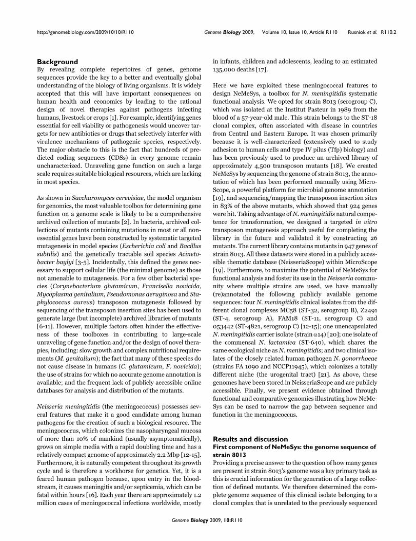
Background: Genome sequences, now available for most pathogens, hold promise for therational design of new therapies. However, biological resources for genome-scale identification ofgene function (notably genes involved in pathogenesis) and/or genes essential for cell viability, whichare necessary to achieve this goal, are often sorely lacking. This holds true for Neisseria meningitidis,one of the most feared human bacterial pathogens that causes meningitis and septicemia.
Results: By determining and manually annotating the complete genome sequence of a serogroupC clinical isolate of N. meningitidis (strain 8013) and assembling a library of defined mutants in up to60% of its non-essential genes, we have created NeMeSys, a biological resource for Neisseriameningitidis systematic functional analysis. To further enhance the versatility of this toolbox, wehave manually (re)annotated eight publicly available Neisseria genome sequences and stored allthese data in a publicly accessible online database. The potential of NeMeSys for narrowing the gapbetween sequence and function is illustrated in several ways, notably by performing a functionalgenomics analysis of the biogenesis of type IV pili, one of the most widespread virulence factors inbacteria, and by identifying through comparative genomics a complete biochemical pathway (forsulfur metabolism) that may potentially be important for nasopharyngeal colonization.
Conclusions: By improving our capacity to understand gene function in an important humanpathogen, NeMeSys is expected to contribute to the ongoing efforts aimed at understanding aprokaryotic cell comprehensively and eventually to the design of new therapies.
Published: 9 October 2009
Genome Biology 2009, 10:R110 (doi:10.1186/gb-2009-10-10-r110)
Received: 18 August 2009Revised: 19 August 2009Accepted: 9 October 2009
The electronic version of this article is the complete one and can be found online at http://genomebiology.com/2009/10/10/R110
Genome Biology 2009, 10:R110
http://genomebiology.com/2009/10/10/R110 Genome Biology 2009, Volume 10, Issue 10, Article R110 Rusniok et al. R110.2
BackgroundBy revealing complete repertoires of genes, genomesequences provide the key to a better and eventually globalunderstanding of the biology of living organisms. It is widelyaccepted that this will have important consequences onhuman health and economics by leading to the rationaldesign of novel therapies against pathogens infectinghumans, livestock or crops [1]. For example, identifying genesessential for cell viability or pathogenesis would uncover tar-gets for new antibiotics or drugs that selectively interfer withvirulence mechanisms of pathogenic species, respectively.The major obstacle to this is the fact that hundreds of pre-dicted coding sequences (CDSs) in every genome remainuncharacterized. Unraveling gene function on such a largescale requires suitable biological resources, which are lackingin most species.
As shown in Saccharomyces cerevisiae, the model organismfor genomics, the most valuable toolbox for determining genefunction on a genome scale is likely to be a comprehensivearchived collection of mutants [2]. In bacteria, archived col-lections of mutants containing mutations in most or all non-essential genes have been constructed by systematic targetedmutagenesis in model species (Escherichia coli and Bacillussubtilis) and the genetically tractable soil species Acineto-bacter baylyi [3-5]. Incidentally, this defined the genes nec-essary to support cellular life (the minimal genome) as thosenot amenable to mutagenesis. For a few other bacterial spe-cies (Corynebacterium glutamicum, Francisella novicida,Mycoplasma genitalium, Pseudomonas aeruginosa and Sta-phylococcus aureus) transposon mutagenesis followed bysequencing of the transposon insertion sites has been used togenerate large (but incomplete) archived libraries of mutants[6-11]. However, multiple factors often hinder the effective-ness of these toolboxes in contributing to large-scaleunraveling of gene function and/or the design of novel thera-pies, including: slow growth and complex nutritional require-ments (M. genitalium); the fact that many of these species donot cause disease in humans (C. glutamicum, F. novicida);the use of strains for which no accurate genome annotation isavailable; and the frequent lack of publicly accessible onlinedatabases for analysis and distribution of the mutants.
Neisseria meningitidis (the meningococcus) possesses sev-eral features that make it a good candidate among humanpathogens for the creation of such a biological resource. Themeningococcus, which colonizes the nasopharyngeal mucosaof more than 10% of mankind (usually asymptomatically),grows on simple media with a rapid doubling time and has arelatively compact genome of approximately 2.2 Mbp [12-15].Furthermore, it is naturally competent throughout its growthcycle and is therefore a workhorse for genetics. Yet, it is afeared human pathogen because, upon entry in the blood-stream, it causes meningitis and/or septicemia, which can befatal within hours [16]. Each year there are approximately 1.2million cases of meningococcal infections worldwide, mostly
in infants, children and adolescents, leading to an estimated135,000 deaths [17].
Here we have exploited these meningococcal features todesign NeMeSys, a toolbox for N. meningitidis systematicfunctional analysis. We opted for strain 8013 (serogroup C),which was isolated at the Institut Pasteur in 1989 from theblood of a 57-year-old male. This strain belongs to the ST-18clonal complex, often associated with disease in countriesfrom Central and Eastern Europe. It was chosen primarilybecause it is well-characterized (extensively used to studyadhesion to human cells and type IV pilus (Tfp) biology) andhas been previously used to produce an archived library ofapproximately 4,500 transposon mutants [18]. We createdNeMeSys by sequencing the genome of strain 8013, the anno-tation of which has been performed manually using Micro-Scope, a powerful platform for microbial genome annotation[19], and sequencing/mapping the transposon insertion sitesin 83% of the above mutants, which showed that 924 geneswere hit. Taking advantage of N. meningitidis natural compe-tence for transformation, we designed a targeted in vitrotransposon mutagenesis approach useful for completing thelibrary in the future and validated it by constructing 26mutants. The current library contains mutants in 947 genes ofstrain 8013. All these datasets were stored in a publicly acces-sible thematic database (NeisseriaScope) within MicroScope[19]. Furthermore, to maximize the potential of NeMeSys forfunctional analysis and foster its use in the Neisseria commu-nity where multiple strains are used, we have manually(re)annotated the following publicly available genomesequences: four N. meningitidis clinical isolates from the dif-ferent clonal complexes MC58 (ST-32, serogroup B), Z2491(ST-4, serogroup A), FAM18 (ST-11, serogroup C) and053442 (ST-4821, serogroup C) [12-15]; one unencapsulatedN. meningitidis carrier isolate (strain α14) [20]; one isolate ofthe commensal N. lactamica (ST-640), which shares thesame ecological niche as N. meningitidis; and two clinical iso-lates of the closely related human pathogen N. gonorrhoeae(strains FA 1090 and NCCP11945), which colonizes a totallydifferent niche (the urogenital tract) [21]. As above, thesegenomes have been stored in NeisseriaScope and are publiclyaccessible. Finally, we present evidence obtained throughfunctional and comparative genomics illustrating how NeMe-Sys can be used to narrow the gap between sequence andfunction in the meningococcus.
Results and discussionFirst component of NeMeSys: the genome sequence of strain 8013Providing a precise answer to the question of how many genesare present in strain 8013's genome was a key primary task asthis is crucial information for the generation of a large collec-tion of defined mutants. We therefore determined the com-plete genome sequence of this clinical isolate belonging to aclonal complex that is unrelated to the previously sequenced
Genome Biology 2009, 10:R110
http://genomebiology.com/2009/10/10/R110 Genome Biology 2009, Volume 10, Issue 10, Article R110 Rusniok et al. R110.3
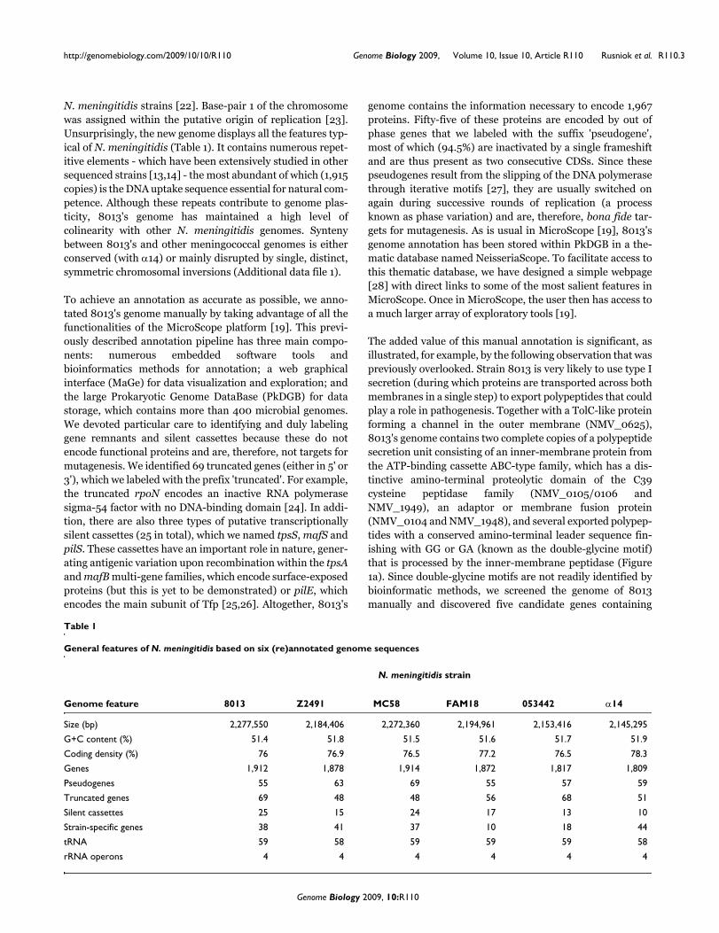
N. meningitidis strains [22]. Base-pair 1 of the chromosomewas assigned within the putative origin of replication [23].Unsurprisingly, the new genome displays all the features typ-ical of N. meningitidis (Table 1). It contains numerous repet-itive elements - which have been extensively studied in othersequenced strains [13,14] - the most abundant of which (1,915copies) is the DNA uptake sequence essential for natural com-petence. Although these repeats contribute to genome plas-ticity, 8013's genome has maintained a high level ofcolinearity with other N. meningitidis genomes. Syntenybetween 8013's and other meningococcal genomes is eitherconserved (with α14) or mainly disrupted by single, distinct,symmetric chromosomal inversions (Additional data file 1).
To achieve an annotation as accurate as possible, we anno-tated 8013's genome manually by taking advantage of all thefunctionalities of the MicroScope platform [19]. This previ-ously described annotation pipeline has three main compo-nents: numerous embedded software tools andbioinformatics methods for annotation; a web graphicalinterface (MaGe) for data visualization and exploration; andthe large Prokaryotic Genome DataBase (PkDGB) for datastorage, which contains more than 400 microbial genomes.We devoted particular care to identifying and duly labelinggene remnants and silent cassettes because these do notencode functional proteins and are, therefore, not targets formutagenesis. We identified 69 truncated genes (either in 5' or3'), which we labeled with the prefix 'truncated'. For example,the truncated rpoN encodes an inactive RNA polymerasesigma-54 factor with no DNA-binding domain [24]. In addi-tion, there are also three types of putative transcriptionallysilent cassettes (25 in total), which we named tpsS, mafS andpilS. These cassettes have an important role in nature, gener-ating antigenic variation upon recombination within the tpsAand mafB multi-gene families, which encode surface-exposedproteins (but this is yet to be demonstrated) or pilE, whichencodes the main subunit of Tfp [25,26]. Altogether, 8013's
genome contains the information necessary to encode 1,967proteins. Fifty-five of these proteins are encoded by out ofphase genes that we labeled with the suffix 'pseudogene',most of which (94.5%) are inactivated by a single frameshiftand are thus present as two consecutive CDSs. Since thesepseudogenes result from the slipping of the DNA polymerasethrough iterative motifs [27], they are usually switched onagain during successive rounds of replication (a processknown as phase variation) and are, therefore, bona fide tar-gets for mutagenesis. As is usual in MicroScope [19], 8013'sgenome annotation has been stored within PkDGB in a the-matic database named NeisseriaScope. To facilitate access tothis thematic database, we have designed a simple webpage[28] with direct links to some of the most salient features inMicroScope. Once in MicroScope, the user then has access toa much larger array of exploratory tools [19].
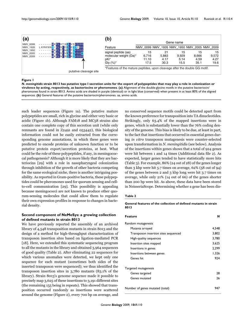
The added value of this manual annotation is significant, asillustrated, for example, by the following observation that waspreviously overlooked. Strain 8013 is very likely to use type Isecretion (during which proteins are transported across bothmembranes in a single step) to export polypeptides that couldplay a role in pathogenesis. Together with a TolC-like proteinforming a channel in the outer membrane (NMV_0625),8013's genome contains two complete copies of a polypeptidesecretion unit consisting of an inner-membrane protein fromthe ATP-binding cassette ABC-type family, which has a dis-tinctive amino-terminal proteolytic domain of the C39cysteine peptidase family (NMV_0105/0106 andNMV_1949), an adaptor or membrane fusion protein(NMV_0104 and NMV_1948), and several exported polypep-tides with a conserved amino-terminal leader sequence fin-ishing with GG or GA (known as the double-glycine motif)that is processed by the inner-membrane peptidase (Figure1a). Since double-glycine motifs are not readily identified bybioinformatic methods, we screened the genome of 8013manually and discovered five candidate genes containing
Table 1
General features of N. meningitidis based on six (re)annotated genome sequences
N. meningitidis strain
Genome feature 8013 Z2491 MC58 FAM18 053442 α14
Size (bp) 2,277,550 2,184,406 2,272,360 2,194,961 2,153,416 2,145,295
G+C content (%) 51.4 51.8 51.5 51.6 51.7 51.9
Coding density (%) 76 76.9 76.5 77.2 76.5 78.3
Genes 1,912 1,878 1,914 1,872 1,817 1,809
Pseudogenes 55 63 69 55 57 59
Truncated genes 69 48 48 56 68 51
Silent cassettes 25 15 24 17 13 10
Strain-specific genes 38 41 37 10 18 44
tRNA 59 58 59 59 59 58
rRNA operons 4 4 4 4 4 4
Genome Biology 2009, 10:R110
http://genomebiology.com/2009/10/10/R110 Genome Biology 2009, Volume 10, Issue 10, Article R110 Rusniok et al. R110.4
such leader sequences (Figure 1a). The putative maturepolypeptides are small, rich in glycine and either very basic oracidic (Figure 1b). Although FAM18 and MC58 strains alsocontain one complete copy of this secretion unit (while onlyremnants are found in Z2491 and 053442), this biologicalinformation could not be easily extracted from the corre-sponding genome annotations, in which these genes werepredicted to encode proteins of unknown function or to beputative protein export/secretion proteins, at best. Whatcould be the role of these polypeptides, if any, in meningococ-cal pathogenesis? Although it is more likely that they are bac-teriocins [29] with a role in nasopharyngeal colonizationthrough inhibition of the growth of other bacteria competingfor the same ecological niche, there is another intriguing pos-sibility. As reported in Gram-positive bacteria, these polypep-tides could be pheromones used for quorum sensing and cell-to-cell communication [29]. This possibility is appealingbecause meningococci are not known to produce other quo-rum-sensing molecules that could allow them to regulatetheir own expression profiles in response to changes in bacte-rial density.
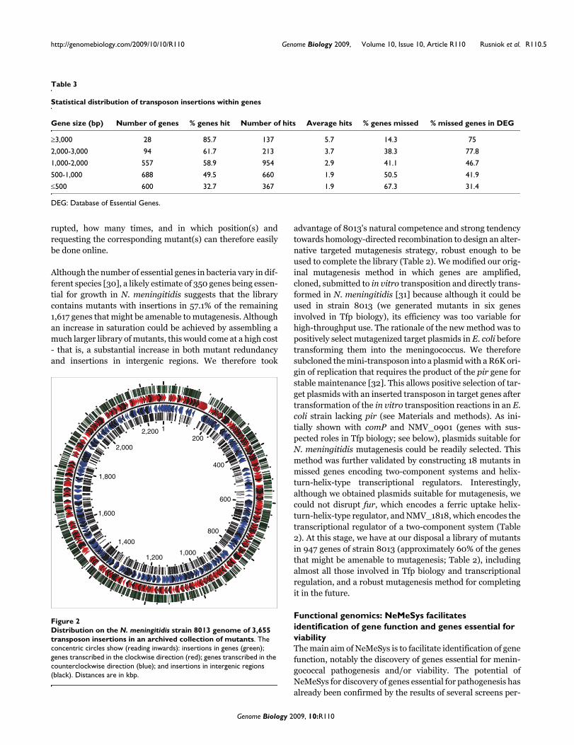
Second component of NeMeSys: a growing collection of defined mutants in strain 8013We have previously reported the assembly of an archivedlibrary of 4,548 transposition mutants in strain 8013 and thedesign of a method for high-throughput characterization oftransposon insertion sites based on ligation-mediated PCR[18]. Here, we extended this systematic sequencing programto all the mutants in the library and obtained 3,964 sequencesof good quality (Table 2). After eliminating 22 sequences forwhich various anomalies were detected, we kept only onesequence for each mutant (sometimes both sides of theinserted transposon were sequenced); we thus identified thetransposon insertion sites in 3,780 mutants (83.1% of thelibrary). Strain 8013's genome sequence made it possible toprecisely map 3,625 of these insertions to 3,191 different sites(the remaining 155 being in repeats). This showed that trans-position occurred randomly as insertions were scatteredaround the genome (Figure 2), every 700 bp on average, and
no conserved sequence motifs could be detected apart fromthe known preference for transposition into TA dinucleotides.Strikingly, only 63.4% of the mapped insertions were ingenes, which is substantially lower than the 76% coding den-sity of the genome. This bias is likely to be due, at least in part,to the fact that insertions that occurred in essential genes dur-ing in vitro transposon mutagenesis were counter-selectedupon transformation in N. meningitidis (see below). Analysisof the insertions within genes shows that a total of 924 geneswere hit between 1 and 14 times (Additional data file 2). Asexpected, larger genes tended to have statistically more hits(Table 3). For example, 86% (24 out of 28) of the genes longerthan 3 kbp were hit 5.7 times on average, 62% (58 out of 94)of the genes between 2 and 3 kbp long were hit 3.7 times onaverage, while only 21% (14 out of 66) of the genes shorterthan 200 bp were hit. As above, these data have been storedin NeisseriaScope. Determining whether a gene has been dis-
N. meningitidis strain 8013 has putative type I secretion units for the export of polypeptides that may play a role in colonization or virulence by acting, respectively, as bacteriocins or pheromonesFigure 1N. meningitidis strain 8013 has putative type I secretion units for the export of polypeptides that may play a role in colonization or virulence by acting, respectively, as bacteriocins or pheromones. (a) Alignment of the double-glycine motifs in the putative bacteriocin/pheromones found in strain 8013. Amino acids are shaded in purple (identical) or in light blue (conserved) when present in at least 80% of the aligned sequences. (b) General features of the putative bacteriocin/pheromones. aa, amino acids.
- - - - - - MK E L HT S E L V E V S GG
L K RK NNI I E L S I E DL E L I Y GG
- - - - - - MK E L T I NDL T L V S GG
- - - - - - MY E L S I V E L E L V S GA
- - - - - - MK E L NI S DL K I V S GG
NMV_0099
NMV_1926
NMV_2005
NMV_1950
NMV_2009
putative cleavage site
Gene name
Feature
signal peptide (aa)molecular weight (Da)*pKi*Gly (%)*
15
NMV_0099
156,71611.10
17.5
NMV_1926
215,883
4.1730.3
NMV_2005
158,666
4.5935.1
NMV_2009
159,572
4.2719.6
NMV_1950
9,929
15.55.14
*Features of the mature peptides, upon cleavage after the double GG motif.
(b)(a)
Table 2
General features of the collection of defined mutants in strain 8013
Feature N
Random mutagenesis
Mutants arrayed 4,548
Transposon insertion sites sequenced 3,802
High-quality sequences 3,780
Insertion sites mapped 3,625
Insertions in genes 2,299
Insertions between genes 1,326
Genes hit 924
Targeted mutagenesis
Genes targeted 28
Genes mutated 26
Number of genes mutated (total) 947
Genome Biology 2009, 10:R110
http://genomebiology.com/2009/10/10/R110 Genome Biology 2009, Volume 10, Issue 10, Article R110 Rusniok et al. R110.5
rupted, how many times, and in which position(s) andrequesting the corresponding mutant(s) can therefore easilybe done online.
Although the number of essential genes in bacteria vary in dif-ferent species [30], a likely estimate of 350 genes being essen-tial for growth in N. meningitidis suggests that the librarycontains mutants with insertions in 57.1% of the remaining1,617 genes that might be amenable to mutagenesis. Althoughan increase in saturation could be achieved by assembling amuch larger library of mutants, this would come at a high cost- that is, a substantial increase in both mutant redundancyand insertions in intergenic regions. We therefore took
advantage of 8013's natural competence and strong tendencytowards homology-directed recombination to design an alter-native targeted mutagenesis strategy, robust enough to beused to complete the library (Table 2). We modified our orig-inal mutagenesis method in which genes are amplified,cloned, submitted to in vitro transposition and directly trans-formed in N. meningitidis [31] because although it could beused in strain 8013 (we generated mutants in six genesinvolved in Tfp biology), its efficiency was too variable forhigh-throughput use. The rationale of the new method was topositively select mutagenized target plasmids in E. coli beforetransforming them into the meningococcus. We thereforesubcloned the mini-transposon into a plasmid with a R6K ori-gin of replication that requires the product of the pir gene forstable maintenance [32]. This allows positive selection of tar-get plasmids with an inserted transposon in target genes aftertransformation of the in vitro transposition reactions in an E.coli strain lacking pir (see Materials and methods). As ini-tially shown with comP and NMV_0901 (genes with sus-pected roles in Tfp biology; see below), plasmids suitable forN. meningitidis mutagenesis could be readily selected. Thismethod was further validated by constructing 18 mutants inmissed genes encoding two-component systems and helix-turn-helix-type transcriptional regulators. Interestingly,although we obtained plasmids suitable for mutagenesis, wecould not disrupt fur, which encodes a ferric uptake helix-turn-helix-type regulator, and NMV_1818, which encodes thetranscriptional regulator of a two-component system (Table2). At this stage, we have at our disposal a library of mutantsin 947 genes of strain 8013 (approximately 60% of the genesthat might be amenable to mutagenesis; Table 2), includingalmost all those involved in Tfp biology and transcriptionalregulation, and a robust mutagenesis method for completingit in the future.
Functional genomics: NeMeSys facilitates identification of gene function and genes essential for viabilityThe main aim of NeMeSys is to facilitate identification of genefunction, notably the discovery of genes essential for menin-gococcal pathogenesis and/or viability. The potential ofNeMeSys for discovery of genes essential for pathogenesis hasalready been confirmed by the results of several screens per-
Table 3
Statistical distribution of transposon insertions within genes
Gene size (bp) Number of genes % genes hit Number of hits Average hits % genes missed % missed genes in DEG
≥3,000 28 85.7 137 5.7 14.3 75
2,000-3,000 94 61.7 213 3.7 38.3 77.8
1,000-2,000 557 58.9 954 2.9 41.1 46.7
500-1,000 688 49.5 660 1.9 50.5 41.9
≤500 600 32.7 367 1.9 67.3 31.4
DEG: Database of Essential Genes.
Distribution on the N. meningitidis strain 8013 genome of 3,655 transposon insertions in an archived collection of mutantsFigure 2Distribution on the N. meningitidis strain 8013 genome of 3,655 transposon insertions in an archived collection of mutants. The concentric circles show (reading inwards): insertions in genes (green); genes transcribed in the clockwise direction (red); genes transcribed in the counterclockwise direction (blue); and insertions in intergenic regions (black). Distances are in kbp.
200
400
600
800
1,0001,200
1,400
1,600
1,800
2,000
2,200 1
Genome Biology 2009, 10:R110
http://genomebiology.com/2009/10/10/R110 Genome Biology 2009, Volume 10, Issue 10, Article R110 Rusniok et al. R110.6
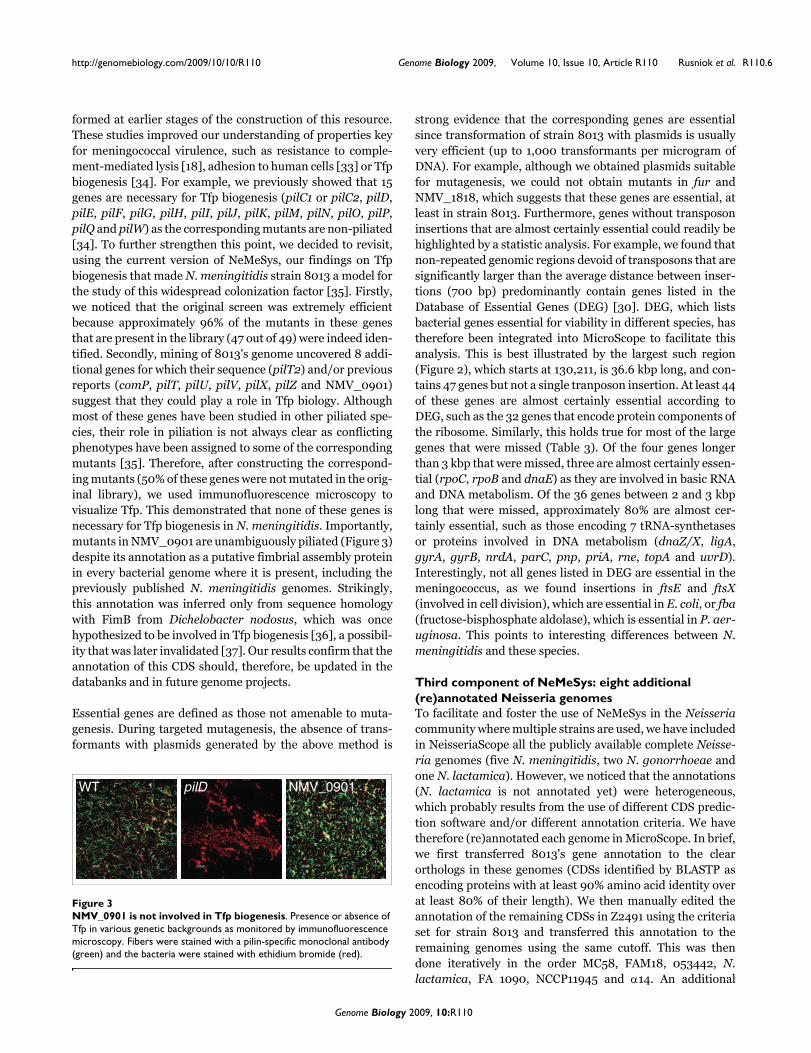
formed at earlier stages of the construction of this resource.These studies improved our understanding of properties keyfor meningococcal virulence, such as resistance to comple-ment-mediated lysis [18], adhesion to human cells [33] or Tfpbiogenesis [34]. For example, we previously showed that 15genes are necessary for Tfp biogenesis (pilC1 or pilC2, pilD,pilE, pilF, pilG, pilH, pilI, pilJ, pilK, pilM, pilN, pilO, pilP,pilQ and pilW) as the corresponding mutants are non-piliated[34]. To further strengthen this point, we decided to revisit,using the current version of NeMeSys, our findings on Tfpbiogenesis that made N. meningitidis strain 8013 a model forthe study of this widespread colonization factor [35]. Firstly,we noticed that the original screen was extremely efficientbecause approximately 96% of the mutants in these genesthat are present in the library (47 out of 49) were indeed iden-tified. Secondly, mining of 8013's genome uncovered 8 addi-tional genes for which their sequence (pilT2) and/or previousreports (comP, pilT, pilU, pilV, pilX, pilZ and NMV_0901)suggest that they could play a role in Tfp biology. Althoughmost of these genes have been studied in other piliated spe-cies, their role in piliation is not always clear as conflictingphenotypes have been assigned to some of the correspondingmutants [35]. Therefore, after constructing the correspond-ing mutants (50% of these genes were not mutated in the orig-inal library), we used immunofluorescence microscopy tovisualize Tfp. This demonstrated that none of these genes isnecessary for Tfp biogenesis in N. meningitidis. Importantly,mutants in NMV_0901 are unambiguously piliated (Figure 3)despite its annotation as a putative fimbrial assembly proteinin every bacterial genome where it is present, including thepreviously published N. meningitidis genomes. Strikingly,this annotation was inferred only from sequence homologywith FimB from Dichelobacter nodosus, which was oncehypothesized to be involved in Tfp biogenesis [36], a possibil-ity that was later invalidated [37]. Our results confirm that theannotation of this CDS should, therefore, be updated in thedatabanks and in future genome projects.
Essential genes are defined as those not amenable to muta-genesis. During targeted mutagenesis, the absence of trans-formants with plasmids generated by the above method is
strong evidence that the corresponding genes are essentialsince transformation of strain 8013 with plasmids is usuallyvery efficient (up to 1,000 transformants per microgram ofDNA). For example, although we obtained plasmids suitablefor mutagenesis, we could not obtain mutants in fur andNMV_1818, which suggests that these genes are essential, atleast in strain 8013. Furthermore, genes without transposoninsertions that are almost certainly essential could readily behighlighted by a statistic analysis. For example, we found thatnon-repeated genomic regions devoid of transposons that aresignificantly larger than the average distance between inser-tions (700 bp) predominantly contain genes listed in theDatabase of Essential Genes (DEG) [30]. DEG, which listsbacterial genes essential for viability in different species, hastherefore been integrated into MicroScope to facilitate thisanalysis. This is best illustrated by the largest such region(Figure 2), which starts at 130,211, is 36.6 kbp long, and con-tains 47 genes but not a single tranposon insertion. At least 44of these genes are almost certainly essential according toDEG, such as the 32 genes that encode protein components ofthe ribosome. Similarly, this holds true for most of the largegenes that were missed (Table 3). Of the four genes longerthan 3 kbp that were missed, three are almost certainly essen-tial (rpoC, rpoB and dnaE) as they are involved in basic RNAand DNA metabolism. Of the 36 genes between 2 and 3 kbplong that were missed, approximately 80% are almost cer-tainly essential, such as those encoding 7 tRNA-synthetasesor proteins involved in DNA metabolism (dnaZ/X, ligA,gyrA, gyrB, nrdA, parC, pnp, priA, rne, topA and uvrD).Interestingly, not all genes listed in DEG are essential in themeningococcus, as we found insertions in ftsE and ftsX(involved in cell division), which are essential in E. coli, or fba(fructose-bisphosphate aldolase), which is essential in P. aer-uginosa. This points to interesting differences between N.meningitidis and these species.
Third component of NeMeSys: eight additional (re)annotated Neisseria genomesTo facilitate and foster the use of NeMeSys in the Neisseriacommunity where multiple strains are used, we have includedin NeisseriaScope all the publicly available complete Neisse-ria genomes (five N. meningitidis, two N. gonorrhoeae andone N. lactamica). However, we noticed that the annotations(N. lactamica is not annotated yet) were heterogeneous,which probably results from the use of different CDS predic-tion software and/or different annotation criteria. We havetherefore (re)annotated each genome in MicroScope. In brief,we first transferred 8013's gene annotation to the clearorthologs in these genomes (CDSs identified by BLASTP asencoding proteins with at least 90% amino acid identity overat least 80% of their length). We then manually edited theannotation of the remaining CDSs in Z2491 using the criteriaset for strain 8013 and transferred this annotation to theremaining genomes using the same cutoff. This was thendone iteratively in the order MC58, FAM18, 053442, N.lactamica, FA 1090, NCCP11945 and α14. An additional
NMV_0901 is not involved in Tfp biogenesisFigure 3NMV_0901 is not involved in Tfp biogenesis. Presence or absence of Tfp in various genetic backgrounds as monitored by immunofluorescence microscopy. Fibers were stained with a pilin-specific monoclonal antibody (green) and the bacteria were stained with ethidium bromide (red).
WT pilD NMV_0901
Genome Biology 2009, 10:R110
http://genomebiology.com/2009/10/10/R110 Genome Biology 2009, Volume 10, Issue 10, Article R110 Rusniok et al. R110.7
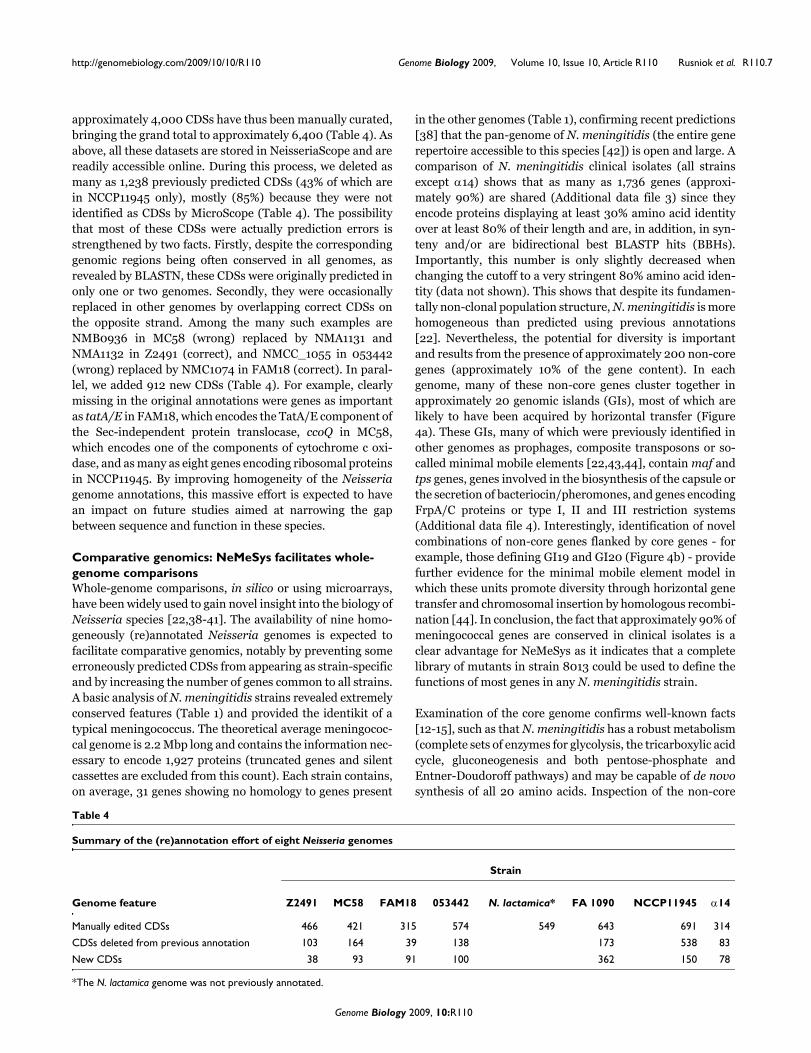
approximately 4,000 CDSs have thus been manually curated,bringing the grand total to approximately 6,400 (Table 4). Asabove, all these datasets are stored in NeisseriaScope and arereadily accessible online. During this process, we deleted asmany as 1,238 previously predicted CDSs (43% of which arein NCCP11945 only), mostly (85%) because they were notidentified as CDSs by MicroScope (Table 4). The possibilitythat most of these CDSs were actually prediction errors isstrengthened by two facts. Firstly, despite the correspondinggenomic regions being often conserved in all genomes, asrevealed by BLASTN, these CDSs were originally predicted inonly one or two genomes. Secondly, they were occasionallyreplaced in other genomes by overlapping correct CDSs onthe opposite strand. Among the many such examples areNMB0936 in MC58 (wrong) replaced by NMA1131 andNMA1132 in Z2491 (correct), and NMCC_1055 in 053442(wrong) replaced by NMC1074 in FAM18 (correct). In paral-lel, we added 912 new CDSs (Table 4). For example, clearlymissing in the original annotations were genes as importantas tatA/E in FAM18, which encodes the TatA/E component ofthe Sec-independent protein translocase, ccoQ in MC58,which encodes one of the components of cytochrome c oxi-dase, and as many as eight genes encoding ribosomal proteinsin NCCP11945. By improving homogeneity of the Neisseriagenome annotations, this massive effort is expected to havean impact on future studies aimed at narrowing the gapbetween sequence and function in these species.
Comparative genomics: NeMeSys facilitates whole-genome comparisonsWhole-genome comparisons, in silico or using microarrays,have been widely used to gain novel insight into the biology ofNeisseria species [22,38-41]. The availability of nine homo-geneously (re)annotated Neisseria genomes is expected tofacilitate comparative genomics, notably by preventing someerroneously predicted CDSs from appearing as strain-specificand by increasing the number of genes common to all strains.A basic analysis of N. meningitidis strains revealed extremelyconserved features (Table 1) and provided the identikit of atypical meningococcus. The theoretical average meningococ-cal genome is 2.2 Mbp long and contains the information nec-essary to encode 1,927 proteins (truncated genes and silentcassettes are excluded from this count). Each strain contains,on average, 31 genes showing no homology to genes present
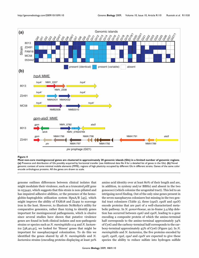
in the other genomes (Table 1), confirming recent predictions[38] that the pan-genome of N. meningitidis (the entire generepertoire accessible to this species [42]) is open and large. Acomparison of N. meningitidis clinical isolates (all strainsexcept α14) shows that as many as 1,736 genes (approxi-mately 90%) are shared (Additional data file 3) since theyencode proteins displaying at least 30% amino acid identityover at least 80% of their length and are, in addition, in syn-teny and/or are bidirectional best BLASTP hits (BBHs).Importantly, this number is only slightly decreased whenchanging the cutoff to a very stringent 80% amino acid iden-tity (data not shown). This shows that despite its fundamen-tally non-clonal population structure, N. meningitidis is morehomogeneous than predicted using previous annotations[22]. Nevertheless, the potential for diversity is importantand results from the presence of approximately 200 non-coregenes (approximately 10% of the gene content). In eachgenome, many of these non-core genes cluster together inapproximately 20 genomic islands (GIs), most of which arelikely to have been acquired by horizontal transfer (Figure4a). These GIs, many of which were previously identified inother genomes as prophages, composite transposons or so-called minimal mobile elements [22,43,44], contain maf andtps genes, genes involved in the biosynthesis of the capsule orthe secretion of bacteriocin/pheromones, and genes encodingFrpA/C proteins or type I, II and III restriction systems(Additional data file 4). Interestingly, identification of novelcombinations of non-core genes flanked by core genes - forexample, those defining GI19 and GI20 (Figure 4b) - providefurther evidence for the minimal mobile element model inwhich these units promote diversity through horizontal genetransfer and chromosomal insertion by homologous recombi-nation [44]. In conclusion, the fact that approximately 90% ofmeningococcal genes are conserved in clinical isolates is aclear advantage for NeMeSys as it indicates that a completelibrary of mutants in strain 8013 could be used to define thefunctions of most genes in any N. meningitidis strain.
Examination of the core genome confirms well-known facts[12-15], such as that N. meningitidis has a robust metabolism(complete sets of enzymes for glycolysis, the tricarboxylic acidcycle, gluconeogenesis and both pentose-phosphate andEntner-Doudoroff pathways) and may be capable of de novosynthesis of all 20 amino acids. Inspection of the non-core
Table 4
Summary of the (re)annotation effort of eight Neisseria genomes
Strain
Genome feature Z2491 MC58 FAM18 053442 N. lactamica* FA 1090 NCCP11945 α14
Manually edited CDSs 466 421 315 574 549 643 691 314
CDSs deleted from previous annotation 103 164 39 138 173 538 83
New CDSs 38 93 91 100 362 150 78
*The N. lactamica genome was not previously annotated.
Genome Biology 2009, 10:R110
http://genomebiology.com/2009/10/10/R110 Genome Biology 2009, Volume 10, Issue 10, Article R110 Rusniok et al. R110.8
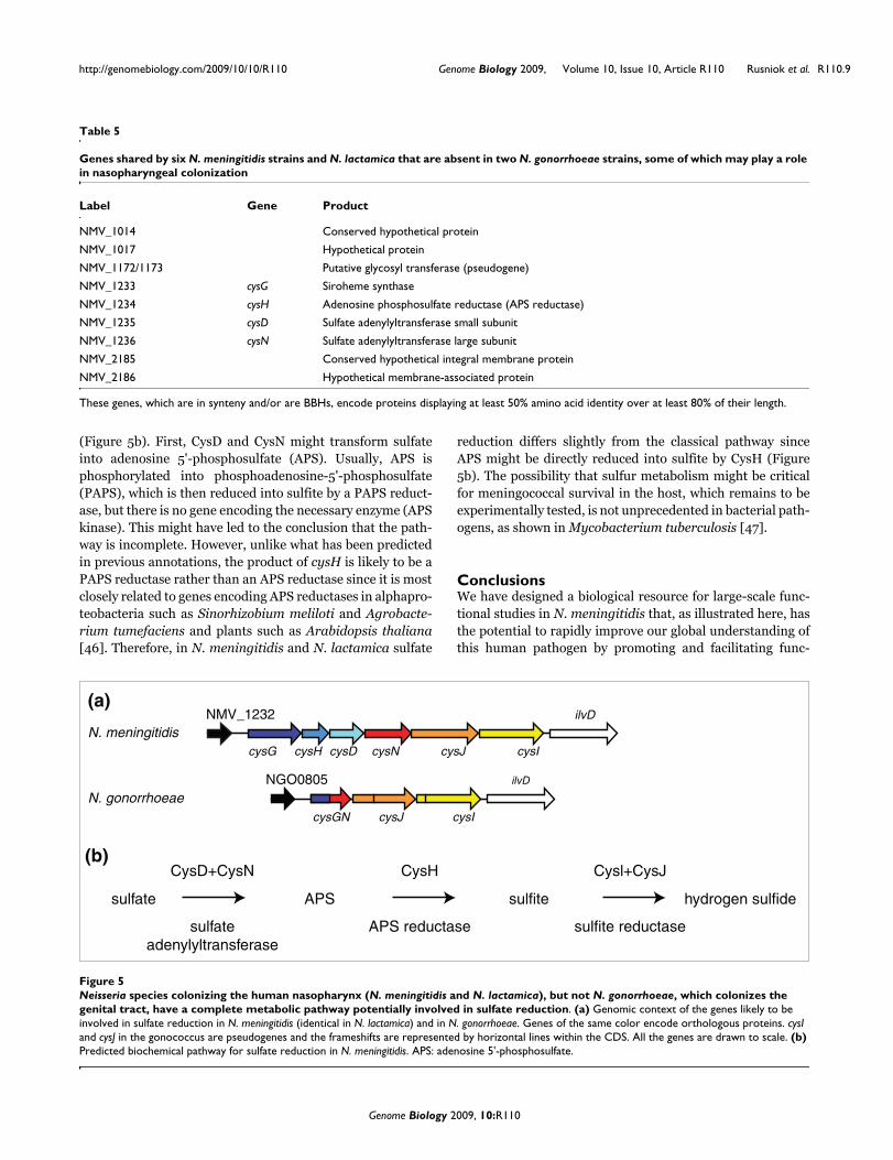
genome outlines differences between clinical isolates thatmight modulate their virulence, such as a truncated pilE genein 053442, which suggests that this strain is non-piliated andhas impaired adhesive abilities, or the presence of the hemo-globin-haptoglobin utilization system HpuA/B [45], whichmight improve the ability of FAM18 and Z2491 to scavengeiron in the host. However, to illustrate NeMeSys's utility forcomparative genomics, rather than trying to identify genesimportant for meningococcal pathogenesis, which is elusivesince several studies have shown that putative virulencegenes are found in both clinical isolates and non-pathogenicstrains or species such as N. meningitidis α14 and N. lactam-ica [38,40,41], we looked for 'fitness' genes that might beimportant for nasopharyngeal colonization. To do this weidentified the genes shared by all N. meningitidis and N.lactamica strains (encoding proteins displaying at least 50%
amino acid identity over at least 80% of their length and are,in addition, in synteny and/or BBHs) and absent in the twogonococci (which colonize the urogenital tract). This led to anintriguing novel finding. Out of the only nine genes present inthe seven nasopharynx colonizers but missing in the two gen-ital tract colonizers (Table 5), three (cysD, cysH and cysN)encode proteins that are part of a well-characterized meta-bolic pathway. In N. gonorrhoeae, an in-frame 3.4 kbp dele-tion has occurred between cysG and cysN, leading to a geneencoding a composite protein of which the amino-terminalhalf corresponds to the amino-terminal approximately 34%of CysG and the carboxy-terminal half corresponds to the car-boxy-terminal approximately 45% of CysG (Figure 5a). In N.meningitidis and N. lactamica, the five proteins encoded bycysD, cysH, cysI, cysJ and cysN are expected to give thesespecies the ability to reduce sulfate into hydrogen sulfide
Most non-core meningococcal genes are clustered in approximately 20 genomic islands (GIs) in a limited number of genomic regionsFigure 4Most non-core meningococcal genes are clustered in approximately 20 genomic islands (GIs) in a limited number of genomic regions. (a) Presence and distribution of GIs possibly acquired by horizontal transfer (see Additional data file 4 for a detailed list of genes in the GIs). (b) Novel genomic context of some minimal mobile elements (MME), regions of high plasticity occupied by different GIs in different strains. Genes of the same color encode orthologous proteins. All the genes are drawn to scale.
(a) Genomic islands
8013
Z2491
MC58
FAM18
053442
Str
ain
GI0
GI2
0/1
GI2
7
GI2
6
GI2
5
GI2
4
GI2
3
GI2
2
GI2
1
GI2
0
GI1
9/1
GI1
9
GI1
8
GI1
7
GI1
6
GI1
5
GI1
4
GI1
3
GI1
2
GI1
1
GI1
0
GI9
GI8
GI7
GI6
/1
GI6
GI5
GI4
/1
GI4
GI3
GI2
GI1
GI2
7/1
absentpresent (identical) present (variable)
(b)
NMV_2207
NMV_2208
NMA0431 NMA0432
NMB2008 NMB2010
hrpA MME
8013
Z2491
MC58
gpm-alaS MME
alaS
NMA1789piv
gpm
piv prophage (GI21)
NMA1791
NMA1792NMA1797
NMA1796NMA1799
gpm
tehA
NMV_0782
NMV_0783/0784
alaS
8013
Z2491
hrpA’ hrpA’
hrpA’hrpA’
hrpA’ hrpA’
Genome Biology 2009, 10:R110
http://genomebiology.com/2009/10/10/R110 Genome Biology 2009, Volume 10, Issue 10, Article R110 Rusniok et al. R110.9
(Figure 5b). First, CysD and CysN might transform sulfateinto adenosine 5'-phosphosulfate (APS). Usually, APS isphosphorylated into phosphoadenosine-5'-phosphosulfate(PAPS), which is then reduced into sulfite by a PAPS reduct-ase, but there is no gene encoding the necessary enzyme (APSkinase). This might have led to the conclusion that the path-way is incomplete. However, unlike what has been predictedin previous annotations, the product of cysH is likely to be aPAPS reductase rather than an APS reductase since it is mostclosely related to genes encoding APS reductases in alphapro-teobacteria such as Sinorhizobium meliloti and Agrobacte-rium tumefaciens and plants such as Arabidopsis thaliana[46]. Therefore, in N. meningitidis and N. lactamica sulfate
reduction differs slightly from the classical pathway sinceAPS might be directly reduced into sulfite by CysH (Figure5b). The possibility that sulfur metabolism might be criticalfor meningococcal survival in the host, which remains to beexperimentally tested, is not unprecedented in bacterial path-ogens, as shown in Mycobacterium tuberculosis [47].
ConclusionsWe have designed a biological resource for large-scale func-tional studies in N. meningitidis that, as illustrated here, hasthe potential to rapidly improve our global understanding ofthis human pathogen by promoting and facilitating func-
Table 5
Genes shared by six N. meningitidis strains and N. lactamica that are absent in two N. gonorrhoeae strains, some of which may play a role in nasopharyngeal colonization
Label Gene Product
NMV_1014 Conserved hypothetical protein
NMV_1017 Hypothetical protein
NMV_1172/1173 Putative glycosyl transferase (pseudogene)
NMV_1233 cysG Siroheme synthase
NMV_1234 cysH Adenosine phosphosulfate reductase (APS reductase)
NMV_1235 cysD Sulfate adenylyltransferase small subunit
NMV_1236 cysN Sulfate adenylyltransferase large subunit
NMV_2185 Conserved hypothetical integral membrane protein
NMV_2186 Hypothetical membrane-associated protein
These genes, which are in synteny and/or are BBHs, encode proteins displaying at least 50% amino acid identity over at least 80% of their length.
Neisseria species colonizing the human nasopharynx (N. meningitidis and N. lactamica), but not N. gonorrhoeae, which colonizes the genital tract, have a complete metabolic pathway potentially involved in sulfate reductionFigure 5Neisseria species colonizing the human nasopharynx (N. meningitidis and N. lactamica), but not N. gonorrhoeae, which colonizes the genital tract, have a complete metabolic pathway potentially involved in sulfate reduction. (a) Genomic context of the genes likely to be involved in sulfate reduction in N. meningitidis (identical in N. lactamica) and in N. gonorrhoeae. Genes of the same color encode orthologous proteins. cysI and cysJ in the gonococcus are pseudogenes and the frameshifts are represented by horizontal lines within the CDS. All the genes are drawn to scale. (b) Predicted biochemical pathway for sulfate reduction in N. meningitidis. APS: adenosine 5'-phosphosulfate.
cysG cysH cysD cysN cysJ cysI
NMV_1232 ilvD
N. meningitidis
cysGN cysJ cysI
NGO0805 ilvD
N. gonorrhoeae
(a)
(b)
sulfate
sulfate
adenylyltransferase
CysD+CysN
APS
CysH
APS reductase
sulfite
Cysl+CysJ
sulfite reductase
hydrogen sulfide
Genome Biology 2009, 10:R110
http://genomebiology.com/2009/10/10/R110 Genome Biology 2009, Volume 10, Issue 10, Article R110 Rusniok et al. R110.10
tional and comparative genomics studies. NeMeSys is viewedas an evolving resource that will be improved, for example,through completion of the collection of mutants (eitherthrough gene-by-gene or systematic targeted mutagenesis ofthe missed genes), further improvement of the accuracy of theannotation by taking into account any new experimental evi-dence, improvement of the website design and content, andaddition of new Neisseria genomes as they become available.There is no doubt that NeMeSys would requite these efforts(thereby justifying its name, which was inspired by an ancientGreek goddess seen as the spirit of divine retribution) by fur-ther improving our capacity to understand gene function inN. meningitidis. Ideally, such studies could contribute to theongoing efforts aimed at comprehensively understanding aprokaryotic cell and help in the design of new therapies.
Materials and methodsBacterial strains and growth conditionsThe sequenced strain (also known as clone 12 or 2C43) is anaturally occurring pilin antigenic variant of the original clin-ical isolate N. meningitidis 8013, which expresses a pilinmediating better adherence to human cells [48]. Meningo-cocci were grown at 37°C in a moist atmosphere containing5% CO2 on GCB agar plates containing Kellog's supplementsand, when required, 100 μg/ml kanamycin. E. coli TOP10(Invitrogen, Paisley, Renfrewshire, UK), DH5α or DH5α λpirwere grown at 37°C in liquid or solid Luria-Bertani medium(Difco, Oxford, Oxfordshire, UK), which contained 100 μg/mlampicillin, 100 μg/ml spectinomycin and/or 50 μg/ml kan-amycin, when appropriate.
Genome sequencingThe complete genome sequence of strain 8013[EMBL:FM999788] was determined by a whole genomeshotgun using a library of small inserts in pcDNA 2.1 (Invitro-gen). We obtained and assembled 32,338 sequences usingdye-terminator chemistry, which gave an approximatelynine-fold coverage of the genome. End sequencing of largeinserts in a pBeloBAC11 library aided in assembly verificationand scaffolding of contigs.
Genome (re)annotationsStrain 8013's genome was annotated using the previouslydescribed MicroScope annotation pipeline [19], which hasembedded software for syntactic analysis and more than 20well-known bioinformatics methods (InterProScan, COGni-tor, PRIAM, tmHMM, SignalP, and so on). In brief, potentialCDSs were first predicted by the AMIGene software [49]using three specific gene models identified by codon usageanalysis, tRNA were identified using tRNAscan-SE [50],rRNA using RNAmmer [51] and other RNA by scanning theRfam database [52]. CDSs were assigned a unique NMV_identifier and were submitted to automatic functional anno-tation in MicroScope [19]. Functional annotation, syntactichomogeneity and start codon position of each CDS present in
the genome were then refined manually during three roundsof inspection of the results obtained using the above bioinfor-matics methods. This led to four major classes: CDSs encod-ing proteins of known function (high homology to proteins ofdefined function), for which the SwissProt annotation wasmost often used; CDSs encoding proteins of putative function(conserved protein motif/structural features or limitedhomology to proteins of defined function), which werelabeled with the prefix 'putative'; and CDSs encoding proteinsof unknown function defined either as 'conserved hypotheti-cal protein' (significant homology to proteins of unkown func-tion outside of Neisseria species) or 'hypothetical protein' (nosignificant homology outside of Neisseria species). However,adjectives were added when localization of the correspondingproteins could be predicted through tmHMM [53] or SignalP[54] (for example, 'hypothetical periplasmic protein' or 'con-served hypothetical integral membrane protein') or proteinmotifs not allowing functional predictions were identifiedthrough InterProScan [55] (for example, 'conserved hypo-thetical TPR-containing protein'). Importantly, during themanual curation of CDSs encoding proteins of unknown func-tion, the dubious ones (typically those with less than 50%coding probability, shorter than 150 bp, overlapping withhighly probable CDSs or RNA on the opposite strand, and soon) were deleted. During this process, self-explanatory com-ments mostly based on InterProScan entries and links to rel-evant literature in PubMed (139 in total) were enteredmanually in the database.
To define truncated genes, for which only partial homologiescould be detected, or out of phase genes, for which homologywas complete but involved at least two consecutive CDSs, weused BLASTP and coding probability results. The corre-sponding open reading frames were trimmed to their biolog-ically significant portions (both on 5' and 3') and labeled withthe prefix 'truncated' or the suffix 'pseudogene', respectively.During this process, putative frameshifts or sequencingerrors in 42 CDSs were amplified and resequenced.
All Neisseria genomes available in GenBank (MC58, Z2491,FAM18, 053442, α14, FA 1090 and NCCP11945) or at theSanger Institute (N. lactamica) were (re)annotated in Micro-Scope using the same approach as above. AMIGene was usedto predict the CDSs, labeling the new ones with a distinctidentifier (for example, NEIMA instead of NMA in Z2491),which were submitted to automatic functional annotation inMicroScope. The functional annotation in N. meningitidisstrain 8013 was then automatically transferred to all clearorthologs, stringently defined as genes endoding proteinsshowing at least 90% BLASTP identity over at least 80% oftheir length. All the remaining CDSs were then annotatedmanually using the same procedure as for strain 8013, start-ing with Z2491 and transferring this new annotation to theremaining genomes using the same cutoff. This was thendone iteratively in the order MC58, FAM18, 053442, N.lactamica, FA 1090, NCCP11945 and α14. Importantly, previ-
Genome Biology 2009, 10:R110

http://genomebiology.com/2009/10/10/R110 Genome Biology 2009, Volume 10, Issue 10, Article R110 Rusniok et al. R110.11
ously predicted CDSs that were not recognized as such byAMIGene were deleted during the process.
Genomic analysesAll the genomic analyses were performed within MicroScopeusing embedded software. Whole-genome comparisons ofgene content (using the mentioned cutoffs) were done usingthe PhyloProfile Synteny functionality [19], which combinesBLASTP, BBH and/or synteny results. Graphical representa-tion of whole-genome syntenies were generated usingLinePlot functionality [19]. Graphical circular representationof the strain 8013 genome with transposon insertions wasgenerated using the CGView software [56]. Characterizationof the sulfate reduction pathway in Neisseria strains coloniz-ing the nasopharynx was done using metabolic pathway pre-dictions built with the Pathway Tools software [57]. GIs ofputative horizontally transferred genes were identified ineach N. meningitidis clinical isolate using the Genomic Islandfunctionality tool [19]. This tool combines detection of syn-teny break points in the query genome in comparison withclosely related genomes, searches for mobility genes, tRNAand direct repeats (if any) at the borders of the synteny breakpoints and finally searches for compositional bias in the querygenome.
Genome-wide collection of defined mutantsThe construction of an archived library of undefined transpo-son mutants in strain 8013 and the design/validation of amethod for large-scale characterization of transposon inser-tion sites based on ligation-mediated PCR have beendescribed [18]. Each mutant is assigned a unique x/y identi-fier, where x indicates the half microtitre plate and y the posi-tion of the mutant. Genomic DNA for each mutant, preparedusing the Wizard Genomic DNA Purification kit (Promega,Southampton, Hampshire, UK), was used to try to amplifysequences flanking the inserted transposons mainly by liga-tion-mediated PCR (other techniques have been tested aswell). Amplified fragments were sequenced with outward-reading primers ISL or ISR internal to the transposon [18].Sequences were trimmed to eliminate regions of poor qualityor corresponding to the transposon and subsequentlymapped on 8013's genome using BLASTN.
Additional mutants were engineered by in vitro transposonmutagenesis on PCR products cloned into pCRII-TOPO orpCR8/GW/TOPO vectors (both from Invitrogen). Initially,mutants in six genes involved in Tfp biology (pilM, pilN, pilO,pilT, pilU and pilZ), four of which have been described previ-ously [58], were constructed by directly transforming trans-position reactions into strain 8013. We used as a donor thepSM1 vector in which the transposon is cloned within a plas-mid with a ColE1 origin of replication [31]. However, the effi-ciency was low, with only zero to two mutants pertransposition reaction. Subsequently, we modified thismethod for high-throughput use by subcloning the mini-transposon into plasmid pGP704, which has a R6K origin of
replication. The mini-transposon, extracted from pSM1 on aXbaI-EcoRI fragment, was cloned into XbaI-EcoRI-cut plas-mid pGP704 [32]. The resulting plasmid pYU29 can replicateonly in the presence of Pir, which is found in E. coli strainssuch as DH5α λpir. Therefore, upon transformation of analiquot of the in vitro transposition reaction in DH5α andselection on plates containing kanamycin (cassette in themini-transposon) and spectinomycin (cassette in the targetvector), target plasmids with an inserted mini-transposon canbe positively selected. As seen initially with the comP andNMV_0901 genes, hundreds of Spr, Kmr transformants couldeasily be obtained while no transformants were obtainedwhen no transposase was added in the transposition reaction(data not shown). Restriction analysis of recombinant plas-mids confirmed that they contained an inserted transposon(data not shown). Transformants containing plasmids suita-ble for N. meningitidis mutagenesis - that is, with an insertionapproximately in the middle of the target gene - were readilyidentified by colony-PCR by using a mix of ISL and ISR, andthe forward primer used to amplify the target gene. Plasmidswere then extracted, used to sequence the site of transposoninsertion with ISL or ISR, and transformed in N. meningi-tidis. This method was validated by constructing mutants in20 genes (NMV_0125, NMV_0126, NMV_0323, NMV_0419,NMV_0433, mtrR, NMV_0658, NMV_0757, NMV_0773,NMV_0774, NMV_0901, hexR, iscR, NMV_1093,NMV_1134, NMV_1850, NMV_2068, NMV_2160, comP andNMV_2258).
Tfp detectionTfps were detected by immunofluorescence microscopy usingthe 20D9 monoclonal antibody, which is specific for the pilinin strain 8013 as described elsewhere [34]. This was doneusing a Nikon Eclipse E600 microscope and digital imageswere recorded with a Nikon DXM1200 digital cameramounted onto the microscope.
Data sharingAs usual in MicroScope [19], all the datasets generated duringthis study have been stored within PkDGB in a thematic sub-database named NeisseriaScope, which is publicly accessiblethrough MaGe. The MaGe web interface can be used to visu-alize genomes (simultaneously with synteny maps in othermicrobial genomes, one of its main features), perform queries(by BLAST or keyword searches) and download all datasets ina variety of formats (including EMBL and GenBank). How-ever, to facilitate access to the genome (re)annotations anddistribution of mutants to the scientific community, we havedesigned a straightforward webpage [28] providing directlinks to some of the most salient features in MicroScope. Ifneeded, once in NeisseriaScope, the user has unlimited accessto the whole array of exploratory tools within the MicroScopeplatform. Eventually, upon completion, the library of mutantswill be made entirely and freely available. In the meantime,up to ten mutants can be requested simultaneously.
Genome Biology 2009, 10:R110
http://genomebiology.com/2009/10/10/R110 Genome Biology 2009, Volume 10, Issue 10, Article R110 Rusniok et al. R110.12
AbbreviationsAPS: adenosine 5'-phosphosulfate: BBH: bi-directional bestBLASTP hit; CDS: coding sequence; DEG: Database of Essen-tial Genes; GI: genomic island; PAPS: phosphoadenosine-5'-phosphosulfate; PkDGB: Prokaryotic Genome DataBase; Tfp:type IV pilus.
Authors' contributionsCR, CB, PG and VP sequenced and assembled strain 8013'sgenome. DV, AL and CM contributed and managed bioinfor-matics resources. DV and VP performed manual annotationand bioinformatics analyses. SF and CMS sequenced transpo-son insertion sites in the library of mutants. HE and VP con-structed mutants by targeted mutagenesis. DB and VPperformed the functional characterization of Tfp biogenesis.VP conceived the study and was responsible for its coordina-tion. CR, DV, CB, CM, PG and VP wrote the paper.
Additional data filesThe following additional data are available with the onlineversion of this paper: a figure showing global pairwisegenome syntenies between strain 8013 and each sequencedN. meningitidis strain (Additional data file 1); a table listinggenes in strain 8013 that have been disrupted in the collectionof mutants (Additional data file 2); a table listing genesshared by all N. meningitidis clinical isolates (Additional datafile 3); a table listing the genomic islands in each N. meningi-tidis clinical isolate likely to have been acquired by horizontaltransfer (Additional data file 4).Additional data file 1Global pairwise synteny between the genome of strain 8013 and the other N. meningitidis genomesStrand conservation is indicated in purple, while strand inversions (because of chromosomal inversions) are in blue. Except in strain α14 where there is conserved colinearity, synteny is mainly dis-rupted by a single chromosomal inversion between the two genes that are indicated. For the 8013/Z2491 comparison, the readout is more difficult because the start of the Z2491 genome was not assigned at the origin of replication. Plots were generated using the LinePlot program within MicroScope after setting the minimum synton size to 40 genes.Click here for fileAdditional data file 2Complete list of genes that have been disrupted in the collection of mutants in strain 8013Genes disrupted through targeted mutagenesis are shaded in light blue.Click here for fileAdditional data file 3Complete list of genes shared by all N. meningitidis clinical isolates (meningococcal core genome) with their annotation and label in each genomeThese genes, which are in synteny and/or are BBHs, encode pro-teins displaying at least 30% amino acid identity over at least 80% of their length.Click here for fileAdditional data file 4Complete list of the genomic islands in each N. meningitidis clini-cal isolate and their distribution in other genomesFor each GI, the first and the last gene are indicated, together with a comment about their putative roles.Click here for file
AcknowledgementsThis work was funded by INSERM and grants from Institut Pasteur/CHUNecker-Enfants Malades, MRT and ANR. We are grateful to Elisabeth Cou-vé (Institut Pasteur) for help with the construction of DNA libraries, ZoéRouy (CEA-Institut de Génomique-Génoscope) for help with databasemanaging and Olivera Francetic (Institut Pasteur) for the kind gift ofpGP704. We thank Xavier Nassif (INSERM) for support. We thank StephenBentley (Wellcome Trust Sanger Institute) for permission to use theunpublished N. lactamica genome sequence. We thank Chiara Recchi (Impe-rial College London), Jean-Marc Reyrat (INSERM) and Christoph Tang(Imperial College London) for critical reading of the manuscript.
References1. Payne DJ, Gwynn MN, Holmes DJ, Pompliano DL: Drugs for bad
bugs: confronting the challenges of antibacterial discovery.Nat Rev Drug Discov 2007, 6:29-40.
2. Scherens B, Goffeau A: The uses of genome-wide yeast mutantcollections. Genome Biol 2004, 5:229.
3. Baba T, Ara T, Hasegawa M, Takai Y, Okumura Y, Baba M, DatsenkoKA, Tomita M, Wanner BL, Mori H: Construction of Escherichiacoli K-12 in-frame, single-gene knockout mutants: the Keiocollection. Mol Syst Biol. 2006, 2:.
4. Kobayashi K, Ehrlich SD, Albertini A, Amati G, Andersen KK, ArnaudM, Asai K, Ashikaga S, Aymerich S, Bessieres P, Boland F, Brignell SC,Bron S, Bunai K, Chapuis J, Christiansen LC, Danchin A, DebarbouilleM, Dervyn E, Deuerling E, Devine K, Devine SK, Dreesen O, Err-ington J, Fillinger S, Foster SJ, Fujita Y, Galizzi A, Gardan R, EschevinsC, et al.: Essential Bacillus subtilis genes. Proc Natl Acad Sci USA2003, 100:4678-4683.
5. de Berardinis V, Vallenet D, Castelli V, Besnard M, Pinet A, Cruaud C,Samair S, Lechaplais C, Gyapay G, Richez C, Durot M, Kreimeyer A,Le Fevre F, Schachter V, Pezo V, Doring V, Scarpelli C, Medigue C,Cohen GN, Marliere P, Salanoubat M, Weissenbach J: A completecollection of single-gene deletion mutants of Acinetobacterbaylyi ADP1. Mol Syst Biol 2008, 4:174.
6. Bae T, Banger AK, Wallace A, Glass EM, Aslund F, Schneewind O,Missiakas DM: Staphylococcus aureus virulence genes identifiedby bursa aurealis mutagenesis and nematode killing. Proc NatlAcad Sci USA 2004, 101:12312-12317.
7. Jacobs MA, Alwood A, Thaipisuttikul I, Spencer D, Haugen E, Ernst S,Will O, Kaul R, Raymond C, Levy R, Chun-Rong L, Guenthner D,Bovee D, Olson MV, Manoil C: Comprehensive transposonmutant library of Pseudomonas aeruginosa. Proc Natl Acad SciUSA 2003, 100:14339-14344.
8. Liberati NT, Urbach JM, Miyata S, Lee DG, Drenkard E, Wu G, Vil-lanueva J, Wei T, Ausubel FM: An ordered, nonredundant libraryof Pseudomonas aeruginosa strain PA14 transposon insertionmutants. Proc Natl Acad Sci USA 2006, 103:2833-2838.
9. Gallagher LA, Ramage E, Jacobs MA, Kaul R, Brittnacher M, Manoil C:A comprehensive transposon mutant library of Francisellanovicida, a bioweapon surrogate. Proc Natl Acad Sci USA 2007,104:1009-1014.
10. Glass JI, Assad-Garcia N, Alperovich N, Yooseph S, Lewis MR, MarufM, Hutchison CA 3rd, Smith HO, Venter JC: Essential genes of aminimal bacterium. Proc Natl Acad Sci USA 2006, 103:425-430.
11. Suzuki N, Okai N, Nonaka H, Tsuge Y, Inui M, Yukawa H: High-throughput transposon mutagenesis of Corynebacteriumglutamicum and construction of a single-gene disruptantmutant library. Appl Environ Microbiol 2006, 72:3750-3755.
12. Tettelin H, Saunders NJ, Heidelberg J, Jeffries AC, Nelson KE, EisenJA, Ketchum KA, Hood DW, Peden JF, Dodson RJ, Nelson WC,Gwinn ML, DeBoy R, Peterson JD, Hickey EK, Haft DH, Salzberg SL,White O, Fleischmann RD, Dougherty BA, Mason T, Ciecko A, Park-sey DS, Blair E, Cittone H, Clark EB, Cotton MD, Utterback TR,Khouri H, Qin H, et al.: Complete genome sequence of Neisseriameningitidis serogroup B strain MC58. Science 2000,287:1809-1815.
13. Parkhill J, Achtman M, James KD, Bentley SD, Churcher C, Klee SR,Morelli G, Basham D, Brown D, Chillingworth T, Davies RM, Davis P,Devlin K, Feltwell T, Hamlin N, Holroyd S, Jagels K, Leather S, MouleS, Mungall K, Quail MA, Rajandream M-A, Rutherford KM, SimmondsM, Skelton J, Whitehead S, Spratt BG, Barrell BG: Complete DNAsequence of a serogroup A strain of Neisseria meningitidisZ2491. Nature 2000, 404:502-506.
14. Bentley SD, Vernikos GS, Snyder LA, Churcher C, Arrowsmith C,Chillingworth T, Cronin A, Davis PH, Holroyd NE, Jagels K, MaddisonM, Moule S, Rabbinowitsch E, Sharp S, Unwin L, Whitehead S, QuailMA, Achtman M, Barrell B, Saunders NJ, Parkhill J: Meningococcalgenetic variation mechanisms viewed through comparativeanalysis of serogroup C strain FAM18. PLoS Genet 2007, 3:e23.
15. Peng J, Yang L, Yang F, Yang J, Yan Y, Nie H, Zhang X, Xiong Z, JiangY, Cheng F, Xu X, Chen S, Sun L, Li W, Shen Y, Shao Z, Liang X, XuJ, Jin Q: Characterization of ST-4821 complex, a unique Neis-seria meningitidis clone. Genomics 2008, 91:78-87.
16. Rosenstein NE, Perkins BA, Stephens DS, Popovic T, Hughes JM:Meningococcal disease. N Engl J Med 2001, 344:1378-1388.
17. Anonymous: Outbreak news. Meningococcal disease, Africanmeningitis belt, epidemic season 2006. Wkly Epidemiol Rec 2006,81:119-120.
18. Geoffroy M, Floquet S, Métais A, Nassif X, Pelicic V: Large-scaleanalysis of the meningococcus genome by gene disruption:resistance to complement-mediated lysis. Genome Res 2003,13:391-398.
19. Vallenet D, Labarre L, Rouy Z, Barbe V, Bocs S, Cruveiller S, Lajus A,Pascal G, Scarpelli C, Medigue C: MaGe: a microbial genomeannotation system supported by synteny results. Nucleic AcidsRes 2006, 34:53-65.
20. Schoen C, Blom J, Claus H, Schramm-Gluck A, Brandt P, Muller T,Goesmann A, Joseph B, Konietzny S, Kurzai O, Schmitt C, FriedrichT, Linke B, Vogel U, Frosch M: Whole-genome comparison ofdisease and carriage strains provides insights into virulenceevolution in Neisseria meningitidis. Proc Natl Acad Sci USA 2008,105:3473-3478.
21. Chung GT, Yoo JS, Oh HB, Lee YS, Cha SH, Kim SJ, Yoo CK: Com-plete genome sequence of Neisseria gonorrhoeaeNCCP11945. J Bacteriol 2008, 190:6035-6036.
22. Hotopp JC, Grifantini R, Kumar N, Tzeng YL, Fouts D, Frigimelica E,
Genome Biology 2009, 10:R110
http://genomebiology.com/2009/10/10/R110 Genome Biology 2009, Volume 10, Issue 10, Article R110 Rusniok et al. R110.13
Draghi M, Giuliani MM, Rappuoli R, Stephens DS, Grandi G, TettelinH: Comparative genomics of Neisseria meningitidis: coregenome, islands of horizontal transfer and pathogen-specificgenes. Microbiology 2006, 152:3733-3749.
23. Mackiewicz P, Zakrzewska-Czerwinska J, Zawilak A, Dudek MR,Cebrat S: Where does bacterial replication start? Rules forpredicting the oriC region. Nucleic Acids Res 2004, 32:3781-3791.
24. Laskos L, Dillard JP, Seifert HS, Fyfe JA, Davies JK: The pathogenicneisseriae contain an inactive rpoN gene and do not utilizethe pilE sigma54 promoter. Gene 1998, 208:95-102.
25. Hagblom P, Segal E, Billyard E, So M: Intragenic recombinationleads to pilus antigenic variation in Neisseria gonorrhoeae.Nature 1985, 315:156-158.
26. Haas R, Meyer TF: The repertoire of silent pilus genes in Neis-seria gonorrhoeae: evidence for gene conversion. Cell 1986,44:107-115.
27. Martin P, Ven T van de, Mouchel N, Jeffries AC, Hood DW, MoxonER: Experimentally revised repertoire of putative contin-gency loci in Neisseria meningitidis strain MC58: evidence fora novel mechanism of phase variation. Mol Microbiol 2003,50:245-257.
28. NeMeSys: a Biological Resource for Neisseria meningitidisSystematic Functional Analysis [http://www.genoscope.cns.fr/agc/nemesys]
29. Dirix G, Monsieurs P, Dombrecht B, Daniels R, Marchal K, Vander-leyden J, Michiels J: Peptide signal molecules and bacteriocins inGram-negative bacteria: a genome-wide in silico screeningfor peptides containing a double-glycine leader sequence andtheir cognate transporters. Peptides 2004, 25:1425-1440.
30. Zhang R, Lin Y: DEG 5.0, a database of essential genes in bothprokaryotes and eukaryotes. Nucleic Acids Res 2009,37:D455-458.
31. Pelicic V, Morelle S, Lampe D, Nassif X: Mutagenesis of Neisseriameningitidis by in vitro transposition of Himar1 mariner. J Bac-teriol 2000, 182:5391-5398.
32. Miller VL, Mekalanos JJ: A novel suicide vector and its use in con-struction of insertion mutations: osmoregulation of outermembrane proteins and virulence determinants in Vibriocholerae requires toxR. J Bacteriol 1988, 170:2575-2583.
33. Helaine S, Carbonnelle E, Prouvensier L, Beretti J-L, Nassif X, PelicicV: PilX, a pilus-associated protein essential for bacterialaggregation, is a key to pilus-facilitated attachment of Neis-seria meningitidis to human cells. Mol Microbiol 2005, 55:65-77.
34. Carbonnelle E, Helaine S, Nassif X, Pelicic V: A systematic geneticanalysis in Neisseria meningitidis defines the Pil proteinsrequired for assembly, functionality, stabilization and exportof type IV pili. Mol Microbiol 2006, 61:1510-1522.
35. Pelicic V: Type IV pili: e pluribus unum? Mol Microbiol 2008,68:827-837.
36. Hobbs M, Dalrymple BP, Cox PT, Livingstone SP, Delaney SF, MattickJS: Organization of the fimbrial gene region of Bacteroidesnodosus: class I and class II strains. Mol Microbiol 1991,5:543-560.
37. Kennan RM, Dhungyel OP, Whittington RJ, Egerton JR, Rood JI: Thetype IV fimbrial subunit gene (fimA) of Dichelobacter nodosusis essential for virulence, protease secretion, and naturalcompetence. J Bacteriol 2001, 183:4451-4458.
38. Schoen C, Tettelin H, Parkhill J, Frosch M: Genome flexibility inNeisseria meningitidis. Vaccine 2009, 27:B103-111.
39. Bille E, Zahar JR, Perrin A, Morelle S, Kriz P, Jolley KA, Maiden MC,Dervin C, Nassif X, Tinsley CR: A chromosomally integratedbacteriophage in invasive meningococci. J Exp Med 2005,201:1905-1913.
40. Snyder LA, Saunders NJ: The majority of genes in the patho-genic Neisseria species are present in non-pathogenic Neisse-ria lactamica, including those designated as 'virulence genes'.BMC Genomics 2006, 7:128.
41. Stabler RA, Marsden GL, Witney AA, Li Y, Bentley SD, Tang CM,Hinds J: Identification of pathogen-specific genes throughmicroarray analysis of pathogenic and commensal Neisseriaspecies. Microbiology 2005, 151:2907-2922.
42. Tettelin H, Riley D, Cattuto C, Medini D: Comparative genomics:the bacterial pan-genome. Curr Opin Microbiol 2008, 11:472-477.
43. Masignani V, Giuliani MM, Tettelin H, Comanducci M, Rappuoli R,Scarlato V: Mu-like Prophage in serogroup B Neisseria menin-gitidis coding for surface-exposed antigens. Infect Immun 2001,69:2580-2588.
44. Snyder LA, McGowan S, Rogers M, Duro E, O'Farrell E, Saunders NJ:
The repertoire of minimal mobile elements in the Neisseriaspecies and evidence that these are involved in horizontalgene transfer in other bacteria. Mol Biol Evol 2007,24:2802-2815.
45. Lewis LA, Gray E, Wang Y-P, Roe BA, Dyer DW: Molecular char-acterization of hpuAB, the haemoglobin-haptoglobin-utiliza-tion operon of Neisseria meningitidis. Mol Microbiol 1997,23:737-749.
46. Abola AP, Willits MG, Wang RC, Long SR: Reduction of adenos-ine-5'-phosphosulfate instead of 3'-phosphoadenosine-5'-phosphosulfate in cysteine biosynthesis by Rhizobium melilotiand other members of the family Rhizobiaceae. J Bacteriol1999, 181:5280-5287.
47. Schelle MW, Bertozzi CR: Sulfate metabolism in mycobacteria.Chembiochem 2006, 7:1516-1524.
48. Nassif X, Lowy J, Stenberg P, O'Gaora P, Ganji A, So M: Antigenicvariation of pilin regulates adhesion of Neisseria meningitidisto human epithelial cells. Mol Microbiol 1993, 8:719-725.
49. Bocs S, Cruveiller S, Vallenet D, Nuel G, Medigue C: AMIGene:Annotation of MIcrobial Genes. Nucleic Acids Res 2003,31:3723-3726.
50. Lowe TM, Eddy SR: tRNAscan-SE: a program for improveddetection of transfer RNA genes in genomic sequence.Nucleic Acids Res 1997, 25:955-964.
51. Lagesen K, Hallin P, Rodland EA, Staerfeldt HH, Rognes T, UsseryDW: RNAmmer: consistent and rapid annotation of ribos-omal RNA genes. Nucleic Acids Res 2007, 35:3100-3108.
52. Gardner PP, Daub J, Tate JG, Nawrocki EP, Kolbe DL, Lindgreen S,Wilkinson AC, Finn RD, Griffiths-Jones S, Eddy SR, Bateman A: Rfam:updates to the RNA families database. Nucleic Acids Res 2009,37:D136-140.
53. Sonnhammer EL, von Heijne G, Krogh A: A hidden Markov modelfor predicting transmembrane helices in protein sequences.Proc Int Conf Intell Syst Mol Biol 1998, 6:175-182.
54. Bendtsen JD, Nielsen H, von Heijne G, Brunak S: Improved predic-tion of signal peptides: SignalP 3.0. J Mol Biol 2004, 340:783-795.
55. Hunter S, Apweiler R, Attwood TK, Bairoch A, Bateman A, Binns D,Bork P, Das U, Daugherty L, Duquenne L, Finn RD, Gough J, Haft D,Hulo N, Kahn D, Kelly E, Laugraud A, Letunic I, Lonsdale D, Lopez R,Madera M, Maslen J, McAnulla C, McDowall J, Mistry J, Mitchell A,Mulder N, Natale D, Orengo C, Quinn AF, et al.: InterPro: the inte-grative protein signature database. Nucleic Acids Res 2009,37:D211-215.
56. Stothard P, Wishart DS: Circular genome visualization andexploration using CGView. Bioinformatics 2005, 21:537-539.
57. Karp PD, Paley S, Romero P: The Pathway Tools software. Bioin-formatics 2002, 18:S225-232.
58. Carbonnelle E, Helaine S, Prouvensier L, Nassif X, Pelicic V: Type IVpilus biogenesis in Neisseria meningitidis: PilW is involved in astep occuring after pilus assembly, essential for fiber stabilityand function. Mol Microbiol 2005, 55:54-64.
Genome Biology 2009, 10:R110
http://www.ncbi.nlm.nih.gov/entrez/query.fcgi?cmd=Retrieve&db=PubMed&dopt=Abstract&list_uids=9479056
http://www.ncbi.nlm.nih.gov/entrez/query.fcgi?cmd=Retrieve&db=PubMed&dopt=Abstract&list_uids=2859529
http://www.ncbi.nlm.nih.gov/entrez/query.fcgi?cmd=Retrieve&db=PubMed&dopt=Abstract&list_uids=2866848
http://www.ncbi.nlm.nih.gov/entrez/query.fcgi?cmd=Retrieve&db=PubMed&dopt=Abstract&list_uids=2836362
http://www.ncbi.nlm.nih.gov/entrez/query.fcgi?cmd=Retrieve&db=PubMed&dopt=Abstract&list_uids=1675418
http://www.ncbi.nlm.nih.gov/entrez/query.fcgi?cmd=Retrieve&db=PubMed&dopt=Abstract&list_uids=9157245
http://www.ncbi.nlm.nih.gov/entrez/query.fcgi?cmd=Retrieve&db=PubMed&dopt=Abstract&list_uids=8332064
http://www.ncbi.nlm.nih.gov/entrez/query.fcgi?cmd=Retrieve&db=PubMed&dopt=Abstract&list_uids=8332064
http://www.ncbi.nlm.nih.gov/entrez/query.fcgi?cmd=Retrieve&db=PubMed&dopt=Abstract&list_uids=9023104
http://www.ncbi.nlm.nih.gov/entrez/query.fcgi?cmd=Retrieve&db=PubMed&dopt=Abstract&list_uids=9023104
http://www.ncbi.nlm.nih.gov/entrez/query.fcgi?cmd=Retrieve&db=PubMed&dopt=Abstract&list_uids=9783223