Neisseria meningitidis RTX Protein FrpC Induces High Levels of Serum Antibodies during Invasive...
11
INFECTION AND IMMUNITY, 0019-9567/01/$04.0010 DOI: 10.1128/IAI.69.9.5509–5519.2001 Sept. 2001, p. 5509–5519 Vol. 69, No. 9 Copyright © 2001, American Society for Microbiology. All Rights Reserved. Neisseria meningitidis RTX Protein FrpC Induces High Levels of Serum Antibodies during Invasive Disease: Polymorphism of frpC Alleles and Purification of Recombinant FrpC RADIM OSIC ˇ KA, 1 JITKA KALMUSOVA ´ , 2 PAVLA KR ˇ I ´ Z ˇ OVA ´ , 2 AND PETER S ˇ EBO 1 * Laboratory of Molecular Biology of Bacterial Pathogens, Institute of Microbiology of the Academy of Sciences of the Czech Republic, Vı ´denska ´ 1083, CZ-142 20 Prague 4, 1 and National Reference Laboratory for Meningococcal Infections, National Institute of Public Health, S ˇ roba ´rova 48, CZ-100 42 Prague 10, 2 Czech Republic Received 8 January 2001/Returned for modification 22 February 2001/Accepted 25 May 2001 The Neisseria meningitidis FAM20 strain secretes two proteins of unknown function, FrpA and FrpC, which contain typical RTX domains found in cytotoxins from other gram-negative pathogens. To evaluate whether the Frp proteins could be involved in meningococcal virulence, 65 isolates of all serogroups were screened by PCR for the presence of both frp genes. The frpA allele was, however, poorly conserved. A single strain harbored an frpA allele of the previously described size, while large insertions were detected in the frpA loci of 22 isolates (34%), and the 42 remaining isolates (65%) did not contain frpA at all. In contrast, frpC alleles, albeit of variable length, were detected in all invasive and most carrier strains. This suggests that meningococci may produce a family of FrpC proteins of various molecular masses. High levels of both immunoglobulin G (IgG) and IgA class antibodies recognizing recombinant FrpC were indeed detected in convalescent-phase sera of most patients at 2 and 4 to 5 weeks after the first symptoms of meningococcal disease. These results show that FrpC-like proteins are produced and may play a role in invasive meningococcal infections. Neisseria meningitidis colonizes the nasopharynges and oro- pharynges of about 10% of healthy individuals. In a small proportion of infected subjects, meningococci can invade the bloodstream and cross the blood-brain barrier, causing septi- cemia and/or meningitis. Eventually, the bacterium causes spo- radic outbreaks and epidemics of invasive meningococcal dis- ease with high mortality and morbidity rates (2, 12). Definition of the factors determining the development of meningococcal disease is, however, difficult, because the available animal models do not adequately reproduce the natural route of in- fection and human pathology. The antigenic hypervariability, polysaccharide capsule pro- duction, adhesion, and signaling mechanisms of meningococci have all been thoroughly studied and are thought to play an important role in meningococcal carriage and disease (2, 21). Unlike a number of other gram-negative bacterial pathogens, however, no proteinaceous exotoxins have so far been impli- cated in meningococcal disease. Recently, three iron-regulated frp alleles of N. meningitidis were sequenced (frpA, frpC, and frpC-like). These frp genes encode large secreted proteins of unknown biological activity (17, 19, 20) which possess the char- acteristic carboxy-proximal RTX (repeat-in-toxin) repetitions of a nonapeptide motif, L-X-G-G-X-G-(D/N)-D-X. Various numbers of such repeats are found in the RTX domains of several cytotoxins involved in the virulence of other gram- negative genera, such as Escherichia, Proteus, Actinobacillus, Morganella, Pasteurella, Bordetella, and Vibrio (1, 9, 22, 23). The assignment of the Frp proteins to the RTX protein family suggests that they might play a role in meningococcal carriage and/or disease. However, no intact frp gene was found in the sequenced genome of the serogroup A isolate Z2491 (13), which contains only fragments of frp genes scattered around the chromosome. In contrast, two different Frp pro- teins are expressed and secreted under iron-limited conditions by the serogroup C isolate FAM20 (18–20). These share large portions of identical sequence, but only 13 nonapeptide re- peats are found in the 122-kDa FrpA, while 43 repeats are present in the 198-kDa FrpC protein (19, 20). The N-terminal 293 amino acid residues of FrpA and the 407 N-terminal res- idues of FrpC, however, do not exhibit any sequence homology to each other or to any known protein. This part of the FrpC protein harbors an Arg-Gly-Asp (RGD) sequence, which for a number of other proteins and bacterial virulence factors has been implicated in binding to integrins of mammalian cell membranes (5, 11, 12). A third type, a 141-kDa FrpC-like protein, is encoded in the genome of the serogroup B isolate MC58 (17). It corresponds to a truncated variant of FrpC, missing residues 251 to 377 from the amino-terminal portion and residues 1319 to 1718 from the repeats. The genome of MC58, however, also contains a gene for a longer FrpC protein nearly identical to that of FAM20 (17). In a limited previous study, production of Frp proteins was detected in five out of eight meningococcal strains tested (19). In this study, we have detected the presence of frp alleles in a set of 65 isolates of N. meningitidis. It is shown for the first time that convalescent-phase sera of a number of patients after invasive meningococcal disease contain high levels of antibod- ies recognizing the FrpC protein. This suggests that FrpC may be involved in the pathogenesis of meningococcal disease. MATERIALS AND METHODS Bacterial strains, growth conditions and plasmids. Antigenically and pheno- typically characterized isolates of N. meningitidis from patients with invasive meningococcal disease and isolates from healthy carriers were from a collection * Corresponding author. Mailing address: Institute of Microbiology CAS, Vı ´denska ´ 1083, CZ-142 20 Prague 4, Czech Republic. Phone: (4202) 475 2762. Fax: (4202) 475 2152. E-mail: [email protected]. 5509 on May 29, 2016 by guest http://iai.asm.org/ Downloaded from
-
Upload
independent -
Category
Documents
-
view
1 -
download
0
Transcript of Neisseria meningitidis RTX Protein FrpC Induces High Levels of Serum Antibodies during Invasive...
INFECTION AND IMMUNITY,0019-9567/01/$04.0010 DOI: 10.1128/IAI.69.9.5509–5519.2001
Sept. 2001, p. 5509–5519 Vol. 69, No. 9
Copyright © 2001, American Society for Microbiology. All Rights Reserved.
Neisseria meningitidis RTX Protein FrpC Induces High Levels ofSerum Antibodies during Invasive Disease: Polymorphism of
frpC Alleles and Purification of Recombinant FrpCRADIM OSICKA,1 JITKA KALMUSOVA,2 PAVLA KRIZOVA,2 AND PETER SEBO1*
Laboratory of Molecular Biology of Bacterial Pathogens, Institute of Microbiology of the Academy of Sciences of theCzech Republic, Vıdenska 1083, CZ-142 20 Prague 4,1 and National Reference Laboratory for Meningococcal
Infections, National Institute of Public Health, Srobarova 48, CZ-100 42 Prague 10,2 Czech Republic
Received 8 January 2001/Returned for modification 22 February 2001/Accepted 25 May 2001
The Neisseria meningitidis FAM20 strain secretes two proteins of unknown function, FrpA and FrpC, whichcontain typical RTX domains found in cytotoxins from other gram-negative pathogens. To evaluate whether theFrp proteins could be involved in meningococcal virulence, 65 isolates of all serogroups were screened by PCRfor the presence of both frp genes. The frpA allele was, however, poorly conserved. A single strain harbored anfrpA allele of the previously described size, while large insertions were detected in the frpA loci of 22 isolates(34%), and the 42 remaining isolates (65%) did not contain frpA at all. In contrast, frpC alleles, albeit ofvariable length, were detected in all invasive and most carrier strains. This suggests that meningococci mayproduce a family of FrpC proteins of various molecular masses. High levels of both immunoglobulin G (IgG)and IgA class antibodies recognizing recombinant FrpC were indeed detected in convalescent-phase sera ofmost patients at 2 and 4 to 5 weeks after the first symptoms of meningococcal disease. These results show thatFrpC-like proteins are produced and may play a role in invasive meningococcal infections.
Neisseria meningitidis colonizes the nasopharynges and oro-pharynges of about 10% of healthy individuals. In a smallproportion of infected subjects, meningococci can invade thebloodstream and cross the blood-brain barrier, causing septi-cemia and/or meningitis. Eventually, the bacterium causes spo-radic outbreaks and epidemics of invasive meningococcal dis-ease with high mortality and morbidity rates (2, 12). Definitionof the factors determining the development of meningococcaldisease is, however, difficult, because the available animalmodels do not adequately reproduce the natural route of in-fection and human pathology.
The antigenic hypervariability, polysaccharide capsule pro-duction, adhesion, and signaling mechanisms of meningococcihave all been thoroughly studied and are thought to play animportant role in meningococcal carriage and disease (2, 21).Unlike a number of other gram-negative bacterial pathogens,however, no proteinaceous exotoxins have so far been impli-cated in meningococcal disease. Recently, three iron-regulatedfrp alleles of N. meningitidis were sequenced (frpA, frpC, andfrpC-like). These frp genes encode large secreted proteins ofunknown biological activity (17, 19, 20) which possess the char-acteristic carboxy-proximal RTX (repeat-in-toxin) repetitionsof a nonapeptide motif, L-X-G-G-X-G-(D/N)-D-X. Variousnumbers of such repeats are found in the RTX domains ofseveral cytotoxins involved in the virulence of other gram-negative genera, such as Escherichia, Proteus, Actinobacillus,Morganella, Pasteurella, Bordetella, and Vibrio (1, 9, 22, 23).
The assignment of the Frp proteins to the RTX proteinfamily suggests that they might play a role in meningococcalcarriage and/or disease. However, no intact frp gene was found
in the sequenced genome of the serogroup A isolate Z2491(13), which contains only fragments of frp genes scatteredaround the chromosome. In contrast, two different Frp pro-teins are expressed and secreted under iron-limited conditionsby the serogroup C isolate FAM20 (18–20). These share largeportions of identical sequence, but only 13 nonapeptide re-peats are found in the 122-kDa FrpA, while 43 repeats arepresent in the 198-kDa FrpC protein (19, 20). The N-terminal293 amino acid residues of FrpA and the 407 N-terminal res-idues of FrpC, however, do not exhibit any sequence homologyto each other or to any known protein. This part of the FrpCprotein harbors an Arg-Gly-Asp (RGD) sequence, which for anumber of other proteins and bacterial virulence factors hasbeen implicated in binding to integrins of mammalian cellmembranes (5, 11, 12). A third type, a 141-kDa FrpC-likeprotein, is encoded in the genome of the serogroup B isolateMC58 (17). It corresponds to a truncated variant of FrpC,missing residues 251 to 377 from the amino-terminal portionand residues 1319 to 1718 from the repeats. The genome ofMC58, however, also contains a gene for a longer FrpC proteinnearly identical to that of FAM20 (17).
In a limited previous study, production of Frp proteins wasdetected in five out of eight meningococcal strains tested (19).In this study, we have detected the presence of frp alleles in aset of 65 isolates of N. meningitidis. It is shown for the first timethat convalescent-phase sera of a number of patients afterinvasive meningococcal disease contain high levels of antibod-ies recognizing the FrpC protein. This suggests that FrpC maybe involved in the pathogenesis of meningococcal disease.
MATERIALS AND METHODS
Bacterial strains, growth conditions and plasmids. Antigenically and pheno-typically characterized isolates of N. meningitidis from patients with invasivemeningococcal disease and isolates from healthy carriers were from a collection
* Corresponding author. Mailing address: Institute of MicrobiologyCAS, Vıdenska 1083, CZ-142 20 Prague 4, Czech Republic. Phone:(4202) 475 2762. Fax: (4202) 475 2152. E-mail: [email protected].
5509
on May 29, 2016 by guest
http://iai.asm.org/
Dow
nloaded from
of strains of the National Reference Laboratory for Meningococcal Infections atthe National Institute of Public Health in Prague, Czech Republic. The strains,however, were not matching pairs of isolates from patients and their correspond-ing individual contacts, since such pairs were not available for this study. Theisolates were grown on Mueller-Hinton Agar (Bio-Merieux) supplemented withdefibrinated sheep blood (chocolate modification) in an atmosphere of 5% CO2
at 37°C for 24 h. The genomic DNA was isolated from plated pooled bacteria asdescribed elsewhere (7). The Escherichia coli strain XL1-Blue (Stratagene) wasused throughout this work for DNA manipulation and was grown at 37°C inLuria-Bertani (LB) medium supplemented with 150 mg of ampicillin/ml forplasmid-containing strains. The BL21(lDE3) E. coli strain (Novagen) was usedfor expression of the FrpC protein and was grown at 30°C in LB mediumcontaining 150 mg of ampicillin/ml. Plasmid pTZ19RMluI is a construct preparedby ligation of PstI-digested pTZ19R (Pharmacia) with a double-stranded adapt-er, 59-TACGCGTATGCA, introducing a new MluI site. Plasmid pT7-7DBsaAIwas constructed by digestion of pT7-7 (16) with BsaAI and religation of thevector with a double-stranded synthetic hexanucleotide 59-GGATCC. pTYB2(the intein-mediated purification with an affinity chitin-binding tag protocol[IMPACT] T7 system) was from New England Biolabs.
PCR amplification. The reaction mixtures contained 10 mM Tris-HCl (pH8.8), 50 mM KCl, 0.1% Triton X-100, MgCl2 (Table 1), 200 mM (each) deoxy-nucleoside triphosphate, 1 mM (each) primer (Table 1 and Fig. 1), 0.5 mg ofgenomic DNA, and deionized water up to the final volume of 100 ml. After 5 minof DNA denaturation at 95°C, PCR was initiated by the addition of 2.5 U of TaqDNA polymerase (Top-Bio, Prague, Czech Republic) for amplification of frag-ments below 2 kb and 1 U of the LA DNA polymerase mix (Top-Bio) foramplification of longer fragments. Thirty cycles were performed under the con-ditions specified in Table 1 for each primer pair. Amplification of the conserved1,779-bp pilAB intergenic region (7) was used as a positive control for theamplification reaction itself and for the quality of template DNA preparations.
Southern hybridization analysis. MluI- and HincII-digested genomic DNAswere fractionated on 0.6% agarose gels, transferred to nylon membranes (Hy-bond N1; Amersham Pharmacia Biotech), and fixed at 80°C for 2 h. The blotswere hybridized with digoxigenin-labeled probes (DIG High Prime DNA label-ing kit; Roche Molecular Biochemicals) corresponding to the frpA1 and frpA2regions of frpA (Fig. 1 and Table 1). Hybridization took place at 38°C overnightin DIG Easy Hyb solution (Roche Molecular Biochemicals). The blot waswashed twice with low-stringency buffer (23 SSC [13 SSC is 0.15 M NaCl plus0.015 M sodium citrate]–0.1% sodium dodecyl sulfate [SDS]) at room temper-ature for 5 min and twice in high-stringency buffer (0.53 SSC–0.1% SDS) at 65°Cfor 15 min. The probe was visualized by immunodetection with anti-digoxigenin–alkaline phosphatase-conjugated antibody using the chemiluminescence sub-strate CSPD (Roche Molecular Biochemicals).
DNA sequencing. Nine of the 1,186-bp and one 2.7-kb PCR products amplifiedwith the orf1-frpC primer pair were cloned and entirely sequenced. The DNAsequences were edited manually, and all ambiguous parts were also resequencedfrom the complementary strand. Comparison of DNA and deduced proteinsequences was performed with the BLAST and MegAlign Lasergene software(DNAStar, Inc., Madison, Wis.).
Cloning of the orf1-frpC locus of N. meningitidis. Based on the nucleotidesequence of strain FAM20, the EcoRI and MluI restriction sites were used toclone the entire orf1-frpC locus from the N. meningitidis isolate 10/96 (C:2a:P1-2,5). The 6- to 10-kb-long MluI-EcoRI fragments of chromosomal DNA werepurified on 0.4% agarose gels and ligated with MluI-EcoRI-digestedpTZ19RMluI to obtain a partial genomic library. Ten pools of 36 individualclones each were formed and screened by PCR using the orf1-frpC primer pair(Table 1) to identify the recombinants with the cloned orf1-frpC locus. Thedetection was refined in two subsequent rounds of PCR screening, progressivelyreducing the pools to six clones each until eight individual genomic clones of the
TABLE 1. Oligonucleotide primers and PCR amplification conditions
Primer pair Sequence 59339a PositionbPCR parametersc
MgCl2 (mM) Denaturation Annealing Extension
Primers and conditions used toamplify frpA and frpC loci
frpA TTAACACCCTATTCACCACTCTTG 193 216 1.6 94°C; 15 s 54°C; 30 s 68°C; 4 minTTATGCAGATGGCGTTACC 3495 3477
frpA1 TTAACACCCTATTCACCACTCTTG 193 216 1.5 94°C; 1 min 55°C; 1 min 72°C; 1 minAGCCATCGCCTCTGTTTG 558 541
frpA2 ATGGAAACGAGAGGCTTGGCG 683 703 1.0 94°C; 1 min 60°C; 1 min 72°C; 1 minAACCACTCTCTTGCCCATTCAG 1004 983
frpC ATGAATGAGGGTGAAGTTGT 1964 1983 1.4 94°C; 15 s 54°C; 30 s 68°C; 5 minTTATGCAGATGGCGTTACC 7453 7435
frpC-non-rtx ATGAATGAGGGTGAAGTTGT 1964 1983 1.4 94°C; 15 s 56°C; 30 s 68°C; 2 minTTATTATGACCAAAGCCTAC 4581 4562
frpC-rtx AGAAAGTGTTGGGTCAGG 4485 4502 1.2 94°C; 15 s 56°C; 30 s 68°C; 2 minTTATGCAGATGGCGTTACC 7453 7435
orf1-frpC ATGAGACCATATGCTACTACC 1754 1774 1.5 94°C; 1 min 55°C; 1 min 72°C; 3 minGGTAACGGCTTTATAGTAACT 2939 2919
Primers used to amplify the59 and 39 ends of frpC
frpC59 cgcgcaTATGAATGAGGGTGAAGTTGTT2585 2606 1.5 94°C; 1 min 60°C; 1 min 72°C; 1 mincccgaaTTCTATCCCCAAACCTAATG 3038 3019
frpC39 cgcggaaTTCTGAACAAGACAACGTAC 7835 7854 1.5 94°C; 1 min 60°C; 1 min 72°C; 1 mincccggaTCCTTATGCAGATGGCGTTAC 8078 8058
frpC39-TYB GTTCCGGAAGTGATTTGG 7807 7824 1.5 94°C; 1 min 52°C; 1 min 72°C; 1 minTGCAGATGGCGTTACCAA 8072 8055
a Lowercase letters indicate the nucleotides that were added for cloning (restriction sites are underlined).b Nucleotide positions of frpA and frpC primers according to sequences published for the FAM20 isolate (accession No. L06299 and L06302, respectively).c Optimal concentration of magnesium and PCR conditions with a given pair of primers.
5510 OSICKA ET AL. INFECT. IMMUN.
on May 29, 2016 by guest
http://iai.asm.org/
Dow
nloaded from
orf1-frpC locus were found. One of them was chosen for further work and wascalled pTZ19RfrpC locus.
Construction of expression vectors. To construct a vector for high-level ex-pression, the open reading frame of frpC was subcloned into the expressionvector pT7-7DBsaAI under the control of the transcription and translation ini-tiation signals of gene 10 from bacteriophage T7 (16). First, the 59 end of frpCwas amplified with the primer pair frpC59 (Table 1). The 466-bp PCR productwas cloned as an NdeI-EcoRI fragment into pT7-7DBsaAI, yielding the pT7-7NdeIEcoRI construct. Next, the 39 end of frpC was amplified by PCR using thesecond pair of primers, frpC39 (Table 1), and the 257-bp PCR fragment wasinserted as an EcoRI-BamHI fragment into pT7-7NdeIEcoRI, yielding pT7-7NdeIBamHI. The absence of undesired mutations in the subcloned PCR prod-ucts was verified by DNA sequencing and comparison to the published sequenceof frpC from the FAM20 strain (19). Next, the entire frpC gene was reconstitutedby insertion of a 5,220-bp BsaAI fragment, encoding the remaining part of frpC,between the two BsaAI sites within the subcloned 59 and 39 ends of frpC onpT7-7NdeIBamHI to yield the plasmid pT7-7frpC. The integrity of the subclonedfrpC gene was confirmed by restriction analysis and partial DNA sequencing.Expression of the 198-kDa FrpC protein was assessed by comparative SDS-polyacrylamide gel electrophoresis of extracts from induced cultures of strainscarrying pT7-7frpC and mock-transformed E. coli.
Starting from pT7-7frpC, the pTYB2frpC plasmid was constructed. The 39 endof frpC was amplified by PCR from pT7-7frpC using the primer pair frpC39-TYB(Table 1) and fused in frame with the gene for intein-chitin binding domain(CBD) by cloning it into the SmaI site of pTYB2 (IMPACT T7 system). Thereading frame and fusion point were verified by DNA sequencing, and an XbaI-Eco47III fragment of pT7-7frpC harboring the remaining part of the frpC genewas added to restore the full-length frpC reading frame in pTYB2frpC. Theresulting plasmid encoded an in-frame fusion of the FrpC protein with theself-excisable intein and CBD. Detailed schemes of the constructs will be pro-vided upon request.
Expression and purification of FrpC. For a typical purification of recombinantFrpC, 500 ml of LB medium supplemented with 150 mg of ampicillin/ml wasinoculated 1:100 with an overnight culture of E. coli BL21(lDE3) harboring theexpression vector pTYB2frpC. The culture was grown with shaking at 30°C to anoptical density at 600 nm of 0.6, and isopropyl-b-D-thiogalactopyranoside (1 mMfinal concentration) was added to induce the synthesis of the FrpC–intein-CBDfusion protein. After an additional 3 h of growth, the cells were collected bycentrifugation, washed, and resuspended in cold 50 mM Tris-HCl–100 mMNaCl–1 mM EDTA (pH 7.4). The cells were then disrupted by sonication at 4°C,and the extract was cleared at 20,000 3 g for 30 min and used for purification ofthe soluble form of FrpC.
All purification steps were performed at 4°C. The extract was loaded on achitin bead column (New England Biolabs) equilibrated in 50 mM Tris-HCl–100mM NaCl–1 mM EDTA (pH 7.4), and the column was washed extensively with10 bed volumes of the same buffer. Buffered 50 mM dithiothreitol solution wasloaded on the column, and the flow was stopped for overnight incubation at 4°Cin order to allow self-excision of the intein-CBD domain from the fusion protein.The free FrpC was then eluted by restoring buffer flow through the column.Fractions containing FrpC were loaded on a DEAE-Sepharose column equili-
brated with 50 mM Tris-HCl–100 mM NaCl–1 mM EDTA (pH 7.4), and thecolumn was washed with 10 bed volumes of the same buffer. FrpC was elutedwith 0.5 M NaCl in column buffer and stored frozen at 220°C. The identity of thepurified protein was verified by amino-terminal sequencing and Western blottingwith the RTX-specific monoclonal antibody 9D4 (10). The yield after the twopurification steps was '2 mg of FrpC per liter of culture.
Serum samples. Human sera were obtained from 19 individuals who sufferedfrom invasive meningococcal disease (septicemia and/or meningitis). Serum sam-ples were drawn from the patients immediately after their admission to thehospital (serum 1), 2 weeks later (serum 2), and 4 to 5 weeks later (serum 3).Furthermore, serum samples were obtained from 11 healthy individuals whowere in close contact with patients during the first stages of invasive disease.While isolation of meningococci from blood or fluids of most these patients wassuccessful, isolation of meningococci from nasopharynx swabs of all but one ofthe respective contact individuals failed. All contacts were also free of any clinicalsymptoms of the meningococcal disease. The serum samples were assayed for thepresence of bactericidal antibodies against meningococci by standard methods.In corresponding cerebrospinal fluid or blood samples, the ethiological agent ofthe disease, N. meningitidis, was identified by cultivation, latex agglutination, andPCR methods. A control group of human sera was obtained from 18 healthyadult volunteers who had no contact with invasive meningococcal disease.
ELISA of human sera. Wells of the PolySorp enzyme-linked immunosorbentassay (ELISA) plates (Nunc, Roskilde, Denmark) were coated overnight at 4°Cwith 100 ml of the purified FrpC protein solution at 1 mg/ml in 0.05 M sodiumcarbonate buffer (pH 9.6). After being washed with phosphate-buffered saline(PBS), pH 7.4, the plates were blocked for 2 h at 37°C with PBS containing 1%bovine serum albumin (Sigma, Steinheim, Germany) and 0.05% Tween-20 inPBS (PBST) and washed three times with PBS. The human sera were initiallydiluted 1:100 and then diluted threefold for seven dilutions (from 1:100 to1:72,900) in 1% bovine serum albumin-PBST. Duplicate 100-ml samples of thediluted sera were incubated in antigen-coated wells at 37°C for 2 h. After theplates were washed with PBST, 100 ml of either horseradish peroxidase-conju-gated swine anti-human immunoglobulin G (IgG) (SwAHu IgG-Px; diluted1:5,000) or horseradish peroxidase-conjugated swine anti-human IgA (SwAHuIgA-Px; diluted 1:1,000) (SEVAC, Prague, Czech Republic) per well was addedin 10% normal swine serum-PBS and incubated for 45 and 60 min, respectively.After the plates were rinsed with PBST, o-phenylenediamine peroxidase sub-strate was added, and the plates were incubated at room temperature in the darkfor 10 (SwAHu IgG-Px) or 40 (SwAHu IgA-Px) min. The reaction was stoppedby the addition of 50 ml of 2 M H2SO4, and absorbance at 492 nm was measured.The cutoff value for determination of both anti-FrpC IgG and anti-FrpC IgAantibody titers was determined as the mean plus 2 standard deviations from thetest results of negative sera from healthy volunteers at 1:100 dilution. The titersof FrpC-specific antibodies were then defined as the reciprocal of the last dilu-tion yielding an A492 greater than the cutoff value.
Nucleotide sequence accession numbers. The GenBank accession numbers ofthe frpA, frpC, and frpC-like alleles of the FAM20 and MC58 isolates are L06302,L06299, and NMB0585, respectively.
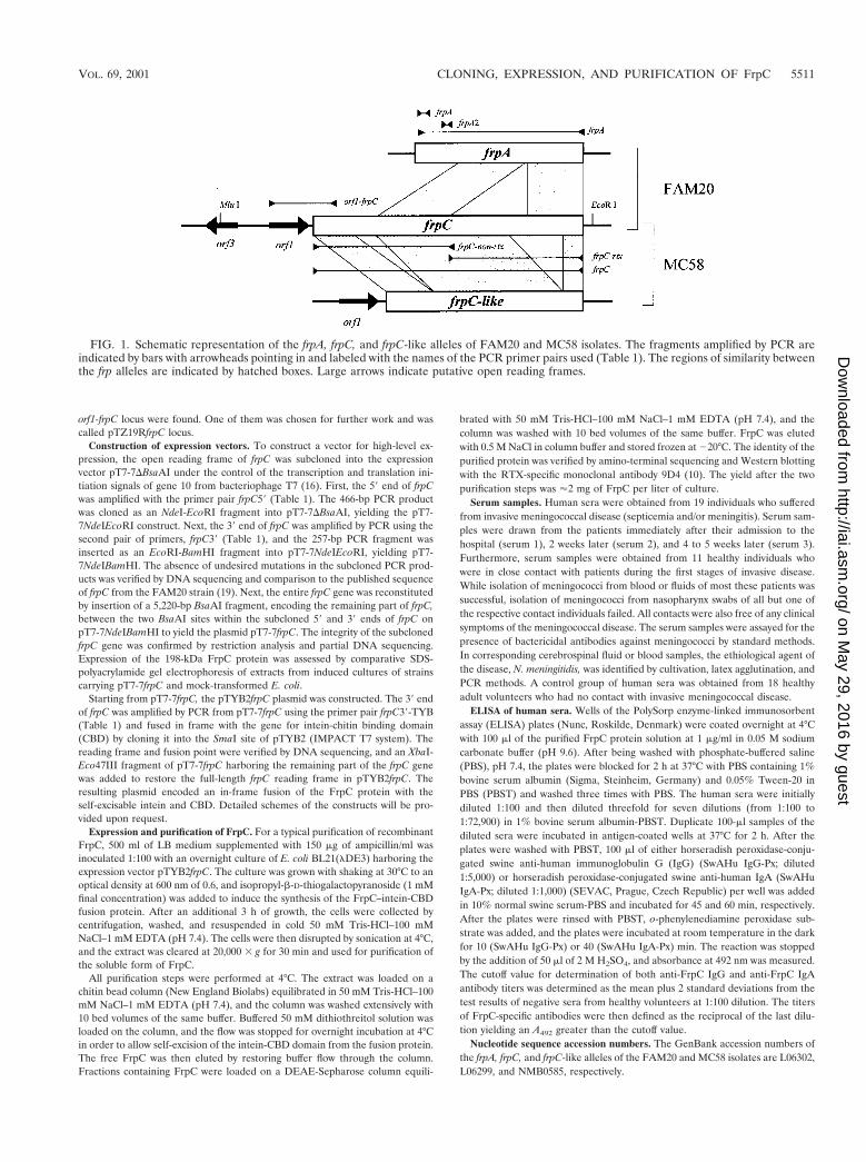
FIG. 1. Schematic representation of the frpA, frpC, and frpC-like alleles of FAM20 and MC58 isolates. The fragments amplified by PCR areindicated by bars with arrowheads pointing in and labeled with the names of the PCR primer pairs used (Table 1). The regions of similarity betweenthe frp alleles are indicated by hatched boxes. Large arrows indicate putative open reading frames.
VOL. 69, 2001 CLONING, EXPRESSION, AND PURIFICATION OF FrpC 5511
on May 29, 2016 by guest
http://iai.asm.org/
Dow
nloaded from
RESULTS
In order to examine whether Frp proteins might be involvedin the virulence of N. meningitidis, we analyzed the presence ofthe frp alleles in a set of 65 meningococcal isolates of allserogroups. This comprised 38 invasive isolates recovered fromblood or cerebrospinal fluids of patients with invasive menin-gococcal disease and 27 carrier isolates recovered from thenasopharynges of healthy individuals.
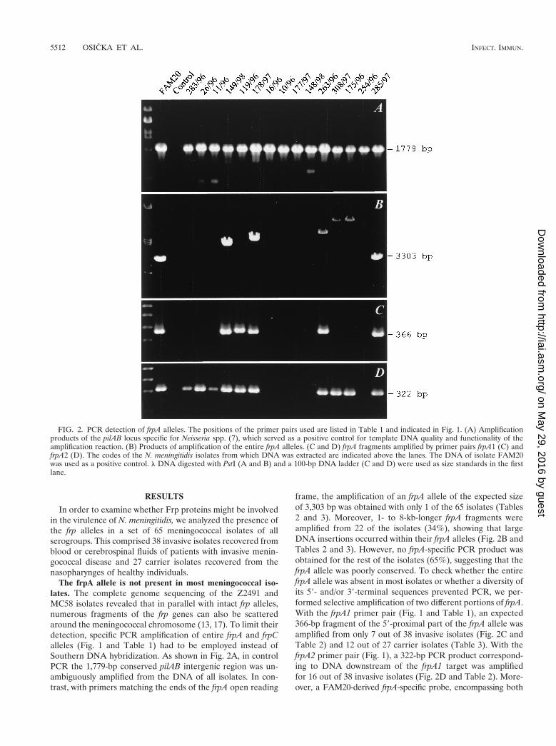
The frpA allele is not present in most meningococcal iso-lates. The complete genome sequencing of the Z2491 andMC58 isolates revealed that in parallel with intact frp alleles,numerous fragments of the frp genes can also be scatteredaround the meningococcal chromosome (13, 17). To limit theirdetection, specific PCR amplification of entire frpA and frpCalleles (Fig. 1 and Table 1) had to be employed instead ofSouthern DNA hybridization. As shown in Fig. 2A, in controlPCR the 1,779-bp conserved pilAB intergenic region was un-ambiguously amplified from the DNA of all isolates. In con-trast, with primers matching the ends of the frpA open reading
frame, the amplification of an frpA allele of the expected sizeof 3,303 bp was obtained with only 1 of the 65 isolates (Tables2 and 3). Moreover, 1- to 8-kb-longer frpA fragments wereamplified from 22 of the isolates (34%), showing that largeDNA insertions occurred within their frpA alleles (Fig. 2B andTables 2 and 3). However, no frpA-specific PCR product wasobtained for the rest of the isolates (65%), suggesting that thefrpA allele was poorly conserved. To check whether the entirefrpA allele was absent in most isolates or whether a diversity ofits 59- and/or 39-terminal sequences prevented PCR, we per-formed selective amplification of two different portions of frpA.With the frpA1 primer pair (Fig. 1 and Table 1), an expected366-bp fragment of the 59-proximal part of the frpA allele wasamplified from only 7 out of 38 invasive isolates (Fig. 2C andTable 2) and 12 out of 27 carrier isolates (Table 3). With thefrpA2 primer pair (Fig. 1), a 322-bp PCR product correspond-ing to DNA downstream of the frpA1 target was amplifiedfor 16 out of 38 invasive isolates (Fig. 2D and Table 2). More-over, a FAM20-derived frpA-specific probe, encompassing both
FIG. 2. PCR detection of frpA alleles. The positions of the primer pairs used are listed in Table 1 and indicated in Fig. 1. (A) Amplificationproducts of the pilAB locus specific for Neisseria spp. (7), which served as a positive control for template DNA quality and functionality of theamplification reaction. (B) Products of amplification of the entire frpA alleles. (C and D) frpA fragments amplified by primer pairs frpA1 (C) andfrpA2 (D). The codes of the N. meningitidis isolates from which DNA was extracted are indicated above the lanes. The DNA of isolate FAM20was used as a positive control. l DNA digested with PstI (A and B) and a 100-bp DNA ladder (C and D) were used as size standards in the firstlane.
5512 OSICKA ET AL. INFECT. IMMUN.
on May 29, 2016 by guest
http://iai.asm.org/
Dow
nloaded from
frpA1 and frpA2 primer sites, failed to hybridize with genomicDNA of the PCR-negative isolates (not shown). Hence, anfrpA allele was absent in most tested meningococcal isolates.
frpC-like alleles of variable size are present in all invasiveand most carrier isolates. In contrast to frpA, the frpC allelescould be amplified from all 38 invasive meningococcal isolatesand from 24 out of 27 carrier isolates when a pair of primersspecific for the full-length frpC allele of FAM20 was used (Fig.1 and Table 1). However, as illustrated in Fig. 3A and sum-
marized in Tables 2 and 3, the sizes of the frpC-specific prod-ucts varied considerably. Therefore, conservation of two dif-ferent frpC portions was investigated.
As shown in Fig. 3B, products of various sizes were obtainedwith the PCR specific for the frpC repeats. Not surprisingly,additional repeat-specific fragments were amplified in parallelwith the major PCR product for most of the strains. Thepresence of more than one copy of frp repeats per chromosomewas, indeed, previously observed for the FAM20 and MC58isolates (Fig. 1). Interestingly, the size differences between theobtained PCR products appeared to be very close to integralmultiples of 600 bp, and fragments of approximately 1.2, 1.8,2.4, 3.0, 3.6, and 4.2 kb were obtained. This suggests that aDNA block encoding an approximately 600-bp repeat was pres-ent in variable numbers of copies in the different frpC alleles.However, only a limited correlation was observed between thesize pattern of the amplified repetitive sequences and entirefrpC alleles (cf. Fig. 3A and B).
As documented in Fig. 3C, the size variation of frpC alleleswas to some extent due also to variations in the nonrepetitiveportion encoding the first 872 residues of FrpC. Amplificationof this part of frpC yielded a 2.6-kb product for most isolates,which was expected for the sequenced frpC alleles fromFAM20 and MC58. However, a 2.2-kb product was amplifiedfrom other isolates, corresponding in size to the expected prod-uct of the shorter frpC-like allele of MC58. Moreover, largerproducts of approximately 3.6 and 4.7 kb were also amplified
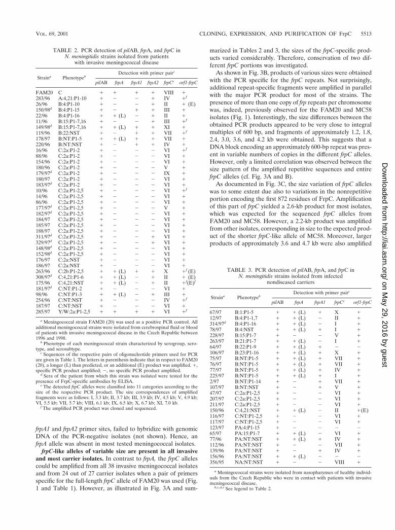
TABLE 2. PCR detection of pilAB, frpA, and frpC inN. meningitidis strains isolated from patients
with invasive meningococcal disease
Straina PhenotypebDetection with primer pairc
pilAB frpA frpA1 frpA2 frpCe orf1-frpC
FAM20 C 1 1 1 1 VIII 1283/96 A:4,21:P1-10 1 2 2 1 IV 1f
26/96 B:4:P1-10 1 2 2 1 II 1 (E)150/98d B:4:P1-15 1 2 1 1 III 122/96 B:4:P1-16 1 1 (L) 2 1 II 111/96 B:15:P1-7,16 1 2 2 1 III 1f
149/98d B:15:P1-7,16 1 1 (L) 1 1 XI 1119/96 B:22:NST 1 2 1 1 VII 1f
178/97 B:NT:P1-5 1 1 (L) 1 1 VII 1220/96 B:NT:NST 1 2 1 1 IV 116/96 C:2a:P1-2 1 2 2 2 VI 1f
88/96 C:2a:P1-2 1 2 2 2 VI 1154/96 C:2a:P1-2 1 2 2 2 VI 1180/96 C:2a:P1-2 1 2 2 2 V 1179/97d C:2a:P1-2 1 2 2 2 IX 1180/97 C:2a:P1-2 1 2 2 2 VI 1183/97d C:2a:P1-2 1 2 2 2 VI 110/96 C:2a:P1-2,5 1 2 2 2 VI 1f
14/96 C:2a:P1-2,5 1 2 2 2 VI 186/96 C:2a:P1-2,5 1 2 2 2 VI 1177/97d C:2a:P1-2,5 1 2 2 2 V 1182/97d C:2a:P1-2,5 1 2 2 2 VI 1184/97 C:2a:P1-2,5 1 2 2 2 VI 1185/97 C:2a:P1-2,5 1 2 2 2 VI 1188/97 C:2a:P1-2,5 1 2 2 2 VI 1311/97d C:2a:P1-2,5 1 2 2 1 VI 1329/97d C:2a:P1-2,5 1 2 2 1 VI 1148/98d C:2a:P1-2,5 1 2 2 2 VI 1152/98d C:2a:P1-2,5 1 2 2 2 VI 1176/97 C:2a:NST 1 2 2 2 VI 1186/97 C:2a:NST 1 2 2 2 VI 1263/96 C:2b:P1-2,5 1 1 (L) 1 1 X 1f (E)308/97d C:4,21:P1-6 1 1 (L) 2 1 II 1 (E)175/96 C:4,21:NST 1 1 (L) 2 1 II 1f(E)f
181/97d C:NT:P1-2 1 2 2 2 VI 198/96 C:NT:P1-3 1 1 (L) 2 1 III 1254/96 C:NT:NST 1 2 2 2 IV 1f
187/97 C:NT:NST 1 2 2 2 VI 1285/97 Y/W:2a:P1-2,5 1 1 1 1 VI 1f
a Meningococcal strain FAM20 (20) was used as a positive PCR control. Alladditional meningococcal strains were isolated from cerebrospinal fluid or bloodof patients with invasive meningococcal disease in the Czech Republic between1996 and 1998.
b Phenotype of each meningococcal strain characterized by serogroup, sero-type, and serosubtype.
c Sequences of the respective pairs of oligonucleotide primers used for PCRare given in Table 1. The letters in parenthesis indicate that in respect to FAM20(20), a longer (L) than predicted, or an additional (E) product was amplified. 1,specific PCR product amplified; 2, no specific PCR product amplified.
d Sera of the patient from which this strain was isolated were tested for thepresence of FrpC-specific antibodies by ELISA.
e The detected frpC alleles were classified into 11 categories according to thesize of the respective PCR product. The size correspondences of amplifiedfragments were as follows: I, 3.3 kb; II, 3.7 kb; III, 3.9 kb; IV, 4.5 kb; V, 4.9 kb;VI, 5.5 kb; VII, 5.7 kb; VIII, 6.1 kb; IX, 6.5 kb; X, 6.7 kb; XI, 7.0 kb.
f The amplified PCR product was cloned and sequenced.
TABLE 3. PCR detection of pilAB, frpA, and frpC inN. meningitidis strains isolated from infected
nondiseased carriers
Straina PhenotypebDetection with primer pairc
pilAB frpA frpA1 frpCe orf1-frpC
67/97 B:1:P1-5 1 1 (L) 1 X 112/97 B:4:P1-1,7 1 1 (L) 2 II 1314/97d B:4:P1-16 1 1 (L) 2 I 178/97 B:4:NST 1 1 (L) 1 I 1228/97 B:15:P1-7 1 2 2 V 1263/97 B:21:P1-7 1 1 (L) 2 I 164/97 B:22:P1-9 1 1 (L) 1 2 2106/97 B:23:P1-16 1 1 (L) 1 X 175/97 B:NT:P1-5 1 1 (L) 1 VII 176/97 B:NT:P1-5 1 1 (L) 1 VII 177/97 B:NT:P1-5 1 1 (L) 1 IV 1225/97 B:NT:P1-5 1 1 (L) 1 I 12/97 B:NT:P1-14 1 2 1 VII 1107/97 B:NT:NST 1 2 1 V 147/97 C:2a:P1-2,5 1 2 2 VI 1207/97 C:2a:P1-2,5 1 2 2 VI 1211/97 C:2a:P1-2,5 1 2 2 VI 1150/96 C:4,21:NST 1 1 (L) 2 II 1(E)116/97 C:NT:P1-2,5 1 2 2 VI 1117/97 C:NT:P1-2,5 1 2 2 VI 1123/97 PA:4:P1-15 1 2 2 2 265/97 PA:15:P1-7 1 1 (L) 2 VI 177/96 PA:NT:NST 1 1 (L) 1 IV 1112/96 PA:NT:NST 1 2 2 VII 1139/96 PA:NT:NST 1 2 1 IV 1156/96 PA:NT:NST 1 1 (L) 2 2 2356/95 NA:NT:NST 1 2 2 VIII 1
a Meningococcal strains were isolated from nasopharynxes of healthy individ-uals from the Czech Republic who were in contact with patients with invasivemeningococcal disease.
b,c,d,e See legend to Table 2.
VOL. 69, 2001 CLONING, EXPRESSION, AND PURIFICATION OF FrpC 5513
on May 29, 2016 by guest
http://iai.asm.org/
Dow
nloaded from
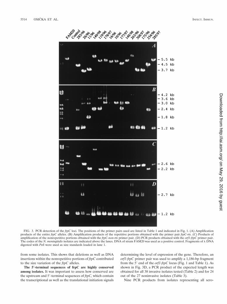
from some isolates. This shows that deletions as well as DNAinsertions within the nonrepetitive portions of frpC contributedto the size variation of the frpC alleles.
The 5*-terminal sequences of frpC are highly conservedamong isolates. It was important to assess how conserved arethe upstream and 59-terminal sequences of frpC, which containthe transcriptional as well as the translational initiation signals
determining the level of expression of the gene. Therefore, anorf1-frpC primer pair was used to amplify a 1,186-bp fragmentfrom the 59 end of the orf1-frpC locus (Fig. 1 and Table 1). Asshown in Fig. 3D, a PCR product of the expected length wasobtained for all 38 invasive isolates tested (Table 2) and for 24out of the 27 noninvasive isolates (Table 3).
Nine PCR products from isolates representing all sero-
FIG. 3. PCR detection of the frpC loci. The positions of the primer pairs used are listed in Table 1 and indicated in Fig. 1. (A) Amplificationproducts of the entire frpC alleles. (B) Amplification products of the repetitive portions obtained with the primer pair frpC-rtx. (C) Products ofamplification of the nonrepetitive portions obtained with the frpC-non-rtx primer pair. (D) PCR products obtained with the orf1-frpC primer pair.The codes of the N. meningitidis isolates are indicated above the lanes. DNA of strain FAM20 was used as a positive control. Fragments of l DNAdigested with PstI were used as size standards loaded in lane 1.
5514 OSICKA ET AL. INFECT. IMMUN.
on May 29, 2016 by guest
http://iai.asm.org/
Dow
nloaded from
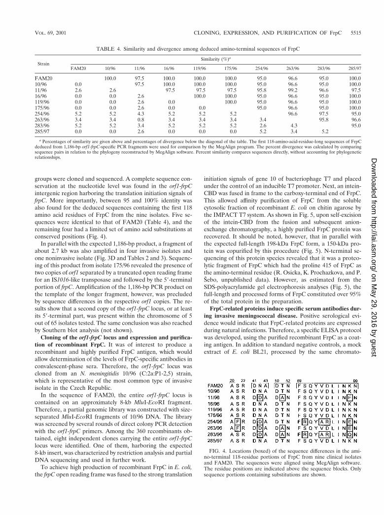
groups were cloned and sequenced. A complete sequence con-servation at the nucleotide level was found in the orf1-frpCintergenic region harboring the translation initiation signals offrpC. More importantly, between 95 and 100% identity wasalso found for the deduced sequences containing the first 118amino acid residues of FrpC from the nine isolates. Five se-quences were identical to that of FAM20 (Table 4), and theremaining four had a limited set of amino acid substitutions atconserved positions (Fig. 4).
In parallel with the expected 1,186-bp product, a fragment ofabout 2.7 kb was also amplified in four invasive isolates andone noninvasive isolate (Fig. 3D and Tables 2 and 3). Sequenc-ing of this product from isolate 175/96 revealed the presence oftwo copies of orf1 separated by a truncated open reading framefor an IS1016-like transposase and followed by the 59-terminalportion of frpC. Amplification of the 1,186-bp PCR product onthe template of the longer fragment, however, was precludedby sequence differences in the respective orf1 copies. The re-sults show that a second copy of the orf1-frpC locus, or at leastits 59-terminal part, was present within the chromosome of 5out of 65 isolates tested. The same conclusion was also reachedby Southern blot analysis (not shown).
Cloning of the orf1-frpC locus and expression and purifica-tion of recombinant FrpC. It was of interest to produce arecombinant and highly purified FrpC antigen, which wouldallow determination of the levels of FrpC-specific antibodies inconvalescent-phase sera. Therefore, the orf1-frpC locus wascloned from an N. meningitidis 10/96 (C:2a:P1-2,5) strain,which is representative of the most common type of invasiveisolate in the Czech Republic.
In the sequence of FAM20, the entire orf1-frpC locus iscontained on an approximately 8-kb MluI-EcoRI fragment.Therefore, a partial genomic library was constructed with size-separated MluI-EcoRI fragments of 10/96 DNA. The librarywas screened by several rounds of direct colony PCR detectionwith the orf1-frpC primers. Among the 360 recombinants ob-tained, eight independent clones carrying the entire orf1-frpClocus were identified. One of them, harboring the expected8-kb insert, was characterized by restriction analysis and partialDNA sequencing and used in further work.
To achieve high production of recombinant FrpC in E. coli,the frpC open reading frame was fused to the strong translation
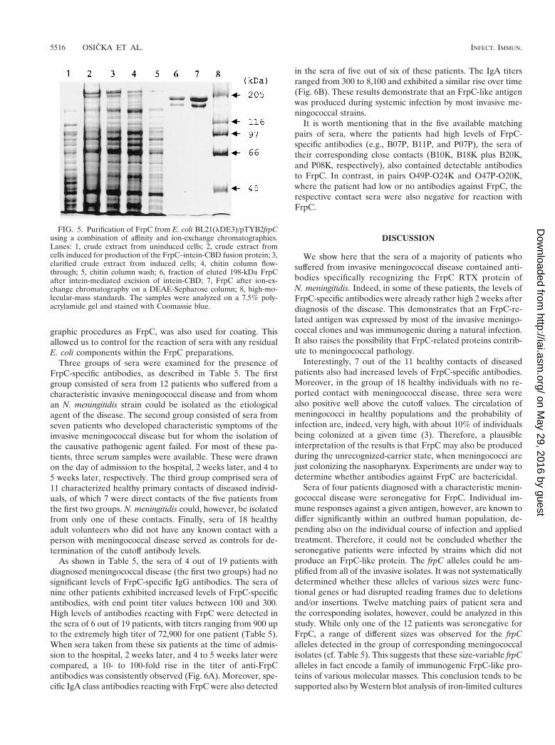
initiation signals of gene 10 of bacteriophage T7 and placedunder the control of an inducible T7 promoter. Next, an intein-CBD was fused in frame to the carboxy-terminal end of FrpC.This allowed affinity purification of FrpC from the solublecytosolic fraction of recombinant E. coli on chitin agarose bythe IMPACT T7 system. As shown in Fig. 5, upon self-excisionof the intein-CBD from the fusion and subsequent anion-exchange chromatography, a highly purified FrpC protein wasrecovered. It should be noted, however, that in parallel withthe expected full-length 198-kDa FrpC form, a 150-kDa pro-tein was copurified by this procedure (Fig. 5). N-terminal se-quencing of this protein species revealed that it was a proteo-lytic fragment of FrpC which had the proline 415 of FrpC asthe amino-terminal residue (R. Osicka, K. Prochazkova, and P.Sebo, unpublished data). However, as estimated from theSDS-polyacrylamide gel electrophoresis analyses (Fig. 5), thefull-length and processed forms of FrpC constituted over 95%of the total protein in the preparation.
FrpC-related proteins induce specific serum antibodies dur-ing invasive meningococcal disease. Positive serological evi-dence would indicate that FrpC-related proteins are expressedduring natural infections. Therefore, a specific ELISA protocolwas developed, using the purified recombinant FrpC as a coat-ing antigen. In addition to standard negative controls, a mockextract of E. coli BL21, processed by the same chromato-
FIG. 4. Locations (boxed) of the sequence differences in the ami-no-terminal 118-residue portions of FrpC from nine clinical isolatesand FAM20. The sequences were aligned using MegAlign software.The residue positions are indicated above the sequence blocks. Onlysequence portions containing substitutions are shown.
TABLE 4. Similarity and divergence among deduced amino-terminal sequences of FrpC
StrainSimilarity (%)a
FAM20 10/96 11/96 16/96 119/96 175/96 254/96 263/96 283/96 285/97
FAM20 100.0 97.5 100.0 100.0 100.0 95.0 96.6 95.0 100.010/96 0.0 97.5 100.0 100.0 100.0 95.0 96.6 95.0 100.011/96 2.6 2.6 97.5 97.5 97.5 95.8 99.2 96.6 97.516/96 0.0 0.0 2.6 100.0 100.0 95.0 96.6 95.0 100.0119/96 0.0 0.0 2.6 0.0 100.0 95.0 96.6 95.0 100.0175/96 0.0 0.0 2.6 0.0 0.0 95.0 96.6 95.0 100.0254/96 5.2 5.2 4.3 5.2 5.2 5.2 96.6 97.5 95.0263/96 3.4 3.4 0.8 3.4 3.4 3.4 3.4 95.8 96.6283/96 5.2 5.2 3.4 5.2 5.2 5.2 2.6 4.3 95.0285/97 0.0 0.0 2.6 0.0 0.0 0.0 5.2 3.4 5.2
a Percentages of similarity are given above and percentages of divergence below the diagonal of the table. The first 118-amino-acid-residue-long sequences of FrpCdeduced from 1,186-bp orf1-frpC-specific PCR fragments were used for comparison by the MegAlign program. The percent divergence was calculated by comparingsequence pairs in relation to the phylogeny reconstructed by MegAlign software. Percent similarity compares sequences directly, without accounting for phylogeneticrelationships.
VOL. 69, 2001 CLONING, EXPRESSION, AND PURIFICATION OF FrpC 5515
on May 29, 2016 by guest
http://iai.asm.org/
Dow
nloaded from
graphic procedures as FrpC, was also used for coating. Thisallowed us to control for the reaction of sera with any residualE. coli components within the FrpC preparations.
Three groups of sera were examined for the presence ofFrpC-specific antibodies, as described in Table 5. The firstgroup consisted of sera from 12 patients who suffered from acharacteristic invasive meningococcal disease and from whoman N. meningitidis strain could be isolated as the etiologicalagent of the disease. The second group consisted of sera fromseven patients who developed characteristic symptoms of theinvasive meningococcal disease but for whom the isolation ofthe causative pathogenic agent failed. For most of these pa-tients, three serum samples were available. These were drawnon the day of admission to the hospital, 2 weeks later, and 4 to5 weeks later, respectively. The third group comprised sera of11 characterized healthy primary contacts of diseased individ-uals, of which 7 were direct contacts of the five patients fromthe first two groups. N. meningitidis could, however, be isolatedfrom only one of these contacts. Finally, sera of 18 healthyadult volunteers who did not have any known contact with aperson with meningococcal disease served as controls for de-termination of the cutoff antibody levels.
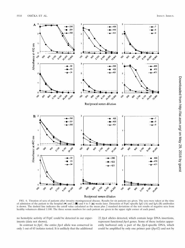
As shown in Table 5, the sera of 4 out of 19 patients withdiagnosed meningococcal disease (the first two groups) had nosignificant levels of FrpC-specific IgG antibodies. The sera ofnine other patients exhibited increased levels of FrpC-specificantibodies, with end point titer values between 100 and 300.High levels of antibodies reacting with FrpC were detected inthe sera of 6 out of 19 patients, with titers ranging from 900 upto the extremely high titer of 72,900 for one patient (Table 5).When sera taken from these six patients at the time of admis-sion to the hospital, 2 weeks later, and 4 to 5 weeks later werecompared, a 10- to 100-fold rise in the titer of anti-FrpCantibodies was consistently observed (Fig. 6A). Moreover, spe-cific IgA class antibodies reacting with FrpC were also detected
in the sera of five out of six of these patients. The IgA titersranged from 300 to 8,100 and exhibited a similar rise over time(Fig. 6B). These results demonstrate that an FrpC-like antigenwas produced during systemic infection by most invasive me-ningococcal strains.
It is worth mentioning that in the five available matchingpairs of sera, where the patients had high levels of FrpC-specific antibodies (e.g., B07P, B11P, and P07P), the sera oftheir corresponding close contacts (B10K, B18K plus B20K,and P08K, respectively), also contained detectable antibodiesto FrpC. In contrast, in pairs O49P-O24K and O47P-O20K,where the patient had low or no antibodies against FrpC, therespective contact sera were also negative for reaction withFrpC.
DISCUSSION
We show here that the sera of a majority of patients whosuffered from invasive meningococcal disease contained anti-bodies specifically recognizing the FrpC RTX protein ofN. meningitidis. Indeed, in some of these patients, the levels ofFrpC-specific antibodies were already rather high 2 weeks afterdiagnosis of the disease. This demonstrates that an FrpC-re-lated antigen was expressed by most of the invasive meningo-coccal clones and was immunogenic during a natural infection.It also raises the possibility that FrpC-related proteins contrib-ute to meningococcal pathology.
Interestingly, 7 out of the 11 healthy contacts of diseasedpatients also had increased levels of FrpC-specific antibodies.Moreover, in the group of 18 healthy individuals with no re-ported contact with meningococcal disease, three sera werealso positive well above the cutoff values. The circulation ofmeningococci in healthy populations and the probability ofinfection are, indeed, very high, with about 10% of individualsbeing colonized at a given time (3). Therefore, a plausibleinterpretation of the results is that FrpC may also be producedduring the unrecognized-carrier state, when meningococci arejust colonizing the nasopharynx. Experiments are under way todetermine whether antibodies against FrpC are bactericidal.
Sera of four patients diagnosed with a characteristic menin-gococcal disease were seronegative for FrpC. Individual im-mune responses against a given antigen, however, are known todiffer significantly within an outbred human population, de-pending also on the individual course of infection and appliedtreatment. Therefore, it could not be concluded whether theseronegative patients were infected by strains which did notproduce an FrpC-like protein. The frpC alleles could be am-plified from all of the invasive isolates. It was not systematicallydetermined whether these alleles of various sizes were func-tional genes or had disrupted reading frames due to deletionsand/or insertions. Twelve matching pairs of patient sera andthe corresponding isolates, however, could be analyzed in thisstudy. While only one of the 12 patients was seronegative forFrpC, a range of different sizes was observed for the frpCalleles detected in the group of corresponding meningococcalisolates (cf. Table 5). This suggests that these size-variable frpCalleles in fact encode a family of immunogenic FrpC-like pro-teins of various molecular masses. This conclusion tends to besupported also by Western blot analysis of iron-limited cultures
FIG. 5. Purification of FrpC from E. coli BL21(lDE3)/pTYB2frpCusing a combination of affinity and ion-exchange chromatographies.Lanes: 1, crude extract from uninduced cells; 2, crude extract fromcells induced for production of the FrpC–intein-CBD fusion protein; 3,clarified crude extract from induced cells; 4, chitin column flow-through; 5, chitin column wash; 6, fraction of eluted 198-kDa FrpCafter intein-mediated excision of intein-CBD; 7, FrpC after ion-ex-change chromatography on a DEAE-Sepharose column; 8, high-mo-lecular-mass standards. The samples were analyzed on a 7.5% poly-acrylamide gel and stained with Coomassie blue.
5516 OSICKA ET AL. INFECT. IMMUN.
on May 29, 2016 by guest
http://iai.asm.org/
Dow
nloaded from
of a set of meningococcal strains, where FrpC-like proteins ofvarious molecular masses were detected (data not shown).
The variability of frpC alleles was at least partly due todifferences in the sizes and/or numbers of the highly redundantRTX repeat blocks. The observed size differences of amplifiedrepetitive domains suggest that these might consist of integralmultiples of a 600-bp DNA block. Such a block is, indeed,present once in the frpA alle of FAM20, twice in the frpC-likeallele of MC58, and four times in the full-length frpC gene ofboth the FAM20 and MC58 strains (17, 19, 20). It is worthnoting that similar variation in the size of the RTX domain wasrecently also observed for a related RTX protein, ApxIVAfrom Actinobacillus (14). For frpC, however, not all size vari-ation could be attributed to the repetitive sequences. Deletionsas well as insertions were also observed in the nonrepetitiveportion of the gene encoding the first 872 residues of FrpC.This further documents the genomic fluidity and high recom-bination frequency of meningococci (4, 6, 8, 13, 17). Moreover,there was no correlation between the detected size of the frpCallele and the serological characteristics of the isolates. Thiswas also to be expected, since the serotype and subtype char-acteristics were previously found to reflect only poorly, if at all,the clonal lineages in meningococci (4, 6, 8).
Interestingly, a rather high sequence conservation of the 59
end of the frpC gene was found in the portion encoding the 118amino-terminal residues of FrpC from nine local isolates. Fiveof the sequences were fully conserved in respect to that of theFAM20 strain, while the remaining four sequences had a lim-ited set of amino acid substitutions at conserved positions. Thesequence encoding the RGD tripeptide, shown to be involvedin binding of other proteins to cellular integrins, was, however,intact in all of the frpC alleles examined. These results suggestthat the N-terminal portion of the FrpC protein may be ratherconserved and subject to selective pressure for conservation offunction.
Characterization of the biological activities of the better-conserved FrpC proteins is now intensely pursued. Except forthe repeat domain, which is highly homologous in all RTXproteins, the FrpC protein exhibits significant sequence simi-larity only to the newly identified RTX protein ApxIVA fromActinobacillus pleuropneumoniae (15). The area of similarity islocated in the central 300 amino acids of ApxIVA and betweenresidues 300 and 600 of the prototype FrpC protein of FAM20,with 42% identical and 50% identical plus similar amino acidresidues, respectively. The biological function of ApxIVA alsoremains unknown. It appears to exhibit a weak hemolytic ac-tivity suggestive of a channel-forming capacity (15). However,
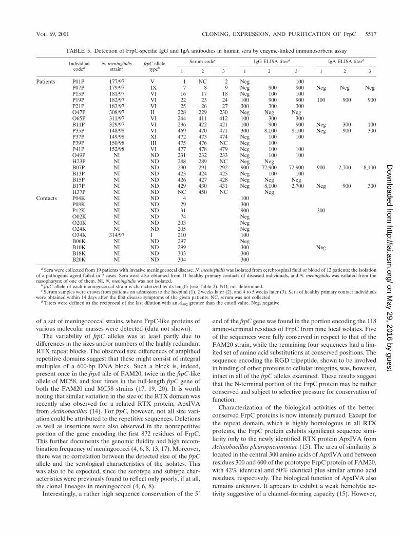
TABLE 5. Detection of FrpC-specific IgG and IgA antibodies in human sera by enzyme-linked immunosorbent assay
Individualcodea
N. meningitidisstraina
frpC alleletypeb
Serum codec IgG ELISA titerd IgA ELISA titerd
1 2 3 1 2 3 1 2 3
Patients P01P 177/97 V 1 NC 2 Neg 100P07P 179/97 IX 7 8 9 Neg 900 900 Neg Neg NegP15P 181/97 VI 16 17 18 Neg 100 100P19P 182/97 VI 22 23 24 100 900 900 100 900 900P21P 183/97 VI 25 26 27 300 300 300O47P 308/97 II 228 229 230 Neg Neg NegO65P 311/97 VI 244 411 412 100 300 300B11P 329/97 VI 296 422 421 100 900 900 Neg 300 100P35P 148/98 VI 469 470 471 300 8,100 8,100 Neg 900 300P37P 149/98 XI 472 473 474 Neg 100 100P39P 150/98 III 475 476 NC Neg 100P41P 152/98 VI 477 478 479 Neg 100 100O49P NI ND 231 232 233 Neg 100 100H23P NI ND 288 289 NC Neg NegB07P NI ND 290 291 292 900 72,900 72,900 900 2,700 8,100B13P NI ND 423 424 425 Neg 100 100B15P NI ND 426 427 428 Neg Neg NegB17P NI ND 429 430 431 Neg 8,100 2,700 Neg 900 300H37P NI ND NC 450 NC Neg
Contacts P04K NI ND 4 100P08K NI ND 29 300P12K NI ND 31 900 300O02K NI ND 74 NegO20K NI ND 203 NegO24K NI ND 205 NegO34K 314/97 I 210 100B06K NI ND 297 NegB10K NI ND 299 300 NegB18K NI ND 303 300B20K NI ND 304 300
a Sera were collected from 19 patients with invasive meningococcal disease. N. meningitidis was isolated from cerebrospinal fluid or blood of 12 patients; the isolationof a pathogenic agent failed in 7 cases. Sera were also obtained from 11 healthy primary contacts of diseased individuals, and N. meningitidis was isolated from thenasopharynx of one of them. NI, N. meningitidis was not isolated.
b frpC allele of each meningococcal strain is characterized by its length (see Table 2). ND, not determined.c Serum samples were drawn from patients on admission to the hospital (1), 2 weeks later (2), and 4 to 5 weeks later (3). Sera of healthy primary contact individuals
were obtained within 14 days after the first disease symptoms of the given patients. NC, serum was not collected.d Titers were defined as the reciprocal of the last dilution with an A492 greater than the cutoff value. Neg, negative.
VOL. 69, 2001 CLONING, EXPRESSION, AND PURIFICATION OF FrpC 5517
on May 29, 2016 by guest
http://iai.asm.org/
Dow
nloaded from
no hemolytic activity of FrpC could be detected in our exper-iments (data not shown).
In contrast to frpC, the entire frpA allele was conserved inonly 1 out of 65 isolates tested. It is unlikely that the additional
22 frpA alleles detected, which contain large DNA insertions,represent functional frpA genes. Some of these isolates appar-ently harbored only a part of the frpA-specific DNA, whichcould be amplified by only one primer pair (frpA2) and not by
FIG. 6. Titration of sera of patients after invasive meningococcal disease. Results for six patients are given. The sera were taken at the timeof admission of the patient to the hospital (●) and 2 (f) and 4 to 5 (Œ) weeks later. Detection of FrpC-specific IgG (A) and IgA (B) antibodiesis shown. The dashed line indicates the cutoff value calculated as the mean plus 2 standard deviations of the test results of negative sera fromhealthy volunteers diluted 1:100. The three serum numbers for each patient are given in the upper right corner of each panel.
5518 OSICKA ET AL. INFECT. IMMUN.
on May 29, 2016 by guest
http://iai.asm.org/
Dow
nloaded from

another matching primer pair immediately upstream (frpA1).This suggests that fragments of frpA were present in the chro-mosomes of this portion of the isolates tested. Moreover, thecombination of Southern blot and PCR analyses showed thatan frpA allele was absent in most of the strains tested. Indeed,a complete sequence of frpA was also not found in the twomeningococcal strains whose genomes have been sequenced.Collectively these data suggest that a complete frpA gene maynot be present in many strains.
Finally, when the observed size variation of frpC alleles isconsidered, it appears inappropriate to continue the differen-tiation of the meningococcal RTX proteins into FrpA andFrpC-like merely on the basis of size. Since large portions ofidentical sequence are shared by FrpA and FrpC, we suggestconsidering FrpA an insertion variant of FrpC and calling allthe RTX proteins of meningococci FrpC. These could then befurther classified according to their sizes.
ACKNOWLEDGMENTS
Stimulating discussions and the gift of purified FAM20 genomicDNA by Xavier Nassif and the gift of the 9D4 monoclonal antibody byE. L. Hewlett are gratefully acknowledged.
This work was supported by grants 310/96/K102 of the Grant Agencyof the Czech Republic and ME167 of the Ministry of Education, Youthand Sports of the Czech Republic and by the French-Czech researchcollaboration award DRI/637 MAE PECO.
REFERENCES
1. Binet, R., S. Letoffe, J. M. Ghigo, P. Delepelaire, and C. Wandersman. 1997.Protein secretion by Gram-negative bacterial ABC exporters—a review.Gene 192:7–11.
2. Booy, R., and J. S. Kroll. 1998. Bacterial meningitis and meningococcalinfection. Curr. Opin. Pediatr. 10:13–18.
3. Caugant, D. A. 1998. Population genetics and molecular epidemiology ofNeisseria meningitidis. APMIS 106:505–525.
4. Caugant, D. A., L. F. Mocca, C. E. Frasch, L. O. Froholm, W. D. Zollinger,and R. K. Selander. 1987. Genetic structure of Neisseria meningitidis popu-lations in relation to serogroup, serotype, and outer membrane proteinpattern. J. Bacteriol. 169:2781–2792.
5. D’Souza, S. E., M. H. Ginsberg, and E. F. Plow. 1991. Arginyl-glycyl-asparticacid (RGD): a cell adhesion motif. Trends Biochem. Sci. 16:246–250.
6. Feil, E. J., M. C. Maiden, M. Achtman, and B. G. Spratt. 1999. The relative
contributions of recombination and mutation to the divergence of clones ofNeisseria meningitidis. Mol. Biol. Evol. 16:1496–1502.
7. Giorgini, D., and M. K. Taha. 1995. Molecular typing of Neisseria meningi-tidis serogroup A using the polymerase chain reaction and restriction endo-nuclease pattern analysis. Mol. Cell. Probes 9:297–306.
8. Holmes, E. C., R. Urwin, and M. C. Maiden. 1999. The influence of recom-bination on the population structure and evolution of the human pathogenNeisseria meningitidis. Mol. Biol. Evol. 16:741–749.
9. Lally, E. T., R. B. Hill, I. R. Kieba, and J. Korostoff. 1999. The interactionbetween RTX toxins and target cells. Trends Microbiol. 7:356–361.
10. Lee, S., M. Gray, L. Guo, P. Sebo, and E. Hewlett. 1999. Epitope mapping ofmonoclonal antibodies against Bordetella pertussis adenylate cyclase toxin.Infect. Immun. 67:2090–2095.
11. Leininger, E., M. Roberts, J. G. Kenimer, I. G. Charles, N. Fairweather, P.Novotny, and M. J. Brennan. 1991. Pertactin, an Arg-Gly-Asp-containingBordetella pertussis surface protein that promotes adherence of mammaliancells. Proc. Natl. Acad. Sci. USA 88:345–349.
12. Leong, J. M., P. E. Morrissey, A. Marra, and R. R. Isberg. 1995. An aspartateresidue of the Yersinia pseudotuberculosis invasin protein that is critical forintegrin binding. EMBO J. 14:422–431.
13. Parkhill, J., M. Achtman, et al. 2000. Complete DNA sequence of a sero-group A strain of Neisseria meningitidis Z2491. Nature 404:502–506.
14. Schaller, A., S. Djordjevic, G. Eamens, W. Forbes, R. Kuhn, P. Kuhnert, M.Gottschalk, J. Nicolet, and J. Frey. 2001. Identification and detection ofActinobacillus pleuropneumoniae by PCR based on the gene apxIVA. Vet.Microbiol. 79:47–62.
15. Schaller, A., R. Kuhn, P. Kuhnert, J. Nicolet, T. J. Anderson, J. I. MacInnes,R. P. Segers, and J. Frey. 1999. Characterization of apxIVA, a new RTXdeterminant of Actinobacillus pleuropneumoniae. Microbiology 145:2105–2116.
16. Tabor, S., and C. Richardson. 1985. A bacteriophage T7 RNA polymerase/promoter system for controlled exclusive expression of genes. Proc. Natl.Acad. Sci. USA 82:1074–1078.
17. Tettelin, H., N. J. Saunders, et al. 2000. Complete genome sequence ofNeisseria meningitidis serogroup B strain MC58. Science 287:1809–1815.
18. Thompson, S. A., and P. F. Sparling. 1993. The RTX cytotoxin-related FrpAprotein of Neisseria meningitidis is secreted extracellularly by meningococciand by HlyBD1 Escherichia coli. Infect. Immun. 61:2906–2911.
19. Thompson, S. A., L. L. Wang, and P. F. Sparling. 1993. Cloning and nucle-otide sequence of frpC, a second gene from Neisseria meningitidis encoding aprotein similar to RTX cytotoxins. Mol. Microbiol. 9:85–96.
20. Thompson, S. A., L. L. Wang, A. West, and P. F. Sparling. 1993. Neisseriameningitidis produces iron-regulated proteins related to the RTX family ofexoproteins. J. Bacteriol. 175:811–818.
21. Tzeng, Y. L., and D. S. Stephens. 2000. Epidemiology and pathogenesis ofNeisseria meningitidis. Microbes Infect. 2:687–700.
22. Welch, R. A. 1991. Pore-forming cytolysins of gram-negative bacteria. Mol.Microbiol. 5:521–528.
23. Welch, R. A., M. E. Bauer, A. D. Kent, J. A. Leeds, M. Moayeri, L. B.Regassa, and D. L. Swenson. 1995. Battling against host phagocytes: Thewherefore of the RTX family of toxins? Infect. Agents Dis. 4:254–272.
Editor: D. L. Burns
VOL. 69, 2001 CLONING, EXPRESSION, AND PURIFICATION OF FrpC 5519
on May 29, 2016 by guest
http://iai.asm.org/
Dow
nloaded from