Myocardial Protective Impact of Mucuna pruriens on ...
20
Available online at www.scholarsresearchlibrary.com Scholars Research Library Der Pharmacia Lettre, 2018, 10 [3]: 37-56 [http://scholarsresearchlibrary.com/archive.html] ISSN 0975-5071 USA CODEN: DPLEB4 37 Scholar Research Library Myocardial Protective Impact of Mucuna pruriens on Isoproterenol Prompted Myocardial Necrosis Rakam Gopi Krishna 1 and Raja Sundararajan 2* 1 Department of Pharmaceutical Chemistry, Chaitanya College of Pharmacy Education and Research, Kishanpura, Hanamkonda, Warangal, Telangana, India 2 GITAM Institute of Pharmacy, Gandhi Institute of Technology and Management - GITAM (Deemed to be University), Visakhapatnam, Andhra Pradesh, India *Corresponding author: M S. Raja, M. Pharm. PhD, Associate Professor, GITAM Institute of Pharmacy, GITAM University, Gandhi Nagar, Rushikonda, Visakhapatnam, Andhra Pradesh, India. Tel: +91 9160508261; E-mail: [email protected] ABSTRACT The current research study performed was to evaluate the myocardial protective outcome of methanolic extract of Mucuna pruriens against isoproterenol prompted myocardial necrosis in rats. The rats stayed pretreated with methanolic extract of Mucuna pruriens at two dissimilar doses of 250 mg/kg and 500 milligram/kg, correspondingly intended for thirty days. Myocardial necrosis was persuaded in the rats by administering isoproterenol (85 mg/kg s.c) injection. The measures of serum cardiac marker enzymes such as creatine kinase myoglobin (CK-MB), lactate dehydrogenase (LDH), serum glutamate pyruvate transaminase (SGPT), serum glutamate oxaloacetate transaminase (SGOT), total cholesterol (TC), triglycerides (TG), low density lipoproteins (LDL), very low density lipoproteins (VLDL), high density lipoproteins (HDL) and total protein (TP) were estimated. In addition, plasma TBARS and plasma LDH levels were also recorded. Antioxidant parameters like superoxide dismutase (SOD), catalase (CAT), glutathione peroxidase (GPx) and glutathione (GSH) were performed in heart tissue homogenate. The result of the current study specified that, methanol extract of Mucuna pruriens pre-co-treatment prohibited nearly entire parameters of isoproterenol persuaded myocardial necrosis in rats. The overhead outcome was established by the histopathological investigation. It can be recognized that methanolic extract of Mucuna pruriens has a substantial outcome on
-
Upload
khangminh22 -
Category
Documents
-
view
1 -
download
0
Transcript of Myocardial Protective Impact of Mucuna pruriens on ...
Available online at www.scholarsresearchlibrary.com
Scholars Research Library
Der Pharmacia Lettre, 2018, 10 [3]: 37-56
[http://scholarsresearchlibrary.com/archive.html]
ISSN 0975-5071
USA CODEN: DPLEB4
37
Scholar Research Library
Myocardial Protective Impact of Mucuna pruriens on Isoproterenol Prompted
Myocardial Necrosis
Rakam Gopi Krishna1 and Raja Sundararajan
2*
1Department of Pharmaceutical Chemistry, Chaitanya College of Pharmacy Education and Research, Kishanpura,
Hanamkonda, Warangal, Telangana, India
2GITAM Institute of Pharmacy, Gandhi Institute of Technology and Management - GITAM (Deemed to be
University), Visakhapatnam, Andhra Pradesh, India
*Corresponding author: M S. Raja, M. Pharm. PhD, Associate Professor, GITAM Institute of Pharmacy, GITAM
University, Gandhi Nagar, Rushikonda, Visakhapatnam, Andhra Pradesh, India. Tel: +91 9160508261; E-mail:
ABSTRACT
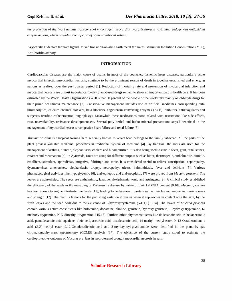
The current research study performed was to evaluate the myocardial protective outcome of methanolic extract of Mucuna
pruriens against isoproterenol prompted myocardial necrosis in rats. The rats stayed pretreated with methanolic extract of
Mucuna pruriens at two dissimilar doses of 250 mg/kg and 500 milligram/kg, correspondingly intended for thirty days.
Myocardial necrosis was persuaded in the rats by administering isoproterenol (85 mg/kg s.c) injection. The measures of serum
cardiac marker enzymes such as creatine kinase myoglobin (CK-MB), lactate dehydrogenase (LDH), serum glutamate pyruvate
transaminase (SGPT), serum glutamate oxaloacetate transaminase (SGOT), total cholesterol (TC), triglycerides (TG), low
density lipoproteins (LDL), very low density lipoproteins (VLDL), high density lipoproteins (HDL) and total protein (TP) were
estimated. In addition, plasma TBARS and plasma LDH levels were also recorded. Antioxidant parameters like superoxide
dismutase (SOD), catalase (CAT), glutathione peroxidase (GPx) and glutathione (GSH) were performed in heart tissue
homogenate. The result of the current study specified that, methanol extract of Mucuna pruriens pre-co-treatment prohibited
nearly entire parameters of isoproterenol persuaded myocardial necrosis in rats. The overhead outcome was established by the
histopathological investigation. It can be recognized that methanolic extract of Mucuna pruriens has a substantial outcome on
Gopi Krishna R, et al. Der Pharmacia Lettre, 2018, 10 [3]: 37-56
______________________________________________________________________________
38
Scholar Research Library
the protection of the heart against isoproterenol encouraged myocardial necrosis through sustaining endogenous antioxidant
enzyme actions, which provides scientific proof of the traditional values.
Keywords: Bidentate tartarate ligand, Mixed transition-alkaline earth metal tartarates, Minimum Inhibition Concentration (MIC),
Anti-biofilm activity.
INTRODUCTION
Cardiovascular diseases are the major cause of deaths in most of the countries. Ischemic heart diseases, particularly acute
myocardial infarction/myocardial necrosis, continue to be the prominent reason of death in together established and emerging
nations as realized over the past quarter period [1]. Reduction of mortality rate and prevention of myocardial infarction and
myocardial necrosis are utmost importance. Today plant-based drugs remain to show an important part in health care. It has been
estimated by the World Health Organization (WHO) that 80 percent of the people of the world rely mainly on old-style drugs for
their prime healthiness maintenance [2]. Conservative management includes use of artificial medicines corresponding anti-
thrombolytics, calcium channel blockers, beta blockers, angiotensin converting enzymes (ACE) inhibitors, anticoagulants and
surgeries (cardiac catheterization, angioplasty). Meanwhile these medications stood related with restrictions like side effects,
cost, unavailability, resistance development etc. Several poly herbal and herbo mineral preparations stayed beneficial in the
management of myocardial necrosis, congestive heart failure and renal failure [3].
Mucuna pruriens is a tropical twining herb generally known as velvet bean belongs to the family fabaceae. All the parts of the
plant possess valuable medicinal properties in traditional system of medicine [4]. By tradition, the roots are used for the
management of asthma, diuretic, elephantiasis, cholera and blood purifier. It is also being used to cure in fever, gout, renal stones,
cataract and rheumatism [4]. In Ayurveda, roots are using for different purpose such as bitter, thermogenic, anthelmintic, diuretic,
emollient, stimulant, aphrodisiac, purgative, febrifuge and tonic. It is considered useful to relieve constipation, nephropathy,
dysmenorrhea, amenorrhea, elephantiasis, dropsy, neuropathy, ulcers, helminthiasis, fever and delirium [5]. Various
pharmacological activities like hypoglycemic [6], anti-epileptic and anti-neoplastic [7] were proved from Mucuna pruriens. The
leaves are aphrodisiac. The seeds are anthelmintic, laxative, alexipharmic, tonic and astringent, [8]. A clinical study established
the efficiency of the seeds in the managing of Parkinson’s disease by virtue of their L-DOPA content [9,10]. Mucuna pruriens
has been shown to augment testosterone levels [11], leading to declaration of protein in the muscles and augmented muscle mass
and strength [12]. The plant is famous for the punishing irritation it creates when it approaches in contact with the skin, by the
fresh leaves and the seed pods due to the existence of 5-hydroxytryptamine (5-HT) [13,14]. The leaves of Mucuna pruriens
contain various active constituents like bufotenine, dopamine, choline, genistein, hydroxy genistein, 5-hydroxy tryptamine, 6-
methoxy tryptamine, N-N-dimethyl, tryptamine. [15,16]. Further, other phytoconstituents like dodecanoic acid, n-hexadecanoic
acid, pentadecanoic acid squalene, oleic acid, ascorbic acid, octadecanoic acid, 14-methyl-methyl ester, 9, 12-Octadecadienoic
acid (Z,Z)-methyl ester, 9,12-Octadecadienoic acid and 2-myristynoyl-glycinamide were identified in the plant by gas
chromatography-mass spectrometry (GCMS) analysis [17]. The objective of the current study stood to estimate the
cardioprotective outcome of Mucuna pruriens in isoproterenol brought myocardial necrosis in rats.
Gopi Krishna R, et al. Der Pharmacia Lettre, 2018, 10 [3]: 37-56
______________________________________________________________________________
39
Scholar Research Library
MATERIALS AND METHODS
Chemicals and drugs
Reduced glutathione, thiobarbituric acid, oxidized glutathione and propranolol be present acquired through SD fine chemicals
Ltd. (Mumbai, India). Isoproterenol is obtained from sigma Aldrich chemicals, USA. Serum biochemical parameters like low
density lipoprotein (LDL), total triglycerides (TG), cholesterol (TC), high density lipoprotein (HDL) and very low density
lipoprotein (VLDL) were analyzed by means of commercially existing reagent methods.
Collection and certification of plant material
Whole plant of Mucuna pruriens (Fabaceae) was collected from Warangal, Telangana, India. It was shade dried away from
sunlight and stored suitably. The plant material was taxonomically identified by Dr. Vatsavaya S Raju, Professor, Plant
Systematics Laboratory, Department of Botany, Kakatiya University, Warangal district, Telangana, India and a voucher sample
remained preserved in herbarium against accession number 4612 for future reference.
Trial animals
Wistar Albino rats strain (200-250 g) remained acquired from Ghosh enterprises Kolkata, India. All the rats were accommodated
in polypropylene cages and preserved in a precise environment (28-32°C) with 12-12 h of light and dark cycle. Each day all the
animals were fed a normal laboratory diet ad libitum and allowed entree to H2O. The procedure was permitted by Institutional
Animal Ethical Committee constituted for the purpose. CPCSEA Registration No: 1287/PO/Re/S/09/CPCSEA. The animals
stood conserved below customary environments in an animal house as per the strategies of committee for the purpose of control
and supervision on experiments on animals (CPCSEA).
Extraction
About 1000 g of herb powder was extracted with methanol in a Soxhlet apparatus. The extraction was done uninterruptedly until
a pure solvent was observed in the thimble. The extra solvent remained detached from methanol extract by means of a rotary
vacuum evaporator and later on concerted on a liquid bath. The percentage yield of the extract was calculated. Lastly dehydrated
extract was deposited in desiccator for cardio-protective work.
Acute toxicity study
Oral acute toxicity research work in investigational rats stood carried out as per OECD-423 guidelines. 4 doses (10, 50, 300, 2000
milligram/kg body weight) of methanol extract of Mucuna pruriens were directed through mouth to groups containing three
animals of the similar age collection and weightiness. The animals were repeatedly checked for 1 hour uninterruptedly and then
for 4 h and finally after each 24 h for 15 days to find any signs of toxicity and death [18].
Gopi Krishna R, et al. Der Pharmacia Lettre, 2018, 10 [3]: 37-56
______________________________________________________________________________
40
Scholar Research Library
Experimental protocol
After acclimatization, the animals were allocated into five groups containing six rats each. Group-I animals received normal
saline and termed as normal control. Group-II animals were treated with isoproterenol (85 mg/kg, s.c.). Group III rats
administered standard propranolol (10 mg/kg, per oral) for 1 week subsequently 2 week saline management. Group IV and Group
V animals were pretreated by methanolic excerpt of Mucuna pruriens at 250 mg/kg and 500 mg/kg BW by orally designed for
thirty days, respectively. After treatment intended for thirty days, rats from group II to group V were administered with
isoproterenol (ISO) 85 mg/kg, sub cutaneous scheduled 29th and 30th day [19]. Entirely rats were evaluated and sacrificed through
cervical dislodgment 24 h later the final hypodermic instillation of isoproterenol treatment. The heart was removed, washed in
cold saltwater and deposited for auxiliary biological research work.
Estimation of biochemical parameters
Preparation of serum from blood
The serum was parted by centrifugation at 2500 rpm at 30°C for 15 min and it remained used for the approximation of cardiac
marker enzymes like Creatine kinase myoglobin (CK-MB), serum glutamic oxaloacetic transaminase (SGOT), serum glutamic
pyruvic transaminase (SGPT), triglycerides (TG), high density lipoprotein (HDL), total cholesterol (TC), very low density
lipoprotein (VLDL) and low density lipoprotein (LDL).
Preparation of plasma from blood
Blood was taken in a heparinised vial and centrifuged at 3000 rpm for half an hour. Further, the plasma was stored at –20°C for
estimation of plasma LDH, total protein and plasma TBARS.
Preparation of heart homogenate
Heart was dissected out, washed with ice-cold saline and a 10% homogenate was prepared in phosphate buffer (50 mM, pH 7.4).
Then homogenate taken is centrifuged at 7000 rpm for 16 min and left out supernatant was used for the assay of lipid
peroxidation (MDA), catalase (CAT), glutathione peroxidase (GPx) reduced glutathione (GSH), superoxide dismutase (SOD).
Evaluation of serum heart protective factors
Estimation of creatine kinase myoglobin activity (CK-MB)
Creatine kinase myoglobin (CK-MB) was estimated spectrophotometrically followed by the process of Lamprecht [20]. The
sample (50μl) was supplementary to cuvette comprising one ml of imidazole buffer consisting of adenosine-mono-phosphate
(5.2mM), adenosine-di-phosphate (2.1mM), NADP (2.1mM), glucose-6-phosphate dehydrogenase (1.6 U/l), Creatine phosphate
(31.2 mM) and N-acetyl cysteine (21 mM). The cuvette containing of sample and imidazole buffer was incubated for 2 min at
room temperature. Absorbance was recorded on 340 nm for 180 sec at every 60 sec. 1 part of Creatine kinase myoglobin

Gopi Krishna R, et al. Der Pharmacia Lettre, 2018, 10 [3]: 37-56
______________________________________________________________________________
41
Scholar Research Library
isoenzyme is well-defined as the quantity of enzyme that will transfer one μ mol of phosphate since phosphocreatine to adenosine
diphosphate per minute at pH of 7.4 on 30oC.
SGPT assay and SGOT
SGPT and SGOT were performed by the process of Reitman and Franke [21]. 0.5 ml of L-aspartate (200 mM) and 0.5 ml of L-
alanine (200 mM) were taken individually and pre incubated with 2 mM of α ketoglutarate for five minutes at 38°C. To this 0.1
ml of serum was added and the capacity was made to 1.0 ml with sodium phosphate buffer (pH 7.4; 0.1M). The reaction blend
was incubated for 60 and 30 min for SGOT and SGPT correspondingly. A half ml of 2, 4-dinitrophenyl hydrazine (1mM) was
supplementary to the reaction combination and kept a side for half an hour at room heat. In conclusion, the color was advanced
by the adding 5 ml sodium hydroxide (0.4 N) and the product obtained was recorded at 505 nm.
Estimation of triglyceride (TG)
Estimation of triglycerides remained executed by the scheme of Foster and Dunn [22]. One ml of isopropanol was added to 0.1
ml of sample and mixed well, followed by 0.4g of alumina and shaken well for 15 min. Centrifuged at 2000 rpm for 10 min and
then 2.0 ml of the supernatant was transferred to appropriately labelled tubes. The tubes were placed in a water bath at 65°C for
15 min for saponification after adding 0.6 ml of the saponification reagent. After cooling 1.0 ml, of sodium metaperiodate was
added followed by 0.5 ml of acetyl acetone reagent. After mixing the tubes at 65°C 1/2hr.The contents were cooled and
absorbance was recorded at 430nm.
Determination of serum total cholesterol (TC)
Assessment of serum total cholesterol was performed according to the way of Zak et al. [23]. To 0.1 ml of the lipid extract, ferric
chloride precipitation reagent up to 4.9 ml was mixed. Centrifuged about some time then supernatant were collected. 2.5 ml
supernatant and ferric chloride diluting reagent 2.5 ml of was added. Concentrated sulfuric acid 4 ml was added. Appropriate
aliquots of the various standards were prepared up to 5 ml with diluting reagent ferric chloride and with concentrated sulfuric
acid 4 ml. The optical density was evaluated at 560 nm. The cholesterol quantity was conveyed as mg/dl of serum.
Estimation of high density lipoprotein (HDL) from serum
HDL was evaluated by Friedewald technique [24]. Serum (1 ml) was mixed with phosphotungstate reagent 0.1 ml and 50 ml of
Mgcl2 reagent. The mixture was centrifuged at room temperature for half an hour at 1500 rpm. Supernatant (0.1ml) was poured in
4.9 ml of FeCl3 precipitating substance; again it was mixed well and centrifuged. From this, 2.5 ml of supernatant was collected.
An ice bath was set. 2.5 ml of diluting chemical and 4 ml of conc H2so4 were mixed by thorough agitation. Working standard
solution of several concentrations were taken, to it diluting reagent 5 ml, and sulphuric acid 4 ml were added. A blank was too
continued. The colour established was read at 560 nm.
Estimation of serum low-density lipoprotein cholesterol (LDL)
The serum level of LDL was measured according to the protocol of Friedewald using the relationship as follows:

Gopi Krishna R, et al. Der Pharmacia Lettre, 2018, 10 [3]: 37-56
______________________________________________________________________________
42
Scholar Research Library
LDL = TC-TGL/5 + HDL
Where LDL is low-density lipoprotein cholesterol, TC is total cholesterol, TGL is triglyceride and HDL is high-density
lipoprotein. The value was expressed in mg/dl.
Estimation of (VLDL) very low density lipoproteins
The very low density lipoproteins (VLDL) contents evaluated from Friedewald. Serum (1 ml) was added to 0.15 ml of sodium
dodecyl sulfate (SDS) solution. The ingredients were thoroughly mixed well and incubated at 37ºCentigrade for two hours. They
were centrifuged in an iced centrifuge at 10,000 g for half an hour. VLDL accumulates as pellicle on the surface. Supernatant was
a combination containing LDL and HDL fraction. The results were indicated as milli gram/dl.
LDL Cholesterol = Total Serum Cholesterol – (Total serum TGL –HSL Cholesterol)
5
VLDL = Total serum TGL
5
Estimation of plasma parameters
Plasma LDH
LDH was estimated by the method of Moldeus et al. [25]. Cuvettes (1 ml) in buffer potassium phosphate at pH 7.0 using 20 μl
sample. NADH was reduced and monitored at 340 nm contrary to the suitable controls at each 15 seconds for 1 min. Data were
conveyed as mU/mL.
Plasma TBARS
Lipid peroxidation was evaluated by the model of Liu et al. [26]. Reagents acetic acid 1.5 ml (20%, pH 3.5), 1.5 ml of
thiobarbituric acid (0.8%) and 0.2 ml of sodium dodecyl sulphate (8.1 %) remained poured to 0.1 ml of supernatant, then boiled
at 100C for 1 hour. The above content was chilled and five ml of n-butanol-pyridine (15:1) mixture, one ml of distilled H2O
were mixed and vortexed vigorously. At 4000 rpm centrifugation was done for 10 min, the organic cover was parted and result
was recorded at 532 nano meters. The calculation was performed by means of a molar extinction constant of 1.56 ×10⁵M-1 cm-
1and the data was expressed as nM/mL.
Estimation of protein
Estimation of protein was précised by the scheme of Lowry et al. [27]. Aliquots of the appropriately diluted serum (1/10 ml to
10/100 ml by 2 consecutive concentrations) were prepared up to one ml with H2O and 5 ml of alkaline Cu chemical was mixed to
all the cylinders comprising blank, containing one ml water and various concentrations having aliquots of standard BSA and
Gopi Krishna R, et al. Der Pharmacia Lettre, 2018, 10 [3]: 37-56
______________________________________________________________________________
43
Scholar Research Library
volume is made to one ml with H2O. The tubes kept at room temperature for 10 minutes. All the tubes were incubated for 20
minutes at room temperature after addition of 0.5 ml. The blue colour settled was recorded at 640nm.
Myocardial TBARS
Lipid peroxidation was performed by the means of Liu et al. The result for TBARS was assessed using a molar extinction
constant of 1.56 ×10⁵ M-1 cm-1 and expressed as nM/gram tissue mass.
Myocardial SOD
SOD activity was evaluated by the procedure described by Kakkar et al. [28]. Assay mixture contained 0.1 ml of supernatant, 1.2
ml sodium pyrophosphate buffer (8.3 pH, 0.052 M), phenazine methosulphate 0.1 ml (186 μm), nitro blue tetrazolium 0.3 ml
(300 μM), 0.2 ml of NADH disodium salt, (NADH, 750 μM). Response was initiated by adding NADH. Later incubation for
30C for 90 sec, the reaction was at a standstill by the adding 0.1 ml of GAA. Reaction mix was agitated strongly with n-butanol
4.0 ml. Blend was allowable to standpoint for ten minutes, after centrifugation layer of butanol was separated. Colour strength of
the chromogen in butanol was read at 560 nm by spectrophotometrically and concentration of SOD was conveyed as U/mg of
protein.
Myocardial catalase
Catalase activity was measured by the method of Aebi [29]. 0.1 ml of supernatant was mixed to cuvette having 1.9 ml of 50 milli
Moles phosphate buffer (pH 7.0). Response was happend by mixing one ml of freshly prepared 30 mM H2O2. The degree of
decay of water was calculated spectrophotometrically at 240 nm. Catalase activity was measured as U/mg of protein. Data
wasstated as U/mg protein.
Myocardial GSH
GSH was evaluated following the model of Ellman [30]. Equal amount of homogenate was added with trichloroacetic acid 10%
and the proteins were separated by centrifugation. To 0.01 ml of this supernatant, phosphate buffer 2 ml (at pH 8.4, 0.3 M), 0.5
ml of 5, 5'-dithio, bis (2-nitrobenzoic acid) [DTNB] and 0.4 ml double distilled H2O was poured. Mix was vortexed and the
absorbance was measured at 412 nm within fifteen minutes. Data were expressed as µg/gram tissue mass.
Myocardial GPx
GPx activity was performed by the protocol designed by Paglia and Valentine [31] and modified by Wendel [32]. The reaction
mix consist of 400 μl 0.25 M buffer i.e., potassium phosphate (pH 7.0), 200 μl supernatant, 100 μl GSH (10 mM), 100 μl
NADPH (2.5 mM) and 100 μl glutathione reductase (6 U/mL). Response was initiated by addition of 100 μl hydrogen peroxide
(12 mM) and absorbance measured at 366 nm at 1-min intermissions at five minutes. GPx was calculated using a molar
extinction constant of 6.22 × 10³ M-1 cm-1. Data was communicated as mU/mg of protein.

Gopi Krishna R, et al. Der Pharmacia Lettre, 2018, 10 [3]: 37-56
______________________________________________________________________________
44
Scholar Research Library
Histo-pathological work
The heart samples of paraffin sections of buffered formalin were stained with hematoxyline and eosin. The sections were studied
under a light microscope.
Statistical analysis
The data were indicated as mean ± S.E.M, which for biological and functional factors were examined statistically by means of
one way ANOVA followed by Dunnet-t-test by means of the SPSS statistical software for comparison with control group.
P<0.05 was measured as significant.
RESULTS
Percentage yield
The calculated yield of methanolic extract of Mucuna pruriens was estimated to be 7.8% w/w
Acute toxicity study
In acute toxicity, there were no deaths or any marks of behavior variations were detected during the 15 days’ time period after
single oral administration of Mucuna pruriens up to the dosage stages of 2000 mg/kg.
Evaluation of serum parameters
CK-MB
The activity of serum CK-MB was assessed as marker of cardiac injury. Isoproterenol induced group (Group II) resulted in the
significant (P<0.01) increase of CK-MB enzyme action as related to that of the control group (Group-I). Even though not
normalized, CK-MB action was significantly (P<0.05) condensed by the extracts of both groups and standard, propranolol
(Group-III) in the following order. Propranolol (Group-III) > Mucuna pruriens at 500 mg/kg (Group-V) > Mucuna pruriens at
250 mg/kg (Group-IV). Prophylactic administration of Mucuna pruriens extract pointedly condensed the raised levels of CK-
MB. The result was mentioned in Table 1.
SGOT activity
The activity of enzyme marker SGOT was increased significantly (P<0.01) in isoproterenol treated rats (group II) when matched
near normal rats. Rats fed with the Mucuna pruriens plant extract (group IV and group-V) and propranolol treated rats (group-III)
have shown a significant (P<0.05; P<0.05) decrease in SGOT levels.

Gopi Krishna R, et al. Der Pharmacia Lettre, 2018, 10 [3]: 37-56
______________________________________________________________________________
45
Scholar Research Library
SGPT activity
The SGPT levels were highly raised in isoproterenol treated rats (group-II). The SGPT level was slight significant (p <0.05)
reduced in rats fed with Mucuna pruriens extract at 250 mg/kg body weight (Group IV) when related to the group treated with
isoproterenol (Group II). While further significant (p <0.01) reduction was observed in the SGPT level in rats fed with Mucuna
pruriens extract at 500 mg/kg body weight (Group V) and in propranolol treated group (Group-III). The outcomes were presented
in Table 1.
Table 1: Outcome of methanolic extract of Mucuna pruriens on CK-MB, SGOT and SGPT during isoproterenol prompted
myocardial necrosis and oxidative stress in experimental rats.
Groups CK-MB
(IU/mg of protein)
SGOT
(IU/mg of protein)
SGPT
(IU/mg of protein)
Group I
(Normal Control)
89.3 ± 8.34 20.56 ± 0.55 26.28 ± 0.78
Group II
(Isoproterenol Control)
183.2 ± 3.49** 100.34 ± 0.56** 89.8 ± 2.03**
Group III
(Propranolol + Isoproterenol)
111.5 ± 4.89** 49.45 ± 0.89** 48.3 ± 0.76**
Group IV
(Mucuna pruriens -250 mg/kg +
Isoproterenol)
181.3 ± 3.89* 30.23 ± 0.20* 30.82 ± 0.77*
Group V
(Mucuna pruriens -500 mg/kg +
Isoproterenol)
139.4 ± 2.01** 40.12 ± 0.99** 41.3 ± 0.55**
Note: Values are mean ± S.D. (n=6). Group-2 (isoproterenol induced) compared with group-1 (control rats). Groups-3
(propranolol), Groups- 4 (methanol extract of Mucuna pruriens 250 mg/kg), Groups- 5 (methanol extract of Mucuna pruriens
500 mg/kg) compared with group-2 (isoproterenol induced rats). ** P<0.01, *P<0.05.
Serum triglyceride
The activity of serum triglyceride was increased significantly (P<0.01) in rats treated with isoproterenol when related to control
normal rats. There is a significant (P<0.05; P<0.01) reduction of serum triglyceride levels were observed in two different doses
i.e., Mucuna pruriens extract at 250 milli gram/kg b.wt (Group-IV) and Mucuna pruriens extract at 500 milligram/kg b.wt
(Group-V)] when compared to the rats treated with isoproterenol (Group-II). Significant (P<0.01) reduction in serum
triglycerides was seen in propranolol treated rats (Group-III) when compared with high dose, low dose and isoproterenol treated
rats (Group-II). The result was tabulated in Table 2.
Serum total cholesterol
Administration of isoproterenol resulted in the increase of serum total cholesterol. There was a significant (P<0.05; P<0.05)
reduction of serum total cholesterol level was observed when methanol extract of Mucuna pruriens at two different doses of i.e
250 milli gram/kg b.wt (Group-IV) and 500 milli gram/kg b.wt (Group-V), respectively. Drastic significant (P<0.01) reduction
was seen in propranolol treated rats (Group-III) when compared to the rats treated isoproterenol (Group-II). The results were
indicated in Table 2.
Gopi Krishna R, et al. Der Pharmacia Lettre, 2018, 10 [3]: 37-56
______________________________________________________________________________
46
Scholar Research Library
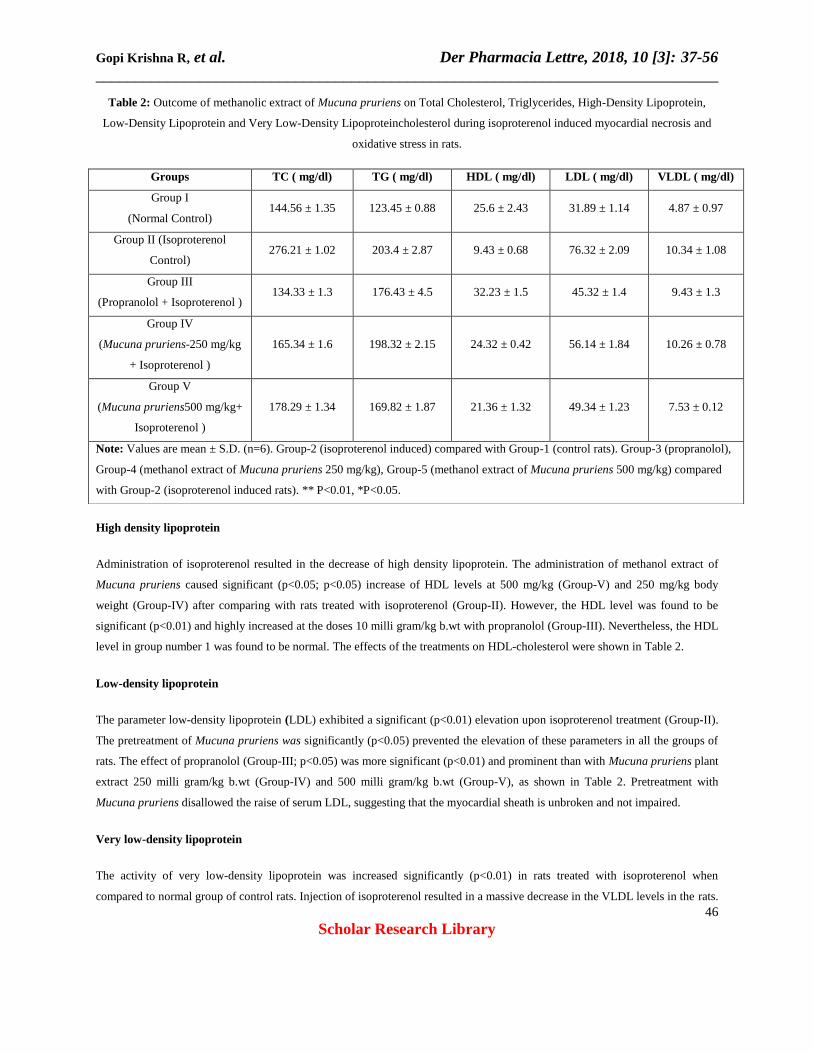
Table 2: Outcome of methanolic extract of Mucuna pruriens on Total Cholesterol, Triglycerides, High-Density Lipoprotein,
Low-Density Lipoprotein and Very Low-Density Lipoproteincholesterol during isoproterenol induced myocardial necrosis and
oxidative stress in rats.
High density lipoprotein
Administration of isoproterenol resulted in the decrease of high density lipoprotein. The administration of methanol extract of
Mucuna pruriens caused significant (p<0.05; p<0.05) increase of HDL levels at 500 mg/kg (Group-V) and 250 mg/kg body
weight (Group-IV) after comparing with rats treated with isoproterenol (Group-II). However, the HDL level was found to be
significant (p<0.01) and highly increased at the doses 10 milli gram/kg b.wt with propranolol (Group-III). Nevertheless, the HDL
level in group number 1 was found to be normal. The effects of the treatments on HDL-cholesterol were shown in Table 2.
Low-density lipoprotein
The parameter low-density lipoprotein (LDL) exhibited a significant (p<0.01) elevation upon isoproterenol treatment (Group-II).
The pretreatment of Mucuna pruriens was significantly (p<0.05) prevented the elevation of these parameters in all the groups of
rats. The effect of propranolol (Group-III; p<0.05) was more significant (p<0.01) and prominent than with Mucuna pruriens plant
extract 250 milli gram/kg b.wt (Group-IV) and 500 milli gram/kg b.wt (Group-V), as shown in Table 2. Pretreatment with
Mucuna pruriens disallowed the raise of serum LDL, suggesting that the myocardial sheath is unbroken and not impaired.
Very low-density lipoprotein
The activity of very low-density lipoprotein was increased significantly (p<0.01) in rats treated with isoproterenol when
compared to normal group of control rats. Injection of isoproterenol resulted in a massive decrease in the VLDL levels in the rats.
Groups TC ( mg/dl) TG ( mg/dl) HDL ( mg/dl) LDL ( mg/dl) VLDL ( mg/dl)
Group I
(Normal Control) 144.56 ± 1.35 123.45 ± 0.88 25.6 ± 2.43 31.89 ± 1.14 4.87 ± 0.97
Group II (Isoproterenol
Control) 276.21 ± 1.02 203.4 ± 2.87 9.43 ± 0.68 76.32 ± 2.09 10.34 ± 1.08
Group III
(Propranolol + Isoproterenol ) 134.33 ± 1.3 176.43 ± 4.5 32.23 ± 1.5 45.32 ± 1.4 9.43 ± 1.3
Group IV
(Mucuna pruriens-250 mg/kg
+ Isoproterenol )
165.34 ± 1.6 198.32 ± 2.15 24.32 ± 0.42 56.14 ± 1.84 10.26 ± 0.78
Group V
(Mucuna pruriens500 mg/kg+
Isoproterenol )
178.29 ± 1.34 169.82 ± 1.87 21.36 ± 1.32 49.34 ± 1.23 7.53 ± 0.12
Note: Values are mean ± S.D. (n=6). Group-2 (isoproterenol induced) compared with Group-1 (control rats). Group-3 (propranolol),
Group-4 (methanol extract of Mucuna pruriens 250 mg/kg), Group-5 (methanol extract of Mucuna pruriens 500 mg/kg) compared
with Group-2 (isoproterenol induced rats). ** P<0.01, *P<0.05.

Gopi Krishna R, et al. Der Pharmacia Lettre, 2018, 10 [3]: 37-56
______________________________________________________________________________
47
Scholar Research Library
Mucuna pruriens at diverse doses of 250 milli gram/kg b.wt (Group IV) and 500 mg/kg body weight (Group V) were
significantly (p<0.05; p<0.01) reduced the increased levels of very low density lipoproteins. Propranolol (10 mg/kg body weight,
Group-III) has shown more significant (p<0.01) effect than the plant extract. The results were presented in Table 2 and Figure 1.
Figure 1: Effect of methanol extract of Mucuna pruriens on myocardial level of TBARS (nM/g of tissue) and myocardial level
of CAT (U/mg of protein) during isoproterenol induced oxidative stress in rats. Values are mean ± S.D. (n=6). Group-2
(isoproterenol induced) compared with Group-1 (control rats). Group-3(Propranolol), Group- 4 (methanol extract of Mucuna
pruriens 250 mg/kg), Group-5 (methanol extract of Mucuna pruriens 500 mg/kg) compared with Group-2 (isoproterenol induced
rats). ** P<0.01, *P<0.05.
Plasma LDH
Plasma LDH was augmented significantly (p<0.01) in isoproterenol induced group (Group-II) in comparison to control (group-1)
as shown in Figure 2. Significant (p<0.01) reduction of LDH was observed more only in group-V (methanolic extract of Mucuna
pruriens at 500 milli gram/kg b.wt) and standard medicine propranolol treated rats (Group-III) when related to other groups.
However, less significant (p<0.05) decrease of plasma LDH was observed with methanol extract of Mucuna pruriens at 250
mg/kg (group IV). The results were indicated in Figure 2.
0 10 20 30 40 50 60 70
Myocardial TBARS
Catalase
Units
Group V
Group IV
Group III
Group II
Group I
**
**
**
**
**
**
*
*
Gopi Krishna R, et al. Der Pharmacia Lettre, 2018, 10 [3]: 37-56
______________________________________________________________________________
48
Scholar Research Library
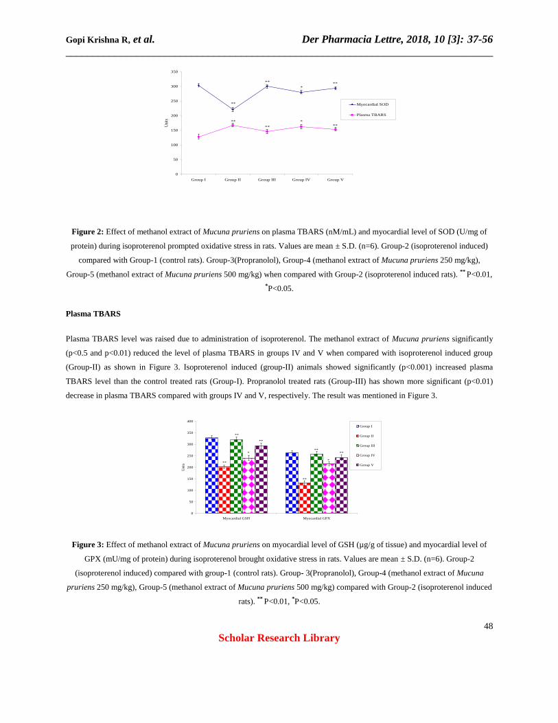
Figure 2: Effect of methanol extract of Mucuna pruriens on plasma TBARS (nM/mL) and myocardial level of SOD (U/mg of
protein) during isoproterenol prompted oxidative stress in rats. Values are mean ± S.D. (n=6). Group-2 (isoproterenol induced)
compared with Group-1 (control rats). Group-3(Propranolol), Group-4 (methanol extract of Mucuna pruriens 250 mg/kg),
Group-5 (methanol extract of Mucuna pruriens 500 mg/kg) when compared with Group-2 (isoproterenol induced rats). ** P<0.01,
*P<0.05.
Plasma TBARS
Plasma TBARS level was raised due to administration of isoproterenol. The methanol extract of Mucuna pruriens significantly
(p<0.5 and p<0.01) reduced the level of plasma TBARS in groups IV and V when compared with isoproterenol induced group
(Group-II) as shown in Figure 3. Isoproterenol induced (group-II) animals showed significantly (p<0.001) increased plasma
TBARS level than the control treated rats (Group-I). Propranolol treated rats (Group-III) has shown more significant (p<0.01)
decrease in plasma TBARS compared with groups IV and V, respectively. The result was mentioned in Figure 3.
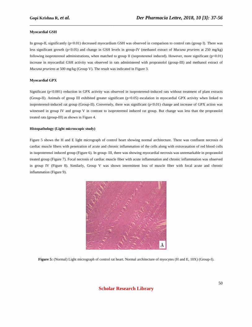
Figure 3: Effect of methanol extract of Mucuna pruriens on myocardial level of GSH (µg/g of tissue) and myocardial level of
GPX (mU/mg of protein) during isoproterenol brought oxidative stress in rats. Values are mean ± S.D. (n=6). Group-2
(isoproterenol induced) compared with group-1 (control rats). Group- 3(Propranolol), Group-4 (methanol extract of Mucuna
pruriens 250 mg/kg), Group-5 (methanol extract of Mucuna pruriens 500 mg/kg) compared with Group-2 (isoproterenol induced
rats). ** P<0.01, *P<0.05.
0
50
100
150
200
250
300
350
Group I Group II Group III Group IV Group V
Uni
ts
Myocardial SOD
Plasma TBARS
**
** **
****
**
*
*
0
50
100
150
200
250
300
350
400
Myocardial GSH Myocardial GPX
Uni
ts
Group I
Group II
Group III
Group IV
Group V**
**
**
**
**
***
*

Gopi Krishna R, et al. Der Pharmacia Lettre, 2018, 10 [3]: 37-56
______________________________________________________________________________
49
Scholar Research Library
TBARS of heart
Significant (p<0.001) increase in TBARS of the heart in group-II (isoproterenol induced rat) were observed. Significant (p<0.01;
p<0.05; p<0.01) reduction in the position of myocardial TBARS was detected in groups III and IV and V in association to the
isoproterenol induced rats (Group-II). Reduction in TBARS level was more in groups treated with propranolol and plant extract
high dose. The result was exposed in Figure 4.
Myocardial SOD
Significant (p<0.01) reduction of myocardial SOD action was witnessed in group-II (isoproterenol induced rat), when related to
control group (group-I). Myocardial SOD raised significantly (p<0.01) in propranolol treated rats 10 mg/kg (group-III) and 500
mg/kg methanolic extract of Mucuna pruriens (group-V) as shown in Figure 2. However, there was less significant (p<0.05) rise
in the level of myocardial SOD activity in methanol extract of Mucuna pruriens at 250 milli gram/kg (group-IV). The result was
mentioned in Figure 4.
Figure 4: Effect of methanol extract of Mucuna pruriens on Plasma-LDH (mU/mL) during isoproterenol induced oxidative stress
in rats. Values are mean ± S.D. (n=6). Group-2 (isoproterenol induced) compared with group-1 (control rats). Group-
3(Propranolol), Group- 4 (methanol extract of Mucuna pruriens 250 mg/kg), Group-5 (methanol extract of Mucuna pruriens 500
mg/kg) compared with Group-2 (isoproterenol induced rats). ** P<0.01, *P<0.05.
Myocardial catalase
In isoproterenol induced group of rats (group-II), there was a significant (p<0.001) reduction in myocardial activity of catalase. In
group IV rats, there was less significant (p<0.05) rise in the of myocardial catalase activity level was detected when associated to
isoproterenol induced rats (group-II). However, more significant (p<0.01) intensification in myocardial catalase activity was
observed in group-V. Nevertheless, significant (p<0.01) increased level of myocardial catalase was seen propranolol treated rats
(group-III), as shown in Figure 5.
900 920 940 960 980 1000 1020 1040
Plasma LDH
mU/mL
Group V
Group IV
Group III
Group II
Group I
**
**
**
*
Gopi Krishna R, et al. Der Pharmacia Lettre, 2018, 10 [3]: 37-56
______________________________________________________________________________
50
Scholar Research Library
Myocardial GSH
In group-II, significantly (p<0.01) decreased myocardium GSH was observed in comparison to control rats (group I). There was
less significant growth (p<0.05) and change in GSH levels in group-IV (methanol extract of Mucuna pruriens at 250 mg/kg)
following isoproterenol administrations, when matched to group II (isoproterenol induced). However, more significant (p<0.01)
increase in myocardial GSH activity was observed in rats administered with propranolol (group-III) and methanol extract of
Mucuna pruriens at 500 mg/kg (Group V). The result was indicated in Figure 3.
Myocardial GPX
Significant (p<0.001) reduction in GPX activity was observed in isoproterenol-induced rats without treatment of plant extracts
(Group-II). Animals of group III exhibited greater significant (p<0.05) escalation in myocardial GPX activity when linked to
isoproterenol-induced rat group (Group-II). Conversely, there was significant (p<0.01) change and increase of GPX action was
witnessed in group IV and group V in contrast to isoproterenol induced rat group. But change was less than the propranolol
treated rats (group-III) as shown in Figure 4.
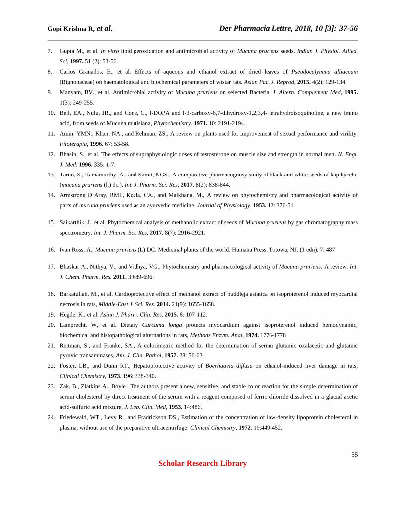
Histopathology (Light microscopic study)
Figure 5 shows the H and E light micrograph of control heart showing normal architecture. There was confluent necrosis of
cardiac muscle fibers with penetration of acute and chronic inflammation of the cells along with extravasation of red blood cells
in isoproterenol induced group (Figure 6). In group- III, there was showing myocardial necrosis was unremarkable in propranolol
treated group (Figure 7). Focal necrosis of cardiac muscle fiber with acute inflammation and chronic inflammation was observed
in group IV (Figure 8). Similarly, Group V was shown intermittent loss of muscle fiber with focal acute and chronic
inflammation (Figure 9).
Figure 5: (Normal) Light micrograph of control rat heart. Normal architecture of myocytes (H and E, 10X) (Group-I).

Gopi Krishna R, et al. Der Pharmacia Lettre, 2018, 10 [3]: 37-56
______________________________________________________________________________
51
Scholar Research Library
Figure 6: (Induced) Isoproterenol (ISO) group showing focal confluent necrosis of muscle fiber with acute and chronic
inflammation along with extravasation of red blood cells (10X, H and E) (Group-II).
Figure 7: Propranolol + Isoproterenol (ISO) treated group showing myocardial necrosis was unremarkable (10X, H and E)
(Group-III).
Figure 8: Mucuna pruriens extract (250 mg/kg) + Isoproterenol (ISO) group, showing focal necrosis of muscle fiber with acute
and chronic inflammation (10X H and E) (Group-IV).

Gopi Krishna R, et al. Der Pharmacia Lettre, 2018, 10 [3]: 37-56
______________________________________________________________________________
52
Scholar Research Library
Figure 9: Mucuna pruriens extract (500 mg/kg) + Isoproterenol (ISO) group, showing occasional loss of muscle fiber with focal
acute and chronic inflammation (10X H and E) (Group-V).
DISCUSSION
Myocardial necrosis can result incidentally owing to disruption in the blood supply to the heart or directly by some element insult
to the myocyte. When the damage to myocyte is high the enzymes existing in lysosomes outflow out of it and go in the
cytoplasm. Therefore a encouraging approach to identify cardiac injury contains monitoring and assessment of certain
cytoplasmic enzymes, as they can be noticed in blood serum [33]. Myocardial infarction is a clinical syndrome ascending from
sudden and stubborn curtailment of myocardial blood supply causing in necrosis of myocardium. This is followed by several
pathophysiology and biochemical variations such as lipid peroxidation, raised levels of cardiac markers and changed lipid profile
etc. [34].
Accordingly, the determination of CK-MB isoenzyme is a useful parameter for assessing myocardial damage. Pretreatment with
Mucuna pruriens prevented depletion of CK-MB isoenzyme from heart as associated to isoproterenol group. Therefore,
administration of Mucuna pruriens plant extract reduced the release of CK-MB isoenzyme from myocardium into the systemic
circulation, an indicative part of cardioprotective action of Mucuna pruriens. Myocardial necrosis leads to increase of cardiac serum
marker enzymes such as SGOT and SGPT that are progressive from the heart into blood [35] and indicating substantial cardio
cellular injury [36]. Administration of methanolic extract of Mucuna pruriens at 2 different doses dropped the augmented levels of
the serum enzymes and formed a consequent recovery to normalization when matched to control group animals [37].
Increase in lipid profile like triglyceride level in isoproterenol treated rats indicates that isoproterenol may be interrupting by
metabolism or biosynthesis of lipids. The increased myocardial triglycerides content observed in isoproterenol prompted
myocardial infracted rats is because of improved uptake of LDL from the blood through myocardial membranes [38]. It is
previously recognized that lipids are the utmost susceptible macromolecules to oxidative stress. Prior treatment with Mucuna
pruriens significantly diminished the points of triglycerides in isoproterenol persuaded myocardial infracted rats. No change was
detected in group of control rats. Isoproterenol brought raise in cholesterol levels could be owed to rise in biosynthesis and
reduction in its utilization. Isoproterenol brings free radical formation, which may leads to cellular cholesterol accumulation by
increasing cholesterol biosynthesis, by declining cholesterol ester hydrolysis and by dropping cholesterol efflux. Pretreatment
with the plant extract reestablished the level of cholesterol.

Gopi Krishna R, et al. Der Pharmacia Lettre, 2018, 10 [3]: 37-56
______________________________________________________________________________
53
Scholar Research Library
Hyper triglyceridemic patients at a threat for cardiovascular disease frequently develop a lipoprotein profile characterized by
raised triglyceride and low HDL cholesterol which origins myocardial membrane damage. Hypertriglyceridemia observed in
isoproterenol treated rats is clinically reported in ischemic heart disease [39]. The level of HDL was depleted in the serum due to
the administration of isoproterenol. Fascinatingly treatment with Mucuna pruriens significantly inverted the effects of
isoproterenol and raised the level of HDL in isoproterenol prompted myocardial infracted rats that clearly pointed out the
reestablishing ability of Mucuna pruriens on myocardial necrosis [40]. The increased myocardial cholesterol content detected in
isoproterenol induced myocardial infracted rats is because of improved uptake of LDL in the blood by myocardial membranes
[41]. A strong positive association has been documented between the danger of emerging ischemic heart disease and serum LDL
level. Pretreatment with Mucuna pruriens prevented the elevation of LDL in serum, signifying that the myocardial membrane is
intact and not damaged. Increase in lipid profile like VLDL in isoproterenol treated groups indicates isoproterenol may be
interfering with metabolism or biosynthesis of lipids. Pretreatment with Mucuna pruriens extract significantly declined (P<0.01)
the raised up levels of VLDL in isoproterenol tempted myocardial infracted rats.
Plasma LDH has been used traditionally as a general diagnostic instrument for myocardial infarction. An increase in the quantity
of LDH can be diagnostic of myocardial infarction. LDH is a cytosolic enzyme, which is essentially present in all the tissues
involved in glycolysis. From the damaged tissue it is released into the blood streams which become a definitive diagnostic and
prognostic criterion [42]. Pretreatment with plant extract of Mucuna pruriens at two different doses to rats challenged with
isoproterenol significantly diminished the elevated activities of the marker enzyme LDH in plasma and significantly returned
their activities in the myocardium. These findings are suggestive of the cardioprotective activity of Mucuna pruriens by its ability
to maintain myocardial integrity, principally by inhibiting lipid peroxidation-induced myocardial damage. It is previously known
that lipids are the most vulnerable macromolecules to oxidative stress and our outcomes exhibited that the level of lipid
peroxides, quantity in terms of TBARS was significantly better in plasma and heart of group treated with isoproterenol. The
metabolism of arachidonic acid through the lipoxygenase and cyclooxygenase pathways fallouts in the formation of reactive
oxygen species and other free radicals [43]. Raised levels of lipid peroxides injure blood vessels, producing enlarged adherence
and accumulation of platelets to the damaged sites [44]. Significant elevation noticed in the levels of plasma TBARS in group II
(isoproterenol-induced rats), which is in line is an sign of the severity of isoproterenol-induced necrotic injury to the myocardial
membrane. In the current study, the previous administration of propranolol and methanol extract of Mucuna pruriens were found
to significantly prevent the elevation levels of plasma TBARS.
Myocardial TBARS level was increased significantly (p<0.001) upon isoproterenol administration and remained high in group-II
when compared to normal control. Methanol extract of Mucuna pruriens has shown significant decrease as compared to
isoproterenol treated group of rats which indicates prevent the formation of reactive oxygen species and other free radicals.
Superoxide dismutase (SOD) is one more reactive oxygen species (ROS) defense enzyme existing entirely in the mitochondrial
matrix and defends cells against the deleterious actions of super oxide anion resulting from the peroxidative development in
tissues. The rats fed with methanol extract of Mucuna pruriens at various doses exposed significant rise in the level of SOD in
the heart tissue as related to that of further groups, demonstrating that the incidence of cardioprotective property in the herbal
extract. Catalase (CAT), a peroxisomal enzyme existing in the mitochondria in the heart. It is one of the antioxidant defense
enzymes which show an essential role in the oxidation of hydrogen peroxide to oxygen and water. In this study, the antioxidant
enzyme activity of catalase was decreased significantly (P <0.05) in rats treated with isoproterenol when related to those of

Gopi Krishna R, et al. Der Pharmacia Lettre, 2018, 10 [3]: 37-56
______________________________________________________________________________
54
Scholar Research Library
control rats. The activity of antioxidant enzyme was retained at near usual in rats pretreated methanol extract of Mucuna
pruriens.
GSH depletion is associated to a many disease states comprising cancer, neurodegenerative and heart related diseases.
Glutathione not only guards cell membranes from oxidative impairment, but also helps to retain the sulfhydryl groups of various
proteins in the reduced form, necessities for their normal role [45]. A significant (p<0.001) decrease in the activity of GSH was
observed. The prior administration of Mucuna pruriens significantly condensed the isoproterenol induced adverse effects and
maintained the level of elevated parameters at near normal. GPx play a critical function in cellular resistance alongside oxidative
stress by prolongation a cascade of reactions. Glutathione peroxidase (GPx) is a selenoprotein which oxidizes 2 molecules of
glutathione (GSH) into oxidized glutathione (GSSG). Reduced levels of GPx in group-II animals were reverted to normal due to
the administration of methanolic extract of Mucuna pruriens. Histopathological study was performed for the confirmations of
biochemical findings. A clear view of the cardiac damage was noted in the isoproterenol treated rats by histopathological study
and those affected tissues were recovered by the administration of Mucuna pruriens extract. Thus Mucuna pruriens has some
protective effect on myocardium against isoproterenol.
CONCLUSION
Histo-pathological and biological conclusions of the present study specify that methanolic extract of Mucuna pruriens possess
cardioprotective activity against isoproterenol made myocardial necrosis in rats. The presence of antioxidant constituents from
plants of flavonoids in the methanolic extract might be liable for cardioprotective activity of Mucuna pruriens. Thus, our study
clearly indicated a significant cardioprotective action of methanolic extract of Mucuna pruriens.
ACKNOWLEDGEMENTS
The authors are thankful to the management of GITAM University, Visakhapatnam, Andhra Pradesh, India, for providing
necessary facilities to carry out the research work.
REFERENCES
1. Zhu, YZ., et al. Cardioprotective effects of nitroparacetamol and paracetamol in acute phase of myocardial infarction in
experimental rats. Oxford, Euromed Communications Limited, 1998. 1-20.
2. Farnsworth, NR., et al. Medicinal plants in therapy. Bulletin WHO 1985, 63: 965-972.
3. Bafna, PA., and Balaraman R., Antioxidant activity of DHC-1-- A herbal formulation, Phytother. Res, 2005. 19: 216-222.
4. Verma, SC., et al. A review on phytochemistry and pharmacological Activity of parts of mucuna pruriens used as an
Ayurvedic medicine. World J. Pharm. Res, 2014. 3(5): 138-158.
5. Warrier, PK., Nambiar, VPK., and Ramankutty, C., In vitro anthelmintic activity of Mucuna pruriens (DC) and Canarium
schweinfurthii (Engl) on Ascaris suum. Indian Medicinal Plants, 1996. 4: 68-72.
6. Pant, M.C., et al. Blood sugar and total cholesterol lowering effect of Glycine soja (Sieb and Zucc.), Mucuna pruriens
(D.C.) and Dolichos biflorus (Linn.) seed diets in normal fasting albino rats. Indian J. Med. Res., 1968. 56: 1808.
Gopi Krishna R, et al. Der Pharmacia Lettre, 2018, 10 [3]: 37-56
______________________________________________________________________________
55
Scholar Research Library
7. Gupta M., et al. In vitro lipid peroxidation and antimicrobial activity of Mucuna pruriens seeds. Indian J. Physiol. Allied.
Sci, 1997. 51 (2): 53-56.
8. Carlos Granados, E., et al. Effects of aqueous and ethanol extract of dried leaves of Pseudocalymma alliaceum
(Bignonaceae) on haematological and biochemical parameters of wistar rats. Asian Pac. J. Reprod, 2015. 4(2): 129-134.
9. Manyam, BV., et al. Antimicrobial activity of Mucuna pruriens on selected Bacteria, J. Altern. Complement Med, 1995.
1(3): 249-255.
10. Bell, EA., Nulu, JR., and Cone, C., l-DOPA and l-3-carboxy-6,7-dihydroxy-1,2,3,4- tetrahydroisoquinoline, a new imino
acid, from seeds of Mucuna mutisiana, Phytochemistry. 1971. 10: 2191-2194.
11. Amin, YMN., Khan, NA., and Rehman, ZS., A review on plants used for improvement of sexual performance and virility.
Fitoterapia, 1996. 67: 53-58.
12. Bhasin, S., et al. The effects of supraphysiologic doses of testosterone on muscle size and strength in normal men. N. Engl.
J. Med. 1996. 335: 1-7.
13. Tarun, S., Ramamurthy, A., and Sumit, NGS., A comparative pharmacognosy study of black and white seeds of kapikacchu
(mucuna pruriens (l.) dc.). Int. J. Pharm. Sci. Res, 2017. 8(2): 838-844.
14. Armstrong D‘Aray, RMI., Keela, CA., and Maikhana, M., A review on phytochemistry and pharmacological activity of
parts of mucuna pruriens used as an ayurvedic medicine. Journal of Physiology. 1953. 12: 376-51.
15. Saikarthik, J., et al. Phytochemical analysis of methanolic extract of seeds of Mucuna pruriens by gas chromatography mass
spectrometry. Int. J. Pharm. Sci. Res, 2017. 8(7): 2916-2921.
16. Ivan Ross, A., Mucuna pruriens (L) DC. Medicinal plants of the world. Humana Press, Totowa, NJ. (1 edn), 7: 487
17. Bhaskar A., Nithya, V., and Vidhya, VG., Phytochemistry and pharmacological activity of Mucuna pruriens: A review. Int.
J. Chem. Pharm. Res. 2011. 3:689-696.
18. Barkatullah, M., et al. Cardioprotective effect of methanol extract of buddleja asiatica on isoproterenol induced myocardial
necrosis in rats, Middle-East J. Sci. Res. 2014. 21(9): 1655-1658.
19. Hegde, K., et al. Asian J. Pharm. Clin. Res, 2015. 8: 107-112.
20. Lamprecht, W, et al. Dietary Curcuma longa protects myocardium against isoproterenol induced hemodynamic,
biochemical and histopathological alternations in rats, Methods Enzym. Anal, 1974. 1776-1778
21. Reitman, S., and Franke, SA., A colorimetric method for the determination of serum glutamic oxalacetic and glutamic
pyruvic transaminases, Am. J. Clin. Pathol, 1957. 28: 56-63
22. Foster, LB., and Dunn RT., Hepatoprotective activity of Boerhaavia diffusa on ethanol-induced liver damage in rats,
Clinical Chemistry, 1973. 196: 338-340.
23. Zak, B., Zlatkins A., Boyle., The authors present a new, sensitive, and stable color reaction for the simple determination of
serum cholesterol by direct treatment of the serum with a reagent composed of ferric chloride dissolved in a glacial acetic
acid-sulfuric acid mixture, J. Lab. Clin. Med, 1953. 14:486.
24. Friedewald, WT., Levy R., and Fradrickson DS., Estimation of the concentration of low-density lipoprotein cholesterol in
plasma, without use of the preparative ultracentrifuge. Clinical Chemistry, 1972. 19:449-452.

Gopi Krishna R, et al. Der Pharmacia Lettre, 2018, 10 [3]: 37-56
______________________________________________________________________________
56
Scholar Research Library
25. Moldeus, P., Hogberg, J., and Orrenius, S., Activation of hepatocyte protein kinase C by redox-cycling quinones. Methods
Enzymol. 1978. 52: 55–62.
26. Liu, J., et al. Antioxidant action of Guilingji in the brain of rats with FeCl3-induced epilepsy. Free Radical Biol. Med, 1990.
9(5):451-454.
27. Lowry, OH., et al. Protein measurement with the folin phenol reagent. J. Biol. Chem, 1951. 193: 265-275.
28. Kakkar, P., Das, B., and Viswanathan, PN., A modified spectrophotometric assay of superoxide dismutase. Indian J.
Biochem. Biophys.1984. 21: 130-132.
29. Aebi, H., Ameliorative effect of Apodytes dimidiata on cisplatin-induced nephrotoxicity in Wistar rats. Am. J. Plant Sci,
1974, 7: 674-684.
30. Ellman, GL., Tissue sulfhydryl groups. Arch. Biochem. Biophys, 1959. 82: 70-77.
31. Paglia, DE., and Valentine, WN., Studies on the quantitative and qualitative characterization of erythrocyte glutathione
peroxidase. J. Lab. Clin. Med. 1967. 70: 158-169.
32. Wendel, A., Glutathione peroxidase. Methods Enzymol, 1981. 77: 325-333.
33. Deodato, B., et al. Cardioprotection by the phytoestrogen genistein in experimental myocardial ischaemia-reperfusion
injury. Br. J. Pharmacol, 1999. 128: 1683-1690.
34. Anversa, P., and Sonnenblick, EH., Ischemic cardiomyopathy: pathophysiologic mechanisms. Prog. Cardiovasc. dis, 1990,
33: 49-70.
35. Vijaya PV., Shyamala Devi, CS., and Ramkumar, KM., Effect of fish oil pretreatment on isoproterenol-induced changes in
myocardial membrane phospholipids. Nutrition. 2006. 22: 1171-1176.
36. Nigam, PK., Biochemical markers of myocardial injury. Indian J. Clin. Biochem, 2007. 22: 10-17.
37. Rotenberg, Z., et al. Cardioprotective potential of lathyrus sativus against experimental myocardial infarction due to
isoproterenol in rats. Clinical Chemistry, 1987. 33: 1419-1420.
38. Paramasivam, R., et l. Recommendation letter for Asian Pacific Journal of Tropical Biomedicine (APJTB). Asian Pac. J.
Trop. Biomed, 2012. S212-S218.
39. Nagarathna, PKM., et al. Survey on content-based image retrieval and texture analysis with applications. International
Journal of Toxicological and Pharmacological Research, 2014. 5(4): 121-128.
40. Gokkusu, C., and Mosta Fazadeh, T., Oxidative stress and serum paraoxonase activity in experimental hypothyroidism:
Effect of vitamin E supplementation. Clin. Chim. Acta, 2003. 328: 155-161.
41. Shaik, H A., et al. J Ethnopharmacol., 2012. 141: 33-40.
42. Deepa, PR., Varalakshmi, P., Effect of nelumbo nucifera flowers on plasma lipids and glucose in young, middle-aged and
aged rats. Toxicology. 2005. 211:77-85.
43. Gutteridge, JMC., and Halliwell, B., The measurement and mechanism of lipid peroxidation in biological systems. Trends
Biochem. Sci, 1990, 15: 129-135
44. Gryglewski, RJ., Prostaglandins, platelets, and atherosclerosis. Crit Rev Biochem, 1980. 7: 291-338.
45. Pastore, A., et al. Analysis of glutathione: implication in redox and detoxification. Clin. Chim. Acta, 2003. 333: 19-39.