Mutations upstream of fabI in triclosan resistant Staphylococcus aureus strains are associated with...
13
RESEARCH ARTICLE Open Access Mutations upstream of fabI in triclosan resistant Staphylococcus aureus strains are associated with elevated fabI gene expression Denis Grandgirard 1† , Leonardo Furi 2† , Maria Laura Ciusa 2 , Lucilla Baldassarri 3 , Daniel R Knight 4,8 , Ian Morrissey 4,9 , Carlo R Largiadèr 5 , Stephen L Leib 1,6 and Marco R Oggioni 2,7* Abstract Background: The enoyl-acyl carrier protein (ACP) reductase enzyme (FabI) is the target for a series of antimicrobial agents including novel compounds in clinical trial and the biocide triclosan. Mutations in fabI and heterodiploidy for fabI have been shown to confer resistance in S. aureus strains in a previous study. Here we further determined the fabI upstream sequence of a selection of these strains and the gene expression levels in strains with promoter region mutations. Results: Mutations in the fabI promoter were found in 18% of triclosan resistant clinical isolates, regardless the previously identified molecular mechanism conferring resistance. Although not significant, a higher rate of promoter mutations were found in strains without previously described mechanisms of resistance. Some of the mutations identified in the clinical isolates were also detected in a series of laboratory mutants. Microarray analysis of selected laboratory mutants with fabI promoter region mutations, grown in the absence of triclosan, revealed increased fabI expression in three out of four tested strains. In two of these strains, only few genes other than fabI were upregulated. Consistently with these data, whole genome sequencing of in vitro selected mutants identified only few mutations except the upstream and coding regions of fabI, with the promoter mutation as the most probable cause of fabI overexpression. Importantly the gene expression profiling of clinical isolates containing similar mutations in the fabI promoter also showed, when compared to unrelated non-mutated isolates, a significant up-regulation of fabI. Conclusions: In conclusion, we have demonstrated the presence of C34T, T109G, and A101C mutations in the fabI promoter region of strains with fabI up-regulation, both in clinical isolates and/or laboratory mutants. These data provide further observations linking mutations upstream fabI with up-regulated expression of the fabI gene. Keywords: Biocide, Resistance, Triclosan, fabI, Microarray, Cross-resistance, Promoter mutation Background Post-genomic research in the past years narrowed down significantly the number of pathway proposed to be suit- able as targets for antimicrobial treatment. Based on this, the type II fatty acid biosynthesis pathway shows much promise [1]. One frequently targeted enzyme in this path- way is the enoyl-acyl carrier protein (ACP) reductase en- zyme (FabI) as witnessed by the multiple Staphylococcus aureus FabI inhibitors in clinical trial [2-5]. This is further underlined by the fact that FabI is the target of the first line anti-tuberculosis drug isoniazid, diazaborines and the biocide triclosan [1,6]. The latter is a synthetic, non-ionic, chlorinated bis-phenol and is present in many health care products for both hospital and consumer use [7]. It pos- sesses broad spectrum antimicrobial activity against many Gram-positive and Gram-negative bacteria, some fungi [8], and protozoa including Plasmodium falciparum and Toxoplasma gondii [8]. Triclosan, unlike other biocides, at low concentrations has a single intracellular target by binding to the active site of the FabI. It forms a stable * Correspondence: [email protected] † Equal contributors 2 LA.M.M.B., Dip. Biotecnologie Mediche, Università di Siena, Siena, Italy 7 Department of Genetics, University of Leicester, Leicester, UK Full list of author information is available at the end of the article © 2015 Grandgirard et al.; licensee BioMed Central. This is an Open Access article distributed under the terms of the Creative Commons Attribution License (http://creativecommons.org/licenses/by/4.0), which permits unrestricted use, distribution, and reproduction in any medium, provided the original work is properly credited. The Creative Commons Public Domain Dedication waiver (http://creativecommons.org/publicdomain/zero/1.0/) applies to the data made available in this article, unless otherwise stated. Grandgirard et al. BMC Genomics (2015) 16:345 DOI 10.1186/s12864-015-1544-y
Transcript of Mutations upstream of fabI in triclosan resistant Staphylococcus aureus strains are associated with...

Grandgirard et al. BMC Genomics (2015) 16:345 DOI 10.1186/s12864-015-1544-y
RESEARCH ARTICLE Open Access
Mutations upstream of fabI in triclosan resistantStaphylococcus aureus strains are associated withelevated fabI gene expressionDenis Grandgirard1†, Leonardo Furi2†, Maria Laura Ciusa2, Lucilla Baldassarri3, Daniel R Knight4,8, Ian Morrissey4,9,Carlo R Largiadèr5, Stephen L Leib1,6 and Marco R Oggioni2,7*
Abstract
Background: The enoyl-acyl carrier protein (ACP) reductase enzyme (FabI) is the target for a series of antimicrobialagents including novel compounds in clinical trial and the biocide triclosan. Mutations in fabI and heterodiploidyfor fabI have been shown to confer resistance in S. aureus strains in a previous study. Here we further determinedthe fabI upstream sequence of a selection of these strains and the gene expression levels in strains with promoterregion mutations.
Results: Mutations in the fabI promoter were found in 18% of triclosan resistant clinical isolates, regardless thepreviously identified molecular mechanism conferring resistance. Although not significant, a higher rate ofpromoter mutations were found in strains without previously described mechanisms of resistance. Some of themutations identified in the clinical isolates were also detected in a series of laboratory mutants. Microarray analysisof selected laboratory mutants with fabI promoter region mutations, grown in the absence of triclosan, revealedincreased fabI expression in three out of four tested strains. In two of these strains, only few genes other than fabIwere upregulated. Consistently with these data, whole genome sequencing of in vitro selected mutants identifiedonly few mutations except the upstream and coding regions of fabI, with the promoter mutation as the mostprobable cause of fabI overexpression. Importantly the gene expression profiling of clinical isolates containingsimilar mutations in the fabI promoter also showed, when compared to unrelated non-mutated isolates, a significantup-regulation of fabI.
Conclusions: In conclusion, we have demonstrated the presence of C34T, T109G, and A101C mutations in the fabIpromoter region of strains with fabI up-regulation, both in clinical isolates and/or laboratory mutants. These dataprovide further observations linking mutations upstream fabI with up-regulated expression of the fabI gene.
Keywords: Biocide, Resistance, Triclosan, fabI, Microarray, Cross-resistance, Promoter mutation
BackgroundPost-genomic research in the past years narrowed downsignificantly the number of pathway proposed to be suit-able as targets for antimicrobial treatment. Based on this,the type II fatty acid biosynthesis pathway shows muchpromise [1]. One frequently targeted enzyme in this path-way is the enoyl-acyl carrier protein (ACP) reductase en-zyme (FabI) as witnessed by the multiple Staphylococcus
* Correspondence: [email protected]†Equal contributors2LA.M.M.B., Dip. Biotecnologie Mediche, Università di Siena, Siena, Italy7Department of Genetics, University of Leicester, Leicester, UKFull list of author information is available at the end of the article
© 2015 Grandgirard et al.; licensee BioMed CeCommons Attribution License (http://creativecreproduction in any medium, provided the orDedication waiver (http://creativecommons.orunless otherwise stated.
aureus FabI inhibitors in clinical trial [2-5]. This is furtherunderlined by the fact that FabI is the target of the firstline anti-tuberculosis drug isoniazid, diazaborines and thebiocide triclosan [1,6]. The latter is a synthetic, non-ionic,chlorinated bis-phenol and is present in many health careproducts for both hospital and consumer use [7]. It pos-sesses broad spectrum antimicrobial activity against manyGram-positive and Gram-negative bacteria, some fungi[8], and protozoa including Plasmodium falciparum andToxoplasma gondii [8]. Triclosan, unlike other biocides, atlow concentrations has a single intracellular target bybinding to the active site of the FabI. It forms a stable
ntral. This is an Open Access article distributed under the terms of the Creativeommons.org/licenses/by/4.0), which permits unrestricted use, distribution, andiginal work is properly credited. The Creative Commons Public Domaing/publicdomain/zero/1.0/) applies to the data made available in this article,

Grandgirard et al. BMC Genomics (2015) 16:345 Page 2 of 13
ternary complex with NAD+. Triclosan inhibits FabI by al-losterically blocking the active site, and therefore preventsbacteria from synthesising fatty acids, which are necessaryfor building cell membranes and for division [9].Several studies have demonstrated that bacteria have
both natural and acquired mechanisms of resistance totriclosan. Natural resistance is present to varyingdegrees in bacterial species, which harbour alternativesto fabI, (fabK, fabL or fabV) [10-13]. In addition, bio-degradation has been found to occur in differentenvironmental species [14]. The primary mechanism ofacquired resistance is due to mutations within thecoding region of fabI, which decrease affinity of theenzyme to triclosan [9,15-19]. Alternatively, active effluxof triclosan has been described in several Gram-negative species, and is mediated by the resistance-nodulation-division (RND) family of pumps [20]. It hasbeen shown that triclosan can activate the transcrip-tional regulator SmeT of the SmeDEF efflux transporterin Stenotrophomonas maltophilia [21].In addition to target modification and efflux, also
titration of the target enzymes has been found to conferresistance. Recent findings have shown the presence ofan additional copy of fabI, horizontally transferred fromS. haemolyticus, in the genome of many S. aureusisolates with reduced susceptibility to triclosan [22].Fatty acid biosynthesis is finely regulated in S. aureus bya feed-forward system that globally controls the expres-sion of genes involved in this metabolic pathway andthat is dependent on the malonyl-CoA intracellularlevels [23,24]. This metabolite was shown to bind to andtherefore to inhibit the activity of the transcriptionalrepressor FapR [23]. FapR is a homodimeric repressorhighly conserved in Gram positive organisms that hasbeen characterized for its inhibitory function of theexpression of genes involved in the fatty acids and phos-pholipids biosynthetic pathways [23,24]. Among others,FapR was shown to directly interact with the promoterof fabI and to physiologically regulate its expression[24]. In analogy to the isoniazide resistance conferringmutation in Mycobacterium tuberculosis [6], increasedamount of the FabI enzyme has been described in triclo-san resistant S. aureus strains [16], however no geneexpression data are available to sustain this finding. Fur-thermore, increased levels of fabI expression have beenobserved in in vitro adapted S. aureus derivatives, with apossible role of promoter mutations in some of thesestrains [25]. Therefore, the aim of this study was tofurther characterize mechanisms of resistance in a co-hort of previously described S. aureus clinical isolatesand in vitro selected mutants with reduced susceptibilityto triclosan by sequencing the putative promoter regionof fabI and by evaluating the levels of gene expressionusing microarray analysis.
ResultsPromoter sequence analysisSeven out of thirty-eight (18%) triclosan resistant S. aureusclinical isolates sequenced were found to have polymor-phisms in the fabI upstream region (Additional file 1:Supplementary data S1). Sequence data showed that theC34T substitution was the most frequent SNP (5 strains),while the SNPs A101C and T109G were found only once(Table 1, Figure 1). The latter SNP was found in clinicalisolate QBR-102278-1052 where the insertion of an IS256element upstream the fabI gene created an eight bp dupli-cation (AAAAAGTC), which generated the T109G poly-morphism (Table 1; Figure 1). No mutations were foundin the nineteen triclosan susceptible isolates (Additionalfile 1: Supplementary data S1). FabI promoter mutationswere found in 3 out of 9 strains of the group of isolateswithout any known triclosan resistance marker, 2/15 inthe strains with sa-fabI mutations and 2/14 in the strainscarrying the sh-fabI gene. When we previously analysedthe sa-fabI locus in the twenty-three in vitro selected tri-closan mutant strains, we found that all (23/23) had amutated sa-fabI gene [22]. Now we found that abouthalf (13/23) of these had an additional sa-fabI promotermutation (Table 1; Figure 1; Additional file 1: Supplementarydata S1). The majority of the mutations in the sa-fabI pro-moter reflected those found in clinical isolates, exceptfor the A7G and A72G SNPs and the A101 deletion(Table 1; Figure 1). Similar to the clinical isolates, theC34T mutation was the most represented polymorphism(Table 1; Figure 1). In order to identify other genetic deter-minants possibly involved in triclosan reduced susceptibil-ity, seven mutants were further analysed by whole genomesequencing. These data revealed that only mutations inthe fabI locus were shared by all strains. MO052 was theonly strain analysed by microarray containing additionalSNPs, but no obvious associations between these andchanges in the transcriptome could be made (Table 2 andAdditional file 2: supplementary data S2).
Gene expression analysisLaboratory mutantsTranscriptomic differences between triclosan susceptiblewild type strains and their resistant isogenic mutantswere analysed by microarrays. Four pairs with differentmutations in the promoter regions were compared(Table 3 and Table 4). Strains with the following promotermutations were analysed: A7G in MO036 and A72G inMO034 were mutations documented only in laboratorystrains, but not in clinical isolates. C34T in MO035 wasthe most frequently observed mutation in both laboratorystrains and clinical isolates. Finally, we also analysedT109G, the second most frequent mutation in the labora-tory strains, which was also identified in a clinical isolate(Table 3, Figure 1). fabI was up-regulated in three of the

Table 1 Genotypes and phenotypes of in vitro mutants and clinical isolates with fabI promoter mutations
Strain Background sa-fabI promoter** sa-fabI‡ sh-fabI‡ MIC*‡ MBC*‡ Comment
RN4220 - wt wt - 1 2
MW2 - wt wt - 0.12 0.12
ATCC6538 - wt wt - 0.12 0.25
MO036 RN4220 A7G mutated - 4 8
MO035 RN4220 C34T mutated - 8 8
MO047 RN4220 C34T mutated - 4 8
MO049 RN4220 C34T mutated - 4 8
MO076 MW2 C34T mutated - 4 8
CR002 ATCC6538 C34T mutated - 4 8
MO034 RN4220 A72G mutated - 8 8
MO077 MW2 A101- mutated - 4 32 1 bp deletion
d7 ATCC6538 A101- mutated - 2 8 1 bp deletion
MO051 ATCC6538 T109G mutated - 4 8
MO052 ATCC6538 T109G mutated - 8 16
MO053 ATCC6538 T109G mutated - 4 8
MO055 ATCC6538 T109G mutated - 4 8
QBR-102278-1097 - C34T mutated - 0.25 32
QBR-102278-1889 - C34T wt - 0.5 16
QBR-102278-1969 - C34T wt - 0.25 32
QBR-102278-2095 - C34T wt - 0.25 32
QBR-102278-2546 - C34T mutated + 1 64
QBR-102278-2363 - A101C wt + 16 32
QBR-102278-1052 - T109G wt + 0.5 64 IS256 insertion
*MIC and MBC to triclosan are expressed as mg/L. **Polymorphic sites are indicated counting backwards from the sa-fabI start site of S. aureus Mu50 (GenBank ID:BA000017; position 1060308). Strains analysed by microarray are indicated in bold. ‡With the exception of strain QBR-102278-2095, these data have been previouslyreported [22].
Figure 1 Mapping of mutations in the intergenic region upstream sa-fabI. The sa-fabI upstream region from the S. aureus Mu50 genome(GenBank ID: BA000017) is reported. Nucleotides in which mutations have been identified are marked in bold. The positions of the mutations arereported with respect to the first nucleotide preceding the start codon of sa-fabI and numbered backwards. The nucleotide substitution isdescribed above the mutation position together with the number of clinical isolates (italicised number) and mutant strains carrying that particularmutation. ATCC6538 sa-fabI upstream region sequence is identical to Mu50, while the naturally occurring polymorphisms identified in the wtstrains RN4220 (A92T; GenBank ID: AFGU01000045), ATCC25923 (A213T, A188C; GenBank ID: CP009361), and MW2 (T224A; GenBank ID: BA000033)with respect to the Mu50 sequence are not reported as they do not affect triclosan susceptibility. The putative −35 and −10 consensus sequences,identified by BPROM, are underlined. The consensus of the transcriptional repressor FapR recognition sequence is reported as mapped in RegPrecise(underlined) [26] or as previously reported by alignment with the experimentally determined one in the fapR upstream region (dotted underlined)[23,24]. The transcriptional start site (+1TSS) as identified by RNAseq [50] and the ribosomal binding site (SD) are also reported.
Grandgirard et al. BMC Genomics (2015) 16:345 Page 3 of 13
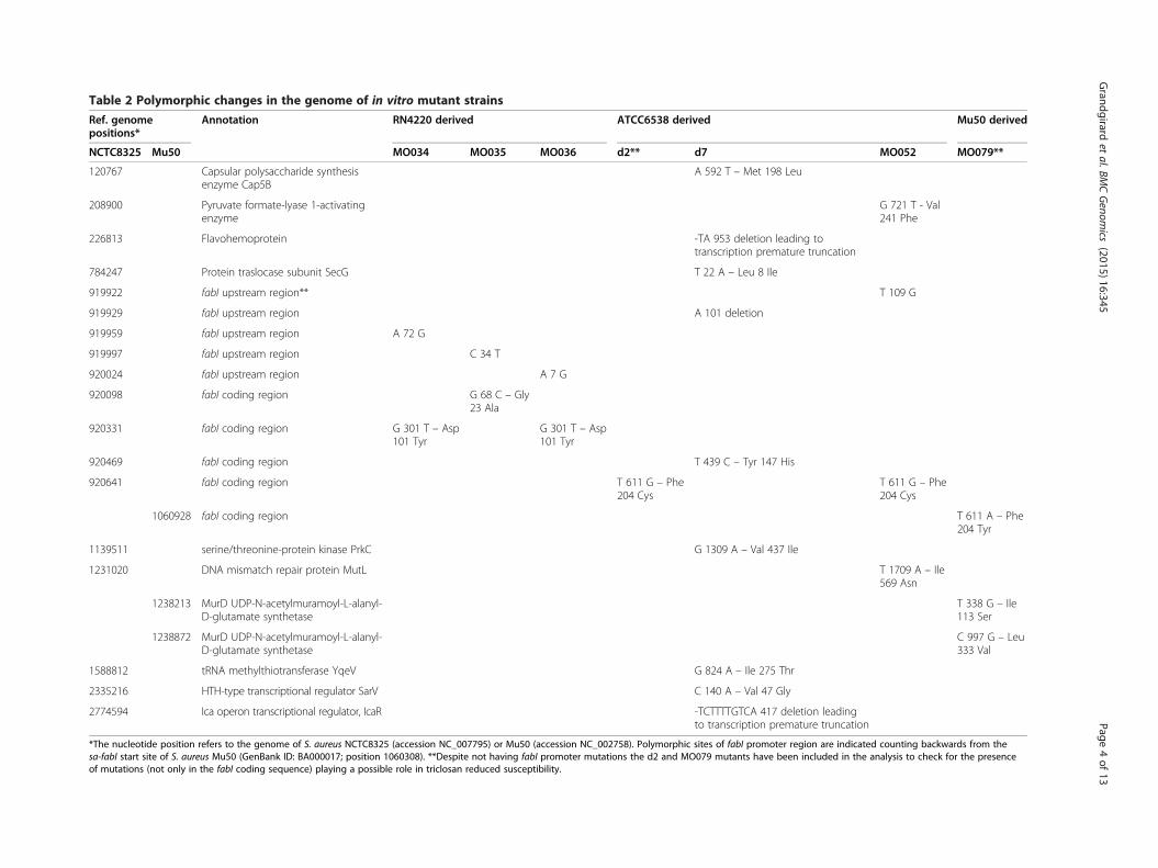
Table 2 Polymorphic changes in the genome of in vitro mutant strains
Ref. genomepositions*
Annotation RN4220 derived ATCC6538 derived Mu50 derived
NCTC8325 Mu50 MO034 MO035 MO036 d2** d7 MO052 MO079**
120767 Capsular polysaccharide synthesisenzyme Cap5B
A 592 T – Met 198 Leu
208900 Pyruvate formate-lyase 1-activatingenzyme
G 721 T - Val241 Phe
226813 Flavohemoprotein -TA 953 deletion leading totranscription premature truncation
784247 Protein traslocase subunit SecG T 22 A – Leu 8 Ile
919922 fabI upstream region** T 109 G
919929 fabI upstream region A 101 deletion
919959 fabI upstream region A 72 G
919997 fabI upstream region C 34 T
920024 fabI upstream region A 7 G
920098 fabI coding region G 68 C – Gly23 Ala
920331 fabI coding region G 301 T – Asp101 Tyr
G 301 T – Asp101 Tyr
920469 fabI coding region T 439 C – Tyr 147 His
920641 fabI coding region T 611 G – Phe204 Cys
T 611 G – Phe204 Cys
1060928 fabI coding region T 611 A – Phe204 Tyr
1139511 serine/threonine-protein kinase PrkC G 1309 A – Val 437 Ile
1231020 DNA mismatch repair protein MutL T 1709 A – Ile569 Asn
1238213 MurD UDP-N-acetylmuramoyl-L-alanyl-D-glutamate synthetase
T 338 G – Ile113 Ser
1238872 MurD UDP-N-acetylmuramoyl-L-alanyl-D-glutamate synthetase
C 997 G – Leu333 Val
1588812 tRNA methylthiotransferase YqeV G 824 A – Ile 275 Thr
2335216 HTH-type transcriptional regulator SarV C 140 A – Val 47 Gly
2774594 Ica operon transcriptional regulator, IcaR -TCTTTTGTCA 417 deletion leadingto transcription premature truncation
*The nucleotide position refers to the genome of S. aureus NCTC8325 (accession NC_007795) or Mu50 (accession NC_002758). Polymorphic sites of fabI promoter region are indicated counting backwards from thesa-fabI start site of S. aureus Mu50 (GenBank ID: BA000017; position 1060308). **Despite not having fabI promoter mutations the d2 and MO079 mutants have been included in the analysis to check for the presenceof mutations (not only in the fabI coding sequence) playing a possible role in triclosan reduced susceptibility.
Grandgirard
etal.BM
CGenom
ics (2015) 16:345
Page4of
13
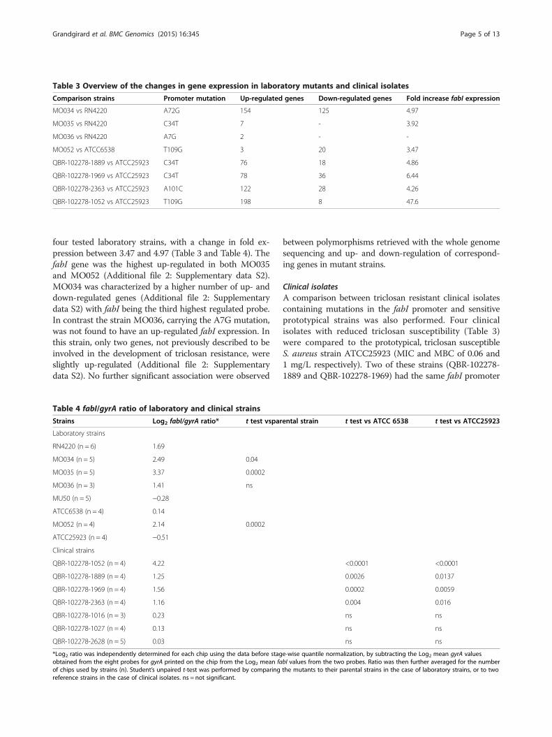
Table 3 Overview of the changes in gene expression in laboratory mutants and clinical isolates
Comparison strains Promoter mutation Up-regulated genes Down-regulated genes Fold increase fabI expression
MO034 vs RN4220 A72G 154 125 4.97
MO035 vs RN4220 C34T 7 - 3.92
MO036 vs RN4220 A7G 2 - -
MO052 vs ATCC6538 T109G 3 20 3.47
QBR-102278-1889 vs ATCC25923 C34T 76 18 4.86
QBR-102278-1969 vs ATCC25923 C34T 78 36 6.44
QBR-102278-2363 vs ATCC25923 A101C 122 28 4.26
QBR-102278-1052 vs ATCC25923 T109G 198 8 47.6
Grandgirard et al. BMC Genomics (2015) 16:345 Page 5 of 13
four tested laboratory strains, with a change in fold ex-pression between 3.47 and 4.97 (Table 3 and Table 4). ThefabI gene was the highest up-regulated in both MO035and MO052 (Additional file 2: Supplementary data S2).MO034 was characterized by a higher number of up- anddown-regulated genes (Additional file 2: Supplementarydata S2) with fabI being the third highest regulated probe.In contrast the strain MO036, carrying the A7G mutation,was not found to have an up-regulated fabI expression. Inthis strain, only two genes, not previously described to beinvolved in the development of triclosan resistance, wereslightly up-regulated (Additional file 2: Supplementarydata S2). No further significant association were observed
Table 4 fabI/gyrA ratio of laboratory and clinical strains
Strains Log2 fabI/gyrA ratio* t test vspar
Laboratory strains
RN4220 (n = 6) 1.69
MO034 (n = 5) 2.49 0.04
MO035 (n = 5) 3.37 0.0002
MO036 (n = 3) 1.41 ns
MU50 (n = 5) −0.28
ATCC6538 (n = 4) 0.14
MO052 (n = 4) 2.14 0.0002
ATCC25923 (n = 4) −0.51
Clinical strains
QBR-102278-1052 (n = 4) 4.22
QBR-102278-1889 (n = 4) 1.25
QBR-102278-1969 (n = 4) 1.56
QBR-102278-2363 (n = 4) 1.16
QBR-102278-1016 (n = 3) 0.23
QBR-102278-1027 (n = 4) 0.13
QBR-102278-2628 (n = 5) 0.03
*Log2 ratio was independently determined for each chip using the data before stagobtained from the eight probes for gyrA printed on the chip from the Log2 mean faof chips used by strains (n). Student’s unpaired t-test was performed by comparingreference strains in the case of clinical isolates. ns = not significant.
between polymorphisms retrieved with the whole genomesequencing and up- and down-regulation of correspond-ing genes in mutant strains.
Clinical isolatesA comparison between triclosan resistant clinical isolatescontaining mutations in the fabI promoter and sensitiveprototypical strains was also performed. Four clinicalisolates with reduced triclosan susceptibility (Table 3)were compared to the prototypical, triclosan susceptibleS. aureus strain ATCC25923 (MIC and MBC of 0.06 and1 mg/L respectively). Two of these strains (QBR-102278-1889 and QBR-102278-1969) had the same fabI promoter
ental strain t test vs ATCC 6538 t test vs ATCC25923
<0.0001 <0.0001
0.0026 0.0137
0.0002 0.0059
0.004 0.016
ns ns
ns ns
ns ns
e-wise quantile normalization, by subtracting the Log2 mean gyrA valuesbI values from the two probes. Ratio was then further averaged for the numberthe mutants to their parental strains in the case of laboratory strains, or to two
Grandgirard et al. BMC Genomics (2015) 16:345 Page 6 of 13
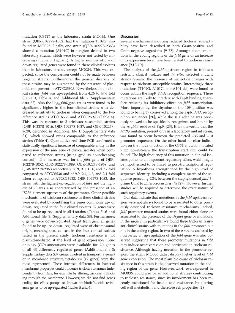
mutation (C34T) as the laboratory strain MO035. Onestrain (QBR-102278-1052) had the mutation T109G, alsofound in MO052. Finally, one strain (QBR-102278-2363)showed a mutation (A101C) in a region deleted in twolaboratory strains, which were however not tested by mi-croarrays (Table 3, Figure 1). A higher number of up- ordown-regulated genes were found in these clinical isolatesthan in laboratory strains, except MO034. This was ex-pected, since the comparison could not be made betweenisogenic strains. Furthermore, the genetic diversity ofthese strains may be augmented by the presence of plas-mids not present in ATCC25923. Nevertheless, in all clin-ical strains, fabI was up-regulated, from 4.26 to 47.6 fold(Table 3, Table 4, and Additional file 2: Supplementarydata S2). Also the Log2 fabI/gyrA ratios were found to besignificantly higher in the four clinical strains with de-creased sensitivity to triclosan when compared to the tworeference strains ATCC6538 and ATCC25923 (Table 4).This was in contrast to 3 triclosan susceptible strains(QBR-102278-1016, QBR-102278-1027, and QBR-102278-2628; described in Additional file 1: Supplementary dataS1), which showed ratios comparable to the referencestrains (Table 4). Quantitative real-time PCR confirmed astatistically significant increase of comparable entity in theexpression of the fabI gene of clinical isolates when com-pared to reference strains (using gyrA as housekeepingcontrol). The increase was for the fabI gene of QBR-102278-1052, QBR-102278-1889, QBR-102278-1969, andQBR-102278-2363 respectively 36.9, 9.0, 15.6, and 7.7 foldcompared to ATCC6538 and of 9.9, 2.4, 4.2, and 2.1 foldwhen compared to ATCC25923. QBR-102278-1052, thestrain with the highest up-regulation of fabI and the high-est MBC was also characterized by the presence of anIS256 element upstream of the promoter. Other possiblemechanisms of triclosan resistance in these clinical strainswere evaluated by identifying the genes commonly up- ordown- regulated in the four clinical isolates. 37 genes werefound to be up-regulated in all 4 strains (Tables 5, 6 andAdditional file 3: Supplementary data S3). Furthermore,6 genes were down-regulated. Apart from fabI, all genesfound to be up- or down- regulated were of chromosomalorigin, meaning that, at least in the four clinical isolatestested in the present study, triclosan resistance is notplasmid-mediated at the level of gene expression. Geneontology (GO) annotations were available for 29 genesof all 43 differently regulated genes (Additional file 3:Supplementary data S3). Genes involved in transport (8 genes)or in membrane structure/metabolism (12 genes) were themost represented. These intrinsic differences in bacterialmembrane properties could influence triclosan tolerance inde-pendently from fabI, for example by altering triclosan traffick-ing through the membrane. However, we did not find genescoding for efflux pumps or known antibiotic/biocide resist-ance genes to be up-regulated (Tables 5 and 6).
DiscussionSeveral mechanisms inducing reduced triclosan suscepti-bility have been described in both Gram-positive andGram-negative organisms [9-22]. Amongst them, muta-tions in the coding regions of the fabI gene or an increasein its expression level have been related to triclosan resist-ance [9,15-19].The analysis of the fabI upstream region in triclosan
resistant clinical isolates and in vitro selected mutantstrains revealed the presence of nucleotide changes withrespect to triclosan susceptible strains. Interestingly threemutations (T109G, A101C, and A101-del) were found tooccur within the FapR DNA recognition sequence. Thesemutations are likely to interfere with FapR binding, there-fore reducing its inhibitory effect on fabI transcription.More importantly, the thymine in the 109 position wasfound to be highly conserved among the FapR DNA recog-nition sequences [26], while the 101 adenine was previ-ously showed to be specifically recognized and bound bythe Arg56B residue of FapR [23]. It is noteworthy that theA72G mutation, present only in a laboratory mutant strain,was found to occur between the predicted −35 and −10promoter sequences. On the other hand no clear indica-tion on the mode of action of the C34T mutation, located7 bp downstream the transcription start site, could befound. The high frequency of this mutation in clinical iso-lates points to an important regulatory effect, which mightbe hypothesised to be linked to post-transcriptional regu-lation. A hypothesis strengthened by the high level ofsequence identity, including a complete match of the se-quence preceding C34, between the staphylococcal fabI 5-prime UTR to Enterococcus faecalis [27]. However furtherstudies will be required to determine the exact nature ofsuch regulatory events.Our data indicate that mutations in the fabI upstream re-
gion were not always found to be associated to other previ-ously described triclosan resistance mechanisms. Indeed,fabI promoter mutated strains were found either alone orassociated to the presence of the sh-fabI gene or mutationsin the sa-fabI. In particular we found three triclosan resist-ant clinical strains with mutations in the fabI promoter, butnot in the coding region. In two of these strains analysed bymicroarray an up-regulation of the fabI gene was also ob-served suggesting that these promoter mutations in fabImay induce overexpression and participate in triclosan re-sistance. Although having mutation in the promoter re-gion, the strain MO036 didn’t display higher level of fabIgene expression. The most plausible cause of triclosan re-sistance in this strain is the observed mutation in the cod-ing region of the gene. However, isaA, overexpressed inMO036, could also be an additional strategy contributingto triclosan resistance, since its involvement has been re-cently mentioned for fusidic acid resistance, by alteringcell wall metabolism and therefore cell properties [28].
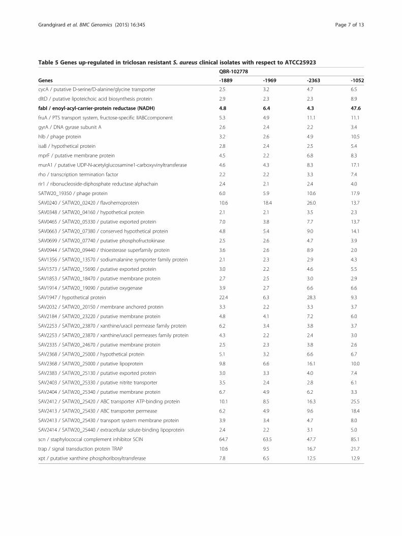
Table 5 Genes up-regulated in triclosan resistant S. aureus clinical isolates with respect to ATCC25923
QBR-102778
Genes -1889 -1969 -2363 -1052
cycA / putative D-serine/D-alanine/glycine transporter 2.5 3.2 4.7 6.5
dltD / putative lipoteichoic acid biosynthesis protein 2.9 2.3 2.3 8.9
fabI / enoyl-acyl-carrier-protein reductase (NADH) 4.8 6.4 4.3 47.6
fruA / PTS transport system, fructose-specific IIABCcomponent 5.3 4.9 11.1 11.1
gyrA / DNA gyrase subunit A 2.6 2.4 2.2 3.4
hlb / phage protein 3.2 2.6 4.9 10.5
isaB / hypothetical protein 2.8 2.4 2.5 5.4
mprF / putative membrane protein 4.5 2.2 6.8 8.3
murA1 / putative UDP-N-acetylglucosamine1-carboxyvinyltransferase 4.6 4.3 8.3 17.1
rho / transcription termination factor 2.2 2.2 3.3 7.4
rir1 / ribonucleoside-diphosphate reductase alphachain 2.4 2.1 2.4 4.0
SATW20_19350 / phage protein 6.0 5.9 10.6 17.9
SAV0240 / SATW20_02420 / flavohemoprotein 10.6 18.4 26.0 13.7
SAV0348 / SATW20_04160 / hypothetical protein 2.1 2.1 3.5 2.3
SAV0465 / SATW20_05330 / putative exported protein 7.0 3.8 7.7 13.7
SAV0663 / SATW20_07380 / conserved hypothetical protein 4.8 5.4 9.0 14.1
SAV0699 / SATW20_07740 / putative phosphofructokinase 2.5 2.6 4.7 3.9
SAV0944 / SATW20_09440 / thioesterase superfamily protein 3.6 2.6 8.9 2.0
SAV1356 / SATW20_13570 / sodium:alanine symporter family protein 2.1 2.3 2.9 4.3
SAV1573 / SATW20_15690 / putative exported protein 3.0 2.2 4.6 5.5
SAV1853 / SATW20_18470 / putative membrane protein 2.7 2.5 3.0 2.9
SAV1914 / SATW20_19090 / putative oxygenase 3.9 2.7 6.6 6.6
SAV1947 / hypothetical protein 22.4 6.3 28.3 9.3
SAV2032 / SATW20_20150 / membrane anchored protein 3.3 2.2 3.3 3.7
SAV2184 / SATW20_23220 / putative membrane protein 4.8 4.1 7.2 6.0
SAV2253 / SATW20_23870 / xanthine/uracil permease family protein 6.2 3.4 3.8 3.7
SAV2253 / SATW20_23870 / xanthine/uracil permeases family protein 4.3 2.2 2.4 3.0
SAV2335 / SATW20_24670 / putative membrane protein 2.5 2.3 3.8 2.6
SAV2368 / SATW20_25000 / hypothetical protein 5.1 3.2 6.6 6.7
SAV2368 / SATW20_25000 / putative lipoprotein 9.8 6.6 16.1 10.0
SAV2383 / SATW20_25130 / putative exported protein 3.0 3.3 4.0 7.4
SAV2403 / SATW20_25330 / putative nitrite transporter 3.5 2.4 2.8 6.1
SAV2404 / SATW20_25340 / putative membrane protein 6.7 4.9 6.2 3.3
SAV2412 / SATW20_25420 / ABC transporter ATP-binding protein 10.1 8.5 16.3 25.5
SAV2413 / SATW20_25430 / ABC transporter permease 6.2 4.9 9.6 18.4
SAV2413 / SATW20_25430 / transport system membrane protein 3.9 3.4 4.7 8.0
SAV2414 / SATW20_25440 / extracellular solute-binding lipoprotein 2.4 2.2 3.1 5.0
scn / staphylococcal complement inhibitor SCIN 64.7 63.5 47.7 85.1
trap / signal transduction protein TRAP 10.6 9.5 16.7 21.7
xpt / putative xanthine phosphoribosyltransferase 7.8 6.5 12.5 12.9
Grandgirard et al. BMC Genomics (2015) 16:345 Page 7 of 13
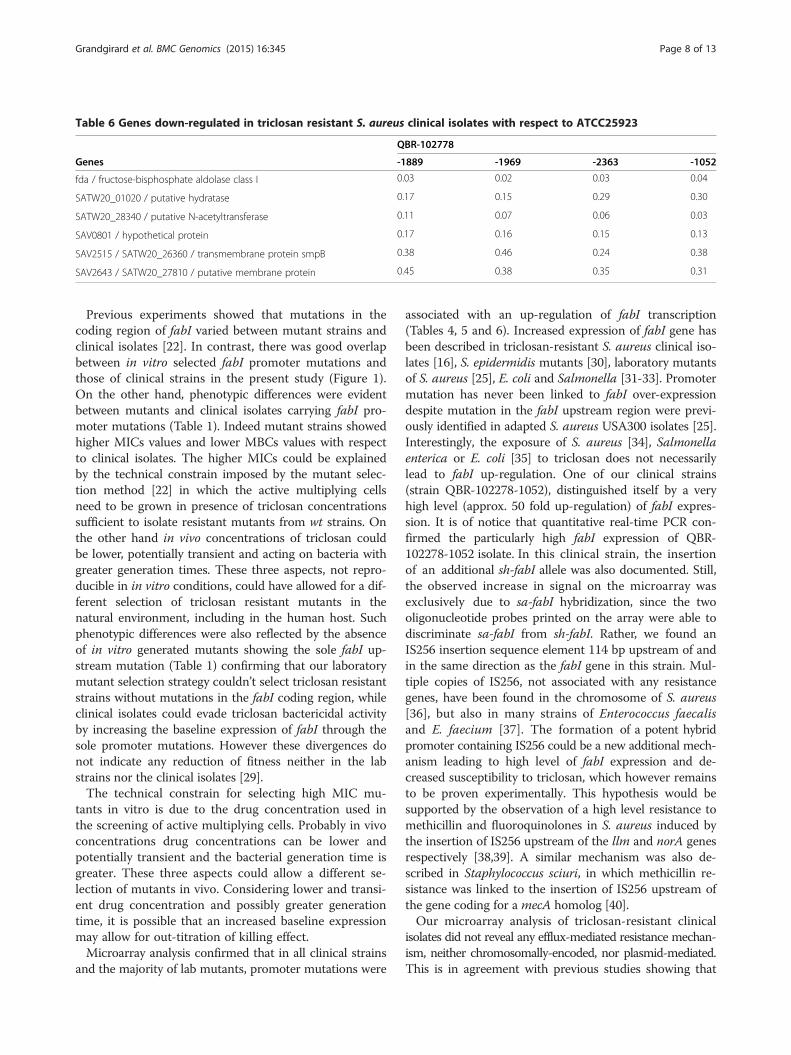
Table 6 Genes down-regulated in triclosan resistant S. aureus clinical isolates with respect to ATCC25923
QBR-102778
Genes -1889 -1969 -2363 -1052
fda / fructose-bisphosphate aldolase class I 0.03 0.02 0.03 0.04
SATW20_01020 / putative hydratase 0.17 0.15 0.29 0.30
SATW20_28340 / putative N-acetyltransferase 0.11 0.07 0.06 0.03
SAV0801 / hypothetical protein 0.17 0.16 0.15 0.13
SAV2515 / SATW20_26360 / transmembrane protein smpB 0.38 0.46 0.24 0.38
SAV2643 / SATW20_27810 / putative membrane protein 0.45 0.38 0.35 0.31
Grandgirard et al. BMC Genomics (2015) 16:345 Page 8 of 13
Previous experiments showed that mutations in thecoding region of fabI varied between mutant strains andclinical isolates [22]. In contrast, there was good overlapbetween in vitro selected fabI promoter mutations andthose of clinical strains in the present study (Figure 1).On the other hand, phenotypic differences were evidentbetween mutants and clinical isolates carrying fabI pro-moter mutations (Table 1). Indeed mutant strains showedhigher MICs values and lower MBCs values with respectto clinical isolates. The higher MICs could be explainedby the technical constrain imposed by the mutant selec-tion method [22] in which the active multiplying cellsneed to be grown in presence of triclosan concentrationssufficient to isolate resistant mutants from wt strains. Onthe other hand in vivo concentrations of triclosan couldbe lower, potentially transient and acting on bacteria withgreater generation times. These three aspects, not repro-ducible in in vitro conditions, could have allowed for a dif-ferent selection of triclosan resistant mutants in thenatural environment, including in the human host. Suchphenotypic differences were also reflected by the absenceof in vitro generated mutants showing the sole fabI up-stream mutation (Table 1) confirming that our laboratorymutant selection strategy couldn’t select triclosan resistantstrains without mutations in the fabI coding region, whileclinical isolates could evade triclosan bactericidal activityby increasing the baseline expression of fabI through thesole promoter mutations. However these divergences donot indicate any reduction of fitness neither in the labstrains nor the clinical isolates [29].The technical constrain for selecting high MIC mu-
tants in vitro is due to the drug concentration used inthe screening of active multiplying cells. Probably in vivoconcentrations drug concentrations can be lower andpotentially transient and the bacterial generation time isgreater. These three aspects could allow a different se-lection of mutants in vivo. Considering lower and transi-ent drug concentration and possibly greater generationtime, it is possible that an increased baseline expressionmay allow for out-titration of killing effect.Microarray analysis confirmed that in all clinical strains
and the majority of lab mutants, promoter mutations were
associated with an up-regulation of fabI transcription(Tables 4, 5 and 6). Increased expression of fabI gene hasbeen described in triclosan-resistant S. aureus clinical iso-lates [16], S. epidermidis mutants [30], laboratory mutantsof S. aureus [25], E. coli and Salmonella [31-33]. Promotermutation has never been linked to fabI over-expressiondespite mutation in the fabI upstream region were previ-ously identified in adapted S. aureus USA300 isolates [25].Interestingly, the exposure of S. aureus [34], Salmonellaenterica or E. coli [35] to triclosan does not necessarilylead to fabI up-regulation. One of our clinical strains(strain QBR-102278-1052), distinguished itself by a veryhigh level (approx. 50 fold up-regulation) of fabI expres-sion. It is of notice that quantitative real-time PCR con-firmed the particularly high fabI expression of QBR-102278-1052 isolate. In this clinical strain, the insertionof an additional sh-fabI allele was also documented. Still,the observed increase in signal on the microarray wasexclusively due to sa-fabI hybridization, since the twooligonucleotide probes printed on the array were able todiscriminate sa-fabI from sh-fabI. Rather, we found anIS256 insertion sequence element 114 bp upstream of andin the same direction as the fabI gene in this strain. Mul-tiple copies of IS256, not associated with any resistancegenes, have been found in the chromosome of S. aureus[36], but also in many strains of Enterococcus faecalisand E. faecium [37]. The formation of a potent hybridpromoter containing IS256 could be a new additional mech-anism leading to high level of fabI expression and de-creased susceptibility to triclosan, which however remainsto be proven experimentally. This hypothesis would besupported by the observation of a high level resistance tomethicillin and fluoroquinolones in S. aureus induced bythe insertion of IS256 upstream of the llm and norA genesrespectively [38,39]. A similar mechanism was also de-scribed in Staphylococcus sciuri, in which methicillin re-sistance was linked to the insertion of IS256 upstream ofthe gene coding for a mecA homolog [40].Our microarray analysis of triclosan-resistant clinical
isolates did not reveal any efflux-mediated resistance mechan-ism, neither chromosomally-encoded, nor plasmid-mediated.This is in agreement with previous studies showing that
Grandgirard et al. BMC Genomics (2015) 16:345 Page 9 of 13
there was no significant increase in the triclosan MBCfor S. aureus strains carrying plasmid-borne qac genescoding for multidrug efflux pumps [41]. Similarly, over-expression of the chromosomal norA multidrug trans-porter gene did not lead to triclosan resistance [42].Efflux-mediated resistance to triclosan is however not tobe excluded in Gram- positive bacteria. Indeed, it has beenrecently shown that, out of 21 S. haemolyticus clinicalstrains, an inhibition of efflux pump activity by carbonylcyanide-m-chlorophenylhydrazone (CCCP) significantlydecreased triclosan MIC in four strains [43] suggesting thepossible presence of a still unidentified efflux system withtriclosan as a substrate. Despite this fact, efflux-mediatedtriclosan resistance to date still remains restricted to Gram-negative species [20,21]. Other mechanisms inducing analteration of membrane metabolism, structure or traffick-ing in S. aureus could not be excluded. This hypothesiswould be supported by the identification of several upreg-ulated genes involved in such mechanisms in clinicalstrains or isaA up-regulation in the laboratory mutantMO036.Apart from the fabI gene or efflux pumps, evidence for
other mechanisms of resistance to triclosan is scarce. Pro-longed exposure of MRSA to triclosan-impregnated sili-con elastomer resulted in the selection of small colonyvariants resistant to triclosan, but the underlying mechan-ism is still not understood and may be non-specific, in-volving a reduction in energy generation and/or transportand the down-regulation of functions such as cell wallsynthesis [44]. High-throughput methods (proteomics,genomics) have been recently applied to laboratory strainsof S. typhimurium and E. coli grown in absence of triclo-san in order to reveal inherent mechanisms of resistanceto triclosan based on changes in genes or proteins expres-sion [32,33,45]. Apart from the consistent up-regulationof fabI, relatively few new mechanisms were proposed.It has been postulated that the increased expression ofdehydrogenases and oxidoreductases using NAD+ as a co-factor, could bind and capture triclosan, reducing its ef-fective intracellular concentration [32,45]. In the presentstudy, apart from fabI, only one putative flavohemo-protein (SATW20_02420) containing identified bind-ing sites for NAD or FAD was highly up-regulated in theclinical strains. In most cases no complete operon wasfound to be over-expressed in the triclosan resistant clin-ical isolates. Exceptions are the co-transcribed fruA andfruB genes and the three genes for an amino acid ABCuptake system (SAV2412-4). This is in accordance withprevious data, which had shown that exposure of S. aureusto triclosan leads to the de-regulation of branchedamino acid uptake and carbohydrate metabolism in-cluding changes in expression of fruA, fruB and xprT[34]. Albeit the partial overlap of data between ourcharacterisation of resistant isolates and the work on
triclosan toxicity, no clear metabolic correlation can bedrawn, which links triclosan and fatty acid metabolismto the observed changes in gene expression. In that re-spect, the current analyses of the six clinical isolatesdevoid of any resistance markers could also providefurther insights, especially if fabI turns out not to beoverexpressed in these strains.To our knowledge, this is the first extensive microarray
analysis comparing both in vitro generated mutants andclinical isolates of S. aureus resistant to triclosan. Thecomparison of the mutants and their parental strains en-ables us to link genetic variations to phenotypic changesmore directly. For clinical strains, comparisons betweenresistant strains and a prototypical strain do not allowsuch direct conclusion and also relate to the choice of theprototypical strain. These results were therefore con-firmed by determining fabI/gyrA ratio. To definitely chal-lenge the hypothesis that mutations in the promoter offabI lead to overexpression of the gene and consequentlyto a reduced susceptibility, prospective genetic manipula-tion would have been a more direct approach. Neverthe-less, whole genome sequencing revealed in three out offour laboratory mutant tested by microarray, that the onlymutation susceptible to explain change in fabI expressionwas located in fabI promoter region. Furthermore, the factthat the same fabI promoter mutations were found in allclinical strains with overexpression of their fabI gene, in-directly provide the evidence for the involvement of suchmutations in fabI up-regulation and possibly in triclosanreduced susceptibility.
ConclusionIn conclusion, molecular changes in the promoter regionof fabI were identified together with fabI over-expressionand triclosan resistance. As such this overexpression offabI has the potential to determine cross-resistance tonovel compounds in clinical trial [2-5]. This adds to therecently described mutations within fabI selected by thenovel compound AFN-1252 which confer cross-resistanceto triclosan [46]. It should be noted that in S. aureus fabIup-regulation acts almost always in addition to the othertriggers of triclosan resistance, such as mutations in cod-ing regions of the fabI gene or the insertion of an additionallele derived from S. haemolyticus. Importantly, we couldnot link triclosan resistance in staphylococci to the pres-ence or over-expression of efflux systems, either of plas-mid or chromosomal origin, a mechanism known tocontribute to resistance in Gram-negative organisms [47]or to the resistance to any other antimicrobial drug [48].In addition we have solid evidence of the fabI up-regulation in four triclosan resistant clinical isolates forwhich the intrinsic genetic diversity between clinical iso-lates so far had eluded comparison of gene expression. In-direct evidences support the relationship between fabI
Grandgirard et al. BMC Genomics (2015) 16:345 Page 10 of 13
overexpression and the presence of mutations mapped inthe FapR binding domain or other regions of the predictedfabI promoter region. This hypothesis is further strength-ened by the finding of the same mutations in fabI-overex-pressing laboratory mutants, which, most importantly,were shown to not carry any other mutation in their gen-ome that could be associated to triclosan resistance. Thiscombined data in laboratory mutants and clinical isolatesopens new avenues to explore mechanisms of triclosan re-sistance in S. aureus.
MethodsBacterial strainsSixty-five S. aureus strains with reduced susceptibility totriclosan were previously selected from a collection of1602 clinical isolates by performing standard MIC andMBC assays [22,48,49]. Of these, fifteen strains with mu-tations in the fabI coding sequence, fourteen with anadditional chromosomal sh-fabI allele (from S. haemoly-ticus), and nine without any known triclosan “resistance”marker were investigated in this work for fabI promotermutations (Additional file 1: Supplementary data S1). Asa control nineteen triclosan susceptible isolates were in-cluded in the analyses (Additional file 1: Supplementarydata S1). In addition twenty-three independent mutants(Additional file 1: Supplementary data S1), with re-duced susceptibility to triclosan, were also analysed. Thesestrains have been previously selected by single-exposureof ATCC6538, MW2, and Mu50 reference strains to0.5 mg/L of triclosan in solid medium or by multiplestep-growth on liquid medium with increasing triclosanconcentrations (from 0.25 mg/L to 4 mg/L) as in the caseof the RN4220 laboratory strain [22].No ethical approval was required to obtain the isolates
used in the study. No ethical approval was required touse the clinical isolates in this study. Isolates were ob-tained during routine microbiological investigations andwere not part of a clinical trial.
Molecular analysisThe upstream region of the fabI gene was amplified inthe fifty-seven clinical isolates and in the twenty-threelaboratory mutants. DNA was amplified using standardPCR conditions and the following primers: 5′-ATCATCTTCGTGCGTATTATC-3′ and 5′-TTCAAGCTCTTTACGGCTA-3′ (Eurofins MWG Operon, Germany).PCR products were sequenced by the Sanger method(Eurofins MWG Operon, Ebersberg, Germany). A selec-tion of S. aureus fabI upstream sequences has beendeposited in GenBank (accession nos. KF583951- KF583970).The putative −35 and −10 sequences have been predictedusing the BPROM tool (http://www.softberry.com/ber-ry.phtml), while the FapR recognition sequence wasmapped by mean of the data available on the RegPrecise
database [26]. The transcriptional start site was identifiedby direct visualisation of RNA-seq alignment data re-trieved from the NCBI Sequence Read Archive (SRA,http://www.ncbi.nlm.nih.gov/Traces/sra/). More pre-cisely the illumina HiSeq data were previously generatedby sequencing of the RNA extracted from a S. aureusNewman strain at the early log phase (SRA ExperimentID: DRX011556, SRA Sample ID: DRS011392) [50].Whole-genome sequencing was performed by the In-stitute of Applied Genomics (University of Udine, Italy)using an Illumina Genome Analyzer II platform (Illu-mina, San Diego, California, USA) for the in vitro se-lected mutants d2, d7, MO034, MO035, MO036, MO052,MO079 and their isogenic wild type strain RN4220,ATCC6538, and Mu50 (Table 2). Mutants to be sequencedhave been selected on the basis of their fabI promoter se-quence in order to analyse one strain for each one of thepreviously identified mutations. Strains d2 and MO079have been also included in order to check for geneticchanges, other than sa-fabI mutations, possibly related totheir triclosan reduced susceptibility phenotypes. Se-quences of both wt strains and mutant strains, werealigned to the reference genome of S. aureus NCTC8325(accession NC_007795), except for Mu50 and MO079 thatwere aligned to the Mu50 genome (accession NC_002758),using the Mosaik Assembler suite (The MarthLab, BostonCollege, Massachusetts, USA). Single nucleotide poly-morphisms (SNPs), insertions and deletions (INDELs)were retrieved with VarScan software [51]. SNPs andINDELs of the wt strains obtained from the alignmentwith the GenBank reference genome were subtractedfrom those found by aligning the mutant strain withthe reference.
Statistical analysisFisher’s exact test was applied to assess if the differencesin the number of clinical isolates with fabI promotermutations among the three groups of strains defined byknown triclosan resistance marker were statisticallysignificant.
Gene expression analysisArray design and productionProbe design was performed by the CustomArray DesignService. Probes of 35–40 bp length were selected basedon melting temperature (Tm), complexity, secondarystructure, GC-content, and specificity. A total of 12’000capture probes were finally used. Furthermore, the arrayalso contained quality control spots, non-specific probesderived from phages, plants, virus and bacteria, as wellas empty, oligonucleotide-free spots. The entire genomesof Mu50 and TW20 were covered in the microarrays,plus additional elements as listed in the Additional file 4:Supplementary data S4. Arrays were synthesized on a
Grandgirard et al. BMC Genomics (2015) 16:345 Page 11 of 13
CustomArray Synthesizer (CombiMatrix, Mukilteo, WA)and quality tested using the standard protocols providedby the manufacturer.
Bacterial growthS. aureus strains were grown overnight in 10 ml trypticsoy broth (TSB) at 37°C at 80 rpm. The cultures were di-luted 1:100 in pre-warmed TSB and grown to logarithmicphase (OD570 = 0.6). 2 ml of each culture (1–5 × 10⁸ col-ony forming units) was harvested in 4 ml of RNAprotect®reagent (Qiagen), incubated for 5 min at room temperatureand centrifuged for 10 min at 5000 × g. The pellet wasthen processed directly for RNA extraction or storedat −80°C for later processing.
RNA purificationTotal RNA was extracted using RNeasy Mini Kit(Qiagen), according to the manufacturer’s instructions,using 50 U recombinant lysostaphin (Sigma) followed byincubation for 5 minutes with 1 ml of hot Qiazol (Qiagen)to lyse bacteria. Bacteria were further disrupted by vibrationwith 50 mg of acid-washed glass beads (Sigma) using aMickle Vibratory Tissue Disintegrator (Mickle LaboratoryEngineering) at maximum speed. Contaminating DNAwas removed using DNA-free™ Kit (Applied Biosystems)and RNA quality tested on an Agilent 2100 Bioanalyzer(Agilent Technologies). RNA concentration and puritywere determined by Nanodrop® ND-1000 spectrophotom-eter (Thermo Scientific). For each strain, at least 4 RNAsamples were prepared from independent cultures.
RNA labelling and fragmentationIsolated, unamplified RNA was labelled with Cy5, usingULS™ Labeling Kit for CombiMatrix arrays (KreatechBiotechnology), according to the manufacturer’s instructions.RNA was finally fragmented with RNA FragmentationReagents (Ambion®).
Array hybridization12 K Customarrays were hybridized with 2 μg of la-belled, fragmented RNA, according to information pro-vided by the manufacturer (Customarray/CombimatrixIncorporated). Microarrays were scanned using the PackardScanArray4000 array scanner and software (ScanArray, ver-sion 3.1, Packard BioChip Technologies). All arrays werescanned with incremental laser power from 15 to 100%.Data were extracted with Microarray Imager software(version 5.8.0, Combi Matrix) and spot intensity expressedas median intensity.
Data analysisScanning data with similar median fluorescence intensitywere chosen for analysis. Fluorescence values of spotswith maximal intensity (signal saturation) at the chosen
laser intensity were extrapolated by linear regression,using values gathered from the two next lower laserintensities.
Gene filteringNon-specific binding was determined from fluorescencevalues of all non-specific probes. The cut-off for specificbinding was set as the upper 95% confidence interval ofthe mean signal intensity of the non-specific probes. Foreach comparison, probes were excluded when the meanvalues for both strains to be compared were under thedetermined cut-off. For comparisons involving in vitrogenerated mutants derived from plasmid-free strainsRN4220 or ATCC6538, analysis was performed usingonly probes sets gathered from the genomes of S. aureusTW20 and Mu50.
Data transformation, normalization and analysisThe fluorescence values were log2 transformed. For eachset of comparison, stage-wise quantile normalization wasperformed, using a script written in the statistical com-puting environment of R (R Development Core Team,2011), according to Deshmukh et al. [52]. Significantlydifferentially regulated genes were determined by usingthe Significance Analysis of Microarrays method (SAM,Excel Add- in version 4.0) originally developed at Stan-ford University lab [53]. For each comparison, the deltavalue was set to obtain a conservative median false dis-covery rate (FDR) of 1% and the fold change cut-offvalue was set to 2. For investigating common up- ordown- regulated genes in the 4 triclosan resistant clinicalstrains, the FDR value was set to 5%.
Quantitative real-time PCRReverse transcription of total RNA to single-strandedcDNA was performed on selected laboratory strains andclinical isolates using the High Capacity RNA-to-cDNAKit (Applied Biosystems). Quantitative real-time PCRwas carried out using SYBR® Green PCR Master Mix(Applied Biosystems) and the reactions were performed intriplicate, according to the manufacturer’s instructions, usinga 7500 Fast Real-time PCR System (Applied Biosystems).The fabI gene was amplified using the primers 5′-GTCCAATCCGTACATTAAGTGCA-3′ and 5′-TCACCTGTAACGCCACTTGATAA-3′. The results were normal-ised to the housekeeping gene gyrA amplified using theprimers 5′-ACGTCAACGTATTGTTGTCACTG-3′ and5′-TTACGCACATCAATAACGACACG-3′. Transcriptionlevels were determined using the 2-ΔΔCT method [54].
Availability of supporting dataThe data sets supporting the results of this article areavailable in the ArrayExpress repository, (http://www.e-bi.ac.uk/arrayexpress/) under accession numbers A-MEXP-

Grandgirard et al. BMC Genomics (2015) 16:345 Page 12 of 13
2362 (S. aureus array design) and E-MTAB-2127 (micro-array raw results).
Additional files
Additional file 1: Supplementary data S1: Relevant information ofS. aureus strains analysed.
Additional file 2: Supplementary data S2: Statistically significantgenes (up- or down-regulated). FabI-specific probes are highlighted inyellow. In bold, the mean value of the fold increase/decrease. Gene arepresented in the decreasing fold change for the upregulated gene andin increase fold change for the downregulated genes. The fold changevalues of every single probe are also indicated. Probes are described asfollow: Probe ID/Probe sequence/Gene Name/NCBI Protein Reference Se-quence number/Protein product name.
Additional file 3: Supplementary data S3: detailed informations onthe genes commonly up- or down-regulated in the 4 clinicalS. aureus characterized by a reduced susceptibility to triclosan.Described are the gene locus and Uniprot ID as found for the TW20 orMU50 genomes, as well as the associated KEGG orthology and geneontologies (GO) when available. GO terms were organized in 3categories: “Biological Process”, “Molecular Function” and “CellularComponent”.
Additional file 4: Supplementary data S4: list of genome sequencesand respective accession numbers used for probes design.
AbbreviationsACP: Enoyl-acyl carrier protein; NAD: Nicotinamide adenine dinucleotide;RND: Resistance-nodulation-division; SNP: Single nucleotide polymorphism;IS256: Insertion sequence element 256; MIC: Minimal inhibitory concentration;MBC: Minimal bactericidal concentration; GO: Gene ontology; CCCP: Carbonylcyanide-m-chlorophenylhydrazone; MRSA: Methicillin-resistant Staphylococcusaureus; TSB: Tryptic soy broth; INDELs: Insertions and deletions.
Competing interestsThe authors declare no financial and no non-financial conflict of interest. IMand DRK were employees of Quotient Bioresearch at the time of the study.
Authors’ contributionsDG: participated in the experimental design, participated in the custom arraydesign, performed and analysed microarray experiments, wrote in part themanuscript. LF: constructed and characterised in vitro mutants, performedgenome analysis, wrote in part the Manuscript. MLC: constructed andcharacterised in vitro mutants. LB: constructed in vitro mutants. DK:performed phenotypic testing of clinical isolates. IM: performed phenotypictesting of clinical isolates and discussion of results. CRL: participated in chipdesign and performance of microarray analysis. SLL: participated in theexperimental design and manuscript writing. MRO: participated in projectsetup and experimental design, wrote in part the manuscript. All authorsread and approved the final manuscript.
AcknowledgementsWork was in part supported by EC project KBBE-227258 (BIOHYPO) andcarried out in part as technology focussed collaboration within ESGIB. Inaddition to the authors, also Jose Luis Martinez, Teresa Coque, GraziellaOrefici, Carlo Viti, Ulku Yetis, Hans Joachim Roedger, Ayse Kalkancy, Pilar Visa,Marina Elli and Diego Mora, participated to the BIOHYPO research project.
Author details1Neuroinfection Laboratory, Institute for Infectious Diseases, University ofBern, Bern, Switzerland. 2LA.M.M.B., Dip. Biotecnologie Mediche, Università diSiena, Siena, Italy. 3Istituto Superiore di Sanità, Roma, Italy. 4QuotientBioresearch, Fordham, UK. 5Institute of Clinical Chemistry, Inselspital, BernUniversity Hospital, and University of Bern, Bern, Switzerland. 6BiologyDivision, Spiez Laboratory, Federal Office for Civil Protection FOCP, Spiez,Switzerland. 7Department of Genetics, University of Leicester, Leicester, UK.8Current address: The University of Western Australia, Nedlands, WA,Australia. 9Current address: IHMA Europe Sa’rl, Epalinges, Switzerland.
Received: 1 May 2014 Accepted: 17 April 2015
References1. Albanesi D, Reh G, Guerin ME, Schaeffer F, Debarbouille M, Buschiazzo A,
et al. Structural basis for feed-forward transcriptional regulation of membranelipid homeostasis in Staphylococcus aureus. PLoS Pathog. 2013;9:e1003108.
2. Bailey AM, Constantinidou C, Ivens A, Garvey MI, Webber MA, Coldham N,et al. Exposure of Escherichia coli and Salmonella enterica serovarTyphimurium to triclosan induces a species-specific response, including drugdetoxification. J Antimicrob Chemother. 2009;64:973–85.
3. Banerjee A, Dubnau E, Quemard A, Balasubramanian V, Um KS, Wilson T,et al. inhA, a gene encoding a target for isoniazid and ethionamide inMycobacterium tuberculosis. Science. 1994;263:227–30.
4. Bayston R, Ashraf W, Smith T. Triclosan resistance in meticillin-resistantStaphylococcus aureus expressed as small colony variants: a novel modeof evasion of susceptibility to antiseptics. J Antimicrob Chemother.2007;59:848–53.
5. Brenwald NP, Fraise AP. Triclosan resistance in methicillin-resistant Staphylococcusaureus (MRSA). J Hosp Infect. 2003;55:141–4.
6. Chang A, Schiebel J, Yu W, Bommineni GR, Pan P, Baxter MV, et al. Rationaloptimization of drug-target residence time: insights from inhibitor bindingto the Staphylococcus aureus FabI enzyme-product complex. Biochemistry.2013;52:4217–28.
7. Chen H, Liu Y, Zhao C, Xiao D, Zhang J, Zhang F, et al. Comparativeproteomics-based identification of genes associated with glycopeptideresistance in clinically derived heterogeneous vancomycin-intermediateStaphylococcus aureus strains. PLoS One. 2013;8:e66880.
8. Ciusa ML, Furi L, Knight D, Decorosi F, Fondi M, Raggi C, et al. A novelresistance mechanism to triclosan that suggests horizontal gene transferand demonstrates a potential selective pressure for reduced biocidesusceptibility in clinical strains of Staphylococcus aureus. Int J AntimicrobAgents. 2012;40:210–20.
9. Condell O, Sheridan A, Power KA, Bonilla-Santiago R, Sergeant K, Renaut J,et al. Comparative proteomic analysis of Salmonella tolerance to the biocideactive agent triclosan. J Proteomics. 2012;75:4505–19.
10. Correa JE, De PA, Predari S, Sordelli DO, Jeric PE. First report of qacG, qacHand qacJ genes in Staphylococcus haemolyticus human clinical isolates.J Antimicrob Chemother. 2008;62:956–60.
11. Couto I, Wu SW, Tomasz A, de Lencastre H. Development of methicillinresistance in clinical isolates of Staphylococcus sciuri by transcriptionalactivation of the mecA homologue native to s. J Bacteriol. 2003;185:645–53.
12. Deshmukh SR, Purohit SG. Microarray Data: Statistical Analysis Using R.Oxford U.K: Alpha Science International Ltd.; 2007.
13. Dyke KG, Aubert S, el Solh N. Multiple copies of IS256 in staphylococci.Plasmid. 1992;28:235–46.
14. Escaich S, Prouvensier L, Saccomani M, Durant L, Oxoby M, Gerusz V, et al.The MUT056399 inhibitor of FabI is a new antistaphylococcal compound.Antimicrob Agents Chemother. 2011;55:4692–7.
15. Fan F, Yan K, Wallis NG, Reed S, Moore TD, Rittenhouse SF, et al. Definingand combating the mechanisms of triclosan resistance in clinical isolates ofStaphylococcus aureus. Antimicrob Agents Chemother. 2002;46:3343–7.
16. Furi L, Ciusa ML, Knight D, Di Lorenzo V, Tocci N, Cirasola D, et al. Evaluationof reduced susceptibility to quaternary ammonium compounds andbisbiguanides in clinical isolates and laboratory-generated mutants ofStaphylococcus aureus. AntimicrobAgents Chemother. 2013;57:3488–97.
17. Heath RJ, Li J, Roland GE, Rock CO. Inhibition of the Staphylococcus aureusNADPH-dependent enoyl-acyl carrier protein reductase by triclosan andhexachlorophene. J Biol Chem. 2000;275:4654–9.
18. Heath RJ, Rock CO. A triclosan-resistant bacterial enzyme. Nature.2000;406:145–6.
19. Heath RJ, Su N, Murphy CK, Rock CO. The enoyl-[acyl-carrier-protein] reductasesFabI and FabL from Bacillus subtilis. J Biol Chem. 2000;275:40128–33.
20. Heath RJ, White SW, Rock CO. Inhibitors of fatty acid synthesis as antimicrobialchemotherapeutics. Appl Microbiol Biotechnol. 2002;58:695–703.
21. Hernandez A, Ruiz FM, Romero A, Martinez JL. The binding of triclosan toSmeT, the repressor of the multidrug efflux pump SmeDEF, inducesantibiotic resistance in Stenotrophomonas maltophilia. PLoS Pathog.2011;7:e1002103.
22. Innocenti N, Golumbeanu M, d’Hérouël AF, Lacoux C, Bonnin RA, Kennedy SP,Wessner F, Serror P, Bouloc P, Repoila F, Aurell E: Whole genome mapping of

Grandgirard et al. BMC Genomics (2015) 16:345 Page 13 of 13
5′ RNA ends in bacteria by tagged sequencing: A comprehensive view inEnterococcus faecalis. RNA. 2015, 21:1018-1030.
23. Ishii K, Adachi T, Yasukawa J, Suzuki Y, Hamamoto H, Sekimizu K. Inductionof virulence gene expression in Staphylococcus aureus by pulmonarysurfactant. Infect Immun. 2014;82:1500–10.
24. Jang HJ, Chang MW, Toghrol F, Bentley WE. Microarray analysis oftoxicogenomic effects of triclosan on Staphylococcus aureus. Appl MicrobiolBiotechnol. 2008;78:695–707.
25. Jones RD, Jampani HB, Newman JL, Lee AS. Triclosan: a review ofeffectiveness and safety in health care settings. Am J Infect Control.2000;28:184–96.
26. Kaplan N, Albert M, Awrey D, Bardouniotis E, Berman J, Clarke T, et al.Mode of action, in vitro activity, and in vivo efficacy of AFN-1252, aselective antistaphylococcal FabI inhibitor. Antimicrob Agents Chemother.2012;56:5865–74.
27. Karlowsky JA, Kaplan N, Hafkin B, Hoban DJ, Zhanel GG. AFN-1252, a FabIinhibitor, demonstrates a Staphylococcus-specific spectrum of activity.Antimicrob Agents Chemother. 2009;53:3544–8.
28. Koboldt DC, Zhang Q, Larson DE, Shen D, McLellan MD, Lin L, et al.VarScan 2: somatic mutation and copy number alteration discovery incancer by exome sequencing. Genome Res. 2012;22:568–76.
29. Livak KJ, Schmittgen TD. Analysis of relative gene expression data usingreal-time quantitative PCR and the 2(−Delta Delta C(T)) Method. Methods.2001;25:402–8.
30. Maki H, Murakami K. Formation of potent hybrid promoters of the mutantllm gene by IS256 transposition in methicillin-resistant Staphylococcusaureus. J Bacteriol. 1997;179:6944–8.
31. McLeod R, Muench SP, Rafferty JB, Kyle DE, Mui EJ, Kirisits MJ, et al. Triclosaninhibits the growth of Plasmodium falciparum and Toxoplasma gondii byinhibition of apicomplexan Fab I. Int J Parasitol. 2001;31:109–13.
32. McMurry LM, McDermott PF, Levy SB. Genetic evidence that InhA ofMycobacterium smegmatis is a target for triclosan. Antimicrob AgentsChemother. 1999;43:711–3.
33. Meade MJ, Waddell RL, Callahan TM. Soil bacteria Pseudomonas putida andAlcaligenes xylosoxidans subsp. denitrificans inactivate triclosan in liquid andsolid substrates. FEMS Microbiol Lett. 2001;204:45–8.
34. Nielsen LN, Larsen MH, Skovgaard S, Kastbjerg V, Westh H, Gram L, et al.Staphylococcus aureus but not Listeria monocytogenes adapt to triclosan andadaptation correlates with increased fabI expression and agr deficiency.BMC Microbiol. 2013;13:177.
35. Noguchi N, Okihara T, Namiki Y, Kumaki Y, Yamanaka Y, Koyama M, et al.Susceptibility and resistance genes to fluoroquinolones in methicillin-resistantStaphylococcus aureus isolated in 2002. Int J Antimicrob Agents. 2005;25:374–9.
36. Novichkov PS, Kazakov AE, Ravcheev DA, Leyn SA, Kovaleva GY, Sutormin RA,et al. RegPrecise 3.0–a resource for genome-scale exploration of transcriptionalregulation in bacteria. BMC Genomics. 2013;14:745.
37. Oggioni MR, Ciusa ML, Furi L, Baldassarri L, Orefici G, Cirasola D, et al. Lackof evidence for reduced fitness of clinical Staphylococcus aureus isolateswith reduced susceptibility to triclosan. Antimicrob Agents Chemother.2012;56:6068–9.
38. Oggioni, M. R., J. R. Coelho, L. Furi, D. R. Knight, C. Viti, G. Orefici, J. L.Martinez, A. T. Freitas, T. M. Coque, and I. Morrissey. 2015. Significantdifferences characterise the correlation coefficients between biocide andantibiotic susceptibility profiles in Staphylococcus aureus. Curr.Pharm.Des.Mar 9. [Epub ahead of print]
39. Parikh SL, Xiao G, Tonge PJ. Inhibition of InhA, the enoyl reductase fromMycobacterium tuberculosis, by triclosan and isoniazid. Biochemistry.2000;39:7645–50.
40. Park HS, Yoon YM, Jung SJ, Kim CM, Kim JM, Kwak JH. Antistaphylococcalactivities of CG400549, a new bacterial enoyl-acyl carrier protein reductase(FabI) inhibitor. J Antimicrob Chemother. 2007;60:568–74.
41. Poole K. Efflux-mediated antimicrobial resistance. J Antimicrob Chemother.2005;56:20–51.
42. Rice LB, Thorisdottir AS. The prevalence of sequences homologous to IS256in clinical enterococcal isolates. Plasmid. 1994;32:344–9.
43. Sanchez P, Moreno E, Martinez JL. The biocide triclosan selectsStenotrophomonas maltophilia mutants that overproduce the SmeDEFmultidrug efflux pump. Antimicrob Agents Chemother. 2005;49:781–2.
44. Schujman GE, Guerin M, Buschiazzo A, Schaeffer F, Llarrull LI, Reh G, et al.Structural basis of lipid biosynthesis regulation in Gram-positive bacteria.EMBO J. 2006;25:4074–83.
45. Schweizer HP. Triclosan: a widely used biocide and its link to antibiotics.FEMS Microbiol Lett. 2001;202:1–7.
46. Sheridan A, Lenahan M, Condell O, Bonilla-Santiago R, Sergeant K, Renaut J,et al. Proteomic and phenotypic analysis of triclosan tolerant verocytotoxigenicEscherichia coli O157:H19. J Proteomics. 2013;80C:78–90.
47. Skovgaard S, Nielsen LN, Larsen MH, Skov RL, Ingmer H, Westh H. Staphylococcusepidermidis isolated in 1965 are more susceptible to triclosan thancurrent isolates. PLoS One. 2013;8:e62197.
48. Smith K, Gemmell CG, Hunter IS. The association between biocide toleranceand the presence or absence of qac genes among hospital-acquired andcommunity-acquired MRSA isolates. J Antimicrob Chemother. 2008;61:78–84.
49. Tkachenko O, Shepard J, Aris VM, Joy A, Bello A, Londono I, et al. Atriclosan-ciprofloxacin cross-resistant mutant strain of Staphylococcus aureusdisplays an alteration in the expression of several cell membrane structuraland functional genes. Res Microbiol. 2007;158:651–8.
50. Tusher VG, Tibshirani R, Chu G. Significance analysis of microarrays appliedto the ionizing radiation response. Proc Natl Acad Sci U S A. 2001;98:5116–21.
51. Yao J, Maxwell JB, Rock CO: Resistance to AFN-1252 arises from missensemutations in Staphylococcus aureus Enoyl-Acyl carrier protein reductase(FabI). J Biol Chem 2013, 288:36261-36271.
52. Yu BJ, Kim JA, Ju HM, Choi SK, Hwang SJ, Park S, et al. Genome-wideenrichment screening reveals multiple targets and resistance genes fortriclosan in Escherichia coli. J Microbiol. 2012;50:785–91.
53. Yu BJ, Kim JA, Pan JG. Signature gene expression profile of triclosan-resistantEscherichia coli. J Antimicrob Chemother. 2010;65:1171–7.
54. Zhu L, Lin J, Ma J, Cronan JE, Wang H. Triclosan resistance of Pseudomonasaeruginosa PAO1 is due to FabV, a triclosan-resistant enoyl-acyl carrier proteinreductase. Antimicrob Agents Chemother. 2010;54:689–98.
Submit your next manuscript to BioMed Centraland take full advantage of:
• Convenient online submission
• Thorough peer review
• No space constraints or color figure charges
• Immediate publication on acceptance
• Inclusion in PubMed, CAS, Scopus and Google Scholar
• Research which is freely available for redistribution
Submit your manuscript at www.biomedcentral.com/submit