Development of Hyperactive Sleeping Beauty Transposon Vectors by Mutational Analysis
G
M
1
Mp
1
2
NQ1
N3
4
a5b6c7d8
9
a10
11
A12
R13
R14
A15
A
16
K17
F18
B19
K20
M21
H22
123
24
o25
[26
c27
o28
r29
f30
b31
f32
m33
34
335
a36
h37
SI
0d
ARTICLE IN PRESSModel
UT 10993 1–8
Mutation Research xxx (2010) xxx–xxx
Contents lists available at ScienceDirect
Mutation Research/Fundamental and MolecularMechanisms of Mutagenesis
journa l homepage: www.e lsev ier .com/ locate /molmutCommuni ty address : www.e lsev ier .com/ locate /mutres
utational and promoter hypermethylation status of FHIT gene in breast canceratients of Kashmir
idda Syeeda,b,d, Syed Akhtar Husainb,d, A. Syed Sameera,b,issar A. Chowdhrib,c, Mushtaq A. Siddiqia,b,∗
Department of Immunology and Molecular Medicine, Sher-I-Kashmir Institute of Medical Sciences, Soura, Srinagar 190011, Kashmir, IndiaDepartment of Clinical Biochemistry, Sher-I-Kashmir Institute of Medical Sciences, Soura, Srinagar 190011, Kashmir, IndiaDepartment of General Surgery, Sher-I-Kashmir Institute of Medical Sciences, Soura, Srinagar 190011, Kashmir, IndiaDepartment of Biosciences, Jamia Millia Islamia, New Delhi, India
r t i c l e i n f o
rticle history:eceived 3 September 2010eceived in revised form 2 November 2010ccepted 12 November 2010vailable online xxx
eywords:HIT
a b s t r a c t
Objective: Fragile histidine triad (FHIT) gene located at chromosome 3p14.2 is a putative tumor suppressorgene involved in the pathogenesis of breast cancer. Both genetic and epigenetic alterations in FHIT havebeen implicated in breast carcinoma. In the present study, our main aim was to study the impact of thesetwo kinds of alterations of FHIT gene in breast cancer patients of Kashmir.Methods: We screened a total of 130 breast cancer patients of Kashmir by PCR-SSCP followed by directsequencing and methylation specific PCR.Results: Mutational screening of FHIT gene revealed significant amount of mutations [40.7% (53/130)] in
reast cancerashmirutationypermethylation
five hot spot exons (exons 5–9), FHIT promoter was found to be hypermethylated in 59 of 130 [45.3%]breast cancer patients in our population.Conclusion: In the present study we have shown a significant association between the mutational andhypermethylation profile of FHIT gene. Hence, we provide the first evidence to our knowledge that thesignificant association of FHIT mutation and hypermethylation leads to the complete inactivation of FHITgene in patients with breast cancer. Silencing of the FHIT gene by promoter hypermethylation occurs in
ially t
38
39
40
41
42
43
44
45
46
47
48
49
breast carcinomas, espec
. Introduction
Breast cancer is the most frequent neoplasm affecting women allver the world and is third most common malignancy in the world1] with more than 1 million women diagnosed with breast can-er each year [2]. Breast cancer has been associated with a varietyf risk factors [3] both genetic and epigenetic changes [4]. Envi-onmental carcinogens have been shown to damage DNA at activeragile sites by disrupting surveillance, which has been shown toe tumorigenic [5,6]. Development of human breast cancer arisesrom genetic alterations that drive the transformation of normal
ammary epithelial cells into highly malignant derivatives [7].
Please cite this article in press as: N. Syeed, et al., Mutational and promotKashmir, Mutat. Res.: Fundam. Mol. Mech. Mutagen. (2010), doi:10.1016/j
The FHIT gene has been identified in the fragile locus, FRA3B, atp14.2 of human chromosome 3 [8], and was recently identified ascandidate tumor suppressor gene [9]. FHIT gene is a member of theistidine triad gene family, encoding a protein similar to the yeast
∗ Corresponding author at: Department of Immunology and Molecular Medicine,her-I-Kashmir Institute of Medical Sciences, Soura, Srinagar 190011, Kashmir,ndia. Tel.: +91 194 2401013x2262; mobile: +91 9419767768; fax: +91 194 2403470.
E-mail address: [email protected] (M.A. Siddiqi).
50
51
52
53
54
027-5107/$ – see front matter © 2010 Published by Elsevier B.V.oi:10.1016/j.mrfmmm.2010.11.001
hose with the significant amount of mutations.© 2010 Published by Elsevier B.V.
diadenosine oligophosphates (ApnA) hydrolase, which are intra-cellular and extracellular signaling molecules involved in cellulardifferentiation and apoptosis.
DNA methylation has also been shown to contribute to car-cinogenesis; hypermethylation of regulatory regions of manytumor suppressor genes has been correlated with decreased geneexpression [10,11]. In previous studies, FHIT gene promoter hyper-methylation has been correlated with loss of gene expression inoesophageal, lung, breast, prostate, bladder, cervical, and oral can-cers [12–17]. A recent study [18] has shown that methylationprofiles in familial breast cancer can be defined by the mutationalstatus that is different from the intrinsic subtypes.
FHIT, through binding or hydrolyzing Ap3A, influences theAp4A/Ap3A ratio, which is known to cause changes in cell prolif-eration or to induce apoptosis [9,23]. However, controversy existswith regard to a tumor suppressor role for FHIT [24,25]. Breast car-cinoma emerges through a multistep process of transformation
er hypermethylation status of FHIT gene in breast cancer patients of.mrfmmm.2010.11.001
of normal epithelium through hyperplasia, atypical hyperplasia, 55
and carcinoma in situ [26]. Loss of expression of a tumor sup- 56
pressor gene is also an important step in tumor progression from 57
premalignant, to in situ, to invasive carcinoma. Because a loss of 58
heterozygosity of 3p loci has been frequently observed in various 59

ARTICLE IN PRESSG Model
MUT 10993 1–8
2 N. Syeed et al. / Mutation Research xxx (2010) xxx–xxx
5–9 sh
c60
s61
c62
p63
[64
65
q66
c67
p68
i69
70
a71
K72
v73
274
275
76
I77
i78
n79
80
f81
c82
w83
T84
C85
286
87
u88
w89
U90
291
92
F93
f94
r95
fi96
o97
I98
g99
r100
a101
U102
103
a104
w105
a106
i107
g108
b109
g110
v111
t112
113
114
115
116
117
118
119
120
121
122
123
124
125
126
127
128
129
130
131
132
133
134
135
136
137
138
3. Results 139
Our study comprised of 130 breast cancer patients, revealed that 140
out of 130 cases 53 [40.7%] were mutated in one or more hot spot 141
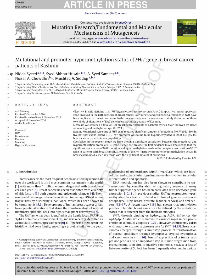
Fig. 1. SSCP analysis of FHIT gene exons
ancers including breast cancer [27–32] recently, abnormal tran-cription of the FHIT gene has been reported in human breast cancerell lines and in primary breast cancers, showing that about 30% ofrimary breast cancers exhibited abnormalities in FHIT transcripts33].
It further seems to be necessary to examine not only the fre-uency of abnormalities of FHIT transcription in primary breastancers but also the association of the abnormalities with clinico-athological characteristics of cancer, which will be of great help
n characterizing the role of the FHIT gene in breast carcinogenesis.Therefore we devised this study to analyze the frequency of FHIT
berrations – both mutations and hypermethylation in our ethnicashmir Population and also to correlate the FHIT gene status witharious clinicopathological characteristics.
. Materials
.1. Patients and tumor tissue procurement
A cohort of 130 randomly selected breast cancer patients admitted to the Sher--Kashmir Institute of Medical Sciences was included in the study. All patientsncluded in the study were both male and female, with the histopathological diag-osis of the breast done under Institutional histopathology department.
Tissue samples consisting of tumor and adjacent normal were collected directlyrom Operation Theater of Department of General Surgery. Only histopathologi-al confirmed tumor tissues were included in the study. Peripheral blood samplesere also collected from the patients before the surgical resection of the tumor.
he study was approved by the Sher-I-Kashmir Institute of Medical Sciences Ethicalommittee.
.2. DNA isolation
Genomic DNA was extracted from tissue samples and peripheral blood samplessing DNA Extraction Kit (Qiagen, USA). The quality of the resulting genomic DNAas stringently assessed by low percentage agarose gel electrophoresis followed byV spectrophotometer.
.3. Conventional PCR-SSCP
In our study we screened the mutations in the open reading frame of theHIT gene, i.e., exons 5–9. Primer designs of these exons (5th to 9th) were takenrom earlier studied reports [35,36]. The DNA samples were amplified for targetegions/exons by conventional PCR. Each PCR reaction was performed in a 50 �lnal volume containing 20–100 ng of genomic DNA, 10 mmol/l Tris–HCl, 10 pmol/lf each primer, 200 �mol/l of each dNTP, and 1 U of Taq DNA polymerase (Genei,ndia). PCR was performed in a thermal cycler (Biorad icycler) using different pro-rams for five exons. Both positive and negative controls were used in each PCReaction. Negative controls were without DNA template to exclude non-specificmplification. The PCR products were run on 1.8% agarose gel and analyzed underV illuminator.
The high-quality amplified products were assessed with SSCP analysis. For thisssessment, 8 �l of each PCR product was added 2 �l of loading solution. The tubeas immediately heat-denatured at 95 ◦C for 5 min, then cooled on ice for 5 min, and
t 4 ◦C for 5 min, respectively, and kept there or on ice until a sample was loaded
Please cite this article in press as: N. Syeed, et al., Mutational and promotKashmir, Mutat. Res.: Fundam. Mol. Mech. Mutagen. (2010), doi:10.1016/j
nto the gel. 4 �l of each processed PCR product were electrophoresed on 1× MDETM
els. The products were run at 600 V for 10–12 h at room temperature in 1× TBEuffer solution (89 mM tris-base, 89 mM boric acid, 2 mM EDTA, and pH 8.0). Theels were stained using a non-radioactive silver staining method and results wereisualised and photographed with an image analyzer, as shown in Fig. 1. Sampleshat showed one or two band-shifts separated from the wild-type bands were iden-
owing mobility shifts in tumor sample.
tified as SSCP positive. All the samples that contained mutations were subjected tothe SSCP analysis procedure at least twice to rule out contamination.
2.4. Sequencing
The samples which showed variant band-shifts in SSCP were re-amplified forsequencing. The PCR products were gel-extracted using a Gel Extraction Kit (Qiagen,USA) and then sent for direct DNA sequencing. DNA sequencing was carried out atMacrogen Inc., Korea. To minimize the sequencing artifacts by PCR, products fromat least two different PCRs were sequenced using forward and reverse primers.
2.5. Methylation-specific polymerase chain reaction
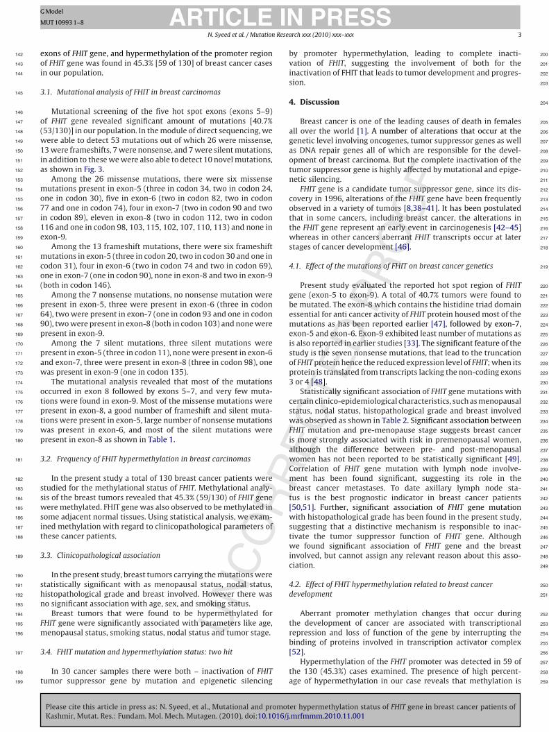
Genomic DNA isolated from tumor and adjacent normal tissues by the proto-col described above was bisulphite modified by using commercial kit (MethylationDirect Kit, Zymo Research) according to manufacturer’s instructions. The modi-fied DNA were amplified by using methylation/unmethylation specific primers asreported earlier [37], 3 �l of bisulphite modified DNA was used in the PCR mixcontaining 1× PCR buffer, 200 �mol/l of each dNTP and 2 U Amp gold Taq DNApolymerase, 0.4 �M of primer and were amplified using the following reaction con-ditions: initial denaturation at 95 ◦C for 5 min followed by 38 cycles at 95 ◦C 50 s,62 ◦C 50 s, 72 ◦C 1 min, with the final incubation at 72 ◦C for 7 min. Ten-�l of the PCRproducts were loaded onto 6% denaturing polyacyrlamide gels and stained usingethidium bromide and visualised under UV illuminator, the PCR generated a 74 bpproduct both for methylated and unmethylated, as shown in Fig. 2.
2.6. Statistical analysis
Pearson’s two proportions test was used to compare the determined FHIT genemutational and methylational status with various clinical parameters. Differenceswith P > 0.05 were accepted as statistically not significant. Calculations were doneusing SPSS for Windows, version 11.5 (SPSS, Chicago, IL, USA).
er hypermethylation status of FHIT gene in breast cancer patients of.mrfmmm.2010.11.001
Fig. 2. Methylation status of p16 and FHIT genes in normal and tumor tissues fromthe same patient (74 bp). Lane marked N: normal breast tissue, T: tumor tissue. Lanemarked M: methylated, lane marked U: unmethylated. Lane L: 100 bp molecularladder. Where T1 represents Bca3 and N1 is its adjacent normal tissue, T2 representsBca7 and N2 is its adjacent normal tissue. T3 represents Bca3.
ING
M
Resea
e142
o143
i144
3145
146
o147
(148
w149
1150
i151
a152
153
m154
o155
7156
i157
1158
e159
160
m161
c162
o163
(164
165
p166
6167
9168
p169
170
p171
a172
w173
174
o175
t176
p177
t178
w179
p180
3181
182
s183
s184
w185
s186
i187
t188
3189
190
s191
h192
n193
194
F195
m196
3197
198
t199
200
201
202
203
204
205
206
207
208
209
210
211
212
213
214
215
216
217
218
219
220
221
222
223
224
225
226
227
228
229
230
231
232
233
234
235
236
237
238
239
240
241
242
243
244
245
246
247
248
249
250
251
252
253
254
ARTICLEModel
UT 10993 1–8
N. Syeed et al. / Mutation
xons of FHIT gene, and hypermethylation of the promoter regionf FHIT gene was found in 45.3% [59 of 130] of breast cancer casesn our population.
.1. Mutational analysis of FHIT in breast carcinomas
Mutational screening of the five hot spot exons (exons 5–9)f FHIT gene revealed significant amount of mutations [40.7%53/130)] in our population. In the module of direct sequencing, weere able to detect 53 mutations out of which 26 were missense,
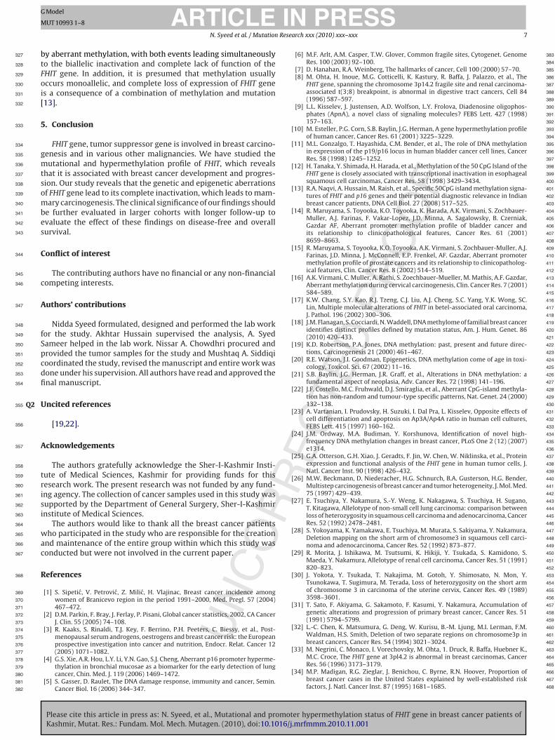
3 were frameshifts, 7 were nonsense, and 7 were silent mutations,n addition to these we were also able to detect 10 novel mutations,s shown in Fig. 3.
Among the 26 missense mutations, there were six missenseutations present in exon-5 (three in codon 34, two in codon 24,
ne in codon 30), five in exon-6 (two in codon 82, two in codon7 and one in codon 74), four in exon-7 (two in codon 90 and two
n codon 89), eleven in exon-8 (two in codon 112, two in codon16 and one in codon 98, 103, 115, 102, 107, 110, 113) and none inxon-9.
Among the 13 frameshift mutations, there were six frameshiftutations in exon-5 (three in codon 20, two in codon 30 and one in
odon 31), four in exon-6 (two in codon 74 and two in codon 69),ne in exon-7 (one in codon 90), none in exon-8 and two in exon-9both in codon 146).
Among the 7 nonsense mutations, no nonsense mutation wereresent in exon-5, three were present in exon-6 (three in codon4), two were present in exon-7 (one in codon 93 and one in codon0), two were present in exon-8 (both in codon 103) and none wereresent in exon-9.
Among the 7 silent mutations, three silent mutations wereresent in exon-5 (three in codon 11), none were present in exon-6nd exon-7, three were present in exon-8 (three in codon 98), oneas present in exon-9 (one in codon 135).
The mutational analysis revealed that most of the mutationsccurred in exon 8 followed by exons 5–7, and very few muta-ions were found in exon-9. Most of the missense mutations wereresent in exon-8, a good number of frameshift and silent muta-ions were present in exon-5, large number of nonsense mutationsas present in exon-6, and most of the silent mutations wereresent in exon-8 as shown in Table 1.
.2. Frequency of FHIT hypermethylation in breast carcinomas
In the present study a total of 130 breast cancer patients weretudied for the methylational status of FHIT. Methylational analy-is of the breast tumors revealed that 45.3% (59/130) of FHIT geneere methylated. FHIT gene was also observed to be methylated in
ome adjacent normal tissues. Using statistical analysis, we exam-ned methylation with regard to clinicopathological parameters ofhese cancer patients.
.3. Clinicopathological association
In the present study, breast tumors carrying the mutations weretatistically significant with as menopausal status, nodal status,istopathological grade and breast involved. However there waso significant association with age, sex, and smoking status.
Breast tumors that were found to be hypermethylated forHIT gene were significantly associated with parameters like age,enopausal status, smoking status, nodal status and tumor stage.
Please cite this article in press as: N. Syeed, et al., Mutational and promotKashmir, Mutat. Res.: Fundam. Mol. Mech. Mutagen. (2010), doi:10.1016/j
.4. FHIT mutation and hypermethylation status: two hit
In 30 cancer samples there were both – inactivation of FHITumor suppressor gene by mutation and epigenetic silencing
PRESSrch xxx (2010) xxx–xxx 3
by promoter hypermethylation, leading to complete inacti-vation of FHIT, suggesting the involvement of both for theinactivation of FHIT that leads to tumor development and progres-sion.
4. Discussion
Breast cancer is one of the leading causes of death in femalesall over the world [1]. A number of alterations that occur at thegenetic level involving oncogenes, tumor suppressor genes as wellas DNA repair genes all of which are responsible for the devel-opment of breast carcinoma. But the complete inactivation of thetumor suppressor gene is highly affected by mutational and epige-netic silencing.
FHIT gene is a candidate tumor suppressor gene, since its dis-covery in 1996, alterations of the FHIT gene have been frequentlyobserved in a variety of tumors [8,38–41]. It has been postulatedthat in some cancers, including breast cancer, the alterations inthe FHIT gene represent an early event in carcinogenesis [42–45]whereas in other cancers aberrant FHIT transcripts occur at laterstages of cancer development [46].
4.1. Effect of the mutations of FHIT on breast cancer genetics
Present study evaluated the reported hot spot region of FHITgene (exon-5 to exon-9). A total of 40.7% tumors were found tobe mutated. The exon-8 which contains the histidine triad domainessential for anti cancer activity of FHIT protein housed most of themutations as has been reported earlier [47], followed by exon-7,exon-5 and exon-6. Exon-9 exhibited least number of mutations asis also reported in earlier studies [33]. The significant feature of thestudy is the seven nonsense mutations, that lead to the truncationof FHIT protein hence the reduced expression level of FHIT; when itsprotein is translated from transcripts lacking the non-coding exons3 or 4 [48].
Statistically significant association of FHIT gene mutations withcertain clinico-epidemiological characteristics, such as menopausalstatus, nodal status, histopathological grade and breast involvedwas observed as shown in Table 2. Significant association betweenFHIT mutation and pre-menopause stage suggests breast canceris more strongly associated with risk in premenopausal women,although the difference between pre- and post-menopausalwomen has not been reported to be statistically significant [49].Correlation of FHIT gene mutation with lymph node involve-ment has been found significant, suggesting its role in thebreast cancer metastases. To date axillary lymph node sta-tus is the best prognostic indicator in breast cancer patients[50,51]. Further, significant association of FHIT gene mutationwith histopathological grade has been found in the present study,suggesting that a distinctive mechanism is responsible to inac-tivate the tumor suppressor function of FHIT gene. Althoughwe found significant association of FHIT gene and the breastinvolved, but cannot assign any relevant reason about this asso-ciation.
4.2. Effect of FHIT hypermethylation related to breast cancerdevelopment
Aberrant promoter methylation changes that occur duringthe development of cancer are associated with transcriptionalrepression and loss of function of the gene by interrupting the
er hypermethylation status of FHIT gene in breast cancer patients of.mrfmmm.2010.11.001
binding of proteins involved in transcription activator complex 255
[52]. 256
Hypermethylation of the FHIT promoter was detected in 59 of 257
the 130 (45.3%) cases examined. The presence of high percent- 258
age of hypermethylation in our case reveals that methylation is 259

ARTICLE IN PRESSG Model
MUT 10993 1–8
4 N. Syeed et al. / Mutation Research xxx (2010) xxx–xxx
F ve) alQ4a
o260
m261
c262
s263
ig. 3. Partial electropherograms representing the mutant (shown by arrows) (abond 8.
Please cite this article in press as: N. Syeed, et al., Mutational and promotKashmir, Mutat. Res.: Fundam. Mol. Mech. Mutagen. (2010), doi:10.1016/j
ne of the major determinants in tumor progression because inajority of the cases, expression of the genes in these breast
ancer cases correlates with hypermethylation of the promoterequences. The rate of hypermethylation is higher than the
ong with its normal sequence (below) of FHIT gene: (a) exons 5 and 6; (b) exons 7
er hypermethylation status of FHIT gene in breast cancer patients of.mrfmmm.2010.11.001
percentage of mutation which suggests that FHIT hypermethy- 264
lation is a more common event in breast carcinoma. Promoter 265
aberrant methylation of FHIT is an important mechanism for 266
inactivation of this tumor suppressor gene in mammary tumori- 267

ARTICLE IN PRESSG Model
MUT 10993 1–8
N. Syeed et al. / Mutation Research xxx (2010) xxx–xxx 5
Table 1Details of FHIT mutations in exons 5–9 of breast cancer patients from Kashmir valley.Q5
Exon involved Codon number Base changeg Amino acid change Effecth
Exon 5 34 GGA > AGA Gly > Arg Missense11 AAG > AAA Lys > Lys Silent20 GAA > GAGC INS G Frameshift24 GCT > CCT Ala > Pro Missense11 AAG > AAA Lys > Lys Silent30 CCT > CAT Pro > His Missense30 CCT > CCTA INS A Frameshift34 GGA > AGA Gly > Arg Missense31 GTG > GTAG INS A Frameshift34 GGA > AGA Gly > Arg Missense20 GAA > GAGC INS G Frameshift24 GCT > CCT Ala > Pro Missense20 GAA > GAGC INS G Frameshift11 AAG > AAA Lys > Lys Silent30 CCT > CCTA INS A Frameshift
Exon 6 64 AGA > TGA Arg > Stop Nonsense82 ATG > AAG Met > Lys Missense74 CAT > CATA INS A Frameshift77 TCT > CCT Ser > Pro Missense69 GTG > GTAG INS A Frameshift64 AGA > TGA Arg > Stop Nonsense74 CAT > CAG His > Gln Missense69 GTG > GTAG INS A Frameshift77 TCT > CCT Ser > Pro Missense64 AGA > TGA Arg > Stop Nonsense82 ATG > AAG Met > Lys Missense74 CAT > CATA INS A Frameshift
Exon 7 90 CAG > CAT Gln > His Missense93 AAG > TAG Lys > Stop Nonsense89 GGA > AGA Gly > Arg Missense90 CAG > TAG Gln > Stop Nonsense90 CAG > CAT Gln > His Missense89 GGA > AGA Gly > Arg Missense90 CAG > CAAG INS A Frameshift
Exon 8 116 GAG > GGG Glu > Gly Missense98 CAT > CAC His > Lys Missense
103 AAG > AAA Arg > Lys Missense98 CAT > CAC His > His Silent
115 GAG > GTG Glu > Val Missense102 AGG > AAG Arg > Lys Missense103 AAG > TAG Lys > Stop Nonsense107 TTT > TCT Phe > Ser Missense
98 CAT > CAC His > His Silent110 AAT > AAG Asn > Lys Missense116 GAG > GTG Glu > Val Missense103 AAG > TAG Lys > Stop Nonsense112 AGC > ACC Ser > Thr Missense
98 CAT > CAC His > His Silent113 ATC > ACC Ile > Thr Missense112 AGC > ACC Ser > Thr Missense
> TT> GCC
> TT
g268
[269
t270
l271
m272
b273
s274
275
c276
[277
c278
m279
W280
t281
s282
283
284
285
286
287
288
289
290
291
292
293
Exon 9 146 TTT135 GCA146 TTT
enesis. Our results were similar to previously reported studies53,54]. We also found hypermethylation in some non-cancerousissues that might represent the appearance of premalignantesions. Thus, detection of FHIT hypermethylation using MSP
ight provide potential new molecular diagnostic markers ofreast carcinomas at an early stage during multistep carcinogene-is.
Clinicopathologically, aberrant FHIT expression has been asso-iated with pathogenesis and prognosis of various tumors8,34,55–61]. FHIT gene hypermethylation was found signifi-ant with some of the clinico-pathological parameters like age,
Please cite this article in press as: N. Syeed, et al., Mutational and promotKashmir, Mutat. Res.: Fundam. Mol. Mech. Mutagen. (2010), doi:10.1016/j
enopausal status, smoking status, nodal status and tumor stage.e found a significant association between FHIT gene hyperme-
hylation and age (>50 years) supported by enormous evidenceuggesting that some of the methylation changes may initi-
Del T FrameshiftAla > Ala SilentDel T Frameshift
ate in subpopulations of normal cells as a function of age andprogressively increase during carcinogenesis [62]. FHIT gene hyper-methylation was also found significant in pre-menopausal womensuggesting that the premenopausal women are at approximatelytwice risk, than the postmenopausal women, which has beenreported earlier that proliferation rate is somewhat lower andrelatively stable in post menopausal women [63–65]. Hyperme-thylation of the FHIT gene was associated with smoking variable;FHIT gene hypermethylation was significantly associated with eversmokers. As most of the female patients in our study originatedfrom far flung rural regions, where they have a tradition of Hukka
er hypermethylation status of FHIT gene in breast cancer patients of.mrfmmm.2010.11.001
smoking (water pipe smoke) [66], which could be one of the possi- 294
ble reasons for hypermethylation. FHIT hypermethylation was also 295
found significantly associated with the lymph node involvement 296
and tumor stage (I and II). 297

ARTICLE IN PRESSG Model
MUT 10993 1–8
6 N. Syeed et al. / Mutation Research xxx (2010) xxx–xxx
Table 2Effect of FHIT mutation and hypermethylation pattern in the breast cancer patients from Kashmir valley.
Variable TotalN = 130 (%)
MutantsM = 53 (%)
OR; P-value; CI(95%)
MethylatedN = 59 (%)
OR; P-value; CI(95%)
SexMales 7 (53.8%) 4 (57.1%) 2.01; 0.44; 0.43–9.39 2 0.46; 0.45; 0.08–2.48Females 123 (94.6%) 49 (39.8%) 57Age>50 13 (10.0%) 8 2.56; 0.13; 0.78–8.31 11 7.90; 0.003; 1.68–37.28≤50 117 (90.0%) 45 48DwellingRural 45 (34.6%) 38 21.7; 0.000; 8.10–58.15 37 13.24; 5.47; 5.36–32.75Urban 85 (65.4%) 15 22Smoking statusNever 93 (71.5%) 38 0.89; 0.83; 0.39–2.03 35 0.33; 0.006; 0.15–0.72Ever 37 (28.5%) 15 24Menopausal statusPre 94 (72.3%) 21 2.71; 0.01; 1.23–5.96 21 2.25; 0.04;1.02–4.96Post 94 (72.3%) 32 38Nodal statusInvolved 34 (26.2%) 25 6.74; 0.000; 2.79–16.26 23 3.48; 0.002; 1.52–7.98Not Involved 96 (73.8%) 28 36Breast involvedR 35 (26.9%) 33 61.87; 0.000;
13.66–280.1230 13.65; 1.38; 4.81–38.73
L 95 (73.1%) 20 29Tumor stageII (a + b) 72 (55.4%) 35 2.10; 0.04; 1.01–4.33 26 0.43; 0.02; 0.21–0.86III (a + b) + IV 58 (44.6%) 18 30Histopathological tumor gradePD 25 (96.0%) 18 5.14; 0.0005;
1.96–13.4713 1.38; 0.50; 0.57–3.33
MD + WD 58 + 47 (24.1%) 18 + 17 27 + 19
Table 3Statistical relation between promoter hypermethylation and mutation of FHIT gene.
FHIT promoter hypermethylation versus FHIT mutation
FHIT methylation OR; P-value; CI(95%)
UnmethylationN = 71
MethylationN = 59
FHIT mutation2
3
4298
299
t300
i301
a302
h303
p304
i305
o306
n307
b308
i309
s310
m311
312
313
314
315
316
317
318
319
320
321
322
TC
Wild: N = 77 48
Mutant: N = 53 23
.3. Two hits inactivate the tumor suppressor role of FHIT
The important feature of our study was the significant associa-ion of FHIT mutation with promoter hypermethylation, as shownn Table 3. In 30 samples, we found both FHIT mutation as wells FHIT promoter hypermethylation. It has been reported that oneit cannot result in the complete loss of function of the tumor sup-ressor gene FHIT, and therefore two hits are required for complete
nactivation [67], where hypermethylation has been considered asne of the hits [21,68]. It is also known that methylation can occurot as a primary event, but secondary to mutation [20,68] as has
Please cite this article in press as: N. Syeed, et al., Mutational and promotKashmir, Mutat. Res.: Fundam. Mol. Mech. Mutagen. (2010), doi:10.1016/j
een reported [69] that methylation of tumor suppressors cannotnitiate tumorigenesis, because in order to control the gene expres-ion and/or methylation the methylator phenotype itself requiresutations in genes that control them.
able 4orrelation of tumor grade versus mutation versus hypermethylation status.
FHIT gene status Tumor grade
Low grade II
Double hit (N = 30) 7Single hit – mutation only (N = 23) 12Single hit – methylation only (N = 29) 15
9 0.46; 0.04; 0.22–0.94
0
Among these 30 samples most of the mutations were truncat-ing mutations, and when these truncating mutations were coupledwith methylation, there are many possibilities that the methy-lated allele has actually harboured a somatic or germ line mutationwhich has predisposed the gene to methylation. We found tumorgrade statistically significant with mutational and hypermethyla-tion status both (Table 4), suggesting that both of these aberrations(mutation + hypermethylation) together are responsible for theprogression of breast cancer to higher grade.
According to Tomlinson et al. [70] “two hits” hypothesis, oneallele of a tumor suppressor can be inactivated by methylation
er hypermethylation status of FHIT gene in breast cancer patients of.mrfmmm.2010.11.001
when the other harbors a mutation or allelic loss. Our results also fit 323
this model, revealing a strong association between FHIT promoter 324
hypermethylation and the mutation, suggesting that in breast car- 325
cinomas, one allele is lost by mutation, and the other is inactivated 326
P-value; �2
High grade III + IV
2311 0.04; 6.4114
ING
M
Resea
b327
t328
F329
o330
i331
[332
5333
334
g335
m336
t337
s338
o339
m340
b341
e342
s343
C344
345
c346
A347
348
f349
S350
p351
c352
d353
fi354
UQ2355
356
A357
358
t359
r360
i361
s362
i363
364
w365
a366
c367
R368
369
370
371
372
373
374
375
376
377
378
379
380
381
382
383
384
385
386
387
388
389
390
391
392
[ 393
394
[ 395
396
397
[ 398
399
400
[ 401
402
403
[ 404
405
406
407
408
[ 409
410
411
412
[ 413
414
415
[ 416
417
418
[ 419
420
421
[ 422
423
[ 424
425
[ 426
427
[ 428
429
430
[ 431
432
433
[ 434
435
436
[ 437
438
439
[ 440
441
442
[ 443
444
445
446
[ 447
448
449
[ 450
451
452
[ 453
454
455
456
[ 457
458
459
[ 460
461
462
ARTICLEModel
UT 10993 1–8
N. Syeed et al. / Mutation
y aberrant methylation, with both events leading simultaneouslyo the biallelic inactivation and complete lack of function of theHIT gene. In addition, it is presumed that methylation usuallyccurs monoallelic, and complete loss of expression of FHIT genes a consequence of a combination of methylation and mutation13].
. Conclusion
FHIT gene, tumor suppressor gene is involved in breast carcino-enesis and in various other malignancies. We have studied theutational and hypermethylation profile of FHIT, which reveals
hat it is associated with breast cancer development and progres-ion. Our study reveals that the genetic and epigenetic aberrationsf FHIT gene lead to its complete inactivation, which leads to mam-ary carcinogenesis. The clinical significance of our findings should
e further evaluated in larger cohorts with longer follow-up tovaluate the effect of these findings on disease-free and overallurvival.
onflict of interest
The contributing authors have no financial or any non-financialompeting interests.
uthors’ contributions
Nidda Syeed formulated, designed and performed the lab workor the study. Akhtar Hussain supervised the analysis, A. Syedameer helped in the lab work. Nissar A. Chowdhri procured androvided the tumor samples for the study and Mushtaq A. Siddiqioordinated the study, revised the manuscript and entire work wasone under his supervision. All authors have read and approved thenal manuscript.
ncited references
[19,22].
cknowledgements
The authors gratefully acknowledge the Sher-I-Kashmir Insti-ute of Medical Sciences, Kashmir for providing funds for thisesearch work. The present research was not funded by any fund-ng agency. The collection of cancer samples used in this study wasupported by the Department of General Surgery, Sher-I-Kashmirnstitute of Medical Sciences.
The authors would like to thank all the breast cancer patientsho participated in the study who are responsible for the creation
nd maintenance of the entire group within which this study wasonducted but were not involved in the current paper.
eferences
[1] S. Sipetic, V. Petrovic, Z. Milic, H. Vlajinac, Breast cancer incidence amongwomen of Branicevo region in the period 1991–2000, Med. Pregl. 57 (2004)467–472.
[2] D.M. Parkin, F. Bray, J. Ferlay, P. Pisani, Global cancer statistics, 2002, CA CancerJ. Clin. 55 (2005) 74–108.
[3] R. Kaaks, S. Rinaldi, T.J. Key, F. Berrino, P.H. Peeters, C. Biessy, et al., Post-menopausal serum androgens, oestrogens and breast cancer risk: the Europeanprospective investigation into cancer and nutrition, Endocr. Relat. Cancer 12
Please cite this article in press as: N. Syeed, et al., Mutational and promotKashmir, Mutat. Res.: Fundam. Mol. Mech. Mutagen. (2010), doi:10.1016/j
(2005) 1071–1082.[4] G.S. Xie, A.R. Hou, L.Y. Li, Y.N. Gao, S.J. Cheng, Aberrant p16 promoter hyperme-
thylation in bronchial mucosae as a biomarker for the early detection of lungcancer, Chin. Med. J. 119 (2006) 1469–1472.
[5] S. Gasser, D. Raulet, The DNA damage response, immunity and cancer, Semin.Cancer Biol. 16 (2006) 344–347.
[
[
PRESSrch xxx (2010) xxx–xxx 7
[6] M.F. Arlt, A.M. Casper, T.W. Glover, Common fragile sites, Cytogenet. GenomeRes. 100 (2003) 92–100.
[7] D. Hanahan, R.A. Weinberg, The hallmarks of cancer, Cell 100 (2000) 57–70.[8] M. Ohta, H. Inoue, M.G. Cotticelli, K. Kastury, R. Baffa, J. Palazzo, et al., The
FHIT gene, spanning the chromosome 3p14.2 fragile site and renal carcinoma-associated t(3;8) breakpoint, is abnormal in digestive tract cancers, Cell 84(1996) 587–597.
[9] L.L. Kisselev, J. Justensen, A.D. Wolfson, L.Y. Frolova, Diadenosine oligophos-phates (ApnA), a novel class of signaling molecules? FEBS Lett. 427 (1998)157–163.
10] M. Esteller, P.G. Corn, S.B. Baylin, J.G. Herman, A gene hypermethylation profileof human cancer, Cancer Res. 61 (2001) 3225–3229.
11] M.L. Gonzalgo, T. Hayashida, C.M. Bender, et al., The role of DNA methylationin expression of the p19/p16 locus in human bladder cancer cell lines, CancerRes. 58 (1998) 1245–1252.
12] H. Tanaka, Y. Shimada, H. Harada, et al., Methylation of the 50 CpG Island of theFHIT gene is closely associated with transcriptional inactivation in esophagealsquamous cell carcinomas, Cancer Res. 58 (1998) 3429–3434.
13] R.A. Naqvi, A. Hussain, M. Raish, et al., Specific 50CpG island methylation signa-tures of FHIT and p16 genes and their potential diagnostic relevance in Indianbreast cancer patients, DNA Cell Biol. 27 (2008) 517–525.
14] R. Maruyama, S. Toyooka, K.O. Toyooka, K. Harada, A.K. Virmani, S. Zochbauer-Muller, A.J. Farinas, F. Vakar-Lopez, J.D. Minna, A. Sagalowsky, B. Czerniak,Gazdar AF, Aberrant promoter methylation profile of bladder cancer andits relationship to clinicopathological features, Cancer Res. 61 (2001)8659–8663.
15] R. Maruyama, S. Toyooka, K.O. Toyooka, A.K. Virmani, S. Zochbauer-Muller, A.J.Farinas, J.D. Minna, J. McConnell, E.P. Frenkel, AF. Gazdar, Aberrant promotermethylation profile of prostate cancers and its relationship to clinicopatholog-ical features, Clin. Cancer Res. 8 (2002) 514–519.
16] A.K. Virmani, C. Muller, A. Rathi, S. Zoechbauer-Mueller, M. Mathis, A.F. Gazdar,Aberrant methylation during cervical carcinogenesis, Clin. Cancer Res. 7 (2001)584–589.
17] K.W. Chang, S.Y. Kao, R.J. Tzeng, C.J. Liu, A.J. Cheng, S.C. Yang, Y.K. Wong, SC.Lin, Multiple molecular alterations of FHIT in betel-associated oral carcinoma,J. Pathol. 196 (2002) 300–306.
18] J.M. Flanagan, S. Cocciardi, N. Waddell, DNA methylome of familial breast canceridentifies distinct profiles defined by mutation status, Am. J. Hum. Genet. 86(2010) 420–433.
19] K.D. Robertson, P.A. Jones, DNA methylation: past, present and future direc-tions, Carcinogenesis 21 (2000) 461–467.
20] R.E. Watson, J.I. Goodman, Epigenetics, DNA methylation come of age in toxi-cology, Toxicol. Sci. 67 (2002) 11–16.
21] S.B. Baylin, J.G. Herman, J.R. Graff, et al., Alterations in DNA methylation: afundamental aspect of neoplasia, Adv. Cancer Res. 72 (1998) 141–196.
22] J.F. Costello, M.C. Fruhwald, D.J. Smiraglia, et al., Aberrant CpG-island methyla-tion has non-random and tumour-type specific patterns, Nat. Genet. 24 (2000)132–138.
23] A. Vartanian, I. Prudovsky, H. Suzuki, I. Dal Pra, L. Kisselev, Opposite effects ofcell differentiation and apoptosis on Ap3A/Ap4A ratio in human cell cultures,FEBS Lett. 415 (1997) 160–162.
24] J.M. Ordway, M.A. Budiman, Y. Korshunova, Identification of novel high-frequency DNA methylation changes in breast cancer, PLoS One 2 (12) (2007)e1314.
25] G.A. Otterson, G.H. Xiao, J. Geradts, F. Jin, W. Chen, W. Niklinska, et al., Proteinexpression and functional analysis of the FHIT gene in human tumor cells, J.Natl. Cancer Inst. 90 (1998) 426–432.
26] M.W. Beckmann, D. Niederacher, H.G. Schnurch, B.A. Gusterson, H.G. Bender,Multistep carcinogenesis of breast cancer and tumor heterogeneity, J. Mol. Med.75 (1997) 429–439.
27] E. Tsuchiya, Y. Nakamura, S.-Y. Weng, K. Nakagawa, S. Tsuchiya, H. Sugano,T. Kitagawa, Allelotype of non-small cell lung carcinoma: comparison betweenloss of heterozygosity in squamous cell carcinoma and adenocarcinoma, CancerRes. 52 (1992) 2478–2481.
28] S. Yokoyama, K. Yamakawa, E. Tsuchiya, M. Murata, S. Sakiyama, Y. Nakamura,Deletion mapping on the short arm of chromosome3 in squamous cell carci-noma and adenocarcinoma, Cancer Res. 52 (1992) 873–877.
29] R. Morita, J. Ishikawa, M. Tsutsumi, K. Hikiji, Y. Tsukada, S. Kamidono, S.Maeda, Y. Nakamura, Allelotype of renal cell carcinoma, Cancer Res. 51 (1991)820–823.
30] J. Yokota, Y. Tsukada, T. Nakajima, M. Gotoh, Y. Shimosato, N. Mon, Y.Tsunokawa, T. Sugimura, M. Terada, Loss of heterozygosity on the short armof chromosome 3 in carcinoma of the uterine cervix, Cancer Res. 49 (1989)3598–3601.
31] T. Sato, F. Akiyama, G. Sakamoto, F. Kasumi, Y. Nakamura, Accumulation ofgenetic alterations and progression of primary breast cancer, Cancer Res. 51(1991) 5794–5799.
32] L.-C. Chen, K. Matsumura, G. Deng, W. Kurisu, B.-M. Ljung, M.I. Lerman, F.M.Waldman, H.S. Smith, Deletion of two separate regions on chromosome3p inbreast cancers, Cancer Res. 54 (1994) 3021–3024.
er hypermethylation status of FHIT gene in breast cancer patients of.mrfmmm.2010.11.001
33] M. Negrini, C. Monaco, I. Vorechovsky, M. Ohta, 1. Druck, R. Baffa, Huebner K., 463
M.C. Croce, The FHIT gene at 3pl4.2 is abnormal in breast carcinomas, Cancer 464
Res. 56 (1996) 3173–3179. 465
34] M.P. Madigan, R.G. Zieglar, J. Benichou, C. Byrne, R.N. Hoover, Proportion of 466
breast cancer cases in the United States explained by well-established risk 467
factors, J. Natl. Cancer Inst. 87 (1995) 1681–1685. 468

ING
M
8 Resea
[469
470
471
[472
473
474
[475
476
477
[478
479
480
481
[482
483
484
485
486
[487
488
489
[490
491
492
[493
494
495
[496
497
[498
499
500
[501
502
503
[504
505
506
[507
508
509
510
[511
512
513
514
[515
516
517
[518
519
[520
521
522
[523
[ 524
525
526
527
528
[ 529
530
531
[ 532
533
534
[ 535
536
537
[ 538
539
540
541
[ 542
543
544
545
[ 546
547
548
549
[ 550
551
552
553
[ 554
555
556
[ 557
558
[ 559
560
561
[ 562
563
564
[ 565
566
567
568
[ 569
570
571
[ 572
573
ARTICLEModel
UT 10993 1–8
N. Syeed et al. / Mutation
35] A. Gemma, K. Hagiwarn, Y. Ke, L.M. Burke, M.A. Khan, M. Nagashima, W.P. Ben-nett, C.C. Harris, FHIT mutations in human primary gastric cancer, Cancer Res.57 (1997) 1435–1437.
36] G. Cecener, Ü. Egeli, B. Tunca, I. Tasdelen, S. Tolunay, N. Bilgel, Importance ofnovel sequence alterations in the FHIT gene on formation of breast cancer,Tumori 93 (2007) 597–603.
37] A.O. Nygren, N. Ameziane, H.M. Duarte, et al., Methylation-specific MLPA (MS-MLPA): simultaneous detection of CpG methylation and copy number changesof up to 40 sequences, Nucleic Acids Res. 33 (14) (2005) e128.
38] J.D. Potter, J.R. Cerhan, T.A. Sellers, P.G. McGovem, C. Drinkard, L.R. Kushi, A.R.Folsom, Progesterone and estrogen receptors and mammary neoplasia in theIowa women’s health study: how many kinds of breast cancers are there?Cancer Epidemiol. Biomar. Prev. 4 (1995) 319–326.
39] K.M. Fong, E.J. Biesterveld, A. Virmani, I. Wistuba, Y. Sekido, S.A. Bader, M.Ahmadian, S.T. Ong, F.V. Rassool, P.V. Zimmerman, G. Giaccone, A.F. Gazdar,J.D. Minna, FHIT and FRA3B 3p14.2 allele loss are common in lung cancer andpreneoplastic bronchial lesions and are associated with cancer-related FHITcDNA splicing aberrations, Cancer Res. 57 (1997) 2256–2267.
40] S. Ingvarson, B.I. Sigbjornsdottir, C. Huiping, J.G. Jonasson, B.A. Agnarsson, Alter-ations of the FHIT gene in breast cancer: association with tumor progressionand patient survival, Cancer Detect. Prev. 25 (2001) 292–298.
41] B.Z. Yuan, C. Keck-Waggoner, D.B. Zimonjic, S.S. Thorgeirsson, N.C. Popescu,Alterations of the FHIT gene in human hepatocellular carcinoma, Cancer Res.60 (2000) 1049–1053.
42] R. Baffia, M.L. Veronese, B. Santoro, B. Mandes, J.P. Palazzo, M. Rugge, E. Santoro,C.M. Croce, K. Huebner, Loss of FHIT expression in gastric carcinoma, Cancer Res.58 (1998) 4708–4714.
43] M. Zou, Y. Shi, N.R. Farid, S.T. al-Sedairy, M.C. Paterson, FHIT gene abnormalitiesin both benign and malignant thyroid tumors, Eur. J. Cancer 35 (1999) 467–472.
44] Z. Guo, S.L. Johansson, J.S. Rhim, J.K. Vishwanatha, Fragile histidine triad geneexpression in primary prostate cancer and in an in vitro model, Prostate 43(2000) 101–110.
45] C. Huiping, J.G. Jonasson, B.A. Agnarsson, B.I. Sigbjornsdottir, K. Huebner, S.Ingvarsson, Analysis of the fragile histidine triad (FHIT) gene in lobular breastcancer, Eur. J. Cancer 36 (2000) 1552–1557.
46] K. Yoshino, T. Enomoto, T. Nakamura, H. Sun, K. Ozaki, R. Nakashima, H. Wanada,J. Saitoh, Y. Watanabe, K. Noda, Y. Murata, FHIT alterations in cancerous andnon-cancerous cervical epithelium, Int. J. Cancer 85 (2000) 6–13.
47] Z. Siprashvili, G. Sozzi, L.D. Barnes, P. McCue, A.K. Robinson, V. Eryomin, L. Sard,E. Tagliabue, A. Greco, L. Fusetti, G. Schwartz, M.A. Pierotti, C.M. Croce, K. Hueb-ner, Replacement of FHIT in cancer cells suppresses tumorigenicity, Proc. Natl.Acad. Sci. U.S.A. 94 (1997) 13771–13776.
48] G. Sozzi, S. Tornielli, E. Tagliabue, L. Sard, F. Pezzella, U. Pastorino, F. Minolitti,S. Pilotti, C. Ratcliffe, M.L. Veronese, P. Goldstraw, K. Huebner, C.M. Croce, MA.Pierotti, Absence of FHIT protein in primary lung tumors and cells lines withFHIT gene abnormalities, Cancer Res. 57 (1997) 5207–5212.
49] C. Nagata, N. Takatsuka, S. Inaba, N. Kawakami, H. Shimizu, Effect of soymilkconsumption on serum estrogen concentrations in premenopausal Japanesewomen, J. Natl. Cancer Inst. 90 (1998) 1830–1835.
50] C.L. Carter, C. Allen, D.E. Henson, Relation of tumor size, lymph node status, and
Please cite this article in press as: N. Syeed, et al., Mutational and promotKashmir, Mutat. Res.: Fundam. Mol. Mech. Mutagen. (2010), doi:10.1016/j
survival in 24,740 breast cancer cases, Cancer 63 (1989) 181–719.51] J. Russo, J. Frederick, H.E. Ownby, G. Fine, M. Hussain, H.I. Kirckstein, et al.,
Predictors of recurrence and survival of patients with breast cancer, Am. J. Clin.Pathol. 88 (1987) 123–131.
52] P.A. Jones, S.B. Baylin, The fundamental role of epigenetic events in cancer, Nat.Rev. Genet. 3 (2002) 415–428.
[
[
[
PRESSrch xxx (2010) xxx–xxx
53] M. Raish, V.S. Dhillon, A. Ahmad, M.A. Ansari, S. Mudassar, M. Shahid, V. Batra,P. Gupta, B.C. Das, N. Shukla, S.A. Husain, Promoter hypermethylation in tumorsuppressing genes p16 and FHIT and their relationship with estrogen receptorand progesterone receptor status in breast cancer patients from Northern India,Transl. Oncol. 4 (2009) 264–270.
54] J.C. Roa, L. Anabalón, O. Tapia, J. Martínez, J.C. Araya, M. Villaseca, P. Guzmán, I.E.Roa, Promoter methylation profile in breast cancer, Rev. Med. Chile 132 (2004)1069–1077.
55] Z. Gatalica, S.M. Lele, B.A. Rampy, B.A. Norris, The expression of FHIT proteinis related inversely to disease progression in patients with breast carcinoma,Cancer (Phila.) 88 (2000) 1378–1383.
56] M. Campiglio, Y. Pekarsky, S. Menard, E. Tagliabue, S. Pilotti, C.M. Croce, FHITloss of function in human primary breast cancer correlates with advanced stageof the disease, Cancer Res. 59 (1999) 3866–3869.
57] Q. Yang, G. Yoshimura, T. Suzuma, T. Tamaki, T. Umemura, M. Nakamura, Y.Nakamura, X. Wang, I. Mori, T. Sakurai, K. Kakudo, Clinicopathological sig-nificance of fragile histidine triad transcription protein expression in breastcarcinoma, Clin. Cancer Res. 7 (2001) 3869–3873.
58] D.L. Greenspan, D.C. Connolly, R. Wu, R.Y. Lei, J.T. Vogelstein, Y.T. Kim, J.E.Mok, N. Munoz, F.X. Bosch, K. Shah, K.R. Cho, Loss of FHIT expression in cer-vical carcinoma cell lines and primary tumors, Cancer Res. 57 (1997) 4697–4698.
59] S.I. Hayashi, K. Tanimoto, K. Hajiro-Nakanishi, E. Tsuchiya, M. Kurosumi, Y.Higashi, K. Imai, K. Suga, K. Nakachi, Abnormal FHIT transcripts in human breastcarcinomas: a clinicopathological and epidemiological analysis of 61 Japanesecases, Cancer Res. 57 (1997) 1981–1985.
60] G. Sozzi, M.L. Veronese, M. Negrini, R. Baffa, M.G. Cotticelli, H. Inoue, S. Tomielli,S. Pilotti, L. De Gregorio, U. Pastorino, M.A. Pierotti, M. Ohta, K. Huebner,C.M. Croce, The FHIT gene 3p14.2 is abnormal in lung cancer, Cell 85 (1996)17–26.
61] J.I. Lee, J.C. Soria, K. Hassan, D. Liu, X. Tang, A. El-Naggar, W.K. Hong, L. Mao, Lossof FHIT expression is a predictor of poor outcome in tongue cancer, Cancer Res.61 (2001) 837–841.
62] N. Ahuja, Q. Li, A.L. Mohan, S.B. Baylin, J.P. Issa, Aging and DNA methylation incolorectal mucosa and cancer, Cancer Res. 58 (1998) 5489–5494.
63] D.J. Ferguson, T.J. Anderson, Morphological evaluation of cell turnover in rela-tion to the menstrual cycle in the “resting” human breast, Br. J. Cancer 44 (1981)177–181.
64] T.A. Longacre, S.A. Bartow, A correlative morphologic study of human breastand endometrium in the menstrual cycle, Am. J. Surg. Pathol. 10 (6) (1986)382–393.
65] J.S. Meyer, Cell proliferation in normal human breast ducts, fibroadenomas, andother ductal hyperplasias measured by nuclear labeling with tritiated thymi-dine. Effects of menstrual phase, age, and oral contraceptive hormones, Hum.Pathol. 8 (1977) 67–81.
66] M.M. Mir, N.A. Dar, S. Gochhait, S.A. Zargar, A.G. Ahangar, R.N. Bamezai, p53Mutation profile of squamous cell carcinomas of the esophagus in Kashmir(India): a high-incidence area, Int. J. Cancer 116 (2005) 62–68.
67] A.G. Knudson Jr., H.W. Hethcote, B.W. Brown, Mutation and childhood cancer:a probabilistic model for the incidence of retinoblastoma, Proc. Natl. Acad. Sci.
er hypermethylation status of FHIT gene in breast cancer patients of.mrfmmm.2010.11.001
U.S.A. 72 (1975) 5116–5120. 574
68] P.A. Jones, P.W. Laird, Cancer epigenetics comes of age, Nat. Genet. 21 (1999) 575
163–167. 576
69] I. Tomlinson, M. Novelli, W. Bodmer, The mutation rate and cancer, Proc. Natl. 577
Acad. Sci. U.S.A. 93 (1996) 14800–14803. 578
70] I.P.M. Tomlinson, R. Roylance, R.S. Houlston. Q3 579
Copyright © 2022 FDOKUMEN