Multiple Roles of Protein Kinase A in Arachidonic Acid-Mediated Ca2+ Entry and Tumor-Derived Human...
12
2010;8:1466-1476. Published OnlineFirst September 24, 2010. Mol Cancer Res Alessandra Fiorio Pla, Tullio Genova, Emanuela Pupo, et al. Cell Migration Entry and Tumor-Derived Human Endothelial 2+ Mediated Ca - Multiple Roles of Protein Kinase A in Arachidonic Acid Updated Version 10.1158/1541-7786.MCR-10-0002 doi: Access the most recent version of this article at: Cited Articles http://mcr.aacrjournals.org/content/8/11/1466.full.html#ref-list-1 This article cites 40 articles, 14 of which you can access for free at: Citing Articles http://mcr.aacrjournals.org/content/8/11/1466.full.html#related-urls This article has been cited by 1 HighWire-hosted articles. Access the articles at: E-mail alerts related to this article or journal. Sign up to receive free email-alerts Subscriptions Reprints and . [email protected] Publications Department at To order reprints of this article or to subscribe to the journal, contact the AACR Permissions . [email protected] Department at To request permission to re-use all or part of this article, contact the AACR Publications American Association for Cancer Research Copyright © 2010 on February 26, 2012 mcr.aacrjournals.org Downloaded from Published OnlineFirst September 24, 2010; DOI:10.1158/1541-7786.MCR-10-0002
-
Upload
independent -
Category
Documents
-
view
2 -
download
0
Transcript of Multiple Roles of Protein Kinase A in Arachidonic Acid-Mediated Ca2+ Entry and Tumor-Derived Human...
2010;8:1466-1476. Published OnlineFirst September 24, 2010.Mol Cancer Res Alessandra Fiorio Pla, Tullio Genova, Emanuela Pupo, et al. Cell Migration
Entry and Tumor-Derived Human Endothelial2+Mediated Ca−Multiple Roles of Protein Kinase A in Arachidonic Acid
Updated Version 10.1158/1541-7786.MCR-10-0002doi:
Access the most recent version of this article at:
Cited Articles http://mcr.aacrjournals.org/content/8/11/1466.full.html#ref-list-1
This article cites 40 articles, 14 of which you can access for free at:
Citing Articles http://mcr.aacrjournals.org/content/8/11/1466.full.html#related-urls
This article has been cited by 1 HighWire-hosted articles. Access the articles at:
E-mail alerts related to this article or journal.Sign up to receive free email-alerts
SubscriptionsReprints and
[email protected] Department atTo order reprints of this article or to subscribe to the journal, contact the AACR
To request permission to re-use all or part of this article, contact the AACR Publications
American Association for Cancer Research Copyright © 2010 on February 26, 2012mcr.aacrjournals.orgDownloaded from
Published OnlineFirst September 24, 2010; DOI:10.1158/1541-7786.MCR-10-0002

1466
Published OnlineFirst September 24, 2010; DOI:10.1158/1541-7786.MCR-10-0002
Angiogenesis, Metastasis, and the Cellular Microenvironment Molecular
CancerResearch
Multiple Roles of Protein Kinase A in ArachidonicAcid–Mediated Ca2+ Entry and Tumor-DerivedHuman Endothelial Cell Migration
Alessandra Fiorio Pla1,2,3, Tullio Genova1, Emanuela Pupo1, Cristiana Tomatis1,2,3,Armando Genazzani4, Roberta Zaninetti4, and Luca Munaron1,2,3
Abstract
Authors' A2Nanostrucand 3Cente(SysBioM),Piemonte O
CorresponHuman BioTurin, Italy.munaron@u
doi: 10.115
©2010 Am
Mol Canc
We recently showed that arachidonic acid (AA) triggers calcium signals in endothelial cells derived fromhuman breast carcinoma (B-TEC). In particular, AA-dependent Ca2+ entry is involved in the early steps oftumor angiogenesis in vitro. Here, we investigated the multiple roles of the nitric oxide (NO) and cyclicAMP/protein kinase A (PKA) pathways in AA-mediated Ca2+ signaling in the same cells. B-TEC stimulationwith 5 μmol/L AA resulted in endothelial NO synthase (NOS) phosphorylation at Ser1177, and NO releasewas measured with the fluorescent NO-sensitive probe DAR4M-AM. PKA inhibition by the use of the mem-brane-permeable PKA inhibitory peptide myristoylated PKI14-22 completely prevented both AA- and NO-induced calcium entry and abolished B-TEC migration promoted by AA. AA-dependent calcium entryand cell migration were significantly affected by both the NOS inhibitor NG-nitro-L-arginine methyl esterand the NO scavenger 2-phenyl-4,4,5,5-tetramethylimidazoline-1-oxyl 3-oxide, suggesting that NO releaseis functionally involved in the signaling dependent on AA. Moreover, pretreatment with carboxyamidotria-zole, an antiangiogenic compound that interferes with agonist-activated calcium entry, prevented AA-depen-dent B-TEC motility. Interestingly, even in the absence of AA, enhancement of the cyclic AMP/PKA pathwaywith the adenylyl cyclase activator forskolin evoked a calcium entry dependent on NOS recruitment and NOrelease. The functional relevance of AA-induced calcium entry could be restricted to tumor-derived endothe-lial cells (EC) because AA evoked a smaller calcium entry in normal human microvascular ECs comparedwith B-TECs, and even more importantly, it was unable to promote cell motility in wound healing assay.This evidence opens an intriguing opportunity for differential pharmacologic treatment between normal andtumor-derived human ECs. Mol Cancer Res; 8(11); 1466–76. ©2010 AACR.
Introduction
Angiogenic growth factors, such as basic fibroblastgrowth factor and vascular endothelial growth factor(VEGF), trigger a signaling cascade involving activationof specific tyrosine kinase receptors and recruitment of celleffectors, including PLC-γ, endothelial nitric oxidesynthase (eNOS), and PLA2, leading to the productionof IP3, nitric oxide (NO), arachidonic acid (AA), and theirmetabolites (1, 2). These pathways promote store-operatedand non–store-operated Ca2+ entry (SOCE and NSOCE,
ffiliations: 1Department of Animal and Human Biology,tured Interfaces and Surfaces Centre of Excellence (NIS),r for Complex Systems in Molecular Biology and MedicineUniversity of Turin, Turin, Italy; and 4DISCAFF, University ofrientale, Novara, Italy
ding Author: Luca Munaron, Department of Animal andlogy, University of Turin, Via Accademia Albertina 13, 10123Phone: 39-011-6704667; Fax: 39-011-6704508. E-mail: luca.nito.it
8/1541-7786.MCR-10-0002
erican Association for Cancer Research.
er Res; 8(11) November 2010
American Asso Copyright © 2010 mcr.aacrjournalDownloaded from
respectively) from extracellular medium into endothelialcells (EC; refs. 3, 4).In particular, we previously reported the ability of AA to
trigger a NSOCE in bovine aortic ECs (BAEC), playing acritical role in the control of proliferation (1, 2, 5-7).Because blood vessels in tumors differ from normal ones
by their altered morphology, blood flow, permeability, andabnormalities in pericytes and in basement membrane,several groups have recently focused their attention ontumor-derived ECs (TEC) as a more adequate model tostudy tumor angiogenesis (8). Interestingly, TECs displaya distinct and unique phenotype different from that of nor-mal vascular ECs at molecular and functional levels (9-11).Recently, TECs obtained from breast carcinomas (B-TEC)have been established and characterized, showing animmature proangiogenic phenotype with enhanced prolifer-ation, motility, and capillary-like tube formation comparedwith “normal” ECs (10, 12). Therefore, they represent aninteresting model to investigate the role of different factorsin the tumoral angiogenic process.We have recently studied the role of AA-induced cy-
tosolic calcium (Cac) signals in the angiogenic process of
ciation for Cancer Research on February 26, 2012s.org
PKA Regulates AA-Activated Calcium Entry
Published OnlineFirst September 24, 2010; DOI:10.1158/1541-7786.MCR-10-0002
B-TECs (5). AA induces B-TEC proliferation and in-creases capillary-like formation in vitro. Notably, calciumentry promoted by the fatty acid is specifically detectedin the early stages of tubule organization and downregu-lated in the later phases of capillary-like organization.The use of the antiangiogenic agent carboxyamidotria-zole (CAI) reduces AA-induced Cac signals and com-pletely prevents AA-induced in vitro tubulogenesis (5).It is well known that NO release, produced by NO
synthases (NOS), mediates angiogenesis (13, 14). NOexposure increases DNA synthesis, proliferation, and mi-gration of ECs. NO mediates the function of many angio-genic factors and interferes with their expression.Furthermore, NO, through cyclic guanosine 3′,5′-mono-phosphate (cGMP)–dependent and cGMP-independentpathways, is able to regulate different types of calcium-permeable channels in ECs (1, 2, 4, 13-20). In this context,we previously reported a crosstalk between arachidonate-activated calcium signals and NO metabolism (6). BothAA and NO actually induce a NSOCE in BAECs (1, 2,5-7); in the same cells, AA promotes NO release even inthe absence of calcium entry, giving rise to a complex signal-ing pathway for NSOCE regulation (6).Here, we tested the modulation of AA-dependent cal-
cium entry by NO in B-TECs, focusing our attentionon the role of the cyclic AMP (cAMP)/protein kinaseA (PKA) pathway. PKA regulates eNOS activity throughserine phosphorylation, and it has been recently reportedthat forskolin, a widely used adenylyl cyclase activator, isable to trigger proangiogenic effects through the PI3K,Akt, and eNOS pathways in human umbilical vascularECs (21, 22). In addition, it is worth noting that anumber of different endothelial calcium-permeable chan-nels are substrates for PKA phosphorylation, includingsome members of the transient receptor potential(TRP) superfamily of proteins. Interestingly, some ofthem (including TRPV1 and TRPV4) are also regulatedby AA and its metabolites, as well as by NO throughS-nitrosylation (23-26).
Materials and Methods
Cell culturesTumor-derived ECs were obtained from breast lobular-
infiltrating carcinoma (B-TEC). ECs were isolated, usinganti-CD105 antibody coupled to magnetic beads, by mag-netic cell sorting using the MACS system (Miltenyi Biotec)and grown in complete EBM (Cambrex) supplementedwith 10% FCS (Cambrex), 50 μg/mL gentamicin (Cam-brex), and 2 mmol/L glutamine (Cambrex) as previouslydescribed (10). Cells were used at passages 3 to 15. More-over, periodically, cells were characterized by the morphol-ogy and expression of a panel of endothelial antigens such asCD105, CD31, Muc-18 (CD146), CD44, and VEGF re-ceptor 2 (KDR: ref. 10). Adult human dermal microvascu-lar ECs (HMEC) were purchased from Lonza and grown inEGM 2-MV medium (Lonza).
www.aacrjournals.org
American Asso Copyright © 2010 mcr.aacrjournalDownloaded from
MaterialsCalcium and NO probes (fura-2-AM and DAR4M-AM)
were purchased from Molecular Probes, Inc. Unless other-wise specified, all other reagents were obtained from Sigma.
Calcium imagingFor ratiometric Cac measurements, cells were loaded
with the acetoxymethyl ester form of fura-2 (2.5 μmol/Lfura-2-AM, 45 minutes at 37°C) and imaged at 0.8-secondintervals using a monochromator system attached to aTE-2000 Nikon inverted microscope with a Fluor 20× ob-jective. Images were acquired using an enhanced charge-coupled device camera and Metafluor software (UniversalImaging Corporation; Crisel Instruments).B-TECs were seeded on glass gelatin-coated coverslips at
a density of 5,000 cells/cm2 in DMEM containing 5%FCS 1 to 2 days before the experiments.During the experiments, B-TECs were maintained in
standard Tyrode solution of the following composition(in mmol/L): NaCl, 154; KCl, 4; CaCl2, 2; MgCl2, 1;HEPES, 5; glucose, 5.5; NaOH to pH 7.35. Cells werecontinuously bathed with a microperfusion system (innerpipette diameter, 250 μm); for experiments in calcium-freeconditions, the external solution was modified by omittingthe CaCl2 salt from the formulation and adding the calci-um chelator EGTA (5 mmol/L). ΔR is a measure of calci-um response amplitude and is calculated as the differencebetween R at the peak of the response and R before agonistapplication.
NO measurementsCells were loaded with DAR4M-AM (5 μmol/L, 30
minutes at 37°C) and excited at 568 nm. Emission signalswere filtered by 610-nm band pass filters and detected withthe scanning head.Confocal fluorimetric measurements were done using an
Olympus Fluoview 200 laser scanning confocal system(Olympus America, Inc.) mounted on an inverted IX70Olympus microscope, equipped with a 60× oil-immersionobjective (numerical aperture, 0.17). X-Y plane images(resolution, 800 × 600 pixels) were acquired every 1.6 sec-onds and stored in the multi TIFF file format.
Wound healing assaysCell motility was investigated as migration of cells into a
wound introduced in a confluent monolayer. B-TECs weregrown to confluence on 12-well culture plates coated with1% gelatin. Cell monolayers were allowed to rest for 12hours in DMEM containing 2% FCS, and a “wound”was made under standard conditions by scraping the mid-dle of the cell monolayer with a P10 pipette tip. Floatingcells were removed by washing with PBS, and the cellmonolayer was treated with test conditions (see Results).Cells did not undergo any significant degree of cell divisionduring the experiments. Experiments were done using aNikon T-E microscope with a 4× objective. Cells were keptat 37°C and 5% CO2 for all experiments; a photo was tak-en every 2 hours using Metamorph software. Cell motility
Mol Cancer Res; 8(11) November 2010 1467
ciation for Cancer Research on February 26, 2012s.org
Fiorio Pla et al.
1468
Published OnlineFirst September 24, 2010; DOI:10.1158/1541-7786.MCR-10-0002
into a wound was measured with ImageJ software and wasexpressed as percentage of cell migration. At least threefields for each condition were analyzed in each indepen-dent experiment. At least three independent experimentswere done for each experimental condition.
Microchemotaxis assayThe assay was done using a 48-well Boyden's micro-
chemotaxis chamber according to the manufacturer's in-struction (Neuro Probe). Briefly, cells grown in completeEBM to subconfluence were suspended (2 × 10⁵ cells/50 μL) in serum-free DMEM containing 0.25% bovine se-rum albumin and placed in the open-bottom wells of theupper compartment of Boyden's chamber. The lowercompartment of the chamber was loaded with10 nmol/L oxytocin, 100 ng/mL VEGF, 5 μmol/L AA,or control medium. Each pair of wells was separated by apolyvinylpyrrolidone-free polycarbonate porous mem-brane (8-μm pores) precoated with gelatin (0.2 mg/mLin PBS). The chamber was then kept overnight in the cellculture incubator. At the end of the incubation period,the cells that migrated through the pores and adheredto the underside of the membrane were fixed and stainedwith the Diff-Quick kit (Biomap) and then mounted on-to glass slides. Chemokinesis (the stimulation of increasedrandom cell motility) was distinguished from chemotaxisby placing the same concentration of AA in both the up-per and lower wells of the chamber, thereby eliminatingthe chemical gradient.For quantitative analysis, the membranes were observed
under a Nikon microscope using a 20× objective. Fiverandom fields of stained cells were counted for each well,and the mean number of migrating cells was calculated.Numbers in the text and figures are expressed as percentof control for clarity.
Protein extraction and Western blot analysisB-TECs were grown in EBM containing 10% FCS until
80% confluent. Cells were detached and suspended inice-cold PBS containing the following protease inhibitors:2 μg/mL aprotinin, 1 mmol/L Na orthovanadate, 0.1mmol/L phenylmethylsulfonyl fluoride, and 10 mmol/L
Mol Cancer Res; 8(11) November 2010
American Asso Copyright © 2010 mcr.aacrjournalDownloaded from
NaF. Cells were centrifuged for 15 minutes at 1,000 × gand resuspended in lysis buffer containing 100 mmol/LTris-HCl (pH 8.0), 1 mmol/L MgCl2 in the presence ofprotease inhibitors and frozen at −80°C for at least 2 hoursbefore use. Cells were subjected to one freeze-thaw cycle inthe lysis buffer mixed with 2× sucrose solution to give a finalconcentration of 250 mmol/L sucrose and then homoge-nized by passing through a syringe tip. Protein concentra-tion was determined using the Quanti-iT protein assay(Invitrogen) following the manufacturer's instructions.Conditions for SDS-PAGE and Western blotting were
as described previously (27). Polyvinylidene difluoridemembranes were blocked and incubated for 1 hour withrabbit IgG anti-eNOS phosphorylated at Ser1179 of thebovine eNOS (corresponding to human Ser1177) anti-body (Zymed, Invitrogen). The membrane was washedwith TBS containing 0.1% Tween 20, incubated as re-quired with horseradish peroxidase–conjugated antimouseIgG antibody or horseradish peroxidase–conjugated anti-rabbit IgG antibody (Amersham), washed, treated withSuperSignal West Pico chemiluminescent substrate(Pierce), and exposed to Amersham Hyperfilms (GEHealthcare). Results were obtained from at least three in-dependent experiments.
Data analysis and statisticsImages obtained from DAR4M-AM confocal measure-
ments were analyzed with ImageJ, a public domain Javaimage processing software tool [Rasband W. ImageJ (imag-ing software). Version 1.32. NIH; 2004]. For each imagesequence, regions of interest corresponding to single cellswere selected, and the mean fluorescence intensity of eachregion of interest was computed.
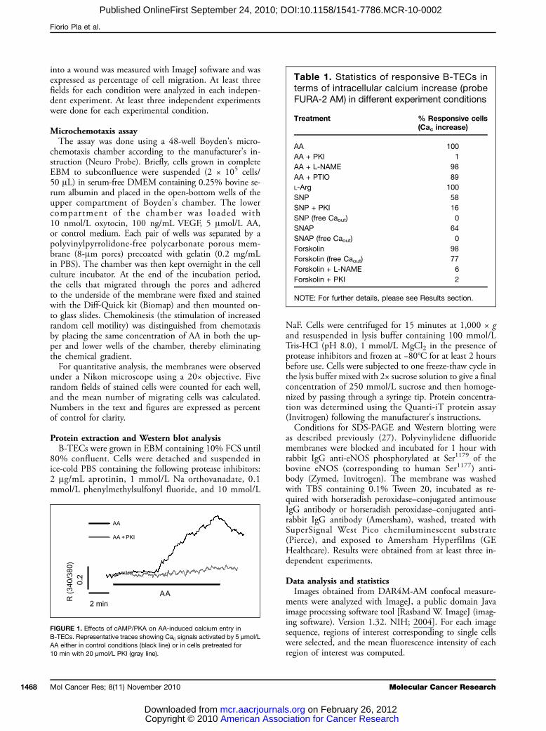
FIGURE 1. Effects of cAMP/PKA on AA-induced calcium entry inB-TECs. Representative traces showing Cac signals activated by 5 μmol/LAA either in control conditions (black line) or in cells pretreated for10 min with 20 μmol/L PKI (gray line).
cias.
Table 1. Statistics of responsive B-TECs interms of intracellular calcium increase (probeFURA-2 AM) in different experiment conditions
Treatment
Molecu
tion for Cancer Research on February 26, 2012org
% Responsive cells(Cac increase)
AA
100 AA + PKI 1 AA + L-NAME 98 AA + PTIO 89 L-Arg 100 SNP 58 SNP + PKI 16 SNP (free Caout) 0 SNAP 64 SNAP (free Caout) 0 Forskolin 98 Forskolin (free Caout) 77 Forskolin + L-NAME 6 Forskolin + PKI 2NOTE: For further details, please see Results section.
lar Cancer Research

PKA Regulates AA-Activated Calcium Entry
Published OnlineFirst September 24, 2010; DOI:10.1158/1541-7786.MCR-10-0002
For fura-2-AM ratiometric measurements, single cellswere selected for each image sequence with Metafluor soft-ware (Universal Imaging). Cytosolic free calcium concentra-tion (Cac) was expressed as a ratio of emitted fluorescence(λ = 510 nm) in correspondence to excitation wavelengthsof 340 and 380 nm.Igor software was used to further analyze both DAR4M-
AM and fura-2-AM measurements. In particular, we con-sidered the agonist-induced slope change as a quantitativecriterion to distinguish a response to an agonist from noise.One-way ANOVA was done to analyze the data sets.
Post hoc tests were used to determine statistically signifi-cant differences among the groups (Student-Newman-Keuls test). The level of significance was P < 0.05.Statistical analysis of wound healing assays was done
using the Wilcoxon-Mann-Whitney test; the level of signif-icance was set at P < 0.05.
www.aacrjournals.org
American Asso Copyright © 2010 mcr.aacrjournalDownloaded from
Results
PKA is required for AA-induced calcium entry in B-TECsPretreatment of B-TECs with the membrane-permeable
PKA inhibitory peptide myristoylated PKI14-22 (10 min-utes, 20 μmol/L) completely abolished AA-induced calci-um entry in 99% of the cells tested (n = 84; Fig. 1; Table1), suggesting a critical role of endogenous PKA in the ac-tivation of calcium signals by AA. The response to AA wasalso entirely prevented in 93% of the cells by pretreatmentwith staurosporine (10 minutes, 100 nmol/L; n = 79), aless specific blocker of serine/threonine protein kinases.
AA promotes B-TEC motility through PKAThe ability of AA to trigger B-TEC motility was tested
using two different techniques: Boyden chamber assay andwound healing assay. We used Boyden's chamber in which
FIGURE 2. AA promotes motility and chemotaxis in B-TECs. A, chemotactic and chemokinetic response of B-TECs to AA (5 μmol/L). The assay wasdone using a Boyden's chamber assay. VEGF (100 ng/mL) and oxytocin (OT; 10 nmol/L) were used as positive controls. Columns, mean of 9 to 23 replicatesfrom 2 to 4 experiments; bars, SEM. *, P < 0.05, compared with controls (ANOVA). CT, chemotaxis; CK, chemokinesis. B, time course (4-6 h) of arepresentative wound healing assay in different conditions: DMEM-0% FCS (CNTR); EBM-10% FCS (EBM); and DMEM-0% FCS + 5 μmol/L AA (AA).C, time course (4-6 h) of a representative wound healing assay in different conditions: DMEM-0% FCS (CNTR); DMEM-0% FCS + 1 μmol/L CAI(CAI); and DMEM-0% FCS + 5 μmol/L AA + 1 μmol/L CAI (AA + CAI). D, time course (4-6 h) of a representative wound healing assay in different conditions:DMEM-0% FCS (CNTR); DMEM-0% FCS + 20 μmol/L PKI (PKI); and DMEM-0% FCS + 5 μmol/L AA + 20 μmol/L PKI (AA + PKI). *, P < 0.05,compared with controls (Wilcoxon-Mann-Whitney test).
Mol Cancer Res; 8(11) November 2010 1469
ciation for Cancer Research on February 26, 2012s.org
Fiorio Pla et al.
1470
Published OnlineFirst September 24, 2010; DOI:10.1158/1541-7786.MCR-10-0002
chemoattraction or chemokinesis was assayed according tothe placement of AA (Fig. 2A). When 5 μmol/L AA waspresent both in the upper and lower chambers, chemokin-esis was observed, although not significant compared withcontrol. On the other hand, when AA was present only inthe lower chamber, a significant migratory effect was de-tected. As a positive control, we used 100 ng/mL VEGFor 10 nmol/L oxytocin (12).Moreover, we tested the ability of AA to increase B-TEC
wound healing in a monolayer. Stimulation of wounded B-TEC monolayer with AA (4-6 hours; 5 μmol/L) induced asignificant reduction of wound compared with the controlcondition (DMEM-0% FCS). As a positive control,wound healing was measured on B-TECs growing inEBM-10% FCS (Fig. 2B).Preincubation of B-TECs with the antiangiogenic com-
pound carboxyamidotriazole (CAI; 1 μmol/L), a well-known inhibitor of agonist-activated calcium channels,completely abolished AA-induced wound healing, clearlysuggesting that calcium signals are required for this effect(Fig. 2C).Having shown that AA-triggered Cac signals are PKA
sensitive, we tested the effect of PKI on AA-dependentwound healing. Preincubation with PKI (10 minutes,20 μmol/L) significantly reduced B-TEC motility specif-
Mol Cancer Res; 8(11) November 2010
American Asso Copyright © 2010 mcr.aacrjournalDownloaded from
ically promoted by AA, whereas no significant effect wasobserved in control conditions (Fig. 2D).
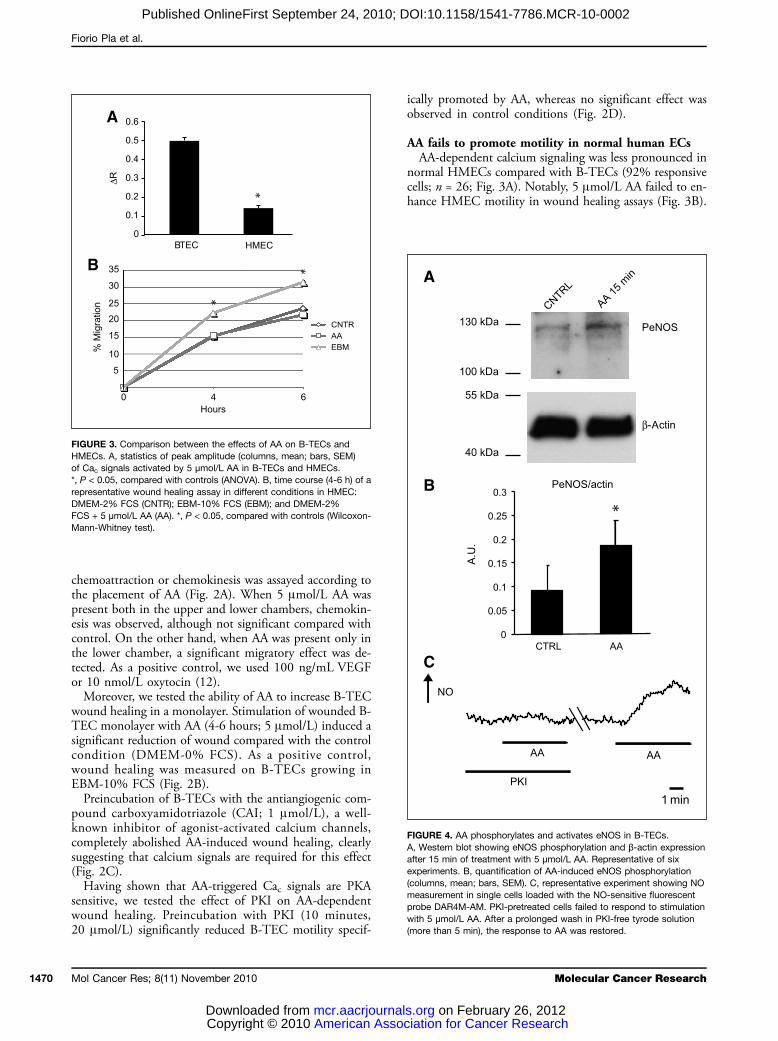
AA fails to promote motility in normal human ECsAA-dependent calcium signaling was less pronounced in
normal HMECs compared with B-TECs (92% responsivecells; n = 26; Fig. 3A). Notably, 5 μmol/L AA failed to en-hance HMEC motility in wound healing assays (Fig. 3B).
FIGURE 3. Comparison between the effects of AA on B-TECs andHMECs. A, statistics of peak amplitude (columns, mean; bars, SEM)of Cac signals activated by 5 μmol/L AA in B-TECs and HMECs.*, P < 0.05, compared with controls (ANOVA). B, time course (4-6 h) of arepresentative wound healing assay in different conditions in HMEC:DMEM-2% FCS (CNTR); EBM-10% FCS (EBM); and DMEM-2%FCS + 5 μmol/L AA (AA). *, P < 0.05, compared with controls (Wilcoxon-Mann-Whitney test).
FIGURE 4. AA phosphorylates and activates eNOS in B-TECs.A, Western blot showing eNOS phosphorylation and β-actin expressionafter 15 min of treatment with 5 μmol/L AA. Representative of sixexperiments. B, quantification of AA-induced eNOS phosphorylation(columns, mean; bars, SEM). C, representative experiment showing NOmeasurement in single cells loaded with the NO-sensitive fluorescentprobe DAR4M-AM. PKI-pretreated cells failed to respond to stimulationwith 5 μmol/L AA. After a prolonged wash in PKI-free tyrode solution(more than 5 min), the response to AA was restored.
Molecular Cancer Research
ciation for Cancer Research on February 26, 2012s.org
PKA Regulates AA-Activated Calcium Entry
Published OnlineFirst September 24, 2010; DOI:10.1158/1541-7786.MCR-10-0002
AA promotes eNOS phosphorylation and activationin B-TECsBecause it is well known that serine/threonine phosphor-
ylation is one of the regulatory mechanisms for eNOSactivity, we performed Western blot experiments to testthe ability of AA to promote eNOS phosphorylation inB-TECs. In particular, we used an antibody recognizing
www.aacrjournals.org
American Asso Copyright © 2010 mcr.aacrjournalDownloaded from
human eNOS on Ser1177, a target for PKA phosphoryla-tion (21). Treatment with AA (15 minutes, 5 μmol/L)induced a significant eNOS phosphorylation comparedwith control conditions (Fig. 4A and B).Even if eNOS phosphorylation is required for its ac-
tivity, it may not be sufficient to activate the enzyme.For this reason, we tested the ability of AA to increaseintracellular NO levels using the NO-sensitive fluores-cent dye DAR4M-AM. Acute stimulation with 5μmol/L AA increased intracellular NO in 79% of thecell tested (n = 68). Pretreatment with PKI (10 minutes,20 μmol/L) completely prevented the response to AA in70% of the cells (n = 104). The inhibitory effect wasreversible, as shown by the recovery of AA-inducedNO increase after intense and prolonged wash (morethan 5 minutes) in 82% of the cells (n = 45; Fig.4C; Table 2).
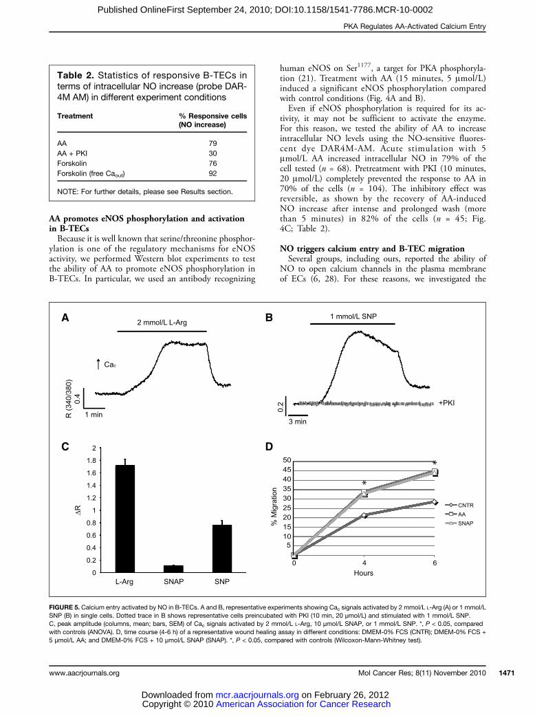
NO triggers calcium entry and B-TEC migrationSeveral groups, including ours, reported the ability of
NO to open calcium channels in the plasma membraneof ECs (6, 28). For these reasons, we investigated the
Table 2. Statistics of responsive B-TECs interms of intracellular NO increase (probe DAR-4M AM) in different experiment conditions
Treatment
% Responsive cells(NO increase)AA
79 AA + PKI 30 Forskolin 76 Forskolin (free Caout) 92NOTE: For further details, please see Results section.
FIGURE 5. Calcium entry activated by NO in B-TECs. A and B, representative experiments showing Cac signals activated by 2 mmol/L L-Arg (A) or 1 mmol/LSNP (B) in single cells. Dotted trace in B shows representative cells preincubated with PKI (10 min, 20 μmol/L) and stimulated with 1 mmol/L SNP.C, peak amplitude (columns, mean; bars, SEM) of Cac signals activated by 2 mmol/L L-Arg, 10 μmol/L SNAP, or 1 mmol/L SNP. *, P < 0.05, comparedwith controls (ANOVA). D, time course (4-6 h) of a representative wound healing assay in different conditions: DMEM-0% FCS (CNTR); DMEM-0% FCS +5 μmol/L AA; and DMEM-0% FCS + 10 μmol/L SNAP (SNAP). *, P < 0.05, compared with controls (Wilcoxon-Mann-Whitney test).
Mol Cancer Res; 8(11) November 2010 1471
ciation for Cancer Research on February 26, 2012s.org
Fiorio Pla et al.
1472
Published OnlineFirst September 24, 2010; DOI:10.1158/1541-7786.MCR-10-0002
effects of NO on Cac signaling in B-TECs. These wereassessed either indirectly by the use of L-Arg (eNOSsubstrate and activator, which enters the cell through aspecific membrane carrier) or directly with sodium nitro-prusside (SNP) and S-nitroso-N-acetylpenicillamine(SNAP), two NO donors. Acute application of 2mmol/L L-Arg induced a large calcium increase (100%responsive cells; n = 53; Fig. 5A and C; Table 1). Con-sistently, also stimulation with either 1 mmol/L SNP(58% responsive cells, n = 80; Fig. 5B and C; Table1) or 10 μmol/L SNAP (64% responsive cells, n =45; Fig. 5C; Table 1) evoked calcium signals, whichwere completely abolished in calcium-free external solu-tion (0% responsive cells for both the donors; n = 19;Table 1). Preincubation with PKI (10 minutes, 20μmol/L) completely abolished the response to SNP in84% of the cells (n = 38; Fig. 5B; Table 1). WhenB-TECs were treated with 10 μmol/L SNAP, their mo-tility was enhanced, as shown by wound healing assay(Fig. 5D).
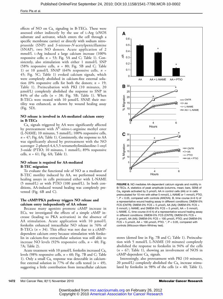
NO release is involved in AA-mediated calcium entryin B-TECsCac signals triggered by AA were significantly affected
by pretreatment with NG-nitro-L-arginine methyl ester(L-NAME; 10 minutes, 5 mmol/L; 100% responsive cells,n = 47; Fig. 6A; Table 1). Consistently, the response to AAwas significantly altered by pretreatment with the NOscavenger 2-phenyl-4,4,5,5-tetramethylimidazoline-1-oxyl3-oxide (PTIO; 10 minutes, 1 mmol/L; 89% responsivecells, n = 61; Fig. 6A; Table 1).
NO release is required for AA-mediatedB-TEC migrationTo evaluate the functional role of NO as a mediator of
B-TEC motility induced by AA, we performed woundhealing assays in cells pretreated either with L-NAME(5 mmol/L) or with PTIO (100 μmol/L). In both con-ditions, AA-induced wound healing was completely pre-vented (Fig. 6B and C).
The cAMP/PKA pathway triggers NO release andcalcium entry independently of AA releaseBecause many agonists promote cAMP increase in
ECs, we investigated the effects of a simple cAMP in-crease (leading to PKA activation) in the absence ofAA stimulation. Acute application with 10 μmol/Lforskolin enhanced intracellular NO release in 76% ofB-TECs (n = 34). This effect was not due to a cAMP-dependent calcium entry because stimulation with forsko-lin in calcium-free extracellular solution was still able toincrease NO levels (92% responsive cells, n = 40; Fig.7A; Table 2).Acute treatment with 10 μmol/L forskolin increased Cac
levels (98% responsive cells, n = 60; Fig. 7B and C; Table1). Only a small Cac response was detectable in calcium-free external solution in 77% of the cells tested (n = 49),suggesting a little contribution from intracellular calcium
Mol Cancer Res; 8(11) November 2010
American Asso Copyright © 2010 mcr.aacrjournalDownloaded from
stores (dotted line in Fig. 7B and C; Table 1). Preincuba-tion with 5 mmol/L L-NAME (10 minutes) completelyabolished the response to forskolin in 94% of the cells(n = 67; Table 1), showing an involvement of NOS incAMP-dependent Cac signals.Interestingly, also pretreatment with PKI (10 minutes,
20 μmol/L) completely abolished the Cac increase stimu-lated by forskolin in 98% of the cells (n = 40; Table 1),
FIGURE 6. NO mediates AA-dependent calcium signals and motility inB-TECs. A, statistics of peak amplitude (columns, mean; bars, SEM) ofCac signals activated by 5 μmol/L AA in control cells (AA) or in cellspreincubated for 10 min with either 5 mmol/L L-NAME or 1 mmol/L PTIO.*, P < 0.05, compared with controls (ANOVA). B, time course (4-6 h) ofa representative wound healing assay in different conditions: DMEM-0%FCS (CNTR); DMEM-0% FCS + 5 μmol/L AA (AA); DMEM-0% FCS +5 mmol/L L-NAME; and DMEM-0% FCS + 5 μmol/L AA + 5 mmol/LL-NAME. C, time course (4-6 h) of a representative wound healing assayin different conditions: DMEM-0% FCS (CNTR); DMEM-0% FCS +5 μmol/L AA (AA); DMEM-0% FCS + 100 μmol/L PTIO; and DMEM-0%FCS + 5 μmol/L AA + 100 μmol/L PTIO. *, P < 0.05, compared withcontrols (Wilcoxon-Mann-Whitney test).
Molecular Cancer Research
ciation for Cancer Research on February 26, 2012s.org

PKA Regulates AA-Activated Calcium Entry
Published OnlineFirst September 24, 2010; DOI:10.1158/1541-7786.MCR-10-0002
providing evidence for a key role of PKA activation ratherthan cAMP-gated channels.
Discussion
The main goal of this work is to investigate the regu-latory mechanisms of previously described AA-activated
www.aacrjournals.org
American Asso Copyright © 2010 mcr.aacrjournalDownloaded from
proangiogenic calcium entry in B-TECs. In B-TECs,low concentrations of AA activate a store-independentcalcium entry with similar properties previously observedin BAECs. This event is involved in tubulogenesis pro-gression and is downregulated in the later phases of tu-bule formation (5, 29). Here, we show that the cAMP/PKA pathway is involved in AA-induced Ca2+ entry. B-TEC pretreatment with PKA inhibitory peptide, PKI,completely abolished AA-induced calcium entry, showingthat PKA is necessary for Cac signals triggered by the fat-ty acid (Fig. 1).As indicated by wound healing and Boyden chamber as-
says, AA at low concentrations is motogenic and chemo-attractive for B-TECs (see Fig. 2A and B). Notably, thedramatic effect of the antiangiogenic compound CAI, anagonist-activated calcium entry inhibitor, reveals that AA-induced wound healing is dependent on calcium signals(Fig. 2C). These data are in agreement with the previouslydescribed calcium-dependent tubulogenic action of the fat-ty acid (5). AA-induced wound healing is also abolished byPKI preincubation, providing a functional link betweenthe cAMP/PKA pathways described above and the biolog-ical activities of the fatty acid (Fig. 2D).The observation that normal HMECs are less responsive
than B-TECs to stimulation with AA in terms of calciumincrease (Fig. 3A) and entirely unaffected by the fatty acidin cell motility (Fig. 3B) is very intriguing for its potentialbiomedical application and could help to develop a moreselective pharmacologic approach to tumor angiogenesis.The simplest explanation could be that calcium increaseactivated by AA in normal ECs is too low in extent to trig-ger the same functional effects as in B-TECs. Nevertheless,other factors responsible for this difference could be thespatiotemporal dynamics of calcium signals and the typeof channels involved (see also below). This point, far be-yond the aim of this work, deserves to be investigated inmore detail in the future.Because NOS are modulated by PKA-dependent phos-
phorylation and we previously reported the ability of AA torelease NO in BAECs (6), we performed single-cell fluori-metric measurements using the NO-sensitive probeDAR4M-AM and confirmed that AA leads to NO releasealso in B-TECs (Fig. 4C). Furthermore, Western blot ex-periments suggest that this is likely due, at least in part, toeNOS phosphorylation in a serine residue target for PKA-dependent phosphorylation (Fig. 4A and B), even if wecannot exclude the involvement of other NOS isoforms(iNOS and nNOS) that could play a role in tumor angio-genesis (13, 30). NOS phosphorylation is functionally rel-evant because pretreatment with PKI leads to a completeinhibition of AA-induced NO release (Fig. 4C). Themechanism responsible for AA-induced PKA recruitmentis unknown in these cells, but in rat aortic endothelium,cAMP increase is mediated by prostaglandin I2 throughits G-protein–coupled membrane receptor (31).Taken together, these data suggest that PKA could regu-
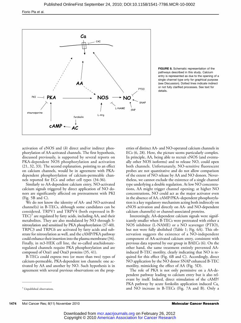
late AA-dependent Cac signals through at least two (possi-bly coexisting) pathways (Fig. 8): (a) phosphorylation and
FIGURE 7. cAMP/PKA–dependent Cac signals in B-TECs. A, arepresentative experiment of NO measurement in single cells loaded withDAR4M-AM and acutely stimulated with 10 μmol/L forskolin (FRK) incalcium-free external solution. B, a representative experiment showingCac increase induced by 10 μmol/L forskolin stimulation; dotted traceindicates a typical response to forskolin in calcium-free externalsolution. C, peak amplitude (columns, mean; bars, SEM) of Cac signalsactivated by acute stimulation with 10 μmol/L forskolin either incontrol conditions (FRK) or in calcium-free external solution (FRK + freeCaout). *, P < 0.05, compared with controls (ANOVA).
Mol Cancer Res; 8(11) November 2010 1473
ciation for Cancer Research on February 26, 2012s.org
Fiorio Pla et al.
1474
Published OnlineFirst September 24, 2010; DOI:10.1158/1541-7786.MCR-10-0002
activation of eNOS and (b) direct and/or indirect phos-phorylation of AA-activated channels. The first hypothesis,discussed previously, is supported by several reports onPKA-dependent NOS phosphorylation and activation(21, 32, 33). The second explanation, pointing to an effecton calcium channels, would be in agreement with PKA-dependent phosphorylation of calcium-permeable chan-nels reported for ECs and other cell types (34-36).Similarly to AA-dependent calcium entry, NO-activated
calcium signals triggered by direct application of NO do-nors are significantly affected on pretreatment with PKI(Fig. 5B and C).We do not know the identity of AA- and NO-activated
channel(s) in B-TECs, although some candidates can beconsidered. TRPV1 and TRPV4 (both expressed in B-TEC)5 are regulated by fatty acids, including AA, and theirmetabolites. They are also modulated by NO through S-nitrosylation and sensitized by PKA phosphorylation (37-40).TRPC3 and TRPC6 are activated by fatty acids and sub-strate for nitrosylation as well, and the cAMP/PKA pathwaycould enhance their insertion into the plasmamembrane (34).Finally, in m3-HEK cell line, the so-called arachidonate-regulated channels require PKA phosphorylation and arecomposed of Orai1 and Orai3 proteins (35, 41).B-TECs could express two (or more than two) types of
calcium-permeable, PKA-dependent ion channels: one ac-tivated by AA and another by NO. Such hypothesis is inagreement with several previous observations on the prop-
5 Unpublished observations.
Mol Cancer Res; 8(11) November 2010
American Asso Copyright © 2010 mcr.aacrjournalDownloaded from
erties of distinct AA- and NO-operated calcium channels inECs (6, 28). Here, the picture seems particularly complex.In principle, AA, being able to recruit eNOS (and eventu-ally other NOS isoforms) and to release NO, could openboth channels. Unfortunately, NO-sensitive fluorescentprobes are not quantitative and do not allow comparisonof the extent of NO release by AA and NO donors. Never-theless, we cannot exclude the existence of a single channeltype underlying a double regulation. At low NO concentra-tions, AA might trigger channel opening; at higher NOconcentrations, NO could act as the major activator evenin the absence of AA. cAMP/PKA–dependent phosphoryla-tion is a key regulatory mechanism acting both indirectly oneNOS activation and directly on AA- and NO-dependentcalcium channel(s) or channel-associated proteins.Interestingly, AA-dependent calcium signals were signif-
icantly smaller when B-TECs were pretreated with either aNOS inhibitor (L-NAME) or a NO scavenger (PTIO),but not were fully abolished (Table 1; Fig. 6A). This ob-servation suggests the existence of a NO-independentcomponent of AA-activated calcium entry, consistent withprevious data reported by our group in BAECs (6). On theother hand, the same treatment entirely prevented AA-induced B-TEC motility, clearly indicating that NO is re-quired for this effect (Fig. 6B and C). Accordingly, directNO application by the NO donor SNAP enhanced B-TECmotility, mimicking the effect of AA (Fig. 5D).The role of PKA is not only permissive on a AA-de-
pendent pathway leading to calcium entry but is also rel-evant by itself. Indeed, direct stimulation of the cAMP/PKA pathway by acute forskolin application induced Cacand NO increase in B-TECs (Fig. 7A and B). Only a
ciation for Cancer on Februarys.org
FIGURE 8. Schematic representation of thepathways described in this study. Calciumentry is represented as due to the opening of asingle channel type only for graphical purpose(see Discussion). Dotted lines indicate indirector not fully clarified processes. See text fordetails.
Molecular Cancer Research
Research 26, 2012

PKA Regulates AA-Activated Calcium Entry
Published OnlineFirst September 24, 2010; DOI:10.1158/1541-7786.MCR-10-0002
small Cac increase was detectable in calcium-free externalsolution, indicating a little contribution from intracellularcalcium stores (dotted line in Fig. 7B; Table 1). The abil-ity of PKI to abolish calcium increase triggered by forsko-lin indicates that the effect is entirely due to PKAactivation and not to a recruitment of cAMP-gated chan-nels (Table 1). From these results, we can argue that ago-nists that increase cAMP levels in ECs, even withoutaffecting AA release, are expected to facilitate the follow-ing calcium entry promoted by AA-releasing proangio-genic stimuli.Remarkably, calcium signals evoked by forskolin were al-
most completely inhibited by the NOS inhibitor L-NAME(Table 1). This evidence suggests that PKA-dependent cal-cium entry is entirely due to the NOS/NO pathway, dif-ferently from AA-induced calcium response, only in partsensitive to L-NAME (Fig. 6A; Table 1). This observationis in agreement with a recent report on proangiogenic ac-tivity triggered by forskolin through the PI3K, Akt, eNOSpathways in human umbilical vascular ECs (22). The abil-ity of L-NAME to exert a near-complete abolishment offorskolin-triggered calcium entry leads us also to confirmthat the PKA-dependent phosphorylation of AA-activatedcalcium channels probably acts in a permissive way, but itis not activatory by itself (i.e., in the absence of AA).
www.aacrjournals.org
American Asso Copyright © 2010 mcr.aacrjournalDownloaded from
In conclusion, here we show for the first time that thecAMP/PKA pathway regulates proangiogenic calcium en-try and is required for motility in breast cancer–derivedhuman ECs but not in ECs from normal tissues. Thepeculiar behavior of B-TECs reported here supports theidea that tumor-derived ECs are a more suitable model toevaluate the clinical potential of antiangiogenic and anti-tumor agents.
Disclosure of Potential Conflicts of Interest
No potential conflicts of interest were disclosed.
Acknowledgments
We thank Prof. Benedetta Bussolati (MBC, Torino, Italy) for kindly providingB-TECs.
Grant Support
TheUniversity of Torino, Regione Piemonte (Ricerca Scientifica Applicata Sanita).The costs of publication of this article were defrayed in part by the payment of
page charges. This article must therefore be hereby marked advertisement inaccordance with 18 U.S.C. Section 1734 solely to indicate this fact.
Received 01/04/2010; revised 08/16/2010; accepted 09/13/2010; publishedOnlineFirst 09/24/2010.
References
1. Munaron L. Intracellular calcium, endothelial cells and angiogenesis.Recent Patents Anticancer Drug Discov 2006;1:105–19.2. Munaron L, Tomatis C, Pla AF. The secret marriage between calcium
and tumor angiogenesis. Technol Cancer Res Treat 2008;7:335–9.3. Munaron L, Antoniotti S, Lovisolo D. Intracellular calcium signals and
control of cell proliferation: how many mechanisms? J Cell Mol Med2004;8:161–8.
4. Munaron L, Pla AF. Endothelial calcium machinery and angiogenesis:understanding physiology to interfere with pathology. Curr MedChem 2009;16:4691–703.
5. Fiorio Pla A, Grange C, Antoniotti S, et al. Arachidonic acid-inducedCa2+ entry is involved in early steps of tumor angiogenesis. MolCancer Res 2008;6:535–45.
6. Mottola A, Antoniotti S, Lovisolo D, Munaron L. Regulation of non-capacitative calcium entry by arachidonic acid and nitric oxide inendothelial cells. FASEB J 2005;19:2075–7.
7. Tomatis C, Fiorio Pla A, Munaron L. Cytosolic calcium microdomainsby arachidonic acid and nitric oxide in endothelial cells. Cell Calcium2007;41:261–9.
8. Carmeliet P. VEGF as a key mediator of angiogenesis in cancer.Oncology 2005;69 Suppl 3:4–10.
9. Bussolati B, Deambrosis I, Russo S, Deregibus MC, Camussi G.Altered angiogenesis and survival in human tumor-derived endothe-lial cells. FASEB J 2003;17:1159–61.
10. Grange C, Bussolati B, Bruno S, Fonsato V, Sapino A, Camussi G.Isolation and characterization of human breast tumor-derived endo-thelial cells. Oncol Rep 2006;15:381–6.
11. Baluk P, Morikawa S, Haskell A, Mancuso M, McDonald DM. Abnor-malities of basement membrane on blood vessels and endothelialsprouts in tumors. Am J Pathol 2003;163:1801–15.
12. Cassoni P, Marrocco T, Bussolati B, et al. Oxytocin induces prolifer-ation and migration in immortalized human dermal microvascular en-dothelial cells and human breast tumor-derived endothelial cells. MolCancer Res 2006;4:351–9.
13. Fukumura D, Kashiwagi S, Jain RK. The role of nitric oxide in tumourprogression. Nat Rev Cancer 2006;6:521–34.
14. Ziche M, Morbidelli L. Nitric oxide and angiogenesis. J Neurooncol2000;50:139–48.
15. Berkels R, Suerhoff S, Roesen R, Klaus W. Nitric oxide causes acGMP-independent intracellular calcium rise in porcine endothelialcells-a paradox? Microvasc Res 2000;59:38–44.
16. Cooke JP, Losordo DW. Nitric oxide and angiogenesis. Circulation2002;105:2133–5.
17. Dedkova EN, Blatter LA. Nitric oxide inhibits capacitative Ca2+ entryand enhances endoplasmic reticulum Ca2+ uptake in bovine vascularendothelial cells. J Physiol (Lond) 2002;539:77–91.
18. Dimmeler S, Dernbach E, Zeiher AM. Phosphorylation of the endo-thelial nitric oxide synthase at ser-1177 is required for VEGF-inducedendothelial cell migration. FEBS Lett 2000;477:258–62.
19. Pepine CJ. The impact of nitric oxide in cardiovascular medicine:untapped potential utility. Am J Med 2009;122:S10–5.
20. Yao X, Huang Y. From nitric oxide to endothelial cytosolic Ca2+: anegative feedback control. Trends Pharmacol Sci 2003;24:263–6.
21. Butt E, Bernhardt M, Smolenski A, et al. Endothelial nitric-oxidesynthase (type III) is activated and becomes calcium independentupon phosphorylation by cyclic nucleotide-dependent protein ki-nases. J Biol Chem 2000;275:5179–87.
22. Namkoong S, Kim CK, Cho YL, et al. Forskolin increases angio-genesis through the coordinated cross-talk of PKA-dependentVEGF expression and Epac-mediated PI3K/Akt/eNOS signaling.Cell Signal 2009;21:906–15.
23. Kwan HY, Huang Y, Yao X. TRP channels in endothelial function anddysfunction. Biochim Biophys Acta 2007;1772:907–14.
24. Kwan HY, Huang Y, Yao XQ, Leung FP. Role of cyclic nucleotides inthe control of cytosolic Ca levels in vascular endothelial cells. ClinExp Pharmacol Physiol 2009;36:857–66.
25. Yao X, Garland CJ. Recent developments in vascular endothelial celltransient receptor potential channels. Circ Res 2005;97:853–63.
26. Yao X, Kwan HY, Huang Y. Regulation of TRP channels by phos-phorylation. Neurosignals 2005;14:273–80.
27. Fiorio Pla A, Maric D, Brazer SC, et al. Canonical transient receptorpotential 1 plays a role in basic fibroblast growth factor (bFGF)/FGF
Mol Cancer Res; 8(11) November 2010 1475
ciation for Cancer Research on February 26, 2012s.org
Fiorio Pla et al.
1476
Published OnlineFirst September 24, 2010; DOI:10.1158/1541-7786.MCR-10-0002
receptor-1-induced Ca2+ entry and embryonic rat neural stem cellproliferation. J Neurosci 2005;25:2687–701.
28. Yao XQ, Huang Y. From nitric oxide to endothelial cytosolic Ca2+: anegative feedback control. Trends Pharmacol Sci 2003;24:263–6.
29. Fiorio Pla A, Munaron L. Calcium influx, arachidonic acid and controlof endothelial cell proliferation. Cell Calcium 2001;30:235–44.
30. Seidel B, Stanarius A, Wolf G. Differential expression of neuronal andendothelial nitric oxide synthase in blood vessels of the rat brain.Neurosci Lett 1997;239:109–12.
31. Ray C. The cellular mechanisms by which adenosine evokes releaseof nitric oxide from rat aortic endothelium. J Physiol (Lond) 2005;570:85–96.
32. Cokic VP, Beleslin-Cokic BB, Tomic M, Stojilkovic SS, Noguchi CT,Schechter AN. Hydroxyurea induces the eNOS-cGMP pathway inendothelial cells. Blood 2006;108:184–91.
33. Hashimoto A, Miyakoda G, Hirose Y, Mori T. Activation of endothelialnitric oxide synthase by cilostazol via a cAMP/protein kinase A- andphosphatidylinositol 3-kinase/Akt-dependent mechanism. Athero-sclerosis 2006;189:350–7.
34. Fleming I, Rueben A, Popp R, et al. Epoxyeicosatrienoic acidsregulate Trp channel dependent Ca2+ signaling and hyperpolariza-tion in endothelial cells. Arterioscler Thromb Vasc Biol 2007;27:2612–8.
Mol Cancer Res; 8(11) November 2010
American Asso Copyright © 2010 mcr.aacrjournalDownloaded from
35. Mignen O, Thompson JL, Shuttleworth TJ. Arachidonate-regulatedCa2+-selective (ARC) channel activity is modulated by phosphoryla-tion and involves an A-kinase anchoring protein. J Physiol 2005;567:787–98.
36. Zhang X, Li L, McNaughton PA. Proinflammatory mediators modu-late the heat-activated ion channel TRPV1 via the scaffolding proteinAKAP79/150. Neuron 2008;59:450–61.
37. Peng H, Lewandrowski U, Müller B, Sickmann A, Walz G, WegierskiT. Identification of a Protein Kinase C-dependent phosphorylationsite involved in sensitization of TRPV4 channel. Biochem BiophysRes Commun 2019;391:1721–5.
38. Yoshida T, Inoue R, Morii T, et al. Nitric oxide activates TRP channelsby cysteine S-nitrosylation. Nat Chem Biol 2006;2:596–607.
39. Jeske NA, Diogenes A, Ruparel NB, et al. A-kinase anchoring proteinmediates TRPV1 thermal hyperalgesia through PKA phosphorylationof TRPV1. Pain 2008;138:604–16.
40. Schnizler K, Shutov LP, Van Kanegan MJ, et al. Protein kinase A an-choring via AKAP150 is essential for TRPV1 modulation by forskolinand prostaglandin E2 in mouse sensory neurons. J Neurosci 2008;28:4904–17.
41. Mignen O, Thompson JL, Shuttleworth TJ. Both Orai1 and Orai3 areessential components of the arachidonate-regulated Ca2+-selective(ARC) channels. J Physiol (Lond) 2008;586:185–95.
Molecular Cancer Research
ciation for Cancer Research on February 26, 2012s.org