Multiple dual-mode centrifugal partition chromatography as an efficient method for the purification...
7
Journal of Chromatography B, 877 (2009) 2067–2073 Contents lists available at ScienceDirect Journal of Chromatography B journal homepage: www.elsevier.com/locate/chromb Multiple dual-mode centrifugal partition chromatography as an efficient method for the purification of a mycosporine from a crude methanolic extract of Lichina pygmaea Catherine Roullier, Marylène Chollet-Krugler ∗ , Aurélie Bernard, Joël Boustie EA 4090 “Substances lichéniques et photoprotection”, UFR Sciences pharmaceutiques et Biologiques, Université Européenne de Bretagne, Université de Rennes 1, 2 Av. du Pr. Léon Bernard, 35043 Rennes Cedex, France article info Article history: Received 13 February 2009 Accepted 19 May 2009 Available online 28 May 2009 Keywords: Centrifugal partition chromatography Multiple-dual-mode Lichina pygmaea Purification Isolation Mycosporine abstract Centrifugal partition chromatography method was applied to the separation and purification of a crude methanolic extract of a cyanobacterial lichen, Lichina pygmaea. A multiple dual-mode was used to separate two compounds of interest, namely mycosporine-serinol and a glutamic acid derivative. These compounds are described here for the first time in a lichen. Their structures were identified by UV, IR, ESI-MS, 1 H NMR, 13 C NMR, and 2D NMR. © 2009 Elsevier B.V. All rights reserved. 1. Introduction As many lichens resist to extreme conditions of UV-radiation and temperature, they are supposed to contain UV screening com- pounds to protect themselves against damages caused by solar radiation. Lichina pygmaea (Lightf.) C. Agardh, a black marine lichen, consists in a symbiosis between a fungus (Ascomycota phylum) and a cyanobacterium (Calothrix genus). It is known to contain primary metabolites, polyols and sugars as mannitol, mannosido-mannitol, glycerol, fructose, glucose, saccharose, galactose, glycogen, arabi- nose and possibly volemitol and xylose, but also polyosides and a galactomannan polysaccharide [1–4]. Besides lipophilic pigments given as xanthophyll carotenoids, phycocyanin, phycoerythrin and chlorophyll A [5], hydrophilic compound like mycosporine-glycine has recently been described in this lichen as an antioxidant [6]. Focusing on mycosporine compounds, we aimed at overcom- ing the usual lack of selectivity, adsorption and tailing problems often occurring with the purification processes of such com- pounds. Discovered in the 60s, mycosporines and MAAs (Mycosporine- like Amino Acids) are water soluble and low molecular weight molecules, that absorb ultraviolet radiation between 310 and ∗ Corresponding author. Tel.: +33 2 232 348 96; fax: +33 2 232 347 04. E-mail address: [email protected] (M. Chollet-Krugler). 365 nm with a molar extinction coefficient between 28 000 and 50 000 M −1 ·cm −1 [7,8]. They are accumulated by a wide range of prokaryotic and eukaryotic microorganisms, marine algae, corals and other marine life forms, as secondary metabolites. Their func- tion as solar radiation filters to prevent from damages caused by ultraviolet radiations is now well established, as their concentration is closely correlated to UV radiation fluctuations [9–11]. However, their implication in different mechanisms such as oxidative stress, salt stress, dessication, thermal stress, fungal reproduction or intra- cellular nitrogen reservoir has also been suggested [12]. Extraction of these compounds is usually operated with solvents as methanol and water in various proportions and their purifica- tion is often costly and time-consuming. Therefore, as commercially available standards are lacking, most of the characterization of mycosporines and MAAs has been based on the comparison of retention times and absorption spectra to data given in the liter- ature [13]. When isolated, purification of these structures proceeds with different techniques, the most employed ones involving purifi- cation on Dowex 50W (elution with water and dilute HCl) or preparative reversed-phase high-performance liquid chromatog- raphy (HPLC) with UV detection [7]. These techniques are often combined in order to obtain the compound with sufficient purity [14–16]. So we applied a liquid–liquid chromatography technique, namely centrifugal partition chromatography (CPC), for the sep- aration and the purification of mycosporines. CPC is a development 1570-0232/$ – see front matter © 2009 Elsevier B.V. All rights reserved. doi:10.1016/j.jchromb.2009.05.040
-
Upload
independent -
Category
Documents
-
view
0 -
download
0
Transcript of Multiple dual-mode centrifugal partition chromatography as an efficient method for the purification...

Mme
CEU
a
ARAA
KCMLPIM
1
aprcamgnggchFiop
lm
1d
Journal of Chromatography B, 877 (2009) 2067–2073
Contents lists available at ScienceDirect
Journal of Chromatography B
journa l homepage: www.e lsev ier .com/ locate /chromb
ultiple dual-mode centrifugal partition chromatography as an efficientethod for the purification of a mycosporine from a crude methanolic
xtract of Lichina pygmaea
atherine Roullier, Marylène Chollet-Krugler ∗, Aurélie Bernard, Joël BoustieA 4090 “Substances lichéniques et photoprotection”, UFR Sciences pharmaceutiques et Biologiques, Université Européenne de Bretagne,niversité de Rennes 1, 2 Av. du Pr. Léon Bernard, 35043 Rennes Cedex, France
r t i c l e i n f o
rticle history:eceived 13 February 2009ccepted 19 May 2009vailable online 28 May 2009
a b s t r a c t
Centrifugal partition chromatography method was applied to the separation and purification of a crudemethanolic extract of a cyanobacterial lichen, Lichina pygmaea. A multiple dual-mode was used to separatetwo compounds of interest, namely mycosporine-serinol and a glutamic acid derivative. These compoundsare described here for the first time in a lichen. Their structures were identified by UV, IR, ESI-MS, 1H
eywords:entrifugal partition chromatographyultiple-dual-mode
ichina pygmaeaurification
solation
NMR, 13C NMR, and 2D NMR.© 2009 Elsevier B.V. All rights reserved.
ycosporine
. Introduction
As many lichens resist to extreme conditions of UV-radiationnd temperature, they are supposed to contain UV screening com-ounds to protect themselves against damages caused by solaradiation. Lichina pygmaea (Lightf.) C. Agardh, a black marine lichen,onsists in a symbiosis between a fungus (Ascomycota phylum) andcyanobacterium (Calothrix genus). It is known to contain primaryetabolites, polyols and sugars as mannitol, mannosido-mannitol,
lycerol, fructose, glucose, saccharose, galactose, glycogen, arabi-ose and possibly volemitol and xylose, but also polyosides and aalactomannan polysaccharide [1–4]. Besides lipophilic pigmentsiven as xanthophyll carotenoids, phycocyanin, phycoerythrin andhlorophyll A [5], hydrophilic compound like mycosporine-glycineas recently been described in this lichen as an antioxidant [6].ocusing on mycosporine compounds, we aimed at overcom-ng the usual lack of selectivity, adsorption and tailing problemsften occurring with the purification processes of such com-
ounds.Discovered in the 60s, mycosporines and MAAs (Mycosporine-ike Amino Acids) are water soluble and low molecular weight
olecules, that absorb ultraviolet radiation between 310 and
∗ Corresponding author. Tel.: +33 2 232 348 96; fax: +33 2 232 347 04.E-mail address: [email protected] (M. Chollet-Krugler).
570-0232/$ – see front matter © 2009 Elsevier B.V. All rights reserved.oi:10.1016/j.jchromb.2009.05.040
365 nm with a molar extinction coefficient between 28 000 and50 000 M−1·cm−1 [7,8]. They are accumulated by a wide range ofprokaryotic and eukaryotic microorganisms, marine algae, coralsand other marine life forms, as secondary metabolites. Their func-tion as solar radiation filters to prevent from damages caused byultraviolet radiations is now well established, as their concentrationis closely correlated to UV radiation fluctuations [9–11]. However,their implication in different mechanisms such as oxidative stress,salt stress, dessication, thermal stress, fungal reproduction or intra-cellular nitrogen reservoir has also been suggested [12].
Extraction of these compounds is usually operated with solventsas methanol and water in various proportions and their purifica-tion is often costly and time-consuming. Therefore, as commerciallyavailable standards are lacking, most of the characterization ofmycosporines and MAAs has been based on the comparison ofretention times and absorption spectra to data given in the liter-ature [13]. When isolated, purification of these structures proceedswith different techniques, the most employed ones involving purifi-cation on Dowex 50W (elution with water and dilute HCl) orpreparative reversed-phase high-performance liquid chromatog-raphy (HPLC) with UV detection [7]. These techniques are often
combined in order to obtain the compound with sufficient purity[14–16].So we applied a liquid–liquid chromatography technique,namely centrifugal partition chromatography (CPC), for the sep-aration and the purification of mycosporines. CPC is a development

2068 C. Roullier et al. / J. Chromatogr. B 877 (2009) 2067–2073
F partitc
obosaa(dpptpavmbphwteraTa
2
2
rpn(C
ig. 1. Kromaton Technologies FCPC 50 mL apparatus (A), the rotor (B) and part of aell. The upper and lower cell walls consist of the interdisk Teflon gaskets.
f countercurrent chromatography (CCC), invented in the late 60sy Ito et al., based on the partition between two immiscible phasesf a solvent system [17,18]. CPC represents one of the few technicalolutions to the challenge of maintaining a phase stationary whilenother is pumped through it [19]. It involves partition cells radi-lly engraved in a disk connected to each other by capillary ductssee Fig. 1). The constant centrifugal force field resulting from theisk rotation causes decantation in each cell, retaining one of thehases against the flow of the other, thus allowing a continuousrocess. CPC can be used either in a normal mode with a polar sta-ionary phase, but also in an inverse mode with an apolar stationaryhase. This technique also allows fractionation to be carried out innormal-phase mode followed by a reversed-phase mode (or vice-ersa), by a switching valve between descending and ascendingodes, called the “dual” mode. CPC allows the use of a wide range of
iphasic systems, and provides important benefits for natural com-ound purification, such as no sample loss on solid support andigh recovery. In this paper, the selectivity problem encounteredith the selected biphasic system was avoided by an efficient use of
he multiple dual-mode (MDM) strategy as described by Delannayt al. [20]. It required iterative dual-mode steps, during the sameun, with only one sample injection, to isolate a mycosporine andnother compound from a crude methanolic extract of L. pygmaea.his work was partially presented as a poster communication [21]nd detailed herein.
. Experimental
.1. Reagents
Solvents such as acetic acid (AcOH), acetonitrile (ACN), chlo-
oform (CHCl3), ethyl acetate (AcOEt) and methanol (MeOH)urchased from Carlo Erba Reactifs (Val de Reuil, France), and-butanol (n-BuOH), dichloromethane (CH2Cl2) and diethyletherEt2O) purchased from VWR (Fontenay sous bois, France) used forPC and TLC analyses were of analytical grade.ion disk (C). Note the connecting ducts centered on the bottom and the top of each
Trifluoroacetic acid (TFA) was purchased from Sigma–Aldrich(Lyon, France) and triethylamine (TEA) from Acros Organics (Hal-luin, France). Acetonitrile used for HPLC analyses was of HPLC gradeand purchased from Carlo Erba Reactifs (Val de Reuil, France). Waterwas of ultrapure quality.
2.2. Preparation of crude extract
Lichen material was collected on the west coast of France, on therocky shore of Dinard (48◦38.09′N, 02◦08.15′W), in December 2007.It was sorted out, washed and dried under ambient atmosphere.After macroscopic and microscopic observations, comparing to areference sample of the “Des Abbayes” herbarium, it was identifiedas L. pygmaea (Lightf.) C. Agardh and a voucher specimen is kept inthe laboratory with the reference JB/07/98.
Extraction by three successive solvents was performed on 270 gof the dried lichen, namely ethyl acetate then methanol andmethanol 50% in water. Separation and analyses were conductedon the methanolic extract of L. pygmaea (meLp), as we character-ized the presence of the mycosporine in it by HPLC, using a diodearray detector and by comparison with a standard kindly providedby Pr Karsten (University of Rostock, Germany).
2.3. FCPC apparatus
The separations were performed on a FCPC® C 50 KromatonTechnologies apparatus (Angers, France) using a rotor made of 800cells for a 57 mL total volume (measured volume). This apparatuswas able to rotate from 800 to 2000 rpm, producing a centrifu-gal acceleration in the partition cell of 84 × g at 1000 rpm and335 × g at 2000 rpm. It was able to support up to 75 bars (1000 psi).
The solvents were pumped by a HPLC pump 422 from Kontroninstruments (Montigny le Bretonneux, France). The samples wereintroduced into the CPC column via a 6-port medium pressureinjection valve (Upchurch Scientific, CIL Cluzeau, Ste Foy la Grande,France) equipped with a 5 mL sample loop. Fractions of 2 mL were
atogr.
cNt
2
tomoStw
C. Roullier et al. / J. Chrom
ollected by a mini-collector MC30 (Köhler Technische Produkte,eulussheim, Germany). Experiments were conducted at room
emperature.
.4. Solvent system screening
Different solvent systems were prepared and evaluated usinghin layer chromatography (TLC). For each, 500 �L of both phasesf the solvent system were mixed, and a small amount of dried
eLp sample was dissolved in this two phases mixture. Then, 10 �Lf each phase was applicated on a TLC plate by an Automatic TLCampler 3 (ATS 3, CAMAG, Muttenz, Switzerland), which providedhe application of an exact volume with an exact width by band-ise spray-on. The plate was revealed in order to visualize polar
Fig. 2. Scheme representing the operating conditions for the Lichina pygmaea
B 877 (2009) 2067–2073 2069
compounds (the TLC system described below). Then, the solventmixture was chosen according to the most equilibrated repartitionof each compound of the meLp sample between each phase of thesolvent system.
2.5. CPC experimental conditions
The separation was performed with the system composed of n-butanol, acetic acid and water (4:1:5, v/v/v), in the isocratic mode.
The rotor was first filled with the lower phase of the solvent system,as the stationary phase (see Fig. 2). The apparatus was rotated at1800 rpm and the upper mobile phase of the solvent mixture wasthen pumped into the inlet of the column (rotor) at a flow rate of4 mL/min in the ascending mode. 500 mg of the meLp sample wasmethanolic extract purification using the “multiple-dual-mode” CPC.
2 atogr.
dlitpcw
mvpiptm4tt
2
b5t1wr0BtaPtFwp
2
(Cfsd
TN
P
34122536151241
125
070 C. Roullier et al. / J. Chrom
iluted in a mixture of 2 mL of the upper phase and 2 mL of theower phase. It was loaded in the 5 mL injection-loop, and injectedn the column in a “sandwich” mode, i.e. at the same time thanhe mobile phase. The back pressure was 25 bars. The stationaryhase retention at the end of the separation represented 52% of theolumn volume (57 mL). The content of the outgoing organic phaseas offline monitored by TLC analysis.
Elution first occurred in the ascending mode (normal-phaseode): the rotor was filled with the lower polar phase of the sol-
ent mixture, and the pumped mobile phase is the apolar upperhase. After collecting eight tubes each containing 2 mL, the switch-
ng valve is turned to the descending mode and the mobile phaseumped is the lower one this time. Then, after collecting again eightubes of 2 mL, we switched again to the ascending mode (upper
obile phase). The separation proceeded that way during the next4 tubes of 2 mL. Extrusion was performed after the 60th tube:he upper phase was pumped in the descending mode to eject theotality of the lower phase out of the rotor.
.6. HPLC analyses
All the fractions obtained with the CPC separation were analysedy HPLC. They were performed on a C18 column (Equisorb, ODS2,�m, 250 mm × 4.6 mm, CIL Cluzeau, Ste Foy la Grande). Each frac-
ion was diluted in acetonitrile/water (10:90) at concentration ofmg/mL and after passing through a 0.2-�m membrane filter, 20 �Las injected into the column, using a gradient elution with a flow
ate of 1 mL/min. An initial isocratic hold until 10 min with 90% of.1% acetic acid in water (solvent A) and 10% of acetonitrile (solvent) was followed by a gradient ranging from 10% to 100% of acetoni-rile between 10 and 90 min. The mobile phase composed of 100%cetonitrile is maintained for 10 min, in order to wash the column.eak detection was carried out online using a diode array detec-or (HPLC 540 DAD, Kontron instruments, Montigny le Bretonneux,rance) at 310 and 254 nm, and absorption spectra (210–400 nm)ere recorded each second directly on the HPLC-separatedeaks.
.7. TLC analyses
The compounds of each fraction were separated on a TLC plate
Merck, Darmstadt, Germany). The developing solvent used wasHCl3/MeOH/H2O (6:4:1, v/v/v). Detection of compounds was per-ormed under UV lamps at 365, 312 and 254 nm, but also bypraying them with sulfuric p-anisaldehyde (and heating) or 2,4-initrophenylhydrazine.
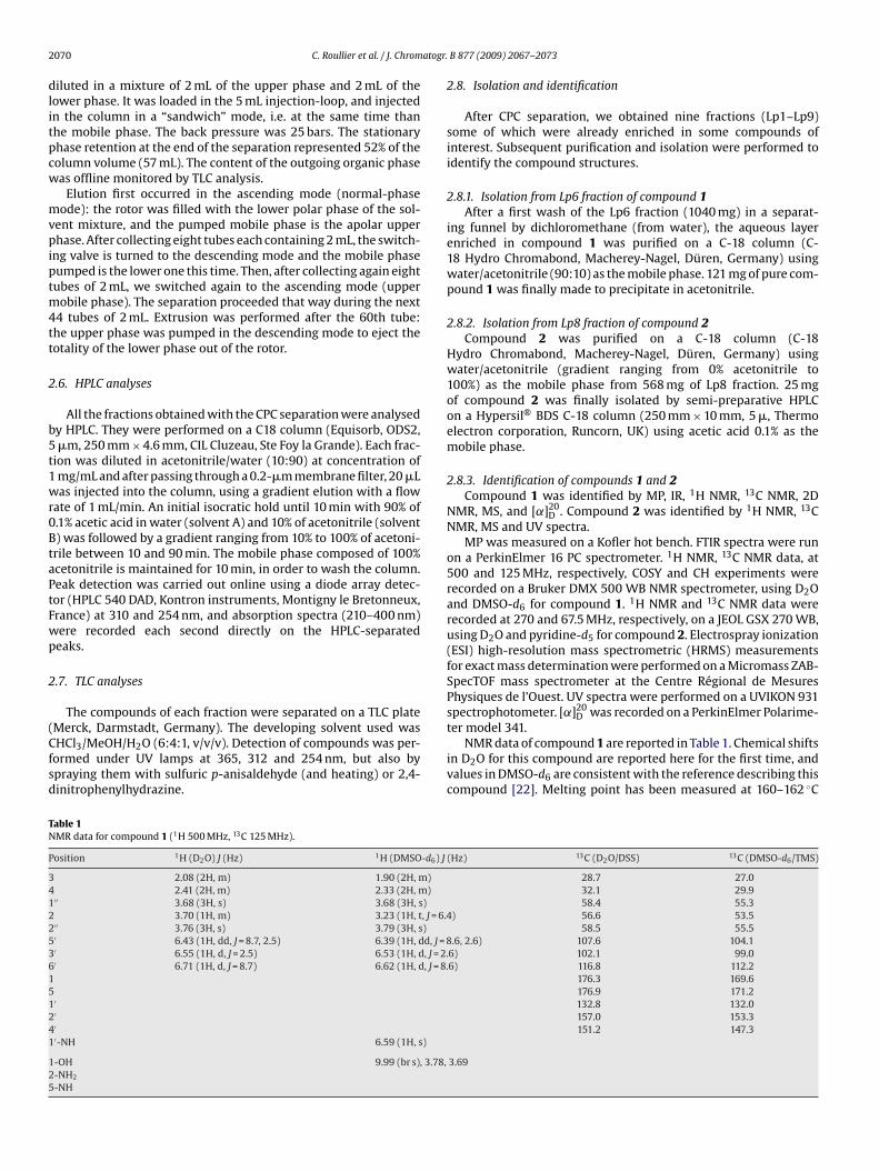
able 1MR data for compound 1 (1H 500 MHz, 13C 125 MHz).
osition 1H (D2O) J (Hz) 1H (DMSO-d6) J
2.08 (2H, m) 1.90 (2H, m)2.41 (2H, m) 2.33 (2H, m)
′′ 3.68 (3H, s) 3.68 (3H, s)3.70 (1H, m) 3.23 (1H, t, J = 6.4
′′ 3.76 (3H, s) 3.79 (3H, s)′ 6.43 (1H, dd, J = 8.7, 2.5) 6.39 (1H, dd, J = 8′ 6.55 (1H, d, J = 2.5) 6.53 (1H, d, J = 2.′ 6.71 (1H, d, J = 8.7) 6.62 (1H, d, J = 8.
′′′′-NH 6.59 (1H, s)
-OH 9.99 (br s), 3.78,-NH2
-NH
B 877 (2009) 2067–2073
2.8. Isolation and identification
After CPC separation, we obtained nine fractions (Lp1–Lp9)some of which were already enriched in some compounds ofinterest. Subsequent purification and isolation were performed toidentify the compound structures.
2.8.1. Isolation from Lp6 fraction of compound 1After a first wash of the Lp6 fraction (1040 mg) in a separat-
ing funnel by dichloromethane (from water), the aqueous layerenriched in compound 1 was purified on a C-18 column (C-18 Hydro Chromabond, Macherey-Nagel, Düren, Germany) usingwater/acetonitrile (90:10) as the mobile phase. 121 mg of pure com-pound 1 was finally made to precipitate in acetonitrile.
2.8.2. Isolation from Lp8 fraction of compound 2Compound 2 was purified on a C-18 column (C-18
Hydro Chromabond, Macherey-Nagel, Düren, Germany) usingwater/acetonitrile (gradient ranging from 0% acetonitrile to100%) as the mobile phase from 568 mg of Lp8 fraction. 25 mgof compound 2 was finally isolated by semi-preparative HPLCon a Hypersil® BDS C-18 column (250 mm × 10 mm, 5 �, Thermoelectron corporation, Runcorn, UK) using acetic acid 0.1% as themobile phase.
2.8.3. Identification of compounds 1 and 2Compound 1 was identified by MP, IR, 1H NMR, 13C NMR, 2D
NMR, MS, and [˛]20D . Compound 2 was identified by 1H NMR, 13C
NMR, MS and UV spectra.MP was measured on a Kofler hot bench. FTIR spectra were run
on a PerkinElmer 16 PC spectrometer. 1H NMR, 13C NMR data, at500 and 125 MHz, respectively, COSY and CH experiments wererecorded on a Bruker DMX 500 WB NMR spectrometer, using D2Oand DMSO-d6 for compound 1. 1H NMR and 13C NMR data wererecorded at 270 and 67.5 MHz, respectively, on a JEOL GSX 270 WB,using D2O and pyridine-d5 for compound 2. Electrospray ionization(ESI) high-resolution mass spectrometric (HRMS) measurementsfor exact mass determination were performed on a Micromass ZAB-SpecTOF mass spectrometer at the Centre Régional de MesuresPhysiques de l’Ouest. UV spectra were performed on a UVIKON 931spectrophotometer. [˛]20
D was recorded on a PerkinElmer Polarime-
ter model 341.NMR data of compound 1 are reported in Table 1. Chemical shiftsin D2O for this compound are reported here for the first time, andvalues in DMSO-d6 are consistent with the reference describing thiscompound [22]. Melting point has been measured at 160–162 ◦C
(Hz) 13C (D2O/DSS) 13C (DMSO-d6/TMS)
28.7 27.032.1 29.958.4 55.3
) 56.6 53.558.5 55.5
.6, 2.6) 107.6 104.16) 102.1 99.06) 116.8 112.2
176.3 169.6176.9 171.2132.8 132.0157.0 153.3151.2 147.3
3.69

C. Roullier et al. / J. Chromatogr. B 877 (2009) 2067–2073 2071
Table 2Stationary phase retention on FCPC 50 apparatus.
Solvent = BuOH/AcOH/H2O (4:1:5)
Rotor speed (rpm) 1800 2000 1800 2000 1800 2000FAD
aR
diHaapicb
3
3
cTteps(BC3
3
3
mr
Table 3HPLC analyses of the Lichina pygmaea (Lp) fractions obtained from FCPC treatment.
Fraction Rt for 1 (254 nm) Rt for 2 (310 nm) Integration (mAbs min)
Lp1Lp2 2.85 min 169.05Lp3Lp4Lp5Lp6 14.55 min 25.34Lp7 2.65 min 16.89
eluted during 26 min. In a second experiment, the ascending elution
low rate (mL/min) 6 6 4 4 2 2scending mode (%R) 49 51 52 49 44 51escending mode (%R) 46 44 47 42 51 46
nd other physicochemical properties are in accordance with theef. [22].
NMR data of compound 2 were compared to NMR valuesescribed for the expected mycosporine-glycine [16,23]. However
n D2O, the characteristic singlet corresponding to the two protons-9 [16,23] was not observed, but a five-proton signal between 3.6nd 3.8 ppm. UV spectrum gave a maximal absorption wavelengtht 309 nm, like mycosporine-glycine, but ESI-MS spectrum gave aositive ion [M+H]+ at m/z 262. High-resolution mass spectrum
ndicated the molecular formula C11H19NO6, and pyridine-d5 NMRonfirmed that compound 2 was mycosporine-serinol, as describedy Favre-Bonvin et al. [24].
. Results and discussion
.1. Solvent system selection
TLC plates were informative for the repartition of water-solubleompounds between the two phases of each solvent system.he more is the number of compounds in common betweenhe upper and the lower phase, the better is the partition. Wessentially focused on two plots, one corresponding to a com-ound absorbing at 254 nm, and giving a positive reaction withulfuric p-anisaldehyde, and the other one absorbing at 312 nmlike the mycosporine previously described in L. pygmaea). Thus,uOH/AcOH/H2O (4:1:5), was selected as the solvent system forPC separation. It can be noticed that this system is acidic with pH–4 and pH 4–5 for lower and upper layers, respectively.
.2. Choice of operating conditions
.2.1. Determination of suitable rotor speed and flow rateDifferent procedures with the CPC apparatus were experi-
ented in order to determine the conditions that provided the bestetention percentage. In a first time, working in an ascending mode,
Fig. 3. Compounds 1 and 2 retention times acc
Lp8 2.87 min 352.76Lp9 2.74 min 108.37
Rt: retention time.
the upper-layer was chosen as the mobile phase, and two rotorspeeds (1800 and 2000 rpm) along with three different flow rates(2, 4 and 6 mL/min) were tested. In a second time, the same proce-dure was conducted in the descending mode, with the lower-layeras the mobile phase. Retention percentage was calculated with thefollowing formula:
%R = Vr − VeVr
× 100
%R: retention percentage; Vr: rotor volume and Ve: ejected station-ary phase volume.
The results are reported in Table 2. The best retention value wasobtained in the ascending mode with a rotor speed of 1800 rpmand a flow rate of 4 mL/min. It is worth to notice that the same con-ditions in the descending mode do not provide the best retentionvalue but a suitable one for a multiple-dual-mode. A slight bleed-ing phenomenon, namely a little loss of the stationary phase, couldoccur but it was found very weak.
3.2.2. Retention time (Rt) determinationThe CPC eluted solvents were collected each 30 s (2 mL/tube)
and checked with TLC. Retention times (±30 s) of each compoundcould then be estimated. A first CPC experiment in the “ascending”mode failed, as the two polar compounds we wanted to isolate wereco-eluted between 4 and 12 min (see Fig. 3). However, compound1 was observed to be eluted during 8 min while compound 2 was
mode was turned to descending mode just before the anticipated4 min elution time. By this way, compound 2 appeared soon afterthe switch at Rt 5.5 min and compound 1 at Rt 8 min. So a firstisolation of compound 2 was achieved with only one dual-mode.
ording to FCPC experimental conditions.
2072 C. Roullier et al. / J. Chromatogr. B 877 (2009) 2067–2073
ea, aga
3
tpcmtmi1mtto
3
2ccpptpsFatsr
c2nvpcl(hmttMs
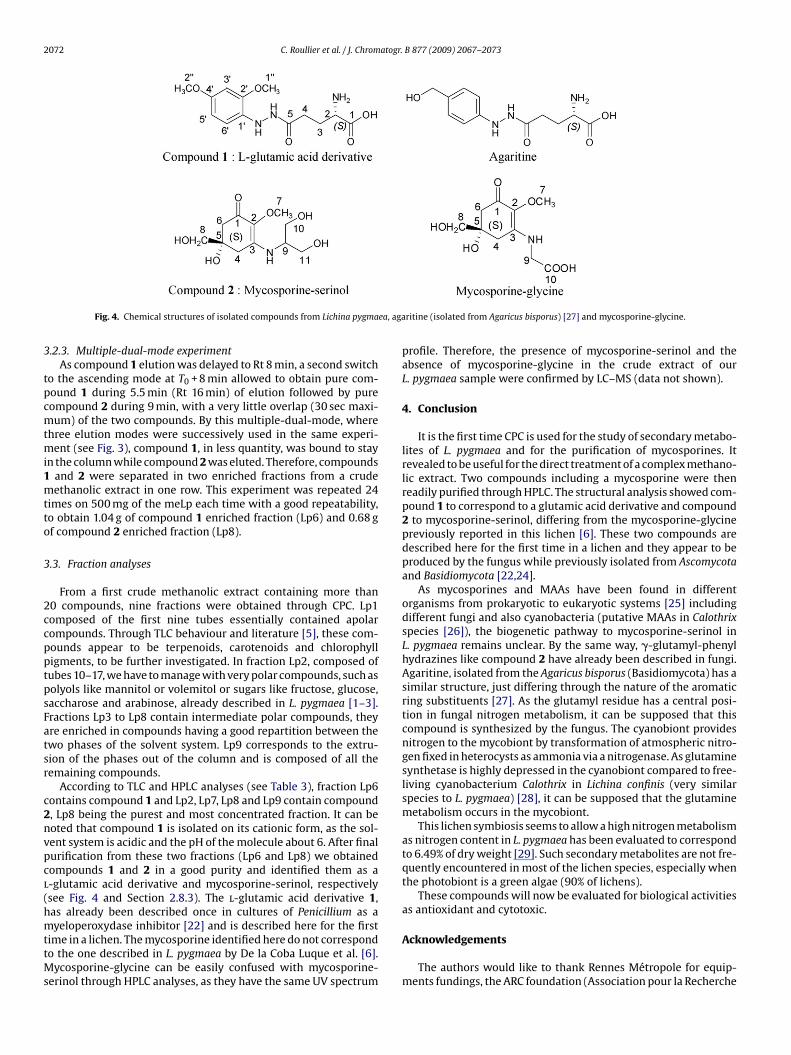
Fig. 4. Chemical structures of isolated compounds from Lichina pygma
.2.3. Multiple-dual-mode experimentAs compound 1 elution was delayed to Rt 8 min, a second switch
o the ascending mode at T0 + 8 min allowed to obtain pure com-ound 1 during 5.5 min (Rt 16 min) of elution followed by pureompound 2 during 9 min, with a very little overlap (30 sec maxi-um) of the two compounds. By this multiple-dual-mode, where
hree elution modes were successively used in the same experi-ent (see Fig. 3), compound 1, in less quantity, was bound to stay
n the column while compound 2 was eluted. Therefore, compoundsand 2 were separated in two enriched fractions from a crudeethanolic extract in one row. This experiment was repeated 24
imes on 500 mg of the meLp each time with a good repeatability,o obtain 1.04 g of compound 1 enriched fraction (Lp6) and 0.68 gf compound 2 enriched fraction (Lp8).
.3. Fraction analyses
From a first crude methanolic extract containing more than0 compounds, nine fractions were obtained through CPC. Lp1omposed of the first nine tubes essentially contained apolarompounds. Through TLC behaviour and literature [5], these com-ounds appear to be terpenoids, carotenoids and chlorophylligments, to be further investigated. In fraction Lp2, composed ofubes 10–17, we have to manage with very polar compounds, such asolyols like mannitol or volemitol or sugars like fructose, glucose,accharose and arabinose, already described in L. pygmaea [1–3].ractions Lp3 to Lp8 contain intermediate polar compounds, theyre enriched in compounds having a good repartition between thewo phases of the solvent system. Lp9 corresponds to the extru-ion of the phases out of the column and is composed of all theemaining compounds.
According to TLC and HPLC analyses (see Table 3), fraction Lp6ontains compound 1 and Lp2, Lp7, Lp8 and Lp9 contain compound, Lp8 being the purest and most concentrated fraction. It can beoted that compound 1 is isolated on its cationic form, as the sol-ent system is acidic and the pH of the molecule about 6. After finalurification from these two fractions (Lp6 and Lp8) we obtainedompounds 1 and 2 in a good purity and identified them as a-glutamic acid derivative and mycosporine-serinol, respectivelysee Fig. 4 and Section 2.8.3). The l-glutamic acid derivative 1,as already been described once in cultures of Penicillium as a
yeloperoxydase inhibitor [22] and is described here for the firstime in a lichen. The mycosporine identified here do not correspondo the one described in L. pygmaea by De la Coba Luque et al. [6].
ycosporine-glycine can be easily confused with mycosporine-erinol through HPLC analyses, as they have the same UV spectrum
ritine (isolated from Agaricus bisporus) [27] and mycosporine-glycine.
profile. Therefore, the presence of mycosporine-serinol and theabsence of mycosporine-glycine in the crude extract of ourL. pygmaea sample were confirmed by LC–MS (data not shown).
4. Conclusion
It is the first time CPC is used for the study of secondary metabo-lites of L. pygmaea and for the purification of mycosporines. Itrevealed to be useful for the direct treatment of a complex methano-lic extract. Two compounds including a mycosporine were thenreadily purified through HPLC. The structural analysis showed com-pound 1 to correspond to a glutamic acid derivative and compound2 to mycosporine-serinol, differing from the mycosporine-glycinepreviously reported in this lichen [6]. These two compounds aredescribed here for the first time in a lichen and they appear to beproduced by the fungus while previously isolated from Ascomycotaand Basidiomycota [22,24].
As mycosporines and MAAs have been found in differentorganisms from prokaryotic to eukaryotic systems [25] includingdifferent fungi and also cyanobacteria (putative MAAs in Calothrixspecies [26]), the biogenetic pathway to mycosporine-serinol inL. pygmaea remains unclear. By the same way, �-glutamyl-phenylhydrazines like compound 2 have already been described in fungi.Agaritine, isolated from the Agaricus bisporus (Basidiomycota) has asimilar structure, just differing through the nature of the aromaticring substituents [27]. As the glutamyl residue has a central posi-tion in fungal nitrogen metabolism, it can be supposed that thiscompound is synthesized by the fungus. The cyanobiont providesnitrogen to the mycobiont by transformation of atmospheric nitro-gen fixed in heterocysts as ammonia via a nitrogenase. As glutaminesynthetase is highly depressed in the cyanobiont compared to free-living cyanobacterium Calothrix in Lichina confinis (very similarspecies to L. pygmaea) [28], it can be supposed that the glutaminemetabolism occurs in the mycobiont.
This lichen symbiosis seems to allow a high nitrogen metabolismas nitrogen content in L. pygmaea has been evaluated to correspondto 6.49% of dry weight [29]. Such secondary metabolites are not fre-quently encountered in most of the lichen species, especially whenthe photobiont is a green algae (90% of lichens).
These compounds will now be evaluated for biological activitiesas antioxidant and cytotoxic.
Acknowledgements
The authors would like to thank Rennes Métropole for equip-ments fundings, the ARC foundation (Association pour la Recherche

atogr.
cAPmR
A
t
R
[
[[
[[[
[
[
[
[
[
[[[25] R.P. Sinha, S.P. Singh, D.P. Hader, J. Photochem. Photobiol. B 89 (2007) 29.
C. Roullier et al. / J. Chrom
ontre le Cancer) for a PhD grant to Catherine Roullier, Dr. Kristinarticus for the lichen determination, Sourisak Sinbandhit andhilippe Jehan (CRMPO) for valuable comments in structure deter-ination and René Havouis (Prof. Moulinoux lab., Fac. Medicine,
ennes) for HPLC–MS experiments.
ppendix A. Supplementary data
Supplementary data associated with this article can be found, inhe online version, at doi:10.1016/j.jchromb.2009.05.040.
eferences
[1] G. Pueyo, Rev. Bryol. Lichénol. 32 (1963) 279.[2] G. Pueyo, Bryologist 66 (1963) 74.[3] G. De Lestang Laisné, Rev. Bryol. Lichénol. 34 (1966) 346.[4] A. Prieto, J.A. Leal, M. Bernabé, D.L. Hawksworth, Mycol. Res. 112 (2008) 381.[5] M. Quillet, Bull. Lab. Maritime Dinard. 47 (1962) 68.[6] F. De La Coba Luque, J. Aguilera Arjona, F. Lopez Figueroa. International Patent
WO2007026036. 08.03.2007 (Universidad De Malaga, Spain).
[7] W.M. Bandaranayake, Nat. Prod. Rep. 15 (1998) 159.[8] W.C. Dunlap, J.M. Shick, J. Phycol. 34 (1998) 418.[9] F. Garcia-Pichel, C.E. Wingard, R.W. Castenholz, Appl. Environ. Microbiol. 59(1993) 170.10] A. Torres, M. Hochberg, I. Pergament, R. Smoum, V. Niddam, V.M. Dembitsky,
M. Temina, I. Dor, O. Lev, M. Srebnik, C.D. Enk, Eur. J. Biochem. 271 (2004) 780.
[[[[
B 877 (2009) 2067–2073 2073
[11] F. Garcia-Pichel, R.W. Castenholz, Appl. Environ. Microbiol. 59 (1993) 163.12] A. Oren, N. Gunde-Cimerman, FEMS Microbiol. Lett. 269 (2007) 1.13] T. Rezanka, M. Temina, A.G. Tolstikov, V.M. Dembitsky, Folia Microbiol. (Prague,
Czech Repub.) 49 (2004) 339.14] H.J. Suh, H.W. Lee, J. Jung, Photochem. Photobiol. 78 (2003) 109.15] H. Young, V.J. Patterson, Phytochemistry 21 (1982) 1075.16] W.M. Bandaranayake, J.E. Bemis, D.J. Bourne, Comp. Biochem. Physiol. C 115
(1996) 281.[17] Y. Ito, R.L. Bowman, Science 167 (1970) 281.18] A. Berthod (Ed.), Countercurrent Chromatography—The Support-Free Liquid
Stationary Phase (Wilson & Wilson’s Comprehensive Analytical Chemistry), vol.38, Elsevier, Amsterdam, 2002.
19] A.P. Foucault, Centrifugal Partition Chromatography, in: Chromatographic Sci-ence Series, vol. 68, Marcel Dekker, New York, 1995.
20] E. Delannay, A. Toribio, L. Boudesocque, J.M. Nuzillard, M. Zeches-Hanrot,E. Dardennes, G. Le Dour, J. Sapi, J.H. Renault, J. Chromatogr. A 1127(2006) 45.
21] C. Roullier, M. Chollet-Krugler, A. Bernard, J. Boustie, Planta Med. 74 (2008)PC81.
22] T. Tanimoto, I. Tanaka, M. Nakajima. International Patent JP2001131133.05.15.2001 (Sankyo Co. Ltd., Japan).
23] S. Ito, Y. Hirata, Tetrahedron Lett. 28 (1977) 2429.24] J. Favre-Bonvin, N. Arpin, C. Brevard, Can. J. Chem. 54 (1976) 1105.
26] S. Brenowitz, R.W. Castenholz, FEMS Microbiol. Ecol. 24 (1997) 343.27] B. Levenberg, J. Biol. Chem. 239 (1964) 2267.28] S. Janson, A.N. Rai, B. Bergman, New Phytol. 124 (1993) 149.29] K.A. Kershaw, Physiological Ecology of Lichens, Cambridge University Press,
Cambridge, 1985.